Note: Descriptions are shown in the official language in which they were submitted.
CA 02814213 2014-01-31
- 1 ¨
HANDHELD REFLECTOMETER FOR MEASURING MACULAR PIGMENT
100011
FIELD OF INVENTION
100021 The present
invention relates to a handheld macular pigment reflectometry
instrument that measures characteristics of the patient's eye, such as macular
pigment, with a
high degree of accuracy and without dilating the patient's pupil. The
invention also relates to
a way to align an instrument to a point or region within a patient's eye that
allows for a rapid,
intuitive and sequential alignment procedure and for rapid data gathering once
alignment is
achieved.
BACKGROUND OF THE INVENTION
[00031 The Retina and Retinal Diseases: The retina is the layer of nerve cells
at the back
of the eye, which convert light into nerve signals that are sent to the brain.
In humans, and in
other primates (but not in most other mammals, or other types of animals), the
retina has a
small yellowish area in the center of the field of vision. That yellowish area
is called the
"macula." It provides fine resolution vision in the center of the visual field
and is essential to
good vision. People who suffer from macular degeneration often lose the
ability to read,
recognize faces, drive, or walk safely on unfamiliar routes.
100041 The surrounding portions of the macula can only provide coarse
resolution. This
physiological feature limits and controls the number of nerve signals that the
brain must
rapidly process, to form coherent rapid-response vision, and it also helps
limit and control the
huge number of rod and cone receptors that the eye must continually regenerate
and recycle,
every day. Many people do not realize the retina can provide only coarse
resolution, outside
of a limited central area, because the eyes and the brain have developed an
extraordinary
ability to synthesize coherent vision from a combination of fine and coarse
resolution.
During that type of vision synthesis, the eye muscles cause the eyes to flit
back and forth over
a larger field of vision, pausing at each location for just an instant while
the eye quickly
"grabs" a fine-resolution image of a limited area. This process occurs so
rapidly that a person
does not notice it happening, and does not pay attention to how a complete
visual image and
impression is being assembled and updated from combinations of fine and coarse
resolution
CA 02814213 2013-04-09
WO 2012/051449 PCT/US2011/056205
- 2 -
images.
[0005] There is also a peculiar anatomic structure in the retinas of humans,
which points out
the difference between fine resolution (provided by the macula) and coarse
resolution
(provided by the remainder of the retina). In humans, the blood vessels that
serve the retina
actually sit in front of the retina, where they can block and interfere with
incoming light,
before the light reaches the retina. This is counter-intuitive, and one should
wonder why the
retina evolved with a physical handicap that literally gets in the way of
good, clear vision.
The answer is, in those parts of the retina, only coarse vision is being
created, and blood
vessels positioned in front of the retina do not interfere with that type of
coarse vision. By
contrast, in the macular region in the center of the retina, the blood vessels
in front of the
retina are lacking and supply is only from blood vessels present anywhere
behind the layer of
neurons with rod and cone receptors. This is consistent with the macula
providing fine
resolution vision, which would be blocked and hindered if the blood vessels
were located in
front of the neurons, in ways that would intercept and blocking portions of
the incoming
light.
[0006] "Retinal degeneration" is a descriptive term, which refers to and
includes an entire
class of eye diseases and disorders. It includes any progressive disorder or
disease that
causes the macula to gradually degenerate, to a point that substantially
impairs or damages
eyesight and vision. Several major categories of retinal degeneration are
known. These
include: (i) age-related macular degeneration, which gradually appears among
some people
over the age of about 65; (ii) diabetic retinopathy, in which problems with
sugar and energy
metabolism damage the entire retina, including the macula; (iii) eye diseases
that affect the
macula due to gene and/or enzyme defects, such as Stargardt's disease, Best's
disease,
Batten's disease, Sjogren-Larsson syndrome, and various other eye disorders
that lead to
gradual degeneration of the macula (and possibly other parts of the retina)
over a span of
time. This is not an exclusive list, and other subclasses and categories also
are known. For
example, age-related macular degeneration is subdivided into wet and dry
forms, depending
on whether abnormal and disruptive blood vessel growth is occurring in the
structural layers
behind the retina.
[0007] The causes and effects of macular degeneration, and efforts to prevent
or treat it, are
described in numerous books (e.g., "Macular Degeneration," by Robert D'Amato
et al (2000)
and "Age-Related Macular Degeneration," by Jennifer Lim (2002)), articles
("Age-Related
CA 02814213 2014-01-31
- 3 -
Macular Degeneration" by Berger et al (1999)) and patents, such as U.S. Patent
No. Re.
38,009, which is assigned to ZeaVision LLC.
[0008] In recent years, awareness has grown, among some researchers but not
among the
general public, of the roles that macular pigment plays, in the health and
longevity of the
macula. Therefore, the two carotenoid pigments that create and provide the
macular pigment
are discussed below.
[0009] The Macular Pigments: Zeaxanthin and Lutein: The macula has a yellowish
color
because it contains unusually high concentrations of two specific pigments,
called zeaxanthin
and lutein. Both are carotenoids, similar to beta-carotene but with hydroxyl
groups coupled
to their end rings (the presence of one or more oxygen atoms causes a
carotenoid to be
categorized as a "xanthophyll", so zeaxanthin and lutein arc sometimes
referred to as
xanthophylls). Both of those two carotenoids are known to be protective and
beneficial, in
human retinas, by mechanisms that include: (1) absorption of destructive
ultraviolet photons;
and (2) quenching of destructive radicals. Both of those mechanisms, and other
potential
protective mechanisms, are discussed below.
[0010] In addition to their involvement in the macula and macular
degeneration, zeaxanthin
and lutein also are present in other eye structures (including the eye lens),
and undesirably
low levels of those two carotenoids appear to be correlated with higher risks
of disorders such
as cataracts. Accordingly, although the discussion herein focuses on macular
degeneration, it
should be recognized that any comments herein about macular pigment levels
also have
varying degrees of relevance to some other eye disorders as well. Similarly,
any comments
herein about macular degeneration should be recognized as including disorders
that are
referred to by other names (such as diabetic retinopathy, Stargardt's disease,
etc.), but that
involve or lead to gradual deterioration of the macula.
[0011] The structures of zeaxanthin and lutein arc very similar because they
are isomers of
each other, differing only in the placement of a double bond in one end ring.
In lutein, the
ring with a "misplaced" double bond is called an "epsilon" ring. All of the
other end rings
have "beta" ring structures, which refer to the sequence of double bonds found
in beta
carotene's two end rings.
100121 However, that single minor structural difference, between zeaxanthin
versus lutein,
has profound effects on the traits, performance, and tissue concentrations of
those two
different molecules, in both plants and animals. Briefly, the lutein molecule
has a bend
CA 02814213 2013-04-09
WO 2012/051449 PCT/US2011/056205
- 4 -
where the epsilon ring joins the "straight chain" segment between the two end
rings. That
bend, near one end, allows lutein to fit properly into ring-shaped "light-
harvesting" structures,
in the chloroplasts of plant cells. Since light-harvesting (which is part of
photosynthesis) is
crucial in plants, lutein evolved as a major and dominant carotenoid, in
essentially all plants.
[0013] By contrast, zeaxanthin does not have a bend at either end. Since it is
relatively
straight, it cannot fit properly into the circular light-harvesting structures
that help carry out
photosynthesis, in plants. Therefore, it evolved in plants in ways that led to
a very different
role in a day-night cycle, in which zeaxanthin and a similar carotenoid called
violaxanthin are
converted back and forth into each other. As a result, zeaxanthin does not
accumulate in
substantial quantities in most types of plants (although a few exceptions are
known, such as
corn and red peppers). Even in dark green plants, such as spinach or kale,
lutein content is
dozens or even hundreds of times greater than zeaxanthin content. On an
aggregate basis, the
total amount of zeaxanthin in typical diets in industrial nations is believed
to be about 1% (or
possibly even less) of the total lutein supply.
[0014] Another important difference between zeaxanthin and lutein is that
zeaxanthin has a
longer and more protective "conjugated cloud" of electrons surrounding it,
compared to
lutein. When a series of carbon atoms are bonded to each other by alternating
double and
single bonds, the electrons become mobile, and are no longer affixed to
specific bond
locations. Those electrons form a flexible and movable electron "cloud". This
same type of
cloud also appears in benzene rings and other "aromatic" organic compounds,
and it is well-
known to chemists.
[0015] That type of flexible and movable electron cloud is ideally suited for
absorbing high-
energy radiation (in the ultraviolet, near-ultraviolet, and deep blue part of
the spectrum),
without suffering damage or breakage of the molecule. In addition, a flexible
and movable
electron cloud is ideally suited for neutralizing and "quenching" oxygen
radicals, which are
aggressively unstable and destructive molecules, containing oxygen atoms
having unpaired
electrons. Oxidative radicals are important damaging agents in any cells and
tissues that are
being bombarded by high levels of UV radiation, since UV radiation often
breaks bonds that
involve oxygen atoms, in ways that create unpaired electrons where the broken
bonds
previously existed.
[0016] All carotenoids are assembled, in plants, from a 5-carbon precursor
called isoprene,
which has two double bonds separated by a single bond. As a result, all
carotenoids have at
CA 02814213 2013-04-09
WO 2012/051449 PCT/US2011/056205
- 5 -
least some sequence of alternating double and single bonds, leading to a
conjugated electron
cloud covering at least part of the carotenoid molecule. This is a basic and
shared trait of all
carotenoids, and it explains how carotenoids provide two crucial benefits
(i.e., absorption of
UV radiation, and quenching of destructive radicals) that are vital to plants,
which must often
sit in direct sunlight for hours each day.
[0017] However, different carotenoids have conjugated electron clouds that
different lengths,
and different potencies and protective traits. In particular, there is a
crucial difference
between the conjugated electron clouds of zeaxanthin and lutein. The placement
of the
double bonds in both of zeaxanthin's two end rings continues and extends the
pattern of
alternating double and single bonds, from the straight chain. This extends
zeaxanthin's
conjugated and protective electron cloud, out over a part of both of
zeaxanthin's two end
rings.
[0018] By contrast, the position of the double bond in lutein's "epsilon" ring
disrupts the
alternating double/single bond sequence, established by the straight-chain
portion of the
molecule. This disrupts and terminates the conjugated electron cloud, and it
prevents the
protective, UV-absorbing, radical-quenching electron cloud from covering any
part of lutein's
epsilon end ring. That structural difference in their end rings becomes highly
important,
because zeaxanthin and lutein are deposited into animal cells in ways that
cause them to
"span" or "straddle" the outer membranes of the cells. It causes zeaxanthin
and lutein to be
deposited into animal cell membranes in a way that places them perpendicular
to the surfaces
of the membrane that surrounds and encloses a cell.
[0019] It is not fully known, at a molecular level, how lutein's lack of
symmetry, and lack of
a protective conjugated electron cloud over one end ring, affect its
deposition in cells in the
human macula. For example, it is not known whether the protective beta rings
at one end of
lutein are consistently or predominantly placed on either the external or
internal surfaces of
cell membranes. In addition, it is not known whether lutein is consistently
deposited, into
human cell membranes, in a membrane-spanning orientation.
[0020] However, other aspects of zeaxanthin and lutein content and deposition
in blood, and
in the macular regions of human retinas, are well-known. Despite the rarity of
zeaxanthin in
food sources (as mentioned above, zeaxanthin content in typical diets is
believed to be less
than about 1% of the lutein supply), zeaxanthin concentrations in human blood
average about
20% of lutein levels. This clearly indicates that the human body does
something that
CA 02814213 2013-04-09
WO 2012/051449 PCT/US2011/056205
- 6 -
indicates a selective preference for zeaxanthin, over lutein.
[0021] Even more revealingly, zeaxanthin is even more concentrated in the
crucially
important center of the human macula, which provides fine-resolution vision in
humans. In
the crucially important center of a healthy human macula, zeaxanthin is
present at levels that
average more than twice the concentrations of lutein. By contrast, lutein is
present in higher
levels around the less-important periphery of the macula. While the mechanisms
which
create that pattern of deposition are not fully understood, it recently has
been reported that
certain enzymes that appear to be involved will clearly bind to zeaxanthin
with relatively high
affinity under in vitro conditions; however, those same enzymes will not bind
to lutein with
any substantial affinity (Bhosale et al 2004).
[0022] Accordingly, these differences in how zeaxanthin and lutein are
deposited in the
macula provide strong evidence that the macula wants and needs zeaxanthin,
more than
lutein. The patterns of deposition, and the known structural and electron
cloud differences,
suggest and indicate that the macula wants and needs zeaxanthin, and it uses
lutein only if
and when it cannot get enough zeaxanthin.
[0023] This belief is also supported by another important finding. The macula
may attempt
to convert lutein into zeaxanthin. However, the conversion process cannot
convert lutein into
the normal stereoisomer of zeaxanthin found in plants and in the diet (the
3R,3'R
stereoisomer). Instead, it converts lutein into a different stereoisomer that
has never been
found in any food sources or mammalian blood. That non-dietary isomer has one
end ring
with the conventional "R" configuration; however, the second end ring has an
unnatural "S"
configuration that is never found in the normal diet. That S-R isomer (and R-S
isomer) is
called meso-zeaxanthin.
[0024] Consequently, while lutein may have benefits, a growing body of
knowledge and
evidence indicates that zeaxanthin is the ideal carotenoid for helping prevent
and treat the
class of eye diseases that fall into the category of retinal degeneration.
[0025] Measuring Macular Pigment: One method of measuring a patient's macular
pigment is objective fundus reflectometry or densitometry. This method
involves
illuminating the retina with a known spectral signature illuminant and
collecting and
measuring the spectral return light with a variety of detectors. The returned
spectral
signature, or the luminance as a function of wavelength, can be used to deduce
much about a
patient's eye health. One use has been to measure the macular pigments in the
immediate
CA 02814213 2013-04-09
WO 2012/051449 PCT/US2011/056205
- 7 -
surrounds of the fovea centralis. It is these pigments that may give an
indication of the level
of natural protection given to the cones against harmful blue light. In
particular, zeaxanthin
and lutein are responsible for much of the absorption of the macular pigment.
In many
macular pigment density measurement schemes, these are measured collective and
reported
as macular pigment optical density.
[0026] U.S. Patent No. 7,467,870 discloses a macular pigment reflectometer
that can
measure and report the optical density contributions of zeaxanthin and lutein
separately. The
macular pigment reflectometer disclosed in the '870 patent is typically a
table-mounted
instrument that may permit a patient to self-align the instrument for accurate
measurement.
Once alignment is achieved, the operator of the macular pigment reflectometer
conducts the
data collection process.
[0027] It has generally been challenging to align precision ophthalmic
instruments to the
human eye. It has been particularly challenging to align an instrument in
order to visualize
one particular feature of the eye such as the fovea. Handheld instruments are
even more
difficult to align because the patient, clinician and instrument are in
simultaneous
asynchronous motion. For alignment, light must get from the instrument,
through the eye, to
the pupil, and on through the posterior chamber to the retina and back out to
the instrument
and on to a detector of some nature.
[0028] Examples of ophthalmic instruments that have been traditionally
difficult to align
include ophthalmic fundus cameras such as the Nidek AFC 230/210 fundus camera,
macular
pigment reflect meters, and optical coherence tomographers. Instrument
designers of these
instruments have attempted to solve alignment challenges in a number of ways.
This
includes changing the field of view and working distance in order to present
both an anterior
and posterior field of view to a detector. This involves interchanging a group
of optics to
provide for the two fields of view which could be switched at will. One
drawback to this
approach is that the instrument is large and bulky because two groups of
optics are required.
Another drawback is the cost of these instruments and that the transition time
is a function of
how fast the instrument or operator can move and then stabilize these groups
of optics.
[0029] Another approach has been to design the instrument with two
simultaneous
viewing channels in which either or both viewing channel could be coupled to
one or more
imaging detectors. This approach eliminates the transition time issue present
in the moving
optics approach. However, this approach is problematic because the optics are
not arranged
CA 02814213 2013-04-09
WO 2012/051449 PCT/US2011/056205
- 8 -
in a spatially efficient approach, resulting in a bulky instrument that is
difficult to operate.
Neither this approach nor the previously described approach is well suited to
the needs of a
handheld instrument in which bulk, speed, and ease of use are important.
[0030] The present invention overcomes these problems by providing a
handheld macular
pigment reflectometer that is a self-contained system, reduces the errors
associated with the
motion of typical handheld devices, can operate in dark or illuminated rooms,
and includes an
enhanced alignment feature.
SUMMARY OF THE INVENTION
[0031] According to one aspect of the invention, a macular pigment
reflectometer is
provided that is handheld. This handheld macular pigment reflectometer is
light and
portable.
[0032] According to another aspect of the invention, a handheld macular
pigment
reflectometer is provided that is a part of a self-contained system. The self-
contained system
includes a docking station in which the macular pigment reflectometer is
placed between
uses. The docking station is used to recharge the battery of the handheld
macular pigment
reflectometer. The docking station also has one or more types of communication
ports, such
as one for a wired or wireless intern& connection, through which the handheld
macular
pigment reflectometer can communicate with a computer or an electronic medical
records
system.
[0033] According to another aspect of the invention, a handheld macular
pigment
reflectometer is provided that operates in a pulsed operating mode wherein
relative
instrument-to-eye motion is reduced and, preferably, nearly eliminated. The
handheld
macular pigment reflectometer contains an on-board spectrometer which is
designed to
capture spectra in very short intervals of time. Thus, there is less relative
motion during
spectral capture and the instrument is more likely to be aimed at the fovea
during capture.
The instrument employs software algorithms that will analyze each captured
spectra to see if
it matches an expected fovea spectra, and will flag the user if the spectral
signature appears to
be suspect, i.e. from outside the foveal region or simply too low in signal.
The instrument
preferably captures a minimum of 5 spectra during each measurement, and after
auto sorting
and analysis, will average the acceptable spectra. Different spectral noise
reduction
techniques may be used, such as boxcar averaging.
CA 02814213 2013-04-09
WO 2012/051449 PCT/US2011/056205
- 9 -
[0034] According to another aspect of the invention, a handheld macular
pigment
reflectometer is provided that utilizes light emitting diode (LED) technology
for its light
source. The LED technology significantly shortens the period of time needed to
achieve
lamp source stability, allowing the light source to be operational nearly
instantaneously. The
LED technology will likely eliminate the need to perform dark calibration
checks for each
macular pigment measurement performed by the handheld macular pigment
reflectometer.
[0035] According to another aspect of the invention, a hand-held macular
pigment
reflectometer is provided that utilizes a plurality of LEDs that make up the
posterior light
source (LED light engine). This plurality of LEDs are combined together to
emit a very
broad spectrum of visible to near-infrared light, namely 400 to 880 nm.
Typically, five or
more LEDs can be combined to create such a light engine, although more or less
may be
used.
[0036] According to another aspect of the invention, a handheld macular
pigment
reflectometer is provided that can be used in an illuminated exam room. The
handheld
macular pigment reflectometer contains an eye-cup light seal. The eye cup fits
to the
patient's facial orbital structure, blocking most of the light from the
illuminated room. In a
preferred embodiment, any light from the room that gets through to the
spectrometer is
measured as background noise and subtracted.
[0037] According to another aspect, the present invention includes a three-
step method of
aligning an ophthalmic instrument to a point or region within an eye. This
method of
alignment allows for a rapid, intuitive, and sequential alignment followed by
rapid data
gathering. During the three-step alignment, the anterior image alignment takes
advantage of
a specular reflection from the cornea of the LED ring light source. This
reflection forms a
bright, sharply defined ring image at the CCD as a result of specular
reflection from the
anterior surface of the cornea, and can also be used to establish both
instrument lateral
positions (X, Y) with respect to the pupil, and also angular location with
respect to the
optical/visual axis of the eye. With both indications, it is much more likely
that when the
switch is made to the narrow retinal field of view, the image will be located
on the visual axis
and, thus, on or near the fovea (i.e., the target tissue of interest in the
macular pigment
reflectometer).
[0038] The retinal image is initiated by squeezing a trigger switch on the
image from the
first position to the second position. The second position shuts down the
anterior LEDs and
CA 02814213 2013-04-09
WO 2012/051449 PCT/US2011/056205
- 10 -
CCD, engages the posterior LEDs ("engine") and posterior CCD. A first anterior
flip mirror
moves to the "OUT" position, the Common path objective lens optics correct for
refractive
error, and the posterior image is displayed. The subject eye fixates on the
posterior LED light
source. The measurement is taken when the trigger switch is squeezed to the
third position.
The screen freezes, the green reticule on the screen turns red, a second flip
mirror engages to
the "IN" position, and all light is directed to the spectrometer through a
fiber optic. The
spectrometer takes five readings in 0.25 seconds, calculates an average, and
then the second
flip mirror is returned to the "OUT" position. If the trigger is held down for
more than 1
second, the red reticule flashes. The optical densities are calculated once
the trigger is fully
released. Position "0" is then the non-engaged, fully released position.
[0039] The invention provides an ophthalmic instrument that employs a
method of using
the disclosed alignment invention. This instrument is designed to provide for
two partially
coincident optical paths, an initial anterior optical path, and an interior
following posterior
optical path. The anterior optical path is designed to facilitate alignment,
both from an
illumination and imaging function. This optical path typically leaves a
central obscuration at
the image plane. This obscuration can be created optically by masks, or
electronically within
the CCD readout/display function. In this invention, the obscuration is not
detrimental to the
function of the anterior image and can be used to facilitate alignment for
rapid transition to
the posterior image. The obscuration is only visible in the anterior mode.
[0040] According to yet another aspect of the invention, an ophthalmic
instrument is
provided that employs the disclosed alignment invention and in which, with the
exception of
the two Common path objective lens groups, each optical path is separate from
the other.
The anterior first flip in-out mirror sequentially engages the two paths,
redirecting the
anterior path and moving out of the way to allow for the posterior path.
[0041] The invention also provides an ophthalmic instrument in which each
optical path
has its own imaging detector. This way, each detector can be optimized for its
use. Earlier
systems that had two fields of view were designed such that they used only one
detector.
[0042] According to a further embodiment, a reflectometry instrument to
measure
macular pigment of a macula of a human eye comprises a housing including a
lower hand-
held portion and an assembly of optical elements arranged within the housing
to sequentially
image the eye with multiple fields of view and to illuminate the eye with
multiple light
sources. The instrument also includes an actuatable trigger switch having a
first trigger
CA 02814213 2013-04-09
WO 2012/051449 PCT/US2011/056205
- 1 1 -
switch position, a second trigger switch position and a third trigger switch
position. The
instrument further includes a spectrally-modifiable light source for emitting
an illumination
beam in a direction toward the macula. The spectrally-modifiable light source
provides a
range of spectra depending on the position of the actuatable trigger. The
instrument further
includes a spectrometer for measuring one or more spectra measurements of a
detection
beam. The detection beam is reflected from the macula, and each of the one or
more spectra
measurements is indicative of the amount of the macular pigment in the macula.
The
spectrometer further combines the one or more spectra measurements to result
in a macular
pigment optical density measurement.
[0043] According to another embodiment, a hand-held reflectometry
instrument to
measure macular pigment of a macula of a human eye comprises a housing
including an
actuatable trigger switch having a first switch position, a second switch
position and a third
switch position. The instrument further includes a first light source within
the housing for
emitting an illumination beam in a direction toward the macula and a
spectrometer within the
housing for measuring a detection beam. The detection beam is a portion of the
illumination
beam reflected from the macula, and is indicative of the amount of the macular
pigment in
the macula.
[0044] According to yet another embodiment, a method of determining an
amount of
macular pigment in the macula of a human eye comprises the acts of using a
multi-step
alignment process involving (i) a first light source to provide an alignment
relative to a
patient's pupil, and (ii) a second light source to provide alignment relative
to the patient's
retina after alignment relative to the patient's pupil. The method also
includes, after the
alignment process, (iii) activating a measurement process by passing an
illumination beam
through a lens system and onto the macula. The method further includes
receiving, with a
spectrometer, a detection beam reflected from the macula, and measuring
characteristics of
the detection beam at the spectrometer.
[0045] Additional aspects of the invention will be apparent to those of
ordinary skill in
the art in view of the detailed description of various embodiments, which is
made with
reference to the drawings, a brief description of which is provided below.
BRIEF DESCRIPTION OF THE DRAWINGS
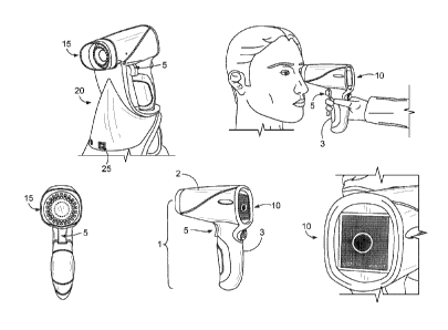
[0046] FIG. 1 is an illustration the handheld macular pigment reflectometer
from a
variety of views.
CA 02814213 2013-04-09
WO 2012/051449 PCT/US2011/056205
- 12 -
[0047] FIG. 2a is an illustration of the internal architecture of a
handheld macular
pigment reflectometer, according to one embodiment.
[0048] FIG. 2b is a schematic drawing of the hand-held macular pigment
reflectometer,
according to another embodiment, illustrating the anterior imaging mode,
trigger position 1.
[0049] FIG. 2c is a schematic drawing of the hand-held macular pigment
reflectometer of
FIG. 2b, illustrating the posterior imaging mode, trigger position 2.
[0050] FIG. 2d is a schematic drawing of the hand-held macular pigment
reflectometer of
FIG. 2b, illustrating the measurement mode, trigger position 3.
[0051] FIG. 3 is an illustration of a handheld macular pigment system.
[0052] FIG. 4 is an illustration of a three-step trigger switched actuated
instrument
alignment.
[0053] FIG. 5a is an illustration of the first position of the trigger
switched actuated
alignment, according to one embodiment.
[0054] FIG. 5b is an illustration of another embodiment of the hand-held
macular
pigment reflectometer corresponding to the first position of the trigger
switched actuated
alignment and showing the anterior light path.
[0055] FIG. 6a is an illustration of the second position of the trigger
switched actuated
alignment, according to one embodiment.
[0056] FIG. 6b is an illustration of another embodiment of the hand-held
macular
pigment reflectometer corresponding to the first position of the trigger
switched actuated
alignment and showing a first flip in-out mirror in its "IN" position.
[0057] FIG. 6c is an illustration of the reflectometer of FIG. 6b showing
the first flip in-
out mirror in its "OUT" position, illustrative of the second trigger position.
[0058] FIG. 6d is an image of a human retina and a corresponding 4-degree
field of view.
[0059] FIG. 7a is an illustration of the third position of the trigger
switched actuated
alignment, according to one embodiment.
[0060] FIG. 7b is an illustration of another embodiment of the hand-held
macular
pigment reflectometer corresponding to the second position of the trigger
switched actuated
alignment and showing the first flip in-out mirror in its "OUT" position.
[0061] FIG. 7c is an illustration of the reflectometer of FIG. 7b showing
the second flip
in-out mirror in its "IN" position.
CA 02814213 2013-04-09
WO 2012/051449 PCT/US2011/056205
- 13 -
[0062] FIG. 7d is an image of a 4-degree field of view of a human retina
and a graph of
the reflection vs. wavelength.
[0063] FIG. 8 is an illustration of a second light source LED or light
engine according to
one embodiment.
[0064] FIG. 9 is an illustration of the illumination spectra for the 4-
degree field of view
(second trigger switch position).
[0065] FIG. 10 is an illustration of the illumination spectra for the
measurement of
macular pigment, 1-degree field of view (third trigger switch position).
DETAILED DESCRIPTION
[0066] FIG. 1 illustrates a handheld macular pigment reflectometry (MPR)
instrument 1
adapted for clinical use. The handheld MPR instrument 1 includes a housing 2
having a
lower portion 3 that is ergonomically shaped to be received by an operator's
hand. The
handheld MPR instrument 1 enables clinician alignment and operation through a
simple
trigger switch 5 on the lower portion 3 actuated by a three-step alignment
process that
corresponds to a first trigger switch position (FIG. 5a), a second trigger
switch position (FIG.
6a), and a third trigger switch position (FIG. 7a), which are discussed below.
[0067] The handheld MPR instrument 1 includes a display 10 that enables the
clinician to
check alignment process and view zeaxanthin and lutein optical density
readings as well as
the status of any diagnostic functions of the handheld MPR instrument 1. The
handheld MPR
instrument 1 includes an eye cup or eye shield 15, which allows the clinician
to use the
device in an illuminated room. The handheld MPR instrument 1 can be coupled to
a base
docking station 20, which can be used to charge a battery 22 located within
the handheld
MPR instrument 1. The battery 22 provides power to various components in the
handheld
MPR instrument 1 through drive circuitry 24. More details of this self-
contained system are
discussed below with respect to FIG. 3.
[0068] FIG. 2a illustrates the internal architecture of the handheld MPR
instrument 1,
according to one embodiment. The handheld MPR instrument 1 contains a first
light source
12 that is preferably arrayed in a ring within the eye cup or eye shield 15
and preferably
utilizes LED illumination. The first light source 12 creates a specular
reflection from the
cornea that is used in the alignment process (described in more detail below
with reference to
FIG. 5a). Light reflected from the anterior portion of the eye is captured,
focused and relayed
CA 02814213 2014-01-31
- 14 -
by a Badal Optometer 45 back to the annular scraping mirror 47. The Badal
Optometer 45
consists of two lenses (the front two lenses in FIG. 2a), with at least one of
them being
moveable relative to the other one as indicated by the double-arrowed line
below the Badal
Optometer 45. In a preferred embodiment, the posterior lens is moveable via a
small motor.
This annular scraping mirror 47 eliminates a central circular portion (e.g. 4
mm diameter) of
the image, and relays the now annular image through a focusing lens 49 to the
anterior charge
coupled device (CCD) imager 50. The images captured by this CCD imager 50 are
displayed
on screen 10 and used for initial alignment of the handheld MPR instrument 1.
[0069] As the trigger 5 is further pulled to its second position (described
in more detail in
FIG. 6a), the ring LED 12 is shut down, and a second light source (or light
engine) 30 is
initiated. This light engine 30 is preferably a combination of a plurality of
LEDs (e.g. five
LEDs) that, when spectrally combined, provide a spectral range from 400 nm to
880 nm.
Once the individual LEDs are ramped to their steady state position, the light
from the light
engine 30 is collimated, strikes a scraping mirror 58 that reflects the
majority of the light, but
passes about 15% of the light. The reflected light passes through the annular
scraping mirror
47, through the Badal optometer 45 (which now acts as refractive error
correction optic), and
on to the patient.
[0070] The light from the light engine 30 passes through the patient's
cornea and lens,
reflects off of the retina and returns back through the eye's optics to the
handheld MPR
instrument 1. The area illuminated on the retina is about 1.0 mm in diameter,
which is
approximately 4 degrees of an arc. In particular the reflectance curve from
this region of the
retina will show the contributing factors of absorption of the carotenoids,
lutein and
zeaxanthin. The reflectance and absorption of the light in the eye is
described in more detail
(e.g. FIGS. 5a and 5b, and accompanying specification) in U.S. Patent No.
7,467,870,
[0071] Light returning to the handheld MPR instrument 1 now passes back
through the
same optics from which it came, except that the returning light will now
largely miss both
scraping mirrors 47 and 58, but will strike the flip-in mirror 67 in its "IN"
position, and
reflect upwardly to the posterior CCD imager 55. In response to the reflected
energy being
received by the CCD imager 55, a 4-degree image of the macula is formed and
displayed on
the screen 10. Further alignment by the clinician may be anticipated, but
getting the pupil
aligned first significantly eases the alignment task.
CA 02814213 2013-04-09
WO 2012/051449 PCT/US2011/056205
- 15 -
[0072] Meanwhile, while the eye is being aligned, the light from the light
engine 30 (15%
of the output from the light engine 30) that is not reflected by the scraping
mirror 58 passes
on to a collection optic 77 and into a fiber 79, which re-directs it to the
spectrometer 35. This
light is checked by the spectrometer 35, and sets the "white balance" or
native spectral
signature for the light source 30 for the spectrometer 35.
[0073] Once the eye is aligned and in focus, the trigger switch 5 is pulled
to its third and
final position (described in more detail in FIG. 7a). The light engine 30
ramps up to its
"flash" condition, which is a pulse of energy such as a 250 millisecond pulse
of light that will
be approximately three times the brightness as the steady-state condition when
the trigger 5 is
in the second position. As this occurs, the flip-in mirror 67 moves to the
"OUT" position,
which will block the "white-balance" light from the fiber 79, but will allow
for all light that
normally would be incident upon the posterior CCD 55 to now enter the
spectrometer 35
(possibly through some collection optics, including a short fiber).
[0074] The spectrometer 35 will take a plurality of spectral readings, such
as five
sequential spectra that are 50 milliseconds each, during the 250 millisecond
flash. The
spectrometer 35 sorts the spectra for a variety of reject criteria, and of the
spectra it keeps,
will average them and calculate the lutein optical density (LOD), the
zeaxanthin optical
density (ZOD), and the combined macular pigment optical density (MPOD). The
numbers
will displayed on display 10 to the clinician, and saved in memory in a
microcomputer
system (not shown). The clinician releases the trigger 5 and can repeat the
procedure, as
necessary. The detail of analysis that determines these density values is set
forth in U.S.
Patent No. 7,467,870, which is herein incorporated by reference in its
entirety.
[0075] FIG. 3 illustrates the handheld macular pigment reflectometer system
in which the
handheld macular reflectometer 1 is self-contained and can recharge its
battery 22 from the
base docking station 20. The battery 22 is used to provide power to the
various components
(e.g., the display 10, the CCD 50, the CCD 55, the first light source 12, the
light engine 30,
the spectrometer 35, motor for moving Badal lens or lenses 45, the Common path
objective
lens 120, motors for moving the first and second flip in-out mirrors, etc.)
through the drive
circuitry 24. By being battery-operated, the wireless handheld device 1 is
more easily
maneuvered by the operator when conducting the testing on the patient. The
base station 20
includes an AC Power-in port 25 for charging the battery. Further, the
handheld macular
pigment reflectometer 1 communicates with an electronic medical records system
70
CA 02814213 2013-04-09
WO 2012/051449 PCT/US2011/056205
- 16 -
accessible by a computer using a USB connection 27, which is part of the base
docking
station 20. The electronic medical records system 70 may be resident within
the computer, or
it may be accessible via the computer through an extranet or intranet
connection.
Furthermore, it should be noted that the base station 20 may have a wireless
connection to the
computer, or a direct connection to a local or remotely located electronic
medical records
system 70 (such that a computer is not needed). Utilizing a device like the
handheld macular
reflectometer 1 for collecting eye-health data for use in diagnosing
conditions and recording
information in a large-scale database is disclosed in U.S. Publication No.
2010/0241450,
which is herein incorporated by reference in its entirety. If used in such a
system, the
handheld macular reflectometer 1 would be connected to the computer and the
computer
would be used to enter details about the patient's information and background
to create
patient file, as shown in U.S. Publication No. 2010/0241450. The output of the
handheld
macular reflectometer 1 would then be sent to the computer and stored in
association with the
patient's file.
[0076] FIG. 4 illustrates more of the details of the three-step actuation
of the trigger 5 and
the output on the display 10 during alignment process. The three-step
actuation preferably
includes some type of tactile feedback, such that the clinician feel this
trigger 5 move
between the three defined positions associated with electrical switches, each
of which
provides a signal corresponding to that particular trigger switch position.
However, the
tactile feedback is very subtle in nature, so as not to impede the motion of
the trigger as it
moves through its three positions and thus not upset the precise alignments
achieved in the
first, second and third trigger switch positions. In the first trigger switch
position 75 (shown
in FIGS. 2b, 5a and 5b), the handheld MPR instrument 1 maintains a wide-field
anterior
image 80 on the display 10 with a central obscuration aligned with the eye
pupil. In the
second switch position 85 (FIGS. 2c, 6a, 6b, 6c), there is a 4-degree central
retinal field of
vision 90 on the display 10 with a circular reticule 87 colored green,
indicating a 1.0-degree
central measurement area. In the third switch position 95 after the light
engine 30 is operated
in the pulsed condition (FIGS. 2d, 7a, 7b, 7c), the handheld MPR instrument 1
freezes the 4-
degree image 100, ramps up the light engine 30, takes a white balance reading,
actuates the
second flip in-out mirror 67, activates the spectrometer 35, turns the
reticule 87 to a red color,
and flashes the reticule 87 in the image 100 if the trigger 5 is held for more
than one second.
CA 02814213 2013-04-09
WO 2012/051449 PCT/US2011/056205
- 17 -
[0077] FIG. 5a illustrates the active optical elements of the handheld
macular pigment
reflectometer 1 when the trigger 5 is in the first position. In the first
position, the LED ring
lights 12 illuminate the eye; the Badal Optometer 45 focuses return light
through the annular
scraping mirror 47 onto the anterior CCD 50, which is coupled to the display
10. The annular
scraping mirror 47 produces central obscuration. While the trigger 5 is in the
first position,
the central obscuration may not overlap the central pupil of the patient's eye
as shown in
image 191. The spectral ring is partially to completely invisible in the image
191 and it is not
possible to view the retina upon transition. When the handheld MPR instrument
1 is slightly
adjusted and the image central obscuration aligns with the natural pupil of
the subject's eye
as show in image 193 on the display 10, a sharp specular reflection of the
illuminating LED
ring 12 off the subject's cornea appears. When both conditions exist, this
will result in a non-
vignetted 4-degree retinal field of view upon transition. In one embodiment,
the light engine
30 may blink or modulate at a low power during this time to (i) provide a
fixation light for
the patient to steady his or her gaze, and (ii) to provide a white reference
beam to the
spectrometer 35 through the fiber optic pickoff 77 and the fiber 79. In this
situation, the
posterior CCD 55 would be turned off, such that it will not matter if a small
amount of return
light strikes it.
[0078] FIG. 6a illustrates the macular pigment reflectometer 1 with the
trigger 5 in the
second position, which occurs as the operator continues to squeeze the trigger
5. In this
position, the anterior CCD 50 and LED ring lights 12 shut down, and the LED
light engine 30
begins operation. The Badal optometer 45 then switches to a refraction-
correction mode, the
flip-in mirror 67 remains in the "IN" position, and the posterior CCD 55õ
which is coupled
to the display 10, becomes active. An image 145 with a 4-degree by 4-degree
field of view is
shown in the display 10 (FIG. 6d). The clinician aligns the handheld MPR
instrument 1 until
the patient's fovea centralis is located within the 1.0-degree circular
reticule 87.
[0079] FIG. 7a illustrates the macular pigment reflectometer 1 after the
trigger 5 has been
advanced to the third position. In this position the flip-in mirror 67 moves
to the "OUT"
position, the image on the posterior CCD 55 freezes, the Badal optometer 45
freezes, the
LED light engine 30 increases in intensity and creates the pulsed output for
0.25 seconds and
the return light is received by the spectrometer 35. The spectrometer 35 takes
multiple
spectral samples, such as five measurements in 0.25 seconds and averages them.
To reduce
the spectral noise, various techniques could be used, such as boxcar averaging
or polynomial
CA 02814213 2014-01-31
- 18 -
smoothing. Because the on-board spectrometer 35 captures spectra in very short
intervals of
time, there is less relative motion during spectral capture and the handheld
MPR instrument 1
is more likely to be aimed at the fovea during capture.
[0080] As shown in the image 155 on the display 10 of FIG. 7d, the reticule
87 on the
display 10 turns red. If the trigger 5 is held in the third position for more
than one second, the
reticule 87 begins to flash on the screen 10. The handheld MPR instrument 1
then calculates
optical density, zeaxanthin optical density and lutein optical density. These
values are
typically reported after the trigger 5 is released. The handheld MPR
instrument 1 can simply
store the raw data and allow the connected computer or medical records system
to calculate
the values, or the handheld MPR instrument 1 may have the on-board processing
to permit
the calculation of the zeaxanthin optical density and lutein optical density.
The details of the
curve-matching functions used to determine the variables for the modeled curve
are set forth
in U.S. Patent No. 7,467,870.
[0081] According to the embodiment described above, the handheld MPR
instrument 1 is
designed to provide for two optical paths that are generally coaxial and
concentric. An outer
anterior optical path is annular in design both from an illumination and
imaging function. An
interior posterior optical path is within the outer optical path. The scraping
mirror 58 is used
to generate the posterior viewing and measurement path, by directing some 85%
of the light
generated by the LED light engine towards the patient's eye. The light
directed towards the
patient's eye will be slightly (about 1-2 degrees) off axis. This is to
prevent specular
reflections from the optical elements and the patient's cornea from reflecting
back into the
spectrometer optical path. The other 15% not directed to the patient will be
directed into the
pickoff fiber optic 77, which is used to periodically make a white reference
measurement
with the spectrometer 35. When the flip-in mirror 67 is in the IN position,
the white
reference can be detected, if required. When the mirror 67 is in the OUT
position, the mirror
67 will block the 15% leaked light from the light engine 30 from reaching the
spectrometer
35, only light that has reflected off the patient's retina will be permitted
to reach the
spectrometer. Annular optical systems typically leave a central obscuration at
the image
plane. In this application, the obscuration is not detrimental to the function
of the anterior
image, and in fact can be used to facilitate alignment for rapid transition to
the posterior
image. Except for within the Badal Optometer refractive power correcting
elements 45, each
optical path is separate from the other. The Badal optometer 45 serves two
distinct purposes,
CA 02814213 2013-04-09
WO 2012/051449 PCT/US2011/056205
- 19 -
depending on the viewing mode. In the anterior mode, the Badal optometer 45
acts as an
autofocus mechanism, and a motor drives one of the lens elements (e.g. the
posterior lens) to
compensate for slight motions of the patient or instrument. The relative
motion of the lens in
this mode is quite small, typically less than 200 um total travel. However,
the bandwidth of
the motion is relatively high, approximately 5 Hz. When the viewing mode
switches to the
posterior mode, the Badal optometer 45 functions more as a typical refractive
power
corrector. The Badal optometer 45 is moved to a position corresponding to the
net refractive
power error of the patient. This motion could be as far as 1.5 mm for a >15
Diopter
correction. Then, from this new position, the lens group 45 returns to a small
motion high
bandwidth autofocus function, but for the posterior CCD 55. The annular
scraping mirror 47
spatially separates the two paths. The hole in the annular scraping mirror 47
will determine,
in large part, the central obscuration. It is also the limiting aperture for
the retinal imaging
optics. And, as described above, each optical path has its own detector, the
anterior CCD 50
and the posterior CCD 55.
[0082] It should be noted, according to some embodiments, that the handheld
MPR
instrument 1 preferably includes a stabilizing lens 97, to help stabilize the
light going to the
spectrometer 35. The stabilizing lens 97 can be a fluidic lens or based on a
LensVector
autofocus technology (e.g., lens on MEMS) from LensVector, Inc. of Mountain
View, CA.
[0083] A further embodiment of the hand-held reflectometer is shown in
FIGS. 2b-d.
These drawings illustrate a sequential series of light paths that correspond
to a three-step
alignment method, which corresponds to a first trigger switch position, a
second trigger
switch position and a third trigger switch position of the hand-held MPR. The
hand-held
MPR instrument 101, shown in FIG. 2b, contains a first light source 112 that
is preferably
arrayed in a ring within an eye cup or eye shield 115 and preferably utilizes
LED
illumination. The first light source 112 creates both a specular reflection
from the cornea that
is used in the alignment process and a diffuse reflection that is used to
image the entire
anterior portion of the eye. As shown in FIG. 2b, diffuse and specular light
reflected from the
anterior portion of the eye is captured, focused and relayed by a Common path
objective lens
120 back to a first flip in-out mirror 125. The Common path objective lens 120
consists of
two lens groups, with a first lens group 120a being fixed relative to the
second lens group
120b. In a preferred embodiment, the second lens group 120b is moveable via a
small motor.
CA 02814213 2014-01-31
- 20 -
This first flip in-out mirror 125 relays the image through a focusing lens
group 130 to an
anterior charge coupled device (CCD) imager 150. The images captured by this
CCD imager
150 are displayed on screen 110 and used for initial alignment of the handheld
MPR
instrument 101. The light from the first light source 112, corresponding to
the first trigger
switch position, preferably corresponds to spectra in the range of 680-900 nm,
and preferably
880 nm.
R10841 As the trigger 105 is further pulled to its second position as shown
in FIG, 2c, the
first light source (ring LED) 112 is shut down, the first flip in-out mirror
125 is moved to the
"OUT" position, and a second light source or light engine 132 is initiated.
This second light
source or light engine 132 is preferably a combination of a plurality of LEDs
(e.g., five
LEDs) that, when spectrally combined, provide a spectral range from 400 nm to
880 nm, and
more preferably in this mode, in the range of 630-880 nm. FIG. 8 illustrates
an example of
one type of light source that provides the desired spectral range. The light
at this point in the
alignment process is preferentially red and infrared illumination from the red
and infrared
LEDs of the light engine 132 as shown in the spectra of FIG. 7d. Once the
individual LEDs
are ramped to their steady state position, the light from the second light
source or light engine
132 is collimated, folded and directed to a chevron mirror 140 that divides
the light into two
beams, and directs those beams through the first lens group 120a of the Common
path
objective lens 120 to the eye. The reflected light passes through the first
lens group 120a of
the Common path objective lens 120 (which now acts as a focusing element), and
on to the
patient.
100851 The light from the second light source or light engine 132 passes
through the
patient's cornea and lens, reflects off of the retina and returns back through
the eye's optics to
the handheld MPR instrument 101. The area illuminated on the retina is about
1.0 mm in
diameter, which is approximately 4 degrees of angular sub tense. In
particular, the
reflectance curve from this region of the retina shows the contributing
factors of absorption of
the carotenoids, lutein and zeaxanthin. The reflectance and absorption of the
light in the eye
is described in more detail in FIG. 7d and in U.S. Patent No. 7,467,870.
100861 Light returning to the handheld MPR instrument 101 now passes back
through the
same optics from which it came, except that the returning light will now pass
in between
chevron mirrors 140 and 141, and miss entirely the second flip in-out mirror
143 in its
CA 02814213 2014-01-31
- 21 -
"OUT" position, and be re-imaged by a posterior optical group 153 to a
posterior CCD
imager 155. In response to the reflected energy being received by the
posterior CCD imager
155, a 4 degree image of the macula is formed and displayed on the screen 110.
Further
alignment by the clinician may be anticipated, but getting the pupil aligned
first (via the
anterior alignment accomplished in the first trigger switch position),
significantly eases the
alignment task. Patient comfort is maximized by utilizing only the red and
infrared LEDs
while in the second trigger switch position, and the retinal pigments
(rhodopsin) are bleached
to a precisely known condition, which can be optimized by the posterior
illumination light
levels. This condition is described in more detail in the Journal of the
Optical Society of
America, "Effect of wavelength on in vivo images of the human cone mosaic,"
Vol. 22, No.
12, December 2005. The patient will also be
provided a low intensity blue fixation light 162, which will contrast with the
red posterior
illumination light from the LED light engine 132. The fixation light 162 is
preferably a blue
440 nm low intensity LED, which is collimated, diffused and apertured so as to
subtend a
small angular space to the patient. It is folded into the optical path by a
dichroic beamsplitter
160.
[0087] Meanwhile, while the eye is being aligned, the light from the
secondary light
source or light engine 132 (2% of the output from the second light source or
light engine) that
is not being collimated and directed to the chevron mirrors 140, 141 passes on
to a small
collection optic 177 and into a fiber 179, which re-directs it to the
spectrometer 135. This
light is available and can be checked by the spectrometer 135 periodically,
and sets the
"white balance" or native spectral signature for the second light source or
light engine 132 for
the spectrometer 135.
[00881 Once the eye is aligned and in focus, the trigger switch 105 is
pulled to its third
and final position, as shown in FIG. 2d. The second light source or light
engine 132 ramps
up to its measurement condition, which includes all LEDs in the light engine
illuminated at a
spectrally optimal condition as shown in FIG. 10, and which provides a pulse
of energy, such
as a few hundred millisecond pulse of light that will be approximately three
times the
brightness as the steady-state condition when the trigger switch 105 is in the
second position.
As this occurs, the first flip in-out mirror 125 remains in the "OUT"
position, and the second
flip in-out mirror 143 moves to the "TN" position which will block the "white-
balance" light
from the fiber 179, but will allow for all light that normally would be
incident upon the
CA 02814213 2014-01-31
- 22 -
posterior CCD 155 to now enter the spectrometer 135 (through some collection
optics,
including a short fiber, not shown). In the third trigger switch position, the
higher intensity
broadband illumination is in the range of 400-880 nm. The fixation LED 162 is
shut down
for this brief period, and the display 110 is frozen on the last frame of the
posterior imaging
mode, but the green reticule displayed has been changed to a red reticule.
[00891 The spectrometer 135 takes a plurality of spectral readings, such as
five sequential
spectra that are 50 milliseconds each, during the few hundred millisecond
measurement
period. The spectrometer 135 and microcomputer (not shown) sorts the spectra
for a variety
of reject criteria, and of the spectra it keeps, will average them and
calculate the lutein optical
density (LOD), the zeaxanthin optical density (ZOD), and the combined macular
pigment
optical density (MPOD). The method of calculation is well detailed in U.S.
Patent No.
7,467,870.
[0090] The numbers will displayed on display 110 to the clinician, and
saved in memory
in a microcomputer system (not shown). The clinician releases the trigger
switch 105 and
can repeat the procedure, as necessary. The detail of analysis that determines
these density
values is set forth in U.S. Patent No. 7,467,870.
[0091] The first trigger switch position, corresponding to the anterior
light path, is further
illustrated in FIG. 5b (as well as shown in FIG. 2b). As noted above, in the
first trigger
switch position, the first light source 112 (LED ring lights) illuminates the
eye, the Common
path objective lens 120 focuses return light through the first flip in-out min-
or 125 and relays
optics onto the anterior CCD 150, which is coupled to the display 110. As
noted above,
while the trigger switch 105 is in the first position, the central obscuration
may not fully
overlap the central pupil of the patient's eye as shown in image 191. The
spectral ring is
partially to completely invisible in the image 191 and it would not be
possible to view the
retina upon transition. When the handheld MPR instrument 101 is slightly
adjusted and the
image central obscuration aligns with the natural pupil of the subject's eye
as show in image
193 on the display 110, a sharp specular reflection of the illuminating LED
ring 112 off the
subject's cornea appears. When both conditions exist, this will result in a
non-vignetted 4-
degree retinal field of view upon transition. In one embodiment, the light
engine may blink
or modulate at a low power during this time to (i) provide a fixation light
for the patient to
steady his or her gaze, and (ii) to provide a white reference beam to the
spectrometer 135
CA 02814213 2013-04-09
WO 2012/051449 PCT/US2011/056205
- 23 -
through the fiber optic pickoff 177 and the fiber 179. In this situation, the
posterior CCD 155
is turned off
[0092] The second trigger switch position, corresponding to the posterior
light path, is
further illustrated in the transition from FIG. 6b to FIG. 6c. The second
trigger switch
position occurs as the operator continues to squeeze trigger switch 105. In
this position, the
anterior CCD 150 and first light source (LED ring lights) 112 shut down, and
the second light
source or light engine 132 becomes the predominant light source. The first
flip in-out mirror
125 moves from its "IN" position to its "OUT" position (see FIG. 6c), and the
posterior CCD
155, which is coupled to the display 110, becomes active. An image 145 with a
4-degree by
4-degree field of view is shown in the display 110 (FIG. 6d). The clinician
aligns the
handheld MPR instrument 101 until the patient's fovea centralis is located
within the 1.0-
degree circular reticule 87.
[0093] The transition from FIG. 7b to FIG. 7c further illustrates the
posterior illumination
measurement path corresponding to the third trigger switch position. In this
position, the
second flip in-out mirror 143 moves from the "OUT" position to the "IN"
position (see FIG.
7c), the image on the posterior CCD 155 freezes, the Common path objective
lens 120
freezes, the LED light engine 132 increases in intensity and creates the
pulsed output for
several hundred milliseconds and the return light is received by the
spectrometer 135. The
spectrometer 135 takes multiple spectral samples or measurements, such as five
measurements in 0.25 seconds, and averages them. To reduce the spectral noise,
various
techniques could be used, such as boxcar averaging or polynomial smoothing.
Algorithms
within the microcomputer compare each spectra to a known modeled or "good"
spectra, and
any spectra not within a pre-determined tolerance band of the known good
spectra are
considered "bad" spectra or data, and are filtered out of the set and
rejected. FIG. 7d shows a
modeled spectra 90 and an actual measured spectra 142. Spectra 142 would be
considered
acceptable data because it is within the tolerance bands of the algorithm.
Because the on-
board spectrometer 135 captures spectra in very short intervals of time, there
is less relative
motion during spectral capture and the handheld MPR instrument 101 is more
likely to be
aimed at the fovea during capture.
[0094] According to the embodiment described above (particularly in FIGS.
2b-d, 5b, 6b-
c and 7b-c), the handheld MPR instrument 101 is designed to provide for two
optical paths
that are partially coincident. An anterior optical path provides both an
illumination and
CA 02814213 2013-04-09
WO 2012/051449 PCT/US2011/056205
- 24 -
imaging function. A posterior optical path utilizes the Common path objective
lens group of
the anterior optical path. The first flip in-out mirror 120 is used to
separate the anterior and
posterior viewing and measurement paths, by directing light reflected from the
cornea and the
anterior eye through relay optics to an optimized CCD imager 150. The light
directed
towards the patient's eye will be off axis. This is to prevent specular
reflections (except from
the cornea) from the optical elements and the patient's cornea from reflecting
back into the
imaging optical path. Light from the light engine will also enter the eye
slightly off axis so as
to avoid corneal, lenticular specular reflections. Light returning from the
retina will
minimize specular reflections to interfere with the image. Stray light from
other sources is
reduced or nearly eliminated by the use of the eye cup 115 surrounding the
space between the
instrument and the patient's eye.
[0095] Light not directed to the posterior illumination chevron mirror 140
will be directed
into the pickoff fiber optic 177, which is used to periodically make a white
reference
measurement with the spectrometer 135. When the second flip in-out mirror 143
is in the
"OUT" position, the white reference can be detected, if required. When the
second flip in-out
mirror 143 is in the "IN" position, the second flip in-out mirror 143 will
block the white
balance light from the light engine from reaching the spectrometer 135. Thus,
only light that
has reflected off the patient's retina will be permitted to reach the
spectrometer. The anterior
optical path is designed to facilitate alignment, both from an illumination
and imaging
function. This optical path typically leaves a central obscuration at the
image plane. This
obscuration can be created optically by masks, or electronically within the
CCD image.
[0096] Except for within the Common path objective lens elements 120, each
optical path
is separate from the other. The Common path objective lens 120 serves two
distinct
purposes, depending on the viewing mode. In the anterior mode, the Common path
objective
second lens group 120b acts as an autofocus mechanism, and a motor drives this
group to
compensate for slight motions of the patient or instrument. The relative
motion of the lens in
this mode is quite small, typically less than 500 um total travel. However,
the bandwidth of
the motion is relatively high, approximately 5 Hz. The refractive power
correction of the
system is accomplished as described herein. In particular, the optical group
consisting of the
posterior view CCD, the fixation light and beamsplitter, the second flip in-
out mirror and the
spectrometer port (with corresponding white balance fiber input) all move
together as a group
along the optical axis. With the patient's refractive error known, the user
dials in the
CA 02814213 2013-04-09
WO 2012/051449 PCT/US2011/056205
- 25 -
refractive error by turning a knob which turns a lead screw. The lead screw is
the active
element of a mechanical stage which translates the group along the optical
axis, depending on
the intended refractive error to correct. For example, to correct for a +5D
refractive error, the
optical path to the group will have to lengthen by approximately 15 mm.
Similarly, a -5D
refractive error would be corrected by shortening the optical path to the
group by 15mm. This
function is not shown in the figures.
[0097] FIG. 8 is an illustration of a second light source or light engine
132 that may be
used with the present invention. This second light source or light engine 132
includes a SMA
connector 134, a plastic optical fiber pigtail 136, a light mixer 137 and a
plurality of fiber-
coupled light emitting diodes 138. These plurality of LEDs 138 are combined
together to
emit a very broad spectrum of visible to near-infrared light, namely 400 to
880 nm. Typically
or more LEDs can be combined together to create a posterior light source (LED
light
engine), although more or less may be used. This combination of LEDs replaces
the tungsten
halogen lamp that was the core illuminator on tabletop designs. Besides being
faster to warm
up, more stable over the long term, longer lifetime, lower power consumption,
lower heat
production, more efficient than the tungsten halogen lamp, the light engine
offers a novel
attribute: rapid and programmable spectral modification.
[0098] As background, the tungsten halogen lamps' spectra can be modified,
by both
power applied and by the use of discrete optical filters. Modification of the
spectra by power
applied simply shifts the spectra by a few nanometers, the higher the power
applied, the bluer
the light. The modification of the spectra by filters is also commonly used.
However, filters
simply change the spectra in discrete steps, by attenuating portions of the
tungsten halogen
emission band. Filters increase the complexity and cost of the system by
adding electro-
mechanical components to move them in and out of the illuminator pathway.
Filters can also
potentially age with time, and thus change in their attenuating
characteristics. Finally, as
mentioned, filters only allow for discrete step function changes in spectra.
[0099] The LED light engine offers a fundamentally different approach. It
allows for
different illumination characteristics depending on the alignment state the
instrument is in,
and allows for those characteristics to be tuned for optimal performance.
Thus, according to
one non-limiting example, in the first trigger switch position, the light
engine is off and only
the fixation light from the posterior optics is illuminated. The anterior
illumination ring is
illuminated for the anterior view. In the second trigger switch position, the
red portion of the
CA 02814213 2013-04-09
WO 2012/051449 PCT/US2011/056205
- 26 -
LED engine is lit, to allow for illumination while the clinician navigates to
the fovea. The
blue fixation light is lit as well, but the exterior ring of LEDs is off and
the blue and white
LEDs of the light engine are turned off or way down in intensity, to allow for
optimal patient
comfort while the clinician aligns to the fovea. The illumination level in
this mode is a
balance between optimal illumination for patient comfort, return signal to the
posterior
sensor, and sufficient illumination to bleach the foveal cones of rhodopsin
(retinal pigments)
to a known and consistent state.
[00100] This will allow the algorithm that analyzes the retinal reflectance to
determine the
macular pigments to work at optimum conditions. In the third trigger switch
position, the
blue and white LEDs are illuminated in combination with the red and infrared
LEDs to
produce the full broad spectrum. These LEDs can be ramped up in intensity to
adequate
levels to allow the spectrometer optimal signal to noise ratio, balancing
patient eye comfort
and safety issues. See Figures 9 and 10 for examples of illumination spectra
for the second
and third trigger switch positions. The advantages of such a measurement
approach are
several. For example, less total power is consumed, the patient is more
comfortable during
the exam, less heat is produced and the measurement is fundamentally more
consistent when
the bleach levels of the cones are precisely controlled. This is important to
a variety of
ophthalmic measurements, not just macular pigment measurement.
Autofluorescence and
dye enhanced fluorescence could benefit in terms of improving background noise
consistency
from this illumination approach, as well as other reflectometry-based
instruments (laser
scanning ophthalmoscopes, retinal thickness analyzers, retinal blood flow
analyzers, etc.)
which would benefit in terms of background noise reduction.
[00101] FIG. 9 is an illustration of the illumination spectra for the 4 degree
field of view
(second trigger switch position). This can be tuned with time to optimize the
tradeoffs
between patient comfort, instrument signal, and patient foveal cone bleach
condition. In this
position, only LEDs 3, 4 and 5 out of the LED light engine (see FIG. 8) are
illuminated.
LEDs 1 and 2 are barely emitting (i.e., "simmer" mode), but will allow for
rapid transition to
the third trigger switch position for taking a measurement.
[00102] FIG. 10 is an illustration of the illumination spectra for the
measurement of
macular pigment, one-degree field of view (third trigger switch position).
This can also be
tuned as a function of the tradeoffs mentioned above for FIG. 9, and also
tuned in terms of
the ramp up of the intensity of illumination.
CA 02814213 2013-04-09
WO 2012/051449 PCT/US2011/056205
-27 -
[00103] Thus, as described herein, a handheld ophthalmic instrument with a
unique human
interface is provided, whereby a user can simply squeeze through a series of
trigger switch
positions to achieve a complex alignment and measurement procedure rapidly
with minimal
effort and unwanted instrument motion. The measurement procedure is comprised
of three
modes ¨ an anterior alignment, posterior alignment, and spectroscopic
measurement and
calculation. Transition through the modes is seamless because of the trigger
switch design as
described herein.
[00104] Other advantages of the hand-held MPR as described herein include
providing
spectra that is different and optimized for each task. For example, according
to one
embodiment, infrared LED light (880 nm typically) is provided for illuminating
the eye
during anterior alignment to ensure the pupil of the eye stays large. Red
(typically 635 nm)
or green-red-IR (typically 530-880 nm) LED light is provided for illuminating
the eye during
posterior alignment, which ensures a comfortable illumination color to the
patient while the
clinician locates the fovea, and still provides sufficient illumination of the
right color band to
ensure a consistent bleach condition of the retinal photopigments (rhodopsin).
This is
important so that the reflection algorithm of the human rhodopsin matches the
assumed
values in the instrument software algorithm. Broadband (typically 400-880 nm)
LED light is
provided for measurement. This consists of the summation of a series of LEDs
into a LED
"engine" whose spectra are chosen and/or modified by phosphors such that the
total spectra
will span the range of 400 to 880 nm without a gap in coverage. The
intensities of each of
the LEDs in the engine may be adjusted to compensate for spectral reflectivity
of the macula
and the spectral transmission and responsivity of the MPR optics and
spectrometer,
respectively.
[00105] In addition to providing a hand-held MPR having the advantages
described herein,
the hand-held MPR of the present invention may be used to provide measurements
of
biomarkers to assist in identifying patients at risk of developing eye
diseases and conditions.
For example, macular pigment optical density (MPOD) is considered a biomarker
for eye
diseases and conditions, such as AMD, diabetic retinopathy, cataract formation
and decreased
visual performance. Visual performance includes contrast sensitivity,
photosensitivity, dark
adaptation, glare recovery, temporal processing speed, reaction time and
hand/eye
coordination. Functionally, improved visual performance is relevant to sports
performance,
aviation and driving ¨ especially at night or in the rain.
CA 02814213 2013-04-09
WO 2012/051449 PCT/US2011/056205
- 28 -
[00106] Furthermore, recent research shows that a patient's MPOD score may be
a
biomarker for patients at risk of developing non eye-related diseases and
conditions,
including diabetes, lowered cognitive function and certain types of cancer.
Thus, diagnostic
use of the hand-held MPR may provide useful information to patients as an
indicator of eye
diseases and non-eye-related conditions. While some specific uses of the hand-
held MPR
are described herein, such uses are not an exhaustive listing, and additional
uses of the hand-
held MPR are contemplated. For example, additional characteristics of the eye
may be
determined using the multistep alignment process described herein, including
measurement
of melanin density and lens optical density, The details of analysis that
determines these
density values is set forth in U.S. Patent No. 7,467,870.
[00107] Other embodiments of the present invention include a reflectometry
instrument to measure macular pigment of a macula of a human eye that
comprises a housing
including a lower hand-held portion; a light source within the housing for
emitting an
illumination beam in a direction toward the macula; a spectrometer within the
housing for
measuring a detection beam, the detection beam being a portion of the
illumination beam
reflected from the macula, the detection beam being indicative of the amount
of the macular
pigment in the macula; and wherein the hand-held portion is graspable by the
operator for
assisting in manual alignment of the illumination beam with the macula.
[00108] Another embodiment of the present invention includes a hand-held
reflectometry instrument to measure macular pigment of a macula of a human
eye, that
comprises a housing including an actuatable trigger having at least a first
position and a
second position; a light source within the housing for emitting an
illumination beam in a
direction toward the macula; a spectrometer within the housing for measuring a
detection
beam, the detection beam being a portion of the illumination beam reflected
from the macula,
the detection beam being indicative of the amount of the macular pigment in
the macula; and
wherein, in the first position, the actuatable trigger permits alignment of
the instrument
relative to the eye and, in the second position, the actuatable trigger causes
the operation of
the light source.
[00109] Another embodiment of the present invention includes a hand-held
reflectometry instrument to measure macular pigment of a macula of a human
eye, that
comprises a housing including an actuatable trigger having at least a first
position and a
second position; a light source within the housing for emitting an
illumination beam in a
CA 02814213 2013-04-09
WO 2012/051449 PCT/US2011/056205
- 29 -
direction toward the macula; a spectrometer within the housing for measuring a
detection
beam, the detection beam being a portion of the illumination beam reflected
from the macula,
the detection beam being indicative of the amount of the macular pigment in
the macula; and
wherein, in the first position, the actuatable trigger permits alignment of
the instrument
relative to the eye and, in the second position, the actuatable trigger causes
the operation of
the light source.
[00110] Another embodiment of the invention includes a reflectometry
instrument to
measure macular pigment of a macula of a human eye, that comprises a housing
including a
lower hand-held portion; a light source within the housing for emitting an
illumination beam
in a direction toward the macula; a spectrometer within the housing for
measuring a detection
beam, the detection beam being a portion of the illumination beam reflected
from the macula,
the detection beam being indicative of the amount of the macular pigment in
the macula; and
a display on the housing that provides at least one of (i) an image of an
output corresponding
to the amount of the macular pigment in the macula, and (ii) an image of the
eye for
alignment or measurement purposes.
[00111] Another embodiment of the present invention includes a
reflectometry system
to measure macular pigment of a macula of a human eye, that comprises a
handheld
reflectometry instrument having a light source for emitting an illumination
beam in a
direction toward the macula, and a spectrometer within the housing for
measuring a detection
beam, the detection beam being a portion of the illumination beam reflected
from the macula,
the detection beam being indicative of the amount of the macular pigment in
the macula; and
a docking station for receiving the handheld reflectometry instrument.
[00112] Another embodiment of the present invention includes a method of
determining the amount of macular pigment in the macula of a human eye, that
comprises
using a multi-step alignment process involving (i) a first light source to
provide an alignment
relative to the patient's pupil, and (ii) a second light source to provide
alignment relative to
the patient's retina after alignment relative to the patient's pupil; after
the alignment process,
passing an illumination beam through a lens system and onto the macula;
receiving, with a
spectrometer, a detection beam reflected from the macula; and measuring
characteristics of
the detection beam at the spectrometer.
[00113] Another embodiment of the present invention includes a method of
determining the amount of macular pigment in the macula of a human eye using a
handheld
CA 02814213 2014-03-31
- 30 -
macular pigment measuring instrument, that comprises displaying a multi-step
alignment process
on a display mounted on the instrument, the displaying includes (i) a first
image providing an
alignment of the instrument relative to the patient's pupil, and (ii) a second
image providing an
alignment of the instrument relative to the patient's retina; after the
alignment process, passing an
illumination beam through a lens system and onto the macula; receiving, with a
spectrometer, a
detection beam reflected from the macula; and measuring characteristics of the
detection beam at
the spectrometer.
[00114]
Another embodiment of the present invention includes a reflectometry
instrument
to measure macular pigment of a macula of a human eye, that comprises a
housing including a
lower hand-held portion; a light source within the housing for emitting an
illumination beam in a
direction toward the macula, the lights source providing a pulse of light that
has a duration of
less than 1 second; a spectrometer within the housing for measuring a
plurality of spectra
samples of a detection beam, the detection beam being a portion of the
illumination beam
reflected from the macula, each of the plurality of spectra samples being
indicative of the amount
of the macular pigment in the macula.