Note: Descriptions are shown in the official language in which they were submitted.
CA 02814658 2013-04-12
WO 2012/051132 PCT/US2011/055670
INTERVERTEBRAL IMPLANT
CROSS-REFERENCE TO RELATED APPLICATION
[0001] This patent application claims priority to, and the benefit of, U.S.
provisional
patent application No. 61/392,638, filed October 13, 2010, the contents of
which are hereby
incorporated herein by reference in their entirety.
FIELD OF THE DISCLOSURE
[0002] The present disclosure generally relates to a method of fixing an
intervertebral
implant, and in particular relates to a method for fixing an intervertebral
implant after
implantation of bone anchors.
BACKGROUND
[0003] Intervertebral implants, such as spacers or total disc replacement
(TDR)
implants, are typically inserted into an intervertebral space disposed between
the respective
endplates of a pair of adjacent vertebral bodies, for instance after the disc
material has been
removed or to augment existing disc material. Adequate stability between
intervertebral
implants and the endplates of the adjacent vertebral bodies allows the implant
to function
properly. For instance, a poor fixation between the implant and the vertebral
bodies can cause
implant migration, incorrect kinematics of portions or of the entire spine and
create a new source
of pain for the patient.
[0004] Conventional fixation of intervertebral implants to the vertebral
bodies is
achieved by screwing fasteners through a portion of the implant, for instance
at the anterior face
of the implant, into one or two adjacent vertebral bodies. Conventional
fixation can be further
achieved by mechanically interlocking pointed structures, such as spikes or
teeth, between the
implant and the endplates of the adjacent vertebral bodies. Conventional
fixation can also be
achieved by inserting the intervertebral implant in the adjacent vertebral
bodies. For instance,
conventional intervertebral implants can include superior and inferior keels
that are inserted into
corresponding cut-outs formed in the adjacent vertebral bodies.
SUMMARY
[0005] In an embodiment, an implant assembly comprises an implant body
defining a
first bone contacting surface and a second bone contacting surface, the first
bone contacting
surface spaced apart from the second bone contacting surface, at least one of
the first or second
bone contacting surfaces defining at least one recess, such that the at least
one recess is
1
CA 02814658 2013-04-12
WO 2012/051132 PCT/US2011/055670
configured to receive a head of a bone anchor so that a shaft of the bone
anchor extends out from
the first side. The implant assembly can further include comprising the at
least one bone anchor,
the at least one bone anchor comprising a shaft extending from the head, the
shaft configured to
be inserted in a vertebral body. The at least one recess can extend into the
implant body in a
transverse direction. The recess can be configured as a pocket hole
penetrating into the implant
body from the first and second bone contact surfaces. The head can be press-
fit into the at least
one recess. The head can be loosely received in the at least one recess. The
head can be secured
to the implant body by a hardenable substance that is injected into the at
least one recess. The
hardenable substance can include a glue, a cement or a polymerizable monomer
or copolymer.
The implant assembly can further include a suture for fixing the at least one
bone anchor to the at
least one recess. Surfaces defining the at least one recess can be at least
partially made of a
shape memory material. The surfaces have an initial configuration, wherein the
head fits loosely
in the at least one recess, and a fixing configuration, wherein the head is
tightly received within
the at least one recess. .
[0006] In an embodiment, the implant assembly can further include a template
that
defines at least one aiming hole corresponding to the arrangement of the at
least one recess of the
implant body. The implant body can define a plurality of recesses, and the
template comprises a
plurality of aiming holes configured to be aligned with respective ones of the
recesses of the
implant body. The aiming holes comprise a first aiming portion and a passage
connected to the
aiming portion and sized greater than the aiming portion. The at least one
bone anchor can
defines a bore that extends along the shaft and the head. The shaft can define
radial perforations
in fluid communication with the bore. The first and second bone contacting
surfaces can be
spaced part along a central axis. The at least one bone anchor can be oriented
at an oblique angle
with respect to the central axis. The the implant assembly can be an
intervertebral implant
assembly. The implant assembly can further include the at least one bone
anchor configured as a
staple. The staple can comprise a first pin, a second pin, and a bridge, the
bridge interconnecting
the first and second pins. The bridge can be press-fit inside the recess. The
bridge can be
loosely received within the recess. The bridge can be secured to the implant
body by a
hardenable substance that is injected into the recess. The recess can be sized
to receive a
plurality of heads. All of the plurality of heads can be secured to the
implant body by a
hardenable substance injected in the recess.
[0007] In an embodiment, an implant assembly includes an implant body sized to
be
received in an intervertebral space. The implant body defines a first bone
contacting surface and
2
CA 02814658 2013-04-12
WO 2012/051132 PCT/US2011/055670
a second contacting surface. The first contacting surface is spaced apart from
the second
contacting surface. The implant body defines a first recess extending into the
first contacting
surface. The first recess extends into a first side of the implant body but
not through the implant
body. The implant body defines a second recess extending into the second
contacting surface.
The second recess extends into a second side of the implant body but not
through the implant
body. The implant assembly further includes a first bone anchor comprising a
first head and a
first shaft. The first head is configured to be received inside the first
recess. The first recess is
configured to receive the first head of the first bone anchor so that the
first shaft of the first bone
anchor extends out from the first side. The implant assembly further includes
a second bone
anchor comprising a second head and a second shaft. The second head is
configured to be
received inside the second recess. The second recess is configured to receive
the second head of
the second bone anchor so that the second shaft of the second bone anchor
extends out from the
second side. The at least one of the first bone anchor or the second bone
anchor is configured as
a hook.
[0008] In an embodiment, a method for fixing an intervertebral implant in an
intervertebral space includes the following the steps of: a) applying a
spreading force to first and
second vertebral bodies that define the intervertebral space so as to distract
the first and second
vertebral bodies; b) inserting at least one bone anchor into the first and
second vertebral bodies;
c) inserting an implant body into the intervertebral cavity such that an
engagement member is
aligned with the inserted bone anchors; and d) releasing the spreading force
so that the bone
anchors are secured to the engagement member of the implant body. The
engagement member
can comprise a recess that extends into the implant body, and the releasing
step comprises the
step of inserting a head of the bone anchors into the recess. The method can
further include the
step of injecting a hardenable substance into the recess. The at least one
bone anchor can define
a cannulation, and the releasing step further comprises the step of inserting
a hardenable
substance into the cannulation. The method can further comprise securing the
bone anchor to the
engagement member with a suture. The method can further include securing the
bone anchor to
the engagement member using a shape changing component.
[0009] In an embodiment, an implant assembly comprisesan implant body defining
a
first bone contacting surface and a second bone contacting surface. The first
bone contacting
surface is spaced apart from the second bone contacting surface. The implant
body comprises at
least one projection extending from at least one of the first or second bone
contacting surfaces in
a transverse direction. The at least one projection is configured to be
received in a cavity of a
head of at least one bone anchor. The implant assembly can further include the
at least one bone
3
CA 02814658 2013-04-12
WO 2012/051132 PCT/US2011/055670
anchor. The at least one bone anchor comprises the head. The head has the
cavity. The cavity is
sized to receive the projection. The projection is configured to be press-fit
inside the cavity. The
projection can be configured to be loosely received by the cavity. The
projection can secured to
the at least one bone anchor in the cavity by a hardenable substance that is
injected into the
cavity. The hardenable substance can include a glue, a cement or a
polymerizable monomer or
copolymer. Surfaces defining the cavity can be at least partially made of a
shape memory
material, wherein the surfaces having an initial configuration, in which the
projection fits loosely
in the cavity, and a fixing configuration, in which the projection is tightly
received within the
cavity.
number
BRIEF DESCRIPTION OF THE DRAWINGS
[0010] The foregoing summary, as well as the following detailed description of
a
preferred embodiment, are better understood when read in conjunction with the
appended
diagrammatic drawings. For the purpose of illustrating the invention, the
drawings show an
embodiment that is presently preferred. The invention is not limited, however,
to the specific
instrumentalities disclosed in the drawings. In the drawings:
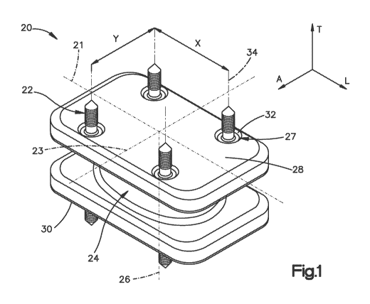
[0011] Fig. 1 is a perspective view of an intervertebral implant constructed
in
accordance with one embodiment;
[0012] Fig. 2 is a side elevation view of the intervertebral implant
illustrated in Fig. 1;
[0013] Fig. 3 is an enlarged view of Region 3 illustrated in Fig. 2 showing an
alternate
version of a recess;
[0014] Fig. 4 is an enlarged view of a portion of an intervertebral implant
similar to
Region 3 of Fig. 2, but constructed in accordance with another embodiment;
[0015] Fig. 5 is a perspective view of an intervertebral implant constructed
in
accordance with another embodiment;
[0016] Fig. 6 is an enlarged view of Region 6 illustrated in Fig. 5;
[0017] Fig. 7 is a side elevation view of an intervertebral implant
constructed in
accordance with another embodiment;
[0018] Fig. 8 is a perspective view of an intervertebral implant constructed
in
accordance with another embodiment;
[0019] Fig. 9 illustrates a magnified view of Region 9 illustrated in Fig. 8;
[0020] Fig. 10 is an enlarged view of a portion of an intervertebral implant
similar to
Region 9 of Fig. 8, but constructed in accordance with another embodiment;
4
CA 02814658 2013-04-12
WO 2012/051132 PCT/US2011/055670
[0021] Fig. 11 is an enlarged view of a portion of an intervertebral implant
similar to
Region 9 of Fig. 8, but constructed in accordance with another embodiment;
[0022] Fig. 12 is a side elevation view of an intervertebral implant
constructed in
accordance with another embodiment;
[0023] Fig. 13 is a top plan view of the intervertebral implant illustrated in
Fig. 12;
[0024] Fig. 14 is a sectional elevation view of a template constructed in
accordance
with one embodiment;
[0025] Fig. 15 is a top plan view of the template illustrated in Fig. 14;
[0026] Fig. 16 is an enlarged top plan view of a portion of a template similar
to Region
D illustrated in Fig. 15, but constructed in accordance with an alternative
embodiment;
[0027] Figs. 17 to 19 are side elevation views showing partial cross sections
of an
intervertebral implant constructed in accordance with another embodiment;
[0028] Fig. 20 is a top plan view of the intervertebral implant illustrated in
Figs. 17-19;
and
[0029] Fig. 21 is a side elevation view of an intervertebral implant
constructed in
accordance with another embodiment.
DETAILED DESCRIPTION
[0030] Referring to Figs. 1-3, an intervertebral implant 20, which can be used
a total
disc replacement, a spacer, a corpectomy device, or any other apparatus
suitable for implantation
in an intervertebral space, includes an implant body 24 and at least one bone
anchor 22, such as a
number of bone anchors 22, configured to fix the implant body 24 in an
intervertebral space 37
defined by a first superior vertebral body 38 and a second vertebral body 40
that present
respective first and second vertebral endplates 39 and 41. In accordance with
the illustrated
embodiment, the implant 20 includes eight bone anchors 22; however, any number
of bone
anchors 22 could be used as desired. The bone anchors 22 can be made from a
metal, polymer or
any reinforced polymer such as a fiber reinforced polymer. In addition, the
bone anchors 22 can
be made of a biodegradable material like surface treated Mg or Iron-based
alloys.
[0031] The implant body 24 defines a first bone contacting surface 28 and an
opposed
second bone contacting surface 30 that is spaced from the first bone
contacting surface 28 along
a central axis 26 that extends along a transverse direction T. The first and
second bone
contacting surfaces 28 and 30 can extend substantially in respective planes
that are substantially
orthogonal to the central axis 26. The first and second bone contacting
surfaces 28 and 30 can
define a substantially rectangular shape in a cross-section orthogonal to the
central axis 26, a
CA 02814658 2013-04-12
WO 2012/051132 PCT/US2011/055670
center of which can lie on the central axis 26. It should be appreciated that
the implant body 24
and bone contacting surfaces 28 and 30 can have any alternative shape as
desired. For example,
either the first bone contacting surface 28 or the second contacting surface
30 can have a
substantially circular or oval shape in a cross-section orthogonal to the
central axis 26. The
implant body 24 defines a longitudinal axis 21 extending along a longitudinal
direction L that is
orthogonal to the transverse direction T, and a lateral axis 23 that extends
along a lateral
direction A that is orthogonal to the transverse and longitudinal directions T
and L. In
accordance with the illustrated embodiment, the transverse direction T is
oriented vertically, and
the longitudinal and lateral directions L and A are oriented horizontally,
though it should be
appreciated that the orientation of the implant 20 may vary during use. As
illustrated, the implant
body 24 defines a longitudinal length, a lateral width, and a transverse
height.
[0032] The implant 20 can define at least one engagement member 27 in the form
of a
recess 32, such as a number of recesses 32, that extend transversely into the
first bone contacting
surface 28. The implant 20 can further define at least one recess 32, such as
a number of
recesses 32, that extend transversely into the second bone contacting surface
30. In accordance
with the illustrated embodiment, the implant 20 defines an equal number (e.g.,
four) of recesses
32 that extend into the bone contacting surfaces 28 and 30. The recesses 32
can be configured as
pockets that each extends along a respective hole axis 34. In accordance with
the illustrated
embodiment, the recesses 32 are disposed proximate to the corners of the bone
contacting
surfaces 28 and 30. The recesses 32 in the first and second bone contacting
surfaces 28, 30 of
the implant body 24 are arranged in such a way that their hole axes 34 are
spaced apart by a
longitudinal distance X and by a lateral distance Y.
[0033] The bone anchors 22 include a first portion in the form of a shaft 36
that is
configured to be anchored in the respective endplates 39 and 41, and a second
portion in the form
of a head 42 that extends, either directly or indirectly, from the shaft 36
and is configured to fit
into the recesses 32 so as to secure the vertebral body 24 to the first and
second vertebral bodies
38 and 40. The head 42 can be inline with the shaft 36, and can define a cross-
sectional
dimension greater or less than that of the shaft 36 (greater as illustrated),
or can define
substantially the same shape as the shaft 36. The bone anchor shafts 36 of the
bone anchors 22
can be provided as screws that define external threading 43 and can be self-
drilling and self-
tapping. Each bone anchor 22 can engage one recess 32 so as to attach the bone
anchor 22 to the
intervertebral implant 20. At least one recess 32 extends into a first side 61
of the implant body
24 but not through the implant body 24, such that the recess 32 is configured
to receive the head
42 of a bone anchor 22 so that the shaft 36 of the bone anchor 22 extends out
from the first side
6
CA 02814658 2013-04-12
WO 2012/051132 PCT/US2011/055670
61. At least one recess 32 extens into a second side 63 of the implant body 24
but not through
the implant body 24, such that the recess 32 is configured to receive the head
42 of a bone anchor
22 so that the shaft 36 of the bone anchor 22 extends out from the second side
63.
[0034] With continued reference to Figs. 1-3, in one embodiment, the
intervertebral
implant 20 includes the implant body 24 defining a central axis 26. The
implant body 24
includes the first bone contacting surface 28, which can be arranged
transversely to the central
axis 26, and the second bone contacting surface 30, which can be arranged
transversely to the
central axis 26. Aside from the first and second bone contacting surfaces 28
and 30, the implant
body 24 can further include a number of recesses 32 in the first bone
contacting surface 28
and/or a number of recesses 32 in the second bone contacting surface 30. The
intervertebral
implant 20 can further include a number of bone anchors 22 each configured to
be anchored to a
bone. Each of the bone anchors 22 can have the shaft 36 configured to be
anchored in a bone
and the head 42 configured and sized to be received within at least one of the
recesses 32. The
heads 42 of the bone anchors 22 that are configured to be anchored to the
first vertebral body 38
are configured and sized to be placed within at least one the recesses 32
located in the first bone
contacting surface 28. The heads 42 of the bone anchors 22 that are configured
to be anchored to
the second vertebral body 40 are configured and sized to be placed within at
least one the
recesses 32 located in the second bone contacting surface 30 of the implant
body 24. In an
embodiment, the intervertebral implant 20 can comprise a number of bone
anchors 22, and the
implant body 24 can comprise a number of recesses 32 in the first bone
contacting surface 28
and a number of recesses 32 in the second bone contacting surface 30.
[0035] With continued reference to Figs. 1-3, in an embodiment, the recesses
32 can be
configured as pocket holes penetrating into the implant body 24 from the first
and/or second
bone contacting surface 28, 30. The bone anchors 22 can be fixable in a stable
manner in the
recesses 32. Each recess 32 can be defined by surfaces of the implant body 24.
For instance, in
the depicted embodiment, each recess 32 is defined by a bottom surface 35 and
an enclosed
lateral surface 51. The enclosed lateral surface 51 can have an annular
configuration and can be
oriented substantially orthogonal to the first or second bone contacting
surface 28 or 30. The
bottom surface 35 can be substantially parallel to the first or second bone
contacting surface 28
or 32.
[0036] Figs. 2 and 3 show variants of suitable recesses 32. For instance, the
recess
shown in Fig. 3 is deeper than the recess shown in Fig. 2. The recesses can
operate in
substantially the same manner; however, the position of the bone anchor 22
inside the recess is
different in Fig. 3 than in Fig. 2. For instance, in the recess shown in Fig.
2, the bone anchor 22
7
CA 02814658 2013-04-12
WO 2012/051132 PCT/US2011/055670
abuts the bottom surface 35 partially defining the recess 32. Hence, when the
head 42 of the
bone anchor 22 is positioned within the recess 32, there is no clearance
between the head 42 of
the bone anchor 22 and the bottom surface 35 partially defining the recess 32.
In addition, when
the head 42 of the bone anchor 22 is positioned within the recess 32, there is
no clearance
between the head 42 and the enclosed lateral surface 51.
[0037] In the recess shown in Fig. 3, the bone anchor 22 is retained above the
bottom
surface 35 partially defining the recess 32, thereby allowing limited axial
movement of the bone
anchor 22 along the hole axis 34. Thus, the bottom surface 35 does not
necessarily contact the
head 42 of the bone anchor 22. Referring to Fig. 3, the recess 32 can define
respective beveled
or conical lead-in sections 33 at their openings so as to facilitate insertion
of the anchor heads 42.
In this regard, the head 42 can also be referred to as an engagement member 29
that is configured
to engage the engagement member 27 of the implant body 24 so as to secure the
bone anchors 22
to the implant body 24. The recesses 32 can have a depth along the transverse
direction T
between a first end 53 and a lower end 55 of the recess 32. In the embodiment
shown in Fig. 3,
the depth of the recess 32 along the transverse direction T is greater than
the length of the bone
anchor heads 42 along the transverse direction T when the bone anchors 22 are
transversely
oriented. However, the depth of the recess 32 along the transverse direction T
can alternatively
be substantially equal to or less than the length of the bone anchor heads 42
along the transverse
direction T as shown in Fig. 2.
[0038] The anchor heads 42 are illustrated as spherical in shape and sized to
snugly fit,
or be press-fit, into the recesses 32. For instance, the spherical anchor
heads 42 can have a
diameter that is substantially equal to the diameter of the recesses 32,
though it should be
appreciated that the anchor heads 42 can define any suitable size and shape as
desired. The
anchor heads 42 can be shaped differently than the anchor shafts 36 such that
only the anchor
heads 42 are configured to mechanically connect to the implant body 24.
Accordingly, the bone
anchors 22 can define a uni-directional plug-in connection between the heads
42 and the recesses
32 formed in the implant body 24. Thus, in this embodiment, the heads 42 of
the bone anchors
22 can be mechanically connectable to the recesses 32 only via a plug-in
connection, wherein the
heads 42 are inserted in the recesses 32. As used herein, the term "plug-in
connection" refers to
a connection where the head 42 is positioned inside the recess 32. The plug-in
connection can be
a uni-directional or an omni-directional plug-in connection. In a uni-
directional plug-in
connection, the head 42 can move in only one direction relative to the recess
32. For example, as
shown in Fig. 3, the head 42 can move only axially along the transverse
direction T but cannot
move in any other direction when positioned in the recess 32. As shown in Fig.
3, the enclosed
8
CA 02814658 2013-04-12
WO 2012/051132 PCT/US2011/055670
lateral wall 51 contacts the head 42, preventing the head 42 from moving in
the longitudinal
direction L and in the lateral direction A when the head 42 is inserted in the
recess 32. The
heads 42 of the bone anchors 22 can be removably positioned within the
recesses 32 via a
unidirectional plug-in connection. In an omni-directional plug-in connection,
the head 42 can
move in more than one direction when inserted in the recess 32. In an omni-
directional plug-in
connection, the head 42 positioned inside the recess 32 can move, for
instance, along the
transverse direction T and along the longitudinal direction L. Alternatively,
in an omni-
directional plug-in connection, the head 42 positioned inside the recess 32
can move along the
transverse direction T, along the longitudinal direction T, and along the
lateral direction A.
[0039] In accordance with one embodiment, the intervertebral implant 20 can be
inserted into the intervertebral space 37 by performing the following steps.
First, a transverse
spreading force F can be applied to the first and second vertebral bodies 38
and 40, so as to
distract the vertebral bodies 38 and 40. The intervertebral disc disposed in
the intervertebral
space 37 can be removed. The bone anchors 22 can then be secured to the
vertebral bodies 38
and 40, for instance by inserting (threadedly inserting as illustrated) the
anchor shafts 36 into the
respective vertebral endplates 39 and 41. Thus, at least one bone anchor 22,
such as a first
number of bone anchors 22, are fixed to the first vertebral body 38, and
another at least one such
as a number of bone anchors 22 are fixed to the second vertebral body 40. As
described in more
detail below with reference to Figs. 14-16, a template 44 can accurately
position the bone
anchors 22 in the respective vertebral bodies 38 and 40.
[0040] After fixing the bone anchors 22 in the first and second vertebral
bodies 38 and
40, the implant body 24 is inserted into the intervertebral space 37, and the
recesses 32 of the
implant body 24 are aligned with the previously set bone anchors 22. For
instance, the recesses
32 that extend into the superior bone contacting surface 28 are aligned with
the bone anchors 22
that have been driven into the superior vertebral body 38, and the recesses 32
that extend into the
inferior bone contacting surface 30 are aligned with the bone anchors 22 that
have been driven
into the inferior vertebral body 40. The spreading force F can then be
released, which causes the
vertebral bodies 38 and 40 to anatomically compress toward each other, thereby
causing the
recesses 32 to receive the respective bone anchor heads 42. Because the anchor
heads 42 are
form-fitted in the recesses 32, the anchor heads 42 are restricted with
respect to lateral and
longitudinal movement relative to the vertebral bodies 38 and 40 in at least
one of or both of the
lateral and longitudinal directions. In other words, in this embodiment, the
bone anchors 22 are
held form-fittingly in the recesses 32. As a result, the heads 42 are
laterally retained in the
9
CA 02814658 2013-04-12
WO 2012/051132 PCT/US2011/055670
recesses 32 to prevent a lateral movement of the implant body 24 relative to
the vertebral bodies
38 and 40.
[0041] Referring to Fig. 2, one method for fixation of an intervertebral
implant
comprises the following steps: (a) applying a spreading force F to the first
and second adjacent
vertebral bodies 38 and 40; (b) removing the intervertebral disc between the
adjacent first and
second vertebral bodies 38 and 40; (c) setting one or more bone anchors 22 in
the natural
endplate 39, 41 of one or each of the first and/or second vertebral bodies 38,
40 before inserting
an intervertebral body 24 having a number of recesses 32 or projections 64
(Fig. 12) in or on the
first bone contacting surface 28 and/or a number of recesses 32 or projections
64 (Fig. 12) in or
on the second bone contacting surface 30 between the adjacent first and second
vertebral bodies
38, 40; (d) inserting the implant body 24 into the intervertebral cavity 37
and aligning the
recesses 32 or the projections 64 of the implant body 24 with the previously
set bone anchors 22;
and (e) releasing the spreading force F so that the head 42 of each of the one
or more bone
anchors 22 in one or each of the first and second vertebral body 38, 40 is
connected to one of the
number of recesses 32 or projections 64 in the first and/or second bone
contacting surface 28, 30.
[0042] With continued reference to Fig. 2, another method for fixing an
intervertebral
implant in an intervertebral space can include the following steps: (1)
positioning and inserting
the bone anchors 22 in the endplates 39, 41 of two adjacent vertebrae 38 and
40; and (2)
inserting the implant 20 into the intervertebral space 37 and engaging the
implant 20 with the
bone anchors 22. The implant can be inserted into the intervertebral space 37
along any suitable
surgical access path, such as an anterior access or a lateral access. The
implant 20 can achieve
reliable primary stability. The bone anchors 22 are implanted directly into
the vertebral bodies 38
and 40 prior to the insertion of the implant 20, thereby reducing or
eliminating the occurrence of
damage to the endplates 39 and 41. The number of bone anchors 22 to be
implanted can be
selected by the surgeon. Therefore, the surgeon can intra-operatively improve
the primary
fixation as desired. The implant 20 can be subsequently inserted between the
two adjacent
vertebral bodies 38, 40 and brought into engagement with the implanted bone
anchors 22 to be
locked in its position.
[0043] The implant 20 can be removed from the bone anchors 22 as desired in
order to
select an implant 20 with another size, height, length, width or lordosis or
in case of a revision
procedure. A total disc replacement implant could e.g. be replaced by an
implant with a similar
bone anchor engagement pattern without removing the bone anchors. The
position/direction of
the bone anchors 22, which can be screws, can be adjusted as desired, such
that any anatomically
desired bone anchor position/direction can be chosen for fixation of the
implant. The surgeon
CA 02814658 2013-04-12
WO 2012/051132 PCT/US2011/055670
can also determine the desired depth to which the bone anchor 22 is inserted
into the vertebral
body 38 or 40 for fixation of the implant 20. One or more bone anchors 22
(e.g., four bone
anchors) can be set in each of the first and second vertebral bodies 38 and
40. The number of
recesses 32 formed in each bone contacting surface 28, 30 can be between one
and four, so that
each one bone anchor 22 engages with one recess 32. The number of recesses 32
in the first
bone contacting surface 28 can differ from the number of recesses 32 in the
second bone
contacting surface 30, and vice-versa.
[0044] One or more up to all of the anchor heads 42 can be
substantially spherical
as illustrated, thereby facilitating the insertion of the anchor heads 42 into
the recesses in any
orientation. Alternatively, one or more up to all of the anchor heads 42 and
the recesses 32 can
be polygonal such that the anchor heads 42 fit into the recesses 32 when the
anchor heads 42 are
at a predetermined angular orientation. The polygonal shape of the anchor
heads 42 can interfere
with the polygonal shape of the recesses 32 so as to prevent the anchor heads
42 from rotating in
the recesses 32 about the hole axis 34 or an axis that is orthogonal to the
hole axis 34.
[0045] Alternatively still, referring to Fig. 4, one or more up to all of the
anchor heads
42 can be sized smaller than the recesses 32 in one or both of the lateral and
longitudinal
directions, such that the anchor heads 42 are loosely received in the recesses
32 in one or both of
the longitudinal and lateral directions. A hardenable substance 48, such
preferably a glue, a
cement or a polymerizable monomer or comonomer, or any suitable alternative
fastener, can then
be inserted or injected into the recesses 32 so as to fasten the loosely
received portion or entirety
of the anchor head 42 to the implant body 24 inside the recesses 32. In other
words, the bone
anchors 22 can fit loosely within the recesses 32 so that, after completing
the insertion of the
intervertebral implant 20, the bone anchors 22 can be glued or otherwise fixed
in place to the
implant body 24 by the surgeon. The bone anchors 22 can be attached to the
bone contacting
surface 28 or 30 with an adhesive made of any polymer based glue, such as a
polyurethane-based
or fibrin glue. Any of the methods for fixing an implant described herein can
further comprise
the step of injecting the hardenable substance 48, preferably a glue, a cement
or a polymerizable
monomer or comonomer into the recesses 32, thereby securely locking the
implant 20 in position
with respect to the two adjacent vertebral bodies 38 and 40 after the bone
anchors 22 have been
inserted into the recess 32 during implantation of the intervertebral implant
20. Thus, the heads
42 of the bone anchors 22 can be connectable to the recesses 32 via the
hardenable substance 48,
which can be, for example, glue, cement or a polymerizable monomer or
comonomer.
[0046] While the bone anchors 22 have been illustrated as screws as described
above, it
should be appreciated that the bone anchors 22 can be provided as nails, pins,
screws, hooks,
11
CA 02814658 2013-04-12
WO 2012/051132 PCT/US2011/055670
staples, or any suitable alternatively constructed bone fixation member as
desired. For instance,
referring now to Figs. 5-6, the intervertebral implant 20 can be constructed
such that the bone
anchors 22 are configured as hooks 52 each having a shaft 36 that can be
configured as a
substantially straight pin or nail that can be pressed into the vertebral
endplates 39 and 41 of the
vertebral bodies 38 and 40. In particular, the shafts 36 can be pressed
directly into the vertebral
bodies 38 and 40, or a hole can be pre-formed in the vertebral bodies and the
shafts 36 can be
pressed into the pre-formed holes. Thus, it should be appreciated that the
shaft 36 can be
unthreaded.
[0047] The heads 42 of the hooks 52 can be angularly offset with respect to
the shaft
36, and can be bent so as to define one or more elbows 45, or can extend
substantially straight or
can be curved or otherwise shaped as desired. In accordance with the
illustrated embodiment,
the heads 42 include a proximal head portion 42a that extends from the shaft
36 and is angularly
offset with respect to the shaft 36, and a distal head portion 42b that is
angularly offset with
respect to the proximal head portion 42a and separated by the proximal head
portion 42a by the
elbow 45.
[0048] With continuing reference to Figs. 5-6, the implant body 24 defines
beveled
lead-in surface 31 that is connected to a substantially horizontal base 47 of
each recess 32 that
defines a mouth 49 of the recess 32. Each recess 32 can further include an
undercut 50 that
extends longitudinally into the implant body 24 from the mouth 49, such that
the implant body
32 defines an overhang 25 that extends over the undercut 50. The undercut 50
is sized
substantially equal to the distal portion 42b of the hook 52.
[0049] During operation, the vertebral bodies 38 and 40 can be spread apart,
and
intervertebral disc material can be removed, and the shafts 36 of the hooks 52
can be inserted
into the vertebral bodies 38 and 40 in the manner described above. The implant
body 24 can
then be positioned such that the heads 42 of the hooks are aligned with the
mouths 49 of the
respective recesses 32, and the heads 42 can be inserted into the recess 32 in
a transverse
direction and subsequently inserted into the undercuts 50 by longitudinally
displacing the
implant body 24. Alternatively, the anchor heads 42 can be installed in the
implant body 24
prior to inserting the anchor shafts 36 into the vertebral bodies 38 and 40.
[0050] The distal head portions 42b can have a cross-sectional dimension
substantially
equal to that of the undercuts 50 so that the distal head portions 42b form-
fittingly engage with
the respective recesses 32. Alternatively, the heads 42, for instance the
distal head portions 42b,
can be sized less than the recess, for instance at the undercut 50, and thus
configured to be
loosely received in the recesses 32. A hardenable substance 48 can be injected
into the recesses
12
CA 02814658 2013-04-12
WO 2012/051132 PCT/US2011/055670
32 so as to fix the anchor heads 42 to the implant body 24 inside the recesses
32. Alternatively
or additionally, any suitable mechanical fastener can fix the anchors 22 to
the implant 24.
[0051] Referring now to Fig. 7, the bone anchors 22 of the intervertebral
implant 20 can
include a cannulation or bore 54 that extends along the shaft axis, and can
extend through or into
the shaft 36, and additionally or alternatively can extend through or into the
head 42. The bone
anchors 22 can further include at least one radial perforation 56 such as a
number of radial
perforations 56 that extend into the shaft 36 and/or head 42 to a depth such
that the radial
perforations 56 are in fluid communication with the cannulation 54. In
accordance with the
illustrated embodiment, the cannulations 54 can define an opening at the
terminal end of the
shaft 36, and can extend into the anchor head 42. Thus, a bone cement 58, or
any other suitable
hardenable substance,can be injected through the cannulation 54 and the radial
perforations 56
into the bone tissue surrounding the bone anchor 22. The bone anchors 22 can
be cannulated
and can comprise radial perforations 56. Thus, a bone cement can be injected
through the
cannulation 54 and the radial perforations 56 into the surrounding bone. Any
of the exemplary
methods for fixing an implant described above can further include the step of
injecting bone
cement 58, or any other suitable hardenable substance, through cannulated and
perforated bone
anchors 22, thereby securely locking the implant 20 in a desired position with
respect to the two
adjacent vertebral bodies 38 and 40.
[0052] Referring now to Figs. 8-11, the bone anchors 22 can alternatively be
configured
as staples 59. In accordance with the illustrated embodiment, the
intervertebral implant 20 can
include at least one staple 59 extending from each of the bone contacting
surfaces 28 and 30, and
a corresponding at least one recess 32 that extends into the implant body 24
from the bone
contacting surfaces 28 and 30. A first portion of the bone anchors 22 (e.g.,
staples 59) is
configured to be disposed in the recess 32, while a second portion of the bone
anchors 22 is
configured to be fixed to the vertebral bodies 38 and 40.
[0053] Each staple 59 includes a pair of spaced legs in the form of pins 60
that provide
the anchor shaft 36, and a bridge 62 that is connected between the two pins 60
and provides the
anchor head 42. In one embodiment, each staple 59 comprises two substantially
parallel pins 60
that are configured to be anchored to bone. The pins 60 are thus configured to
extend into the
vertebral bodies 38 and 40 so as to fix the staples 59 to the vertebral
bodies, and the bridge 62 is
configured to be disposed in the recess 32 so as to be connected to the
implant body 24. The
pins 60 can be tapered toward their distal ends along a direction away from
the bridge 62.
[0054] As illustrated in Fig. 9, the bridge 62 of each bone anchor 22 can be
sized
substantially equal with the respective recesses such that the bridge 62 is
press-fit inside the
13
CA 02814658 2013-04-12
WO 2012/051132 PCT/US2011/055670
recess 32. The bridge 62 can be inserted into the recesses 32 along a
direction oblique to the
central transverse axis 26 of the implant body 24. Alternatively, as
illustrated in Fig. 10, the
bridge 62 can be sized smaller than the recess such that the bridge 62 is
loosely received in the
recess 32. The bone anchors 22 can be configured as staples 59 forming an
oblique
unidirectional plug-in connection to prevent the implant from migrating in the
intervertebral
space. In the oblique unidirectional plug-in connection, the bridge 62 in
inserted inside the
recesses 32 at an oblique angle relative to the central axis 26. Moreover, in
this oblique
unidirectional plug-in connection, the bridge 26 can move only in one
direction (i.e., at the
oblique angle with respect to the central axis 26. To achieve the oblique
unidirectional plug-in
connection, the implant body 24 can include at least one angled lateral
surface 57 partially
defining the recess 32. The angled lateral surface 57 is oriented at an
oblique angle relative to
the central axis 26 and/or the bone contacting surfaces 28 or 30. The bridge
62 can be inserted
into the recesses 32 in a direction along the central axis 26 of the implant
body 24. Referring to
Fig. 11, the recess 32 can be sized larger than the bridge 62 of the bone
anchor 22. The bridging
portions 62 can be inserted into the recesses 32 in a direction along the
central axis 26 of the
implant body 24 or oblique to the central axis 26. Furthermore, the second
anchor heads 42 can
be fixed to the anchor body 24 inside the recesses 32 using a hardenable
substance 48 that can be
injected into the recesses 32.
[0055] Referring to Figs. 12-13, the bone anchors 22 of the intervertebral
implant 20
can be configured as pins 65 that each defines a head 42 configured to engage
the implant body
24 so as to fix the pin 65 to the implant body 24, and a shaft 36 configured
to be fixed to the
vertebral bodies 38 and 40. The shaft 36 can include a tapered distal tip 67
configured to
facilitate insertion into the vertebral bodies 38 and 40. The tapered distal
tips 67 can have a
substantially frusto-conical shape or any other suitable shape. Each bone
anchor 22 can include
a cavity 66 that extends into the head 42. The cavities 66 can be cylindrical
or any alternative
shape as desired.
[0056] The implant body 24 includes a number of engagement members 27 in the
form
of projections 64 that extend out from the first bone contacting surface 28
and the second bone
contacting surface 30. The projections 64 are configured to engage
complementary engagement
members 29 of the bone anchors 22 provided as the heads 42, and in particular
the cavities 66
formed into the heads 42. The number of projections 64 that extend from each
bone contacting
surface 28, 30 can be between one and four, such that each one bone anchor
engages with one
projection 64. The number of projections 64 of the first bone contacting
surface 28 can differ
from the number of projections 64 of the second bone contacting surface 30,
and vice-versa.
14
CA 02814658 2013-04-12
WO 2012/051132 PCT/US2011/055670
[0057] During operation, the shafts 36 of the bone anchors 22, which can be
cylindrically shaped or alternatively shaped as desired, can be pressed or
hammered into the
endplates 39 and 41 of the vertebral bodies 38 and 40. The projections 64 and
cavities 66 can be
substantially equally sized such that the projections 64 are press-fit inside
the cavities 66.
Alternatively, the cavities 66 can be sized greater than the projections 64,
and an adhesive, such
as a glue, can provide fixation of the projections to the bone anchors 22
inside the cavities 66.
[0058] It should thus be appreciated that the implant body 24 includes at
least one
engagement member 27, and the bone anchors 22 includes a complementary
engagement
member 29 configured to mate with the engagement member 27 of the implant body
24 so as to
secure the bone anchors 22 to the implant body 24. In one embodiment, the
engagement member
27 of the implant body 24 can be provided as a recess, such as the recess 32.
In another
embodiment, the engagement member 27 of the implant body 24 can be provided as
a protrusion,
such as the protrusion 64. In one embodiment, the engagement member 29 of the
bone anchors
22 can be a protrusion in the form of the head 42 that is received in the
recess 32. In another
embodiment, the engagement member 29 of the bone anchors 22 can be a cavity
such as the
cavity 66 formed in the head 42 that is configured to receive the protrusion
64. The implant
body 24 and the bone anchors 22 can include any alternatively constructed
engagement member
suitable to fix the bone anchors 22 to the implant body 24 such that the bone
anchors 22 can also
be fixed to the vertebral bodies 38 and 40.
[0059] With continued reference to Figs. 12 and 13, in one embodiment, the
intervertebral implant 20 comprises an implant body 24 defining the central
axis 26, the first
bone contacting surface 28 that is arranged transversely to the central axis
26, and the second
bone contacting surface 30 that is arranged transversely to the central axis
26. The implant body
24 further includes a number of projections 64 that extend from the first bone
contacting surface
28 along the transverse direction T and/or a number of projections that extend
from the second
bone contacting surface 30 along the transverse direction T. The implant 20
can further include
a number of bone anchors 22 each having the shaft 36 that is configured to be
anchored to bone
and the head 42 that defines the cavity 66 configured to engage one of the
projections 64. The
implant body 24 can further comprise a number of projections 66 that extend
from the first bone
contacting surface 28 in the transverse direction T and a number of
projections 66 that extend
from the second bone contacting surface 30 in the transverse direction T. The
bone anchors 22
can be only held form-fittingly on the projections 66. This configuration
prevents lateral
movement of the heads 42 relative to the implant body 24. Each projection 64
can fit loosely in
a cavity 66 in an axial direction so that, after completing the insertion of
the intervertebral
CA 02814658 2013-04-12
WO 2012/051132 PCT/US2011/055670
implant, the bone anchors 22 can be glued or otherwise fixed in place to the
implant body 24 by
the surgeon. Thus, the bone anchor 22 can initially engage the projections 64
with a loose fit.
Then, a polymerizeable mass can be introduced into the cavities 66 so that the
heads 42 of the
bone anchors 22 polymerize with the mass. The heads 42 of the bone anchors 22
are connectable
to the projections 64 via a harndenable substance, such as glue, cement or a
polymerizable
monomer or comonomer.
[0060] With continued reference to Figs 12 and 13, in another embodiment, the
bone
anchors 22 can be fixable in a stable manner through the projections 64. The
number of
projections 64 can be between one and four, so that each bone anchor 22
engages one projection
64. The number of projections 64 that extend from the first bone contacting
surface 28 can differ
from the number of projections 64 that extend from the second bone contacting
surface 30, and
vice-versa. The heads 42 of the bone anchors 22 can be mechanically
connectable to the
projections 64, via, for instance, a plug-in connection. In a plug-in
connection, the projections
64 are inserted inside the cavities 66. This plug-in connection can be a uni-
directional plug-in
connection. In a uni-directional plug-in connection, a projection 64 is
inserted inside a cavity 66
so that the bone anchor 22 can only move in one direction. For instance, in an
uni-directional
plug-in connection, when the projection 64 is inserted inside the cavity 66,
the bone anchor 22
can only move in the transverse direction T. In use, upon connection of the
heads 42 to the
projections 64, there is no axial or lateral clearance between each of the
heads 42 of the bone
anchors 22 and the projections 64. The heads 42 of the bone anchors 22 can be
removably
connected to the projections 64 via a uni-directional plug-in connection as
described above. The
bone anchors 22 can be in the form of pins or screws. The bone anchors 22 can
be cannulated
and can comprise radial perforations as described above with respect to the
embodiment
illustrated in Fig. 7. For instance, the bone anchors 22 can define a
cannulation or bore 54 (Fig.
7) extending along the transverse direction T along the shaft 36 and the head
42. Radial
perforations 56 (Fig. 7) are in fluid communication with the cannulation 54
(Fig. 7) and extend
from the cannulation 54 through the wall forming the shaft 36 along the
longitudinal direction L
and along the lateral direction A. Bone cement can be injected through the
cannulation 54 (Fig.
7) and the radial perforations 56 (Fig. 7) into the bone. The bone anchors 22
can be configured
as staples each comprising two or more substantially parallel pins configured
to be anchored to
bone as described above with respect to Figs. 9-11. The heads 42 of the bone
anchors 22 can be
removably connected to the projections 64 in an oblique direction relative to
the central axis 26
of the implant body 24.
16
CA 02814658 2013-04-12
WO 2012/051132 PCT/US2011/055670
[0061] Referring now to Figs. 14 and 15, an intervertebral implant assembly
can
include the implant 20 and a template 44 that is configured to properly
position the bone anchors
22 such that they are aligned with the engagement members (e.g., recesses 32
or projections 64)
prior to inserting the bone anchors 22 into the vertebral bodies 38 and 40.
The template 44
includes a first or upper plate 68, a second or lower plate 70 that is
transversely spaced from the
upper plate 68, and a spacer 72 disposed between the upper and lower plates 68
and 70, for
instance at an end of the plates 68 and 70.
[0062] The upper and lower plates 68 and 70 can be elongate along a central
longitudinally axis 74 and a central lateral axis 76. The template 44 includes
numberone or more
aiming holes 46 that extend transversely through the upper and lower plates 68
and 70. In the
illustrated embodiment, the aiming holes 46 are spaced along the longitudinal
distance X, and the
lateral distance Y, such that the aiming holes 46 are arranged so as to
correspond to the
arrangement of the engagement members of the implant body 24. The aiming holes
46 can be
substantially cylindrical as illustrated in Fig. 15 and can be sized
substantially equal to or greater
than the heads 42 of the bone anchors 22. Accordingly, during operation, after
the bone anchors
22 have been inserted through the aiming holes 46 and into the respective
vertebral bodies 38
and 40, the template 44 can be displaced along the transverse direction so as
to allow the
spherical heads 42 of the bone anchors 22 to pass through the passage holes
46.
[0063] Alternatively, referring to Fig. 16, the aiming holes 46 can be key-
hole shaped
having a first aiming portion 77 and a passage 78 connected to the aiming
portion 77 and
horizontally (e.g., longitudinally) offset from the aiming portion and
defining a size that is
greater than the aiming portion 77. The aiming portion 77 can have a shape
that is sized
substantially the same as at least a portion of the bone anchor shaft 36, so
as to ensure that the
bone anchor 22 is accurately positioned in the vertebral bodies 38 and 40 when
the shaft 36 is
inserted through the aiming portion 77. The passage 78 can be sized greater
than the shaft 36
and the head 42, such that once the bone anchors have been inserted into the
vertebral bodies
through the aiming portion 77, the template 44 can be translated
longitudinally so as to align the
passages 78 with the bone anchors 22. The template 44 can then be translated
in the transverse
direction to slide the passages 78 over the heads 42 and remove the template
44 from the
intervertebral space 37. A number of aiming holes 46 can be provided in the
template 44 in an
arrangement that corresponds to the arrangement of the recesses 32 or
projections 64 of the
implant 20. Any of the methods for fixing an implant described herein can
further comprise the
step of setting one or more bone anchors using the template 44 which includes
one or more
aiming holes 46 that position the one or more bone anchors 22 as desired in
one or each of the
17
CA 02814658 2013-04-12
WO 2012/051132 PCT/US2011/055670
first and second vertebral bodies 38, 40 so that the bone anchors 22 are
engageable with recesses
32 or projections 64.
[0064] Referring now to Figs. 17-20, the intervertebral implant 20 includes
the implant
body 24 and at least one bone anchor 22. In accordance with the illustrated
embodiment, the
recess 32 can be sized to receive a number of anchor heads 42. Thus, the
engagement member
27 of the implant 20 can be configured to receive at least one, such as a
number of bone anchors
22, so as to fix the received bone anchors 22 to the implant body 24. The bone
anchors 22, such
as screws, pins or other bone anchors can be coupled two by two. The first and
second bone
contacting surfaces 28 include one recess 32 in accordance with the
illustrated embodiment.
Thus, the implant 20 can include at least one recess 32 in both surfaces 28
and 30, such that the
recess 32 retains at least one up to all of the bone anchors 22 that are fixed
to the implant body
24 at the respective surfaces 28 and 30. Thus, a number of bone anchors 22 set
in the natural
endplate 39 or 41 of the same vertebral body 38 or 40 can engage one recess 32
on the respective
bone contacting surface 28, 30 of the implant body 24. The number of recesses
32 in the first
bone contacting surface 28 can differ from the number of recesses 32 in the
second bone
contacting surface 30, and vice-versa.
[0065] The recesses 32 are illustrated as substantially rectangular having a
longitudinal
length L and a lateral width W. The length L and the width W of each recess 32
are dimensioned
such that the head 42 of the number of (e.g., four) bone anchors 22 can be
positioned in the
recess. The shafts 36 of the bone anchors 22 can be threadedly driven or
otherwise inserted into
the first and second vertebral bodies 38 and 40. The heads 42 can be
configured as screw heads
which are placed in the respective recess 32 and fixed to the implant body 24
via a hardenable
substance 48 that is injected into the recess 32, or any alternative
mechanical fastener. As
illustrated in Fig. 17, the bone anchors 22 can be oriented such that their
central axes extend
substantially parallel to the central axis 26 of the implant body 24. The bone
anchors 22 can be
implanted in the vertebral bodies 38, 40 at an angle of 90 with regard to the
surface of the
endplate 28. 30. Alternatively, the bone anchors 22 can be oriented such that
their central axes
are oblique with respect to the central axis 26. For instance, the central
axes of the bone anchors
22 can diverge from each other in a transverse direction out from the implant
body 24 toward the
tips 80, or can alternatively converge toward each other in a transverse
direction out from the
implant body 24 toward the tips 80. The bone anchors 22 can be implanted in
the vertebral
bodies 38, 40 at an angle deviating from 90 with regard to the surface of the
endplate 28, 30.
The bone anchors 22 can include bone screws, pins or staples. The heads 42 of
the bone anchors
18
CA 02814658 2013-04-12
WO 2012/051132 PCT/US2011/055670
22 can engage with the recesses 32 so that the bone anchors 22 are in an
oblique direction
oblique relative to the central axis 26 of the implant body 24.
[0066] It should be appreciated that any of the engagement members or
surfacing
defining the recesses of the previously described embodiments (.e.g., surfaces
35, 51, or 57) or
the surfaces defining the cavity 66 can include a shape changing component or
be made at least
partially from a shape memory material, such as a shape memory polymer or a
shape memory
alloy. The shape memory material will be configured to have a first, initial,
configuration and a
second, fixing, configuration. In the initial configuration, the shape memory
material allows the
bone anchor to be positioned in the recess. In the fixing configuration, the
shape memory
material moves to hold fixedly the bone anchor in the recess. The transition
from the initial to
the fixing configuration is activated by the application of light or heat
thereto, though it is
appreciated that other activation methods are available depending on the shape
memory material.
The process of transitioning the shape memory material from the initial
configuration to the
fixing configuration can of course be reversed from the fixing configuration
to the initial
configuration as desired.
[0067] The shape memory material can be any suitable material as desired. For
example, the shape memory material could include polymers such as
thermoplastic multiblock
copolymers like polyurethanes, polyesterurethanes or multiphase polymer
networks like poly(e-
caprolactone) dimethacrylate and n-butyl acrylate, multiblock copolymers
containing poly(L-
lactide) and poly[glycolide-co-(e-caprolactone)]-segments. The shape memory
material could
also be an alloy such as NiTi, Ag-Cd 44/49 at.% Cd, Au-Cd 46.5/50 at.% Cd, Cu-
Al-Ni 14/14.5
wt.% Al and 3/4.5 wt.% Ni, Cu-Sn approx. 15 at.% Sn, Cu-Zn 38.5/41.5 wt.% Zn,
Cu-Zn-X (X =
Si, Al, Sn), Fe-Pt approx. 25 at.% Pt, Mn-Cu 5/35 at.% Cu, Fe-Mn-Si, Pt
alloys, Co-Ni-Al, Co-
Ni-Ga, Ni-Fe-Ga, Ti-Pd in various concentrations, Ni-Ti (-55% Ni), Ni-Ti-Nb,
and Ni-Mn-Ga.
[0068] The bone anchors can include a mechanical interlocking mechanism, such
as
threads, a ratchet mechanism or shaft, that expands in volume by an
introduction of gas, water or
vapor creation or via a shape changing component, such as shaft 36, component
comprising a
shape memory material (e.g. a shape memory polymer or a shape memory alloy) to
fix the bone
anchors to the vertebral bodies. Suitable shape memory polymers may include
thermoplastic
multiblock copolymers like polyurethanes, polyesterurethanes or multiphase
polymer networks
like poly(e-caprolactone) dimethacrylate and n-butyl acrylate, multiblock
copolymers containing
poly(L-lactide) and poly[glycolide-co-(e-caprolactone)] -segments. Suitable
shape memory
alloys may include NiTi, Ag-Cd, Cu-Al-Ni, Cu-Sn, Cu-Zn, Cu-Zn-X (X = Si, Al,
Sn), Fe-Pt,
Mn-Cu, Fe-Mn-Si, Pt alloys, Co-Ni-Al, Co-Ni-Ga, Ni-Fe-Ga, Ti-Pd, Ni-Ti-Nb, and
Ni-Mn-Ga A
19
CA 02814658 2013-04-12
WO 2012/051132 PCT/US2011/055670
bone screw combination can be used to fix the implant to the vertebral bodies,
such as a first
screw that extends through the head of a second screw.
[0069] With reference to Fig. 21, it should be further appreciated that a
suture 82 can be
used to fix the bone anchor 22 in the recess 32 of the implant body. The bone
anchor 22 and
recess 32 may be any or a combination of the bone anchors 22 and recesses 32
described of the
type described herein. The suture 82 can provide the primary fixation and/or
can be used in
conjunction with any of the other engagement members previously described to
fix the bone
anchor 22 to the implant body 24. The suture 82 may be a single thread or a
double thread.
[0070] In this further embodiment, the implant body 24 can define a channel 84
that
starts from a location in the recess 32 and ends at an opening 86 on a surface
of the implant body
24, which can be a non-bone contacting surface. Thus, the channel 84 comprises
at least one
first opening 92 located at or near the recess 32, or any other engagement
member 27, the second
opening 86 on a surface of the implant body 24 other than a bone-contacting
surface 28, 30, and
one or more passageways 94 extending between the first opening 92 and the
second opening 86.
The opening 86 is accessible to, for example, a surgeon when a bone contacting
surface abuts or
is engagement with bone. The channel 84 allows the suture 82 to be passed from
the bone
anchor 22 to the surface where it is tied in order to fix the bone anchor 24
to the implant body 24.
The head 42 of the bone anchor 22 can define an eyelet or hole 88 configured
and sized to
receive at least a portion of the suture 82. The suture 82 can be inserted
through the hole 88 and
positioned around the head 42, and then tied to the head 42. The channel 84
may be a single
passageway or may be two closely aligned passageways.
[0071] In the variant of the channel 84 where the single passageway is used,
the suture
82 is tied in a suitable configuration to provide an anchoring object to
prevent the suture from
withdrawing into the channel 84. The anchoring object could be a suitable knot
90, as
illustrated, or the suture could be fixed to a body that serves as the
anchoring object.
[0072] In the embodiment where the implant body features two closely aligned
passageways, the two passageways are separated by a part of the surface of the
implant body.
The surgeon will thread a strand of the suture, for example the double
threaded suture, down
either passageway and can then tie those strands together to thereby use the
part of the surface as
an anchoring object.
[0073] In one embodiment, the bone anchors 22 can be fixed by preliminary
insertion
of a suture 82 into the implant body 24. The endplate may comprise a channel
in which a suture
82 is fixedly retained. The channel 84 may comprise at least one passageway
94. The channel
84 may have an opening 92, 86 in both a recess and on a non-bone contacting
surface. The
CA 02814658 2013-04-12
WO 2012/051132 PCT/US2011/055670
suture 82 may be fixed to the implant body 24 using an anchoring object such
as a knot 90 in the
suture 82, an external body and/or a part of the implant body 24. In one
embodiment, a kit can
comprise the implant 20 and the template 44.
[0074] An intervertebral implant kit can include at least one implant body 24
such as a
number of implant bodies 24, at least one bone anchor 22 such as a number of
bone anchors 22,
and/or at least one template 44 such as a number of templates 44. The implant
bodies 24 can be
constructed in accordance with any of the embodiments described herein, and
can be constructed
the same as or differently from each other. The bone anchors 22 can be
constructed in
accordance with any of the embodiments described herein, and can be
constructed the same as or
differently from each other. The templates 44 can be constructed in accordance
with any of the
embodiments described herein, and can be constructed the same as or
differently from each
other.
[0075] Although the disclosure has been described in detail, it should be
understood
that various changes, substitutions, and alterations can be made herein
without departing from
the spirit and scope of the invention as defined by the appended claims.
Moreover, the scope of
the present disclosure is not intended to be limited to the particular
embodiments of the process,
machine, manufacture, composition of matter, means, methods and steps
described in the
specification. As one of ordinary skill in the art will readily appreciate
from the disclosure of the
present invention, processes, machines, manufacture, composition of matter,
means, methods, or
steps, presently existing or later to be developed that perform substantially
the same function or
achieve substantially the same result as the corresponding embodiments
described herein may be
utilized according to the present disclosure.
21