Note: Descriptions are shown in the official language in which they were submitted.
CA 02814825 2015-06-09
METHOD AND APPARATUS FOR DETECTING SEIZURES
BACKGROUND
[0002] A seizure may be characterized as abnonnal or excessive synchronous
activity in the
brain. At the beginning of a seizure, neurons in the brain may begin to fire
at a particular location. As
the seizure progresses, this firing of neurons may spread across the brain,
and in some cases, many
areas of the brain may become engulfed in this activity. Seizure activity in
the brain may cause the
brain to send electrical signals through the peripheral nervous system to
different muscles. For
example, an electrical signal may originate in the central nervous system and
initiate the propagation
of an electrical signal through motor neurons. A motor neuron may, for
example, communicate with a
muscle through interaction with the motor end plate of a muscle fiber; thereby
initiating an action
potential and depolarization of muscle cells within a given motor unit.
Depolarization typically results
from the coordinated flow of ions, e.g., sodium and potassium cations, through
channels within a
muscle cell membrane. That is, changes in states of ion channels initiate a
change in the permeability
of a cell membrane, and subsequent redistribution of charged ions. Current
flow through muscle cells
may initiate a corresponding flow in the tissue above the muscle and thus an
electrical signature at the
surface of the skin.
[0003] Techniques designed for studying and monitoring seizures have typically
relied upon
electroencephalography (EEG), which characterizes electrical signals using
electrodes attached to the
scalp or head region of a seizure prone individual, or seizure patient. EEG
electrodes may be
positioned so as to measure such activity, that is, electrical activity
originating from neuronal tissue.
Compared to EEG, electromyography (EMG) is a little-used technique in which an
electrode may be
placed on or near the skin, over a muscle, to detect an electrical current or
change in electric potential
in response to redistribution of ions within muscle fibers.
[0004] Detecting an epileptic seizure using electroencephalography (EEG)
typically requires
attaching many electrodes and associated wires to the head and using
amplifiers to monitor brainwave
activity. The multiple EEG electrodes may be very cumbersome and generally
require some
technical expertise to apply and monitor. Furthermore, confirrnMg a seizure
requires observation in
an environment provided with video n-onitors and video recording equipment.
Unless used in a
staffed clinical environment, such equipment is frequently not intended to
determine if a seizure is in
progress but rather provide a historical record of the seizure after the
incident. Such equipment is
usually meant for hospital-like environments where a video camera recording or
caregiver's
1
CA 02814825 2015-06-09
observation may provide corroboration of the seizure, and is typically used as
part of a more
intensive care regimen such as a hospital stay for patients who experience
multiple seizures. A
hospital stay may be required for diagnostic purposes or to stabilize a
patient until suitable
medication can be administered. Upon discharge from the hospital, a patient
may be sent home with
little further monitoring. However, at any time after being sent home the
person may experience
another seizure, perhaps fatal.
[0005] A patient should in some cases be monitored at home for some length of
time in case
another seizure should occur. Seizures with motor manifestations may have
patterns of muscle
activity that include rhythmic contractions of some, most, or all of the
muscles of the body. A seizure
could, for example, result in Sudden Unexplained Death in Epilepsy (SUDEP).
The underlying causes
of SUDEP are not well understood; however, some possible mechanisms causing
SUDEP may
include tonic activation of the diaphragm muscle so as to prevent breathing,
neurogenic pulmonary
edema, asystole, and other cardiac dysrhythmia. If a sleeping person
experiences a seizure involving
those conditions, then caregivers may not be aware that the seizure is
occurring, and thus be unable to
render timely aid.
[0006] While there presently exist ambulatory devices for diagnosis of
seizures, they are
EEG-based and are generally not designed or suitable for long-term home use or
daily wearability.
Other seizure alerting systems may operate by detecting motion of the body,
usually the extremities.
Such systems may generally operate on the assumption that while suffering a
seizure, a person will
move erratically and violently. For example, accelerometers may be used to
detect violent extremity
movements. However, depending upon the type of seizure, this assumption may or
may not be true.
Electrical signals sent from the brain during the seizure are frequently
transmitted to many muscles
simultaneously, which may result in muscles fighting each other and
effectively canceling out violent
movement. In other words, the muscles may work to make the person rigid rather
than cause actual
violent movement. Thus, the seizure may not be consistently detected with
accelerometer-based
detectors.
[0007] Accordingly, there is a need for an epileptic seizure detection method
and apparatus
that can be used in a non-institutional or institutional envirornnent without
many of the cumbersome
electrodes to the head or extremities. Such an apparatus may be minimally
intrusive, minimally
interfere with daily activities and be comfortably used while sleeping. There
is also a need for an
epileptic seizure detection method and apparatus that accurately detects a
seizure with motor
manifestations and may alert one or more local and/or remote sites of the
presence of a seizure.
Furthermore, there is a need for an epileptic seizure detection method and
apparatus that may be used
in a home setting and which may prov'de robust seizure detection, even in the
absence of violent
motion, and which may be personalizable, e.g., capable of being tailored for
an individual or specific
population demographic.
2
CA 02814825 2015-06-09
SUMMARY
[0008] In some embodiments, a method of detecting seizures may comprise
receiving an
EMG signal and processing the received EMG signal to determine whether a
seizure characteristic is
present in the EMG signal during a time window.
[0009] In some embodiments, an apparatus for detecting seizures with motor
manifestations
may comprise one or more EMG electrodes capable of providing an EMG signal
substantially
representing seizure-related muscle activity; and a processor configured to
receive the EMG signal,
process the EMG signal to determine whether a seizure may be occurring, and
generate an alert if a
seizure is determined to be occurring based on the EMG signal.
[00101 In some embodiments, apparatuses and methods comprise a detection unit
which
includes EMG electrodes and a base unit in communication and physically
separated from said
detection unit, wherein the base station is configured for receiving and
processing EMG signals from
the detection unit, determining from the processed EMG signals whether a
seizure may have occurred,
and sending an alert to at least one caregiver. In some embodiments, the base
station may separately
process the data provided by the detection unit for verification of the alarm
condition. If the base
station agrees with the alarm, then the base station may generate an alarm to
remote devices and local
sound generators. Having the base station agree to the detection unit's alarm
may introduce a voting
concept. Both devices must vote on the decision and agree to sound the alarm.
This may be used to
limit false alarms.
100111 In some embodiments, a method and apparatus for detecting a seizure and
providing a
remote warning of that incident is provided. Such a method may detect seizures
using EMG
electrodes. One or more EMG electrodes may be attached to an individual's body
and one or more
characteristics from the signal output of the one or more EMG electrodes may
be analyzed. EMG
output may be compared to general seizure characteristics and to one or more
threshold values. If one
or more values of the output data exceed one or more thresholds an event may
be registered, e.g.,
logged on a register. Analysis of events logged in registers for different
characteristics of the output
data may be used to assess whether a seizure incident is declared and whether
an alarm is sent to one
or more locations.
[0012] In some embodiments, an apparatus for detecting seizures with motor
manifestations
may include a detector unit and a base unit. The detector unit may include one
or more
electromyography (EMG) electrodes, and optionally one or more
electrocardiography (ECG)
electrodes. The detector unit and base unit may be in communication with each
other, such as by
wireless communication. The detector unit and base unit may include electronic
components
configured to execute instructions for evaluation of EMG signal data. The base
unit may be enabled
for sending an alami to one or more remote locations. Alternatively, the base
unit may be in
3
CA 02814825 2015-06-09
communication with a separate transceiver. That transceiver may be physically
distinct but within the
general locale of the base unit. That transceiver may be enabled for sending
an alarm to one or more
remote locations.
[0013] In some embodiments, an alarm protocol may be initiated based on a
convolution of
data in a plurality of data registers. Individual registers may, for example,
each be responsive to
detection of a different seizure variable. An alarm protocol may be initiated
if a supervisory
algorithm, that supervisory algorithm responsive to the values in the
plurality of registers, determines
that an alarm protocol should be initiated.
[0014] In some embodiments, seizure detection methods as described herein may
be adaptive.
For example, threshold values may be adjusted as seizure data is collected
from one or more patients.
In. addition, algorithms, which may be used to determine whether a seizure
incident is declared, may
be modified. Algorithms may, for example, be modified by adjusting variable
coefficients. Those
coefficients may be associated with, and weight, seizure variables. The
adjustment of such
coefficients may be based on seizure data that is collected from one or more
patients, including, but
not limited to an individual patient, or other patients, such as those of a
particular demographic. The
association between registered events, the initiation of alarm protocols, and
seizure related incidents,
e.g., declared events, actual seizures and inaccurately reported incidents,
may be tracked and used to
update variables in a detection method and thus improve the accuracy of a
seizure detection method or
apparatus.
[0015] In some embodiments, a historical record of patient seizure data and
related incidents
may be collected. A user may analyze a historical record and modify or change
one or more sub-
methods or alter the distribution of sub-methods that are included in a method
for detecting a seizure.
A sub-method may, for example, be a set of instructions which may be used to
increment a counter.
Sub-methods may include data, including for example, threshold values,
weighting coefficients and
other data, may be provided in a template file, may have a "factory default"
setting, and may change
as the method adapts to a particular patient,
[0016] In some embodiments, the value of a plurality of seizure variables may
be determined
for a patient. Individual seizure variables may be selected and analyzed using
algorithms such that
events logged for an individual seizure variable is unlikely to trigger an
alarm; however, the
convolution of events logged for the plurality of seizure variables may raise
the confidence with
which a seizure may be detected.
[0017] In some embodiments, a method and apparatus may be used, for example,
to initiate
an alarm protocol, create a log of seizure incidents to help medically or
surgically manage the patient,
activate a Vagal Nerve Stimulator, or activate other stimulating devices that
may be used to abort or
attenuate a seizure. In some embodiments, a log of seizure related incidents
may prompt a physician
to understand more quickly the failure of a treatment regimen.
4
CA 02814825 2016-09-06
10017a1 In another embodiment of the present invention there is provided an
apparatus
for detecting seizures with motor manifestations, the apparatus comprising:
one or more
electromyography electrodes configured to provide an electromyography signal
representing
seizure-related .musele activity; a processor configured to receive the
electromyography signal
and process the electromyography signal to determine when a seizure is
occurring based on the
electromyography signal; said processor configured to detect bursts of the
electromyography
signal, assign certainty values to individual bursts among said detected
bursts, and determine a
burst count contribution to seizure detection based on a number of said
detected bursts weighted
as a function of the certainty values assigned to said individual bursts; said
processor
configured to qualify bursts against a minimum threshold duration and maximum
threshold
duration; said processor configured to determine said certainty values based
on how well the
individual bursts compare to a reference burst in terms of one or more burst
characteristics
selected from the group of characteristics including burst signal-to-noise
ratio, burst width, and
burst amplitude; said processor configured to identify the presence of a
plurality of bursts over
a time window, determine the periodicity of bursts over said time window, and
determine a.
periodicity contribution to seizure detection; said processor further
configured to combine said
burst count contribution and said periodicity contribution using a supervisory
algorithm to
determine a seizure detection value, and compare said seizure detection value
to a threshold
seizure detection value indicative of when a seizure is occurring; and said
processor further
configured to generate an alert if a seizure is occurring.
I0017b] In a further embodiment of the present invention there is provided a
method of
monitoring a patient for motor manifestations of seizure activity comprising.:
monitoring, the
patient by collecting an electromyography signal using electromyography
electrodes;
processing, with a processor of the electromyography signal to detect bursts,
assign certainty
values to individual bursts among said detected bursts, and determine a burst
count contribution
to seizure detection based on a number of said detected bursts weighted as a
function of the
certainty values assigned to said individual bursts: processing to qualify
bursts against a
minimum threshold duration and maximum threshold duration; wherein said
certainty values
are based on how well the individual bursts compare to a reference burst in
terms of one or
more burst characteristics selected from the group of characteristics
including burst signal-to-
noise ratio, burst width, and burst amplitude; identifying the presence of a
plurality of bursts
ovcr a time window. determining the periodicity of bursts over said time
window, and
determine determining a periodicity contribution to seizure detection:
integrating said burst
4a
CA 02814825 2016-09-06
count contribution and said periodicity contribution into a supervisory
algorithm to determine
if said seizure activity is occurring: and initiating an alert if a seizure is
occurring.
[0017e] In yet another embodiment of the present invention there is provided a
system
of apparatuses for monitoring a patient for seizure activity, the system
comprising: one or .more
electromyography electrodes configured to provide an electromyography, signal;
a processor
configured to receive the electromyography signal and process the
electromyography signal to
detect bursts characteristic of seizure activity based on whether regions of
the
electromyography signal meet criteria suitable to be qualified as bursts,
including a comparison
of criteria values to thresholds; wherein said criteria values include a
duration width and one
Or more of a signal-to-noise ratio and an amplitude: wherein said thresholds
include a minimum
duration width, a maximum duration width, and one or more of a minimum signal-
to-noise
ratio, minimum amplitude. and maximum amplitude: and wherein said processor is
configured
to determine a detected burst count and include said detected burst count in a
determination of
whether to send an alert indicating detection of said seizure activity to one
or more remote
devices.
10017d1 In yet a further embodiment ofthe present invention there is provided
a method
for monitoring a patient for seizure activity. the method comprising:
collecting an
electromyography signal from the patient using one or more electrmnyography
electrodes;
processing with one or more processors the electromyography signal to detect
bursts
characteristic of seizure ;Activity based on whether regions of the
electromyography signal .meet
criteria suitable to be qualified as said bursts, including a comparison of
criteria values to
thresholds; wherein said criteria values include a duration width and one or
more of a signal-
to-noise ratio and an amplitude: wherein said thresholds include a minimum
duration width. a
maximum duration width, and one or more of a minimum signal-to-noise ratio.
minimum
amplitude, and maximum amplitude; determining a detected burst count; and
weighting said
detected burst count in determining Whether to send an alert indicating
detection ofsaid seizure
activity to one or more remote devices.
(0017e] In another embodiment of the present invention there is provided a
method of
monitoring a patient for seizure activity comprising: collecting an
electromyography signal
from the patient using one or more electromyography electrodes; processing
with a processor
the electromyography signal to detect bursts, assign certainty values to
individual bursts among
said detected buists, and determine a burst count contribution to seizure
detection based on a
number of said detected bursts weighted as a function of the certainty values
assigned to said
4b
CA 02814825 2016-09-06
individual bursts; wherein said certainty values are based on how well one or
more
characteristics of the individual bursts compare to one or more reference
burst characteristics:
said. one or more characteristics selected from a group of characteristics
consisting of burst
signal-to-noise ratio, burst duration width. and burst amplitude; wherein
burst detection
includes determining the presence of bursts based on whether regions of the
electromyography
signal meet criteria suitable to be qualified as bursts, including a
comparison of criteria values
to thresholds; wherein said criteria values include a duration width and one
or more of a signal-
to-noise ratio and an amplitude; wherein said thresholds include a minimum
duration width. a
maximum duration width, and one or more of a minimum signal-to-noise ratio.
minimum
amplitude, and maximum amplitude; including said burst count contribution into
an algorithm
to determine whether a seizure is occurring; and initiating an alert if a
seizure is occurring.
[001711 In a further embodiment oldie present invention there is provided a
method for
reviewing patient electromyography data, the method comprising: downloading
from a
computer memory an electromyography signal. the signal collected using one or
more
electromyography electrodes disposed on a patient while monitoring, the
patient during one or
more monitoring periods; processing with a processor the electromyography
signal to detect
bursts. assign certainty values to individual bursts among said detected
bursts, and determine a
burst count contribution to seizure detection based on a number of said
detected bursts weighted
as a function of the certainty values assigned to said individual bursts:
wherein said certainty
values are based On how well one or more characteristics of the individual
bursts compare to
one or more reference burst characteristics; said one or more characteristics
selected from a
group of characteristics consisting of burst signal-to-noise ratio, burst
duration width. and burst
amplitude: vherein burst detection includes determining the presence of bursts
based on
whether regions of the electromyography signal meet criteria suitable to be
qualified as bursts,
including a comparison of criteria values to thresholds; wherein said criteria
values include a
duration width and one or more of a signal-to-noise ratio and an amplitude;
wherein said
thresholds include a minimum duration width, a maximum duration width, and one
or more of
a minimum signal-to-noise ratio. minimum amplitude, and maximum amplitude;
including said
burst count contribution into an algorithm to determine whether a seizure is
occurring; and
initiating an alert if a seizure is occurring.
[0017g] In yet another embodiment of the present invention there is provided
an
apparatus for detecting seizures with motor manifestations, the apparatus
including: one or
more electromyography electrodes for providing an electromyography signal
substantially
4c
=
CA 02814825 2016-09-06
representing seizure-related muscle activity: a processor configured to
receive the
electromyography signal, process the electromyography signal to determine
whether a seizure
may be occurring. and generate an alert if a seizure is determined to be
occurring based on the
electromyography signal: said processor being configured to detect bursts of
electromyography
signal, assign certainty values to individual burst members among said
detected bursts, and
weight the number of said detected bursts as a function of the certainty
values assigned to said
individual burst members; wherein said certainty values are determined based
on a comparison
of how said individual burst members compare to a reference burst in terms of
one or more
burst characteristics selected from the group of characteristics including
burst signal-to-noise
ratio. burst width and burst amplitude; said processor being further
configured to identify the
presence of a plurality of bursts over a time window, determine the
periodicity of bursts over
said time window. and determine a periodicity contribution to seizure
detection; and said
processor being, further configured to use a supervisory algorithm to
determine a seizure
detection value using the certainty value weighted number ofdetected bursts
and the periodicity
contribution, and compare said seizure detection value to a threshold seizure
detection value
suitable to indicate if said seizure is occurring.
4d
CA 02814825 2015-06-09
BRIEF DESCRIPTION OF THE DRAWINGS
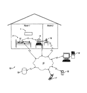
[0018] Fig. 1 illustrates one embodiment of a seizure detection system.
[0019] Fig. 2 illustrates one embodiment of a detection unit and base station
for a
seizure detection system.
[0020] Fig. 3 illustrates one embodiment of a base station.
[0021] Fig. 4 illustrates one embodiment of a method for detecting seizure
related incidents.
[0022] Fig. 5A and Fig. 5B illustrate exemplary EMG time domain data for a
patient.
[0023] Fig. 6A and Fig. 6B illustrate exemplary EMG frequency domain data for
a patient.
[0024] Fig. 7 illustrates one embodiment of a burst detection algorithm.
[0025] Fig. 8A and Fig. 8B illustrate exemplary model forms or envelopes of
signal bursts
after filtering, rectification and peak dete-tion.
[0026] Figs. 9A, 9B and 9C illustrate another embodiment of a burst and burst
train
detection algorithm.
[0027] Fig. 10 illustrates one embodiment of a periodicity algorithm.
[0028] Fig. 11 illustrates one embodiment of a GTC waveform detection
algorithm.
[0029] Fig. 12 illustrates a second embodiment of a GTC waveform detection
algorithm.
[0030] Fig. 13 illustrates one embodiment of a waveform regularity detection
algorithm.
[0031] Fig. 14 illustrates one embodiment of a supervisory algorithm.
[0032] Figs. 14A, 14B and 14C illustrate another embodiment of a supervisory
algorithm.
[0033] Fig. 15 illustrates one embodiment of a method of data collection.
[0034] Fig. 16 illustrates one embodiment of a method of updating a template
file.
[0035] Fig. 17 illustrates one embodiment of a method of adjusting the state
of a detection
unit in a method of seizure monitoring.
[0036] Fig. 18 illustrates one embodiment of an amplitude detection algorithm.
[0037] Fig. 19 illustrates a further embodiment of a method for detecting
seizure
related incidents.
[0038] Fig. 20 illustrates a still further embodiment of a method for
detecting seizure
related incidents.
[0039] Fig. 21 illustrates how model data in a procedure for analysis of data
bursts may
be organized.
[0040] Fig. 22 illustrates how model data for analysis of data bursts is
combined with data
from a GTC accumulation register and how data in those registers may be
analyzed in a supervisory
algorithm.
[00411 Fig. 23 illustrates exemplary EMG electrical data for a patient.
CA 02814825 2015-06-09
[0042] Fig. 24 illustrates exemplary EMG electrical data for a patient while
non-seizure
moving.
[0043] Fig. 25 illustrates exemplary EMG electrical data for a patient who is
sleeping.
[0044] Fig. 26 illustrates exemplary EMG electrical data for a patient at the
onset of a seizure.
[0045] Fig. 27 illustrates exemplary EMG electrical data for a patient as the
seizure
progresses.
[0046] Fig. 28 illustrates exemplary EMG electrical data for a patient that
has been filtered.
[0047] Fig. 29 illustrates further exemplary EMG electrical data for a patient
that has also
been filtered.
[0048] Fig. 30 illustrates the same exemplary EMG electrical data as shown in
Fig. 29 and
filtered using a different filter protocol.
[0049] Fig 31 illustrates exemplary EMG electrical data for a patient showing
short-lived
data events.
[0050] Fig. 32 illustrates still further exemplary EMG electrical data for a
patient that has
been filtered.
[0051] Fig. 33 illustrates exemplary EMG electrical data for a patient showing
sustained
signals.
[0052] Fig. 34 illustrates another exemplary EMG electrical data for a patient
that has been
filtered.
[0053] Fig. 35 illustrates another exemplary EMG electrical data for a
patient.
[0054] Fig. 36 illustrates yet another exemplary EMG electrical data.
DETAILED DESCRIPTION
[0055] The apparatuses and methods described herein may be used to detect
seizures and
timely alert caregivers of a seizure using EMG, among other things. The
apparatuses and method may
be used, for example, to initiate an alarm protocol, create a log of seizure
incidents to help medically
or surgically manage the patient, activate a Vagal Nerve Stimulator, or
activate other stimulating
devices that may be used to abort or attenuate a seizure. In some embodiments,
a log of seizure related
incidents may prompt a physician to understand more quickly the failure of a
treatment regimen. The
apparatuses and methods may comprise a process and device and/or system of
devices for detecting
seizures with motor manifestations including, but not limited to Tonic-Clonic,
Tonic-only, or Clonic-
only seizures. A "motor manifestation" may in some embodiments generally refer
to muscle activity,
whether sustained or otherwise.
[0056] Apparatuses as described herein may be useful for monitoring a person
to determine
whether the person may be having a seizure, and for initiating an alarm. The
methods described herein
may be flexible, e.g., such methods may be customized for an individual.
Moreover, such methods
6
CA 02814825 2015-06-09
may be adaptive, and may improve as data is collected, e.g., for a given
patient or for a certain patient
demographic. Furthermore, apparatuses described herein may be suited for
organizing and/or
prioritizing the collection of large amounts of data, e.g., data that may be
collected in a substantially
continuous manner, such as while a seizure-prone individual is in a home
setting.
[0057] In general terms, EMG electrode signals may be collected and processed
to determine
seizure variables. A "seizure variable" may in some embodiments refer to a
criterion or criteria of one
or more portions of data collected from the output signal of a detector. For a
given set of data, a
seizure variable may have one or more numerical values associated with it. For
example, the
amplitude of a signal may be a seizure variable that may have one or more
numerical values
associated with it for a given set of data. A value of a seizure variable may
be compared to a threshold
level and may be used as an input in an algorithm for determining whether a
seizure may have
occurred.
[0058] A processing method may include calculating one or more seizure
variable values and
may further include comparing such values to one or more thresholds that may
characterize a seizure.
Data registers may be populated based upon such a comparison, and used to
evaluate whether to
initiate an alarm protocol. The weighting of data in different registers, and
thus the importance of
different characteristics of EMG data, may be customized for an individual
patient or patient
demographic, and may adapt as the system obtains more information for a
patient or patient
demographic.
[0059] A variety of suitable systems may be suitable for collecting large
amounts of EMG
and other patient-related data, organizing such data for system optimization,
and for initiating an
alarm in response to a suspected seizure. Fig. 1 illustrates an exemplary
embodiment of such a system.
In the embodiment of Fig. 1, a seizure detection system 10 may include a
detection unit 12, an
optional base station 14, an optional video monitor 9 and an optional alert
transceiver 16. The
detection unit may comprise one or more EMG electrodes capable of detecting
electrical signals from
muscles at or near the skin surface of a patient, and delivering those
electrical EMG signals to a
processor for processing. The base station may comprise a computer capable of
receiving and
processing EMG signals from the detection unit, determining from the processed
EMG signals
whether a seizure may have occurred, and sending an alert to a caregiver. An
alert transceiver may be
carried by, or placed near, a caregiver to receive and relay alerts
transmitted by the base station.
[0060] In using the apparatus of Fig. 1, for example, a person 11 susceptible
to epileptic
seizures may be resting in bed, or may be at some other location as daily
living may include, and may
have a detection unit 12 in physical contact with or in proximity to his or
her body. The detection unit
12 may be a wireless device so that a person may be able to get up and walk
around without having to
bc tethered to an immobile power source or to a bulkier base station 14. For
example, the detection
unit 12 may be woven into a shirt sleeve, or may be mounted to an armband or
bracelet. In other
7
CA 02814825 2015-06-09
embodiments, one or more detection units 12 may be placed or built into a bed,
a chair, an infant car
seat, or other suitable clothing, furniture, equipment and accessories used by
those susceptible to
seizures. The detection unit 12 may comprise a simple sensor, such as an
electrode, that may send
signals to the base station for processing and analysis, or may comprise a
"smart" sensor having some
data processing and storage capability. In some embodiments, a simple sensor
may be connected via
wire or wirelessly to a battery-operated transceiver mounted on a belt worn by
the person.
[0061] The system may monitor the patient, for example, while resting, such as
during the
evening and nighttime hours. If the detection unit 12 on the patient detects a
seizure, the detection unit
12 may communicate via wire or wirelessly, e.g., via a communications network
or wireless link, with
the base station 14 and may send some signals to the base station device for
more thorough analysis.
For example, the detection unit 12 may process and use EMG signals (and
optionally ECG and
temperature sensor signals) to make an initial assessment regarding the
likelihood of occurrence of a
seizure, and may send those signals and its assessment to the base station 14
for separate processing
and confirmation. If the base station 14 confirms that a seizure is likely
occurring, then the base
station 14 may initiate an alarm for transmission over the network 15 to alert
a caregiver by way of
email, text, or any suitable wired or wireless messaging indicator. In some
embodiments, if one or
more of the detection unit 12, the base station 14, or a caregiver, e.g., a
remotely located caregiver
monitoring signals provided from the base station, determines that a seizure
may be occurring, a video
monitor 9 may be triggered to collect information.
[0062] The base station 14, which may be powered by a typical household power
supply and
contain a battery for backup, may have more processing, transmission and
analysis power available for
its operation than the detection unit 12, inay be able to store a greater
quantity of signal history, and
evaluate a received signal against that greater amount of data. The base
station 14 may communicate
with an alert transceiver 16 located remotely from the base station 14, _such
as in the bedroom of a
family member, or to a wireless device 17, 18 carried by a caregiver or
located at a work office or
clinic. The base station 14 and/or transceiver 16 may send alerts or messages
to caregivers, or medical
personnel via any suitable means, such as through a network 15 to a cell phone
17, personal digital
assistant (PDA) 18 or other client device. The system 10 may thus provide an
accurate log of seizures,
which may allow a patient's physician to understand more quickly the success
or failure of a treatment
regimen. Of course, the base station 14 may simply comprise a computer having
installed a program
capable of receiving, processing and analyzing signals as described herein,
and capable of transmitting
an alert. In other embodiments, the system 10 may simply comprise, for
example, EMG electrodes and
a smartphone, such as an iphoneTM, configured to receive EMG signals from the
electrodes for
processing the EMG signals as described herein using an installed program
application. In further
embodiments, so-called "cloud" computing and storage may be used via network
15 for storing and
processing the EMG signals and related data. In yet other embodiments, one or
more EMG electrodes
8
CA 02814825 2015-06-09
could be packaged together as a single unit with a processor capable of
processing EMG signals as
disclosed herein and sending an alert over a network. In other words, the
apparatus may comprise a
single item of manufacture that may be placed on a patient and that does not
require a base station
separate transceiver.
[0063] In the embodiment of Fig. 1, the signal data may be sent to a remote
database 19 for
storage. In some embodiments, signal data may be sent from a plurality of
epileptic patients to a
central database 19 and "anonyinized" re provide a basis for establishing and
refining generalized
"baseline" sensitivity levels and signal characteristics of an epileptic
seizure. The database 19 and
base station 14 may be remotely accessed via network 15 by a remote computer
13 to allow updating
of detector unit and/or base station software, and data transmission. The base
station 14 may generate
an audible alarm, as may a remote transceiver 16. All wireless links may be
two-way for software and
data transmission and message delivery confirmation. The base station 14 may
also employ one or all
of the messaging methods listed above for seizure notification. The base
station 14 may provide an
"alert cancel" button to terminate the incident warning.
[00641 In some embodiments, a transceiver may additionally be mounted within a
unit of
furniture or some other structure, e.g., an environmental unit or object. If a
detection unit is
sufficiently close to that transceiver, such a transceiver may be capable of
sending data to a base
station. Thus, the base station may be aware that information is being
received from that transducer,
and therefore the associated environmental unit. In some embodiments, a base
station inay select a
specific template file, e.g., such as including threshold values and other
data as described further
herein, that is dependent upon whether or not it is receiving a signal from a
certain transceiver. Thus,
for example, if the base station receives information from a detector and from
a transducer that is
associated with a bed or crib it may treat the data differently than if the
data is received from a
transducer associated with another environmental unit, such as, for example,
clothing typically worn
while an individual may be exercising
[0065] The embodiment of Fig. 1 may be configured to be minimally intrusive to
use while
sleeping or minimally interfere in daily a,tivities, may require a minimum of
electrodes such as one or
two, may require no electrodes to the head, may detect a seizure with motor
manifestations, may alert
one or more local and/or remote sites of the presence of a seizure, and may be
inexpensive enough for
home use.
[0066] Fig. 2 illustrates an embodiment of a detection unit 12 or detector.
The detection unit
12 may include EMG electrodes 20, and may also include ECG electrodes 21. The
detection unit 12
may further include amplifiers with leads-off detectors 22. In some
embodiments, one or more leads-
off detectors may provide signals that indicate whether the electrodes are in
physical contact with the
person's body, or otherwise too far from the person's body to detect muscle
activity, temperature,
brain activity or other patient phenomena.
9
CA 02814825 2015-06-09
[0067] The detection unit 12 may further include a temperature sensor 23 to
sense the
person's temperature. Other sensors (not shown) may be included in the
detection unit as well, such as
accelerometers. Signals from electrodes 20 and 21, temperature sensor 23 and
other sensors may be
provided to a multiplexor 24. The multiplexor 24 may be part of the detection
unit 12 or may be part
of the base station 14 if the detection unit 12 is not a smart sensor. The
signals may then be
communicated from the multiplexor 24 to one or more analog-to-digital (A-D)
converters 25. The
analog-to-digital converters may be part of the detection unit 12 or may be
part of the base station 14.
The signals may then be cotrununicated to one or more microprocessors 26 for
processing and
analysis as disclosed herein. The microprocessors 26 may be part of the
detection unit 12 or may be
part of the base station 14. The detection unit 12 and/or base station 14 inay
further include memory
of suitable capacity. The microprocessor 26 may communicate signal data and
other information
using a transceiver 27. Communication by and among the components of the
detection unit 12 and/or
base station 14 may be via wired or wireless communication.
[0068] Of course, the exemplary detection unit of Fig. 2 may be differently
configured. Many
of the components of the detector of Fig. 2 may be in base station 14 rather
than in the detection unit
12. For example, the detection unit may simply comprise an EMG electrode 20 in
wireless
communication with a base station 14. In such an embodiment, A-D conversion
and signal processing
may occur at the base station 14. If an ECG electrode 21 is included, then
multiplexing may also
occur at the base station 14.
[0069] In another example, the detection unit 12 of Fig. 2 may comprise an
electrode portion
having one or more of the EMG electrode 20, ECG electrode 21 and temperature
sensor 23, in wired
or wireless communication with a small belt-worn transceiver portion. The
transceiver portion may
include a multiplexor 24, an A-D converter 25, microprocessor 26, transceiver
27 and other
components, such as memory and input/output (I/0) devices (e.g., alarm cancel
buttons and visual
display).
[0070] Fig. 3 illustrates an emL )diment of a base station 14 that may include
one or more
microprocessors 30, a power source 31, a backup power source 32, one or more
1/0 devices 33, and
various communications means, such as an Ethernet connection 34 and
transceiver 35. The base station
14 may have more processing and storage capability than the detection unit 12,
and may include a
larger electronic display for displaying EMG signal graphs for a caregiver to
review EMG signals in
real-time as they are received from the detection unit 12 or historical EMG
signals from memory. The
base station 4 may process EMG signals and other data received from the
detection unit 12. If the base
station 14 detennines that a seizure is likely occurring, it may send an alert
to a caregiver via
transceiver 35.
[0071] Various devices in the apparatus of Figs. 1-3 may communicate with each
other via
wired or wireless communication. The system 10 may comprise a client-server or
other architecture,
CA 02814825 2015-06-09
and may allow communication via network 15. Of course, the system 10 may
comprise more than one
server and/or client. In other embodiments, the system 10 may comprise other
types of network
architecture, such as a peer-to-peer architecture, or any combination or
hybrid thereof.
[0072] Fig. 4 illustrates an exemplary method 36 of monitoring EMG and other
signals for
seizure characteristics, and initiating an alari-n response if a seizure is
detected. Such a method may
involve collecting of EMG signals, calculating one or more values of a seizure
variable, and using
such seizure variable data to populate processor or memory registers. In
general, one or more seizure
variables and one or more registers may be included in data analysis. In a
step 38, EMG signals and
other detector output signals may be collected. Output signals may be
collected in a substantially
continuous manner or periodically. Output signals may be processed in a step
40 to obtain seizure
variable data. The data values may be used to populate one or more detection
registers, as shown in
step 42. Processing of output signals and population of detection registers
may be executed during a
defined period of time, i.e., collection time window. At the expiration of
such a collection time
window, each detection register may transfer its contents, if any, to one or
more accumulation
registers (as shown in step 44), and the contents of one or more detection
registers, if any, may be
cleared. After expiration of the collection dine window, and after adjustment
(increase or leakage) of
accumulation registers, the cycle may repeat itself (as shown by line 46),
i.e., detector output may be
collected during a subsequent collection window. Periodically, a supervisory
algorithm may analyze
the contents of one or more accumulation registers to detefmine whether a
seizure is likely occurring
(step 48). If the supervisory algorithm determines that the sum of values or a
weighted sum of values
in the accumulation registers exceeds a threshold then an alarm protocol may
be initiated (step 50).
Alternatively, the supervisory register may determine that the contents of
accumulation registers do
not indicate that a seizure is likely and the system may wait for a next
analysis period (step 52).
[0073] As discussed below, a supervisory algorithm may comprise a number of
sub-routines
that use various seizure variable values in the accumulation and/or detection
registers. As shown by
way of example in Fig. 4, methods may involve the population of individual
detection registers with a
data value and addition of such a data value to accumulation registers (steps
38, 40, 42, and 44). A
sub-method may include steps involved in the population of individual
detection registers and
accumulation registers. Each sub-method may consider one or more
characteristics of the collected
data and perform process analysis on s-,ch characteristics. Individual sub-
methods may include, by
way of nonlimiting example, detection of signal bursts and detection of GTC
waveforms. Sub-
methods may process data in the time domain, the frequency domain, or, in some
embodiments,
process portions of data in both the time domain and frequency domain. Before
discussion of those
individual sub-methods in greater detail, it is helpful to consider some
general aspects of data
collection, the detectors used, as well as processing steps, such as data
filtration that may be involved
11
CA 02814825 2015-06-09
in various sub-methods. In addition, it is instructive to discuss exemplary
EMG signal data, as shown
in Figs. 5 and 6 discussed in more detail further herein.
[0074] As indicated in step 38 of Fig. 4, in some embodiments, detection of
seizures may be
accomplished exclusively by analysis of EMG electrode data. In other
embodiments, a combination of
EMG and other detectors may be used. For example, temperature sensors,
accelerometers, ECG
detectors, other detectors, or any combinations thereof, may be used.
Accelerometers may, for
example, be placed on a patient's extremities to detect the type of violent
movement that may
characterize a seizure. Similarly, ECG sensors may be used to detect raised or
abnormal heart rates
that may characterize a seizure. Thus, a monitoring device may detect an
epileptic seizure without the
customary multitude of wired electrodes attached to the head, as typical with
EEG. Combination of
EMG electrodes with other detectors may, for example, be used with
particularly difficult patients.
Patients with an excessive amount of loose skin or high concentrations of
adipose tissue, which may
affect the stability of contact between an electrode and the skin, may be
particularly difficult to
monitor. In some embodiments, an electrode may be attached to a single muscle,
and in other
embodiments a combination of two or more electrodes may be used. Electrodes
may, for example, be
attached to an agonist and antagonist muscle group or signals from other
combinations of different
muscles may be collected.
[0075] In general, the system described herein is compatible with any type of
EMG electrode,
such as, for example, surface monopolar electrodes or bipolar differential
electrodes or electrodes of
any suitable geometry. Such electrodes may, for example, by positioned on the
surface of the skin,
may or may not include application of a gel, and may, in some embodiments, be
Ag/AgC1 electrodes.
The use of a bipolar EMG electrode arrangement, e.g., with a reference lead
and two surface inputs,
allows for the suppression of noise that is common to those inputs. That is, a
differential amplifier
may be used, and a subtraction of the signals from one input with respect to
the other may be
accomplished, and any differences in signal between the inputs amplified. In
such an approach,
signals that are common to both inputs (such as external noise) may be
substantially nullified and
preferential amplification of signals originating from muscle depolarization
may be achieved.
[0076] An EMG signal may be collected for a given time period, e.g., a time
domain electrode
signal may be collected. Time domain electrode data, may be converted to
frequency data, i.e.,
spectral content, using techniques such as Fast-Fourier Transform (FFT). In
reference to Fig. 4, the
conversion of data between the time and frequency domain may be included in a
processing step 40.
Other aspects of data processing may include smoothing data, application of
one or more frequency
filters, fitting data in a given region to a õarticular function, and other
processing operations
[0077] Fig. 5 (which comprises Figs. 5a and 5b) provides an example of EMG
data 54
collected over a time period of about 2 seconds. The data in Fig. 5 may
exemplify data collected by
placing a bipolar differential electrode over the biceps or triceps of a
patient. Fig. 6 illustrates some of
12
CA 02814825 2015-06-09
the EMG data 54 of Fig. 5 converted to the frequency domain. The EMG data 74
in Fig. 6 may
represent, for example, a one-second epoch of the EMG data 54 converted to the
frequency domain.
For an EMG electrode, visual representation of frequency domain data may also
be refen-ed to as a
spectral graph.
[0078] Referring now to the time domain data for the graph of Fig. 5, the
vertical axis or
scale in Fig. 5a is signal amplitude, e.g., the differential signal between
the pair of EMG electrode
inputs, and the horizontal axis or scale shows time (in Fig. 5, the time
window is approximately two
seconds). In reference to any of the graphs described herein the tenn
amplitude may be used, and
such may refer to either the magnitude of signal, or absolute value of
magnitude, as may be
appropriate for a given calculation. Signals collected may, for example, be
rectified, and unless
otherwise noted, detection of bursts as described herein involves rectified
signal data. As shown in
Fig. 5, the amplitude (or absolute value of the amplitude) appears to
experience a sustained increase
62 at least three times (56, 58, and 60) during the 2-second period. Such
sustained increase may be
indicative of what is referred to as a burst, or signal or data burst. As
discussed in more detail
below, fluctuations in time periods between suspected bursts, such as 66 or
68, may be used to
calculate a baseline. Fluctuations in a baseline region, i.e., noise, may be
related to a peak to peak
value, a root mean square (RMS) value or other metric. Fig. 5b illustrates a
portion of the EMG data
54, namely, the region of data including burst 60 and adjacent period. In Fig.
5b, a RMS noise value
72 and amplitude 70 are indicated. The signal-to-noise ratio (SNR or S/N) of
burst 60 is, in this
example, about 4:1, i.e., amplitude 70 is about four times larger than the
noise value 72. The EMG
data of Fig. 5 is discussed in further detail with regards to a burst
detection sub-method in Fig. 7.
[0079] Referring now to the exemplary data of Fig. 6 (which comprises Figs. 6A
and 6B), the
vertical scale represents the magnitude of a given frequency (which may be
referred to as spectral
density) and the horizontal scale is signal frequency. Note that the spectral
data in Fig. 6 indicates a
curving slope with decreasing magnitude as the frequency increases, i.e., the
spectral density
generally decreases as the frequency increases. The ratio of spectral density
at a lower frequency to
the spectral density at a higher frequency may be a seizure variable that, for
any given portion of
electrode data, may have an associated value. For example, for the data shown
in Fig. 6 the ratio of
spectral density at a frequency of about 200 Hz (76) to the spectral density
at about 400 Hz (78) may
have a value of about 1.1.
[0080] Also, as illustrated in the expanded portion of the same data in Fig.
6b, which shows at
least a portion of the characteristic GTC waveform, a region of elevated
spectral density 80, i.e., a
relatively high-frequency "bump" between approximately 300-500 Hz, and
particularly around 400
Hz 82 is shown. That is, the spectral density 80 at frequency 82 in that
region is elevated above the
spectral density 84, e.g., within a "slumped" region, approximately located at
a frequency 86 of
about 300 Hz. The term "slump region" or "slump" may in some embodiinents
refer to a portion of
13
CA 02814825 2015-06-09
spectral data generally possessing the property of having positive curvature,
i.e., a slump region
refers to a local minimum in a set of data. The term "bump region" or "bump"
may in some
embodiments refer to a portion of spectral data where the data generally
possesses the property of
having negative curvature, i.e., a bump region refers to a local maximum in a
set of data. To
generally possess a positive or negative curvature means that local
fluctuations in individual data
points may be averaged or smoothed out of the data. That is, neglecting local
fluctuations, e.g., due
to noise, a data set may possess a property of curvature.
[0081] The ratio of spectral density at a frequency 86 to the spectral density
at a frequency 82,
or slump to bump ratio, may be used as a seizure variable. In some
embodiments, the slump to bump
ratio may be used as a metric for detection of a GTC waveform. However, more
advanced data
analysis techniques, e.g., looking at a greater number of data points and/or
advanced pattern
recognition algorithms, may also be used to identify a GTC waveform. In some
embodiments, a
detection unit may include instructions for calculation of a slump to bump
ratio and a base unit may
calculate a slump to ratio and also corroborate the slump to bump calculation
with more advanced
pattern recognition analyses. The EMG.flata of Fig. 6 and the above data
features are discussed in
further detail with regards to a GTC waveform detection sub-method as
described, for example, in
Figs. 11 and 12.
[0082] Referring back to Fig. 4, the collection of EMG data may be
accomplished with a
detection unit and that detection unit may execute an initial analysis and
processing of data. In some
embodiments, if the detection unit determines that a seizure is likely
occurring, it may send data to a
base station, where further processing may occur. Thus, a detection unit, a
base station or both may
process EMG signals, and either or both devices may execute a seizure
detection sub-method. Such a
sub-method may characterize particular features of EMG data, and may, based
upon such a
characterization, direct the transfer of data between data registers and
accumulation registers. Those
aspects of sub-methods, such as described herein in reference to Figs. 7 and
10-13, may involve
aspects of steps 38, 40, 42, 44, and 46 of method 36. A sub-method may feed
data into a supervisory
algorithm.
[0083] Fig. 7 illustrates one embodiment of a sub-method 88 which may be used
for analysis
of data bursts. In a step 90 of Fig. 7, a detection unit and/or base station
may select a protocol for
analysis of data bursts. The selection of an analysis protocol may, for
example, be indicated in a
template file. Such a template file may include instructions to choose a
routine to smooth data, a
routine to filter data, a routine to treat the data in some other manner or
combinations of routines
thereof. Such routines may be executed by either the detection unit, base
station or both. The analysis
protocol may include a peak detection program, which, for example, after band-
pass filtering and
rectification may identify and shape a data burst, as shown in the examples of
Fig. 9 and Fig. 10. Any
suitable peak detection technique may be used (e.g., continuous wavelet
transfonn), and may in some
14
CA 02814825 2015-06-09
embodiments include, for example, data smoothing techniques (e.g., moving
average filter, Savitzky-
Golay filter, Gaussian filter, Kaiser Window, various wavelet transforms, and
the like), baseline
correction processes (e.g., monotone minimum, linear interpolation, Loess
normalization, moving
average of minima, and the like) and peak-finding criteria (SNR,
detection/intensity threshold, slopes
of peaks, local maximum, shape ratio, ridge lines, model-based criterion, peak
width, and the like).
[0084] A peak detector may have separate attack and decay rates. These rates
may be
individually adjusted. Since there frequently may be plenty of sustained
amplitude during a real burst,
fear of the peak detected signal decaying too quickly during bursts is
generally not a problem.
Therefore, the decay rate may be set to decay rather quickly following a
burst. Usually the time
between bursts is longer than the burst itself, and so there may be no reason
to speed up the decay.
However, a noise spike between bursts could artificially cause the peak
detector output to jump up to
a level that would make distinguishing real seizure bursts a problem.
Therefore, the attack rate may be
carefully controlled to prevent this from occurring.
[0085] In step 91 of the method of Fig. 7, a burst detection algorithm may be
initiated. Burst
analysis may be triggered, for example, by detection of an EMG signal having
an amplitude value that
meets or exceeds a burst analysis amplitude threshold. Within the burst
detection window, the EMG
data may be analyzed for elevated amplitude using, e.g., a peak detection
program. Regions of
elevated amplitude may be classified as potential bursts. For example,
referring back to Fig. 5, at least
three periods of sustained elevation of amplitude (56, 58, and 60) may be
identified in the
approximately 2-second epoch. Regions of elevated amplitude within the burst
detection window may
be measured for amplitude, width, and a SNR may also be determined. A portion
of data, e.g.,
identified as a possible peak, may have amplitude associated with it, e.g.,
peak amplitude, median,
mean or other metric may be calculated.
[0086] In step 92 of Fig. 7, EMG signal data, such as within a certain time
period (burst
detection window), rnay be analyzed for bursts. For example, for suspected
data burst 56, amplitude 62
may be measured. A burst may have an amplitude that is elevated over
surrounding portions of data,
and that elevated amplitude may extend for a period of time. That is, a burst
may have a burst width,
such as burst width 64. To determine a burst width, a leading edge of a burst
and a trailing edge of a
burst may be detennined. To detect the leading edge and trailing edge of a
burst, changes in amplitude
for successive data points may be measured, e.g., the rate of change of
amplitude with time may be
calculated. Any other suitable technique, such as those described above, may
be used, as well. In
some embodiments, burst width may be categorized by calculating, for a region
of time, whether a
threshold minimum amplitude is met at a given probability, e.g., where a
majority of points show
elevated amplitude above some threshold.
[0087] Signal to noise calculations inay involve, for example, establishing a
baseline by
deterinining fluctuations in detector signal, i.e., baseline noise, in a time
period immediately prior to
CA 02814825 2015-06-09
data in a time suspected of containing bursts. For example, an EMG signal may
be relatively quiet in
the time leading up to a seizure, as discussed in more detail in connection
with Fig. 25, below. That
quiet period may be used to establish a baseline.
[0088] A baseline may also be established by looking at fluctuations between
burst periods
within the same time window suspected of having bursts. For example, referring
back to the EMG
data of Fig. 5, data fluctuations in time periods between suspected bursts,
such as the data in the time
periods 66 or 68, may be used to calculate a baseline. Fluctuations in a
baseline region, i.e., noise,
may be related to a peak to peak value, a RMS value or other suitable baseline
detection metric. In
Fig. 5 an expanded region of data, i.e., the region of data including burst 60
and adjacent period, is
shown in Fig. 5b, and a root mean square noise value 72 and amplitude 70 are
approximately
indicated. The S/N of burst 60 may, for example, be about four, i.e.,
amplitude 70 is about four fold
larger than the noise value 72.
[0089] It should be noted that the baseline established by looking at
fluctuations between
burst periods may be different than the baseline established by looking at a
pre-seizure quiet time.
Thus, different peak detection algorithms may be run for each, or the same
algorithm may be ramped
up or down with respect to baseline detection depending on whether detecting
quiet time or seizure
activity. For example, a baseline detector may be a peak detector having a
much longer time constant
than a peak detector used for signal envelope generation. This baseline
detector may rise up to a
higher level during a tonic phase but may ramp down during a clonic phase of
activity. A negative
peak detector may also be employed to ramp a baseline detector down more
quickly during relatively
quiet times so as to distinguish the bursts more readily.
[0090] In step 94, the burst detection algorithm may determine if the EMG
signal data within a
burst detection window meet various requirements or thresholds or other
criteria to qualify regions of
elevated amplitude as bursts. For example, the algorithm may determine whether
one or more regions
of elevated amplitude meet requirements for amplitude, width, and time between
regions of elevated
amplitude to qualify as seizure bursts. For example, a sub-method for
detecting bursts may detect
amplitudes above a certain threshold that are closer than Y seconds apart and
farther than Z seconds
apart. Such requirements (or burst criteria) may be provided in a template
file. For example, referring
to Table 1, the minimum S/N criteria may be pulled from the template file and
compared to the
calculated value of S/N for each suspected burst.
[0091] Generally, a burst may be characterized by a sudden increase in the
amplitude of the
EMG electrode signal from a lower amplitude level, maintenance of that
increased amplitude level for
a specified minimum amount of time, return of the amplitude level to a lower
level of electrode signal
after no more than a specified maximum time, and maintenance of the lowered
amplitude level for a
specified minimum time. Fig. 8A and Fig. 8B illustrate exemplary model fonns
or envelopes of signal
bursts after filtering, rectification and peak detection. Generally, the lower
amplitude signal level may
16
CA 02814825 2015-06-09
not go to zero. The lower amplitude above zero is signal noise. The ratio of
the burst amplitude level
to the noise level is the SNR. For example, if the signal level of the burst
is 1 volt, and the noise is
0.35 volts, then the SNR would be 1/0.35, or 2.86. In the example of Fig. 8,
the peak amplitude 120 of
EMG signal data may be compared to criterion associated with peak amplitude.
If the amplitude 120
is greater than a minimum amplitude criterion 120a, and less than a maximum
amplitude criterion
120b, then the ratio of peak amplitude to the level of noise 102 may be
determined and compared to a
burst amplitude criterion, e.g., a SNR threshold. If the peak amplitude meets
the SNR. threshold, then
the EMG signal data may qualify as a burst (or the start of a burst) with
respect to amplitude. A
maximum burst amplitude requirement may be helpful in eliminating from
consideration elevated
amplitude EMG data caused from external noise sources that may introduce
amplitude well above the
amplitudes capable of being produced by the human body.
[0092] Fig. 8A also shows the region of elevated amplitude as having a width
114. The width
114 may be compared to a minimum burst width (dashed line 116) and a maximum
burst width
(dashed line 118). As may be seen in Fig. 8B, the width 114 falls between the
minimum and
maximum burst width thresholds, and thus qualifies the region of elevated
amplitude as a burst with
respect to width. A maximum burst width requirement may be helpful in
eliminating from
consideration elevated amplitude EMG data that is from voluntary muscle
activity, a noise source or
is caused by electrode connectivity problems. That could help eliminate
falsely identifying real or
apparent high-amplitude muscle activity as a seizure.
[0093] Fig. 8B shows examples of two successive bursts (104 and 106) separated
by a time
period 108. In Fig. 8B the time between bursts 108 may, for example be
compared to criterion values
associated with a minimum period between successive bursts (dashed line 110)
and a maximum period
between successive bursts (dashed line 112). If a sufficient quantity of
bursts succeed each other within
the minimum and maximum time periods, then successive bursts may qualify as a
burst train indicative
of a seizure. However, not all burst trains indicate a seizure, and a
periodicity algorithm (discussed in
more detail below) may be used to further evaluate the likelihood that a
seizure is occurring. For
example, extremely regular bursts may not indicate a seizure. Sporadic bursts
may not indicate a
seizure, either, or if spaced sufficiently far apart, represent minimal threat
of imminent harm from
seizure.
[00941 After reaching the end of the burst detection window, the burst
detection algorithm
may wait for a delay period before analyzing data in a subsequent burst
detection window. By adding
a delay, the burst detection algorithm may ensure that new data is analyzed.
If analysis of a burst
window, or analysis of one or more successive burst detection windows reveals
no bursts or near-
bursts, then the burst detection sub-method may pause, as seen at step 95,
until the burst analysis
amplitude threshold triggers activation of the sub-method.
17
CA 02814825 2015-06-09
[0095] The burst amplitude, width and periodicity values may be stored in
registers for use by
a supervisory algoritlun to determine the likelihood of a seizure occurring.
If the supervisory
algorithm determines that a seizure is occurring, then it may declare an
alarm, and cause the base
station 14 to send an alert to a caregiver.
[0096] Criterion values may, for example, be included in a template file. More
specifically,
Table 1 lists exemplary criteria that that may be included in a template file
which may be used in a
sub-method for evaluation of data bursts. Each criterion may be a variable
that may be changed to
adjust the sensitivity of the seizure detection method. Of course, not all of
the criteria need be used.
For example, maximum burst amplitude may be considered optional if unduly
limiting for a
particular patient. Likewise, additional criteria may be used. For example, if
signal amplitude is
sufficiently high to trigger the burst detection sub-method, but does not
quite meet the minimum
burst amplitude even though it meets burst width criteria, then its variance
from the minimum burst
amplitude may be negatively weighted by a certainty value criterion. A
certainty value criterion
may be, for example, a percentage value. If the measured amplitude is 95% of
the minimum burst
amplitude, then the certainty value may be set accordingly. If successive
bursts have sufficient
periodicity to qualify as a burst train, the negatively-weighted burst may be
included in the train to
further test periodicity. If a certain number of negatively-weighted bursts
appear in the data, then a
supervisory algorithm may lower the minimum burst amplitude thresholds to
increase the
sensitivity of the burst detection method for the particular patient being
monitored. Similar
weighting may be done with respect to signal values that do not quite meet the
other burst criteria.
Certainty values may be used by the burst detection method, other sub-methods
described herein,
and the supervisory algorithm.
[00971 TABLE 1: Template data for a burst detection sub-method
Variable Value / unit Type
Burst analysis minimum amplitude threshold xx amplitude Criterion for
initiation of burst
Burst detection window XX seconds Routine selection
Delay between adjacent burst detection XX seconds Routine selection
Minimum burst width XX seconds Criterion for burst count
Maximum burst width XX seconds Criterion for burst count
Burst envelope peak detector attack rate XX Routine selection
Burst envelope peak detector decay rate XX Routine selection
Minimum burst amplitude XX amplitude Criterion for burst count
Maximum burst amplitude XX amplitude Criterion for burst count
Minimum S/N XX Criterion for burst count
Minimum period between successive bursts XX seconds Criterion for burst
count
18
CA 02814825 2015-06-09
Variable Value / unit Type
Maximum period between successive bursts XX seconds Criterion for burst count
Decay rate XX Data feature / weighting
Decay rate (S/N) modifier XX Data feature / weighting
Selection of filter protocol (if applied) XX Routine selection
Selection of smoothing protocol (if applied) XX Routine selection
Calculation method XX Routine selection
Baseline calculation method XX Routine selection
Coefficient (combination with supervisory XX Weighting coefficient
algorithm)
For clarity, the "XX" is simply a value placeholder, and should not be
construed to connote
magnitude or precision in any way.
[0098] Referring back to Fig. 7, in a step 96, one or more detection registers
may be loaded
with burst values for a detection window. For example, a burst count register
may be used to contain a
value corresponding to the number of detected bursts within the burst
detection window. For example,
if the two-second time period of Fig. 5 was a burst detection window, then the
EMG data within that
window may be analyzed for bursts. In Fig. 5, for example, the EMG signal data
shows three bursts.
Thus, a value of 3 may be stored in the burst count register. Other registers
may be used to store other
burst values, such as amplitude, periodicity, width, certainty values, and so
forth.
[0099] Following each burst detection cycle, e.g., analysis of a burst
detection window, the
detection register may, in some embodiments, add its contents to one or more
burst accumulation
registers (step 97). Before analyzing the data in subsequent burst detection
windows, the detection
registers may be cleared to allow storage of burst data for the subsequent
burst detection windows.
The detection registers may then begin storing burst values during another
cycle, or, in some
embodiments, begin counting bursts after a certain delay period.
[0100] In some embodiments, the EMG signal data may be written to a circular
buffer in
RAM in the device hardware. One advantage of such a strategy may be that less
RAM is used because
the processed data may store only a pattern of the data, such as peak detected
values, and not a point
by point data file of full signal data. That is, a voltage (or other
electrical parameter that reflects
amplitude of the detection unit) at each corresponding point in time need not
be stored. For example,
in some embodiments, only the data necessary to derive a model form such as
indicated in Fig. 8A
and Fig. 8B may be stored. It should be appreciated in those figures that
noise in regions between
detected bursts is depicted to be maintained at a constant level. Thus, only a
calculated value of the
noise, e.g., such as RMS amplitude (102), may be stored and not all of the
individual fluctuations in
the baseline data. Thus, the data file in RAM may be significantly compressed.
In some embodiments,
as opposed to storing a compression of the data in a time window, all raw data
from a given window
19
CA 02814825 2015-06-09
may be stored in a circular buffer in RAM. It should thus be appreciated that
an algorithm may look at
any given preceding time window at any point in the algorithm. Such may be
used, for example, to
consider how any given value of EMG data has changed between one or more time
windows.
[0100.1] In some embodiments, each burst may be weighted with a value that is
not only
related to detection of a burst but also related to the certainty of burst
detection. Certainty values may,
for example, be related to the normalized amplitude or the ratio of the
normalized amplitude to
detector noise. For example, a signal burst may be characterized by transition
from approximately
100% of the normalized amplitude to approximately 35% of the nonnalized
amplitude. The certainty
value may be approximately 65, which number may be loaded into a register
whose maximum value
could be approximately 100.
[0101] As denoted in step 97, one or more of the detection registers may add
their contents to
one or more accumulation registers. For example, a burst count detection
register may add its value to
a burst count accumulation register.
[0102] In step 98, the accumulation registers may, in addition to accepting a
data value from
the detection register, adjust the value of any previous data which may be
held. For example, in some
embodiments, the burst count accumulation register may hold a value that is
related to the quantity of
bursts collected in a preceding number of burst detection cycles. That is,
each time the burst count
detection register adds contents from one cycle, the burst count accumulation
register may remove a
data value that was added during some preceding cycle. Thus, the burst count
accumulation register
may, in some embodiments, act as a moving sum based on the sum of counts from
a number of
preceding burst detection windows. In such an embodiment, the computer may
store in memory, e.g.,
in any number of additional registers, the appropriate data value to add or
subtract from the burst
count accumulation register. In other embodiments, at the completion of a
cycle, the burst count
detection register may add any contents, e.g., value of collected bursts, to
the burst count
accumulation register and then remove a certain value, i.e., it may leak at a
certain rate. A leakage
rate, or decay rate as shown in Table 1, may be included in a template file
and may be adjusted to
customize the burst detection sub-method to a particular patient or patient
demographic. In some
embodiments, the leakage rate may be a value that is modified based upon
another criterion. For
example, the burst count accumulation register may be modified if one or more
successive burst
detection windows do not contain any bursts.
[0103] In other embodiments, the rate of decay of the burst count accumulation
register may
depend upon the S/N of bursts counted in One or more given time window. In
further embodiments,
the burst count accumulation register may be modified based on how the S/N of
bursts is changing.
That is, the average S/N of detected bursts may be tracked, e.g., the average
S/N value of bursts in
given time windows may, at least for some period of time, be stored in memory,
such as in a circular
RAM buffer. If the S/N of bursts changes between time windows, such a change
may be analyzed,
CA 02814825 2015-06-09
and used to modify the decay rate of the burst count accuinulation register.
In general, if the S/N of
bursts is increasing the decay rate of the burst count accumulation register
will drop by some factor
and if the S/N of bursts is decreasing the decay rate of the burst count
accumulation register will
increase by some factor. In addition, during step 98 the contents of the burst
count accumulation
register, may decay in a manner that is dependent upon various negative
weighting factors. For
example, if no bursts are detected in a cycle, such may be an indication that
a seizure is not occurring,
and the rate of decay of the burst count accumulation register may be
adjusted. Again, to analyze data
in preceding time windows, either point by point data or a model shape may be
stored in a circular
buffer of RAM in the system hardware. Referring back to Fig. 4, the value
stored in the burst count
accumulation register is an example of one value that may be examined with a
supervisory algorithm.
[0104] In step 99, the burst detection algorithm may wait for a time period
equal to the burst
detection window delay value before analyzing EMG signal data in subsequent
burst detection
windows. The burst detection registers may be cleared in step 100 before
analyzing EMG data in the
next burst detection window. In some embodiments, the burst detection
algoritlun may continue to run
until it finds one or more burst detection windows that do not contain any
bursts or near-bursts, or
until the supervisory algorithm triggers an alarm.
[0105] In general, the presence of qualified bursts, and a large value being
stored in the burst
count accumulation register, may increase the probability that a seizure event
is declared. It is also an
aspect of methods described herein, negative weighting factors may be used,
for example, with
respect to signal characteristics that diminish the likelihood that a seizure
is occurring. For example,
as discussed above, different negative weighting factors, such as the absence
of bursts in a preceding
time window, or a decreasing S/N may influence the leakage rate of an
accumulation register.
[0106] Figs. 9A, 9B and 9C illustrate another embodiment of a burst and burst
train detection
algorithm. The flowcharts of Figs. 9A-9C show logic flow, not actual routines.
In an actual routine,
they would be called by the supervisory algorithm or be scheduled as one-time
passes by timer
interrupt, not infinite loops. There are two main routines, the burst
detection algorithm (Figs. 9A and
9B), and the burst train detection algorithm (Fig. 9C). The burst detection
algorithm looks for a burst
that meets the requirements of amplitude (both min and max) and minimum width.
If the minimum
spacing between detected bursts is too small, the burst train detection
algorithm will catch it. A burst
train detection algorithm may rely on a periodicity algorithm, as discussed
below.
10106.11 Fig. 9A illustrates one embodiment of a burst detection logic flow
373. Burst
detection logic flow 373 may include obtaining a signal sample in the step
374. In the step 376, the
logic flow may include determining if the signal amplitude is greater than a
maximum allowed
amplitude level. If the signal amplitude is greater than the maxnnum allowed
level, then the
supervisory algorithm may be infonned in the step 378, and the burst detection
logic flow may then
start over (step 380). If in the step 376 the signal amplitude is not greater
than a maximum level, then
21
CA 02814825 2015-06-09
the flow 373 may, in the step 382, determine if the signal amplitude is
greater than noise by at least
sotne SNR. If the signal amplitude is greater than the SNR, then the burst
detection logic flow 373, as
shown in step 384, may include determining if it was the first time in the
routine that the required
SNR level was met. If it was the first time the SNR level was met, then the
routine may start a burst
timer (step 386) and take another signal sample (step 374). If it was not the
first time that the SNR
level was met within the flow, then the flow may include determining, in the
step 388, if the burst
tin-ter has exceeded a minimum burst duration threshold. If the burst timer
has exceeded the minimum
burst duration threshold, then the signal may, in the step 390, be pre-
qualified as a burst. The pre-
qualified burst may then be processed in the exemplary flow 393 shown in
Figure 9B as shown in the
(step 392).
[0106.21 The burst detection logic flow 393 in Fig. 9B may include taking a
signal sample
(step 394). The signal sample may, for example, be a signal sample pre-
qualified as a burst as
described in Fig. 9A. In the step 396, the routine may determine if the signal
amplitude is greater than
a maximum allowed amplitude level. If the signal amplitude is greater than the
maximum level, the
supervisory algorithm may be informed in the step 398, and the burst detection
flows 373, 393 may
start over (step 400). That is, a new signal sample may be taken in the step
374 of Fig. 9A. If the
signal amplitude is not greater than the maximum allowed amplitude level, the
routine may then
include determining, in the step 402, if the signal amplitude is greater than
background by at least a
SNR. If the signal atnplitude is greater than noise by at least the SNR, the
routine may, in the step
404, determine if the burst duration timer has exceeded a maximum burst
duration threshold.
Considering the flows 373 and 393 together, the duration of activation of the
burst timer, which may
be initiated in the step 386 of the flow 373, may be evaluated against each of
a minimum burst
duration threshold 388 and maximum burst duration threshold 404. That is,
viewing the flows 373 and
393 together data may be compared against each of a minimum duration threshold
and a maximum
duration threshold. If the maximum burst duration threshold is exceeded in the
step 404, as shown in
the step 406, it may be deemed that the burst is too long, and the process may
start over (as shown in
step 400). If the maximum burst duration threshold is not exceeded in the step
404, a next signal
sample may be taken in the step 394. And, as long as the signal amplitude does
not exceed the
maximum allowed level in step 396, the flow 393 may loop back to the step 402
and evaluate if the
signal amplitude is greater than noise by at least a SNR. If in the step 402,
it is determined that the
taken signal sample (which may include signal sample in any number of loops of
the flow 393) does
not exceed background by the SNR level, a burst may be deemed to be over and
the burst may be
qualified (step 408). As indicated in the step 408, a signal qualified as a
burst may be characterized by
a certainty value, the center of the burst may be deten-nined and the burst
data may be written into a
circular memory buffer for further analysis.
22
CA 02814825 2015-06-09
[0106.3] I.n the burst train detection logic flow of Fig. 9C, data in a
circular buffer may be
analyzed. In the step 410, the routine may scan the circular buffer for the
most recent burst centers. In
the step 412, the logic flow may eliminate bursts that are too close together
or too far apart from
consideration. In the step 414, the routine may evaluate whether there are
enough bursts within a
window to qualify as a burst train. If enough bursts are detected, the routine
may, in the step 416,
qualify the scanned data in the circular buffer as a burst train, calculate a
certainty value, and deliver
the burst data to a supervisory routine for further analysis. In the step 414,
if there are not enough
bursts within the burst train window the flow may exit.
[0107] In Fig. 10 an additional exemplary algoritlun 113 (the periodicity
algorithm) is
described that may, in some cases, act to suppress the initiation of a seizure
alarm. The periodicity
algorithm accomplishes this task by looking at the circular buffer over a time
frame and examining
how regular the detected bursts were. A periodicity algorithm may scan
different data values from
various time windows that the burst detection algorithm wrote into a circular
buffer, and examine the
periodicity of signal characteristics, including those that may not be
indicative of a seizure.
[0108] In some embodiments, variables in the periodicity algorithm may be:
= Periodicity Time Window (in seconds)
= Minimum Average (or Standard) Deviation Allowed (percentage)
[0109] The periodicity time window variable is the period of time over which
the periodicity
algorithm scans data. For example, the periodicity time window may be
sufficient to include some
number of burst detection windows from the burst detection algorithm. The
Deviation Allowed
variable is the minimum value of how far from a single frequency the bursts
may be distributed to
qualify as a seizure. If the bursts huddle too closely around a specific
frequency, for example 1Flz,
then that burst train may not indicate a real seizure. In some embodiments,
values for the periodicity
algorithm may be empirically selected for default. This variable could be
altered based upon patient
history, experience, patient modeling and learning, and/or human feedback. In
some embodiments, a
patient may, for example, partake in different activities, such as, for
example brushing teeth,
exercising, walking or other activities to collect data that may be used to
establish defaults for the
periodicity algorithm.
[0110] In step 115 of the exemplary method of Fig. 10, the average duration of
the period
between bursts within the periodicity time window may be calculated. In step
117, each actual
duration of the value of each such time period may be subtracted from the
average time value, and the
absolute values of the differences used to calculate, in step 119, the average
deviation of the periods,
and convert the average deviation to a percentage.
23
CA 02814825 2015-06-09
(Will In step 121 the average deviation percentage may be compared to
threshold values
such as a minimum threshold value of average deviation percentage as indicated
in Fig. 10. Such
threshold values may be taught to the system in operation and may be
customized for the particular
environment that an individual may commonly occupy.
[01121 For example, if in a periodicity time window (measuring in seconds),
nine bursts were
detected at the following times:
12, 13, 13.75, 14.35, 15, 15.8, 16.2, 16.5, 17.4
there would be 8 time periods between bursts. So, over a periodicity time
window including the
foregoing epoch of 5.4 seconds, there were nine bursts with eight periods
between bursts. The average
period may be calculated as 5.4/8=-0.675 seconds per burst. The time periods
between bursts are as
follows:
13-12=1
13.75-13=-0.75
14.35-13.75=0.6
15-14.35=0.65
15.8-15=0.8
16.2-15.8=0.4
16.5-16.2=0.3
I 7.4-16.5=0.9
In this example, a simplified method allows the time around which a burst is
centered to serve as a
time stamp for that burst. In other words, each time the burst algorithm
qualifies a burst, a time stamp
may be written into a circular buffer for use by the periodicity algorithm. In
other embodiments, real
burst width may be used to calculate the actual length of the time periods
between bursts. For
example, if the burst occurring at 12 seconds lasted for 0.02 seconds, then
the tiine period between the
burst starting at 12 and the burst starting at 13 would be 0.98 seconds. The
absolute value of the
deviations from the average may be calculated as follows:
1-0.675= 0.325
0.75-0.675=0.075
0.675-0.6=0.075
0.675-0.65=-0.025
0.8-0.675=0.125
0.675-0.4=0.275
0.675-0.3=0.375
0.9-0.675=0.225
24
CA 02814825 2015-06-09
Averaging the absolute values inay be accomplished as follows:
Sum of all deviations: 0.325+0.075+0.075+0.025+0.125+0.275+0.375+0.225= 1.5
Average deviation: 1.5/8= 0.1875
The average deviation percentage is: 0.1875/0.675= 27.8%. That is a
significant deviation from the
average and is unlikely to be artificial. If a minimum threshold value of the
average deviation
percentage is set, for example, to 15%, then the periodicity algorithm would
declare that confidence is
high that this is a seizure and would not vote against declaring that a
seizure alarm (step 123). The
result may be placed in a register for use by the supervisory algorithm.
[0113] In another simplified example, the burst train could look like this (in
seconds):
17, 17.5, 18.02, 18.51, 19.04, 19.56, 20.1, 20.6, 21.13
So, over a periodicity time window including the foregoing epoch of 4.13
seconds, there were nine
bursts with eight periods between bursts. The average period may be calculated
as 4.13/8= 0.51625
seconds per burst. The individual times between bursts are as follows:
17.5-17=0.5
18.02-17.5=0.52
18.51-18.02=0.49
19.04-18.51=0.53
19.56-19.04=0.52
20.1-19.56=0.45
20.6-20.1=0.5
21.13-20.6=0.53
The absolute value of the deviations from the average are as follows:
0.51625-0.5=0.01625
0.52-0.51625=0.00375
0.51625-0.49=0.02625
0.53-0.51625=0.01375
0.52-0.51625=0.00375
0.51625-0.45=0.06625
0.51625-0.5=0.01625
0.53-0.51625=0.01375
The sum of all deviations may be calculated as follows:
CA 02814825 2015-06-09
0.01625+0.00375+0.02625+0 .01375+0.00375+0.06625+0.01625+0.01375= 1.6
The average deviation is therefore: 1.6/8= 0.02
The average deviation percentage in this example is thus: 0 .02/0.51625=
3.8'7%. This example thus
shows a very regular pattern. If the minimum threshold value of the average
deviation percentage was
set to 15%, then the algorithm would declare that confidence is very low that
a true seizure is occurring
and would vote against declaring a seizure alann (step 125). The result may be
placed in a register for
use by the supervisory algorithm.
[0114] Of course, standard deviation calculations may be substituted for
average deviation
calculations for a more statistically accurate result.
[0115] The supervisory algorithm may use the results of the values provided by
the
periodicity algorithm. That is, in steps 123 or 125 the algorithm may add
either a positive or negative
value to the supervisory algorithm. Therefore, as indicated in Fig. 10, the
periodicity algorithm may
either vote for declaring that bursts in the periodicity time window are
indicative of a seizure (step
123) or vote for declaring that bursts in the periodicity time window are not
indicative of a seizure
(step 125). The particular value added may depend upon comparison to
thresholds in step 121. The
value added to the supervisory algorithm may, in some embodiments, depend not
only on the
particular decision, at step 121, but also on the certainty in which the
decision was qualified. In
addition, the value added to the supervisory algoritlun may depend on other
features measured. For
example, characteristic patterns in an environment may not only have a certain
periodicity they may
also have certain amplitude. For example, an algorithm may learn that a
certain period is typically
identified with a certain signal amplitude and when those characteristics are
viewed together, an
additive or super-additive value may modulate the supervisory algorithm.
[0116] In a real seizure, the bursts can look like they are spaced evenly.
However, these are
generated by the body and may be only rarely evenly spaced. Real seizures are
generally characterized
by some variance in the spacing between bursts. Other sources of signals, that
is, sources that are not
derived from seizure muscle activity, may be picked up by the EMG electrodes.
For example,
mechanical vibration of the room or bed could result in a rhythmic vibration
of the arm or other muscle
to which the electrodes are attached. This could cause signals which may be
picked up from the
electrodes and may have an elevated amplitude. However, these signals may be
very regular in
frequency. Likewise, regular voluntary body movements, such as from brushing
teeth, may produce
bursts that look like a seizure. Whatever the source of interference at the
electrodes that may look like
bursts, the periodicity algorithm evaluates the periodicity of pseudo-bursts
as being too regular and
therefore not indicative of a seizure.
26
CA 02814825 2015-06-09
[0117] Fig. 11 illustrates one embodiment of another sub-method that may also
contribute a
value that may be that may be examined with a supervisory register. In Fig.
11, one embodiment of a
GTC waveform detection algorithm 130 is illustrated. Fig. 12 illustrates
another embodiment of a
GTC waveform detection algorithm 146. As previously described, in some
embodiments, the
detection unit and the base station may analyze data in the same or different
ways. The embodhnent
of Fig. 10 may, for example, be useful as an initial screen of data, i.e., it
may be used to determine
whether a data set is sent to a base station. The embodiment of Fig. 12 may,
for example, involve the
comparison of a spectral shape to a large number of files stored in memory and
may be executed by a
base station.
[0118] In a step 132, as shown in Fig. 11, a detection unit and/or base
station may select an
analysis protocol. The selection of an analysis protocol may, for example, be
indicated in a template
file. Such a template file may include instructions to choose a routine to
smooth data, a routine for
data filtration, a routine to treat the data in some other manner, or
combinations of routines thereof.
Such routines may be executed at various steps in sub-method 130. In a step
134, data may be
collected and FFT methods may be used to convert data between the time and
frequency domains. In
collection of EMG data, suitable sample rates may be used as appropriate, for
example, to avoid
aliasing of the frequency domain data. In a step 136, the frequency value
associated with a local
minimum value and a local maximum value of the power density may be
determined. To accomplish
such, the data may typically be smoothed and a parabolic function fit to the
data in a frequency
region suspected of being a local maximum. In attempting to find local extreme
values, the sub-
method may find that the EMG data does not meet criteria to be classified as a
GTC waveform. For
example, the sub-method may find that in a given region expected to show a
local maximum or local
minimum value, the data does not exhibit such behavior.
[0119] The sub-method may, if local maximum and local minimum values are
found,
calculate the area under the power density/frequency curve for a region
associated with the
determined local extreme values (step 138). For example, the program may
calculate the area under a
region of 10 Hz centered on the determined local maximum and also calculate
the area under a region
of 10 Hz centered on the determined local minimum. The ratio of these areas
may be calculated, i.e., a
slump to bump ratio may be calculated, in a step 140, and compared to a
threshold ratio, e.g.,
minimum and maximum threshold for acceptable slump to bump ratios. If the
slump to bump ratio is
within the threshold bounds a value may be added to a GTC detection register
in a step 142. The value
added to the GTC detection register, may, in some embodiments, be related to
the certainty in which
the slump to bump ratio was detected. In a next step 144, the value of the GTC
detection register may
be added to a GTC accumulation register. That is at the completion of a cycle,
i.e., after each GTC
collection window, the GTC detection register may add any contents, e.g., a
value reflecting a
detected slump to bump ratio, to the GTC accumulation register. In some
embodiments, the GTC
27
CA 02814825 2015-06-09
collection window may be the same as the burst detection window, i.e., the GTC
waveform detection
algorithm may analyze the same data that the burst detection algorithm
analyzes. The GTC
accumulation register may then be changed by a certain value, e.g., it may
leak at a certain rate.
[01201 Referring to Fig. 12, in a step 148, another embodiment of the waveform
detection
algorithm may, for example, create an image in memory representing the
spectral content of the EMG
signal over a certain period of time. For example, one or more detectors may
collect data over a
certain time window and then that data may be converted to the frequency
domain for spectral
analysis. In a step 150, the waveform detection algorithm may evaluate the
image, e.g., spectral data,
and look for a characteristic GTC waveform. Any number of spectral regions,
such as a high
frequency region of the spectrum may be analyzed. In a step 152, a GTC
accumulation register may
be populated in a manner that depends on how the spectral data compares to a
stored GTC waveshape
template.
[0121] Fig. 13 illustrates one embodiment of a waveform regularity detection
algorithm 154.
Like a periodicity algorithm, a waveform regularity detection algorithm may be
used to determine if
bursts are too regular in waveform to originate from seizure activity. In a
step 156, the amplitude and
burst width of EMG signal data during a time period may be determined. This
may be accomplished in
much the same way as described in for the burst detection algorithm. In a step
158 a waveform may be
calculated, e.g., data from a sub-period of time around a burst may be
converted to the frequency
domain and a waveform calculated. The waveform may be calculated and compared
to waveforms that
were collected for other bursts in the time period. In some embodiments, if
those waveforms are too
uniform, e.g., identical or very similar in at least some characteristics,
then a regularity accumulation
register may be incremented. Differences between waveforms may be calculated
in a manner similar to
that of a periodicity algorithm, e.g., by determining an average waveform,
calculating the average
deviation of each waveform, and determining the percentage difference of the
average deviation from
the average waveform. If that percentage difference falls below a regularity
threshold requirement
(another variable), then a regularity detection register may be populated. In
succeeding detection
cycles, the regularity detection register may add its contents to a regularity
accumulation register. In
some embodiments, the waveform may look for uniformity within a given time
period by converting
data collected over that time period to the frequency domain and detecting a
spike in amplitude over a
very nanow frequency range. In a step 150, if the waveform regularity
decreases then the regularity
accumulation register may decay. As previously noted, some seizure variables
may either enhance or
weigh against the declaration of an alarm. hi some embodirnents, the value a
regularity accumulation
register may serve to suppress the declaration of an alarm. Refening back to
Fig. 4, the values stored in
either GTC accumulation register of sub-methods 130 or 146, or the value
stored in the regularity
accumulation register, such as described in sub-method 160, may be a value
that may be used by a
supervisory algoritlun.
28
CA 02814825 2015-06-09
[0122] The value stored in all or some of the above referenced accumulation
detection
registers, e.g., such as described in relation to Figs. 7, 11-13, and 18, or
input from other algoritluns,
e.g., as discussed in Fig. 10, may be periodically evaluated, such as in a
step 48 of Fig. 4, which
describes the use of a supervisory algorithm. The supervisory algorithm may be
the overall seizure
detection program running in the processor of a device in the seizure
detection system 10, such as the
detection unit 12 or base unit 14. Among other things, the supervisory
algorithm may determine
whether a seizure is in process. The supervisory algorithm may accomplish this
by evaluating the
conclusions of the other sub-methods or algorithms that analyze EMG signal
data, and perhaps other
data such as temperature or heart rate, as well. A supervisory algorithm may
convolute data in one or
more registers that correspond to seizure variables. For example, as discussed
above, a sub-method
may, e.g., identify a specific characteristic of data, calculate a certainty
value, and increment a register
value. A supervisory algorithm may then take the register values and multiply
each value by a
coefficient (e.g., from zero to one) to give more weight to certain seizure
variables, and then may add
all of the resultant products together. If the sum of the products exceeds a
threshold value, then a
seizure may be declared as detected, and an alert sent accordingly. For
example, an example would be
TOTAL = a(register 1) + b(register 2) +....z(register 26). If TOTAL ever goes
over the detection
threshold, then a seizure detection may be declared.
[0123] Fig. 14 illustrates one embodiment of a supervisory algorithm 162. In a
step 164, the
supervisory algorithm may periodically evaluate one or more of the detection
and accumulation
registers. That is, the supervisory algorithm may determine the value stored
in such registers. In a step
166, the supervisory algorithm may multiply, or convolute in some other
manner, the value in each
register by an appropriate weighting coefficient. Such weighting coefficients
may, for example, be
associated with a template file. For example, table 1 indicates a coefficient
that may be used to adjust
the value of the burst count accumulation register. A sum of the values in
accessed detection and
accumulation registers may be added together in a step 168. In a step 170, the
sum determined in step
168 may be compared to an overall threshold. If the sum is larger than the
threshold, then a seizure
alarm protocol may be initiated (step 172). In some embodiments, a supervisory
algorithm may
evaluate the output of a portion of the registers. For example, one or more
registers may be evaluated,
convoluted with coefficients, compared to a threshold, and if appropriate, an
alarm protocol may be
initiated. In some embodiments, the coefficient by which one seizure variable
is modified may depend
upon the value of another seizure variable. For example, the system may learn
that when two seizure
variables are simultaneously elevated, or related in some other way, that the
system may detect a
seizure with higher confidence.
[0124] Fig. 14A illustrates another embodiment of a supervisory algorithm. A
supervisory
algorithm may analyze the processed EMG data with respect to a different
seizure characteristics. A
supervisory algoritlun may integrate or average over time its results and
continually update its
29
CA 02814825 2015-06-09
conclusions. This may serve to remove short glitches or spikes in the data
that could lead to a false
positive. In the embodiment of Fig. 14A, the supervisory algorithm uses
register values from some of
the foregoing sub-algorithms as follows:
= Burst Train Detect flag and Certainty value
= Periodicity good or bad and Certainty value
= GTC waveform Detect and Certainty value
In this embodiment, each sub-algorithm could produce a flag indicating a
detection, or, in the case of
the periodicity, a flag that votes against detection. Each may have a
coefficient or multiplier variable
(A, B, C, D) that establishes each sub-algorithm's importance or weight in the
overall determination
of seizure declaration. As discussed above, certainty values may range from 0
to 100%, with 100
being the highest certainty. The supervisory algorithm uses the Certainty
Value to gauge confidence
in the results of the Burst Detection algoritlun.
[0125] Generally, a Certainty value may be used by one algorithm to transmit
to another
algorithm how certain the first algorithm was in its judgment. For a burst
detection algorithm, for
example, one metric may be the average SNR during the burst normalized to a
max value of 50.
Another metric may be how closely the burst looks like an ideal burst, e.g.,
through waveform
regularity analysis. A burst that is barely greater in width than the minimum
may not rate as high as
one 5 times wider than the minimum. Also, a burst that is too close to the
maximum may given a
lower certainty value. For example, as suggested herein, a reference burst
width could originally come
from empirical data from many test patients experiencing actual seizures, and
be a factory default.
Later, as data from the patient is gathered, a more representative ideal width
could be established for
that patient. The rating of a burst width could be normalized to a max value
of 50 and added to the
SNR value for a maximum of 100. Other metrics could be factored in as well and
each could be
weighted differently. One example of a method of weighting would be to
normalize each to a different
value:
SNR 40%
Width 35%
Amplitude 25%
A similar process for establishing certainty values could be implemented for
each sub-algorithm.
[0126] An equation that the supervisory algorithm could use to quantify the
decision process
is:
Seizure_detection = A*(Burst_Train_Flag*Certainty) +
B*(Periodicity_good_flag*Certainty_good) ¨
C*(Peridodicity_bad_flag*Certainty_bad) + D(GTC_flag*Certainty_value)
CA 02814825 2015-06-09
If the sum is greater than a Seizure Detection Threshold variable value, then
the supervisory algoritlun
declares a seizure. Other seizure variables may be used, such as Seizure
Length could be used to
specify how long (time in seconds) the seizure must be in process before an
alarm is generated. If the
sum is less than a Seizure Detection Threshold variable value, then the
supervisory algorithm may be
inactive for a period of time before re-scanning sub-method registers.
101271 It can be seen from the above equations that if the periodicity is
good, it adds to the
summation with one weight. If the periodicity is bad, it subtracts from the
summation with another
weight. This allows the periodicity algorithm to strongly vote against a
seizure detection if it
determines that the EMG signals include obvious interference such as harmonics
from the power
mains, fluorescent lights, etc. Other inputs such as temperature or heart rate
could be added with their
own coefficients and certainty values. Sometimes heart rate can be detected
with EMG electrodes and
thus would require no more electrodes. However, dedicated electrodes for heart
rate and temperature
may provide better signals with respect to those phenomena.
[0128] An aspect of systems and methods described herein is that they may be
readily
customized and adapted as more data regarding general seizure characteristics
for a patient, or
patient demographic, is collected. Such methods may use algorithms that may
have a set of routines,
coefficients, or other values that may be included in a modifiable template
file. It may, in some
embodiments, also be useful that a detection system, e.g., a system that is
designed to quickly detect
seizures, has an accurate log of the data and also a log of the condition of a
patient. That is, for
example, a detection system that has accurately logged the event it is
intended to detect and the
detection data itself (and correlated those events in time), may, as described
below, be optimized.
1.01291 To appreciate the concept of a template file and adaptive aspects of
systems described
herein, reference may now be made to Figs. 15 and 16. Fig. 15 shows at a high
level, a method 174 of
data collection. Such a method may be used to optimize the detection of
seizures. In method 174 an
initial template file may be generated or selected for an individual (step
176). Once a template is
generated or selected it may be added to computer memory of a detection unit
and/or a base station.
An example of some data that may be included in a template file was shown in
Table 1.
[0130] A number of approaches may be used for establishing an initial template
file. In some
embodiments, a patient may be monitored for a period of time in a hospital or
other controlled setting
and data, such as data derived from EMG electrode outputs, may be collected
and correlated with the
presence or absence of seizures, i.e., general seizure characteristics for an
individual may be
established. From that data, an operator or software may generate an initial
template file or select an
appropriate file from a list of pre-generated templates. In some embodiments,
an initial template file
may be obtained using historical data from a general patient demographic. For
example, a patient may
be defined by various characteristics including, for example, any combination
of age, gender,
ethnicity, weight, level of body fat, fat content in the arms, fat content in
the legs, fitness level, or the
31
CA 02814825 2015-06-09
patient may be defined by other characteristics. The patient's medical history
including, for example,
history of having seizures, current medications, or other factors may also be
considered. Once a
template file is generated or selected it may be included in computer memory
within a detection unit
and base unit and an individual may use the detection unit in a home-setting.
[01311 In step 178 a patient while in a home-setting may collect and process
EMG output or
other detector output, such as using a detection unit. It should be noted, as
indicated in Fig. 1, that a
detection unit may be in communication with a base unit, transceiver and also
with a data storage unit.
Thus, any portion of data may be collected, processed and also sent to a data
archive. In Fig. 15, the
storage of detector data is illustrated in step 180. Any portion of the data,
e.g., raw data or processed
data may be stored. In some embodiments, data inay be converted to a model
form that allows one to
access the data and determine how that data would have behaved if analyzed in
another algorithm. For
example, the noise value in periods between shaped data bursts may be stored
as value and may not
include a point by point data file that includes all fluctuations in the
baseline. Bursts may themselves be
shaped and this pattern may be stored. In some embodiments, data may be added
to a storage archive
and more than one different template applied to that data. That is, the data
may be analyzed with any
number of template files and the results of that analysis stored for future
review. In that light, the
results of running different pre-generated templates may be stored and not raw
data or other processed
data. Of course, the results of running those pre-generated templates may be
evaluated and it may be
detennined, e.g., after comparison of those results with data reflecting the
physical states of patients,
that one template, i.e., a template that was not used to monitor a patient,
would have in fact detected the
patient's seizures in a preferred manner.
[01321 Adapting an algorithm to better detect seizures in an individual
patient or patient
demographic may depend not only on the organization of detector data but also
upon corroborating
information, e.g., for any given portion of detector data, the physical
condition of the patient. That
is, it may, in some embodiments, be useful to document, along with EMG or
other detector data, a
record of what actually occurred at certain points in a data stream. Such
information may, for
example, be identified by a caregiver, as indicated in step 182. A caregiver
may also provide such
information to a data storage facility, may store
the information (step 184). Alternatively,
one caregiver may provide such information to an operator who may execute an
optimization
procedure. Information provided to data storage may include, for example,
whether a suspected
seizure was verified to be a seizure, whether a suspected seizure was in fact
something different, the
location of the patient when an incident occurred, severity of the seizure,
time of the incident, any
medical care that may have been issued and other information as well. At least
some of this
information may also be provided by the patient or individual.
[01331 hi addition, in some embodiments, a patient may also provide
information related to
general seizure characteristics. For example, a patient may receive an alert
from the detector unit that
32
CA 02814825 2015-06-09
a seizure is in progress (step 186). An individual, if alert, and aware that
they are in fact not
experiencing a seizure, may be given the option of sending a message to a
caregiver and/or to a data
storage unit that a false positive was alerted by the system. In some
embodiments, an individual may
communicate the presence of a false dett..,:tion by simultaneously pressing
two buttons on an attached
device, e.g., the detection unit or another unit. Of course, the requirement
that an individual
simultaneously press two buttons may minimize the risk that an inadvertent
signal is sent. Any other
suitable approach to minimize inadvertent messages may also be used. A message
sent in this manner,
e.g., sent to a storage facility from a patient (step 188), may include a time
stamp to correlate a false
positive event with the data which initiated the false positive event. Such
information may be stored in
a data storage facility (step 190)
[0134] An individual may, in some embodiments, also be given the option to
provide
additional information, e.g., other inforrnation that may be associated with
any false positive event,
or seizure incident. Such supporting information may include an activity they
were engaged in or
the physical location they were at when they received notification that a
seizure is in progress. Also,
a detector unit may, as previously described, be an input/output device, and
thus, a seizure alert may
be sent to a detector unit, or other unit carried or worn by a patient, from a
base unit. That is, if the
base unit controls initiation of an alarm, the base station may inform the
detector unit (which is
physically near the patient) that a seizure has been detected. In some
embodiments, a device
including means for reporting information, such as a false positive event, to
a caregiver or data
storage facility may be worn around the wrist or on the belt of a patient. An
operator may access
data in a data storage facility and organize the information 192,
[0135] A method 194 of optimizing seizure detection, and updating a template
file, is shown
in Fig 16. In step 196 an operator may add any new data, e.g., data collected
in a home-setting for a
patient, to any previously stored data for that patient, i.e., an operator may
update a data file.
Alternatively, an operator may add newly collected data for a patient to a
body of data that is
associated with a patient demographic. The system may in step 198, for
example, use the initial
template file (or a currently used template file for that patient), and
characterize detection metrics for
the system as applied to the individual's updated data file. Metrics of the
system may include listing
seizure events that were correctly identified, seizure events that were
missed, false positives, and in
some embodiments, a determination of the severity of an event that was
considered to be a seizure.
Also, for any given reported event, e.g., a seizure incident or false positive
detection, the operator
may, in some embodiments, be provided with a listing of the data in different
registers at the time of
the event. Such infonnation may, for example, be recalculated (during
optimization) from original
signal data or from stored values. In a step 200, the operator may execute a
computer program to
select fields of information, e.g., weighting coefficients, thresholds,
criteria, and selected processing
routines, from the initial template file (or currently used template) and vary
those fields. The operator
33
CA 02814825 2015-06-09
may also manually select and adjust One or more fields. The system may
characterize detection
metrics (step 202) while varying template fields and select new settings (step
204) for an updated
template file. Of course, the updated template file may be downloaded to
either or both of the
detection unit and base station.
[0136] One aspect of methods and apparatuses described herein is that they
are, in various
embodiments, able to organize information between a detection unit and base
station or between those
units and a data archive. In addition, some embodiments inay be used to
organize the collection of
portions of data that are most relevant.
[0137] In some embodiments, the rate at which data may be collected may depend
upon
whether or not an electrode is in a given state, such as an active state,
resting state, or engaged in a
polling operation. For example, Fig. 17 illustrates one embodiment of a method
206 of detecting
seizures in which the rate of data collection depends upon the state of an
electrode. Method 206 may,
for example, be used to toggle a detection unit and/or base station between a
"sleep" mode, i.e.,
characterized by operations within dashed line 208, and a mode of
substantially continuous operation,
such as active state 214. As shown in Fig. 17, a detector and/or base unit may
be configured to exist in
the resting state 200 for a portion of time while in a "sleep mode." While in
the resting state 210 a
detector or base unit may be silent, e.g., it may not be monitoring or
collecting data from a patient.
The resting state may include instructions to periodically exit the resting
state 210 and, for example,
collect detector data for a period of time. That is, a detector may enter a
polling operation step 212
where data is collected. The duration of an individual polling operation may
be sufficient to collect
data as needed to make a decision regarding the state of an electrode. That
is, for example, based on
data collected during polling step 212 a detector may revert back to the
resting state 210 or may enter
another state, such as active state 214.
[0138] Any of various routines may be used to collect data for toggling
between a resting and
active state. An amplitude detection algorithm may, for example, be used to
switch an electrode
between a resting and active state. Figure 18 illustrates one embodiment of an
amplitude detection
algorithm 216. An EMG signal amplitude may be, for example, a peak value, a
mean value, a median
value, an integrated value, or other value that may be measured at a given
time point or over a
selected time interval. EMG signal amplitude may be normalized or calibrated
for a patient's baseline
activity. As shown in Fig. 18, in a step 218 one or more electrodes in a
resting state may "wake up"
and measure the EMG signal amplitude. For example, as illustrated in step 220,
if the amplitude is
above a threshold level, then the one or more electrodes may continue to
measure the EMG signal
amplitude and if the threshold level is not obtained, the one or more
electrodes may return to a resting
state. By having a period of time in which a detection unit is in "sleep"
mode, a system may conserve
battery life, minimize the amount of data that is stored in memory, minimize
the amount of data that is
transferred over a network, or serve other functions. In some embodiments, a
decision to enter an
34
CA 02814825 2015-06-09
active state, and monitor a patient in a more continuous manner, may be made
based on factors in
addition to amplitude detection.
[0139] Additional embodiments that may be used to allocate data collection
among devices
are shown in Figs. 19 and 20, In the embodiment of Fig. 19, an EMG electrode
in a detection unit
detects an EMG signal, determines the spectral content of the signal, and may
compare the spectral
content to a model GTC waveform stored in the detection unit's memory. If the
spectral content is
substantially similar to the GTC waveform, then the detector unit may send
approximately ten
seconds-worth of EMG signal to the base station. Preferably, the sent EMG
signal includes the
signal that formed the basis of the comparison. The base station may
independently determine the
spectral content of the received signal, and compare the spectral content to
the GTC waveform
stored at the base station. If the spectral.content is substantially similar
to the GTC waveform, then
the base station may send an alert to a remote station or caregiver. Thus, in
one embodiment, for
an alert to be sent, both the detection unit and base station must each
determine that the spectral
content of the EMG signal is substantially similar to the GTC waveform.
[01401 In the embodiment of Fig. 20, an EMG electrode in a detection unit
detects an EMG
signal, determines the spectral content of the signal, and compares the
spectral content to the GTC
waveform stored in the detection unit. If the spectral content is
substantially similar to the GTC
waveform, then the detector unit may send approximately ten seconds-worth of
EMG signal to the
base station. Preferably, the sent EMG signal includes the signal that formed
the basis of the
comparison. The base station may independently determine the spectral content
of the received
signal, and compare the spectral content to the GTC waveform stored at the
base station. The base
station may also analyze the received signal for burst activity, as described
above, such as regular
periodicity, to determine if burst thresholds are met. If the spectral content
is substantially similar to
the GTC waveform, and the base station recognizes burst activity that meets
the burst thresholds,
then the base station may send an alert to a remote station or caregiver.
[01411 Similarly, processing of EMG signal data for various seizure variable
values may be
accomplished at the detection unit, at the base station, or both, depending on
processor existence and
capability, and storage capacity.
[0142] Some additional processing techniques that may be used in the above
algorithms or in
other sub-methods are described below. For example, in some embodiments, a
register may be
populated in a manner such the level, or alue of the contents, of the register
is related to the time that
a seizure variable may be above threshold, related to the inagnitude of a
certain characteristic of data,
e.g., seizure variable, or both. For example, a register may be loaded with a
set numerical value every
X seconds that a certain characteristic is maintained above a threshold. Thus,
if a given number of
time periods, e.g., nX seconds, are maintained with the characteristic above
threshold, the method
may advocate a seizure detection. If the characteristic drops below threshold,
the register may be reset
CA 02814825 2015-06-09
or decremented in some manner. In such an embodiment, an alarm may be
triggered based on the
number of time periods that a certain characteristic is above threshold. A
register (e.g., a first register)
may also be loaded with a numerical value every X seconds that a certain
characteristic is above a
threshold, and that numerical value may be proportional to the magnitude of
signal or number of
events detected over the provided time period. At the completion of every X
seconds, a second
register may be populated in a manner that depends upon the first register,
e.g., whether it is
maintained above a certain level. In such an embodiment, an alarm may be
triggered, for example, if
the second register is populated for a certain number of consecutive time
periods. The first register
may, in sonie embodiments decrement at a certain rate. For example, the first
register may be loaded
every X seconds in a manner proportional to the magnitude or number of
registered events and also
decremented each X second period. Thus, the first register may either increase
in value or decrease in
value as dependent upon how it is incremented or decremented. In some
embodiments, an alarm may
be triggered if either the second register exceeds a certain threshold, if the
first register exceeds
threshold, or if either or both exceeds a certain threshold. If a
characteristic evaluated is of a type
where an integration calculation is needed, then the method may increment the
register a specific
amount every X seconds. If the register is set to decay more slowly than the
rate of increment, then
the register value will increase over time. A slower rate of increase may
allow the method to slowly
build up to a higher confidence level of seizure detection.
[0143] In some embodiments, an EMG electrode in a detection unit may detect an
EMG
signal, determine the spectral content of the signal, and compare the spectral
content to the GTC
waveform stored in the detection unit. If the spectral content is
substantially similar to the GTC
waveform, then the detector unit may send an alert to the base station, a
remote station, and or
caregiver. The detector unit may send the alert without requiring
corroborative analysis by the base
station. In yet other embodiments, the detector unit tnay further analyze the
EMG signal for seizure
burst activity, as described above, such as regular periodicity, to determine
if burst thresholds are met.
If the spectral content is substantially similar to the GTC waveform, and the
detector unit recognizes
burst activity that meets the burst thresholds, then the detector unit may
send an alert to a base station,
a remote station and/or caregiver.
[01441 In some embodiments, the seizure detection system may be provided with
a
generalized GTC waveform and calibrated for a patient's baseline activity,
e.g., sleeping, daytime
activity, etc. When waveform activity increases, the seizure detection system
may compare the signals
collected by the detection unit to the generalized GTC waveform. The seizure
detection system may
begin to characterize the signals and look for elevated signal amplitudes. The
seizure detection system
may process the signals to generate spectral content by well understood
methods such as Fast-Fourier
Transform (FFT). The seizure detection system may apply filtering to more
clearly reveal higher-
frequency "bursts." The seizure detection system may determine if the
processed signal fits the
36
CA 02814825 2015-06-09
generalized seizure characteristics by measuring one or more of the factors of
amplitude, count, thne
length of train, and periodicity of bursts and comparing those factors against
stored patterns and
thresholds. If the thresholds are exceeded, then an alarm may be sent, e.g.,
to the base station together
with data. The base station may separately process the data for verification
of the alarm condition. If
the base station agrees with the alarm, then the base station may generate an
alarm to remote devices
and local sound generators. An alarm may comprise an audible signal, or a text
message, or email, or
trigger vibration in a PDA, or other suitable attention-getting mechanisms. hi
some embodiments,
having the base station agree to the detection unit's alarm introduces a
voting mechanism for reducing
false alarms. Both devices must vote on the decision and agree to sound the
alann. This may be used
to limit false alarms. Of course, a processor in a patient-mounted unit may
process the EMG signals
based on burst detection, and may separately process the EMG signals based on
GTC waveform, and
may send an alert if both processes indicate that an alarm protocol should be
initiated. Thus, voting
may occur within a device, as well.
[0145] In some embodiments, during or after a seizure event, a human operator
may review
and adjust thresholds based upon the severity of the seizure or possibly the
non-detection of an actual
seizure because of high thresholds. Many people have seizures and do not
realize that they had a
seizure, e.g., the short-lived seizures discussed above. flaying this data to
review may help medically
manage the person with seizures. Also, a human operator may evaluate the data
and conclude that a
seizure did not occur, and either cancel the alarm or instruct the seizure
detection system that the
detected waveform did not indicate a seizure. Likewise, a human operator may
instruct the seizure
detection system that an undetected seizure had occurred by, e.g., specifying
the time during which
the seizure occuned. For example, the graphs in the figures discussed above
may comprise a rolling
"window" of EMG activity, and the human operator may "rewind" the recorded
signal and indicate to
the seizure detection system the time wialow in which the seizure occurred. In
some embodiments,
the base unit may include a visual display that allows display of EMG signals
in time and spectral
domain to allow a caregiver to view historical seizure data. In some
embodiments, the base station
may visually depict the signal and provide a graphic user interface (GUI) that
allows human
operators to accomplish the "window" selection and define other operating
thresholds and
conditions. For example, the system 10 of Fig. 1 may include a video camera
that records the
patient while sleeping to allow a caregiver to review the EMG signal in
coordination with video
footage to assess a patient's condition corresponding that EMG signal. Thus,
video data may be
stored along with EMG signal data, and reviewed, for example, on the base
station GUI along with
the EMG signal graphs. In other words, the base station could allow a
caregiver to view EMG
signal graphs and corresponding video data side-by-side. The seizure detection
system may thus
have additional data points against which to evaluate future seizure events
for that particular
patient. The seizure detection system may employs adaptively intelligent
software to "learn" the
37
CA 02814825 2015-06-09
patient's seizure patterns, and over time effectively customize the
generalized GTC waveform to
better detect seizures in that patient.
[0146] An apparatus for detecting seizures is preferably man-portable, and may
include a
detection unit that may be attached to the body, such as by use of an elastic
arm band. The detection
unit may be battery powered, and inay wirelessly communicate with the base
station. The detection
unit may include sufficient data storage, processing and transmission
capability to receive, buffer,
process and transmit signals. The detection unit may process the signals and
conduct a simplified
comparison, e.g., using two factors of amplitude and frequency, with the
generalized seizure
detection requirements stored in the detection unit. When the detection unit
determines that a seizure
is occurring, it can download both its analysis and the raw signal data to a
bedside base station for
more complex processing. The base station may have much more power, larger
storage capability
and greater processing speed and power, and be better able overall to process
the information. It
could have a larger database of patterns to compare against. As the seizure
detection system "learns"
the patient's patterns, the base station may modify the generalized seizure
detection requirements to
more closely model the patient's pattern. The base station may update the
detection device
periodically with the modified generalized seizure detection requirements.
Likewise, the base station
may transmit raw and processed signal data to a remote computer for further
analysis and
aggregation with signal data from other units in use. For example, multiple
base stations may
transmit data for multiple patients to a remote computer. Each base station
may not receive the other
base station's data, but the remote computer may serve as a common repository
for data.
Aggregation of the data may allow further data points upon which to further
refine the generalized
seizure detection requirements, thresholds and statistical information that
may be supplied to base
stations and detection units as a factory default.
[0147] As previously noted, in some embodiments, in addition to using EMG,
electrocardiography (ECG) may be used to corroborate (or contradict) the
occurrence of a seizure.
This option could be used with particularly difficult patients. Patients with
an excessive amount of
loose skin or high concentrations of adipose tissue may be particularly
difficult to monitor. For
example, a factor associated with reliable EMG measurements, is the stability
of the contact between
the electrodes and skin. For some patients this may be difficult to achieve in
a reliable manner. ECG
data may be included in a method for determining a likelihood of whether a
seizure related incident is
taking place (or has taken place) and ECG data may be used to determine
whether a seizure should be
declared, e.g., an alarm initiated. Moreover, skin and fat are inherently a
type of frequency filter.
[0148] Heart rate may, for example, elevate during a seizure, e.g., a patient
may become
tachycardic. As discussed further herein, if the EMG processing portion of the
seizure detection
apparatus detennines that a seizure may be in progress and the heart rate does
not go up, then the
confidence of the detection may be reduced. For exatnple, epileptic patients
that use a beta blacker
38
CA 02814825 2015-06-09
drug may not experience a rise in heart rate. In such situations, a method
incorporating heart rate as a
factor may be provided with a coefficient to lower the weight given to that
factor. Thus, the disclosed
detection method and apparatus may be idjusted or readily customized according
to patient-specific
considerations, such as use of a particular drug regimen. In some embodiments,
ECG may be used to
detect other cardiac dysrhythmia, such as bradycardia or asystole following a
seizure, and to send an
alarm if such a condition is detected. Data from a temperature sensor situated
as to detect patient
temperature may also be used to corroborate occurrence of a seizure or to
initiate an alarm.
[0149] Generally, the devices of a seizure detection system tnay be of any
suitable type and
configuration to accomplish one or more of the methods and goals disclosed
herein. For example, a
server may comprise one or more computers or programs that respond to commands
or requests from
one or more other computers or programs, or clients. The client devices, may
comprise one or more
computers or programs that issue commands or requests for service provided by
one or more other
computers or programs, or servers. The various devices in Fig. 1, e.g., 12,
13, 14, 16, 17, 18 and/or
19, may be servers or clients depending on their function and configuration.
Servers and/or clients
may variously be or reside on, for example, mainframe computers, desktop
computers, PDAs,
smartphones (such as Apple's iPhoneTM, Motorola's AtrixTM 4G, and Research In
Motion's
BlackberryTM devices), tablets, netbooks, portable computers, portable media
players with network
communication capabilities (such as Microsoft's Zune IIDTM and Apple's iPod
TouchTm devices),
cameras with network communication capabilities, wearable computers, and the
like.
[0150] A computer may be any device capable of accepting input, processing the
input
according to a program, and producing output. A computer may comprise, for
example, a processor,
memory and network connection capability. Computers may be of a variety of
classes, such as
supercomputers, mainframes, workstations, microcomputers, PDAs and
smartphones, according to the
computer's size, speed, cost and abilities. Computers may be stationary or
portable, and may be
programmed for a variety of functions, such as cellular telephony, media
recordation and playback,
data transfer, web browsing, data processing, data query, process automation,
video conferencing,
artificial intelligence, and much more.
[0151] A program may comprise any sequence of instructions, such as an
algorithm,
whether in a form that can be executed by a computer (object code), in a form
that can be read by
humans (source code), or otherwise. A program may comprise or call one or more
data stmctures
and variables. A program may be embodied in hardware or software, or a
combination thereof. A
program may be created using any suitable programming language, such as C,
C++, Java, Perl,
PHP, Ruby, SQL, and others. Computer software may comprise one or more
programs and related
data. Examples of computer software include system software (such as operating
system software,
device drivers and utilities), middleware (such as web servers, data access
software and enterprise
messaging software), application software (such as databases, video games and
media players),
39
CA 02814825 2015-06-09
firmware (such as device specific software installed on calculators, keyboards
and mobile phones),
and prograimning tools (such as debuggers, compilers and text editors).
[0152] Memory may comprise any computer-readable medium in which information
can be
temporarily or permanently stored and retrieved. Examples of memory include
various types of RAM
and ROM, such as SRAM, DRAM, Z-RAM, flash, optical disks, magnetic tape, punch
cards,
EEPROM. Memory may be virtualized, and may be provided in, or across one or
more devices and/or
geographic locations, such as RAID technology.
[0153] An I/0 device may comp_ise any hardware that can be used to provide
information to
and/or receive information from a computer. Exeinplary I/0 devices include
disk drives, keyboards,
video display screens, mouse pointers, printers, card readers, scanners (such
as barcode, fingerprint,
iris, QR code, and other types of scanners), RFID devices, tape drives, touch
screens, cameras,
movement sensors, network cards, storage devices, microphones, audio speakers,
styli and
transducers, and associated interfaces and drivers.
[0154] A network may comprise a cellular network, the Internet, intranet,
local area network
(LAN), wide area network (WAN), Metropolitan Area Network (MAN), other types
of area networks,
cable television network, satellite network, telephone network, public
networks, private networks,
wired or wireless networks, virtual, switched, routed, fully connected, and
any combination and
subnetwork thereof. The network may use a variety of network devices, such as
routers, bridges,
switches, hubs, repeaters, converters, receivers, proxies, firewalls,
translators and the like. Network
connections may be wired or wireless, and may use multiplexers, network
interface cards, modems,
IDSN terminal adapters, line drivers, and the like. The network may comprise
any suitable topology,
such as point-to-point, bus, star, tree, mesh, ring and any combination or
hybrid thereof.
[0155] Wireless technology may take many forms such as person-to-person
wireless, person-
to-stationary receiving device, person-to-a-remote alerting device using one
or more of the available
wireless technology such as ISM band devices, WiFi, Bluetooth, cell phone SMS,
cellular
(CDMA2000, WCDMA, etc.), WiMAX, WLAN, and the like.
[0156] Communication in and among computers, I/0 devices and network devices
may be
accomplished using a variety of protocols. Protocols may include, for example,
signaling, error
detection and correction, data formatting and address mapping. For example,
protocols may be
provided according to the seven-layer Open Systems Interconnection model (OSI
model), or the
TCP/IP model.
[0157] Although the foregoing specific details describe certain embodiments of
this invention,
persons reasonably skilled in the art will recognize that various changes may
be made in the details of
this invention without departing from the spirit and scope of the invention as
defined in the appended
claims and considering the doctrine of equivalents. Therefore, it should be
understood that this
invention is not to be limited to the specific details shown and described
herein.
CA 02814825 2015-06-09
[0158] Additional information related to the methods and apparatus herein
described may be
understood in connection with the examples provided below.
EXAMPLES:
[0159] Example 1:
[0160] In one example, a patient who may be susceptible to having seizures may
be
monitored. The patient may, for example, be monitored during a period
immediately following a
hospitalization, or at some other time where they are at risk for SUDEP. It
may be useful to set up the
monitoring protocol for the patient, based at least in part, upon data
obtained for the patient while the
patient is monitored for seizures in a controlled setting. For example, during
hospitalization the
patient may be monitored and data may be collected for determining general
seizure characteristics.
The patient may, for example, be monitored with EMG over a period of several
days, or some other
interval, as necessary to collect data associated with a statistically
significant number of seizures.
During the period of hospitalization, the patient EMG data may be collected by
placing bipolar
differential electrodes on or near one or more pairs of muscles, e.g., agonist
and antagonist muscle
pairs. EMG data may, for example, be collected from a first group of muscles,
e.g., the biceps and
triceps, and a second group of muscles, e.g., the hamstrings and quadriceps.
EMG data from time
periods with known seizures and also intervals with non-seizure periods may be
collected, archived
and an operator may analyze the data.
[0161] An operator may analyze the data and characterize how the patient data
relates to a
seizure variable, including, for example, seizure variables characteristic of
a burst. An operator may,
for example, measure the amplitude, width, and determine the signal to noise
(S/N) ratio for portions
of data that are elevated, i.e., periods that may he characterized as data
bursts. Signal to noise
calculations may involve, establishing a baseline by determining fluctuations
in detector signal, i.e.,
baseline noise, in a nine period immediately prior to data in a time suspected
of containing bursts.
Various filters may be applied to the data, e.g., digitized data inay be
subjected to a 3rd order
Butterworth filter from 300 Hz to 500 Hz or filtered in another manner. Using
data that is filtered, the
operator may, for example, repeat measurement of amplitude, width, and signal
to noise (S/N) ratio
for data at times that appears to contain data bursts. The operator may then
select threshold values
associated with burst measurements. Alternatively, an operator may opt to use
threshold values typical
for all patients or patients of a certain demographic.
[0162] Similarly, an operator may, for example, determine the frequency
position of local
minimum values and local maximum valies of power density for the spectral
data. For example, data
from a certain time window, such as five seconds, may be collected and
converted to spectral data (in
the frequency domain). The operator may determine local maximum and minimum
values and specify
41
CA 02814825 2015-06-09
a range of frequencies on either side of the local maximum value and local
minimum value and an
algoritlu-n may calculate the area under the power density/frequency curves.
The ratio of these areas
may be used as the value of a seizure variable, e.g., a slump to bump ratio. A
threshold value for the
slump to bump ratio may be specified by the operator or selected from a
template file for all patients,
or patients of a certain demographic.
[0163] An operator may import archived data, i.e., data from periods collected
in which a
seizure was present and other non-seizure periods, into a computer program
using the selected
threshold values and instructions for exNuting an algorithm. The algoritlun
may, for a given time
window, e.g., 5 seconds, calculate values of burst related seizure variables.
For example, for any time
period, software may detect possible bursts, and may also measure amplitude,
width and SIN. If bursts
meet the criteria established, e.g., are within the set thresholds, the
computer may populate a value in a
burst detection register. To clarify the flow of data in the algorithm, model
data from Example 1 may
be referenced to Fig. 21. Fig. 21 shows how model data in a procedure (270)
for analysis of data bursts
may be organized, and how data may be transferred between a detection register
in computer memory
and an accumulation register, also in computer memory. In a first interval of
time (271), data may be
analyzed, and for example, it may be determined that three events meet
threshold requirements for
characterization as bursts. In a step (272) data may be transferred to a
detection register. The detection
register (273) in Fig. 21 is represented by dashed line (273), and the flow of
information within the
detection register (273) is represented by blocks (274, 275, 277, and 278),
which represent the
detection register (273) in different states. As data is transferred in step
(272), the detection register in a
state (274), i.e., storing a data value of zero, may become populated with a
value of three, as shown in
state (275). In a step (279), the data value stored in the detect register
(273) may be transferred to an
accumulation register (280). In Fig. 21 the burst train accumulation register
(280) is represented by
dashed line (280), and the flow of information within the burst train
accumulation register (280) is
represented by blocks (281, 282, 284, and 285), which represent the
accumulation register in different
states. In step (279), the accumulation register in a state (281), i.e., a
store storing a data value of zero,
may become populated with a value of three, as reflected in state (282).
Referring back to the
detection register (273), upon transferring contents to the accumulation
register (280), in step (276),
the detection register (273) may clear its contents, as reflected in state
(277). As reflected in step
(286), in a second interval of time (286), another interval of data may be
analyzed, and for example, it
may be determined that five events meet threshold requirements and are
characterized as bursts. In
step (287), the detect register (273), now in state (277) may receive data
associated with the measured
burst value from step (287), i.e., a value of five. The detect register (273)
may now hold a data value
of five, as shown by state (278). Prior to transfer of data from the detect
register, i.e., in state (278) to
the burst train accumulation register (280), the burst train accumulation
register (280), may be
subjected to an adjust accumulation register step (283). That is, in step
(283) the burst train
42
CA 02814825 2015-06-09
accumulation register may be adjusted in value. For example, as illustrated in
Example 1, the
accumulation register is shown to "leak" a value of one during the adjust
accumulation register step
(283). Thus, if step 283 denotes a leakage value of one and if a greater
number of bursts are detected
in successive time intervals, e.g., steps (271) and (286), then the
accumulation register will increase in
value. For example, as shown in Example 1, in step (288), the detect register
(273) transfers its
contents to the burst train accumulation register (280), while the burst train
accumulation register is in
state (284), and a value content of five is transferred to the burst train
accumulation register (280).
The accumulation register would then hold a data value of seven, as shown for
state (285).
[0164] In addition to the steps 1.)ove, an algoritlun may also involve other
registers, e.g., a
GTC accumulation register. For example, as described in relation to Fig. 22, a
GTC accumulation
register (290) may be populated. Thus, it should be appreciated that at any
point in time, the burst
train accumulation register (280) and the GTC accumulation register (290) may
hold a value. A
supervisory algorithm (162), may be used to analyze the data in those
registers (285) and (290). To
clarify the flow of data in Example 1, reference is now made to Fig. 22, as
well as the general
description of supervisory algorithms in Fig. 14.
[01651 As shown in Fig. 22, and using illustrative data for this Example 1,
the GTC
accumulation register (290) is shown to have a value of five. The burst train
accumulation detection
register (280) is shown to be in a state (285), and as noted previously, holds
a value of seven. In a step
291 of the supervisory algorithm, the values of the registers are multiplied
by a coefficient. That
coefficient may be pulled from a template file and used as a weighting factor
for associated seizure
variables. That is, as shown in Example 1, a GTC weighting coefficient (298)
may be 1.5 and a burst
coefficient (299) may have a value of 1Ø The, weighted value of the two
seizure variables following
multiplication with their associated coefficients may then be 7.5 (292) and 7
(293). In a step (294)
those values may be added together, and as shown in Figure 22, a sum value,
e.g., 14.5, may become
associated with a supervisory register (295). In a step (296) the value of
held in the superviany
register (295) may be compared to a threshold value. For example, a threshold
value for reporting a
seizure may be 14, and thus, an alarm protocol would be triggered.
[0166] In Example 1, the data that is input into the algorithm is historical
data from a patient's
time in the hospital. Thus, the operator may in step (297) compare the results
determined by the
algorithm to the actual state of the patient at the time that the data was
collected. That is, an operator
may compare the result that would have been initiated with the actual course
that was appropriate. An
operator may thus compare, for all of the data that is available, how
accurately the algorithm detects
actual seizures and whether the algorithm would have detected any false
positives, e.g., decisions to
declare an alarm when the proper course of action was to not report a seizure
incident.
[01671 The computer program may allow the operator to manually adjust
coefficients,
including for example threshold values for burst or GTC waveform detection
(such as slump to
43
CA 02814825 2015-06-09
bump), GTC coefficient (298), burst coefficient (299), or combinations
thereof. The program may be
set to automatically adjust any combination of the aforementioned coefficients
in an optimization
routine, wherein the computer may modify the coefficients and look for an
ideal combination that
provides both accurately detects seizures and also minimizes false positive
detections.
[0168] The patient in Example 1 may be sent home and monitored with a
configuration of
EMG electrodes that closely resembles the configuration of EMG electrodes used
to optimize the
detection algorithm. As the patient is monitored, data may be collected and
the presence of any
detected seizures, missed seizures (if present), and false positives may be
reported. The system
may periodically analyze the available archived data, including any archived
data derived while
the patient is at home, and re-optimize - combination of coefficients. Thus,
the system may adapt
to better monitor a given patient over time.
[0169] Example 2:
[0170] In this Example 2, a patient may be set up to be monitored in a home
setting using a
pair of EMG electrodes on the biceps and triceps. The patient may be set up to
be monitored based on
a template file for patients that share a demographic with the patient. In
Example 2, the patient may be
an obese male and an initial set of coefficients and thresholds may be used to
monitor the patient
based on a set of coefficients and thresholds optimized for the entire set of
data from all obese males
for which data is available. As distinguished, from Example 1, the patient in
this example may be
monitored without previous evaluation in a hospital setting. That is, the
patient may be monitored
with weighting coefficients derived entirely by importing values associated
with other patients, e.g.,
patients that share characteristics with the patient. The patient in Example 2
may be monitored for
several weeks and the system may record electrode data. For the model data in
Example 2, the
system may accurately detect five seizure events but miss one seizure event.
The system may then
be optimized with archived data front the patient. That is, data from the
patient may be used to
adjust coefficients to improve the accuracy of detecting all events.
[0171]
[0172] Example 3:
[0173]
[0174] In Fig. 23, the top trace labeled "EMG1-raw" shows EMG electrical
activity
using a bipolar EMG electrode arrangement. The trace labeled "EMG2-raw" is
from a similar bipolar
electrode arrangement (differential electrode) on the triceps of the same arm.
The vertical scale in the
Fig. 23 graphs, EMG1-raw and EMG2-raw, is signal amplitude, e.g., the
differential signal between
either the pair of EMG electrode inputs on the biceps or the differential
signal between the pair of
EMG electrode inputs on the triceps, and the horizontal scale shows time (in
Fig. 23, the time window
is approximately 4h28'55" to approximately 41129'00"). Fig. 23 shows the
collection of 5 seconds
worth of patient data. In some embodiments, data may be collected over some
other time period.
44
CA 02814825 2015-06-09
Attachment of EMG electrodes on opposing muscle groups, e.g., such as the
biceps and triceps, may
be beneficial for several reasons. For example, as further discussed below, an
electrode configuration
that involves opposing muscles inay be useful in the interpretation of data
wherein a patient is
involved in certain activities, e.g., non-seizure motion, and differentiation
of data collected while the
patient is engaged in such activity from electrode data collected while the
patient is experiencing a
seizure.
[0175] Still referring to Fig. 23, the bottom left graph (labeled "EMG1
Spectral Analysis") is
a representation of the frequencies of data collected from the EMG electrode
over the biceps (spectral
content). The bottom right graph (labeled "EMG2 Spectral Analysis") is a
representation of the
frequencies of the triceps EMG electrode. Data collected over a given time
period, i.e., time domain
electrode data, may be converted to fi-e,:uency data, i.e., spectral content,
using techniques such as
Fast-Fourier Transform (FFT). For the spectral data, the horizontal scale is
signal frequency, and the
vertical scale is the signal amplitude, which for the spectral data described
herein may be referred to
as the spectral density. Note that the spectral data in Fig. 23 indicates a
curving slope with decreasing
amplitude as the frequency increases, i.e., the spectral density generally
decreases as the frequency
increases. The ratio of spectral density at low frequencies to the spectral
density at higher frequencies
is a seizure variable that, for any given set of electrode data, may have an
associated value. For
example, for the data shown in Fig. 23 the ratio of spectral density at a
frequency of about 200 Hz
(298) to the spectral density at about 400 Hz (300) may have a value of about
5Ø The ratio of
spectral densities at those frequencies, or at other frequencies, may be a
seizure variable and the value
of that seizure variable, such as derived from data in Fig. 23, may be
generally characteristic of non-
seizure muscle activity, such as moving in bed or moving arms. In some cases,
such as in Fig. 6,
where the ratio at 200 Hz to 400 Hz is lower, such a ratio may be indicative
of seizure activity.
[01761
[0177] Example 4:
[0178]
[0179] Fig. 24 provides a spectral graph of EMG signals 'at a different window
of time than
those of Fig. 23, namely, from approximately 4h39'30" to approximately
4h39'35" when the patient is
again non-seizure moving. The spectral graph shows a high spectral density
across a wide group of
frequencies in the frequency band. Some normal voluntary muscle movement is a
coordinated
contraction of agonist and antagonistic muscles in a cooperative way to
achieve a particular motion. In
contrast to Fig. 23, and to illustrate the coordination of different muscle
groups, in Fig. 24, the data in
"EMG1-raw" and the data in "EMG2-raw" are from different electrodes associated
with an agonist and
antagonist muscle group, i.e., data from those muscles are superimposed upon
each other. In some
embodiments, the coordination of signals between electrodes on agonist and
antagonist muscles may be
used as a negative weighting factor for detection of a seizure. Often
during seizures this
CA 02814825 2015-06-09
coordination is lost. Instead, the muscles tend to lock up with muscles
fighting each other. A good
example of a scenario wherein coordination of agonist and antagonist muscles
is lost may be seen in
the tonic phase of a motor seizure when the biceps and triceps muscles are
both stimulated. These
muscles will fight each other with very high amplitude signals but the an-ns
may not move much at all.
That is, data traces from different electrodes where a phase relationship is
maintained for some period
of time may be evidence that an individual is not experiencing a seizure.
[0180]
[0181] Example 5:
[0182]
[0183] The data shown in Figs. 25-27 are collectively indicative of how
electrode data
may change as patients transition from a non-seizure state to the experiencing
of an actual seizure. Fig.
25 shows a relatively quiet time (from ti,me approximately 7h20'40" to
approximately 7h20'45") of
EMG signals obtained during sleep just prior to a seizure. The spectral graph
shows only relatively low
frequency activity. The amplitude of electrode data at the far right of the
time domain graph (later
times), e.g., the amplitude at a point (304), is increased over data
illustrated at earlier times, e.g., the
amplitude at a point (302). That is, the amplitude of electrode data is
increasing as the seizure
approaches. In some embodiments, achieving a signal amplitude may trigger a
change in state for an
EMG electrode or initiate transfer of data between a detector and base unit
and/or data storage unit.
Changing states for detectors from sleep to active is discussed above.
Achieving an amplitude at a
point (304), or achieving such an amplitude with a certain frequency for data
points over a certain
period, e.g., such as a one second interval (306), may be used as a criteria
that initiates the transfer of
data between a detection unit and base unit and/or data archive.
[0184] Fig. 26 shows the EMG signals recorded during sleep at the onset of a
seizure
(showing time approximately 7h21'00" to approximately 71121'05"). The two
lower spectral graphs
("EMG 1 Spectral Analysis" and "EMG2 Spectral Analysis") show a minor "bump"
(308) (with poor
signal to noise) in the spectral display at the higher frequencies, between
approximately 350-450 Hz,
and a minor "slump" (310) in the spectral display at lower frequencies,
between about 250-350 Hz. In
brief, the data in Fig. 4 shows the beginning structure of a "GTC waveform,"
which is shown in Fig. 5
more clearly. However, at first, during a seizure, electrode data derived from
muscles, e.g., muscles
whose activity is in a process of building up, during a seizure may show the
"GTC waveform" only
poorly (if at all), and while the spectral density is greater at higher
frequencies than typically seen for
non-seizure data, such data, at the start of a seizure, may seem random or
show only minor variations
in spectral density across high frequency regions. Some electrical signals
associated with normal
voluntary muscle activity, recorded with macro-electrodes are almost entirely
below 300 Hz.
However, electrical frequencies recorded with macro-electrodes frequently
extend above 300 Hz in a
sustained manner during a seizure with motor manifestations. In some
embodiments, the duration of
46
CA 02814825 2015-06-09
time in which a threshold spectral density is achieved, e.g., at some high
frequency, may be a seizure
variable.
[0185] Fig. 27 shows the evolution of the EMG signals as the seizure
progresses (showing
time approximately 7h21'20" to approximately 71121'25"). As may be seen in the
bottom right
spectral graph, which corresponds to the triceps electrode, the characteristic
GTC waveform shows a
region of elevated spectral density, i.e., a relatively high-frequency "bump"
between approximately
300-500 Hz, and particularly around 400 Hz. That is, the spectral density at a
point (312) in that
region is elevated above the spectral density (314), e.g., within a "slumped"
region, approximately
located within a range of about 250 Hz to 350 Hz. The ratio of spectral
density at the point (312) to
the spectral density at the point (314), or slump to bump ratio, may be used
as a seizure variable. In
comparison of the spectral graph in Figures 26 and 27 it should be noted that
as the patient begins to
transition into a seizure that the GTC waveform changes. For example, a
measureable slump to bump
ratio becomes present in Fig. 27. As tly; ratio becomes measureable, a GTC
detection register may
become populated with an increasing value. If the GTC detection register
becomes populated with a
value that is greater than the leakage rate of the GTC accumulation register
the value in the GTC
accumulation register may increase over successive time periods.
[0186] In some embodiments, the slump to bump ratio may be used as a metric
for detection
of a GTC waveform. However, more advanced data analysis techniques, e.g.,
looking at a greater
number of data points and/or advanced pattern recognition algorithms, may also
be used to identify a
GTC waveform. For example, in some embodiments a detection unit may include
instructions for
calculation of a slump to bump ratio and a base unit may calculate a slump to
ratio and also
corroborate the slump to bump calculation with more advanced pattern
recognition analyses.
[0187] For this patient, the EMG data bursts have significant noise, i.e.,
large statistical
fluctuations, at time points between them. Other patients may have less noise,
resulting in GTC
waveforms that are more clearly visible, and slump to bump ratios with greater
signal to noise. A
variety of analysis techniques may be used to improve the signal to noise for
detection of a GTC
waveform and/or slump to bump ratio. For example, in some embodiments,
spectral data over a
certain frequency range may be integrated, e.g., the area of the spectral
curve within a frequency range
of a "bump region" may be calculated. Also, the area of the curve within a
frequency range of a
"slump region" may be calculated. The specific ranges for slump to bump used
for integration may be
optimized for a given patient. That is, historical electrode data may be
accessed from a data
repository, different ranges for the slump region and/or the bump region may
be selected, and
different values for the slump to bump calculated for each selected ranges.
Some slump to bump
ratios, e.g., selected with some ranges, may show better S/N ratios and/or
better correlation with the
presence of a seizure than a slump to bump calculated with other ranges. That
is, general seizure
characteristics for the slump to bump ratio using frequency data in one range
may prove to be more
47
CA 02814825 2015-06-09
useful, i.e., show better con-elation with the presence of a seizure, than a
slump to bump ratio using
another frequency range. Thus, a slump to bump seizure variable may be
optimized for a given patient
and may be updated periodically as historical data is collected for the
patient.
[0188] In some embodiments, data in a predetermined frequency range, e.g., a
range for a
patient that typically shows a slump, may be smoothed and the local minimum in
the data established.
The area under a curve approximately centered on the local minimum may be
calculated. Similarly,
the algorithm may analyze data in another predetermined frequency range, e.g.,
a range for a patient
that typically shows a bump. Data in that range may be smoothed, a local
maximum established, and
the area under the curve approximately centered on the local maximum may be
calculated. The area
under the local minimum, area under the local maximum, and ratio of those
integrals may be used as
seizure variables. In some embodiments, a detector unit may perform a
calculation of the slump to
bump ratio for a given portion of electrode data and a base station may
perforni more advanced
pattern recognition techniques on the electrode data.
[0189]
[0190] Example 6:
[0191]
[0192] In Example 6, and associated Figs. 28-31, some aspects of data
filtering are
described. Fig. 28 illustrates additional EMG data for the same patient also
during a seizure. In this
embodiment, the EMG 2 signal at time approximately 71122'50" to approximately
7h22'55" has been
filtered with a 3rd order Butterworth filter from 300 Hz to 500 Hz. When
filtering is applied to the
EMG 2 signal, the time domain data shows a series of bursts, i.e., regions of
elevated EMG signal
amplitude separated by lower amplitude signals, with high signal to noise. For
example, at least four
different burst regions (316, 318, 320, and 322) may be detected in the data
of Fig. 28. The bursts
shown in Fig. 28 may be categorized based upon the number of bursts, e.g.,
such as four, within a
time window, the period between adjacent bursts (324, 326, and 328) and the
time duration of a burst
(330). Such burst features may be seizure variables. Referring now to the
spectral graphs in Fig, 28,
application of a high frequency filter in this embodiment, clearly illustrates
the presence of high
intensity frequency data. Fig. 28 also shows sharp, brief frequency "spikes"
in the bottom two graphs.
Those spikes may generally correspond to noise from overhead lighting at
household frequency of 60
Hz, and may generally appear at 60 Hz han-nonics. Such interferences may be
recognized and an
algorithm may include instructions to disregard such data signatures. Also,
the EMG1 signal (biceps)
shows sustained contraction (tonic activity), and the EMG2 signal (triceps)
shows periodic contraction
(clonic activity). Thus, and in contrast to the data illustrated in Fig. 27,
such agonist and antagonist
muscle groups do not necessarily have a correlated phase between them.
[0193] The lower right graph of Fig. 29 in particular shows even more
dramatically how
filtering from 350 Hz to 450 Hz, in the EMG 2 signal, can reveal bursts (332,
334, and 336) and high
48
CA 02814825 2015-06-09
frequency information (338) out of the electrode signal (showing time
approximately 7h22'10" to
approximately 7h22'15"). The selection of a given filter may in some
embodiments be adjusted for a
given patient.
[0194] Fig. 30 shows the exact same frame as Fig. 29, except the EMG 2 signal
is unfiltered.
It is evident from the spectral display that the lower frequencies have a
higher amplitude as compared
to the data in Fig. 29. Furthermore, bursts associated with the time domain
data clearly have much
lower signal to noise ratios. Based on the data in Fig. 29 and Fig. 30, it
should be appreciated that
electrode data may be filtered in any of various ways. The value of a given
seizure variable may be
determined from data collected using a flter that improves the signal to noise
of the calculated value.
For example, burst width and burst count may be collected from an electrode
that uses a filter, such as
a 3rd order Butterworth filter from 300 Hz to 500 Hz (Fig. 28) or a filter
from 350 Hz to 450 Hz (Fig.
29). Other seizure variables, such as the slump to bump ratio of a GTC
waveform may be collected
without use of a filter or with another filter, such as one that passes a
lower range of frequencies, as
shown in Fig. 30. As shown in Fig. 30 a slump region (340) and a bump region
(342) may be
detected.
[0195] Fig. 31 provides another good example of increasing the discrimination
of seizure
bursts for the EMG 2 signal with respect to the noise (time approximately
7h25'22" to approximately
7h25'27"), e.g., increasing the signal to noise ratio of the spectral data by
filtering the raw data. For
example, representative burst (344) shows a high signal to noise ratio. Note
the relative irregularity of
the bursts (344, 346, and 348), as shown in the time domain data, which may be
a factor that tends to
indicate a seizure. That is, the periods between adjacent bursts, such as
burst interval (350) and burst
interval (352), have different values. In Fig. 31 the EMG 1 data, which has
not been filtered, shows a
characteristic GTC waveform, with a detectable slump (354) and bump (356).
[0196]
[0197] Example 7:
[0198]
[0199] In Example '7, and associated Figs. 32-34, some aspects of data that
may, for
example, include features that may apply negative weighting to detection
algorithm are discussed.
Fig. 32 may indicate a short-lived seizure preceding the foregoing seizure
(time approximately
5h17'41" to approximately 5h17'46"). Several bursts (358, 360, and 362) appear
to have occurred,
and are evident in both the EMG 1 and EMG 2 signals. Those bursts may be of
relatively low concern
due to their short duration. Some patients experience many of these short
seizures. Comparison, of
such short bursts with archived data, e.g., historical data for such patients,
may be used to modify,
e.g., a minimum burst detection width criteria. Thus, the algorithm may adapt
to selectively neglect
some data features, i.e., short and inconsequential bursts, and the algorithm
may become better
adapted to avoid initiation of unnecessary alarms.
49
CA 02814825 2015-06-09
[0200] Fig. 33 provides an example of high amplitude signals even after the
EMG 2 signal has
been filtered (time approximately 51115'46 to approximately 5h15'51"). As the
upper two wavefornis
show ("EMG I-raw" and "EMG2-raw"), the signals are highly uniform, a
characteristic that may be
detected and may be used to assess that the data may not indicate a seizure.
The bursts are also very
close together (the burst period is too small). Such a characteristic may also
be detected and used to
qualify the data and weigh against a determination that a seizure may be
occurring. In some
embodiments, either the signal uniformity or time period between regions of
elevated amplitude may
be used to disqualify data events or may be used to apply a negative weight to
a seizure variable, e.g.,
amplitude bursts. Data that is highly uniform or has too short a period
between data events may
indicate an interfering signal, such as fre:n a nearby electrical device. In
real seizures, huge spikes at
several discrete frequencies are rare or nonexistent. Again, historical data
may be collected for a
patient and analyzed. Coefficients may be adjusted to adapt the algorithm and
avoid initiation of
unnecessary alarms.
[0201] Fig. 34 (time approximately 4h39'36" to approximately 4h39'40")
provides another
example of sustained signals that may not trigger an alarm because they are
too uniform and/or have
too short a period between repeating data events. Such characteristics may be
attributed to external
noise and are typically not associated with a seizure.
[0202]
[0203] Example 8:
[0204]
[0205] In Example 8, and associated Figures 35 and 36, data from another
patient who
exhibits data bursts is shown. Here, as well, a differential bipolar electrode
with two inputs was
placed over the person's biceps (graph not shown), and also over the persons
triceps (upper graph
labeled "EMG2-raw"). The vertical scale shows the amplitude of the signal. The
middle graph
(labeled "EMG2 filtered 350-450) shows the signal of the upper graph filtered
to show 350-450Hz
frequencies. Note how well defined the bursts are, e.g., representative bursts
(364) and (366), and how
well the 350-450 Hz filtering works to reveal the characteristic GTC waveform,
as seen in the middle
graph and in the lower right graph (labeled "EMG2 Spectral Analysis"). The
period of the bursts is
fairly regular but not the same from burst to burst. In that light, it should
be appreciated that while
some seizures show fairly regular periodicity, real seizures are subject to
fluctuations that are greater
than some sources of noise, e.g., from man-made sources or from voluntary
muscle activity. The
balance between near perfect regularity for an artificial source of noise and
the periodicity of burst
trains may be balanced for an individual patient, such as by varying
coefficients and threshold
variables in a periodicity algorithm.
[0206] Fig. 36 continues the waveform of this patient, and shows how well
ordered, but not
completely uniform, a series of bursts (368, 370, and 372) may be. This
pattern may be typical for
CA 02814825 2015-06-09
sortie patients and may provide a very characteristic pattern that may be
assigned very high weight in
an algoridun.
[0207] The scope of the claims should not be limited by the preferred
embodiments set forth
in the examples, but should be given the broadest interpretation consistent
with the description as a
whole.
51