Note: Descriptions are shown in the official language in which they were submitted.
CA 02815097 2013-04-16
WO 2012/054501
PCT/US2011/056737
TITLE
HUMAN MULTIPOTENT EMBRYONIC STEM CELL-LIKE PROGENITOR
CELLS
BACKGROUND OF THE INVENTION
Field of the Invention
[001] The present invention relates to a population of
precursor/progenitor cells,
particularly enriched with multipotent embryonic stem cell-like mesenchymal
common
progenitor cells (MCPCs), and method for enriching the same.
Description of Related Art
[002] In regenerative medicine, to identify a source of stem cells of high
safety and
efficacy is the first step of the development of bio materials for repairing
and renewing
damaged and defective tissues. Human embryonic stem cells (hESCs) can remain
undifferentiated, if cultured under appropriate conditions, and begin to
spontaneously
differentiate into various types of cells, which is a good indication that a
culture of
embryonic stem cells is a source for producing various types of cells.
However, it is
not an efficient way because to control the differentiation of embryonic stem
cells is
required (see: Stem Cells: Scientific Progress and Future Research Directions.
Department of Health and Human Services. June 2001.).
[003] Mesenchymal stem cells, or MSCs, are multipotent stem cells that
can
differentiate into a variety of cell types. MSCs have been isolated from
placenta,
adipose tissue, lung, bone marrow, dental pulp, and blood. Cell types that
MSCs have
been shown to differentiate into in vitro or in vivo osteoblasts,
chondrocytes, myocytes,
adipocytes, and beta-pancreatic islets cells. MSCs were found to be rare in
bone
marrow, representing ¨1 in 10,000 nucleated cells. Although not immortal, they
have
the ability to expand manyfold in culture while retaining their growth and
multilineage
potential. Pittenger et al. (Science 284, 143 (1999)) discloses that isolated
mesenchymal
(stem) cells were uniformly positive for 5H2, 5H3, CD29, CD44, CD71, CD90,
CD106,
CD120a, CD124, and many other surface proteins, while the mesenchymal cells
were
negative for other markers of the hematopoietic lineage, including the
lipopolysaccharide receptors CD14, CD34, and the leukocyte common antigen
CD45.
MSCs are identified by the expression of many molecules including CD44 and
CD105
and are negative for the hematopoietic markers CD34, CD45, and CD14.
1
CA 02815097 2013-04-16
WO 2012/054501
PCT/US2011/056737
[004] It was reported that amniotic mesenchymal stromal cells and human
chorionic mesenchymal stromal cells could be isolated from placenta. The
surface
antigen expression of these cells is given in Table A below, showing that they
cannot
express CD45, CD34, CD14 and HLA-DR (Parolini et al., Stem Cells 26: 300-311,
2008).
[005] Table
A. Specific antigen expression at passages 2-4 for amniotic
mesenchymal stromal cells and human chorionic mesenchymal stromal cells
Positive (>95%) Negative (< 2%)
CD90 CD45
CD73 CD34
CD105 CD14
HLA-DR
[006] Caplice (US 7,790,453 B2) taught blood-derived, adult smooth muscle
progenitor cells which were positive for CD34. However, the smooth muscle
progenitor cells disclosed by Caplice are not characterized as mesenchymal
stromal
stem/progenitor cells and said progenitor cells have limited differentiation
potential.
[007] Lucas et al. (US 7,259,011 B2) taught isolated human pluripotent
adult stem
cells (PPASCs) expressing CD13, CD34, CD56, and CD117. The PPASCs according
to Lucas et al. did not express CD10, CD14, and stage specific embryonic
antigen
SSEA2. The PPASCs are not characterized as mesenchymal stromal stem/progenitor
cells, either.
[008] Hariri (US 7,468,276 B2) taught isolated human placental stem cells
that are
OCT4+ and CD34. The human placental stem cells disclosed by Hariri were 55EA3-
and 55EA4-. The human placental stem cells of Hariri were not characterized as
mesenchymal stromal stem/progenitor cells.
[009] Edinger et al. (US 2008/0206343 Al) discloses non-adherent, CD34+CD45-
stem cells isolated from placenta. The placental stem cells according to
Edinger et al.
are non-adherent, and thus were not mesenchymal.
[0010] For
tissue engineering, an enriched population of multipotent stem cells that
are juvenile and prolonged self-renewal are desired.
SUMMARY OF THE INVENTION
Accordingly, the present invention provides a plurality of embryonic stem cell-
like
2
CA 02815097 2013-04-16
WO 2012/054501
PCT/US2011/056737
precursor cells, which is an enriched population of multipotent human
mesenchymal
common progenitor cells (MCPCs). The cells are isolated from a human somatic
tissue by a systemic screening of human mesenchymal stromal stem/progenitor
cells
followed by a cell sorting by a cell antigen selected from the group
consisting of CD34,
CD117, CD133, CD201, GloboH and combination thereof, and cultured in a medium
supplemented with at least one or more steroids, and one or more growth
factors. It
was unexpectedly found in the invention that a polulation of multipotent human
mesenchymal common progenitor cells (called as "MCPCs") expressed CD34õ which
is different from known human mesenchymal stromal (stem) cells as not
expressing
CD34. The MCPCs cells of the invention exhibited sphere-like clonogenicty in
early
passages and expressed multipotent embryonic stem cells (ESCs) like
characteristics.
[0011] In one aspect, the invention provides an enriched population of
multipotent
human mesenchymal common progenitor cells (MCPCs), which are identified as
mesenchymal stromal stem/progenitor cells having at least the following
characteristics:
CD14+, CD34, CD117, CD133 + (AC133+), CD201+, Nestin+, SSEA3+, SSEA4+, and
GloboH+.
[0012] In another aspect, the invention provides a method for producing
the
enriched population of multipotent human MCPCs according to the invention,
comprising isolating from a human somatic tissue by a systemic screening of
human
mesenchymal stromal stem/progenitor cells followed by a cell sorting by a cell
antigen
selected from the group consisting of CD34, CD117, CD133, CD201, GloboH and
combination thereof, and culturing in a medium supplemented with at least one
or more
steroids selected from the group consisting of a corticosteroid and
cholesterol and one
or more growth factors selected from the group consisting of epidermal growth
factor
(EGF), fibroblast growth factor (FGF), insulin-like growth factor (IGF),
insulin,
platelet-derived growth factor (PDGF), IL-6, and thrombopoietin (TPO).
[0013] In one further aspect, the invention provides a composition
comprising the
enriched population of multipotent human MCPCs according to the invention
encapsulated in alginate.
[0014] In yet aspect, the invention provides a feeder cell layer for stem
cell culture
comprising the enriched population of multipotent human MCPCs of the
invention.
[0015] In further yet aspect, the invention provides a stem cell niche
comprising the
3
CA 02815097 2013-04-16
WO 2012/054501
PCT/US2011/056737
enriched population of multipotent human MCPCs according to the invention
seeded on
a scaffold.
BRIEF DESCRIPTION OF THE DRAWINGS
[0016] The foregoing summary, as well as the following detailed
description of the
invention, will be better understood when read in conjunction with the
appended
drawing.
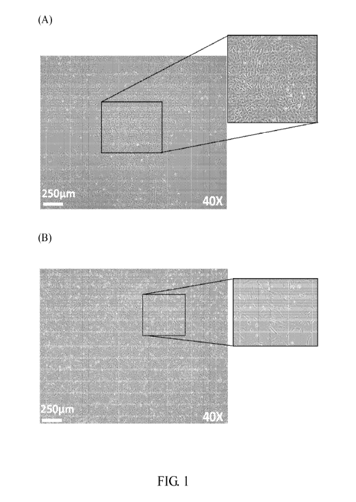
[0017] Fig. 1 shows the cell morphology of human placenta amnionic
mesenchymal
cells. Fig lA is a phase contrast image of AM-MSCs-CD34+ cells. Fig 1B is a
phase
contrast image of AM-MSCs-CD34- cells.
[0018] Fig. 2 shows the profiling of cell surface marker of AM-MSCs-CD34+
and
AM-MSCs-CD34- cells; wherein Fig. 2A shows the expression of CD29, CD44, CD73,
CD90, CD105, CD31, CD56, EGFR, and PDGFR; and Fig. 2B shows the expression
of CD34, CD117, CD133, SSEA1, SSEA3, SSEA4, GloboH, and CD201.
[0019] Fig. 3A provides a schematic illustration of neural and
oligodendrocyte
differentiation of CD34 sorted MSCs.
[0020] Fig. 3B provides a schematic illustration of dopaminergic neuron
differentiation of CD34 sorted MSCs.
[0021] Fig. 4 shows Tull, TH, and MAP2 expression of CD34+ or CD34- AM-
MSC
after dopaminergic neuron induction, wherein Fig. 4A provides Tull (top), GFAP
(middle), and DAPI (bottom) immunofluorescence staining images of CD34+ AM-MSC
induced neurons; Fig. 4B provides Tull (top), GFAP (middle), and DAPI (bottom)
immunofluorescence staining images of CD34- AM-MSC induced neurons; Fig. 4C
provides TH (top), MAP2 (middle), and DAPI (bottom) immunofluorescence
staining
images of CD34+ AM-MSC induced neurons; And Fig. 4D provides TH (top), MAP2
(middle), and DAPI (bottom) immunofluorescence staining images of CD34- AM-MSC
induced neurons.
[0022] Fig. 5 shows the cell morphology of human endometrium mesenchymal
cells;
wherein Fig 5A is a phase contrast image of EnMSCs-CD34+ cells; and Fig. 5B is
a
phase contrast image of EnMSCs-CD34- cells.
[0023] Fig. 6 shows the cardiomyogenic differentiation potentials of
EnMSCs-CD34; wherein Fig 6A provides Troponin T (top), myosin heavy chain
(MHC) (middle), and DAPI (bottom) immunofluorescence staining images of CD34+
4
CA 02815097 2013-04-16
WO 2012/054501
PCT/US2011/056737
EnMSCs after cardiomyogenic induction; and Fig 6B provides Troponin T (top),
myosin heavy chain (MHC) (middle), and DAPI (bottom) immunofluorescence
staining
images of CD34- EnMSCs after cardiomyogenic induction.
[0024] Fig. 7 shows the numbers of expanded hematopoietic stem cells
(HSCs) with
specific surface marker(s) when co-culturing with murine MS-5 feeder, MSCs-
CD34+
feeder, or MSCs-CD34- feeder.
DESCRIPTION OF THE EMBODIMENTS
[0025] As used herein, the article "a" or "an" means one or more than
one (that is,
at least one) of the grammatical object of the article, unless otherwise made
clear in the
specific use of the article in only a singular sense.
[0026] According to the invention, it is unexpectedly found that a
plurality of
precursor cells are isolated from human somatic tissues, and identified as
mesenchymal
stromal stem/progenitor cells expressing at least CD14, CD34, CD117, CD133
(AC133),
CD201, Nestin, SSEA3, SSEA4, and GloboH (called as "MCPCs"). The MCPCs may
be obtained by a method comprising the steps of isolating from a human somatic
tissue
by a systemic screening of human mesenchymal stromal stem/progenitor cells
followed
by a cell sorting by a cell antigen selected from the group consisting of
CD34, CD117,
CD133, CD201, GloboH, and combination thereof, and culturing in a medium
supplemented with at least one or more steroids selected from the group
consisting of a
corticosteroid, cholesterol, and combination thereof, and one or more growth
factors
selected from the group consisting of epidermal growth factor (EGF),
fibroblast growth
factor (FGF), insulin-like growth factor (IGF), insulin, platelet-derived
growth factor
(PDGF), IL-6, thrombopoietin (TPO), and combination thereof The MCPCs of the
invention adhere to a tissue culture surface, which is different from the
CD34, CD45-
placental stem cells as disclosed in Edinger et al. (US 2008/0206343 Al).
[0027] The MCPCs of the invention can be isolated from human somatic
tissues
including but not limited to neonatal placenta (e.g. amnion, chorion, and
umbilical cord),
endometrium, gingival, bone marrow, and adipose. Preferably, the MCPCs are
isolated from placenta, endometrium, and gingival. More preferably, the MCPCs
are
isolated from placenta amniotic tissue. According to the invention, the MCPCs
isolated from placenta amniotic tissue are called as AM-MSCs-CD34+ cells; the
MCPCs
isolated from endometrium are called as EnMSCs-CD34 + cells; and the MCPCs
isolated
5
CA 02815097 2013-04-16
WO 2012/054501
PCT/US2011/056737
from gingival are called as GMSCs-CD34+ cells. Specifically, AM-MSCs-CD34+
cells, EnMSCs-CD34+ cells, and GMSCs-CD34+ cells have consistent profiling of
cell
surface markers expression. In the invention, the MCPCs exhibited sphere-like
clonogenicty in early passages and expressed multipotent embryonic stem cell
(ESCs)
like characteristics in vitro. Morphologically, the MCPCs of the invention are
shorter
than CD34- MSCs. Specifically, the MCPCs of the invention have a higher growth
rate as compared to CD34- MSCs or unsorted MSCs, indicating that MCPCs of the
invention are more proliferative and younger.
[0028] According to the invention, the MCPCs homogenously express
embryonic
(e.g. Oct-4, Nanog, Rex-1, Sox-2), stemness (e.g. CD117, CD34, CD44) surface
antigens, in addition to present various lineage markers, including MSC (e.g.
CD29,
CD90, CD73, CD105, CD106), hem-angiogenic (e.g. AC133, CD34), myo-nurogenic
(e.g. CD54, Nestin, NSE). Further, the MCPCs of the invention are prolonged
self-renewal. According to some embodiments of the invention, the MCPCs (e.g.,
AM-MSCs-CD34 + cells) could retain their specific cell marker expression as
CD34,
CD54, CD117, and AC133 positive, and their MSC marker expression even after 20
passages.
[0029] Accordingly, the invention provides an enriched population of
multipotent
human mesenchymal common progenitor cells (MCPCs), which are identified as
mesenchymal stromal stem/progenitor cells which have at least the following
characteristics: CD14+, CD34, CD117+, CD133+ (AC133+), CD201+, Nestin+,
SSEA3+,
SSEA4+, and GloboH+.
[0030] According to the invention, the enriched population of multipotent
human
MCPCs further have at least one of the following characteristics: CD44+,
CD54+,
CD56+, CD105+, CD146+, and PDGFR+. In one embodiment of the invention, the
enriched population of multipotent human MCPCs are identified as mesenchymal
stromal stem/progenitor cells, having the following characteristics: CD14+,
CD34,
CD117+, CD133+ (AC133+), CD201+, Nestin+, SSEA3+, SSEA4+, GloboH+, CD44+,
CD54+, CD56+, CD105+, CD146+ and PDGFR+.
[0031] According to the invention, the enriched population of multipotent
human
MCPCs have the potentials to differentiate into cells or tissues of ectodermal
lineage,
mesodermal lineage, and endodermal lineage. In one embodiment of the
invention,
6
CA 02815097 2013-04-16
WO 2012/054501
PCT/US2011/056737
AM-MSCs-CD34+ cells were examined and found that they were multipotent in
differentiation of various types of somatic cells, including endoderm,
mesoderm, or
ectoderm cells.
[0032] In one
embodiment of the invention, the enriched population of multipotent
human MCPCs have the potentials of adipogenic differentiation, osteogenic
differentiation, chondrogenic differentiation,
neurogenic differentiation,
cardiomyogenic differentiation, endothelial differentiation, and hepatic
differentiation.
[0033] In
addition, the invention provides a method for producing an enriched
population of multipotent human MCPCs, comprising isolating from a human
somatic
tissue by a systemic screening of human mesenchymal stromal stem/progenitor
cells
and a cell sorting by a cell antigen selected from the group consisting of
CD34, Nestin,
CD117, CD133 and combination thereof; and culturing in a medium supplemented
with
at least one or more steroids and one or more growth factors.
[0034] As used
herein, the term "steroid" refers to a type of organic compound that
contains a specific arrangement of four cycloalkane rings that are joined to
each other.
In the invention, the steroid used in the medium according to the method may
be one
selected from the group consisting of a corticosteroid, cholesterol and
combination
thereof
[0035] As used
herein, the term "corticosteroid" refers to a class of steroid hormone.
Examples of corticosteroid include but are not limited to a group A
corticosteroid (e.g.
hydrocortisone, hydrocortisone acetate, and cortisone acetate), a group B
corticosteroid
(e.g. triamcinolone acetonide, triamcinolone alcohol, and mometasone), a group
C
corticosteroid (e.g. betamethas one, betamethas one sodium phosphate, and
dexamethasone), and a group D corticosteroid (e.g. hydrocortisone-17-valerate,
aclometasone dipropionate, and hydrocortisone-17-butyrate).
[0036] The
term "growth factor" as used herein refers to a naturally occurring
substance capable of stimulating cellular growth, proliferation and cellular
differentiation, which can regulate a variety of cellular processes, and
typically act as
signaling molecules between cells. In the invention, the growth factor used in
the
medium according to the method may be one or more selected from the group
consisting of epidermal growth factor (EGF), fibroblast growth factor (FGF),
insulin-like growth factor (IGF), insulin, platelet-derived growth factor
(PDGF), IL-6,
7
CA 02815097 2013-04-16
WO 2012/054501
PCT/US2011/056737
thrombopoietin (TPO), and combination thereof
[0037] According to some embodiments of the invention, the medium may be
further supplemented with and one or more vitamins.
[0038] As used herein, the term "vitamin" refers to a nutrient in tiny
amounts by an
organism. In the invention, examples of vitamins include but are limited to
vitamin A,
vitamin B-complex, vitamin E, biotin, p-aminobenzoic acid, menadione, and
combination thereof
[0039] In the invention, the medium may be also supplemented with one or
more
compounds selected from the group consisting of uracil, sodium acetate,
ribose,
Guanine HC1, deoxyribose, adenosine, adenine sulphate, ferric nitrate and
combination
thereof
[0040] In preferred embodiments of the invention, the cell sorting is
performed by
using fluorescence-activated cell sorting (FACS) flow cytometry. Preferably,
the cell
sorting is a CD34, CD117, CD133, CD201, or GloboH cell antigen FACS.
[0041] According to the invention, the MCPCs may be encapsulated in
alginate to
form a composition.
[0042] According to the invention, a feeder cell layer for stem cell
culture is
provided as well. The feeder cell layer comprises the enriched population of
multipotent human MCPCs of the invention. Alternatively, the enriched
population of
multipotent human MCPCs may be seeded on the scaffold to form a stem cell
niche.
[0043] Accordingly, the invention provides a better source of ESC-like
clonogenic
stem cells that are derived from non-embryonic neonatal or adult tissue and
multipotent
in differentiation to various types of cells, than known sources, such as
ESCs. The
MCPCs of the invention were found to be of proliferative ability and
differentiation
potentials. They are of a great potential to be used for clinical regenerative
therapies.
The invention is further described in the following non-limiting examples.
[0044] Human tissues used in the following examples were obtained using a
protocol approved by Institutional Review Board of Cathay General Hospital &
Taipei
Medical University Institutional Review Board.
[0045] Example 1: Preparation and Characterization of AM-MSCs-CD34+ Cells
[0046] Amnionic Mesenchymal Cell Isolation
[0047] (1) Isolation of amnion membrane:
8
CA 02815097 2013-04-16
WO 2012/054501
PCT/US2011/056737
[0048] Amnion membrane (about 300 cm2, n = 9) was stripped from chorion,
washed in 3x150 ml changes of lxHank's buffer to remove blood.
[0049] (2) Removal of the amnionic epithelial cells:
[0050] To deplete the amnionic epithelial cells (Am-EpCs), washed amnion
membrane was cut into 2-3 cm2 fragments and incubated in 100 ml of 0.1%
Trypsin-EDTA (Sigma; St Louis, MO) with lx Hanks balanced salt solution
(Gibco;
CAT# 14185-052; Grand Island, NY) for an about 250 cm2 membrane, with 4 times
of
each 15 min reactions in water bath, at 37 C.
[0051] (3) Collection of clonogenic amnionic mesenchymal stromal cells
(AM-MSCs):
[0052] For the amnionic mesenchymal cells (AM-MSCs) isolation, the Am-
EpCs
depleted amnion membrane was subjected to wash with Hank's buffer one time and
digested with collagenase lA at 37 C for 45-60 minutes. An appropriate volume
of
Hank's buffer and a 40 mm nylon cell strainer were used to collect the
clongenic
AM-MSCs.
[0053] (4) Incubation of AM-MSCs:
[0054] After a 170g centrifugation, AM-MSCs were plated in CELL-BIND T75
flasks at a density of 5 x 10⁴ cells per cm2 and incubated in a 5% CO2, 37 C.
The
collected AM-MSCs were incubated in a medium containing Medium 199 (the M199
conditioned medium) [(Lonza CAT# 12-118F; Switzerland), supplemented with
fetal
bovine serum (FBS), epidermal growth factor (EGF), and hydrocortisone. Fresh
medium were changed in every 3-4 days during the purification incubation, and
cells
expanded to 80% confluence in 7 days. The attached culturing cells were
harvested with
0.1% Trypsin-EDTA, and split into the new passage culture with a seeding
density of
lx i05 cells per T75 flask.
[0055] Alternatively, the collected CM-MSCs can be cultured in RPMI-1640
(GIBCO; Grand Island, NY) supplemented with fetal bovine serum (FBS) (10%),
sodium pyruvate (0.1 mM), basic fibroblast growth factor (bFGF), and EGF (10
ng/ml).
Cells were split when they reached to 70-80% confluence, the culture medium
was
changed every 3-4 days.
[0056] Flow Cytometry Analysis
[0057] For FACS analysis, freshly harvested AM-MSCs were trypsinized and
9
CA 02815097 2013-04-16
WO 2012/054501
PCT/US2011/056737
incubated with aliquot florescence (FITC or PE) conjugated monoclonal
antibodies
(mAbs), suggested by the manufacturer, for 30 minutes at 4 C in 100 n1
phosphate
buffer. Cell markers were tested including for the mesenchymal stem cell (MSC)
lineage (CD29, CD90, CD73, CD105, CD106, Vimentin), stemness (CD34, CD44,
CD117), hem-angiogenic (AC133, CD34), myo-neurogenic (CD54, Nestin), and
myofibroblast markers (Vimentin, alpha smooth muscle actin). Cells were
analyzed
using a FACSCanto flow cytometry system (BD Bioscience, San Jose, CA). The
flow-cytometric data were processed with FCS Express V3 software (De Novo;
Canada).
[0058] Flow Cytometry Sorting
[0059] For isolation of the CD34 + AM-MSCs sub-population, the expanded
primary
AM-MSCs cultured at passages 2-3 were used to label with CD34 antibody. Up to
3 x
106 cells were sorted by a FACS Aria flow cytometry (BD Bioscience, San Jose,
CA)
following the manufacture's instruction. CD34 positive (CD34) and CD34
negative
(CD34-) cells were then analyzed, sorted, and collected.
[0060] In brief, 3-4 x 10⁶ harvested third passage AM-MSCs were
trypsinized and
labelled with PE-conjugated CD34 suggested by the manufacturer, for 15 minutes
at
room temperature in 100 n1 phosphate buffer. Then filtered cells through a 40
nm nylon
cell strainer (Becton, Dickinson and Company CAT# 352235) and sorting
AM-MSCs-CD34+ and AM-MSCs-CD34- populations with BD FACS Aria. After
sorting, the AM-MSCs-CD34+ and AM-MSCs-CD34- populations were re-analyzed for
the positive fraction and expanded in the M199 conditioned medium as described
above
or the RPMI conditioned medium. The cell morphology of AM-MSCs (passage 3) are
given in Fig. 1, wherein the shape of AM-MSCs-CD34+ cells (Fig. 1A) is shorter
than
AM-MSCs-CD34- cells (Fig. 1B) and both showed stable in later passages. The
initial
doubling time of AM-MSCs-CD34- cells is about 42 hours, which is longer than
that of
AM-MSCs-CD34+ at being 34 hours. The CD34 expression of AM-MSCs would be
checked for each five passages in the following days. After CD34 + sorting 20
passages, CD34 + AM-MSC could still retain their specific cell marker
expression as
CD34, CD54, CD117, and CD133 (AC133) positive and their MSC marker expression.
It was found that the high expression each of CD and gene markers other than
CD34
could be observed in the cells at each passage, but the expression of CD34 (50-
60%) of
CA 02815097 2013-04-16
WO 2012/054501
PCT/US2011/056737
the cells at passage 1 was less than those of passages 3 or 9 (about 100%).
Both
CD34+ and CD34- sorted AM-MSC expressed CD29, CD44, CD73, CD90, CD105,
EGFR positive, and CD31 negative. However, CD34+ AM-MSC expressed more
CD56 and PDGFR than CD34- AM-MSC. See Figure 2A. Further, CD34+
AM-MSC could express more CD117, CD133, SSEA1, SSEA3, SSEA4, Globo H, and
CD201 than CD34- AM-MSC (Fig. 2B). Specific gene expression was also examined
by RT-PCR (data not shown). The results show that AM-MSCs-CD34+ cells express
"early genes" including Sox-2, Oct-4, Rex-1, and Nanog, ectodermal lineage
genes, e.g.
neurogenic differentiation markers including Nestin, NSE, NFM, NCAM, MAP2,
mesodermal lineage genes, e.g. cardiomyogenic differentiation markers
including
MyoD, GATA-4, and MLC-2a, and endodermal lineage genes, e.g. hepatic
differentiation makers including Albumin and HGF.
[0061] Example 2: Induction of Differentiation
[0062] (1) Vasculogenic differentiation
[0063] The AM-MSCs-CD34+ cells at the number of 2 x10⁵ at passage 5 were
used
for the vasculogenic differentiation induction. Harvested cells were cultured
in EGM-2
medium (Cambrex) for a 7 days induction. Analysis of the capillary formation
was
performed using Matrigel (BD Biosciences). Specifically, after the induction
culture,
the AM-MSCs-CD34+ were trypsinized and plated onto Matrigel coated (Matrigel:
M199 = 1:1) 24 well cluster, with a cell density of 105 cells per well.
Capillary-like
structures were observed by optical microscopy after 2, 4, 24, and 48 hours in
the
following 3 days.
[0064] (2) Cardiomyogenic differentiation
[0065] Sorted AM-MSCs-CD34+ cells at passages 4-6 were harvested for
induction
of cardiomyogenic differentiation. The AM-MSCs-CD34+ cells were incubated
overnight in the growth medium [EGM-2:M199 (v:v = 1:3) supplemented with 10%
FBS, and MEM nonessential amino acids (1x) (GIBCO)]. On the next morning,
cells
were transferred into the cardiomyogenic differentiation medium, [IMDM
(GIBCO):
Ham's F12 nutrient mixture with G1utaMAX-1 (GIBCO) (v:v = 1:1) supplemented 2%
horse serum (GIBCO), lx MEM nonessential amino acids, lx
insulin-transferrin-selenium (GIBCO)] with a cell density of 104 per cm2.
After 6-8
hours, a cardiomyocytic differentiation agent, 5-azacytidine (Sigma) was added
into the
11
CA 02815097 2013-04-16
WO 2012/054501
PCT/US2011/056737
medium to make a 5 laM final concentration. 4u1/m1 of 5-azacytidine (0.25 mM)
stock
solution was added into the differentiation medium daily, and changed back to
the
differentiation medium without 5-azacytidine on the day 4. On day six of the
differentiation assay, ascorbic acid (104 M) (Sigma) and TGF-31 (1 ng/ml)
(PeoproTech) were added to the medium. From this point forward, ascorbic acid
and
TGF-31 were supplemented every other days and twice weekly, respectively. The
medium were changed every 2-3 days, depending on the medium pH changes. Cell
debris should be removed by PBS washes, when medium changes. The
Cardiomyogenic
AM-MSCs-CD34+ cells were fixed for histochemical staining after 28-day
differentiation culture. By a 5-azacytidine myogenic induction, CD34+ AM-MSCs
(P5)
transdifferentiated easily into cardiomyocytes expressing MyoD, GATA-4, MLC-2a
genes (data not shown). After cardiomyogenic differentiation 28 days and
examined
by histochemical staining, both CD34+ and CD34- AM-MSCs expressed myosin heavy
chain (MHC), but only induced CD34+ AM-MSCs formed typical cardiomyocyte
morphology and expressed terminal differentiated marker Troponin T (data not
shown).
[0066] (3) Hepatic differentiation
[0067] The expressions of hepatic differentiation cell markers were
given in Table 1
below.
Table 1. Expressions of hepatic differentiation cell markers of AM-MSCs-CD34+
cells
(passage 6).
Cell Marker AM-MSCs-CD34+ Control
Gene DAPI +++ +++
Cy3 (Albumin) +++
FITC +++
(Cytokeratin)
Protein GAPDH +++ +++
Albumin +++
HGF ++
[0068] (4) Adipogenic, osteogenic, and chondrogenic differentiation
[0069] The AM-MSCs-CD34+ cells obtained by the method as mentioned in
Example 1 and expanded passages 5-6 were used for multi-lineage
differentiation
inductions. The adipogenic, osteogenic, chondrogenic, and neurogenic
differentiation
protocols were used by the methods given below.
12
CA 02815097 2013-04-16
WO 2012/054501
PCT/US2011/056737
[0070] The AM-MSCs or AM-MSCs-CD34+ pre-conditioning in Dulbecco's
modified Eagle's medium (DMEM/LG, GIBCO) supplemented with 10% FBS
(Hyclone) were used for the lineage differentiation cultures shown as
following:
1) Adipogenesis (AM): DMEM/LG medium supplemented with 10% FBS, 0.5 mM
isobutyl-methylxanthine, 1 laM dexamethasone, 10 laM insulin, 200 laM
indomethacin.
2) Osteogenesis (OM): DMEM/LG medium supplemented with 10% FBS, 0.1 laM
dexamethasone, 50 laM ascorbate-2-phosphate, 10 mM P-glycerolphosphate.
3) Chondrogenesis (CM): DMEM/LG medium supplemented with 1% FBS, 6.25 lag/m1
insulin, 10 ng/ml TGF-31 (R&D), 50 nM ascorbate-2-phosphate.
4) Neurogenesis (NM): DMEM/LG medium supplemented with 5 lag/m1 insulin, 200
laM indomethacin, 0.5 mM isobutyl-methylxanthine. (Reagents used above for
differentiation were all from Sigma; St.Louis, MO)
[0071] For adipogenic, osteogenic, and neurogenic differentiation, the
cell density
was 3x10⁴ cells/cm2. For chondrogenic differentiation, a higher cell density
of
1-2 x10⁵/10 ial was used for chondrosphere formation. Medium was changed every
three to four days for all differentiation assays, and cells were fixed for
histochemical
staining after 14 days of adipogenic, osteogenic, chondrogenic
differentiation. After
14 days, intracellular oil droplets were formed under Oil Red 0 stain, and
calcified
extracellular matrix was present and positive for von Kossa staining (data not
shown).
In chondrogenic differentiation, AM-MSCs-CD34+ cells formed cartilage ball in
3 days.
AM-MSCs-CD34+ cells cultured in neurogenic differentiation medium (Zuk's
protocol,
P4, Day 21) exhibited neural morphology and expressed neural markers including
Nestin, NSE, NFM, NCAM, and MPA2, while AM-MSCs-CD34- cells did not (data not
shown).
[0072] (5) Neurogenic differentiation
[0073] Step 1 : Neurosphere Formation: Cells were seeded at a density of
1000 per
well with neurosphere medium (NS medium). NS Medium: DMEM/HG/F12 (1:1) +
lx B27 + 20 ng/ml EGF + 20 ng/ml FGF2 +2 lag/m1 Heparin. Primary neurospheres
(selecting mainly floating neurospheres) larger than 75 um were counted after
7 days in
vitro. Step2: Neural Differentiation Assay: Dissociated neurospheres to single
cells by
trypsin-EDTA solution and culture the cells with: DMEM/F12 + 5% FBS for 24
hrs.
These cells were treated with specific neural cell differentiation medium. The
medium
13
CA 02815097 2013-04-16
WO 2012/054501
PCT/US2011/056737
used for neuronal differentiation was DMEM/F12 supplemented with 2% FBS, 10
ng/ml PDGF, 50 ng/ml BNDF, and 50 ng/ml GDNF. Fig. 3A is a schematic
illustration of neural and oligodendrocyte differentiation of CD34 sorted
MSCs. Fig.
3B is a schematic illustration of dopaminergic neuron differentiation of CD34
sorted
MSCs.
[0074] After 7 to 9 days, the differentiation capacity was verified by
using
immunofluorescence staining (GFAP conjugated with FITC, Hochest 33258, and
Tull
conjugated with rhodamine). After primary neurosphere formation, CD34+ and
CD34-
AM-MSC both expressed Tull (neuron specific marker). However, the GFAP (glia
specific marker) expression was dim for CD34+ AM-MSC induced neurons. On the
other hand, some CD34- AM-MSC induced neuron expressed GFAP, which suggests
that some induced CD34- AM-MSC cells differentiate into neurons while some of
them
differentiate into glia cells. In B27 induction, CD34+ AM-MSC expressed Galc,
and
Tull, but not GFAP. For dopaminergic neuron differentiation, as detected by
immunofluorescence staining, CD34+ AM-MSC induced neurons were Tull, TH, and
MAP2 positive, while only a small population of CD34- AM-MSC induced neurons
expressed said markers (Fig. 4).
[0075] (6) Conclusion
[0076] CD34+ AM-MSCs expressed early genes and showed multipotent
differentiation potential. Specific gene expression of CD34+ AM-MSCs is
provided in
Table 2 below.
[0077] Table 2. Summary of CD34+ AM-MSCs gene expression.
CD34+ AM-MSCs
Ectoderm Mesoderm
Endoderm (Hepatic
Early Genes (Neurogenic (Cardiomyogenic
Differentiation)
Differentiation) Differentiation)
Sox-2 + Nestin + MyoD + Albumin +
Oct-4 + NSE + GATA-4 + HGF +
Rex-1 + NFM + MLC-2a + -
-
Nanog + NCAM + - - - -
- - MAP2 + - - - -
14
CA 02815097 2013-04-16
WO 2012/054501 PCT/US2011/056737
[0078] Example 3: EnMSCs-CD34+ cells, GMSCs-CD34+ cells, and CD34+ MSCs
Enriched from Other Somatic Tissues
[0079] Primary endometrial and gingival tissues were collected from
donors from
Taipei medical hospital and Dr. Wells Dental clinic follows the IRB guide
line.
EnMSCs and GMSCs were obtained from endometrial and gingival tissues,
respectively,
by similar process set forth in Example 1. EnMSCs and GMSCs were then subject
to
CD34 sorting.
[0080] Phase contrast images of CD34 sorted human endometrium derived
mesenchymal stem cells (P5) are given in Fig. 5. The morphology of EnMSCs-
CD34+
cells was very similar to AM-MSCs-CD34+. Further, as provided in Table 3
below,
AM-MSCs-CD34+ cells, EnMSCs-CD34+ cells, and GMSCs-CD34+ cells were
identified as having consistent profiling of cell surface marker expression.
Specifically, the mesenchymal common progenitor cells (MCPCs) of the invention
were
CD14+, CD34+, Nestin+, CD117+, CD133+ (AC133+), SSEA3+, and SSEA4+. Further,
the MCPCs of the invention were also characterized as CD44+, CD54+, CD105+,
CD146+, or PDGFR+.
[0081] Table 3. Cell surface markers expression of MCPCs and CD34- MSCs.
Percentage of FACS markers expression: -: 0-20%, +: 20-40%, ++: 40-80%, +++:
80-100%.
AM-MSCs (P4) EnMSCs (P4) GMSCs (P4)
CD34+ CD34- CD34+ CD34- CD34+ CD34-
CD29 +++ +++ +++ +++ +++ +++
CD44 +++ +++ +++ +++ +++ +++
CD73 +++ +++ +++ +++ +++ +++
CD90 +++ +++ +++ +++ +++ +++
CD105 +++ +++ +++ +++ +++ +++
EGFR +++ +++ +++ +++ +++ +++
Integrin a2131 +++ +++ +++ +++ +++ +++
E-cadherin - - - - - -
CA 02815097 2013-04-16
WO 2012/054501
PCT/US2011/056737
CD34 +++ - +++- +++ -
CD54 +++ + +++ + +++ +
PDGFR ++ + ++ + ++ +
Nestin +++ + +++ + +++ +
CD14 +++ - +++- +++ -
CD117 +++ - +++- +++ -
AC133 +++ - +++- +++ -
CD146 +++ - +++- +++ -
[0082] The comparison on cell morphologies and cell doubling times of
MCPCs
and CD34- MSCs are given in Table 4 below.
[0083] Table 4. Comparison of cell morphology and cell doubling time.
Cell Morphology Cell Doubling
Time
AM-MSC CD34+: shorter. CD34+: 34hrs.
(CD34+/CD34-) CD34-: longer and thinner CD34-: 42hrs.
EnMSC CD34+: shorter. CD34+: 33hrs.
(CD34+/CD34-) CD34-: longer and thinner CD34-: 47hrs.
GMSC CD34+: shorter. CD34+: 32hrs.
(CD34+/CD34-) CD34-: longer and thinner CD34-: 45hrs.
[0084] Potentials of endothelial differentiation and chondrogenic
differentiation of
MCPCs and CD34- MSCs are given in Table 5 below.
[0085] Table 5. Potentials of endothelial differentiation and
chondrogenic
differentiation.
Endothelial Differentiation Chondrogenic Differentiation
AM-MSCs CD34+: CD31+, KDR+, CD34+: Bigger cartilage ball
(CD34+/CD34-) more tube formation. (Diameter > 2x),
CD34-: CD31+, KDR+, less CD34-: Little cartilage ball
16
CA 02815097 2013-04-16
WO 2012/054501
PCT/US2011/056737
tube formation. (Diameter < 100 nm).
EnMSCs CD34+: CD31+, KDR+, CD34+: Bigger cartilage ball
(CD34+/CD34-) more tube formation. (Diameter > 2x),
CD34-: CD31+, KDR+, less CD34-: Little cartilage ball
tube formation. (Diameter < 100 nm).
GMSCs CD34+: CD31+, KDR+, CD34+: Bigger cartilage ball
(CD34+/CD34-) more tube formation. (Diameter > 2x),
CD34-: CD31+, KDR+, less CD34-: Little cartilage ball
tube formation. (Diameter < 100 nm).
[0086] Comparison of embryonic gene expression of MCPCs and CD34- MSCs
are
given in Table 6 below. MCPCs of the invention showed stronger embryonic gene
expression than CD34- MSCs.
[0087] Table 6. Comparison of embryonic gene expression
Early Gene Detection
AM-MSCs CD34+: Sox-2 +++, Oct-4 +++, Rex-1 +++, Nanog +++
(CD34+/CD34-) CD34-: Sox-2 +++, Oct-4 ++, Rex-1 ++, Nanog ++
EnMSCs CD34+: Sox-2 +++, Oct-4 +++, Rex-1 +++, Nanog +++
(CD34+/CD34-) CD34-: Sox-2 +++, Oct-4 ++, Rex-1 +++, Nanog ++
GMSCs CD34+: Sox-2 +++, Oct-4 +++, Rex-1 +++, Nanog +++
(CD34+/CD34-) CD34-: Sox-2 +++, Oct-4 ++, Rex-1 ++, Nanog ++
[0088] Potentials of neurogenic differentiation and cardiomyogenic
differentiation
of MCPCs and CD34- MSCs are given in Table 7 below. The cardiomyogenic marker
immunofluorescence staining images of EnMSCs are as shown in Fig. 6.
[0089] Table 7. Potentials of neurogenic differentiation and cardiomyogenic
differentiation.
Neurogenic Differentiation Cardiomyogenic Differentiation
17
CA 02815097 2013-04-16
WO 2012/054501
PCT/US2011/056737
AM-MSCs CD34+: Nestin+, TuJ1+, CD34+: Myosin Heavy Chain+,
(CD34+/CD34-) GFAP-, typical neuron Troponin T+.
forming. CD34-: Myosin Heavy Chain+,
CD34-: Nestin+, Troponin T-.
Tull (less+), GFAP-
EnMSCs CD34+: Nestin+, TuJ1+, CD34+: Myosin Heavy Chain+,
(CD34+/CD34-) GFAP-, Troponin T+.
neurosphere-like structure. CD34-: Myosin Heavy Chain+,
CD34-: Nestin+, TuJ1+, Troponin T-.
GFAP-
GMSC CD34+: Nestin+, TuJ1+, CD34+: Myosin Heavy Chain+,
(CD34+/CD34-) GFAP-, Troponin T+.
neurosphere-like structure. CD34-: Myosin Heavy Chain+,
CD34-: Nestin+, TuJ1+, Troponin T-.
GFAP-
[0090] CD34+ MSCs can be enriched from many other somatic tissues
according to
the method of the invention. Normally, only 2-3% of MSCs are CD34+. After
isolated and enriched in culture, percentage of CD34+ MSCs ranging from 15% to
78%
depending on which tissue they were isolated from and the donor (See Table 8
below).
The culture-enriched CD34+ MSCs can be subject to FACS cell sorting for
further
enrichment to obtain a population of MSCs enriched with 99% or more CD34+
MSCs.
[0091] Table 8. Enrichment of Stem/progenitor marker and gene expressions
in
Human Tissue MSCs
% Enriched
% CD34+ MSCs
Marker
Enrichment in Culture
Tissues
iiiiNeoNatat Placenti¨ 40 ¨ 70 (-55 )---1
r
Amnion 48 ¨53 (-50)
Chorion 40 ¨ 62 (-51)
Umbilical Cord 34 ¨ 45 (-40)
Endometrium 45 ¨ 78 (-61)
Gingiva 27 ¨35 (-31)
18
CA 02815097 2013-04-16
WO 2012/054501
PCT/US2011/056737
Bone Marrow 20 ¨30 (-25)
Adipose 15 ¨ 30 (-23)
[0092] Example 4: MCPCs as Feeder Cells
[0093] MCPCs of the invention were used to prepare a stromal feeder for
expansion
of hematopoietic stem cells (HSCs). Stromal cells (MS-5, or MSCs) were seeded
in
plate and wait for confluence to become feeder. 2-4x 10⁴ CD34+ mononuclear
cells
(MNCs) were co-cultured with feeder in 1 ml HSC medium (X-VIV010 + 50 ng/ml
SCF + 50 ng/ml Flt-3L + (20 ng/m1)10U/m1 TPO + 10 ng/ml IL-6). After 7 days or
14
days, suspension cells were counted and subjected to flow cytometry analysis
(for
CD34+CD38-, CD34+CD133+, CD34+CXCR4+, etc.).
[0094] The results are shown in Fig. 7. When co-culturing with the MCPCs
(AM-MSCs-CD34+ cells) feeder, more engrafting CD34+CD38- primitive HSCs were
obtained, as compared to murine MS-5 feeder or MSCs-CD34- feeder.
[0095] Although the present invention is illustrated by the above
embodiments,
these embodiments are not used to limit the present invention. It will be
apparent to
those skilled in the art that various modifications and variations can be made
to the
structure of the present invention without departing from the scope or spirit
of the
invention. In view of the foregoing, it is intended that the present invention
cover
modifications and variations of this invention provided they fall within the
scope of the
following claims and their equivalents.
19