Note: Descriptions are shown in the official language in which they were submitted.
CA 02815359 2013-04-19
WO 2012/054735
PCT/US2011/057115
SYSTEMS AND METHODS FOR ASSESSING
BIOMOLECULE CHARACTERISTICS
GOVERNMENT RIGHTS
[0001] This work was supported by National Institutes of Health grant
2R44H004199-03-NIH/NHGRI; by National Institute of Standards and Technology
grant
70NANB7H7027N NIST-ATP 2007; and by National Institutes of Health grant
1R43HG004817-01 NIH/DHHS. The government has certain rights in this
disclosure.
RELATED APPLICATIONS
[0002] The present application claims priority to United States Application
61/407,302, "Nanoanalyzer Systems and Methods," filed on October 27, 2010;
United States
Application 61/394,915, "DNA Damage Detection in Nanochannel Array," filed on
October
20, 2010; United States Application 61/407,182, "Single Molecule DNA
Nanochannel
Analysis for Genomic Studies," filed on October 27, 2010; and United States
Application
61/418,516, "DNA Damage Detection in Nanochannel Array," filed on December 1,
2010.
These applications are incorporated herein in their entireties for any and all
purposes.
TECHNICAL FIELD
[0003] The present disclosure relates to the field of nucleic acid analysis,
to the field
of nanofluidics, and to the field of optical instrumentation.
BACKGROUND
[0004] The genomes of living organisms are constantly at risk for endogenous
and
environmentally induced DNA alterations. DNA lesions at specific genomic sites
can lead to
changes in nucleotide sequence. DNA molecules can be damaged in numerous ways,
including (a) mismatches arising during DNA replication; (b) damage resulting
from
instability of DNA molecules, such as incorporation of uracil, deamination of
bases,
depurination and depyrimidination; (c) damage due to environmental factors.
For example,
ionizing radiation produces modified bases and strand breaks, and UV radiation
produces
cyclobutane pyrimidine dimers and other photoproducts. Exemplary DNA damage
scenarios
are illustrated in Figure 1.
[0005] The consequences of DNA damage result in DNA fragmentation (double
strand DNA breaks), single stranded DNA breaks and modified bases. Currently,
there is
- 1 -
CA 02815359 2013-04-19
WO 2012/054735
PCT/US2011/057115
limited available high-throughput and sensitive methods to detect these events
without the
need of DNA amplification, which amplification could conceal those
modifications.
Accordingly, there is a need in the art for methods and systems for detection
of polynucleotide
damage.
SUMMARY
[0006] In meeting the described challenges, the present disclosure first
provides
methods, the methods including converting a first site on a polynucleotide to
a first moiety
capable of supporting polymerase extension; effecting extension at the first
moiety so as to
incorporate a first label at or proximate to the first site; linearizing a
portion of the
polynucleotide that includes the first label; and imaging the first label.
[0007] The disclosure also provides analysis systems, the systems suitably
including
a sample stage configured to receive a fluidic chip that comprises one or more
nanochannels
having a characteristic dimension in the range of from 1 nm to about 250 nm,
an illumination
source configured to illuminate a sample disposed within the fluidic chip; and
an image
collector configured to collect an image of an illuminated sample disposed
within the fluidic
chip.
[0008] Also provided are methods, the methods including contacting a first
single-
strand break in a polynucleotide with an alkaline phosphatase so as to give
rise to a first
moiety capable of supporting polymerase extension; contacting the moiety with
a polymerase
and a labeled nucleotide so as to incorporate a label into the
polyoligonucleotide; linearizing
at least a portion of the polynucleotide by confining the first label within a
nanochannel; and
imaging the first label.
[0009] Additionally provided are methods, the methods including applying
to
a single-strand break in a polynucleotide a DNA polymerase having 3' to 5'
exonuclease
activity so as to convert the non-extendable single strand break into a
polymerase-extendable
sites; and applying a DNA polymerase and a labeled deoxynucleotide so as to
incorporate a
label into the polynuclotide.
[0010] The disclosure also provides methods, the methods including disposing a
polynucleotide having an abasic site within a porous matrix material;
contacting the
polynucleotide with an alkaline material so as to covert the abasic site to a
single strand break
in the polynucleotide, so as to convert a single strand break in the
polynucleotide to a double
strand break in the polynucleotide, or both; converting a single strand break
in the
polynucleotide, a double strand break in the polynucleotide, or both, to a
moiety capable of
- 2 -
CA 02815359 2013-04-19
WO 2012/054735
PCT/US2011/057115
supporting polymerase extension; and contacting the moiety with a polymerase
and a labeled
nucleotide so as to incorporate one or more labels into the polynucleotide.
[0011] Further disclosed are additional methods, these methods including
disposing
a polynucleotide within a porous matrix material; converting a first site on a
polynucleotide to
a first moiety capable of supporting polymerase extension; effecting extension
at the first
moiety so as to incorporate a first label at or proximate to the first site;
linearizing at least a
portion of the polynucleotide by confining the first label within a
nanochannel; and imaging
the first label.
[0012] Further disclosed are kits, the kits including a quantity of an N-
glysosylase; a
quantity of an apurinic/apyrimidinic lysase, a 3'-phosphodiesterase, or both;
a quantity of a
polymerase; and a quantity of a labeled nucleotide.
[0013] Kits may also include a quantity of an alkaline material; a
quantity of
an apurinic/apyrimidinic lysase, a 3'-phosphodiesterase, or both; a quantity
of a polymerase;
and a quantity of a labeled nucleotide.
[0014] Also provided are systems. These systems suitably include a kit that
includes
(a) a quantity of a polymerase, (b) a quantity of a labeled nucleotide, and
(c) a quantity of one
or more of an apurinic/apyrimidinic lysase, a 3'-phosphodiesterase, or
Endonuclease IV, the
kit being adapted to engage with a sample imager, the sample imager comprising
a sample
stage adapted to engage with a fluidic chip that includes one or more
nanochannels, an
illumination source capable of optical communication with a sample disposed
within a
nanochannel of the fluidic chip, an image collector capable of collecting an
image of an
illuminated sample disposed within the nanochannel.
[0015] Other methods provided herein include linearizing a region of a
polynucleotide that includes at least one label, the label having been
incorporated into the
polynucleotide by polymerase extension, the polymerase extension being
performed on a
moiety that was converted from an abasic site, a single strand break, or both.
[0016] Additional methods disclosed herein include incorporating a label at or
proximate to a site of damage on a polynucleotide; linearizing a region of the
polynucleotide
that includes the label; and imaging the label.
[0017] The present disclosure also provides systems, the systems including a
base
configured to receive a fluidic chip; an illuminator configured to illuminate
a polynucleotide
sample disposed within the fluidic chip; and an image collector configured to
collect an image
from the polynucleotide sample disposed within the fluidic chip.
- 3 -
CA 02815359 2013-04-19
WO 2012/054735
PCT/US2011/057115
BRIEF DESCRIPTION OF THE DRAWINGS
[0018] The summary, as well as the following detailed description, is further
understood when read in conjunction with the appended drawings. For the
purpose of
illustrating the invention, there are shown in the drawings exemplary
embodiments of the
invention; however, the invention is not limited to the specific methods,
compositions, and
devices disclosed. In addition, the drawings are not necessarily drawn to
scale. In the
drawings:
[0019] Figure 1 illustrates exemplary DNA lesions that may result from DNA
damage, which lesions include single strand breaks (SSBs), double strand
breaks (DSBs), and
modified bases;
[0020] Table 1 illustrates an example of the types and quanitities of DNA
lesions
resulting from exposure of DNA to ionizing radiation in the form of gamma rays
generated
from Cesium 137 (137Cs);
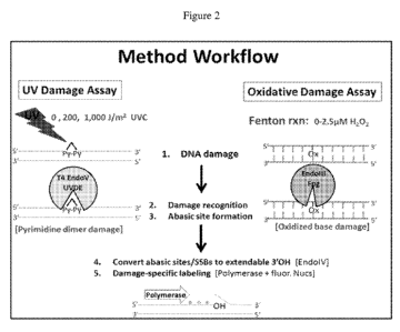
[0021] Figure 2 illustrates an illustrate process according to the present
disclosure
using N-glycosylases to recognize ultraviolet (UV)- damaged bases and also
oxidative
damaged bases, followed by fluorescent labeling of the DNA damage sites;
[0022] Figure 3 illustrates exemplary size distributions of human genomic DNA
purified from three different DNA purification protocols: 51: Buccal Gentra
Pure Gene kit
(Qiagen); S2: Cell Gentra Pure Gene kit (Qiagen); S3: Easy DNA kit
(Invitrogen). The
relative size distributions were assayed using pulsed field gel
electrophoresis (PFGE, left-
hand panel of figure) while a size histogram was generated for the same DNA
samples flowed
through and imaged in a nanochannel array (middle panel of figure),
quantification of DNA
mass greater than 100Kbp in length as a ratio to DNA mass less than 100Kbp is
provided in
the right-hand panel;
[0023] Figure 4 (top panel) illustrates the size histograms for fosmid DNA
subjected
to UV damage and subsequently subjected to DNA repair enzymes EndonucleaseIV
and T4
EndonucleaseV in conjunction with Vent(exo-) polymerase and fluorescent
nucleotides, and
the bottom panel illustrates an exemplary single strand nicking density of
fosmid DNA as a
function of UVC exposure, where UVC exposure ranged from 0 ¨ 5,000 J/m2;
[0024] Figure 5 presents exemplary size histograms for fosmid DNA subjected to
UV damage and then contacted with to DNA repair enzymes Endonuclease IV and
UVDE,
with Vent(exo-) polymerase and fluorescent nucleotides;
- 4 -
CA 02815359 2013-04-19
WO 2012/054735
PCT/US2011/057115
[0025] Figure 6 presents exemplary size histograms for fosmid DNA subjected to
hydrogen peroxide (H202) damage and subsequently subjected to DNA repair
enzymes
Endonuclease IV and Endonuclease III along with Vent(exo-) polymerase and
fluorescently-
labeled nucleotides, the H202 treatment of fosmid DNA ranged from 0 ¨ 2.5 p.M;
[0026] Figure 7 presents an alternative DNA damage assessment assay that
includes
disposing cells in a porous matrix;
[0027] Figure 8 presents data from triplicate human cell samples subjected to
0 p.M
vs. 500 p.M hydrogen peroxide and processed using the alternative cell-based
DNA damage
assay presented in Figure 7. Figure 8A illustrates size histograms of human
genomic DNA
following hydrogen peroxide (H202) treatment of human B cells. The cells were
embedded
in agarose and lysed, followed by alkaline treatment before being subjected to
the DNA
repair enzyme Endonuclease IV in conjunction with Vent(exo-) polymerase and
fluorescent
nucleotides before P-agarase digestion of the cell plugs, Figure 8B
illustrates the average
molecule length and average label density (labels/100kb);
[0028] Figure 9 illustrates a single molecule imaging of fluorescently-labeled
DNA
within a nanochannel array (A) and subsequent data analysis (B), which
demonstrate
increased labeling density in a dose-dependent manner and reduced molecular
size for human
genomic DNA treated with UVC radiation. The UVC-treated samples analyzed in
the
nanochannel array were also run on a pulsed field gel electrophoresis (PFGE)
gel (C) for
molecular sizing comparison, as shown in the figure;
[0029] Figure 10 depicts a processing path for detecting oxidative damage to
DNA;
[0030] Figure 11 illustrates an exemplary mapping of data from DNA processed
according to the present disclosure;
[0031] Figure 12 presents a comparison between existing illumination systems
and
illumination systems used in the present disclosure;
[0032] Figure 13 depicts a schematic of an autofocus system used in the
disclosed
systems;
[0033] Figure 14 illustrates external and internal views of a system according
to the
present disclosure;
[0034] Figure 15 illustrates an internal view of a system according to the
present
disclosure;
[0035] Figure 16 illustrates an internal view of a system according to the
present
disclosure; and
- 5 -
CA 02815359 2013-04-19
WO 2012/054735
PCT/US2011/057115
[0036] Figure 17 presents an illustrative imaging workflow according to the
present
disclosure.
DETAILED DESCRIPTION OF ILLUSTRATIVE EMBODIMENTS
[0037] The present invention may be understood more readily by reference to
the
following detailed description taken in connection with the accompanying
figures and
examples, which form a part of this disclosure. It is to be understood that
this invention is not
limited to the specific devices, methods, applications, conditions or
parameters described
and/or shown herein, and that the terminology used herein is for the purpose
of describing
particular embodiments by way of example only and is not intended to be
limiting of the
claimed invention. Also, as used in the specification including the appended
claims, the
singular forms "a," "an," and "the" include the plural, and reference to a
particular numerical
value includes at least that particular value, unless the context clearly
dictates otherwise. The
term "plurality", as used herein, means more than one. When a range of values
is expressed,
another embodiment includes from the one particular value and/or to the other
particular
value. Similarly, when values are expressed as approximations, by use of the
antecedent
"about," it will be understood that the particular value forms another
embodiment. All ranges
are inclusive and combinable.
[0038] It is to be appreciated that certain features of the invention which
are, for
clarity, described herein in the context of separate embodiments, may also be
provided in
combination in a single embodiment. Conversely, various features of the
invention that are,
for brevity, described in the context of a single embodiment, may also be
provided separately
or in any subcombination. Further, reference to values stated in ranges
include each and every
value within that range. Any and all documents cited in this application are
incorporated
herein by reference in their entireties.
[0039] In a first aspect, the present disclosure provides methods. These
methods
may be used, for example, to assess the presence, type, and extent of damage
that may be
present on a polynucleotide.
[0040] The methods suitably include converting a first site on a
polynucleotide to a
first moiety capable of supporting polymerase extension; effecting extension
at the first
moiety so as to incorporate a first label at or proximate to the first site;
linearizing a portion of
the polynucleotide that includes the first label; and imaging the first label.
[0041] Linearizing may be effected in a number of ways. In one embodiment, the
linearizing is effected by confining, within a nanochannel, a portion of the
polynucleotide that
- 6 -
CA 02815359 2013-04-19
WO 2012/054735
PCT/US2011/057115
includes the first label. Suitable nanochannels are described in United States
patent
application number 10/484,293, now granted and incorporated herein by
reference in its
entirety. A nanochannel used to linearize a polyoligonucleotide suitably has a
trench width of
less than about 250 nm, less than about 200 nm, less than about 150 nm, less
than about 100
nm, or even less than about 50 nm. The nanochannel may have a trench depth of
less than
about 200 nm, less than about 150 nm, less than about 100 nm, or even about 2
nm. The
nanochannel suitably has a characteristic dimension (depth, width, length) in
the range of
from 1 nm to about 250 nm. United States patent application 10/484,293
describes various
ways of fabricating such nanochannels and nanochannel arrays.
[0042] The nanochannels may themselves be enclosed, either in whole or in
part,
and may also be of uniform or of varying depth, as described in United States
patent
application 11/536,178, the entirety of which is incorporated herein by
reference. The
nanochannels may also include posts, pillars, or other obstacles so as to
modulate the passage
of polynucleotides transported within the nanochannels, as described in United
States patent
application 11/536,178. The nanochannel may be of sufficient length to contain
at least a
portion of the polynucleotide, and the labeled portion of the polynucleotide
is suitably within
the region being elongated.
[0043] The first site of the polynucleotide is suitably a damaged site, and
may be a
single-strand break in the polynucleotide, or even a double-strand break in
the polynucleotide.
First sites suitable for the disclosed methods also include cyclobutane-
pyrimidine dimers,
photoproducts (e.g., 6-4 photoproducts), thymine dimers, oxidized pyrimidines,
abasic sites
(e.g., apurinic sites, apyrimidinic sites). Valence isomers of the foregoing
and Dewar valence
isomers of the foregoing are also suitable, as are combinations of any of
these sites.
[0044] Conversion of the first site suitably gives rise to an apyrimidic site,
an
apurinic site, a single-strand break (suitably non-extendible), or some
combination of these.
The converting may be effected by contacting the first site with an enzyme
that hydrolyzes an
apurinic site, hydrolyzes an apyrimidinic site, or both. The contacting may be
accomplished
by contacting the first site with an N-glycosylase, an alkaline material, or
even with both.
[0045] A variety of compounds may be used as N-glycosylases, including
Endonuclease III, T4 Endonuclease V, Endonuclease VIII, ultraviolet DNA
endonuclease,
formamidopyrimidine DNA glycosylase, and the like. Combinations of compounds
may be
used to effect conversion.
[0046] In one embodiment, the first site may be an abasic site, which abasic
site is
contacted with an alkaline material. A variety of alkaline materials (e.g., a
basic solution)
- 7 -
CA 02815359 2013-04-19
WO 2012/054735
PCT/US2011/057115
may be used. The alkaline material then suitably converts the abasic site to a
single-strand
break. This aspect of the disclosed methods is illustrated in Figure 7, which
figure illustrates
the conversion of an abasic site to a single strand break ("SSB") by
application of an alkaline
treatment. An alkaline treatment may also be used to convert a single strand
break to a double
strand break, which conversion is effected by contacting the single strand
break with the
alkaline solution so as to effect conversion to a double strand break.
[0047] A user may further contact an abasic (apyrimidic/apurinic) site, or a
single-
strand break (suitably non-extendible), or both with an apurinic/apyrimidinic
lysase, a
phosphodiesterase, or any combination thereof Endonuclease IV is considered an
especially
suitable lysase for this purpose, although other so-called AP lysases
including (but not limited
to) AP endonuclease class I, endodeoxyribonuclease (apurinic or apyrimidinic),
deoxyribonuclease (apurinic or apyrimidinic), E. coli endonuclease III, phage-
T4 UV
endonuclease, Micrococcus luteus UV endonuclease, AP site-DNA 5'-
phosphomonoester-
lyase, and X-ray endonuclease III, may also be used.
[0048] As described, a feature of the polynucleotide may labeled by conversion
of
the feature into a site capable of label incorporation. In such embodiments,
this may be
accomplished by using a N-glycosylase to convert a base into a chemical
configurations
capable of label incorporation. This conversion may be effected by incubating
a N-
glycosylase with a polynucleotide. This in turn results in conversion of
damaged DNA base
into an abasic (i.e., apurinic/apyrimidinic) site. This conversion may occur
by cleavage of a
N-glycosyl bond between a nucleotide sugar and base. An abasic endonuclease
may then be
applied to convert the abasic site into a polymerase-extendable site.
Subsequent application
of a DNA polymerase and a fluorescent deoxynucleotide then results in
incorporation of a
fluorescent label at the site of DNA damage.
[0049] A user may label oxidized purine damage. This may be accomplished by
application of FPG (formamidopyrimidine [fapy]-DNA glycosylase) so as to
convert an
oxidized purine into an abasic site. The user may then apply an abasic (i.e.,
apurinic/apyrimidic) endonuclease or other abasic endonuclease to convert the
abasic site into
a polymerase-extendable site. The user may then apply DNA polymerase and a
fluorescent
nucleotide (or deoxynucleotide), which in turn fluorescently labels the site
of original DNA
oxidative damage.
[0050] Oxidized pyrimidine damage may also be labeled. This is suitably
accomplished by application of Endonuclease III, Endonuclease VIII, or both so
as to convert
oxidized pyrimidine into an abasic site. The user may then apply an abasic
(i.e.,
- 8 -
CA 02815359 2013-04-19
WO 2012/054735
PCT/US2011/057115
apurinic/apyrimidic) endonuclease so as to convert an abasic site into a
polymerase-
extendable site. The site may then be labeled by application of DNA polymerase
with a
fluorescent deoxynucleotide.
[0051] Single strand breaks and abasic sites may also be labeled. This is
accomplished by using an abasic endonuclease to non-extendable single strand
breaks and
abasic sites into polymerase-extendable sites. The user may then apply DNA
polymerase and
fluorescent (or otherwise labeled) deoxynucleotides to label the polymerase-
extendable site.
[0052] Extension of the polynucleotide is suitably accomplished by contacting
the
polynucleotide with a polymerase and a nucleotide (including a
deoxynucleotide) that
includes the first label. A label may be a fluorophore, a radioactive
particle, and the like.
This labeling may be accomplished by binding a fluorescent probe to a section
or feature of
the oligonucleotide. The probe may include a portion that is complimentary to
a portion of
the oligonucleotide, and the user may act to expose the complimentary portion
of the
oligonucleotide. The label need not necessarily be attached directly to the
nucleotide, as the
nucleotide may itself include a moiety that then binds to the label or to some
other,
complementary moiety that is bound to the fluorophore.
[0053] The first moiety is suitably one that is capable of supporting
polymerase
extension, such as a 3'-OH structure. In this way, the user may apply the
polymerase and
labeled nucleotide or nucleotides so as to incorporate the label or labels at
or nearby to the site
of the first moiety (and, by extension, at or nearby to the first site, which
site in turn
corresponds to the location of the polynucleotide damage or lesion). The label
may be at the
site of the damage, or be within 1, 5, 10, 15, 20, 50, or even 100 bases from
the site of the
damage.
[0054] The user may then, as described herein, linearize a portion of the
polynucleotide that includes the label and image or otherwise visualize the
label. This may be
accomplished by linearizing the polynucleotide within a nanochannel, as
described elsewhere
herein. The linearizing may also be accomplished by affixing a portion (e.g.,
an end) of the
polynucleotide to a substrate and then elongating a portion of the
polynucleotide by
application of a gradient force (e.g., an electrical gradient), or even by
allowing a fluid in
which the polynucleotide is suspended to evaporate so as to elongate the
polynucleotide by
action of the advancing air/fluid front of the drop.
[0055] The labeled, elongated polynucleotide is suitably imaged in linearized
form,
e.g., within a nanochannel. This imaging enables the user to locate any labels
disposed on the
polynucleotide. Imaging also enables the user to determine whether a
particular label is or is
- 9 -
CA 02815359 2013-04-19
WO 2012/054735
PCT/US2011/057115
not present on the polyoligonucleotide. For example, the user may introduce a
probe that
fluoresces at a wavelength at 560nm to the polyoligonucleotide, where the
probe is
complimentary to a specific base sequence. If the probe is not detected at the
imaging step,
the user will then understand that the specific capture sequence for the probe
was not present
on the polyoligonucleotide.
[0056] The user may suitably characterize at least one structural feature of
the
polynucleotide, wuch as locating the position of at least one label on the
polynucleotide,
determining the relative positions of two or more labels on the
polynucleotide, calculating the
number of labels in a length of the polynucleotide, or even determining the
number of labels
present in a sample that contains the polynucleotide.
[0057] The user may also correlate the presence or location of the first label
to a
structural characteristic of the polynucleotide, or even correlating the
presence or location of
two or more labels to a structural characteristic of the polynucleotide. As
one example, the
user may process a polynucleotide according to the disclosed methods.
Detecting the
presence of a label indicates to the user that the polynucleotide under
examination contains
some damage. Locating the label within the larger context of the
polynucleotide will indicate
to the user that the damage has occurred at a particular location within the
polynucleotide.
For example, the user may determine that a label resides within a region of
the polynucleotide
that corresponds to a particular gene, which in turn suggests that the
subject's ability to
express that particular gene may be impaired or altered.
[0058] The user may also determine that the presence of multiple labels is
suggestive
of damage at multiple locations. A user may use different labels (e.g., first
and second
fluorophores, which molecules in turn differ from one another in terms of
structure or even in
terms of their excitation and/or emission wavelengths). In this way, the user
may apply
different labels to different locations (e.g., via successive rounds of
polymerase/nucleotide
application) and then assay the polynucleotide for the presence of these
labels. In this way,
the user may determine that there is damage at multiple sites on a
polynucleotide.
[0059] A user may construct a data set based on the imaged polyoligonucleotide
within the nanochannel so as to analyze each labeled feature comprising the
polynucleotide to
obtain a set of observed data values. This information may include information
regarding
presence of labels, the spacing between labels, the sequence of labels on the
polyoligonucleotide, and the like. The user can then characterize the
polynucleotide based on
this set of observed data values. As one example, the user may determine that
the spacing
between labels that are a certain distance apart in a "normal" individual
suggests that the
- 10 -
CA 02815359 2013-04-19
WO 2012/054735
PCT/US2011/057115
individual possesses a mutation in their gene between the locations for those
two labels. The
user may also determine that the presence of particular labels (as opposed to
the labels'
absence) indicates the presence of mutation.
[0060] Different labels may be used to indicate the presence of different
kinds of
damage. For example, as shown in Figure 2, a user may test a polynucleotide
for the presence
of UV- and oxidative-caused damage. The user may incorporate a first label
during
processing of UV-damaged sites and a second label (that differs in excitation
and/or emission
characteristics from the first label) during processing of oxidation-damaged
sites. By assaying
for the presence of both labels, the user may determine the presence and
location of UV- and
oxidation-damaged sites on the polynucleotide.
[0061] In some embodiments, at least part of the methods (e.g., conversion of
the
first site, creation of the moiety used to support extension) are performed
while the
polynucleotides reside within a porous matrix, such as agarose or
polyacrylamide. For
example, the polynucleotide may reside in cells that are themselves disposed
within the
porous matrix. The cells may be lysed, and the polynucleotides ¨ still
residing within the
matrix ¨ may be processed according to the disclosed methods. Alternatively,
the
polynucleotides may be recovered (e.g., by lysing) from cells and then
disposed within the
porous matrix. By processing the polynucleotides within the porous matrix, a
user may avoid
the fluid handling steps that are associated with amplification and other
processes, which fluid
handling may introduce shear forces that can damage the polynucleotides being
analyzed. A
user may digest (using a restriction enzyme) a polynucleotide as part of the
disclosed
methods.
[0062] In embodiments where the user employs a matrix, the user may affix at
least
part of the polynucleotide to the matrix, though this is not a requirement.
This may be
effected by a biotin-avidin pairing, by a receptor-ligand reaction, by an
antibody-antigen
reaction, and the like.
[0063] Imaging the label may be effected by illuminating the label. In the
case of a
fluorophore label, the user may image the label by illuminating the label with
illumination
having the fluorophore's excitation wavelength and then collecting
illumination reflected
from the label with an image collector, such as a CCD or CMOS device.
[0064] The present disclosure also provides systems. These systems suitably
include
a sample stage configured to receive a fluidic chip that comprises one or more
nanochannels
having a characteristic dimension in the range of from 1 nm to about 250 nm,
an illumination
source configured to illuminate a sample disposed within the fluidic chip; and
an image
-11-
CA 02815359 2013-04-19
WO 2012/054735
PCT/US2011/057115
collector configured to collect an image of an illuminated sample disposed
within the fluidic
chip.
[0065] In some embodiments, the system includes a detector capable of
detecting a
first beam of illumination reflected from a sample disposed within the fluidic
chip. Such
detectors may be CCD cameras, focal plane arrays, CMOS devices, photodiodes,
photodiode
arrays, position sensing devices, EMCCDs, CCDs, PMTs, avalanche photodiodes,
and the
like. One exemplary arrangement is shown in Figure 13, which figure
illustrates an
exemplary autofocus system in which illumination is delivered from an
illumination source to
the sample, reflected back from the sample, and collected by an image
collector. The position
of the sample may in turn be adjusted in response to the position of the
reflected illumination
on the image collector. An exemplary system is described in patent application
PCT/U52010/035253, "Devices And Methods For Dynamic Determination Of Sample
Spatial
Orientation And Dynamic Repositioning," filed May 18, 2010, the entirety of
which is
incorporated herein by reference in its entirety. The system may include
detector capable of
detecting the positions of first and second beams of illumination reflected
from a sample
disposed within the fluidic chip; in such embodiments, the system applies two
or more beams
of illumination to the sample.
[0066] The position of the stage or fluidic chip may be modulated by a
controller
that is configured to translate the stage in response to a position of the
first beam of
illumination reflected from the sample disposed within the fluidic chip. As
described above,
the chip may be translated in response to the location of illumination
reflected from the
sample to the image collector. The controller may use as an input the distance
between first
and second positions of at least one of the first or second beams of
illumination reflected from
the sample disposed within the fluidic chip.
[0067] Systems according to the present disclosure may include one or more
optical
filters. Such filters may be present in a filter wheel or other device capable
of changing the
filter that is in place. The filter or filters are suitably disposed within
the illumination path
between the source of illumination and the sample such that the filter may be
used to alter the
wavelength of illumination provided to the sample disposed within the fluidic
chip, or,
alternatively, to filter illumination reflected from the sample.
[0068] Systems may include one, two, or even more sources of illumination. The
illumination sources may be lasers, LEDs, incandescent bulbs, ultraviolet
sources, and the
like. A system may include two (or more) sources of illumination that are
configured to
provide illumination of different wavelengths. By using such different sources
of
- 12 -
CA 02815359 2013-04-19
WO 2012/054735
PCT/US2011/057115
illumination, or by using illumination filters, a user may apply illumination
of multiple
wavelengths to a sample, which in turn provides the ability to excite labels
having different
excitation wavelengths.
[0069] Systems may also include a beam expander disposed in an illumination
path
between the illumination source and the sample. Keplerian beam expanders,
Galilean beam
expanders, and the like are all suitable for this purpose. Suitable optical
components may be
purchased from, e.g., Thorlabs (www.thorlabs.com) and Newport
(A,orw.nevvrpoiteom). The
beam expander acts to spread excitation light over the entire field of view.
Expansion of the
beam provides uniform illumination which allows for uniform excitation of the
fluorophores
in the field of view.
[0070] The system may include a source of electric or other (e.g., pressure)
field that
is configured to motivate a fluid sample into or within a nanochannel of the
fluidic chip. Such
a field may be a static field or a varying field. The system may be configured
to apply the
field at the request of the user or automatically such that the system applies
the field when a
fluidic chip is placed into the system.
[0071] Fluid chips may include one or more indicia diposed thereon. Such
indicia
may be barcodes, images, alphanumeric text, and the like. The chip may also
include indicia
that are themselves a shape of the chip ¨ for example, the index of a chip may
be a curve, peg,
slot, or other protrusion formed in or on the chip. The system may include a
reader or other
device that is adapted to configure the system in accordance with one or more
indicia disposed
on a fluidic chip. For example, a chip may include a particular index or
indicia that indicate
that the chip contains a sample that is to be assessed for the presence of UV
damage or a
sample that has already been processed to assess UV damage. The system may in
turn self-
configure in response to indicia on the chip so as to, for example, apply
illumination of the
wavelengths that correspond to the excitation wavelengths of fluorophore
labels incorporated
into the polynucleotide sample during earlier processing.
[0072] The disclosed systems may include various elements. Descriptions of
exemplary embodiments of these elements are provided
Multiple Illumination Sources
[0073] The systems may include multiple illumination (e.g., laser) sources of
differing wavelengths. Each source may in turn fluorescently excite
fluorescent dyes with
differing spectral characteristics. Lasers may be of the same or different
types, including diode
pumped solid state lasers and diode lasers. Typical wavelengths span the range
the UV to
infrared. Non-laser sources such as lamps and LEDs can also be used as
excitation sources for
- 13 -
CA 02815359 2013-04-19
WO 2012/054735
PCT/US2011/057115
fluorescent imaging. Multiple wavelengths can be used to illuminate labels or
tags that
fluoresce or are otherwise visible at different wavelengths from one another.
For example,
application of radiation at 300 nm and 500 nm will enable the user to locate
probes (if any)
that fluoresce at one of those wavelengths. In this way, the user can apply
different
wavelengths to a sample to quickly determine whether a particular probe (e.
g., a probe
attached to an adenoside base) is present or not present on the sample or even
at a particular
location. By attaching different probes to different bases, the user can then
illuminate the
sample appropriately to determine the location (or absence) of particular
bases that the user
has sought to incorporate into a sample.
[0074] Figure 9 shows single molecule imaging of fluorescently-labeled DNA
within
a nanochannel array (A) and subsequent data analysis (B). These demonstrate an
increased
labeling density in a dose-dependent manner and a reduced molecular size for
human genomic
DNA that was irradiated with UVC radiation. The UVC-treated samples analyzed
in the
nanochannel array were also run on a pulsed field gel electrophoresis (PFGE)
gel (panel C)
for molecular sizing comparison, as shown in the figure, which figure shows
dose-dependent
sizing data.
Beam shaping and high magnification
[0075] To achieve illumination of a broad area of the nanochannel array, the
systems
may also include beam expansion optics to expand the diameter of the laser
beam and more
uniformly illuminate the field of view. Both Keplerian and Galilean beam
expanders can be
employed. Typical expansion factors range from lx to 30X. As needed, beam
scanning
optics can be implemented for applications requiring high laser intensity.
Beam scanning may
be performed by scanning mirrors, micromirrors or other beam deflection
systems known to
those of ordinary skill in the are.
Wide field epi-illumination
[0076] Another feature of some embodiments of the disclosed systems is the use
of
wide field epi-illumination to consistently image fluorescent single
molecules. In many single
molecule imaging applications, a total internal reflection (TIRF) scheme is
used for imaging.
In such a scheme, incident excitation light strikes the imaging plane at an
angle that permits
only a small fraction of the light to penetrate into the sample area.
[0077] A consequence of the TIRF approach is that material (typically liquid,
but not
in all cases) that is distal to the imaging plane (more than 100 nm away) is
not excited and
will therefore not contribute any background signal.
- 14 -
CA 02815359 2013-04-19
WO 2012/054735
PCT/US2011/057115
[0078] TIRF systems are complex and difficult to align correctly. By contrast,
an
epi-illumination system does not rely on the incidence angle of the excitation
light and is
consequently easier to align and is more stable. The disclosed systems make
use of this
approach because of the unique nature of the nanochannel array. The
nanochannel array
confines molecules and reagents to depths of 100 nm or less, which obviates
the need for
TIRF illumination. In conjunction with the autofocus system, the system has a
stable optical
system capable of reliably delivering high speed single molecule detection
without the need
for complex or bulky vibration dampening. This is a key advantage of using epi-
illumination
when performing single molecule detection and is enabled by the nanochannel
array
technology. The epi-illumination system enables a user to illuminate and
detect from the
same side of the sample, which acts to reduce the amount of excitation light
that enters the
detector.
[0079] An exemplary illumination scheme is illustrated in Figure 12. As shown
in
the upper panel of that figure, in a TIRF system, only objects within the
evanescent field are
excited. This in turn reduces the background signal from other objects that
are too far from
the evanescent field to become excited.
[0080] The disclosed systems may, however, utilize standard wide-field
illumination. Because fluorescent objects (e.g., fluorescent labels attached
to polynucleotides)
are constrained close to the surface of the chip or stage, there is very
little background signal
from other sample material in the vicinity of the specific molecule under
analysis. As shown
in the bottom panel of the figure (which figure is a head-on view of a
nanochannel array that
contains polynucleotide samples), nanochannels act to constrain the sample
polyoligonucleotides close to the surface of the chip or stage. The channels
may be
dimensioned such that they accommodate a single polynucleotide.
Autofocus system
[0081] An autofocus system may employ a separate infrared laser coupled with a
multi-position sensor to monitor the distance between the imaging lens and the
sample plane.
The system runs autonomously from all other components of the system and can
perform
primary focusing (ie. find the correct focus position) and track the focus
position once found.
The system can make adjustments with a precision of lOnm at frequencies of
100Hz, although
such precision is not a requirement, as precision of 100 nm, or even 1000 or
5000 nm is
suitable. Frequencies of less than 100 Hz (e.g., 50 Hz, 20 Hz, 10 Hz, or even
5 or 1 Hz) are
suitable. Such precise adjustments are achieved using a piezoelectric drive
that precisely
controls motion of the main imaging lens. The autofocus system may be adapted
to work with
- 15 -
CA 02815359 2013-04-19
WO 2012/054735
PCT/US2011/057115
the nanochannel arrays; the specific geometry of the array results in an
optical response that
must be accommodated by the autofocus unit. Sub-100 nm features are not
uncommon. Sharp
focus of each field of view can be maintained by dynamically moving the array
above the
objective lens while imaging at capture rates of 1, 10, 20, 50, or 100
frames/second. The
autofocus system enables reliable, robust imaging and image analysis of a
sample. An
exemplary system is set forth in patent application PCT/U52010/035253,
"Devices And
Methods For Dynamic Determination Of Sample Spatial Orientation And Dynamic
Repositioning," filed May 18, 2010, and incorporated herein in its entirety.
[0082] A schematic view of a suitable autofocus system is shown in Figure 13.
As
shown in that figure, a sample is illuminated by collimated light (e.g., a
laser), which then
reflects from the sample and is collected by a radiation detector (e.g., a CCD
or CMOS
device). The system may then compare the location of the reflected beam on the
detector with
the location of the beam that corresponds to optimal focus and may then move
the sample
stage accordingly so that the reflected beam lies on the location on the
detector that
corresponds to optimal focus.
Multi-color fluorescence detection
[0083] The system is designed to detect fluorescent signals of differing
wavelengths.
A multi-position high speed filter wheel allows for discrimination of multiple
(e. g., 10)
fluorescent colors, which allows for multiplexing. Many different fluorescent
moieties can be
used, including organic fluorophores, quantum dots, dendrimers, fluorescent
beads and
metallic dots. The system can deliver sensitivity at the single fluorophore
level; the optimal
configuration will depend on the nature of the fluorescent moiety and the
requirements of the
assay. This enables the user, in some embodiments, to detect the presence of a
single label (e.
g., a fluorophore linked to a base) is present in a sample.
High sensitivity camera
[0084] The system may also include a camera to record images of individual
fluorescent molecules. An electron multiplying CCD camera with high quantum
efficiency
covering the entire emission spectra of fluorescent stains and dyes is
considered particularly
suitable. Although other types of cameras and detection devices can also be
accommodated
performance and efficiency suffers. The camera may also be cooled below room
temperature
to minimize the impact of thermal and minimize electron noise. Temperatures of
about -20 C
to about -100 C may be used to cool the camera. For applications that are less
demanding in
their fluorescent sensitivity, detectors without electron multiplying
capability are suitable.
These include conventional CCDs, CMOS detectors, photomultipliers and
photodiodes. The
- 16 -
CA 02815359 2013-04-19
WO 2012/054735
PCT/US2011/057115
system can include photon-counting capability, which capability is useful for
certain single
molecule analysis applications. Suppliers of suitable such devices include
Princeton
Instruments Cascade, Hamamatsu ImagEM, Andor iXon and Neo SCMOS,
Stage
[0085] The system suitably includes a XY stage capable of sub-100nm precision
when moving from one field of view to the next, although precisions in the
range of lOs of
nm, 100s of nm, or even 1000s of nm are suitable. The stage may accommodate a
nanochannel array chip. During data collection, the stage (in some
embodiments) executes a
raster scan routine during which the nanochannel array is imaged in part or in
whole. Multiple
images may be collected so as to address an entire array of nanochannels.
These images are
then stitched together to generate a composite view of the entire array. The
precision of the
stage enables a stitching-together of the images. Stitched images allow
detection of
biomolecules larger than a single field of view, i.e. 1MB fragment of DNA. An
exemplary
image is shown in Figure 11, which figure shows a visual representation of the
presence of
various labels on various regions of a polynucleotide. The various
polynucleotide segments
may, for example, be the products of a digestion of the polynucleotide. Each
segment may
then be assessed for the presence or absence of various types of damage by
inspecting the
processed segment for the presence or absence of labels that correspond to the
different types
of damage. The user may then assemble the various segments into a cohesive map
of the
entire polynucleotide, which map includes the location or locations of the
various types of
damage the polynucleotide may have suffered.
[0086] Figure 11 is an exemplary screenshot that illustrates application of
the
disclosed systems and methods. In this view, the upper left corner two file
folders icons 1101
allow users to select and upload various files (e.g., reference files or
sample data files). The
middle three stacked windows 1103 adjacent to the "Map" button 1104 represent
enzymes
that bind to a specific sequence motifs, e.g., nicking enzymes, restriction
endonucleases,
homing enzymes, methyltransferases, or even the specific sequence motif
itself, such as
CTCCAGC or other sequence.
[0087] The horizontal bar 1105 with vertical grey stripes, immediately below
these
buttons, is a schematic of the target genomic region (uploaded file in the
upper left corner)
with a theoretic grey scale barcode reflecting the GC content of the region,
darker of higher
GC content, lighter more AT rich, and the like. A toggle region 1106 (defined
by the two
thicker vertical bars) may be slid along the region, with the area enclosed
within the toggle
shown in the window below. A user may also use control buttons 1113 to more
forwards or
- 17 -
CA 02815359 2013-04-19
WO 2012/054735
PCT/US2011/057115
backwards along the polynucleotide being analyzed. A user may also input a
specific base
position of interest to set the region to be shown and expanded in larger
window 1116.
[0088] The three horizontal lines (1107, 1108, and 1109) may include dots
(including colored dots) or other icons, which dots or icons show the
predicted
labeling/cutting sites would distribute throughout the region, if one were to
select these
individual enzymes/sequence motifs in the windows above. Below these lines
1107, 1108,
and 1109 are three buttons 1110, 1111, and 1112, each of which can be used to
display the
number of labels on the sample, so as to allow the user to assess the labeling
density on the
sample. For example, if the user were to click on button 1110, the the window
would display
the highlighted genomic region showing where the labels that correspond to
that button are.
Such labels may represent the result of labeling from nicking enzyme Nb.NbvcI.
By clicking
on another button (e.g., button 1111), the user may visualize labeling derived
from enzyme
BspQI.
[0089] The stacked segments 1114 represent actual digitized data generated
from
images of labeled sample. The system may align the signature patterns of
different segments
of the sample, in total or partial overlaps alignment with each other.
Reference bar 1115 may
then show this combined mapped information. Alternatively, these visual "nano-
contigs"
could can form a consensus contiguous positional signature pattern that
reflects true structural
information of the genomic region. This may also provide a reference map for
sequencing in
the case of de novo sequencing, in that there are no reference sequence files
to upload or
compare against in the first instance.
Touch screen interface with user friendly control software
[0090] The system may include a graphical user interface. Such an interface
may
comply with ISO 13485 and FDA 12 CFR 11 guidelines, depending on the user's
needs. The
interface may support separate user level login. A graphic user interface may
be used to
minimize user interaction and to simplify run recipe setup. A resistive touch
screen may be
used to translate user input for run recipe setup parameters. Run recipes and
operations are
user-definable and can be tailored for specific experiments or applications
allowing for easy
comparison of results from similar runs. Run results can be analyzed on board
or data can be
exported for archival or detailed analysis on a separate computer workstation.
Custom microcontroller for high throughput image acquisition at run-time
[0091] A separate microcontroller, acting as a slave, may be used for managing
and
synchronizing events necessary for high speed image capture. The controller
may act to
synchronize the laser with the camera exposure and ensures that the filter
wheel and XY stage
- 18 -
CA 02815359 2013-04-19
WO 2012/054735
PCT/US2011/057115
respond immediately after the image is captured. The microcontroller
interprets run recipe
parameters entered by the user into a sequence of executable commands. These
commands
provide sample voltage loading conditions, laser sequence order, and laser
pulse time
durations, scan repetition number, and the like.
On-board computer with custom control software
[0092] A software application may be run on an on-board computer, with the
application acting as a master to the microcontroller slave. The custom
software application
translates user run recipe input plus data analysis parameters and provides a
conduit for direct
subassembly component interactions. The software application may suitably be
compliant
with ISO 13485 and 21 CFR 11, depending on the user's needs.
Electrode bundle and evaporation control
[0093] In some instances, nanochannel sample reservoir evaporation can impact
run
results. An electrode bundle may be used to mitigate and control sample
reservoir
evaporation.
[0094] Samples are loaded into the reservoirs along with run buffers to effect
molecule loading of nanochannels. An electric field is used to load the
samples as they carry
an electric potential, positive or negative. The electric field sample loading
is input by the user
as part of the run recipe and controlled by the microcontroller. E-field
loading parameters can
be either positively or negatively charged, dictated by the net charge of
sample being loaded.
These are, in some cases, optimized in 0.1 VDC increments, although finer
resolution may be
used. Optimization is a function of sample net charge, sample length and
molecular make-up,
and the user may set specific e-field loading parameters for each molecular
species as desired.
The systems may be configured to add additional buffer or other solutions when
needed so as
to maintain or achieve a particular fluid content within the system. In one
embodiment, the
electrodes are supported by a Teflon block that nests or otherwise engages
with the fluidic
chip. This nesting action provides a seal between the electrodes and chip
reservoirs that
serves to minimize interaction with the surrounding environment. In this way,
evaporative
loss to the environment is minimized. Electric field may be applied by
electrodes immersed
in the sample input and output wells. Voltage is suitably applied in the range
of 0.1-100 V
over fixed times that can range from 0.1 s to several minutes. Standard op-
amps are used to
apply the voltage and are controlled by a microcontroller.
- 19 -
CA 02815359 2013-04-19
WO 2012/054735
PCT/US2011/057115
Wide-field illumination for single molecule imaging of non-tethered molecules
[0095] Existing single molecule imaging approaches rely on total internal
reflection
(TIRF) to achieve single molecule sensitivity. In this configuration,
excitation light is incident
at an angle (TIRF angle) resulting in an evanescent electromagnetic field near
the surface of
the imaging plane. This evanescent field typically extends 100nm above the
imaging plane.
Any fluorescent moieties exposed to this field are fluorescently excited, thus
producing
emitted light than can be detected using an appropriate fluorescent detector.
Fluorescent
objects outside of the range of this evanescent field are not excited and thus
do no contribute
to the background fluorescent signal.
[0096] TIRF, however, has several disadvantages. First, the optics are
sensitive to
alignment. The incident light must impinge upon the sample at the correct
angle, otherwise no
evanescent field will be produced. Second, the limited volume within which
objects can be
detected often requires that the objects be tethered to the surface to prevent
them from
migrating away from the imaging plane via thermal diffusion. This requires
additional
chemical or physical tethering mechanisms.
[0097] The disclosed systems operate with nanochannel arrays to permit the use
of
standard wide-field imaging for single molecule detection. The nanochannel
array is used to
constrain fluorescent (or other labeled) moieties near to the imaging plane.
Because of this,
there is little possibility of background fluorescence from other moieties.
This obviates the
need for TIRF imaging, in turn allowing a simpler and more stable optical
system.
Furthermore, because the molecules are restricted from diffusing away from the
imaging
plane, the fluorescent moieties do not need to be tethered to the surface.
[0098] A further feature of the disclosed systems system is the incorporation
of an
autofocus system capable of working with nanochannel devices and arrays, as
samples are
imaged on the system. The autofocus system uses an additional laser which is
collinear with
the main excitation lasers. This allows for integration of this sub-system
with the main
imaging components. Furthermore, the autofocus system may be specifically
aligned to work
with nanochannel arrays being imaged on the disclosed systems. Other autofocus
systems are
generally designed to work with a featureless glass substrate and will either
not directly
integrate with other components in the system or cannot accommodate a
nanostructured
surface such as that of a nanochannel array.
- 20 -
CA 02815359 2013-04-19
WO 2012/054735
PCT/US2011/057115
High Speed Automated Operation
[0099] The disclosed systems are capable of accommodating high speed imaging
with single molecule sensitivity. The filter wheel, camera, XY stage and
lasers are suitably
selected and configured to permit imaging at 10, 20, 30, or even more frames
per second.
Suitable stages are available from, e.g., Aerotech, Physik Instrumente, and
Applied Scientific
Imaging. Suitable filter wheels are available from, e.g., Sutter Instrument
Company, Finger
Lakes Instrumentation, and Applied Scientific Imaging. Suitable lasers may be
purchased
from, e.g., Cobolt AB, Crystal Laser, and other vendors of optical equipment.
The speed of
each individual device may be coordinated by a microcontroller that sequences
the various
operations during the imaging routine. In one embodiment, to acquire an image,
the XY stage
moves to the field of view of interest at which point the excitation laser is
fired and the
camera is set for image acquisition. This raster type sequence is repeated
until the entire
nanochannel array has been imaged.
[0100] The imaging speed is then coupled with the automated loading sequence
enabled by the electrode bundle. Using appropriate voltages for the sample of
interest (for
example, ranging from -30V to +30 V), the sample is loaded into the array in
preparation for
imaging. The loading sequence can then be repeated after each imaging scan, in
turn allowing
for rapid data collection. As an example, when using double-stranded DNA, data
acquisition
rates representing up to 1 Gbp per minute of imaged DNA can be achieved. The
automation
and autonomous sequencing of events provides for an easy to use platform
requiring minimal
user intervention and minimal maintenance. The system also accommodates
automated
loading of nanochannel arrays and automated dispensing of sample. This allows
integration
with robotic systems, further improving throughput and overall speed of
analysis.
Variety of Samples Accommodated
[0101] The systems are also designed as an open platform in the sense that a
broad
range of sample types can be accommodated. Suitable samples include biological
samples
such as DNA, RNA, proteins, biopolymers, and other complexes that include such
species.
Other macromolecules such as polymers, dendrimers, oligomers, and the like can
also be
analyzed. When a sample or sample analysis may require particular
environmental conditions
such as heating or cooling, such requirements can also be accommodated
according to the
sample type and specific requirements, as heaters, coolers, and fluid/gas
sources can also be
incorporated into the disclosed systems.
[0102] An exemplary system is shown in Figure 14. That figure shows (upper
panel) an exterior view of the system, which exterior view includes the
cabinet (which
- 21 -
CA 02815359 2013-04-19
WO 2012/054735
PCT/US2011/057115
encloses the various system modules and units) and a touch screen controller,
which may be
used to provide user input to the system and also to present data gathered by
the system.
[0103] The lower panel of Figure 14 presents an interior view of the exemplary
system. As shown in that view, the system may include a sample stage, which
stage engages
with a nanochannel-bearing chip or other substrate. The stage may be moveable
in response
to reflected illumination from the sample. The system may include a barcode
reader, which
reader may read information from a barcode or other indicia present on the
fluidic chip, which
information may be used to configure one or more aspects of the system, such
as illumination
wavelength. The system may also include an e-field probe arm, which probe arm
may be
used to sense or even apply an electric field to a sample disposed within the
fluidic chip (or to
draw the sample into the chip). One or more lasers may be used to apply the
illumination to
the sample, and a filter wheel may be used to filter applied or reflected
illumination. The
camera acts to collect illumination reflected or emitted from the sample. A
card cage contains
various processing and control units.
[0104] A barcode may be applied to a chip by way of an adhesive label, or, in
some
cases, is inscribed directly on the fluidic chip. When the system reads the
barcode, it can
determine, for example, (a) whether the chip has already been used; and (b) if
the chip is
designed to support a particular assay.
[0105] Figure 15 presents a detailed view of the components of an exemplary
system. At the upper right hand corner of the figure is shown two lasers (523
nm and 473
nm). These wavelengths are not mandatory, and a user may use lasers or other
illuminators as
desired. The illumination from the lasers is passed among mirrors and dichroic
mirrors, and
may be passed through a beam expander. A illustrative 14x beam expander is
shown,
although other beam expanders may of course be used. A periscope mirror is
used to direct
the illumination beam to and from the sample, which sample is positioned above
the objective
lens. A tube lens may be used to carry illumination toward the EMCCD camera
shown at the
lower right of the figure.
[0106] A filter wheel and periscope mirrors may be used to provide only
certain
wavelengths to the camera, thus enabling the camera to image, visualize, or
even discriminate
between different labels. The filter wheel may be motorized so as to enable
rapid positioning
of one or more filters in the optical train of the system. As one non-limiting
example, a filter
wheel may include a multi-position rotary wheel driven by a stepper motor with
optical
encoder. Typical filters would include dielectric coated glass providing
either bandpass, low
pass, or high pass filtering of fluorescence illumination. Typical center
wavelengths are in the
- 22 -
CA 02815359 2013-04-19
WO 2012/054735
PCT/US2011/057115
visible spectrum (400-700 nm), but are not limited to this range. In the case
of bandpass
filters, typical bandwidth is 30-60 nm, but may be of other ranges.
[0107] Figure 16 provides an alternative view of the system shown in Figure
15. At
the right side of the figure is shown the 532 nm laser head. The laser head
lies behind (in this
view) the EMCCD camera. The camera is in optical communication with a filter
wheel.
[0108] In this view, an objective lens is shown at the left hand side of the
figure, the
objective lens being positioned above the stage. The stage is suitably
moveable in the z-
direction so as to place the sample into focus for imaging. As described
elsewhere herein, the
movement of the stage is suitably modulated by a controller, which controller
actuates the
stage based on an autofocus system.
[0109] As shown in the left hand side of Figure 16, there may be an autofocus
dichroic mirror positioned to direct illumination that is reflected from the
sample to an
autofocus sensor. The illumination used in the autofocus components may be in
the infrared
region, as illustrated by the IR laser unit present in the autofocus module.
IR illumination is
not a requirement, as illumination using other wavelengths may also be used.
The autofocus
prism may direct illumination to or from a sensor or detector.
[0110] Based on the location of the reflected illumination on the autofocus
sensor or
detector, the system may move the stage (and sample) up or down to place the
sample into
optimal focus. As described in, e.g., patent application PCT/US2010/035253,
"Devices And
Methods For Dynamic Determination Of Sample Spatial Orientation And Dynamic
Repositioning," filed May 18, 2010 (incorporated herein by reference in its
entirety), the
autofocus system may record a reference spot on the detector that corresponds
to the
illumination reflected from the sample at optimal focus, and then adjusts the
position of the
stage so as to maintain the reflected illumination at that reference spot.
[0111] For example, the user and system may determine that when a sample is in
optimal focus, a beam of IR radiation generated from the IR laser and
reflected from the
sample strikes the autofocus detector at location xl, yl. If, during
processing, the beam
strikes the detector at location x2, y2, the system may translate the stage
upwards or
downwards (or may even tilt the stage) so as to return the beam-strike
location on the detector
to xl, yl.
[0112] The view shown in Figure 16 also illustrates a mirror and an exemplary
tube
lens (shown at the bottom of the figure) that are used to direct illumination
from an
illuminated sample to the filter wheel and EMCCD device. A periscope mirror
arrangement
-23 -
CA 02815359 2013-04-19
WO 2012/054735
PCT/US2011/057115
may be used to direct illumination from the tube lens to the filter wheel
region and to the
EMCCD camera.
[0113] Figure 17 illustrates an exemplary sequence of operations according to
the
present disclosure. As shown in the figure, a user may begin by loading a
nanochannel array
(e.g., in the form of a cartridge or chip) into an analyzer system. The sample
may then be
loaded (suitably in fluid form) into the nanochannels. An electrode bundle may
then engage
to load the sample into the array. Because polynucleotides can be charged or
may include one
or more charged groups, application of an electric field may act to load the
sample into the
channels. The imaging components of the system are suitably focused onto the
sample, using
the autofocus methods described herein, the methods described in patent
application
PCT/US2010/035253, or by other suitable autofocus methods known to those of
ordinary skill
in the art.
[0114] An illumination source (e.g., a laser) is then used to generate
fluorescence
from one or more labels, which is then collected by an image collector. The
stage,
illumination source, or both, may then move such that a different portion of
the stage and a
different sample are illuminated, and the system gathers information from this
next sample.
The system may image any or all fields of view of a given sample.
[0115] The present disclosure provides other methods, the methods including
contacting a first single-strand break in a polynucleotide with an alkaline
phosphatase so as to
give rise to a first moiety capable of supporting polymerase extension;
contacting the moiety
with a polymerase and a labeled nucleotide so as to incorporate a label into
the
polyoligonucleotide; linearizing at least a portion of the polynucleotide by
confining the first
label within a nanochannel; and imaging the first label.
[0116] A variety of alkaline phosphatases may be used; shrimp alkaline
phosphatase
is considered especially suitable for the disclosed methods. Suitable
linearizing and imaging
methods are described elsewhere herein. The user may also, as described in
this disclosure,
correlate the presence or location of the labeled nucleotide (or multiple
labeled nucleotides) to
a structural characteristic of the polynucleotide.
[0117] Additional methods disclosed herein include applying to a single-strand
break
in a polynucleotide a DNA polymerase having 3' to 5' exonuclease activity so
as to convert
the non-extendable single strand break into a polymerase-extendable sites; and
applying a
DNA polymerase and a labeled deoxynucleotide so as to incorporate a label into
the
polynuclotide. Suitable labels are described elsewhere herein, and include
fluorophores (e.g.,
fluorescein, YOYO, Texas Red, and the like). This technique can be used to
label non-OH-3'
- 24 -
CA 02815359 2013-04-19
WO 2012/054735
PCT/US2011/057115
modifications. A variety of fluorophores are available from Fisher and Sigma
chemical
suppliers, as well as from Molecular Probes (www.molecularprobes.com). The
polymerase of
the disclosed methods is suitably applied essentially in the absence of free
nucleotides or even
free deoxynucleotides.
[0118] This disclosure also provides additional methods, which methods further
include disposing a polynucleotide having an abasic site within a porous
matrix material;
contacting the polynucleotide with an alkaline material so as to covert the
abasic site to a
single strand break in the polynucleotide, so as to convert a single strand
break in the
polynucleotide to a double strand break in the polynucleotide, or both;
converting a single
strand break in the polynucleotide, a double strand break in the
polynucleotide, or both, to a
moiety capable of supporting polymerase extension; contacting the moiety with
a polymerase
and a labeled nucleotide so as to incorporate one or more labels into the
polynucleotide. The
user may then, as described elsewhere herein, image or otherwise locate or
detect the one or
more labels.
[0119] With that information, the user may further correlate the presence or
position
of one or more labels to a structural characteristic of the polynucleotide, as
described
elsewhere herein. A user may further ¨ in any of the disclosed methods or
systems ¨ correlate
the structural information of the polynucleotide to a damage state or even a
disease state.
[0120] The polynucleotide may, in some embodiments, be disposed within a cell.
The cell may in turn be lysed so as to liberate the polynucleotide. The user
may amplify the
polynucleotide, digest the polynucleotide, or any of the foregoing. The cell,
the
polynucleotide, or both, may be disposed within the porous matrix material.
Converting a
single strand break in the polynucleotide may be accomplished by contacting
the single strand
break with an endonuclease having 3' phosphodiesterase activity.
[0121] The user may at least partially decompose the matrix material so as to
liberate
the polynuclotide. The polynucleotide may be processed while it resides within
the porous
matrix, or it may be processed outside of the matrix. As described elsewhere
herein, the use
of the porous matrix can reduce or even eliminate fluid handling steps that
give rise to shear
forces that can in turn damage the polynucleotide. Also as described elsewhere
herein, the
user may image one or more labels and correlate the imaged label to a
structural characteristic
of the polynucleotide. It should be understood that "imaging" does not require
that an image
or other depiction of the polynucleotide under analysis be displayed on a
monitor or other
devices for the user to view. Instead, the term "imaging" should be understood
to refer to
collecting illumination reflected or emitted from a label. Further processing
of that collected
- 25 -
CA 02815359 2013-04-19
WO 2012/054735
PCT/US2011/057115
illumination can include construction of a video or other image that enables a
user to view the
label in position on the polynucleotide.
[0122] Other methods provided herein include disposing a polynucleotide within
a
porous matrix material; converting a first site on a polynucleotide to a first
moiety capable of
supporting polymerase extension; effecting extension at the first moiety so as
to incorporate a
first label at or proximate to the first site; linearizing at least a portion
of the polynucleotide by
confining the first label within a nanochannel; and imaging the first label.
As described
elsewhere herein, a user may correlate the imaged label to a structural
characteristic of the
polynucleotide, or even to a damage or disease state of the polynucleotide's
donor. The
polynucleotide may be disposed within a cell, which cell may be lysed and may
also be
disposed within the porous matrix. The user may further (a) lyse the cell so
as to liberate the
polynucleotide, (b) amplify part or all of the polynucleotide, (c) digest the
polynucleotide, or
even some combination of the foregoing. The user may also at least partially
decompose the
matrix so as to liberate the polynuclotide. This may be effected by heat,
light, chemical
exposure, microwaves, or by other methods useful in decomposing matrix
materials.
[0123] Converting the first site may be effected by contacting the first site
with an
N-glycosylase. Suitable N-glycosylases are described elsewhere herein.
Extension may be
accomplished by contacting the polynucleotide with a polymerase and a
nucleotide
comprising the first label, as described elsewhere herein. The user may, as
explained above,
also correlate the presence or location of one or more labels to a structural
characteristic of the
polynucleotide.
[0124] Kits according to the present disclosure suitable include a quantity of
an N-
glysosylase; a quantity of an apurinic/apyrimidinic lysase, a 3 '-
phosphodiesterase, or both;
a quantity of a polymerase; and a quantity of a labeled nucleotide.
[0125] Suitable agents are described elsewhere herein. The reagents of the kit
may
be disposed within a package adapted to engage with a device capable of
effecting
dispensation of one or more of the kit's reagents. As one example, the kit may
include
pouches of the foregoing agents, and the kit may then be insertable into a
receiver of the
system, with the receiver being configured to apply pressure to the
appropriate pouch so as to
apply the appropriate reagent to a sample. The kit may include entrance and
exit ports, which
ports may be used for the passage of agents into or out of the kits.
[0126] Other kits according to the present disclosure include a quantity of an
alkaline material; a quantity of an apurinic/apyrimidinic lysase, a 3'-
phosphodiesterase, or
both; a quantity of a polymerase; and a quantity of a labeled nucleotide.
These kits may be
- 26 -
CA 02815359 2013-04-19
WO 2012/054735
PCT/US2011/057115
used to perform the methods described elsewhere herein, which methods include
the
formation of polymerase-extendable sites on damaged polynucleotides.
[0127] Alternative systems are also provided herein. These systems suitably
include a kit that includes (a) a quantity of a polymerase, (b) a quantity of
a labeled
nucleotide, and (c) a quantity of one or more of an apurinic/apyrimidinic
lysase, a 3'-
phosphodiesterase, or Endonuclease IV, the kit being adapted to engage with a
sample imager,
the sample imager comprising a sample stage adapted to engage with a fluidic
chip that
includes one or more nanochannels, an illumination source capable of optical
communication
with a sample disposed within a nanochannel of the fluidic chip, and an image
collector
capable of collecting an image of an illuminated sample disposed within the
nanochannel.
[0128] Additional methods disclosed herein also include linearizing a region
of a
polynucleotide that includes at least one label, the label haying been
incorporated into the
polynucleotide by polymerase extension, the polymerase extension being
performed on a
moiety that was converted from an abasic site, a single strand break, or both.
[0129] Incorporation and conversion approaches are described elsewhere herein.
The methods may also include imaging the at least one label. Linearizing may
be
accomplished by the other methods presented in this disclosure, which methods
include
confining the region of a polynucleotide that contains at least one label
within a nanochannel.
The user may then correlate the presence or location of one or more labels to
a structural
characteristic of the polynucleotide. The user may also correlate the presence
or location of
the labels or even the structural characteristic of the polynucleotide to a
disease or damage
state of the polynucleotide.
[0130] This disclosure also provides methods, the methods including
incorporating a
label at or proximate to a site of damage on a polynucleotide; linearizing a
region of the
polynucleotide that includes the label; and imaging the label. The user may
determine the
presence, spacing, or both, or two or more labels on the polynucleotide. The
user may then
correlate the presence or location of one or more labels to a structural
characteristic of the
polynucleotide. The user may also correlate the presence or location of the
labels or even the
structural characteristic of the polynucleotide to a disease or damage state
of the
polynucleotide.
[0131] Label incorporation is suitably accomplished by converting the site of
damage to a moiety capable of supporting polymerase extension. The conversion
may include
converting the site of damage to an intermediate. The intermediate is in turn
suitably
converted by one or more steps to the moiety capable of supporting polymerase
extension.
- 27 -
CA 02815359 2013-04-19
WO 2012/054735
PCT/US2011/057115
[0132] Alternative systems are also provided by the present disclosure. These
systems suitably include a base configured to receive a fluidic chip; an
illuminator configured
to illuminate a polynucleotide sample disposed within the fluidic chip; and an
image collector
configured to collect an image from the polynucleotide sample disposed within
the fluidic
chip.
[0133] These systems (and the other systems disclosed herein) may include an
optical medium placing the illuminator into optical communication with a
sample disposed
within the fluidic chip. The optical medium may be a fiber, a lens, a mirror,
and the like. The
system may also include one or more filters that are capable of changing the
wavelength of
illumination supplied by the illuminator to the polynucleotide sample.
[0134] Systems may also include a gradient source capable of communicating
with
the polynucleotide sample disposed within the fluidic chip. The gradient
source may include
a source of pressure, a source of electrical potential, a source of current, a
magnetic field
source, or any combination thereof The illuminator may be a laser, a LED, or
other source
of illumination known to those of ordinary skill in the art. The system may be
configured to
apply illumination of two or more wavelengths to the polynucleotide sample, as
described
elsewhere herein.
[0135] The disclosure thus provides methods of assessing DNA damage. These
methods also include correlating detected DNA damage to the quality of genomic
DNA based
on success or failure in downstream sequencing assays. This assessment may be
effected by
comparing the label profile (i.e., location of labels and types of labels) of
the damaged DNA
under analysis to the label profile of control DNA. For example, a user may
compare the
label profile of a DNA sample (i.e., possibly damaged) to the profile of a
control (undamaged)
DNA sample to determine whether the sample DNA contains any damaged locations.
[0136] The user may also assess the quality (including the degree of DNA
damage)
of genomic and cDNA libraries for sequencing or other assays. That includes
the
measurement of library insert size, fragment size distribution, fragment size
uniformity, as
well as library DNA damage such as double strand breaks, single strand nicks,
abasic sites,
base lesions, DNA adducts, fragment end quality assessment (for adaptor
ligation, vector
ligation, and the like). The user may assess the quality of a library or
individual clones, by
correlating data derived from measuring backbone labeling of library DNA
fragments, and/or
DNA damage specific site labeling along these fragments.
[0137] The disclosed systems enable users to identify and analyze small
biological
samples (e.g., DNA) on a single-molecule and on a molecule-by-molecule basis,
in parallel
- 28 -
CA 02815359 2013-04-19
WO 2012/054735
PCT/US2011/057115
format. The system provides high-resolution analyses of macromolecules, which
in turn
enables performance of numerous (and new) applications in the fields of life
science research,
clinical research, diagnostics, and personalized medicine.
[0138] Typical complex genomes are composed of multiploid chromosomal DNA.
Each individual chromosome can range from hundreds of thousands to hundreds of
millions
of base pairs in length. These molecules can be conceptualized as semi-
flexible biopolymers
that form ball-like random coils in solution when extracted from cells.
[0139] The disclosed methods, using nanochannels, can unravel, sort, elongate,
and/or confine native state genomic DNA fragments (and other polymeric
molecules) into an
orderly, linear format. The technology does not require front-end
amplification or shearing of
the sample DNA into small fragments, and thus preserves clinically valuable
genomic
structural information such as copy number variations (CNVs), balanced
lesions, or other such
genomic rearrangements and features. Because of the single-molecule analysis
capability of
the technology, only minute quantities of sample are required, which
represents a departure
from other genomic analysis platforms.
Exemplary Embodiments
[0140] The following are exemplary embodiments of the disclosed methods and
systems. These embodiments are illustrative only and should not be read as
limiting the scope
of the present disclosure.
Detection of DNA size distribution in nano-channel array
[0141] In one model system, DNA samples are prepared using various DNA sample
preparation kits, including Buccal swab DNA using Gentra PureGeneTM kit,
cultured cell
DNA using Gentra PureGeneTM kit, and cultured cell DNA using Easy DNATM kit.
[0142] The three samples shown in Figure 3 demonstrate a different size
distribution
on pulse-field gel. Without being bound to any single theory, this difference
in size
distribution is due to purification-induced damage in the form of double
strand breaks (DSBs).
Purification-induced DSBs can be assessed by analyzing the size distribution
of DNA imaged
within a nanochannel array, as described elsewhere herein. DSBs can be
quantified based on
shifts in the center of mass toward a lower DNA length, on a decrease in the
percentage of
DNA molecules above certain length, or even on a decrease in the percentage of
DNA
molecules between a certain range of lengths (Figure 3).
Detection of DNA double strand breaks due to UV damage in nano-channel array.
[0143] UV radiation of DNA induces not only two of the most abundant mutagenic
and cytotoxic DNA lesions such as cyclobutane¨pyrimidine dimers (CPDs) and 6-4
- 29 -
CA 02815359 2013-04-19
WO 2012/054735
PCT/US2011/057115
photoproducts (6-4PPs) and their Dewar valence isomers, such exposure may also
generate
double strand and single strand DNA breaks. Because of double strand DNA
breaks caused
by UV radiation, the length distribution of damaged DNA molecules will shift
to a shorter
length, and in turn the amount of double strand breaks can be inferred from
DNA length
measurements.
Detection of single stranded breaks due to UV damage in nano-channel array:
[0144] Single strand DNA breaks caused by UV radiation can be measured in
nanochannel arrays as described above. In one embodiment, one may incorporate
fluorescent
dye nucleotides at these break sites by action of DNA polymerase, which
polymerase acts to
incorporate the labeled nucleotides at the break site. The labeled DNA
molecules are then
elongated (e.g., into linear form) in a nanochannel array and can be
individually imaged using
fluorescent microscopy. By determining the location(s) of these fluorescent
labels along the
DNA backbone, the distribution and the density of the single strand breaks can
be established
with accuracy.
Detection of UV-induced cyclobutane pyrimidine dimers with T4 endonuclease V
and vent polymerase in nano-channel array.
[0145] UV radiation induces two of the most abundant mutagenic and cytotoxic
DNA lesions: cyclobutane¨pyrimidine dimers (CPDs) and 6-4 photoproducts (6-
4PPs) and
their Dewar valence isomers. T4 Endonuclease V, however, functions as part of
the base-
excision repair pathway and recognizes and removes pyrimidine dimers. The
enzyme then
cleaves the glycosyl bond of the 5' pyrimidine of the dimer and the 3'
phosphodiester bond,
which in turn results in an SSB in the DNA. The resulting nick site contains a
free OH group
at 3' end of DNA molecules, and a Vent (or other) polymerase may then be used
to
incorporate fluorescent nucleotides at the break site. The labeled DNA
molecules are then
elongated within nanochannels and are then imaged using multicolor fluorescent
microscopy.
[0146] By determining the location of one or more fluorescent labels along the
DNA
backbone, the distribution and the density of UV-introduced
cyclobutane¨pyrimidine dimers
can be established with accuracy (Figure 4). Additionally, frank DSBs and DSBs
due to
clustered UV damage that are converted to SSBs can be measured by generating a
size
distribution of molecule lengths (Figure 4). A similar size distribution
assessment was made
for DNA subjected to UVC damage but incubated with UVDE (UV damage
endonuclease)
(Figure 5). This distribution demonstrates a dose response to damage in terms
of molecule
size.
- 30 -
CA 02815359 2013-04-19
WO 2012/054735
PCT/US2011/057115
[0147] It should be understood that fluorescent imaging is not the only way in
which
a nucleotide may be detected. Nucleotides may also be labeled with a
radioactive material,
such as an isotope, which radioactive material may in turn be detected and
located after the
nucleotide has been incorporated into the polynucleotide sample. Fluorescent
imaging is
considered especially suitable.
Damaged bases recognized and labeled by Endonuclease IV and vent polymerase,
and detected in nanochannel array:
[0148] Endonuclease IV can act on a variety of oxidative damage in DNA. The
enzyme is characterized as an apurinic/apyrimidinic (AP) endonuclease that
will hydrolyse
intact AP sites in DNA. AP sites are cleaved at the first phosphodiester bond
that is 5 to a
lesion, leaving a hydroxyl group at the 3' terminus and a deoxyribose 5 '-
phosphate at the 5'
terminus. The enzyme also has a 3 ' -diesterease activity and can release
phosphoglycoaldehyde, intact deoxyribose 5-phosphate and phosphate from the 3'
end of
DNA.
Damaged bases recognized and labeled by Formamidopyrimidine DNA glycocylase
(FPG) and vent polymerase, and detected in nano-channel array.
[0149] Formamidopyrimidine DNA glycocylase is a member of the base-excision
repair (BER) pathway of DNA repair enzymes. FPG functions as both an N-
glycosylase and
AP-lyase. FPG recognizes and excises damaged bases from double-stranded DNA
and
hydrolyses the N-glycosyl bond creating an apurinic/apyrimidic (AP) site. This
enzyme
cleaves the 3' and 5' phosphodiester bonds of AP sites producing a gap in the
DNA leaving 3'
and 5'-phosphate termini. FPG identifies and removes many modified bases with
mutagenic
potential including: 8-oxoguanine, 8-oxoadenine, formamidopyrimidines (FapyA,
FapyG,
methyl-fapy-guanine, aflatoxin Bi-fapy-guanine), 5-hydroxy-cytosine, 5-hydroxy-
uracil and
ring-opened N-7 guanine adducts (7-methylguanine).
Damaged bases recognized and labeled by Endonuclease III and vent polymerase,
and detected in nano-channel array.
[0150] Endonuclease III is an N-glycosylase capable of removing the following
pyrimidine lesions to create AP sites: urea, 5, 6 dihydroxythymine, thymine
glycol, 5-
hydroxy-5 methylhydanton, uracil glycol, 6-hydroxy-5, 6-dihdrothimine and
methyltartronylurea. Endonuclease III combined with Endonuclease IV, Vent(exo-
)
polymerase and fluorescent nucleotides can label sites of oxidized pyrimidine
damage as well
as sites consisting of single strand breaks (SSBs) and apurinic/apyrimidic
(AP) sites along the
DNA backbone. By determining the fluorescent labels along the DNA backbone,
the
-31 -
CA 02815359 2013-04-19
WO 2012/054735
PCT/US2011/057115
distribution and the density of oxidized pyrimidines can be established with
great accuracy
(Figure 6b). Additionally, frank DSBs and DSBs due to clustered oxidized
pyrimidine
damage converted to SSBs can be measured by size distribution of molecule
lengths (Figure
6a).
Use of alkaline treatment of DNA embedded gel plugs to enhance sensitivity to
damaged bases, in the form of abasic sites, which can then be recognized and
labeled by
Endonuclease IV and vent polymerase, and detected in nano-channel array.
[0151] In an alternative cell-based DNA damage assay (Figure 7), alkaline
solutions
may be used to convert damage-induced abasic sites into single strand breaks
(SSBs) without
cooperation of enzymatic activity. SSBs derived from alkaline-converted abasic
sites (as well
as the majority of frank SSBs), however, may not be always extendable by
polymerases. As
set forth elsewhere herein, Endonuclease IV possesses 3'-phosphodiesterase
activity, which
activity in turn permits conversion of a significant proportion of non-
extendable SSBs with 3'
blocking groups into polymerase-extendable nick sites that can be
fluorescently labeled with
fluorescent nucleotides during polymerase extension.
[0152] Alkaline treatment also has the effect of converting closely spaced
SSBs into
DSBs via local alkali-induced denaturation of the DNA backbone¨leading to a
more
sensitive assay for detecting damage-induced double strand breaks (DSBs).
Embedding cells
in agarose to yield purified DNA eliminates shearing forces associated with
direct handling of
DNA, such as pipetting, that can lead to fragmentation, permitting improved
detection of true
damage-induced fragmentation. Additionally, the porous gel matrix allows
buffer exchange
in order to facilitate appropriate buffer conditions necessary for subsequent
enzymatic
reactions following alkaline treatment. In Figure 8, a cell-based oxidative
assay is
demonstrated with hydrogen peroxide as the oxidative damage agent. The raw DNA
size
histograms (Figure 8A) of treated vs. untreated cells, demonstrates a clear
shift to smaller
fragment sizes for DNA purified from hydrogen peroxide-treated cells compared
to the
untreated control. The average size of the DNA and the label incorporation
density (Figure
8B) after hydrogen peroxide treatment demonstrate clear oxidative damage
detection in the
form of DSBs and SSBs, respectively.
[0153] Figure 10 illustrates an exemplary analysis pathway for assessing
oxidative
damage. In oxidative damage, the most significant consequence of the oxidative
stress is
thought to be the DNA modifications, which can cause mutations and genomic
instability.
Oxidation products formed in DNA include strand breaks, base-less sugars or AP
(apuriniciapyrimidinic) sites, and oxidized bases. As shown in the figure,
labels (e.g.,
- 32 -
CA 02815359 2013-04-19
WO 2012/054735
PCT/US2011/057115
fluorescent labels) may be incorporated at the site of the oxidative damage,
which labels may
be later imaged.
[0154] As shown, in one oxidative damage labeling chemistry, Endonuclease Ill
is
an N-glycosylase capable of converting oxidized pyrimidines into apyrimidic
(AP) sites and
non-extendable single strand breaks (SSBs). Endonuclease IV is in turn used to
convert AP
sites and non-extendable SSBs into single strand breaks containing a 3'-OH
capable of
polymerase extension. Fluorescentlylabeled nucleotides are then incorporated
at the site of
base damage by DNA polymerase and imaged within the nano-channel array in
order to
measure DNA damage by label density and molecule length distribution.
Additional Material
[0155] Further disclosure is found in the following patent application
documents,
each of which is incorporated herein by reference in its entirety for all
purposes. Patent
application PCT/US2007/016408, "Nanonozzle Device Arrays: Their Preparation
And Use
For Macromolecular Analysis," filed July 19, 2007; patent application
PCT/US2008/058671,
"Methods Of Macromolecular Analysis Using Nanochannel Arrays," filed March 28,
2008;
patent application PCT/US2009/046427, "Integrated Nanofluidic Analysis Devices
And
Related Methods," filed June 5, 2009; patent application PCT/US2009/049244,
"Methods
And Devices For Single-Molecule Whole Genome Analysis," filed June 30, 2009;
patent
application PCT/U52009/064996, "Polynucleotide Mapping And Sequencing," filed
November 19, 2009; patent application PCT/U52010/035253, "Devices And Methods
For
Dynamic Determination Of Sample Spatial Orientation And Dynamic
Repositioning," filed
May 18, 2010; patent application PCT/US2010/050362, "Nanochannel Arrays And
Near-
Field Illumination Devices For Polymer Analysis And Related Methods," filed
September 27,
2010; and patent application PCT/U52010/053513, "Methods And Related Devices
For
Single Molecule Whole Genome Analysis," filed October 21, 2010.
[0156] The disclosed assays can directly image the size distribution of the
molecular
population and nucleotide modifications of DNA molecules in nano-channel
array. The assay
may begin with enzymatic labeling of specific nucleotide modifications (single
strand breaks
or chemical modifications) on long genomic DNA molecules with fluorophores or
other
labels. The labeled DNA molecules are then linearized (e.g., inside a
nanochannel array) and
imaged with high resolution fluorescence microscopy. By localizing fluorescent
labels on the
DNA backbone, the structural information of the genome and the distribution of
modified
nucleotides on individual DNA molecule can be inferred with great accuracy.
The
- 33 -
CA 02815359 2013-04-19
WO 2012/054735
PCT/US2011/057115
miniaturized nanoarray device together with the flexible and efficient
labeling chemistry
enables direct imaging analysis of whole genome at single molecule level.
[0157] Certain DNA damage can block DNA polymerase progression, in turn
affecting PCR efficiency. Specific DNA lesions also affect the fidelity of
polymerase
incorporation with misincorporation resulting in mutations. For example, in
the presence of
8-oxo-7,8-dihydro-20-deoxyguanosine (8-oxodG), Taq DNA polymerase inserted
dCMP and
to a lesser extent dAMP. In another case, the presence of a single 8-oxo-7,8-
dihydro-2-
deoxyadenosine, abasic sites, or a cis-syn thymidine dimer dramatically
reduced amplification
efficiency.
[0158] Many sequencing technologies require the construction of sequencing
library
from genomic DNA, whose quality and genome representation determine the final
sequencing
results. The quality of the sequencing library is determined by the quality of
genomic DNA
and the library construction processes. The disclosed methods do not
necessarily require PCR
and, as described above, allow the user to assess the quality of a library.
- 34 -