Note: Descriptions are shown in the official language in which they were submitted.
CA 02815897 2013-04-25
WO 2012/065625 PCT/EP2010/067499
1
AN ASSEMBLY WITH A GUIDE WIRE AND A FIXATOR FOR ATTACHING TO A BLOOD VESSEL
The present invention relates to an assembly of a guide wire and a fixator or
fixing element
for attachment or fixing, preferably releasably, to the inner surface of a
blood vessel inside
e.g. a human being. This fixator is especially useful when positioning a
branched stent graft
inside a blood vessel of a person.
A number of elements are used for introduction into and use in human blood
vessels either
for permanent positioning therein, such as stents and grafts, and/or for
temporary use, such
as fixators, emboli filters, vascular plugs, catheters, guide wires and the
like.
Elements of these types may be seen in U52009/0326551, U56969395, U56371971,
U52008/0119889, U57316655, U52010/0152769, U52006/0129180, U57776062,
W02005/105191, CA2613117, and U56371971.
Fixators are not often used but may be used for positioning in a blood vessel
and fastening
thereto in order for a guide wire attached thereto to be able to guide other
elements to or
toward that blood vessel, whereas emboli filters are attached to a guide wire
and are used for
filtering emboli travelling with the blood flow and which may otherwise cause
clogging of a
more narrow blood vessel downstream. Regaining an earlier acquired position in
a blood
vessel is time consuming during a surgical procedure, so the use of a good
fixator is
beneficial.
In a first aspect, the invention relates to an assembly of:
- a guide wire having a distal end for introduction into a blood vessel and
a
proximal end,
- a fixator for releasably attaching to an inner side of the blood vessel
and
- means preventing the fixator from travelling distally beyond the
prevending
means,
wherein:
- the fixator is adapted to move toward the proximal end of the guide wire
independently of the guide wire and
CA 02815897 2013-04-25
WO 2012/065625 PCT/EP2010/067499
2
- the fixator is adapted to maintain attachment to the inner side
of the blood
vessel, when a pulling force of at least 0.1N is applied to the guide wire
and, via the
preventing means, to the fixator.
Presently, a guide wire is an elongated element adapted to be introduced into
a blood vessel
of a person. Often, a guide wire has a slippery surface, such as a hydrophilic
surface, so as to
be introduced into the blood vessel without harming the vessel. Typical guide
wires for
catheterization have a circumference of between 0.14 and 0.89 mm. However, any
thickness
may in principle be used. The guide wire may be made of a polymer or a
metal/alloy, such as
nitinol.
The guide wire may be of a type having an outer sleeve which is rather pliable
and an inner,
stiffer, element which may be introduced into the sleeve, when the guide wire
is desired
more stiff and which may be withdrawn, when the guide wire is desired more
pliable.
In this context, the distal end of the guide wire is that intended to be
introduced into the
blood vessel, whereas the proximal end is normally intended to extend out of
the person.
Naturally, the proximal end may also extend into the blood vessel of the
person but will then
be the end introduced the latest or the end being closest, along the blood
vessels, to the
user/surgeon.
Naturally, the fixator may be introduced or be introducible into any blood
vessel of a person
or animal. Usually, the present fixator is for use in arteries of the
person/animal, such as the
aorta or one of the arteries directly receiving blood from the aorta, but this
is not a limitation.
In the present context, the fixator may engage or attach itself to the inner
side of the blood
vessel in any desirable manner. A preferred manner is a friction attachment by
which the
fixator purely by friction attaches the blood vessel and thereby counteracts
removal thereof
along the axis of the blood vessel. A typical manner of obtaining a friction
engagement is to
provide a collapsible fixator inside the blood vessel in the collapsed form
and allow it to
expand so as to contact the inner side of the blood vessel. Usually, the
expanding fixator will
expand to be limited by the inner dimensions of the blood vessel so as to
exert a
predetermined force to the inner side of the blood vessel to stay in place.
Naturally, also other manners of engaging or attaching to the blood vessel are
known, such
as stent grafts with small spikes/hooks or nails which travel into the blood
vessel wall in
order to maintain or fix the element in the desired position.
CA 02815897 2013-04-25
WO 2012/065625 PCT/EP2010/067499
3
When the fixator is prevented from travelling distally beyond the preventing
means, it is
ensured that the guide wire can not be pulled and thus separated from the
fixator when the
fixator is deployed and attached to the vessel. This preventing may be a
fixing of the fixator
to the guide wire. In that situation, the fixing will be detachable in order
for the fixator to be
movable proximally and independently of the guide wire and/or the preventing
means.
Alternatively, the fixator may be movable in relation to the guide wire and a
stopping
element may be provided preventing movement of the fixator beyond a
predetermined point
at the distal end. Naturally, part of the fixator may be allowed to travel
distally of the
preventing means, as long as one part thereof is not allowed to.
When the fixator is not in the fully deployed state, such as in a compressed
state, it may be
allowable to have the fixator independently movable in relation to the guide
wire and/or
preventing means, even though it may be desired to also in this situation or
state prevent it
from moving distally of the preventing means and/or the distal end of the
guide wire, as it
may then be lost in the blood vessel.
In one embodiment, the guide wire extends through a part of the fixator
defining an aperture
with predetermined inner dimensions, where the preventing means or stop has
outer
dimensions exceeding those of the aperture, so that the stop cannot move into
and/or
through the aperture. This stop may be a separate element fixed to the guide
wire or an
expanded part of the guide wire. Alternatively, a knot may be made on the
guide wire.
In the present context, the fixator is able to move independently proximally
of the guide wire
and/or the preventing means so that it may be moved while the guide wire
remains
stationary. In fact, the fixator is preferably adapted to move along the guide
wire. This is
facilitated by the above structure where the fixator has an element encircling
the guide wire,
so that the guide wire extends through an aperture or the like of the guide
wire. This has the
advantage that the position of the fixator within the blood vessel is known
(it is on the guide
wire) even when it is not positioned or attached in the target blood vessel.
In order for the fixator to fulfil its function as a fixator, it is to
maintain attachment to the
inner side of the blood vessel, when a pulling force of at least 0.1N is
applied to the guide
wire and, via the preventing means, to the fixator. As mentioned, the function
of the
preventing means may be seen as to prevent the guide wire from fully detaching
from the
fixator, when the fixator is attached to the blood vessel and the proximal end
of the guide
wire is pulled.
The pulling of the guide wire may be intentional or non-intentional.
Intentional pulling may
be caused by re-direction of the guide wire or when directing additional
elements into or
CA 02815897 2013-04-25
WO 2012/065625 PCT/EP2010/067499
4
toward the target blood vessel along the guide wire. Also, usually real-time
imaging of the
position of the fixator and other elements provided in the blood vessels of
the person is
performed, so that the identity or position of a fixator may be ascertained by
pulling the
proximal end or guide wire and identifying the fixator moving due to the
pulling. Naturally,
the fixator may move without detaching from the blood vessel. This detectable
movement
may be a slight sliding of the fixator within the blood vessel or simply the
blood vessel
moving as a result of the force exertion.
In the present context, the maintaining of the attachment is a movement of no
more than
than 1nnnn of the fixator in relation to the blood vessel when the force is
exerted in at least
10 seconds, such as at least 30 seconds, preferably at least 60 seconds. It is
noted that no
movement is desired, as any movement of the fixator while attached may cause
damage to
the blood vessel.
Depending on the type of surgical procedure and a number of other parameters,
the fixator
may be adapted to withstand a pulling force of more than 0.1N, such as 0.2N or
more,
preferably 0.3N or more, such as 0.4N or more, preferably 0.5N or more, such
as 0.6N or
more, preferably 0.7N or more, such as 0.8N or more, preferably 0.9N or more,
such as 1N
or more, preferably 1.5N or more, such as 2N or more, preferably 2.5N or more,
such as 3N
or more, preferably 3.5N or more, such as 4N or more, preferably 5N or more.
In the present context, the force which the fixator can withstand may be
determined by
testing the fixator in an animal blood vessel newly harvested from the animal
and when
immersed in saline. Blood vessels having diameters like those in human beings
may be
harvested from sheep, pigs, calves or cows. During the testing, the saline is
not pumped
through the vessel but kept more or less stand still. The force is exerted
along a longitudinal
axis of the blood vessel.
Clearly, a fixator will be adapted to be used in blood vessels of a given size
or having a size
within a specified diameter range. Thus, the testing should be performed under
the same
conditions, i.e. the fixator should be tested in a blood vessel having a size
to which the
fixator is prepared.
In a preferred embodiment, the fixator is designed to cover a minimum cross
sectional area
across the blood vessel to not to any substantial degree disturb the blood
flow through the
fixator in the blood vessel, thus securing e.g. arterial supply to the end-
organ, for example
the kidney or the intestine.
CA 02815897 2013-04-25
WO 2012/065625 PCT/EP2010/067499
One manner of determining a cross sectional area of the fixator is to project
the fixator onto
a plane perpendicular to the longitudinal axis of the blood vessel. In this
manner, a measure
of the cross section may be obtained, such as as a percentage of the inner
cross section of
the blood vessel, but if e.g. a basket-type fixator of the type seen in fig. 1
is used, this cross
5 section will not be that actually seen by the flowing blood. This basket-
type fixator will have a
fixing part engaging or attached to the blood vessel and which therefore is
not relevant as to
the cross section covered across the blood vessel cross section. However, this
basket-type
fixator may also have two end parts (proximal and distal parts) extending
between the guide
wire and the fixing part. These end parts will, in the projection, be
overlapping and thus give
an erroneous measure for the cross section seen by the blood. In this
situation, the cross
section of that end part having the largest cross section is a better measure
for the cross
section seen by the blood.
Preferably, the cross section(s) cover(s) less than 40%, such as less than
30%, preferably
less than 20%, such as less than 10%, preferably less than 6% of the vessel
cross section.
Naturally, the fixator may have a non-thronnbogenic surface quality and flow
promoting
hydrodynamic design. Non-thronnbogenic surfaces may be obtained by electro
polishing the
surfaces, for example.
In one embodiment, the fixator has:
a deformable portion having a central portion adapted to attach to the inner
side of the blood vessel at a predetermined length thereof, along a first
longitudinal direction
or axis of the blood vessel,
a distal part attached to the deformable portion, and
a proximal part attached to the deformable portion.
Preferably, the deformable portion is adapted to exert at least substantially
the same force to
the blood vessel along all of the predetermined length when the pulling force
of at least 0.1N
is exerted to the guide wire and fixator.
Preferably one or both of the distal and proximal parts is adapted to engage
the guide wire
and/or the preventing means. In a preferred embodiment, both the distal and
proximal parts
define apertures through which the guide wire is adapted to slide. Even more
preferably, the
preventing means is then fixed to the guide wire and is not able to travel
through the
aperture of one or both of the distal and proximal ends.
CA 02815897 2013-04-25
WO 2012/065625 PCT/EP2010/067499
6
Firstly, the central portion will usually be those parts of the fixator which
extend or are
adapted to extend the farthest from a central longitudinal axis of the
fixator. Usually, the
blood vessels are tubular with a circular cross section at least locally
around the fixator, so
that the central portion normally is a tubular portion positioned the farthest
from the central
axis. As will be described further below, this tubular portion need not have
the same cross
section along its entire length.
Preferably, the predetermined length is between 2nnnn and 30nnnn, such as
between 3nnnn and
20nnnn, preferably between 5 and 20nnnn, such as between 10 and 16nnnn.
In one situation, the deformable portion forms a closed or unbroken surface
adapted to
engage, touch or attach to the blood vessel. In another situation, the
deformable portion
comprises openings or holes. The deformable portion of the latter situation
may be more
easily compressed and expanded and may be made of a weave or braided element.
The
openings or holes of the deformable portion may have a cross section of
between 0.01nnnn2
and 10 nnnn2, such as between 0.1 nnnn2 and 1 nnnn2. The larger the openings,
the lower will
the contact surface between the weave/braided element be, but the more easily
may the
weave/braided element be compacted for introduction into the blood vessel.
Naturally, a transition or intermediate part may be present between the
central portion and
the distal/proximal parts at which transition a slight force may be exerted to
the blood vessel
wall. Such parts are not relevant in relation to the preferred embodiment,
where the primary
focus is to ensure that no local parts exist where an excessive force is
applied.
In this context, the exerting of at least the same force along the
predetermined length may
mean that, along this length, the force exerted at all positions along the
length will be within
20% of a mean value of the force exerted along the length, such as within 10%
of the mean
value, preferably within 5% of the mean value.
In another situation, the "at least the same force" may mean that, along the
length, no
position exists at which a force exceeding a mean value of the force exerted
along the length
by more than 20%, such as 10%, preferably 5% of the mean value. Naturally, a
lower force
exertion is a much smaller problem than an excessive force exertion.
Usually, the force exerted at a point along the direction will be the same
around the
circumference of the central portion at a given position along the direction.
Thus, the force
may be summed or integrated around the circumference for the individual
points. If the force
deviates more than e.g. 10% around this circumference, individual angular
positions around
CA 02815897 2013-04-25
WO 2012/065625 PCT/EP2010/067499
7
the direction may also be taken into account in order to identify or prevent
such force
"peaks".
In that or another embodiment, the fixator has:
- a deformable portion having a central portion adapted to attach to the
inner
side of the blood vessel,
- a distal part attached to the deformable portion,
- a proximal part attached to the deformable portion and being
translatable,
along a second longitudinal axis, in relation to the distal part, the distal
part being positioned
closer to the distal end of the guide wire than the proximal part,
the central portion of the deformable portion circumscribing, in a plane
perpendicular to the
second longitudinal axis, a larger cross section when a first distance exists
between the
proximal and distal parts compared to a when a second distance exists between
the proximal
and distal parts, the second distance being larger than the first distance.
The discussion and function of the central/deformable portions and the
distal/proximal parts
may be as those described above.
The deformable portion circumscribes a cross section or a cross sectional area
by the
outermost parts of the deformable part defining this cross section or area.
Naturally, the
deformable part may comprise only a thin layer/weave or the like of material
so that the
overall cross section of the deformable portion is a narrow, closed curve, but
it may also have
an internal structure in order to keep the deformable portion expanded so as
to attach to the
blood vessel. One general, preferred type of deformable portion is a pre-
shaped element
automatically expanding when in the vessel. In this situation, no inner
structure may be
required to obtain the expansion.
In this context, the second longitudinal axis preferably may be an axis around
which the
deformable portion or the central portion is symmetric. Also, it may be
desired that the
proximal and distal parts define apertures at the second longitudinal axis, so
that the guide
wire may extend through the proximal and distal parts along the second
longitudinal axis.
Usually, the first and second axes will be parallel or at least substantially
parallel when the
fixator is positioned in the blood vessel.
CA 02815897 2013-04-25
WO 2012/065625 PCT/EP2010/067499
8
When forcing the distal and proximal parts toward each other from the second
to the first
position, the cross section circumscribed by the deformable portion increases.
This cross
section may be the cross section at one position along (in a plane
perpendicular to) the
longitudinal axis or may be a mean cross section along the longitudinal axis
over the length
or extent of the deformable portion or central portion.
Usually, when positioned in the blood vessel, the expansion of the deformable
portion is
limited by the blood vessel. Outside the blood vessel, the expansion usually
can take place
for the deformable portion to reach cross sections larger than that of the
blood vessel
diameter or type for which the deformable portion or fixator is intended.
In a preferred embodiment, the preventing means prevent the distal part from
travelling
beyond the distal end. In this respect, the proximal part preferably is
movable in relation to
the distal part, the central portion and the guide wire, so that the pulling
force is exerted to
the distal part, which may, in the above fixator, cause the deformable portion
to attempt to
obtain a larger cross section and thus engage the blood vessel with a higher
gripping force.
The reason for this is that the attachment of the deformable portion to the
blood vessel will
act to have the pulling force actually force the distal part toward the
proximal part which is
more fixed in relation to the blood vessel. Thus, as the grip or engagement
increases when
the guide wire is pulled, the force with which the deformable portion engages
the blood
vessel, when no or only little force is exerted, may be low or weak, which
causes less
damage to the vessel walls.
However, due to the fact that the pulling force in this situation acts between
the distal part
and the central portion, the central portion will typically react by trying to
increase the cross
sectional area the most at the most distal parts. This increase is
counteracted by the blood
vessel wall, whereby a larger force is exerted thereto. This may not be
desired, and different
manners exist of counteracting this effect.
In one situation, the central portion has a rest shape that:
- circumscribes a first cross sectional area in a plane perpendicular to
the second
longitudinal axis and at a first position along the second longitudinal axis
and
- circumscribes a second cross sectional area in a plane perpendicular to
the
second longitudinal axis and at a second position along the second
longitudinal axis,
wherein the second position is closer to the distal part than the first
position, the second
cross sectional area being smaller than the first cross sectional area.
CA 02815897 2013-04-25
WO 2012/065625 PCT/EP2010/067499
9
In this context, a rest shape is the shape which the central portion has when
no forces act on
it (except possibly gravity), including forces acting to force the distal and
proximal parts
toward each other, such as when the central portion is positioned on a table
or horizontal
surface.
Also, in this context, the first cross sectional area is at least 2%, such as
at least 5%,
preferably at least 7%, such as at least 10%, preferably as at least 15%, such
as at least
20%, preferably at least 40%, such as at least 60% larger than the second
cross sectional
area.
When this fixator is positioned in the blood vessel, the central portion may
or may not attach
to the inner surface of the blood vessel at the second position with the
smaller cross sectional
area, when no or a small pulling force is exerted. However, when a pulling
force is applied to
the guide wire, the lower cross sectional area at the second position
preferably acts to
increase in size and/or have a more even force exerted to the blood vessel
along the length
or area of the deformable portion or the central portion. As described above,
the pulling of
the distal part primarily acts to increase the cross sectional area at
positions closer to the
distal end.
Preferably, the second position is a position within or at a distance of at
the most 80%, such
as at the most 60%, preferably at the most 40%, such as at the most 25%,
preferably at the
most 10%, such as at the most 5%, preferably at the most 2% of an extent of
the central
portion or the deformable portion along the second axis, from the distal end
of the central
portion.
In another situation, the central portion is adapted to, when the proximal and
distal parts are
forced toward each other along the second longitudinal axis:
- circumscribe a third cross sectional area in a plane perpendicular to the
second
longitudinal axis and at a third position along the second longitudinal axis,
and,
- circumscribe a fourth cross sectional area in a plane perpendicular to
the
second longitudinal axis and at a fourth position along the second
longitudinal axis,
wherein the third position is closer to the distal part than the fourth
position, the third cross
sectional area being smaller than the fourth cross sectional area.
CA 02815897 2013-04-25
WO 2012/065625 PCT/EP2010/067499
As mentioned above, the cross sections of the deformable portion or central
portion will be
limited by the blood vessel. Thus, this situation is normally seen when the
fixator is outside
the vessel and not limited in that manner.
When the proximal and distal parts are forced toward each other with the above-
mentioned
5 at least 0.1N, such as 0.2N, preferably 0.3N, such as 0.4N, preferably
0.5N, such as 0.6N,
preferably 0.7N, such as 0.8N, preferably 0.9N, such as 1N, preferably 1.5N,
such as 2N,
preferably 2.5N, such as 3N, preferably 3.5N, such as 4N, preferably 5N, it
will expand
(obtain a larger cross sectional area) more at the fourth position and thus
not at the distal
part. As indicated above, preferably, the third position is a position within
or a distance of at
10 the most 80%, such as at the most 60%, preferably at the most 40%, such
as at the most
25%, preferably at the most 10%, such as at the most 5%, preferably at the
most 2% of an
extent of the central portion or the deformable portion along the second axis,
from the distal
end of the central portion.
In general, the deformable portion may comprise a wire mesh or braided wires.
The wire
density of the deformable portion preferably is between 0.1 and 15 wires per
mm, such as
between 0.2 and 5 wires per mm, preferably between 0.5 and 3 wires/mm along
the
longitudinal direction. Also, the wire thickness may be between 0.01nnnn and
1nnnn, between
0.05 mm and 0.5 mm, preferably between 0.07 mm and 0.2 mm.
In a preferred embodiment, 40 wires (0.1nnnn diameter) are used in a braid
having a
maximum diameter of 7 mm over a length of 14 mm when expanded and which, in
the non-
expanded shape, has a length of 40nnnn.
In one situation, the wire mesh/braid has a wire density of the wire
mesh/braid being higher
at one of the second position, the third position, and a distal end of the
central portion, than
at one of the first position, the fourth position, and a proximal end, of the
central portion. In
this situation, the higher wire density (number of wires per distance unit
along the second
axis) will make the expansion (increase in cross sectional area) lower than
where the wire
density is lower; the higher wire density makes the pertaining parts of the
deformable
portion more stiff.
An alternative to or in addition to the wire density difference, the
deformable portion may
comprise a wire mesh/braid, wherein a wire thickness of the wire mesh/braid is
higher at one
of a the second position, the third position, and a distal end of the central
portion, than at
one of the first position, the fourth position, and a proximal end of the
central portion. This
wire thickness increase will also make the pertaining part stiffer.
CA 02815897 2013-04-25
WO 2012/065625 PCT/EP2010/067499
11
A further alternative or addition is one comprising a circumference limiting
element at one of
the second position, the third position, and a distal end of the central
portion. In this manner,
the circumference and thus cross sectional area at the third position may be
limited so as to
exert only a predetermined force to the blood vessel. Any pulling of the guide
wire will thus
direct the force to other parts of the deformable portion further toward the
proximal portion.
A second aspect of the invention relates to a tubular element having:
- a main tube having an inner space defined between a first and a
second end
portion along a longitudinal axis of the main tube and at least an end opening
at the first end
portion from the inner space to surroundings of the main tube,
- at least a first and a second side opening each being positioned between
the
first and second end portions,
- at least a first and a second transport wire, each transport
wire having a first
part and a second part, the first parts of each transport wire extending from
inside the main
tube and out of the tubular element through the end opening, the second part
of the first
transport wire extending from inside the main tube and out of the tubular
element through
the first side opening, and the second part of the second part of the second
transport wire
extending through from inside the main tube and out of the tubular element
through the
second side opening.
Preferably, the main tube has a wall defining an end opening at each end
portion and through
which the longitudinal axis extends, where the side opening is formed in the
wall so as to
open is into the main volume from a lateral position or angle, i.e. an angle
not identical to
the longitudinal axis at the longitudinal position of the side opening. As
will be made clear
further below, the tubular element may have any number of side openings. The
side
openings may be positioned at any position in the main tube and in relation to
any other side
opening(s).
The tubular element may comprise means for fastening the main tube to the main
blood
vessel of the person if desired. Such means may be hook like or spike like
elements for
travelling into a wall of the blood vessel or may be expanding elements or
hook like elements
adapted to expand and/or engage the inner side of the blood vessel, such as
irregularities
thereof.
Usually, the tubular element will resemble, mimic or copy the structure and
overall shape of
the main vessel into which it is adapted to be positioned. However, the
tubular element may
CA 02815897 2013-04-25
WO 2012/065625 PCT/EP2010/067499
12
have a smaller cross section, perpendicular to a longitudinal axis thereof, in
order to e.g.
treat aneurisms, for example, which increase the blood vessel cross section.
Then, the
positions of the side openings may preferably correspond to the positions the
branch vessels
in order for blood, subsequent to the deployment of the tubular element, to be
able to flow
from the inner volume through the side openings and into the branch vessels.
Preferably, the tubular element is formed of a material which is at least
substantially
impermeable to blood, as it may have a desired function of forming a new blood
vessel or at
least forming an inner, pressure reducing, element in a blood vessel. Thus,
preferably, blood
flow from the surroundings (when deployed) of the tubular element and into the
inner
volume is possible only via the openings.
The tubular element may be collapsible and/or expandable in order to be more
easily
positioned within the blood vessel(s) of the person/animal.
The transport wire may be any type of wire adapted to (or useful for) be
introduced into a
blood vessel of a person. Presently, a transport wire is an elongated element
adapted to be
introduced into a blood vessel of a person. Often, a transport wire has a
slippery surface,
such as a hydrophilic surface, so as to be introduced into the blood vessel
without harming
the vessel. Typical transport wires for catheterization have a circumference
of between 0.14
and 0.89 mm. However, any thickness may in principle be used. The transport
wire may be
made of a polymer or a metal/alloy, such as nitinol.
According to the invention, the transport wire has a first part extending from
inside the main
tube and out of the tubular element through the end opening and a second part
extending
from the inner volume and out of the tubular element through the side opening.
Thus, the
transport wire may preferably be pulled out from the tubular element by puling
any of the
first and second parts. Preferably, the transport wire is a single,
longitudinal element.
The present tubular element may be a simple tubular element having merely
holes therein,
such as the elements usually denoted a fenestrated graft. Alternatively, the
tubular element
may have a main tube and one or more branch tubes, such as the so-called
branched grafts.
Thus:
- the tubular element may comprise a branch tube attached to the
main tube and
opening into the main tube, the first side opening being an opening from the
surroundings
into the branch tube, and
CA 02815897 2013-04-25
WO 2012/065625 PCT/EP2010/067499
13
- the second part of the transport wire then may extend from the inner
volume of
the main tube and out of the tubular element through the branch tube and the
first side
opening.
In this context, the tubular element preferably has a main tube having, if
having a circular
cross section, a larger radius or, more generally, a larger cross section,
than the branch tube,
which may also have any desired cross section.
Preferably, the main tube and branch tube are assembled, such as fixed to each
other, if not
provided as a monolithic element, in a liquid (typically blood) impermeable
manner, so that
liquid (blood) is not able to escape from inside the main tube and to the
surroundings of the
tubular element through any interface there between. In addition, preferably,
the main tube
and the branch tube comprise at least essentially liquid (typically blood)
impermeable walls in
order to e.g. be adapted to alleviate a blood pressure to the walls of the
main blood vessel.
except via one of the openings of the main and branch tubes. Consequently, the
tubular
element preferably is at least substantially liquid impermeable except at the
openings
thereof.
Naturally, any number and combination of side openings and branch tubes may be
provided.
Also, the transport wires may be replaced by a single element having multiple
second parts
extending as described but a single first part extending as described but
attached to all
second parts.
A third aspect of the invention relates to a tubular element or composition
having:
- a main tube having an inner space defined between a first and a second
end
portion along a longitudinal axis of the main tube and at least a first end
opening at the first
end portion from the inner space to surroundings of the main tube,
- a side opening positioned between the first and second end portions
between
the inner space and the surroundings,
- a transport wire having a first part, which extends from the inner space
and out
of the tubular element through the end opening, and a second part, which
extends from the
inner space and out of the tubular element through the side opening,
CA 02815897 2013-04-25
WO 2012/065625 PCT/EP2010/067499
14
for use in a method comprising positioning the tubular element in a main blood
vessel,
having a branch vessel, of a person or animal by:
- providing an assembly having a guide wire and a fixator adapted to be
releasably attached to an inner side of the branch vessel, the fixator being
attached to the
guide wire, which guide wire has a distal end for introduction into the branch
vessel and a
proximal end,
- introducing the fixator into the branch vessel, via the main blood
vessel, and
fixing the fixator to the branch vessel,
- providing the tubular element inside the main blood vessel so that the
first part
of the transport wire is engageable from outside the patient,
- fixing or attaching the second part of the transport wire to the guide
wire, and
- pulling the first part of the transport wire to have the guide wire
extend from
the fixator through the side opening, the inner volume, and the end opening,
and
- leaving the tubular element in the patient.
The fixator and tubular element may be as those described according to the
first and second
aspects, where it is noted that the tubular element according to the third
aspect of the
invention needs only have a single side opening and a single transport wire.
The step of providing the tubular element inside the main blood vessel may
comprise
providing the tubular element therein in a fully collapsed or partly collapsed
state, such as a
state in which a cross sectional area or circumference thereof, perpendicular
to the
longitudinal axis, is smaller than in a fully deployed state, which is the
final state which the
tubular element is to maintain in the blood vessel. In this situation, it may
be desired to
position the tubular element before fully deploying it. This positioning may
be a positioning
along the longitudinal axis or along the main blood vessel as well as a
rotational positioning
in order to have the side opening correspond, in position, to the branch
vessel or at least an
opening thereof into the main vessel. When the tubular element is in the fully
deployed state,
it may contact the vessel walls of the main vessel and thus be more difficult
to reposition.
CA 02815897 2013-04-25
WO 2012/065625 PCT/EP2010/067499
This positioning of the tubular element may be performed during the pulling
step or after the
pulling step by exerting a force to the guide wire so as to force the side
opening toward the
branch vessel in which the fixator is fixed.
Having thus positioned the tubular element, the method may comprise, pursuant
to the
5 pulling step, bringing the tubular element from the fully or partly
collapsed state and to a
fully deployed state in order to e.g. fix the tubular element inside the main
vessel.
Such tubular elements may be introduced into a blood vessel in a collapsed
state while
guided by a guiding catheter, brought to a partly collapsed state while
engaged or maintained
10 in the partly collapsed state, such as by the guiding catheter, and
positioned, before being
released from the guiding catheter and allowed to obtain the fully deployed
state.
Having provided the tubular element within the main vessel, the first part is
engageable from
outside the patient. In a preferred embodiment, the first part extends to the
outside of the
15 patient, usually through a percutaneous arterial puncture, but if
positioned inside a blood
vessel of the patient, it is still accessible for e.g. snaring using another
guide wire or a
snaring catheter. This is standard procedure for endovascular surgeons.
The attachment of the guide wire to the fixator may be permanent or
releasable. Further
below, a releasable attachment or restraining is described which has a number
of
advantages.
The fixing or attaching of the second part of the transport wire to the guide
wire may be an
attachment of any type, such as a if one of the second part and the guide wire
has a hook
and the other a loop, or if one of the second part and the guide wire has a
claw or the like
adapted to engage, fix or grab the other, or, for example, if one of the
second part and the
guide wire has a snare. Often a snare is provided on a wire which may be
withdraw into a
catheter so as to fix an element extending into the snare. Additionally, the
two wires may be
attached to each other using a third element, such as a clamp or the like, or
they may simply
be tied to each other by e.g. a knot. In fact, the attachment need not be a
fixing. If e.g. the
second part has a snare, it may be guided, such as by pulling the first part
and fixing the
proximal part of the guide wire. In this manner the snare will move toward the
branch vessel
and when being sufficiently close thereto, the proximal part of the guide wire
may be
released so that the pulling of the transport wire may re-route the guide wire
which, due to
the operation of the snare, will follow the snare to the outside of the
person, as the snare
slides along the guide wire as fixed by the fixator at the distal end thereof,
CA 02815897 2013-04-25
WO 2012/065625 PCT/EP2010/067499
16
It is noted that a replacement of the transport wire with a subsequent wire,
which is then
pulled in order to re-track the guide wire will be tantamount to performing
the same
operation using the transport wire.
The pulling step may comprise any manner of transporting, withdrawing,
translating or
moving the first part of the transport wire and consequently the second part
and the guide
wire. Any means or method may be used for this movement, such as an engine, a
translating
element, a spring or the like. A simple manner would be for a surgeon or the
like to pull the
first part, if external to the person and otherwise engaged by another
element, to obtain the
overall result of the guide wire finally extending through the branch tube and
the main tube
(usually along the same path formerly occupied by the transport wire). Thus,
the guide wire
may now be used for guiding elements through the main and branch tubes and
into or
toward the branch vessel.
It is noted that if no branch tube is present, such as if a so-called
fenestrated graft is used, it
is also possible to provide a secondary tubular element which is guided by the
guide wire and
which then is attached to or engages the main tube, usually at the side
opening. Then, the
secondary tubular element may be a flairing tube in order to ensure that it
engages the side
opening and does not detach from the main tube and travel into the branch
vessel.
As mentioned above,
- the tubular element may comprise a branch tube attached to the main tube
and
opening into the main tube, the side opening being an opening from the
surroundings into
the branch tube, and
- the second part of the transport wire may then extend from the inner
volume of
the main tube and out of the tubular element through the branch tube and the
side opening.
One result of this is that the branch tube may now be directed toward or
actually into the
branch vessel, as it may be guided along the guide wire extending from the
branch vessel
and into the branch tube, during or after the pulling step. Exerting a force
to the guide wire,
subsequent or under the pulling step, may force the branch vessel toward or
into the branch
vessel. Secondly, elements may subsequently be introduced into the branch
vessel via the
guide wire and the branch tube, such as when introducing a secondary tubular
element
intended to extend from the branch tube (such as be fixed to or engaging the
branch tube)
and into the branch vessel, such as further into the branch vessel than the
branch tube, may
be obtained by using the guide wire as a guide for the introduction of this
secondary tube.
This is described further below
CA 02815897 2013-04-25
WO 2012/065625 PCT/EP2010/067499
17
In general, the guide wire preferably has a length sufficient for it to extend
out of the person
after the pulling step. Then, the transport wire can be pulled or moved to
outside the
patient/animal and may then be discarded. Alternatively, the guide wire will
extend part of
the path from the fixator to outside the patient and the transport wire,
attached to the guide
wire will extend the remainder of the path. In this situation, the
combined/attached guide
wire and transport wire may perform the subsequent guiding operation.
In one embodiment:
o the introducing step comprises the step of having the guide wire of the
fixed
fixator extend to the outside of the person,
o the providing step comprises having also the second part of the transport
wire
extend to the outside of the patient, and
o the fixing/attaching step comprises fixing or attaching the second part
to the
guide wire outside the person.
This is a simple manner of obtaining a swift and secure fixing/attaching by
performing it
outside the body.
Especially in this situation, it is preferred that the introducing step
comprises introducing the
fixator into a blood vessel of a person through a percutaneous opening into a
blood vessel,
and wherein the providing step providing the tubular element inside the main
blood vessel
through the same opening of the person.
Alternatively, the fixing/attaching step may comprise fixing or attaching the
second part to
the guide wire inside a blood vessel of the person. In this manner, shorter
guide wire/second
part may be used, as one thereof needs not extend to the outside of the
person.
Another alternative would be to introduce different fixators through different
blood vessels
(such as from arteries in the person's arms or legs), introduce a tubular
element with
multiple openings and multiple transport wires through one blood vessel and
subsequently
re-route transport wires and/or guide wires to enable snaring and subsequently
be able to
introduce additional tubular elements, if desired, through the openings though
which the
guide wires finally extend.
Having positioned the tubular element in the blood vessel(s), the fixator and
the guide wire
may be used for guiding further elements into the main tube, the branch tube,
the main
vessel and/or the branch vessel.
CA 02815897 2013-04-25
WO 2012/065625 PCT/EP2010/067499
18
However, in some situations, the fixator may be in the way of such elements,
especially when
the branch vessel is not sufficiently long, such as if it divides into smaller
vessels closely to
the main vessel. In such situations, it may be desired to remove the fixator
before
introducing such further elements. Alternatively, the fixator may be collapsed
and these other
elements be introduced over the collapsed fixator. As such further elements
are usually
adapted to be guided over a standard guide wire, the fixator could be
collapsible to obtain a
final shape having an outer diameter corresponding to that of the guide wire.
Thus, the guide
wire could have a narrow portion adapted to receive the fixator when
collapsed.
In such situations, among others, the step of providing the assembly may
comprise providing
an assembly further comprising means preventing the fixator from travelling
distally beyond
the preventing means and/or distally of the distal end of the guide wire,
where the fixator
may move independently of the guide wire and/or preventing means,
the method further comprising the steps of, subsequent to the pulling step:
o removing the fixator while maintaining the distal end of the guide wire
inside
the branch vessel, and
o introducing another element along the guide wire.
Thus, the still positioned guide wire (or the guide wire attached to the
transport wire) may
subsequently be used for introducing other elements into the branch vessel,
such as filters,
stents or grafts.
In fact, in a preferred embodiment, the introducing step comprises introducing
another
tubular element along the guide wire and positioning the other tubular element
so as to
extend from inside the tubular element (main tube and/or branch tube) and into
the blood
vessel.
This is especially useful when the positioning step comprises positioning the
other tubular
element so as to cover an area of the branch vessel where the fixator was
fixed. In this
manner, any vascular wall damage caused by the fixator may be covered by the
other
tubular element so as to avoid blood clotting which may be caused by such
damages.
In a particular embodiment, the main blood vessel has a plurality of branch
vessels and
wherein:
- the tubular element has:
- a plurality of side openings,
CA 02815897 2013-04-25
WO 2012/065625 PCT/EP2010/067499
19
- a plurality of transport wires, the first part of
all transport wires
extending from the inner volume of the main tube and out of the tubular
element through the
end opening and the second part of each transport wire extending from the
inner volume of
the main tube and out of the tubular element through a separate side opening,
- the providing step comprises providing a plurality of the assemblies,
- the introducing step comprises introducing a fixator into each of the
branch
vessels and fixing the fixators in the branch vessels,
- the fixing/attaching step comprises fixing/attaching each guide wire to a
second
part of a separate transport wire, where the branch vessel in which the guide
wire is fixed
corresponds to the second side through which the second part extends, and
- the pulling step comprises pulling the first parts of the transport wires
so as to
have the guide wires extend from the individual fixators through the
individual side openings
and the main tube and toward, preferably to, the outside of the patient.
The position correspondence preferably is a position or angling in which a
straight line from,
such as perpendicularly to, the longitudinal axis or volume centre of the main
tube may pass
through a centre of the side opening or branch tube and enter the branch
vessel, preferably
at a central or longitudinal axis thereof.
Naturally, the plurality of transport wires may be replaced by a single
element having a
number of second parts extending as described but only a single part, for
example,
connected to all second parts, and extending as the first parts described.
It is clear that the tubular element may have a combination of one or more
second openings
and one or more branch tubes, where a side opening is an opening into a tube
between the
end portions thereof.
Typically, all side openings are provided in, or all branch tubes extend from,
the main tube,
and a fixator is used for each side opening/branch tube, but this is not a
requirement. A
branch tube may extend from another branch tube, which extends from (such as
is fixed to
or the like) the main tube. Also, a branch tube extending from the main tube
may have a
side opening therein. The positioning of the fixators will also make the
positioning of this type
of tubular element possible in the blood vessels of a person.
In that situation, the other branch tube - or the intermediate branch tube -
may not need a
fixator in the corresponding blood vessel, as the farther blood vessel into
which the first
branch tube is to be positioned (or toward which the first branch tube is to
extend) may have
a fixator, which may also be used for positioning the intermediate branch
tube.
CA 02815897 2013-04-25
WO 2012/065625 PCT/EP2010/067499
In the following, preferred embodiments of the invention are described with
reference to the
drawing, wherein:
Fig. 1 is a schematic illustration of a fixator according to a first
embodiment of the invention,
when disconnected from a retrieving catheter and with a delivery catheter
retracted from the
5 fixing part;
Figs. 2 and 3 are schematic illustrations of the fixator in fig. 1 in
different situations of use;
Fig. 4 illustrates a second embodiment of a fixator according to the
invention;
Fig. 5 is an exploded view of the fixator in fig. 4;
Fig. 6 illustrates a third embodiment of a fixator according to the invention;
10 Fig. 7 illustrates a fourth embodiment of a fixator according to the
invention;
Fig. 8 illustrates the force exertion during attachment to the blood vessel;
Fig. 9 illustrates the force exertion outside the blood vessel;
Figs. 10-12 illustrate different embodiments of a deformable portion for the
present fixator;
and
15 Figs. 13-16 illustrate a surgical procedure using the present fixator
for positioning a branched
stent graft in the aorta of a person.
In the following description the terms "distal" and "proximal" are used to
denote the mutual
location of two corresponding parts, wherein the heart is used as reference,
such that
anatomical structures that are closer to the heart are denoted as proximal and
details that
20 are farther from the heart are denoted as distal. For parts of a medical
device, such as the
present fixator, the definition is instead based on the surgeon as reference.
Hence, details
that are closer to the surgeon are denoted as proximal and details that are
farther from the
surgeon are denoted as distal.
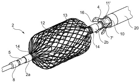
In figure 1, a first embodiment of a fixator 1 in accordance with the
invention is shown. The
fixator involves a flow transparent retainer or fixing part 2, which in the
shown embodiment
consists of a metal frame basket. The fixing part 2 is arranged on a guide
wire 3. In fig. 1, a
CA 02815897 2013-04-25
WO 2012/065625 PCT/EP2010/067499
21
distal tubular sleeve 5 is arranged at the distal end 2a of the fixing part.
The distal sleeve 5 is
fixed to the guide wire 3, whereas the proximal end 2b of the fixing part 2 is
arranged to
slide over the guide wire 3 by means of a proximal tubular sleeve 4. The
proximal and distal
sleeves 4 and 5 jointly limit the possible deformation of the fixing part 2 as
its ends 2a and
2b are forced toward each other, which will be described below. The distal end
2a of the
fixator 1 comprises a distal end part 6, which may be a continuation of the
guide wire 3, and
which is soft and pliable in order not to cause damage in the target vessel.
Guide wires for
catheterisation are typically of the dimensions between 0.14 and 0.89 mm in
circumference.
These are very pliable and atraunnatic with a hydrophilic slippery surface
that allows
catheterisation of small, stenotic and kinked arteries, without damage to the
target vessel
wall.
The proximal end 2b of the fixing part 2 involves a connecting member 7 for
connecting the
fixing part 2 to a retrieving catheter 10. In the shown first embodiment, the
connecting
member 7 has internal threads that are arranged on the inside of the proximal
tubular sleeve
4. The retrieving catheter 10, on the other hand, comprises a corresponding
connecting
member 11 in the form of external threads for mutual connection of the
retrieving catheter
10 and the fixing part 2.
In general (see fig. 10), the fixing part or deformable portion 2 has a
central portion C, which
attaches to or engages the vessel when deployed, and which is connected to the
sleeves 4/5
via the end portions 2a and 2b.
Preferably, the fixator 1 also has or is supplied with or inside a delivery
catheter 20 in the
form of a hose of a diameter adapted to house the fixing part 2 and the
retrieving catheter
10. The delivery catheter 20 enables the positioning of the fixing part 2 as
it allows the fixing
part to be fully housed therein during the introduction and positioning of the
fixator 1. It
would however also be possible to keep the fixing part collapsed without
housing it inside a
delivery catheter 20, e.g. by keeping the sleeves separated from each other by
means of e.g.
a screw controlled arrangement.
The length of the guide wire 3, the retrieving catheter 10 and the delivery
catheter 20 must
be sufficient to allow their respective proximal ends to be accessible to and
manoeuvrable by
the surgeon when the corresponding distal ends are located in a target vessel.
Typically, the
guide wire 3, the retrieving catheter 10 and the delivery catheter 20 all have
lengths between
0.5 and 2.8 meters.
The function of the fixator will be described step by step in an exemplary
mode of use and
with reference to figures 2-4. A further description is found in relation to
figures 13-16.
CA 02815897 2013-04-25
WO 2012/065625 PCT/EP2010/067499
22
In a first step, as shown in fig. 2, the delivery catheter 20 is inserted into
a target vessel 30,
defined by a vessel wall 31 and an opening 32 into e.g. the aorta. During the
insertion of the
fixator 1, only the pliable distal end part 6 of the fixator 1 extends outside
of the delivery
catheter 20. The fixing part 2 is undeployed or collapsed in the radial
direction such that it
fits inside the delivery catheter 20. In order to allow for the radial
collapse, the fixing part 2
is extended in the axial direction with the distal sleeve 5 at a relatively
large distance from
the proximal sleeve 4. During the insertion, the connecting member 7 of the
fixing part 2 is
connected to the connecting member 11 of the retrieving catheter 10. The
insertion of a
catheter into an unblocked vessel is in itself conventional and is therefore
not described in
detail in this application.
In a second step, when the delivery catheter 20 is located inside the target
vessel 30, the
fixing part 2 is pushed out from the inside of the delivery catheter 20. The
pushing of the
fixing part 2 is achieved by means of mutual movement of the delivery catheter
20 on the
one hand, and the retrieving catheter 10 on the other hand. As the fixing part
2 exits the
delivery catheter 20 it strives to regain its original shape, which is
individually adapted to the
diameter of the vessel 30 such that it exerts a certain pressure on the vessel
wall 31. This
pressure should be as low as possible in order not to harm the vessel, but it
must however be
sufficient to keep the fixator from moving with respect to the vessel. The
fixing part 2 has a
flow transparent form that allows nutritive blood flow through it. In the
present embodiment,
the fixing part 2 comprises crosswise woven threads, which are adapted to
expand to a
diameter that is slightly larger than an inside diameter of the relevant blood
vessel so as to
exert a pressure on the blood vessel wall that restrains the fixing part 2
from moving with
respect to the target blood vessel 30. The blood flow is allowed to flow
through the crosswise
woven threads.
Even though it is possible to provide the fixing part 2 with means for
obtaining the expanded
shape, it is preferred that the fixing part, and here the crosswise woven
threads, has an
expanded rest shape, so that the expansion merely is a movement toward the
rest shape.
This type of fixing part may be obtained by providing the threads in the
desired, expanded
shape and heat treating the threads to have or give this expanded shape the
rest shape.
A third step, where the fixing part 2 is fully deployed outside the delivery
catheter 20 and at
location inside the target vessel, is illustrated in fig. 3. In this third
step, the retrieving
catheter 10 is disconnected from the fixing part 2. In the present embodiment,
this
disconnecting is achieved in that the retrieving catheter 10 is rotated with
respect to the
fixing part 2, which is restricted from rotating due to its contact with the
vessel wall 31, such
that the connecting member 11 of the retrieving catheter 10 is unscrewed from
the
connecting member 7 of the fixing part 2.
CA 02815897 2013-04-25
WO 2012/065625 PCT/EP2010/067499
23
In a fourth step, when the retrieving catheter 10 has been disconnected from
the fixing part
2, both the retrieving catheter 10 and the delivery catheter 20 may be
withdrawn from the
target vessel and leaving only the fixing part 2 and the guide wire 3 in the
vessel 30. The
fixing part 2 is arranged to not hinder the blood flow through the vessel.
In order to ensure that the fixing part 2 is not disconnected from the guide
wire 3 when the
guide wire 3 is pulled, the proximal sleeve 4 and/or the distal sleeve 5 is
releasably fixed to
the guide wire 3, such as via a threaded connection, a snap fit or the like.
Fig. 4 illustrates a second embodiment of a fixator according to the
invention.
Naturally, details corresponding to details of the first embodiment are
denoted with the same
reference numerals, whereas details that are different from details of the
first embodiment
but that have the same function are denoted with the same reference numerals
with an
added apostrophe. A number of differences exist both in construction and use
of the first and
second embodiments. It is clear that such features may be interchanged between
the
embodiments if desired.
In the second embodiment of the fixator 1, the threaded attachment between the
proximal
sleeve 4 and the retrieving catheter 10 of fig. 1 is replaced by a snap-on
lock, including a
connecting member 11' on the retrieving catheter 10 in the form of a claw like
grasping unit
with claws or projections 16 and a corresponding connecting member 7' in the
form of a ring
shaped stopper on, or in connection to, the proximal sleeve 4. The shape of
the projections
16 is adapted to interlock with the ring shaped stopper as the connecting
member 11' is
retracted into the delivery catheter 20, and as the delivery catheter 20 is
pushed over the
connecting member 11'. Thus, the delivery catheter forces the projections to
grasp over the
connecting member when the fixator is retracted towards the retrieval catheter
by the guide
wire.
In figure 4, individual threads/wires 12 of the basket shaped fixing part 2
are clearly visible.
The threads/wires 12 may be welded together at crossing points 13, or they may
be braided
such that they pass each other by turns over and under each other. The ends 14
of the
threads are securely fastened to the sleeves 4 and 5, respectively, either by
welding, gluing
or sewing or in any other suitable manner. Further, in figure 4, the
connecting member 7' in
the form of the ring shaped stopper may be seen inside the claw like grasping
unit that
constitutes the connecting member 11' on the retrieving catheter 10.
CA 02815897 2013-04-25
WO 2012/065625 PCT/EP2010/067499
24
In the second embodiment, both sleeves 4 and 5 are arranged to slide over the
guide wire 3.
However, a stopper 8 positioned on the guide wire 3 prevents the sleeves from
moving over
the distal end of the guide wire 3 and thus disconnect fully from the guide
wire 3.
Alternatively, the distal sleeve 5 may be detachably fixed, using e.g. any of
the fastening
methods between catheter 10 and sleeve 4. The reason for this detachability or
slidability will
be described further below.
Further, from this view it is apparent that the function of the sleeves is
somewhat different in
this embodiment with respect to their function in the first embodiment. In
this embodiment,
the connecting member 7' is arranged directly on the proximal sleeve 4 of the
fixing part. The
proximal sleeve 4 is partly and fixedly housed inside a protective sleeve 15
(see also fig. 5),
which extends inside the basket shaped fixing part 2 and also partly houses
the distal sleeve
5. When the fixing part 2 is in its deployed shape, there is a gap between the
distal and the
proximal sleeves 5 and 4, respectively. As the guide wire 3 is pulled, or the
fixing part 2 is
allowed to expand toward its rest shape, the fixing part 2 is fixed to the
vessel wall, and any
pulling force applied to the distal sleeve 5 will thus act to compress the
fixing part in the axial
direction. Thus, the sleeves move closer to each other, until the proximal end
5b of the distal
sleeve 5 reaches the distal end 4a of the proximal sleeve 4. The contact
between these ends
of the sleeves thus limits the axial deformation of the fixing part 2. The
distal end 5a of the
distal sleeve 5 is arranged to interact with the stopper 8 on the guide wire 3
and limit the
axial movement of the guide wire 3 with respect to the fixing part 2, as
described above.
In a third embodiment the guide wire 3' is a hypotube, as is shown in fig. 6.
The hypotube
may be made of Nitinol or stainless steel and is preferably coated by a
hydrophilic coating,
such as e.g. PTFE, in order to create a slippery contact surface to the
retrieving catheter 10.
The hypotube may be just as flexible as a guide wire, or more flexible. The
suitable size of a
hypotube may range from 0,5 mm to about 2 mm with a wall thickness of about
0,04 to 0,2
mm.
Preferably, the hypotube should have a sufficiently large inner diameter to
successfully house
a stiff conveying wire 25. The stiff conveying wire 25 is helpful for guiding
the insertion of the
fixator 1. In order for the guide wire 3' to be rerouted, it has to be
flexible and pliable.
However, due to the pliability of the guide wire 3', it may be difficult to
control the guide wire
3' and to guide it into the target blood vessel. Hence, the stiff conveying
wire 25 will make it
possible to control the guide wire 3' during insertion. The conveying wire 25
enables the
insertion of further catheters and or stent branches on the guide wire. With a
stiff conveying
wire inside the guide wire 3', the stent graft branch can be introduced over
the stiff
CA 02815897 2013-04-25
WO 2012/065625 PCT/EP2010/067499
conveying wire 25, either directly over the conveying wire 25 or over the
(hypo-)tubular
guide wire 3' housing a conveying wire.
The stiff conveying wire 25 may be withdrawn from inside the guide wire 3'
when the fixing
part 2 has been located in the target vessel 30. When the stiff conveying wire
25 has been
5 withdrawn from inside the guide wire 3', the guide wire is sufficiently
pliable and flexible to
be rerouted inside an arterial system in an atraunnatic manner.
The guide wire 3' may be provided with an opening 26 near its distal end. With
such an
opening 26 the guide wire 3' may constitute a conduit for locally distributing
a
pharmaceutical via said opening 26. In many situations, e.g. when treating
tumours, it is of
10 interest to deliver a pharmaceutical agent locally, especially since
certain pharmaceuticals,
although effectively treating a disease process at one location, may be
harmful if distributed
systematically. Until now there has been no reliable way of delivering a
pharmaceutical
endovascularily over a period of time.
By means of a guide wire 3' in the form of a hypotube comprising a fixing part
2 it is possible
15 to fix the end of the hypotube inside a target vessel and to deliver a
desired amount of a
pharmaceutical through the opening 26 at the desired location, without risking
that the
hypotube will move and lose this location.
Naturally, the fixator of fig. 6 may, for most parts, be similar to the
fixator according the first
and second embodiments. For example, a stopper 8 may be provided on the guide
wire 3' for
20 interaction with the distal sleeve 5, and a protective sleeve 15, which
extends inside the
basket shaped fixing part 2, is arranged to partly house the distal sleeve 5.
Further, the distal
end 6 of the guide wire 3' is preferably soft and pliable in order not to
cause damage inside
the body. Also, the proximal part of the guide wire 3', e.g. proximal to the
fixing part 2, is
also pliable in order to allow rerouting. In a conventional manner, the tip of
the guide wire 3',
25 may include a 180 bend (not shown) that prevents arterial damage in the
target vessel.
Fig. 7 illustrates a fourth embodiment of a fixing part 2' for use in a
fixator according to the
invention. This fixing part 2' has the shape of a helical spring and is still
releasably fixed to
the guide wire 3 with a stopper 8'. The distal end of the fixing part 2' may
have a ring-shaped
element through which the guide wire 3 extends and which engages with the
stopper 8' to
prevent the fixing part 2' from moving over the distal end of the guide wire
3. This fixing part
2' has the advantage of being extremely simple in manufacture as well as
presenting very
little flow resistance in the blood vessel.
CA 02815897 2013-04-25
WO 2012/065625 PCT/EP2010/067499
26
Introduction and retraction of the fixing part 2' may be performed using a
catheter.
Withdrawing the pre-formed fixing part 2' will simply rotate this without
scraping or
damaging the vessel wall.
In the present embodiments the fixing part 2/2' preferably comprises a metal
structure of
weaved, coiled and/or braided wires or threads, preferably from Nitinol. Other
bioconnpatible
materials with similar properties may also be used, e.g. other alloys or
plastics. The material
must be sufficiently flexible to allow it to be collapsed without being
plastically deformed, but
at the same time sufficiently rigid to exert a pressure when released inside a
vessel. In a
specific method of producing the fixing part 2/2', a Laser cut length of a
braided Nitinol tube
is drawn around a template of a desired shape. The ends of the Nitinol tube
are shrunk
around the ends of the template and a heat treatment is performed in this
position, such that
the Nitinol basket, i.e. the fixator, adapts to this new shape. The fixator
will then strive to
regain this shape whenever unaffected by exterior forces.
Alternative fixing parts 2/2' may have a larger general contact area with the
blood vessel,
such as when using a piece of cloth, material or the like, which is supported
on the coiled
spring or the braided wire so as to better even out or enlarge the actual
contact surface
between the fixing part and the blood vessel wall.
As will be described in more detail below, the advantageous arrangement of the
above
embodiments enables the fixing part to remain in place as the guide wire 3 is
subject to
tension, e.g. from rerouting of its proximal end. The proximal sleeve 4 is
arranged to slide on
the guide wire 3 such that it remains unaffected by it, whereas the distal
sleeve 5 is
prevented from travelling toward the distal end of the guide wire 3. Due to
this arrangement
any pulling forces on the guide wire 3 will compress the fixing part 2 in the
axial direction,
due to the friction between the vessel wall 31 and the proximal part of the
fixing part 2, such
that the fixing part 2 is expanded in the radial direction, see fig. 8. Hence,
the pressure
against the vessel wall 31 will increase as a function of the pulling force on
the guide wire,
such that the increased friction force between the fixing part 2 and the
vessel wall 31
instantaneously increases with the increased pulling force. Therefore, by
means of the
increased friction force, the fixing part 2 is kept in place.
This arrangement allows for the fixing part to exert only a minimum force on
the vessel wall
31 as long as it is unaffected by any pulling force, in order to minimise the
traumatic effect
on said vessel. Also, during most parts of a normal operational procedure, the
guide wire is
not affected by any forces at all. The function of the fixator 1 is mainly to
retain the position
inside the target vessel. Pulling forces normally only arise when the guide
wire 3 is being
rerouted. The axial compression of the fixing part may be limited by
interaction of the sleeves
CA 02815897 2013-04-25
WO 2012/065625 PCT/EP2010/067499
27
4 and 5, as they come into contact with each other in response to a pulling
force on the guide
wire 3. Hence, the maximum radial extension of the fixing part 2, and thus the
maximal
radial force exerted by it on the vessel wall, can be limited by the available
distance between
the sleeves; the greater the distance, the greater the possible axial
compression and
consequent radial extension.
It has been found, however, that even though, as is seen in figure 8, the
fixing part 2 is
bounded perpendicular to its longitudinal axis, of the dimensions (primarily
thickness or
radius) of the blood vessel 30, there may be a difference along the
longitudinal direction of
the force exerted to the blood vessel. The cause is that as the fixing part or
deformable
portion 2 engages the vessel wall 31 and the distal sleeve pulled, the force
is not distributed
evenly over the area engaged by the fixing part 2 but mainly at the distal
part thereof. In fig.
9, the shape of a fixing part 2 in an element more flexible than a blood
vessel is illustrated. It
is seen that the cross sectional area (or radius if circular or having
rotation symmetry) at the
longitudinal position A is larger than at position B which is positioned more
proximal than A.
Thus, in order to distribute this force more evenly, different solutions are
illustrated in figs.
10-12.
In fig. 10, the fixing part 2 has an asymmetric shape, when non-stressed
and/or in a non-
compressed state, over the longitudinal length which is to engage the blood
vessel, where a
part of the fixing part 2 closer to the distal end or sleeve 5 has a smaller
cross sectional area
(or radius if circular or having rotation symmetry) at position A than closer
to the proximal
sleeve 4, such as at position B.
Thus, when positioned in the blood vessel 30 and without pulling the guide
wire 3, the shape
of the fixing part 2 of figure 10 may look like in figure 8, where the force
exerted to the wall
31 is uneven but still quite low. When the guide wire 3 is pulled, however,
the increased force
exerted on the wall 31 may be more even, as the more narrow rest shape of the
distal part
(around position A) of the fixing part 2 will act to counter-act a large
expansion and thus
force increase at that part.
In fig. 11, the rest shape or non-stressed/non-compressed shape of the fixing
part 2 may be
symmetric but the expandability of the fixing part 2 asymmetric along the
longitudinal
direction. In fig. 11, the fixing part 2 is provided as a wire mesh with a
higher wire density at
position A compared to position B. Thus, when compressing the fixing part 2
outside the
blood vessel 30, an asymmetric shape as that illustrated in fig. 10 may be
obtained. Also,
when compressing the fixing part 2 or pulling the guide wire 3 when the fixing
part 2 is
CA 02815897 2013-04-25
WO 2012/065625 PCT/EP2010/067499
28
deployed in the vessel 30, the higher wire density at position A will act to
even out the
pressure exerted and ensure that more force is applied around position B.
Naturally, the same functionality may be obtained by adapting the wire
thickness or other
parameters of the wire mesh.
In fig. 12, another manner is illustrated which prevents excessive force
exerting on the
vessel 30 close to the distal end of the fixing part 2. In this embodiment, a
circumference
limiting element 40 is provided at the distal end at position A. This element
40 prevents the
circumference of the fixing part 2 from exceeding a predetermined length,
whereby any
further deformation will be required at the more proximal parts, such as
around position B.
This circumference limiting element 40 may be a non-stretchable element, such
as a band, a
wire or the like.
In general, it is noted that different types of materials for the fixing part
2 and different
constellations thereof may be chosen. The overall functionality is that the
fixing part 2 should
be able to engage the vessel wall while allowing a blood flow there through.
The overall advantages of using the present fixator 1 is now described with
reference to figs.
13-16. This description of a use of the fixator is limited to the positioning
of a branched stent
graft in a person to treat aortic aneurysms with aneurysm extension in the
thoraco-
abdominal aorta. It is noted that many other reasons exist for wishing to
catheterize blood
vessels of small dimensions such as the renal arteries to the kidneys, the
visceral arteries to
the intestines or the arteries supplying the liver, etc. Accessing these
arteries and
maintaining a catheterization of these arteries is difficult especially when
concomitant
catheterisation of several arteries is taking place simultaneously.
Fig. 13 illustrates the human artery system with the aorta 101 from which a
renal artery 102
feeds a kidney and an artery 103 leads toward a leg. As is usual in many of
these
procedures, the aorta 101 is accessed via the common iliac artery 103 via an
arterial
puncture. Then, a fixator with a fixing part 2 and a guide wire 3 is
positioned as indicated
above. The guide wire 3 extends out of the arterial puncture.
It is the goal of the procedure to position the graft 150 inside the system
100 with the main
lumen 152 of the graft 150 in the aorta 101 and the branch 154 toward and into
the artery
102. The graft 150 has a vertical longitudinal axis, openings at the upper and
lower ends as
well as a side opening in the branch 154. In the graft 150, a transport wire
162 is positioned
which stretches through the main lumen 152 and the branch 154 with ends 164
and 162,
respectively.
CA 02815897 2013-04-25
WO 2012/065625 PCT/EP2010/067499
29
In fig. 14, the graft 150 has been positioned in the aorta 101.
Conventionally, the graft 150
may be compressed, positioned within a delivery introducer and positioned in
the aorta 101.
The graft 150 is introduced through the same artery puncture, and the ends 162
and 164
extend outside the person together with the guide wire 3. In this manner, the
subsequent
fixing/snaring is facilitated, as this may be performed outside the body of
the person.
Having positioned the graft 150 in the aorta 101, it is partly deployed in
order to make the
transport wire movable within the graft 150.
Subsequently, the guide wire 3 and the end 164 are attached to each other, and
the end 162
pulled, so that the guide wire 3 and end 164 are re-introduced into the system
100 and
through the branch 154, the main lumen 152 and out through the artery puncture
so that
(see fig. 15) the guide wire 3 now extends from the fixing part 2 through the
branch 154, the
main lumen 152 and out of the patient. This procedure or re-positioning of the
guide wire 3
will require some pulling of the fixing part 2, whereby its capability in this
respect is used.
Having now ensured that the branch 154 is directed toward the artery 102
(guided by the
guide wire 3), the graft 150 may be fully deployed or extended/expanded so
that it may, if
desired, contact the wall of the aorta 101 and thus fix itself thereto. Also,
subsequently, an
extender graft 156 is positioned (see fig. 16) stretching from the branch 154
and into the
artery 102. This extender graft 156 is positioned using the guide wire 3 which
already is
positioned in the artery 102.
However, in order for the extender graft 156 to be able to actually cover the
part of the
vessel wall 31 where the fixing part 2 engaged the blood vessel (which may
have been
slightly damaged during the procedure), it is necessary to actually remove the
fixing part 2
after re-positioning of the guide wire 3 but before introducing the extender
graft 156.
As the fixing part 2 is able to slide along the guide wire 3, this is
possible. The fixing part 2 is
removed using a delivery catheter as described above, while maintaining the
guide wire 3 in
place in the artery 102. Having then removed the fixing part 2, the guide wire
3 is
subsequently used for introducing the extender graft 156 as illustrated in
fig. 16.
Naturally, more such grafts or grafts with more branches may be used. However,
such use
and positioning may be performed as described, with the assistance of the use
of the fixator
and the re-routing of the guide wire 3 using the transport wire 160.
CA 02815897 2013-04-25
WO 2012/065625 PCT/EP2010/067499
Also, the attachment of the guide wire 3 to the transport wire 160 may be
performed within
the blood vessel of the person. Such intra corporeal snaring or attachment is
a conventional
procedure.
Naturally, all guide wires and fixators may be introduced through the same
percutaneous
5 opening in the person as the tubular element. This facilitates snaring.
Alternatively, the
fixators may be guided to the desired vessels along any desired route and from
different
arteries, such as from the arteries in the arms and legs. Then, when the
tubular element has
been introduced, guide wires and/or transport wires may be re-routed to allow
the desired
attaching/snaring, where after the guide wires may extend to the outside of
the person
10 through any opening desired. The use of multiple openings at different
positions of the
person may be desired in order to prevent entanglement of the wires inside the
person.
In one situation, the fixator is attached to the guide wire via an attaching
wire which has the
function of placing the fixator in the target vessel and retrieving it at the
end of the
procedure before placing the stent graft connection from the main graft 150 to
the target
15 vessel.
The fixing part of the fixator according to the embodiments above may be
provided in
different sizes, e.g. 5, 6, 7, 8, 9, 10, 11, 12, 13 14 mm in diameter, or
fractions thereof, for
use in arteries of corresponding diameter. The collapsed fixing part 2
diameter is typically 8
French (2.67 mm diameter on the French catheter scale), and may vary from
about 6 to
20 about 12 French (about 2 to about 4 mm), including sizes of 6, 7, 8, 9,
10, 11, 12 French and
half sizes there between, depending on the diameter of the delivery catheter
to be used to
house the fixing part. The size of the delivery catheter is accordingly also
from about 6 to
about 12 French (about 2 to about 4 mm), including sizes of 6, 7, 8, 9, 10,
11, 12 French and
half sizes there between. The retrieving catheter has a size that allows it to
fit outside the
25 guide wire and inside the delivery catheter, e.g. from about 3 to about
10 French (about 1 to
about 3.3 mm), including sizes of 3, 4, 5, 6, 7, 8, 9, 10 French and half
sizes there between.
Usually, the guide wire and/or the hypotube guide wire is very pliable and
usually have a
hydrophilic surface allowing catheterization of narrow, stenotic arteries
without damage to
the target vessel.
30 It is noted that the branched graft 150 may be replaced by a so-called
fenestrated graft
which has not branch(es) 154 but merely a side opening. The positioning is
similar in that the
guide wire will extend from the fixator 2 through the side opening and out
through the graft
150 as described.
CA 02815897 2013-04-25
WO 2012/065625 PCT/EP2010/067499
31
One embodiment of the fixator may be one made of 40 braided 100 pm diameter
Nitinol
wires provided as a, when compacted, 40 mm long element which may be used in
e.g. 5-
7nnnn blood vessels. When expanded in a 5 mm blood vessel, this fixator will
occlude about
58% of the blood vessel cross sectional area, and the openings seen by the
blood when
flowing through the fixator will be 0.015-0.18 nnnn2, whereas the occluding
percentage in a
7nnnn blood vessel is about 47% and the openings 0.06-0.25nnnn2.
In that situation, the extender graft 156 may also be used, where it may be
desired to use a
flairing graft having a larger diameter within the lumen 152 so as to ensure
that the extender
graft is fixed to the graft 150 and is not allowed to release itself therefrom
and move into the
vessel 102.
Naturally, the graft 150 may have any number of branches 154 or side openings,
and even a
combination of one or more branches 154 and side openings.
Also, a graft with no transport wires may be used, as these may subsequently
be routed
through the graft using standard methods. In this manner, a transport wire may
be routed
and the corresponding guide wire re-routed before additional transport wires
are introduced.
In this manner, entangling of the wires may be prevented.