Note: Descriptions are shown in the official language in which they were submitted.
CA 02816031 2013-04-24
WO 2012/061160 PCT/US2011/057755
METHODS AND APPARATUS TO IDENTIFY EYE COVERINGS FOR
VISION
CROSS-REFERENCES TO RELATED APPLICATIONS
[0001] The present PCT application claims priority to U.S. App. Ser.
Nos.:61/406,504, filed on
October 25, 2010, entitled "METHODS AND APPARATUS TO IDENTIFY THERAPEUTIC
SHIELDS FOR VISION AND PAIN", attorney docket no. 26322A-000700U5; and U.S.
App.
Ser. No. 61/480,231, filed on April 28, 2011, entitled "METHODS AND APPARATUS
TO
IDENTIFY EYE COVERINGS FOR VISION", attorney docket no. 26322A-000710U5; each
assigned to NexisVision, Inc., the full disclosures of which are incorporated
herein by reference.
STATEMENT AS TO RIGHTS TO INVENTIONS MADE UNDER
FEDERALLY SPONSORED RESEARCH AND DEVELOPMENT
[0002] NOT APPLICABLE
BACKGROUND OF THE INVENTION
[0003] The present invention is generally directed to vision and treatment of
the eye to provide
improved vision. Although specific reference is made to coverings for vision
correction such as
the correction of refractive error and also to treatment of eyes having
epithelial defects following
photorefractive keratectomy, embodiments of the present invention may comprise
extended wear
contact lenses that can be used to correct vision in many ways such as with
one or more of
aberration correction, multifocal correction, presbyopia correction, and
astigmatism correction.
[0004] The eye includes several tissues that allow patients to see. The cornea
of the eye is an
anterior tissue of the eye that is clear in healthy eyes and refracts light so
as to form an image on
the retina. The retina is a posterior tissue of the eye that senses light from
the image formed
thereon and transmits signals from the image to the brain. The cornea includes
an outer layer of
tissue, the epithelium, which protects the underlying tissues of the cornea,
such as Bowman's
membrane, the stroma and nerve fibers that extend into the stroma and Bowman's
membrane.
The healthy eye includes a tear film disposed over the epithelium. The tear
film can smooth
small irregularities of the epithelium so as to provide an optically smooth
surface. The tear film
is shaped substantially by the shape of the underlying epithelium, stroma, and
Bowman's
membrane, if present. The tear film comprises a liquid that is mostly water
and does include
i
CA 02816031 2013-04-24
WO 2012/061160 PCT/US2011/057755
additional components, such as mucoids and lipids. The many nerve fibers of
the cornea provide
sensation to promote blinking that can cover the cornea with the tear film.
The nerve fibers also
sense pain so that one will normally avoid trauma to the cornea and also avoid
direct contact of
an object to the cornea so as to protect this important tissue.
[0005] Work in relation to embodiments of the present invention suggests that
at least some of
the prior contact lenses and therapeutic coverings can be less than ideal in
at least some
instances. Many contact lenses and therapeutic coverings can be left in the
eye for less than ideal
amounts of time, as the patient removing and replacing the contact lens or
therapeutic covering
can be somewhat cumbersome and in at least some instances patients may leave
the contact lens
or therapeutic covering in the eye for amounts of time that can be longer than
would be ideal.
Although extended wear lenses can be left in the eye for somewhat longer
amounts of time, the
amount of time such lenses can be left in the eye can be less than ideal. Work
in relation to
embodiments of the present invention also suggests that tear flow of the prior
contact lenses can
be less than ideal, and that less than ideal tear flow may be related to the
potential complications
and can limit the amount of time such lenses can be left in the eye.
[0006] In the healthy cornea, the proper amount of hydration of the cornea,
sometimes referred
to as dehydration of the cornea, is maintained such that the cornea remains
clear. The cornea
includes a posterior endothelial layer that pumps water from the cornea into
the adjacent anterior
chamber. The epithelium inhibits flow of water from the tear liquid into the
cornea, such that the
corneal stroma can be maintained with the proper amount of hydration with
endothelial pumping.
The endothelial pumping of water from the cornea to maintain the proper
hydration and
thickness of the eye is often referred to as deturgescence. When the corneal
epithelium heals, the
layer of cells forming over the defect can be at least somewhat irregular in
at least some
instances, such that the vision of the patient can be less than ideal.
[0007] As the post-ablation cornea may have a complex shape, many of the prior
commercially
available lenses may not fit the ablated cornea as well as would be ideal, and
in at least some
instances fitting of lenses can be time consuming and awkward. Rigid gas
permeable
(hereinafter "RGP") lenses can be uncomfortable for the patient and difficult
to fit.
Commercially available contact lenses having a rigid central portion and a
soft peripheral skirt
can be difficult and/or time consuming to fit to the ablated cornea and may
not fit very well in at
least some instances. The ablated cornea may comprise an abrupt change in
curvature near the
2
CA 02816031 2013-04-24
WO 2012/061160 PCT/US2011/057755
edge of the ablation, and in at least some instances it can be difficult to
fit such lenses near the
edge of the ablation. Also, at least some of the commercially available
contact lenses may not be
suitable for extended wear and may be removed each day, which can be somewhat
awkward for
a patient and can result in lack of compliance and lenses remaining in the eye
longer than would
be ideal in at least some instances.
[0008] In light of the above, it would be desirable to provide improved
contact lenses for
vision correction and coverings for treatments related to epithelial defects
of the cornea, such as
epithelial defects following photorefractive keratectomy (hereinafter "PRK").
Ideally, these
contact lenses and coverings would provide treatments that improve tear flow
and avoid at least
some of the deficiencies of known techniques while providing improved patient
comfort and/or
vision.
BRIEF SUMMARY OF THE INVENTION
[0009] Embodiments of the present invention provide improved methods and
apparatus to fit
and identify coverings to treat eyes. The treated eye may comprise a natural
eye, or an eye
having an epithelial defect of the eye, such as an eye ablated with PRK
refractive surgery. In
many embodiments, the covering can be identified and fit to the eye so as to
provide one or more
of improved hydration or flow of tear liquid under the covering. The covering
can be fit and
identified based on an inner corneal curvature and an outer corneal curvature
and one or more of
a limbus sag height or a conjunctival sag height.
[0010] The covering to fit the eye may comprise a contact lens and can provide
improved
hydration and tear flow such that the covering can be left on the eye to
correct vision for
extended amounts of time. The covering may comprise one or more structures to
provide
hydration under the covering such that the covering can remain in the eye and
correct vision for
an extended amount of time. In many embodiments, the covering comprises a
layer of hydrogel
extending along a lower surface of the covering to provide hydration to a
surface of the eye.
Alternatively or in combination, the covering may comprise a material having
fenestrations and
an outer portion shaped to contact the conjunctiva to pump tear liquid when
the eye blinks. The
covering may comprise a deflectable outer portion having a resistance to
deflection such that a
chamber is formed when the covering is placed on the eye and the eye is open
with the eyelids
separated. The resistance to deflection of the deflectable outer portion is
configured such that the
outer portion deflects inward toward the cornea when the eyelid closes to pump
tear liquid. The
3
CA 02816031 2013-04-24
WO 2012/061160 PCT/US2011/057755
fenestrations can draw tear liquid into the chamber located under the covering
when the eye
opens and the chamber can expand. The outer portion of the covering comprises
a sclera
coupling portion shaped to contact the conjunctiva to define the chamber when
the covering is
placed on the eye. The fenestrations and sclera coupling portion of the
covering can pass tear
liquid away from the chamber when the eye closes and pressure of one or more
eyelids urges the
covering toward the cornea such that the chamber volume decreases. In many
embodiments,
opening of the eye so as to separate the eyelids reduces pressure on the outer
portion of the
covering such that the outer portion of the covering over an outer portion of
the cornea can
separate from the outer portion of the cornea so as to draw liquid through the
fenestrations and
into the chamber located under the covering. The sclera coupling portion of
covering may
contact the conjunctiva to inhibit the flow of tear liquid under the sclera
coupling portion when
the eye opens and tear liquid is drawn through the fenestrations, for example
with formation of a
seal where the covering contacts the conjunctiva. When the eye blinks
subsequently, the
pressure of the one or more eyelids can urge the covering toward the cornea
such that tear liquid
can pass through the fenestrations, and the sclera coupling portion may
separate slightly from the
conjunctiva to pass tear liquid under the sclera coupling portion, so as to
rinse the cornea, the
limbus, the conjunctiva and the underside of the covering with the pumped tear
liquid. The
covering may comprise a material having high oxygen permeability such as
silicone such that the
covering may provide improved tear flow and high oxygen permeability. This
improved flow of
tear liquid can allow the covering such as a contact lens to be worn for
extended amounts of time
of at least about one week, for example thirty days or sixty days or more. The
improved tear
flow can improve healing and vision of eyes with epithelial defects, for
example epithelial
defects following PRK.
[0011] In many embodiments, the identified covering comprises an inner optical
component
for vision, such as a lens, and an outer coupling component to hold the inner
component in
relation to the pupil to improve vision. The coupling component may comprise a
deflectable
material that inhibits passage of the tear liquid through the material such
that the tear liquid
passes through the fenestrations when the eye blinks and an eyelid exerts
pressure on the optical
component. The outer coupling component may comprise the fenestrations to pass
the tear
liquid and the outer sclera coupling portion to contact the conjunctiva. The
optical component
may comprise a first material and first thickness corresponding to a first
rigidity. The coupling
component may comprise a second material and a second thickness corresponding
to a second
4
CA 02816031 2013-04-24
WO 2012/061160 PCT/US2011/057755
rigidity. The second material can be softer than the first material and the
second thickness can be
less than the first thickness such that the coupling component can be
deflected with the eyelid,
and such that the coupling component can be deflected by an amount greater
than the optical
component when the eyelids close to cover the first component and the second
component. The
optical component can be more rigid than the coupling component, such that the
optical
component can provide vision when the outer portion is deflected with one or
more eyelids.
[0012] The covering can be identified such that the alignment of the optical
component to the
pupil provided with the coupling to the conjunctiva and underlying sclera can
be beneficial for
vision. The optical component can be held at a substantially fixed location in
relation to the
pupil so as to provide improved vision such as presbyopia correction and
vision correction of
aberrations that may depend on location of the pupil such as measured
wavefront aberrations,
spherical aberration, coma and trefoil.
[0013] The covering can be identified such that the optical component and
coupling
component can be helpful to improve vision and regeneration of the epithelium
in eyes with
epithelial defects. The optical component can smooth the cornea and may smooth
irregularities
of the epithelium and ablated stroma. The coupling component can support the
optical
component so as to resist sliding movement of the optical component and
provide an
environment to promote regeneration of the epithelium. The pumping of the tear
liquid may
improve tear flow to the regenerating epithelium near the epithelial defect so
as to promote
regeneration of the epithelium over the defect. The pumping of the tear liquid
can also promote
delivery of a medicament, for example a steroid, to the ablated region so as
to inhibit corneal
infiltrates and haze.
[0014] In many embodiments, the covering can be identified based on one or
more of pre-
operative eye data used to determine the ablation, ablation data of the laser
such as the amount of
ablative correction, and dimensions across the ablated region of the eye. The
covering may
comprise an inner portion having a lower surface comprising a curvature, and
the curvature can
be less than a curvature of the ablated profile to improve vision and inhibit
formation of one or
more irregularities of the epithelium. The one or more irregularities may be
located on an inner
portion of the ablation comprising a center of the ablation, and the covering
may comprise a
resistance to deflection to inhibit formation of the one or more
irregularities near the center of the
ablation. The irregularity may comprise an elevated profile of the epithelium
located on the
CA 02816031 2013-04-24
WO 2012/061160 PCT/US2011/057755
inner portion comprising the center of the ablation, and the inner portion of
the covering may
comprise resistance to deflection and provide pressure in response to
deflection of the inner
portion so as to inhibit formation of the one or more irregularities of the
epithelium. As the
covering can resist deflection, the covering can be identified based on eye
measurement data and
ablation data so as to provide comfort and improved vision to the patient when
the covering is
placed on the eye and improves vision. A plurality of coverings having
portions sized to fit the
patient can be provided. A covering to treat the patient can be identified
among the plurality of
coverings based on data corresponding to an untreated portion of the eye, data
corresponding to a
treated portion of the eye, and an array of data corresponding to rigidity of
the plurality of
coverings. The array of data may comprise the unique identifiers arranged such
that the unique
identifier can be determined from the array based on the data corresponding to
an untreated
portion of the eye and the data corresponding to a treated portion of the eye.
The identified
covering can be placed on the eye and promote regeneration of the epithelium
with improved
vision and comfort.
[0015] In a first aspect, embodiments provide a method of treating an eye of a
patient, in which
the eye has a cornea. The eye is measured to determine data of the eye
corresponding to an inner
ablated portion of the cornea and an outer unablated portion of the cornea
away from the ablated
portion. A covering of a plurality of coverings is identified to treat the eye
based on the data of
the eye and an array of data corresponding to the plurality of therapeutic
coverings.
[0016] In many embodiments, the covering is placed on the eye.
[0017] In many embodiments, the covering comprises an inner covering portion
and an outer
covering portion, the inner covering portion contacting the inner ablated
portion of the cornea
and an outer covering portion contacting an unablated portion when placed on
the cornea and
wherein the inner covering portion prior to placement on the eye has a
covering curvature no
more than a curvature of the ablated portion of the cornea and wherein the
outer covering portion
comprises a curvature prior to placement on the eye no more than the outer
unablated portion of
the cornea and wherein the covering resists movement of the inner portion when
placed on the
eye.
[0018] In many embodiments, the outer portion of the covering extends to a
conjunctiva of the
eye and couples to the sclera of the eye to resist movement of the inner
portion.
6
CA 02816031 2013-04-24
WO 2012/061160 PCT/US2011/057755
[0019] In many embodiments, the inner portion of the covering prior to
placement comprises a
substantially uniform thickness and an amount of curvature corresponding to
less optical power
than the optical power of the ablated portion of the cornea, the amount of
curvature of the inner
portion prior to placement within a range from about -1D to about -3D relative
to the ablated
portion of the cornea.
[0020] In many embodiments, the inner portion of the covering deflects at
least about 1D so as
to conform at least partially to the ablation and promote smooth epithelial
regeneration and
vision.
[0021] In many embodiments, the inner portion of the covering comprises an
amount of
rigidity within a range from about 1E-4 to about 5E-4 (Pa*m^3) and the outer
portion of the
covering comprises an outer amount of rigidity less than the amount of
rigidity of the inner
portion.
[0022] In many embodiments, measuring the eye comprises determining a
conjunctiva sag
height, in which the conjunctiva sag height corresponds to a portion of a
conjunctiva of the eye at
a radial location away from a reference axis of the eye. The covering
comprises a covering sag
height at a covering location corresponding to the radial location of the
portion of conjunctiva,
and the covering is identified such that the covering sag height is greater
than the conjunctiva sag
height.
[0023] In many embodiments, the covering is deflected at the covering location
when the
covering is placed on the eye.
[0024] In many embodiments, the conjunctiva sag height is determined based on
a
measurement of a sclera of the eye corresponding to the radial location.
[0025] In many embodiments, measuring the eye comprises determining a limbus
sag height,
the limbus sag height corresponding to a portion of a limbus of the eye at a
radial location away
from a reference axis of the eye and wherein the covering comprises a covering
sag height at a
covering location corresponding to the radial location of the portion of the
limbus and wherein
the covering is identified such that the covering sag height is no more than
the limbus sag height.
[0026] In many embodiments, the covering is deflected a first amount at a
first covering
location corresponding to a portion of the conjunctiva when the covering is
placed on the eye and
wherein the covering is deflected a second amount at a second covering
location corresponding
7
CA 02816031 2013-04-24
WO 2012/061160 PCT/US2011/057755
to a portion of the limbus when the covering is placed on the eye, the second
amount less than
the first amount such that pressure from the covering to the limbus is
inhibited.
[0027] In many embodiments, the covering comprises an inner portion having a
hydrogel layer
extending along a lower surface to contact the ablated portion and the
unablated portion of the
cornea and wherein the covering comprises an outer portion comprising a
sticky, tacky surface to
contact the conjunctiva and inhibit movement of the covering when the inner
portion contacts the
cornea
[0028] In another aspect, embodiments provide an apparatus to treat an eye.
The apparatus
comprises an input to receive data of the eye. The data of the eye corresponds
to an inner ablated
portion of the cornea and an outer portion of the cornea away from the inner
ablated portion.
The apparatus comprises an output. At least one processor is coupled to the
input and the output.
The at least one processor comprises at least one computer readable memory.
The at least one
computer readable memory has instructions to store an array of data
corresponding to a plurality
of therapeutic coverings and instructions to identify a covering of the
plurality based on the array
and the data of the eye corresponding to the inner ablated portion and the
outer portion.
[0029] In many embodiments, the apparatus comprises the plurality of
coverings.
[0030] In many embodiments, the instructions are configured to identify a
covering having an
inner portion comprising a lower surface curvature flatter than the inner
ablated portion of the
eye to inhibit one or more irregularities of the epithelium.
[0031] In many embodiments, the lower surface curvature of the identified
covering is flatter
prior to placement than the inner ablated portion of the eye by at least about
1D.
[0032] In many embodiments, the inner portion of the covering comprises a
substantially
uniform thickness and the instructions are configured to identify a covering
prior to placement
corresponding to hyperopia of the eye to improve vision and inhibit an
epithelial irregularity
located on an inner portion of the ablation and corresponding to
nearsightedness of the eye.
[0033] In many embodiments, the instructions are configured to identify the
covering to inhibit
formation of the epithelial irregularity based on one or more of a modulus of
the inner portion of
the covering, a thickness of the inner portion of the covering, or an amount
of rigidity of the
inner portion of the covering.
8
CA 02816031 2013-04-24
WO 2012/061160 PCT/US2011/057755
[0034] In many embodiments, the array of data comprises a plurality of unique
identifiers
corresponding to the plurality of coverings.
[0035] In many embodiments, the plurality of unique identifiers corresponds to
a rigidity of an
inner portion of each of the plurality of coverings.
[0036] In many embodiments, the covering comprises an amount of rigidity of
the inner
portion within a range from about 1E-4 Pa*m^3 to about 5E-4 Pa*m^3.
[0037] In many embodiments, the plurality of unique identifiers comprises 10
or more unique
identifiers corresponding to an amount of rigidity of the inner portion of at
least about 3E-4
Pa*m^3.
[0038] In many embodiments, the covering comprises an amount of rigidity of
the inner
portion within a range from about 1E-4 Pa*m^3 to about 5E-4 Pa*m^3.
[0039] In many embodiments, the array of data comprises a first dimension
corresponding to
the inner ablated portion and a second dimension corresponding to the outer
portion away from
the ablated portion.
[0040] In many embodiments, the array comprises a table, the first dimension
corresponding to
rows of the table, the second dimension corresponding to columns of the table
and wherein the
plurality of unique identifiers is stored in the rows and the columns of the
table.
[0041] In many embodiments, the display is visible to the user and the
instructions are
configured to show the unique identifier on the display.
[0042] In many embodiments, the instructions are configured to receive a
conjunctiva sag
height, the conjunctiva sag height corresponding to a portion of a conjunctiva
of the eye at a
radial location away from a reference axis of the eye. The instructions are
configured such that
the identified covering comprises a covering sag height at a covering location
corresponding to
the radial location of the portion of conjunctiva and wherein the instructions
are configured such
that the covering sag height is greater than the conjunctiva sag height.
[0043] In many embodiments, the covering comprises an inner portion having a
hydrogel layer
extending along a lower surface to contact the ablated portion and the
unablated portion of the
cornea, and the covering comprises an outer portion at the covering location
comprising a sticky
9
CA 02816031 2013-04-24
WO 2012/061160 PCT/US2011/057755
tacky surface to contact the conjunctiva and inhibit movement of the covering
when the inner
portion contacts the cornea.
[0044] In many embodiments, the inner portion of the covering comprises a low
water content
water inhibiting layer beneath the hydrogel layer, and the outer portion of
the covering at the
covering location to contact the conjunctiva comprises a soft hydrophobic
material.
[0045] In many embodiments, the water inhibiting layer comprises silicone
elastomer and the
hydrogel layer comprises silicone hydrogel.
[0046] In many embodiments, instructions are configured such that the
identified covering is
deflected at the covering location when the covering is placed on the eye.
[0047] In many embodiments, the instructions are configured to receive the
conjunctiva sag
height based on a measurement of a sclera of the eye corresponding to the
radial location.
[0048] In many embodiments, the instructions are configured to receive a
measurement the eye
corresponding to a limbus sag height, in which the limbus sag height
corresponds to a portion of
a limbus of the eye at a radial location away from a reference axis of the
eye. The instructions
are configured such that the covering comprises a covering sag height at a
covering location
corresponding to the radial location of the portion of limbus, and the
instructions are configured
such that the covering sag height is no more than the limbus sag height.
[0049] In many embodiments, the instructions are configured to identify the
covering such that
the identified covering is deflected a first amount at a first covering
location corresponding to a
portion of the conjunctiva when the covering is placed on the eye, and the
instructions are
configured to identify the coveringsuch that the covering is deflected a
second amount at a
second covering location corresponding to a portion of the limbus when the
covering is placed
on the eye, the second amount less than the first amount such that pressure
from the covering
over the limbus is inhibited.
[0050] In another aspect embodiments provide an apparatus to treat an eye. The
apparatus
comprises an input, an output and at least one processor. The input is
configured to receive data
of the eye, and the data of the eye correspond to an inner portion of the
cornea and one or more
of a limbus or a conjunctiva of the eye. The at least one processor is coupled
to the input and the
output. The at least one processor comprises at least one computer readable
memory. The at
least one computer readable memory has instructions to store an array of data
corresponding to a
CA 02816031 2013-04-24
WO 2012/061160 PCT/US2011/057755
plurality of coverings and instructions to identify a covering of the
plurality based on the data of
the eye and the array of data corresponding to the plurality of coverings,
such tear liquid is
pumped under the covering when the eye blinks.
BRIEF DESCRIPTION OF THE DRAWINGS
[0051] FIG. 1 shows an eye suitable for incorporation of the covering as
described herein, in
accordance with embodiments of the present invention;
[0052] Figure 1-1A shows an ablated eye immediately following refractive
surgery resulting in
an epithelial defect, suitable for incorporation in accordance with
embodiments of the present
invention;
[0053] Figure 1-1B shows an ablated eye about 1 day following refractive
surgery resulting in
an epithelial defect, suitable for incorporation, in accordance with
embodiments of the present
invention;
[0054] Figure 1-1C shows an ablated eye when the epithelium has regenerated
following
refractive surgery resulting in an increased epithelial thickness centrally at
about 3 days, suitable
for incorporation, in accordance with embodiments of the present invention;
[0055] Figure 1-2A shows a covering positioned on an eye having an epithelial
defect, in
which the covering abuts the cornea to seal the cornea, in accordance with
embodiments of the
present invention;
[0056] Figure 1-2B shows a smooth layer of regenerated epithelium
substantially cover an
ablated profile, in accordance with embodiments of the present invention;
[0057] Figure lA shows a covering positioned on an eye having an epithelial
defect, in which
an outer portion of the covering abuts and conforms at least partially to the
cornea to seal the
cornea, in accordance with embodiments of the present invention;
[0058] Figure 1A1 shows a covering positioned on an eye and blinking of the
eye, in
accordance with embodiments of the present invention;
[0059] Figure 1B1 shows a covering sized to seal a cornea, in accordance with
embodiments of
the present invention;
11
CA 02816031 2013-04-24
WO 2012/061160 PCT/US2011/057755
[0060] Figure 1B2 shows the covering conforming to ablated stromal tissue and
guiding
regeneration of the epithelium over the ablated stroma, so as to promote
vision, in accordance
with embodiments of the present invention;
[0061] Figure 1B2A shows a covering forming an indentation in the epithelium
such that the
epithelium extends over at least a portion of the perimeter of the covering,
in accordance with
embodiments of the present invention;
[0062] Figure 1B2B shows a covering forming an indentation in the epithelium,
in accordance
with embodiments of the present invention;
[0063] Figure 1B2C shows a covering abutting the cornea to seal the cornea
without forming a
substantial indentation in the epithelium, in accordance with embodiments of
the present
invention;
[0064] Figure 1C shows a covering comprising a single piece of material having
an inner
thickness greater than an outer thickness, in accordance with embodiments of
the present
invention;
[0065] Figure 1C1 shows a covering as in Figs. 1-2A to 1B2 having an inner
portion
comprising an inner thickness and an inner material and an outer portion
comprising an outer
thickness and an outer material, in which the inner thickness is greater than
the outer thickness,
in accordance with embodiments of the present invention;
[0066] Figure 1C 1A shows a covering as in Fig. 1C1 adhered to the cornea with
a smooth
upper surface and a lower surface conforming to irregularity of the cornea
comprising a central
island of the stroma, in accordance with embodiments of the present invention;
[0067] Figure 1C2 shows a covering as in Figs. 1-2A to 1B2 having an inner
portion
comprising an inner thickness and an inner material and an outer portion
comprising an outer
thickness and an outer material, in which the inner thickness is greater than
the outer thickness
and the outer material extends around the inner material, in accordance with
embodiments of the
present invention;
[0068] Figure 1C2A shows a covering as in Fig. 1C1 adhered to the cornea with
a smooth
upper surface and a lower surface conforming to irregularity of the cornea
near the edge of the
ablation, in accordance with embodiments of the present invention;
12
CA 02816031 2013-04-24
WO 2012/061160 PCT/US2011/057755
[0069] Figure 1C2A1 shows a covering having a layer of hydrogel material on a
posterior
surface of the covering, in accordance with embodiments of the present
invention;
[0070] Figure 1C2B shows a covering having a layer of hydrogel material on a
posterior
surface of the covering extending less than a maximum distance across the
covering such that
end portions of the covering are configured to engage the epithelium of the
eye away from the
hydrogel layer and inhibit movement of the covering when placed on the eye, in
accordance with
embodiments of the present invention;
[0071] Figure 1C2C shows a covering having an annular layer of hydrogel
material on a
posterior surface of the covering such that an inner portion of the covering
contacts the cornea
away from the hydrogel layer and an outer portion of the covering contacts the
cornea away from
the covering when placed on the eye, in accordance with embodiments of the
present invention;
[0072] Figure 1C3 shows a covering having a tricurve profile to fit sclera
with slopes of the
curved profiles aligned so as to inhibit ridges at the boundaries of the
curved portions as in
Figure 1B2 and having a layer of hydrogel material on a lower surface, in
accordance with
embodiments of the present invention;
[0073] Figure 1C4 shows a side cross-sectional view covering having a tricurve
profile to fit
the cornea, limbus and sclera with slopes of the curved profiles aligned so as
to inhibit ridges at
the boundaries of the curved portions and having a hydrogel material on a
lower surface
extending less than a maximum distance across the covering to engage the
conjunctiva with the
covering away from the hydrogel material, in accordance with embodiments of
the present
invention;
[0074] Figure 105 shows a fenestration having a posterior end covered with a
layer of
hydrogel extending along the posterior surface of the covering, in accordance
with embodiments
of the present invention;
[0075] Figure 106 shows a fenestration extending through a layer of hydrogel
extending along
the posterior surface of the covering, in accordance with embodiments of the
present invention;
[0076] Figure 1D shows a covering as in Figs. 1-2A to 1B2 having an inner
portion comprising
an inner thickness and an inner material and an outer portion comprising an
outer thickness and
an outer material, in which the inner thickness is substantially similar to
the outer thickness, in
accordance with embodiments of the present invention;
13
CA 02816031 2013-04-24
WO 2012/061160 PCT/US2011/057755
[0077] Figures lE and 1F show top and side views, respectively, of a covering
comprising an
inner portion and an outer portion, as in Figs. lA to 1B2 and a peripheral rim
portion disposed
around the outer portion, in accordance with embodiments of the present
invention;
[0078] Figure 1G shows a covering comprising an inner portion and an outer
portion
comprising a taper, in accordance with embodiments of the present invention;
[0079] Figure 1G1 shows a covering comprising an inner portion and an outer
portion
comprising a taper and an outer rim of substantially uniform thickness
peripheral to the taper, in
accordance with embodiments of the present invention;
[0080] Figures 1G1A to 1G1G show a covering as in Fig. 1G1 and dimensions
suitable for use
with experiments, clinical studies, and patient treatment, in accordance with
embodiments of the
present invention;
[0081] Figure 1H1 shows spatial frequency and elevation smoothing of an
epithelial
irregularity transferred to a front surface of a covering as in Fig. 1-2A, in
accordance with
embodiments of the present invention;
[0082] Figure 1H2 shows spatial frequency and elevation smoothing of the
epithelial
irregularity with a plot of height relative to a reference sphere for the
upper surface of the
covering and the upper surface of the irregularity, in accordance with
embodiments of the
present invention;
[0083] Figure 111 shows inhibition of transfer of an epithelial irregularity
to a front surface of
a covering, in accordance with embodiments of the present invention;
[0084] Figure 112 shows elevation smoothing of the epithelial irregularity
with a plot of height
relative to a reference sphere for the upper surface of the covering and the
upper surface of the
irregularity, in accordance with embodiments of the present invention;
[0085] Figure 113 shows a thickness profile of the covering as in Fig. 112 so
as to smooth the
front surface of the covering, in accordance with embodiments of the present
invention;
[0086] Figure 1J1 shows a covering having a bicurve profile to fit an ablated
cornea, in
accordance with embodiments of the present invention;
[0087] Figure 1J2 shows a covering having an oblate profile to fit an ablated
cornea, in
accordance with embodiments of the present invention;
14
CA 02816031 2013-04-24
WO 2012/061160 PCT/US2011/057755
[0088] Figure 1J3 shows a covering having a tricurve profile to fit sclera and
an ablated
cornea, in accordance with embodiments of the present invention;
[0089] Figure 1J4 shows a covering having a curved profile to fit sclera and
an oblate profile
to fit ablated cornea, in accordance with embodiments of the present
invention;
[0090] Figure 1J5 shows a covering having a tricurve profile to fit sclera and
an ablated cornea
similar to Fig. 1J3, in accordance with embodiments of the present invention;
[0091] Figure 1J6 shows a tapered edge of the covering having a tricurve
profile to fit sclera
and an ablated cornea as in Fig. 1J5, in accordance with embodiments of the
present invention;
[0092] Figure 1K shows a covering having fenestrations on an outer portion to
pass a
medicament when the cornea is sealed, in accordance with embodiments of the
present
invention;
[0093] Figure 1L shows fitting of a covering to a cornea, in accordance with
embodiments of
the present invention;
[0094] Figure 1M shows deflection of a portion of a covering in response to an
epithelial
irregularity so as to smooth the irregularity, in accordance with embodiments
as described
herein;
[0095] Figure 1N shows a test apparatus to measure deflection of a portion of
a lens in
response to a load, in accordance with embodiments as described herein;
[0096] Figure 2A1 shows a covering positioned on an eye and blinking of the
eye, in
accordance with embodiments of the present invention;
[0097] Figure 2A2 shows the covering of Figure 2A1 that is capable of pumping
tear liquid
under the covering, in accordance with embodiments of the present invention;
[0098] Figure 2A3 shows a schematic illustration of the covering of Figures
2A1 and 2A2
pumping tear liquid when the eye closes, in accordance with embodiments of the
present
invention;
[0099] Figure 2A4 shows a schematic illustration of the covering of Figure 2A1
and 2A2
pumping tear liquid when the eye opens, in accordance with embodiments of the
present
invention;
CA 02816031 2013-04-24
WO 2012/061160 PCT/US2011/057755
[0100] Figure 2B1 shows a covering having a tricurve profile to fit sclera,
which covering may
be used to fit an ablated cornea, in accordance with embodiments of the
present invention;
[0101] Figure 2B2 shows a covering having a tricurve profile to fit sclera
with slopes of the
curved profiles aligned so as to inhibit ridges at the boundaries of the
curved portions, in
accordance with embodiments of the present invention;
[0102] Figure 2B2-1 shows alignment of the slope of the lower surface of the
corneal
contacting portion with the slope of the lower surface of the sclera coupling
portion, such that
pressure to the limbus is decreased substantially, in accordance with
embodiments of the present
invention;
[0103] Figure 2B3 shows a tapered edge of the covering of Figure 2B1, in
accordance with
embodiments of the present invention;
[0104] Figure 2B4 shows a plan view of the covering having a tricurve profile
to fit the cornea,
limbus, and sclera with slopes of the curved profiles aligned so as to inhibit
ridges at the
boundaries of the curved portions, in accordance with embodiments of the
present invention;
[0105] Figure 2B5 shows a side sectional view of the covering of Figure 2B4
and
corresponding curved portions to couple to the cornea, limbus and, sclera, in
accordance with
embodiments of the present invention;
[0106] Figure 2B6 shows a side sectional view of the covering of Figure 2B4
and
corresponding curved portions of the upper surface, in accordance with
embodiments of the
present invention;
[0107] Figure 2B7 shows a tapered edge of the covering of Figure 2B4, in
accordance with
embodiments of the present invention;
[0108] Figure 3A shows a covering comprising a contact lens placed on the eye
with the
eyelids separated, in accordance with embodiments of the present invention;
[0109] Figure 3B shows a side sectional view of the covering of Fig. 3A with
the eyelids
closing, in accordance with embodiments of the present invention;
[0110] Figure 3C shows a front view the covering of Fig. 3A with the eyelids
closing, in
accordance with embodiments;
16
CA 02816031 2013-04-24
WO 2012/061160 PCT/US2011/057755
[0111] Figure 3D shows side profile the covering of Fig. 3A with the eyelids
opening, in
accordance with embodiments of the present invention;
[0112] Figure 3E shows a covering comprising a contact lens placed on the eye
such that the
covering is supported with an inner portion of the cornea and the conjunctiva
with the covering
separated from an outer portion of the cornea so as to define a chamber when
the eyelids are
separated, in accordance with embodiments of the present invention;
[0113] Figure 3F shows a side sectional view of the covering of Fig. 3E with
the eyelids
closing, in accordance with embodiments of the present invention;
[0114] Figure 3F1 shows a side sectional view of the covering of Fig. 3F with
rotation of the
eye when the lids close such that sliding of the covering along the epithelium
is inhibited when
tear liquid is pumped, in accordance with embodiments of the present
invention;
[0115] Figure 3G shows a side sectional view of the covering of Fig. 3E with
the eyelids
opening, in accordance with embodiments of the present invention;
[0116] Figure 3H shows a side sectional view of the covering of Fig. 3E with
the eyelids
located at an intermediate location such that the chamber comprises an
intermediate volume, in
accordance with embodiments of the present invention;
[0117] Figure 31 shows a side sectional view of the covering of Fig. 1C4
placed on the eye
with hydrogel contacting the eye, in accordance with embodiments of the
present invention;
[0118] Figure 4 shows apparatus and coverings to treat an eye, in accordance
with
embodiments of the present invention;
[0119] Figure 4A shows data structures and a method of identifying a covering,
in accordance
with embodiments of the present invention; and
[0120] Figure 4B shows data structures and the method of identifying the
covering as Fig. 4A,
in which the fit parameters comprise a two fit parameters and the data array
comprises a two
dimensional look up table, in accordance with embodiments of the present
invention.
17
CA 02816031 2013-04-24
WO 2012/061160 PCT/US2011/057755
DETAILED DESCRIPTION OF THE INVENTION
[0121] Embodiments of the present invention as described herein can be
combined with the
therapeutic covering device for pain management and vision as described in
U.S. Pat. App. No.
12/384, 659, filed April 6, 2009, entitled "Therapeutic Device for Pain
Management and Vision",
the full disclosure of which is incorporated herein by reference and suitable
for combination in
accordance with some embodiments of the present invention as described herein.
[0122] The embodiments described herein can be used to treat eyes in many ways
with a
covering. Although specific reference is made to treating epithelial defects
of the eye, the
covering described herein can be used for long term vision correction with
extended wear contact
lenses that inhibit swelling of the cornea when the covering is positioned on
the eye for an
extended period.
[0123] The coverings as described herein can seal the cornea, so as to restore
deturgescence of
the cornea to decrease pain and improve vision. The covering can be configured
in many ways
to seal the cornea, and the covering comprises a substantially oxygen
permeability to promote
growth of the epithelium and to guide the growth of the epithelium such that
the epithelium
regenerates smoothly for patient vision. The restoration of deturgescence of
the cornea can
decrease irregularities of the cornea such as ablated stromal irregularities,
for example central
islands. The sealing of the cornea with the environment to promote epithelial
regeneration can
result in improved epithelial smoothness and an improved profile of the
ablated stromal surface
under the regenerating epithelium.
[0124] In many embodiments, the covering comprises an at least partially
conformable portion,
such that the at least partially conformable portion can one or more of match
or grossly
approximate the corrected corneal curvature so as to provide vision of at
least about 20/30, and
such that the at least partially conformable portion substantially does not
conform to the corneal
irregularities caused by epithelial healing and edema, such as irregularities
of the epithelium and
central islands that may appear post-ablation in ablated eye.
[0125] In many embodiments, the at least partially conformable portion of the
covering can be
configured so as to conform at least partially to the epithelium when the
covering is positioned
on the epithelium so as to deflect the epithelium.
18
CA 02816031 2013-04-24
WO 2012/061160 PCT/US2011/057755
[0126] The epithelium can conform to the covering so as to seal the covering,
for example with
deformation of the epithelium such as with one or more of indentation or
overgrowth of the
epithelium around a perimeter of the covering.
[0127] In many embodiments, the curvature of the covering can match
substantially the profile
of the ablated region, so as to provide visions of at least about 20/30 when
positioned on the
cornea.
[0128] As used herein, mathematical equations and scientific notation can be
used to values in
many ways understood by a person of ordinary skill in the art, for example so
as to express data
in accordance with notations used in many commercially available spreadsheets
such as
ExcelTM commercially available from Microsoft. As used herein the symbol "E"
can be used to
express an exponent in base 10, such that 1E1 equals about 10, 2E1 equals
about 20, and 4E2
equals about 400. As used herein the symbol "A" can be used to express an
exponent, such that
A^13 equals AB. Units can be expressed in many ways and as would be understood
by a person
of ordinary skill in the art, for example "m" as meters, "Pa" as the Pascal
unit for pressure, and
"MPa" as Mega Pascal.
[0129] FIG. 1 shows an eye 2 suitable for incorporation of the covering 100 as
described
herein. The eye has a cornea 10 and a lens 4 configured to form an image on
the retina 5, and the
image can form on a fovea 5F corresponding to high visual acuity. The cornea
can extend to a
limbus 6 of the eye, and the limbus can connect to a sclera 7 of the eye. The
eye 2 has a pars
plana PP located near limbus 6. A conjunctiva 7C of the eye can be disposed
over the sclera 7.
The lens can accommodate to focus on an object seen by the patient. The eye
has an iris 8 that
defines a pupil 9 that may expand and contract in response to light. The eye
also comprises a
choroid CH disposed between the sclera 7 and the retina 5. The eye has a
vitreous humor VH
extending between the lens and the retina. The retina 5 senses light of the
image and converts
the light image to neural pulses that are processed and transmitted along an
optic nerve ON to the
brain of the patient.
[0130] Figure 1-1A shows an ablated eye immediately following refractive
surgery, for
example PRK surgery resulting in an epithelial defect. The eye 2 comprises an
iris 8 that defines
a pupil 9, through which light passes such that the patient can see. Cornea 10
includes an
epithelium 12 disposed over a stroma 16. The epithelium 12 comprises an
unablated
outerperipheral portion having 12P having a thickness 12T that can be about 50
um. A tear
19
CA 02816031 2013-04-24
WO 2012/061160 PCT/US2011/057755
liquidcovers the anterior surface of epithelium 12. In at least humans,
primates and some birds, a
Bowman's membrane 14 is disposed between epithelium 12 and stroma 16. Bowman's
membrane 14 comprises an acellular substantially collagenous tissue with a
thickness of about 5
to 10 microns. Stroma 16 comprises a substantially collagenous tissue with
keratocytes disposed
therein. In some animals, Bowman's membrane may be absent and the epithelium
may be
disposed adjacent to the stromal layer. An endothelium 18 is disposed under
stroma 16.
Endothelium 18 comprises a layer of cells that pump water from cornea 10
toward iris 8. Tear
liquid also covers surfaces of the cornea that are exposed by the epithelial
defect, such as an
exposed surface of Bowman's membrane and an exposed stromal surface.
[0131] In a normal healthy eye, epithelium 12 is disposed across cornea 10 and
is a protective
layer. Epithelium 12 covers nerves of the cornea and minimizes the flow of
water from the tear
film of the eye to into the stroma. Epithelium 12 in most human patients can
be about 40 to 60
microns thick, for example about 50 microns. When epithelium 12 is intact,
endothelium 18 can
pump water from stroma 16 and maintain hydration in the cornea at a proper
level. The
mechanism by which the stroma of the cornea remains properly hydrated can be
referred to as
deturgescence. Deturgescence of the cornea can be important because excess
hydration of the
cornea can result in swelling of the cornea and light scattering, or haze,
that can degrade vision.
The total thickness of normal cornea 10 from endothelium 18 to tear liquid in
most human
patients can be from about 400 to 600 microns. A healthy cornea with normal
hydration
comprises about 80 to 85% water. Edema of the cornea due to swelling of the
cornea, for
example with additional water, can increase the thickness of the cornea.
[0132] With refractive surgery, for example PRK, the epithelium can be removed
to ablate a
refractive correction into Bowman's membrane 14 and/or stroma 16. An initial
profile of the
anterior surface of stroma and/or Bowman's membrane is ablated to an ablated
profile 20 to
correct the patient's vision. The profile of tissue removed to correct vision
is described in U.S.
Pat. No. 5,163,934, entitled "Photorefractive Keratectomy", the disclosure of
which may be
suitable for combination in accordance with some embodiments of the present
invention
described herein. Ablated profile 20 generally comprises an optical zone that
extends across the
cornea to correct refractive error of the eye and may correct aberrations of
the eye, for example
wavefront aberrations. Ablated profile 20 is bounded by boundary 20B that may
circumscribe
the ablated profile. The ablation profile 20 comprises a maximum dimension
across, for example
a diameter 20D.
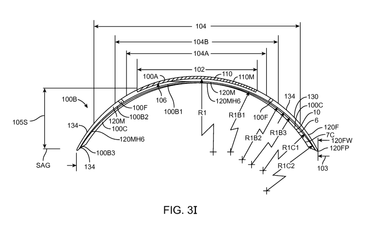
CA 02816031 2013-04-24
WO 2012/061160 PCT/US2011/057755
[0133] The epithelium may comprise an inner boundary that moves centripetally
inward as
indicated by arrows 30
[0134] Figure 1-1B shows an ablated eye about 1 to 2 days following refractive
surgery
resulting in an epithelial defect. The epithelium has at least partially
covered the ablation. The
epithelium may comprise irregularities and an inner boundary that moves
centripetally inward as
indicated by arrows 30. The thickness profile 12RP of the regenerating
epithelium 12R can be
irregular and degrade vision. The inner portion of the epithelium near the
boundary may
comprise a height greater than an outer portion of the epithelium away from
the boundary of the
epithelium. The portion of the ablation not covered with the epithelium and
the inner portion of
the epithelium near the boundary can result in aberrations, for example
aberrations
corresponding to a meniscus of the tear and a far sighted portion of the
cornea. As variation in
epithelial healing among individuals can be observed, the epithelial defect of
at least some
individuals can be present at 2 and 3 days post-op, with corresponding
aberrations.
[0135] Figure 1-1C shows an ablated eye when the epithelium has regenerated
following
refractive surgery resulting in an increased epithelial thickness centrally
when the epithelium has
regenerated, for example at about 3 days post-op. The regenerating epithelium
may have an
irregularity 121, for example corresponding to an increased elevation of an
inner portion of the
epithelium near the center of the ablation, for example. Work in relation to
embodiments as
described herein suggests that the natural regeneration of the epithelium can
provide an inner
portion having an increased central elevation with optical power that may
correspond to about 1
to 3 Diopters of additional optical power. The regenerating epithelium
comprises a thickness
profile 12RP extending along the surface of Bowman's membrane 14 and the
ablation 20. With
PRK the thickness profile 12RP of the epithelium can regenerate for at least
one week, for
example one month, such that vision can be degraded when the thickness profile
12RP of the
epithelium regenerates, and PRK surgery of the cornea can be combined in
accordance with
embodiments described herein so as to improve vision.
[0136] In many embodiments as described herein, irregularities of the cornea
are decreased
when the epithelium regenerates so as to provide one or more of improved
vision or comfort.
The coverings as described herein can be configured so as to decrease an
effect on vision of
corneal irregularity 121, decrease the height profile of irregularity 121,
decrease transfer of
irregularity 121 to an anterior surface of the covering, smooth irregularity
121 with the covering,
21
CA 02816031 2013-04-24
WO 2012/061160 PCT/US2011/057755
regenerate epithelium 12 such that irregularity 121 is decreased, or
combinations thereof. In
many embodiments, the covering 100 as described herein can be placed on the
eye such that a
smooth layer 12S of regenerated epithelium 12R substantially covers the
ablated profile so as to
provide improved vision sooner than would occur without covering, for example
at about 3 to 4
days post-op with PRK. In many embodiments, the covering can provide an
environment 100E
as described herein so as to guide epithelial regeneration and smooth the
regenerated epithelium.
[0137] In many embodiments, the cornea 10 of an eye 2 has an epithelial defect
11 following
refractive surgery such as PRK, and a covering 100 positioned over the
epithelial defect 11.
[0138] Figure 1-2A shows a covering 100 positioned on cornea 10 an eye 2
having an
epithelial defect 11, in which the covering abuts the cornea to seal the
cornea. The covering may
comprise a curved body, for example a curved contact lens body shaped to fit
the cornea.
[0139] The covering 100 can be sized to cover the ablated profile and
epithelial defect. The
inner portion 110 comprises a dimension across 102 that can be sized to extend
across a majority
of the ablation, and the outer portion 120 comprises a dimension across 104
sized to extend
across at least the epithelial defect and contact the epithelium on opposite
sides of the defect.
[0140] The dimension 102 extending across a majority of the ablation may
extend about 6 to 8
mm, for example, and may be sized larger than the ablation. The dimension 104
may comprise
about 12 to 14 mm across, for example so as to extend to the limbus and can be
sized to the
limbus of the patient for example. Work in relation to embodiments suggests
that the covering
sized to extend to the limbus and circumferentially around the limbus can be
centered on the
cornea. The covering may extend such that the outer rim of the covering
contacts the
conjunctiva disposed above the sclera peripheral to the limbus, for example,
and that such
configurations may center the lens on the cornea, for example.
[0141] The thickness of the covering can be sized and shaped in many ways. The
inner portion
110 of the covering comprises a thickness 106 and the outer portion 120 of the
covering
comprises a thickness 108. The thickness 106 of the inner portion may comprise
a substantially
uniform thickness such that the inner portion comprises an optical power of no
more than about
+/- 1D prior to placement on the eye, for example when held in front of the
eye and separated
from the cornea by a distance. Alternatively, the thickness of the inner
portion may vary so as
comprise optical power, for example optical power to correct vision of the
patient.
22
CA 02816031 2013-04-24
WO 2012/061160 PCT/US2011/057755
[0142] Figure 1-2B shows a smooth layer 12S of regenerated epithelium 12R
substantially
covering an ablated profile. The environment 100E is configured to guide
epithelial regeneration
and smooth the regenerated epithelium. The regenerating epithelium comprises a
thickness
profile 12RP.
[0143] The epithelium grows centripetally from circumscribing boundary 12E
toward the
center of ablated profile 20 to cover the exposed stroma, as indicated by
arrows 30.
[0144] The covering 100 may comprise an inner portion 110 and an outer portion
120. The
outer portion 120 can be configured to form a seal 100S with the cornea near
the edge of the
ablation and the epithelial defect, for example with a soft conformable
material such as silicone
or silicone hydrogel. The inner portion 110 is positioned over the pupil and
configured for the
patient to see, and may comprise a rigidity greater than the outer portion, so
as to smooth
irregularities of the epithelium when the cornea heals. Alternatively, the
inner portion may
comprise a rigidity equal to or less than the rigidity of the outer portion as
well. For example,
the inner portion may comprise silicone and the outer portion may comprise
silicone, and the
inner portion may comprise one or more of a more rigid silicone or a greater
thickness such that
the inner portion can be more rigid than the outer portion so as to smooth the
epithelium.
Although the inner portion can be more rigid than the outer portion, the inner
portion is
sufficiently soft, flexible and conformable so as to conform at least
partially to the ablated profile
20 in the stroma, such that the patient receives the benefit of the vision
correction with the
ablation profile 20 when the patient looks through the inner portion and the
inner portion
smoothes the epithelium. Work in relation to embodiments of the present
invention suggests that
the regenerating epithelium is softer than the underlying stroma of ablation
profile 20, such that
the inner portion can be configured to conform to the shape of the ablation
profile 20 when the
inner portion smoothes the epithelium disposed under the inner portion.
[0145] The covering 100 may comprise one or more of many optically clear
materials, for
example synthetic materials or natural material such collagen based materials,
and combinations
thereof, such as described in U.S. Pat. App. Ser. No. 12/384, 659, filed April
6, 2009, entitled
"Therapeutic Device for Pain Management and Vision", U.S. Pub. No. US 2010-
0036488 A1,
published onll February 2010. For example, the lens material may comprise a
naturally
occurring material, such as collagen based material. Alternatively or in
combination, the lens
material may comprise a known synthetic material, for example hydroxyethyl
methacrylate
23
CA 02816031 2013-04-24
WO 2012/061160 PCT/US2011/057755
(HEMA) hydrogel, hydrogel, silicone, for example hydrated silicone and
derivatives thereof.
For example, the optically clear material may comprise one or more of
silicone, silicone
hydrogel, silicone comprising resin, silicone comprising silicate, acrylate,
orcollagen. The cured
silicone may comprise silicone that is two-part heat cured and RTV (room
temperature
vulcanized). For example, polydimethyl siloxane such as NuSil, or
poly(dimethyl) (diphenyl)
siloxane may be used to mold the covering, for example with less than 10%
water content so as
to increase oxygen diffusion through the covering. The covering 100 may
comprise
perfluoropolyethers or fluorofocal. The lens material can be elastic, for
example a stretchable
elastic material such as silicone, such that the lens can seal the cornea. The
lens material can be
cured with a hardness and size and shape such that the covering comprises a
modulus within a
range from about 4 to about 20 MPa. The material may comprise, for example,
silicone
elastomer having optically clear silicate disposed therein and a water content
of no more than
about 10%, for example no more than about 5%, such that the lens covering has
a very high Dk
exceeding 150, and the silicone lens comprising silicate can be treated to
provide a wettable
surface. The lens may comprise hydrogel, for example silicone hydrogel, and
can be formed
with a water content within a range from about 5% to about 35% and a modulus
within a range
from about 4 to about 20 MPa, such that the covering conforms at least
partially to the ablated
stroma.
[0146] The covering may comprise silicone or silicone hydrogel having a low
ionoporosity
such that covering seals to the cornea. For example, covering may comprise
silicone hydrogel
comprising a low ion permeability, and the range of water can be from about 5
% to about 35%,
such that the Dk is 100 or more. The low ion permeability may comprise an
Ionoton Ion
Permeability Coefficient of no more than about 0.25 x 10⁻³ cm2/sec so as to
seal the cornea, for
example no more than about 0.08 x 10⁻³ cm2/sec. The low ion permeability
comprises an
Ionoton Ion Permeability Coefficient of no more than about 2.6 x 10⁻⁶ mm2/min
to seal the
cornea, for example no more than about 1.5 x 10⁻⁶ mm2/min.
[0147] The covering 100 may comprise a wettable surface coating 134 disposed
on at least the
upper side of the covering, such that the tear film of the patient is smooth
over the covering and
the patient can see. The wettable surface coating may comprise a lubricious
coating for patient
comfort, for example to lubricate the eye when the patient blinks. The
wettable coating may
comprise a contact angle no more than about 80 degrees. For example, the
coating may
comprise a contact angle no more than about 70 degrees, and the contact angle
can be within a
24
CA 02816031 2013-04-24
WO 2012/061160 PCT/US2011/057755
range from about 55 to 65 degrees to provide a surface with a smooth tear
layer for vision. For
example, the wettable coating can be disposed of both an upper surface and a
lower surface of
the covering. Alternatively, the lower surface may comprise a hydrophobic
surface material and
the lower hydrophobic surface may comprise the inner portion 110 and the outer
portion 120. At
least the outer portion 120 may comprise a lower surface composed of a sticky,
tacky material,
for example a hydrophobic material. The inner portion may also comprise the
lower surface
comprised of the sticky, tacky, hydrophobic material. The upper surface may
comprise the
wettable coating extending over at least the inner portion 110.
[0148] The wettable coating may comprise one or more of many materials. For
example, the
wettable coating may comprise polyethylene glycol (PEG), and the PEG coating
can be disposed
on ParyleneTM. Alternatively, the wettable coating may comprise a plasma
coating, and the
plasma coating comprise a luminous chemical vapor deposition (LCVD) film. For
example, the
plasma coating comprises at least one of ahydrocarbon, for example CH4, 02 or
fluorine
containing hydrocarbon, for example CF4 coating. Alternatively or in
combination, the wettable
coating may comprise a polyethylene glycol (PEG) coating or 2-
hydroxyethylmethacrylate
(HEMA). For example, the wettable coating may comprise HEMA disposed on a
ParyleneTM
coating, or the wettable coating may comprise N-vinylpyrrolidone (NVP)
disposed on a
ParyleneTM coating.
[0149] The covering 100 may comprise a lower surface corresponding to one or
more of many
suitable shapes to fit the covering to the cornea. For example, the lower
surface of the covering
may correspond to base radius of curvature. With post ablation corneas, the
covering may
conform substantially to the cornea. The covering may comprise a second curve
in combination
with a first curve, such that the lower surface comprises a bicurve surface.
Alternatively, the
lower surface may correspond to an aspheric surface. For example, an aspheric
surface may
comprise an oblate shape and conic constant to fit a post PRK eye. Also, it
may be helpful to fit
the covering to the cornea, for example with selection of one covering from a
plurality of sizes.
[0150] Figure lA shows the covering 100 having the thickness 108 of the outer
portion sized
such that the outer portion can conform to the epithelium. The thickness of
the outer portion can
be substantially constant, or may vary as described herein below.
[0151] Figure 1A1 shows covering 100 positioned on an eye and blinking of the
eye. An
upper lid and a lower lid can blink over the eye. Work in relation to
embodiments suggests that
CA 02816031 2013-04-24
WO 2012/061160 PCT/US2011/057755
the upper lid can exert a downward movement 20 and that the lower lid can
exert an upper
movement 22 on the eye. The downward movement 20 can be greater than the upper
movement
22. The wettable coating material as described herein can decrease force and
movement
transferred from the lids to the covering so as to inhibit motion of the
covering. The downward
movement 20 greater than the upward movement 22 can affect epithelial growth
near the
perimeter of covering 100.
[0152] Figure 1B1 shows covering 100 as in Fig. 1-2A prior to placement on the
cornea. The
covering 100 may comprise a base radius R1 of curvature, and the base radius
of curvature may
be slightly shorter than the ablated cornea such that the covering can be
steeper than the cornea
prior to placement on the cornea. The covering 100 comprises a first
configuration 100C1 prior
to placement on the cornea.
[0153] The base radius R1 can be sized to the cornea in many ways. For
example, base radius
R1 may have a radius corresponding to the outer unablated portion of the
cornea. Alternatively
or in combination, the base radius R1 may have a radius corresponding to the
post ablated eye.
[0154] The covering 100 may comprise a modulus within a range from about 4 MPa
to about
20 MPa, such that central portion can conform at least partially to the
ablated stroma and so that
the covering can smooth corneal irregularities and stromal irregularities of
the ablated cornea.
The covering may comprise an elastomeric stretchable material such that the
covering can stretch
to fit the cornea, for example. The covering having the modulus within a range
from about 4
MPa to about 20 MPa can be formed in many ways as described herein. For
example, the
covering may comprise a single piece of material having a substantially
uniform thickness
extending across the ablated cornea and at least a portion of the unablated
cornea, and the single
piece of material may comprise an elastic material such as a silicone
elastomer or a hydrogel.
Alternatively, the covering may comprise a single piece of material having a
non-uniform
thickness extending across the ablated cornea and at least a portion of the
unablated cornea. The
covering can be shaped in many ways and may comprise a single piece of one
material, or may
comprise a single piece composed to two similar materials, or may comprise a
plurality of
materials joined together.
[0155] The covering 100 may comprise one or more outer portions extending
outside the inner
central portion, and these outer portions may seal the cornea when the inner
portion conforms at
least partially to the ablated stroma. For example, the covering 100 may
comprise outer portion
26
CA 02816031 2013-04-24
WO 2012/061160 PCT/US2011/057755
additional shapes disposed outward from a central portion as described herein.
For example, the
covering may comprise a bicurve having a second radius of curvature disposed
peripheral to the
inner radius R1 of curvature to fit the unablated portion of the cornea. For
example, the second
and outer radius of curvature may comprise a shorter radius of curvature when
the central portion
is treated for myopia. The covering may comprise a third radius of curvature
longer than the
second radius of curvature so as to fit the sclera under the conjunctiva. The
covering may
comprise an oblate shape to fit the ablated and non-ablated portions of the
cornea, and the
covering may extend over the sclera with an outer portion, for example.
[0156] Figure 1B2 shows the covering as in Fig. 1B1 conforming to ablated
stromal tissue and
smoothing the epithelium over the ablated stroma. The cornea comprises an
ablated surface 20
to correct vision that may have a corresponding radius of curvature, for
example radius R2. The
ablated profile 20 may comprise additional, alternative, or combinational
shapes with those
corresponding to radius R2, such as aberrations ablated into the cornea to
correct aberrations of
the eye and astigmatism ablated into the cornea, and the inner portion 110 of
covering 100 can
conform to these ablated profiles of the cornea such that the patient can
receive the benefit of the
ablative vision correction when the covering is positioned on the cornea. For
example, the
cornea ablation profile 20 may correspond to radius of curvature R2, and the
inner portion 110
can flatten from configuration 100C1 corresponding to radius of curvature R1
prior to placement
to a second configuration 100C2 corresponding substantially to the ablated
profile 20, such the
patient can see with the benefit of ablation profile 20. For example, the
second configuration
100C2 can comprise a conforming radius of curvature R12 that corresponds
substantially to
radius of curvature R2. The profile corresponding to the first configuration
100C1 of the
covering 100 is shown positioned over cornea 10 to illustrate the change in
profile of the
covering from configuration 100C1 prior to placement to conforming second
configuration
100C2 of the covering 100 when positioned on the cornea.
[0157] The conformable covering 100 comprises sufficient rigidity so as to
smooth the
epithelium when covering 100 is positioned on the cornea over the ablation
profile 20. The
epithelium comprises a peripheral thickness 12T that may correspond
substantially to a thickness
of the epithelium prior to debridement of the epithelium to ablate the cornea.
The epithelium
also comprises regenerating epithelium 12R disposed over the ablation profile
20. The covering
100 can smooth the epithelium 12R when conforming to the cornea in the second
configuration
100C2. For example, irregularities 121 of the regenerating epithelium 12R
disposed over the
27
CA 02816031 2013-04-24
WO 2012/061160 PCT/US2011/057755
ablation can be smoothed when the epithelium regenerates along the inner
portion of covering
100, such that the irregularities 121 of the regenerating epithelium 12R are
thinner than the
thickness 12T of the peripheral epithelium.
[0158] Work in relation to the embodiments as described herein indicates that
an at least
partially conformable covering having a modulus within a range from about 4
MPa to about 20
MPa can conform at least partially to the ablated stroma and smooth
irregularities of the
epithelium and stroma so as to improve vision as described herein. The
covering having the
modulus within the range from about 4 MPa to about 20 MPa can be formed in
many ways as
described herein.
[0159] The conformable covering 100 may comprise a perimeter 120P with
rigidity sufficient
to indent the epithelium along at least a portion of the perimeter so as to
seal the cornea with seal
100S. The portion 12C of the epithelium 12 can extend over the perimeter 120P
of the covering
100.
[0160] Figure 1B2A shows a covering as in Fig. 1B2 forming an indentation 12IT
in the
epithelium such that the epithelium 12 extends over at least a portion of the
perimeter 120P of
the covering. The covering forms indentation 12IT in the epithelium such that
the epithelium
comprises an indentation thickness 12T that is less than an outer thickness of
the epithelium 12.
The indentation of the epithelium with the covering can help to seal the
cornea with the
perimeter.
[0161] Figure 1B2B shows a covering as in Fig. 1B2 forming indentation 12IT in
the
epithelium. The covering forms indentation 12IT in the epithelium such that
the epithelium
comprises an indentation thickness 12T that is less than an outer thickness of
the epithelium 12T.
The indentation of the epithelium with the covering can help to seal the
cornea with the
perimeter.
[0162] Work in relation to embodiments described herein suggests the
indentation of the
covering can vary radially around the eye of the patient, in accordance with
orientation of the
covering on the eye when the covering comprises a substantially constant
rigidity of the outer
portion, for example a substantially constant rigidity around the perimeter.
The inferior portion
of the covering may comprise a greater amount of epithelial covering over the
perimeter than the
superior portion of the covering. For example, Fig. 1B2A may correspond to a
first portion of
covering 100 at an inferior location of the cornea and Fig. 1B2B may
correspond to a second
28
CA 02816031 2013-04-24
WO 2012/061160 PCT/US2011/057755
portion of the covering at a superior location of the cornea. Work in relation
to embodiments
also suggests that there may be variability in covering of the perimeter with
the epithelium
between the nasal portion of the perimeter, and the temporal portion of the
perimeter, although
both the nasal and temporal locations can comprise covering intermediate and
between the more
extensive covering of the inferior portion and the less extensive covering of
the superior portion
of the perimeter.
[0163] Figure 1B2C shows a covering abutting the cornea to seal the cornea
without forming a
substantial indentation in the epithelium. The covering may comprise a chamfer
to contact and
seal the cornea. The rigidity of the outer portion can be determined based on
the thickness of the
outer portion of the covering, hardness of the material, and chamfer angle so
as to contact the
epithelium to seal the cornea without substantial deformation of the
epithelium.
[0164] The covering may comprise a non-uniform rigidity around the outer
portion of the
covering comprising the perimeter. For example, the covering may comprise a
superior portion
corresponding to a superior location on the cornea and an inferior portion
corresponding to an
inferior location on the cornea. The superior portion may comprise a rigidity
less than the
inferior portion. For example, the superior portion may comprise the rigidity
less than the
inferior portion, such that deformation of the epithelium is inhibited when
the perimeter abuts the
cornea is sealed. Alternatively, the superior portion may comprise the
rigidity less than the
inferior portion such the deformation of the epithelium with the covering
comprises a
substantially constant amount around the perimeter, for example a deformation
of no more than
about 10 um, for example 5 um.
[0165] Fig. 1C shows a therapeutic covering as in Fig. 1-2A comprising a
covering molded
with a homogeneous material, in which the outer portion comprises a thickness
configured to
conform with the cornea and in which the inner portion 110 comprises thickness
configured to
smooth the epithelium and conform to the ablated profile 20. The outer portion
120 may
comprise a thickness of no more than about 100 microns. For example, the outer
portion 120
may comprise a thickness of about 50 microns at the boundary with the inner
portion 110, and
linearly taper from 50 microns at the boundary with the inner portion to about
20 microns at the
periphery of the outer portion 120. The inner portion 110 may comprise a
thickness of no more
than about 250 microns, for example no more than about 200 microns. For
example, the inner
portion may comprise a thickness of about 100 microns. For example, the
thickness of each of
29
CA 02816031 2013-04-24
WO 2012/061160 PCT/US2011/057755
the inner portion and the outer portion may comprise no more than about 50
microns so as to
provide substantial oxygen transport and epithelial regeneration. Many
materials can be used as
described herein, and the covering may comprise one or more materials. For
example, the
covering may comprise a single piece of material such as silicone having a
water content within
a range from about 0.1 % to about 10%, for example no more than about 1%, and
a hardness
Shore A durometer parameter within a range from about 5 to about 90, for
example within a
range from about 40 to about 85.
[0166] Figure 1C1 shows a covering 100 having an inner portion 110 comprising
an inner
thickness and an inner material 110M and an outer portion 120 comprising an
outer thickness
and an outer material 120M, in which the inner thickness is greater than the
outer thickness. The
inner material 110M may comprise many materials and may comprise an optically
clear silicone,
for example silicone with resin. The inner material may comprise silicone
positioned in a mold
with the outer portion 120 formed around the inner portion. The inner portion
may comprise a
hardness similar to the outer portion. The outer material 120M of the outer
portion 120 may
comprise a material similar to the inner portion. For example, the outer
material 120M may
comprise silicone and the inner material 110M may comprise silicone. This use
of similar
materials on the inner and outer portions can improve adhesion of the inner
portion to the outer
portion. The outer material 120M may extend along the inner portion 110, for
example along the
underside of the inner portion 110, such that the inner material 110M is held
in a pocket of the
outer material 120M. Alternatively, the inner material 110M may extend
substantially across the
thickness of the inner portion 110, such that the outer material 120M
comprises a substantially
annular shape with the inner material 110M comprising a disc shaped portion
disposed within the
annulus and extending substantially from the upper surface coating to the
lower surface coating
when present.
[0167] Figure 1C 1A shows a covering as in Fig. 1C1 adhered to the cornea with
a smooth
upper surface, and a lower surface conforming to irregularity of the cornea,
for example an
irregularity comprising a central island lOCI of the ablated stroma. The
central island lOCI may
comprise an outward protrusion in the ablated profile of the stroma at least
about 1 micron
outward and about 2.5 mm across, for example. The upper surface may comprise a
substantially
rigid material for vision correction, and the lower surface may comprise a
soft material so as to
deflect to irregularities of the cornea when the upper surface provides
optical correction. For
example, the lower surface may comprise an indentation 110I when positioned on
the irregularity
CA 02816031 2013-04-24
WO 2012/061160 PCT/US2011/057755
of the cornea. Although the lower surface comprising the soft material can
deflect to correspond
to the ablation profile 20, the upper surface comprising the rigid material
may comprise a
predetermined curvature selected by a health care provider so as to fit the
ablation profile and
correspond to the refractive correction of the patient so as to provide vision
correction.
[0168] Figure 1C2 shows a covering as in Figs. 1-2A to 1B2 having inner
portion 110
comprising an inner thickness and inner material 110M and outer portion 120
comprising an
outer thickness and outer material 120M, in which the inner thickness can be
greater than the
outer thickness and the outer material 120M extends around the inner material
110M. The
covering 100 may comprise at least a bicurve covering having at least a second
radius RIB. The
inner portion 110M may comprise three layers of material, a first layer 100L1
of a first material
110M1, a second layer 100L2 of a second material 110M2 and a third layer 100L3
of a third
material 110M3. The second material 110M2 may comprise a rigid material, for
example one or
more of a rigid gas permeable material, a rigid silicone, or a rigid silicon
acrylate. The first
material 110M1 and the third material 110M3 may comprise a soft material, for
example a soft
elastomer or soft hydrogel such as one or more of a soft optically clear
silicone or a soft silicone
hydrogel. The first material, the third material, and the outer material 120M
may comprise
similar materials, such that the second layer of rigid material 110M2 is
encapsulated with the
first soft material 110M1, the third soft material 110M3 and on the perimeter
with the soft outer
material 120M. In many embodiments, the second rigid material 110M2 comprises
a material
similar to each of the first material 110M1, the third material 110M3 and the
outer material
120M, for example each may comprise silicone, such that the corresponding
portions of the
covering 100 can be bonded together with the silicone similar to silicone
elastomer material, for
example. In many embodiments, the covering 100 can be formed in a mold with
rigid second
material 110M2 placed in the mold and encapsulated within a single piece of
material
comprising first material 110M1, third material 110M3 and outer material 120M,
such that first
material 110M1, third material 110M3 and outer material 120M comprise
substantially the same
material, for example silicone elastomer. The rigid second material 110M2 may
comprise
silicone bonded to each of first material 110M1, third material 110M3 and the
outer material
120M, for example with curing such that first material 110M1, third material
110M3 and outer
material 120M comprise the same soft silicone material bonded to the second
material 110M2
comprising rigid silicone.
31
CA 02816031 2013-04-24
WO 2012/061160 PCT/US2011/057755
[0169] The soft material comprising soft outer portion 120 composed of soft
material 120M,
first layer 100L1 composed of soft material 110M1 and third layer 100L3
composed of soft
material 120M3 can provide improved comfort and healing for the patient. The
soft material can
deflect, bend or indent so as to conform at least partially to the tissue of
the eye when the rigid
portion comprising rigid material 110M2 corrects vision of the patient. The
dimension 102
across inner portion 110 can be sized to substantially cover the ablation zone
and slightly smaller
than the ablation dimensions, such as ablation diameter 20D, so that the
epithelium can grow
inward and contact the layer 100L1110L1 of soft first material 110M1 without
substantial
disruption from the rigid material 120M2 when the inner portion 110M corrects
vision with the
layer of rigid material 110M2. The eyelid can also move over the third layer
100L3 for
improved comfort. The soft first material 110M1 and soft third material 110M3
may comprise
soft elastomer or soft hydrogel, for example, and may each comprise the same
material so as to
encapsulate the second layer 100L2 of rigid second material 110M2.
[0170] The soft material comprising soft outer portion 120 composed of soft
material 120M,
first layer 100L1 composed of soft material 110M1 and third layer 100L3
composed of soft
material 110M3 can have a modulus within a range from about 1 to 20 MPa, for
example within
a range from about 1 to 5 MPa.
[0171] The material inner material 120M and 110M2 of second layer 100L2 can
have a
modulus within a range from about 5 to about 35 or more, for example as set
forth in Table A
below. For example, when material 120M comprises silicone elastomer or layer
100L2 of
material 110M2 comprises silicone elastomer, the modulus can be within a range
from about 5 to
about 35 MPa, for example within a range from about 20 to about 35 MPa.
[0172] The layers of covering 100 can comprise dimensions so as to provide
therapeutic
benefit when placed on eye 2. The thickness of layer 100L1 can be from about 5
um to about 50
um, for example, within a range from about 10-30 um, such that the layer 100L1
can provide a
soft at least partially conformable material to receive the lens. The middle
layer 100L2 can be
from about 20 um to about 150 um, for example, and material 110M2 can have a
modulus
greater than first material 110M1 of first layer 100L1, so as to deflect the
epithelium of the eye
when the middle layer is deflected. The third layer 100L3 can be within a
range from about 5
um to 50 um, for example within a range from about 10 um to about 30 um, and
can cover
second layer 100L2 so as to retain the second layer in the inner portion 110
of the covering 100.
32
CA 02816031 2013-04-24
WO 2012/061160 PCT/US2011/057755
[0173] Figure 1C2A shows a covering as in Fig. 1C1 placed on the cornea with a
smooth upper
surface and a lower surface conforming to irregularity of the cornea near the
edge of the ablation.
As the epithelium can be about 50 um thick, in many embodiments the dimension
102 is sized so
as to cover substantially the ablated cornea for vision correction and smaller
than the ablation
zone, such that the outer portion 120 can conform at least partially to the
epithelium. The outer
portion 120 may extend to the sclera, and comprise a tri-curve covering 100 as
described herein,
with the inner portion 110 having first layer 100L1 of first material 110M1,
second layer 100L2
of second material 110M2, and third layer 100L3 of third material 110M3.
[0174] Figure 1C2A1 shows a covering having a layer of hydrogel material on a
posterior
surface of the covering. The covering 100 may comprise a wettable surface
coating 134
disposed on at least the upper side of the covering as described herein. The
layer of hydrogel
material may comprise an inner portion of the layer of hydrogel material
110MHG and an outer
portion of the layer of hydrogel material 120MHG. The layer of hydrogel
material extends to the
fenestration so as to couple the hydrogel material to the fenestration. The
hydrogel material can
be coupled to the fenestration in many ways. For example, the layer of
hydrogel material may
cover the fenestration, or the fenestration 100F may extend through the
hydrogel material. The
fenestration 100F extending through the layer of hydrogel material can
encourage pumping of
the tear liquid as described herein. Alternatively or in combination, the
layer of hydrogel
material covering a posterior surface of the fenestration 100F to couple the
fenestration 100F to
the hydrogel layer may encourage movement of a therapeutic agent along the
hydrogel layer
toward a central portion of the cornea for example. The hydrogel may extend
along a deflectable
portion of the covering so as to exert at least some pressure on the hydrogel
layer to encourage
movement of one or more of tear liquid or the therapeutic agent along the
hydrogel layer when
the patient blinks, for example.
[0175] The hydrogel layer as described herein may encourage regeneration of
the epithelium
and may provide a soft surface to contact the epithelium regenerating over the
ablation so as to
encourage epithelial regeneration under the optical component as described
herein, and the
optical component can resist deformation so as to protect the epithelium and
provide an
environment to encourage regeneration of the epithelium.
[0176] The hydrogel material may comprise one or more of the hydrogel
materials as
described herein. The hydrogel material extending along the lower surface can
increase comfort
33
CA 02816031 2013-04-24
WO 2012/061160 PCT/US2011/057755
of the covering when placed on the eye. The hydrogel material may comprise a
substantially
uniform thickness within a range from about 1 um to about 100 um, for example
from about 2
um to about 50 um and in many embodiments within a range from about 5 um to
about 20 um.
The hydrogel material extending along the posterior surface may comprise on or
more of the
hydrogel materials as described herein combined with one or more of materials
110M, 110M1,
110M2, 110M3 or 120M as described herein. For example the one or more of
materials 110M,
110M1, 110M2, 110M3 or 120M may comprise silicone such as silicone elastomer
comprising
siloxane, and the hydrogel may comprise a hydrogel such as silicone hydrogel
material as
described herein.
[0177] Figure 1C2A shows a covering having a layer of hydrogel material on a
posterior
surface of the covering. The covering 100 may comprise a wettable surface
coating 134
disposed on at least the upper side of the covering as described herein. The
layer of hydrogel
material may comprise an inner portion of the layer of hydrogel material
110MHG and an outer
portion of the layer of hydrogel material 120MHG. The layer of hydrogel
material extends to the
fenestration so as to couple the hydrogel material to the fenestration. The
hydrogel material can
be coupled to the fenestration in many ways. For example, the layer of
hydrogel material may
cover the fenestration, or the fenestration 100F may extend through the
hydrogel material. The
fenestration 100F extending through the layer of hydrogel material can
encourage pumping of
the tear liquid as described herein. Alternatively or in combination, the
layer of hydrogel
material covering a posterior surface of the fenestration 100F to couple the
fenestration 100F to
the hydrogel layer may encourage movement of a therapeutic agent along the
hydrogel layer
toward a central portion of the cornea for example. The hydrogel may extend
along a deflectable
portion of the covering so as to exert at least some pressure on the hydrogel
layer to encourage
movement of one or more of tear liquid or the therapeutic agent along the
hydrogel layer when
the patient blinks, for example.
[0178] The hydrogel layer as described herein may encourage regeneration of
the epithelium
and may provide a soft surface to contact the epithelium regenerating over the
ablation so as to
encourage epithelial regeneration under the optical component as described
herein, and the
optical component can resist deformation so as to protect the epithelium and
provide an
environment to encourage regeneration of the epithelium.
34
CA 02816031 2013-04-24
WO 2012/061160 PCT/US2011/057755
[0179] The hydrogel material may comprise one or more of the hydrogel
materials as
described herein. The hydrogel material extending along the lower surface can
increase comfort
of the covering when placed on the eye. The hydrogel material may comprise a
substantially
uniform thickness within a range from about 1 um to about 100 um, for example
from about 2
um to about 50 um and in many embodiments within a range from about 5 um to
about 20 um.
The hydrogel material extending along the posterior surface may comprise on or
more of the
hydrogel materials as described herein combined with one or more of materials
110M, 110M1,
110M2, 110M3 or 120M as described herein. For example the one or more of
materials 110M,
110M1, 110M2, 110M3 or 120M may comprise silicone such as silicone elastomer
comprising
siloxane, and the hydrogel may comprise a hydrogel such as silicone hydrogel
material as
described herein.
[0180] Figure 1C2B shows a covering having a layer of hydrogel material on a
posterior
surface of the covering extending less than a maximum distance across the
covering such that
end portions of the covering are configured to engage the epithelium of the
eye away from the
hydrogel layer and inhibit movement of the covering when placed on the eye. In
many
embodiments, the material 120M can couple to the surface of the eye, for
example the epithelium
so as to inhibit movement of the covering. The material 120M may comprise a
sticky tacky
hydrophobic material such as silicone to engage the epithelium to inhibit
movement, and the
material 120M may be coated with one or more coatings as described herein, for
example with
vapor deposition. The hydrogel material can be coupled to the fenestration in
many ways. For
example, the layer of hydrogel material may cover the fenestration, or the
fenestration 100F may
extend through the hydrogel material.
[0181] Figure 1C2C shows a covering 100 having an annular layer of hydrogel
material
120MHG on a posterior surface of the covering such that an inner portion of
the covering
contacts the cornea away from the hydrogel layer and an outer portion of the
covering contacts
the cornea away from the covering when placed on the eye. Work in relation to
embodiments
suggests that the annular hydrogel layer can provide an environment to
encourage growth of the
epithelium along the posterior surface of inner material 110M1 as described
herein, and the
lower surface of material 110M1 can be coated with a material having a
thickness less than the
hydrogel, for example.
CA 02816031 2013-04-24
WO 2012/061160 PCT/US2011/057755
[0182] Figure 1C3 shows a shows a covering having a tricurve profile to fit
sclera with slopes
of the curved profiles aligned so as to inhibit ridges at the boundaries of
the curved portions as in
Figure 1B2 and having a layer of hydrogel material 120MHG on a lower surface.
The hydrogel
material 120M may extend substantially across the posterior surface of the
covering. The
covering may extend along the lower surface a distance less than a distance
across the covering
so as to provide a portion of the covering without the hydrogel to engage the
eye, for example
the epithelium of the eye that may comprise one or more of the corneal
epithelium or the
conjunctival epithelium. Alternatively, the covering may extend substantially
along the posterior
surface of the covering corresponding to the distance across the covering so
as to provide the
hydrogel covering over the outer portion of the covering that engages the eye.
[0183] Figure 1C4 shows a plan view covering having a tricurve profile to fit
the cornea,
limbus and sclera with slopes of the curved profiles aligned so as to inhibit
ridges at the
boundaries of the curved portions and having a hydrogel material on a lower
surface extending
less than a maximum distance across the covering to engage the conjunctiva
with the covering
away from the hydrogel material. Alternatively, the covering may extend
substantially along the
posterior surface of the covering corresponding to the distance across the
covering so as to
provide the hydrogel covering over the outer portion of the covering that
engages the eye. The
hydrogel covering may comprise an annular shape extending along the lower
surface as
described herein.
[0184] Figure 105 shows a fenestration 100F having a posterior end 100FPE
covered with a
layer of hydrogel material 29MHG extending along the posterior surface of the
covering 100, in
accordance with embodiments of the present invention;
[0185] Figure 106 shows a fenestration 100F extending through a layer of
hydrogel material
120MHG extending along the posterior surface of the covering 100, in
accordance with
embodiments of the present invention;
[0186] Fig. 1D shows atherapeutic covering 100 comprising a first inner
material 110M and a
second outer material 120M, in which the outer portion 120 comprises a
hardness configured to
conform with epithelium of the cornea and in which the inner portion 110
comprises second
hardness configured to smooth the epithelium and conform to the ablated
profile 20. The outer
material 120M may comprise many materials as herein. The Shore A hardness of
each of the
inner portion and the outer portion can be within a range from about 5 to
about 90. For example,
36
CA 02816031 2013-04-24
WO 2012/061160 PCT/US2011/057755
the outer material 120M may comprise silicone having a hardness Shore A
durometer parameter
from about 20 to about 50,for example from about 20 to about 40, and the inner
material 110M
may comprise silicone having a hardness durometer parameter from about 40 to
about 90, for
example from about 50 to about 90. The outer portion comprises a perimeter
120P, and the
perimeter may comprise a peripheral and circumferential edge structure to abut
the epithelium to
form the seal with the epithelium, for example when the base radius of the
covering is less than
the cornea. The peripheral and circumferential edge structure can be shaped in
many ways to
define an edge extending around the perimeter to abut the epithelium, for
example with one or
more of a taper of the edge portion extending to the perimeter, a bevel of the
edge portion
extending to the perimeter or a chamfer of the edge portion extending to the
perimeter. The
inner portion 110 may comprise inner thickness and inner material 110M and the
outer portion
120 may comprise an outer thickness and outer material 120M, in which the
inner thickness is
substantially similar to the outer thickness.
[0187] The peripheral edge structure to abut the epithelium can be used with
many
configurations of the inner portion as described herein. For example, the
inner portion may
comprise an RGP lens material having a lower rigid surface to contact and
smooth the cornea
and an upper rigid optical surface. Alternatively, the inner portion may
conform to the cornea as
described herein. The outer portion may comprise a skirt, and the skirt may
comprise the
peripheral edge structure to abut and seal the cornea, such as the chamfer.
The rigidity of the
outer portion comprising the edge structure can be determined to seal the
cornea with one or
more of hardness and thickness, as described herein.
[0188] Figures lE and 1F show top and side views, respectively, of covering
100 comprising
inner portion 110, outer portion 120, and a peripheral rim portion 140
disposed around outer
portion 120. The peripheral portion 140 can be more rigid than outer portion
120. Work in
relation to embodiments suggests that in some instances the lower sticky,
tacky surface of outer
portion 120 can stick to itself during deployment onto the eye, and the
peripheral portion 140 can
improve handling when the covering is placed on the eye. The covering may
comprise a single
piece of material or may comprise multiple pieces adhered together, for
example molded
together. For example, the covering may comprise an inner thickness of inner
portion 110 and
an outer thickness of outer portion 120, in which the inner thickness is
greater than the outer
thickness. The peripheral portion 140 may comprise a thickness, and the
thickness of the
peripheral portion 140 can be greater than the thickness of outer portion 120
such that the
37
CA 02816031 2013-04-24
WO 2012/061160 PCT/US2011/057755
peripheral portion 140 is more rigid than the outer portion 120. The thickness
of the inner
portion 110 and the thickness of the peripheral portion 140 can be
substantially similar, and these
portions may comprise substantially the same thickness and rigidity.
[0189] Figure 1G shows covering 100 comprising inner portion 110 and outer
portion 120,
such that outer portion 120 comprises a taper 120T of thickness 108 extending
between the
perimeter of the inner portion 110 and the perimeter of the outer portion 120.
The taper may
comprise a substantially linear change in thickness 108 extending between the
perimeter of the
inner portion and the perimeter of the outer portion.
[0190] Figure 1G1 shows a covering 100 comprising inner portion 110 and an
outer portion
120 comprising the taper as in Fig. 1G, and an outer rim or flange 120F of
substantially uniform
thickness peripheral to the taper 120T. The outer taper may extend from the
dimension across
102 of the inner portion 110 to the dimension across 154A that is less than
the dimension across
104 of the outer portion. The rim of substantially uniform thickness may
comprise an annular
shape having a thickness within a range from about 10 um thick to about 40 um
thick, and may
comprise a width 154B within a range from about 0.05 to about 0.8 mm, for
example about 0.5
mm. The rim, for example flange 120F, may comprise a thickness of no more than
about 50 um,
such that the flange comprises a thickness no more than the epithelium.
[0191] The covering 100 can be dimensioned in many ways. The total diameter
across can be
from about 6 mm to about 12 mm, for example about 10 mm. The inner portion may
comprise a
diameter within a range from about 4 mm to 8 mm, for example about 6 mm. The
annular rim
comprising flange 120F can extend around the perimeter of the covering with a
thickness of
about within a range from about 5 um to about 50 um, for example about 35 um.
The annular
rim comprising flange 120F may comprise an inner diameter of within a range
from about 5 mm
to about 11 mm, for example about 9 mm and an outer diameter within a range
from about 6 mm
to about 12 mm, for example about 10 mm and corresponding to the perimeter of
the covering.
The annular rim may comprise a width of within a range from about 0.1 mm to
about 1 mm, for
example 0.5 mm, extending circumferentially around the covering. The outer
portion 120 may
comprise the rim with flange 120F and a taper 120T that extended from inner
portion 110 to the
rim comprising perimeter 120P. The taper in thickness can be substantially
uniform between the
outer diameter of the inner portion and the inner diameter of the rim, and the
boundaries of the
taper can be rounded and smoothed near the inner portion and the rim. The
central portion may
38
CA 02816031 2013-04-24
WO 2012/061160 PCT/US2011/057755
comprise a substantially uniform thickness within a range from about 50 um to
about 150 um, for
example about 50 um. The base radius of curvature of the lower surface of the
covering can be
within a range from about 7 mm to about 8 mm. The lower surface may comprise
an aspheric
surface or a bicurve surface and combinations thereof. The upper surface of
the covering can
comprise a radius of curvature along the inner portion within about 0.1 mm
curvature of the
lower surface, such that the covering is substantially uniform with no
substantial refractive
power, for example refractive power within about +/- 1D.
[0192] Figures 1G 1A to 1G1H show a covering as in Fig. 1G1 and dimensions
suitable for use
in accordance with embodiments as described herein such as with experiments,
clinical studies
and patient treatment. Fig. 1G 1A shows an isometric view of covering 100
having the inner
portion 110, the outer portion 120, the taper 120T and rim comprising flange
120F. Fig. 1G1B
shows a bottom view of covering 100. Fig. 1G1C shows a side view of the
covering 100. Fig.
1G1D shows a top view of the covering 100. Fig. 1G1E shows a side cross
sectional view of
covering 100 along section D-D. Fig. 1G1F shows detail C of cross-section D-D,
including the
radius of curvature R1 of the lower surface of the inner portion 110, and the
upper radius of
curvature Rupper of the inner portion 110. The upper radius of curvature
Rupper may
correspond substantially to the lower radius of curvature R1 prior to
placement on the eye, for
example to within about +/- 1 D of optical power, such that the inner portion
110 prior to
placement may comprise no substantial optical power. Detail C shows a side
cross sectional
view of covering 100 of the inner portion. Fig. 1G1G shows detail B of cross-
section D-D.
Detail B shows a side cross sectional view of the rim comprising flange 120F.
The flange 120F
has a thickness 109. Flange 120F may comprise a taper extending along a width
102FW, for
example from a first thickness 109A of about 35 um to second thickness 109B of
about 25 um
extending along width 120FW near the chamfer. Flange 120F comprises a
chamfered edge
120FE to contact the cornea or conjunctiva along perimeter 120P of the
covering.
[0193] Figure 1H1 shows spatial frequency and elevation smoothing of an
epithelial
irregularity 121 transferred to a front surface 110U of covering 100 as in
Fig. 1-2A. The
regenerating epithelium 12R comprises an irregularity 121. The covering 100
conforms
substantially to the shape ablated in the stroma when positioned on the eye as
noted above. The
covering 100 comprises a rigidity so as to conform substantially to the
ablation profile 20 over
about at least about 3 to 4 mm of the ablated stroma such that the patient can
see and receive
optical correction with the ablated surface. The regenerating epithelium
comprises a thickness
39
CA 02816031 2013-04-24
WO 2012/061160 PCT/US2011/057755
profile 12RP that includes irregularity 121. The conformable covering
comprises a thickness
profile of thickness 106 that encompasses a deformation thickness over the
irregularity 106D.
The thickness of the covering can vary over the epithelium to smooth the
irregularity transmitted
to the front surface of the covering so as to improve patient vision
consistent with the ablation
profile 20 when the covering conforms to the ablation profile 20. For example
thickness 106D
over the irregularity can be less than thickness 106 away from the
irregularity. The irregularity
may comprise an indentation and the covering may be thinner over the
indentation. The silicone
elastomer and hydrogel materials as described can be at least somewhat
compressible so as to
conform at least partially to the cornea and form an indentation so as to
receive a portion of the
cornea comprising one or more of epithelium or ablated stroma and decrease
aberrations.
[0194] Experimental studies of optical coherence tomography (hereinafter
"OCT") images and
PentacamTM images and topography images, noted below, indicate that the
thickness of the
inner portion of the covering 100 can vary so as to decrease optical
aberrations along the upper
surface when the covering is adhered to the cornea. This variation in
thickness can be related to
one or more of stretching of the covering over the irregularity or compression
of the covering
over the irregularity.
[0195] The irregularities of the epithelium generally comprise spatial
frequencies that are
greater than the spatial frequencies of the vision correcting portion of the
ablation. The covering
can provide spatial filtering of the frequencies of the underlying surface so
as to inhibit relatively
higher spatial frequencies of epithelial irregularities and pass relatively
lower spatial frequencies
corresponding to vision correction, such as lower spatial frequencies
corresponding to sphere and
cylinder. The spatial frequencies ablation profile 20 that are useful to
correction vision can be
lower than the spatial frequencies of the irregularities, and the spatial
dimensions of the vision
correction greater than the dimensions of the irregularities. For example, the
spatial frequencies
of the vision correction can correspond to periods of oscillation less than
the periods of
oscillation of the irregularities.
[0196] Figure 1H2 shows spatial frequency and elevation smoothing of the
epithelial
irregularity with a plot of relative height relative for the upper surface of
the covering and the
upper surface of the irregularity. The irregularity of the regenerating
epithelium 12R may
comprise a profile height 12RPH and profile width 12RPW. The upper surface of
the covering
may comprise a profile 110UP. The irregularity of the upper surface
corresponding comprises a
CA 02816031 2013-04-24
WO 2012/061160 PCT/US2011/057755
width 110UPW and a height 110UPH. Height 110UPH is less than height 12RPH so
as to
correspond to smoothing of the irregularity. Width 110UPW is greater than
width 12RPW so as
to correspond to smoothing of the irregularity. Profile 110UP of the upper
surface of the
covering corresponds to lower frequencies than profile 12RP, such that the
covering comprise a
low pass spatial frequency filter. This can be seen with the PentacamTM and
topography data
shown below in conjunction with OCT images showing that the covering and
cornea conform
without a substantially gap disposed therebetween. Alternatively or in
combination, the covering
can smooth the cornea when a gap is present, for example when a portion of the
cornea is
smoothed with contact to the covering and the gap provides an environment for
the epithelium to
grow smoothly over the ablation.
[0197] Based on the teachings described herein, a person of ordinary skill in
the art can
conduct studies to determine empirically the rigidity of the inner portion so
as to pass
substantially vision correction spatial frequencies of the ablation to the
upper surface of the
covering and inhibit spatial frequencies of the irregularities of the ablated
stroma and epithelium,
for example with PentacamTM and topography studies as described in the
experimental section.
[0198] Work in relation to the embodiments as described herein indicates that
a covering
comprising a modulus within a range from about 4 MPa to about 20 MPa can
provide smoothing
with low pass spatial frequency filtering as described with reference to Figs.
1H1 and 1H2. The
covering may comprise an elastically stretchable material, for example an
elastomer or a
hydrogel, such that the lens can conform at least partially to the ablated
stroma and exert at least
some pressure on the ablated stroma and epithelium when at least partially
conformed so as to
smooth irregularities of the epithelium and irregularities of the stroma. The
covering can
comprise a thickness and a hardness so as to provide the spatial frequency
filtering to improve
vision in post-PRK patients with the modulus within the range from about 4 MPa
to about 20
MPa. For example, the lens thickness can be increased to increase the modulus,
decreased to
decrease the modulus. The hardness of the material can be increased to
increase the modulus
and decreased to decrease the modulus. The modulus within the range from about
4 MPa to
about 20 MPa can attenuate substantially higher spatial frequencies
corresponding to
irregularities of the epithelium and stroma so as to smooth the high spatial
frequencies
corresponding to the irregularities that can degrade vision, and can conform
substantially to
lower spatial frequencies that correspond to the vision correction so as to
pass the lower spatial
frequencies corresponding to vision correction so that the patient can
experience an improvement
41
CA 02816031 2013-04-24
WO 2012/061160 PCT/US2011/057755
in vision when the epithelium regenerates under the covering. For example, the
high spatial
frequencies may correspond to frequencies greater than about 1/6 (0.17) cycles
per mm, and the
low spatial frequencies may correspond to frequencies less than about 1/6 (0.1
7) cycles per mm.
A person of ordinary skill in the art can determine the modulus and
corresponding spatial
frequencies to attenuate and pass, in accordance with the teachings as
described herein. For
example, the modulus of the covering can be measured with known methods and
apparatus to
measure the modulus of a contact lens, and measurements with PentacamTM images
as
described herein can be used to determine the relationship of the modulus of
the measured lens
coverings to smooth irregularities, conformation of the lens coverings to the
ablation, and vision.
[0199] Figure 111 shows an inhibition of transfer of a corneal irregularity to
a front surface of a
covering, for example one or more of a stromal irregularity or an epithelial
irregularity. The
front surface of the covering comprises an optical surface for vision without
substantially
transfer of the irregularity to the front surface of the covering.
[0200] Figure 112 shows elevation smoothing of the epithelial irregularity
with a plot of height
relative to a reference sphere for the upper surface of the covering and the
upper surface of the
irregularity. The plot shows a substantially spherical front surface of the
covering, such that the
transfer of the irregularity to the front surface is inhibited.
[0201] Figure 113 shows a thickness profile of the covering as in Fig. 112 so
as to smooth
irregularities transferred to the front surface of the covering. The thickness
profile can vary in
response to the underlying surface, for example with a decrease in thickness
corresponding to an
elevation in the surface profile of the cornea.
[0202] Figure 1J1 shows covering 100 having a bicurve profile to fit an
ablated cornea. The
bicurve profile may comprise an inner portion having a lower surface
comprising a radius of
curvature R1 and an outer portion having a radius of curvature RIB. The inner
portion may
comprise a radius selected to fit approximately the post-ablated cornea, for
example to within
about +/- 2D. The outer portion may comprise the radius of curvature R1B sized
to correspond
to the outer unablated cornea, for example to within about +/- 2D. The
covering may comprise
an elastic material with a modulus within a range from about 4 MPa to about 20
MPa, such that
the covering can conform at least partially to the cornea and smooth
irregularities of the cornea
as described herein. R1 can be longer than R1B, for example with PRK ablation
to treat myopia.
R1 can be shorter than R2, for example with PRK ablation to threat hyperopia.
42
CA 02816031 2013-04-24
WO 2012/061160 PCT/US2011/057755
[0203] Figure 1J2 shows covering 100 having an oblate profile to fit an
ablated cornea, for
example a cornea ablated for myopia. The covering may comprise an apical
radius of curvature
corresponding to R1 near a center of the covering, and a peripheral radius of
curvature, based on
the conic constant of the oblate profile of the lower surface of covering 100.
Alternatively, the
lower covering 100 may comprise a prolate ellipsoid shape to fit a PRK
ablation to treat
hyperopia.
[0204] Figure 1J3 shows covering 100 having a tricurve profile to fit sclera
and an ablated
cornea. The tricurve covering may comprise an inner portion with an inner
lower surface having
radius of curvature R1 and an outer portion comprising an outer lower surface
having radius of
curvature R1B, as described above. The covering may comprise a third portion
130 disposed
outside the outer portion and having a third radius of curvature R1C sized to
fit the sclera and
contact the conjunctiva disposed over the sclera. Work in relation to
embodiments suggests that
coupling to the sclera may improve alignment of the lens on the cornea.
[0205] The covering 100 having the tricurve profile may comprise dimensions
sized to fit the
cornea and sclera of the eye 2. The covering 100 having the tricurve profile
may comprise an
inner portion 110 and an outer portion 120 as described herein. The outer
portion 120 may
comprise the third scleral portion 130S having curvature R1C shaped to fit the
sclera of the eye,
for example shaped so as to contact the conjunctiva of the eye such that the
conjunctiva is
located between the sclera and the scleral portion 130S. The inner portion 110
may comprise a
dimension 102 and the outer portion 120 may comprise a dimension 104 as
described herein.
The covering 100 may comprise a sag height 105 extending between an upper
location of the
inner portion 110 and the outer boundary of outer portion 120 shaped to fit
the cornea. The third
portion 130 may comprise a dimension across 103.
[0206] The dimension 102, the dimension 104 and the dimension 103 can be sized
to the eye
based on measurements of the eye. The dimension 103 may correspond to an
annular region of
the sclera extending from the limbus to the outer boundary of the third
portion across a distance
within a range from about 1 to 4 mm, for example within a range from about 1.5
to 2 mm. The
size of the limbus of the eye can be measured so as to correspond to dimension
104, for example,
and can be within a range from about 11 to 13 mm. The ablation zone can
correspond to
dimension 102, and dimension 102 corresponding to the rigid inner portion can
be sized about
43
CA 02816031 2013-04-24
WO 2012/061160 PCT/US2011/057755
0.5 to about 2 mm less than the dimension across the ablation zone, such that
the soft outer
portion 120 contacts the eye near the edge of the ablation and the epithelial
debridement.
[0207] The radius of curvature R1C of portion 130 can be determined so as to
fit the eye, and
can be within a range from about 12 mm +/- 3 mm. The outer portion can be fit
to within about
+/- 0.5 mm, for example to within about +/- 0.25 mm.
[0208] The dimensions of the covering 100 can be determined in many ways, for
example with
topography measurements of the cornea and sclera. The corneal and scleral
topography can be
measured with many instruments, such as with the OrbscanTM topography system
commercially
available from Bausch and Lomb, and the PentacamTM Scheimpflug camera system
commercially available from Oculus. The ablation profile can be combined with
the topography
to determine the shape of the eye.
[0209] The dimensions of covering 100 can be sized to one or more of the
cornea and sclera
based on tolerances that may be determined clinically.
[0210] The outer portion 120 and the third portion 130 may comprise openings
such as
fenestrations as described herein, for example when the material comprises
silicone.
[0211] The outer portion 120 and third portion 130 may comprise a hydrogel
material, for
example a silicone hydrogel material, and the inner portion 110 may comprise
the rigid material
110M, for example second layer 100L2 and second material 110M2 between first
layer 100L1 of
first material 110M1 and third layer 100L3 of third material 110M3 as
described herein.
[0212] As the tricurve covering may couple to the sclera so as to provide
environment 100E to
promote epithelial regeneration without substantially sealing the cornea, the
outer portion 120 of
the covering and the third portion 130 of the covering may comprise
substantially water
permeable material, for example when the inner portion 110 comprises the rigid
material as
described herein.
[0213] Figure 1J4 shows covering 100 having a curved profile to fit sclera and
an oblate
profile to fit ablated cornea. The covering comprises the inner portion having
the lower surface
with the oblate profile having radius of curvature R1 comprising an apical
radius of curvature
and radius of curvature RO, and an outer portion comprising a lower surface
having radius R1C
to couple to the sclera as described herein. The apical radius of curvature
may comprise a first
44
CA 02816031 2013-04-24
WO 2012/061160 PCT/US2011/057755
radius of curvature and the radius of curvature RO may comprise a second
radius of curvature
corresponding to a conic constant of the oblate profile.
[0214] The portions of the coverings as described herein, for example the
inner portion and the
outer portion, may comprise a junction wherein a first portion connects with a
second portion,
and the junction may have the modulus as described herein. The covering may
comprise a
contact lens having a central lens portion having a center stiffness of at
least about 2 psi*mm2
coupled to an outer lenticular junction portion having a lenticular junction
stiffness of at least
about 5 psi*mm2.
[0215] Figure 1J5 shows a covering 100 having the tricurve profile to fit
sclera and the ablated
cornea similar to Fig. 1J3. The modulus and thickness of the sclera contacting
portion can be
configured in many ways to fit many eyes with comfort and so as to resist
movement of the inner
portion 120. The modulus of sclera coupling portion 130 may be no more than
about 5 MPa and
the thickness no more than about 200 um so as to stretch substantially for
comfort and resist
movement of the inner portion when placed on the sclera.
[0216] The dimension 103 of sclera contacting portion 130 may correspond to an
annular
region of the sclera extending from the limbus to the outer boundary of the
third portion across a
distance within a range from about 1 to 4 mm, such that the dimension 103 can
be from about 12
mm to about 16 mm, for example from about 14 mm to about 16 mm.
[0217] The radius of curvature R1C, thickness and modulus of the portion 130
can be
configured so as to fit the eye to resist movement of inner portion 120 and
with comfort. The
radius of curvature R1C can be sized less than the radius of curvature of the
sclera and
conjunctiva. For example, the radius of curvature R1C can be no more than
about 10 mm, for
example no more than about 9 mm when the curvature of the scleral portion of
the eye is at least
about 12 mm for example. The third relative rigidity may comprise no more than
about 4E-5
Pa*m^3 so as to stretch substantially for comfort and resist movement of the
inner portion when
the outer portion is placed on the sclera.
[0218] The thickness of the third portion having radius of curvature R1C can
vary, for example
from a thickness of about 200 um to a tapered edge.
[0219] Figure 1J6 shows a tapered edge of the covering having a tricurve
profile to fit sclera
and an ablated cornea as in Fig. 1J5. The third portion 130 may comprise a
flange 120F having a
CA 02816031 2013-04-24
WO 2012/061160 PCT/US2011/057755
narrowing taper extending a distance 120FW to a chamfer 120FE. The chamfer
120FE can be
defined along an outer rim where a first convexly curved lower surface joins a
second convexly
curved upper surface. The convex surfaces along the outer rim allow the
covering to slide along
the conjunctiva and the narrowing taper permits the third portion of the
covering to stretch
substantially and couple to the sclera and conjunctiva with decreased
resistance for comfort.
[0220] The dimensions of the covering 100 can be determined in many ways, for
example with
topography measurements of the cornea and sclera. The corneal and scleral
topography can be
measured with many instruments, such as with the OrbscanTM topography system
commercially
available from Bausch and Lomb, and the PentacamTM Scheimpflug camera system
commercially available from Oculus. The ablation profile can be combined with
the topography
to determine the shape of the eye.
[0221] Figure 1K shows covering 100 having inner portion 110 and outer portion
120, and
fenestrations 100F extending through the thickness of the covering on the
outer portion so as to
pass a medicament when the cornea is sealed. The medicament may comprise an
anesthetic, an
analgesic, or other medication, for example. The covering sealed to the cornea
can inhibit the
egress of the medicament toward the epithelial defect so that
reepithelialization is not delayed.
For example, an anesthetic such as proparacaine, lidocaine can be used to
inhibit pain when the
epithelium regenerates.
[0222] Figure 1L shows fitting of a covering 100 to a cornea. The covering may
comprise a
base curvature, for example first radius of curvature R1 of inner portion 110
that may correspond
to a radius of curvature when the covering comprises first configuration 100C1
prior to
placement on the cornea. The covering may comprise a second radius of
curvature RIB. The
ablated cornea may comprise a second radius of curvature R2. The outer
unablated portion of
the curvature may comprise a corneal radius of curvature RC. The second radius
of R2 of the
outer portion 120 can be sized to fit the outer unablated portion of the
cornea having radius of
curvature RC, for example to within about +/- 1D corresponding to within about
+/- 0.2 mm for
RC of about 8 mm.
[0223] The first radius of curvature R1 can be greater than the ablated radius
curvature R2
such that the curvature of the inner portion of the covering is less than the
curvature of the
cornea. As the curvature is inversely related to the radius of curvature, the
inner portion 110 has
a curvature less than the curvature of the ablation profile 20 of the cornea
when the base radius
46
CA 02816031 2013-04-24
WO 2012/061160 PCT/US2011/057755
of curvature R1 of the inner portion is greater than the radius of curvature
R2 of the ablated
cornea. The covering having substantially uniform thickness as described
herein with the
curvature less than the ablated cornea can correct visual aberrations that may
be related to
epithelial irregularity 121, for example so as to correct temporary myopia
related to irregularity
121.
[0224] Work in relation to embodiments indicates that environment 100E to
promote epithelial
regeneration can be enhanced when the curvature of the inner portion 110 is
less than the
curvature of the ablated cornea corresponding to radius R2. The epithelium 12
may comprise a
thickness T extending between an anterior surface of the epithelium and a
posterior surface of
the epithelium, and the thickness T can vary across the surface of the
cornea.The base radius of
curvature R1 sized greater than the radius of curvature R2 of the ablated
profile 20 can define
environment 100E with a concave meniscus profile such that pressure near the
boundary of inner
portion 110 is decreased to encourage epithelial migration inward as indicated
by arrows 30 and
pressure near a center of inner portion 110 is increased so as to inhibit
formation of irregularity
121 and provide smooth regeneration of the epithelium. For example, the inner
portion of the
covering can have a curvature corresponding to about 1 to about 2.5 D less
optical power than
the ablated profile 20. This amount of lesser curvature of the covering can
correct temporary
myopia related to epithelial irregularity 121 and may also smooth the
irregularity based on the
deflection pressure as described herein, for example.
[0225] While the outer portion 120 can be fit in many ways, the outer portion
120 may
comprise radius of curvature R1B corresponding to about 0 to 2D less optical
power than the
corresponding optical power of the unablated cornea having curvature RC. For
example, the
unablated portion of the cornea may have an optical power of about 43D, and
the outer portion
120 may have a curvature R1B corresponding to about 41 to 43D, such that the
covering is fit on
the cornea with a fit ranging from matched to loose. Such fitting can be used
with tri-curved
coverings as described herein.
[0226] The tri-curve and oblate covering profiles as described herein can be
sized similarly to
the bicurve surface so as to provide inner portion 110 with a decreased
curvature and increased
radius of curvature relative to ablation profile 20 so as to promote
epithelial regeneration. For
example inner portion 110 may comprise an increased apical radius of curvature
relative to the
radius of curvature of the ablation profile 20 of the cornea.
47
CA 02816031 2013-04-24
WO 2012/061160 PCT/US2011/057755
[0227] The amount of decreased curvature of inner portion 110 can be
characterized in many
ways, for example with Diopters of the cornea and Diopters of the front or
back surface of the
inner portion of the covering. In many embodiments the covering may comprise
an inner portion
having radius of curvature R1 that can be about 2D less than the optical power
of the ablated
cornea. For example, when the cornea is ablated from about 43D to about 40D,
the base radius
of curvature R1 of covering 100 correspond about 38D, two Diopters flatter
than the ablated
cornea so as to provide environment 100E.
[0228] The deflectable coverings having the amount of relative rigidity within
the ranges as
described herein can be fit to the ablated cornea in many ways. As the
covering deflects, the
patient can be fit with a covering that can be flatter or steeper than the
ablation prior to
placement on the eye, and when the covering is placed on the eye the covering
can deflect
substantially in response to the shape of the ablation so that the patient can
see and receive the
visual benefit of the ablation profile.
[0229] In many preferred embodiments, the amount of the difference in
curvature between the
front surface of the ablation profile and the covering prior to placement on
the eye can be within
a range from about OD to about 3D so as to promote vision and epithelial
regeneration. For
example, the covering prior to placement with configuration 100C1 can be
flatter than the cornea
by an amount within a range from about 1D to about 3D, and when placed on the
eye the
covering deflects so as to conform at least partially to the ablated cornea.
The epithelium may
comprise a thickness of about 50 um. The covering prior to placement with
configuration 100C1
having a curvature flatter than the cornea can decrease pressure to the
epithelium near the edge
of the covering as the covering with the flatter curvature may be deflected
less when the inner
portion conforms to the ablation. The covering prior to placement with
configuration 100C1
having a curvature flatter than the cornea can increase pressure to the
epithelium along the inner
portion of the ablation as the covering may be deflected less when the inner
portion conforms to
the ablation.
[0230] In many embodiments the inner portion 110 has a substantially uniform
thickness and
no substantial optical power such that the optical power of the covering
corresponding to the
index of refraction of the covering, the upper surface of the covering, and
the lower surface of
the covering, comprises no more than about +/- 1.5D, for example no more than
+/- 1D. When
the covering having the substantially uniform thickness is placed on the eye
and deflected so as
48
CA 02816031 2013-04-24
WO 2012/061160 PCT/US2011/057755
to conform at least partially to the ablation and smooth the inner 2-3 mm of
the cornea, the
covering corresponds substantially to the ablation profile such that the
patient can see.
[0231] Figure 1M shows deflection of a portion of a covering in response to an
epithelial
irregularity so as to smooth the irregularity. The regenerating epithelium
comprises a smoothed
regeneration profile 12RPS and a smoothed irregularity 12S. For reference, the
regeneration
profile 12RP without the covering and irregularity 121 without the covering
are shown. The
covering can smooth the epithelium with pressure corresponding to deflection
of the covering as
described when the covering 100 comprises a second configuration 100C2 as
described herein.
[0232] Figure 1N shows a test apparatus 190 to measure deflection of a portion
of a lens in
response to a load. The test apparatus 190 may comprise a rigid support having
an aperture 192,
such that deflection of the covering 100 through the aperture 192 can be
measured. The aperture
192 has a dimension across 194 that can be sized smaller than the dimension
across inner portion
110, so as to measure a deflection 110D of the inner portion 110 in response
to a load 196. The
deflection 110D may comprise a peak deflection, for example a distance. The
load 196 may
comprise a point load or a load distributed over an area corresponding to
diameter 104, for
example a pressure from a gas or liquid on the lower side of the covering. The
covering may
comprise a first configuration 100C1 corresponding to the shape of the
covering prior to
placement on the eye, and the covering may comprise a second configuration
100C2 when
placed on the eye, and the amounts of force and/or pressure to deflect
covering 100 can be
determined such that covering 100 can be deflected without substantially
degrading vision and so
as to smooth the epithelium. For example, the covering may deflect slightly so
as to decrease
vision no more than about 1 or 2 lines of visual acuity and such that the
covering can smooth the
epithelium and provide environment 100E as described herein.
[0233] The modulus and thickness of the covering can be used to determine an
amount of
relative rigidity of the covering 100, the corresponding amount of force to
deflect the covering
100 across a distance, and the corresponding amount of pressure to smooth the
epithelium with
the deflected covering as described herein.
[0234] The amount of relative rigidity can be determined based on the modulus
multiplied with
cube of the thickness. The amount of deflection corresponds to the 6th power
of the deflected
span across the covering, the modulus, and the cube of the thickness. The
approximately fourth
order relationship of the span to the deflection can allow the coverings as
described herein to
49
CA 02816031 2013-04-24
WO 2012/061160 PCT/US2011/057755
conform at least partially to the ablation profile within a range from about 4
to 6 mm, and inhibit
substantially irregularities having diameters of about 3 mm or less, for
example.
[0235] The deflection can be approximated with the following equation:
Deflection (constant)*(Load*SpanA4)/(Modulus*thickness^3)
[0236] The above approximation can be useful to understand the properties of
covering 100,
for example with a substantially uniform thickness of the inner portion. The
substantially
uniform thickness may comprise a thickness that is uniform to within about +/-
25%, for example
to within about +/- 10%, such that the covering can conform substantially to
at least a majority of
the surface area of an ablation zone and inhibit irregularities over a smaller
portion of the
ablation zone corresponding to no more than a minority of the surface area of
the ablation. In
many embodiments, the covering conforms over an area having diameter of at
least about 4 mm
and inhibits irregularities over an area having a diameter of no more than
about 4 mm, for
example less inhibits irregularities over an area of no more than about 3 mm.
For example,
based on the above equations, the deflection is related to the fourth power of
the span, such that
for a comparable load, a 2 mm span will have about 1/16th the deflection of a
4 mm span.
Similarly, a 3 mm span will have a deflection that is about 1/16th the
deflection of a 6 mm span.
As the deflection is related to the cube of the thickness, doubling the
thickness can decrease the
deflection by about a factor of 8. The above approximations can be combined
with clinical
testing to determine thicknesses and moduli suitable for incorporation in
accordance with
embodiments as described herein.
[0237] The equations for deflection of an unsupported circular span of a
material having a
substantially uniform thickness are:
t2
E=E(E2 (-)
ti t 2 1t2
CA 02816031 2013-04-24
WO 2012/061160 PCT/US2011/057755
"Relative" Rigidity
= Ec(t1 + t2) 3
3WR4
y= ____________
16Et3 (5 +
yl6Et3
W _ _______________________________
(5 + 12)(1 ¨ v)3R4
where:
W = evenly distributed load over the surface, Pressure (Pa)
R=span of unsupported material (m)
E=Young's Modulus (Pa)
t=Thickness (m)
v=Poisson's Ratio (unit-less, assumed to be constant among materials)
y= Deflection (m)
[0238] Equations for deflection is described in Theory and Analysis of Elastic
Plates,
Junuthula Narasimha Reddy, p.201 equation 5.3.43(1999).
[0239] Although the above equations describe relative rigidity for a
substantially flat surface,
the equations can approximate a curved surface and a person of ordinary skill
in the art can
determine the deflection load and relative rigidity empirically based on the
teachings described
herein, for example with finite element modeling.
[0240] Table Al. Material, modulus, thickness, relative rigidity Dk/and
deflection load of
inner portions of coverings as described herein.
51
CA 02816031 2013-04-24
WO 2012/061160
PCT/US2011/057755
Uniform
Button Flexural Flexural Relative
Button Button Material
Thickness Modulus Modulus Rigidity DO
Material Thickness Dk
(m) (MPa) (Pa) (Pa*m^3)
(um)
Rigid
250 2.50.E-04 35 35000000 5.47E-04 600 240
Silicone
Rigid
200 2.00.E-04 35 35000000 2.80E-04 600 300
Silicone
Rigid
150 1.50.E-04 35 35000000 1.18E-04 600 400
Silicone
Rigid
100 1.00.E-04 35 35000000 3.50E-05 600 600
Silicone
Rigid
50 5.00.E-05 35 35000000 4.38E-06 600 1200
Silicone
Exemplary
293 2.93.E-04 20 20000000 5.03E-04 600 205
Silicone
Exemplary
272 2.72.E-04 20 20000000 4.02E-04 600 221
Silicone
Exemplary
250 2.50.E-04 20 20000000 3.13E-04 600 240
Silicone
Exemplary
215 2.15.E-04 20 20000000 1.99E-04 600 279
Silicone
Exemplary
200 2.00.E-04 20 20000000 1.60E-04 600 300
Silicone
Exemplary
175 1.75.E-04 20 20000000 1.07E-04 600 343
Silicone
Exemplary
150 1.50.E-04 20 20000000 6.75E-05 600 400
Silicone
Exemplary
100 1.00.E-04 20 20000000 2.00E-05 600 600
Silicone
Exemplary
50 5.00.E-05 20 20000000 2.50E-06 600 1200
Material
enflufocon
A (Boston 25 2.50.E-05 1900 1900000000 2.97E-05 18
72
ES)
enflufocon
50 5.00.E-05 1900 1900000000
2.38E-04 18 36
A
enflufocon
150 1.50.E-04 1900000000 6.41E-03 12
A 1900 18
hexafocon
B (Boston 25 2.50.E-05 1160000000 1.81E-05 564
X02) 1160 141
hexafocon
5.00.E-05 1160000000 1.45E-04 282
B 50 1160 141
hexafocon
1.50.E-04 1160000000 3.92E-03 94
B 150 1160 141
52
CA 02816031 2013-04-24
WO 2012/061160 PCT/US2011/057755
[0241] As shown in Table A1, an RGP material such as an enflufocon or
hexafocon having a
thickness of about 50 um can have a relative rigidity suitable for epithelial
smoothing and so as
to conform at least partially to the ablated stroma. The rigid silicone having
a modulus of about
20 MPa and a thickness of about 250 um will provide a relative rigidity 3E-4
and deflection
under load similar to the RGP material having a thickness of about 50 um and
modulus of about
1900 MPa so as to provide a relative rigidity of about 2.4E-4. Commercially
available RGP lens
materials as shown in Table Al can be combined in accordance with embodiments
as described
herein so as to provide covering 100. Based on the teachings described herein,
a person of
ordinary skill in the art can determine the thickness of the covering based on
the modulus and the
intended relative rigidity.
[0242] Work in relation to embodiments in accordance with clinical studies as
described herein
has shown that the inner portion 110 of the covering 100 having the relative
rigidity of about 3E-
4 (3x10⁻⁴Pa*m^3) can be effective so to improve vision and conform at least
partially of the eye
so as to provide at least some comfort and improve fitting. Many eyes have
been measured with
many coverings and work in relation to embodiments indicates that an inner
portion 110 having a
relative rigidity within a range from about 1E-4 to about 5E-4 (Pa*m^3) can
allow the covering
to conform to the ablation and smooth the epithelium as described herein. For
example, inner
portion 110 may have a relative rigidity within a range from about 2E-4 to
about 4E-4, and the
eye can be fit accordingly based on the deflection of the covering 100.
[0243] The relative rigidity can be related to the amount of deflection of the
covering 100 on
the eye. Work in relation to embodiments indicates that a relative rigidity of
inner portion 110
about 3E-4 can deflect about +/- 2D when placed on the eye so as to conform to
an ablation to
within about +/- 2D across the approximately 5 or 6 mm ablation diameter when
an inner
diameter of about 2 or 3 mm is smoothed. A covering 100 having a relative
rigidity of about 1.5
E-4 can deflect about +/- 4D when placed on the eye so as to conform to an
ablation to within
about +/- 4D across an approximately 5 or 6 mm diameter when an inner diameter
of about 2 or
3 mm is smoothed.
[0244] The outer portion of the covering may comprise a relatively rigidity
less than the inner
portion to fit an outer portion of the eye such as an outer portion of the
cornea or to fit the sclera
when placed on the conjunctiva.
53
CA 02816031 2013-04-24
WO 2012/061160 PCT/US2011/057755
[0245] The coverings as described herein may comprise a relative rigidity
corresponding to a
range within two or more values of many of the coverings of Table Al, for
example a relative
rigidity within a range from about 2.50E-06 to about 6.41E-03(Pa*m^3), and two
or more
intermediate values for example within a range from about 6.75E-05 to about
5.47E-
04(Pa*m^3). Based on the teachings described herein the covering can have a
relative rigidity
within one or more of many ranges such as within a range from about 0.5 E-3 to
about 10 E-3
(Pa*m^3), for example a range from about 1 E-3 to about 6 E-3, for example.
Based on the
teachings described herein, a person of ordinary skill in the art can conduct
clinical studies to
determine empirically the thickness and modulus corresponding to a relative
rigidity of the inner
portion 110 for the covering 100 so as to smooth irregularities and conform
substantially to the
ablation zone.
[0246] Table A2. Pressure for 5 um deflection at diameters of 3, 4, 5 and 6 mm
for coverings
of Table Al.
Pressure Required to obtain 5um
deflection (Pa)
Button Relative
Button3mm 4mm 5mm 6mm
Thickness Rigidity
Material span span span span
(um) (Pa*m^3)
Rigid
250 5.47E-04
Silicone 1002.2 317.1 129.9 62.6
Rigid
200 2.80E-04
Silicone 513.1 162.4 66.5 32.1
Rigid
150 1.18E-04
Silicone 216.5 68.5 28.1 13.5
Rigid
100 3.50E-05
Silicone 64.1 20.3 8.3 4.0
Rigid
50 4.38E-06
Silicone 8.0 2.5 1.0 0.5
Exemplary
293 5.03E-04
Silicone 921.9 291.7 119.5 57.6
Exemplary
272 4.02E-04
Silicone 737.6 233.4 95.6 46.1
Exemplary
250 3.13E-04
Silicone 572.7 181.2 74.2 35.8
Exemplary
215 1.99E-04
Silicone 364.3 115.3 47.2 22.8
Exemplary
200 1.60E-04
Silicone 293.2 92.8 38.0 18.3
Exemplary
175 1.07E-04
Silicone 196.4 62.2 25.5 12.3
54
CA 02816031 2013-04-24
WO 2012/061160 PCT/US2011/057755
Exemplary
150 6.75E-05
Silicone 123.7 39.1 16.0 7.7
Exemplary
100 2.00E-05
Silicone 36.7 11.6 4.8 2.3
Exemplary
50 2.50E-06
Silicone 4.6 1.4 0.6 0.3
enflufocon
A (Boston 25 2.97E-05
ES) 54.4 17.2 7.1 3.4
enflufocon
50 2.38E-04
A 435.2 137.7 56.4 27.2
enflufocon
150 6.41E-03
A 11751.3 3718.2 1523.0 734.5
hexafocon
B (Boston 25 1.81E-05
X02) 33.2 10.5 4.3 2.1
hexafocon
1.45E-04
B 50 265.7 84.1 34.4 16.6
hexafocon
3.92E-03
B 150 7174.5 2270.1 929.8 448.4
[0247] The data of Table Al and A2 show that the pressure to deflect a 3 mm
zone a distance
of 5 um can be about three times the pressure to deflect a 4 mm zone the
distance of 5 um, and
about 15 times the pressure to deflect the 6 mm zone the 5 um distance. For
example, for the
relative rigidity of about 3.13E-4 (Pa*m^3), the 5 um deflection pressures are
572.7, 181.2, 74.2,
35.8 (Pa) for diameters of 3, 4, 5 and 6 mm, respectively, such that the
central 3 mm of inner
portion 110 can provide a compressive force to irregularities of about 570 Pa
when the inner
portion 110 conforms to the ablation across a 6 mm span with a pressure of
about 35 Pa, for
example.
[0248] The relative rigidity and deflection pressures can be determined for
many coverings
based on the teachings described herein, for example for coverings having a
plurality of layers
having a plurality of materials.
[0249] Table A3. Relative Rigidity of Layered Coverings
Total
Thick Material 1 (Rigid) Material 2 (Soft)
Composite
Relative
ness Layered
Rigidity
Material Flexural Composite
Thickness Modulus Thickness Thickness
(Pa*m^3)
Modulu Modulus
(m) (Pa) (m) (m)
s (Pa) (Pa)
CA 02816031 2013-04-24
WO 2012/061160 PCT/US2011/057755
Exemplary
2.00E+0
270
Silicone 2.40E-04 2.00E+07 3.00E-05 6 2.70E-04
1.80E+07 3.54E-04
Shield
um
Soft and
thick 2.00E+0
Hard are 1.35E-04 2.00E+07 1.25E-04 2.70E-04 1.13E+07
1.99E-04
Equal 6
Exemplary
2.00E+0
Silicone 1.20E-04 2.00E+07 3.00E-05 6 1.50E-04
1.64E+07 5.54E-05
150 Shield
um Soft and
thick Hard w/ 2.00E+0
7.50E-05 2.00E+07 7.50E-05 1.50E-04 1.10E+07 3.71E-05
Equal 6
thickness
[0250] When two or more materials are combined so as to provide two or more
layers, the
relative rigidity of each layer can be combined so as to determine a total
composite rigidity. For
example, the combined rigidity can be determined for a covering having first
layer 100L1 of first
material, a second layer 100L2 of second material 110M2 and third layer 100L3
of third material
100L3, in which the first and third materials can be the same material.
[0251] A weighted average system can be used to treat the two layers as one
material. The
relative amounts of each material and the moduli of the two materials can be
combined to
determine a composite modulus based on the weight average of the thickness of
each layer. For
example, with 90um of 20Mpa material layer and a 10um of 5MPa material layer
can be
combined so as to determine the composite modulus as
20MPa*0.9 + 5MPa*0.1 = 18.5MPa
[0252] The equations described herein accommodate many layers of different
materials and
thicknesses.
[0253] Based on the composite modulus, one can multiply the composite modulus
by the
overall thickness cubed, in the present example 18.5MPa *100^3. Although these
calculations
can be based on approximations, a person of ordinary skill in the art can
conduct simulations, for
example finite element modeling simulations, so as to determine the amount of
relative rigidity,
pressures and deflection forces and pressures as described herein.
[0254] The index of refraction of one or more layers of covering 100 may
correspond
substantially to the index of refraction of the cornea.
56
CA 02816031 2013-04-24
WO 2012/061160 PCT/US2011/057755
[0255] One or more of the materials 110M1, 110M2 or 110M3 may comprise an
index of
refraction within a range from about 1.38 to about 1.43 so as to match the
index of refraction of
the cornea to within about +/- 0.05. For example, the materials 110M1 and
110M3 may
comprise an optically transparent soft silicone elastomer having an index of
refraction of about
1.41 and the material 110M2 may comprise an optically transparent rigid
silicone elastomer
having an index of refraction of about 1.43, for example available from NuSil.
Alternatively,
material 110M1 and material 110M3 may comprise silicone hydrogel and material
110M2 may
comprise silicone, for example.
[0256] While the covering may comprise similar materials such as a more rigid
silicone
combined with a softer silicone, the covering may comprise dissimilar
materials. For example,
and RGP material can be combined with a hydrogel, such as the bicurve or
tricurve embodiments
as described herein. The covering can extend at least to the limbus for
stability. The RGP
material may comprise the second layer 10012 of the second material 110M2, for
example in
accordance with Table A, and the hydrogel may comprise the first layer 1 001 1
of the first
material 110M1 and the third layer 10013 of the third material 110M3. The
hydrogel may have
an index of refraction from about 1.38 to about 1.42 so as to match the index
of refraction of the
cornea of about 1.377 to within about 0.05 and may comprise one or more of
HEMA, NVP,
GMA, MMA, SiH, TRS, HEMA/NVP, MMANVP, HEMA/GMA, or SiH/TRS, commercially
available from Vista Optics, UK, for example. The hydrogel comprising HEMANVP,
MMANVP, or HEMA/GMA may have water content within a range from about 40% to
about
70% so as to comprise the index of refraction within the range from about 1.38
to about 1.43. A
water content of about 40% corresponds to an index of refraction of about 1.43
and a water
content of about 70% corresponds to an index of refraction of about 1.38. The
hydrogel
comprising SiH/TRS may comprise water content within a range from about 20% to
about 70%
so as to comprise the index of refraction within the range from about 1.38 to
about 1.43. With
these SiH hydrogels a water content of about 20% corresponds to an index of
refraction of about
1.43 and a water content of about 70% corresponds to an index of refraction of
about 1.38.
COVERINGS CONFIGURED TO PUMP TEAR LIQUID
[0257] Figure 2A1 shows covering 100 configured to pump tear liquid when
positioned on a
blinking eye.
57
CA 02816031 2013-04-24
WO 2012/061160 PCT/US2011/057755
[0258] Figure 2A2 shows the covering of Figure 2A1 configured to pump tear
liquid under the
covering. The covering 100 has inner portion 110 and outer portion 120, and
fenestrations 100F
extending through the thickness of the covering on the outer portion so as to
tear liquid TL,
which may comprise a medicament. The medicament may comprise an anesthetic, an
analgesic,
or other medication, for example.
[0259] The covering 100 comprises an optical component 100A and a coupling
component
100B. The optical component 100A may comprise an inner portion 110 of covering
100 and the
coupling component 100B may comprise an outer portion 120 of covering 100. The
optical
component 100A comprises rigidity sufficient to resist deformation such that
the optical
component 100A can correction vision of the eye. The optical component 100A
may comprise a
single layer of material, or a plurality of layers of materials. The coupling
component 100B may
comprise a rigidity less than optical component 100A, such that the coupling
component can one
or more of deflect or elastically deform so as to conform to the cornea when
covered with the
eyelid. The coupling component 100B may comprise an inner component 100B1 to
couple to
the optical component, an outer portion 100B3 to couple to the sclera, and an
intermediate
portion 100B2. The intermediate portion 100B2 can extend between the inner
component 100B1
and the outer component 100B3 so as define a chamber when placed on the eye.
[0260] The optical component 100A and the coupling component 100B can pump
tear liquid
under the cornea when the eye closes and opens, for example when the eye
blinks. The outer
component 100B3 comprising outer portion 120 may comprise fenestrations 100F.
For example,
the intermediate portion 100B2 may comprise fenestrations 100F. The outer
portion 120 may
comprise outer portion 100B3 comprising a sclera coupling portion 130 to
contact the
conjunctiva over the sclera and peripheral portion 120P. The sclera coupling
portion 130 may
comprise a thin flange portion extending to the peripheral portion 120P. The
sclera coupling
portion may comprise a thin elastic portion capable of elastic deformation
when the eye blinks to
allow the optical component to move downward. Alternatively or in combination,
the outer
portion 120 may comprise a rigidity sufficient to deflect when the eye blinks.
[0261] Figure 2A3 shows a schematic illustration of the covering of Figures
2A1 and 2A2
pumping tear liquid when the eye closes, in accordance with embodiments of the
present
invention;
58
CA 02816031 2013-04-24
WO 2012/061160 PCT/US2011/057755
[0262] When placed on the eye, the covering 100 can define a chamber with the
lower surface
of the covering extending along the cornea, the limbus and conjunctiva over
the sclera. When
the eyelids are separated, the covering 100 is held loosely on the eye with
slight pressure from
the eyelids extending under the outer portion of the covering. When the eye
blinks, the lids
extend over the outer portion 120 of the covering and inner portion 110 so as
to exert pressure on
the covering such that the covering is urged downward toward the cornea and
the volume of the
chamber under the covering is decreased. The downward movement of the optical
component
100A of the inner portion 110 of the covering 100 can move the covering
downward so as to
pass pumped tear liquid 100TL through the fenestrations, and in many
embodiments the pumped
tear liquid 100TL can pass under the peripheral portion 120P.
[0263] Figure 2A4 shows a schematic illustration of the covering of Figure 2A1
and 1A2
pumping tear liquid when the eye opens, in accordance with embodiments of the
present
invention.
[0264] When the eyelids open, the pressure on the covering is decreased, such
that the
covering can move away from the cornea and increase the volume of the chamber.
The
movement of the optical portion 100A away from the cornea can draw pumped tear
liquid 100TL
into the covering through the fenestrations, and contact of the peripheral
portion 120P and sclera
coupling portion 130 with the conjunctiva can inhibit flow of tear liquid
under the peripheral
portion 120P. In many embodiments, the peripheral portion 120P and sclera
coupling portion
130 can contact the conjunctiva so as to form a seal when the eyelids open and
the optical
portion 100A moves away from the cornea.
[0265] The fenestrations 100F can be located away from the optical component,
for example
about 3.5 to about 4.5 mm from a center of the optical component to decrease
optical artifacts of
the fenestrations 100F. However, the fenestrations may be located within the
optical component
when one or more of sufficiently small or sufficiently few so as to not
produce perceptible visual
artifacts. The fenestrations may comprise a patter to indicate the orientation
of the covering 100
on the cornea. For example the upper fenestration and lower fenestrations may
indicated a 90
degree axis on the patient and horizontal fenestrations can be provided to
indicated the location
of the 180 degree axis on the patient. The fenestrations may comprise
additional fenestrations to
be located inferiorly to indicate that the covering is not flipped by 180
degrees on the patient, for
example upside down. The additional inferior fenestrations may also couple to
the rivulet
59
CA 02816031 2013-04-24
WO 2012/061160 PCT/US2011/057755
comprising tear liquid that forms near the lower lid, so as to facilitate
pumping of tear liquid.
For example, when the eye blinks the lower lid may extend over the inferior
fenestrations and the
upper lid may extend downward to couple to the lower rivulet. When the eye
opens and the
eyelids separate the upper eyelid can draw tear liquid of the rivulet over the
upper fenestration
and the lower eyelid can move inferiorly so as to pass the rivulet over the
inferior rivulets.
[0266] The covering 100 may comprise a base radius R1 of curvature
corresponding to a
curvature of a central portion of the cornea. The covering 100 comprises a
first configuration
100C1 when placed on the cornea and the eyelids are spaced apart and a second
configuration
100C2 when placed on the cornea and the blinks such that the eyelids. The
first configuration
100C1 and the second configuration 100C2 pump tear liquid under the covering
100.
[0267] The covering 100 may comprise a lower surface corresponding to one or
more of many
suitable shapes to fit the covering to the cornea, such as a natural unablated
cornea or an ablated
cornea following refractive surgery such as PRK. The lower surface of the
inner portion 110 of
the covering 100 may correspond to base radius of curvature. With post
ablation corneas, the
covering can resist deformation and smooth the epithelium over about 3 mm and
may deflect so
as to conform substantially to the ablated cornea over a larger dimension such
as 6 mm. The
covering may comprise a second curve in combination with a first curve, such
that the lower
surface comprises a bicurve surface. Alternatively, the lower surface may
correspond to an
aspheric surface. For example, an aspheric surface may comprise an oblate
shape and conic
constant to fit a post PRK eye. The curved and aspheric surfaces as described
herein can fit non-
ablated eyes and the covering can be selected by based on the curvature of an
unablated central
region of the cornea. Also, it may be helpful to identify a covering that fits
the cornea, for
example with selection of one covering from a plurality of sizes.
[0268] The covering 100 may comprise an inner portion 110 having an optical
component 1
100A. The optical component 100A may comprise an inner portion 110 of the
covering 100.
The optical component may have a modulus within a range from about 5 MPa to
about 40 MPa,
and a thickness within a range from about 100 um to about 300 um such that
central portion can
have sufficient rigidity to resist deformation and smooth irregularities and
correct vision. The
covering may comprise an elastomeric stretchable material such that the
covering can stretch to
fit the cornea, for example. The covering having the modulus within a range
from about 4 MPa
to about 40 MPa can be formed in many ways as described herein. For example,
the covering
CA 02816031 2013-04-24
WO 2012/061160 PCT/US2011/057755
may comprise a single piece of material having a non-uniform thickness
extending across the
cornea. The covering can be shaped in many ways and may comprise a single
piece of one
material, or may comprise a single piece composed to two similar materials, or
may comprise a
plurality of materials joined together.
[0269] Figure 2B1 shows covering 100 having a tricurve profile to fit sclera
and cornea. The
tricurve profile can be used to fit an unablated natural eye, in which the
base curvature R1
corresponds to the optically used central portion of the cornea. For ablated
corneas, the base
curvature R1 may correspond to the ablated cornea. The tricurve covering may
comprise an
inner portion with an inner lower surface having radius of curvature R1 and an
outer portion
comprising an outer lower surface having radius of curvature R1B. The outer
portion 120 may
comprise the sclera coupling portion 130 having a third radius of curvature
R1C sized to fit the
conjunctiva located over the sclera and contact the conjunctiva so as to
inhibit sliding movement
of inner portion 110. Work in relation to embodiments suggests that coupling
to the sclera may
improve alignment of the lens on the cornea.
[0270] The covering 100 having the tricurve profile may comprise dimensions
sized to fit the
cornea and sclera of the eye 2. The covering 100 having the at least tricurve
profile may
comprise an inner portion 110 and an outer portion 120 as described herein.
The outer portion
120 may comprise the third sclera coupling portion 130 having curvature R1C
shaped to fit the
sclera of the eye, for example shaped so as to contact the conjunctiva of the
eye such that the
conjunctiva is located between the sclera and the sclera coupling portion 130.
The inner portion
110 may comprise a dimension 102 and the outer portion 120 may comprise a
dimension 104 as
described herein. The covering 100 may comprise a sag height 105 extending
between an upper
location of the inner portion 110 and the outer boundary of outer portion 120
shaped to fit the
cornea. The sclera coupling portion 130 may comprise a dimension across 103.
[0271] The dimension 102, the dimension 104, the dimension 103, the dimension
105 and the
dimension 105S can be sized to the eye based on measurements of the eye. The
dimension 103
may correspond to an annular region of the sclera extending from the limbus to
the outer
boundary of the sclera coupling portion across a distance within a range from
about 1 to 4 mm,
for example within a range from about 1.5 to 2 mm. The size of the limbus of
the eye can be
measured so as to correspond to dimension 104, for example, and can be within
a range from
about 11 to 13 mm. The dimension 105 may correspond to a height of the eye
from the vertex of
61
CA 02816031 2013-04-24
WO 2012/061160 PCT/US2011/057755
the cornea to the limbus, and the dimension 105S may correspond to the sag
height were the
outer location of the covering couples to the conjunctiva covering the sclera.
[0272] The dimension 102 may correspond to an inner region of the natural
cornea or the
dimension across an ablation. Dimension 102 may correspond to the more rigid
inner portion
110 can be sized about 0.5 to about 2 mm less than the dimension across the
ablation zone, such
that the soft and less rigid outer portion 120 contacts the eye near the edge
of the ablation and the
epithelial debridement.
[0273] The radius of curvature R1C of portion 130 can be determined so as to
fit the eye, and
can be within a range from about 12 mm +/- 3 mm. The radius R1B of the outer
portion can be
fit to within about +/- 0.5 mm, for example to within about +/- 0.25 mm.
[0274] The dimensions of the covering 100 can be determined in many ways, for
example with
topography measurements of the cornea and sclera. The corneal and scleral
topography can be
measured with many instruments, such as with the OrbscanTM topography system
commercially
available from Bausch and Lomb, and the PentacamTM Scheimpflug camera system
commercially available from Oculus, and commercially available optical
coherence tomography
(OCT). The ablation profile can be combined with the topography to determine
the shape of the
eye.
[0275] The dimensions of covering 100 can be sized to one or more of the
cornea and sclera
based on tolerances that may be determined clinically.
[0276] The outer portion 120 and sclera coupling portion 130 may comprise a
hydrogel
material, for example a silicone hydrogel material, and the inner portion 110
may comprise the
rigid material 110M, for example second layer 100L2 and second material 110M2
between first
layer 100L1 of first material 110M1 and third layer 100L3 of third material
110M3 as described
herein.
[0277] The portions of the coverings as described herein, for example the
inner portion and the
outer portion, may comprise a junction wherein a first portion connects with a
second portion,
and the junction may have the modulus as described herein. The covering may
comprise a
contact lens having a central lens portion having a center stiffness of at
least about 2 psi*mm2
coupled to an outer lenticular junction portion having a lenticular junction
stiffness of at least
about 5 psi*mm2.
62
CA 02816031 2013-04-24
WO 2012/061160 PCT/US2011/057755
[0278] Figure 2B2 shows covering 100 having a tricurve profile to fit sclera
with slopes of the
curved profiles aligned so as to inhibit ridges at the boundaries of the
curved portions, in
accordance with embodiments of the present invention. The inner portion 110
comprises the
optical component 100A and the outer portion 120 comprises the coupling
component 100B.
The coupling component 100B may comprise a thin layer of material 120M
extending under the
optical component 100A for improved comfort and support of the optical
component. The outer
portion 120 comprising coupling component 100B may comprise fenestrations 100F
as described
herein. The inner portion 120 comprises first radius R1 along the lower
surface and a first
anterior radius R1A along the upper surface. The outer portion 120 couples to
the inner portion
with a second radius R1B aligned with the first radius R1A at a boundary
corresponding to
dimension 102. The outer portion 120 has a second anterior radius RIBA
extending along the
anterior surface. The outer portion 120 comprising second radius R1B along the
lower surface to
contact the cornea may couple to sclera coupling portion 130 at a location
corresponding to the
limbus of the eye, for example along a boundary corresponding to dimension
104. Work in
relation to embodiments suggests that formation of a ridge near the boundary
of the cornea
contacting portion and sclera coupling portion may decrease epithelial cell
migration somewhat
more than would be ideal, and the alignment of the curved profiles to inhibit
ridge formation can
provide a smooth transition over the limbus and may decrease mechanical
pressure to the limbus.
The sclera contacting portion 130 comprises an upper surface having an
anterior radius of
curvature RICA.
[0279] The inner portion 110 can be curved to fit an ablated eye or a non-
ablated eye. The
modulus and thickness of the sclera coupling portion can be configured in many
ways to fit many
eyes with comfort and so as to resist movement of the inner portion 120. The
modulus of sclera
coupling portion 130 may be no more than about 5 MPa and the thickness no more
than about
200 um, for example no more than 100 um, so as to stretch substantially for
comfort and resist
movement of the inner portion when placed on the sclera.
[0280] The dimension 103 of sclera coupling portion 130 may correspond to an
annular region
of the sclera extending from the limbus to the outer boundary of the sclera
coupling portion
across a distance within a range from about 1 to 4 mm, such that the dimension
103 can be from
about 12 mm to about 16 mm, for example from about 14 mm to about 16 mm.
63
CA 02816031 2013-04-24
WO 2012/061160 PCT/US2011/057755
[0281] The radius of curvature R1C, thickness and modulus of the portion 130
can be
configured so as to fit the eye to resist movement of inner portion 110 and
with comfort. The
radius of curvature R1C can be sized less than the radius of curvature of the
sclera and
conjunctiva. For example, the radius of curvature R1C can be no more than
about 10 mm, for
example no more than about 9 mm when the curvature of the sclera portion of
the eye is at least
about 12 mm for example. The third relative rigidity may comprise no more than
about 4E-5
Pa*m^3 so as to stretch substantially for comfort and resist movement of the
inner portion when
the outer portion is placed on the sclera.
[0282] The thickness of the sclera coupling portion having radius of curvature
R1C can vary,
for example from a thickness of about 100 um to a tapered edge.
[0283] Figure 2B2-1 shows alignment of the slope of the lower surface of the
corneal
contacting portion comprising second radius R1B with the slope of the lower
surface of the
sclera coupling portion 130 comprising radius R1C, such that pressure to the
limbus is decreased
substantially. The second slope corresponding to second radius R1B is given by
a height R1BY
and a length R1BX, and the third slope corresponding to third radius R1C is
given by height
R1CY and width R1CX. The second slope is aligned with the third slope such
that no substantial
ridge is formed at the location corresponding to the limbus. For example, the
first slope can be
substantially equal to the second slope. The slope of the inner portion 110
can be aligned with
the slope of the second portion 120 at a location corresponding to dimension
102 in a similar
manner.
[0284] Figure 2B3 shows a tapered edge of the covering of Figure 2B1 having a
tricurve
profile to fit sclera and cornea. The sclera coupling portion 130 may comprise
a flange 120F
having a narrowing taper extending a distance 120FW to a chamfer 120FE. The
chamfer 120FE
can be defined along an outer rim where a first convexly curved lower surface
joins a second
convexly curved upper surface. The convex surfaces along the outer rim allow
the covering to
slide along the conjunctiva and the narrowing taper permits the sclera
coupling portion of the
covering to stretch substantially and couple to the sclera and conjunctiva
with decreased
resistance for comfort.
[0285] The dimensions of the covering 100 can be determined in many ways, for
example with
one or more topography measurements or tomography measurements of the cornea
and sclera.
The corneal and sclera topography can be measured with many instruments, such
as with the
64
CA 02816031 2013-04-24
WO 2012/061160 PCT/US2011/057755
OrbscanTM topography system commercially available from Bausch and Lomb, and
the
PentacamTM Scheimpflug camera system commercially available from Oculus. The
tomography can be measured with optical coherence tomography (hereinafter
"OCT") so as to
determine the sag height of the limbus and conjunctiva, for example with OCT
measurement
systems commercially available from Zeiss/Humphrey. The ablation profile can
be combined
with the topography to determine the shape of the eye.
[0286] Figure 2B4 shows a plan view covering 100 having a multi-curve profile
to fit the
cornea, limbus and sclera with slopes of the curved profiles aligned so as to
inhibit ridges at the
boundaries of the curved portions, in accordance with embodiments of the
present invention.
The covering 100 comprises fenestrations 100F and optical component 100A for
vision
correction and outer coupling component 100B that may pump tear liquid as
described herein.
[0287] Figure 2B5 shows a side sectional view of the covering of Figure 2B4
and
corresponding curved portions to couple to the cornea, limbus and sclera, in
accordance with
embodiments of the present invention;
[0286] The inner portion 110 comprises optical component 100A which may
comprise
material 110M. The outer portion 120 comprises coupling component 100B which
may
comprise outer material 120M. The inner portion 110 is coupled to the outer
portion along a
boundary corresponding to dimension 102. The lower surface of inner portion
110 has a shape
profile corresponding to a first radius Rl. The outer portion 120 couples to
the inner portion
with a first outer radius R1B1 of curvature, such that the slopes are aligned
as described herein at
a location corresponding to dimension 102. The outer portion 120 comprises a
second outer
radius R1B2 of curvature coupled to the first outer radius of curvature R1B1.
The first outer
radius R1B1 of curvature is coupled to the second outer radius R1B2 of
curvature with the slopes
aligned as described herein at a location corresponding to dimension 104A. The
outer portion
120 comprises a third outer radius R1B3 of curvature coupled to the second
outer radius of
curvature R1B2. The second outer radius R1B2 of curvature is coupled to the
third outer radius
R1B3 of curvature with the slopes aligned as described herein at a location
corresponding to
dimension 104B.
[0288] The first outer radius of curvature R1B1, the second outer radius of
curvature R1B2,
and the third outer radius of curvature R1B3 may comprise values determined
from a patient
population. The first radius of curvature R1 may comprise a value determined
based on the
CA 02816031 2013-04-24
WO 2012/061160 PCT/US2011/057755
patient population. Alternatively or in combination, the first radius of
curvature R1 may
correspond to a post ablation profile.
[0289] The first outer radius of curvature R1B1, the second outer radius of
curvature R1B2,
and the third outer radius of curvature R1B3 can be combined or replaced with
an aspheric
surface such as a conic surface. The conic surface can be determined in
accordance with first
outer radius of curvature R1B1, the second outer radius of curvature R1B2, and
the third outer
radius of curvature R1B3, such that the conic surface corresponds to values
determined from a
patient population.
[0290] The sclera coupling portion 130 may have a lower surface comprising a
first sclera
coupling radius R1C1 of curvature and a second sclera coupling portion having
a second sclera
coupling radius R1C2 of curvature. The first sclera coupling portion
comprising radius R1C1
can be aligned to the third radius R1B3 at a location corresponding to
dimension 104. The
second sclera coupling portion comprising radius R1C2 can be aligned to the
first sclera coupling
portion having radius R1C1 at a location corresponding to dimension 120FW
corresponding to
an inner boundary of tapering flange 120F.
[0291] Figure 2B6 shows a side sectional view of the covering of Figure 2B4
and
corresponding curved portions of the upper surface, in accordance with
embodiments of the
present invention. The upper surface may comprise an inner anterior radius of
curvature R1A, a
first outer anterior radius of curvature R1B1A, a second outer anterior radius
of curvature
R1B2A. The sclera coupling portion 130 may comprise a first anterior radius
R1C1A of
curvature and a second anterior coupling radius R1C2A of curvature.
[0292] Figure 2B7 shows a tapered edge of the covering of Figure 2B4, in
accordance with
embodiments of the present invention;
[0293] Figure 3A shows a covering 100 comprising a contact lens placed on the
eye with the
eyelids separated, in accordance with embodiments of the present invention.
The covering 100 is
placed on the eye such that the tear liquid TL extends under at least a
portion of the covering
between the covering and the cornea so as to provide a chamber 100C. The
covering 100 can be
fit so as to match substantially the curvature of the cornea (hereinafter "on
K") or fit slightly
flatter than the cornea so as to provide chamber 100C. Alternatively or in
combination, the
flange 120F and sclera coupling portion 120S of the outer portion 120 may
comprise an angle
steeper than the conjunctiva such the covering is urged away from the cornea
near inner portion
66
CA 02816031 2013-04-24
WO 2012/061160 PCT/US2011/057755
110 so as to provide chamber 100C. The covering 100 comprises a sag height
105S1
corresponding to the elevation distance from the center of the covering to the
outer perimeter
120P of the sclera coupling portion 130. The eye lids can be separated for the
patient to see an
object.
[0294] Figure 3B shows a side sectional view of the covering of Fig. 3A with
the eyelids
closing.
[0295] Figure 3C shows a front view the covering of Fig. 3A with the eyelids
closing, in
accordance with embodiments. The eyelids can close with a downward movement
22A of the
upper eyelid and an upward movement 22B of the lower eyelid. The closing of
the eyelids exerts
pressure on the covering 100 such that covering 100 comprises second
configuration 100C2.
The second configuration 100C2 comprises the sag height 105 decreased to
second sag height
105S2 such that the volume of chamber 100C decreases and urges pumped tear
fluid 100TL
from under the covering. The pumped tear liquid 100TL flows radially outward
under the outer
portion 120P and through fenestrations 100F such as fenestrations not covered
by the eyelid.
The pressure of the eyelid can urge the covering 100 toward cornea 10 so as to
decrease the
volume of chamber 100C. The volume of chamber 100C can decrease substantially
when the
outer portion 120 comprising flange 120F deflects with elastic deformation.
Alternatively or in
combination, the outer portion 120 corresponding to the cornea can deflect so
as to decrease the
volume of chamber 100C. In many embodiments, the inner portion 110 comprising
optical
component 100A may deflect with pressure of the eyelid so as to decrease the
volume of
chamber 100C.
[0296] Figure 3D shows side profile the covering of Fig. 3A with the eyelids
opening, in
accordance with embodiments of the present invention. When the eyelids retract
with upward
movement 22C of the upper eyelid and downward movement 22D of the lower
eyelid, the
covering 100 can return to the first configuration 100C1 having first sag
height 105S1, such that
the volume of the chamber increases. The outer portion 120 comprising flange
120F and
peripheral portion 120P of the sclera coupling portion 130 may contact the
conjunctiva so as to
form a contact seal with the conjunctiva. The contact seal with the
conjunctiva encourages flow
of the tear liquid TL through the fenestrations 100F and into the chamber
100C, such that
pumped tear liquid 100TL can be located between the cornea and the covering
100.
67
CA 02816031 2013-04-24
WO 2012/061160 PCT/US2011/057755
[0297] The tear rivulet of the lower lid can move upward when the eyes close
so as to provide
tear liquid on the surface of the eye, and at least a portion of the rivulet
can couple to the upper
lid when the lids contact each other. When the upper lid moves upward with
movement 22C and
the lower lid moves downward with movement 22D, the upper lid provides tear
liquid TL near
the upper fenestrations to pass through the upper fenestrations and the lower
lid can provide tear
liquid TL near the lower fenestrations to move through the lower
fenestrations.
[0298] Repeated blinking of the eye may occur naturally, so as to pump tear
liquid under the
covering and rinse the cornea and conjunctiva under the covering. This pumping
and rinsing
provided by the covering can extend the amount of time the covering can be
worn by a patient
such as a patient having a normal unablated eye, and may encourage epithelial
regenerations in
post PRK eyes, for example.
[0299] Figure 3E shows a covering comprising a contact lens placed on the eye
such that the
covering is supported with an inner portion of the cornea and the conjunctiva
with the covering
separated from an outer portion of the cornea so as to define a chamber when
the eyelids are
separated, in accordance with embodiments of the present invention. The
covering 100 may
contact the cornea at an inner portion of the cornea, for example at a central
location. The inner
portion 110 can be sized to fit the cornea centrally as described herein, for
example with on K
fitting. The outer portion of the covering 120 comprising flange 120F and
sclera coupling
portion 130 can be sized to contact the conjunctiva when the inner portion 110
contacts the sclera
centrally, such that chamber 100C is formed over the outer portion of the
cornea with a gap
extending between the outer portion of the cornea and the covering. The outer
portion 120 of the
covering extending over the outer portion of the cornea may have a curvature
less than the
cornea, such that the outer portion 120 over the outer portion of the cornea
can form chamber
100C when the inner portion 110 is supported with the cornea and the outer
portion 120
comprising flange 120F is coupled to the conjunctiva. The fenestrations 100F
can be located on
the covering to correspond with a location of chamber 100C and the gap when
the eyelids are
open. The outer portion 120 comprises a resistance to deflection sufficient to
form chamber
100C when the eyelids are open and insufficient to resist deflection when the
eyelids close and
move over the outer portion such that the outer portion moves toward the
cornea and decrease
the gap distance when the eyelids at least partially cover the outer portion
120.
68
CA 02816031 2013-04-24
WO 2012/061160 PCT/US2011/057755
[0300] The covering 100 can be fit to the cornea to encourage formation of the
chamber 100C
and such that covering 100 comprises an initial configuration 100C1 with
chamber 100C formed
beneath. The cornea may comprise a limbus sag height 105L corresponding to an
elevational
distance extending from a vertex of the cornea to the limbus. The limbus may
be located a radial
distance 105RL from a measurement axis of the eye. The eye may comprise a
conjunctiva sag
height 105C at a radial distance 105R from the axis of the eye. The covering
may comprise a
limbus sag height 105L at a location corresponding to the radial distance
105RL to the limbus.
The covering may comprise a conjunctiva sag height 105CC at a conjunctiva
contacting location
corresponding to the radial distance 105R of the conjunctiva, for example
along flange 120F.
In many embodiments, the sag height 105LC of the covering at the location
corresponding to the
limbus is no more than the limbus sag height 105L, and the sag height 105CC of
the covering at
the location corresponding to the conjunctiva is no more than the conjunctiva
sag height 105C,
such that pressure to the limbus is decreased. When the covering is placed on
the eye, the
conjunctiva coupling portion 130 comprising flange portion 120F can deflect
such that the sag
height of the conjunctiva contacting portion is decreased from 105CC the sag
height of the
conjunctiva to the sag height of the conjunctiva 105C, such that the sag
height of the covering
comprises a sag deflected sag height 105S2.
[0301] Figure 3F shows a side sectional view of the covering of Fig. 2E with
the eyelids
closing such that covering 100 comprises a configuration 100C2 with chamber
100C having a
decreased volume. When the eyelids close, the upper and lower lids exert
pressure on the
covering such that the covering is urged toward the outer portion of cornea
and the conjunctiva.
The outer portion of the covering over the outer portion of the cornea may not
have sufficient
resistance to deflection such that the outer portion of the covering is
deflected downward toward
the outer portion of the cornea. The gap distance extending between the outer
portion of the
covering over the outer portion of the cornea is decreased, such that the
volume of chamber
100C decreases and pumped tear liquid 100TL flow from chamber 100C through
fenestrations
100F and under the conjunctiva contacting portion 130 comprising flange
portion 120F. The
upper eyelid can extend across the pupil so as to cover inferior and superior
fenestrations 100F.
The upper eyelid may contact the lower eyelid so as to draw the tear liquid of
the rivulet
superiorly when the eye opens, such that tear liquid of the rivulet can be
drawn into the chamber
through the inferior and superior fenestrations.
69
CA 02816031 2013-04-24
WO 2012/061160 PCT/US2011/057755
[0302] The deflection of the outer portion of the covering over the outer
portion of the cornea
can be provided with a covering having a relative rigidity within a range from
about 1.0 E-6
Pa*m^3 to about 6 E-4 Pa*m^3, for example from about 2.5 E-6 Pa*m^3 to about 5
E-4 Pa*
m^3. Table A2 shows values suitable of relative rigidity and corresponding
ranges of outer
portion 120 corresponding to the outer portion of the cornea that can be
determined based on the
teachings described herein so as to determine the relative rigidity of the
outer portion of the
covering to provide resistance to deflection and form the chamber with the gap
when the eyelid
is away from the portion of the covering and so as to deflect toward the
cornea and decrease the
gap and corresponding chamber volume when the eyelid covers the portion of the
covering.
[0303] The deflection of the sclera contacting portion 130 to couple to the
conjunctiva can be
provided with the sclera contacting portion 130 comprising a relative rigidity
of no more than
about 2 E-4 Pa*m^3, for example no more than about 1 E-4 Pa*m^3, and in many
embodiments
no more than about 2 E-5 Pa*m^3. Table A2 shows values suitable of relative
rigidity and
corresponding ranges of sclera coupling portion 130 that can be determined
based on the
teachings described herein so as to determine the relative rigidity of the
sclera coupling portion
of the covering to provide resistance to deflection and form the chamber with
the gap when the
eyelid is away from the portion of the covering and so as to deflect toward
the cornea and
decrease the gap and corresponding chamber volume when the eyelid covers the
outer portion of
the covering over the outer portion of the cornea.
[0304] The deflection of the flange portion 120F to couple to the conjunctiva
can be provided
with the flange portion 130 comprising a relative rigidity of no more than
about 1 E-4 Pa*m^3,
for example no more than about 2 E-5 Pa*m^3, and in many embodiments no more
than about
2.5 E-6 Pa*m^3. Table A2 shows values suitable of relative rigidity and
corresponding ranges of
outer flange portion 120F that can be determined based on the teachings
described herein so as to
determine the relative rigidity of the flange portion 120F of the covering to
provide resistance to
deflection and form the chamber with the gap when the eyelid is away from the
portion of the
covering and so as to deflect toward the cornea and decrease the gap and
corresponding chamber
volume when the eyelid covers the outer portion of the covering over the outer
portion of the
cornea.
[0305] Figure 3F1 shows a side sectional view of the covering of Fig. 3F with
rotation of the
eye when the lids close such that sliding of the covering along the epithelium
is inhibited when
CA 02816031 2013-04-24
WO 2012/061160 PCT/US2011/057755
tear liquid is pumped, in accordance with embodiments of the present
invention. The axis of the
eye can rotate superiorly such that the covering slides along the upper lid
and the lower lid. The
axis of the eye may comprise one or more known axis of the eye and can be
determined in many
ways by a person of ordinary skill in the art.
[0306] Figure 3G shows a side view sectional view of the covering of Fig. 3E
with the eyelids
opening, in accordance with embodiments of the present invention. The opening
of the eyelids
decreases pressure and allows the outer portion of the covering above the
outer portion of the
cornea to move away from the cornea. The tear liquid TL may pass through
fenestrations 100F
and into the chamber 100C. The outer portion of the covering comprising
portion 130 and flange
120F can contact the conjunctiva to inhibit tear flow and may seal the
covering.
[0307] Figure 3H shows a side view sectional view of the covering of Fig. 3E
with the eyelids
located at an intermediate location such that the chamber comprises an
intermediate
configuration 100C12 volume, in accordance with embodiments. The optical
component 100A
comprising inner portion 110 may comprise sufficient rigidity and resistance
to deflection so as
to provide vision for the patient when the covering comprises intermediate
portion 100C12
having outer portion 120 deflected so as to decrease volume of chamber 100C.
For example, the
patient can close the eyelids to the pupil margin to deflect the outer portion
and the optical
component 100A and inner portion 110 can remain substantially undeflected such
that the patient
can have vision of 20/20 or better (metric 6/6 or better) with a portion of
one or more eye lids
contacting the inner portion 110. Opening of the eyelids can increase the
chamber volume and
pump tear liquid and closing of the eyelids can decrease chamber volume and
pump tear liquid.
[0308] Figure 31 shows a side view sectional view of the covering of Fig. 1C4
placed on the
eye with hydrogel contacting the eye. The covering 100 comprises the layer of
hydrogel material
120MHG extending along the posterior surface of the covering so as to contact
the eye with at
least a portion of the hydrogel layer. The covering 100 can be dimensioned to
form chamber
100C defined at least in part with the layer of hydrogel material. The
fenestration may extend
through the hydrogel layer so as to provide pumping as described herein.
Alternatively or in
combination, the posterior end of the fenestration can be covered with the
hydrogel material to
couple the cornea to the fenestration with the layer of hydrogel material. The
fenestrations
covered with the layer of hydrogel material 120MHG can be located along the
deflectable
portion of the covering so as to encourage movement of water and therapeutic
agents along the
71
CA 02816031 2013-04-24
WO 2012/061160 PCT/US2011/057755
hydrogel material, for example when the eye blinks. The hydrogel layer may
comprise a
medium to pass liquid and therapeutic agent from the fenestration to a desired
location of the
cornea, for example with wicking of the liquid and therapeutic agent to a
central location of the
cornea. The covering comprising the hydrogel layer extending along the lower
surface as
described herein can be fit to an unablated eye to provide refractive
correction or fit to an ablated
eye as described herein.
[0309] Clinical testing in accordance with embodiments has shown that the
curved portions of
the covering can be fit with on K- values in accordance with corneal
curvatures and sag heights
and limbus sag heights and conjunctiva sag heights of a patient population.
[0310] Appendix I shown herein below provides dimensions and fit parameters
for covering
100 in accordance with embodiments and teachings as described herein. The
coverings may
comprise one or more of the materials in the A series Tables shown herein, for
example. The
dimensions and fit parameters of the coverings can provide pumping of the tear
liquid when
placed on the cornea in accordance with embodiments described herein. The
tables of Appendix
I identify the coverings for use with steep K corneas, medium K corneas and
flat K corneas, for
example. The K values listed can be based on population norms, such that the
coverings provide
pumping as described herein when placed on the eye. The coverings can be used
with non-
ablated eyes or ablated eyes, and the covering can be identified at least in
part based on the first
inner curvature Rl.
[0311] Table B1 shows covering 100 having a diameter of approximately 14 mm
across and
can be fit on K or flatter, for example as described herein. The table lists
R1 corresponding to the
center ablated portion of the cornea. The inner portion 110 comprising optical
component 100A
and inner coupling component 100B1 has dimension R1 extends about 5 mm across,
and the
ablation zone can be larger, for example about 6 mm. The portion corresponding
to radius
R1B1 has dimensions of about 5-7 mm across, and the curvature can be expressed
with
keratometry values (K-values) corresponding to the optical power of the eye in
Diopters (D). The
portion corresponding to radius R1B2 has dimensions of about 7-9 mm across.
The portion
corresponding to radius R1B3 has dimensions of about 9-11 mm across. The
portion
corresponding to R1C1 can extend from about 11 to 13.5 mm across, and may
comprise
curvature having one or more values between portion R1B3 and portion R1C2, for
example a
radius of curvature between about 8 mm and about 12 mm such as about 10 mm.
The portion
72
CA 02816031 2013-04-24
WO 2012/061160 PCT/US2011/057755
corresponding to R1C2 can extend from about 13.5 to 14 mm across. The sag
height of the
portion R1C2 can be from about 3.1 to about 3.4 mm, for example. The portion
corresponding
to R1C1 can be fit to the cornea in many ways as described herein, for example
with the tangent
of portion R1C1 aligned with R1B3 on the inner boundary and R1C2 along an
outer boundary so
as to inhibit ridge formation as described herein.
[0312] Table B2 shows covering 100 having a diameter of approximately 14 mm
across and
can be fit on K or flatter, for example as described herein. The table lists
R1 corresponding to the
center ablated portion of the cornea. The inner portion 110 comprising optical
component 100A
and inner coupling component 100B1 has dimension R1 extends about 5 mm across,
and the
ablation zone can be larger, for example about 6 mm. The portion corresponding
to radius
R1B1 has dimensions of about 5-7 mm across, and the curvature can be expressed
with
keratometry values (K-values) corresponding to the optical power of the eye in
Diopters (D). The
portion corresponding to radius R1B2 has dimensions of about 7-9 mm across.
The portion
corresponding to radius R1B3 has dimensions of about 9-11 mm across, and these
values range
from about 35.75 to about 40, such that each value is somewhat flatter at the
peripheral portion
than corresponding values of Table Bl. For example, Table B1 lists the values
for R1B3 as
having a range from about 36.75 to about 41D. The portion corresponding to
R1C1 can extend
from about 11 to 13.5 mm across. The portion corresponding to R1C2 can extend
from about
13.5 to 14 mm across. The sag height of the portion R1C2 can be from about 3.1
to about 3.4
mm, for example. The portion corresponding to R1C1 can be fit to the cornea in
many ways as
described herein, for example with the tangent of portion R1C1 aligned with
R1B3 on the inner
boundary and R1C2 along an outer boundary so as to inhibit ridge formation as
described herein.
[0313] Table B3 shows covering 100 having a diameter of approximately 16 mm
across and
can be fit on K or flatter, for example as described herein. The table lists
R1 corresponding to the
center ablated portion of the cornea. The inner portion 110 comprising optical
component 100A
and inner coupling component 100B1 has dimension R1 extends about 5 mm across,
and the
ablation zone can be larger, for example about 6 mm. The portion corresponding
to radius
R1B1 has dimensions of about 5-7 mm across, and the curvature can be expressed
with
keratometry values (K-values) corresponding to the optical power of the eye in
Diopters (D). The
portion corresponding to radius R1B2 has dimensions of about 7-9 mm across.
The portion
corresponding to radius R1B3 has dimensions of about 9-10.5 mm across, and
these values range
from about 36.75 to about 41. The portion corresponding to R1C can extend from
about 13 to
73
CA 02816031 2013-04-24
WO 2012/061160 PCT/US2011/057755
about 16 mm across. The sag height of the portion R1C2 can be less than about
3.6 mm, for
example, such that portion R1C2 can be deflected when placed on the eye. The
portion
corresponding to R1C1 can be fit to the cornea in many ways as described
herein.
[0314] Table B4 shows covering 100 having curvatures for use with non-ablated
eyes so as to
pump tear liquid as described herein, for example with an extended wear
contact lens. Covering
100 has a diameter of approximately 14 mm across and can be fit on K or
flatter, for example as
described herein. The table lists R1 corresponding to the center ablated
portion of the cornea.
The inner portion 110 comprising optical component 100A and inner coupling
component 100B1
has dimension R1 extends about 5 mm across. The curvatures of the inner
portion
corresponding to R1 have curvature values corresponding to optical powers from
about 39 D to
about 48D, which can be based on population data for unablated eyes and
combined with the
curvatures for portions R1B1 to R1B3 and R1C1 and R1C2, for example. The
portion
corresponding to radius R1B1 has dimensions of about 5-7 mm across, and the
curvature can be
expressed with keratometry values (K-values) corresponding to the optical
power of the eye in
Diopters (D). The portion corresponding to radius R1B2 has dimensions of about
7-9 mm across.
The portion corresponding to radius R1B3 has dimensions of about 9-11 mm
across. The portion
corresponding to R1C1 can extend from about 11 to about 13.5 mm across. The
portion
corresponding to R1C2 can extend from about 13.5 to 14 mm across. The sag
height of the
portion R1C2 can be from about 3.1 to about 3.4 mm, for example. The portion
corresponding
to R1C1 can be fit to the cornea in many ways as described herein, for example
with the tangent
of portion R1C1 aligned with R1B3 on the inner boundary and R1C2 along an
outer boundary so
as to inhibit ridge formation as described herein.
[0315] Although Tables B1-B4 list specific curvature values by way of example,
a person of
ordinary skill in the art can determine many curvature values based on the
teachings and
embodiments described herein and one or more of the curvatures can be combined
with an
aspheric surface, for example an aspheric surface having a conic constant.
[0316] METHODS AND APPRATUS TO IDENTIFTY COVERINGS FOR EYE
TREATMENT
[0317] Figure 4 shows apparatus 200 and a plurality 208 of coverings to treat
an eye. The
apparatus 200 comprises a cornea and sclera eye measurement system 202, a
wavefront
74
CA 02816031 2013-04-24
WO 2012/061160 PCT/US2011/057755
measurement system, a laser ablation system 204, a topography measurement
system 205, a user
device 206 and the plurality of coverings 208.
[0318] The plurality of coverings 208 may comprise coverings 100 as described
herein or
commercially available coverings identified based on the teachings as
described herein. The
plurality of coverings 208 may comprise an inventory of coverings comprising a
first covering, a
second covering, a third covering, and up to an Nth covering.
[0319] The sclera measurement system 202 may comprise a processor having a
computer
readable memory and communication circuitry. The sclera measurement system may
measure
the conjunctiva over the sclera, and may measure the cornea when the sclera is
measured. The
cornea and sclera measurement system 202 may comprise a system to measure the
cornea and
the sclera to determine the shape of the covering to fit the eye. The cornea
and sclera
measurement system 202 may comprise components of a fluorescence topography
based system
for example components of the system commercially available from PAR
technologies. The
system may be modified so as to measure at least about 14 mm across the
conjunctiva, sclera and
cornea, for example.
[0320] The laser ablation system 204 may comprise a processor having a
computer readable
memory and communication circuitry, and may comprise a components of a
commercially
available excimer laser ablation system available from such as the Wavelight
Allegretto Wave
Eye-Q laser ablation system commercially available from Alcon, the Technolas
laser ablation
system commercially from Bausch and Lomb, the Star laser ablation system
commercially
available from Abbott Medical Optics, or the MEL laser ablation system
commercially available
from Meditech, for example.
[0321] The corneal topography measurement system 205 may comprise a processor
having a
computer readable memory and communication circuitry. The corneal topography
measurement
system 205 may comprise one or more of a Placido ring based system, a
Scheimpflug imaging
based system, or a florescence topography based system, for example. The
corneal topography
measurement system may comprise a commercially available Placido Ring based
system: such as
OrbscanTM, AtlasTM, KeratronTM, TMSTM, MagellanTM, iTraceTM or a PentacamTM,
for
example. The corneal topography system may comprise as Scheimpflug system:
such as the
commercially available PentacamTM and GalileiTM. The commercially available
corneal
CA 02816031 2013-04-24
WO 2012/061160 PCT/US2011/057755
topography based system may comprise a fluorescence topography system such as
the PAR
corneal topography system.
[0322] The user communication device 206 may comprise a processor having a
computer
readable memory, communication circuitry and a display, for example a personal
digital
assistance such as an iPhoneTM, an iPadTM, a BlackberryTM, a tablet PC. The
plurality of
coverings 208 may comprise a first covering 100, a second covering 100 and an
Nth covering
100, for example a 10th covering 100. The plurality of coverings may comprise
an inventory of
coverings having shape profiles, sizes, moduli and rigidity so as to treat the
eye as described
herein.
[0323] The eye measurement system may measure the eye from the cornea to the
sclera to
identify the covering, for example based on curvature of the sclera, curvature
of the cornea, and
curvature of the ablated portion of the cornea.
[0324] The above systems and components can be interchanged and combined in
many ways.
For example, the sclera measurement system can be used to measure the
topography of the
cornea to fit the covering 100. The laser ablation system 204 may be combined
with one or
more measurement systems. For example, the Orbscan Placido ring based system
may be
combined with a Technolas laser ablation system and the Zyoptix wavefront
measurement
system, each available from Bausch and Lomb. The commercially available STARTM
excimer
laser ablation system may be combined with a WaveScanTM wavefront measurement
system,
both available from Abbot Medical Optics. The commercially available MEL laser
ablation
system may be combined with the Atlas Placido topography system, for example
commercially
available form Zeiss Meditec. The wavefront system and refraction measurement
system may
comprise a combined Hartmann Shack topography and wavefront system, or a full
gradient
topography system, for example as is commercially available from Wavefront
Sciences, Inc.
[0325] Figure 4A shows data structures 200 and a method 300 of identifying a
covering. The
data structures 200 may comprise data structures of one or more of the system
202, the system
204, the device 206, for example, and may comprise data structures of a
distributed processor
system.
[0326] A step310inputsdata 210
[0327] A step 320 inputs patient measurement data 220
76
CA 02816031 2013-04-24
WO 2012/061160 PCT/US2011/057755
[0328] A step 321 determines refractive error 222. The refractive error 222
may comprise one
or more of:
sphere (manifest), cylinder, axis
aberrations, coma, spherical, Zernicke
wavefront profile map
[0329] A step 322 measures corneal topography (Kinner pre, Kouter pre) 224.
The corneal
topography data 224 may comprise one or more of:
curvature (1/R) and optical power (D)
asphericity
cylinder
aberrations
elevation
sag height corneal vertex to limbus to sclera
[0330] A step 323 measure limbus measurement data 226. The data 226 may
comprise one or
more of:
A diameter or a sag height of the limbus, for example the sag height and
diameter of the
limbus
[0331] A step 324 measures scleral measurement data 228. The data 228 may
comprise one or
more of:
curvature of sclera, a sag height of the sclera at an axial location, or an
angle of the
sclera at the axial location, for example a sag height where an outer portion
of the covering 100
contacts the sclera.
[0332] A step 325 determines age data 229 and ethnicity data 229B, for example
corresponding to eye color and size and pupil size
[0333] A step 330 determines laser ablation data- 230. The laser ablation data
230 may
comprise one or more of:
77
CA 02816031 2013-04-24
WO 2012/061160 PCT/US2011/057755
manufacturer 232 determined with a step 331;
programmed ablation (e.g. sphere, cylinder& axis) 234 determined with step
332; and
ablation profile data 236 determined with step 332.
[0334] The ablation profile data may comprise one or more of:
dimensions of outer Boundary
circular, oval, elliptical
maximum depth of ablation
3D topography shape of ablation
a Q value for an optimized aspheric ablation
a physician nomogram, for example to adjust ablation depth
[0335] A step 340 determines input covering data- 240
[0336] A step 341 identifies available coverings 241
[0337] A step 342 provides a Table of available coverings- 242. The table 242
may comprise
one or more of:
(N dimensional array);
outer portion D/curvature/R (Dimension X);
inner portion D/curvature/R (Dimension Y);
limbal portion diameter (Dimension Z);
limbal portion D/cuvature/R (Dimension M);
scleral portion D/curvature/R (Dimension N);
[0338] A step 342M determines Manufacturer ¨ 242M
[0339] A step 343 determines rigidity- 243
[0340] A step 344 determines Portions (inner, outer, scleral)- 244
[0341] A step 345 determines Dimensions of each portion- 245
78
CA 02816031 2013-04-24
WO 2012/061160 PCT/US2011/057755
[0342] A step 346 determines Rigidity of each portion- 246
[0343] A step 345 determines Thickness of each Portion- 247
[0344] A step 346 determines Modulus of each Portion- 248
[0345] A step 350 determines Ablated Corneal Profile Data- 250. The data 250
may comprise
one or more of:
e.g. Subtract Ablation Profile Data from Corneal Topography Data
e.g. Kinner post = Kinner pre - Sph. Equiv. of ablation
(Sph. Equiv. = Sphere + Cylinder/2)
[0346] A step 360 outputs Ablated Corneal Profile Data- 260
[0347] A person of ordinary skill in the art will recognize that steps 350 and
360 may not be
necessary when the cornea comprises an unablated cornea. Alternatively or in
combination, the
ablation profile can be set to zero to determine the covering for a non-
ablated cornea.
[0348] A step 361 determines Inner ablated portion profile data may comprise
one or more of:
e.g. Kinner post
e.g. 3D profile of ablated cornea
[0349] A step 362 determines outer unablated portion data
e.g. K (Sph. Equiv) at 5 mm = Kouter
[0350] A step 364 provides one or more of the Limbus Diameter or the limbus
sag height
[0351] A step 365 provides one or more of the Scleral Curvature or the scleral
sag height at an
axial location of the sclera, for example.
[0352] A step 370 determines fit identification parameters based on cornea
output data 270
[0353] The cornea output data may comprise fit profiles for an unablated
cornea such as a
cornea to receive an extended wear contact lens for refractive correction, or
fit profiles for an
ablated cornea such as a cornea receiving a PRK ablation, or combinations
thereof.
[0354] A step 371 Determines Unablated Outer Corneal Portion Fit Parameter X-
272. The
parameter 272 may comprise one or more of:
79
CA 02816031 2013-04-24
WO 2012/061160 PCT/US2011/057755
e.g. X = (Kouter) - (Outer Fit offset)
e.g. Outer fit offset within range from 0 to 2
e.g. X = (Kouter in Diopters) - (Fit offset within range from 0 to 2.0D)
e.g. X - (Kouter in Diopters) - (1 D)
[0355] A step 372. Determine Inner Corneal Fit Parameter Y- 274. The parameter
274 may
comprise one or more of:
e.g. Y = (Kinner) - (Vision/Epi promoting offset)
e.g. Y = (Kinner)- (Vision/Epi promoting offset within a range from about -1
to 2.5)
e.g. Y = (Kinner in Diopters) - (Koffset within range from about 1 to about
2.5)
[0356] A step 373 Determines a Limbal Fit Parameter Z- 276, such as
e.g. Z = Limbus diameter or sag height or combinations thereof
[0357] A step 374 Determines Scleral D/curvature/R (Dimension N)- 278, such as
e.g. M = D/curvature/R of sclera or sag height of the sclera at an axial
location or a
combination thereof
[0358] A step 380 identifies a Shield Based on Fit Identification Parameters-
280. The
identification of the shield covering can be based on the fit identification
parameters and the
array of data comprising the table of available coverings 242.
[0359] A step 381 Determines shield based on values of Dimensions (X, Y, ...N)-
282
[0360] A step 382 Looks up shield identifier in table based on values of each
of Dimensions of
(X, Y,....N)-284, such as
e.g. look up shield in XY table
[0361] A step 383 Determines when fit is acceptable based on profile
differences- 286
[0362] A step 384 Assigns Shield- 288
[0363] A step390 Displays shield identifier to health care provided- 290
CA 02816031 2013-04-24
WO 2012/061160 PCT/US2011/057755
[0364] Table I. shows a look up table in accordance with embodiments. The look
up table
comprises a unique identifier.
[0365] Table. I. Look Up Table for Shield 100
X/Y X= Outer power X= Outer power X= Outer power X= Outer power
38-40D 38-40D 38-40D 38-40D
Y = Inner Power Al B1 Cl D1
36-38D
Y = Inner power A2 B2 C2 D2
38-40D
Y = Inner Power A3 B3 C3 D3
40-42D
Y= Inner power A4 B4 C4 D4
42-44D
[0366] The method 300 can be performed separately from the structures 200.
[0367] Figure 4B shows data structures and the method of identifying the
covering as Fig. 4A,
in which the fit parameters comprise a two fit parameters and the data array
comprises a two
dimensional look up table. The K's of Fig. 4B refer to the optical power of
the cornea in
Diopters (D).
[0368] It should be appreciated that the specific steps illustrated in Figures
4A and 4B provide
a particular method of identifying a covering, according to embodiments of the
present
invention. Other sequences of steps may also be performed according to
alternative
embodiments. For example, alternative embodiments of the present invention may
perform the
steps outlined above in a different order. Moreover, the individual steps
illustrated in Figures 4A
and 4B may include multiple sub-steps that may be performed in various
sequences as
appropriate to the individual step. Furthermore, additional steps may be added
or removed
81
CA 02816031 2013-04-24
WO 2012/061160 PCT/US2011/057755
depending on the particular applications. One of ordinary skill in the art
would recognize many
variations, modifications, and alternatives.
[0369] Tables A to C show tables and data structures suitable for
incorporation with data
structures 200 and method 300. The tables of Appendix I show similar data
structures for a
plurality of coverings listed in the tables that can be identified similarly.
[0370] Table A. Table A shows a screening log having a plurality of patients
and a covering
identified for each eye of each patient, in accordance with embodiments.
Screening_Log I
Pre-op
Mid K
Subject ID Sphere Cylinder center K SHIELD CODE
(D)
(D)
EMT OD -1.25 -0.5 42.9 42.2 DZ
EMT OS -1.25 -0.5 42.0 41.7 DZ
AEC OD -1.75 -1.50 44.0 43.4 EC
AEC OS -1.25 -0.25 43.5 43.0 EC
JML OD -2.25 -1 43.4 42.9 DZ
JML OS -2.25 -0.5 43.4 43.1 EA
EGT OD -1.50 -0.75 43.7 43.4 EC
EGT OS -2.00 -0.25 44.6 44.0 EC
REC OD -2.75 -0.5 44.3 43.8 EA
REC OS -2.50 -0.5 44.2 43.8 EA
JGG OD -1.00 -0.75 42.6 42.4 DZ
JGG OS -1.50 -1 42.5 42.3 DZ
NGT OD -2.50 -1.25 44.5 43.7 EA
NGT OS -3.00 -1.25 44.1 43.5 DY
DCG OD -0.75 -0.5 43.8 43.4 EF
DCG OS -1.25 0 43.9 43.6 EF
JRJ OD -2.50 0 44.1 43.6 EC
JRJ OS -2.00 0 43.6 43.1 EC
REC OD -2.75 -0.5 44.3 43.8 EA
REC OS -2.50 -0.5 44.2 43.8 EA
DLL OD -2.75 -0.5 44.1 43.8 EA
DLL OS -2.75 0 45.0 44.5 ED
[0371] Table A shows: a subject ID; eye (OD or OS): refraction including
sphere, cylinder:
pre-op K(D) in spherical equivalents for the inner portion; Mid K in spherical
equivalents in the
outer portion; and the corresponding shield code comprising the unique
identifier corresponding
the covering that fits the patient. For patient EMT OD, the refraction is -
1.25 sph, -0.5 cyl, the
82
CA 02816031 2013-04-24
WO 2012/061160
PCT/US2011/057755
pre-op center K is 42.9 D and the mid periphery K is 42.2 D, such that the
identified shield code
is DZ. If a patient cannot be fit, a code "not a candidate" can be assigned
and no covering is
provided for the patient.
[0372] Table B comprises an array of data to identify a covering for the eye
from among a
plurality of coverings, and the properties for a plurality of coverings and
the corresponding
properties of each covering 100 of the plurality. As used herein, a covering
encompasses a
shield, and corresponding unique identifier can also be referred to herein as
a code.
Mid-Peripheral/
Center Fitting
Center/Inner Portion Mid/Outer Portion Outer Portion
SHIELD Range (-1.00 to - . .
Curve Curve 2.00
Fitting Range (0
)
to -1.50)
Center
UNIQUE R1 /INNER
ID Center OPTICAL .R1B max Mid (D) min (D)
min (D) max
Code (mm) POWER Mid (mm) (D) (D)
(D)
DV 9.0 37.5 8.1 41.5
38.50 39.50 41.50 43.00
DW 9.0 37.5 7.8 43.0
38.50 39.50 43.00 44.50
DX 8.8 38.5 8.1 41.5
39.50 40.50 41.50 43.00
DY 8.8 38.5 7.8 43.0
39.50 40.50 43.00 44.50
DZ 8.5 39.5 8.1 41.5
40.50 41.50 41.50 43.00
EA 8.5 39.5 7.8 43.0
40.50 41.50 43.00 44.50
EB 8.5 39.5 7.6 44.5
40.50 41.50 44.50 46.00
EC 8.3 40.5 7.8 43.0
41.50 42.50 43.00 44.50
ED 8.3 40.5 7.6 44.5
41.50 42.50 44.50 46.00
EF 8.1 41.5 7.8 43.0
42.50 43.50 43.00 44.50
EG 8.1 41.5 7.6 44.5
42.50 43.50 44.50 46.00
EG 8.1 41.5 7.6 44.5
42.50 43.50 44.50 46.00
[0373] As shown in Table B, the plurality of coverings has an inner radius of
curvature R1
corresponding to the inner portion 110, and an outer radius of curvature R1B
corresponding to an
outer portion 120 of the covering. The radius of curvature R1 of the inner
portion 110 is listed in
mm and also in optical power in Diopters. The "Center Fitting Range" (-1 to -
2) shows that the
inner portion can be fit with a covering having a curvature less than the
ablated cornea, so as to
promote epithelial regeneration. The covering can deflect and conform at least
partially to the
eye with the amount relative rigidity as described herein, for example an
amount corresponding
to a modulus of about 20 MPa and a thickness of about 200 um. The outer
portion 120 can have
83
CA 02816031 2013-04-24
WO 2012/061160 PCT/US2011/057755
a fitting range of about 1.5D, for example from about 41.5D to about 43D. The
outer portion
120 corresponding to the unablated portion of the cornea can be flatter than
the outer portion of
the cornea, for example flatter within a range from about 0 to 1.5D. The
flatter outer portion 120
can be coupled to a scleral portion 130, that can contact the conjunctiva and
couple to the sclera.
The scleral portion 130 can resist movement when the inner portion 110 and
outer portion 120
provide the environment 100E to promote smooth regeneration of the epithelium.
[0374] Table C. Values to Identify Coverings.
Shield
allocation
Pre-op Shield
Planned Shield
Subj
Sph (D) SE (D) Cyli CTR/ Outer SHIELD
CTR/
Outer CTR Outer
Inner
ID (D) Inner K (D)
K (I)) CODE Inner
K (D) diff (D) diff (D)
K (D) K (D)
EMT
DZ 39.50 41.50 -1.90 -0.70
OD -1.25 -0.5 -1.50 42.9 42.2 41.40
AGP
DZ 39.50 41.50 -1.13 -0.20
OS -1.25 -0.25 -1.38 42.0 41.7 40.63
AML
EC 40.50 43.00 -1.00 -0.40
OD -1.75 -1.5 -2.50 44.0 43.4 41.50
AML
EC 40.50 43.00 -1.63 0.00
OS -1.25 -0.25 -1.38 43.5 43.0 42.13
AMZ
DZ 39.50 41.50 -1.15 -1.40
OD -2.25 -1 -2.75 43.4 42.9 40.65
AMZ
EA 39.50 43.00 -1.40 -0.10
OS -2.25 -0.5 -2.50 43.4 43.1 40.90
BPS
EC 40.50 43.00 -1.33 -0.40
OD -1.5 -0.75 -1.88 43.7 43.4 41.83
BPS
EC 40.50 43.00 -1.98 -1.00
OS -2 -0.25 -2.13 44.6 44.0 42.48
DGU
EA 39.50 43.00 -1.80 -0.80
OD -2.75 -0.5 -3.00 44.3 43.8 41.30
DGU
EA 39.50 43.00 -1.95 -0.80
OS -2.5 -0.5 -2.75 44.2 43.8 41.45
JGG
DZ 39.50 41.50 -1.73 -0.90
OD -1 -0.75 -1.38 42.6 42.4 41.23
JGG
DZ 39.50 41.50 -1.00 -0.80
OS -1.5 -1 -2.00 42.5 42.3 40.50
KGC
EA 39.50 43.00 -1.88 -0.70
OD -2.5 -1.25 -3.13 44.5 43.7 41.38
KGC
DY 38.50 43.00 -1.98 -0.50
OS -3 -1.25 -3.63 44.1 43.5 40.48
RBT
EF 41.50 43.00 -1.30 -0.40
OD -0.75 -0.5 -1.00 43.8 43.4 42.80
RBT
EF 41.50 43.00 -1.15 -0.60
OS -1.25 0 -1.25 43.9 43.6 42.65
SVD
EC 40.50 43.00 -1.10 -0.60
OD -2.5 0 -2.50 44.1 43.6 41.60
84
CA 02816031 2013-04-24
WO 2012/061160
PCT/US2011/057755
SVD
E
OS -2 0 -2.00 43.6 43.1 41.60 C 40.50
43.00 -1.10 -0.10
-2.75 -0.5 -3.00 44.3 43.8 41.30 EA 39.50 43.00 -1.80 -0.80
-2.5 -0.5 -2.75 44.2 43.8 41.45 EA 39.50 43.00 -1.95 -0.80
-2.75 -0.5 -3.00 44.1 43.8 41.10 EA 39.50 43.00 -1.60 -0.80
-2.75 0 -2.75 45.0 44.5 42.25 ED 40.50 44.50 -1.75 0.00
[0375] Table C shows the covering assigned to each eye of each patient based
on the pre-op
refraction, the pre-op K's of the inner portion and the outer portion, the
array of data
corresponding to the fit coverings and the parameters of the coverings, and
the logic steps to
assign the covering based on the teachings described herein. For example,
patient EMT is
assigned a covering DZ based on the outer K of 42.2 and the planned inner K of
41.4 so as to
provide a covering flatter than the ablated cornea by about 1.9 D. The "-" of
the -1.9 D
indicates that the covering corresponds to an optical power less than the
ablated cornea by 1.9D.
The outer difference of -0.70 D indicates that the outer unablated portion of
the cornea is fit with
a covering having a curvature less than the outer portion of the cornea. As
the covering may
comprise the scleral portion 130 having radius of curvature RIC, the movement
of the covering
on the eye can be resisted when the scleral portion contacts the conjunctiva
to couple to the
sclera.
[0376] While the covering can be identified in many ways, the covering can be
identified
based on the covering within a range of values and a sequence of logic steps,
for example.
[0377] Table D. Inventory of coverings.
SHIELD
Qty Used
Code
DV 18 0
DW 9 0
DX 13 0
DY 16 1
DZ 16 6
EA 10 7
EB 12 0
EC 15 6
CA 02816031 2013-04-24
WO 2012/061160 PCT/US2011/057755
ED 17 1
EF 20 0
EG 18 3
EG 14 0
[0378] The inventory of coverings can show a number of coverings available for
each covering
and the number of coverings used, such that the health care provider can
determine whether
additional coverings should be ordered, for example.
[0379] The embodiments as described herein can be combined in many ways. As
used herein
like alphanumeric characters describe like structures, elements and methods
and are
interchangeable among the figures and supporting text to the full extent
described and as
understood by a person of ordinary skill in the art in accordance with the
embodiments described
herein.
[0380] While the exemplary embodiments have been described in some detail, by
way of
example and for clarity of understanding, those of skill in the art will
recognize that a variety of
modifications, adaptations, and changes may be employed. Hence, the scope of
the present
invention should be limited solely by the appended claims.
86
PATENT
Attorney Docket No.: 92550-823912 (000720PC)
APPENDIX I.
0
tµ.)
o
1-
tµ.)
TABLE B1
-a-,
c,
c,
=
R1B1 R1B2 R1B3 R1 C2
14mm R1 5-7mm 7-9mm 9-11mm 13.5-14mm
SAG DIA
multicurve center BC K (D) K (D) K (D) K (D)
-- mm
designs (D)
Steep K 36.5 43.50 42.25 39.50 <12nnnn BC (140nnicron thick)
3.1-3.4 13.8-14.1nnnn
Medium 36.5 42.00 40.75 38.25 <12nnnn BC (140nnicron thick)
3.1-3.4 13.8-14.1nnnn
Flat K 36.5 40.50 39.25 36.75 <12nnnn BC
(140nnicron thick) 3.1-3.4 13.8-14.1nnnn
Steep K 38.5 44.25 43.00 40.25 <12nnnn BC (140nnicron thick)
3.1-3.4 13.8-14.1nnnn
Medium 38.5 42.75 41.50 39.00 <12nnnn BC (140nnicron thick)
3.1-3.4 13.8-14.1nnnn .
Flat K 38.5 41.25 40.00 37.50 <12nnnn BC
(140nnicron thick) 3.1-3.4 13.8-14.1nnnn E
03
--.1
Steep K 40.5 45.00 43.75 41.00 <12nnnn BC (140nnicron thick)
3.1-3.4 13.8-14.1nnnn
i'
Medium 40.5 43.50 42.25 39.75 <12nnnn BC (140nnicron thick)
3.1-3.4 13.8-14.1nnnn
Flat K 40.5 42.00 40.75 38.25 <12nnnn BC
(140nnicron thick) 3.1-3.4 13.8-14.1nnnn
Iv
n
,-i
cp
t..,
=
-a-,
u,
-4
-4
u,
u,
TABLE B2
Flatter periphery design
0
R1 R1B1 R1B2 R1B3 R1 C2
n.)
o
1-,
14mm multicurve center BC 5-7mm 7-9mm 9-11mm 13.5-14mm
SAG (mm) DIA n.)
-a-,
designs (D) K (D) K (D) K (D) K (D)
cr
1-,
1-,
Steep K 36.5 43.50 42.25 38.50 <12nnnn BC
(140nnicron thick) 3.1-3.4 13.8-14.1nnnn c:
o
Medium 36.5 42.00 40.75 37.25 <12nnnn BC
(140nnicron thick) 3.1-3.4 13.8-14.1nnnn
Flat K 36.5 40.50 39.25 35.75 <12nnnn BC
(140nnicron thick) 3.1-3.4 13.8-14.1nnnn
Steep K 38.5 44.25 43.00 39.25 <12nnnn BC
(140nnicron thick) 3.1-3.4 13.8-14.1nnnn
Medium 38.5 42.75 41.50 38.00 <12nnnn BC
(140nnicron thick) 3.1-3.4 13.8-14.1nnnn
Flat K 38.5 41.25 40.00 36.50 <12nnnn BC
(140nnicron thick) 3.1-3.4 13.8-14.1nnnn
Steep K 40.5 45.00 43.75 40.00 <12nnnn BC
(140nnicron thick) 3.1-3.4 13.8-14.1nnnn
Medium 40.5 43.50 42.25 38.75 <12nnnn BC
(140nnicron thick) 3.1-3.4 13.8-14.1nnnn
Flat K 40.5 42.00 40.75 37.25 <12nnnn BC
(140nnicron thick) 3.1-3.4 13.8-14.1nnnn
E
03
i'
.0
n
,-i
cp
t..,
=
-a-,
u,
-4
-4
u,
u,
TABLE B3.
0
tµ.)
o
,-,
tµ.)
R1B1 R1B2 R1B3
Large shield 10.5-13mm
SAG c:
1-,
5-7mm 7-9mm 9-10.5mm
13-16mm DIA
(16mm) K (D) K (D) K (D) K (D)
(mm) c:
=
multicurve R1
designs center BC
Steep K 36.5 43.50 42.25 39.50 <10.0nnnn/33.75D <14.5 mm/23D
3.6 15.6-16.1nnnn
Medium 36.5 42.00 40.75 38.25 <10.0nnnn/33.75D
<14.5 mm/23D 3.6 15.6-16.1nnnn
Flat K 36.5 40.50 39.25 36.75 <10.0nnnn/33.75D <14.5 mm/23D
3.6 15.6-16.1nnnn
Steep K 38.5 44.25 43.00 40.25 <10.0nnnn/33.75D <14.5 mm/23D
3.6 15.6-16.1nnnn
Medium 38.5 42.75 41.50 39.00 <10.0nnnn/33.75D
<14.5 mm/23D 3.6 15.6-16.1nnnn
Flat K 38.5 41.25 40.00 37.50 <10.0nnnn/33.75D <14.5 mm/23D
3.6 15.6-16.1nnnn
Steep K 40.5 45.00 43.75 41.00 <10.0nnnn/33.75D <14.5 mm/23D
3.6 15.6-16.1nnnn
03 Medium 40.5 43.50 42.25 39.75 <10.0nnnn/33.75D
<14.5 mm/23D 3.6 15.6-16.1nnnn E
co
Flat K 40.5 42.00 40.75 38.25 <10.0nnnn/33.75D <14.5 mm/23D
3.6 15.6-16.1nnnn
i'
Iv
n
,-i
cp
t..,
=
7:-:-5
u,
-4
-4
u,
u,
TABLE B4.
R1 R1B1 R1B2 R1B3
R1C
Multicurve center BC 5-7mm K 7-9mm K 9-11mm
13.5-14mm SAG (mm) DIA
0
______________ CL designs (D) (D) (D) K (D)
K (D) n.)
o
1-,
Steep K 40 41.75 39.00 39.00 <12nnnn
BC (140nnicron thick) 3.1-3.4 13.8-14.1nnnn n.)
CL central
-a-,
Medium 40.00 39.75 37.25 37.25 <12nnnn
BC (140nnicron thick) 3.1-3.4 13.8-14.1nnnn c:
1-,
curve 1
Flat K 40.00 37.75 35.25 35.25 <12nnnn
BC (140nnicron thick) 3.1-3.4 13.8-14.1nnnn c:
o
Steep K 42.00 43.75 41.00 41.00 <12nnnn
BC (140nnicron thick) 3.1-3.4 13.8-14.1nnnn
CL central
Medium 42.00 41.75 39.25 39.25 <12nnnn
BC (140nnicron thick) 3.1-3.4 13.8-14.1nnnn
curve 2
Flat K 42.00 39.75 37.25 37.25 <12nnnn
BC (140nnicron thick) 3.1-3.4 13.8-14.1nnnn
Steep K 44.000 44.75 42.00 42.00 <12nnnn
BC (140nnicron thick) 3.1-3.4 13.8-14.1nnnn
CL central
Medium 44.00 43.25 40.75 40.75 <12nnnn
BC (140nnicron thick) 3.1-3.4 13.8-14.1nnnn
curve 3
Flat K 44.00 41.75 39.25 39.25 <12nnnn
BC (140nnicron thick) 3.1-3.4 13.8-14.1nnnn
Steep K 46.00 46.75 44.00 44.00 <12nnnn
BC (140nnicron thick) 3.1-3.4 13.8-14.1nnnn
CL central
Medium 46.00 45.25 42.75 42.75 <12nnnn
BC (140nnicron thick) 3.1-3.4 13.8-14.1nnnn .
curve 4
Flat K 46.00 43.75 41.25 41.25 <12nnnn
BC (140nnicron thick) 3.1-3.4 13.8-14.1nnnn
co
E
Q
i'
IV
n
,-i
cp
t..,
=
-a-,
u,
-4
-4
u,
u,