Note: Descriptions are shown in the official language in which they were submitted.
SYSTEM AND METHOD FOR ASSOCIATION
OF A GUIDING AID WITH A PATIENT TISSUE
Related Application
This application claims priority from US. Provisional Application
No. 61/408,359, filed October 29, 2010.
Technical Field
"[he present invention relates to a system and method for association of a
guiding aid with a patient tissue and, more particularly, to a system and
method for
associating at least one landmark with the patient tissue for assisting with
attachment of a stock prosthetic implant to the patient tissue.
Background of the Invention
In the installation of a prosthetic shoulder joint into a patient's body, a
glenoid component is implanted into the glenoid vault of the patient's
scapula. An
obverse surface of the glenoid component is configured for articulating
contact
with a humeral component carried by the patient's humerus. A reverse surface
of
the glenoid component is secured to the bone surface of the glenoid vault.
Because the shoulder prosthesis is normally provided to correct a
congenital or acquired defect of the native shoulder joint, the glenoid vault
often
exhibits a pathologic, nonstandard anatomic configuration. A surgeon must
compensate for such pathologic glenoid vault anatomy when implanting the
glenoid component in striving to achieve a solid anchoring of the glenoid
component into the glenoid vault. Detailed preoperative planning, using two-
or
three-dimensional internal images of the shoulder joint, often assists the
surgeon in
compensating for the patient's anatomical limitations. During the surgery, an
elongated pin may be inserted into the surface of the patient's hone, at a
predetermined trajectory and location, to act as a passive landmark or active
guiding structure in carrying out the preoperatively planned implantation,
This
"guide pin" may remain as a portion of the implanted prosthetic joint or may
be
removed before the surgery is concluded. This type of pin-guided installation
is
CA 2816337 2018-02-23
;A 02816337 2013-04-26
WO 2012/058349
PCT/US2011/057949
-2-
common in any joint replacement procedure--indeed, in any type of surgical
procedure in which a surgeon-placed fixed landmark is desirable.
In addition, and again in any type of surgical procedure, modern minimally
invasive surgical techniques may dictate that only a small portion of the bone
or
other tissue surface being operated upon is visible to the surgeon. Depending
upon
the patient's particular anatomy, the surgeon may not be able to precisely
determine the location of the exposed area relative to the remaining, obscured
portions of the bone through mere visual observation. Again, a guide pin may
be
temporarily or permanently placed into the exposed bone surface to help orient
the
surgeon and thereby enhance the accuracy and efficiency of the surgical
procedure.
A carefully placed guide pin or other landmark, regardless of the reason
provided, will reduce the need for intraoperative imaging in most surgical
procedures and should result in decreased operative time and increased
positional
accuracy, all of which are desirable in striving toward a positive patient
outcome.
Summary of the Invention
In an embodiment of the present invention, an apparatus for associating a
plurality of landmarks with a patient tissue is described. The patient tissue
includes a primary patient tissue area and an anatomically differentiated
bordering
secondary patient tissue area. The apparatus is at least partially customized
responsive to preoperative imaging of the patient tissue. Means are provided
for
mating with the primary patient tissue area in a preselected relative
orientation.
Means are provided for fixing a first landmark to the primary patient tissue
area in
at least one of a predetermined marking location and a predetermined marking
trajectory. Means are provided for fixing a second landmark to the secondary
patient tissue area in at least one of a predetermined marking location and a
predetermined marking trajectory.
In an embodiment of the present invention, an apparatus for associating a
plurality of landmarks with a patient tissue is described. Each landmark is
associated with the patient tissue in at least one of a predetermined marking
location and a predetermined marking trajectory. The patient tissue includes a
primary patient tissue area and an anatomically differentiated bordering
secondary
;A 02816337 2013-04-26
WO 2012/058349
PCT/US2011/057949
-3-
patient tissue area. The apparatus is at least partially customized responsive
to
preoperative imaging of the patient tissue. A base has a lower base surface
contoured to mate with both the primary and secondary patient tissue areas in
a
preselected relative orientation. The lower base surface is spaced apart from
an
upper base surface by a base body. A plurality of base apertures extend
between
the upper and lower base surfaces through the base body. A plurality of
guiding
bosses protrude from the base. Each guiding boss has a guiding bore extending
therethrough. Each guiding bore extends collinearly with a corresponding base
aperture to permit insertion of a landmark through the apparatus. Each guiding
bore and corresponding base aperture cooperatively define at least one of the
predetermined marking location and the predetermined marking trajectory for
the
landmark. At least one landmark is guided by the apparatus into engagement
with
a marking location in the primary patient tissue area and at least one
landmark is
guided by the apparatus into engagement with a marking location in the
secondary
patient tissue area.
In an embodiment of the present invention, an apparatus for associating a
plurality of landmarks with a patient tissue is described. Each landmark is
associated with the patient tissue in at least one of a predetermined marking
location and a predetermined marking trajectory. The patient tissue includes a
primary patient tissue area and an anatomically differentiated bordering
secondary
patient tissue area. The apparatus is at least partially customized responsive
to
preoperative imaging of the patient tissue. A base has a lower base surface
contoured to mate with the primary patient tissue area in a preselected
relative
orientation. The lower base surface is spaced apart from an upper base surface
by
a base body. A stem has longitudinally separated first and second stem ends.
The
first stem end is attached directly to the base and the stem extends upward
from the
base. At least one spacing atm is attached directly to the second stem end.
Each
spacing arm is longitudinally spaced from the base and has an arm guide
aperture
laterally spaced from the stem. The arm guide aperture is configured to guide
placement of a landmark inserted at least partially therethrough in at least
one of
the predetermined marking location and the predetermined marking trajectory.
The marking location is in the secondary patient tissue area.
;A 02816337 2013-04-26
WO 2012/058349
PCT/US2011/057949
-4-
In an embodiment of the present invention, a method of associating a
plurality of landmarks with a patient tissue is described. Each landmark is
associated with the patient tissue in at least one of a predetermined marking
location and a predetermined marking trajectory. The patient tissue includes a
primary patient tissue area and an anatomically differentiated bordering
secondary
patient tissue area. A landmark guide having a base at least partially
customized
responsive to preoperative imaging of the patient tissue is provided. The base
has
a lower base surface contoured to mate with the primary patient tissue area in
a
preselected relative orientation. The base of the landmark guide is mated with
the
primary patient tissue area in a preselected relative orientation. A first
landmark is
fixed to the primary patient tissue area in at least one of the predetermined
marking
location and the predetermined marking trajectory. A second landmark is fixed
to
the secondary patient tissue area in at least one of the predetermined marking
location and the predetermined marking trajectory.
In an embodiment of the present invention, an apparatus for associating a
plurality of landmarks with a patient tissue is described. Each landmark is
associated with the patient tissue in at least one of a predetermined marking
location and a predetermined marking trajectory. The removal of a
predetermined
amount of resection patient tissue and rearrangement of a remaining patient
tissue
is guided. The apparatus is at least partially customized responsive to
preoperative
imaging of the patient tissue. A first guide is configured to contact the
resection
patient tissue and the remaining patient tissue and to guide surgical contact
with
the patient tissue. A first guide base has a lower first guide base surface
contoured
to mate with both the resection and remaining patient tissues in a preselected
relative orientation. The lower first guide base surface is spaced apart from
an
upper first guide base surface by a first guide base body. At least one first
guide
landmark guiding aperture extends between the upper and lower first guide base
surfaces through the first guide base body to permit insertion of at least one
landmark therethrough. A plurality of first guide cutting guide apertures
extend
between the upper and lower first guide base surfaces through the first guide
base
body to permit penetration of at least one cutting tool through the first
guide. At
least one of the first guide landmark guiding apertures defines at least one
of the
;A 02816337 2013-04-26
WO 2012/058349
PCT/US2011/057949
-5-
predetermined marking location and the predetermined marking trajectory for a
first landmark and a plurality of the first guide cutting guide apertures each
defines
at least one cutting plane location and orientation for a cutting tool to make
at least
one resection cut into the patient tissue. The first guide is configured to
cut the
resection patient tissue for removal from the remaining patient tissue.
Brief Description of the Drawings
For a better understanding of the invention, reference may be made to the
accompanying drawings, in which:
Fig. 1 is a side view of a first example use environment;
Fig. 2 is a front view of the example use environment of Fig. 1;
Fig. 3 is a partial perspective view of the example use environment of
Fig. 1;
Fig. 4 is a top view of an embodiment of the present invention;
Fig. 5 is a perspective bottom view of the embodiment of Fig. 4 from a first
side;
Fig. 6 is a perspective bottom view of the embodiment of Fig. 4 from a
second side;
Fig. 7 is a perspective top view of the embodiment of Fig. 4 from the
second side;
Figs. 8-10 are example user views of a program for generating the
embodiment of Fig. 4;
Fig. 11 is a front view of the embodiment of Fig. 4 in a second
configuration;
Fig. 12 is a front view of the embodiment of Fig. 11 in a second
configuration in the example use environment of Fig. 2;
Fig. 13 is a top view of the embodiment of Fig. 4 in a third configuration;
Fig. 14 is a top view of the embodiment of Fig. 4 in a fourth configuration;
Fig. 15 is a top view of the embodiment of Fig. 4 in a fifth configuration;
Fig. 16 is a top view of a second example use environment;
Fig. 17 is a top view of an embodiment of the present invention;
;A 02816337 2013-04-26
WO 2012/058349
PCT/US2011/057949
-6-
Fig. 18 is a top view of the use environment of Fig. 16 as modified through
the use of the embodiment of Fig. 17;
Fig. 19 is a side view of the embodiment of Fig. 17 in a second
configuration;
Fig. 20 is a partial bottom view of the embodiment of Fig. 19;
Fig. 21 is a schematic side view of the embodiment of Fig. 19 in the use
environment of Fig. 16;
Fig. 22 is a perspective top view of the embodiment of Fig. 17 in a third
configuration;
Fig. 23 is a perspective bottom view of the embodiment of Fig. 22;
Fig. 24 is a perspective side view of the embodiment of Fig. 22;
Fig. 25 is a front view of an embodiment of the present invention in a third
example use environment;
Fig. 26 is a schematic side view of the embodiment of Fig. 25;
Fig. 27 is a front view of the embodiment of Fig. 25 in a second
configuration;
Figs. 28A-28B depict an example use sequence of the second configuration
of Fig. 27;
Fig. 29 is a schematic side view of the embodiment of Fig. 25 in a third
configuration and in a fourth example use environment
Fig. 30 is a perspective top view of the embodiment of Fig. 17 in a fourth
configuration;
Fig. 31 is a perspective bottom view of the embodiment of Fig. 30;
Fig. 32 is a perspective top view of the embodiment of Fig. 17 in a fifth
configuration;
Fig. 33 is a perspective bottom view of the embodiment of Fig. 32;
Fig. 34 is a perspective top view of the embodiment of Fig. 17 in a sixth
configuration;
Fig. 35 is a perspective bottom view of the embodiment of Fig. 34;
Fig. 36 is a perspective top view of the embodiment of Fig. 17 in a seventh
configuration;
Fig. 37 is a perspective bottom view of the embodiment of Fig. 36;
;A 02816337 2013-04-26
WO 2012/058349
PCT/US2011/057949
-7-
Fig. 38 is a perspective top view of the embodiment of Fig. 17 in a eighth
configuration; and
Fig. 39 is a perspective bottom view of the embodiment of Fig. 38.
Description of Embodiments
The patient tissue is shown and described herein at least as a scapula or a
pelvis and the prosthetic implant component is shown and described herein at
least
as a glenoid prosthetic shoulder component or an acetabular prosthetic hip
component, but the patient tissue and corresponding prosthetic implant
component
could be any desired types such as, but not limited to, hip joints, shoulder
joints,
knee joints, ankle joints, phalangeal joints, metatarsal joints, spinal
structures, long
bones (e.g., fracture sites), or any other suitable patient tissue use
environment for
the present invention.
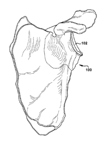
Fig. 1 depicts a portion of the external surface of a (left) scapula 100,
viewed from the anterior direction toward the posterior direction, which is an
example of a possible patient tissue use environment for the described
systems,
apparatuses, and methods. The humerus (not shown) of a patient attaches to the
scapula 100 at the glenoid fossa 102 to form the ball-and-socket shoulder
joint.
The glenoid fossa 102 is shown in greater detail in Fig. 2, a view taken
orthogonally from Fig. 1. The term "lateral" is used herein to refer to a
direction
which, in Fig. 2, lies substantially within the plane of the drawing as shown
by
directional arrow 104 and includes all of the superior, inferior, anterior,
and
posterior directions. The term "longitudinal" is used herein to refer to a
direction
defined perpendicular to the plane created by directional arrow 104, with the
longitudinal direction being substantially into and out of the plane of the
drawing
in Fig. 2 and representing the proximal (toward the medial plane of the body)
and
distal (out from the body) directions, respectively.
Fig. 3 is a partial perspective view of the scapula 100, with particular
emphasis on the glenoid fossa 102. For the sake of discussion, the glenoid
fossa 102 itself is referred to herein as a primary patient tissue area 108.
That is,
the primary patient tissue area 108 is a portion which directly receives an
implant
and/or is otherwise directly affected by a surgical procedure. In contrast, a
;A 02816331 2013-04-26
WO 2012/058349
PCT/US2011/057949
-8-
secondary patient tissue area 110 is one which does not receive an implant
and/or
is not directly affected by a surgical procedure. In Fig. 3, the secondary
patient
tissue area 110 borders the primary patient tissue area 108 and is
anatomically
differentiated from the primary patient tissue area (i.e., the glenoid fossa
102) by
the glenoid rim, indicated approximately by differentiation line 312. Here,
the
differentiation line 312 generally indicates an arbitrary (i.e., "depending on
individual discretion") position along the continuous transition between the
glenoid
fossa 102 and the supporting structures (e.g., the glenoid rim, the glenoid
neck, the
base of the coracoid, and/or the glenoid wall). However, regardless of the
precise
position of the differentiation line 312 for a particular application of the
present
invention, one of ordinary skill in the art should be able to distinguish
between a
primary patient tissue area 108 (one which is directly affected by a surgical
procedure) and a secondary patient tissue area 110 (one which is incidentally
affected by a surgical procedure, if at all) for the purposes of the present
invention.
A distinction is made herein between the primary and secondary patient
tissue areas 108 and 110 because the present invention relates to the
association of
at least one landmark with at least one of the primary and secondary patient
tissue
areas. The term "landmark" is used herein to indicate any guiding aid which
serves as a detectable indicator of a particular position on a "marked"
substrate
(here, the patient tissue). The landmarks discussed with respect to the
present
invention are presumed to be affixed or otherwise rigidly associated with a
particular patient tissue so that a user can confidently maintain a sense of
physical
and/or visual orientation within the operative field. Suitable landmarks may
include, but are not limited to, visual "written" marks (e.g., a thin layer of
a
substance left behind after contact with a crayon, surgical pen, or the like),
other
written marks outside the visual spectrum (e.g., a UV-fluorescent paint),
guide
pins, fasteners (e.g., screws, nails, staples, or the like), radioactive tags,
bovie
cautery burn marks, metallic or nonmetallic devices attached to the desired
landmark site (e.g., a rivet, tack, or the like), or even modifications of the
patient
tissue itself (e.g., notches, inscribed lines, drill holes, or the like).
Depiction of one
type of landmark 114 in the Figures herein merely serves as an example and
does
;A 02816337 2013-04-26
WO 2012/058349
PCT/US2011/057949
-9-
not preclude the use of a different type of landmark, even in a similar use
environment to those depicted, for a particular application of the present
invention.
Three landmarks 114a, 114b, and 114c are shown in Fig. 3 as having been
associated with the primary and secondary patient tissue areas 108 and 110.
Landmarks 11 4a and 114b are three-dimensional pins which have been inserted
into the primary and secondary patient tissue areas 108 and 110, respectively.
Landmark 114c is a visible two-dimensional cross mark on the secondary patient
tissue area 110.
Any landmark 114, regardless of type, will be located at a predetermined
marking location with respect to the primary and/or secondary patient tissue
areas 108 and 110. A three-dimensional landmark, like the marking pins shown
as
landmarks 114a and 114b in Fig. 3, may also have a predetermined marking
trajectory which, like the marking location, holds some significance for the
user.
For example, landmarks 114a and 114b do not have parallel trajectories as
depicted
in Fig. 3. While the marking trajectory of a three-dimensional landmark 114
(i.e., one protruding noticeably from the patient tissue surface) might have
no
significance, the following discussion presumes, for ease of reference, that
the
marking trajectory of the three-dimensional landmark is intentionally provided
and
is held substantively constant during the tenure of the landmark at the
marking
location.
It is contemplated that a landmark 114 will normally be rigidly affixed to a
particular marking location on the primary or secondary patient tissue area
108
or 110 in order to serve as a reliable lodestar for the user. However, in
certain
situations, the marking location of the landmark 114 may move (as seen from an
outside point of reference) after placement, of its own accord, by action of a
user,
or by action of the substrate patient tissue, and these situations do not pass
out of
the domain of the present invention merely by virtue of such intentional or
unintentional post-placement landmark motion.
The marking location and marking trajectory of each landmark 114 are
predetermined by a user before the landmark is associated with the patient
tissue.
This predetermination may occur intraoperatively, as the user is able to
directly see
the condition of the surgical site. However, it is contemplated that a
-10-
predetermination of the desired marking location and desired marking
trajectory
for each landmark 114 could be accomplished preoperatively, with reference to
preoperative imaging of the patient tissue, For example, a system similar to
that of
U.S. Patent Application No. 13/282,550, filed October 27, 2011, titled "System
of Preoperative Planning and Provision of Patient-Specific Surgical Aids" and
U.S. Provisional Patent Application No. 61/408,392, filed October 29, 2010 and
titled "System of Preoperative Planning and Provision of Patient-Specific
Surgical
Aids", or any suitable preoperative planning system could be used. In this
manner,
a user can create a patient tissue model for observation, manipulation,
rehearsal,
or any other preoperative tasks.
The term "model" is used herein to indicate a replica or copy of a physical
item, at any relative scale and represented in any medium, physical or
virtual. The
patient tissue model may be a total or partial model of a subject patient
tissue, and
may be created in any suitable manner. For example, and as presumed in the
below description, the patient tissue model may be based upon computer
tomography ("CT") data imported into a computer aided drafting ("CAD") system.
Additionally or alternatively, the patient tissue model may be based upon
digital or
analog radiography, magnetic resonance imaging, or any other suitable imaging
means. The patient tissue model will generally be displayed for the user to
review
and manipulate preoperatively, such as through the use of a computer or other
graphical workstation interface.
Once the user is satisfied with her preoperative planning tasks, virtual
landmarks may be virtually placed on the patient tissue model. In order to
transfer
those virtual landmarks to the physical world for intra-operative use, a
patient-specific apparatus (shown in Fig. 4 as a guide 416) may be at least
partially
customized responsive to preoperative imaging of the patient tissue.
Accordingly,
at least a part of the guide 416 is a patient-specific, single-use, bespoke
component
suited only for use at the indicated surgical site, though one of ordinary
skill in the
art could create a guide (not shown) which uses a patient-specific
"disposable"
structure connected to a stock, generic "reusable" carrier.
CA 2816337 2018-02-23
;A 02816337 2013-04-26
WO 2012/058349
PCT/US2011/057949
-11-
The patient's name, identification number, surgeon's name, and/or any
other desired identifier may be molded into, printed on, attached to, or
otherwise
associated with the guide 416 in a legible manner. The guide 416 may be made
by
any suitable method such as, but not limited to, selective laser sintering
("SLS"),
fused deposition modeling ("FDM"), stereolithography ("SI,A"), laminated
object
manufacturing ("LOM"), electron beam melting ("EBM"), 3-dimensional printing
("3DP"), contour milling, computer numeric control ("CNC"), other rapid
prototyping methods, or any other desired manufacturing process.
The guide 416 assists the user by associating a plurality of landmarks 114
with patient tissue, each landmark being associated with the patient tissue in
at
least one of a predetermined marking location and a predetermined marking
trajectory. As depicted in Figs. 4-7, a base 418 may have a lower base surface
520
contoured to mate with both the primary and secondary patient tissue areas 108
and 110 in a preselected relative orientation. The term "mate is used herein
to
indicate a relationship in which the contours of two structures are at least
partially
matched or coordinated in at least two dimensions. In the described mating
relationship depicted in Figs. 4-7 as an example of the present invention, the
lower
base surface 520 mates or nests into contact with the surfaces of both the
primary
and secondary patient tissue areas 108 and 110 to provide the guide 416 with
at
least one of location and stabilization assistance with respect to the patient
tissue.
'the lower base surface 520 is spaced apart from an upper base surface 422
by a base body 424. A plurality of base apertures 526 extend between the upper
and lower base surfaces 422 and 520 through the base body 424. The base
apertures 526 are shown here as extending substantially longitudinally through
the
base body 424, but may have any desired orientation with respect to the base
418.
A plurality of guiding bosses 428 may protrude from the base 418 in certain
configurations of the present invention. As shown in the Figures, the guiding
bosses 428 protrude substantially longitudinally outward from the upper base
surface 422, but the guiding bosses may have any desired orientation with
respect
to the base 418. Each guiding boss 428 has a guiding bore 428 extending
therethrough. Each guiding bore 428 extends collinearly with a corresponding
base aperture 526 to permit insertion of a landmark 114 through the guide 416.
;A 02816337 2013-04-26
WO 2012/058349
PCT/US2011/057949
-12-
The term "insertion of a landmark through" is intended to encompass both a
physical feeding of a three-dimensional landmark itself through the indicated
structure for affixation to the underlying patient tissue (e.g., by
penetration), as
well as the temporary introduction of a marking device (e.g., a pen, bovie,
rasp,
other marking actuator or substance dispenser, or the like) through the
indicated
structure for affixation of a two-dimensional landmark 114 directly onto the
patient
tissue.
Each guiding bore 430 and corresponding base aperture 526 cooperatively
defines at least one of the predetermined marking location and the
predetermined
marking trajectory (shown in Fig. 5 by trajectory lines 532) for an associated
landmark 114. In the embodiment shown in Figs. 4-7, at least one landmark 114
is
guided by the guide 416 into engagement with a marking location in the primary
patient tissue area 108 (via the rightmost guiding bore 430 in the orientation
of
Fig. 5) and at least one landmark is guided by the guide into engagement with
a
marking location in the secondary patient tissue area 110 (via the leftmost
guiding
bore 430 in the orientation of Fig. 5),
Fig. 6 depicts the guide 416 of Fig. 4 in a slightly different orientation in
space, such that the contour of the lower base surface 520 may be seen in more
detail. In the orientation of Fig. 6, the leftmost portion of the lower base
surface 520 appears relatively broad and flat and is configured to mate with
the
surface of the glenoid fossa 102 (i.e., the primary patient tissue area 108
here).
The differentiation line 312 from Fig. 3 is shown "ghosted" into Fig. 6 and
extends
somewhat into and out of the plane of the page due to the camber of the
depiction
in Fig. 6. With the addition of the differentiation line 312, it can be
clearly seen
that the rightmost portion of the lower base surface 520 does not mate with
the
primary patient tissue area 108, but instead dips sharply downward relative to
the
rest of the lower base surface to mate with the bordering secondary patient
tissue
area 110. Particularly when there is a "lip" or "rim" between the primary and
secondary patient tissue areas 108 and 110, such as with the glenoid fossa
102, the
ability of the lower base surface 520 to concurrently nest with both of these
patient
tissue areas may be helpful to the user in quickly and securely nestling the
guide 416 down into the desired mating relationship with the patient tissue.
;A 02816337 2013-04-26
WO 2012/058349
PCT/US2011/057949
-13-
In Fig. 7, the upper base surface 422 and protruding guiding bosses 428 can
be seen in detail. Particularly when a marking trajectory (such as that shown
by
trajectory lines 532) is defined by the base aperture 526, with or without the
assistance of a guiding bore 430, it may be helpful for the guiding bosses 428
to
provide a longer guiding structure for the inserted landmark 114. In other
words,
an elongate landmark 114 might precess within a relatively short base
aperture 526, but the presence of the guiding boss 428 can support and
stabilize
insertion of the landmark to better guide the landmark along the predetermined
marking trajectory.
Figs. 8-10 depict the generation of a suitable design for an example
guide 416 during a preoperative planning procedure. Figs. 8-10 are example
user
views of a computer program for implementing a method using the present
invention, with a perspective view on the left side of each Figure and
coronal,
sagittal (looking distally from underneath the perspective view, as shown),
and
transverse views, respectively, from top to bottom on the right side of each
Figure.
In Fig. 8, a stock glenoid implant 834 is shown associated with a glenoid
fossa 102 of a patient's scapula 100, embodied in a model produced using
preoperative imaging. The glenoid implant 834 may be virtually placed as
desired
on the scapula 100 by the user, or may be automatically placed by the computer
program with or without a final check/adjustment by the user. The glenoid
implant 834 appears to overlap with the glenoid fossa 102 in particularly the
coronal (top right) and transverse (bottom right) views of Fig. 8, but this
overlap
(when present) is acceptable at the planning stage of Fig. 8 since the
physical
glenoid fossa 102 will be prepared via machining or other alteration(s) as
desired
during installation of the physical glenoid implant 834 at the surgical site,
and this
overlap will be corrected by removal of the interfering patient tissue. In
fact,
relatively precisely placed landmarks 114 are useful during many surgeries
because the site preparation procedure commonly erodes, moves, or destroys
natural landmarks which otherwise would help the user with placement or
orientation during the surgical procedure.
Implant stem 836, visible in cross-section in the coronal and transverse
portions of Fig. 8, is a tubular anchoring extension from the underside of the
;A 02816337 2013-04-26
WO 2012/058349
PCT/US2011/057949
-14-
glenoid implant 834 which is inserted into the patient tissue of the glenoid
fossa 102 during use. One consideration that a user may have during placement
of
the glenoid implant 834 using the computer program shown in Figs. 8-10 is
being
able to locate the implant stem 836 in a solid portion of the patient's
scapula 100.
Another, similar consideration is the location of screws or other fasteners
(not
shown) which are commonly used to secure the glenoid implant 834 to the
glenoid
fossa 102. The user will want to ensure that the proper locations and
trajectories
are chosen for affixation of the selected fasteners into relatively robust
areas of the
patient's scapula 100. Once the glenoid implant 834 (including the implant
stem 836 and the associated fasteners) has been virtually placed as desired
into a
final installation position, the user can decide where to place one or more
landmarks 114, using the guide 416 and relatively early in the surgical
process, to
facilitate later tasks during the surgery. For example, the user of the Fig. 8
example may wish to place a guide pin as a landmark 114 at each of the marking
locations 838 indicated by cross marks. As shown in Fig. 8, one marking
location 838a is placed in the primary patient tissue area 108 and another
marking
location 838b is placed in the secondary patient tissue area 110. For certain
surgical procedures, both of these locations may be marked as desired for bone
preparation and final implant positioning. Landmarks 114 may be placed before
the patient tissue is altered or modified, with the marking locations 838
corresponding to each landmark being specified during the preoperative
surgical
planning and/or simulation, or in any other suitable manner.
For example, a guide pin is displayed as a three-dimensional landmark 114
at the marking location 838a spaced apart from the glenoid implant 834 over
the
image of scapula 100 in Fig. 8, while an aperture or cavity formed in the
scapula is
shown as a two-dimensional landmark 838b (i.e., represented by a cross mark
when seen from above or below) corresponding to a central portion of the
glenoid
implant in Fig. 8. In fact, the "negative" aperture-type landmark 838b of Fig.
8 is
configured to receive a device shaft implant stem 836 of the glenoid implant
834,
which helps to locate and stabilize the glenoid implant with respect to the
scapula 100. One of ordinary skill in the art would readily be able to instead
provide a "positive" pin- or shaft-type landmark (not shown) protruding from
the
;A 02816337 2013-04-26
WO 2012/058349
PCT/US2011/057949
-15-
scapula 100 and adapted to be received in a cavity (not shown) of another type
of
device, in an axle-type manner.
Optionally, the marking locations 838 may be chosen to comport to
common landmark 114 placements to facilitate use of standard tools (not shown)
with the guide 418. For example, two marking locations 838 may be provided to
indicate a line bisecting the scapula 100 for that patient so that the user
has a
standardized reference line. In this example, then, generic surgical tools
which use
the scapula-bisecting line as a landmark in every patient will encounter a
patient
tissue which has been standardized, through use of personalized landmark 114
placements, to meet a universal expectation of the user. In other words, and
more
generally, the marking location 838 choices can be set for a particular
patient tissue
in order to compensate for any peculiarities of that patient tissue and
accordingly
provide the user with a surgical site that may be addressed using stock (i.e.,
not
patient-specific) tools and/or techniques. This type of "universal
registration" may
be especially helpful in automation-assisted surgeries.
In Fig. 9, a user view of the computer program shows a guide blank 940
superimposed on the scapula 100. Since the guide 418 will be used to place the
landmark(s) 114 before the surgical site is altered, the lower base surface
422
should be designed as a mirror image of the surface of the glenoid fossa 102,
to
mate with the primary and secondary patient tissue areas 108 and 110 as
desired.
The resolution of the preoperative imaging scans and the available precision
of the
chosen manufacturing method for the guide 416 will determine how precisely
this
mating is accomplished. As is apparent in Fig. 9, the guide blank 940 contacts
and
mates with both the primary and secondary tissue areas 108 and 110. The
marking
locations 838a and 838b identified in the view of Fig. 8 are represented as a
small
circle and a cross mark, respectively, on the guide blank 940 in Fig. 9.
Turning to Fig. 10, the areas of the guide blank 940 which overlap with the
patient tissue of the scapula 100 have been removed by the computer program,
generating the complex contour of the lower base surface 520 (most apparent in
the
coronal view). Additionally, base apertures 526 and corresponding guiding
bosses 428 with guiding bores 430 have been placed at the desired marking
locations 838, the base apertures and guiding bores being collinear to
cooperatively
;A 02816337 2013-04-26
WO 2012/058349
PCT/US2011/057949
-16-
define desired marking trajectory lines 532. Once the preoperative planning
has
been accomplished, through user input and/or automatic programming, the design
of the guide 416 is complete and the guide can be manufactured and prepared
for
use (e.g., mechanically or chemically cleaned, cured, sterilized, or the like)
using
any suitable process(es).
Figs. 11-15 depict various options for configurations of the guide 416.
These different configurations, along with other (non-depicted)
configurations, of
guides 416 can be selected/designed and used by one of ordinary skill in the
art to
provide desired landmark-placement properties for different patient tissues.
Structures of Figs. 11-15 that are the same as or similar to those described
with
reference to Figs. 4-10 have the same reference numbers. As with all alternate
configurations shown and described herein, description of common elements and
operation similar to those in previously described configurations will be
omitted,
for clarity. In the second configuration, shown in Figs. 11-12, the guide 416
is
relatively large compared to that of Figs. 4-10 (although the Figures herein
are not
drawn to scale). As can be seen in the comparative views of Figs. 10 and 12,
the
guide 416 of the second configuration (shown in situ in Fig. 12) covers more
of the
glenoid fossa 102 than does the guide 416 of the first configuration (shown in
situ(
in Fig. 10). Additionally, the leftmost base aperture 526 and guiding bore 430
(as seen in the orientation of Fig. 12) is located substantially in an
anterior portion
of the secondary patient tissue area 110 for the second configuration, while
the
corresponding structures in the first configuration are located substantially
more
superiorly within the secondary patient tissue area 110.
The third configuration of the guide 416, shown in Fig. 13, seems similar to
that of Figs. 11-12, with the addition of at least one marking notch 1342. The
marking notch(es) 1342 may be useful for guiding contact with the patient
tissue
for placing a two-dimensional (e.g., via a pen, bovie, crayon, or other
marking
device) or three-dimensional landmark 114 at a desired marking location 838,
particularly if achieving a precise marking trajectory is not important.
Fig. 14 shows a fourth configuration of the guide 416 which includes
features from several of the previously defined configurations. The guide 416
of
Fig. 14 has a blockier shape than that of Fig. 13, which may provide
efficiencies in
;A 02816337 2013-04-26
WO 2012/058349
PCT/US2011/057949
-17-
design and/or fabrication. That is, the blockier shape of the fourth
configuration
guide 416 may be better suited to a design scheme involving the provision of a
generic guide blank 940. In contrast, the contoured upper base surface 422
exhibited by the third configuration guide 416 requires more extensive
smoothing
and shaping operations (during the virtual modeling of the computer program
and/or during physical manufacture), particularly if the base body 424 is
configured to have a substantially uniform thickness by some degree of
mirroring
of the lower base surface 520 (dictated by the glenoid fossa 102) with the
contour
of the upper base surface 422. In the fourth configuration of Fig. 14, the
marking
notch 1342 of the guide 416 is somewhat rounded and may be operative to assist
with placement of a three-dimensional landmark 114, such as a guide pin, at
the
marking location 838, optionally with some degree of imposed marking
trajectory.
A fifth configuration of the guide 416 is shown in Fig. 15. The guide 416
shown in Fig. 15 is similar to that shown in Fig. 16, with a guiding boss 428
in
place of the marking notch 1342, and with the addition of a handling boss
1544.
The handling boss 1544 protrudes from the base 418 and is configured for
manipulation by the user to at least partially control a position of the guide
416.
Sometimes the available maneuvering space in a surgical field is relatively
restricted, and it may be useful for a forceps, socket driver (perhaps with a
frictional fit or other feature to accept the handling boss 1544), Kocher
tool,
hemostat, or other user-manipulated handling tool (not shown) to selectively
interact with the handling boss to hold the guide 416 steady and/or to move
the
guide 416 to a desired position. One or more features, such as indents,
apertures,
cavities, lugs, undercuts, or any other suitable structures could be provided
to the
handling boss 1544 to facilitate gripping of the guide 416 by any handling
tool, in
general, and/or by a particular handling tool (perhaps one chosen in
conjunction
with the chosen glenoid implant 834). Optionally, the handling boss 1544 may
also be a guiding boss 428. However, in some situations it will be desirable
for
each of the guiding bosses 428 to be accessible for landmark 114 placement at
the
same time that a handling tool is engaged with the handling boss 1544, so the
handling boss could be a separate structure in those situations.
;A 02816337 2013-04-26
WO 2012/058349
PCT/US2011/057949
-18-
Regardless of the specific configuration chosen for a particular patient, the
guide 416 will generally be used relatively early in the surgical procedure.
The
guide 416 has a base 418 at least partially customized (e.g., custom-
manufactured
and/or custom-configured) responsive to preoperative imagining of the patient
tissue. The base 418 of the guide 416 is mated with at least one of the
primary and
secondary patient tissue areas 108 and 110 in a preselected relative
orientation.
When the base 418 is mated with both the primary and secondary patient tissue
areas 108 and 110, the mating may be concurrent for both those patient tissue
areas.
At least one landmark 114 is guided by the guide 416 to a marking
location 838 in the primary patient tissue area 108 and fixed to the primary
patient
tissue area 108 in at least one of a predetermined marking location 838 and an
predetermined marking trajectory, such as by passing of the landmark 114 along
a
marking notch 1342 or through a base aperture 526 (optionally with the
assistance
of a guiding bore 430). Optionally, at least one additional landmark 114 may
be
guided by the guide 416 to a marking location 838 in the secondary patient
tissue
area 110 and fixed to the secondary patient tissue area 110 in at least one of
a
predetermined marking location 838 and an predetermined marking trajectory,
such as by passing of the additional landmark along a marking notch 1342 or
through a base aperture 526 (optionally with the assistance of a guiding bore
430).
Once the desired number of landmarks 114 are affixed to the primary
and/or secondary patient tissue areas 108 and 110, the guide 416 is removed
from
the surgical site in any suitable manner, optionally with the assistance of a
handling boss 1544. When at least one landmark 114 is a guide pin or other
elongate three-dimensional structure, the guide pin may deflect, if needed, to
allow
the guide 416 to be lifted longitudinally off the protruding end guide pin.
Alternately, the guide 416 may include at least one frangible portion to allow
substantially laterally-oriented removal of the guide 416 from around the
guide
pin. As another example, the guide 416 could include one or more slots
(not shown) to allow removal of the guide by sliding the guide sideways away
from the guide pin.
-19-
Regardless of the manner in which the guide 416 is removed from the
primary and secondary patient tissue areas 108 and 110, the landmark(s) 114
remain behind and the surgical site attains a configuration akin to that shown
in
Fig. 3. The user can then proceed with the surgical procedure with confidence
that
the landmark(s) 114 are substantially located as configured in the
preoperative
plan. The patient tissue at the primary patient tissue area 108 can be altered
and
the landmark(s) 114 that remain as placed using the guide 416 can be used to
orient
such alteration or for any other surgical task. For example, a prosthetic
implant
(such as the glenoid implant 834, when the patient tissue is a scapula 100)
may be
placed, optionally with the assistance of another patient-specific guide, such
as that
disclosed in U.S. Patent Application No. 13/282,495, filed October 27, 2011,
titled "System and Method for Assisting with Attachment of a Stock Implant to
a Patient Tissue" and U.S. Provisional Patent Application No. 61/408,324,
filed
October 29, 2010 and titled "System and Method for Assisting with Attachment
of a Stock Implant to a Patient Tissue".
Figs. 16-24 depict a guide 416' according to certain aspects of a second
embodiment of the present invention. The guide 416' of Figs. 16-24 is similar
to
the guide 416 of Figs. 1-15 and therefore, structures of Figs. 16-24 that are
the
same as or similar to those described with reference to Figs. 1-15 have the
same
reference numbers with the addition of a "prime" mark. Description of common
elements and operation similar to those in the previously described first
embodiment will not be repeated with respect to the second embodiment.
Fig. 16 depicts an example use environment for the guide 416' of the
second embodiment. Directional arrow 104' indicates the superior/inferior and
anterior/posterior directions. The body of ischium, body of ilium, and body of
pubis are shown generally at 1646, 1648, and 1650, respectively. The
acetabulum
1652 (here, the primary patient tissue area 108'), which is formed in part by
these
three bodies 1646, 1648, and 1650, has a recessed acetabular fossa 1654 and is
surrounded by an acetabular margin 1656 (here, the secondary patient tissue
area 110', shown approximately in Fig. 16 as being outside the dashed
differentiation line 312').
CA 2816337 2018-02-23
;A 02816337 2013-04-26
WO 2012/058349
PCT/US2011/057949
-20-
In accordance with the present invention. Fig. 17 depicts a guide 416'
including a base 418', a stem 1758, and at least one spacing arm 1760. The
base 418' has a lower base surface 520' (shown partially in phantom line in
Fig. 17) spaced apart from an upper base surface 422' by a base body 424'. The
lower base surface 520' is contoured to mate with the acetabulum 1652 in a
preselected relative orientation thereto. The base 418' may include a base
guide
aperture 1762 configured to guide placement of a landmark 114' inserted at
least
partially therethrough in at least one of a predetemiined marking location and
a
predetermined marking trajectory, the marking location being in the primary
patient tissue area 108'.
The stem 1758 has longitudinally separated first and second stem
ends 1764 and 1766, respectively. The first stem end 1764 is attached directly
to
the base 418', either permanently or removably. The stem 1758 extends
longitudinally upward from the base 418' (substantially out of the plane of
the
paper, in Fig. 17).
At least one spacing arm 1760 (two shown) is attached directly to the
second stem end 1766, either permanently or removably. Each spacing arm 1760
is longitudinally spaced from the base 418' and has an arm guide aperture 1768
laterally spaced from the stem 1758. The arm guide aperture 1768 is configured
to
guide placement of a landmark (not shown in this Figure) inserted at least
partially
therethrough at a predetermined landmark trajectory (represented by trajectory
line 532'). The spacing arm(s) 1760 are shown in the Figures as extending
orthogonally from the stem 1758 at the second stem end 1766, in order to place
landmarks 114' in the acetabular margin 1656 (the secondary patient tissue
area 110') as will be discussed below. The spacing aini(s) 1760 could extend
at
any suitable angle or position from the stem 1758, or could even be smoothly
formed as a single integral piece with the stem. In the latter event, the
second stem
end 1766 may not be clearly delineated from the spacing arm(s) 1760.
The stem 1758 and spacing arm(s) 1760 could have any of a myriad of
configurations, depending upon the application of the present invention. A
spacing
arm 1760 is used herein to indicate any structure which is located at some
distance
from base 418' contacting a primary patient tissue area 108', and the spacing
arm
;A 02816337 2013-04-26
WO 2012/058349
PCT/US2011/057949
includes structure which can guide a landmark 114' to a secondary patient
tissue
area 110'. A stem 1758 is used herein to indicate any structure which extends
between and connects the base 418' and at least one spacing arm 1760.
The guide 416' may be at least partially customized responsive to
preoperative imaging of the patient tissue. For example, the lower base
surface 520' of the base 418' could be at least partially configured through
the use
of computer tomography ("CT") data of the patient tissue to have a
longitudinally
downward-protruding portion corresponding to the acetabular fossa 1656.
Additionally or alternatively, the lower base surface 520' could be at least
partially
configured through use of patient scans including digital or analog
radiography,
magnetic resonance imaging, or any other suitable imaging means. The patient
tissue preoperative images are optionally displayed for review and
manipulation
before/during configuration of the lower base surface 520', such as through
the use
of a computer or other graphical workstation interface described above with
reference to the first embodiment of the present invention. The configuration
of
the lower base surface 520' is described herein as being perfoimed using
three-dimensional images; however, one or more two-dimensional depictions of
the patient tissue may also or instead be consulted during configuration of
the
lower base surface 520' or any other preoperatively configured structure
herein.
The lower base surface 520' is configured to mate with a primary patient
tissue surface 108', as will be discussed below. In the described mating
relationship, the lower base surface 520' mates or nests into contact with the
surface of the acetabulum 1652 to provide the base 418' with at least one of
location and stabilization assistance with respect to the patient tissue.
Though the
lower base surface 520' is shown herein as covering a substantial portion of
the
acetabulum 1652, the lower base surface 520' may contact any suitable portion
of
the primary patient tissue area 108' sufficient to stabilize the guide 416' in
a
desired manner.
Figs. 19-21 depict a guide 416' which is a second configuration of the
embodiment of the present invention in the second embodiment of Fig. 17. The
guide 416' of Figs. 19-21 mainly differs from the guide 416' of Fig. 17 in the
provision of at least one outrigger 1770 as an extension of the base 418'. The
underside (tissue-contacting) surface of each outrigger 1770 forms a portion
of the
lower base surface 520' and is accordingly contoured to mate with a portion of
the
acetabulum 1652' (the primary patient tissue surface 108') and the surrounding
secondary patient tissue surface 110'. For example, and as shown in the bottom
view of Fig. 20, the outrigger(s) 1770 may extend laterally beyond the
remaining
acetabular-contacting portion of the base 418'. The outrigger(s) 1770 shown in
Figs. 19-21 may contact, or even hook over, the acetabular margin 1656' to
assist
with positioning and/or stabilizing of the guide 416' as shown in the cross-
sectional side view of Fig. 21. The side view of Fig. 19 and the bottom view
of
Fig. 20 also clearly show a protrusion 1972 formed by the contour of the lower
base surface 520' and shaped to mate with the acetabular fossa 1654 (shown in
Fig. 16). Because each patient's bone structure is unique, at least a portion
of the
guide 416' (e.g., the outriggers 1770 and lower base surface 520') is
customized
responsive to preoperative imaging of the patient tissue.
Fig. 21 also shows an orthopedic guidewire 2174 acting as a landmark.
One example of a suitable guidewire 2174 is disclosed in U.S. Patent
Application No. 13/178,324, filed July 7, 2011, titled "Method and Apparatus
for
Providing a Relative Location Indication During a Surgical Procedure" and
U.S. Provisional Patent Application Serial No. 61/362,722, filed July 9, 2010,
and titled "Method and Apparatus for Providing a Relative Location Indication
During a Surgical Procedure". Additionally, Fig. 21 shows a pair of
conventionally-
configured guide pins acting as three-dimensional landmarks 114', though any
suitable number, combination, and/or types of two- or three-dimensional
landmarks
114' and/or guidewires may be provided for a particular use environment of the
present
invention, and may be associated with either or both of the base guide
aperture(s)
762' and the arm guide aperture(s) 1768'.
At least a portion of the guidcwire 2174 is insertable through the base guide
aperture 1762' and into the underlying acetabulum 1652' when the guide 416' is
mated with the patient tissue in the preselected relative orientation.
Similarly, at
least a portion of each of the landmarks 114' is insertable through the arm
guide
CA 2816337 2018-02-23
;A 02816337 2013-04-26
WO 2012/058349
PCT/US2011/057949
-23-
aperture 1768' and into the underlying second patient tissue area 110', shown
here
as being located just beyond an acetabular margin 1656', when the guide 416'
is
mated with the patient tissue in the preselected relative orientation.
A distal end 2176 of the landmark 114' or guidewire 2174 is configured to
remain inserted into the patient tissue when the guide 416' is removed from
the
patient tissue. It is contemplated that the base guide aperture 1762' and/or
arm
guide aperture 1768' will be sized to pass over the respective landmark 114'
or
guidewire 2174, leaving these guiding landmark structures in place such as in
the
configuration shown in Fig. 18. The landmark(s) 114' and/or guidewire(s) 2174
may remain in place for as long as the user desires, though normally will be
removed from the patient's body before the surgical procedure is concluded.
The
landmark(s) 114' and/or guidewire(s) 2174 also may be used for any reason in
conjunction with any type or number of processes, during or after the surgical
procedure in which they were installed. A common guiding function for a
landmark 114' or guidewire 2174 is to guide the positioning of another
structure,
either directly (via contact) or indirectly (spaced apart from the guided
structure).
Figs. 22-24 depict a guide 416' which is a third configuration of the second
embodiment of the present invention and combines features of both the previous
configurations of the second embodiment, as well as some features of the
guide 416 of the first embodiment. The guide 416' of Figs. 22-24 has a very
complex base structure with a bifurcated lower base surface 520' which
concurrently contacts at least a portion of a primary patient tissue area 108'
(i.e., contacts an acetabulum 1652 with the leftmost portion of the lower base
surface, as shown in the orientation of Fig. 23) and at least a portion of a
secondary
patient tissue area 110' (i.e., contacts an acetabular margin 1656 with the
rightmost
portion of the lower base surface, as shown in the orientation of Fig. 23). A
plurality of guiding bosses 428' are provided to the guide 416' of Figs. 22-
24,
including two guiding bosses located on an extended portion 2278 of the base
body 424' to place landmarks 114' in the secondary patient tissue area 110'
and
one guiding boss located on a central portion 2280 of the base body to place a
landmark 114' in the primary patient tissue area 108'.
;A 02816337 2013-04-26
WO 2012/058349
PCT/US2011/057949
-24-
The guiding boss 428' located on the central portion 2280 of the base
body 424' is noticeably longer than the other guiding bosses, and may serve
several functions for the guide 416'. The guiding boss 428' located on the
central
portion 2280 of the base body 424' may guide a landmark 1114 through a guiding
bore 430' thereof; may guide a rasp, drill, or other tissue modification tool
(not
shown) therethrough, optionally providing a "stop" function to limit insertion
of
the tissue modification tool into the underlying patient tissue; and/or may
serve as
a handling boss for user manipulation by hand and/or with a handling tool.
Figs. 25-29 depict a guide 416" according to a third embodiment of the
present invention. The guide 416" of Figs. 25-29 is similar to the guide 416
of
Figs. 1-15 and therefore, structures of Figs. 25-29 that are the same as or
similar to
those described with reference to Figs. 1-15 have the same reference numbers
with
the addition of a double "prime" mark. Description of common elements and
operation similar to those in the previously described first and second
embodiments will not be repeated with respect to the second embodiment.
The guide 416" of the third embodiment of the present invention may be
used both for associating a plurality of landmarks 114" with a patient tissue
in at
least one of a predetermined marking location and a predetermined marking
trajectory, and for guiding the removal of a predetermined amount of resection
patient tissue and rearrangement of a remaining patient tissue, as will be
described.
One example of a potential use environment for the guide 416" of the third
embodiment is in conjunction with a surgical procedure to correct a congenital
or
acquired orthopedic malunion.
Figs. 25-26 depict a guide 416" in a first configuration in front and side
views, respectively, in a use environment of a patient tissue footling at
least a
portion of a patient tissue such as, but not limited to, a femur, humerus,
radius,
ulna, tibia, fibula, metatarsal, phalange, another type of long bone shaft, a
flat bone
such as the mandible, a facial bone, a scapula body, a bone of the wrist or
ankle, or
any other patient tissue. In these Figures, the primary patient tissue area
108" is a
resection patient tissue 108" (shaded in Fig. 26) and the secondary patient
tissue
area 110" is a remaining patient tissue 110". The guide 416" of Figs. 25-26 is
;A 02816337 2013-04-26
WO 2012/058349
PCT/US2011/057949
-25-
configured to contact the resection patient tissue 108" and the remaining
patient
tissue 110" and to guide surgical contact with the patient tissue.
The guide 416" of Figs. 25-26 has a base 418" having a lower base
surface 520" contoured to mate with both the resection and remaining patient
tissues 108" and 110" in a preselected relative orientation. The lower base
surface 520" is spaced apart from an upper base surface 422" by a base
body 424", as shown in Fig. 26.
As shown in Figs. 25-26, at least one base aperture 526" (two shown here)
guides a landmark 114" into contact with the underlying tissue surface in at
least
one of a predetermined marking location 838" and a predetermined marking
trajectory. A plurality of first guide cutting guide apertures 2582 extend
between
the upper and lower base surfaces 422" and 520" through the base body 424" to
permit penetration of at least one cutting tool (shown schematically at 2584)
through the guide 416". The cutting guide apertures 2582 each define at least
one
cutting plane location and cutting plane orientation for the cutting tool 2584
to
make at least one resection cut into the patient tissue.
More specifically, the guide 416" is configured to cut the resection patient
tissue 108" for removal from the remaining patient tissue 110". The resection
patient tissue 108" is shaded in the Figures, and the cutting plane locations
and
orientations are chosen to correspond to the borders of the resection patient
tissue.
Because the resection patient tissue 108" in the Figures is located
intermediate two
areas of remaining patient tissue 110", at least two cutting plane locations
and
orientations are needed to excise the resection patient tissue 108". If there
were no
remaining patient tissue 110" to one side (e.g., the topmost side in the
orientation
of Fig. 26), only one cutting plane location and orientation would be needed
to
sever the resection patient tissue 108". However, the latter situation would
not be
a true case of correction of a malunion, but merely an amputation. A
patient-specific guide 416" could be produced and used for an amputation if
desired. However, though not excluding an amputation situation from
application
of a guide 416", this description presumes for ease of discussion that at
least two
cuts will be made to excise an area of resection patient tissue 108" from a
surrounding area of remaining patient tissue 110".
;A 02816337 2013-04-26
WO 2012/058349
PCT/US2011/057949
-26-
Optionally, and as shown in Fig. 26, at least one guiding boss 428" may
protrude from the upper base surface 422" in association with at least one of
the
base apertures 526" and the cutting guide apertures 2582, as shown in Fig. 26.
The guiding bosses 428" shown in Fig. 26 may be helpful in avoiding precession
of the cutting tools 2584 and thereby assist in guiding the cutting tools to
make
accurate cuts according to the preoperative plan embodied in the guide 416".
Once the resection patient tissue 108" has been cut and removed from the
remaining patient tissue 110", the remaining patient tissue can be rearranged
to
correct two dimensions of deformity. From the deformed position of Fig. 26,
therefore, the remaining patient tissue 110" areas can be collapsed together
after
removal of the shaded resection patient tissue 108" for correction in both the
proximal-distal and superior-inferior dimensions. Accordingly, the patient
tissue
shown in Fig. 27 is composed entirely of remaining patient tissue and is
substantially cylindrical along a superior-inferior axis 2786.
If there still remains a third degree of deformity, such as rotation about the
superior-inferior axis 2786, to be corrected, then an optional guide 416"
having a
second configuration may be provided as shown in Figs. 27-28A, the guide 416"
of the second configuration being configured to guide surgical contact with
the
remaining patient tissue 110" after removal of the resection patient tissue
108".
The guide 416" has a lower base surface 520" contoured to mate with the
remaining patient tissue 110" in a preselected relative orientation after
removal of
the resection patient tissue 108". A plurality of base apertures 526" permit
insertion of at least one landmark (two shown here, at 114a" and 114b-)
through
the guide 416", the inserted landmarks either being extant at the surgical
site
before the guide of the second configuration is introduced or being inserted
with
the assistance of the guide of the second configuration.
At least one of the base apertures 526" of the guide 416" of the second
configuration defines at least one of the predetermined marking location and
the
predetermined marking trajectory for a landmark 114a", 114b". For example, and
as shown in the front view of Fig. 27 and the corresponding top view of Fig.
28A,
the two landmarks 114a" and 114b" have substantially different marking
locations and marking trajectories for their penetration into the remaining
patient
;A 02816337 2013-04-26
WO 2012/058349
PCT/US2011/057949
_27_
tissue 110". The landmarks 114a" and 114b" can therefore be used as indicators
to aid in correction of the third degree of deformity.
Namely, one portion of the remaining patient tissue 110' can be rotated
about the superior-inferior axis 2786 (e.g., as indicated by rotation arrow
2888).
Because the resection patient tissue 108" was fairly recently removed, an
excision
seam 2790 (visible in Fig. 27) separates the upper and lower (in the
orientation of
Fig. 27) portions 2792 and 2794, respectively of the remaining patient tissue
110"
and petinits relative rotation of those portions to correct the third degree
of
deformity.
Due to preoperative planning of the desired third-dimension rotation and
embodiment of that planning in the guide 416" of Figs. 27-28A, the
landmarks 114a" and 114b" can be placed in the respective upper and lower
portions 2792 and 2784 of the remaining patient tissue 110- at trajectories
that
help guide the rotation during the surgery. For example, and as shown in the
sequence of Figs. 28A-28B, the landmarks 114a" and 114b" can be placed
relatively askew in the remaining patient tissue 110" at predetermined marking
trajectories (as shown in Fig. 28A). Relative rotation of the upper and lower
portions 2792 and 2784 about the superior-inferior axis 2786 then will
reposition
the landmarks 114a" and 114b" into a second orientation with respect to one
another--such as the substantially parallel orientation shown in Fig. 28B--to
indicate to the user that the desired third-dimension rotation has been
achieved.
This second orientation can be approximated by the user's own observation or
can
be measured or otherwise subjectively indicated.
It is contemplated that the landmarks 114a and 114b" will each be
substantially rigidly held within its respective upper and lower portions 2792
and 2784 of the remaining patient tissue 110", so as not to introduce an
unwanted
amount of inaccuracy into the rotation procedure. However, one of the upper
and
lower portions 2792 and 2794 might be configured to move with respect to the
guide 416", with the respective landmark 114a" or 114b" precessing therein,
during the rotation procedure.
Optionally, at least one base aperture 526 of the guide 416" of the second
configuration may also or instead define a location and/or trajectory for
insertion
;A 02816337 2013-04-26
WO 2012/058349
PCT/US2011/057949
-28-
of a fastener (not shown) into the remaining patient tissue 110". Accordingly,
the
guide 416" may be configured to guide the placement of at least one fastener
to
retain the remaining patient tissue in a desired final arrangement.
The guide 416" of the second configuration might also or instead include
at least one cutting guide aperture 2582 to permit penetration of a cutting
tool 2584
through the guide 416". In this instance, the guide 416" would be configured
to
define at least one cutting plane location and orientation for a cutting tool
2584 to
make at least one secondary cut into the remaining patient tissue 110", the
secondary cut being configured to assist with the correction of the third
dimension
of defoi
Fig. 29 depicts a third configuration of a guide 416" according to the third
embodiment of the present invention. In Fig. 29, the guide 416" is configured
to
assist with correction of a malunion or other deformity in the head of a
femur,
humerus, tibia, phalange, mandible, scapula, or any other suitable bone or
other
patient tissue. The guide 416" of the third configuration 416" can be used
similarly to the guides 416" of the first and second configurations.
Figs. 30-39 depict a guide 416' according to certain additional aspects of
the second embodiment of the present invention. The guide 416' of Figs. 30-37
is
similar to the guide 416 of Figs. 1-15 and the guide 416' of Figs. 16-24 and
therefore, structures of Figs. 30-39 that are the same as or similar to those
described with reference to Figs. 1-15 and/or 16-24 have the same reference
numbers with the addition of a "prime" mark. Description of common elements
and operation similar to those in the previously described first embodiment
will not
be repeated with respect to the second embodiment.
Figs. 30-39 depict fourth through eighth configurations of a guide 416' of
the second embodiment of the present invention and combines features of the
previous three configurations of the second embodiment, as well as some
features
of the guide 416 of the first embodiment. The guides 416' of Figs. 30-39 each
have a relatively complex base structure with a lower base surface 520' which
is
spread across a plurality of extended portions 2278. The various segments of
the
lower base surface 520' concurrently contact at least a portion of a primary
patient
tissue area 108' and at least a portion of a secondary patient tissue area
110'. A
;A 02816337 2013-04-26
WO 2012/058349
PCT/US2011/057949
-29-
plurality of guiding bosses 428' are provided to the guides 416' of Figs. 30-
39,
including at least one "outboard" guiding boss located on an extended portion
2278
of the base body 424' to place landmarks 114' in the secondary patient tissue
area 110' and a guiding boss located on a central portion 2280 of the base
body to
place a landmark 114' in the primary patient tissue area 108'.
The guiding boss 428' located on the central portion 2280 of the base
body 424' in the guides 416' of Figs. 30-39 is noticeably larger than the
other
guiding boss(es), and may serve several functions for the guides 416'. The
guiding
boss 428' located on the central portion 2280 of the base body 424' may guide
a
landmark 1114 through a guiding bore 430' thereof; may guide a rasp, drill, or
other tissue modification tool (not shown) therethrough, optionally providing
a
"stop" function to limit insertion of the tissue modification tool into the
underlying
patient tissue; and/or may serve as a handling boss for user manipulation by
hand
and/or with a handling tool.
The guides 416' of Figs. 30-39 differ from each other mainly in the number
and configuration(s) of extended portions 2278, which may be chosen to aid in
stability, positive location, or any other characteristic/property of the
guide with
respect to the patient tissue area(s) 108' and/or 110'. As with all
embodiments of
the present invention, any extended portions 2278 present might include at
least a
portion of the lower base surface 520' or another patient-specific feature, or
might
be generic in structure. In cases where an extended portion 2278 is generic in
structure, the location and/or dimensions of the extended portion may have
patient-
specific aspects in order to provide some locating function or assistance to
the user.
The extended portions 2278 shown in Figs. 30-39 have locations,
configurations,
numbers, and are otherwise depicted in arrangements which help illustrate
examples of guides 416' for various use environments of the present invention.
The depicted guides 416' are not limiting as to the extended portions or any
other
properties of guides (not shown) for particular use environments of the
present
invention, which can be provided by one of ordinary skill of the art in a
particular
situation.
In Figs. 30-31, three extended portions 2278 have segments of the lower
base surface 520' which contact different portions of the secondary patient
tissue
;A 02816337 2013-04-26
WO 2012/058349
PCT/US2011/057949
-30-
area 110' (e.g., an acetabular rim) while a "central" portion of the lower
base
surface 520', located on or near the central portion 2280 of the base body
424',
contacts the primary patient tissue area 108'.
In Figs. 32-33, three extended portions 2278 have segments of the lower
base surface 520' which contact different portions of the secondary patient
tissue
area 110' (e.g., an acetabular rim) while a "central" portion of the lower
base
surface 520', located on or near the central portion 2280 of the base body
424',
contacts the primary patient tissue area 108'.
In Figs. 34-35, four extended portions 2278 have segments of the lower
base surface 520' which contact different portions of the secondary patient
tissue
area 110' (e.g., an acetabular rim) while a "central" portion of the lower
base
surface 520', located on or near the central portion 2280 of the base body
424',
contacts the primary patient tissue area 108'.
In Figs. 36-37, five extended portions 2278 have segments of the lower
base surface 520' which contact different portions of the secondary patient
tissue
area 110' (e.g., an acetabular rim) while a "central" portion of the lower
base
surface 520', located on or near the central portion 2280 of the base body
424',
contacts the primary patient tissue area 108'.
In Figs. 38-39, a single extended portion 2278 has an elongated segment of
the lower base surface 520' which contacts at least a portion of the secondary
patient tissue area 110' (e.g., an acetabular rim) while a "central" portion
of the
lower base surface 520', located on or near the central portion 2280 of the
base
body 424', contacts the primary patient tissue area 108' .It is contemplated
that a
plurality of slightly different guides 416 may be prepared for a particular
surgical
procedure, to allow desired landmark 114 placement regardless of
intraoperative
complications. For example, because the user will not necessarily be able to
clear
away surrounding patient tissue in situ as preoperatively planned, several
guides 416 based upon differently sized, shaped, and/or oriented guide blanks
940
may be provided. Though each of these alternate guides 416 may be configured
for placement of landmarks 114 in the same positions, the base bodies 424 may
mate with different amounts and/or locations of the primary and/or secondary
patient tissue areas 108 and 110. The user can then select one guide 416 from
a
-31-
range of alternates available, depending upon how much of the primary and/or
secondary patient tissue area 108 and 110 was actually able to be
substantially
prepared for mating with the guide 416. As a variation of this option, a range
of
guides 416 embodying different landmark 114 placement schemes could be
provided, with the user choosing one of the range of guides 416 once the true
condition of the patient tissue can be seen during the surgical procedure. In
this
latter situation, a range of surgical plans are made preoperatively and the
user
chooses one of those plans for proceeding after the surgical procedure is
underway.
An prosthetic implant is used as an example herein. However, it is
contemplated that the disclosed guide 416 may be used additionally or
alternatively
with an instrument, such as that disclosed in U.S. Patent Application No.
13/282,528 , filed October 27, 2011, titled "System and Method for Assisting
with
Arrangement of a Stock Instrument with Respect to a Patient Tissue" and U.S.
Provisional Patent Application No. 61/408,376, filed October 29, 2010 and
titled
"System and Method for Assisting with Arrangement of a Stock Instrument with
Respect to a Patient Tissue".
While aspects of the present invention have been particularly shown and
described with reference to the preferred embodiment above, it will be
understood
by those of ordinary skill in the art that various additional embodiments may
be
contemplated without departing from the spirit and scope of the present
invention.
For example, the specific methods described above for using the guides 416 are
merely illustrative; one of ordinary skill in the art could readily determine
any
number of tools, sequences of steps, or other means/options for placing the
above-described apparatus, or components thereof, into positions substantively
similar to those shown and described herein. Any of the described structures
and
components could be integrally formed as a single piece or made up of separate
sub-components, with either of these formations involving any suitable stock
or
bespoke components and/or any suitable material or combinations of materials;
however, the chosen material(s) should be biocornpatible for most applications
of
the present invention. The mating relationships formed between the described
structures need not keep the entirety of each of the "mating" surfaces in
direct
CA 2816337 2018-02-23
;A 02816337 2013-04-26
WO 2012/058349
PCT/US2011/057949
-32-
contact with each other but could include spacers or holdaways for partial
direct
contact, a liner or other intermediate member for indirect contact, or could
even be
approximated with intervening space remaining therebetween and no contact.
Though certain components described herein are shown as having specific
geometric shapes, all structures of the present invention may have any
suitable
shapes, sizes, configurations, relative relationships, cross-sectional areas,
or any
other physical characteristics as desirable for a particular application of
the present
invention. An adhesive (such as, but not limited to, bone cement) could be
used in
conjunction with the system and method described herein. The guide 416 may
include a plurality of structures cooperatively forming the base body and
temporarily or permanently attached together in such a manner as to pelinit
relative
motion (e.g., pivoting, sliding, or any other motion) therebetween. Any
structures
or features described with reference to one embodiment or configuration of the
present invention could be provided, singly or in combination with other
structures
or features, to any other embodiment or configuration, as it would be
impractical to
describe each of the embodiments and configurations discussed herein as having
all of the options discussed with respect to all of the other embodiments and
configurations. A device or method incorporating any of these features should
be
understood to fall under the scope of the present invention as determined
based
upon the claims below and any equivalents thereof.
Other aspects, objects, and advantages of the present invention can be
obtained from a study of the drawings, the disclosure, and the appended
claims.