Note: Descriptions are shown in the official language in which they were submitted.
CA 02816369 2013-04-29
WO 2012/084056 PCT/EP2010/070682
10
Devices and Methods for Monitoring the Rotational Orientation of Bone
Fragments
Field of the Invention
The present invention relates to devices and methods for monitoring a rotary
or
rotational orientation of extremity bone fragments when implanting an
intramedullary implant, and in particular to a device and a method for
monitoring the
rotary orientation of extremity bone fragments of the leg when implanting an
intramedullary nail.
Background of the Invention
A common therapeutic approach to setting and realigning extremity bone
fractures is
to implant an intramedullary implant, i.e. a nail for re-establishing or
restoring the
original position of the extremity bone fragments. Such fractures are
generally
fractures of the femur or the tibia. A main problem when dealing with the re-
establishment of the position of the extremity bone fragments is to find the
correct
rotary orientation of the extremity bone fragments to avoid substantial
damages of
the hip or the knee owing to a mal-position of the extremity bone fragments.
A common approach for dealing with the orientation problem is to freehand
estimate
the correct rotary orientation of the extremity bone fragments with respect to
each
CA 02816369 2014-10-24
- 2
other. This however leads to substantial deviations of the rotary orientation
of the
fragments, so that substantial damages of the hip or the knee may occur.
Another approach is to permanently monitor the entire rotary and positional
orientation
of the extremity bone fragments and the intramedullary nail. This however
leads to a
high X-ray load and further does not allow for exact positioning of the bone
fragments,
as the geometry of the anatomy does not allow for exact spatial impression of
the
position and orientation of the bone fragments.
A femoral neck anteversion guide is for example known from U.S. Pat. No.
5,728,128,
according to which a femoral neck anteversion guide is provided for use with a
femur
having a prepared intramedullary channel, wherein the guide includes a
radiolucent
stem having a distal end for inserting into the prepared intramedullary
channel, and a
radio opaque angle locator wire embedded within the stem at a known angle for
allowing the femoral neck angle and femoral neck anteversion to be determined.
This
however also leads to a high X-ray load and further may lead to a non-exact
positioning
of the extremity bone fragments with respect to their orientation in relation
to each
other.
Summary of the Invention
It would be desirable to provide an improved device and method for assisting a
surgical
incision for implanting an intramedullary nail so as to improve the accuracy
of the
rotary orientation of extremity bone fragments.
CA 02816369 2013-04-29
WO 2012/084056 PCT/EP2010/070682
-3 -
It should be noted that the following described exemplary embodiments of the
invention may apply also for the method, the device, the program element and
the
computer readable medium.
According to an aspect of the invention there is provided a device for
assisting a
positioning of a first intramedullary bone fragment and a second
intramedullary bone
fragment of a fractured intramedullary bone to be restored with respect to
each other
by an intramedullary implant having a proximal end coupled to a targeting
device
and a distal end, the device comprising a first rotary orientation determining
unit for
determining a rotary orientation of the intramedullary implant being locked in
a
predefined orientation to the first intramedullary bone fragment with one of
the
proximal end and the distal end of the intramedullary implant with respect to
an
artificial horizon; a second rotary orientation determining unit for
determining a
rotary orientation of the second intramedullary bone fragment with respect to
the
artificial horizon; a matching unit for matching the rotary orientation of the
intramedullary implant with respect to the rotary orientation of the second
intramedullary bone fragment based on the rotary orientation of the
intramedullary
implant with respect to the artificial horizon and the rotary orientation of
the second
intramedullary bone fragment with respect to the artificial horizon; wherein
at least
one of the first rotary orientation determining unit and the second rotary
orientation
determining units comprise a sensing unit for sensing the actual position of a
distal
locking means of the intramedullary implant, which sensing allows a
positioning of
the first intramedullary bone fragment and the second intramedullary bone
fragment
in the predefined rotary orientation to each other.
The proximal end of a bone is to be understood as the end pointing toward the
centre
of the human body, wherein the distal end of a bone is to be understood as the
end
pointing away from the centre of the human body. For example, the hip end of
the
femur is proximal, whereas the knee end of the femur is distal, and the knee
end of
the tibia is proximal, whereas the talar end of the tibia is distal. The
proximal end of
CA 02816369 2013-04-29
WO 2012/084056 PCT/EP2010/070682
- 4 -
the implant is the end pointing toward a targeting device to which the implant
is
fixed for handling, wherein the distal end of the implant is the end pointing
away
from the targeting device. Thus, generally the distal end of the
intramedullary
implant firstly enters the bone, when being implanted from one of the proximal
or
distal ends of the bone.
One of the rotary orientation units may be a targeting device, having coupled
thereon
the intramedullary implant. The targeting device may have a positioning
system,
based e.g. on a triangulation. Additionally or alternatively the targeting
device may
have a gravity sensor for determining the rotary orientation with respect to
e.g. the
floor. One of the rotary orientation units may be an imaging system, which may
provide rotary orientation information on a projection direction of an image
taken by
the imaging device. The imaging device may be an x-ray examination apparatus
in
form of a C-aim device. It should be noted that the artificial horizon may be
eliminated if the device for assisting a positioning of a first intramedullary
bone
fragment and a second intramedullary bone fragment of a fractured
intramedullary
bone to be restored with respect to each other (in the following referred to
as "device
for assisting") directly communicates with the imaging device, i.e. is capable
of
directly determining the relative position of the imaging system with respect
to the
position and/or orientation of the targeting device.
According to an exemplary embodiment the device further comprises a reference
unit, which may be coupled to an imaging device and being adapted for
providing
information concerning the rotary orientation of the imaging device with
respect to
the artificial horizon. As an option, this information may be directly
transferred to the
device for assisting.
This allows to automatically determine relative positions of different
fragments or
elements to each other, without the need for the surgeon to manually transfer
the
orientation data from the imaging device to the device for assisting a
positioning of a
CA 02816369 2013-04-29
WO 2012/084056 PCT/EP2010/070682
- 5 -
first intramedullary bone fragment and a second intramedullary bone fragment
of a
fractured intramedullary bone to be restored with respect to each other. The
reference
device may for example provide the imaging orientation of an x-ray C-arm
device,
when being fixedly connected to the C-arm. Such a reference device later on
can be
supplementary added and calibrated.
According to an exemplary embodiment the reference unit is adapted for
providing
the orientation of that intramedullary bone fragment, when the imaging device
is in
an orientation corresponding to a unique imaging projection direction with
respect to
the corresponding intramedullary bone fragment. A unique imaging projection
direction is a projection direction in which an anatomical landmark when
imaged
shows a characteristic which allows a determination of a clearly defined
orientation.
Examples thereof will be explained later.
Thus, the reference device may directly provide the rotary orientation
information
without any further action of the surgeon.
According to an exemplary embodiment the device further comprises an image
recognition unit for an image recognition of an image of an anatomical
landmark of
the intramedullary bone fragment, provided by an imaging device, and an
analyzing
unit for analyzing the recognized image with respect to an actual imaging
projection
of an anatomical landmark of the intramedullary bone fragment, taken by the
imaging device in relation to an artificial horizon.
Thus, the image recognition may be used to determine the objects imaged and to
also
determine the spatial orientation and position of these objects. These imaged
objects
and the corresponding orientations and positions may be analysed so as to give
information to surgeon on how to act further, or so as to directly act on
devices used
by the surgeon. For example, the analysed information may be used to control
the
imaging device to automatically arrive at a unique imaging projection
direction of an
CA 02816369 2013-04-29
WO 2012/084056 PCT/EP2010/070682
- 6 -
anatomical landmark.
According to an exemplary embodiment the device further comprises an actuator
for
actuating the projection orientation of the imaging device so as to arrive at
the unique
imaging projection direction of the anatomical landmark of the respective
intramedullary bone fragment.
According to an exemplary embodiment the device further comprises an external
transmitting unit for transmitting a signal towards an internal transceiving
unit,
which internal transceiving unit is implantable in a predefined position with
respect
to a distal locking means of the intramedullary implant, wherein the external
transmitting unit being positioned in a predefined position, and an external
receiving
unit for receiving a signal from the internal transceiving unit being
representative for
the relative position of the internal transceiving unit with respect to the
external
transmitting unit.
Thus, the device may directly determine the position of the implant, in
particular a
distal locking means of the implant, so that this information does not need to
be
transferred manually be the surgeon. The external transmitting unit and/or the
external receiving unit may be implemented in the targeting device. In this
case the
targeting device functionally belongs to the device for assisting.
According to an exemplary embodiment the device for assisting further
comprises
the internal transceiving unit, wherein the signal transmission from the
external
transmitting unit and the internal tranceiving unit is conducted vvirelessly,
and the
signal transmission from the internal transceiving unit to the external
receiving unit
is conducted by acoustic vibrations or acoustic waves.
According to an exemplary embodiment the external transmitting unit is fixedly
connected to the targeting device.
CA 02816369 2013-04-29
WO 2012/084056 PCT/EP2010/070682
- 7 -
According to an exemplary embodiment the external receiving unit is fixedly
and
acoustically connected to the targeting device.
According to an aspect of the invention there is provided a method for
operating a
device for assisting of positioning a first intramedullary bone fragment and a
second
intramedullary bone fragment of a fractured extremity bone to be restored with
respect to each other by an intramedullary implant having a proximal end
coupled to
a targeting device and a distal end, the method comprises determining the
orientation
of the first intramedullary bone fragment by fitting a first unique imaging
projection
direction of an anatomical landmark of the first intramedullary bone fragment
to the
first intramedullary bone fragment; determining a future position of the
intramedullary implant in a predefined orientation to the first intramedullary
bone
fragment; determining a locking position of one of the proximal end and the
distal
end of the intramedullary implant to the first intramedullary bone fragment;
determining a future position of the first intramedullary bone fragment and
the
second intramedullary bone fragment in a predefined rotary orientation with
respect
to each other by determining a second unique imaging projection direction of
an
anatomical landmark of the second intramedullary bone fragment, wherein the
orientation of the first unique imaging projection direction and the second
unique
imaging projection direction to each other corresponds to the predefined
rotary
orientation of the first intramedullary bone fragment and the second
intramedullary
bone fragment to each other; determining a locking position of the other of
the
proximal end and the distal end of the intramedullary implant to the second
intramedullary bone fragment; wherein determining a locking position of the
distal
end of the intramedullary implant to the respective intramedullary bone
fragment is
conducted by sensing the actual position of a distal locking means of the
intramedullary implant, and determining a locking position of the respective
intramedullary bone fragment to the intramedullary implant such that the
intramedullary implant allows a positioning of the first intramedullary bone
fragment
CA 02816369 2013-04-29
WO 2012/084056 PCT/EP2010/070682
- 8 -
and the second intramedullary bone fragment in the predefined rotary
orientation to
each other.
According to an exemplary embodiment sensing the actual position of a distal
locking means of the intramedullary implant comprises using an actual position
of a
proximal locking means of the intramedullary implant and the orientation of
the first
unique imaging projection direction and the second unique imaging projection
direction are used for providing the surgeon with positional information for
the first
intramedullary bone fragment and the second intramedullary bone fragment.
According to an exemplary embodiment providing the surgeon with positional
information comprises providing displacement information, based on which the
surgeon can bring the first intramedullary bone fragment and the second
intramedullary bone fragment in the predetermined rotary orientation to each
other.
According to an exemplary embodiment sensing the actual position of a distal
locking means of the intramedullary implant is conducted by transmitting a
signal
from an external transmitting unit being positioned in a predefined position,
receiving the signal by an internal tranceiving unit being fixedly mounted
relative to
the distal locking means of the intramedullary implant and being actuated by
the
external transmitting unit, and transmitting by acoustic vibrations or
acoustic waves a
signal to an external receiving unit as indicative of the relative position of
the
internal transceiving unit with respect to the external transmitting unit.
According to an aspect of the invention there is provided a method for
positioning a
first intramedullary bone fragment and a second intramedullary bone fragment
of a
fractured extremity bone to be restored with respect to each other by an
intramedullary implant having a proximal end coupled to a targeting device and
a
distal end, the method comprises positioning of the intramedullary implant in
a
predefined rotary orientation to the first intramedullary bone fragment;
locking one
CA 02816369 2013-04-29
WO 2012/084056 PCT/EP2010/070682
- 9 -
of the proximal end and the distal end of the intramedullary implant to the
first
intramedullary bone fragment; determining a rotary orientation of the
intramedullary
implant with respect to the first intramedullary bone fragment; positioning of
the first
intramedullary bone fragment and the second intramedullary bone fragment in a
predefined rotary orientation with respect to each other by matching the
rotary
orientation of the intramedullary implant with respect to the second
intramedullary
bone fragment; locking the other of the proximal end and the distal end of the
intramedullary implant to the second intramedullary bone fragment; wherein
locking
the distal end of the intramedullary implant to the respective intramedullary
bone
fragment is conducted by sensing the actual position of a distal locking means
of the
intramedullary implant, and positioning and locking, the respective
intramedullary
bone fragment to the intramedullary implant such that the intramedullary
implant
allows a positioning of the first intramedullary bone fragment and the second
intramedullary bone fragment in the predefined rotary orientation to each
other.
The term "allow" includes a possible future positioning as well as an already
established positioning.
It should be noted that the first procedural positioning step, the first
procedural
locking step and the procedural determining step can be conducted in a varying
order. Insofar the positioning and locking is conducted in a tolerable range
allowing
the subsequent procedural steps, the determining can be conducted afterwards.
However, if it is to be expected that the positioning and locking, without the
determining step, leads to an intolerant relative positioning of the bone
fragment and
intramedullary implant to each other, the determining step should be conducted
before or during the positioning and locking step.
According to an exemplary embodiment determining a rotary orientation of the
intramedullary implant with respect to the first intramedullary bone fragment
comprises fitting the first intramedullary bone fragment to a first unique
imaging
CA 02816369 2013-04-29
WO 2012/084056 PCT/EP2010/070682
- 10 -
projection direction of an anatomical landmark of the first intramedullary
bone
fragment, and matching the rotary orientation of the intramedullary implant
with
respect to the second intramedullary bone fragment comprises fitting the
second
intramedullary bone fragment to a second unique imaging projection direction
of an
anatomical landmark of the second intramedullary bone fragment, wherein the
rotary
orientation of the first unique projection and the second unique projection to
each
other corresponds to the predefined rotary orientation of the first
intramedullary bone
fragment and the second intramedullary bone fragment to each other.
According to an exemplary embodiment sensing the actual position of a distal
locking means of the intramedullary implant comprises using an actual position
of a
proximal locking means of the intramedullary implant, and the orientation of
the first
unique imaging projection direction and the second unique imaging projection
direction are used to provide the surgeon with positional information for the
first
intramedullary bone fragment with respect to the second intramedullary bone
fragment.
According to an exemplary embodiment sensing, the actual position of a distal
locking means of the intramedullary implant is conducted by transmitting a
signal
from an external transmitting unit, receiving the signal by an internal
transceiving
unit being fixedly mounted relative to the distal locking means of the
intramedullary
implant and being actuated by the external transmitting unit, and transmitting
by
acoustic vibrations or acoustic waves the received signal to an external
receiving
unit, as indicative of the relative position of the transceiving unit with
respect to the
external transmitting unit.
According to an exemplary embodiment the locking direction of an end, of the
intramedullary implant corresponds to the respective unique imaging projection
direction, of the respective intramedullary bone fragment, to be locked to
that
respective end of the intramedullary implant.
CA 02816369 2013-04-29
WO 2012/084056 PCT/EP2010/070682
- 11 -
According to a further aspect of the invention there is provided a method for
positioning a proximal femur fragment and a distal femur fragment with respect
to
each other by an antegrade intramedullary femur nail having a proximal end
coupled
to a targeting device and a distal end, the method comprising positioning of
the
antegrade intramedullary femur nail in a predefined orientation to the
proximal femur
fragment; determining a rotary orientation of the antegrade intramedullary
femur nail
with respect to the proximal femur fragment by fitting the proximal femur
fragment
to a first unique imaging projection direction of an anatomical landmark of
the
proximal femur fragment and sensing the rotary orientation of a proximal
locking
means of the antegrade intramedullary femur nail with respect to the first
unique
imaging projection direction; locking the proximal end of the antegrade
intramedullary femur nail to proximal femur fragment; positioning of the
proximal
femur fragment and the distal femur fragment in a predefined rotary
orientation with
respect to each other by matching the rotary orientation of the antegrade
intramedullary femur nail with respect to the distal femur fragment by sensing
the
rotary orientation of a distal locking means of the antegrade intramedullary
femur
nail, and by fitting the distal femur fragment to a second unique imaging
projection
direction of an anatomical landmark of the distal femur fragment with respect
to the
rotary orientation of the distal locking means; locking the distal end of the
antegrade
intramedullary femur nail to the distal femur fragment such that the proximal
femur
fragment and the distal femur fragment are positioned in the predefined rotary
orientation to each other.
Thus, it can be guaranteed that the orientation of the first unique imaging
projection
direction and the second unique imaging projection direction to each other
corresponds to the predefined rotary orientation of the proximal femur
fragment and
distal femur fragment to each other.
According to an exemplary embodiment locking the distal end of the antegrade
CA 02816369 2013-04-29
WO 2012/084056 PCT/EP2010/070682
- 12 -
intramedullary femur nail to the distal femur fragment is conducted by sensing
the
position of the distal locking means of the antegrade intramedullary femur
nail, and
positioning and locking the distal femur fragment to the antegrade
intramedullary
femur nail.
According to a further aspect of the invention there is provided a method for
positioning a proximal femur fragment and a distal femur fragment with respect
to
each other by an antegrade intramedullary femur nail having a proximal end
coupled
to a targeting device and a distal end, the method comprises positioning of
the
antegrade intramedullary femur nail in a predefined orientation to the distal
femur
fragment; determining a rotary orientation of the antegrade intramedullary
femur nail
with respect to the distal femur fragment by fitting the distal femur fragment
to a first
unique imaging projection direction of an anatomical landmark of the distal
femur
fragment and sensing the rotary orientation of a distal locking means of the
antegrade
intramedullary femur nail with respect to the first unique imaging projection
direction; locking the distal end of the antegrade intramedullary femur nail
to the
distal femur fragment; positioning of the proximal femur fragment and the
distal
femur fragment in a predefined rotary orientation with respect to each other
by
matching the rotary orientation of the antegrade intramedullary femur nail
with
respect to the proximal femur fragment by sensing the rotary orientation of a
proximal locking means of the antegrade intramedullary femur nail, and by
fitting the
proximal femur fragment to a second unique imaging projection direction of an
anatomical landmark of the proximal femur fragment with respect to the rotary
orientation of the proximal locking means; locking the proximal end of the
antegrade
intramedullary femur nail to the proximal femur fragment such that the
proximal
femur fragment and the distal femur fragment are positioned in the predefined
rotary
orientation to each other.
Thus, it can be guaranteed that the orientation of the first unique imaging
projection
direction and the second unique imaging projection direction to each other
CA 02816369 2013-04-29
WO 2012/084056 PCT/EP2010/070682
- 13 -
corresponds to the predefined rotary orientation of the proximal femur
fragment and
distal femur fragment to each other.
According to an exemplary embodiment locking the proximal end of the antegrade
intramedullary femur nail to the proximal femur fragment is conducted by
sensing
the position of the proximal locking means of the antegrade intramedullary
femur
nail, and positioning and locking the proximal femur fragment to the antegrade
intramedullary femur nail.
According to a further aspect of the invention there is provided a method for
positioning a proximal femur fragment and a distal femur fragment with respect
to
each other by an retrograde intramedullary femur nail having a proximal end
coupled
to a targeting device and a distal end, the method comprises positioning of
the
retrograde intramedullary femur nail in a predefined orientation to the distal
femur
fragment; determining a rotary orientation of the retrograde intramedullary
femur
nail with respect to the distal femur fragment by fitting the distal femur
fragment to a
first unique imaging projection direction of an anatomical landmark of the
distal
femur fragment and sensing the rotary orientation of a proximal locking means
of the
retrograde intramedullary femur nail with respect to the first unique imaging
projection direction; locking the proximal end of the retrograde
intramedullary femur
nail to distal femur fragment; positioning of the proximal femur fragment and
the
distal femur fragment in a predefined rotary orientation with respect to each
other by
matching the rotary orientation of the retrograde intramedullary femur nail
with
respect to the proximal femur fragment by sensing the rotary orientation of a
distal
locking means of the retrograde intramedullary femur nail, and by fitting the
proximal femur fragment to a second unique imaging projection direction of an
anatomical landmark of the proximal femur fragment with respect to the rotary
orientation of the distal locking means; locking the distal end of the
retrograde
intramedullary femur nail to the proximal femur fragment such that the
proximal
femur fragment and the distal femur fragment are positioned in the predefined
rotary
CA 02816369 2013-04-29
WO 2012/084056 PCT/EP2010/070682
- 14 -
orientation to each other.
Thus, it can be guaranteed that the orientation of the first unique imaging
projection
direction and the second unique imaging projection direction to each other
corresponds to the predefined rotary orientation of the proximal femur
fragment and
distal femur fragment to each other.
According to an exemplary embodiment locking the distal end of the retrograde
intramedullary femur nail to the proximal femur fragment is conducted by
sensing
the position of the distal locking means of the retrograde intramedullary
femur nail,
and positioning and locking the proximal femur fragment to the retrograde
intramedullary femur nail.
According to a further aspect of the invention there is provided a method for
positioning a proximal tibia fragment and a distal tibia fragment with respect
to each
other by an antegrade intramedullary tibia nail having a proximal end coupled
to a
targeting device and a distal end, the method comprising positioning of the
antegrade
intramedullary tibia nail in a predefined orientation to the distal tibia
fragment;
determining a rotary orientation of the antegrade intramedullary tibia nail
with
respect to the distal tibia fragment by fitting the distal tibia fragment to a
first unique
imaging projection direction of an anatomical landmark of the distal tibia
fragment
and sensing the rotary orientation of a distal locking means of the antegrade
intramedullary tibia nail with respect to the first unique imaging projection
direction;
locking the distal end of the antegrade intramedullary tibia nail to the
distal tibia
fragment; positioning of the proximal tibia fragment and the distal tibia
fragment in a
predefined rotary orientation with respect to each other by matching the
rotary
orientation of the antegrade intramedullary tibia nail with respect to the
proximal
tibia fragment by sensing the rotary orientation of a proximal locking means
of the
antegrade intramedullary tibia nail, and by fitting the proximal tibia
fragment to a
second unique imaging projection direction of an anatomical landmark of the
CA 02816369 2013-04-29
WO 2012/084056 PCT/EP2010/070682
- 15 -
proximal tibia fragment with respect to the rotary orientation of the proximal
locking
means; locking the proximal end of the antegrade intramedullary tibia nail to
the
proximal tibia fragment such that the proximal tibia fragment and the distal
tibia
fragment are positioned in the predefined rotary orientation to each other.
Thus, it can be guaranteed that the orientation of the first unique imaging
projection
direction and the second unique imaging projection direction to each other
corresponds to the predefined rotary orientation of the proximal tibia
fragment and
distal tibia fragment to each other.
According to an exemplary embodiment locking the proximal end of the antegrade
intramedullary tibia nail to the proximal tibia fragment is conducted by
sensing the
position of the proximal locking means of the antegrade intramedullary tibia
nail, and
positioning and locking the proximal tibia fragment to the antegrade
intramedullary
tibia nail.
According to a further aspect of the invention there is provided a method for
positioning a proximal tibia fragment and a distal tibia fragment with respect
to each
other by an antegrade intramedullary tibia nail having a proximal end coupled
to a
targeting device and a distal end, the method comprising positioning of the
antegrade
intramedullary tibia nail in a predefined orientation to the proximal tibia
fragment;
determining a rotary orientation of the antegrade intramedullary tibia nail
with
respect to the proximal tibia fragment by fitting the proximal tibia fragment
to a first
unique imaging projection direction of an anatomical landmark of the proximal
tibia
fragment and sensing the rotary orientation of a proximal locking means of the
antegrade intramedullary tibia nail with respect to the first unique imaging
projection
direction; locking the proximal end of the antegrade intramedullary tibia nail
to
proximal tibia fragment; positioning of the proximal tibia fragment and the
distal
tibia fragment in a predefined rotary orientation with respect to each other
by
matching the rotary orientation of the antegrade intramedullary tibia nail
with respect
CA 02816369 2013-04-29
WO 2012/084056 PCT/EP2010/070682
- 16 -
to the distal tibia fragment by sensing the rotary orientation of a distal
locking means
of the antegrade intramedullary tibia nail, and by fitting the distal tibia
fragment to a
second unique imaging projection direction of an anatomical landmark of the
distal
tibia fragment with respect to the rotary orientation of the distal locking
means;
locking the distal end of the antegrade intramedullary tibia nail to the
distal tibia
fragment such that the proximal tibia fragment and the distal tibia fragment
are
positioned in the predefined rotary orientation to each other.
Thus, it can be guaranteed that the orientation of the first unique imaging
projection
direction and the second unique imaging projection direction to each other
corresponds to the predefined rotary orientation of the proximal tibia
fragment and
distal tibia fragment to each other.
According to an exemplary embodiment locking the distal end of the antegrade
intramedullary tibia nail to the distal tibia fragment is conducted by sensing
the
position of the distal locking means of the antegrade intramedullary tibia
nail, and
positioning and locking the distal tibia fragment to the antegrade
intramedullary tibia
nail.
It may be seen as a gist of the present invention to improve the result when
restoring
an orientation of bone fragments by using a unique projection of
characterizing
anatomical landmarks and by using a known orientation of implant landmarks as
an
inertial reference.
It should be noted that the above features may also be combined. The
combination of
the above features may also lead to synergetic effects, even if not explicitly
described
in detail.
These and other aspects of the present invention will become apparent from and
elucidated with reference to the embodiments described hereinafter.
CA 02816369 2013-04-29
WO 2012/084056 PCT/EP2010/070682
- 17 -
Brief Description of the Drawings
Exemplary embodiments of the present invention will be described in the
following
with reference to the following drawings.
Fig. 1 illustrates an example of a targeting device coupled to an
intramedullary implant located internal to a bone fracture.
Fig. 2 illustrates a device according to an exemplary embodiment of the
invention.
Fig. 3 illustrates a detailed view of the device and intramedullary
implant and
bone fracture according to an exemplary embodiment of the invention.
Fig. 4 illustrates a schematic flow of a method for operating the
device of Fig.
2 according to an exemplary embodiment of the invention.
Figs. 5a-5d illustrate several procedural steps of a method for setting and
orienting
a femur fracture with an antegrade entry of the intramedullary femur
nail with primary proximal locking.
Figs. 6a-6c illustrate a method for setting and orienting a femur fracture
with an
antegrade entry of a femur nail with proximal distal locking.
Figs. 7a-7c illustrate a tibia fracture with retrograde entry of the
intramedullary nail
and primary distal locking.
Figs. 8a-8c illustrate a method for dealing a tibia fracture with antegrade
entry of
the intramedullary tibia nail with primary distal locking.
CA 02816369 2013-04-29
WO 2012/084056 PCT/EP2010/070682
- 18 -
Figs. 9a-9d illustrate a tibia fracture with an antegrade entry of the
intramedullary
tibia nail with primary proximal locking.
Fig. 10 illustrates procedural steps and alternatives thereto for a method
for
dealing a re-establishment of the orientation of extremity bone
fragments according to an exemplary embodiment of the invention.
Fig. 11 illustrates a more detailed visualization of the procedural
steps of the
method for setting the re-establishment of the orientation of extremity
bone fragments according to an exemplary embodiment of the
invention.
Detailed Description of Exemplary Embodiments
Fig. 1 illustrates an example of an intramedullary implant and bone fracture
and an
overview on the position and orientation of an implanted intramedullary
implant and
extremity bone fragments. The illustration of Fig. 1 relates in particular to
a femur
bone having implanted therein a femur nail, but may also be transferred to
other
intramedullary bones like the tibia. Fig. 1 illustrates an antegrade
implantation of the
femur nail, i.e. to insert the nail from the hip side of the femur. The
intramedullary
nail may be fixed to a targeting device.
As can be seen from Fig. 1, the extremity bone 10, e.g. the femur, is
illustrated in two
extremity bone fragments 20, 30. In this embodiment, the first extremity bone
fragment 20 is the proximal part of the femur and the second extremity bone
fragment 30 is the distal femur fragment. Both fragments are connected by the
implanted intramedullary femur nail 40, which femur nail 40 is connected to a
targeting device 70 for monitoring and guiding the implantation procedure. The
femur nail 40 has a proximal end 50 which proximal end is connected to the
targeting
CA 02816369 2013-04-29
WO 2012/084056 PCT/EP2010/070682
- 19 -
device, and a distal end 60, which points away from the targeting device 70.
It should
be noted that the definition of the proximal end of the intramedullary nail 40
relates
to the end which is connected to the targeting device 70, wherein the distal
end is
defined as the end, pointing away from the targeting device. In the embodiment
illustrated in Fig. 1, the proximal end 50 of the intramedullary nail 40
corresponds to
the proximal end of the femur 20, wherein the distal end 60 of the
intramedullary nail
corresponds to the distal femur fragment 30. However, when implanting the
femur
nail from the knee side of the femur, i.e. retrograde from the distal end of
the femur,
the targeting device 70 will be in a position proximate to the distal end 30
of the
femur bone 10. In this case, which, however, is not illustrated in Fig. 1, the
proximal
end 50 of the intramedullary nail 40 would correspond to the distal end 30 of
the
femur 10, and the distal end 60 of the intramedullary nail 40 would correspond
to the
proximal end 20 of the femur. In other words, the definition of proximal and
distal
with respect to the bone refers to the position of the bone with respect to
the centre of
the human body, so that the proximal part of the bone points towards the
centre of
the human body, wherein the distal part of the bone points away from the
centre of
the human body. To the contrary, the definition of the proximal end and the
distal
end of the intramedullary implant refers to the position of the targeting
device 70, so
that, in particular when applying a retrograde implantation, the definition of
proximal
and distal of the implant does not correspond to the definition of proximal
and distal
of the bone.
As can be seen from Fig. 1, the implant comprises a locking device 62 for
fixing the
implant to the bone fragment. Such locking devices are provided on the distal
end 60
of the implant, so that the distal locking means 62 allows a locking of the
implant to
the distal portion of the femur 30 in the fracture of Fig. 1. Likewise, a
proximal
locking means 52 is provided at the proximal end 50 of the intramedullary nail
40,
allowing a locking of the proximal part of the femur 20.
As a general explanation, particular geometries of the bone are reproducibly
known
CA 02816369 2013-04-29
WO 2012/084056 PCT/EP2010/070682
- 20 -
and may be used as anatomical landmarks for an orientation. Such anatomical
landmarks are particular unique projections of particular parts of the bone,
which are
for example the femur neck and femur head, the condyle at the knee or the
talar
bone. The geometries of an intact extremity bone, in particular the rotary or
rotational orientations of the bone ends are known, so that a unique
projection of
anatomical landmarks can be used to re-establish or restore the position of
the bone
fragments before any fracture of the bone occurred. For example, the femur at
the
femur neck has an anteversion of 10 ¨ 15 over the neutral frontal axis. The
sub-talar
joint of the tibia for example has an outward rotational shift of 20 ¨ 25
over the
neutral frontal axis. When entirely extending the intact knee joint, the
frontal planes
are almost identical and rotational movement of the knee joint is not
possible.
Thus, according to the clinical practice, at the lower extremity bones, at
least three
particular imaging device positionings, in particular x-ray device
projections, can be
conducted under a precise defined rotation: (i) a lateral positioning of the
distal
femur with a precise projection of the condyles, which corresponds to the
frontal
plane of the femur and tibia in a full extended knee position, (ii) an
anterior-
posterior ("AP") positioning of the sub-talar, as well as (iii) a lateral
positioning of
the sub-talar with a projection of the joint slit without any overlap. For
this
positioning, the imaging device must be inclined by 20 ¨ 25 in the AP path of
rays
and in the lateral path of rays laterally inclined downwardly. The femur
condyles can
be positioned precise at an AP view so as to arrive at a symmetrical condyle
imaging
without an overlap of the notch. Likewise, a lateral positioning of the
femoral neck
can be obtained with a straight imaging of the front edge and a slightly
rolling
imaging of the back edge, so that a central position of the circular imaging
of the
femoral head can be obtained. With a reduced precision, the proximal femur can
be
imaged AP via a half/partial imaging of the trochanter minor, and the proximal
tibia
via a half/partial imaging of the fibula head.
In particular, when dealing with comminuted fractures, problems can generally
be
CA 02816369 2013-04-29
WO 2012/084056 PCT/EP2010/070682
-21 -
expected at a positioning of the rotary orientation of the femur and the
tibia, which
result in rotational deviations of more than 100, thus deviating by more than
10%.
The previously described imaging settings or positionings may be used to
determine
the rotation or rotary orientation of the femur and tibia, respectively. With
this
respect, the frontal plane of the knee can be defined by a projection of the
condyles.
Later on, when fixing e.g. a C-arm of the imaging device the hip and talar can
be
imaged, so that a unique projection of the hip and talar can be used for a
positioning
of the bone fragments. At the hip joint, the femur head can be seen with a 2/3
circumference of the femur axis. At the tibia, the imaging device can be
rotated by
90 into the anterior posterior position, so that the talar can be imaged
precisely at
outer rotation of the C-arm.
The previously described principle of the rotation monitoring by an imaging
device
can be simplified and conducted more precisely if one of the main fragments of
the
15 bone is fixedly connected to the intramedullary nail in a defined way.
If for example
the locking close to the knee joint is established exactly in the plane of the
condyles,
the positioning of the imaging device can be oriented at the second main
fragment
which is already unlocked, so as to subsequently fix the second main fragment.
The
positioning of the locking holes at the intramedullary implant can be
conducted by a
20 so-called active nail tip targeting ("ANTT") procedure. The ANTT
procedure allows
precisely determining the position of the nail tip by locating the nail tip.
In particular
the rotary position of a locking hole at the nail tip can be determined by
imaging the
locking hole. As the locking hole even if having a thread is almost
cylindrical, the
projection direction of the imaging device is exactly at the locking direction
when the
locking hole is exactly circular in the image. However, when the imaging
projection
direction deviates from the locking direction, the circular locking hole is
not circular
any longer but e.g. lenticular. It should be noted that ANTT can be used to
determine
local displacements of the distal locking means owing to bending of the
implant, as
well as to determine the rotary orientation of the distal locking means and
thus of the
implant.
CA 02816369 2013-04-29
WO 2012/084056 PCT/EP2010/070682
- 22 -
The active nail tip technology allows determining the exact rotation of the
nail with
respect to the bone. With the imaging device, the condyle plane can be exactly
determined and both measurements can be technically connected so as to obtain
information on the nail rotation over the frontal plane. The targeting device
being
fixedly connected with the nail is in a defined rotational position allowing
the correct
joint positioning of the bone by the imaging device. The principle can be used
for all
intramedullary nails, in particular antegrade or retrograde, proximal or
distal locking
first.
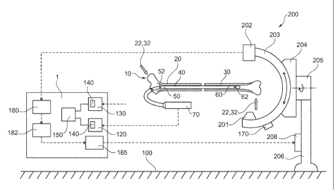
Fig. 2 illustrates a device for assisting the repositioning of bone parts 20,
30
according to an exemplary embodiment of the invention. The device 1 includes a
first rotary orientation determining unit 120 and a second rotary orientation
determining unit 130. The first rotary orientation determining unit 120 is
adapted for
determining a rotary orientation of the intramedullary implant being locked in
a
predefined orientation to the first intramedullary bone fragment with respect
to an
artificial horizon 100. Thus, it is possible to determine the rotary
orientation of the
first bone part 20 by a first unique imaging projection direction 22, which
can be
attained by an imaging device 200. If for example the anatomical geometry of
the
femoral head is known or can be sufficiently exactly determined by a targeting
device, this may also serve as an indicative for a first rotary orientation of
the
intramedullary implant being locked in a predefined orientation to the first
intramedullary bone fragment 20, here the proximal femur fragment 20. The
second
rotary orientation determining unit 130 serves for determining a rotary
orientation of
the second intramedullary bone fragment, here the distal femur fragment 30
with
respect to an artificial horizon 100 such as the floor of an operating room,
for
example. Both relative positions, that of the intramedullary implant being
locked to
the first intramedullary bone fragment as well as the second intramedullary
bone
fragment can be supplied to a matching unit 150 so as to determine the
relative
position of the intramedullary implant being locked to the first
intramedullary bone
CA 02816369 2013-04-29
WO 2012/084056 PCT/EP2010/070682
- 23 -
fragment with respect to the second intramedullary bone fragment. Thus, the
absolute
orientation of the respective bone fragment relative to the artificial horizon
can be
used for determining the relative orientation of the first and second
intramedullary
bone fragment to each other. Both, the first and the second rotary orientation
determining unit 120, 130 comprise a sensing unit 140 for sensing the actual
position
of a distance locking means of the intramedullary implant so that the first
intramedullary bone fragment and the second intramedullary bone fragment can
be
brought into a predefined rotary orientation to each other. The matching unit
150 can
inform the surgeon on a fracture in which the intramedullary bone fragments
20, 30
are in the correct orientation with respect to each other. The matching unit
or the
device for assisting the surgeon may have a display or any other output device
a like
voice generator for providing information on the present orientation of the
fragments
or the required actions to arrive at the predetermined orientation of the
fragments
with the surgeon.
For determining the correct orientation of the bone fragments, an anatomical
landmark like the known geometry of the end portions of a bone can be used, in
particular a unique projection, 22, 32 thereof. For this purpose, the imaging
device
can be brought into an orientation providing the unique projection. When
arriving at
the position of the imaging device providing the unique projection, the
imaging
device can provide the orientation of the imaging device relative to the
artificial
horizon 100, which may be for example the floor of the room. Fig. 2
illustrates the
position of the imaging device in the unique imaging projection direction 32
of the
condyles of the femur. In this orientation, the x-ray source 201 radiates into
the
direction of the X-ray sensitive array 202, which illustrates the unique
projection in
the precise defined orientation, for example of the matching and overlapping
condyles. To arrive at the orientation of the unique imaging projection
direction, the
imaging device 200 can be moved along a circular trajectory with the C-arm 203
over a base 204. Further degrees of freedom can be established by further
pivoted
connections, e.g. between a base 204 and a bearing 205 connected to a fixed
pole 206
CA 02816369 2013-04-29
WO 2012/084056 PCT/EP2010/070682
- 24 -
having a defined position with respect to the artificial horizon 100. The
orientation of
the projection direction of the x-ray source 201 can be monitored by a
reference
device 170 which for example may be capable of determining the spatial
orientation
of the x-ray source 201 fixedly connected with respect to the orientation of
the
reference device or reference unit 170. The imaging device 200 may provide the
imaging information to an image recognition unit 180 which may be provided in
device 1. The image recognition unit 180 may conduct an image recognition of
the
obtained image of the obtained images so that a connected analyzing unit 182
may
conduct an image analyzing so as to provide the surgeon with the information
whether the imaging device 200 has arrived at the unique imaging projection
direction 32. Further, the analyzing unit 182 can calculate a linear or
rotational shift
of the x-ray source 201 which is necessary to arrive at the unique imaging
direction
32, and can provide this information to an actuator 184. This actuator may
provide
respective controlling information to the imaging device, which may be adapted
to
automatically shift the C-arm 203 into the correct position of the correct
unique
imaging projection direction. The transmission of information can be conducted
wirelessly or by a signal line. For this purpose, the imaging device 200 can
be
provided with a receiving and controlling unit 208 to conduct the automatic
repositioning of the C-arm 203 and the x-ray source 201 and the x-ray
sensitive array
202. Thus, without a further required action of the surgeon, the device 1 can
provide
information whether the orientation of the extremity bone fragments 20, 30 are
in the
predefined orientation with respect to each other and which particular
additional
rotation has to be conducted by the surgeon to arrive at the correct
positioning. The
device 1 for assisting the surgeon may have a display or any other output
device a
like voice generator for providing information on the present orientation of
the
fragments or the required actions to arrive at the predetermined orientation
of the
fragments with the surgeon. Inparticular, when the first extremity bone part
20 is
fixedly connected to the intramedullary implant 40 and the intramedullary
implant 40
is fixedly connected to the targeting device, the targeting device 70 may
provide the
rotary orientation of the first intramedullary bone fragment 20 to the device
1,
CA 02816369 2013-04-29
WO 2012/084056 PCT/EP2010/070682
- 25 -
whereas the imaging device 200 when automatically arriving at the unique
imaging
direction projection provides the respective rotary orientation of the second
intramedullary bone fragment 30 to the device, so that the device 1 can
directly
provide the surgeon on the success of the rotary orientation of the extremity
bone
fragments 20, 30. When arriving at the predefined orientation of the extremity
bone
fragments 20, 30, the second fragment 30 can be locked by the locking device
62 to
the distal end of the intramedullary nail 40 arriving at a fixedly locked
orientation of
the first and second intramedullary bone fragment 20, 30 with respect to each
other.
Fig. 3 illustrates a detailed view of the device and intramedullary implant
and bone
fracture according to an exemplary embodiment of the invention, and a
schematic
overview on the positioning of the distal locking device 62 with respect to
the
targeting device 70. For this purpose, the targeting device 70 may include an
external
transmitting unit 72 transmitting a particular signal to an internal
transceiving unit
64. The internal transceiving unit together with the external transmitting
unit 72 can
determine the relative position, so as to arrive at information on the
relative position
of the internal transceiving unit 64 with respect to the external transmitting
unit 72.
The internal transceiving unit 64 then transmits this information via the
intramedullary nail 40 which may be provided at the targeting device 70. The
transmission of the information from the internal transceiving unit to the
external
receiving unit 74 can be conducted by an acoustic signal transmission via the
intramedullary nail 40. Thus, the external receiving unit 74 provides
information on
the relative position of the internal transceiving unit 64 with respect to the
external
transmitting unit 72, and when providing the internal transceiving unit 64
close to the
locking device 62, also information on the relative position of the locking
device 62
with respect to the targeting device 70. This information may be combined with
a
relative position of the targeting device over an artificial horizon 100, thus
leading
also to information on the relative position of the distal locking means 62
with
respect to the artificial horizon. The artificial horizon may be the floor of
the
operating room, but may be also any other point serving as a reference point
for
CA 02816369 2013-04-29
WO 2012/084056 PCT/EP2010/070682
- 26 -
determining the relative position of the single components with respect to
this
reference point. For example, the device 1 or a particular vertex of the
device 1 may
be used as artificial horizon. For determining the orientation of the
targeting device
or the imaging device over an artificial horizon 100 like the floor of the
operation
__ room a gravity sensor may be used. In fact also positioning systems similar
to GPS
like systems may be used. An operation room may be provided with active or
passive
reference points serving as triangulation points corresponding to the
satellites in a
GPS system. The imaging device may also refer to the artificial horizon or the
reference point so that the entire system may be used with any imaging system,
__ insofar the imaging system is capable of informing the surgeon on its
relative
position, e.g. by an optical or electronic inclination scale or positioning
system. It
should be noted that the artificial horizon 100 may be eliminated if the
device 1
directly communicates with the imaging system, i.e. is capable of directly
determining the relative position of the imaging system with respect to the
internal
__ tranceiving unit. The positional information can be provided to the device
1 so as to
arrive at the correct positional information on the distal locking device 62.
When
combining the positional information on the distal locking means 62 obtained
by the
active nail tip technology as described in Fig. 3 and combining this
information with
the information obtained by the imaging device 200 by the information on the
unique
__ imaging projection direction, this information can be obtained to arrive at
the correct
predefined position of the first and second intramedullary extremity bone
fragment.
Fig. 4 illustrates a schematic flow of a method for operating the device of
Fig. 2
according to an exemplary embodiment of the invention. The method for
operating
__ the device allow an assisting of positioning a first intramedullary bone
fragment 20
and a second intramedullary bone fragment 30 of a fractured extremity bone 10
to be
restored with respect to each other by an intramedullary implant 40. The
implant has
a proximal end 50 coupled to a targeting device 70 and a distal end 60. The
device
determines S210 the orientation of the first intramedullary bone fragment by
fitting a
__ first unique imaging projection direction 22 of an anatomical landmark of
the first
CA 02816369 2013-04-29
WO 2012/084056 PCT/EP2010/070682
- 27 -
intramedullary bone fragment to the first intramedullary bone fragment. This
first
unique imaging projection direction may be for example the femur head and the
femur neck being imaged in a projection direction such that the diameter of
the neck
and the outer circumference of the head are concentric. For the surgeon it is
clear that
this corresponds to a predefined position. With respect to this position, the
device
further determines S220 a future position of the intramedullary implant 40,
e.g. a
femur nail, in a predefined orientation to the first intramedullary bone
fragment. The
device 1 further determines S230 a locking position of one of that end of the
implant,
which corresponds to the first bone fragment. This may be the proximal end or
the
distal end of the intramedullary implant. Now the surgeon may use the position
determined by the device to conduct the locking of the first intramedullary
bone
fragment to the implant. As the orientation of the first bone fragment 20 is
known
and also the orientation of the implant is known, the implant when locked to
the first
fragment represents the orientation of the first fragment. Now, an implant
landmark
like a distal locking means 62 in form of a cylindrical hole may serve as an
indicative
for the orientation of the first bone fragment 20. It is to be expected that
the implant,
in particular when being provided as a long intramedullary nail, will bend as
to
follow the intramedullary channel of the bone. However, the deformation is
considered as being limited to the bending, so that not torsion deformation is
expected. Thus, even is bending the nail, the rotary orientation of the nail,
and at the
same time the distal locking hole 62 remains unamended, so that the distal
locking
hole 62 may serve as a rotary or rotational indicator for the rotary
orientation of the
first bone fragment 20. It should be noted that the device may firstly
determine the
locking position S230 and then determine the rotary orientation of the first
fragment
20 and the implant 40 in S210 and S220, if the locking position is in a
general
allowable range. A matter of fact, the order of S210 and S220 can be vice
versa, and
S230 may be conducted between S210 and S220, and S210, S220 and S230 at least
partially may be conducted at the same time or interleaved. The device 1 based
on
the previous determinations now may determine S240 a future position of the
first
intramedullary bone fragment and the second intramedullary bone fragment in a
CA 02816369 2013-04-29
WO 2012/084056 PCT/EP2010/070682
- 28 -
predefined rotary orientation with respect to each other. This can be done by
determining a second unique imaging projection direction 32 of an anatomical
landmark of the second intramedullary bone fragment, and to bring the second
unique imaging direction 32 into a relation to the orientation of the distal
locking
hole 62, which is represents the first unique imaging direction of the first
fragment.
This information may be used to provide the surgeon with information, on how
to
amend the current orientation so as to arrive at the optimal predefined
orientation of
the first unique projection and the second unique projection to each other,
thus
corresponding to the predefined rotary orientation of the first intramedullary
bone
fragment and the second intramedullary bone fragment to each other. The device
1
may only inform the surgeon on the subsequent steps, e.g. on how to rotate the
second fragment 20 over the implant 40 being locked to the first fragment 10,
but
may also control the imaging device to move into the position in which the
imaging
device has the imaging direction which corresponds to the optimal predefined
orientation, so that the surgeon only has to rotate the second fragment into a
position
meeting the second unique imaging projection direction, here the matching
contours
of the condyles. Determining the locking position of the distal end of the
intramedullary implant to the respective intramedullary bone fragment is
conducted
by sensing S252 the actual position of a distal locking means 62 of the
intramedullary implant, and determining S255 a locking position of the
respective
intramedullary bone fragment to the intramedullary implant such that the
intramedullary implant, in particular the predefined geometry of the distal
locking
means allows a positioning of the first intramedullary bone fragment and the
second
intramedullary bone fragment in the predefined rotary orientation to each
other.
As additional assistance, which is however not mandatory, the device
determines a
locking position of the other of the proximal end and the distal end of the
intramedullary implant to the second intramedullary bone fragment, in Fig. 2
the
distal end of the implant to the distal fragment 20. This will be conducted by
the
procedure as described with respect to Fig. 3. In particular the bending
displacement
CA 02816369 2013-04-29
WO 2012/084056 PCT/EP2010/070682
- 29 -
of the nail tip 60 as the distal end of the implant will be determined so that
a locking
screw may inserted at the predefined position. This avoids an erroneous
drilling for
the locking procedure at the distal end of the implant. Although this
targeting
procedure may also be applied to the locking of the proximal end of the
implant, the
proximal locking means 52 of the implant 40 as a rule is sufficiently exact
defined by
the targeting device 70. In other words, possible bending deformations at the
proximal end do not lead to a substantial deformation, but the distal end
does. It
should be noted that the locking targeting S250 at the distal end may be left
out when
no bending is expected or other targeting methods are used. In this case step
S250
can be left out without departing the invention.
The sensing S252 of the actual position of a distal locking means 62 of the
intramedullary implant 40 may be conducted by transmitting S253 a signal from
an
external transmitting unit 72 being positioned in a predefined position, e.g.
with
respect to the targeting device 70. An internal tranceiving unit 64 receives
S254 the
signal. The internal tranceiving unit 64 is fixedly mounted relative to the
distal
locking means 62 of the intramedullary implant 40 and being actuated by the
external
transmitting unit. The internal tranceiving unit 64 provides information on
relative
position with respect to the external transmitting unit 72 based on the
received signal.
For this purpose the signal may comprise a combination of different
frequencies,
phase shifts etc, allowing the internal tranceiving unit 64 to determine the
relative
position with respect to the external transmitting unit 72. The relative
position may
include linear displacement as well as rotational displacement. Although a
relevant
elongation of the implant is not expected and also no torsion, the internal
transceiving unit may be adapted to determine displacements in all six degrees
of
freedom. It should be noted that the internal transceiving unit 64 may also
transmit a
raw data signal and the evaluation thereof may be conducted somewhere else,
e.g. in
a particular unit (not shown) in the device 1. Although transmitting S255 may
be
conducted by acoustic vibrations or acoustic waves, the signal may also be
transferred as a wireless radio signal, a signal via wire or any other
appropriate signal
CA 02816369 2013-04-29
WO 2012/084056 PCT/EP2010/070682
- 30 -
transmission. The signal, as indicative of the relative position of the
internal
transceiving unit 64 with respect to the external transmitting unit 72, may be
transmitted to an external receiving unit 74 being located at the targeting
device.
In the following five different operations will be described with respect to
figs. 5, 6,
7, 8 and 9. The respective steps refer to the step remarks in said figs. and
to Fig. 11
illustrating a more detailed visualization of the procedural steps of the
method for
dealing the re-establishment of the orientation of extremity bone.
Figs. 5a-5d illustrate several procedural steps of a method for dealing a
femur
fracture with an antegrade entry of the intramedullary femur nail with primary
proximal locking. The nail will be inserted and the neck screw will be placed
in the
centre of the neck and the femur head (Fig. 5a). The nail is rotated by 10 to
15
outward in the fragment and can be localized by the method described with
respect to
Fig. 3. A collimation unit at the imaging device 200 then is rotated by 10
to 15
inwardly, and the distal fragment is rotary positioned while monitored via the
imaging device 200, so that the contour of the condyles match, i.e. are in the
same
plane as the collimation unit. Now the distal locking can be conducted using
the
method described with respect to Fig. 3.
In more detail, the method for positioning a proximal femur fragment 20 and a
distal
femur fragment 30 with respect to each other by an antegrade intramedullary
femur
nail 40 having a proximal end 50 coupled to a targeting device 70 and a distal
end 60
is conducted by the following steps. As can be seen in Fig. 5a, the antegrade
intramedullary femur nail is positioned S10 in a predefined orientation to the
proximal femur fragment 20 and a rotary orientation of the antegrade
intramedullary
femur nail with respect to the proximal femur fragment is determined S30. This
can
be done by fitting S36 the proximal femur fragment to a first unique imaging
projection direction 22 of an anatomical landmark of the proximal femur
fragment
and sensing S32 the rotary orientation of a proximal locking means 52 of the
CA 02816369 2013-04-29
WO 2012/084056 PCT/EP2010/070682
- 31 -
antegrade intramedullary femur nail with respect to the first unique imaging
projection direction. However this step can be left out, if the locking of the
proximal
end 50 of the femur nail 40 can be conducted otherwise, i.e. free hand by the
extended experience of a surgeon, using the targeting device 70. If the
correct
position of the proximal femur fragment to the femur nail 40 is found, the
proximal
end of the antegrade intramedullary femur nail is locked S20 to the proximal
femur
fragment. The implant landmark of the femur nail, e.g. the distal locking hole
62 is
used as an indicative for the rotary orientation of the proximal femur
fragment. The
proximal femur fragment and the distal femur fragment 30 are positioned S40 in
a
predefined rotary orientation with respect to each other by matching S50 the
rotary
orientation of the antegrade intramedullary femur nail with respect to the
distal femur
fragment by sensing S52 the rotary orientation of a distal locking means 62 of
the
antegrade intramedullary femur nail, and by fitting S56 the distal femur
fragment to a
second unique imaging projection direction 32 of an anatomical landmark of the
distal femur fragment with respect to the rotary orientation of the distal
locking
means. For this purpose the imaging device, e.g. in form of a C-arm moves to
the
distal end, i.e. the femur condyles, as can be seen in Fig. 5b, and can be
adjusted by
the x-ray calculation of the inclination of the femur head of +3 , as can be
seen in
Fig. 5a, and subtracting 10 (to 15 ) antetorsion as described above,
resulting in -7
(to 12 ) antetorsion, which should be the orientation of the imaging device,
e.g.
visible on a scale of the imaging device. Subsequently the condyles fragment
is
rotated to be in line with the orientation of the imaging device 200,
resulting in an
image of the unique imaging projection direction 32 of the femur condyles, as
can be
seen in Fig. 5c. Subsequently, the distal locking means 62 of the femur nail
will be
locked by using the method described with respect to Fig. 3, as can be seen in
Fig.
5d. Locking the distal end of the antegrade intramedullary femur nail to the
distal
femur fragment may be conducted by sensing S62 the position of the distal
locking
means of the antegrade intramedullary femur nail, and positioning S63 and
locking
S64 the distal femur fragment to the antegrade intramedullary femur nail.
Thus, a
locking S60 can be established between the distal end of the antegrade
CA 02816369 2013-04-29
WO 2012/084056 PCT/EP2010/070682
- 32 -
intramedullary femur nail to the distal femur fragment such that the proximal
femur
fragment and the distal femur fragment are positioned in the predefined rotary
orientation to each other.
Figs. 6a-6c illustrate a method for dealing a femur fracture with an antegrade
entry of
a femur nail with proximal distal locking. The distal locking will be
conducted with
the method described with respect to Fig. 3. Subsequently, the axis of the
condyles
will be determined and the difference will, be transferred to a collimator
unit being
fixed to the targeting device 70. Then the collimator unit will be proximal of
the
femur adjusted to arrive at the desired angle of the femur neck. The setting
of the
femur neck may be checked by an x-ray collimator unit or k-wire markers. If
the
rotation matches the predefined rotary orientation the proximal locking
finished the
procedure.
In more detail, the method for positioning a proximal femur fragment 20 and a
distal
femur fragment 30 with respect to each other will be described in the
following. The
antegrade intramedullary femur nail 40 has a proximal end 50 coupled to a
targeting
device 70 and a distal end 62. The antegrade intramedullary femur nail will be
positioned S10 in a predefined orientation to the distal femur fragment and
locking
S20 of the distal end of the antegrade intramedullary femur nail to the distal
femur
fragment is conducted, e.g. using the method described with respect to Fig. 3,
as can
be seen in Fig. 6a. Afterwards a rotary orientation of the antegrade
intramedullary
femur nail with respect to the distal femur fragment is determined by fitting
S36 the
distal femur fragment to a first unique imaging projection direction 32 of an
anatomical landmark of the distal femur fragment and sensing S32 the rotary
orientation of a distal locking means 62 of the antegrade intramedullary femur
nail
with respect to the first unique imaging projection direction. For this
purpose the
imaging device will be adjusted until the condyles are in line, i.e. show
matching
contours in the imaging device 200 resulting in the first unique imaging
projection
direction 22, which is at the distal femur condyles. The orientation angel of
the
CA 02816369 2013-04-29
WO 2012/084056 PCT/EP2010/070682
- 33 -
imaging device will be noticed, which is +5 , as can be seen in Fig. 6b, which
is -3
visible on the ANTT display after distal locking. Then the proximal femur
fragment
and the distal femur fragment will be positioned S40 in a predefined rotary
orientation with respect to each other by matching S50 the rotary orientation
of the
antegrade intramedullary femur nail with respect to the proximal femur
fragment by
sensing S52 the rotary orientation of a proximal locking means 52 of the
antegrade
intramedullary femur nail, and by fitting S56 the proximal femur fragment to a
second unique imaging projection direction 22 of an anatomical landmark of the
proximal femur fragment with respect to the rotary orientation of the proximal
locking means, as can be seen in Fig. 6c. For this step, the imaging device
will be
moved to the proximal end of the femur, and be adjusted until it is in line
with the
femur neck, which is a -5 orientation of the imaging device. Then the
targeting
device 70 with the implant 40 having locked thereon the distal femur fragment
30 is
rotated until +2 is visible on the ANTT display. This +2 results from -3
ANTT
display, as can be seen in Fig. 6b, +5 of the distal imaging device
orientation, as can
be seen in Fig. 6b, -5 of the proximal imaging device orientation, as can be
seen in
Fig. 6c, +5 for 15 anteversion. Finally the proximal end of the antegrade
intramedullary femur nail will be locked S60 to the proximal femur fragment
such
that the proximal femur fragment and the distal femur fragment are positioned
in the
predefined rotary orientation to each other. As an option the proximal end of
the
antegrade intramedullary femur nail may be locked to the proximal femur
fragment
by sensing S62 the position of the proximal locking means of the antegrade
intramedullary femur nail, and positioning S63 and locking S64 the proximal
femur
fragment to the antegrade intramedullary femur nail.
Figs. 7a-7c illustrate a tibia fracture with retrograde entry of the
intramedullary nail
and primary distal locking. A retrograde insertion means to enter the bone
from the
distal end and to move the implant 40 from the distal end to the proximal end
of the
bone. Thus the direction of a retrograde insertion is rotated by 180 over the
antegrade insertion. This results in the distal end 60 of the implant 40
corresponding
CA 02816369 2013-04-29
WO 2012/084056 PCT/EP2010/070682
- 34 -
to the proximal end 20 of the bone 10 and the proximal end 50 of the implant
40
corresponding to the distal end 30 of the bone 10. For this method, at first,
the distal
femur fragment will be locked to the proximal end of the implant, subsequently
the
rotary orientation of the nail is determined by using the ANTT method
described
with respect to Fig. 3, and the unique imaging projection direction of the
femur
condyles. This rotary orientation will be transferred to the collimation unit.
The
femur neck will be collimated or a k-wire marker will be set, so that
subsequently the
distal end of the implant can be locked to the proximal end of the femur using
the
ANTT described with respect to Fig. 3.
In more detail, the method for positioning a proximal femur fragment 20 and a
distal
femur fragment 30 with respect to each other by an retrograde intramedullary
femur
nail 40 having a proximal end 50 coupled to a targeting device 70 and a distal
end 60
will be conducted as follows. The retrograde intramedullary femur nail will be
positioned S10 in a predefined orientation to the distal femur fragment.
Subsequently
a rotary orientation of the retrograde intramedullary femur nail with respect
to the
distal femur fragment will be determined by fitting S36 the distal femur
fragment to
a first unique imaging projection direction 32 of an anatomical landmark of
the distal
femur fragment, as can be seen in Fig. 7a. For this purpose the imaging device
is
adjusted with the matching contours of the condyles, resulting in a +100
orientation
of the imaging device in the example shown in Fig. 7a. The rotary orientation
of a
proximal locking means 52 of the retrograde intramedullary femur nail with
respect
to the first unique imaging projection direction is sensed. The latter may be
conducted by the ANTT method described with respect to Fig. 3, till then
targeting
device 70 is in line with the orientation of the imaging device, i.e. the
first unique
imaging direction. Then the proximal end of the retrograde intramedullary
femur nail
will be locked S20 to distal femur fragment, as can be seen in Fig. 7b.
Subsequently,
the proximal femur fragment and the distal femur fragment are positioned in a
predefined rotary orientation with respect to each other by matching S50 the
rotary
orientation of the retrograde intramedullary femur nail with respect to the
proximal
CA 02816369 2013-04-29
WO 2012/084056 PCT/EP2010/070682
- 35 -
femur fragment. For this purpose, the imaging device is moved to the proximal
femur
end and adjusted to be in line with the femoral neck as the second unique
imaging
projection direction. In the example shown in Fig. 7c, the imaging device
orientation
is parallel to the floor (i.e. 00 over the floor). As an option the rotary
orientation of a
distal locking means 62 of the retrograde intramedullary femur nail can be
sensed
and the proximal femur fragment can be fitted S56 to the second unique imaging
projection direction 22 of an anatomical landmark, i.e. the head of the
proximal
femur fragment with respect to the rotary orientation of the distal locking
means. The
targeting device 70 with the condyles fragment will be rotated until -5 is
visible on
the ANTT display. This results from the orientation of the imaging device of
00 in
Fig. 7c, +100 orientation of the condyles in Fig. 7a and the required
anteversion of
assumed 15 . Finally the distal end of the retrograde intramedullary femur
nail is
locked to the proximal femur fragment such that the proximal femur fragment
and
the distal femur fragment are positioned in the predefined rotary orientation
to each
other. The locking can optionally be supported by the method as described with
respect to Fig. 3, wherein locking the distal end of the retrograde
intramedullary
femur nail to the proximal femur fragment is conducted by sensing S62 the
position
of the distal locking means of the retrograde intramedullary femur nail, and
positioning S63 and locking S64 the proximal femur fragment to the retrograde
intramedullary femur nail.
Figs. 8a-8c illustrate a method for dealing a tibia fracture with antegrade
entry of the
intramedullary tibia nail with primary distal locking. This method starts with
a distal
locking of the as described with respect to Fig. 3, as can be seen in Fig. 8a.
The
imaging device will be AP adjusted to the sub-talar joint. The rotation of the
tibia
nail will be determined und the collimating unit is proximally set and
adjusted at the
targeting device, and subsequently re-adjusted with a 20 to 25 rotation
outwardly.
The orientation of the imaging device is checked with respect to the unique
imaging
projection direction of the condyles of the femur at the full extended knee.
CA 02816369 2013-04-29
WO 2012/084056 PCT/EP2010/070682
- 36 -
In more detail, the method for positioning a proximal tibia fragment 20 and a
distal
tibia fragment 20 with respect to each other by an antegrade intramedullary
tibia nail
40 having a proximal end 50 coupled to a targeting device 70 and a distal end
60 is
conducted as follows. The antegrade intramedullary tibia nail is positioned
S10 in a
predefined orientation to the distal tibia fragment and the distal end of the
antegrade
intramedullary tibia nail is locked S20 to the distal tibia fragment. The ANTT
method described with respect to Fig. 3 may be used, as can be seen in Fig.
8a. Then
a rotary orientation of the antegrade intramedullary tibia nail with respect
to the
distal tibia fragment will be determined by fitting the distal tibia fragment
to a first
unique imaging projection direction 32 of an anatomical landmark, here the
talar
joint of the distal tibia fragment and sensing S 32 the rotary orientation of
a distal
locking means 62 of the antegrade intramedullary tibia nail with respect to
the first
unique imaging projection direction. In this example the imaging device is
adjusted
to arrive at the unique imaging projection direction of the talar joint. The
orientation
of the imaging device in this example is -100 (90 +10 ), wherein +2 are
visible on
the ANTT display after distal locking, as can be seen in Fig. 8b. Subsequently
the
proximal tibia fragment and the distal tibia fragment are positioned S40 in a
predefined rotary orientation with respect to each other by matching S50 the
rotary
orientation of the antegrade intramedullary tibia nail with respect to the
proximal
tibia fragment by sensing S52 the rotary orientation of a proximal locking
means 52
of the antegrade intramedullary tibia nail, and by fitting S56 the proximal
tibia
fragment to a second unique imaging projection direction 22 of an anatomical
landmark of the proximal tibia fragment, here the condyles with respect to the
rotary
orientation of the proximal locking means. For this purpose, the imaging
device will
be adjusted till in line with the unique imaging projection direction of the
condyles,
here +5 , as can be seen in Fig. 8c. The targeting device with the tibia nail
having
locked thereon the distal tibia fragment is rotated to arrive at the rotary
orientation of
+17 resulting from +2 from ANTT display as can be seen in Fig. 8b, +15 . +15
result from the +5 of the condyles inclination as can be seen in Fig. 8c and
the
(90 )+10 from the talar inclination, as can be seen in Fig. 8b to arrive at
the required
CA 02816369 2013-04-29
WO 2012/084056 PCT/EP2010/070682
- 37 -
200 outward rotation. Finally the proximal end of the antegrade intramedullary
tibia
nail is locked S60 to the proximal tibia fragment such that the proximal tibia
fragment and the distal tibia fragment are positioned in the predefined rotary
orientation to each other. Optionally, the locking can be conducted as
outlined with
respect to Fig. 3, wherein locking the proximal end of the antegrade
intramedullary
tibia nail to the proximal tibia fragment is conducted by sensing S62 the
position of
the proximal locking means of the antegrade intramedullary tibia nail, and
positioning S63 and locking S64 the proximal tibia fragment to the antegrade
intramedullary tibia nail.
Figs. 9a-9d illustrate a tibia fracture with an antegrade entry of the
intramedullary
tibia nail with primary proximal locking. The proximal locking will be
conducted via
the targeting device. The condyle plane is determined in the full extended
knee, and
the collimation unit is adjusted to the frontal plane. Subsequently, the
outward
rotation for the talar joint is adjusted and the tibia is rotated to be locked
in the
correct position.
In more detail the method for positioning a proximal tibia fragment 20 and a
distal
tibia fragment 30 with respect to each other by an antegrade intramedullary
tibia nail
40 having a proximal end 60 coupled to a targeting device 70 and a distal end
50will
be conducted as follows. A rotary orientation of the antegrade intramedullary
tibia
nail with respect to the proximal tibia fragment is determined by fitting S36
the
proximal tibia fragment to a first unique imaging projection direction 22 of
an
anatomical landmark of the proximal tibia fragment, as can be seen in Fig. 9a.
The
imaging device is adjusted to be in line with the unique imaging projection
direction
of the condyles, i.e. the anatomical landmark of the proximal tibia fragment.
In this
example, the orientation of the imaging device is 0 . The antegrade
intramedullary
tibia nail is positioned S10 in a predefined orientation to the proximal tibia
fragment
and the rotary orientation of a proximal locking means 52 of the antegrade
intramedullary tibia nail is sensed S32 with respect to the first unique
imaging
CA 02816369 2013-04-29
WO 2012/084056 PCT/EP2010/070682
- 38 -
projection direction. In this example, the targeting device 70 as a
representation of
the tibia nail is brought in line with unique condyle projection of Fig. 9a,
as can be
seen in Fig. 9b. Then the proximal end of the antegrade intramedullary tibia
nail is
locked S20 to proximal tibia fragment. Then the imaging device is moved to the
distal tibia end, as can be seen in Fig. 9c, and moved by 90 to arrive in the
correct
reference plane and then 200 to 23 are added to obtain the predefined rotary
orientation of the distal tibia fragment and the proximal tibia fragment to
each other,
as can be seen in Fig. 9c. The proximal tibia fragment and the distal tibia
fragment
are positioned in a predefined rotary orientation with respect to each other
by
matching S50 the rotary orientation of the antegrade intramedullary tibia nail
with
respect to the distal tibia fragment by sensing S52 the rotary orientation of
a distal
locking means 62 of the antegrade intramedullary tibia nail via the targeting
device,
and by fitting S56 the distal tibia fragment to a second unique imaging
projection
direction 32 of an anatomical landmark of the distal tibia fragment with
respect to the
rotary orientation of the distal locking means. The latter will be obtained by
rotated
the distal fragment until the sub-talar joint is fully visible, i.e. the
anatomical
landmark of the talar joint corresponds to the preset orientation of the
imaging
device, i.e. the unique imaging projection direction of the distal end of the
tibia, as
can be seen in Fig. 9d. Finally the distal end of the antegrade intramedullary
tibia nail
is locked S60 to the distal tibia fragment such that the proximal tibia
fragment and
the distal tibia fragment are positioned in the predefined rotary orientation
to each
other. As an option, locking can be conducted by the ANTT, as described with
respect to Fig. 3, wherein locking the distal end of the antegrade
intramedullary tibia
nail to the distal tibia fragment is conducted by sensing S62 the position of
the distal
locking means of the antegrade intramedullary tibia nail, and positioning S63
and
locking S64 the distal tibia fragment to the antegrade intramedullary tibia
nail.
Fig. 10 illustrates procedural steps and alternatives thereto for a
generalized method
for dealing a re-establishment of the orientation of extremity bone fragments
according to an exemplary embodiment of the invention. The method for
positioning
CA 02816369 2013-04-29
WO 2012/084056 PCT/EP2010/070682
- 39 -
a first intramedullary bone fragment 20 and a second intramedullary bone
fragment
30 of a fractured extremity bone 10 to be restored with respect to each other
by an
intramedullary implant 40 having a proximal end 50 coupled to a targeting
device 70
and a distal end 60 can generally be expressed by the following steps which
should
cover all of the five above described methods. The generalized method
comprises
positioning S10 of the intramedullary implant in a predefined rotary
orientation to
the first intramedullary bone fragment, locking S20 one of the proximal end
and the
distal end of the intramedullary implant to the first intramedullary bone
fragment,
determining S30 a rotary orientation of the intramedullary implant with
respect to the
first intramedullary bone fragment, positioning S40 of the first
intramedullary bone
fragment and the second intramedullary bone fragment in a predefined rotary
orientation with respect to each other by matching S50 the rotary orientation
of the
intramedullary implant with respect to the second intramedullary bone
fragment,
locking S60 the other of the proximal end and the distal end of the
intramedullary
implant to the second intramedullary bone fragment, wherein locking the distal
end
of the intramedullary implant to the respective intramedullary bone fragment
is
conducted by sensing S22, S62 the actual position of a distal locking means 62
of the
intramedullary implant, and positioning S28, S68 and locking S29, S69 the
respective intramedullary bone fragment to the intramedullary implant such
that the
intramedullary implant allows a positioning of the first intramedullary bone
fragment
and the second intramedullary bone fragment in the predefined rotary
orientation to
each other.
As can be seen from Fig. 10, the order of the steps is not fixed and may
differ. This
can also be taken from the detained description of the five above described
methods,
in which the order of the steps S10, S20 and S30 may differ according to
required
order.
The method may optionally use unique imaging projection direction of
anatomical
landmarks, such that determining S30 a rotary orientation of the
intramedullary
CA 02816369 2013-04-29
WO 2012/084056 PCT/EP2010/070682
- 40 -
implant 40 with respect to the first intramedullary bone fragment 20 may
comprise
fitting S36 the first intramedullary bone fragment to a first unique imaging
projection
direction 22 of an anatomical landmark of the first intramedullary bone
fragment,
and matching S50 the rotary orientation of the intramedullary implant with
respect to
the second intramedullary bone fragment 30 comprises fitting S56 the second
intramedullary bone fragment to a second unique imaging projection direction
32 of
an anatomical landmark of the second intramedullary bone fragment, wherein the
rotary orientation of the first unique projection and the second unique
projection to
each other corresponds to the predefined rotary orientation of the first
intramedullary
bone fragment and the second intramedullary bone fragment to each other.
Further,
sensing S22, S62 the actual position of a distal locking means 62 of the
intramedullary implant 40 may comprise using an actual position of a proximal
locking means 52 of the intramedullary implant, and the orientation of the
first
unique projection 22 and the second unique projection 32 are used to provide
the
surgeon with positional information for the first intramedullary bone fragment
20
with respect to the second intramedullary bone fragment 30. In addition,
Furthermore, sensing S22, S62 the actual position of a distal locking means 62
of the
intramedullary implant 40 may be conducted by transmitting S23, S63 a signal
from
an external transmitting unit 72, receiving S24, S64 the signal by an internal
transceiving unit 64 being fixedly mounted relative to the distal locking
means 62 of
the intramedullary implant 40 and being actuated by the external transmitting
unit,
transmitting S25, S65 by acoustic vibrations or acoustic waves the received
signal, as
indicative of the relative position of the transceiving unit with respect to
the external
transmitting unit, to an external receiving unit 74, as it is described with
respect to
Fig. 3 in more detail. This method is also referred to as ANTT method, which
means
an active nail tip targeting. The transceiving unit 64 may be provided as an
active
unit capable of calculating the relative position of the nail tip and of
transmitting this
information to the targeting device so as to simplify the determination of the
position
of the locking holes in the nail implant. With this respect, the locking
direction of an
CA 02816369 2013-04-29
WO 2012/084056 PCT/EP2010/070682
-41 -
end 50, 60 of the intramedullary implant 40 may correspond to the respective
unique
imaging projection direction 22, 32 of the respective intramedullary bone
fragment
20, 30 to be locked to that respective end of the intramedullary implant.
It should be noted that the general inventive principle may also be applied to
other
fractures with fragments having an anatomical landmark.
It should be noted that the term 'comprising' does not exclude other elements
or
steps and the 'a' or 'an' does not exclude a plurality. Also elements
described in
association with the different embodiments may be combined.
It should be noted that the reference signs in the claims shall not be
construed as
limiting the scope of the claims.