Note: Descriptions are shown in the official language in which they were submitted.
SYSTEM AND METHOD FOR ELECTROSURGICAL CONDUCTIVE GAS
CUTTING FOR IMPROVING ESCHAR, SEALING VESSELS AND TISSUES
INVENTORS: JEROME CANADY, M.D., EDSON VIEIRA,
NICHOLAS VIEIRA, AND KIMBERLY WILEY, M.D.
[0001]
[0002]
STATEMENT REGARDING FEDERALLY
SPONSORED RESEARCH OR DEVELOPMENT
[0003] None.
BACKGROUND OF THE INVENTION
Field Of The Invention
[0004] The present invention relates to electrosurgical systems and methods,
and more
particularly, electrosurgical systems and methods using argon plasma during
cutting
modes of operation.
Brief Description Of The Related Art
[0005] The standard means for controlling traumatic and surgical blood loss
are
electrosurgical generators and lasers which respectively direct high-frequency
electrical
currents or light energy to localize heat in bleeding vessels so as to
coagulate the
overlying blood and vessel walls. Hemostasis and tissue destruction are of
critical
1
CA 2816424 2019-01-03
CA 02816424 2013-04-29
WO 2012/061535
PCMJS2011/059025
importance when removing abnormal tissue during surgery and therapeutic
endoscopy.
For monopolar electrosurgery electrical energy originates from an
electrosurgical
generator and is applied to target tissue via an active electrode that
typically has a small
cross-sectional surface-area to concentrate electrical energy at the surgical
site. An
inactive return electrode or patient plate that is large relative to the
active electrode
contacts the patient at a location remote from the surgical site to complete
and electrical
circuit through the tissue. For bipolar electrosurgery, a pair of active
electrodes are used
and electrical energy flows directly through the tissue between the two active
electrodes.
00061 U.S. Patent No. 4,429,694 to McGreevy disclosed a variety of different
electrosurgical effects that can be achieved depending primarily on the
characteristics of
the electrical energy delivered from the electrosurgical generator. The
electrosurgical
effects included pure cutting effect, a combined cutting and hemostasis
effect, a
fulguration effect and a desiccation effect. Fulguration and desiccation
sometimes are
referred to collectively as coagulation.
1l0071 A conventional desiccation procedure, shown in FIG. 1B, typically is
performed
by holding the active electrode in contact with the tissue. Radiofrequency
(RF) current
passes from the electrode directly into the tissue to produce heating of the
tissue by
electrical resistance heating. The heating effect destroys the tissue cells
and produces an
area of necrosis spreading radially from the point of contact between the
electrode and
the tissue. The necrosis is usually deep.
[00081 A conventional fulguration procedure, shown in FIG. 1A, may be obtained
by
varying the voltage and power applied by the electrosurgical generator.
Conventional
fulguration procedures typically were performed using a waveform which has a
high peak
2
CA 02816424 2013-04-29
WO 2012/061535
PCT/US2011/059025
voltage but a low duty cycle. If the active electrode was brought close to but
not
touching the tissue and the peak voltage was sufficient to produce an RF arc,
fulguration
would occur at the point where the arc contacted the tissue. Due to the low
duty cycle,
the power per unit time applied to the tissue was low enough so that cutting
effects were
minimized.
0009j A conventional cutting procedure, shown in FIG. 1C, may be obtained by
delivering sufficient power per unit time to the tissue to vaporize cell
moisture. If the
power applied is high enough a sufficient amount of steam is generated to form
a steam
layer between the active electrode and the tissue. When the steam layer forms,
a plasma
consisting of highly ionized air and water molecules forms between the
electrode and the
tissue. An RF arc then develops in the plasma. At the location where the arc
contacts the
tissue, the power density becomes extremely high and instantaneously disrupts
the tissue
architecture. New steam is thereby produced to maintain the steam layer. If
the power
density is sufficient, enough cells are destroyed to cause a cutting action to
occur. A
repetitive voltage wave form, such as a sinusoid, delivers a continuous
succession of arcs
and produces a cut with very little necrosis and little hemostasis.
0010 It also was possible to create a combined combination of effects by
varying the
electrical waveform applied to the tissue. Specifically, a combination of
conventional
cutting and desiccation could be produced by periodically interrupting the
continuous
sinusoidal voltage typically used to perform a conventional cutting procedure.
If the
interruption was sufficient, the ionized particles in the plasma between the
electrode and
the tissue would collapse, causing the electrode to momentarily come into
contact with
3
CA 02816424 2013-04-29
WO 2012/061535
PCT/US2011/059025
the tissue. That touching would desiccate the tissue thereby sealing off blood
vessels in
the vicinity of the electrode.
ON 11 Conventional electrosurgical generators typically have both "cut" or
cutting and
"coag" or coagulation modes of operation. As previously noted, the cut mode
typically
will have a low voltage waveform form with a high duty cycle, e.g. 100%. The
coag
mode of an electrosurgical generator typically creates a waveform with large
amplitude
but short duration "spikes" to achieve hemostasis (coagulation). For example,
in coag
mode an electrosurgical generator may use a high voltage wave form at a 6%
duty cycle.
The surrounding tissue is heated when the waveform spikes and then cools down
(between spikes), producing coagulation of the cells. Fulguration is achieved
in the coag
mode of the electrosurgical generator, with the tip of the surgical "active
electrode" held
above (but not in contact with) the tissue. Electrosurgical desiccation is
achieved in either
the cut or coag modes of the generator. The difference between desiccation and
fulguration is the tip of the "active electrode" must contact the tissue as in
FIG. 1B in
order to achieve desiccation. Typically, the more desired mode to achieve
tissue
desiccation through direct tissue contact is the cut mode. Different degrees
of hemostasis
(coagulation) can be achieved by utilizing varying degrees of "Blended"
waveforms, e.g.,
50% on/50% off, 40% on/60% off, or 25% on/75% off.
Is00121 Another method of monopolar electrosurgery via argon plasma technology
was
described by Morrison US patent #4,040,426 in 1977 and McGreevy U.S. Patent
No.
4,781,175. This method, referred to as argon plasma coagulation (APC) or argon
beam
coagulation is a non-contact monopolar thermoablative method of
electrocoagulation that
has been widely used in surgery for the last twenty years. In general, APC
involves
4
CA 02816424 2013-04-29
WO 2012/061535
PCT/US2011/059025
supplying an ionizable gas such as argon past the active electrode to target
tissue and
conducting electrical energy to the target tissue in ionized pathways as non-
arcing diffuse
current. Canady described in U.S. Patent No. 5,207,675 the development of APC
via a
flexible catheter that allowed the use of APC in endoscopy. These new methods
allowed
the surgeon, endoscopist to combine standard monopolar electrocautery with a
plasma
gas for coagulation of tissue.
0013i APC has been demonstrated to be effective in the coagulation of blood
vessels
and human tissue during surgery. APC functions in a noncontact manner. The
electrical
current is initiated only when the tip of the handpiece or catheter is within
one centimeter
of the target tissue and produces a homogenous lmm to 2mm well-delineated
eschar. The
eschar created by APC is further characterized by a decrease absence of
charring and
carbonization compare to eschar resulting from conventional electrosurgical
fulguration.
The eschar remains firmly attached to the tissue, in contrast to other
coagulation
modalities where there is an overlying charred layer of coagulated blood.
There is
minimal tissue necrosis with APC.
0014 In U.S. Patent Nos. 5,217,457 and 5,088,997 to Delahuerga et at.
disclosed a
device for performing procedure referred to as "argon shrouded cut." The
device was an
electrosurgical pencil having an exposed electrode with a distal end defining
a tip for
cutting biological tissue and a nose piece mounted about the electrode to
define a
pathway for a stream of inert gas which shrouds the electrode at or near its
tip. When in
coagulation mode, a convergent stream of inert gas was directed directly onto
the tip of
the electrode. In coagulation mode, the voltage was sufficient to initiate an
electrical
discharge in the inert gas. In cut mode, the stream of ionized gas was
directed to impinge
5
CA 02816424 2013-04-29
WO 2012/061535
PCT/US2011/059025
obliquely on the electrode at a point adjacent to but away from the tip of the
electrode. In
cutting mode, the open circuit voltage was generally not high enough to
continuously
plasmatize the inert gas and initiate and maintain an electrical discharge.
Accordingly, in
cut mode the function of the inert gas is to provide a shroud around the
electrode rather
than to initiate electrical discharge.
0015j A multitude of literature exists that discloses and discusses various
commercially
available electrosurgical generators and the voltage waveforms produced by
those
generators. For example, A. Erwine, "ESU-2000 Series Product Overview A
Paradigm
Shift in Electrosurdery Testing Technology and Capability Is Here," BC Group
International, Inc. (2007) describes electrosurgical generators from ERBE
Elektromedizin
GmbH and ConMed Corporation, among others.
SUMMARY OF THE INVENTION
NO16 In a preferred embodiment, the present invention is an electrosurgical
method for
simultaneously cutting and coagulating tissue with an electrosurgical device
having an
electrode and a channel wherein said channel has a port near a proximal end of
said
electrode for directing a gas onto said proximal end of said electrode. The
method
comprises the steps of causing a gas to flow through said channel and exit
said port,
applying high-frequency energy to said electrode while said gas flows through
said
channel, wherein said high-frequency energy applied to said electrode
continuously
plasmatizes gas exiting said port, initiating an electrical discharge from
said electrode
through said continuously plasmatized gas to said tissue, cutting tissue with
said
electrode, maintaining said electrical discharge from said electrode through
said
plasmatized gas while cutting tissue with said electrode to cause coagulation
of tissue
6
CA 02816424 2013-04-29
WO 2012/061535
PCT/US2011/059025
adjacent said proximal end of said electrode simultaneously with said cutting.
The gas
may comprise an inert gas such as argon. The step of applying high-frequency
energy to
said electrode may comprise applying 70-100W of power to said electrode. The
step of
causing a gas to flow through said channel may comprise causing an inert gas
to flow
through said channel at a flow rate of 7 Limin. The electrosurgical device is
connected to
an electrosurgical generator, said generator having a cut mode comprising a
repeating
voltage waveform and a coag mode comprising a varying voltage waveform, and
wherein
said step of applying high-frequency energy to said electrode comprises
activating said
electrosurgical generator in said cut mode. The repeating voltage waveform maY
be a
sinusoidal waveform. The inert gas may exit the port in a direction
substantially parallel
to said electrode. A portion of said channel adjacent said port in said
channel may be
held at an angle of 45 to 60 to a surface of target tissue. The
simultaneously cutting
and coagulating causes a low depth of injury to said tissue and a small
diameter of injury
to said tissue.
14)4)17. In another embodiment, the present invention is an electrosurgical
device. The
device comprises means for initiating an electrical discharge from an
electrode through
continuously plasmatized inert gas to tissue and means for simultaneously
cutting tissue
with an energized electrode and coagulating said tissue by maintaining said
electrical
discharge from said electrode through said plasmatized inert gas while cutting
said tissue
with said energized electrode. The means for simultaneously cutting tissue and
coagulating said tissue using a plasmatized inert gas may comprise a housing
having an
opening at a distal end, an electrode extending from said distal end of said
housing, a
channel within said housing, said channel having a port adjacent said
electrode extending
7
CA 02816424 2013-04-29
WO 2012/061535
PCT/US2011/059025
from said housing, means for causing an inert gas to flow through said channel
and exit
said port, means for applying high-frequency energy to said electrode while
said inert gas
flows through said channel, wherein said high-frequency energy applied to said
electrode
continuously plasmatizes inert gas exiting said port, means for initiating an
electrical
discharge from said electrode through said continuously plasmatized inert gas
to said
tissue, and means for maintaining said electrical discharge from said
electrode through
said plasmatized inert gas while cutting tissue with said electrode to cause
coagulation of
said tissue simultaneously with said cutting. The electrosurgical device may
further
comprising telescoping nozzle connected to said housing, wherein said
telescoping
nozzle is adjustable to change a length of said electrode extending from said
housing.
The electrode extends 2-25 mm from said telescoping nozzle.
0018 In a preferred embodiment, the electrosurgical device comprises a
housing, an
electrode, wherein the electrode extends through the housing and a portion of
the
electrode extends from a distal end of the housing, a connector for connecting
the
electrode to an electrosurgical generator, a channel in the housing, a port at
a proximate
end of the channel for connecting the channel to a source of pressurized inert
gas, and a
port at a distal end of the channel for discharging inert gas flowing through
the channel,
and controls for initiating a flow of an inert gas through the channel and
applying high-
frequency electrical energy to the electrode, wherein the controls provide for
a
conventional cut mode, a conventional coagulation mode, an argon plasma
coagulation
mode, and a plasma cut mode. The plasma cut mode comprises maintaining an
electrical
discharge from the electrode through plasmatized inert gas being discharged
from the
8
CA 02816424 2013-04-29
WO 2012/061535
PCT/US2011/059025
channel while cutting tissue with the electrode to cause coagulation of the
tissue
simultaneously with the cutting.
1.9019 In one embodiment, the controls comprise three buttons in the housing
for
allowing operating the device in the cut mode, the conventional coagulation
mode, the
argon plasma coagulation mode, and the plasma cut mode. In another embodiment,
the
controls comprise a footswitch for allowing operating the device in the cut
mode, the
conventional coagulation mode, the argon plasma coagulation mode, and the
plasma cut
mode. The simultaneously cutting and coagulating may cause a low depth of
injury to the
tissue. It may also cause a small diameter of injury to the tissue. The flow
rate of the
inert gas through the channel may be between 0.1 and 10 L/min.
[NM Still other aspects, features, and advantages of the present invention are
readily
apparent from the following detailed description, simply by illustrating
preferable
embodiments and implementations. The present invention is also capable of
other and
different embodiments and its several details can be modified in various
obvious respects,
all without departing from the spirit and scope of the present invention.
Accordingly, the
drawings and descriptions are to be regarded as illustrative in nature, and
not as
restrictive. Additional objects and advantages of the invention will be set
forth in part in
the description which follows and in part will be obvious from the
description, or may be
learned by practice of the invention.
BRIEF DESCRIPTION OF THE DRAWINGS
9
CA 02816424 2013-04-29
WO 2012/061535
PCT/US2011/059025
0021 For a more complete understanding of the present invention and the
advantages
thereof, reference is now made to the following description and the
accompanying
drawings, in which:
00224 FIG. lA is a diagram illustrating a conventional fulguration mode of
operation of
an electrosurgical device.
00231 FIG. 1B is a diagram illustrating a conventional desiccation mode of
operation of
an electrosurgical device.
E00241 FIG. 1C is a diagram illustrating a conventional cutting mode of
operation of an
electrosurgical device.
100251 FIG. 2A is a perspective view of an electrosurgical handpiece having
its electrode
retracted within its housing in accordance with a first preferred embodiment
of the
present invention.
100261 FIG. 2B is a perspective view of an electrosurgical handpiece having
its electrode
extending out from a distal end of its housing in accordance with a first
preferred
embodiment of the present invention.
'-,0027.1 FIG. 2C is an assembly drawing of an electrosurgical handpiece in
accordance
with a first preferred embodiment of the present invention.
100281 FIG. 3A is a diagram illustrating an experimental setup for testing in
argon
coagulation mode.
10029 FIG. 3B is a diagram illustrating an experimental setup for testing a
preferred
embodiment of the present invention in hybrid plasma cut mode.
00301 FIG. 4A is a graph of pig's liver sample temperature and spark length as
function
of power with a USMI SS-200E/Argon 2 system in conventional coagulation mode.
CA 02816424 2013-04-29
WO 2012/061535
PCT/US2011/059025
00311 FIGs. 4B-C are tables of the numerical values corresponding to the graph
in FIG.
4A.
[00321 FIG. 5A is a graph of pig's liver sample temperature as function of
power at
various argon flow rate settings with a USMI SS-200E/Argon 2 system in argon
plasma
coagulation mode.
00331 FIG. 5B is a graph of pig's liver sample temperature as function of
argon flow
rate at various power settings with a USMI SS-200E/Argon 2 system in argon
plasma
coagulation mode.
00341 FIG. 5C is a graph of argon beam length as function of power at various
argon
flow rate settings with a USMI SS-200E/Argon 2 system in argon plasma
coagulation
mode.
00151 FIG. 5D is a graph of argon beam length as function of argon flow rate
at various
power settings with a USMI SS-200E/Argon 2 system in argon plasma coagulation
mode.
00361 FIGs. 5E-F are tables of the numerical values corresponding to the
graphs in
FIGs. 5A-D.
t00371 FIG. 6A is a graph of pig's liver sample temperature as function of
power
performed with a USMI SS-200E/Argon 2 system in conventional cut mode.
100381 FIG. 6B is a table of the numerical values corresponding to the graph
in FIG. 6A.
[00391 FIG. 7A is a graph of pig's liver sample temperature as a function of
power at
various flow rates performed with a USMI SS-200E/Argon 2 system in hybrid
plasma cut
mode in accordance with the present invention.
0040 FIG. 7B is a graph of pig's liver sample temperature as a function of gas
flow rate
at various power settings performed with a USMI SS-200E/Argon 2 system in
hybrid
plasma cut mode in accordance with the present invention.
11
CA 02816424 2013-04-29
WO 2012/061535
PCT/US2011/059025
0041 FIG. 7C is a table of numerical values corresponding to the graphs in
FIGs. 7A
and 7B.
100421 FIG. 8A is a graph of pig's liver sample temperature and spark length
as function
of power with a USMI SS-601MCa/Argon 4 system in conventional coagulation
mode.
[00431 FIGs. 8B-C are tables of the numerical values corresponding to the
graph in FIG.
8A.
100441 FIG. 9A is a graph of pig's liver sample temperature as function of
power at
various argon flow rate settings with a USMI SS-601MCa/Argon 4 system in argon
plasma coagulation mode.
100451 FIG. 9B is a graph of pig's liver sample temperature as function of
argon flow
rate at various power settings with a USMI SS-601MCa/Argon 4 system in argon
plasma
coagulation mode.
100461 FIG. 9C is a graph of argon beam length as function of power at various
argon
flow rate settings with a USMI SS-601MCa/Argon 4 system in argon plasma
coagulation
mode.
[0047i FIG. 9D is a graph of argon beam length as function of argon flow rate
at various
power settings with a USMI SS-601MCa/Argon 4 system in argon plasma
coagulation
mode.
100481 FIGs. 9E-F are tables of the numerical values corresponding to the
graphs in
FIGs. 9A-D.
10049 FIG. 10A is a graph of pig's liver sample temperature as function of
power
performed with a USMI SS-601MCa/Argon 4 system in conventional cut mode.
100S0l FIG. 10B is a table of the numerical values corresponding to the graph
in FIG.
10A.
12
CA 02816424 2013-04-29
WO 2012/061535
PCT/US2011/059025
0051 FIG. 11A is a graph of pig's liver sample temperature as a function of
power at
various flow rates performed with a USMI SS-601MCa/Argon 4 system in hybrid
plasma
cut mode in accordance with the present invention.
100521 FIG. 11B is a graph of pig's liver sample temperature as a function of
gas flow
rate at various power settings performed with a USMI SS-601MCa/Argon 4 system
in
hybrid plasma cut mode in accordance with the present invention.
0053 FIG. 11C is a table of numerical values corresponding to the graphs in
FIGs. 11A
and 11B.
10054 FIG. 12A is a tissue image illustrating depth of injury of 1.2mm at a
power setting
.. of 20W with a USMI SS-200E/Argon 2 system in conventional cut mode.
100551 FIG. 12B is a tissue image illustrating depth of injury of 1.5mm at a
power setting
of 20W with a USMI SS-200E/Argon 2 system in conventional coagulation mode.
[0056 FIG. 12C is a tissue image illustrating depth of injury of 0.1mm at a
power setting
of 20W and a flow setting of 0.1 1/min. with a USMI SS-200E/Argon 2 system in
hybrid
plasma cut mode.
00571 FIG. 12D is a tissue image illustrating depth of injury of 0.6 mm at a
power
setting of 20W a flow setting of 0.5 Umin. with a USMI SS-200E/Argon 2 system
in
argon plasma coagulation mode.
100581 FIGs. 13A and 13B are a table and graph of conventional cut data with a
USMI
SS-200E/Argon 2 system.
00591 FIGs. 14A and 14B are a table and graph of conventional coagulation data
with a
USMI SS-200E/Argon 2 system.
-00601 FIGs. 15A and 15B are a table and graph of argon plasma coagulation
data with a
USMI SS-200E/Argon 2 system.
13
CA 02816424 2013-04-29
WO 2012/061535
PCT/US2011/059025
.00611 FIGs. 16A and 16B are a table and graph of hybrid plasma cut data with
a USMI
SS-200E/Argon 2 system in hybrid plasma cut mode in accordance with a
preferred
embodiment of the present invention.
00621 FIGs. 17A and 17B are a table and graph of hybrid plasma cut data with a
USMI
SS-601MCa/Argon 4 system in hybrid plasma cut mode in accordance with a
preferred
embodiment of the present invention.
00631 FIG. 18A is a table of depth of injury data with a USMI SS-200E/Argon 2
system
in conventional cut mode.
00641 FIG. 18B is a table of depth of injury data with a USMI SS-200E/Argon 2
system
in conventional coagulation mode.
[00651 FIG. 18C is a table of depth of injury data with a USMI SS-200E/Argon 2
system
in argon coagulation mode.
100661 FIG. 18D is a table of depth of injury data with a USMI SS-200E/Argon 2
system
in hybrid plasma cut mode in accordance with a preferred embodiment of the
present
invention.
[00671 FIG. 18E is a table of depth of injury data with a USMI SS-601MCa/Argon
4
system in hybrid plasma cut mode in accordance with a preferred embodiment of
the
present invention.
[00681 FIG. 19A is a graph comparing depth of injury data for a USMI SS-
200E/Argon 2
system in argon plasma coagulation mode and in hybrid plasma cut mode.
1(10691 FIG. 19B is a graph comparing depth of injury data for a USMI SS-
200E/Argon 2
system in hybrid plasma cut mode and a USMI SS-601MCa/Argon 4 system in hybrid
plasma cut mode.
14
CA 02816424 2013-04-29
WO 2012/061535
PCT/US2011/059025
0070 FIG. 19C is a graph comparing depth of injury data for a USMI SS-
200E/Argon 2
system in conventional cut mode, conventional coagulation mode, argon plasma
coagulation mode with a gas flow rate of 2.5 1/min and in hybrid plasma cut
mode with a
gas flow rate of 2.5 1/min.
[0071 FIG. 19D is a graph comparing depth of injury data for a USMI SS-
200E/Argon 2
system in conventional cut mode, conventional coagulation mode, argon plasma
coagulation mode with a gas flow rate of 5 l/min and in hybrid plasma cut mode
with a
gas flow rate of 5 1/min.
00721 FIG. 20A is a graph of depth of injury data for a USMI SS-200E/Argon 2
in argon
plasma coagulation mode.
[00731 FIG. 20B is a graph of depth of injury data for a USMI SS-200E/Argon 2
in
hybrid argon cut mode in accordance with a preferred embodiment of the present
invention.
0074 FIG. 20C is a graph of depth of injury data for a USMI SS-601MCa/Argon 4
system in hybrid argon cut mode in accordance with a preferred embodiment of
the
present invention.
00751 FIG. 21A is a tissue image of in vivo porcine skin Hybrid Plasma cut
20wg3
liters/min, 2 sec., depth of injury 0.2mm, eschar 1.5mm.
[0076 FIG. 21B is a tissue image of in vivo skin Conventional Cut: 20wg3
liters/min,
2 sec. depth of injury (0.4mm), eschar (2.5mm).
[0077. FIG. 21C is a tissue image of in vivo Conventional Coagulation: 20wg3
liters/min, 2 sec., depth of injury (3.4mm), eschar (5.0mm).
.0078 FIG. 21D is a tissue image of Argon Plasma Coagulation: 20wg 3
liters/min, 2
sec., depth of injury 2.0mm, eschar 5.0mm.
CA 02816424 2013-04-29
WO 2012/061535
PCT/US2011/059025
00791 FIG. 21E is a tissue image of Argon Plasma Coagulation: depth of injury
(1.0mm), eschar (10.0mm) 440w, 3 liters/min.
1.00801 FIG. 21F is a tissue image of in vivo Hybrid Plasma cut: depth of
injury (0.2mm),
Eschar (1.4mm), 40w@3 Liters/min.
100811 FIG. 21G is a tissue image of in vivo porcine resection of 1st part of
Duodenum
with Hybrid plasma cut, depth of Injury (0.2mm) eschar (1.0mm) 4; 40w, 3
liters/min, 3
sec.
00821 FIG. 21H is a tissue image of in vivo porcine resection of Sternum:
depth of
injury 0.6mm, 120w4 5 liters/min.
[00831 FIG. 211 and 21J is a tissue image of in vivo resection of sternum in
vivo porcine
model minimal bone marrow damage (0.2mm) 4120w, 5 liters/min.
DETAILED DESCRIPTION OF THE PREFERRED EMBODIMENTS
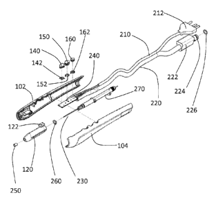
00841 A preferred embodiment of an electrosurgical device 100 in accordance
with the
present invention is described with reference to FIGs. 2A-2C. The
electrosurgical device,
handpiece or pencil 100 has a rigid housing 110 and telescoping nozzle or tip
120. The
rigid housing may be formed, for example, from molded sides 102 and 104. The
two
sides 102, 104 are joined to form housing 110 having a hollow chamber within.
Within
the housing 110 is an electrode 230, electrode tubing 270 and a fiberglass
plate 240. The
electrode 230 extends through the electrode tubing 270. The electrode tubing
additional
has within it a channel, tube or other means for conducting the inert gas from
the distal
end of tubing 220 through the electrode tubing 270 and out of the electrode
tubing 270.
The inert gas leaving the channel in the electrode tubing then passes out of
an opening at
the distal end of the nozzle 120. The fiberglass plate 240 and electrode 230
are
16
CA 02816424 2013-04-29
WO 2012/061535
PCT/US2011/059025
connected to electrical cable assembly 210. The electrode tubing is connected
at its distal
end to the hose tubing 220. An 0-ring is placed between the telescoping nozzle
and the
electrode tubing to form a seal therebetween. A ceramic tip 250 may be placed
at a distal
end of the telescoping tip or nozzle 120 to protect the nozzle 120 from heat
damage
where the electrode passes through an opening at the distal end of the nozzle
120. The
electrical cable assembly extends from a proximal end of the housing 110 and
has at its
distal end a plug 212. During operation of the device, the connector 212 is
connected to
an electrosurgical generator. The PVC hose tubing also extends from the
proximal end of
the housing 110 and has at its distal end a gas connector body 222, a gas
connector tip
224 and an 0-ring 226. During operation of the device, the gas connector
assembly (222,
224, 226) is connected to a source of an inert gas such as argon.
00851 The housing 110 has a plurality of opening or holes for accommodating a
plurality of controls or buttons 140, 150, 160. The telescoping nozzle or tip
120 has a
control element 122 extending through a slot 112 in the housing 110. The
control
element, tab, know or slider 122 is used by a surgeon to move the telescoping
tip 120 into
or out of an opening in a distal end of the housing 120. Three controls or
buttons 140,
150, 160, extend out of openings in the housing 110 and have springs 152
between them
and fiberglass plate or connected 240 to bias the controls or buttons away
from the plate
or connector 240.
I(J0861 The electrosurgical device of the present invention can be operated,
for example,
in four different modes: conventional cut mode, conventional coagulation mode,
argon
plasma coagulation mode, and hybrid plasma cut mode. The eschar resulting from
cutting
and coagulation in the hybrid plasma cut mode in accordance with the present
invention
17
CA 02816424 2013-04-29
WO 2012/061535
PCT/US2011/059025
is substantially better than conventional fulguration, cutting and argon
plasma
coagulation techniques. In addition there is substantial absence of charring,
carbonization, tissue necrosis and destruction of adjacent tissue. Thus,
tissue can be
precisely cut and the adjacent vessels simultaneously sealed with minimal
depth of
injury, tissue necrosis, eschar and carbonization.
-0087j An inert gas combined with high-frequency energy in the plasma cut mode
can
precisely cut through tissues (i.e. skin, muscle, bone or vascular) with
substantial speed
and accuracy.
00881 Any generator that provides high-frequency voltage to ionize the inert
gas to form
a gas stream can be used. Preferred generators include the Canady PlasmaTM
Electrosurgery Unit model (SS-601 MCa) and the Canady PlasmaTM Electrosurgery
Unit
model (SS-200E) that are preferably used with the Argon plasma units Canady
PlasmaTM
Argon 4 Coagulator (CPC 4) and Canady PlasmaTM Argon 2 Coagulator (CPC 2),
respectively. The CPC 4 provides a controlled flow of inert gas to the
electrosurgical
device during argon plasma coagulation mode and in hybrid plasma cut mode. The
flow
rate and the power can be manually set. In a coagulation mode, the generator
delivers, for
example, a peak-to-peak voltage of less than 9000 volts. In a cut mode, for
example, the
generator delivers a peak-to-peak voltage of less than 3800 volts. Most
preferably, a
peak-to-peak voltage of 100 to 9000 volts is delivered by the generator.
10089I Any accessory devices can be attached to the electrosurgery unit/plasma
unit
combination. Exemplary devices are an electrosurgical device (a handpiece) or
an argon
plasma flexible probe (catheter), rigid or laparoscopic.
18
CA 02816424 2013-04-29
WO 2012/061535
PCT/US2011/059025
0090i For operating the electrosurgical device, high-frequency current can be
activated
by two push buttons for the conventional cut mode and the conventional
coagulation
mode, respectively. Argon gas may be delivered by activating a third push
button. This
activation will allow the argon plasma coagulation mode and the hybrid plasma
cut mode.
The plasma cut mode will cut and coagulate the tissue at the same time. It can
be easily
switched between the different modes by activating the respective buttons. The
plasma or
electrical current can also be activated by a footswitch.
E009I The telescoping nozzle of the electrosurgical device can be extended or
shortened
over the electrode as desired when performing plasma procedures. In a
preferred
embodiment, the electrode extends 2 to 25 mm outside the telescoping nozzle.
[0092! The electrode can be of any common material of the state of the art. In
a preferred
embodiment, the electrode is a tungsten wire.
NO931 In a preferred embodiment, the present invention is an electrosurgical
method for
achieving cutting and coagulating simultaneously with a source of inert,
ionizable gas in
combination with high-frequency energy. The source of inert, ionizable gas can
be any
kind of inert, ionizable gas. The preferred type of gas for use in cutting is
pure argon.
Argon gas causes a decrease in tissue temperature which limits micro-
destruction of
tissue, improves through conductivity of tissue and allows high-frequency
cutting
through tissue at low tissue temperatures. Inert gas also dissipates oxygen
molecules from
the surgical area and prevents oxidation of tissue which causes decrease local
tissue
temperature and prevents carbonization. Flow rates can vary and can be
adjusted
depending on the tissue that is being cut.
19
CA 02816424 2013-04-29
WO 2012/061535
PCT/US2011/059025
00941 A high-frequency current supplied by an electrosurgical generator is
transmitted
through an electrode. Electrodes can be composed, for example, of tungsten,
stainless
steel, ceramic or any electrical conducting material. An electrical discharge
is created
between the active electrode and the tissue. The discharge is ignited by AC
voltage with a
typical amplitude and frequency at 4 kV and or greater than 350 kHz
respectively. The
voltage waveform preferably is a sinusoidal waveform that contains alternate
positive and
negative sections of approximately equal amplitudes. An inert gas flows
through the
channel containing the electrode. The electrode contacts the tissue and
delivers an ionized
plasma high-frequency current through the tissue. A new phenomenon has been
created
by the present invention, which can precisely cut through the tissue and
simultaneously
seal adjacent vessels and tissue with.
00951 The present invention is further evidenced by the following examples.
Ex Vivo Porcine Model
1M961 All ex vivo porcine experiments were carried out on explant porcine
liver samples
Micropropulsion and Nanotechnology Laboratory (MpNL), George Washington
University, Washington, D.0 and WEM Equipamentos Plasma Research Laboratory,
Ribeirao Preto ¨ Sao Paulo, Brazil. Liver samples were immediately placed in
10%
formalin solution ph 7.0 and sent for H & E preparation of the pathological
slides and
interpretation at Laboratorio de Patologia Cirurgica Dr Prates, Ribeirao Preto-
Sao Paulo,
Brazil
In Vivo Porcine Model
CA 02816424 2013-04-29
WO 2012/061535
PCT/US2011/059025
00971 In vivo porcine surgical operations were performed at the University of
Sao
Paulo, Department of Surgery and Anatomy, Animal Research Laboratory, Ribeirao
Preto, SP, Brazil. Approval was obtained by the institution animal research
director.
Three dalland female swine (mean weight 14.5kg) were used in this study.
Anesthesia
was induced with ketamine 50mg/cc mixed and dopaser ¨ xilazina 200mg/l0cc,
intramuscular. Animals were then intubated, and anesthesia was maintained with
Na
Pentathol to effect. The skin was prepped with alcohol and draped in the usual
sterile
manner. Mercedes, abdominal midline, and median stemotomy were made during the
operations with the plasma scalpel. Multiple surgical procedures were
performed median
stemotomy, gastric resection, partial splenectomy, partial nephrectomy,
partial
hepatectomy, wedge resection of the liver, intestinal resection and skin
incisions.
Operations were video-recorded. Observations of surgical bleeding during the
procedure
were recorded. Depth of injury and eschar was compared with four high
frequency
operations modes: conventional cut and coagulation, argon plasma coagulation
and
hybrid argon plasma cut. Samples of the skin, liver, stomach, intestine, and
bone were
placed in 10% formalin solution ph 7.0 and sent for H & E preparation of the
pathological slides and measurement of depth of injury and diameter of eschar
at
Laboratorio de Patologia Cirurgica Dr Prates, Ribeirao Preto- Sao Paulo,
Brazil. Animals
were sacrificed by using an intravenous injection of pentobarbital sodium and
phenytoin
sodium.
(00981 The hybrid plasma scalpel blade of the present invention was used in
combination
with USMI's SS-200E/Argon 2 and SS-601MCa/Argon 4 to evaluate in four high
frequency operation modes: (i) conventional cut; (ii) conventional
coagulation; (iii)
21
CA 02816424 2013-04-29
WO 2012/061535
PCT/US2011/059025
conventional argon plasma coagulation (APC); and (iv) hybrid plasma cut. As
described
above in the background of the invention, conventional cut and coagulation
modes do not
involve the use of an inert gas such as argon. Instead, they are performed by
touching the
target tissue with the active electrode. Conventional argon plasma coagulation
is
performed as it was described above in the background of the invention. The
hybrid
plasma cut mode is the mode of the present invention described above in the
detailed
description of the preferred embodiments. The hybrid plasma scalpel used in
all four
modes is as described above with respect to FIGs. 2-C.
00991 Four parameters were measured: plasma discharge column length, tissue
heating,
diameter of eschar and depth of injury by high frequency operation mode. The
length of
the plasma was characterized by the maximal length of the discharge plasma
column
observed at tissue treatment with the hybrid plasma scalpel at which the
discharge can be
sustained. The treatments were video-recorded by digital camera Nikon Coolpix
995 (15
frames/s) and the maximal length of discharge plasma column (L) was measured
by post-
experiment evaluation of recorded videos. The tissue heating was characterized
by the
temperature growth (d7) of pig's liver sample appeared as result of
application of hybrid
plasma scalpel. AT was measured using the thermocouple (Type K) probes
embedded in
the pig's liver. The accuracy of temperature and length measurements were 5 'V
and
0.5 mm respectively. Tissue temperature prior to treatment was 18-20 C.
Eschar
diameter produce by the plasma scalpel blade was measured using a digital
caliber.
Pathologists used an Motim Camera 1000, 1.3 an Olympus Microscope Bx 41 to
calculate the depth of injury.
22
CA 02816424 2013-04-29
WO 2012/061535
PCT/US2011/059025
001001 The pig's liver samples were treated by the hybrid plasma scalpel as
following.
In coagulation mode, the pig's liver sample was treated by 5 consecutive
applications of
the hybrid plasma scalpel to the same point of the liver sample (total
treatment duration
was ¨5 s). The thermocouple was located about 3 mm under the treated point as
shown in
FIG. 3A. In cut mode, a 5 mm straight cut in the pig's liver sample was
created by five
consecutive passes with hybrid plasma scalpel along the cut (total duration ¨5
s) and
thermocouple probe was located about 3 mm aside from the cut (see FIG. 3B).
The
hybrid plasma scalpel was used with both the Argon 2/SS- 200E and Argon 4/
SS601MCa systems with flow rates from 0.5 to 5 liters/minute and from 0.1,
3.0, 7.0 and
10.0 liter/minute respectively. Data and graphs of results from these
experiments are
shown in FIGs. 4-11 and 13-20 and images of the treated tissue are shown in
Ms. 12A-
D and 21A-J.
[001011 Data and graphs for testing of each of the four operating modes are
shown in the
drawings as follows: i) conventional cut shown in FIGs. 6A-6B, 10A-B, 13A-B
and 18A;
(ii) conventional coagulation shown in FIGs. 4A-C, 14A-B and 18B; (iii)
conventional
argon plasma coagulation shown in FIGs. 5A-F, 9A-F, 15A-B and 18C; and (iv)
hybrid
plasma cut shown in FIGs. 7A-C, 11A-C, 16A-B, 17A-B (with Argon 4/ SS601MCa),
18D and 18E (with Argon 4/ SS601MCa). Graphs comparing performance in the
various
modes of operation are shown in the graphs in FIGs. 19A-D and 20A-C.
1001021 FIGs. 19C-D show comparisons of the depth of injury found in the four
modes
of operation performed with the Argon 2/SS- 200E system. FIG. 19C shows the
comparison with both the conventional argon plasma coagulation mode and the
hybrid
plasma cut mode of the present invention at an argon flow rate of 2.5 L/min.
FIG. 19D
23
CA 02816424 2013-04-29
WO 2012/061535
PCT/US2011/059025
showsn the comparison using an argon flow rate of 5 L/min. One can see form
FIG. 19C
that at lower power settings, e.g., below 70W, and a flow rate of 2.5 L/min.,
the hybrid
plasma cut mode of the present invention results in the depth of tissue injury
being
greater than the depth of injury in conventional argon plasma coagulation
mode. Since
the electrosurgical generator is in a cutting mode similar to (or identical
to) conventional
electrosurgical cutting when the hybrid plasma cut mode of the present
invention is used,
it is logical that it would result in a greater depth of injury than a
conventional argon
plasma coagulation mode. At mid to high power ranges, e.g. 70-100W (see item
1920),
however, the hybrid plasma cut mode of the present invention results in a
smaller depth
of injury than conventional argon plasma coagulation and conventional
electrosurgical
cutting. The result is vastly superior to conventional electrosurgical cutting
(0.7-1.5mm
depth for hybrid plasma cut versus 2.5 ¨ 3.7mm for conventional cut) and
significantly
better than conventional APC (0.6mm for plasma cut versus 1.2mm for
conventional
APC). FIG. 19D shows similar results for an argon flow rate of 5 L/min. In
lower
power ranges (see item 1940) the depth of injury for hybrid plasma cut tends
to track the
depth of injury with conventional electrosurgical cutting. In mid to high
power ranges,
e.g., 70-100W (see item 1930), however, the hybrid plasma cut mode of the
present
invention provides superior, i.e., smaller, depth of injury versus both
conventional argon
plasma coagulation (see item 1930) and conventional electrosurgical cutting.
1001031 FIGs. 19A shows a comparison of the depth of injury in the hybrid
argon cut
mode of the present invention versus the conventional argon plasma coagulation
mode at
argon flow rates of 2.5 and 5.0 L/min. The graph in FIG. 19A shown that with
the Argon
2/SS- 200E system, the hybrid plasma cut mode of the present invention
achieves a
24
CA 02816424 2013-04-29
WO 2012/061535
PCT/US2011/059025
substantially superior result compared to conventional argon plasma
coagulation at
settings of about 70-90W and 2.5 L/min (see item 1902) and 30-50W at 5L/min
(see
1904). FIG. 19B shows a comparison of the hybrid plasma cut mode of the
present
invention performed with the two different test systems. In FIG. 19B, one can
see that
with the Argon 4/SS601MCa system, the hybrid plasma cut mode of the present
invention achieves an unexpectedly superior result at settings of about 50-80W
and 7
L/min (see item 1910) but also is superior to conventional APC in the power
range of 50-
100W at 7 L/min.
001041 As shown in FIG. 20A, the depth of injury associated with conventional
argon
plasma coagulation is not very dependent upon the argon flow rate. As each
power level
tested on the Argon 2/SS- 200E system in conventional APC mode, the depth of
injury
varied only by a small amount (approximately < 2mm) at each flow rate tested.
In
contrast, in the hybrid plasma cut mode of the present invention, significant
variations in
the depth of injury were found at various combinations of power and argon flow
rate as
shown in FIGs. 20B and 20C. In FIG. 20B, it can be seen that at higher power
levels of
60-100W on the Argon 2/SS- 200E system in hybrid plasma cut mode, the depth of
injury
decreases dramatically in the argon flow rate range 2020 of 1-3 L/min at a
power level of
100W decreases steadily as the flow rate increases up the 5 L/min., which was
the highest
flow rate tested on that system. With that system, the graph in FIG. 20B shows
a
particular beneficial effect at a power level of about 80W and an argon flow
rate of about
2.5 L/min. In FIG. 20C, it similarly can be seen that at higher power levels
of 60-100W
on the Argon 4/ SS601MCa system in hybrid plasma cut mode the depth of injury
decreases dramatically in the argon flow rate range 2030 of 6-8 L/min. In can
be seen in
the graph of FIG. 20C that with this more powerful system, a particularly
beneficial
effect is achieved with power levels of 60-100W and an argon flow rate of
approximately
7.0 L/min.
[00105] The foregoing description of the preferred embodiment of the invention
has
been presented for purposes of illustration and description. It is not
intended to be
exhaustive or to limit the invention to the precise form disclosed, and
modifications and
variations are possible in light of the above teachings or may be acquired
from practice
of the invention. The embodiment was chosen and described in order to explain
the
principles of the invention and its practical application to enable one
skilled in the art to
utilize the invention in various embodiments as are suited to the particular
use
contemplated. It is intended that the scope of the invention be defined by the
claims
appended hereto, and their equivalents.
26
CA 2816424 2019-01-03