Note: Descriptions are shown in the official language in which they were submitted.
CA 02816459 2013-04-29
WO 2012/061213 PCT/US2011/058212
-1-
METHOD OF DETECTING A BIOLOGICAL ACTIVITY
CROSS REFERENCE TO RELATED APPLICATIONS
This application claims the benefit of U.S. Provisional Patent Application No.
61/408,966 and 61/408,977, both filed on November 1, 2010, which are
incorporated herein by
reference in their entirety.
BACKGROUND
Methods for the detection of a cell (e.g., a pathogenic microorganism or a
cancer cell)
in a sample often involve the detection of a biological activity (e.g., an
enzyme activity or a
biochemical pathway) known to be associated with the particular cell. Often,
the biological
activity is detected using an indicator system that is changed via the
biological activity to a
biological derivative.
Some methods employ two indicator systems to detect a particular type of cell.
For
example, methods to detect E. coli can include a first indicator system that
includes lactose in
combination with a pH indicator. The fermentation of lactose to organic acids
indicates the
presence of a member of the coliform bacteria (which includes E. coli and
other enteric
microorganisms). The methods also include a second indicator system, such as 4-
methylumbellifery1-13-D-glucuronic acid, which is used to detect the enzyme 13-
glucuronidase,
an enzyme found in most E. coli. Thus, in a method employing both indicator
systems, the
accumulation of acidic end products from lactose, along with the accumulation
of a fluorescent
compound (4-methylumbelliferone) can indicate the presence of E. coli in a
sample.
The detection of a particular biological activity in a sample may be
indicative of viable
cells in the sample. Bacterial spores, for example, include biological
activities (e.g., enzyme
activities such as a-glucopyranosidase or 13-glucopyranosidase) that may be
used in methods
(e.g., including rapid methods) detect the presence of viable spores in a
sample. Destruction of
one of these or other biological activities can be used to verify and/or
validate the efficacy of a
sterilization process.
SUMMARY OF THE INVENTION
The present disclosure generally relates to methods to detect a biological
activity in a
sample. The inventive methods provide means to detect biological activity with
at least two
(e.g., "first" and "second") indicator reagents. The methods provide for
rapid, sensitive
detection of a biological derivative of the second indicator reagent in a
reaction mixture that,
initially, includes a high enough concentration of a first indicator reagent
to interfere with the
detection of the biological derivative.
CA 02816459 2013-04-29
WO 2012/061213 PCT/US2011/058212
-2-
In one aspect, the present disclosure provides a method of detecting a
biological
activity. The method can comprise providing a sample that may comprise a
source of one or
more predetermined biological activities, a first indicator system comprising
a first indicator
reagent with a first absorbance spectrum, a second indicator system comprising
a second
indicator reagent that is converted by a second predetermined biological
activity to a second
biological derivative with a second emission spectrum, and a substrate that
receives and
concentrates the first indicator reagent from an aqueous mixture. The first
indicator reagent can
be converted by a first predetermined biological activity to a first
biological derivative. The
first absorbance spectrum can include detectable absorbance in at least a
portion of wavelengths
present in the second emission spectrum. The method further can comprise
forming a first
aqueous mixture comprising the sample, the first indicator reagent, and the
second indicator
reagent. The method further can comprise bringing the first aqueous mixture
into fluid
communication with the substrate to form a second aqueous mixture in which the
concentration
of the first indicator reagent is lower than the concentration of the first
indicator reagent in the
first aqueous mixture. The method further can comprise detecting a presence or
absence of
fluorescence from the second biological derivative.
In some embodiments, detecting the presence or absence of fluorescence from
the
second biological derivative can comprise detecting the presence or absence of
fluorescence in
the second aqueous mixture. In some embodiments, the method further can
comprise observing
the substrate to detect the first indicator reagent or the first biological
derivative. In any of the
above embodiments, a concentration of first indicator reagent in the first
aqueous mixture can
be sufficient to prevent the detection of an otherwise detectable amount of
the second
biological derivative. In any of the above embodiments, the method further can
comprise
providing a nutrient to facilitate growth of a biological cell, wherein
forming the first aqueous
mixture comprises forming a mixture that includes the nutrient. In any of the
above
embodiments, the method further can comprise exposing the biological activity
to a sterilant.
The sterilant can be selected from the group consisting of steam, ethylene
oxide, hydrogen
peroxide, formaldehyde, and ozone.
In any of the above embodiments, the first indicator reagent can comprise a
chromophore, wherein detecting a biological derivative of the first reagent
comprises detecting
a color. In any of the above embodiments, the first indicator reagent can
comprise a
chromogenic indicator. In any of the above embodiments, the first indicator
reagent can
comprise a pH indicator or an enzyme substrate. In some embodiments, the first
indicator
reagent can comprise bromocresol purple.
CA 02816459 2013-04-29
WO 2012/061213 PCT/US2011/058212
-3-
In any of the above embodiments, the second indicator reagent can comprise a
fluorogenic compound. The fluorogenic compound can comprise a fluorogenic
enzyme
substrate.
In any of the above embodiments, detecting the presence or absence of the
second
biological derivative further can comprise measuring a quantity of the second
biological
derivative. In any of the above embodiments, detecting the presence or absence
of the first
biological derivative further can comprise measuring a quantity of the first
biological
derivative.
In any of the above embodiments, the method further can comprise providing an
instrument that detects the first indicator reagent or the biological
derivative of the second
indicator reagent and using the instrument to detect the first indicator
reagent or the biological
derivative of the second indicator reagent.
In some embodiments, the method further can comprise providing an instrument
that
detects the first indicator reagent or the second biological derivative and
using the instrument to
detect the first indicator reagent or the second biological derivative. In
some embodiments, the
method further can comprise providing an instrument that detects the first
indicator reagent and
the second biological derivative and using the instrument to detect the first
indicator reagent
and the second biological derivative.
In another aspect, the present disclosure provides a method of detecting a
biological
activity. The method can comprise providing a housing, a container, a source
of a second
predetermined biological activity, and a substrate. The housing can comprise
first and second
chambers. The container can contain a first aqueous liquid. The container can
be disposed in
the first chamber. At least a portion of the container can be frangible. The
first aqueous liquid
can comprise a first indicator system comprising a first indicator reagent
with a first absorbance
spectrum and a second indicator system comprising a second indicator reagent
that is converted
by a predetermined biological activity to a second biological derivative with
a second emission
spectrum, wherein the first absorbance spectrum includes detectable absorbance
in at least a
portion of wavelengths present in the second emission spectrum. The first
indicator reagent can
be converted by a first predetermined biological activity to a first
biological derivative. The
source of the predetermined biological activity can be disposed in the second
chamber. The
substrate can be disposed in the housing and can receive and concentrate the
first indicator
reagent from the first aqueous liquid. The method further can comprise
bringing the first
aqueous mixture into fluid communication with the substrate to form a second
aqueous mixture
in which the concentration of the first indicator reagent is lower than the
concentration of the
first indicator reagent in the first aqueous mixture. The method further can
comprise detecting
a presence or absence of fluorescence from the second biological derivative.
In some
CA 02816459 2013-04-29
WO 2012/061213 PCT/US2011/058212
-4-
embodiments, detecting the presence or absence of fluorescence from the second
biological
derivative can comprise detecting the presence or absence of fluorescence in
the second
aqueous mixture. In some embodiments, bringing the first aqueous mixture into
fluid
communication with the substrate to form a second aqueous liquid can comprise
fracturing at
least a portion of the frangible container. In some embodiments, the
biological sterilization
indicator further can comprises a breaker disposed in the housing, wherein
fracturing the
frangible container comprises urging the container and the breaker against one
another. In
some embodiments, the housing of the biological sterilization indicator can
include a first
portion and a second portion. The second portion can be adapted to be coupled
to the first
portion, the second portion being movable with respect to the first portion,
when coupled to the
first portion, between a first position and a second position. The method
further can comprise
moving the second portion of the housing from the first position to the second
position.
In another aspect, the present disclosure provides a system to detect a
predetermined
biological activity. The system can comprise a first indicator system
comprising a first
indicator reagent with a first absorbance spectrum, a second indicator system
comprising a
second indicator reagent that is converted by a predetermined biological
activity to a second
biological derivative with a second emission spectrum, a vessel configured to
hold a liquid
medium, a substrate that receives and concentrates the first indicator reagent
from an aqueous
mixture, and an instrument configured to receive the vessel and to detect the
first indicator
reagent or a biological derivative of the second indicator reagent. The first
indicator reagent
can be converted by a first predetermined biological activity to a first
biological derivative.
The first absorbance spectrum includes detectable absorbance in at least a
portion of
wavelengths present in the second emission spectrum. In some embodiments, the
instrument
can be configured to detect the first biological derivative. In some
embodiments, the system
further can comprise a processor. In any of the above embodiments of the
system, the
instrument further can be configured to regulate a temperature of a liquid
medium. In any of
the above embodiments of the system, the instrument can be configured to
detect both the first
indicator reagent and the second biological derivative.
The words "preferred" and "preferably" refer to embodiments of the invention
that may
afford certain benefits, under certain circumstances. However, other
embodiments may also be
preferred, under the same or other circumstances. Furthermore, the recitation
of one or more
preferred embodiments does not imply that other embodiments are not useful,
and is not
intended to exclude other embodiments from the scope of the invention.
The terms "comprises" and variations thereof do not have a limiting meaning
where
these terms appear in the description and claims.
CA 02816459 2013-04-29
WO 2012/061213
PCT/US2011/058212
-5-
As used herein, "a," "an," "the," "at least one," and "one or more" are used
interchangeably. Thus, for example, a substrate can be interpreted to mean
"one or more"
substrates.
The term "and/or" means one or all of the listed elements or a combination of
any two
or more of the listed elements.
"Biological activity", as used herein, refers to any specific catalytic
process or groups
of processes associated with a biological cell. Nonlimiting examples of
biological activities
include catabolic enzyme activities (e.g., carbohydrate fermentation
pathways), anabolic
enzyme activities (e.g. synthetic pathways for nucleic acids, amino acids, or
proteins), coupled
reactions (e.g., a metabolic pathway) biomolecule-mediated redox reactions
(e.g., electron
transport systems), and bioluminescent reactions. "Predetermined" biological
activity means
that the method is directed toward the detection of a specific biological
process (e.g., an
enzyme reaction) or group of biological processes (e.g., a biochemical
pathway). It will be
appreciated by a person having ordinary skill in the art that certain
predetermined biological
activities may be associated with a particular type of cell (e.g., a cancer
cell or a
microorganism) or a pathological process.
"Biological derivative", as used herein, refers to a product a biological
activity. This
includes, for example, products of enzyme reactions and biological electron
transport systems.
"Biomolecules", as used herein, can be any chemical compound that occurs
naturally in
living organisms, as well as derivatives or fragments of such naturally
occurring compounds.
Biomolecules consist primarily of carbon and hydrogen, along with nitrogen,
oxygen,
phosphorus, and sulfur. Other elements sometimes are incorporated but are much
less
common. Biomolecules include, but are not limited to, proteins, polypeptides,
carbohydrates,
polysaccharides, lipids, fatty acids, steroids, prostaglandins,
prostacyclines, vitamins, cofactors,
cytokines, and nucleic acids (including DNA, RNA, nucleosides, nucleotides,
purines, and
pyrimidines), metabolic products that are produced by living organisms
including, for example,
antibiotics and toxins. Biomolecules may also include derivatives of naturally
occurring
biomolecules, such as a protein or antibody that has been modified with
chemicals (e.g.,
oxidized with sodium periodate). Biomolecules may also include crosslinked
naturally
occurring biomolecules, or a crosslinked product of a naturally occurring
biomolecule with a
chemical substance. Thus, "biomolecule" includes, but is not limited to, both
unmodified and
modified molecules (e.g., glycosylated proteins, oxidized antibodies) and
fragments thereof
(e.g., protein fragments). Fragments of biomolecules can include those
resulting from
hydrolysis due to chemical, enzymatic, or irradiation treatments, for example.
Also herein, the recitations of numerical ranges by endpoints include all
numbers
subsumed within that range (e.g., 1 to 5 includes 1, 1.5, 2, 2.75, 3, 3.80, 4,
5, etc.).
CA 02816459 2013-04-29
WO 2012/061213 PCT/US2011/058212
-6-
The above summary of the present invention is not intended to describe each
disclosed
embodiment or every implementation of the present invention. The description
that follows
more particularly exemplifies illustrative embodiments. In several places
throughout the
application, guidance is provided through lists of examples, which examples
can be used in
various combinations. In each instance, the recited list serves only as a
representative group
and should not be interpreted as an exclusive list.
BRIEF DESCRIPTION OF THE FIGURES
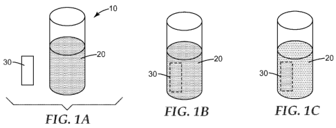
FIG. lA is a top perspective view of a substrate and a vessel holding a liquid
medium
comprising an indicator reagent.
FIG. 1B is a top perspective view of the vessel of FIG. lA immediately after
immersion of the substrate of FIG lA into the liquid medium.
FIG. 1C is a top perspective view of the vessel of FIG. 2A after a period of
time.
FIG. 2 is a drawing of a u.v.-visible absorbance spectrum of an aqueous
solution of
bromocresol purple and a fluorescence emission spectrum of a solution of 4-
methylumbelliferone.
FIG. 3 is a block diagram of one embodiment of a method of detecting a
biological
activity according to the present disclosure.
FIG. 4 is a front perspective view of a biological sterilization indicator
according to one
embodiment of the present disclosure, the biological sterilization indicator
including a housing
that includes a first portion and a second portion.
FIG. 5 is a rear perspective view of the biological sterilization indicator of
FIG. 4.
FIG. 6 is a front exploded view of the biological sterilization indicator of
FIGS. 4-5.
FIG. 7 is a side cross-sectional view of the biological sterilization
indicator of FIGS. 4-
6, taken along line 4-4 of FIG. 4, the biological sterilization indicator
shown in a first state, and
the second portion of the housing of the biological sterilization indicator
shown in a first
position.
FIG. 8 is a top cross-sectional view of the biological sterilization indicator
of FIGS. 4-
6, taken along line 5-5 of FIG. 6.
FIG. 9 is a side cross-sectional view of the biological sterilization
indicator of FIGS. 4-
8, the biological sterilization indicator shown in a second state, and the
second portion of the
housing of the biological sterilization indicator shown in a second position.
FIG. 10 is a top cross-sectional view of the biological sterilization
indicator of FIGS. 4-
9, with portions removed for clarity.
CA 02816459 2013-04-29
WO 2012/061213 PCT/US2011/058212
-7-
DETAILED DESCRIPTION
The present disclosure relates to a rapid method for detecting a biological
activity. The
method includes the use of two or more indicator reagents. The method includes
providing a
liquid mixture comprising a first and a second indicator reagent, wherein the
first indicator
reagent is present in the mixture at a concentration sufficient to interfere
with the detection
(e.g., optical detection) of an otherwise detectable quantity of a biological
derivative of the
second indicator reagent. The inventive method provides rapid, sensitive
detection of a
biological activity by sequestering at least a portion of the interfering
quantity of first indicator
reagent from the bulk of the liquid mixture in order to facilitate detection
of the biological
derivative of the second indicator reagent. The method further provides a
means to more easily
observe the first indicator reagent or a biological derivative thereof. The
inventive method can
be used in a system for the automated detection of a biological activity.
The inventive system and/or method of the present disclosure can be used to
detect a
biological activity (e.g., an activity associated with an enzyme, a cell, or a
microorganism). In
some embodiments, the inventive system and/or method can be used, for example,
to detect a
biological activity associated with a particular type of microorganism (e.g.,
a vegetative cell or
a spore) that has survived exposure to a process (e.g., a disinfection
process, a food or beverage
preparation process, a sterilization process).
The inventive method relates to the detection of a biological activity in a
sample. The
sample can be any sample that includes a biological activity as defined
herein. Nonlimiting
examples of suitable samples include suspensions or cultures of cells (e.g.,
mammalian cells,
insect cells, yeast cells, filamentous fungi, bacterial cells), environmental
samples (e.g., surface
swabs), food (e.g., raw materials, in-process samples, and finished-product
samples),
beverages, clinical samples (e.g., blood, urine, sputum, tissue, mucous,
feces, wound exudate,
pus), and water (e.g., surface water, potable water, process water).
Microorganisms (e.g., bacteria, fungi, viruses) are a source of biological
activity and
can be analyzed in a test sample that may be derived from any source, such as
a physiological
fluid, e.g., blood, saliva, ocular lens fluid, synovial fluid, cerebral spinal
fluid, pus, sweat,
exudate, urine, mucus, lactation milk, or the like. Further, the test sample
may be derived from
a body site, e.g., wound, skin, nares, scalp, nails, etc.
Samples of particular interest include mucus-containing samples, such as nasal
samples
(from, e.g., anterial nares, nasopharyngeal cavity, nasal cavities, anterior
nasal vestibule, etc.),
as well as samples from the outer ear, middle ear, mouth, rectum, vagina, or
other similar
tissue. Examples of specific musosal tissues include buccal, gingival, nasal,
ocular, tracheal,
bronchial, gastrointestinal, rectal, urethral, ureteral, vaginal, cervical,
and uterine mucosal
membranes.
CA 02816459 2013-04-29
WO 2012/061213 PCT/US2011/058212
-8-
Besides physiological fluids, other test samples may include other liquids as
well as
solid(s) dissolved in a liquid medium. Samples of interest may include process
streams, water,
soil, plants or other vegetation, air, surfaces (e.g., contaminated surfaces),
and the like.
Samples can also include cultured cells. Samples can also include samples on
or in a device
comprising cells, spores, or enzymes (e.g., a biological indicator device).
Solid samples may be disintegrated (e.g., by blending, sonication,
homogenization) and
may be suspended in a liquid (e.g., water, buffer, broth). In some
embodiments, a sample-
collection device (e.g., a swab, a sponge) containing sample material may be
used in the
method. Alternatively, the sample material may be eluted (e.g., rinsed,
scraped, expressed)
from the sample-collection device before using the sample material in the
method. In some
embodiments, liquid or solid samples may be diluted in a liquid (e.g., water,
buffer, broth).
Suitable samples also liquid and/or solid samples that have been exposed to a
sterilant.
Nonlimiting examples of these samples include spore suspensions, spore strips,
and coupons of
various materials onto which a suspension of spores or vegetative microbial
cells have been
applied.
Suitable samples also include cell-suspension media (e.g., culture broth, semi-
solid cell
culture media, and tissue culture media, filtrate) that contain cells or
previously contained cells.
Suitable samples also include cell lysates. Cell lysates may be produced by
chemical means
(e.g., detergents, enzymes), mechanical means (sonic vibration,
homogenization, French Press),
or by other cell lytic means known in the art.
FIGS. lA through 1C illustrate the process of receiving and concentrating from
a liquid
medium an indicator reagent (or a biological derivative thereof) onto or into
a substrate
according to the present disclosure. FIG. lA shows a top perspective view of
one embodiment
of a substrate 30 and a vessel 10 containing a liquid mixture 20 comprising a
colored indicator
reagent. FIG. 1B shows a top perspective view of the vessel 10 of FIG. lA
immediately after
immersing the substrate 30 in the liquid mixture 20. FIG. 1C shows a top
perspective view of
the vessel 10 of FIG. 1B after a period of time sufficient to permit the
substrate 30 to receive
and concentrate the colored indicator reagent from the liquid mixture 20. It
can be seen in FIG.
1C that the color of the liquid mixture has become less intense, while the
substrate 30 has
received and retained the colored indicator reagent and, thereby, has changed
from its initial
colorless state to a colored state.
In some embodiments, the substrate may passively receive and concentrate the
indicator reagent or biological derivative thereof (e.g., by simple diffusion
of the reagent or
derivative through the liquid medium). Alternatively or additionally (not
shown), the substrate
may actively receive and concentrate the indicator reagent and/or biological
derivative (e.g., the
substrate may be moved relative to the liquid via mixing or tumbling and/or
the liquid medium
CA 02816459 2013-04-29
WO 2012/061213
PCT/US2011/058212
-9-
may be moved relative to the substrate via fluid flow that is generally
lateral, tangential, or
orthogonal to a major surface of the substrate).
Indicator Reagents
The prior art includes a number of chromic and fluorogenic enzyme substrates
of
diverse origin which are known, commercially available, that have been used in
methods to
detect predetermined biological activities, and are suitable for use as the
first or second
indicator reagent according to the present disclosure. Among these are a
variety of fluorogenic
4-methylumbelliferyl derivatives (hydrolysable to 4-methylumbelliferone);
derivatives of 7-
amido-4-methyl-coumarin, e.g. as disclosed in GB Patent No. 1,547,747 and
European Patent
No. 0,000,063, each of which is incorporated herein by reference in its
entirety;
diacetylfluorescein derivatives; and fluorescamine.
The first indicator reagent, according to the present disclosure, comprises a
reagent that
has a first absorption spectrum and, thus, it absorbs light in the ultraviolet
and/or visible
wavelengths of the electromagnetic spectrum.
In some embodiments, the first indicator reagent can be an indicator dye
(e.g., a pH
indicator dye, a redox dye). The specific indicator dye used to detect any
given biological
activity will be selected according to criteria that are known in the art,
including, for example,
compatibility (e.g., preferably non-inhibitory) with the biological activity
to be detected,
solubility, detection system (e.g., visual and/or automated).
In any of the embodiments of the method, the indicator dye may be a pH
indicator
suitable to detect the biological activity. The indicator dye can be selected
according to criteria
known in the art such as, for example, pH range, compatibility with the
biological activity, and
solubility. In some embodiments, a salt form of the pH indicator may be used,
for example, to
increase the solubility of the pH indicator in an aqueous mixture. Nonlimiting
examples of
suitable pH indicator dyes include, for example, liyrnol blue, tropeolin 00,
methyl yellow,
methyl orange, bromphenoi blue, bromocresol green, methyl red, bromthymol
blue, phenol red,
neutral red, phenolphthaein. thymolphthalein, alizarin yellow, tropeolin 0,
nitramine,
trinitrobenzoic acid, thymol blue, bromphenol blue, tetrabromplienol blue,
bromoeresol green,
bromocresol putple, methyl red, bromihrnol blue, phenol red, Congo red, and
cresol red.
In any of the embodiments of the method, the indicator dye may be an oxidation-
reduction indicator (also called a redox indicator) suitable to detect the
biological activity.
Oxidation-reduction indicator dyes may be pH-dependent or pH-independent.
Nonlimiting
examples of oxidation-reduction indicator dyes include 2,2'-Bipyridine (Ru
complex),
Nitrophenanthroline (Fe complex), N-Phenylanthranilic acid, 1,10-
Phenanthroline (Fe
complex), N-Ethoxychrysoidine, 2,2 -Bipyridine (Fe complex), 5,6-
Dimethylphenanthroline
CA 02816459 2013-04-29
WO 2012/061213 PCT/US2011/058212
-10-
(Fe complex), o-Dianisidine, Sodium diphenylamine sulfonate,
Diphenylbenzidine,
Diphenylamine, Viologen, Sodium 2,6-Dibromophenol-indophenol, Sodium 2,6-
Dichlorophenol-indophenol, Sodium o-Cresol indophenol, Thionine (syn. Lauth's
violet),
Methylene blue, Indigotetrasulfonic acid, Indigotrisulfonic acid,
Indigodisulfonic acid,
Indigomonosulfonic acid, Phenosafranin, Safranin T, and Neutral red.
In some embodiments, the first indicator reagent can be a sulfonphthalein pH
indicator
(e.g. bromocresol purple), as shown in Example 4. The sulfonphthalein pH
indicator (e.g.,
bromocresol purple) can be present in the aqueous mixture at a concentration
of about 0.03 g
per liter. The sulfonphthalein pH indicator can be received and concentrated
by a substrate
(e.g. a charged nylon substrate such as, for example, MAGNAPROBE 0.45 micron
charged
nylon membrane, part number NPOHY00010, available from GE Osmonics Labstore,
Minnetonka, MN). The substrate can be configured as a generally planar strip
(e.g. a strip that
is about 3 mm by about 10 mm).
The second indicator reagent, according to the present disclosure, can be
converted to a
second biological derivative. The second biological derivative comprises a
reagent that has a
second absorption spectrum. Furthermore, the second biological derivative has
a characteristic
second emission spectrum (e.g., a fluorescent emission spectrum). In some
embodiments, the
second biological derivative has a characteristic second absorption spectrum
that includes
wavelengths in the ultraviolet portion of the electromagnetic energy spectrum.
The second
emission spectrum of the second biological derivative may include wavelengths
in the visible
portion of the electromagnetic energy spectrum.
Suitable compounds for use as a second indicator reagent include fluorogenic
compounds (e.g., fluorogenic enzyme substrates). Fluorogenic enzyme substrates
include 4-
methylumbelliferyl derivatives, 7-amido-4-methylcoumarin derivatives, and
diacetylfluorescein
derivatives.
Suitable 4-methylumbelliferyl derivatives include, for example: 4-
methylumbellifery1-
2-acetamido-4, 6-0-benzylidene-2-deoxy-fl-D-glucopyranoside; 4-
methylumbelliferyl acetate;
4-methylumbelliferyl-N-acetyl- 0-D-galactosaminide; 4-methylumbelliferyl-N-
acetyl-a-D-
glucosaminide; 4-methylumbelliferyl-N-acetyl- 0-D-glucosaminide; 2'-(4-
methylumbellifery1)-
a-D-N-acetyl neuraminic acid; 4-methylumbelliferyl a-L-arabinofuranoside; 4-
methylumbelliferyl a-L-arabinoside; 4-methylumbelliferyl butyrate; 4-
methylumbelliferyl 13-D-
cellobioside; methylumbelliferyl 0-D-N, N'diacetyl chitobioside; 4-
methylumbelliferyl elaidate;
4-methylumbelliferyl 0-D-fucoside; 4-methylumbelliferyl a-L-fucoside; 4-
methylumbelliferyl
0-L-fucoside; 4-methylumbelliferyl a-D-galactoside; 4-methylumbelliferyl 0-D-
galactoside; 4-
methylumbelliferyl a-D-glucoside; 4-methylumbelliferyl 0-D-glucoside; 4-
methylumbelliferyl
0-D-glucuronide; 4-methylumbelliferyl p-guanidinobenzoate; 4-
methylumbelliferyl heptanoate;
CA 02816459 2013-04-29
WO 2012/061213 PCT/US2011/058212
-11-4-methylumbelliferyl a-D-mannopyranoside; 4-methylumbelliferyl I3-D-
mannopyranoside; 4-
methylumbelliferyl oleate; 4-methylumbelliferyl palmitate; 4-
methylumbelliferyl phosphate; 4-
methylumbelliferyl propionate; 4-methylumbelliferyl stearate; 4-
methylumbelliferyl sulfate; 4-
methylumbelliferyl I3-D-N, N', N"-triacetylchitotriose; 4-methylumbelliferyl
2,3,5-tri-o-
benzoyl-a-L-arabinofuranoside; 4- methylumbelliferyl-p-trimethylammonium
cinnamate
chloride; and 4-methylumbelliferyl I3-D-xyloside.
Suitable 7-amido-4-methylcoumarin derivatives include, for example: L-alanine-
7-
amido-4-methylcoumarin; L-proline 7-amido-4-methylcoumarin; L-tyrosine-7-amido-
4-
methylcoumarin; L-leucine-7-amido-4-methylcoumarin; L-phenylalanine-7-amido-4-
methylcoumarin; and 7-glutarylphenylalanine-7-amido-4-methylcoumarin.
Suitable peptide derivatives of 7-amido-4-methyl coumarin include, for
example: N-t-
BOC-Ile-Glu-Gly-Arg 7-amido-4-methylcoumarin; N-t-BOC-Leu-Ser-Thr-Arg 7-amido-
4-
methylcoumarin; N-CBZ-Phe-Arg 7-amido-4-methyl-coumarin; Pro-Phe-Arg 7-amido-4-
methylcoumarin; N-t-BOC-Val-Pro-Arg 7-amido-4-methylcoumarin; and N-glutaryl-
Gly-Arg
7-amido-4-methylcoumarin.
Suitable diacetylfluorescein derivatives include, for example, fluorescein
diacetate,
fluorescein di-(13-D-galactopyranoside), and fluorescein dilaurate.
Where the biological activity to be detected is alpha-D-glucosidase,
chymotrypsin, or
fatty acid esterase, e.g., from Geobacillus stearothermophilus, preferred
fluorogenic enzyme
substrates are 4-methylumbelliferyl-alpha-D-glucoside, 7-glutarylphenylalanine-
7-
amido4methyl coumarin, or 4-methylumbelliferyl heptanoate, respectively. Where
the
biological activity to be detected is alpha-L-arabinofuranosidase, e.g.,
derived from Bacillus
subtilis, a preferred fluorogenic enzyme substrate is 4-methylumbelliferyl-
alpha-L-
arabinofuranoside. Where the biological activity to be detected is beta-D-
glucosidase, e.g.,
derived from Bacillus subtilis, a preferred fluorogenic enzyme substrate is 4-
methylumbelliferyl-beta-D-glucoside.
In order to carry out the method of the present invention in detecting a
biological
activity comprising an enzyme, the operator should be knowledgeable concerning
the enzyme
activity to be detected and the enzyme substrates that will react with the
enzyme so as to
produce a product which can be detected either by its fluorescence, color,
etc. (see M. Roth,
Methods of Biochemical Analysis, Vol. 7, D. Glock, Ed., Interscience
Publishers, New York,
NY, 1969, which is incorporated herein by reference in its entirety). The
appropriate enzyme
substrate to be utilized will depend upon the biological activity to be
detected.
Methods of the present disclosure include a first indicator reagent with a
first
absorption spectrum and a second indicator reagent that is converted by a
biological activity to
a second biological derivative with a second emission spectrum, wherein the
first absorption
CA 02816459 2013-04-29
WO 2012/061213 PCT/US2011/058212
-12-
spectrum at least partially overlaps the second emission spectrum. Thus, when
both the first
indicator reagent and the second biological derivative are present in a liquid
mixture, the first
indicator reagent may absorb at least a portion of the light emitted by the
second indicator
reagent, thereby diminishing the ability to detect the second biological
derivative.
A drawing can illustrate the relationship between a first indicator reagent
and a second
biological derivative according to the present disclosure. FIG. 2 shows the
absorbance
spectrum of Bromocresol Purple (hereinafter, called "BCP"), an exemplary first
indicator
reagent, and the fluorescence emission spectrum of 4-methylumbelliferone
(hereinafter, called
"4MU"), a possible biological derivative of 4-methylumbelliferyl 13-D-
glucoside, an exemplary
second indicator reagent. The spectra were obtained as described in Examples 1
and 2.
Line "A", which shows the absorbance spectrum of BCP, indicates an absorbance
maximum in the visible range around 600 nm, with relatively less absorbance by
BCP in the
425-550 nm wavelengths. The data show an absorbance peak in the visible
wavelengths
around 600 nm and an absorbance peak in the ultraviolet wavelengths at <330nm.
Line "B",
which shows the fluorescence emission spectrum of 4MU indicates an emission
maximum
around 450 nm, with relatively less emission in the ranges from 375-425 nm and
from 475-525
nm. It can be seen in FIG. 2 that the absorbance spectrum of BCP substantially
overlaps the
entire fluorescence emission peak (centered around 450 nm) of 4MU.
A person of ordinary skill in the relevant art will recognize that the amount
of
absorbance of any particular wavelength of light by a solution containing a
first indicator
reagent will be influenced by the concentration of first indicator reagent in
the solution and the
molar extinction coefficient of the indicator reagent at the selected
wavelength. The skilled
person will also recognize that the amount of light emission of any particular
wavelength by a
solution containing a biological derivative of a second indicator reagent will
be influenced by
the concentration of the second biological derivative in the solution and the
fluorescence
quantum yield of the biological derivative. Therefore, the concentration of
the first indicator
reagent in the liquid mixture can be selected in conjunction with an
appropriate substrate to
permit i) the substrate to remove enough first indicator substrate from the
liquid mixture to
allow more sensitive detection of the second biological derivative and ii) the
first indicator
reagent (or biological derivative thereof) to be easily detected on the
substrate material.
The combination of bromocresol purple and 4-methylumbelliferyl-a-D-glucoside
represents an example of suitable first and second indicator reagents,
respectively, according to
the present disclosure. This combination can be used to detect a first
biological activity such as
the fermentation of a carbohydrate to acid end products and a second
biological activity such as
-a-D-glucosidase enzyme activity, for example. These activities can indicate
the presence or
absence of a viable spore following the exposure of a biological sterilization
indicator to a
CA 02816459 2013-04-29
WO 2012/061213 PCT/US2011/058212
-13-
sterilization process, for example. The bromocresol purple can be used at a
concentration of
about 0.03 g/L in the aqueous mixture, for example. The 4-methylumbelliferyl-a-
D-glucoside
can be used, for example, at a concentration of about 0.05 to about 0.5 g/L
(e.g., about 0.05 g/L,
about 0.06 g/L, about 0.07 g/L, about 0.08 g/L, about 0.09 g/L, about 0.1 g/L,
about 0.15 g/L,
about 0.2 g/L, about 0.25 g/L, about 0.3 g/L, about 0.35 g/L, about 0.4 g/L,
about 0.45 g/L,
about 0.5 g/L) .in the aqueous mixture.
Thus, according to the present disclosure, the first indicator reagent may
interfere with
the detection of an otherwise detectable amount of the biological derivative
of the second
indicator reagent. The spectral interference between any proposed first and
second indicator
reagents can be demonstrated by a person of ordinary skill in the art by
performing the
following simple experiment.
First, the operator makes a relatively-dilute, but fluorescently-detectable,
aqueous
solution of the expected biological derivative of the proposed second
indicator reagent. For
example, if the second indicator reagent is a 4-methylumbelliferyl compound,
the expected
biological derivative is 4MU. The solution can contain, for example, about
0.05 to 0.2
micrograms per milliliter 4MU. Next, the operator adds an effective amount of
the proposed
first indicator reagent. For example, if BCP is the proposed first indicator
reagent, it can be
added at a concentration (e.g., 0.04 milligrams per milliliter) that is used
in microbiological
growth media for the detection of fermentative microorganisms. By comparing
the
fluorescence of the 4MU solutions with and without the BCP, it can be
determined whether the
first indicator reagent (in this example, the BCP) can interfere with the
detection of the
biological derivative of the second indicator reagent (in this case, the 4MU).
The operator can
then test whether adding reduced amounts of BCP to the 4MU solution improves
the detection
of relatively low concentrations of 4MU. This type of experiment easily can be
performed with
any combination of first and second indicator reagents. An example of this
procedure is shown
in Example 3.
Substrate
Suitable substrates, according to the present disclosure, are configured to
receive and
concentrate the indicator reagent. The ability of the substrate to concentrate
the indicator
reagent or biological derivative thereof can be affected by one or more of a
variety of forces
known in the art and discussed herein. Thus, a person of ordinary skill in the
art may select a
substrate that is known to be positively-charged to concentrate an indicator
reagent (or
biological derivative thereof) that is known to be negatively-charged, for
example. Conversely,
a person of ordinary skill in the art may select a substrate that is known to
be negatively-
charged to concentrate an indicator reagent (or biological derivative thereof)
that is known to
CA 02816459 2013-04-29
WO 2012/061213 PCT/US2011/058212
-14-
be positively-charged. A person of ordinary skill in the art may select a
substrate that is known
to have hydrophobic properties to concentrate an indicator reagent (or
biological derivative
thereof) that is known to comprise hydrophobic portions that would be retained
by a
hydrophobic substrate. Additionally, a person of ordinary skill in the art may
easily select a
suitable substrate material by contacting, for a period of time, a candidate
substrate material
with a liquid comprising the indicator reagent or biological derivative
thereof and analyzing the
substrate to determine whether a detectable amount of the indicator reagent or
derivative
thereof accumulates onto or in the substrate.
It will be apparent to a person of ordinary skill in the art that the
substrate material can
be selected according to known properties of the indicator reagent or the
biological derivative
thereof. For example, a positively-charged substrate may be selected for use
in the method
when the biological derivative of the indicator reagent is a negatively-
charged molecule.
Furthermore, a negatively-charged substrate may be selected for use in the
method when the
biological derivative of the indicator reagent is a positively-charged
molecule.
Alternatively, the suitability of any given substrate material for use with a
given first
indicator reagent in the inventive method can be readily determined using the
following
experimental approach. In a suitable vessel (e.g., a test tube), a source of
predetermined
biological activity (e.g., microbial cells capable of fermenting a
carbohydrate to acidic end
products) can be added with a first indicator reagent (e.g., a pH indicator)
to a liquid medium
selected to facilitate the biological activity (e.g. a broth medium comprising
the fermentable
carbohydrate). The liquid medium can be contacted with a candidate substrate
under
conditions to facilitate the predetermined biological activity and the
substrate can be removed
from the medium, optionally rinsed and/or blotted to remove excess liquid, and
observed
visually or instrumentally (e.g., with a spectroreflectometer or a
fluorometer) to determine
whether the substrate material concentrated the first indicator reagent and/or
a biological
derivative thereof during contact with the liquid medium. In the illustrative
example, a suitable
substrate/indicator combination would show evidence that either the first
indicator reagent or
the biological derivative thereof concentrated onto or into the substrate
material (out of the
liquid medium) during the contact period. A control reaction without substrate
material can be
run to confirm the presence of the biological activity in the mixture.
The substrate may be fabricated in a generally planar sheet form (e.g., a
membrane
strip, as shown in FIG. 1A). The size and/or effective surface area of the
substrate can also
affect the ability of the substrate to concentrate the indicator reagent (or
biological derivative
thereof). Preferred materials for the substrate include porous materials
(e.g., woven materials,
nonwoven materials, a porous membranes, microporous membranes, filter paper).
In some
embodiments, particularly preferred substrate materials include charged
membranes such as,
CA 02816459 2013-04-29
WO 2012/061213 PCT/US2011/058212
-15-
for example, charged nylon membranes (e.g., MAGNAPROBE 0.45 micron charged
nylon
membrane, part number NPOHY00010, available from GE Osmonics Labstore,
Minnetonka,
MN).Substrates used in the present disclosure can be fabricated from a variety
of materials.
U.S. Patent No. 6,562,297, which is incorporated herein by reference in its
entirety, describes
membranes for the immobilization of pH indicators. Nonlimiting examples of
suitable
substrate materials include, for example, natural materials (e.g., cellulose),
synthetic materials
(e.g. nylon), and combinations and/or derivatives thereof.
Method of Detecting a Biological Activity:
FIG. 3 shows a block diagram of one embodiment of a method to detect one of a
plurality of biological activities according to the present disclosure.
The method includes the step 40 of providing a sample that may include one of
a
plurality of predetermined biological activities, first and second indicator
reagents, and a
substrate that receives and concentrates from an aqueous medium the first
indicator reagent
and, optionally, a biological derivative thereof.
In some embodiments, the method may include the optional step 45 of exposing
the
biological activity to a disinfectant, an antibiotic, or a sterilant. This
optional step may be
included to determine the efficacy of a sterilization process or to detect a
predetermined
biological activity (or microorganism) subsequent to a selective enrichment
culture process.
Exposing the biological activity to a sterilant may comprise exposing the
biological activity to a
sterilization process. Sterilization processes include exposing the sample,
for example, to
sterilants such as steam, dry heat, ethylene oxide, formaldehyde, peroxides,
hydrogen peroxide,
peracetic acid, ozone, or mixtures thereof (e.g., a mixture of ozone and
hydrogen peroxide.
The method includes the step 50 of forming a first aqueous mixture comprising
the
sample and the first and second indicator reagents. The first aqueous mixture
is formed in an
aqueous medium. The source of biological activity in the method can be any
sample
comprising, or suspected of comprising one or more biological activities, as
described herein.
"Aqueous medium", as used herein, refers to an aqueous liquid in which the
first and second
indicator reagents are or can be dissolved or suspended. Preferably, the
medium does not
substantially interfere with the detection of a predetermined biological
activity to be detected.
In some embodiments, the aqueous medium may comprise a component (i.e., a
buffering agent)
to adjust the pH of the medium. The aqueous medium further may comprise a
reagent (e.g., a
detergent, a cofactor, a cell lysis agent) that is known in the art to
facilitate the detection of one
or more biological activities.
In some embodiments, the sample comprises water and, thus, the sample itself
may be
considered an aqueous medium. In any embodiment, the sample may optionally be
mixed with
CA 02816459 2013-04-29
WO 2012/061213 PCT/US2011/058212
-16-
a second liquid (e.g., an aqueous medium, a diluent, a buffer, a solution to
neutralize a
disinfectant) before mixing the sample with the first and second indicator
reagents.
In some embodiments, the aqueous medium can be combined with the first and/or
second indicator reagents before the medium is mixed with the sample. In some
embodiments,
the first and second indicator reagents and the sample can be added
sequentially to the aqueous
medium to form the first aqueous mixture. In some embodiments, the first and
second indicator
reagents can be combined with an aqueous medium and the sample simultaneously
to form the
first aqueous mixture. In any of the embodiments, either or both of the first
and second
indicator reagents initially may be in the form of a dry reagent, a liquid, a
gel, or a film before
the reagent is combined with an aqueous medium and/or a sample to form the
first aqueous
mixture.
The first indicator reagent can be any suitable reagent described herein.
Because the
first indicator reagent is selected to detect a predetermined biological
activity, the chemical
nature of the first indicator reagent and biological derivatives thereof are
known and, thus,
suitable substrate materials can be identified as described herein. The second
indicator reagent
can be any suitable reagent described herein.
In any embodiment of the method, forming the first aqueous mixture can
comprise
forming a first aqueous mixture that includes a nutrient. The nutrient can be
provided to
facilitate the growth of a target cell or microorganism, for example, and may
be provided as a
mixture of nutrients. Nutrients and nutrient media to facilitate the growth of
microorganisms
are known in the art and can be found, for example, in the "Handbook of
Microbiological
Media" by Ronald Atlas, published by CRC Press, Boca Raton, FL. Matner et al.
(U.S. Patent
No. 5,073,488) describes a nutrient medium for the growth and detection of
bacterial spores in
a biological sterilization indicator. Nutrients and nutrient media for
facilitating the growth of
eukaryotic cells (e.g., mammalian cells, insect cells) are also known in the
art and include, for
example, sugars (e.g., glucose), amino acids, vitamins (e.g., thiamin,
niacin), choline, inositol,
serum, and mixtures thereof.
Methods of the present disclosure further include the step 60 of bringing the
first
aqueous mixture into fluid communication with the substrate to form a second
aqueous
mixture. Typically, the process of bringing the first aqueous mixture into
fluid communication
with the substrate occurs in a vessel (e.g., a tube, a bottle, a flask, a
microwell). In any of the
embodiments, the vessel may be sealed to minimize evaporation and/or to
prevent
contamination by an exogenous biological activity, for example. In any of the
embodiments,
bringing the first aqueous mixture into fluid communication with the substrate
may include
contacting the liquid mixture and the substrate under conditions that
facilitate the
predetermined biological activity. A person of ordinary skill in the art will
recognize
CA 02816459 2013-04-29
WO 2012/061213 PCT/US2011/058212
-17-
conditions that facilitate the predetermined biological activity. The
conditions may include, for
example, the pH, ionic strength or buffering capacity of the mixture; the
concentration of first
and/or second indicator reagents; presence of cofactors in the mixture or
vessel; and/or
temperature of the mixture.
In any of the embodiments of the method, bringing the first aqueous mixture
into fluid
communication with the substrate can include controlling the temperature of
the mixture. In
some embodiments, the temperature may be controlled at a temperature higher
than ambient
temperature (e.g., a temperature that facilitates a reaction, such as a
catalytic reaction or
binding reaction, involving the biological activity) using a heating block, an
incubator, or some
other suitable heating means known in the art. In some embodiments, the
temperature of the
mixture may be controlled at a temperature lower than ambient temperature. In
some
embodiments, the mixture may be subjected to a transient temperature shift
(e.g., a heat shock
or a cold shock) to facilitate the detection of the predetermined biological
activity.
Bringing the first aqueous mixture into fluid communication with the substrate
according to the present disclosure comprises concentrating the first
indicator reagent and;
optionally, a biological derivative thereof; onto and/or into the substrate.
As a result of this, the
concentration of the first indicator reagent in the second aqueous mixture is
lower than the
concentration of the first indicator reagent in the first aqueous mixture. As
discussed above, the
substrate is selected to receive and concentrate the first indicator reagent.
The substrate
receives the first indicator reagent via contact with the aqueous medium. The
substrate retains
the first indicator reagent or biological derivative thereof via a variety of
means. Without being
bound by theory, the accumulation of the first indicator reagent or biological
derivative thereof
onto and/or into the substrate material may occur through one or more of a
variety of chemical
attractive forces including, but not limited to, ionic interaction,
hydrophobic interaction, van
der Waal's forces, and hydrogen bonding, for example.
The process of receiving and concentrating the first indicator reagent or
biological
derivative thereof by the substrate occurs during the period of fluidic
communication between
the aqueous medium and the substrate. During this period of fluidic
communication, the first
indicator reagent or biological derivative thereof accumulates on the
substrate at a rate that may
be dependent upon a number of factors including, for example, the
concentration of the first
indicator reagent (or biological derivative thereof), the surface area of the
substrate material
contacting the liquid medium, the porosity of the substrate, a charge density
associated with the
substrate material, and/or other substances in the liquid medium that can
interact with the
substrate and/or the first indicator reagent (or biological derivative
thereof) in a way that
interferes with the receiving or concentrating the first indicator reagent or
biological derivative
thereof by the substrate. Receiving and concentrating at least a portion of
the first indicator
CA 02816459 2013-04-29
WO 2012/061213 PCT/US2011/058212
-18-
reagent or biological derivative thereof onto the substrate can occur within a
relatively short
contact period (e.g., within several minutes) and may continue over a longer
contact period
(e.g., up to 1 hour, up to 2 hours, up to 4 hours, up to 18 hours, up to 24
hours, up to 7 days, up
to two weeks). In some embodiments, the first indicator reagent may
concentrate onto or into
the substrate within a relatively short period of time (e.g., minutes hours),
whereas the first
biological derivative, if present, may not be detectably concentrated on or in
the substrate for a
relatively longer period of time (e.g., hours, days).
During any of the periods of fluidic communication between the aqueous mixture
and
the substrate described above, the substrate may receive and concentrate all
or a portion of the
first indicator reagent (or biological derivative thereof). In some
embodiments, the substrate
receives and concentrates at least 5 percent of the first indicator reagent
(or biological
derivative thereof). In some embodiments, the substrate receives and
concentrates at least 10
percent of the first indicator reagent (or biological derivative thereof). In
some embodiments,
the substrate receives and concentrates at least 20 percent of the first
indicator reagent (or
biological derivative thereof). In some embodiments, the substrate receives
and concentrates at
least 30 percent of the first indicator reagent (or biological derivative
thereof). In some
embodiments, the substrate receives and concentrates at least 40 percent of
the first indicator
reagent (or biological derivative thereof). In some embodiments, the substrate
receives and
concentrates at least 50 percent of the first indicator reagent (or biological
derivative thereof).
In some embodiments, the substrate receives and concentrates at least 75
percent of the first
indicator reagent (or biological derivative thereof). In some embodiments, the
substrate
receives and concentrates at least 80 percent of the first indicator reagent
(or biological
derivative thereof). In some embodiments, the substrate receives and
concentrates at least 90
percent of the first indicator reagent (or biological derivative thereof). In
some embodiments,
the substrate receives and concentrates greater than 90 percent of the first
indicator reagent (or
biological derivative thereof). In some embodiments, the substrate receives
and concentrates
greater than 95 percent of the first indicator reagent (or biological
derivative thereof).
Determining that the substrate receives and concentrates the first indicator
reagent (or
biological derivative thereof) easily can be accomplished by bringing a liquid
medium
comprising the first indicator reagent (or biological derivative thereof) into
fluid
communication with the substrate for a period of time and analyzing the
substrate for the
presence of the reagent (or biological derivative thereof), as shown in
Example 1. Preferably,
any excess liquid medium is removed from the substrate (e.g., by blotting or
by centrifugation)
before analyzing the substrate so that the amount of reagent or biological
derivative associated
with the substrate indicates the amount retained by the substrate. Suitable
analysis methods
will be apparent to a person of ordinary skill in the art. For example, a
substrate that receives
CA 02816459 2013-04-29
WO 2012/061213 PCT/US2011/058212
-19-
and concentrates a colored first indicator reagent (e.g., a pH indicator) can
be analyzed by
reflectance spectroscopy using, for example, an X-Rite model 530P portable
Spectrodensitometer.
Thus, when a liquid medium comprising a sample and the first indicator reagent
(or
biological derivative thereof) is brought into fluid communication with a
suitable substrate, the
concentration of the first indicator reagent (or biological derivative
thereof) in the bulk liquid
medium decreases as the first indicator reagent (or biological derivative
thereof) is received and
concentrated by the substrate. This feature of the invention facilitates the
detection of relatively
small concentrations of the biological derivative of the second indicator
reagent because at least
a portion of the interference (i.e., the absorption of the fluorescence) by
the first indicator
reagent is removed as the first indicator reagent is concentrated onto the
substrate from the
aqueous mixture. In some embodiments, the first indicator reagent and/or a
biological
derivative thereof, when in a freely-diffusible form (i.e., in the bulk liquid
medium) may inhibit
the biological activity. In these embodiments a further advantage of the
invention is that the
substrate can effectively sequester at least a portion of the first indicator
reagent, thereby
reducing the inhibition of the biological activity by the first indicator
reagent.
Referring back to FIG. 3, the method further may include the optional step 65
of
facilitating the growth of cells. Facilitating the growth of cells is used
broadly to include
providing conditions (e.g., nutrients, germinants, buffers, oxidation-
reduction potential, gasses)
to facilitate, for example, the germination of spores, energy metabolism,
biosynthesis, and/or
cell division. Facilitating the growth of cells may result in the
amplification of one or more
predetermined biological activities from the original sample and, thereby, can
improve the
sensitivity for detecting the predetermined biological activities.
Methods of the present disclosure further include the step 70 of detecting a
biological
derivative of the second indicator reagent (herein, called "second biological
derivative"). In
some embodiments, the second biological derivative can be detected in an
aqueous medium.
Detecting the presence or absence of the second biological is indicative of
the presence or
absence, respectively, of the corresponding predetermined biological activity
in the sample.
The second biological derivative can be detected by several means. In some
embodiments, the second biological derivative can be detected optically. In
some
embodiments, detecting the second biological derivative may comprise detecting
the biological
derivative visually. In some embodiments, detecting the second biological
derivative may
comprise detecting the biological derivative using an instrument. For example,
if the second
biological derivative can be detected using an optical instrument such as a
fluorometer.
In any of the embodiments, detecting the presence or absence of the second
biological
derivative thereof may further comprise measuring the quantity of the second
biological
CA 02816459 2013-04-29
WO 2012/061213 PCT/US2011/058212
-20-
derivative. Measuring the quantity may be done by any means known in the art
including, for
example measuring the quantity using an instrument (e.g., a fluorometer). In
some
embodiments, measuring the quantity of second biological derivative may
comprise comparing
the fluorescence in the aqueous mixture to a fluorescent standard.
In any of the embodiments, methods of the present disclosure optionally
include the
step 75 of detecting the first indicator reagent or a first biological
derivative thereof. The
means for detecting the first indicator reagent or the first biological
derivative depends upon the
nature of the first indicator reagent or the first biological derivative, as
will be appreciated by a
person of ordinary skill in the art. For example, if the first indicator
reagent is a chromic
(colored) and/or the first biological derivative is a chromic compound, then
the first indicator
reagent and/or the first biological derivative may be detected optically
(either visually or by an
instrument (e.g., a spectrophotometer)). In some embodiments, detecting the
first indicator
reagent or first biological derivative may further comprise detecting the
first indicator reagent
or first biological derivative in a portion of the aqueous mixture that is not
associated with the
substrate (e.g., in the bulk liquid). For example, if the first indicator
reagent or first biological
derivative is detected using an optical instrument such as a
spectrophotometer, the optical path
does not intersect any portion of the substrate.
In any of the embodiments, detecting the presence or absence of the first
indicator
reagent or first biological derivative may further comprise measuring the
quantity of the first
indicator reagent or first biological derivative. Measuring the quantity may
be done by any
means known in the art including, for example measuring the quantity using an
instrument
(e.g., a spectrophotometer, a spectrodensitometer).
U.S. Patent Nos. 5,252,484 and 5,418,167; each of which is incorporated herein
by
reference in its entirety; describe an embodiment of a rapid readout
biological indicator wherein
the biological indicator comprises an enzyme carrier (spore strip) and an
ampoule contain a
solution with 4-methylumbelliferyl-a-D-glucoside ("MUG", a fluorogenic enzyme
substrate)
and bromocresol purple ("BCP", a pH indicator). MUG is known to be hydrolyzed
by
enzymatic activity to 4-methylumbelliferone (4MU), a fluorescent derivative of
MUG. As
shown in Examples 1 and 2 of U.S. Patent No. 5,252,484, the 4MU produced by
enzymatic
hydrolysis of the MUG can be detected visually by fluorescence within minutes
after the
enzyme carrier is brought into fluid communication with the solution
containing MUG and
BCP.
The present investigators have discovered that the concentration of BCP used
in a
solution similar to that described in Example 1 of U.S. Patent No. 5,252,484
is sufficient to
interfere with the detection of low concentrations of 4MU in an aqueous
solution. Removal of
at least a portion of the BCP from the solution according to the present
disclosure will permit
CA 02816459 2013-04-29
WO 2012/061213 PCT/US2011/058212
-21-
the detection of smaller quantities of 4MU in a biological indicator, thereby
permitting earlier
detection of biological activity (e.g., spores, enzymes) that have been
exposed to a sterilization
process and were not thereby inactivated and/or killed.
System for Detecting a Biological Activity
The present disclosure includes a system for detecting a predetermined
biological
activity in a sample. The system can be used according to the inventive method
to detect the
one or more biological activities in a sample. The system includes a first
indicator system
comprising a first indicator reagent that can be converted by a first
predetermined biological
activity to a first biological derivative. The first indicator reagent has a
first absorption
spectrum and, optionally a first emission spectrum. The system further
includes a second
indicator system comprising a second indicator reagent that can be converted
by a second
predetermined biological activity to a second biological derivative. The
second biological
derivative has a first absorption spectrum and a second emission spectrum.
The system further includes an instrument configured to receive a liquid
sample that
may comprise the first indicator reagent, the second indicator reagent, the
first biological
derivative, the second biological derivative or any combination of two or more
of the
foregoing. The instrument may be configured to withdraw the liquid sample from
an external
container via a "sipper" means, as known in the art of analytical instruments.
Alternatively, the
instrument may be configured to receive a vessel (e.g., a tube, a microwell
plate, or the like)
containing the liquid sample.
The instrument is configured to detect the second biological derivative.
Optionally, the
instrument further can be configured to detect the first indicator reagent,
the second indicator
reagent, the first biological derivative, or any combination of two or more of
the foregoing.
The indicator reagent of the system can be any suitable indicator reagent, as
described
herein, to detect the particular predetermined biological activity. The first
and second indicator
reagents may be provided in a kit, for example, which optionally may include
an aqueous
medium (e.g., a buffer, a suspending medium, a diluent) in which to mix the
indicator reagent
and the sample. As discussed herein, the sample may comprise water and, thus,
may constitute
the aqueous medium. Optionally, the kit may further include a vessel (e.g., a
tube, a cuvette, or
the like) in which to form an aqueous mixture comprising the sample and the
first and second
indicator reagents. In some embodiments, the system can be used with a
biological sterilization
indicator such as, for example, the biological indicators in U.S. Patent
Application Nos.
61/408,977 and 61/408,988, filed on November 1, 2010, and the biological
indicators
described in U.S. Patent No. 5,252,484; each of which is incorporated herein
by reference in its
entirety.
CA 02816459 2013-04-29
WO 2012/061213 PCT/US2011/058212
-22-
Instruments to detect the absorption spectra of chromic compounds are known in
the art
and include, for example, a variety of commercially-available
spectrophotometers and
spectrodensitometers. Instruments to detect the emission spectra of
fluorescent compounds are
also known in the art and include, for example, a variety of commercially-
available
fluorometers. Such instruments can be readily adapted to detect an indicator
reagent (or
biological derivative thereof) associated with a liquid sample and/or a
substrate positioned at a
predetermined location.
In some embodiments, the substrate can be removed from the aqueous mixture and
positioned (e.g., on a surface or in a cuvette) such that the indicator
reagent (or biological
derivative thereof) can be detected by the instrument. U.S. Patent No.
6,025,189; which is
incorporated herein by reference in its entirety, describes an instrument
configured to detect, at
a predetermined location in a self-contained biological indicator, a
fluorescent signal associated
with a biological activity. It is within ordinary skill in the art to modify
such an instrument to
detect a chromic signal.
In some embodiments, the system may further comprise a processor. In some
embodiments, the instrument may comprise a microprocessor capable of
controlling the
instrument and collecting and/or transmitting data associated with detecting
the indicator
reagent or biological derivative thereof. In some embodiments of the system,
the processor
may comprise an external processor. The external computer may comprise a
personal
computer (PC), desktop computer, laptop computer, handheld computer,
workstation, or the
like. For example, software programs can be loaded on external computer to
control the
instrument and/or to facilitate the collection, transfer and/or analysis of
data from the
instrument.
In some embodiments, the system may further comprise means to regulate the
temperature of a liquid. The means for temperature control can include any
means known in
the art such as, for example, thermocouples and heat-exchangers.
Advantageously, these
embodiments provide a system that can facilitate the biological activity by
controlling the
temperature and can detect the product of the biological activity.
Biological Sterilization Indicators:
FIGS. 4-10 illustrate the biological sterilization indicator 100 according to
one
embodiment of the present disclosure. Other suitable embodiments of biological
sterilization
indicators are described in co-pending PCT Publication No. WO 2011/011189,
entitled
"Biological Sterilization Indicator and Method of Using Same"; US Patent
Application No.
61/409,042, entitled "Biological Sterilization Indicator System and Method";
US Patent
Application No. 61/408,997, entitled "Biological Sterilization Indicator
System and Method";
CA 02816459 2013-04-29
WO 2012/061213 PCT/US2011/058212
-23-
and US Patent Application No. 61/408,977, entitled "Biological Sterilization
Indicator and
Method of Using Same"; each of which is incorporated herein by reference in
its entirety.
The biological sterilization indicator 100 can include a housing 102, which
can include
a first portion 104 and a second portion 106 (e.g., a cap) adapted to be
coupled together to
provide a self-contained biological sterilization indicator. In some
embodiments, the first
portion 104 and second portion 106 can be formed of the same materials, and in
some
embodiments, the first portion 104 and the second portion 106 can be formed of
different
materials. The housing 102 can define a reservoir 103 of the biological
sterilization
indicator 100 in which other components can be positioned and into which a
sterilant can be
directed during a sterilization process.
The housing 102 can be defined by at least one liquid impermeable wall, such
as a wall
108 of the first portion 104 and/or a wall 110 of the second portion 106. It
should be
understood that a one-part unitary housing 102 may also be employed or that
the first and
second portions 104 and 106 can take on other shapes, dimensions, or relative
structures
without departing from the spirit and scope of the present disclosure.
Suitable materials for the
housing 102 (e.g., the walls 108 and 110) can include, but are not limited to,
a glass, a metal
(e.g., foil), a polymer (e.g., polycarbonate (PC), polypropylene (PP),
polyphenylene (PPE),
polythyene, polystyrene (PS), polyester (e.g., polyethylene terephthalate
(PET)), polymethyl
methacrylate (PMMA or acrylic), acrylonitrile butadiene styrene (ABS), cyclo
olefin polymer
(COP), cyclo olefin copolymer (COC), polysulfone (PSU), polyethersulfone
(PES),
polyetherimide (PEI), polybutyleneterephthalate (PBT)), a ceramic, a
porcelain, or
combinations thereof.
In some embodiments, the biological sterilization indicator 100 can further
include a
frangible container 120 that contains a liquid 122, and which is dimensioned
to be received
within the biological sterilization indicator 100, for example, within at
least a portion of the
housing 102 (e.g., at least within the first portion 104 of the housing 102).
The frangible
container 120 can be formed of a variety of materials, including, but not
limited to, one or more
of metal (e.g., foil), a polymer (e.g., any of the polymers listed above with
respect to the
housing 102), glass (e.g., a glass ampoule), and combinations thereof. In some
embodiments,
only a portion of the container 120 is frangible, for example, the container
120 can include a
frangible portion or cover (e.g., a frangible barrier, film, membrane, or the
like). The frangible
container 120 can have a first state in which it is intact and the liquid 122
is contained therein,
and a second state in which at least a portion of the container 120 is
fractured. In the second
state of the container 120, the liquid 122 can be in fluid communication with
the reservoir 103
of the biological sterilization indicator 100, e.g., when the container 120 is
positioned in the
biological sterilization indicator 100.
CA 02816459 2013-04-29
WO 2012/061213 PCT/US2011/058212
-24-
As shown in the illustrated embodiment, the container 120 can be held in place
within
the biological sterilization indicator 100 and/or fractured by an insert 130,
which is described in
greater detail below. The container 120 can be fractured, for example, by
urging the container
120 against the insert 130 (e.g., an insert that functions as a breaker) or by
urging the insert 130
against the container 120.
The first portion 104 of the housing 102 can be adapted to house a majority of
the
components of the biological sterilization indicator 100, and can be referred
to as a "tube,"
"tubular body," "base," or the like. The housing 102 can include a reservoir
103 that can be
defined by one or both of the first portion 104 and the second portion 106 of
the housing 102.
The biological sterilization indicator 100 can further include spores or
another source(s) of
biological activity 115 (or a locus of spores) positioned in fluid
communication with the
reservoir 103. As shown in FIGS. 4-6, the second portion 106 of the housing
102 can include
one or more apertures 107 to provide fluid communication between the interior
of the housing
102 (e.g., the reservoir 103) and ambience. For example, the one or more
apertures 107 can
provide fluid communication between the spores 115 and ambience during a
sterilization
process, and can serve as an inlet into the biological sterilization indicator
100 and as an inlet of
a sterilant path 164 (described in greater detail below). In some embodiments,
the second
portion 106 of the housing 102 can be coupled to a first (e.g., open) end 101
of the first
portion 104 of the housing 102, and the spores 115 can be positioned at a
second (e.g., closed)
end 105, opposite the first end 101, of the first portion 104 of the housing
102.
In some embodiments, a barrier or filter (e.g., a sterile barrier; not shown)
can be
positioned in the sterilant path 164 (e.g., at the inlet formed by the
aperture 107) to inhibit
contaminating or foreign organisms, objects or materials from entering the
biological
sterilization indicator 100. Such a barrier can include a gas-transmissive,
microorganism-
impermeable material, and can be coupled to the housing 102 by a variety of
coupling means,
including, but not limited to, an adhesive, a heat seal, sonic welding, or the
like. Alternatively,
the barrier can be coupled to the sterilant path 164 via a support structure
(such as the second
portion 106) that is coupled to the first portion 104 of the housing 102
(e.g.,. in a snap-fit
engagement, a screw-fit engagement, a press-fit engagement, or a combination
thereof).
During exposure to a sterilant, the sterilant can pass through the barrier
into the sterilant
path 164 and into contact with the spores 115.
In some embodiments, as shown in the illustrated embodiment, the housing 102
can
include a lower portion 114 and an upper portion 116, which can be at least
partially separated
by an inner wall (or partial wall) 118, ledge, partition, flange, or the like,
in which can be
formed an opening 117 that provides fluid communication between the lower
portion 114 and
the upper portion 116. In some embodiments, the lower portion 114 of the first
portion 104 of
CA 02816459 2013-04-29
WO 2012/061213 PCT/US2011/058212
-25-
the housing 102 (sometimes referred to as simply "the lower portion 114" or
the "the lower
portion 114 of the housing 102") can be adapted to house the spores 115 or a
locus of spores.
In some embodiments, the lower portion 114 can be referred to as the
"detection portion" or
"detection region" of the housing 102, because at least a portion of the lower
portion 114 can
be interrogated for signs of spore growth. In addition, in some embodiments,
the upper portion
116 of the first portion 104 of the housing 102 (sometimes referred to as "the
upper portion
116" or the "the upper portion 116 of the housing 102" for simplicity) can be
adapted to house
at least a portion of the frangible container 120, particularly before
activation.
In some embodiments, the portion of the reservoir 103 that is defined at least
partially
by the upper portion 116 of the housing 102 can be referred to as a first
chamber (or reservoir,
zone, region, or volume) 109 and the portion of the reservoir 103 that is
defined at least
partially by the lower portion 114 of the housing 102 can be referred to as a
second chamber (or
reservoir, zone, region, or volume) 111. In some embodiments, the second
chamber 111 can be
referred to as a "spore growth chamber" or a "detection chamber," and can
include a volume to
be interrogated for spore viability to determine the efficacy of a
sterilization process.
The first chamber 109 and the second chamber 111 can be positioned in fluid
communication with each other to allow a sterilant and the liquid 122 to move
from (i.e.,
through) the first chamber 109 to the second chamber 111. In some embodiments,
the degree
of fluid connection between the first chamber 109 and the second chamber 111
(e.g., the size of
an opening, such as the opening 117, connecting the first chamber 109 and the
second chamber
111) can increase after, simultaneously with, and/or in response to the
activation step (i.e., the
liquid 122 being released from the container 120). In some embodiments, the
control of fluid
communication (or extent of fluid connection) between the first chamber 109
(e.g., in the upper
portion 116) and the second chamber 111 (e.g., in the lower portion 114) can
be provided by at
least a portion of the insert 130.
The container 120 can be positioned and held in the first chamber 109 during
sterilization and when the container 120 is in a first, unfractured, state.
The spores 115 can be
housed in the second chamber 111 and in fluid communication with ambience when
the
container 120 is in the first state. The first chamber 109 and the second
chamber 111 can be
configured such that the container 120 is not present in the second chamber
111, and
particularly, not when the container 120 is in its first, unfractured, state.
A sterilant can move
into the second chamber 111 (e.g., via the first chamber 109) during
sterilization, and the liquid
122 can move into the second chamber 111 (e.g., from the first chamber 109)
during activation,
when the container 120 is fractured and the liquid 122 is released into the
interior of the
housing 102.
CA 02816459 2013-04-29
WO 2012/061213 PCT/US2011/058212
-26-
As a result, when the container 120 is in the first state, the first chamber
109 and the
second chamber 111 can be in fluid communication with one another, and with
ambience (e.g.,
during sterilization). For example, the first chamber 109 and the second
chamber 111 can be in
fluid communication with ambience via the one or more apertures 107. In some
embodiments,
the first chamber 109 and the second chamber 111 can be in fluid communication
with
ambience in such a way that the first chamber 109 is positioned upstream of
the second
chamber 111 when a sterilant is entering the biological sterilization
indicator 100. That is, the
first chamber 109 can be positioned between the sterilant inlet (e.g., the one
or more apertures
107) and the second chamber 111, and the sterilant inlet can be positioned on
an opposite side
of the first chamber 109 than the second chamber 111.
As shown in FIGS. 7 and 9, in some embodiments, the first chamber 109 can be
defined by one or both of the first portion 104 and the second portion 106,
particularly when
the container 120 is in the first state. In addition, in some embodiments, the
first chamber 109
can include a first end 112 positioned adjacent the open end 101 of the first
portion 104 of the
housing 102, adjacent the second portion 106 of the housing 102, and/or at
least partially
defined by the second portion 106. The first chamber 109 can further include a
second end 13
positioned adjacent and in fluid communication with the second chamber 111 and
positioned
toward the closed end 105 of the housing 102. The first end 112 of the first
chamber 109 can
be at defined by the first portion 104 and/or the second portion 106 of the
housing 102.
As further shown in FIGS. 7 and 9, in some embodiments, the second chamber 111
can
include a first end 124 positioned adjacent and in fluid communication with
the first chamber
109 and positioned toward the open end 101 of the housing 102, and a second
end 125 at least
partially defined by, including, or positioned adjacent the closed end 105 of
the housing 102.
Said another way, as shown in FIGS. 7 and 9, the biological sterilization
indicator 100
can include a longitudinal direction DL, and in some embodiments, the first
chamber 109 can be
positioned longitudinally above the second chamber 111.
In some embodiments, the second chamber 111 can be at least partially defined
by, can
include, or can be positioned adjacent the closed end 105 of the biological
sterilization indicator
100. In addition, in some embodiments, the second chamber 111 can be smaller
(e.g., in
volume and/or cross-sectional area) than at least one of the first chamber 109
and the volume of
the liquid 122 in the container 120 that will be released when the biological
sterilization
indicator 100 is activated. As a result, in such embodiments, the second
chamber 111 can
exhibit an air-lock effect where gas (e.g. air) that is present in the second
chamber 111 can
inhibit fluid movement into the second chamber 111. In some embodiments, as
described in
greater detail below, a fluid path that allows the second chamber 111 to vent
to another portion
CA 02816459 2013-04-29
WO 2012/061213 PCT/US2011/058212
-27-
of the biological sterilization indicator 100 can facilitate fluid movement
into the second
chamber 111.
In some embodiments, the wall 118 (sometimes referred to as a "separating
wall") can
be angled or slanted, for example, oriented at a non-zero and non-right angle
with respect to the
longitudinal direction DL of the housing 102 (e.g., where the longitudinal
direction DL extends
along the length of the housing 102). Such angling or slanting of the wall 118
can facilitate the
movement of the liquid 122 from the upper portion 116 to the lower portion 114
after
sterilization and after the container 120 has been broken to release the
liquid 122.
As shown in FIGS. 4-6, in some embodiments, the wall 118 can be at least
partially
formed by a change in the inner dimension of the housing 102. For example, as
shown, the
wall 118 can be formed by a decrease in a cross-sectional area from a first
longitudinal position
in the first chamber 109 to a second longitudinal position in the second
chamber 111. In
addition, by way of example only, the internal cross-sectional shape of the
housing 102 can
change at the transition from the first chamber 109 to the second chamber 111
from being
substantially round (e.g., with one flat side that makes up less than 50% of
the perimeter) in the
first chamber 109 to substantially parallelepipedal (e.g., substantially
square) in the second
chamber 111.
Furthermore, in some embodiments, the wall 118 can also be at least partially
formed
by a change in the outer dimension of the housing 102. As shown in FIGS. 4-6,
in some
embodiments, the housing 102 includes a step (or ledge, overhang, transition,
or the like) 123
that is angled consistently with the wall 118 (if the wall 118 is angled), and
which includes a
change in the outer shape and dimension of the housing 102. However, it should
be understood
that in some embodiments, even if the inner dimension of the housing 102
changes to create a
second chamber 111 that has a different cross-sectional shape or dimension
than the first
chamber 109, the outer shape and dimension of the housing 102 need not change,
or change
consistently with the change in the inner shape and/or dimension. For example,
in some
embodiments, the step 123 can be oriented substantially perpendicularly with
respect to the
longitudinal direction DL.
In some embodiments, the reservoir 103 has a volume of at least about 0.5
milliliters (mL), in some embodiments, at least about 1 mL, and in some
embodiments, at least
about 1.5 mL. In some embodiments, the reservoir 103 has a volume of no
greater than about 5
mL, in some embodiments, no greater than about 3 mL, and in some embodiments,
no greater
than about 2 mL.
In some embodiments, the frangible container 120 has a volume of at least
about
0.25 mL, in some embodiments, at least about 0.5 mL, and in some embodiments,
at least about
1 mL. In some embodiments, the frangible container 120 has a volume of no
greater than about
CA 02816459 2013-04-29
WO 2012/061213 PCT/US2011/058212
-28-
mL, in some embodiments, no greater than about 3 mL, and in some embodiments,
no greater
than about 2 mL.
In some embodiments, the volume of the liquid 122 contained in the frangible
container 120 is at least about 50 microliters, in some embodiments, at least
about 75
5 microliters, and in some embodiments, at least about 100 microliters. In
some embodiments,
the volume of the liquid 122 contained in the frangible container 120 is no
greater than about 5
mL, in some embodiments, no greater than about 3 mL, and in some embodiments,
no greater
than about 2 mL.
In some embodiments, the first chamber 109 (i.e., formed by the upper portion
116 of
the first portion 104 of the housing 102) has a volume of at least about 500
microliters (or cubic
millimeters), in some embodiments, at least about 1000 microliters, in some
embodiments, at
least about 2000 microliters, and in some embodiments, at least about 2500
microliters. In
some embodiments, the first chamber 109 has a volume of no greater than about
5000
microliters, in some embodiments, no greater than about 4000 microliters, and
in some
embodiments, no greater than about 3000 microliters. In some embodiments, the
first chamber
109 has a volume of about 2790 microliters, or 2800 microliters.
In some embodiments, the second chamber 111 (i.e., formed by the lower portion
114
of the first portion 104 of the housing 102) has a volume of at least about 5
microliters, in some
embodiments, at least about 20 microliters, and in some embodiments, at least
about 35
microliters. In some embodiments, the second chamber 111 has a volume of no
greater than
about 250 microliters, in some embodiments, no greater than about 200
microliters, in some
embodiments, no greater than about 175 microliters, and in some embodiments,
no greater than
about 100 microliters. In some embodiments, the second chamber 111 has a
volume of about
208 microliters, or 210 microliters.
In some embodiments, the volume of the second chamber 111 is at least about 5%
of
the volume of the first chamber 109, and in some embodiments, at least about
7%. In some
embodiments, the volume of the second chamber 111 is no greater than about 20%
of the
volume of the first chamber 109, in some embodiments, no greater than about
15%, in some
embodiments, no greater than about 12%, and in some embodiments, no greater
than about
10%. In some embodiments, the volume of the second chamber 111 is about 7.5%
of the
volume of the first chamber 109.
In some embodiments, the volume of the second chamber 111 is no greater than
about
60% of the volume of the liquid 122 housed in the container 120, in some
embodiments, no
greater than about 50%, and in some embodiments, no greater than about 25%. In
some
embodiments, designing the second chamber 111 to have a volume that is
substantially less
CA 02816459 2013-04-29
WO 2012/061213 PCT/US2011/058212
-29-
than that of the liquid 122 housed in the container 120 can ensure that the
additional liquid
volume can compensate for unintended evaporation.
In some embodiments, the first chamber 109 (i.e., formed by the upper portion
116 of
the first portion 104 of the housing 102) has a cross-sectional area (or
average cross-sectional
area) at the transition between the first chamber 109 and the second chamber
111, or at the
position adjacent the second chamber 111, of at least about 25 mm2; in some
embodiments, at
least about 30 mm2; and in some embodiments, at least about 40 mm2. In some
embodiments,
the first chamber 109 has a cross-sectional area at the transition between the
first chamber 109
and the second chamber 111, or at the position adjacent the second chamber
111, of no greater
than about 100 mm2, in some embodiments, no greater than about 75 mm2, and in
some
embodiments, no greater than about 50 mm2.
In some embodiments, the second chamber 111 (i.e., formed by the lower portion
114
of the first portion 104 of the housing 102) has a cross-sectional area at the
transition between
the first chamber 109 and the second chamber 111, or at the position adjacent
the first chamber
109, of at least about 5 mm2, in some embodiments, at least about 10 mm2, and
in some
embodiments, at least about 15 mm2. In some embodiments, the second chamber
111 has a
cross-sectional area (or average cross-sectional area) of no greater than
about 30 mm2, in some
embodiments, no greater than about 25 mm2, and in some embodiments, no greater
than about
mm2.
In some embodiments, the cross-sectional area of the second chamber 111 at the
transition between the first chamber 109 and the second chamber 111 can be no
greater than
about 60% of the cross-sectional area of the first chamber 109 at the
transition, in some
embodiments, no greater than about 50%, in some embodiments, no greater than
about 40%,
and in some embodiments, no greater than about 30%.
In some embodiments, the biological sterilization indicator 100 can further
include a
substrate 119. In some embodiments, as shown in FIGS. 4-7 and 9, the substrate
119 can be
dimensioned to be positioned adjacent the wall 118, and particularly, to rest
atop the wall 118.
The substrate 119 can be positioned between the upper portion 116 (i.e., the
first chamber 109)
and the lower portion 114 (i.e., the second chamber 111) of the biological
sterilization indicator
100 and, in some embodiments, can at least partially define the first chamber
109 and the
second chamber 111. As such, in some embodiments, the substrate 119 can be
positioned
between the container 120 and the spores 115. In some embodiments, the
substrate 119 can be
positioned in the first chamber 109, or on a first chamber side of the wall
118, such that the
substrate 119 is not positioned in the second chamber 111.
In addition, the substrate 119 can be positioned to minimize diffusion of an
assay signal
(e.g., fluorescence) out of the second chamber 111. In some embodiments,
depending on the
CA 02816459 2013-04-29
WO 2012/061213 PCT/US2011/058212
-30-
material makeup of the substrate 119, the substrate 119 can also absorb dyes,
indicator
reagents, or other materials from solution that may inhibit accurate reading
of a signal from the
biological sterilization indicator 100 (i.e., "inhibitors"). In some
embodiments, as shown in
FIGS. 4-7, 9 and 10, the substrate 119 can include one or more apertures 121,
which can be
configured to control (i.e., facilitate and/or limit, depending on number,
size, shape, and/or
location) fluid movement between the first chamber 109 and the second chamber
111 of the
biological sterilization indicator 100, and particularly, which can facilitate
movement of the
liquid 122 to the spores 115 when the container 120 is fractured. By way of
example only,
particular benefits or advantages were observed when the aperture 121 was
positioned front of
(or "forward of') the center of the substrate 119, as shown. In the embodiment
illustrated in
FIGS. 4-10, the "front" of the biological sterilization indicator 100 or
components therein can
generally be described as being toward a flat face 126. In general, the
"front" of the biological
sterilization indicator 100 can refer to the portion of the biological
sterilization indicator 100
that will be interrogated by a reading apparatus.
In addition, by way of example only, the aperture 121 is illustrated as being
circular or
round; however, other cross-sectional aperture shapes are possible and within
the scope of the
present disclosure. Furthermore, by way of example only, and as shown in FIG.
6, the substrate
119 is shaped to substantially fill the first chamber cross-sectional area at
the transition between
the first chamber 109 and the second chamber 111. However, other shapes of the
substrate 119
are possible and can be adapted to accommodate the housing 102, the first
chamber 109, the
second chamber 111, the wall 118, or another component of the biological
sterilization
indicator 100.
As mentioned above, the second chamber 111 can include a volume to be
interrogated.
Such a volume can be assayed for spore viability to determine the lethality or
effectiveness of a
sterilization procedure. In some embodiments, the volume to be interrogated
can be all or a
portion of the second chamber 111. In some embodiments, the substrate 119 can
be positioned
outside of the volume to be interrogated, which can minimize the number of
structures in the
volume that may interfere with the assaying processes. For example, in some
embodiments, the
substrate 119 can be positioned such that the substrate 119 is not in direct
contact with at least
one of the spores 115, the spore carrier 135, and the spore reservoir 136. In
some
embodiments, the substrate 119 can be positioned such that the substrate 119
is not located
between a detection system (e.g., an optical detection system, such as a
fluorescence excitation
source and an emission detector) and at least one of the spores 115, the spore
carrier 135, and
the spore reservoir 136. The substrate 119 can have the above positions when
the container 120
is in the first state and/or the second state, but particularly, when the
container 120 is in the
second state.
CA 02816459 2013-04-29
WO 2012/061213 PCT/US2011/058212
-31-
In some embodiments, substrate position in the biological sterilization
indicator 100
can affect the correlation of a rapid detection system for spore viability
(e.g., fluorescence
detection) with a slower (e.g., overnight or 24-hr) detection system (e.g., a
pH indicator that can
exhibit a color change (e.g., in 24 hr) in response to spore growth). For
example, in some
embodiments, the substrate 119 can improve the correlation of fluorescence
readings at various
timepoints with growth results after 24 hrs. Particularly, when the substrate
119 is positioned
in a "first" position ¨ as described herein and as shown in FIGS. 1, 2, and 4-
7 ¨ the
fluorescence can accurately correlate to growth. Such correlation can be an
improvement over
other substrate positions and biological sterilization indicators with no
substrate.
In addition, the substrate 119 can be positioned in the biological
sterilization
indicator 100 such that the substrate 119 is not in direct contact with the
container 120 when the
container 120 is in the first state. For example, in some embodiments, the
substrate 119 can be
positioned in the first chamber 109 (e.g., adjacent a bottom end (e.g., the
second end 113) of the
first chamber 109), but even in such embodiments, the substrate 119 can be
positioned such that
the substrate 119 does not contact the container 120. For example, as shown in
FIGS. 4-5 and
7-9, in some embodiments, the insert 130 can be positioned between the
container 120 and the
substrate 119 when the container 120 is in the first state, such that the
insert 130 holds the
container 120 in the first state. The insert 130, or a portion thereof, can be
positioned adjacent
the substrate 119. For example, as shown in the illustrated embodiment, the
substrate 119 can
be positioned between (e.g., sandwiched between) the insert 130 and the wall
118. As such, in
some embodiments, the substrate 119 can be positioned between the insert 130
and the second
chamber 111.
As mentioned above, in some embodiments, the substrate 119 can be positioned
and
configured to control or affect fluid flow in the biological sterilization
indicator 100, and
particularly, to control fluid flow between the first chamber 109 and the
second chamber 111.
For example, in some embodiments, the substrate 119 can be configured (e.g.,
sized, shaped,
oriented, and/or constructed of certain materials) to control the rate at
which a sterilant is
delivered to the second chamber 111 (and to the spores 115). For example, the
sterilant
delivery rate can be less than it otherwise would be if the substrate 119 were
not present
between the first chamber 109 and the second chamber 111.
Furthermore, in some embodiments, the substrate 119 can be configured (e.g.,
sized,
shaped, positioned, oriented, and/or constructed of certain materials) to
control the rate at
which detectable products diffuse out of the volume to be interrogated. In
some embodiments,
the detectable product can include a signal (e.g., a fluorescent signal) that
indicates spore
viability, and in some embodiments, the detectable product can be the spore(s)
115 itself.
Controlling the diffusion of detectable products out of the volume to be
interrogated can be
CA 02816459 2013-04-29
WO 2012/061213 PCT/US2011/058212
-32-
particularly useful in embodiments in which the volume of the liquid 122 is
greater than the
volume of the second chamber 111 (or of the volume to be interrogated),
because the liquid 112
in such embodiments can extend in the biological sterilization indicator 100
to a higher level
than the second chamber 111 (or the volume to be interrogated) when the
container 120 is in its
second, fractured, state. In such embodiments, detectable products can be free
to move
throughout the full volume of the liquid 122 (i.e., to a volume outside of the
volume to be
interrogated), unless there is some barrier or means for controlling such
diffusion, such as the
substrate 119. For example, in some embodiments, the substrate 119 can be
positioned at a
level just above the volume to be interrogated (i.e., below the level of the
liquid 122), to inhibit
movement of the detectable products to the portion of the liquid 122 that is
positioned above
the substrate 119.
In some embodiments, the substrate 119 can control sterilant delivery rate
(e.g., into the
second chamber 111) and/or the diffusion rate of detectable products (e.g.,
out of the second
chamber 111) by providing a physical barrier or blockage to the sterilant
and/or the detectable
products. Such a physical barrier can also function to collect broken portions
of the container
120 when the container 120 is in the second, fractured, state to inhibit
movement of the broken
portions into the volume to be interrogated where the broken portions could
block, refract,
reflect, or otherwise interfere with detection processes (e.g., optical
detection processes).
In addition, in some embodiments, the liquid 122, either before or after
coming into
fluid communication with the spores 115, can include one or more inhibitors,
or other
components, that may interfere with an accurate assay or detection process. In
some
embodiments, examples of inhibitors can include at least one of dyes,
indicator reagents, other
materials or substances that may inhibit a reaction (e.g., an enzymatic
reaction) necessary for
detection of spore viability (e.g., salts, etc.), other materials or
substances that may interfere
with the detection process, or combinations thereof. In such embodiments, the
substrate 119
can be configured to absorb and/or selectively concentrate one or more
inhibitors from the
liquid 122, or at least from the volume of the liquid 122 to be interrogated.
For example, in some embodiments, more than one indicator reagent can be
present in
the liquid 122, either before contacting the spores 115 or as a result of
contacting the
spores 115. In such embodiments, while a first indicator reagent (e.g., used
for fluorescence
detection) may be necessary for spore viability detection, a second indicator
reagent (e.g., a pH
indicator) may actually interfere with the detection of the first indicator
reagent. By way of
example only, in embodiments in which the second indicator reagent is a pH
indicator (e.g., one
or more of bromocresol purple, methyl red, or a combination thereof), the pH
indicator may
conflict or interfere with the fluorescence reading of the first indicator
reagent, for example, in
embodiments in which the pH indicator emits electromagnetic radiation at a
wavelength that is
CA 02816459 2013-04-29
WO 2012/061213 PCT/US2011/058212
-33-
similar to the spectral band of the fluorescence of the first indicator
reagent (e.g., when the pH
indicator exhibits a color shift). In such embodiments, the substrate 119 can
be configured
(e.g., formed of an appropriate material) to absorb and/or selectively
concentrate the second
indicator reagent when positioned in contact with the liquid 122 to reduce the
concentration of
the second indicator reagent in the liquid 122, or at least in the volume of
the liquid 122 to be
interrogated.
In addition, in some embodiments (e.g., in embodiments in which the wall 118
is
slanted and the substrate 119 is positioned adjacent the wall 118), the
substrate 119 can be
angled or slanted, for example, oriented at a non-zero and non-right angle
with respect to the
longitudinal direction DL of the housing 102. Such angling or slanting of the
substrate 119 can
facilitate the movement of the liquid 122 from the first chamber 109 to the
second chamber 111
after sterilization and after the container 120 has been broken to release the
liquid 122.
In some embodiments, the substrate 119 can be formed of a variety of materials
to
accomplish one or more of the above functions. Examples of substrate materials
can include,
but are not limited to, cotton, glass wool, cloth, nonwoven polypropylene,
nonwoven rayon,
nonwoven polypropylene/rayon blend, nonwoven nylon, nonwoven glass fiber or
other
nonwoven fibers, filter papers, microporous hydrophobic and hydrophilic films,
glass fibers,
open celled polymeric foams, and semi-permeable plastic films (e.g., particle
filled films,
thermally induced phase separation (TIPS) membranes, etc.), and combinations
thereof. For
example, in embodiments in which the substrate 119 can be used to selectively
concentrate one
more indicator reagents (e.g., bromocresol purple (BCP)), the substrate 119
can be formed of a
charged nylon (such the reprobing, charged transfer membranes available from
GE Osmonics
(under the trade designation "MAGNAPROBE" (e.g., 0.45 micron, Catalog No.
NPOHY00010,
Material No. 1226566)).
An example of a method and system that can employ the substrate 119 is also
described
in co-pending US Patent Application No. 61/408,887, filed November 1, 2010,
entitled
"Method of Detecting a Biological Activity," which is incorporated herein by
reference in its
entirety.
In some embodiments, at least a portion of one or more of the insert 130, the
wall 118,
and/or the substrate 119, or an opening therein, can provide fluid
communication between the
first chamber 109 (e.g., in the upper portion 116) and the second chamber 111
(e.g., in the
lower portion 114), and/or can control the fluid communication between the
first chamber 109
and the second chamber 111 (e.g., by controlling the extent of fluid
connection between the
first chamber 109 and the second chamber 111).
The biological sterilization indicator 100 can include a first fluid path 160
that can be
positioned to fluidly couple the first chamber 109 and the second chamber 111,
and which can
CA 02816459 2013-04-29
WO 2012/061213 PCT/US2011/058212
-34-
allow sterilant (e.g., during sterilization, when the container 120 is in a
first, unfractured, state)
and/or the liquid 122 (e.g., after sterilization and during activation, when
the container 120 is in
a second, fractured, state) to reach the spores 115. In the illustrated
embodiment the first fluid
path 160 can generally be defined by one or more of the following: (1) the
insert 130, e.g., via
an aperture 177 described below, an opening formed in the insert 130, and/or
any open spaces
around the insert 130, such as between the insert 130 (e.g., a front portion
thereof) and the
housing 102; (2) the wall 118, e.g., the aperture 117 defined by the wall 118;
(3) the substrate
119, e.g., the aperture 121 formed therein, or any open spaces around the
substrate 119, such as
between the substrate 119 (e.g., a front portion thereof) and the housing 102;
(4) the housing
102, e.g., any openings or spaces formed therein; and combinations thereof. As
a result, the
first fluid path 160 is generally represented in the illustrated embodiment by
an arrow in FIGS.
7 and 10.
The biological sterilization indicator 100 can further include a second fluid
path 162
positioned to fluidly couple the second chamber 111 with another chamber or
portion of the
biological sterilization indicator 100, such as the first chamber 109. The
second fluid path 162
can be further positioned to allow gas that was previously present in the
second chamber 111 to
be displaced and to exit the second chamber 111, for example, when the
sterilant and/or the
liquid 122 is moved into the second chamber 111. As such, the second fluid
path 162, which is
described in greater detail below, can serve as an internal vent in the
biological sterilization
indicator 100.
In some embodiments, the substrate 119 can provide a physical barrier or
blockage
between the first chamber 109 and the second chamber 111 which can allow for
at least one of
the following: controlling the sterilant delivery rate/kill rate at which
sterilant is delivered into
the second chamber 111; controlling the diffusion of spores 115 and/or
detectable products out
of the second chamber 111; controlling the delivery rate of the liquid 122 to
the second
chamber 111 (and to the spores 115) when the container 120 is in the second,
fractured, state;
or a combination thereof.
Because, in some embodiments, the substrate 119 can provide a physical barrier
to
delivering the liquid 122 to the second chamber 111 during activation (i.e.,
when the
container 120 is in the second state), aperture 121 in the substrate 119
and/or the angle of the
substrate 119 can be controlled to effect a desired liquid delivery rate. In
addition, or
alternatively, the second fluid path 162 can provide a vent for any gas or air
that is trapped in
the second chamber 111 to facilitate moving the liquid 122 through or past the
substrate 119
and into the second chamber 111 when desired.
CA 02816459 2013-04-29
WO 2012/061213 PCT/US2011/058212
-35-
In addition, or alternatively, the housing 102 can be configured (e.g., formed
of an
appropriate material and/or configured with microstructured grooves or other
physical surface
modifications) to facilitate moving the liquid 122 to the second chamber 111
when desired.
In some embodiments, the liquid 122 can include a nutrient medium for the
spores,
such as a germination medium that will promote germination of surviving
spores. In some
embodiments, the liquid 122 can include water (or another solvent) that can be
combined with
nutrients to form a nutrient medium. Suitable nutrients can include nutrients
necessary to
promote germination and/or growth of surviving spores and may be provided in a
dry form
(e.g., powdered form, tablet form, caplet form, capsule form, a film or
coating, entrapped in a
bead or other carrier, another suitable shape or configuration, or a
combination thereof) in the
reservoir 103, for example, in a region of the biological sterilization
indicator 100 near the
spores 115.
The nutrient medium can generally be selected to induce germination and
initial
outgrowth of the spores, if viable. The nutrient medium can include one or
more sugars,
including, but not limited to, glucose, fructose, cellibiose, or the like, or
a combination thereof.
The nutrient medium can also include a salt, including, but not limited to,
potassium chloride,
calcium chloride, or the like, or a combination thereof. In some embodiments,
the nutrient can
further include at least one amino acid, including, but not limited to, at
least one of methionine,
phenylalanine, and tryptophan.
In some embodiments, the nutrient medium can include indicator molecules, for
example, indicator molecules having optical properties that change in response
to germination
or growth of the spores. Suitable indicator molecules can include, but are not
limited to, pH
indicator molecules, enzyme substrates, DNA binding dyes, RNA binding dyes,
other suitable
indicator molecules, or a combination thereof.
As shown in FIGS. 4-10, the biological sterilization indicator 100 can further
include
an insert 130. In some embodiments, the insert 130 can be adapted to hold or
carry the
container 120, such that the container 120 is held intact in a location
separate from the
spores 115 during sterilization. That is, in some embodiments, the insert 130
can include (or
function as) a carrier 132 (see FIG. 4) for the container 120, particularly,
before the container
120 is broken during the activation step (i.e., the step in which the liquid
122 is released from
the container 120 and introduced to the spores 115, which can occur after a
sterilization
process). In some embodiments, the insert 130 can be further adapted to allow
the container
120 to move at least somewhat in the housing 102, e.g., longitudinally with
respect to the
housing 102. The insert 130 of the illustrated embodiment is described in
greater detail below.
Examples of other suitable inserts and carriers are described in co-pending
PCT Publication
No. WO 2011/011189.
CA 02816459 2013-04-29
WO 2012/061213 PCT/US2011/058212
-36-
In some embodiments, the biological sterilization indicator 100 can further
include a
spore carrier 135, as shown in FIGS. 4-7 and 9. However, in some embodiments,
the insert 130
can be modified to include a portion adapted to house the spores 115. For
example, in some
embodiments, the insert 130 and the spore carrier 135 can be integrally formed
as one insert
comprising a first portion adapted to hold and eventually fracture the
container 120, when
desired, and a second portion adapted to house the spores 115 in a region of
the biological
sterilization indicator 100 that is separate from the container 120 during
sterilization (i.e., prior
to fracture).
As shown in FIGS. 4-7 and 9, the spore carrier 135 can include a spore
reservoir 136
(which can also be referred to as a depression, divot, well, recess, or the
like), in which the
spores 115 can be positioned, either directly or on a substrate. In
embodiments employing a
nutrient medium that is positioned to be mixed with the liquid 122 when it is
released from the
container 120, the nutrient medium can be positioned near or in the spore
reservoir 136, and the
nutrient medium can be mixed with (e.g., dissolved in) the water when the
water is released
from the container 120. By way of example only, in embodiments in which the
nutrient
medium is provided in a dry form, the dry form can be present within the
reservoir 103, the
spore reservoir 136, on a substrate for the spores, or a combination thereof.
In some
embodiments, a combination of liquid and dry nutrient media can be employed.
In some embodiments, the spore reservoir 136 has a volume of at least about
1 microliter, in some embodiments, at least about 5 microliters, and in some
embodiments, at
least about 10 microliters. In some embodiments, the spore reservoir 136 has a
volume of no
greater than about 250 microliters, in some embodiments, no greater than about
175 microliters,
and in some embodiments, no greater than about 100 microliters.
As shown in FIGS. 7 and 9, in some embodiments, the biological sterilization
indicator
100 can further include a rib or protrusion 165 that can be coupled to or
integrally formed with
a wall 108 of the housing 102, which can be positioned to maintain the spore
carrier 135 in a
desired location in the housing 102 and/or at a desired angle or orientation,
for example, with
respect to detection systems (e.g., optical detection systems) of the reading
apparatus 12.
As shown in FIGS. 4-7 and 9, the second portion 106 of the housing 102 can be
adapted to be coupled to the first portion 104. For example, as shown, the
second portion 106
can be adapted to be coupled to the upper portion 116 (e.g., the first end
101) of the first
portion 104 of the housing 102. In some embodiments, as shown in FIGS. 4-7,
the second
portion 106 can be in the form of a cap that can be dimensioned to receive at
least a portion of
the first portion 104 of the housing 102.
As shown in FIGS. 4-5 and 7-8, during sterilization and before activation, the
second
portion 106 can be in a first "unactivated" position 148 with respect to the
first portion 104, and
CA 02816459 2013-04-29
WO 2012/061213 PCT/US2011/058212
-37-
the container 120 can be in a first, intact, state. As shown in FIG. 9, the
second portion 106 of
the housing 102 can be moved to a second "activated" position 150 (e.g., where
the second
portion 106 is fully depressed) with respect to the first portion 104, and the
container 120 can
be in a second, fractured, state. For example, after sterilization, the
biological sterilization
indicator 100 can be activated by moving the second portion 106 from the first
position 148 to
the second position 150 (i.e., a sufficient amount) to cause fracturing of the
container 120 and
to release the liquid 122 from the container 120, to allow the liquid 122 to
be in fluid
communication with the spores 115. The biological sterilization indicator 100
can be activated
prior to positioning the biological sterilization indicator 100 in a well of a
reading apparatus,
after positioning the biological sterilization indicator 100 in the well, or
as the biological
sterilization indicator 100 is positioned in the well (i.e., the biological
sterilization indicator 100
can be slid into place in the reading apparatus, and the second portion 106
can continue to be
pressed until it is in its second position 150, e.g., in which the bottom of
the well provides
sufficient resistance to move the second portion 106 to its second position
150). The second
position 150 can be located closer to the closed end 105 of the first portion
104 of the
biological sterilization indicator 100 than the first position 148.
As shown in the illustrated embodiment, in some embodiments, the first portion
104 of
the housing 102 can include a step, overhang, or flat-to-round transition 152.
The step 152 is
shown as being exposed when the second portion 106 is in its first position
148 and as being
obscured or covered when the second portion 106 is in its second position 150.
As such, the
step 152 can be detected to determine whether the second portion 106 is in the
first position 148
(i.e., the biological sterilization indicator 100 is unactivated), or is in
the second position 150
(i.e., the biological sterilization indicator 100 is activated). Using such
features of the
biological sterilization indicator 100 to determine a status of the biological
sterilization
indicator 100, for example, to confirm whether the biological sterilization
indicator 100 has
been activated, is described in greater detail in co-pending US Application
No. 61/409,042.
The longitudinal position of the step 152 is shown by way of example only;
however, it should
be understood that the step 152 can instead be located at a different
longitudinal position (e.g.,
closer to the closed end 105 of the biological sterilization indicator 100),
or, in some
embodiments, the transition from a rounded portion to a flat face can be
gradual, tapered, or
ramped.
A variety of coupling means can be employed between the first portion 104 and
the
second portion 106 of the housing 102 to allow the first portion 104 and the
second portion 106
to be removably coupled to one another, including, but not limited to, gravity
(e.g., one
component can be set atop another component, or a mating portion thereof),
screw threads,
press-fit engagement (also sometimes referred to as "friction-fit engagement"
or "interference-
CA 02816459 2013-04-29
WO 2012/061213 PCT/US2011/058212
-38-
fit engagement"), snap-fit engagement, magnets, adhesives, heat sealing, other
suitable
removable coupling means, and combinations thereof. In some embodiments, the
biological
sterilization indicator 100 need not be reopened and the first portion 104 and
the second portion
106 need not be removably coupled to one another, but rather can be
permanently or semi-
permanently coupled to one another. Such permanent or semi-permanent coupling
means can
include, but are not limited to, adhesives, stitches, staples, screws, nails,
rivets, brads, crimps,
welding (e.g., sonic (e.g., ultrasonic) welding), any thermal bonding
technique (e.g., heat and/or
pressure applied to one or both of the components to be coupled), snap-fit
engagement, press-fit
engagement, heat sealing, other suitable permanent or semi-permanent coupling
means, and
combinations thereof. One of ordinary skill in the art will recognize that
some of the
permanent or semi-permanent coupling means can also be adapted to be
removable, and vice
versa, and are categorized in this way by way of example only.
As shown in FIGS. 7 and 9, the second portion 106 can be movable between a
first
longitudinal position 148 with respect to the first portion 104 and a second
longitudinal position
150 with respect to the first portion 104; however, it should be understood
that the biological
sterilization indicator 100 could instead be configured differently, such that
the first and second
positions 148 and 150 are not necessarily longitudinal positions with respect
to one or both of
the first portion 104 and the second portion 106 of the housing 102.
The second portion 106 can further include a seal 156 (e.g., a projection, a
protrusion, a
flap, flange, o-ring, or the like, or combinations thereof) that can be
positioned to contact the
first end 101 of the first portion 104, and particularly, an open upper end
157 of the first
portion 104 to close or seal (e.g., hermetically seal) the biological
sterilization indicator 100
after the second portion 106 has been moved to the second position 150 and the
liquid 122 has
been released from the container 120 (i.e., when the container 120 is in a
second, fractured,
state). That is, the spores 115 can be sealed from ambience when the container
120 is in the
second state. The seal 156 can take a variety of forms and is shown in FIGS. 7
and 9by way of
example as forming an inner ring or cavity that together with the wall 110 of
the second portion
106 is dimensioned to receive the upper end 157 of the first portion 104 of
the housing 102 to
seal the biological sterilization indicator 100.
In some embodiments, one or both of the seal 156 and the upper end 157 can
further
include a structure (e.g., a protrusion) configured to engage the other of the
upper end 157 and
the seal 156, respectively, in order to couple the second portion 106 of the
housing 102 to the
first portion 104 of the housing 102.
In addition, in some embodiments, the second portion 106 of the housing 102
can be
coupled to the first portion 104 of the housing 102 to seal the biological
sterilization
indicator 100 from ambience after activation. Such sealing can inhibit
contamination,
CA 02816459 2013-04-29
WO 2012/061213 PCT/US2011/058212
-39-
evaporation, or spilling of the liquid 122 after it has been released from the
container 120,
and/or can inhibit contamination of the interior of the biological
sterilization indicator 100.
The insert 130 will now be described in greater detail.
As shown in FIGS. 4-5 and 7, during sterilization and before activation, the
second
portion 106 can be in a first position 148 with respect to the first portion
104. In the first
position 148, the container 120 can be held intact in a position separate from
the lower
portion 114, the second chamber 111, or the spores 115, and the liquid 122 can
be contained
within the container 120.
As shown in FIG. 9, after sterilization, the biological sterilization
indicator 100 can be
activated to release the liquid 122 from the container 120 to move the liquid
122 to the second
chamber 111. That is, the second portion 106 of the housing 102 can be moved
to a second
position 150 with respect to the first portion 104. When the second portion
106 is moved from
the first position 148 to the second position 150, the seal 156 of the second
portion 106 of the
housing 102 can engage the upper end 157 of the first portion 104 to seal the
reservoir 103 of
the biological sterilization indicator 100 from ambience. In such embodiments,
the second
portion 106 can reversibly engage the first portion 104 in the second position
150, and in some
embodiments, the second portion 106 can irreversibly engage the first portion
104. However, it
should be understood that the structures and coupling means for the first
portion 104 and the
second portion 106 are shown in illustrated embodiment by way of example only,
and any of
the above-described coupling means can instead be employed between the first
portion 104 and
the second portion 106 of the housing 102.
The insert 130 can be adapted to hold or carry the container 120, such that
the container
120 is held intact in a location separate from the spores 115 during
sterilization. That is, as
mentioned above, in some embodiments, the insert 130 can include (or function
as) a carrier
132 for the container 120, particularly, before the container 120 is broken
during the activation
step (i.e., the step in which the liquid 122 is released from the container
120 and introduced to
the spores 115, which typically occurs after a sterilization process).
In addition, the insert 130 can be adapted to hold the container 120 intact in
a position
in the housing 102 that maintains at least a minimal spacing (e.g., a minimal
cross-sectional
area of space) between the container 120 and the housing 102 and/or between
the container 120
and any other components or structures in the housing 102 (e.g., at least a
portion of the
insert 130, such as the carrier 132, etc.), for example, to maintain a
substantially constant
sterilant path 164 in the biological sterilization indicator 100. In some
embodiments, the insert
130 can be adapted to hold the container 120 in a substantially consistent
location in the
housing 102.
CA 02816459 2013-04-29
WO 2012/061213 PCT/US2011/058212
-40-
In some embodiments, as shown in FIG. 6, at least a portion of the housing 102
can
include a tapered portion 146 in which the housing 102 (e.g., the wall 108
and/or an inner
surface thereof) generally tapers in the longitudinal direction DL of the
housing 102. As a
result, the cross-sectional area in the housing 102 can generally decrease
along the longitudinal
direction DL.
In some cases, without providing the means to maintain at least a minimal
spacing
around the container 120 (e.g., between the container 120 and surrounding
structure), there can
be a possibility that the container 120 can become positioned in the housing
102 (e.g., in the
tapered portion 146) in such a way that it obstructs or blocks the sterilant
path 164. However,
the biological sterilization indicator 100 of the present disclosure is
designed to inhibit this
from occurring. For example, in the illustrated embodiment, the insert 130
(and particularly,
the carrier 132) can be configured to hold the container 120 out of the
tapered portion 146 of
the housing 102, such that at least a minimal cross-sectional area is
maintained around the
container 120 in any orientation of the biological sterilization indicator 100
prior to activation.
For example, in the embodiment illustrated in FIGS. 4-8, even if the
biological sterilization
indicator 100 is tipped upside down, the container 120 may fall away from
contact with the
insert 130, but in no orientation, is the container 120 moved any closer to
the tapered
portion 146, or the spores 115 until activation of the biological
sterilization indicator 100. In
addition, until activation, at least a minimal spacing (and particularly, a
cross-sectional area of
that spacing) between the container 120 and the housing 102 and/or the insert
130 can be
maintained to provide a substantially constant sterilant path 164, for
example, around the
container 120, through the first fluid path 160 and into the second chamber
111.
In some embodiments, the relative sizing and positioning of the components of
the
biological sterilization indicator 100 can be configured such that, before
activation, the
container 120 is held intact in a substantially consistent location in the
biological sterilization
indicator 100. Such a configuration can provide a substantially constant
sterilant path 164 and
can maintain the container 120 in a position such that the container 120 is
not able to move
substantially, if at all, in the biological sterilization indicator 100 before
activation.
In some embodiments, at least a portion of the insert 130 can be adapted to
allow the
container 120 to move in the housing 102, e.g., longitudinally with respect to
the housing 102,
between a first (longitudinal) position in which the container 120 is intact
and a second
(longitudinal) position in which at least a portion of the container 120 is
fractured. By way of
example only, the insert 130 can include one or more projections or arms 158
(two
projections 158 spaced about the container 120 are shown by way of example
only) adapted to
hold and support the container 120 before activation and to allow the
container 120 to move in
the housing 102 during activation, for example, when the second portion 106 is
moved with
CA 02816459 2013-04-29
WO 2012/061213 PCT/US2011/058212
-41-
respect to the first portion 104 of the housing 102. The projections 158 can
also be adapted
(e.g., shaped and/or positioned) to fracture the container 120 in a desired
manner when the
biological sterilization indicator is activated. As a result, the insert 130
can sometimes function
to hold the container 120 intact before activation, and can function to break
the container 120
during activation. As a result, the insert 130, or a portion thereof, can
sometimes be referred to
as a "carrier" (e.g., the carrier 132) and/or a "breaker."
By way of example only, the projections 158 are shown in FIGS. 4 and 6-10 as
being
coupled to a base or support 127 adapted to abut the separating wall 118. For
example, the
base 127 can be dimensioned to be received in the reservoir 103 and
dimensioned to sit atop,
abut, or otherwise cooperate with or be coupled to the separating wall 118.
Such coupling with
an internal structure of the biological sterilization indicator 100 can
provide the necessary
resistance and force to break the container 120 when desired. In some
embodiments, however,
the insert 130 does not include the base 127, and the projections 158 can be
coupled to or form
a portion of the housing 102. In some embodiments, the insert 130 is
integrally formed with or
provided by the housing 102.
As shown, the insert 130 can further include a sidewall 131 that connects the
projections 158 and is shaped to accommodate an inner surface of the housing
102 and/or an
outer surface of the container 120. Such a sidewall 131 can provide support
and rigidity to the
projections 158 to aid in reliably breaking the container 120 in a consistent
manner. The
sidewall 131 can also be shaped and dimensioned to guide the container 120 in
a desired
manner as it is moved in the housing 102 during activation, for example, to
contact the
projections 158 in a desired way to reliably fracture the container 120. The
sidewall 131 and/or
the wall 108 of the housing 102 (or an inner surface thereof) can also be
shaped to define at
least a portion of the second fluid path 162 of the biological sterilization
indicator 100, for
example, between an outer surface of the insert 130 and an inner surface of
the housing 102.
For example, in some embodiments, as shown in FIGS. 4-5, 8 and 9, the sidewall
131 of the
insert 130 can include a channel (or groove, recess, or the like) 169
configured to form at least
a portion of the second fluid path 162.
The second fluid path 162 can function as an "internal vent" or a "vent
channel" within
the biological sterilization indicator 100 to allow gas (e.g., displaced gas,
such as air that had
been trapped in the second chamber 111 (e.g., near the closed end 105 of the
biological
sterilization indicator 100) to escape the second chamber 111 of the
biological sterilization
indicator 100. In some embodiments, the second fluid path 162 can provide an
escape, or
internal vent, for a gas present in the second chamber 111 during activation
to facilitate moving
the liquid 122 into the second chamber 111 from the first chamber 109 as it is
released from the
container 120. Additionally or alternatively, in some embodiments, the second
fluid path 162
CA 02816459 2013-04-29
WO 2012/061213 PCT/US2011/058212
-42-
can provide an escape, or internal vent, for a gas present in the second
chamber 111 during
sterilization to facilitate moving a sterilant into the second chamber 111 of
the biological
sterilization indicator 100 and to the spores 115, with more efficient
sterilant penetration into
the second chamber 111.
By way of example only, as shown in FIGS. 5 and 10, the second fluid path 162
can be
at least partially defined by both a portion of the insert 130 (e.g., the
channel 169) and by a
channel (or groove, recess, or the like) 163 formed in the wall 108 of the
housing 102 (e.g., in
an inner surface of the wall 108). However, it should be understood that in
some embodiments,
the second fluid path 162 can be formed entirely of the housing 102 or of
various combinations
of other components of the biological sterilization indicator 100 such that
the second fluid path
162 provides fluid connection between the second chamber 111 and another
internal portion or
region of the biological sterilization indicator 100. For example, the second
fluid path 162 need
not be formed by both the housing 102 and the insert 130, but can be formed by
one of these
components, or other components. In addition, as shown in FIGS. 5 and 10, the
channel 163
that defines at least a portion of the second fluid path 162 is molded into an
outer surface and
an inner surface of the housing 102, such that the channel 163 is visible on
the inside and the
outside of the housing 102. However, the outer surface of the housing 102 need
not include
such a shape, and rather, in some embodiments, the outer surface of the
housing 102 can remain
substantially uniform or unchanged, and the inner surface of the housing 102
(e.g., a wall 108
of the housing 102) can include the channel 163.
Furthermore, in some embodiments, neither the insert 130 nor the housing 102
include
the channel 169 or the channel 163, respectively, but rather the insert 130
and the housing 102
are shape and dimensioned such that a space or gap is provided between the
insert 130 and the
housing 102 that is in fluid communication with the second chamber 111, and
such a space or
gap functions as the second fluid path 162.
As further shown in FIGS. 7 and 9, in some embodiments, the first fluid path
160
and/or the second fluid path 162 can be at least partially defined by one or
more of the
wall 118, the substrate 119, the insert 130, and the housing 102. In addition,
at least one of the
first fluid path 160 and the second fluid path 162 can be defined at least
partially by the spore
carrier 135, or a portion thereof.
In some embodiments, the biological sterilization indicator 100 can include
the
following components arranged in the following order when the container 120 is
in a first,
unfractured, state: the closed end 105 of the housing 102 of the biological
sterilization indicator
100, the second chamber 111, the substrate 119, the insert 130, the first
chamber 109, the
container 120, the open end 101 of the housing 102 (or the second portion 106
of the housing
102).
CA 02816459 2013-04-29
WO 2012/061213 PCT/US2011/058212
-43-
As shown in the illustrated embodiment, the second fluid path 162 can allow
the
second chamber 111 to vent to another portion of the biological sterilization
indicator 100, such
as the first chamber 109. In some embodiments, the second fluid path 162 can
exit the second
chamber 111 at a position located above (e.g., vertically above) the position
at which the first
fluid path 160 enters the second chamber 111, particularly, in embodiments in
which the
second fluid path 162 vents the second chamber 111 back to the first chamber
109. Said
another way, in some embodiments, the second fluid path 162 can extend from
the second
chamber 111 to a position (e.g., a fourth level L4., described below) in the
biological
sterilization indicator 100 that is above the position (e.g., a first level L1
or a second level L2,
described below) at which the first fluid path 160 enters the second chamber
111. Furthermore,
in some embodiments, the position at which the second fluid path 162 enters
the first chamber
109 can be located above (e.g., vertically above) the position at which the
first fluid path 160
enters the second chamber 111.
In some embodiments, the first fluid path 160 can be positioned to fluidly
couple the
second chamber 111 with a proximal portion of the biological sterilization
indicator 100 (e.g., a
portion of the first chamber 109 that is located proximally or adjacent the
second chamber 111,
e.g., at the first level L1 and/or the second level L2), and the second fluid
path 162 can be
positioned to fluidly couple the second chamber 111 with a distal portion of
the biological
sterilization indicator 100 (i.e., a portion of the first chamber 109 that is
located further from
the second chamber 111, e.g., at a third level L3, described below, and/or the
fourth level L4).
As a result, the position at which the second fluid path 162 enters the first
chamber 109 can be
positioned further from the second chamber 111 than the position at which the
first fluid path
160 enters the second chamber 111.
More specifically and by way of example only, with reference to FIGS. 7 and 9,
in
some embodiments, fluid can enter the second chamber 111 at a variety of
locations, such as at
the first level, height, or position (e.g., longitudinal position) L1 located
generally at the front of
the insert 130, the substrate 119, the housing 102, and/or the second chamber
111, as well as at
the second level, height, or position (e.g., longitudinal position) L2 located
approximately at the
level of the aperture 121 in the substrate 119. As described above, it should
be understood that
the variety of opening and spaces between the first chamber 109 and the second
chamber 111
that allow fluid to move into the second chamber 111 can collectively be
referred to as the first
fluid path 160. As further illustrated in FIG. 7, in some embodiments, gas
(e.g., displaced gas)
can exit the second chamber 111 via the second fluid path 162 (i.e., as fluid
moves into the
second chamber 111 via the first fluid path 160) at the third level, height,
or position (e.g.,
longitudinal position) L3 located generally at the back of the insert 130, the
substrate 119, the
housing 102, and/or the second chamber 111.
CA 02816459 2013-04-29
WO 2012/061213 PCT/US2011/058212
-44-
In the vertically upright orientation of the biological sterilization
indicator 100 shown
in FIGS. 7 and 9, the third level L3 is located at or above both the first
level L1 and the second
level L2. In addition, in some embodiments, the third level L3 can still be
located at or above
both the first level L1 and the second level L2 in operation of the biological
sterilization
indicator 100 (e.g., when seated in a well of a reading apparatus, during
sterilization, and/or
during activation). That is, in some embodiments, the biological sterilization
indicator 100 can
be tilted in operation (e.g., toward the left-hand side of FIG. 7 or 9, toward
the right-hand side
of FIG. 4 or 6, into the page of FIG. 4 or 6, and/or out of the page of FIG. 7
or 9).
The first, second, and third levels L1, L2, and L3 are shown by way of example
only;
however, it should be understood that the exact location at which the first
fluid path 160 enters
the second chamber 111 and/or the exact location at which the second fluid
path 162 exits the
second chamber 111 can be different than what is illustrated in FIGS. 7 and 9.
As shown in FIGS. 7 and 9, the second fluid path 162 is at least partially
defined by the
channel 169 of the insert 130 and/or the channel 163 of the housing 102, which
will generally
be referred to as simply "the channel" in the following discussion, which can
be interpreted to
refer to at least a portion of the channel 163 and/or the channel 169 of the
illustrated
embodiment. In the illustrated embodiment, the channel has an entrance that
can be described
as being located at any point in the second chamber 111, or at the third level
L3, and an exit that
is positioned generally at the fourth level, height, or position (e.g.,
longitudinal position) L4. As
shown in FIGS. 7 and 9, the exit position of the channel (i.e., the fourth
level L4) is generally
located above the position at which the first fluid path 160 connects with the
second chamber
111 (i.e., the first level L1 and/or the second level L2), for example, in
operation of the
biological sterilization indicator 100.
Said another way, the first fluid path 160 can be positioned to fluidly couple
the second
(lower) end 113 of the first chamber 109 to the first (upper) end 124 of the
second chamber
111. The second fluid path 162, on the other hand, can be positioned to
fluidly couple the
second chamber 111 (e.g., the first (upper) end 124 of the second chamber 111)
to an upper
portion (e.g., the first (upper) end 112) of the first chamber 109.
Furthermore, in some embodiments, the position or level at which the second
fluid
path 162 (or the channel) connects with the second chamber 111 can be
described as being
located at portion of the second chamber 111 that is the last to fill with the
liquid 122 when the
container 120 is in its second, fractured, state.
In some embodiments, when the container 120 is in the second, fractured,
state, and the
second chamber 111 is at least partially filled with the liquid 122, the
liquid 122 can have a
level, height or position (e.g., longitudinal position) L, and the second
fluid path 162 can extend
between a position below the level L and a position above the level L. As a
result, as the
CA 02816459 2013-04-29
WO 2012/061213 PCT/US2011/058212
-45-
second chamber 111 fills with the liquid 122 when the container is in the
second state, the
second chamber 111 can continually be vented by the second fluid path 162.
In some embodiments, the first fluid path 160 can function as the main or
primary fluid
communication path between the first chamber 109 and the second chamber 111,
and the
second fluid path 162 can serve as an accessory or secondary fluid
communication path
between the second chamber 111 and the first chamber 109 (e.g., when the
second fluid
path 162 exits in the first chamber 109 and not another portion of the
biological sterilization
indicator 100). In such embodiments, the collective space, volume and/or area
of the second
fluid path 162 can be substantially less than that of the first fluid path
160. In some
embodiments, at least a portion of the first fluid path 160 and the second
fluid path 162 can be
described as being substantially isolated from one another or as being
substantially parallel and
non-intersecting. In some embodiments, the first fluid path 160 and the second
fluid path 162
can each extend substantially longitudinally (e.g., substantially parallel to
the longitudinal
direction DO between the first chamber 109 and the second chamber 111.
That is, generally, the biological sterilization indicator 100 that includes
(1) a first fluid
path, such as the first fluid path 160, configured to accommodate at least a
majority of the fluid
movement from the first chamber 109 to the second chamber 111, and (2) a
second fluid path,
such as the second fluid path 162, configured to vent gas from the second
chamber 111 would
have advantages over a biological sterilization indicator 100 that included
either only one
internal chamber, or only one fluid path connecting the first chamber 109 and
the second
chamber 111, such that gas would have to exit the second chamber 111 via the
same fluid path
that fluid enters the second chamber 111.
By configuring the first fluid path 160 and the second fluid path 162 as shown
in the
illustrated embodiment, in some embodiments, the biological sterilization
indicator 100 can at
least partially eliminate any air-lock effect that may occur as a result of
trying to move a
sterilant and/or the liquid 122 into the second chamber 111. In addition, in
some embodiments,
the second fluid path 162 can allow for the biological sterilization indicator
100 to be activated,
and the liquid 122 to be moved into the second chamber 111 due to gravity,
while the
biological sterilization indicator 100 remains in the same orientation (e.g.,
a substantially
vertically upright orientation, as shown in FIGS. 4-5, 7and 9), without
requiring that the
biological sterilization indicator 100 to be tipped upside down, or otherwise
re-oriented in order
to move the liquid 122 into the second chamber 111.
With continued reference to the insert 130, the projections 158 of the insert
130 are
illustrated as being relatively rigid and stationary. That is, in some
embodiments, the
projections 158 may not be adapted to substantially flex, distort, deform or
otherwise heed to
the container 120 as it is moved in the housing 102. Rather, in some
embodiments, as shown in
CA 02816459 2013-04-29
WO 2012/061213
PCT/US2011/058212
-46-
FIGS. 4-7 and 9, the projections 158 can each be configured to have an upper
end 159 atop
which the container 120 can be positioned and held intact before activation.
As shown in FIG.
4-5 and 7, in some embodiments, the projections 158 can be positioned to
fracture the
container 120 at its radiused end, for example, when an oblong or capsule-
shaped container 120
is employed.
One potential advantage of having the projections 158 form at least a portion
of the
carrier 132 is that the bottom of the container 120 can be unrestricted when
the container 120 is
fractured, such that the liquid 122 can be released from the container 120 and
moved toward the
spores 115 with relative ease and reliability.
In such embodiments, the insert 130 can be used to fracture the container 120
in a
direction that is substantially perpendicular to a flat side of the container
120, for example,
when an oblong or capsule-shaped container 120 is employed. In such
embodiments, fracturing
the container 120 along its side can be achieved, along with maintaining some
open spaces
around the lower end of the container 120 to facilitate moving the liquid 122
from the container
120 to the proximity of the spores 115 when the container 120 is fractured.
As mentioned above, the projections 158 can be adapted to fracture the
container 120
as the container 120 is moved with respect to the housing 102 (e.g., along the
longitudinal
direction DO, for example, in response to the second portion 106 of the
housing 102 being
moved with respect to the first portion 104 of the housing 102 (e.g., from the
first position 148
to the second position 150).
In some embodiments, the projections 158 can include one or more edges (e.g.,
tapered
edges) or points or otherwise be configured to concentrate the crushing force
to increase the
pressure on the container 120 in the regions adjacent the projections 158, and
to facilitate
fracturing the container 120 more easily and in one or more desired regions.
In some
embodiments, such concentration of force can reduce the total effort or force
needed to move
the second portion 106 with respect to the first portion 104 and to fracture
the container 120 (or
a portion thereof).
As shown in FIGS. 4-7 and 9, the projections 158 are integrally formed with
the
base 127 of the insert 130; however, it should be understood that the
projections 158 can
instead be integrally formed with the wall 108 of the housing 102. In
addition, in some
embodiments, the projections 158 can be coupled to the housing 102, or the
projections 158 and
the base 127 can be provided by separate inserts. In such embodiments, the
projections 158 can
each be a separate insert, or multiple projections 158 can be provided by one
or more inserts.
In addition, the insert 130 can be configured to abut the wall 118 to inhibit
movement of the
first portion the insert 130 into the proximity of the spores 115 (e.g., the
lower portion 114 of
the housing 102).
CA 02816459 2013-04-29
WO 2012/061213 PCT/US2011/058212
-47-
In addition, in some embodiments, as shown in FIGS. 4-7 and 9, the projections
158
can extend a distance along the longitudinal direction DL, and the length
and/or thickness (e.g.,
which can vary along the length) of the projections 158 can be tailored to
control the fracturing
of the container 120 at a desired position in the housing 102 and in a desired
manner. The
configuration of the projections 158 is shown in FIGS. 4-10 by way of example
only.
In general, each of the projections 158 is shown by way of example only as
increasing
in thickness (e.g., inwardly toward the container 120 or center of the housing
102) along the
longitudinal direction DL toward the spores 115. Such a configuration can
decrease the cross-
sectional area that is available to the container 120, as the container 120 is
moved toward the
spores 115, for example, in response to the second portion 106 being moved to
the second
position 150.
Furthermore, the biological sterilization indicator 100 is shown in FIGS. 3-10
as
including two projections 158 and a sidewall 131 by way of example only, but
it should
understood that one projection 158 or as many as structurally possible, and
other
configurations, can be employed. In addition, the projections 158 can be
shaped and
dimensioned as desired, depending on the shape and dimensions of the housing
102, on the
shape and dimensions of the container 120, on the shape and dimensions of the
insert 130,
and/or on the manner and position desired for fracturing the container 120.
As mentioned above, in some embodiments, at least a portion of the housing 102
can
be tapered (see, e.g., the tapered portion 146 in FIG. 6). As a result, the
cross-sectional area in
the housing 102 can generally decrease along the longitudinal direction DL.
However, it should
be understood that the inner dimensions of the housing 102 can generally
decrease in the
tapered portion along the longitudinal direction D1 without the outer
dimensions of the housing
102 changing. In some embodiments, the outer dimensions of the housing 102 can
be uniform
along its length, even though the inner portion of the housing 102 tapers
along its length. In
some embodiments, the one or more projections 158 alone can vary in thickness
(i.e., toward
the container 120, e.g., in a radial direction) along the longitudinal
direction DL, such that the
cross-sectional area available to the container 120 generally decreases as the
container 120 is
moved in the housing 102 during activation, even though the dimensions of the
housing 102 do
not change (e.g., even if the housing 102 does not include any tapered portion
146, either
internally or externally).
As shown in FIGS. 4-10, the upper end 159 of each of the projections 158
includes a
rounded, curved or arcuate surface, which can facilitate movement of the
container 120 from
the first position 148 in which the container 120 sits at least partially
above the upper end 159
of the projection 158 to a position in which the container 120 is forced, at
least partially, into
the smaller cross-sectional area region in between the projections 158 (or
between the wall 108
CA 02816459 2013-04-29
WO 2012/061213 PCT/US2011/058212
-48-
of the housing 102 and one or more projections 158). In addition, the rounded
upper end 159
can inhibit premature breakage of the container 120, which can inhibit
premature activation of
the biological sterilization indicator 100 (i.e., premature release of the
liquid 122).
In some embodiments, as shown in FIG. 6, the insert 130 can be sized and
shaped to
allow the container 120 to be held above the projections 158 and out from the
region adjacent
any portion of an inwardly-facing surface of one or more of the projections
158 to inhibit
accidental or premature activation of the biological sterilization indicator
100. Such a
configuration can also inhibit inadvertent breakage due to shock or material
expansion (e.g.,
due to exposure to heat during a sterilization process).
The carrier 132, which can be formed at least partially by the upper ends 159
of the
projections 158, can be configured to hold a bottom portion of the container
120, and the
projections 158 can be positioned to fracture the container 120 at a location
near the bottom of
the container 120 as it is positioned in the housing 102. Such a configuration
can allow the
container 120 to be broken near its bottom and can facilitate removal of the
liquid 122 from the
container 120, which can enhance the availability of the liquid 122 to the
spores 115, and can
enhance the reliability of releasing the liquid 122 into fluid communication
with the spores 115
(e.g., with the spore reservoir 136). Such a configuration is shown by way of
example only,
however, and it should be understood that the projections 158 can be
configured and positioned
to fracture the container 120 in any desired manner.
Some embodiments of the present disclosure provide optimal and safe breakage
of a
frangible container 120 with relatively low force, while enhancing transfer of
liquid 122 to the
spore region (e.g., the second chamber 111 of the housing 102) of the
biological sterilization
indicator 100, and/or enhancing containment of the liquid 122 in the spore
region of the
biological sterilization indicator 100. In addition, some embodiments of the
present disclosure
operate to drive a liquid to a particular area of the biological sterilization
indicator 100, such as
a detection chamber (e.g., the second chamber 111) of the biological
sterilization indicator 100.
In the embodiment illustrated in FIGS. 4-10, the insert 130 is illustrated as
including
two projections 158 that are approximately equally spaced about the container
120 and/or about
the sidewall 131. However, in some embodiments, the sidewall 131 can include
one solid (e.g.,
substantially annular or semi-annular) projection 158 that extends radially
inwardly from the
sidewall 131. Furthermore, in some embodiments, the sidewall 131 can extend
further around
the inner surface of the housing 102 than what is illustrated. However,
employing one or more
narrower (e.g., in an angular dimension) projections 158, such as those shown
in FIGS. 4-10,
can provide a substantially constant or substantially unobstructed sterilant
path 164 around the
container 120.
CA 02816459 2013-04-29
WO 2012/061213 PCT/US2011/058212
-49-
Whether the insert 130 includes one or more projections 158 or sidewalls 131,
the
insert 130 can be configured to hold the container 120 in the housing 102 in a
consistent
location to provide a substantially constant sterilant path 164 during
sterilization. For example,
rather than allowing the container 120 to move or roll around (e.g., radially
and/or
longitudinally) in the housing 102 before activation (e.g., during
sterilization), the insert 130
can hold the container 120 in a substantially consistent position, which can
allow a sterilant a
substantially consistent and relatively unobstructed path between an outer
surface of the
container 120 and an inner surface of the housing 102, with little or no
opportunity for
inadvertent blockage.
As shown in the illustrated embodiment, the insert 130 can further include one
or more
projections 161 positioned substantially horizontally or perpendicularly with
respect to the
longitudinal direction DL of a biological sterilization indicator (e.g., when
the insert 130 is
positioned in a biological sterilization indicator). The projections 161 can
be referred to as
"second projections" or "horizontal projections," while the projections 158
used to hold and/or
break the container 120 can be referred to as "first projections" or "vertical
projections." The
second projections 161 are not angled downwardly like the base 127. As a
result, the second
projections 161 can be used for a variety of purposes. For example, the second
projections 161
can stabilize the insert 130 (e.g., aid in holding the insert 130 in a desired
position in the
housing 102 of the biological sterilization indicator 100) under the force of
fracturing the
container 120. In addition, the second projections 161 can function to retain
and/or collect
fractured portions of the container 120 after it has been fractured to inhibit
movement of such
portions into the proximity of spores in the biological sterilization
indicator, which could
negatively affect spore growth and/or detection of spore growth. Other shapes
and
configurations of the second projections 161 can be employed that still allow
for fluid
movement down to the spores 115 while inhibiting solid movement down to the
spores 115.
In some embodiments, the insert 130 (e.g., the base 127) can be adapted for
one or
more of facilitating or allowing fluid movement (e.g., movement of the liquid
122) into the
second chamber 111 (i.e., the lower portion 114) of the housing 102;
minimizing movement of
fractions or portions (e.g., solids) of the fractured container 120 into the
second chamber 111 of
the housing 102, that is, collecting and/or retaining portions of the
fractured container 120;
and/or minimizing diffusion of the spores 115 and/or signals out of the second
chamber 111 of
the housing 102. For example, in some embodiments, the base 127 can be
configured to
function as a grate or filter. In some embodiments, spore growth is determined
by fluorescent
indicators/molecules (e.g., fluorophores) or other markers. In some
embodiments, if the liquid
level after activation in the biological sterilization indicator 100 is above
the location of the
spores 115, such molecules or markers, or the spores 115 themselves, can move
or diffuse away
CA 02816459 2013-04-29
WO 2012/061213 PCT/US2011/058212
-50-
from or out of the spore reservoir 136 and, potentially, out of the second
chamber 111 of the
housing 102. As a result, portions of the biological sterilization indicator
100 (e.g., the insert
130) can be configured to inhibit undesirable diffusion of various indicators,
molecules, and/or
markers out of the second chamber 111 of the biological sterilization
indicator 100. In some
embodiments, as described above, the substrate 119 can also inhibit such
undesirable diffusion.
In the embodiment illustrated in FIGS. 4-7, the base 127 of the insert 130 is
generally
U-shaped or horseshoe-shaped and includes a central aperture 177 (see FIG. 5)
that facilitates
the movement of sterilant toward the spores 115 during sterilization and the
movement of the
liquid 122 toward the spores 115 during activation. The horseshoe shape of the
base 127 can
increase the opening between the upper portion 116 (i.e., the first chamber
109) and the lower
portion 114 (i.e., the second chamber 111) of the housing 102; however, this
shape is shown by
way of example only, and other shapes can be employed.
In some embodiments, the insert 130 can be described as including one or more
downwardly-extending projections 127 adapted to abut or otherwise couple to
the wall 118 or
another internal structure of the biological sterilization indicator 100 to
provide a base or
support for the insert 130, to inhibit movement of the insert 130 and
container 120 relative to
the housing 102 before activation, and/or to provide resistance or force to
aid in breaking the
container 120 during activation. As a result, in some embodiments, the base
127 can instead be
referred to as "third projections" 127.
As shown in the illustrated embodiment, in some embodiments, the insert 130
can be
configured to reside entirely in the first chamber 109 of the biological
sterilization
indicator 100, such that the insert 130 does not extend into the second
chamber 111 where it
could potentially interfere with interrogation or detection processes.
Furthermore, the
insert 130 can be configured to inhibit movement of other portions of the
biological
sterilization indicator 100 (e.g., the fractured container 120) into the
second chamber 111.
The insert 130 of the illustrated embodiment is generally symmetrical about a
central
longitudinal line of symmetry, such that there are two identical first
projections 158, two
identical second projections 161, and two identical third projections 127.
However, the
insert 130 need not include any lines of symmetry, and the first projections
158 need not be the
same as one another, the second projections 161 need not be the same as one
another, and the
third projections 127 need not be the same as one another. The insert 130, and
the various
projections 158, 161 and 127 can be sized and positioned to control the
sterilant path 164, for
example, to tailor the kill/survival rate of the biological sterilization
indicator 100, to inhibit
inadvertent fracture of the container 120, to facilitate movement of the
container 120 in the
housing 120, to mate with or engage the housing 102, and/or to control the
breakage of the
container 120.
CA 02816459 2013-04-29
WO 2012/061213 PCT/US2011/058212
-51 -
By way of example only, the illustrated insert 130 is shown as being a unitary
device
that includes at least the following: means for holding the container 120
before activation, for
fracturing the container 120 during activation; for allowing movement of the
container 120 in
the housing 102; for providing a substantially constant sterilant path 164,
for collecting and/or
retaining portions of the fractured container 120 after activation (or at
least partially inhibiting
movement of portions of the fractured container 120 into the second chamber
111 of the
housing 102); and/or for minimizing diffusion of the spores 115 and/or signals
from the second
chamber 111 to the first chamber 109 of the housing 102 after activation.
However, it should
be understood that in some embodiments, the insert 130 can include multiple
portions that may
not be part of a single, unitary device, and each of the portions can be
adapted to do one or
more of the above functions.
The insert 130 is referred to as an "insert" because in the illustrated
embodiment, the
device that performs the above functions is a device that can be inserted into
the reservoir 103
(and, particularly, the first chamber 109) of the housing 102. However, it
should be understood
that the insert 130 can instead be provided by the housing 102 itself or
another component of
the biological sterilization indicator 100 and need not necessarily be
insertable into the
housing 102. The term "insert" will be described throughout the present
disclosure for
simplicity, but it should be understood that such a term is not intended to be
limiting, and it
should be appreciated that other equivalent structures that perform one or
more of the above
functions can be used instead of, or in combination with, the insertable
insert 130.
Furthermore, in the illustrated embodiment, the insert 130 is both insertable
into and removable
from the housing 102, and particularly, into and out of the first portion 104
(and the first
chamber 109) of the housing 102. However, it should be understood that even if
the insert 130
is insertable into the housing 102, the insert 130 need not be removable from
the housing 102,
but rather can be fixedly coupled to the housing 102 in a manner that inhibits
removal of the
insert 130 from the housing 102 after positioning the insert 130 in a desired
location.
In some embodiments, at least a portion of the housing 102, for example, the
lower
portion 114 of the housing 102, can be transparent to an electromagnetic
radiation wavelength
or range of wavelengths (e.g., transparent to visible light when visible-light
optical detection
methods are employed), which can facilitate detection of spore growth. That
is, in some
embodiments, as shown in FIGS. 6, 7 and 9, at least a portion of the housing
102 can include or
form a detection window 167.
In addition, in some embodiments, as shown in FIG. 6, at least a portion of
the
housing 102, for example, the lower portion 114 can include one or more planar
walls 168.
Such planar walls 168 can facilitate detection (e.g., optical detection) of
spore growth. In
addition, as shown and described above, the wall 108 of the first portion 104
of the housing 102
CA 02816459 2013-04-29
WO 2012/061213 PCT/US2011/058212
-52-
can include one or more stepped or tapered regions, such as the step 152, the
step 123, and a
tapered wall, or step, 170. The tapered wall 170 can function to reduce the
overall thickness
and size of the lower portion, or detection portion, 114 of the housing 102,
such that the outer
dimensions of the housing 102 are reduced in addition to the inner dimensions.
Such a
reduction in size and/or thickness of the lower portion 114 of the biological
sterilization
indicator 100 can facilitate detection. In addition, having one or more
features, such as the
steps and/or tapered walls 123, 152, 170 can allow the biological
sterilization indicator 100 to
be coupled to a reader or detection device in only one orientation, such that
the biological
sterilization indicator 100 is "keyed" with respect to a reading apparatus,
which can minimize
user error and enhance reliability of a detection process. In some
embodiments, one or more
portions of the biological sterilization indicator 100 can be keyed with
respect to a reading
apparatus.
The biological sterilization indicator of the present disclosure generally
keeps the liquid
122 and the spores 115 separate but in relatively close proximity (e.g.,
within the self-contained
biological sterilization indicator 100) during sterilization, such that the
liquid 122 and the
spores 115 can be readily combined after exposure to a sterilization process.
The liquid 122
and the spores 115 can be incubated during a detection process (e.g., the
reading apparatus 12
can incubate the biological sterilization indicator 100), or the biological
sterilization indicator
100 can be incubated prior to a detection process. In some embodiments, when
incubating the
spores with the liquid 122, an incubation temperature above room temperature
can be used. For
example, in some embodiments, the incubation temperature is at least about 37
C, in some
embodiments, the incubation temperature is at least about 50 C (e.g., 56 C),
and in some
embodiments, at least about 60 C. In some embodiments, the incubation
temperature is no
greater than about 60 C, in some embodiments, no greater than about 50 C, and
in some
embodiments, no greater than about 40 C.
A detection process can be adapted to detect a detectable change from the
spores 115
(e.g., from within the spore reservoir 136) or the liquid 122 surrounding the
spores 115. That
is, a detection process can be adapted to detect a variety of characteristics,
including, but not
limited to, electromagnetic radiation (e.g., in the ultraviolet, visible,
and/or infrared bands),
fluorescence, luminescence, light scattering, electronic properties (e.g.,
conductance,
impedance, or the like, or combinations thereof), turbidity, absorption, Raman
spectroscopy,
ellipsometry, or the like, or a combination thereof. Detection of such
characteristics can be
carried out by one or more of a fluorometer, a spectrophotometer, colorimeter,
or the like, or
combinations thereof. In some embodiments, such as embodiments that measure
fluorescence,
visible light, etc., the detectable change is measured by detecting at a
particular wavelength.
CA 02816459 2013-04-29
WO 2012/061213 PCT/US2011/058212
-53-
The spores and/or the liquid 122 can be adapted (e.g., labeled) to produce one
or more
of the above characteristics as a result of a biochemical reaction that is a
sign of spore viability.
As a result, no detectable change (e.g., as compared to a baseline or
background reading) can
signify an effective sterilization process, whereas a detectable change can
signify an ineffective
sterilization process. In some embodiments, the detectable change can include
a rate at which
one or more of the above characteristics is changing (e.g., increasing
fluorescence, decreasing
turbidity, etc.).
In some embodiments, spore viability can be determined by exploiting enzyme
activity.
As described in Matner et al., U.S. Patent No. 5,073,488, entitled "Rapid
Method for
Determining Efficacy of a Sterilization Cycle and Rapid Read-out Biological
Indicator," which
is incorporated herein by reference, enzymes can be identified for a
particular type of spore in
which the enzyme has particularly useful characteristics that can be exploited
to determine the
efficacy of a sterilization process. Such characteristics can include the
following: (1) the
enzyme, when subjected to sterilization conditions which would be sufficient
to decrease a
population of 1 X 10⁶ test microorganisms by about 6 logs (i.e., to a
population of about zero as
measured by lack of outgrowth of the test microorganisms), has a residual
activity which is
equal to "background" as measured by reaction with a substrate system for the
enzyme; and (2)
the enzyme, when subjected to sterilization conditions sufficient only to
decrease the
population of 1 X 10⁶ test microorganisms by at least 1 log, but less than 6
logs, has enzyme
activity greater than "background" as measured by reaction with the enzyme
substrate system.
The enzyme substrate system can include a substance, or mixture of substances,
which is acted
upon by the enzyme to produce a detectable enzyme-modified product, as evident
by a
detectable change.
In some embodiments, the biological sterilization indicator 100 can be assayed
in a
single-side mode, where the biological sterilization indicator 100 includes
only one detection
window (e.g., detection window 167 of FIG. 6) that is positioned, for example,
near the
spores 115. In some embodiments, however, the biological sterilization
indicator 100 can
include more than one detection window (e.g., a window formed by all or a
portion of both
parallel walls 168 of the lower portion 114 of the housing 102), such that the
biological
sterilization indicator 100 can be assayed via more than one detection window.
In
embodiments employing multiple detection windows, the detection windows can be
positioned
side-by-side (similar to a single-side mode), or the detection windows can be
oriented at an
angle (e.g., 90 degrees, 180 degrees, etc.) with respect to one another.
In general, the spores 115 are positioned within the spore reservoir 136 which
is in
fluid communication with the reservoir 103. In some embodiments, the spore
reservoir 136
forms a portion of the reservoir 103 (e.g., a portion of the second chamber
111). As shown in
CA 02816459 2013-04-29
WO 2012/061213 PCT/US2011/058212
-54-
FIG. 7, the reservoir 103 is in fluid communication with ambience (e.g., via
the aperture 107)
during sterilization to allow sterilant to enter the reservoir 103 during a
sterilization process to
sterilize the spores 115. The container 120 can be configured to contain the
liquid 122 during
sterilization to inhibit the liquid 122 from being in fluid communication with
the spores 115,
the reservoir 103, and the sterilant during sterilization.
Various details of the spores 115 and/or spore reservoir 136 will now be
described in
greater detail.
In some embodiments, the spores 115 can be positioned directly in the lower
portion 114 of the housing 102, or the spores 115 can be positioned in a spore
reservoir, such as
the spore reservoir 136 (e.g., provided by the spore carrier 135). Whether the
spores 115 are
positioned directly in the lower portion 114 of the housing 102 or in a spore
reservoir, the
spores 115 can be provided in a variety of ways. In some embodiments, the
spores 115 can be
in a spore suspension that can be positioned in a desired location in the
biological sterilization
indicator 100 and dried down. In some embodiments, the spores 115 can be
provided on a
substrate (not shown) that can be positioned and/or secured in a desired
location in the
biological sterilization indicator 100. Some embodiments can include a
combination of spores
115 provided in a dried down form and spores 115 provided on a substrate.
In some embodiments, the substrate can be positioned to support the spores 115
and/or
to help maintain the spores 115 in a desired locus. Such a substrate can
include a variety of
materials, including, but not limited to, paper, a polymer (e.g., any of the
polymers listed above
with respect to the housing 102), an adhesive (e.g., acrylate, natural or
synthetic rubber,
silicone, silicone polyurea, isocyanate, epoxy, or combinations thereof), a
woven cloth, a
nonwoven cloth, a microporous material (e.g., a microporous polymeric
material), a reflective
material (e.g., a metal foil), a glass, a porcelain, a ceramic, a gel-forming
material (e.g., guar
gum), or combinations thereof. In addition, or alternatively, such a substrate
can include or be
coupled to a hydrophilic coating to facilitate bringing the liquid 122 into
intimate contact with
the spores 115 (e.g., when the liquid 122 employed is aqueous). In addition,
or alternatively,
such a hydrophilic coating can be applied to any fluid path positioned to
fluidly couple the
liquid 122 and the spores 115. In some embodiments, in addition to, or in lieu
of a hydrophilic
coating, a hydrophobic coating can be applied to other portions of the housing
102 (e.g., the
lower portion 114 of the housing 102) and/or spore reservoir 136, such that
the liquid 122 is
preferentially moved into contact with the spores 115.
Some embodiments of the biological sterilization indicator 100 do not include
the spore
carrier 135. Rather, the spore reservoir 136 is provided by the lower portion
114 of the housing
102 itself, and the spores 115 can be positioned in the lower portion 114,
adsorbed to an inner
surface or wall of the lower portion 114, or combinations thereof. In some
embodiments, the
CA 02816459 2013-04-29
WO 2012/061213 PCT/US2011/058212
-55-
spores 115 can be provided on a substrate that is positioned in the lower
portion 114 of the
housing 102.
In some embodiments, the spores 115 can be positioned in one locus of spores
or in a
plurality of loci of spores, all of which can be positioned either in the
reservoir 103, in the
lower portion 114 of the housing 102, and/or in the spore reservoir 136. In
some embodiments,
having multiple loci of spores can maximize the exposure of the spores to
sterilant and to the
liquid 122, can improve manufacturing (e.g., placement of the spores can be
facilitated by
placing each locus of spores in a depression within the biological
sterilization indicator 100),
and can improve detection characteristics (e.g., because spores in the middle
of one large locus
of spores may not be as easily detected). In embodiments employing a plurality
of loci of
spores, each locus of spores can include a different, known number of spores,
and/or each locus
of spores can include different spores, such that a plurality of spore types
can be tested. By
employing multiple types of spores, the biological sterilization indicator 100
can be used for a
variety of sterilization processes and a specific locus of spores can be
analyzed for a specific
sterilization process, or the multiple types of spores can be used to further
test the effectiveness,
or confidence, of a sterilization process.
In addition, in some embodiments, the biological sterilization indicator 100
can include
a plurality of spore reservoirs 136, and each spore reservoir 136 can include
one or more loci of
spores 115. In some embodiments employing a plurality of spore reservoirs 136,
the plurality
of spore reservoirs 136 can be positioned in fluid communication with the
reservoir 103.
In some embodiments, the spores 115 can be covered with a cover (not shown)
adapted
to fit in or over the spores 115 and/or the spore reservoir 136. Such a cover
can help maintain
the spores within the desired region of the biological sterilization indicator
100 during
manufacturing, sterilization and/or use. The cover, if employed, can be formed
of a material
that does not substantially impede a detection process, and/or which is at
least partially
transmissive to electromagnetic radiation wavelengths of interest. In
addition, depending on
the material makeup of the cover, in some embodiments, the cover can
facilitate wicking the
liquid 122 (e.g., the nutrient medium) along the spores 115. In some
embodiments, the cover
can also contain features for facilitating fluid flow into the spore reservoir
136 (or to the spores
115), such as capillary channels, hydrophilic microporous fibers or membranes,
or the like, or a
combination thereof. In addition, in some embodiments, the cover can isolate a
signal, or
enhance the signal, which can facilitate detection. Such a cover can be
employed whether the
spores 115 are positioned within the spore reservoir 136 or directly in the
lower portion 114 of
the housing 102. In addition, such a cover can be employed in embodiments
employing a
plurality of loci of spores. The cover can include a variety of materials,
including, but not
limited to, paper, a polymer (e.g., any of the polymers listed above with
respect to the housing
CA 02816459 2013-04-29
WO 2012/061213 PCT/US2011/058212
-56-
102), an adhesive (e.g., acrylate, natural or synthetic rubber, silicone,
silicone polyurea,
isocyanate, epoxy, or combinations thereof), a woven cloth, a nonwoven cloth,
a microporous
material (e.g., a microporous polymeric material), a glass, a porcelain, a
ceramic, a gel-forming
material (e.g., guar gum), or combinations thereof.
In some embodiments, the biological sterilization indicator 100 can further
include a
modified inner surface, such as a reflective surface, a white surface, a black
surface, or another
surface modification suitable to optimize the optical properties of the
surface. A reflective
surface (e.g., provided by a metal foil) can be positioned to reflect a signal
sent into the spore
reservoir 136 from an assaying or detection device and/or to reflect any
signal generated within
the spore reservoir 136 back toward the assaying device. As a result, the
reflective surface can
function to improve (e.g., improve the intensity of) a signal from the
biological sterilization
indicator 100. Such a reflective surface can be provided by an inner surface
of the housing
102; a material coupled to the inner surface of the housing 102; an inner
surface the spore
reservoir 136; a material coupled to the inner surface of the spore reservoir
136; or the like; or
the reflective surface can form a portion of or be coupled to a spore
substrate; or a combination
thereof.
Similarly, in some embodiments, the biological sterilization indicator 100 can
further
include a white and/or black surface positioned to increase and/or decrease a
particular signal
sent into the spore reservoir 136 from an assaying device and/or to increase
and/or decrease a
particular signal generated within the spore reservoir 136. By way of example
only, a white
surface can be used to enhance a signal, and a black surface can be used to
reduce a signal (e.g.,
noise).
In some embodiments, the spores 115 can be positioned on a functionalized
surface to
promote the immobilization of the spores 115 on the desired surface. For
example, such a
functionalized surface can be provided by an inner surface of the housing 102,
an inner surface
of the spore reservoir 136, can form a portion of or be coupled to a spore
substrate, or the like,
or a combination thereof.
In some embodiments, the spores 115 are positioned (e.g. applied by coating or
another
application method) on a microstructured or microreplicated surface (e.g.,
such microstructured
surfaces as those disclosed in Halverson et al., PCT Publication No. WO
2007/070310,
Hanschen et al., US. Publication No. US 2003/0235677, and Graham et al., PCT
Publication
No. WO 2004/000569, all of which are incorporated herein by reference). For
example, such a
microstructured surface can be provided by an inner surface of the housing
102, can be
provided by an inner surface of the spore reservoir 136, can form a portion of
or be coupled to a
spore substrate, or the like, or a combination thereof.
CA 02816459 2013-04-29
WO 2012/061213 PCT/US2011/058212
-57-
In some embodiments, the biological sterilization indicator 100 can further
include a
gel-forming material positioned to be combined with the spores 115 and the
liquid 122 when
the liquid 122 is released from the container 120. For example, the gel-
forming material can be
positioned near the spores 115 (e.g., in the spore reservoir 136), in the
lower portion 114 of the
housing 102, can form a portion of or be coupled to a spore substrate, or the
like, or a
combination thereof. Such a gel-forming material can form a gel (e.g., a
hydrogel) or a matrix
comprising the spores and nutrients when the liquid 122 comes into contact
with the spores. A
gel-forming material (e.g., guar gum) can be particularly useful because it
has the ability to
form a gel upon hydration, it can aid in localizing a signal (e.g.,
fluorescence), it can anchor the
spores 115 in place, it can help minimize diffusion of the spores 115 and/or a
signal from the
spore reservoir 136, and/or it can enhance detection.
In some embodiments, the biological sterilization indicator 100 can further
include an
absorbent or a wicking material. For example, the wicking material can be
positioned near the
spores 115 (e.g., in the spore reservoir 136), can form at least a portion of
or be coupled to a
spore substrate, or the like, or a combination thereof. Such a wicking
material can include a
porous wicking pad, a soaking pad, or the like, or a combination thereof, to
facilitate bringing
the liquid 122 into intimate contact with the spores.
In some embodiments, the frangible container 120 can be configured to
facilitate
fracturing of the frangible container 120 in a desired manner. For example, in
some
embodiments, a lower portion of the frangible container 120 can be formed of a
thinner and/or
weaker material, such that the lower portion preferentially fractures over
another portion of the
frangible container 120. In addition, in some embodiments, the frangible
container 120 can
include a variety of features positioned to facilitate fracturing of the
frangible container 120 in a
desired manner, including, but not limited to, a thin and/or weakened area, a
score line, a
perforation, or the like, or combinations thereof.
The frangible container 120 can have a first closed state in which the liquid
122 is
contained within the frangible container 120 and a second open state in which
the frangible
container 120 has fractured and the liquid 122 is released into the reservoir
103 and/or the spore
reservoir 136, and in fluid communication with the spores 115.
In some embodiments, the biological sterilization indicator 100 can be
activated (e.g.,
the second portion 106 can be moved to the second position 150) manually. In
some
embodiments, the biological sterilization indicator 100 can be activated by a
reading apparatus
(e.g., as the biological sterilization indicator 100 is positioned in the
reading apparatus). In
some embodiments, the biological sterilization indicator 100 can be activated
with a device
(e.g., an activation device) independent of such a reading apparatus, for
example, by
positioning the biological sterilization indicator 100 in the device prior to
positioning the
CA 02816459 2013-04-29
WO 2012/061213 PCT/US2011/058212
-58-
biological sterilization indicator 100 in a well of a reading apparatus. In
some embodiments,
the biological sterilization indicator 100 can be activated by a combination
of two or more of
the reading apparatus, a device independent of the reading apparatus, and
manual activation.
One or both of the biological sterilization indicator 100 and another device,
such as a
reading apparatus can be further configured to inhibit premature or accidental
fracturing of the
frangible container 120. For example, in some embodiments, the biological
sterilization
indicator 100, activation device, or reading apparatus can include a lock or
locking mechanism
that is positioned to inhibit the second portion 106 of the housing 102 from
moving into the
second position 150 until desired. In such embodiments, the biological
sterilization indicator
100 cannot be activated until the lock is moved, removed or unlocked. In
addition, or
alternatively, in some embodiments, the biological sterilization indicator
100, activation device,
and/or reading apparatus can include a lock or locking mechanism that is
positioned to inhibit
the second portion 106 of the housing 102 from moving from the second position
150 back into
the first position 148 after activation.
In some embodiments, as shown in the illustrated embodiment, at least a
portion of the
housing can be flat (e.g., the parallel walls 168), and can be substantially
planar with respect to
the spore reservoir 136, and one or both of the parallel walls 168 or a
portion thereof (e.g., the
detection window 167) can be sized such that at least one dimension of the
wall 168 (or
detection window 167) substantially matches at least one dimension of the
spore reservoir 136
and/or the locus of spores 115. Said another way, the wall 168 or a portion
thereof (e.g., the
detection window 167) can include a cross-sectional area that is substantially
the same size as
the cross-sectional area of the spore reservoir 136 and/or the locus of spores
115. Such size
matching between the wall 168/detection window 167 and the spore reservoir 136
and/or the
locus of spores 115 can maximize the signal detected during a detection or
assaying process.
Alternatively, or in addition, the wall 168 or detection window 167 can be
sized to match the
reservoir 103 (e.g., at least one dimension or the cross-sectional areas can
be sized to match).
Such size matching between detection zones can improve spore assaying and
detection.
The biological sterilization indicator 100 illustrated in FIGS. 4-10, at least
the portion
of the biological sterilization indicator 100 where the spores 115 are
positioned, is relatively
thin (i.e., the "z dimension" is minimized), such that an optical path from
the spores to the wall
168 (or detection window 167) is minimized and/or any effect of interfering
substances in the
liquid 122 (or nutrient medium) is minimized.
In use, the biological sterilization indicator 100 can be placed along with a
sterilizing
batch for a sterilization process. During sterilization, a sterilant is in
fluid communication with
the reservoir 103 (i.e., the first chamber 109 and the second chamber 111),
the spore reservoir
136, and the spores 115 primarily via the sterilant path 164, such that
sterilant can reach the
CA 02816459 2013-04-29
WO 2012/061213 PCT/US2011/058212
-59-
spores to produce sterilized spores. As described above, the cooperation of
the first fluid path
160 and the second fluid path 162 can facilitate movement of the sterilant
into the second
chamber 111, and particularly, into the closed end 105 of the biological
sterilization indicator
100. In addition, during sterilization, the frangible container 120 is in a
closed state, held intact
at least partially by the carrier 132 of the insert 130. When the frangible
container 120 is in a
closed state, the liquid 122 is protected from the sterilant and is not in
fluid communication
with the reservoir 103 (particularly, the second reservoir 111 formed at least
partially by the
lower portion 114 of the housing 102), the spore reservoir 136, the spores
115, or the sterilant
path 164.
Sterilization can further include moving a sterilant from the first chamber
109 to the
second chamber 111 via the first fluid path 160 when the container 120 is in
the first state, and
moving displaced gas (e.g., trapped air) out of the second chamber 111 via the
second fluid
path 162 in response to, or to facilitate, moving the sterilant from the first
chamber 109 to the
second chamber 111.
Following sterilization, the effectiveness of the sterilization process can be
determined
using the biological sterilization indicator 100. The second portion 106 of
the housing 102 can
be unlocked, if previously locked in the first position 148, and moved from
the first position
148 (see FIG. 6) to the second position 150 (see FIG. 7) to cause activation
of the biological
sterilization indicator 100. Such movement of the second portion 106 can cause
the frangible
container 120 to move in the housing 102, for example, along the longitudinal
direction DL
from a position above the upper ends 159 of the projections 158 to a position
within the interior
of the projections 158, which can cause the frangible container 120 to
fracture. Fracturing the
frangible container 120 can change the frangible container 120 from its closed
state to its open
state and release the liquid 122 into the reservoir 103, and into fluid
communication with the
spore reservoir 136 and the spores 115. The liquid 122 can either include
nutrient medium
(e.g., germination medium) for the spores, or the liquid 122 can contact
nutrient medium in a
dry form (e.g., in a powdered or tablet form) to form nutrient medium, such
that a mixture
including the sterilized spores and nutrient medium is formed. The mixture can
then be
incubated prior to or during a detection or assaying process, and the
biological sterilization
indicator 100 can be interrogated for signs of spore growth.
Activation can further include moving the liquid 122 from the first chamber
109 to the
second chamber 111 via the first fluid path 160 when the container 120 is in
the second state,
and moving displaced gas (e.g., trapped air) out of the second chamber 111 via
the second fluid
path 162 in response to, or to facilitate, moving the liquid 122 from the
first chamber 109 to the
second chamber 111 via the first fluid path 160.
CA 02816459 2013-04-29
WO 2012/061213 PCT/US2011/058212
-60-
To detect a detectable change in the spores 115, the biological sterilization
indicator 100 can be assayed immediately after the liquid 122 and the spores
115 have been
combined to achieve a baseline reading. After that, any detectable change from
the baseline
reading can be detected. The biological sterilization indicator 100 can be
monitored and
measured continuously or intermittently. In some embodiments, a portion of, or
the entire,
incubating step may be carried out prior to measuring the detectable change.
In some
embodiments, incubation can be carried out at one temperature (e.g., at 37 C,
at 50-60 C,
etc.), and measuring of the detectable change can be carried out at a
different temperature (e.g.,
at room temperature, 25 C, or at 37 C).
The readout time of the biological sterilization indicator 100 (i.e., the time
to determine
the effectiveness of the sterilization process) can be, in some embodiments,
less than 8 hours, in
some embodiments, less than 1 hour, in some embodiments, less than 30 minutes,
in some
embodiments, less than 15 minutes, in some embodiments, less than 5 minutes,
and in some
embodiments, less than 1 minute.
EMBODIMENTS
Embodiment 1 is a method of detecting a biological activity, comprising:
providing
a sample that may comprise a source of one or more predetermined
biological activities;
a first indicator system comprising a first indicator reagent with a first
absorbance spectrum, wherein the first indicator reagent can be converted by a
first
predetermined biological activity to a first biological derivative;
a second indicator system comprising a second indicator reagent that is
converted by a predetermined biological activity to a second biological
derivative with a second
emission spectrum; and
a substrate that receives and concentrates the first indicator reagent
from an aqueous mixture;
forming a first aqueous mixture comprising the sample, the first indicator
reagent, and the second indicator reagent;
bringing the first aqueous mixture into fluid communication with the substrate
to form a second aqueous mixture in which a concentration of the first
indicator reagent is
lower than the concentration of the first indicator reagent in the first
aqueous mixture; and
detecting a presence or absence of fluorescence from the second biological
derivative;
CA 02816459 2013-04-29
WO 2012/061213 PCT/US2011/058212
-61-
wherein the first absorbance spectrum includes detectable absorbance in at
least a portion of wavelengths present in the second emission spectrum.
Embodiment 2 is the method of embodiment 1, wherein, detecting the presence or
absence of fluorescence from the second biological derivative comprises
detecting the presence
or absence of fluorescence in the second aqueous mixture.
Embodiment 3 is the method of embodiment lor embodiment 2, further comprising
observing the substrate to detect the first indicator reagent or the first
biological derivative.
Embodiment 4 is the method of any one of the preceding embodiments, wherein a
concentration of first indicator reagent in the first aqueous mixture is
sufficient to prevent
detection of an otherwise detectable amount of the second biological
derivative.
Embodiment 5 is the method of any one of the preceding embodiments, further
comprising providing a nutrient to facilitate growth of a biological cell,
wherein forming the
first aqueous mixture comprises forming a mixture that includes the nutrient.
Embodiment 6 is the method of any one of the preceding embodiments, further
comprising exposing the biological activity to a sterilant.
Embodiment 7 is the method of embodiment 6, wherein the sterilant is selected
from a
group consisting of steam, ethylene oxide, hydrogen peroxide, formaldehyde,
and ozone.
Embodiment 8 is the method of any one of the preceding embodiments, further
comprising exposing the biological activity to a temperature shift for a
period of time.
Embodiment 9 is the method of any one of the preceding embodiments, wherein
the
first indicator reagent comprises a chromophore, wherein detecting the first
biological
derivative comprises detecting a color
Embodiment 10 is the method of embodiment 9, wherein the first indicator
reagent
comprises a chromogenic indicator.
Embodiment 11 is the method of embodiment 9 or embodiment 10, wherein the
first
indicator reagent comprises a pH indicator or an enzyme substrate.
Embodiment 12 is the method of embodiment 11, wherein the first indicator
reagent is
selected from a group consisting of Bromocresol Purple, Bromocresol Green,
Congo Red, and
Methyl Orange.
Embodiment 13 is the method of any one of the preceding embodiments, wherein
the
second indicator reagent comprises a fluorogenic compound.
Embodiment 14 is the method of embodiment 13, wherein the fluorogenic compound
comprises a fluorogenic enzyme substrate.
Embodiment 15 is the method of any one of the preceding embodiments, wherein
detecting the presence or absence of the second biological derivative further
comprises
measuring a quantity of the second biological derivative.
CA 02816459 2013-04-29
WO 2012/061213 PCT/US2011/058212
-62-
Embodiment 16 is the method of any one of the preceding embodiments, wherein
detecting the presence or absence of the first biological derivative further
comprises measuring
a quantity of the first biological derivative.
Embodiment 17 is the method of embodiment 16, wherein measuring the quantity
of
the first biological derivative comprises comparing an amount of color
measured in a portion of
the second aqueous mixture not associated with the substrate to a color
standard.
Embodiment 18 is the method of any one of the preceding embodiments, further
comprising:
providing an instrument that detects the first indicator reagent or the second
biological
derivative; and
using the instrument to detect the first indicator reagent or the second
biological
derivative.
Embodiment 19 is the method of any one of the preceding embodiments, further
comprising:
providing an instrument that detects the first indicator reagent and the
second
biological derivative; and
using the instrument to detect the first indicator reagent and the second
biological
derivative.
Embodiment 20 is a method of detecting a biological activity, comprising:
providing a biological sterilization indicator comprising;
a housing comprising first and second chambers;
a container containing a first aqueous liquid, the container disposed in
a first chamber, wherein at least a portion of the container is frangible, the
liquid comprising a
first indicator system comprising a first indicator reagent with a first
absorbance spectrum and a
second indicator system comprising a second indicator reagent that is
converted by a second
predetermined biological activity to a second biological derivative with a
second emission
spectrum, wherein the first indicator reagent can be converted by a first
predetermined
biological activity to a first biological derivative, wherein the first
absorbance spectrum
includes detectable absorbance in at least a portion of wavelengths of the
second emission
spectrum;
a source of the second predetermined biological activity disposed in a
second chamber; and
a substrate that receives and concentrates the first indicator reagent
from the first aqueous liquid, the substrate disposed in the housing;
CA 02816459 2013-04-29
WO 2012/061213 PCT/US2011/058212
-63-
bringing the first aqueous liquid into fluid communication with the substrate
to
form a second aqueous liquid in which the concentration of the first indicator
reagent is lower
than the concentration of the first indicator reagent in the first aqueous
liquid; and
detecting a presence or absence of fluorescence from the second biological
derivative in the second aqueous mixture.
Embodiment 21 is the method of embodiment 20, wherein bringing the first
aqueous
liquid into fluid communication with the substrate comprises fracturing at
least a portion of the
frangible container.
Embodiment 22 is the method of embodiment 21, wherein the biological
sterilization
indicator further comprises a breaker disposed in the housing and wherein
fracturing the
frangible container comprises urging the container and the breaker against one
another.
Embodiment 23 is the method of any one of embodiments 20 through 21, wherein
the
housing of the biological sterilization indicator includes:
a first portion, and
a second portion adapted to be coupled to the first portion, the second
portion being
movable with respect to the first portion, when coupled to the first portion,
between a first
position and a second position;
wherein the method further comprises moving the second portion of the housing
from
the first position to the second position.
Embodiment 24 is the method of embodiment 23, wherein the housing includes a
longitudinal direction, and wherein moving the second portion of the housing
includes moving
the second portion of the housing in the longitudinal direction.
Embodiment 25 is the method of embodiment 23, further comprising moving the
container in the housing in response to moving the second portion of the
housing from the first
position to the second position.
Embodiment 26 is the method of embodiment 25, wherein moving the container in
the
housing causes the container to fracture.
Embodiment 27 is a system to detect a predetermined biological activity,
comprising:
a first indicator system comprising a first indicator reagent with a first
absorbance spectrum, wherein the first indicator reagent can be converted by a
first
predetermined biological activity to a first biological derivative;
a second indicator system comprising a second indicator reagent that is
converted by a predetermined biological activity to a second biological
derivative with a second
emission spectrum;
a vessel configured to hold a liquid medium;
CA 02816459 2013-04-29
WO 2012/061213 PCT/US2011/058212
-64-
a substrate that receives and concentrates the first indicator reagent from an
aqueous mixture; and
an instrument configured to receive the vessel and to detect the first
indicator
reagent or the second biological derivative
wherein the first absorbance spectrum includes detectable absorbance in at
least a portion of wavelengths present in the second emission spectrum.
Embodiment 28 is the system of embodiment 27, further comprising a processor.
Embodiment 29 is the system of embodiment 27 or embodiment 28, wherein the
instrument is further configured to regulate the temperature of the liquid
medium.
Embodiment 30 is the system of any one of embodiments 27 through 29, wherein
the
instrument is configured to detect both the first indicator reagent and the
second biological
derivative.
The present invention is illustrated by the following examples. It is to be
understood
that the particular examples, materials, amounts, and procedures are to be
interpreted broadly in
accordance with the scope and spirit of the invention as set forth herein.
EXAMPLES
REFERENCE EXAMPLE 1 - Absorbance Spectrum of Bromocresol Purple (BCP).
This reference example shows the absorbance spectrum of Bromcresol purple.
Bromocresol purple obtained from Sigma Chemical Co., St. Louis, MO, (catalog
number B-5880), was dissolved in phosphate buffered saline, pH 7.3, at a
concentration of
0.004. The solution was placed into a quartz cuvette and the u.v.-visible
absorbance spectrum
was scanned using the 1 cm cuvette adapter provided with the TECAN INFINITE
M200 Plate
Reader (Tecan US, Durham, NC). The scan parameters are presented in Table 1.
The results are shown in the graph illustrated in FIG. 2. Absorbance peaks can
be seen
at wavelengths of about 300 nm, about 380 nm, and about 600 nm.
Table 1. Scan parameters for BCP absorbance spectrum.
Mode Fluorescence Top Reading
Emission Wavelength Start 380 nm
Emission Wavelength End 650 nm
Emission Wavelength Step Size 2 nm
Emission Scan Number 136
Excitation Wavelength 350 nm
Bandwidth (Em) 280...850: 20 nm
Bandwidth (Ex) (Range 1) 230...295: 5 nm
CA 02816459 2013-04-29
WO 2012/061213 PCT/US2011/058212
-65-
Bandwidth (Ex) (Range 2) 296...850: 9 nm
Gain 80 Manual
Number of Reads 10
Integration Time 20 ms
Lag Time 0 ms
Settle Time 0 ms
REFERENCE EXAMPLE 2 Emission spectrum of 7-hydroxy-4-methylcoumarin (4-
methylumbelliferone).
This reference example shows the emission spectrum of 4-methylumbelliferone.
.4-methylumbelliferone (4MU), catalog number m1381, obtained from Sigma
Chemical Co., St. Louis, MO, was dissolved in phosphate buffered saline, pH
7.3, at a
concentration of 0.004 mg/mL. The solution was placed into a quartz cuvette
and the emission
spectrum was recorded using the 1 cm cuvette adapter provided with the TECAN
INFINITE
M200 Plate Reader. The scan parameters are presented in Table 2.
The results are shown in the graph illustrated in FIG. 2. An emission peak can
be seen
at a wavelength of about 450 nm.
Table 2. Scan parameters for 4-methylumbelliferone emission spectrum.
Mode Absorbance
Wavelength Start 250 nm
Wavelength End 800 nm
Wavelength Step Size 2 nm
Scan Number 276
Bandwidth (Range 1) 230...295: 5 nm
Bandwidth (Range 2) 296...1000: 9 nm
Gain 80 Manual
Number of Reads 10
Settle Time 0 ms
Part of Plate Bl-B1
REFERENCE EXAMPLE 3 - Effect of BCP on the detection of 4MU
This reference example shows the effect of BCP on the detection of 4MU
fluorescence when the two compounds are present in the same solution.
A stock solution of 4-methylumbelliferone (4MU), prepared as described in
Example 2, was serially diluted in the phosphate buffered saline to the
concentrations
CA 02816459 2013-04-29
WO 2012/061213 PCT/US2011/058212
-66-
shown in Table 3. A stock solution of bromocresol purple (BCP) was prepared in
phosphate buffered saline, as described in Example 1. The bromocresol purple
(0.03
mg/mL final concentration) was mixed with the respective solutions of 4MU
shown in
Table 3. Triplicate aliquots (100 microliters/well) of each respective
solution were loaded
into a 96-well plate and the fluorescence in each well was measured using a
TECAN
INFINITE M200 Plate Reader. The excitation wavelength was 350 nm and the
detection
was at 420 rim. The results, listed as relative fluorescence units (RFU), are
shown in Table
3. The data show that, at every concentration of 4MU tested, the presence of
bromocresol
purple in the solution resulted in a decrease in measurable fluorescence.
Table 3 - Fluorescent detection of 4MU in the presence or absence of BCP
4MU Concentration 4MU Without BCP 4MU With BCP
(mg/mL) RFU RFU
0.004 9725 5761
0.0004 927 582
0.00004 128 94
0.000004 50 40
0 41 37
The results are an average of three replicates. All values are reported in
Relative
Fluorescence units (RFUs).
REFERENCE EXAMPLE 4. - Adsorption of BCP from a Liquid Medium
This reference example shows the adsorption of BCP from a growth medium onto a
substrate material.
A spore growth media solution was prepared consisting of 17 g of a
bacteriological
peptone, 0.17 g of L-alanine and 0.03 g bromocresol purple pH indicator dye,
per liter of water.
The pH of the nutrient medium solution was adjusted to 7.6 with 0.1 N sodium
hydroxide.
To each of 60 borosilicate glass tubes (12 mL, VWR Cat #53283-802) was added
1.0
mL of the prepared growth media and capped with linerless cap closures (VWR
Cat # 66010-
680).
Two different substrate materials were evaluated: GE charged nylon (MAGNAPROBE
0.45 micron charged nylon membrane, part number NPOHY00010, available from GE
Osmonics Labstore, Minnetonka, MN) and paper (Whatman Grade 1 Chr cellulose
chromatography paper, available from Whatman Inc. USA, Piscataway, NJ).
Twenty strips of each of the two substrate materials were cut to size, 4 mm x
10 mm.
All the strips were pre-sterilized by placing them in a Propper CHEX-ALL II
Instant Sealing
CA 02816459 2013-04-29
WO 2012/061213 PCT/US2011/058212
-67-
Pouch (Propper, Manufacturing Inc., Long Island City, NY) and sterilizing them
for 30 minutes
in a steam liquid cycle at 121 C in an AMSCO sterilizer (Steris, Mentor, OH).
The sterilized substrate strips were aseptically removed from the pouch and
transferred
to the glass tubes, five strips of the nylon substrate per each of 20 tubes
and five strips of the
paper substrate per each of 20 different tubes.
Spore strips were acquired from disassembled 1292 ATTEST Rapid Readout
Biological Indicators Steam Sterilizers (3M, St. Paul, MN), containing G.
stearothermophilus
spores, (ATCC 7953). The spore strips were cut into equal quarters, each
approximately 6.4
mm x 6.4 mm, and added to glass tubes according to Table 4 and further
described below. One
(6.4 mm x 6.4 mm) piece of a 1292 ATTEST spore strip was added to each of 10
glass tubes,
each containing 5 pieces of the nylon substrate and growth media. One piece of
the spore strip
was added to each of 10 glass tubes, each tube containing 5 pieces of the
Whatman paper and
growth media. One piece of spore strip was added to each of 10 glass tubes,
each tube
containing only growth media, no substrate. No spore strip piece was added to
the remaining
30 tubes: 10 tubes containing 5 pieces of nylon substrate, 10 tubes containing
5 pieces of the
paper substrate, and 10 tubes containing no substrate.
Table 4 Preparation of Samples for Example 4
Number Growth 5 strips of
Spore
Sample
of tubes Media Substrate Strip
1. Nylon + spores 10 yes nylon
Yes
2. Nylon w/o spores 10 yes
nylon None
3. Paper + spores 10 yes paper
Yes
4. Paper w/o Spores 10 yes
paper None
5. Control - No substrate + spores 10 yes none
Yes
6. Control - No substrate w/o spores 10 yes
none None
Two tubes of each of the above samples were selected for the following
observations
and analyses at 1 minute time point. The color of the nylon or paper substrate
material while in
the tube was compared to the color of the surrounding liquid growth media as
to whether the
substrate was darker or lighter than the media. The color of substrate
materials were observed
and recorded when taken out of the glass tubes containing growth media.
The nylon and paper substrate strips were removed from the tubes and placed on
a
KIMWIPE (Kimberly-Clark) before densitometry readings were taken using an X-
Rite 530P
densitometer (X-Rite, Grand Rapids MI). The optical density setting on the X-
Rite 530P
densitometer was set to "color" to provide the V filter results. The X-Rite
densitometer was set
to "compare" for substrate results of AE with Pantone 2665U and 102U selected.
The CIE76
CA 02816459 2013-04-29
WO 2012/061213
PCT/US2011/058212
-68-
formula was used to calculate the AE at each Pantone. The AE value is the
distance in L*A*B
colorspace from a measured point to a reference value, a Pantone color. A
lower AE indicates a
measured color is closer to the reference value. A value of about 2.5 AE's is
about the
minimum threshold for a human eye to differentiate color. The two reference
values used were
Pantone 2665U (a light purple) and Pantone 102U (bright yellow). Note that,
because these
two values are not diametrically opposed on the "color wheel", an increase in
AE at 2665U
does not necessarily mean an exact decrease in AE at 102U. In other words AE
at 2665U only
indicates whether or not something got more "purple", not whether or not
something got more
"yellow".
The color of the media in each tube was also observed and recorded (Table 5).
In
triplicate, an amount of 200 [EL of media was removed from each tube and
placed in a 96 well
plate (COSTAR CLS-3603-48EA black tissue culture treated 96 well plate with
clear bottom)
and the optical density at 590 nm and 430 nm was measured with a SYNERGY 4
spectrophotometer with Gen 5 software. OD measurements were taken using
Monochromater,
(BioTek, Winooski, VT).
The remaining tubes were incubated at 56 C. At each of the following times: 30
minutes, 1 hour, 4 hours and 24 hours of incubation; 2 tubes of each sample
were removed
from the incubator, visually observed and instrumentally measured as described
above.
Table 5 Color Observations of Media
Sample 1 min 30 min 1 hr 4 hrs 24hrs
1. Nylon + spores Purple Purple Purple Purple Yellow
3. Paper + spores Purple Purple Purple Purple Yellow
5. Control - No substrate + spores Purple Purple Purple Purple Yellow
2. Nylon w/o spores Purple Purple Purple Purple Purple
4. Paper w/o Spores Purple Purple Purple Purple Purple
6. Control - No substrate w/o spores Purple Purple Purple Purple Purple
The media in all vials remained purple until after the 4 hour reading. Those
samples
without spores remained purple after 24 hours. All samples with spores had
turned a visually
yellow color by 24 hours due to growth of cells leading to a decrease in pH of
the media,
indicated by the BCP pH indicator dye.
At each time interval, before the substrate was removed from the media, the
color of
the substrate was compared to that of the media (see Table 6). If there was
any difference
between the two colors the difference was documented. In all instances when
the nylon
substrate was used as the substrate, the substrate appeared a darker shade of
the color than the
surrounding media. In all instances when paper was used as the substrate, the
substrate
CA 02816459 2013-04-29
WO 2012/061213 PCT/US2011/058212
-69-
appeared as a lighter shade of the color of the surrounding media. These
results show that the
nylon substrate is superior to the paper substrate in receiving and
concentrating the indicator
reagent.
Table 6 Substrate Color vs. Media Color
Sample 1 minute 0.5 hours 1 hour 4 hours
24 hours
Darker
1. Nylon + Spores Darker Darker Darker Darker
Yellow
2. Nylon w/o spores Darker Darker Darker Darker
Darker
Lighter
3. Paper + Spores Lighter Lighter Lighter Lighter
Yellow
4. Paper w/o Spores Lighter Lighter Lighter Lighter
Lighter
In most instances "Darker" meant that the substrate was a visibly darker
purple color
than the media, with the exception of 24 hrs nylon with spores, which was a
darker yellow
color. In most instances "Lighter" meant that the substrate was a visibly
lighter purple color
than the media, with the exception of 24 hrs paper with spores, which was a
lighter yellow
color.
For the samples with spores, the OD measurement at 590 nm at 24 hours will not
show
the differences in the intensity of the yellow color. Therefore, only the OD
values taken at 430
nm at 24 hours were evaluated.
Table 7 Average Optical Density of Media at 430 nm at 24 hours
Sample 24 hrs
1. Nylon + spores 0.271
3. Paper + spores 0.827
5. Control - no substrate + spores 0.835
The 24 hour readings at 430 nm of the media samples with spores in the
presence of
paper substrate and the media sample with no substrate (Control) with spores,
both have similar
values of OD of 0.827 and 0.835 respectively, as shown in Table 7. However,
the media
sample with spores in the presence of nylon had an OD of only 0.271; which is
0.5 OD units
less than the control or the sample with the paper substrate. This shows that
the intensity of the
yellow color of the media in the presence of nylon was reduced due to the
nylon substrate
receiving and concentrating the indicator reagent.
CA 02816459 2013-04-29
WO 2012/061213 PCT/US2011/058212
-70-
Table 8 Average Optical Density of Media at 590 nm
Sample lmin 30min lhr 4hr 24hrs
1. Nylon with spores 1.124 1.122 0.698
1.063 ***
3. Paper with spores 1.404 1.786 1.440
1.697 ***
5. Control - No substrate with spores 1.402 1.801 1.463
1.776 ***
2. Nylon w/o spores 1.158 1.136 0.653
1.102 1.122
4. Paper w/o Spores 1.435 1.716 1.468
1.863 1.708*
6. Control-No substrate w/o spores 1.345 1.797 1.465
1.828 1.812
All values represent n= 6 (3 readings x 2 tubes).
*n= 5 readings: 3 readings for tube 1 and 2 reading for tube 2.
*** Samples are yellow in color and therefore OD at 590 nm does not accurately
measure the
color of the media.
The absorbance of the control with no substrate (with and without spores) at 1
minute
was considered the initial baseline OD measurement for the media. Table 8
shows that even at
1 minute the OD at 590 nm of the sample media, with spores, in the presence of
the nylon,
(1.124) was less than the OD of the sample media in the presence of the paper
with spores
(1.404) or the Control with spores (1.402). This difference indicates that the
intensity of the
purple color of the media was already reduced due to the nylon substrate
rapidly receiving and
concentrating the BCP indicator reagent. At 24 hours the OD at 590 nm of the
sample media
without spores in the presence of the nylon was 1.122, which is much lower
than the OD of the
media in the presence of paper (1.708) or the OD of the Control sample without
spores, 1.812.
Table 9 Pantone Color of Substrate at 24 hours
1. Nylon substrate + spores Yellow Pantone 102U
2. Nylon substrate w/o spores Purple Pantone 1345U
3. Paper substrate + spores Yellow Pantone 100U
4. Paper substrate w/o spores Purple Pantone 256U
Initial Purple Media Color Purple Pantone 2665U
Yellow Media Color Yellow Pantone 102U
CA 02816459 2013-04-29
WO 2012/061213 PCT/US2011/058212
-71-
Table 10 Average Densitometry Reading of Substrate using V filter
Sample 0 min 1 min 0.5 hrs 1 hrs 4 hrs 24
hrs
1. Nylon + spores 0.05 0.17 0.30 0.22 0.26
***
2. Nylon w/o spores 0.05 0.20 0.24 0.28 0.26
0.26
3. Paper + spores 0.11 0.12 0.26 0.16 0.12
***
4. Paper w/o spores 0.11 0.11 0.15 0.14 0.11
0.18
*** Substrate samples with spores at 24 hours are yellow in color and V filter
does not
accurately measure the color of the substrate.
Table 11 Average Densitometry Reading of Substrate: A E from Pantone 2665U
(Purple)
Sample 0 min 1 min 0.5 hrs 1 hrs 4 hrs 24
hrs
1. Nylon + spores 68.18 64.48 63.57 64.36 56.42
***
2. Nylon w/o spores 68.18 66.37 58.47 74.45 54.62
57.49
3. Paper + spores 66.23 68.18 61.27 67.06 66.96
***
4. Paper w/o spores 66.23 68.70 65.29 63.69 67.45
59.31
*** Substrate samples with spores at 24 hours are yellow in color and V filter
does not
accurately measure the color of the substrate.
Table 12 Average Densitometry Reading of Substrate: A E from Pantone 102U
(Yellow)
Sample 0 min 24 hrs
Nylon substrate + Spores 83.78 56.83
Paper substrate + Spores 83.67 76.15
The above tables show the densitometry readings of the substrates after
exposure to
media (with and without spores) for varying lengths of time. The time 0
reading for each
substrate is the initial densitometry reading before the substrate sample is
placed into the media.
When evaluating the substrates that are purple, the V filter and the A E
(Pantone 2665U)
showed the most contrast. The average densitometry readings with the V filter
for the nylon
substrate as shown in Table 10 increased and remained elevated throughout the
experiment
(with the only exception being when the substrate was yellow at the 24 hour
time point for the
"with spore" sample). In contrast, the densitometry readings for the paper
substrate remained
fairly constant across the time points. Likewise, the nylon substrate A E
(Pantone 2665U) value
shown in Table 11 generally decreased throughout the experiment (with the only
exception
being when the substrate was yellow at the 24 hour time point for the "with
spore" sample).
This indicated the nylon substrate was receiving and concentrating the BCP
indicator reagent.
While in contrast, the A E (Pantone 2665U) value for the paper substrate
remained fairly
constant.
CA 02816459 2013-04-29
WO 2012/061213 PCT/US2011/058212
-72-
Table 12 illustrates that at the 24 hour time point the A E(Pantone 102U)
value for the
nylon substrate was considerably lower than the E(Pantone 102U) value for the
paper
substrate, indicating that the nylon substrate was closer to the pantone 102U
color (more bright
yellow) than the paper substrate.
REFERENCE EXAMPLE 5 Nylon Substrate Adsorption of BCP from a Liquid Medium
after
Two 24hr Incubations
This reference example shows the adsorption of BCP from a liquid growth medium
onto a nylon substrate.
The same media and components used in Example 4 were used in Example 5. To
each
of 4 glass tubes was added 1.0 mL of the prepared growth media. One piece of a
1292
ATTEST spore strip cut to approximately 6.4 mm x 6.4 mm was added to each
glass tube. The
tubes were placed in an incubator at 56 C for 24 hours to promote the growth
of the G.
stearothermophilus cells. After the 24 hours incubation, five (5) strips (each
cut to 4mm x
lOmm) of the nylon substrate were added to two (2) of the tubes. The tubes
were placed in an
incubator at 56 C for another 24 hours. After the second 24 hour incubation
period (24 hours
after the addition of the nylon substrate) to the tubes, the following
analyses were performed.
The nylon substrate pieces were removed from the tubes, placed on a KIMWIPE
and
densitometry readings of the substrate strips were taken. From each tube three
aliquots of 200
[EL were taken and placed into in a 96 well plate. The optical density at 430
nm of the media
was measured.
Table 13 Average OD of Media at 430 nm at 48 hours; 24 hours after
nylon substrate
Sample Average OD
Media containing nylon substrate + spores 0.365
Control media - no substrate + spores 1.241
n=12 (3 readings from each of 4 tubes)
n=3 (1 reading from the one control tube)
Table 13 shows the decrease in the OD at 430 nm of the media 24 hours after
the nylon
substrate was added to the tubes, compared to the control, where no substrate
was added. The
difference in the OD measurement between the two samples indicates the
difference in the
amount of yellow present in the sample media. This shows that the intensity of
the yellow color
of the media in the presence of nylon was reduced due to the nylon substrate
receiving and
concentrating the indicator reagent.
CA 02816459 2013-04-29
WO 2012/061213 PCT/US2011/058212
-73-
Table 14 Average Densitometry Reading of Substrate: A E from Pantone 102U
(Yellow)
Sample Avg. AE (102U)
Nylon substrate + spores after 24hrs in media 37.86
Nylon substrate before media 83.78
n=10 (5 strips of nylon substrate x 2 tubes)
Table 14 shows the AE (102U) value of the nylon substrate 24 hours after being
added to a tube of media with spores that had already been incubated for 24
hours. This
was compared to nylon substrate that was not placed into media. The difference
in the AE
measurements between the two samples indicates that the substrate exposed to
(yellow)
media with growth is closer in color to pantone 102U (bright yellow) than the
substrate not
exposed to the media.
REFERENCE EXAMPLE 6 -Absorption of BCP from a Liquid Medium by Various
Substrates
This reference example shows the adsorption of BCP from a liquid growth medium
onto various substrate materials.
A spore growth media solution was prepared consisting of 17 grams of a
bacteriological peptone C, 0.17 grams of L-alanine and 0.03 grams bromocresol
purple (BCP)
pH indicator dye, per liter of water. The pH of the nutrient medium solution
was adjusted to 7.6
with 0.1 N sodium hydroxide.
To each borosilicate glass tube (12 mL, VWR Cat #53283-802) was added 1.0 mL
of
the prepared growth media and capped with linerless cap closures (VWR Cat #
66010-680).
Four different substrate materials were evaluated: (1) GE charged nylon
(MAGNAPROBE 0.45 micron charged nylon membrane, part number NPOHY00010,
available from GE Osmonics Labstore, Minnetonka, MN); (2) BIO-RAD high-
strength nylon
membrane positively charged with quaternary amine groups (ZETA-PROBE GT
Genomics,
Cat#162-0196, available from BIO-RAD LifeSciences, Hercules, CA); (3) 0.2 M
nitrocellulose (Cat# LC-2000, available from Invitrogen Corporation Carlsbad,
CA), and (4)
0.2 M polyvinylidene difluoride (PVDF) membrane (Cat# LC-2002, available from
Invitrogen Corporation Carlsbad, CA). Several strips of each of the substrate
materials were
cut to size: 4 mm x 10 mm, enough for one (1) strip for each glass tube.
All the strips were pre-sterilized by placing them in a Propper CHEX-ALL II
Instant
Sealing Pouch (Propper, Manufacturing Inc., Long Island City, NY) and
sterilizing them for 30
minutes in a steam liquid cycle (at 121 C) in an AMSCO sterilizer (Steris,
Mentor, OH).
The strips were then aseptically transferred to each tube. Two tubes of each
substrate were
evaluated along with two control tubes that contained no substrate.
CA 02816459 2013-04-29
WO 2012/061213 PCT/US2011/058212
-74-
The following observations and analyses were performed at 0 time, 30 minutes,
1 hour,
4 hours and 24 hour time points: (1) the color of the substrate material in
each tube was
compared to the color of the surrounding media of the same tube. (darker or
lighter), (2) the
substrate material was removed from the tube, placed on a KIMWIPE to blot dry
and then
densitometry readings were taken with the V filter as described above, (3)
removed 20O L of
the media from each tube and transferred in triplicate into a 96 well plate
(COSTAR CLS-
3603-48EA black tissue culture treated 96 well plate with clear bottom) and
the optical density
of the media at 590 nm. was measured with a SYNERGY 4 spectrophotometer with
Gen 5
software. OD measurements were taken using a Monochromater, (BioTek, Winooski,
VT).
The remaining tubes were incubated at 56 C. At each of the following times: 30
minutes, 1 hour, 4 hours and 24 hours of incubation; 2 tubes of each sample
were removed
from the incubator, visually observed and instrumentally measured as describe
above.
Table 15. Substrate Color vs. Media Color for Various Substrate s
Bio-Rad
GE
ZETA- Invitrogen Invitrogen
Time Point MAGNAPROBE
PROBE Nitrocellulose PVDF
Nylon
Nylon
0 hr Lighter Lighter Lighter Lighter
0.5 hr Darker Darker Lighter Lighter
1 hr Darker Darker Lighter Lighter
4 hr Darker Darker Lighter Lighter
24 hr Darker Darker Lighter Lighter
Darker = Substrate was visibly darker purple color than the media
Lighter = Substrate was visibly lighter purple color than the media
At each reading before the substrate was removed from the media, the color of
the
media was visually compared to that of the substrate. The difference between
the color of
the substrate and the color of the media was observed and reported in Table
15. After 30
minutes in contact with the media, both nylon substrate materials were visibly
darker than
the media and remained darker throughout the entire experiment.
CA 02816459 2013-04-29
WO 2012/061213 PCT/US2011/058212
-75-
Table 16 Average Densitometry Reading of Various Substrates after Media with
BCP
Bio-Rad
GE
ZETA- Invitrogen Invitrogen
Time Point" MAGNAPROBE
PROBE Nitrocellulose PVDF
Nylon
Nylon
0 hr 0.300 0.425 0.050 0.035
0.5 hr 0.965 0.735 0.195 0.030
1 hr 0.930 0.785 0.255 0.025
4 hr 1.035 0.720 0.220 0.035
24 hr 1.015 0.735 0.240 0.040
Table 16 shows the Densitometry readings of the substrate materials after
exposure to
media for varying lengths of time. The time 0 reading for each substrate is
the initial
densitometry reading within 30 seconds of the substrate being placed into the
media. In all
instances the nylon substrates densitometry increased within 30 minutes and
remained elevated
throughout the experiment.
Table 17 O.D. at 590 nm of Media in Presence of Various Substrate Materials
Bio-Rad
GE
Time ZETA- Invitrogen Invitrogen
Control (Media
MAGNAPROBE
Point PROBE Nitrocellulose PVDF only)
Nylon
Nylon
0 hr 1.972 1.952 2.012 1.988 1.957
0.5 hr 1.535 1.762 1.981 1.985 1.965
1 hr 1.166 1.662 2.143 1.970 1.990
4 hr 1.108 1.704 2.071 1.995 1.958
24 hr 0.935 1.842 2.217 2.329 2.156
Table 17 shows the average optical density reading of the media removed from
the tube
containing each substrate material at the specified time. It is noticeable
that at each time point,
the OD for the media which was in the presence of either nylon substrate was
lower than the
OD reading for the media containing either the nitrocellulose or the PVDF.
Additionally, the
nitrocellulose or the PVDF show very little change in OD reading and are quite
similar to the
Control OD values.
CA 02816459 2013-04-29
WO 2012/061213 PCT/US2011/058212
-76-
REFERENCE EXAMPLE 7 ¨Substrate Absorption of Methyl Red (MR) from a Liquid
Medium
This reference example shows the adsorption of BCP from a liquid growth medium
onto various substrate materials.
A spore growth media solution was prepared consisting of 17 grams of a
bacteriological peptone, 0.17 grams of L-alanine and 0.03 grams methyl red pH
indicator dye,
per liter of water. The pH of the nutrient medium solution was adjusted to 4.2
with 0.1 N
hydrochloric acid.
To each borosilicate glass tubes (12 mL, VWR Cat #53283-802) was added 1.0 mL
of
the prepared growth media and capped with linerless cap closures (VWR Cat #
66010-680).
Two different substrate materials were evaluated: GE charged nylon
(MAGNAPROBE 0.45 micron charged nylon membrane, part number NPOHY00010,
available from GE Osmonics Labstore, Minnetonka, MN), and BIO-RAD high-
strength nylon
membrane positively charged with quaternary amine groups (Zeta-Probe GT
Genomics,
Cat#162-0196, available from Bio-Rad LifeSciences, Hercules, CA). Several
strips of each
substrate material were cut to size: 4 mm x 10 mm, enough for one (1) strip
for each glass
tube.
All the strips were pre-sterilized by placing them in a Propper CHEX-ALL II
Instant
Sealing Pouch (Propper, Manufacturing Inc., Long Island City, NY) and
sterilizing them for 30
minutes in a steam liquid cycle (at 121 C) in an AMSCO sterilizer (Steris,
Mentor, OH).
The strips were then aseptically transferred to each tube.
The following observations and analyses were performed for two tubes of each
substrate at 0 time, 30 minutes, 1 hour, 4 hours and 24 hour time points: (1)
the substrate
material was removed from the tube, placed on a KIMWIPE to blot dry and then
densitometry
readings were taken with the V filter as performed above, (2) the color of the
substrate material
in each tube was compared to the color of the surrounding media of the same
tube. (darker or
lighter).
The remaining tubes were incubated at 56 C. At each of the following times: 30
minutes, 1 hour, 4 hours and 24 hours of incubation; 2 tubes of each sample
were removed
from the incubator, visually observed and instrumentally measured as describe
above.
CA 02816459 2013-04-29
WO 2012/061213 PCT/US2011/058212
-77-
Table 18. Average Densitometry Reading of Substrate after Methyl Red, V filter
GE Bio-Rad
Time Point MAGNAPROBE ZETA-PROBE
Nylon Nylon
0 hr 0.160 0.200
0.5 hr 0.285 0.330
1 hr 0.325 0.376
4 hr 0.205 0.450
24 hr 0.470
The above tables show the densitometry readings of the substrates after
exposure to
media for a varying length of time. The time 0 reading for each substrate is
the initial
densitometry reading within 30 seconds of the substrate being placed into the
media. In all
instances the Nylon substrates densitometry increased within 30 minutes and
remained elevated
throughout the experiment.
Table 19. Substrate Color vs. Media Color after Methyl Red
GE Bio-Rad
Time Point MAGNAPROBE ZETA-PROBE
Nylon Nylon
0 hr Lighter Lighter
0.5 hr Darker Darker
1 hr Darker Darker
4 hr Darker Darker
24 hr Darker Darker
Darker = Substrate was visibly darker than the media
Lighter = Substrate was visibly lighter than the media
At each reading before the substrate materials were removed from the media,
the color
of the media was compared to that of the substrate (see Table 19). The
difference between the
color of the substrate and the color of the media was observed and reported.
After 30 minutes
in contact with the media, both of the nylon substrate materials were visibly
darker than the
media and remained darker throughout the entire experiment.
CA 02816459 2013-04-29
WO 2012/061213
PCT/US2011/058212
-78-
REFERENCE EXAMPLE 8 Inhibition of Acridine Orange (AO) Detection with BCP and
Methyl Red
This reference example shows the effect of BCP and methyl red on the detection
of
acridine orange fluorescence when one of the pH indicators (i.e., BCP or MR)
is present in a
solution with acridine orange.
A spore growth media solution was prepared consisting of 17 grams of a
bacteriological peptone and 0.17 grams of L-alanine. A volume of 200 L of the
growth media
was added to each well in two (2) 96 well plates.
A dilution series of pH indicator solutions was made for both Methyl Red (MR)
and
Bromocresol Purple (BCP) starting at 4.8g/L and diluted down to 0.75g/L. A
dilution series of
acridine orange was made starting at 1:50 and diluting down to 1:800
In plate #1, 20 L the appropriate dilution of BCP was added to each row of
the plate
and 20 L of the appropriate dilution of acridine orange (AO) was added to
each column of
plate #1. In plate #2, 20 L of the appropriate dilution of Methyl Red was
added to each row of
the plate and 20 L of the appropriate dilution of acridine orange (AO) was
added to each
column of plate #2. See Table 20 for set up of plate #1 and plate #2.
Table 20 Set up for 96 Well Plate #1 BCP and Plate #2 MR
A B C D E F
Column: Initial Dilution of OA
1:50 1:100 1:200 1:400 1:800 No
Row: Initial Conc. of BCP or MR
AO AO AO AO AO AO
Row 1: 4.8g/L BCP or MR - - - - - -
Row 2: 2.4g/L BCP or MR - - - - - -
Row 3: 1.2g/L BCP or MR - - - - - -
Row 4: 0.6g/L BCP or MR - - - - - -
Row5: 0.3g/L BCP or MR - - - - - -
Row 6: 0.15g/L BCP or MR - - - - - -
Row 7: 0.075g/L BCP or MR - - - - - -
Row 8: No pH indicator - - - - - -
Plates #1 and #2 were placed into the SYNERGY 4 spectrophotometer and
absorbance
readings taken at 590 nm. Additionally, fluorescence excitation/emission
readings at
435nm/530nm were also collected (see Tables 21A-B).
CA 02816459 2013-04-29
WO 2012/061213
PCT/US2011/058212
-79-
Table 21A: Inhibition of Acridine Orange Detection with BCP
Row 1:50A0 1:100 AO 1:200A0
Initial [BCP] 590nm 435/530nm 590nm 435/530nm 590nm 435/530nm
4.8g/L BCP ** 94.5 ** 111.0 ** 88.0
2.4g/L BCP 3.709 173.0 ** 176.5 3.991 163.0
1.2g/L BCP 2.189 267.0 2.243 299.5 2.427 338.0
0.6g/L BCP 1.244 399.0 1.236 505.5 1.305 531.5
0.3g/L BCP 0.726 552.0 0.713 717.0 0.645 717.0
0.15g/L BCP 0.401 635.0 0.408 821.0 0.400 904.5
0.075g/L BCP 0.264 670.5 0.274 912.0 0.241 958.0
0 BCP 0.139 758.5 0.128 1059.0 0.101
1114.5
** Signal above detection threshold of instrument
Table 21B: Inhibition of Acridine Orange Detection with BCP
Row 1:400 1:800 No AO
Initial [BCP] 590 435/530 590 435/530 590 435/530
4.8g/L BCP ** 73.5 ** 45.5 ** 7.5
2.4g/L BCP ** 105.5 ** 80.5 ** 16.0
1.2g/L BCP 2.194 255.5 2.488 184.0 2.190 29.5
0.6g/L BCP 1.325 445.0 1.318 289.0 1.223 48.5
0.3g/L BCP 0.684 607.0 0.653 418.0 0.696 67.5
0.15g/L BCP 0.389 746.5 0.373 486.5 0.402 84.5
0.075g/L BCP 0.269 838.0 0.234 535.0 0.278 97.0
0 BCP 0.117 941.0 0.098 635.5 0.123 116.0
** Signal above detection threshold of instrument
For all acridine orange concentrations, as the amount of Bromocresol Purple in
solution
decreased the signal generated by the acridine orange increased. In other
words the presence of
BCP masked the acridine orange signal. For example, for the row with an
initial BCP
concentration of 0.3g/L, between about 27-36% of the acridine orange
fluorescence signal is
lost, compared to the row with 0 BCP.
Table 22A: Inhibition of Acridine Orange Detection with Methyl Red
Row 1:50A0 1:100 AO 1:200A0
Initial [MR] 590nm 435/530nm 590nm 435/530nm 590nm 435/530nm
4.8g/L MR ** 364.0 ** 473.5 ** 540.5
2.4g/L MR 2.211 484.5 3.835 434.5 1.848 470.0
CA 02816459 2013-04-29
WO 2012/061213 PCT/US2011/058212
-80-
1.2g/L MR 2.466 462.0 1.102 672.5 0.889 579.0
0.6g/L MR 0.733 656.0 1.238 768.5 1.075 612.0
0.3g/L MR 0.571 722.0 0.412 996.0 1.693 882.0
0.15g/L MR 1.123 664.0 0.644 940.5 0.882 891.5
0.075g/L MR 0.588 694.0 0.377 1036.0 0.367
1066.5
0 MR 0.566 741.5 0.390 1048.5 0.303
1053.5
** Signal above detection threshold of instrument
Table 22B: Inhibition of Acridine Orange Detection with Methyl Red
Row 1:400A0 1:800A0 No AO
Initial [MR] 590nm 435/530nm 590nm 435/530nm 590nm 435/530nm
4.8g/L MR 0.517 390.0 ** 329.0 ** 27.0
2.4g/L MR 1.806 476.5 3.764 248.0 3.236 35.0
1.2g/L MR 3.938 401.5 2.821 327.0 2.488 48.5
0.6g/L MR 1.883 599.0 1.571 456.5 1.604 71.0
0.3g/L MR 0.892 733.0 1.398 478.5 0.879 81.5
0.15g/L MR 0.751 741.5 0.441 543.0 0.330 93.5
0.075g/L MR 0.292 891.5 0.293 622.0 0.245 109.0
0 MR 0.275 876.0 0.232 658.0 0.247 116.5
** Signal above detection threshold of instrument
Like BCP, the methyl red also masked the acridine orange fluorescence signal.
The
higher the concentration of methyl red the lower the detected fluorescence
signal of acridine
orange. For example, for the row with an initial methyl red concentration of
0.3g/L, between
about 3-27% of the acridine orange fluorescence signal is lost, compared to
the row with no
methyl red (see Tables 22A-B).
EXAMPLES 1-3: Detecting a Biological Activity
3M ATTEST 1291 Rapid Readout Biological Indicators are obtained from 3M
Company, St. Paul, MN. Charged nylon membrane (MAGNAPROBE 0.45 micron charged
nylon membrane, part number NPOHY00010) is obtained from GE Osmonics Labstore
(Minnetonka, MN).
The caps of the biological indicators are removed and the glass ampules are
removed
by inverting the biological indicator tube. The ampules are set aside for
later use. The nylon
membrane is cut into small strips (0.5 cm x 2 cm). A strip is placed
(lengthwise) adjacent the
wall at the bottom of the biological indicator tubes and the glass ampule is
replaced in each
tube. The caps are carefully replaced on each tube.
CA 02816459 2013-04-29
WO 2012/061213 PCT/US2011/058212
-81-
The modified biological indicators are subjected to exposure to steam for
varying
lengths of time (shown in Table 23). The steam exposure is conducted at 270
F/132 C Gravity
Steam in a H&W Steam Resistometer (available from H&W Technology LLC,
Rochester, NY).
Following exposure to the steam, the biological indicators are allowed to cool
and the ampules
are crushed in the biological indicators according to the manufacturer's
instructions.
The ampules are placed in an incubator at 56 C and periodically (e.g., after
15
minutes, 30 minutes, 45 minutes, 60 minutes, 90 minutes, 120 minutes, 3 hours,
4 hours, 6
hours, 8 hours, 12 hours, 18 hours, 24 hours, 36 hours, 48 hours, and/or 72
hours) removed to
observe the color of the liquid medium, the color of the nylon membrane, and
the fluorescence
of the liquid medium. The fluorescence can be detected visually by
illuminating the tubes with
a hand-held ultraviolet light source or, alternatively, the tubes (or the
liquid therefrom) can be
placed in a suitable fluorometer to measure the fluorescence.
The biological indicators that are subjected to little or no steam exposure
(e.g., 0-1
minutes steam exposure) will show conversion of the pH indicator (bromcresol
purple) from
purple to yellow in the broth and on the nylon membrane. These biological
indicators will also
show substantial conversion of the fluorogenic enzyme substrate to a
fluorescent end product
(4-methylumbelliferone).
The biological indicators that are subjected to a lethal steam exposure (e.g.,
> 15
minutes) will show accumulation of the bromcresol purple on the nylon
membrane, but will not
show substantial conversion of the indicator from purple to yellow. These
biological indicators
will not show substantial conversion of the fluorogenic enzyme substrate to a
fluorescent end
product.
Table 23. Substrate in first position ¨ Fluorescence
Example Steam Exposure
No. (minutes)
1 0
2 1
3 15
PREPARATORY EXAMPLE 1 ¨ Preparation of a Biological Sterilization Indicator
(BI)
To exemplify the present disclosure, several biological sterilization
indicators (BIs)
were prepared, according to the descriptions provided above and as shown in
FIGS. 4-7. The
particular details of the BIs used in the examples are provided below.
As shown in FIGS 4-7, the biological sterilization indicator 100 included a
housing 102, which contained a first portion 104 (e.g., a hollow tube) and a
second
portion 106 (e.g., a cap) that were coupled together to provide a self-
contained biological
CA 02816459 2013-04-29
WO 2012/061213 PCT/US2011/058212
-82-
sterilization indicator. The cap was molded polypropylene with general
dimensions of
approximately 21 mm long by 14 mm in diameter. The first portion 104 (hollow
tube) was
molded polycarbonate, with the general dimensions of about 52 mm long and 12
mm in
diameter at top, with the shape shown in FIGS. 4-6. The total volume of the
first portion 104
(e.g., a hollow tube) was approximately 3 mL.
As shown in FIGS. 4-6, the second portion (cap) 106 of the housing 102
included 6
apertures or openings 107, which provided fluid communication between the
interior of the
housing 102 (e.g., the reservoir 103) and ambience. A filter paper material
(not shown in FIGS
4-6) which acted as a barrier; was positioned in the sterilant path over the
apertures 107 and
held in place with a pressure sensitive adhesive backed paper label. The
filter paper material
was the same material present in the cap of currently available 3M ATTEST 1291
Rapid
Readout Biological Indicators for Steam Sterilizers, available from 3M Company
of St. Paul,
MN.
The biological sterilization indicator 100 further included a frangible
container 120 that
contained liquid growth media 122. The frangible container 120 was made of
borosilicate glass
and contained the spore growth media. The media consisted of a modified
Tryptic Soy Broth
(TSB) containing a pH indicator bromocresol purple, and a fluorescent enzyme
substrate 4-
Methylumbelliferyl-alpha-D-glucoside. The ampoule was approximately 40 mm long
by about
4 mm in diameter and held approximately 500 lut of media liquid. The liquid
growth media
122 was the same media used in product currently available from 3M Company of
St. Paul,
MN as 3M ATTEST 1291 Rapid Readout Biological Indicators for Steam
Sterilizers.
As shown in FIGS. 4-7, the liquid media container 120 was held in place within
the
biological sterilization indicator 100 by an insert 130. The insert (also
called a breaker) 130
served to both hold the container 120 in place and function to facilitate the
controlled breakage
of the container 120, which occurs during an activation step of the BI, when
the second portion
106 is pushed down to break the liquid media container 120. The insert 130 was
a molded
polycarbonate structure with approximate dimension of 22 mm long by 9 mm wide.
The second portion 106 had a seal projection 156 positioned to contact the
first end 101
of the first portion 104, at open upper end 157 of the first portion 104 to
close or seal (e.g.,
hermetically seal) the biological sterilization indicator 100 after
activation.
The biological sterilization indicator 100 further included G.
stearothermophilus spores
(ATCC 7953) 115 positioned in fluid communication with the first portion 104.
The spores
115 were deposited in a spore reservoir 136 of a polypropylene spore carrier
135 (9 mm x 4
mm). The spores 115 were deposited directly onto the polypropylene surface,
and the spore
reservoir 136 had a volume of approximately 15 L.
CA 02816459 2013-04-29
WO 2012/061213 PCT/US2011/058212
-83-
The housing 102 included a lower portion 114 (that at least partially defined
a first
chamber 109) and an upper portion 116 (that at least partially defined a
second chamber 111),
which were partially separated by an inner partial wall or ledge 118, in which
was formed an
opening 117 that provided fluid communication between the first chamber 109
and the second
chamber 111. The second chamber 111 was adapted to house the spores 115. The
first
chamber 109 was adapted to house the frangible container 120, particularly
before activation.
The wall 118 was angled or slanted, at a non-zero and non-right angle with
respect to the
longitudinal direction of the housing 102, as shown in FIGS 4-7.
The second chamber 111, which can also be referred to as the "spore growth
chamber"
or "detection chamber," included a volume to be interrogated for spore
viability to determine
the efficacy of a sterilization process.
The liquid media container 120 was positioned and held in the first chamber
109 during
sterilization and when the container 120 was unfractured. The spores 115 were
housed in the
second chamber 111 and in fluid communication with ambience during
sterilization. The
sterilant moved into the second chamber 111 (e.g., via the first chamber 109)
during
sterilization. Afterwards, the liquid media 122 moved into the second chamber
111 (e.g., from
the first chamber 109) during activation, when the container 120 was fractured
and the liquid
122 was released into the interior of the housing 102.
The first chamber 109 had a volume of about 2800 microliters (empty of all
internal
components). The cross-sectional area of the first chamber 109, immediately
above the
wall 118 was approximately 50 mm2. The second chamber 111 had a volume of
about
210 microliters. The cross-sectional area of the second chamber 111,
immediately below the
wall 118, was approximately 20 mm2.
The biological sterilization indicator 100 further included a substrate 119.
The
substrate 119 was approximately 9 mm x 8 mm in size, and was dimensioned to
rest atop the
wall 118. The substrate 119 was positioned between the first chamber 109 and
the second
chamber 111 of the biological sterilization indicator 100. The substrate 119
included an
aperture 121 formed therethrough of about 3.2 mm (0.125 inch) in diameter, the
hole was
approximately centered in the substrate. The substrate 119 was positioned
between (e.g.,
sandwiched between) the insert 130 and the wall 118. The substrate 119 was
formed of a
charged nylon, and particularly, was a reprobing, charged transfer membrane
available from
GE Water & Process Technologies, Trevose, PA, under the trade designation
"MAGNAPROBE" (0.45 micron pore size, 30 cm X 3 m roll, Catalog No. NPOHY00010,
Material No. 1226566).
The biological sterilization indicator 100 had a vent feature 162 as shown in
FIG 7,
positioned to fluidly couple the second chamber 111 with the first chamber
109. Also, as shown
CA 02816459 2013-04-29
WO 2012/061213 PCT/US2011/058212
-84-
in FIG. 7, the biological sterilization indicator 100 had a rib or protrusion
165 that was
integrally formed with a wall 108 of the housing 102, which was positioned to
maintain the
spore carrier 135 in a desired location in the housing 102.
The housing 102 was tapered (see, e.g., the tapered portion 146 in FIG. 6) so
that the
cross-sectional area in the housing 102 generally decreased along the
longitudinal direction Di,.
EXAMPLE 1 ¨ Correlation of Fluorescence Readings with Growth after 24 hours
Biological indicators (BI) of the design shown in FIGS 4-7 and described above
in
Preparatory Example 1 were built with ¨1 X 10⁷ CFU of a G. stearothermophilus
ATCC 7953
spore crop. Some of the BI's (results shown in Tables 26 and 27 and discussed
below) were
made without the substrate material. The liquid growth media 122 was the same
as that used in
3M ATTEST 1292 Rapid Readout Biological Indicators for Steam Sterilizers,
available from
3M Company of St. Paul. Each BI was then run through a steam sterilization
cycle of varying
lengths of 1 minute, 1 minute 45 seconds, 2 minutes, 2 minutes 15 seconds, 2
minutes 30
seconds, and 3 minutes at 270 F/132 C Gravity Steam in a H&W Steam
Resistometer
(available from H&W Technology LLC, Rochester, NY). Following sterilization,
the BI's
were allowed to cool and activated in a 490 AUTOREADER reading apparatus,
available from
3M Company, St. Paul, MN, similar to the 290 AUTOREADER reading apparatus,
available
from 3M Company; certain features of the 490 AUTOREADER reading apparatus are
described in co-pending U.S. Application Nos. 61/409042 (Docket No.
66175U5002) and
61/408997 (Docket No. 66176U5002). Fluorescent readings at excitation/emission
365/460nm
were taken every 1 minute for 60 minutes. If fluorescence was detected, it was
reported as
"YES;" if no fluorescence was detected, it was reported as "NF" (i.e., no
fluorescence). Also,
after 24 hours of incubation in the reading apparatus, the BIs were removed
and evaluated for
growth, based on a color change (of the pH indicator) in the media from purple
to yellow. If
the color change was observed, it was reported as "YES;" if no color change
was observed, it
was reported as "NO."
The results shown in Table 24 and Table 25, below, indicate a good correlation
between the fluorescence results and the 24 hour growth confirmation results
when the
substrate is positioned in the first location for all the BIs exposed to all
lengths of sterilization
cycles.
The results shown in Table 26 and Table 27, below, indicate inconsistent
results were
observed for the BIs when no substrate was present, particularly at the 2
minutes 15 seconds, 2
minutes 30 seconds and 3 minute cycle times.
CA 02816459 2013-04-29
WO 2012/061213 PCT/US2011/058212
-85-
Table 24. Fluorescence observations
Cycle Time Fluorescence; n=5
1:00 YES YES YES YES YES
1:45 YES YES YES YES YES
2:00 YES YES YES YES YES
2:15 NF YES NF YES YES
2:30 NF NF NF NF NF
3:00 NF NF NF NF NF
Table 25. Growth observations (color change, after 24 hrs)
Cycle Time Growth after 24 hrs; n=5
1:00 YES YES YES YES YES
1:45 YES YES YES YES YES
2:00 YES YES YES YES YES
2:15 NO YES NO YES YES
2:30 NO NO NO NO NO
3:00 NO NO NO NO NO
Table 26. No Substrate ¨ Fluorescence
Cycle Time Fluorescence; n=5
1:00 YES YES YES YES YES
1:45 YES YES YES YES YES
2:00 YES YES YES YES YES
2:15 YES NF NF YES YES
2:30 NF NF NF NF NF
3:00 NF NF NF NF NF
Table 27. No Substrate ¨ Growth after 24 hrs
Cycle Time Growth after 24 hrs; n=5
1:00 YES YES YES YES YES
1:45 YES YES YES YES YES
2:00 YES YES YES YES YES
2:15 NO NO YES YES YES
2:30 NO NO NO NO NO
3:00 NO NO NO NO NO
Notwithstanding that the numerical ranges and parameters setting forth the
broad
scope of the invention are approximations, the numerical values set forth in
the specific
CA 02816459 2013-04-29
WO 2012/061213 PCT/US2011/058212
-86-
examples are reported as precisely as possible. All numerical values, however,
inherently
contain a range necessarily resulting from the standard deviation found in
their respective
testing measurements.
All headings are for the convenience of the reader and should not be used to
limit the
meaning of the text that follows the heading, unless so specified.
The complete disclosures of all patents, patent applications, publications,
and nucleic
acid and protein database entries which are cited herein, are hereby
incorporated by reference
as if individually incorporated. Various modifications and alterations of this
invention will
become apparent to those skilled in the art without departing from the scope
and spirit of this
invention, and it should be understood that this invention is not to be unduly
limited to the
illustrative embodiments set forth herein.