Note: Descriptions are shown in the official language in which they were submitted.
CA 02816737 2013-05-27
SYSTEM AND METHOD FOR MAPPING ANATOMICAL STRUCTURES AND
MARKING THEM ON A SUBSTRATE
BACKGROUND
Technical Field
[0002] The present disclosure relates to an apparatus and method for
mapping of internal
anatomical features and printing them on a substrate. More particularly, the
present disclosure
relates to providing an internal probe to capture an image of a surgical site
with anatomical
features and a printing device for printing the image of the anatomical
features on a mesh
substrate.
[0003] The present application claims priority to, and the benefit of,
U.S. Provisional
Patent Application Serial No. 61/661,563, filed on June 19, 2012, the entire
contents of which
are incorporated herein by reference.
Description of the Related Art
[0004] Image guided surgery has become more and more common, in part
because of the
ability of a surgeon to view internal images of a patient's anatomy and pre-
plan a medical
operation. In this way, for example, pre-acquired images of the anatomical
body are used to plan
the course of the medical procedure, whether the medical procedure is
diagnostic, therapeutic, or
surgical in nature. The pre-acquired images may also be used, to some extent,
during the
1
CA 02816737 2013-05-27
medical procedure for orientation of the surgeon with respect to the internal
anatomy of the
patient.
[0005] The images of a patient's external or internal anatomy used in
image guided
surgery may be generated by, for example, computerized tomography (CT),
magnetic resonance
imaging (MM), video, ultrasound, and X-rays. Images may also be captured using
angiography,
single photon emission computer tomography, and positron emission tomography
(PET).
[0006] Hernias are abnormal protrusions of an organ or other body
structure through a
defect or natural opening in a covering membrane, e.g., a wall of a cavity
that normally contains
the organ or other body structure. For example, inguinal hernias are,
typically, caused by soft
tissue from the intestines protruding through the inguinal wall. Ventral
hernias, on the other
hand, are caused by internal organs pushing through to a weak spot in the
abdominal wall.
[0007] The use of prosthetic mesh has now become accepted practice in the
treatment of
patients with both inguinal and ventral hernias, as well as other types of
hernias, e.g., hiatal,
femoral, umbilical, diaphragmatic, etc. To endoscopically apply the mesh for
hernia repair, a
surgical region (i.e., adjacent the cavity wall) is, typically, insufflated.
Subsequently, a surgeon
selects points on the cavity wall where the surgeon believes a peripheral edge
of the mesh, i.e.,
the expected corners of a mesh (assuming a rectangular mesh), will be affixed.
[0008] In certain instances, prior to affixing the mesh, the mesh is,
initially, held in
position by pressing on the mesh from outside the body while observing the
mesh through a
laparoscope or, conversely, pressing upward against the mesh with the use of
one or more
suitable devices, e.g., an atraumatic grasper or the like. Thereafter, the
surgical mesh is often
affixed, e.g., sutured or tacked using a fastener, to the cavity wall by
conventional techniques.
[0009] Unfortunately, this method has shortcomings. Once the mesh is
initially held in
2
CA 02816737 2013-05-27
position, a surgeon does not know what anatomical features are located behind
the mesh. When
suturing or tacking the mesh to the surface, the surgeon must be aware of the
anatomical features
behind the mesh so as to avoid tacking or stapling into nerves or blood
vessels, which can cause
acute and chronic pain as well as bleeding. Accordingly, a need exists for
mapping the
anatomical structures and marking them on the mesh so the surgeon will be
aware of the proper
suturing positions when affixing the mesh to the tissue surface.
SUMMARY
[0010] The
present disclosure provides a method for mapping anatomical structures and
marking them on an image to be printed on a substrate. The method includes the
steps of
inserting an imaging device into a surgical site, obtaining an image of a
defect in the surgical site
from the imaging device, adjusting the image, transmitting the image to a
printer, and printing
the image on a substrate. The printed image may be a size directly
proportional to the defect in
the surgical site. The adjusting step may further include the steps of setting
a minimum margin
to be maintained between the perimeter of the defect and the perimeter of the
substrate, and
measuring the defect. Additionally or alternatively, the adjusting step may
further include
identifying at least one anatomical feature of the surgical site and marking
the anatomical feature
on the image. Additionally or alternatively, a substrate of sufficient size
and/or shape may be
selected which maintains the minimum margin between the perimeter of the
defect and the
perimeter of the substrate. Additionally, the image may be previewed and
edited prior to being
printed on the substrate. The method may further include the steps of
inserting the substrate into
the surgical site and aligning the substrate over the defect in the surgical
site. Additionally, the
method may further include the steps of obtaining a second image of the defect
in the surgical
site with the substrate over the defect and comparing the first image to the
second image.
3
CA 02816737 2013-05-27
[0011] In some embodiments, the substrate that the image is printed on is
a mesh.
Alternatively, the substrate that the image is printed on is a starch based
paper, e.g., rice paper,
where the starch based paper is attached to a mesh.
[00121 The present disclosure also provides a system for mapping
anatomical structures
and marking them on an image to be printed on a substrate including an image
capturing unit for
capturing an image of a defect in a surgical site, an image processing unit
for adjusting the
captured image, and a transmitting unit for transmitting the image to a
printer for printing the
image on a substrate. The printed image may be a size directly proportional to
defect in the
surgical site. The image processing unit may further be configured to set a
minimum margin to
be maintained between the perimeter of the defect and the perimeter of the
substrate, measure the
size of the defect, and select a shape and size of the substrate sufficient to
maintain the minimum
margin set. Additionally or alternatively, the image processing unit may
identify at least one
anatomical feature of the surgical site and mark the anatomical features on
the image to be
printed on the substrate. Additionally, the image processing unit may preview
and edit the image
for printing.
[0013] In some embodiments of the system, the substrate that the image is
printed on is a
mesh. Alternatively, the substrate that the image is printed on is a starch
based paper, e.g., a rice
paper, where the starch based paper is attached to a mesh.
BRIEF DESCRIPTION OF THE DRAWINGS
[0014] Various embodiments of the present disclosure are described
hereinbelow with
references to the drawings, wherein:
[00151 Fig. 1 is a perspective view of a system according to an embodiment
of the
4
CA 02816737 2013-05-27
present disclosure;
[0016] Fig. 2A is a perspective view of a mesh according to an embodiment
of the
present disclosure;
[00171 Fig. 2B is a perspective view of a film before being attached to a
mesh substrate
according to an embodiment of the present disclosure;
[0018] Fig. 3 is a view of the printed image on the mesh placed over a
hernia according
to an embodiment of the present disclosure;
[0019] Fig. 4 is a flow-chart of a method for mapping anatomical
structures according to
an embodiment of the present disclosure; and
[0020] Fig. 5 is a flow-chart of a method for mapping anatomical
structures according to
a second embodiment of the present disclosure.
DETAILED DESCRIPTION
[0021] Detailed embodiments of the present disclosure are disclosed
herein; however, the
disclosed embodiments are merely examples of the disclosure, which may be
embodied in
various forms. Therefore, specific structural and functional details disclosed
herein are not to be
interpreted as limiting, but merely as a basis for the claims and as a
representative basis for
teaching one skilled in the art to variously employ the present disclosure in
virtually any
appropriately detailed structure.
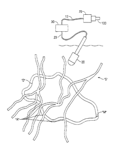
[0022] Referring to Fig. 1, there is disclosed a system 10 for use in
minimally invasive
surgery. The system 10 is configured to map anatomical structures or features
"A" of a surgical
site "S" and mark the anatomical features "A" on a substrate 100. System 10
includes an image
capturing unit 20, an image processing unit 30, and a printing unit 70. Image
capturing unit 20 is
CA 02816737 2013-05-27
configured to obtain or capture an image of the surgical site "S." Surgical
site "S" includes a
defect "D", for example a hernia defect, and anatomical features "A." All or a
portion of the
anatomical features "A" may be disposed on all or a portion of the defect "D"
of the surgical site
[0023] Continuing with reference to Fig. 1, the image capturing unit 20 is
operatively
coupled to the image processing unit 30 via line 23. Although image capturing
unit 20 is shown
as being operatively coupled to image processing unit 30 via line 23, image
capturing unit 20
may be coupled to image processing unit 30 by any means, such as, without
limitation,
wirelessly. Image processing unit 30 is operatively coupled to printing unit
70 via cable 12.
Although image processing unit 30 is shown as being operatively coupled to
printing unit 70 via
cable 12, image processing unit 30 may be coupled to printing unit 70 by any
means, such as,
without limitation, wirelessly. Additionally, or alternatively, image
capturing unit 20 may be
operatively coupled to printing unit 70 directly, and image capturing unit 20
may be configured
to perform all of the operations of image processing unit 30. Additionally, or
alternatively,
image capturing unit 20 may be operatively coupled to printing unit 70, and
printing unit 70 may
be configured to perform all of the operations of image processing unit 30.
[0024] The image capturing unit 20 is positioned within a surgical site
"S" to obtain an
image of the defect "D" and all of the anatomical features "A" and transmit
the image to the
image processing unit 30. As described above, the image capturing unit 20 may
transmit the
image to the image processing unit 30 via line 23 or a wireless connection
(not shown).
[0025] The image processing unit 30 is configured to adjust and/or scale
the image
captured by the image capturing unit 20. In addition, the image processing
unit 30 may be
configured to measure the size of the defect "D" and may further be configured
to identify a
6
CA 02816737 2013-05-27
perimeter, i.e., edges, of the defect "D." Additionally, or alternatively, a
user may manually
measure the size of the defect "D" and input the measurements via a graphic
user interface.
[0026] Upon adjusting and/or scaling the image, the image processing unit
30 may
further be configured to set a desired minimum margin "M" based on the edges
of a portion of
the surgical site "S" and the edges of defect "D." The minimum margin "M" may
be
automatically set by the image processing unit 30 or it may be selected by the
user, as will be
described in further detail below. The minimum margin "M" acts as a reference
point to indicate
the minimum distance required between the perimeter, i.e., edges, of the
defect "D" and the edge
of the substrate 100. By setting a minimum margin "M," an appropriate shape
and/or size
substrate 100 may be selected, as will be described in further detail below.
The minimum
margin "M" acts as only a minimum value, and it is understood that the edge of
the substrate 100
may exceed the minimum value as allowed by the surgical site "S" and/or as
desired by the user.
Additionally, or alternatively, and as will be described in further detail
below, the portion or area
defined by the minimum margin "M" may be a region where a user/surgeon may
affix the
substrate 100 to the surgical site "S" over the defect "D."
[0027] Upon setting a desired minimum margin "M," the image processing
unit 30 may
further be configured to select an appropriate substrate 100 shape and/or size
from a collection of
common shapes and sizes based on the measured size of the defect "D," the
minimum margin
"M" required between the edge of the defect "D" and the edge of the substrate
100, and the
surgical site "S." It is preferred that the size of the substrate 100 is large
enough to overlap each
minimum margin "M," without exceeding the size of the surgical site "S."
However, the size of
the substrate 100 may be the same size of the region defined by the defect and
the combined
minimum margins "M." Additionally, or alternatively, a user may select a
substrate 100 shape
7
CA 02816737 2013-05-27
and the image processing unit 30 would then select the appropriate size of the
selected shape in
accordance with the set minimum margins "M" so that the substrate 100 will be
sure to properly
fit over the defect "D."
[0028] As described above, the image processing unit 30 may be further
configured to
identify the edges of the defect "D" and mark the edges of the defect "D" on
the image. The
identification and marking of the edges of the defect "D" may be accomplished
by means of
image recognition software. Additionally or alternatively, image processing
unit 30 may be
operated by a user via a graphical user interface and a surgeon/user may
identify and/or mark the
edges of the defect "D" manually via a graphical user interface.
[0029] In addition, the image processing unit 30 may be configured to
identify and mark
the anatomical structures or features "A," such as, without limitation,
arteries, bones, and the like
on the image for printing on the substrate 100. The identification and marking
of the anatomical
structures or features "A" on the image may be accomplished by means of image
recognition
software. Additionally or alternatively, as noted above, the image processing
unit 30 may be
operated by a user via a graphical user interface and a surgeon/user may
identify and/or mark the
anatomical structures or features "A" on the image manually via the graphical
user interface.
[00301 In addition, the image processing unit 30 may be further configured
to optimize
the substrate 100 placement to achieve the desired minimum margins "M" around
the perimeter
of the defect "D." More particularly, subsequent to substrate 100 placement on
the defect "D," if
the substrate 100 does not line up with the margins "M," the image processing
unit 30 is
configured to re-select a second substrate 100 shape and/or size that would be
sufficient to
maintain the minimum margin "M" around the entire perimeter of defect "D."
[0031] Image processing unit 30 transmits the adjusted image to the
printing unit 70 for
8
CA 02816737 2013-05-27
printing the image onto a substrate 100. Image processing unit 30 may be
connected to printing
unit 70 wirelessly (not shown) or via wire 12 as shown.
[0032] Continuing with reference to Fig. 1, printing unit 70 is configured
to print the
image captured by image capturing unit 20 subsequent to proper adjustment by
image processing
unit 30. Printing unit 70 prints the image on a substrate 100 which can take
the form of a mesh
100a (Fig. 2A) or a film 100b (Fig. 2B) which can be attached to a mesh 100c
(Fig. 2B). The
printing unit 70 prints the image captured including all of the anatomical
features "A" which
were identified and marked by the image processing unit 30 onto film 100b
(Fig. 2B).
Subsequent to printing the image onto the film 100b, film 100b may be attached
to a mesh 100c
(Fig. 2B). Alternatively, the printing unit 70 prints the image captured by
the image capturing
unit 20 including all of the anatomical features "A" which were identified and
marked by the
image processing unit 30 directly onto the mesh 100a (Fig. 2A).
[0033] Turning now to Fig. 2A, the substrate 100 is shown as a mesh 100a.
The surgical
mesh 100a and 100c (Fig. 2B) described herein may include porous fabrics made
from
intertwined filaments. The filaments may be monofilaments or multi-filaments
and, in
embodiments, a plurality of multi-filaments may be combined to form yarns. The
filaments may
extend horizontally and vertically in a manner which produces sections where
the filaments
cross-over one another creating points of common intersection. The surgical
mesh 100a and/or
100c (Fig. 2B) may be woven, non-woven, knitted or braided. In some
embodiments, the
filaments may form two-dimensional or three-dimensional meshes.
[0034] Continuing with reference to Fig. 2A, the image is printed directly
on to the mesh
100a which includes landmarks 25. Landmarks 25 are printed images of the
anatomical features
"A" (Figs. 1 and 3) which would be present behind the mesh 100a when the mesh
100a is placed
9
CA 02816737 2013-05-27
over the defect "D" in the surgical site "S" (Figs. 1 and 3). Landmarks 25
assist a surgeon in
providing the surgeon with an image of the anatomical features "A" which are
located behind the
mesh 100a. With landmarks 25 in sight, a surgeon may avoid tacking, or
otherwise affixing,
those areas in which anatomical features "A" are located by not tacking, or
otherwise affixing, in
regions where the landmarks 25 are visible. Additionally, or alternatively,
landmarks 25 may
assist a surgeon with proper placement of mesh 100a over the defect "D" of the
surgical site "S."
Specifically, a surgeon may line up each edge of the landmarks 25 with the
corresponding
anatomical feature "A" so as to ensure proper placement of the mesh 100a.
[0035] Although mesh 100a is shown as a rectangular shape in Fig. 2A, it
is understood
that mesh 100a will take the shape/size as set by image processing unit 30
(Fig. 1) or as set by
the user, as described above. It is understood that any given distance between
the edge of mesh
100a and the edge of defect "D" may not be lower than the distance set as the
minimum margin
"M" (Fig. 1).
[0036] Turning now to Fig. 2B, substrate 100 is shown as a film 100b which
is
configured to attach to a mesh 100c, similar to the mesh of 100a described
above. The image
captured by image capturing unit 20 and adjusted by image processing unit 30
may be printed by
printing unit 70 directly onto film 100b. The film 100b may be a biopolymer or
film such as,
without limitation, a starch-based paper such as rice-film. As shown in Fig.
2B, landmarks 25 are
printed directly onto the film 100b which represent the anatomical features
"A" on the defect
"D" of the surgical site "S." Subsequent to printing the image onto the film
100b, the film 100b
is adhered to the mesh 100c. As described above with respect to mesh 100a,
with landmarks 25
of mesh 100c in sight, a surgeon may avoid tacking, or otherwise affixing,
those areas in which
anatomical features "A" are located by not tacking, or otherwise affixing, in
regions where the
CA 02816737 2013-05-27
landmarks 25 are visible. Additionally, or alternatively, landmarks 25 may
assist a surgeon with
proper placement of mesh 100c over the defect "D" of the surgical site "S."
Specifically, a
surgeon may line up each edge of the landmarks 25 with the corresponding
anatomical feature
"A" so as to ensure proper placement of the mesh 100c.
[0037] Although film 100b and mesh 100c are shown as a rectangular shape
in Fig. 2B, it
is understood that film 100b and mesh 100c may take the shape/size as set by
image processing
unit 30 (Fig. 1) or as set by the user, as described above. It is understood
that any given distance
between the edge of film 100b and mesh 100c and the edge of defect "D" may not
be lower than
the distance set as the minimum margin "M" (Fig. I).
[0038] Turning now to Fig. 3, substrate 100, i.e., mesh 100a, or film 100b
and mesh
100c, is shown placed over the defect "D" of the surgical site "S." All or a
portion of anatomical
features "A" are covered by substrate 100. Substrate 100 includes landmarks 25
in the portions
where anatomical features "A" are blocked by substrate 100. Landmarks 25 may
be used by a
surgeon to identify which portions of the substrate 100 may not be tacked, or
otherwise affixed,
thereby assisting the surgeon in identifying regions that should not be
tacked, or otherwise
affixed, i.e., regions including anatomical features "A."
[0039] Turning now to Fig. 4, a method 200 for mapping anatomical features
"A" (Fig.
1) on a substrate 100 (Fig. 1) is shown. At step 210, an image capturing unit
20, such as, without
limitation, a laparoscope, is inserted into a surgical site "S," i.e., a
patient's body through an
opening into a cavity. Subsequent to inserting the imaging device 20 into the
surgical site "S," at
step 220, an image is captured of the surgical site "S" which includes the
defect "D" and
anatomical features "A."
[0040] At step 230, the image processing unit 30 (Fig. 1) adjusts the
scale of the image to
11
CA 02816737 2013-05-27
a desired size and measures the defect "D." Additionally or alternatively, and
as described
above, the measurement of defect "D" may be carried out manually by the
surgeon by means
known in the art such as with a tape measure. The manually measured size of
defect "D" may
then be entered as data into the graphical user interface as described above.
As noted above, the
image processing unit 30 is configured to adjust and/or scale the image
captured by the image
capturing unit 20, for example to correct the angle, planarity or size of the
image. The image
processing unit 30 may further be configured to set a desired minimum margin
"M" based on the
edges of a portion of the surgical site "S," and the edges of the defect "D."
Upon adjusting
and/or scaling the image and setting the desired minimum margins "M" to be
maintained, the
image processing unit 30 may further be configured to select an appropriate
substrate 100 shape
and/or size from a collection of common shapes and sizes sufficient to
maintain the minimum
margin "M" and cover the area of defect "D."
[0041] At step 235, a determination is made as to whether the image will
be printed
directly onto mesh 100a (Fig. 2A) or onto film 100b (Fig. 2B). If at step 235
a determination is
made to print onto mesh 100a (Fig. 2A) then the method 200 proceeds to step
240a.
Alternatively, if at step 235 a determination is made to print onto film 100b
(Fig. 2B), the
method 200 will proceed to step 240b. The determination may be made by any
component of
system 10 (Fig. 1) such as, without limitation, image processing unit 30 (Fig.
1). Alternatively, a
user may make the determination via the graphical user interface described
above.
[0042] At step 240a, the printing unit 70 prints the image directly onto
the mesh 100a
(Fig. 2A). Alternatively, at step 240b, the printing unit 70 prints the image
onto a film 100b
(Fig. 2B) which is subsequently attached to a mesh 100c (Fig. 2B) at step
240bb.
[0043] Continuing with reference to Fig. 4, at step 250, either mesh 100a
(Fig. 2A) or
12
CA 02816737 2013-05-27
mesh 100c (Fig. 2B) with the image of the anatomical features "A" is inserted
into the surgical
site "S." At step 260, the mesh 100a or 100c is aligned onto the defect "D"
such that landmarks
25 (Fig. 3) line up with the anatomical features "A" of the surgical site "S"
located around or on
the defect "D."
[0044] Turning now to Fig. 5, a method 300 for mapping anatomical features
"A" (Fig.
1) on a substrate 100 (Fig. 1) is shown. At step 310, an image capturing unit
20, such as, without
limitation, a laparoscope, is inserted into a surgical site "S," i.e., a
patient's body through an
opening into a cavity. Subsequent to inserting the imaging device 20 into the
surgical site "S," at
step 320, a first image is captured of the surgical site "S."
[0045] At step 330, the image processing unit 30 (Fig. 1) adjusts the
scale of the image to
a desired size and measures the defect "D." Additionally or alternatively, and
as described
above, the measurement of defect "D" may be carried out manually by the
surgeon by means
known in the art such as with a tape measure. The manually measured size of
defect "D" may
then be entered as data into the graphical user interface as described above.
As noted above, the
image processing unit 30 is configured to adjust and/or scale the image
captured by the image
capturing unit 20. Upon adjusting and/or scaling the image, the image
processing unit 30 may
further be configured to select an appropriate substrate 100 shape and/or size
from a collection of
available shapes and sizes. The image processing unit 30 may further be
configured to set a
desired minimum margin "M" based on the edges of at least a portion of the
surgical site "S" and
the edges of the defect "D" to ensure that the substrate 100 will be a proper
shape and/or size
sufficient to maintain the desired minimum margins "M" around the perimeter of
defect "D"
while covering the area of defect "D." For example, a user could input the
desired minimum
margin "M" size, e.g., 4 or 5 cm, and the image processing unit 30 could
optimize mapping of
13
CA 02816737 2013-05-27
the defect "D" onto the mesh 100a or the film 100b to ensure that the desired
minimum margin
"M" is maintained on at least a portion of the substrate 100, including
restricting the user's
choice of substrate shapes or sizes to only those shapes and sizes sufficient
to maintain the
desired minimum margin "M" around the perimeter of the defect "D."
[00461 At step 335, a determination is made as to whether the image will
be printed
directly onto mesh 100a (Fig. 2A) or onto film 100b (Fig. 2B). If at step 335
a determination is
made to print onto mesh 100a (Fig. 2A) then the method 300 proceeds to step
340a.
Alternatively, if at step 335 a determination is made to print onto film 100b
(Fig. 2B), the
method 300 will proceed to step 340b. The determination may be made by any
component of
system 10 (Fig. 1) such as, without limitation, image processing unit 30 (Fig.
1). Alternatively, a
user may make the determination via the graphical user interface described
above.
[00471 At step 340a, the printing unit 70 prints the image directly onto
the mesh 100a
(Fig. 2A). Alternatively, at step 340b, the printing unit 70 prints the image
onto a film 100b
(Fig. 2B) which is subsequently attached to a mesh 100c (Fig. 28) at step
340bb.
[00481 Continuing with reference to Fig. 5, at step 350, either mesh 100a
(Fig. 2A) or
mesh 100c (Fig. 2B) with the image of the anatomical features "A" is inserted
into the surgical
site "S." At step 360, the mesh 100a or 100c is aligned onto the defect "D"
such that landmarks
25 (Fig. 3) line up with the anatomical features "A" of the surgical site "S."
At step 370 a
second image of the surgical site "S" is obtained with the mesh 100a or 100c
attached to the
defect "D" prior to affixing, i.e., tacking, the mesh 100a or 100c to the
defect "D." At step 380,
the first image captured at step 320 is compared to the second image captured
at step 370 to
ensure that the landmarks 25 are aligned with the anatomical features "A" of
the surgical site "S"
behind the mesh 100a (Fig. 2A) or 100c (Fig. 2B).
14
CA 02816737 2013-05-27
[0049]
While several embodiments of the disclosure have been shown in the drawings,
it
is not intended that the disclosure be limited thereto, as it is intended that
the disclosure be as
broad in scope as the art will allow and that the specification be read
likewise. Therefore, the
above description should not be construed as limiting, but merely as
exemplifications of
particular embodiments. Those skilled in the art will envision other
modifications within the
scope and spirit of the claims appended hereto.