Note: Descriptions are shown in the official language in which they were submitted.
WO 2012/061759
PCT/US2011/059411
FOLATE RECEPTOR ALPHA AS A DIAGNOSTIC AND PROGNOSTIC
MARKER FOR FOLATE RECEPTOR ALPHA-EXPRESSING CANCERS
RELATED APPLICATIONS
This application claims the benefit of the filing date of U.S. Provisional
Application No. 61/410,497, filed on November 5, 2010, and U.S. Provisional
Application No. 61/508,444, filed on July 15, 2011.
BACKGROUND OF THE INVENTION
In humans, the high affinity receptor for folate comes in three isoforms:
alpha,
beta, and gamma. The alpha and beta forms are typically bound to the membranes
of
cells by a glycosyl phosphatidylinositol (GPI) anchor. They recycle between
extracellular and endocytic compartments and are capable of transporting
folate into the
cell. Soluble forms of FRa may be derived by the action of proteases or
phospholipase
on membrane anchored folate receptors.
Folate receptor alpha (also referred to as FRa, FR-alpha, FOLR-1 or FOLR1) is
expressed in a variety of epithelial tissues, including those of the choroid
plexus, lung,
thyroid, kidney, uterus, breast, Fallopian tube, epididymis, and salivary
glands.
Weitman, SD et al. Cancer Res 52: 3396-3401 (1992); Weitman SD et al, Cancer
Res
52: 6708-6711. Overexpression of FRa has been observed in various cancers,
including
lung cancer (e.g., bronchioalveolar carcinomas, carcinoid tumors, and non-
small cell
lung cancers, such as adenocarcinomas); mesothelioma; ovarian cancer; renal
cancer;
brain cancer (e.g., anaplastic ependymoma, cerebellar juvenile pilocytic
astrocytoma,
and brain metastases); cervical cancer; nasopharyngeal cancer; mesodermally
derived
tumor; squamous cell carcinoma of the head and neck; endometrial cancer;
endometrioid
adenocarcinomas of the ovary, serous cystadenocarcinomas, breast cancer;
bladder
cancer; pancreatic cancer; bone cancer (e.g., high-grade osteosarcoma);
pituitary cancer
(e.g., pituitary adenomas); colorectal cancer and medullary thyroid cancer.
See e.g.,
U.S. Patent No. 7,754,698; U.S. Patent Application No. 2005/0232919; WO
2009/132081; Bueno R et al. J of Thoracic and Cardiovascular Surgery, 121(2) :
225-
233 (2001); Elkanat H & Ratnam M. Frontiers in Bioscience, 11, 506-519 (2006);
CA 2816991 2018-02-22
CA 02816991 2013-05-03
WO 2012/061759
PCT/US2011/059411
Fisher R.E. J Nucl Med, 49: 899-906 (2008); Franklin, WA et al. Int J Cancer,
Suppl 8:
89-95 (1994); Hartman L.C. etal. Int J Cancer 121: 938-942 (2007); Iwakiri S
et al.
Annals of Surgical Oncology, 15(3): 889-899: Parker N. et al. Analytical
Biochemistry,
338: 284-293 (2005); Weitman, SD et al. Cancer Res 52: 3396-3401 (1992); Saba
N.F.
et al. Head Neck, 31(4): 475-481 (2009); Yang R et al. Clin Cancer Res 13:
2557-2567
(2007). In some types of cancers (e.g., squamous cell carcinoma of the head
and neck),
a higher level of FRa expression is associated with a worse prognosis, whereas
in other
types of cancers (e.g., non-small-cell lung cancers), a higher level of FRa
expression is
associated with a better prognosis. See, e.g., Iwakiri S et al. Annals of
Surgical
Oncology, 15(3): 889-899; Saba N.F. ei al. Head Neck, 31(4): 475-481 (2009).
Earlier detection of cancer improves survival rates and quality of life. To
improve the likelihood of early detection and treatment, a pressing need
exists for non-
invasive methods for diagnosing cancer, for determining the level of risk of
developing
cancer, and for predicting the progression of cancer. The present invention
satisfies
these needs for FRa-expressing cancers.
SUMMARY OF THE INVENTION
The present invention provides methods of assessing whether a subject is
afflicted with FRa-expressing cancers such as lung or ovarian cancer, methods
of
assessing the progression of FRa-expressing cancers such as lung or ovarian
cancer in a
subject afflicted with the FRoc-expressing cancers, methods of stratifying an
FRa-
expressing cancer subject into one of at least four cancer therapy groups,
methods of
assessing the efficacy of MORAb-003 treatment of ovarian cancer or lung cancer
and
kits for assessing whether a subject is afflicted with PRoc-expressing cancers
such as
lung or ovarian cancer or for assessing the progression of FRa-expressing
cancers such
as lung or ovarian cancer in a subject.
Methods of Assessing Whether a Subject is Afflicted with an FRa Expressing
Cancer
In a first aspect, the present invention provides a method of assessing
whether a
subject is afflicted with an FRa-expressing cancer, by determining the level
of folate
receptor alpha (FRa) which is not bound to a cell, in a sample derived from
the subject;
and comparing the level of folate receptor alpha (FRa) which is not bound to a
cell with
the level of FRa in a control sample, wherein a difference between the level
of FRa in
2
CA 02816991 2013-05-03
WO 2012/061759
PCT/US2011/059411
the sample derived from the subject and the level of FRa in the control sample
is an
indication that the subject is afflicted with an FRa-expressing cancer;
wherein the level
of FRa in the sample derived from the subject is assessed by contacting the
sample with
an antibody that binds FRa. In a particular embodiment, the sample is either
urine,
serum, plasma or ascites.
In another aspect, the present invention is directed to a method of assessing
whether a subject is afflicted with an FRa-expressing cancer, by determining
the level of
folate receptor alpha (FRa) which is not bound to a cell in a urine sample
derived from
the subject; and comparing the level of folate receptor alpha (FRa) which is
not bound to
a cell in the urine sample derived from the subject with the level of I-Ra in
a control
sample, wherein a difference between the level of FRa in the urine sample
derived from
the subject and the level of FRa in the control sample is an indication that
the subject is
afflicted with an FRa-expressing cancer. In a further aspect, the present
invention
provides a method of assessing whether a subject is afflicted with a cancer
that expresses
FRa, by determining the level of folate receptor alpha (FRa) which is not
bound to a cell
in a serum sample derived from the subject; and comparing the level of folate
receptor
alpha (FRa) which is not bound to a cell in the serum sample derived from the
subject
with the level of FRa in a control sample, wherein a difference between the
level of FRa
in the serum sample derived from the subject and the level of FRa in the
control sample
is an indication that the subject is afflicted with an FRa-expressing cancer.
In various embodiments of the foregoing aspects of the invention, the FRa-
expressing cancer is selected from the group consisting of lung cancer,
mesothelioma,
ovarian cancer, renal cancer, brain cancer, cervical cancer, nasopharyngeal
cancer,
squamous cell carcinoma of the head and neck, endometrial cancer, breast
cancer,
bladder cancer, pancreatic cancer, bone cancer, pituitary cancer, colorectal
cancer and
medullary thyroid cancer. In a particular embodiment, the FRa-expressing
cancer is
ovarian cancer. In another embodiment, the FRa-expressing cancer is non-small
cell
lung cancer, such as an adenocarcinoma.
In another aspect, the present invention is directed to methods of assessing
whether a subject is afflicted with ovarian cancer, by determining the level
of folate
receptor alpha (FRa) which is not bound to a cell in a urine sample derived
from the
subject, wherein the presence of FRa which is not bound to a cell in the urine
sample at
3
CA 02816991 2013-05-03
WO 2012/061759
PCT/US2011/059411
a concentration of greater than about 9100 pg/ml is an indication that the
subject is
afflicted with ovarian cancer.
In various aspects of the foregoing aspects of the invention, the presence of
FRa
in the urine sample at a concentration of greater than about 9500 pg/mL, about
10,000
pg/mL, about 11,000 pg/mL, about 12,000 pg/mL, about 13,000 pg/mL, about
14,000
pg/mL, about 15,000 pg/mL, about 16,000 pg/mL, about 17,000 pg/mL, about
18,000
pg/mL, about 19,000 pg/mL, or about 20,000 pg/mL is an indication that the
subject is
afflicted with ovarian cancer.
In various aspects, the level of FRa is determined by contacting the sample
with
an antibody that binds FRa. For example, the antibody is selected from the
group
consisting of:
(a) an antibody that binds the same epitope as the MORAb-003 antibody;
(b) an antibody comprising SEQ ID NO:1 (GFTFSGYGLS) as CDRH1, SEQ ID
NO:2 (MISSGGSYTYYADSVKG) as CDRH2, SEQ ID NO:3 (HGDDPAWFAY) as
CDRH3, SEQ ID NO:4 (SVSSSISSNNLH) as CDRL1, SEQ ID NO:5 (GTSNLAS) as
CDRL2 and SEQ ID NO:6 (QQWSSYPYMYT) as CDRL3;
(c) the MOV18 antibody;
(d) an antibody that binds the same epitope as the MOV18 antibody;
(e) the 548908 antibody;
(f) an antibody that binds the same epitope as the 548908 antibody;
(g) the 6D398 antibody;
(h) an antibody that binds the same epitope as the 6D398 antibody;
(i) an antibody that binds the same epitope as the 26B3 antibody;
(j) an antibody comprising SEQ ID NO:55 (GYFMN) as CDRH1, SEQ ID
NO:56 (RIPPYNGDTFYNQKFKG) as CDRH2, SEQ ID NO:57 (GTHYFDY) as
CDRH3, SEQ ID NO:51 (RTSENIFSYLA) as CDRL1 SEQ ID NO:52 (NAKTLAE)
as CDRL2 and SEQ ID NO:53 (QHHYAFPWT) as CDRL3;
(k) the 26B3 antibody;
(1) an antibody that binds the same epitope as the 19D4 antibody:
(m) an antibody comprising SEQ ID NO:39 (HPYMH) as CDRH1, SEQ ID
NO:40 (RIDPANGNTKYDPKFQG) as CDRH2, SEQ ID NO:41 (EEVADYTMDY) as
CDRH3, SEQ ID NO:35 (RASESVDTYGNNFIH) as CDRL1, SEQ ID NO:36
(LASNLES) as CDRL2 and SEQ ID NO:37 (QQNNGDPWT) as CDRL3;
4
CA 02816991 2013-05-03
WO 2012/061759
PCT/US2011/059411
(n) the 19D4 antibody;
(o) an antibody that binds the same epitope as the 9F3 antibody;
(p) an antibody comprising SEQ ID NO:31 (SGYYWN) as CDRH1, SEQ ID
NO:32 (YIKSDGSNNYNPSLKN) as CDRH2, SEQ ID NO:33 (EWKAMDY) as
CDRH3, SEQ ID NO:27 (RASSTVSYSYLH) as CDRL1, SEQ ID NO:28 (GTSNLAS)
as CDRL2 and SEQ ID NO:29 (QQYSGYPLT) as CDRL3;
(q) the 9F3 antibody;
(r) an antibody that binds the same epitope as the 24FI2 antibody;
(s) an antibody comprising SEQ ID NO:47 (SYAMS) as CDRH1, SEQ ID
NO:48 (EIGSGGSYTYYPDTVTG) as CDRH2, SEQ ID NO:49 (ETTAGYFDY) as
CDRH3, SEQ ID NO:43 (SASQGINNFLN) as CDRL1, SEQ ID NO:44 (YTSSLHS) as
CDRL2 and SEQ ID NO:45 (QHFSKLPWT) as CDRL3;
(t) the 24FI2 antibody;
(u) an antibody that comprises a variable region light chain selected from the
group consisting of LK26HuVK (SEQ ID NO: 13); LK26HuVKY (SEQ ID NO: 14);
LK26HuVKPW (SEQ ID NO: 15); and LK26HuVKPW,Y (SEQ ID NO: 16);
(v) an antibody that comprises a variable region heavy chain selected from the
group consisting of LK26HuVH (SEQ ID NO: 17); LK26HuVH FAIS,N (SEQ ID NO:
18); LK26HuVH SLF (SEQ ID NO: 19); LK26HuVH LI (SEQ ID NO: 20); and
LK26KOLHuVH (SEQ ID NO: 21);
(w) an antibody that comprises the heavy chain variable region LK26KOLHuVH
(SEQ ID NO: 21) and the light chain variable region LK26HuVKPW,Y (SEQ ID NO:
16);
(x) an antibody that comprises the heavy chain variable region LK26HuVH SLF
(SEQ ID NO: 19) and the light chain variable region LK26HuVKPW,Y (SEQ ID NO:
16); and
(y) an antibody that comprises the heavy chain variable region LK26HuVH
FAIS,N (SEQ ID NO: 18) and the light chain variable region LK26HuVKPW,Y (SEQ
ID NO: 16).
In a particular embodiment, the antibody binds the same epitope as the MORAb-
003 antibody. In another embodiment, the antibody includes SEQ ID NO:1
(GFTFSGYGLS) as CDRH1. SEQ ID NO:2 (MISSGGSYTYYADSVKG) as CDRH2,
SEQ ID NO:3 (HGDDPAWFAY) as CDRH3, SEQ ID NO:4 (SVSSSISSNNLH) as
CA 02816991 2013-05-03
WO 2012/061759
PCT/US2011/059411
CDRL1, SEQ ID NO:5 (GTSNLAS) as CDRL2 and SEQ ID NO:6 (QQWSSYPYMYT)
as CDRL3. In another embodiment, the antibody is the MOV18 antibody. In yet
another embodiment, the antibody binds the same epitope as the MOV18 antibody.
In a
further embodiment, the antibody comprises a variable region light chain
selected from
the group consisting of LK26HuVK (SEQ ID NO: 13); LK26HuVKY (SEQ ID NO: 14);
LK26HuVKPW (SEQ ID NO: 15); and LK26HuVKPW,Y (SEQ ID NO: 16).
Alternatively or in combination, the antibody includes a variable region heavy
chain
selected from the group consisting of LK26HuVH (SEQ ID NO: 17); LK26HuVH
FAIS,N (SEQ ID NO: 18); LK26HuVH SLF (SEQ ID NO: 19); LK26HuVH 1,1 (SEQ
ID NO: 20); and LK26KOLHuVH (SEQ ID NO: 21). In certain embodiments, the
antibody includes (i) the heavy chain variable region LK26KOLHuVH (SEQ ID NO:
21) and the light chain variable region LK26HuVKPW,Y (SEQ ID NO: 16); the
heavy
chain variable region LK26HuVH SLF (SEQ ID NO: 19) and the light chain
variable
region LK26HuVKPW,Y (SEQ ID NO: 16); or the heavy chain variable region
LK26HuVH FAIS,N (SEQ ID NO: 18) and the light chain variable region
LK26HuVKPW,Y (SEQ ID NO: 16).
In a particular embodiment, the level of FRa in the sample derived from said
subject is assessed by contacting the sample with a pair of antibodies
selected from the
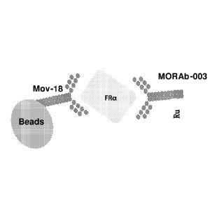
group consisting of (a) MOV18 antibody immobilized to a solid support and
labeled
MORAB-003 antibody; (b) 9F3 antibody immobilized to a solid support and
labeled
24F12 antibody; (c) 26B3 antibody immobilized to a solid support and labeled
19D4
antibody; and (d) 9F3 antibody immobilized to a solid support and labeled 26B3
antibody.
In certain embodiments, the antibody is selected from the group consisting of
a
murine antibody, a human antibody, a humanized antibody, a bispecific
antibody, a
chimeric antibody, a Fab, Fab.2, ScFv, SMIP, affibody, avimer, versabody,
nanobody,
and a domain antibody. Alternatively, or in combination, the antibody is
labeled, for
example, with a label selected from the group consisting of a radio-label, a
biotin-label,
a chromophore-label, a fluorophore-label, or an enzyme-label.
In certain embodiments, the level of FRa is determined by using a technique
selected from the group consisting of western blot analysis, radioimmunoassay,
immunofluorimetry, immunoprecipitati on, equilibrium dialysis, immunodiffusi
on,
6
CA 02816991 2013-05-03
WO 2012/061759
PCT/US2011/059411
solution phase assay, electrochemiluminescence immunoassay (ECLIA) and ELISA
assay.
In various embodiments of the foregoing aspects of the invention, the control
sample is a standardized control level of FRa in a healthy subject.
In certain embodiments, the sample is treated with guanidine prior to
determining the level of FRa in the sample. Alternatively or in combination,
the sample
is diluted prior to determining the level of FRa in the sample. Alternatively,
or in
combination, the sample is centrifuged, vortexed, or both, prior to
determining the level
of FRa in the sample.
In yet another aspect, the present invention is directed to a method of
assessing
whether a subject is afflicted with ovarian cancer, by determining the level
of folate
receptor alpha (FRa) which is not bound to a cell in a sample derived from the
subject;
and comparing the level of folate receptor alpha (FRa) which is not bound to a
cell in
the sample with the level of FRa in a control sample, wherein a difference
between the
levels of FRa in the sample derived from the subject and in the control sample
is an
indication that the subject is afflicted with ovarian cancer; wherein the
level of FRa in
the sample derived from the subject is assessed by contacting the sample with
(a)
MOV18 antibody immobilized to a solid support and labeled MORAB-003 antibody,
(b)
9F3 antibody immobilized to a solid support and labeled 24F12 antibody. (c)
26B3
antibody immobilized to a solid support and labeled 19D4 antibody, and (d) 9F3
antibody immobilized to a solid support and labeled 26B3 antibody. For
example, the
sample may be urine, serum. plasma or ascites.
Methods of Assessing the Progression of an FRa Expressing Cancer in a Subject
In a further aspect, the present invention is directed to a method of
assessing the
progression of an FRa-expressing cancer in a subject afflicted with an FRa-
expres sing
cancer, by determining the level of folate receptor alpha (FRa) which is not
bound to a
cell, in a sample derived from the subject; and comparing the level of folate
receptor
alpha (FRa) which is not bound to a cell with the level of FRa in a control
sample,
wherein an increase in the level of FRa in the sample derived from the subject
as
compared with the level of FRa in the control sample is an indication that the
cancer will
progress rapidly; and wherein a decrease in the level of FRa in the sample
derived from
the subject as compared with the level of FRa in the control sample is an
indication that
7
CA 02816991 2013-05-03
WO 2012/061759
PCT/US2011/059411
the cancer will progress slowly or will regress, thereby assessing the
progression of the
FRa-expres sing cancer in the subject; wherein the level of FRa which is not
bound to a
cell in the sample derived from the subject is assessed by contacting the
sample with an
antibody that binds FRa. In a particular embodiment, the sample is urine,
serum, plasma
or ascites.
In another aspect, the present invention provides a method of assessing the
progression of an FRa-expres sing cancer in a subject afflicted with an FRa-
expres sing
cancer, by determining the level of folate receptor alpha (FRa) which is not
bound to a
cell in a urine sample derived from the subject; and comparing the level of
folate
receptor alpha (FRa) which is not bound to a cell in the urine sample derived
from the
subject with the level of FRa in a control sample, wherein an increase in the
level of
FRa in the urine sample derived from the subject as compared with the level of
FRa in
the control sample is an indication that the cancer will progress rapidly; and
wherein a
decrease in the level of FRa in the urine sample derived from the subject as
compared
with the level of FRa in the control sample is an indication that the cancer
will progress
slowly or will regress, thereby assessing the progression of the FRa-
expressing cancer in
the subject.
In a further aspect, the present invention provides methods of assessing the
progression of an FRa-expres sing cancer in a subject afflicted with an FRa-
expres sing
cancer, by determining the level of folate receptor alpha (FRa) which is not
bound to a
cell in a serum sample derived from the subject; and comparing the level of
folate
receptor alpha (FRa) which is not bound to a cell in the serum sample derived
from the
subject with the level of FRa in control sample, wherein an increase in the
level of PRa
in the serum sample derived from the subject as compared with the level of FRa
in the
control sample is an indication that the cancer will progress rapidly; and
wherein a
decrease in the level of FRa in the serum sample derived from the subject as
compared
with the level of FRa in the control sample is an indication that the cancer
will progress
slowly or will regress, thereby assessing the progression of the FRa-
expressing cancer in
the subject.
In various embodiments of the foregoing aspects of the invention, the FRa-
expressing cancer is selected from the group consisting of lung cancer,
mesothelioma,
ovarian cancer, renal cancer, brain cancer, cervical cancer, nasopharyngeal
cancer,
squamous cell carcinoma of the head and neck, endometrial cancer, breast
cancer,
8
CA 02816991 2013-05-03
WO 2012/061759
PCT/US2011/059411
bladder cancer, pancreatic cancer, bone cancer, pituitary cancer, colorectal
cancer and
medullary thyroid cancer. In a particular embodiment, the FRa-expressing
cancer is
ovarian cancer. In another embodiment, the FRa-expres sing cancer is non-small
cell
lung cancer, such as an adenocarcinoma.
In another aspect, the present invention is directed to methods of assessing
whether a subject is afflicted with ovarian cancer, by determining the level
of folate
receptor alpha (FRa) which is not bound to a cell in a urine sample derived
from the
subject, wherein the presence of FRa which is not bound to a cell in the urine
sample at
a concentration of greater than about 9100 pg/ml is an indication that the
subject is
afflicted with ovarian cancer.
In various aspects of the foregoing aspects of the invention, the presence of
FRa
in the urine sample at a concentration of greater than about 9500 pg/mL, about
10,000
pg/mL, about 11,000 pg/mL, about 12,000 pg/mL, about 13,000 pg/mL, about
14,000
pg/mL, about 15,000 pg/mL, about 16,000 pg/mL, about 17,000 pg/mL, about
18,000
pg/mL, about 19,000 pg/mL, or about 20,000 pg/mL is an indication that the
subject is
afflicted with ovarian cancer.
In various aspects, the level of FRa is determined by contacting the sample
with
an antibody that binds FRa. For example, the antibody is selected from the
group
consisting of:
(a) an antibody that binds the same epitope as the MORAb-003 antibody;
(b) an antibody comprising SEQ ID NO:1 (GFTFSGYGLS) as CDRH1, SEQ ID
NO:2 (MISSGGSYTYYADSVKG) as CDRH2, SEQ ID NO:3 (HGDDPAWFAY) as
CDRH3, SEQ ID NO:4 (SVSSSISSNNLH) as CDRL1, SEQ ID NO:5 (GTSNLAS) as
CDRL2 and SEQ ID NO:6 (QQWSSYPYMYT) as CDRL3;
(c) the MOV18 antibody;
(d) an antibody that binds the same epitope as the MOV18 antibody;
(e) the 548908 antibody;
(f) an antibody that binds the same epitope as the 548908 antibody;
(g) the 6D398 antibody;
(h) an antibody that binds the same epitope as the 6D398 antibody;
(i) an antibody that binds the same epitope as the 26B3 antibody;
(j) an antibody comprising SEQ ID NO:55 (GYFMN) as CDRHI , SEQ ID
NO:56 (RIFFYNGDTFYNQKFKG) as CDRH2, SEQ ID NO:57 (GTHYFDY) as
9
CA 02816991 2013-05-03
WO 2012/061759
PCT/US2011/059411
CDRH3, SEQ ID NO:51 (RTSENIFSYLA) as CDRL1 , SEQ ID NO:52 (NAKTLAE)
as CDRL2 and SEQ ID NO:53 (QHHYAFPWT) as CDRL3;
(k) the 26B3 antibody;
(1) an antibody that binds the same epitope as the 19D4 antibody:
(m) an antibody comprising SEQ ID NO:39 (HPYMH) as CDRH1, SEQ ID
NO:40 (RIDPANGNTKYDPKFQG) as CDRH2, SEQ ID NO:41 (EEVADYTMDY) as
CDRH3, SEQ ID NO:35 (RASESVDTYGNNFIH) as CDRL1, SEQ ID NO:36
(LASNLES) as CDRL2 and SEQ ID NO:37 (QQNNGDPWT) as CDRL3;
(n) the 19D4 antibody;
(o) an antibody that binds the same epitope as the 9F3 antibody;
(p) an antibody comprising SEQ ID NO:31 (SGYYWN) as CDRH1, SEQ ID
NO:32 (YIKSDGSNNYNPSLKN) as CDRH2, SEQ ID NO:33 (EWKAMDY) as
CDRH3, SEQ ID NO:27 (RASSTVSYSYLH) as CDRL1, SEQ ID NO:28 (GTSNLAS)
as CDRL2 and SEQ ID NO:29 (QQYSGYPLT) as CDRL3;
(q) the 9F3 antibody;
(r) an antibody that binds the same epitope as the 24F12 antibody;
(s) an antibody comprising SEQ ID NO:47 (SYAMS) as CDRH1, SEQ ID
NO:48 (EIGSGGSYTYYPDTVTG) as CDRH2. SEQ ID NO:49 (ETTAGYFDY) as
CDRH3, SEQ ID NO:43 (SASQGINNFLN) as CDRL1, SEQ ID NO:44 (YTSSLHS) as
CDRL2 and SEQ ID NO:45 (QHFSKLPWT) as CDRL3;
(t) the 24F12 antibody;
(u) an antibody that comprises a variable region light chain selected from the
group consisting of LK26HuVK (SEQ ID NO: 13); LK26HuVKY (SEQ ID NO: 14);
LK26HuVKPW (SEQ ID NO: 15); and LK26HuVKPW,Y (SEQ ID NO: 16);
(v) an antibody that comprises a variable region heavy chain selected from the
group consisting of LK26HuVH (SEQ ID NO: 17); LK26HuVH FAIS,N (SEQ ID NO:
18); LK26HuVH SLF (SEQ ID NO: 19); LK26HuVH 1,1 (SEQ ID NO: 20); and
LK26KOLHuVH (SEQ ID NO: 21);
(w) an antibody that comprises the heavy chain variable region 1K26KO1HuVH
(SEQ ID NO: 21) and the light chain variable region LK26HuVKPW,Y (SEQ ID NO:
16);
CA 02816991 2013-05-03
WO 2012/061759
PCT/US2011/059411
(x) an antibody that comprises the heavy chain variable region LK26HuVH SLF
(SEQ ID NO: 19) and the light chain variable region LK26HuVKPW,Y (SEQ ID NO:
16); and
(y) an antibody that comprises the heavy chain variable region LK26HuVH
FAIS,N (SEQ ID NO: 18) and the light chain variable region LK26HuVKPW,Y (SEQ
ID NO: 16).
In a particular embodiment, the antibody binds the same epitope as the MORAb-
003 antibody. In another embodiment, the antibody includes SEQ ID NO:1
(GFTFSGYGLS) as CDRH1, SEQ ID NO:2 (MISSGGSYTYYADSVKG) as CDRH2,
SEQ ID NO:3 (HGDDPAWFAY) as CDRH3, SEQ ID NO:4 (SVSSSISSNNLH) as
CDRL1, SEQ ID NO:5 (GTSNLAS) as CDRL2 and SEQ ID NO:6 (QQWSSYPYMYT)
as CDRL3. In another embodiment, the antibody is the MOV18 antibody. In yet
another embodiment, the antibody binds the same epitope as the MOV18 antibody.
In a
further embodiment, the antibody comprises a variable region light chain
selected from
the group consisting of LK26HuVK (SEQ ID NO: 13); LK26HuVKY (SEQ ID NO: 14):
LK26HuVKPW (SEQ ID NO: 15); and LK26HuVKPW,Y (SEQ ID NO: 16).
Alternatively or in combination, the antibody includes a variable region heavy
chain
selected from the group consisting of LK26HuVH (SEQ ID NO: 17): LK26HuVH
FAIS,N (SEQ ID NO: 18); LK26HuVH SLF (SEQ ID NO: 19); LK26HuVH 1,1 (SEQ
ID NO: 20); and LK26KOLHuVH (SEQ ID NO: 21). In certain embodiments, the
antibody includes (i) the heavy chain variable region LK26KOLHuVH (SEQ ID NO:
21) and the light chain variable region LK26HuVKPW,Y (SEQ ID NO: 16); the
heavy
chain variable region LK26HuVH SLF (SEQ ID NO: 19) and the light chain
variable
region LK26HuVKPW,Y (SEQ ID NO: 16); or the heavy chain variable region
LK26HuVH FAIS,N (SEQ ID NO: 18) and the light chain variable region
LK26HuVKPW,Y (SEQ ID NO: 16).
In a particular embodiment, the level of FRa in the sample derived from said
subject is assessed by contacting the sample with a pair of antibodies
selected from the
group consisting of (a) MOV18 antibody immobilized to a solid support and
labeled
MORAB-003 antibody; (b) 9F3 antibody immobilized to a solid support and
labeled
24F12 antibody; (c) 26B3 antibody immobilized to a solid support and labeled
19D4
antibody; and (d) 9F3 antibody immobilized to a solid support and labeled 26B3
antibody.
11
CA 02816991 2013-05-03
WO 2012/061759
PCT/US2011/059411
In certain embodiments, the antibody is selected from the group consisting of
a
murine antibody, a human antibody, a humanized antibody, a bispecific
antibody, a
chimeric antibody, a Fab, Fab'2, ScFv, SMIP, affibody, avimer, versabody,
nanobody,
and a domain antibody. Alternatively, or in combination, the antibody is
labeled, for
example, with a label selected from the group consisting of a radio-label, a
biotin-label,
a chromophore-label, a fluorophore-label, or an enzyme-label.
In certain embodiments, the level of FRa is determined by using a technique
selected from the group consisting of western blot analysis, radioimmunoassay,
immunofluorimetry, immunoprecipitation, equilibrium dialysis, immunodiffusion,
solution phase assay, electrochemiluminescence immunoassay (ECLIA) and ELISA
assay.
In various embodiments of the foregoing aspects of the invention, the control
sample is a standardized control level of FRa in a healthy subject. In another
embodiment, the control sample is a sample previously obtained from the
subject.
In certain embodiments, the sample is treated with guanidine prior to
determining the level of FRa in the sample. Alternatively or in combination,
the sample
is diluted prior to determining the level of FRa in the sample. Alternatively,
or in
combination, the sample is centrifuged, vortexed. or both, prior to
determining the level
of FRa in the sample.
In a further aspect, the present invention provides methods of assessing the
progression of ovarian cancer in a subject afflicted with ovarian cancer, by
determining
the level of folate receptor alpha (FRa) which is not bound to a cell in a
sample derived
from the subject; and comparing the level of folate receptor alpha (FRa) which
is not
bound to a cell in the sample with the level of FRa in a control sample,
wherein an
increase in the level of FRa in the sample derived from the subject as
compared with the
level of FRa in the control sample is an indication that the ovarian cancer
will progress
rapidly; and wherein a decrease in the level of FRa in the sample derived from
the
subject as compared with the level of FRa in the control sample is an
indication that the
ovarian cancer will progress slowly or will regress, thereby assessing the
progression of
ovarian cancer in the subject; wherein the level of FRa in the sample derived
from the
subject is assessed by contacting the sample with (a) MOV18 antibody
immobilized to a
solid support and labeled MORAB-003 antibody, (b) 9F3 antibody immobilized to
a
solid support and labeled 24F1 2 antibody, (c) 26B3 antibody immobilized to a
solid
12
CA 02816991 2013-05-03
WO 2012/061759
PCT/US2011/059411
support and labeled 19D4 antibody, and (d) 9F3 antibody immobilized to a solid
support
and labeled 26B3 antibody. For example, the sample may be urine, serum, plasma
or
ascites.
Methods of Stratifying an FRa Expressing Cancer into Cancer Therapy Groups
In a further aspect, the present invention provides a method of stratifying a
subject afflicted with an FRa-expressing cancer into one of at least four
cancer therapy
groups by determining the level of folate receptor alpha (FRa) which is not
bound to a
cell, in a sample derived from the subject; and stratifying the subject into
one of at least
four cancer therapy groups based on the level of folate receptor alpha (FRa)
which is not
bound to a cell; wherein the level of 1-Ra which is not bound to a cell in the
sample
derived from the subject is assessed by contacting the sample with an antibody
that
binds FRa. For example, the sample is selected from the group consisting of
urine,
serum, plasma or ascites.
In yet another aspect, the present invention provides a method of stratifying
a
subject afflicted with an FRa-expressing cancer into one of at least four
cancer therapy
groups by determining the level of folate receptor alpha (FRa) which is not
bound to a
cell in a urine sample derived from the subject; and stratifying the subject
into one of at
least four cancer therapy groups based on the level of folate receptor alpha
(FRa) which
is not bound to a cell in the sample. In a further aspect, the present
invention is directed
to methods of stratifying a subject afflicted with an FRa-expressing cancer
into one of at
least four cancer therapy groups by determining the level of folate receptor
alpha (FRa)
which is not bound to a cell in a serum sample derived from the subject; and
stratifying
the subject into one of at least four cancer therapy groups based on the level
of folate
receptor alpha (FRa) which is not bound to a cell in the serum sample.
In various embodiments of the foregoing aspects of the invention, the FRa-
expressing cancer is selected from the group consisting of lung cancer,
mesothelioma,
ovarian cancer, renal cancer, brain cancer, cervical cancer, nasopharyngeal
cancer,
squamous cell carcinoma of the head and neck, endometrial cancer, breast
cancer,
bladder cancer, pancreatic cancer, bone cancer, pituitary cancer, colorectal
cancer and
medullary thyroid cancer. In a particular embodiment, the FRa-expressing
cancer is
ovarian cancer. In another embodiment, the FRa-expressing cancer is non-small
cell
lung cancer, such as an adenocarcinoma.
13
CA 02816991 2013-05-03
WO 2012/061759
PCT/US2011/059411
In another aspect, the present invention is directed to methods of assessing
whether a subject is afflicted with ovarian cancer, by determining the level
of folate
receptor alpha (FRa) which is not bound to a cell in a urine sample derived
from the
subject, wherein the presence of FRa which is not bound to a cell in the urine
sample at
a concentration of greater than about 9100 pg/ml is an indication that the
subject is
afflicted with ovarian cancer.
In various aspects of the foregoing aspects of the invention, the presence of
FRa
in the urine sample at a concentration of greater than about 9500 pg/mL, about
10,000
pg/mL, about 11,000 pg/mL, about 12,000 pg/mL, about 13,000 pg/mL, about
14,000
pg/mL, about 15,000 pg/mL, about 16,000 pg/mL, about 17,000 pg/mL, about
18,000
pg/mL, about 19,000 pg/mL, or about 20,000 pg/mL is an indication that the
subject is
afflicted with ovarian cancer.
In various aspects, the level of FRa is determined by contacting the sample
with
an antibody that binds FRa. For example, the antibody is selected from the
group
consisting of:
(a) an antibody that binds the same epitope as the MORAb-003 antibody;
(b) an antibody comprising SEQ ID NO:1 (GFTFSGYGLS) as CDRH1, SEQ ID
NO:2 (MISSGGSYTYYADSVKG) as CDRH2, SEQ ID NO:3 (HGDDPAWFAY) as
CDRH3, SEQ ID NO:4 (SVSSSISSNNLH) as CDRL1, SEQ ID NO:5 (GTSNLAS) as
CDRL2 and SEQ ID NO:6 (QQWSSYPYMYT) as CDRL3;
(c) the MOV18 antibody;
(d) an antibody that binds the same epitope as the MOV18 antibody;
(e) the 548908 antibody;
(f) an antibody that binds the same epitope as the 548908 antibody;
(g) the 6D398 antibody;
(h) an antibody that binds the same epitope as the 6D398 antibody;
(i) an antibody that binds the same epitope as the 26B3 antibody;
(j) an antibody comprising SEQ ID NO:55 (GYFMN) as CDRH1, SEQ ID
NO:56 (RIFPYNGDTFYNQKFKG) as CDRH2, SEQ ID NO:57 (GTHYFDY) as
CDRH3, SEQ ID NO:51 (RTSENIFSYLA) as CDRL1 , SEQ ID NO:52 (NAKTLAE)
as CDRL2 and SEQ ID NO:53 (QHHYAFPWT) as CDRL3;
(k) the 26B3 antibody;
(1) an antibody that binds the same epitope as the 19D4 antibody;
14
CA 02816991 2013-05-03
WO 2012/061759
PCT/US2011/059411
(m) an antibody comprising SEQ ID NO:39 (HPYMH) as CDRH1, SEQ ID
NO:40 (RIDPANGNTKYDPKFQG) as CDRH2, SEQ ID NO:41 (EEVADYTMDY) as
CDRH3, SEQ ID NO:35 (RASESVDTYGNNFIH) as CDRL1, SEQ ID NO:36
(LASNLES) as CDRL2 and SEQ ID NO:37 (QQNNGDPWT) as CDRL3;
(n) the 19D4 antibody;
(o) an antibody that binds the same epitope as the 9F3 antibody;
(p) an antibody comprising SEQ ID NO:31 (SGYYWN) as CDRH1, SEQ ID
NO:32 (YIKSDGSNNYNPSLKN) as CDRH2, SEQ ID NO:33 (EWKAMDY) as
CDRH3, SEQ ID NO:27 (RASSTVSYSYLH) as CDRL1, SEQ ID NO:28 (GTSNLAS)
as CDRL2 and SEQ ID NO:29 (QQYSGYPLT) as CDRL3;
(q) the 9F3 antibody;
(r) an antibody that binds the same epitope as the 24F12 antibody;
(s) an antibody comprising SEQ ID NO:47 (SYAMS) as CDRH1, SEQ ID
NO:48 (EIGSGGSYTYYPDTVTG) as CDRH2, SEQ ID NO:49 (ETTAGYFDY) as
CDRH3, SEQ ID NO:43 (SASQGINNFLN) as CDRL1, SEQ ID NO:44 (YTSSLHS) as
CDRL2 and SEQ ID NO:45 (QHFSKLPWT) as CDRL3;
(t) the 24F12 antibody;
(u) an antibody that comprises a variable region light chain selected from the
group consisting of LK26HuVK (SEQ ID NO: 13); LK26HuVKY (SEQ ID NO: 14);
LK26HuVKPW (SEQ ID NO: 15); and LK26HuVKPW,Y (SEQ ID NO: 16);
(v) an antibody that comprises a variable region heavy chain selected from the
group consisting of LK26HuVH (SEQ ID NO: 17); LK26HuVH FAIS,N (SEQ ID NO:
18); LK26HuVH SLF (SEQ ID NO: 19); LK26HuVH 1,1 (SEQ ID NO: 20); and
LK26KOLHuVH (SEQ ID NO: 21);
(w) an antibody that comprises the heavy chain variable region LK26KOLHuVH
(SEQ ID NO: 21) and the light chain variable region LK26HuVKPW,Y (SEQ ID NO:
16);
(x) an antibody that comprises the heavy chain variable region LK26HuVH SLF
(SEQ ID NO: 19) and the light chain variable region LK26HuVKPW,Y (SEQ ID NO:
16); and
(y) an antibody that comprises the heavy chain variable region LK26HuVH
FAIS,N (SEQ ID NO: 18) and the light chain variable region LK26HuVKPW,Y (SEQ
ID NO: 16).
CA 02816991 2013-05-03
WO 2012/061759
PCT/US2011/059411
In a particular embodiment, the antibody binds the same epitope as the MORAb-
003 antibody. In another embodiment, the antibody includes SEQ ID NO:1
(GFTFSGYGLS) as CDRH1, SEQ ID NO:2 (MISSGGSYTYYADSVKG) as CDRH2,
SEQ ID NO:3 (HGDDPAWFAY) as CDRH3, SEQ ID NO:4 (SVSSSISSNNLH) as
CDRL1, SEQ ID NO:5 (GTSNLAS) as CDRL2 and SEQ ID NO:6 (QQWSSYPYMYT)
as CDRL3. In another embodiment, the antibody is the MOV18 antibody. In yet
another embodiment, the antibody binds the same epitope as the MOV18 antibody.
In a
further embodiment, the antibody comprises a variable region light chain
selected from
the group consisting of LK26HuVK (SEQ ID NO: 13); LK26HuVKY (SEQ ID NO: 14);
LK26HuVKPW (SEQ ID NO: 15); and LK26HuVKPW,Y (SEQ ID NO: 16).
Alternatively or in combination, the antibody includes a variable region heavy
chain
selected from the group consisting of LK26HuVH (SEQ ID NO: 17); LK26HuVH
FAIS,N (SEQ ID NO: 18); LK26HuVH SLF (SEQ ID NO: 19); LK26HuVH 1,1 (SEQ
ID NO: 20); and LK26KOLHuVH (SEQ ID NO: 21). In certain embodiments, the
antibody includes (i) the heavy chain variable region LK26KOLHuVH (SEQ ID NO:
21) and the light chain variable region LK26HuVKPW,Y (SEQ ID NO: 16); the
heavy
chain variable region LK26HuVH SLF (SEQ ID NO: 19) and the light chain
variable
region LK26HuVKPW,Y (SEQ ID NO: 16); or the heavy chain variable region
LK26HuVH FAIS,N (SEQ ID NO: 18) and the light chain variable region
LK26HuVKPW,Y (SEQ ID NO: 16).
In a particular embodiment, the level of FRa in the sample derived from said
subject is assessed by contacting the sample with a pair of antibodies
selected from the
group consisting of (a) M0V18 antibody immobilized to a solid support and
labeled
MORAB-003 antibody; (b) 9F3 antibody immobilized to a solid support and
labeled
24F12 antibody; (c) 26B3 antibody immobilized to a solid support and labeled
19D4
antibody; and (d) 9F3 antibody immobilized to a solid support and labeled 26B3
antibody.
In certain embodiments, the antibody is selected from the group consisting of
a
murine antibody, a human antibody, a humanized antibody, a bispecific
antibody, a
chimeric antibody, a Fab, Fab'2, ScFv, SMIP, affibody, avimer, versabody,
nanobody,
and a domain antibody. Alternatively, or in combination, the antibody is
labeled, for
example, with a label selected from the group consisting of a radio-label, a
biotin-label,
a chromophore-label, a fluorophore-label, or an enzyme-label.
16
CA 02816991 2013-05-03
WO 2012/061759
PCT/US2011/059411
In certain embodiments, the level of FRa is determined by using a technique
selected from the group consisting of western blot analysis, radioimmunoassay,
immunofluorimetry, immunoprecipitation, equilibrium dialysis, immunodiffusion,
solution phase assay, electrochemiluminescence immunoassay (ECLIA) and ELISA
assay.
In various embodiments of the foregoing aspects of the invention, the control
sample is a standardized control level of FRa in a healthy subject.
In certain embodiments, the sample is treated with guanidine prior to
determining the level of FRa in the sample. Alternatively or in combination,
the sample
is diluted prior to determining the level of FRa in the sample. Alternatively,
or in
combination, the sample is centrifuged, vortexed, or both, prior to
determining the level
of FRa in the sample.
In a particular embodiment, the subject is stratified in Stage I, Stage II,
Stage III
or Stage IV ovarian cancer.
In a further aspect, the present invention provides a method of stratifying an
ovarian cancer subject into one of at least four cancer therapy groups by
determining the
level of folate receptor alpha (FRa) which is not bound to a cell in a sample
derived
from the subject; and stratifying the subject into one of at least four cancer
therapy
groups based on the level of folate receptor alpha (FRa) which is not bound to
a cell in
the sample; wherein the level of FRa in the sample derived from the subject is
assessed
by contacting the sample with (a) MOV18 antibody immobilized to a solid
support and
labeled MORAB-003 antibody, (b) 9F3 antibody immobilized to a solid support
and
labeled 24F12 antibody, (c) 26B3 antibody immobilized to a solid support and
labeled
19D4 antibody, and (d) 9F3 antibody immobilized to a solid support and labeled
26B3
antibody. For example, the sample may be urine, serum, plasma or ascites.
Methods of Monitoring the Efficacy of MORAb-003 Treatment of Ovarian Cancer or
Lung Cancer
In one aspect, the present invention provides a method of monitoring the
efficacy
of MORAb-003 treatment of ovarian cancer or lung cancer in a subject suffering
from
ovarian cancer or lung cancer, by determining the level of folate receptor
alpha (FRa)
which is not bound to a cell, in a sample derived from the subject, wherein
the subject
has been previously administered MORAb-003; and comparing the level of folate
17
CA 02816991 2013-05-03
WO 2012/061759
PCT/US2011/059411
receptor alpha (FRa) which is not bound to a cell with the level of FRa in a
control
sample, wherein an increase in the level of FRa in the sample derived from the
subject
as compared with the level of FRa in the control sample is an indication that
the
MORAb-003 treatment is not efficacious; and wherein a decrease in the level of
FRa in
the sample derived from the subject as compared with the level of FRa in the
control
sample is an indication that the MORAb-003 treatment is efficacious. In
particular
embodiments, the level of FRa which is not bound to a cell in the sample
derived from
the subject is assessed by contacting the sample with an antibody that binds
FRa. For
example, the sample may be urine, serum, plasma or ascites.
In a further aspect, the present invention provides a method of monitoring the
efficacy of MORAb-003 treatment of ovarian cancer or lung cancer in a subject
suffering from ovarian cancer or lung cancer, by determining the level of
folate receptor
alpha (FRa) which is not bound to a cell in a urine sample derived from the
subject,
wherein the subject has been previously administered MORAb-003; and comparing
the
level of folate receptor alpha (FRa) which is not bound to a cell in the urine
sample
derived from the subject with the level of FRa in a control sample. wherein an
increase
in the level of FRa in the urine sample derived from the subject as compared
with the
level of FRa in the control sample is an indication that the MORAb-003
treatment is not
efficacious; and wherein a decrease in the level of FRa in the urine sample
derived from
the subject as compared with the level of FRa in the control sample is an
indication that
the MORAb-003 treatment is efficacious.
In yet another aspect, the present invention is directed to a method of
monitoring
the efficacy of MORAb-003 treatment of ovarian cancer or lung cancer in a
subject
suffering from ovarian cancer or lung cancer, by determining the level of
folate receptor
alpha (FRa) which is not bound to a cell in a serum sample derived from the
subject,
wherein the subject has been previously administered MORAb-003; and comparing
the
level of folate receptor alpha (FRa) which is not bound to a cell in the serum
sample
derived from the subject with the level of FRa in a control sample, wherein an
increase
in the level of FRa in the serum sample derived from the subject as compared
with the
level of FRa in the control sample is an indication that the MORAb-003
treatment is not
efficacious; and wherein a decrease in the level of FRa in the serum sample
derived
from the subject as compared with the level of FRa in the control sample is an
indication
that the MORAb-003 treatment is efficacious.
18
CA 02816991 2013-05-03
WO 2012/061759
PCT/US2011/059411
In various aspects, the level of FRa is determined by contacting the sample
with
an antibody that binds FRa. For example, the antibody is selected from the
group
consisting of:
(a) an antibody that binds the same epitope as the MORAb-003 antibody;
(b) an antibody comprising SEQ ID NO:1 (GFTFSGYGLS) as CDRH1, SEQ ID
NO:2 (MISSGGSYTYYADSVKG) as CDRH2, SEQ ID NO:3 (HGDDPAWFAY) as
CDRH3, SEQ ID NO:4 (SVSSSISSNNLH) as CDRL1, SEQ ID NO:5 (GTSNLAS) as
CDRL2 and SEQ ID NO:6 (QQWSSYPYMYT) as CDRL3;
(c) the MOV18 antibody;
(d) an antibody that binds the same epitope as the MOV18 antibody;
(e) the 548908 antibody;
(f) an antibody that binds the same epitope as the 548908 antibody;
(g) the 6D398 antibody;
(h) an antibody that binds the same epitope as the 6D398 antibody;
(i) an antibody that binds the same epitope as the 26B3 antibody;
(j) an antibody comprising SEQ ID NO:55 (GYFMN) as CDRH1, SEQ ID
NO:56 (RIFPYNGDTFYNQKFKG) as CDRH2, SEQ ID NO:57 (GTHYFDY) as
CDRH3, SEQ ID NO:51 (RTSENIFSYLA) as CDRL1 SEQ ID NO:52 (NAKTLAE)
as CDRL2 and SEQ ID NO:53 (QHHYAFPWT) as CDRL3;
(k) the 26B3 antibody;
(1) an antibody that binds the same epitope as the 19D4 antibody;
(m) an antibody comprising SEQ ID NO:39 (HPYMH) as CDRH1, SEQ ID
NO:40 (RIDPANGNTKYDPKFQG) as CDRH2, SEQ ID NO:41 (EEVADYTMDY) as
CDRH3, SEQ ID NO:35 (RASESVDTYGNNFIH) as CDRL1. SEQ ID NO:36
(LASNLES) as CDRL2 and SEQ ID NO:37 (QQNNGDPWT) as CDRL3;
(n) the 19D4 antibody;
(o) an antibody that binds the same epitope as the 9F3 antibody;
(p) an antibody comprising SEQ ID NO:31 (SGYYWN) as CDRH1, SEQ ID
NO:32 (YIKSDGSNNYNPSLKN) as CDRH2, SEQ ID NO:33 (EWKAMDY) as
CDRH3, SEQ ID NO:27 (RASSTVSYSYLH) as CDRL1, SEQ ID NO:28 (GTSNLAS)
as CDRL2 and SEQ ID NO:29 (QQYSGYPLT) as CDRL3;
(q) the 9F3 antibody;
(r) an antibody that binds the same epitope as the 24F12 antibody;
19
CA 02816991 2013-05-03
WO 2012/061759
PCT/US2011/059411
(s) an antibody comprising SEQ ID NO:47 (SYAMS) as CDRH1, SEQ ID
NO:48 (EIGSGGSYTYYPDTVTG) as CDRH2. SEQ ID NO:49 (ETTAGYFDY) as
CDRH3, SEQ ID NO:43 (SASQGINNFLN) as CDRL1, SEQ ID NO:44 (YTSSLHS) as
CDRL2 and SEQ ID NO:45 (QHFSKLPWT) as CDRL3;
(t) the 24F12 antibody;
(u) an antibody that comprises a variable region light chain selected from the
group consisting of LK26HuVK (SEQ ID NO: 13); LK26HuVKY (SEQ ID NO: 14);
LK26HuVKPW (SEQ ID NO: 15); and LK26HuVKPW,Y (SEQ ID NO: 16);
(v) an antibody that comprises a variable region heavy chain selected from the
group consisting of LK26HuVH (SEQ ID NO: 17); LK26HuVH FAIS,N (SEQ ID NO:
18); LK26HuVH SLF (SEQ ID NO: 19); LK26HuVH LI (SEQ ID NO: 20); and
LK26KOLHuVH (SEQ ID NO: 21);
(w) an antibody that comprises the heavy chain variable region LK26KOLHuVH
(SEQ ID NO: 21) and the light chain variable region LK26HuVKPW,Y (SEQ ID NO:
16);
(x) an antibody that comprises the heavy chain variable region LK26HuVH SLF
(SEQ ID NO: 19) and the light chain variable region LK26HuVKPW,Y (SEQ ID NO:
16); and
(y) an antibody that comprises the heavy chain variable region LK26HuVH
FAIS,N (SEQ ID NO: 18) and the light chain variable region LK26HuVKPW,Y (SEQ
ID NO: 16).
In a particular embodiment, the antibody binds the same epitope as the MORAb-
003 antibody. In another embodiment, the antibody includes SEQ ID NO:1
(GFTFSGYGLS) as CDRH1, SEQ ID NO:2 (MISSGGSYTYYADSVKG) as CDRH2,
SEQ ID NO:3 (HGDDPAWFAY) as CDRH3, SEQ ID NO:4 (SVSSSISSNNLH) as
CDRL1, SEQ ID NO:5 (GTSNLAS) as CDRL2 and SEQ ID NO:6 (QQWSSYPYMYT)
as CDRL3. In another embodiment, the antibody is the M0V18 antibody. In yet
another embodiment, the antibody binds the same epitope as the MOV18 antibody.
In a
further embodiment, the antibody comprises a variable region light chain
selected from
the group consisting of LK26HuVK (SEQ ID NO: 13); LK26HuVKY (SEQ ID NO: 14);
LK26HuVKPVV (SEQ ID NO: 15); and LK26HuVKPW,Y (SEQ ID NO: 16).
Alternatively or in combination, the antibody includes a variable region heavy
chain
selected from the group consisting of LK26HuVH (SEQ ID NO: 17); LK26HuVH
CA 02816991 2013-05-03
WO 2012/061759
PCT/US2011/059411
FAIS,N (SEQ ID NO: 18); LK26HuVH SLF (SEQ ID NO: 19); LK26HuVH 1,1 (SEQ
ID NO: 20); and LK26KOLHuVH (SEQ ID NO: 21). In certain embodiments, the
antibody includes (i) the heavy chain variable region LK26KOLHuVH (SEQ ID NO:
21) and the light chain variable region LK26HuVKPW,Y (SEQ ID NO: 16); the
heavy
chain variable region LK26HuVH SLF (SEQ ID NO: 19) and the light chain
variable
region LK26HuVKPW,Y (SEQ ID NO: 16); or the heavy chain variable region
LK26HuVH FAIS,N (SEQ ID NO: 18) and the light chain variable region
LK26HuVKPW,Y (SEQ ID NO: 16).
In a particular embodiment, the level of FRa in the sample derived from said
subject is assessed by contacting the sample with a pair of antibodies
selected from the
group consisting of (a) MOV18 antibody immobilized to a solid support and
labeled
MORAB-003 antibody; (b) 9F3 antibody immobilized to a solid support and
labeled
24F12 antibody; (c) 26B3 antibody immobilized to a solid support and labeled
19D4
antibody; and (d) 9F3 antibody immobilized to a solid support and labeled 26B3
antibody.
In certain embodiments, the antibody is selected from the group consisting of
a
murine antibody, a human antibody, a humanized antibody, a bispecific
antibody, a
chimeric antibody, a Fab, Fab'2, ScFv, SMIP, affibody, avimer, versabody,
nanobody,
and a domain antibody. Alternatively, or in combination, the antibody is
labeled, for
example, with a label selected from the group consisting of a radio-label, a
biotin-label,
a chromophore-label, a fluorophore-label, or an enzyme-label.
In certain embodiments, the level of FRa is determined by using a technique
selected from the group consisting of western blot analysis, radioimmunoassay,
immunofluorimetry, immunoprecipitation, equilibrium dialysis, immunodiffusion,
solution phase assay, electrochemiluminescence immunoassay (ECLIA) and ELISA
assay.
In various embodiments of the foregoing aspects of the invention, the control
sample is a standardized control level of FRa in a healthy subject. In another
embodiment, the control sample is a sample previously obtained from the
subject.
In certain embodiments, the sample is treated with guanidine prior to
determining the level of FRa in the sample. Alternatively or in combination,
the sample
is diluted prior to determining the level of FRa in the sample. Alternatively,
or in
21
CA 02816991 2013-05-03
WO 2012/061759
PCT/US2011/059411
combination, the sample is centrifuged, vortexed, or both, prior to
determining the level
of FRa in the sample.
Methods of Predicting Whether a Subject Will Respond to MORAb-003 treatment
In one aspect, the present invention provides a method for predicting whether
a
subject suffering from an FRa expressing cancer, such as ovarian cancer or
lung cancer,
will respond to treatment with MORAb-003, by determining the level of folate
receptor
alpha (FRa) which is not bound to a cell in a sample derived from the subject;
and
comparing the level of folate receptor alpha (FRa) which is not bound to a
cell in the
sample derived from the subject with the level of FRa in a control sample,
wherein a
difference between the level of FRa in the sample derived from the subject and
the level
of FRa in the control sample is an indication that the subject will respond to
treatment
with MORAb-003.
In one aspect, the present invention provides a method for predicting whether
a
subject suffering from an FRox expressing cancer, such as ovarian cancer or
lung cancer,
will respond to treatment with MORAb-003, by determining the level of folate
receptor
alpha (FRa) which is not bound to a cell in a urine sample derived from the
subject; and
comparing the level of folate receptor alpha (FRa) which is not bound to a
cell in the
urine sample derived from the subject with the level of FRa in a control
sample, wherein
a difference between the level of FRa in the urine sample derived from the
subject and
the level of FRa in the control sample is an indication that the subject will
respond to
treatment with MORAb-003.
In a further aspect, the present invention provides a method for predicting
whether a subject suffering from an FRa expressing cancer, such as ovarian
cancer or
lung cancer, will respond to treatment with MORAb-003, by determining the
level of
folate receptor alpha (I-Ra) which is not bound to a cell in a serum sample
derived from
the subject; and comparing the level of folate receptor alpha (FRa) which is
not bound to
a cell in the serum sample derived from the subject with the level of FRa in a
control
sample, wherein a difference between the level of FRa in the serum sample
derived from
the subject and the level of FRa in the control sample is an indication that
the subject
will respond to treatment with MORAb-003.
In further embodiments, the FRa-expressing cancer is selected from the group
consisting of lung cancer, mesothelioma, ovarian cancer, renal cancer, brain
cancer,
22
CA 02816991 2013-05-03
WO 2012/061759
PCT/US2011/059411
cervical cancer, nasopharyngeal cancer, squamous cell carcinoma of the head
and neck,
endometrial cancer, breast cancer, bladder cancer, pancreatic cancer, bone
cancer,
pituitary cancer, colorectal cancer and medullary thyroid cancer. In a
particular
embodiment, the FRa-expres sing cancer is ovarian cancer. In another
embodiment. the
FRa-expressing lung cancer is non-small cell lung cancer, such as
adenocarcinoma.
In a further aspect, the present invention provides methods for predicting
whether a subject suffering from ovarian cancer will respond to treatment with
MORAb-
003, by determining the level of folate receptor alpha (FRa) which is not
bound to a cell
in a urine sample derived from the subject, wherein the presence of FRa which
is not
bound to a cell in the urine sample at a concentration of greater than about
9100 pg/ml is
an indication that the subject will respond to treatment with MORAb-003.
In various embodiments of the foregoing aspects of the invention, the presence
of
FRa in the urine sample at a concentration of greater than about 9500 pg/mL,
about
10,000 pg/mL, about 11,000 pg/mL, about 12,000 pg/mL, about 13,000 pg/mL,
about
14,000 pg/mL, about 15,000 pg/mL, about 16,000 pg/mL, about 17,000 pg/mL,
about
18,000 pg/mL, about 19,000 pg/mL, or about 20,000 pg/mL is an indication that
the
subject is afflicted with ovarian cancer.
In various embodiments of the foregoing aspects of the invention, the level of
FRa is determined by contacting the sample with an antibody that binds FRa.
For
example, the antibody is selected from the group consisting of:
(a) an antibody that binds the same epitope as the MORAb-003 antibody;
(b) an antibody comprising SEQ ID NO:1 (GFTFSGYGLS) as CDRH1, SEQ ID
NO:2 (MISSGGSYTYYADSVKG) as CDRH2, SEQ ID NO:3 (HGDDPAWFAY) as
CDRH3, SEQ ID NO:4 (SVSSSISSNNLH) as CDRL1, SEQ ID NO:5 (GTSNLAS) as
CDRL2 and SEQ ID NO:6 (QQWSSYPYMYT) as CDRL3;
(c) the MOV18 antibody;
(d) an antibody that binds the same epitope as the MOV18 antibody;
(e) the 548908 antibody;
(f) an antibody that binds the same epitope as the 548908 antibody;
(g) the 6D398 antibody;
(h) an antibody that binds the same epitope as the 6D398 antibody;
(i) an antibody that binds the same epitope as the 26B3 antibody;
23
CA 02816991 2013-05-03
WO 2012/061759
PCT/US2011/059411
(j) an antibody comprising SEQ ID NO:55 (GYFMN) as CDRH1, SEQ ID
NO:56 (RIFPYNGDTFYNQKFKG) as CDRH2, SEQ ID NO:57 (GTHYFDY) as
CDRH3, SEQ ID NO:51 (RTSENIFSYLA) as CDRL1 SEQ ID NO:52 (NAKTLAE)
as CDRL2 and SEQ ID NO:53 (QHHYAFPWT) as CDRL3;
(k) the 26B3 antibody;
(1) an antibody that binds the same epitope as the 19D4 antibody;
(m) an antibody comprising SEQ ID NO:39 (HPYMH) as CDRH1, SEQ ID
NO:40 (RIDPANGNTKYDPKFQG) as CDRH2, SEQ ID NO:41 (EEVADYTMDY) as
CDRH3, SEQ ID NO:35 (RASESVDTYGNNFIH) as CDRL1, SEQ ID NO:36
(LASNLES) as CDRL2 and SEQ ID NO:37 (QQNNGDPWT) as CDRL3;
(n) the 19D4 antibody;
(o) an antibody that binds the same epitope as the 9F3 antibody;
(p) an antibody comprising SEQ ID NO:31 (SGYYWN) as CDRH1, SEQ ID
NO:32 (YIKSDGSNNYNPSLKN) as CDRH2, SEQ ID NO:33 (EWKAMDY) as
CDRH3, SEQ ID NO:27 (RASSTVSYSYLH) as CDRL1, SEQ ID NO:28 (GTSNLAS)
as CDRL2 and SEQ ID NO:29 (QQYSGYPLT) as CDRL3;
(q) the 9F3 antibody;
(r) an antibody that binds the same epitope as the 24F12 antibody;
(s) an antibody comprising SEQ ID NO:47 (SYAMS) as CDRH1. SEQ ID
NO:48 (EIGSGGSYTYYPDTVTG) as CDRH2, SEQ ID NO:49 (ETTAGYFDY) as
CDRH3, SEQ ID NO:43 (SASQGINNFLN) as CDRL1, SEQ ID NO:44 (YTSSLHS) as
CDRL2 and SEQ ID NO:45 (QHFSKLPWT) as CDRL3;
(t) the 24F12 antibody;
(u) an antibody that comprises a variable region light chain selected from the
group consisting of LK26HuVK (SEQ ID NO: 13); LK26HuVKY (SEQ ID NO: 14);
LK26HuVKPW (SEQ ID NO: 15); and LK26HuVKPW,Y (SEQ ID NO: 16);
(v) an antibody that comprises a variable region heavy chain selected from the
group consisting of LK26HuVH (SEQ ID NO: 17); 1K26HuVH FAIS,N (SEQ ID NO:
18); LK26HuVH SLF (SEQ ID NO: 19); LK26HuVH LI (SEQ ID NO: 20); and
LK26KOLHuVH (SEQ ID NO: 21);
(w) an antibody that comprises the heavy chain variable region 1K26KO1HuVH
(SEQ ID NO: 21) and the light chain variable region LK26HuVKPW,Y (SEQ ID NO:
16);
24
CA 02816991 2013-05-03
WO 2012/061759
PCT/US2011/059411
(x) an antibody that comprises the heavy chain variable region LK26HuVH SLF
(SEQ ID NO: 19) and the light chain variable region LK26HuVKPW,Y (SEQ ID NO:
16); and
(y) an antibody that comprises the heavy chain variable region LK26HuVH
FAIS,N (SEQ ID NO: 18) and the light chain variable region LK26HuVKPW,Y (SEQ
ID NO: 16).
In a particular embodiment, the antibody binds the same epitope as the MORAb-
003 antibody. In another embodiment, the antibody includes SEQ ID NO:1
(GFTFSGYGLS) as CDRH1, SEQ ID NO:2 (MISSGGSYTYYADSVKG) as CDRH2,
SEQ ID NO:3 (HGDDPAWFAY) as CDRH3, SEQ ID NO:4 (SVSSSISSNNLH) as
CDRL1, SEQ ID NO:5 (GTSNLAS) as CDRL2 and SEQ ID NO:6 (QQWSSYPYMYT)
as CDRL3. In another embodiment, the antibody is the MOV18 antibody. In yet
another embodiment, the antibody binds the same epitope as the MOV18 antibody.
In a
further embodiment, the antibody comprises a variable region light chain
selected from
the group consisting of LK26HuVK (SEQ ID NO: 13); LK26HuVKY (SEQ ID NO: 14):
LK26HuVKPW (SEQ ID NO: 15); and LK26HuVKPW,Y (SEQ ID NO: 16).
Alternatively or in combination, the antibody includes a variable region heavy
chain
selected from the group consisting of LK26HuVH (SEQ ID NO: 17): LK26HuVH
FAIS,N (SEQ ID NO: 18); LK26HuVH SLF (SEQ ID NO: 19); LK26HuVH 1,1 (SEQ
ID NO: 20); and LK26KOLHuVH (SEQ ID NO: 21). In certain embodiments, the
antibody includes (i) the heavy chain variable region LK26KOLHuVH (SEQ ID NO:
21) and the light chain variable region LK26HuVKPW,Y (SEQ ID NO: 16); the
heavy
chain variable region LK26HuVH SLF (SEQ ID NO: 19) and the light chain
variable
region LK26HuVKPW,Y (SEQ ID NO: 16); or the heavy chain variable region
LK26HuVH FAIS,N (SEQ ID NO: 18) and the light chain variable region
LK26HuVKPW,Y (SEQ ID NO: 16).
In a particular embodiment, the level of FRa in the sample derived from said
subject is assessed by contacting the sample with a pair of antibodies
selected from the
group consisting of (a) MOV18 antibody immobilized to a solid support and
labeled
MORAB-003 antibody; (b) 9F3 antibody immobilized to a solid support and
labeled
24F12 antibody; (c) 26B3 antibody immobilized to a solid support and labeled
19D4
antibody; and (d) 9F3 antibody immobilized to a solid support and labeled 26B3
antibody.
CA 02816991 2013-05-03
WO 2012/061759
PCT/US2011/059411
In certain embodiments, the antibody is selected from the group consisting of
a
murine antibody, a human antibody, a humanized antibody, a bispecific
antibody, a
chimeric antibody, a Fab, Fab'2, ScFv, SMIP, affibody, avimer, versabody,
nanobody,
and a domain antibody. Alternatively, or in combination, the antibody is
labeled, for
example, with a label selected from the group consisting of a radio-label, a
biotin-label,
a chromophore-label, a fluorophore-label, or an enzyme-label.
In certain embodiments, the level of FRa is determined by using a technique
selected from the group consisting of western blot analysis, radioimmunoassay,
immunofluorimetry, immunoprecipitation, equilibrium dialysis, immunodiffusion,
solution phase assay, electrochemiluminescence immunoassay (ECLIA) and ELISA
assay.
In various embodiments of the foregoing aspects of the invention, the control
sample is a standardized control level of FRa in a healthy subject.
In certain embodiments, the sample is treated with guanidine prior to
determining the level of FRa in the sample. Alternatively or in combination,
the sample
is diluted prior to determining the level of FRa in the sample. Alternatively,
or in
combination, the sample is centrifuged, vortexed, or both, prior to
determining the level
of FRa in the sample.
In a further aspect, the present invention provides a method for predicting
whether a subject suffering from an FRa expressing cancer, such as ovarian
cancer or
lung cancer, will respond to treatment with MORAb-003, by determining the
level of
folate receptor alpha (1-1(a) which is not bound to a cell in a sample derived
from the
subject; and comparing the level of folate receptor alpha (FRa) which is not
bound to a
cell in the sample with the level of FRa in a control sample, wherein a
difference
between the levels of FRa in the sample derived from the subject and in the
control
sample is an indication that the subject will respond to treatment with MORAb-
003;
wherein the level of FRa in the sample derived from the subject is assessed by
contacting the sample with (a) MOV18 antibody immobilized to a solid support
and
labeled MORAB-003 antibody, (b) 9F3 antibody immobilized to a solid support
and
labeled 24F12 antibody, (c) 26B3 antibody immobilized to a solid support and
labeled
19D4 antibody, and (d) 9F3 antibody immobilized to a solid support and labeled
26B3
antibody. For example, the sample may be urine, serum, plasma or ascites.
26
CA 02816991 2013-05-03
WO 2012/061759
PCT/US2011/059411
In various embodiments of the foregoing aspects of the invention, the MORAb-
003 for treatment is (a) an antibody that comprises the heavy chain amino acid
sequence
as set forth in SEQ ID NO:7 and the light chain amino acid sequence as set
forth in SEQ
ID NO:8; (b) an antibody that binds the same epitope as the MORAb-003
antibody; or
(c) an antibody comprising SEQ ID NO:1 (GFTFSGYGLS) as CDRH1, SEQ ID NO:2
(MISSGGSYTYYADSVKG) as CDRH2, SEQ ID NO:3 (HGDDPAWFAY) as CDRH3,
SEQ ID NO:4 (SVSSSISSNNLH) as CDRL1, SEQ ID NO:5 (GTSNLAS) as CDRL2
and SEQ ID NO:6 (QQWSSYPYMYT) as CDRL3.
Methods of Treating a Subject Having Ovarian Cancer or Lung Cancer
In another aspect, the present invention provides methods of treating a
subject
having ovarian cancer or lung cancer by determining the level of folate
receptor alpha
(FRa) which is not bound to a cell, in a sample derived from said subject (for
example,
urine, serum, plasma or ascites); and comparing the level of folate receptor
alpha (FRa)
which is not bound to a cell with the level of FRa in a control sample,
wherein a
difference between the level of FRa in the sample derived from said subject
and the
level of FRa in the control sample is an indication that the subject is
afflicted with
ovarian cancer or lung cancer; and administering a therapeutically effective
amount of
MORAb-003 to said subject.
In another aspect, the present invention provides methods of treating a
subject
having ovarian cancer or lung cancer by determining the level of folate
receptor alpha
(FRa) which is not bound to a cell, in a urine sample derived from said
subject; and
comparing the level of folate receptor alpha (FRa) which is not bound to a
cell with the
level of FRa in a control sample, wherein a difference between the level of
FRa in the
urine sample derived from said subject and the level of FRa in the control
sample is an
indication that the subject is afflicted with ovarian cancer or lung cancer;
and
administering a therapeutically effective amount of MORAb-003 to said subject.
In another aspect, the present invention provides methods of treating a
subject
having ovarian cancer or lung cancer by determining the level of folate
receptor alpha
(FRa) which is not bound to a cell, in a serum sample derived from said
subject; and
comparing the level of folate receptor alpha (FRa) which is not bound to a
cell with the
level of FRa in a control sample, wherein a difference between the level of
FRa in the
serum sample derived from said subject and the level of FRa in the control
sample is an
27
CA 02816991 2013-05-03
WO 2012/061759
PCT/US2011/059411
indication that the subject is afflicted with ovarian cancer or lung cancer;
and
administering a therapeutically effective amount of MORAb-003 to said subject.
In a further aspect, the present invention provides methods for treating a
subject
suffering from ovarian cancer by determining the level of folate receptor
alpha (FRa)
which is not bound to a cell in a urine sample derived from the subject,
wherein the
presence of FRa which is not bound to a cell in the urine sample at a
concentration of
greater than about 9100 pg/ml is an indication that the subject will respond
to treatment
with MORAb-003; and administering a therapeutically effective amount of MORAb-
003
to said subject.
In particular embodiments, the level of FRa which is not bound to a cell in
the
sample derived from said subject is assessed by contacting the sample with an
antibody
that binds FRa.
In various embodiments of the foregoing aspects of the invention, the presence
of
FRa in the urine sample at a concentration of greater than about 9500 pg/mL,
about
10,000 pg/mL, about 11,000 pg/mL, about 12,000 pg/mL, about 13,000 pg/mL,
about
14,000 pg/mL, about 15,000 pg/mL, about 16,000 pg/mL. about 17,000 pg/mL.
about
18,000 pg/mL, about 19,000 pg/mL, or about 20,000 pg/mL is an indication that
the
subject is afflicted with ovarian cancer.
In various embodiments of the foregoing aspects of the invention, the level of
FRa is determined by contacting the sample with an antibody that binds FRa.
For
example, the antibody is selected from the group consisting of:
(a) an antibody that binds the same epitope as the MORAb-003 antibody;
(b) an antibody comprising SEQ ID NO:1 (GFTFSGYGLS) as CDRH1, SEQ ID
NO:2 (MISSGGSYTYYADSVKG) as CDRH2, SEQ ID NO:3 (HGDDPAWFAY) as
CDRH3, SEQ ID NO:4 (SVSSSISSNNLH) as CDRL1, SEQ ID NO:5 (GTSNLAS) as
CDRL2 and SEQ ID NO:6 (QQWSSYPYMYT) as CDRL3;
(c) the MOV18 antibody;
(d) an antibody that binds the same epitope as the MOV18 antibody;
(e) the 548908 antibody;
(f) an antibody that binds the same epitope as the 548908 antibody;
(g) the 6D398 antibody;
(h) an antibody that binds the same epitope as the 6D398 antibody;
(i) an antibody that binds the same epitope as the 26B3 antibody;
28
CA 02816991 2013-05-03
WO 2012/061759
PCT/US2011/059411
(j) an antibody comprising SEQ ID NO:55 (GYFMN) as CDRH1, SEQ ID
NO:56 (RIFPYNGDTFYNQKFKG) as CDRH2, SEQ ID NO:57 (GTHYFDY) as
CDRH3, SEQ ID NO:51 (RTSENIFSYLA) as CDRL1 SEQ ID NO:52 (NAKTLAE)
as CDRL2 and SEQ ID NO:53 (QHHYAFPWT) as CDRL3;
(k) the 26B3 antibody;
(1) an antibody that binds the same epitope as the 19D4 antibody;
(m) an antibody comprising SEQ ID NO:39 (HPYMH) as CDRH1, SEQ ID
NO:40 (RIDPANGNTKYDPKFQG) as CDRH2, SEQ ID NO:41 (EEVADYTMDY) as
CDRH3, SEQ ID NO:35 (RASESVDTYGNNFIH) as CDRL1, SEQ ID NO:36
(LASNLES) as CDRL2 and SEQ ID NO:37 (QQNNGDPWT) as CDRL3;
(n) the 19D4 antibody;
(o) an antibody that binds the same epitope as the 9F3 antibody;
(p) an antibody comprising SEQ ID NO:31 (SGYYWN) as CDRH1, SEQ ID
NO:32 (YIKSDGSNNYNPSLKN) as CDRH2, SEQ ID NO:33 (EWKAMDY) as
CDRH3, SEQ ID NO:27 (RASSTVSYSYLH) as CDRL1, SEQ ID NO:28 (GTSNLAS)
as CDRL2 and SEQ ID NO:29 (QQYSGYPLT) as CDRL3;
(q) the 9F3 antibody;
(r) an antibody that binds the same epitope as the 24F12 antibody;
(s) an antibody comprising SEQ ID NO:47 (SYAMS) as CDRH1. SEQ ID
NO:48 (EIGSGGSYTYYPDTVTG) as CDRH2, SEQ ID NO:49 (ETTAGYFDY) as
CDRH3, SEQ ID NO:43 (SASQGINNFLN) as CDRL1, SEQ ID NO:44 (YTSSLHS) as
CDRL2 and SEQ ID NO:45 (QHFSKLPWT) as CDRL3;
(t) the 24F12 antibody;
(u) an antibody that comprises a variable region light chain selected from the
group consisting of LK26HuVK (SEQ ID NO: 13); LK26HuVKY (SEQ ID NO: 14);
LK26HuVKPW (SEQ ID NO: 15); and LK26HuVKPW,Y (SEQ ID NO: 16);
(v) an antibody that comprises a variable region heavy chain selected from the
group consisting of LK26HuVH (SEQ ID NO: 17); 1K26HuVH FAIS,N (SEQ ID NO:
18); LK26HuVH SLF (SEQ ID NO: 19); LK26HuVH LI (SEQ ID NO: 20); and
LK26KOLHuVH (SEQ ID NO: 21);
(w) an antibody that comprises the heavy chain variable region 1K26KO1HuVH
(SEQ ID NO: 21) and the light chain variable region LK26HuVKPW,Y (SEQ ID NO:
16);
29
CA 02816991 2013-05-03
WO 2012/061759
PCT/US2011/059411
(x) an antibody that comprises the heavy chain variable region LK26HuVH SLF
(SEQ ID NO: 19) and the light chain variable region LK26HuVKPW,Y (SEQ ID NO:
16); and
(y) an antibody that comprises the heavy chain variable region LK26HuVH
FAIS,N (SEQ ID NO: 18) and the light chain variable region LK26HuVKPW,Y (SEQ
ID NO: 16).
In a particular embodiment, the antibody binds the same epitope as the MORAb-
003 antibody. In another embodiment, the antibody includes SEQ ID NO:1
(GFTFSGYGLS) as CDRH1, SEQ ID NO:2 (MISSGGSYTYYADSVKG) as CDRH2,
SEQ ID NO:3 (HGDDPAWFAY) as CDRH3, SEQ ID NO:4 (SVSSSISSNNLH) as
CDRL1, SEQ ID NO:5 (GTSNLAS) as CDRL2 and SEQ ID NO:6 (QQWSSYPYMYT)
as CDRL3. In another embodiment, the antibody is the MOV18 antibody. In yet
another embodiment, the antibody binds the same epitope as the MOV18 antibody.
In a
further embodiment, the antibody comprises a variable region light chain
selected from
the group consisting of LK26HuVK (SEQ ID NO: 13); LK26HuVKY (SEQ ID NO: 14):
LK26HuVKPW (SEQ ID NO: 15); and LK26HuVKPW,Y (SEQ ID NO: 16).
Alternatively or in combination, the antibody includes a variable region heavy
chain
selected from the group consisting of LK26HuVH (SEQ ID NO: 17): LK26HuVH
FAIS,N (SEQ ID NO: 18); LK26HuVH SLF (SEQ ID NO: 19); LK26HuVH 1,1 (SEQ
ID NO: 20); and LK26KOLHuVH (SEQ ID NO: 21). In certain embodiments, the
antibody includes (i) the heavy chain variable region LK26KOLHuVH (SEQ ID NO:
21) and the light chain variable region LK26HuVKPW,Y (SEQ ID NO: 16); the
heavy
chain variable region LK26HuVH SLF (SEQ ID NO: 19) and the light chain
variable
region LK26HuVKPW,Y (SEQ ID NO: 16); or the heavy chain variable region
LK26HuVH FAIS,N (SEQ ID NO: 18) and the light chain variable region
LK26HuVKPW,Y (SEQ ID NO: 16).
In a particular embodiment, the level of FRa in the sample derived from said
subject is assessed by contacting the sample with a pair of antibodies
selected from the
group consisting of (a) MOV18 antibody immobilized to a solid support and
labeled
MORAB-003 antibody; (b) 9F3 antibody immobilized to a solid support and
labeled
24F12 antibody; (c) 26B3 antibody immobilized to a solid support and labeled
19D4
antibody; and (d) 9F3 antibody immobilized to a solid support and labeled 26B3
antibody.
CA 02816991 2013-05-03
WO 2012/061759
PCT/US2011/059411
In certain embodiments, the antibody is selected from the group consisting of
a
murine antibody, a human antibody, a humanized antibody, a bispecific
antibody, a
chimeric antibody, a Fab, Fab'2, ScFv, SMIP, affibody, avimer, versabody,
nanobody,
and a domain antibody. Alternatively, or in combination, the antibody is
labeled, for
example, with a label selected from the group consisting of a radio-label, a
biotin-label,
a chromophore-label, a fluorophore-label, or an enzyme-label.
In certain embodiments, the level of FRa is determined by using a technique
selected from the group consisting of western blot analysis, radioimmunoassay,
immunofluorimetry, immunoprecipitation, equilibrium dialysis, immunodiffusion,
solution phase assay, electrochemiluminescence immunoassay (ECLIA) and ELISA
assay.
In various embodiments of the foregoing aspects of the invention, the control
sample is a standardized control level of FRa in a healthy subject.
In certain embodiments, the sample is treated with guanidine prior to
determining the level of FRa in the sample. Alternatively or in combination,
the sample
is diluted prior to determining the level of FRa in the sample. Alternatively,
or in
combination, the sample is centrifuged, vortexed, or both, prior to
determining the level
of FRa in the sample.
In a further aspect, the present invention provides a method for treating a
subject
suffering from ovarian cancer or lung cancer, by determining the level of
folate receptor
alpha (FRa) which is not bound to a cell in a sample derived from the subject;
and
comparing the level of folate receptor alpha (FRa) which is not bound to a
cell in the
sample with the level of FRa in a control sample, wherein a difference between
the
levels of FRa in the sample derived from the subject and in the control sample
is an
indication that the subject will respond to treatment with MORAb-003; wherein
the level
of FRa in the sample derived from the subject is assessed by contacting the
sample with
(a) MOV18 antibody immobilized to a solid support and labeled MORAB-003
antibody,
(b) 9F3 antibody immobilized to a solid support and labeled 24F12 antibody,
(c) 26B3
antibody immobilized to a solid support and labeled 19D4 antibody, and (d) 9F3
antibody immobilized to a solid support and labeled 26B3 antibody. For
example, the
sample may be urine, serum, plasma or ascites.
In various embodiments of the foregoing aspects of the invention, the MORAb-
003 for treatment is (a) an antibody that comprises the heavy chain amino acid
sequence
31
CA 02816991 2013-05-03
WO 2012/061759
PCT/US2011/059411
as set forth in SEQ ID NO:7 and the light chain amino acid sequence as set
forth in SEQ
ID NO:8; (b) an antibody that binds the same epitope as the MORAb-003
antibody; or
(c) an antibody comprising SEQ ID NO:1 (GFTFSGYGLS) as CDRH1, SEQ ID NO:2
(MISSGGSYTYYADSVKG) as CDRH2, SEQ ID NO:3 (HGDDPAWFAY) as CDRH3,
SEQ ID NO:4 (SVSSSISSNNLH) as CDRL1, SEQ ID NO:5 (GTSNLAS) as CDRL2
and SEQ ID NO:6 (QQWSSYPYMYT) as CDRL3.
Kits of the Invention
In one aspect, the present invention provides a kit for assessing whether a
subject
is afflicted with an FRa-expressing cancer or for assessing the progression of
an FRa-
expressing cancer in a subject, the kit including means for determining the
level of folate
receptor alpha (FRa) which is not bound to a cell in a sample derived from the
subject;
and instructions for use of the kit to assess whether the subject is afflicted
with an FRa-
expressing cancer or to assess the progression of an 1-Ra-expressing cancer.
For
example, the 1-Ra-expressing cancer is selected from the group consisting of
lung
cancer, mesothelioma, ovarian cancer, renal cancer, brain cancer, cervical
cancer,
nasopharyngeal cancer, squamous cell carcinoma of the head and neck,
endometrial
cancer, breast cancer, bladder cancer, pancreatic cancer, bone cancer,
pituitary cancer,
colorectal cancer and medullary thyroid cancer. In a particular embodiment,
the FRa-
expressing cancer is ovarian cancer. In yet another embodiment, the FRa-
expressing
cancer is non-small cell lung cancer, for example, adenocarcinoma. In a
further
embodiment, the sample is either urine, serum. plasma or ascites.
In a further embodiment, the means includes a folate receptor alpha (FRa)
binding agent, for example, an antibody. In a further embodiment, the antibody
is
selected from the group consisting of
(a) an antibody that binds the same epitope as the MORAb-003 antibody;
(b) an antibody comprising SEQ ID NO:1 (GFTFSGYGLS) as CDRH1, SEQ ID
NO:2 (MISSGGSYTYYADSVKG) as CDRH2, SEQ ID NO:3 (HGDDPAWFAY) as
CDRH3, SEQ ID NO:4 (SVSSSISSNNLH) as CDRL1, SEQ ID NO:5 (GTSNLAS) as
CDRL2 and SEQ ID NO:6 (QQWSSYPYMYT) as CDRL3;
(c) the MOV18 antibody;
(d) an antibody that binds the same epitope as the MOVI 8 antibody;
(e) the 548908 antibody;
32
CA 02816991 2013-05-03
WO 2012/061759
PCT/US2011/059411
(f) an antibody that binds the same epitope as the 548908 antibody;
(g) the 6D398 antibody;
(h) an antibody that binds the same epitope as the 6D398 antibody;
(i) an antibody that binds the same epitope as the 26B3 antibody;
(j) an antibody comprising SEQ ID NO:55 (GYFMN) as CDRH1, SEQ ID
NO:56 (RIFPYNGDTFYNQKFKG) as CDRH2, SEQ ID NO:57 (GTHYFDY) as
CDRH3, SEQ ID NO:51 (RTSENIFSYLA) as CDRL1 , SEQ ID NO:52 (NAKTLAE)
as CDRL2 and SEQ ID NO:53 (QHHYAFPWT) as CDRL3;
(k) the 26B3 antibody;
(1) an antibody that binds the same epitope as the 19D4 antibody;
(m) an antibody comprising SEQ ID NO:39 (HPYMH) as CDRH1, SEQ ID
NO:40 (RIDPANGNTKYDPKFQG) as CDRH2, SEQ ID NO:41 (EEVADYTMDY) as
CDRH3, SEQ ID NO:35 (RASESVDTYGNNFIH) as CDRL1, SEQ ID NO:36
(LASNLES) as CDRL2 and SEQ ID NO:37 (QQNNGDPWT) as CDRL3;
(n) the 19D4 antibody;
(o) an antibody that binds the same epitope as the 9F3 antibody;
(p) an antibody comprising SEQ ID NO:31 (SGYYWN) as CDRH1. SEQ ID
NO:32 (YIKSDGSNNYNPSLKN) as CDRH2, SEQ ID NO:33 (EWKAMDY) as
CDRH3, SEQ ID NO:27 (RASSTVSYSYLH) as CDRL1, SEQ ID NO:28 (GTSNLAS)
as CDRL2 and SEQ ID NO:29 (QQYSGYPLT) as CDRL3;
(q) the 9F3 antibody;
(r) an antibody that binds the same epitope as the 24F12 antibody;
(s) an antibody comprising SEQ ID NO:47 (SYAMS) as CDRH1, SEQ ID
NO:48 (EIGSGGSYTYYPDTVTG) as CDRH2. SEQ ID NO:49 (ETTAGYFDY) as
CDRH3, SEQ ID NO:43 (SASQUNNFLN) as CDRL1, SEQ ID NO:44 (YTSSLHS) as
CDRL2 and SEQ ID NO:45 (QHFSKLPWT) as CDRL3;
(t) the 24E12 antibody;
(u) an antibody that comprises a variable region light chain selected from the
group consisting of LK26HuVK (SEQ ID NO: 13); LK26HuVKY (SEQ ID NO: 14);
LK26HuVKPW (SEQ ID NO: 15); and LK26HuVKPW,Y (SEQ ID NO: 16);
(v) an antibody that comprises a variable region heavy chain selected from the
group consisting of LK26HuVH (SEQ ID NO: 17); LK26HuVH FAIS,N (SEQ ID NO:
33
CA 02816991 2013-05-03
WO 2012/061759
PCT/US2011/059411
18); LK26HuVH SLF (SEQ ID NO: 19); LK26HuVH 1,1 (SEQ ID NO: 20); and
LK26KOLHuVH (SEQ ID NO: 21);
(w) an antibody that comprises the heavy chain variable region LK26KOLHuVH
(SEQ ID NO: 21) and the light chain variable region LK26HuVKPW.Y (SEQ ID NO:
16);
(x) an antibody that comprises the heavy chain variable region LK26HuVH SLF
(SEQ ID NO: 19) and the light chain variable region LK26HuVKPW.Y (SEQ ID NO:
16); and
(y) an antibody that comprises the heavy chain variable region LK26HuVH
FAIS,N (SEQ ID NO: 18) and the light chain variable region LK26HuVKPW,Y (SEQ
ID NO: 16).
In certain embodiments, the antibody is labeled including, but not limited to,
a
radio-label, a biotin-label, a chromophore-label, a fluorophore-label, or an
enzyme-label.
In yet another embodiment, the kit includes a means for obtaining a sample
from
the subject.
The present invention is further illustrated by the following detailed
description
and drawings.
BRIEF DESCRIPTION OF THE DRAWINGS
Figure 1 is a schematic depiction of an electrochemiluminescence immunoassay
(ECLIA) method for assessing the FRa in urine as described in the Examples.
MOV18
antibody attached to solid supports bound FR a in urine. The FRoc was
subsequently
detected by binding to Ru-labeled MORAb-003 antibody.
Figure 2 shows the distribution of FRG( levels in the urine of ovarian cancer
subjects and normal control subjects as measured by ECLIA (see Example 1).
Figure 3 depicts the detection of FRa in urine of ovarian cancer patients
(pale
band on lane 1, clear band on lane 2) using immunoblotting (see Example 5).
Figure 4 shows the distribution of FRcc levels in the urine of ovarian cancer
subjects and normal control subjects as measured by ECLIA after the urine was
treated
with guanidine, as described in Example 7.
Figure 5 depicts an ROC curve showing the sensitivity and specificity of the
ECLIA measurement of FRia levels in urine after the urine was treated with
guanidine,
34
CA 02816991 2013-05-03
WO 2012/061759
PCT/US2011/059411
as described in Example 7. The area under the curve (AUC) measures the
accuracy of
the test in separating ovarian cancer from control subjects. A cutoff value
(above which
the test results were considered abnormal) of 9100 pg/mL was used.
Figure 6 shows the distribution of FRa levels in ovarian cancer (OC) and
normal
control subjects after correction for creatinine levels. There is a
statistically significant
difference between ovarian cancer patients and controls in creatinine-
corrected levels of
FRa (p=0.007) (see Example 8).
Figure 7 depicts an ROC analysis of creatinine-corrected FRoc levels
determined
using electrochemiluminescence assay (ECLIA) of guanidine-treated urine
samples (see
Example 8).
Figure 8 is a schematic depiction of the enzyme immunoassay (EIA) method
used for assessing the level of FRa (i.e., FRa) in samples, as described in
Example 9.
MOV-18 served as the capture antibody, which bound FRa from biological fluids.
The
FRa was detected by binding to biotinylated MORAb-003, which was detected
using
avidin conjugated to horseradish peroxidase (avidin-HRP).
Figure 9 depicts results obtained for the measurement of FRa in serum using
one- and two-step incubation procedures, as described in Example 9.
Figure 10 is a schematic depiction of the three different combinations of
capture
and detector antibodies that were used with the enzyme immunoassay (EIA)
method for
assessing the level of FRa in human plasma, as described in Example 11.
Figure 11 shows the plasma concentrations of FRa (pg/mL) for individual
subjects determined using EIA with three combinations of capture and detector
antibodies, as described in Example 11.
Figure 12 depicts the relationship between OD values and FRoc concentrations
(see Example 11).
Figure 13 shows the distribution of plasma FRoc concentrations in subjects
with
ovarian cancer and normal control subjects as determined using EIA (see
Example 12).
Figure 14 depicts the correlation between FRU plasma concentrations determined
using EIA and ECLIA (see Example 12).
Figure 15 shows correlations between ECLIA measures of FRa levels in serum
and urine. The correlation for lung cancer patients was r=0.24 (upper panel)
and the
correlation for ovarian cancer patients was r=-0.76 (lower panel) (see Example
13).
CA 02816991 2013-05-03
WO 2012/061759
PCT/US2011/059411
Figure 16 shows the correlation of serum versus plasma FRa levels for assays
conducted using pair 1 (see Example 16).
Figure 17 shows the correlation of serum versus plasma FRa levels for assays
conducted using pair 2 (see Example 16).
Figure 18 shows the correlation of serum FRa levels for assays conducted using
pair 1 versus pair 2 (see Example 16).
Figure 19 shows the correlation of plasma FRa levels for assays conducted
using
pair 1 versus pair 2 (see Example 16).
Figure 20 shows the intraday correlation of serum FRa levels for assays
conducted using pair 2 (Example 16).
DETAILED DESCRIPTION OF THE INVENTION
The present invention is based, at least in part, on the unexpected discovery
that
folate receptor alpha (FRa), not bound to a cell, is found at elevated levels
in the body
fluids, for example, urine or serum, of a subject having an FRa-expressing
cancer such
as lung or ovarian cancer as compared to a control sample. Moreover, the
present
invention is based, at least in part, on the identification of an
immunological assay that
exhibits the necessary sensitivity for assessing FRa levels in samples, where
prior
attempts to do so repeatedly failed. As a result, the present invention
provides methods
for diagnosing an FRoc-expressing cancer by assessing levels of an FRa not
bound to a
cell in samples derived from the subject. Indeed, the present invention
overcomes the
challenges observed during prior attempts to develop an FRa based diagnostic
assay for
FRa-expressing cancers such as ovarian cancer by providing an immunological
assay
capable of accurately assessing levels of FRa not bound to a cell in samples.
Accordingly, methods and kits for assessing whether a subject has or is at
risk for
developing an FRa-expressing cancer and, further, for assessing the
progression of FRa-
expressing cancer are provided. In various embodiments, the methods involve
the
comparison of levels of FRa not bound to a cell in samples, for example, urine
and
serum, as compared to control levels, in assessing the presence, degree or
risk of
development of ovarian cancer in the subject. In particular embodiments, the
methods
involve the use of the MORAb-003 antibody, antibodies that bind the same
epitope as
the MORAb-003 antibody or antibodies having SEQ ID NO:1 (GFTFSGYGLS) as
CDRH1, SEQ ID NO:2 (MISSGGSYTYYADSVKG) as CDRH2, SEQ ID NO:3
36
CA 02816991 2013-05-03
WO 2012/061759
PCT/US2011/059411
(HGDDPAWFAY) as CDRH3, SEQ ID NO:4 (SVSSSISSNNLH) as CDRL1, SEQ ID
NO:5 (GTSNLAS) as CDRL2 and SEQ ID NO:6 (QQWSSYPYMYT) as CDRL3, in
assessing the levels of FRa not bound to a cell in a sample, e.g., urine or
serum.
Alternatively or in addition, the MOV18 antibody or an antibody that binds the
same
epitope of the MOV18 antibody, the 548908 antibody, an antibody that binds the
same
epitope of the 548908 antibody, the 6D398 antibody or an antibody that binds
the same
epitope of the 548908 antibody may be used in accordance with the methods of
the
present invention.
Various aspects of the invention are described in further detail in the
following
subsections:
I. Definitions
As used herein, each of the following terms has the meaning associated with it
in
this section.
The articles "a" and "an" are used herein to refer to one or to more than one
(i.e.
to at least one) of the grammatical object of the article. By way of example.
"an
element" means one element or more than one element.
As used herein, the term "subject" refer to human and non-human animals,
including veterinary subjects. The term "non-human animal" includes all
vertebrates,
e.g., mammals and non-mammals, such as non-human primates, mice, rabbits,
sheep,
dog, cat, horse, cow, chickens, amphibians, and reptiles. In a preferred
embodiment, the
subject is a human.
The terms "cancer" or "tumor" are well known in the art and refer to the
presence, e.g., in a subject, of cells possessing characteristics typical of
cancer-causing
cells, such as uncontrolled proliferation, immortality, metastatic potential,
rapid growth
and proliferation rate, and certain characteristic morphological features.
Cancer cells are
often in the form of a tumor, but such cells may exist alone within a subject,
or may be
non-tumorigenic cancer cells, such as leukemia cells. As used herein, the term
"cancer"
includes pre-malignant as well as malignant cancers.
As used herein, an -FRa-expressing cancer" includes any type of cancer
characterized in that the cancer cells express FR. In particular embodiments,
the FRa
expressing cancer includes cancerous conditions characterized in that the
cancer cells are
37
CA 02816991 2013-05-03
WO 2012/061759
PCT/US2011/059411
capable of secreting, shedding, exporting or releasing FRa in such a manner
that
elevated levels of FRa are detectable in a biological sample from the subject.
FRa-
expressing cancers include, but are not limited to, lung cancer (e.g.,
bronchioalveolar
carcinomas, carcinoid tumors, and non-small cell lung cancers, such as
adenocarcinomas); mesothelioma; ovarian cancer; renal cancer; brain cancer
(e.g.,
anaplastic ependymoma and cerebellar juvenile pilocytic astrocytoma); cervical
cancer;
nasopharyngeal cancer; mesodermally derived tumor; squamous cell carcinoma of
the
head and neck; endometrial cancer; endometrioid adenocarcinomas of the ovary,
serous
cystadenocarcinomas, breast cancer; bladder cancer; pancreatic cancer; bone
cancer
(e.g., high-grade osteosarcoma); pituitary cancer (e.g., pituitary adenoma).
See e.g.,
U.S. Patent No. 7,754,698; U.S. Patent Application No. 2005/0232919; WO
2009/132081; Bueno R et al. J of Thoracic and Cardiovascular Surgery, 121(2) :
225-
233 (2001); Elkanat H & Ratnam M. Frontiers in Bioscience, 11, 506-519 (2006);
Franklin, WA et al. Int J Cancer, Suppl 8: 89-95 (1994); Hartman L.C. etal.
Int J
Cancer 121: 938-942 (2007); Iwakiri S etal. Annals of Surgical Oncology,
15(3): 889-
899; Weitman, SD etal. Cancer Res 52: 3396-3401 (1992); Saba N.F. et al. Head
Neck,
31(4): 475-481 (2009); Yang R et al. Clin Cancer Res 13: 2557-2567 (2007). In
a
particular embodiment, the FRa-expres sing cancer is ovarian cancer. In
another
embodiment, the FRa-expres sing cancer is lung cancer such as non-small cell
lung
cancer. In other embodiments, the FRa-expressing cancer is colorectal cancer
and
medullary thyroid cancer.
As used herein, a subject who is "afflicted with an FRa-expressing cancer" is
one who is clinically diagnosed with such a cancer by a qualified clinician
(for example,
by the methods of the present invention), or one who exhibits one or more
signs or
symptoms (for example, elevated levels of FRa in biological fluids) of such a
cancer and
is subsequently clinically diagnosed with such a cancer by a qualified
clinician (for
example, by the methods of the present invention). A non-human subject that
serves as
an animal model of FRa-expressing cancer may also fall within the scope of the
term a
subject "afflicted with an FRa-expressing cancer."
The term "ovarian cancer" refers to the art recognized disease and includes
each
of epithelial ovarian cancer (EOC; >90% of ovarian cancer in Western
countries), germ
cell tumors (circa 2-3% of ovarian cancer), and stromal ovarian cancer.
Ovarian cancer
is stratified into different groups based on the differentiation of the tumor
tissue. In
38
CA 02816991 2013-05-03
WO 2012/061759
PCT/US2011/059411
grade I, the tumor tissue is well differentiated. In grade II, tumor tissue is
moderately
well differentiated. In grade III, the tumor tissue is poorly differentiated.
This grade
correlates with a less favorable prognosis than grades I and II.
Ovarian cancer is stratified into different stages based on the spread of the
cancer. Stage I is generally confined within the capsule surrounding one
(stage IA) or
both (stage TB) ovaries, although in some stage I (i.e. stage IC) cancers,
malignant cells
may be detected in ascites, in peritoneal rinse fluid, or on the surface of
the ovaries.
Stage II involves extension or metastasis of the tumor from one or both
ovaries to other
pelvic structures. In stage IIA, the tumor extends or has metastasized to the
uterus, the
fallopian tubes, or both. Stage IIB involves extension of the tumor to the
pelvis. Stage
IIC is stage HA or JIB in which malignant cells may be detected in ascites, in
peritoneal
rinse fluid, or on the surface of the ovaries. In stage III, the tumor
comprises at least one
malignant extension to the small bowel or the omentum, has formed extrapelvic
peritoneal implants of microscopic (stage IIIA) or macroscopic (<2 centimeter
diameter,
stage IIIB; > 2 centimeter diameter, stage IIIC) size, or has metastasized to
a
retroperitoneal or inguinal lymph node (an alternate indicator of stage IIIC).
In stage IV.
distant (i.e. non-peritoneal) metastases of the tumor can be detected.
The durations of the various stages of ovarian cancer are not presently known,
but are believed to be at least about a year each (Richart et al., 1969, Am.
J. Obstet.
Gynecol. 105:386). Prognosis declines with increasing stage designation. For
example,
5-year survival rates for human subjects diagnosed with stage I, II. III, and
IV ovarian
cancer are 80%, 57%. 25%, and 8%. respectively.
Each of the foregoing types, groups and stages of ovarian cancer are
encompassed by the term "ovarian cancer" as used herein.
As used herein, the term -lung cancer" refers to a disease in tissues of the
lung
involving uncontrolled cell growth, which, in some cases, leads to metastasis.
Lung
cancer is the most common cause of cancer-related death in men and women. The
majority of primary lung cancers are carcinomas of the lung, derived from
epithelial
cells. The main types of lung cancer are small cell lung carcinoma (SCLC) and
non-
small cell lung carcinoma (NSCLC). In a particular embodiment, the FRa-
expressing
cancer is a non-small cell lung cancer.
Small cell lung cancer or small cell lung carcinoma (SCLC) is a malignant
cancer of the lung, wherein the cancer cells have a flat shape and scanty
cytoplasm;
39
CA 02816991 2013-05-03
WO 2012/061759
PCT/US2011/059411
therefore, SCLC is sometimes called "oat cell carcinoma." SCLC is generally
more
metastatic than NSCLC and is sometimes seen in combination with squamous cell
carcinomas.
As used herein, the term "non-small cell lung cancer." also known as non-small
cell lung carcinoma (NSCLC), refers to epithelial lung cancer other than small
cell lung
carcinoma (SCLC). There are three main sub-types: adenocarcinoma, squamous
cell
lung carcinoma, and large cell lung carcinoma. Other less common types of non-
small
cell lung cancer include pleomorphic, carcinoid tumor, salivary gland
carcinoma, and
unclassified carcinoma. Adenocarcinomas account for approximately 40% of lung
cancers, and are the most common type of lung cancer in people who have never
smoked. Squamous cell carcinomas account for about 25% of lung cancers.
Squamous
cell carcinoma of the lung is more common in men than in women and is even
more
highly correlated with a history of tobacco smoking than are other types of
lung
carcinoma. There are at least four variants (papillary, small cell, clear
cell, and basaloid)
of squamous cell carcinoma of the lung. Large cell lung carcinomas are a
heterogeneous
group of malignant neoplasms originating from transformed epithelial cells in
the lung.
Large cell lung carcinomas are carcinomas that lack light microscopic
characteristics of
small cell carcinoma, squamous cell carcinoma, or adenocarcinoma.
Different staging systems are used for SCLC and NSCLC. SCLC is categorized
as limited disease confined to the ipsilateral hemithorax or as extensive
disease with
metastasis beyond the ipsilateral hemithorax.
NSCLC may be categorized using the tumor-nodes-metastasis (TNM) staging
system. See Spira J & Ettinger, D.S. Multidisciplinary management of lung
cancer, N
Engl J Med, 350:382- (2004) (hereinafter Spira); Greene FL, Page DL, Fleming
ID, Fritz
AG, Balch CM, Haller DG, et al (eds). AJCC Cancer Staging Manual. 6th edition.
New
York: Springer-Verlag, 2002:167-77 (hereinafter Greene); Sobin LH, Wittekind
CH
(eds). International Union Against Cancer. TNM classification of malignant
tumours. 6th
edition. New York: Wiley-Liss (2002) (hereinafter Sobin). In addition, NSCLC
is
typically treated according to the stage of cancer determined by the following
classification scheme (see
http://www.cancer.gov/cancertopics/pdq/treatment/non-small-
cell-lung/Patient/page2#Keypoint10).
CA 02816991 2013-05-03
WO 2012/061759
PCT/US2011/059411
In the occult (hidden) stage, cancer cells are found in sputum (mucus coughed
up
from the lungs), but no tumor can be found in the lung by imaging or
bronchoscopy, or
the tumor is too small to be checked.
In stage 0 (carcinoma in situ), abnormal cells are found in the lining of the
airways. These abnormal cells may become cancer and spread into nearby normal
tissue.
Stage 0 is also called carcinoma in situ.
Stage I, in which cancer has formed, is divided into stages IA and TB.
In Stage IA, the tumor is in the lung only and is 3 centimeters or smaller.
In Stage IB, the cancer has not spread to the lymph nodes and one or more of
the
following is true: (i) The tumor is larger than 3 centimeters but not larger
than 5
centimeters; (ii) cancer has spread to the main bronchus and is at least 2
centimeters
below where the trachea joins the bronchus; (iii) cancer has spread to the
innermost
layer of the membrane that covers the lung; (iv) part of the lung has
collapsed or
developed pneumonitis (inflammation of the lung) in the area where the trachea
joins the
bronchus.
In Stage HA, cancer has spread to certain lymph nodes on the same side of the
chest as the primary tumor; the cancer is (a) 5 cm or smaller, (b) has spread
to the main
bronchus. and/or (c) has spread to the innermost layer of the lung lining. OR,
cancer has
not spread to lymph nodes; the cancer is (d) larger than 5 cm but not larger
than 7 cm,
(e) has spread to the main bronchus, and/or (f) has spread to the innermost
layer of the
lung lining. Part of the lung may have collapsed or become inflamed. Stage IIA
is
divided into two sections depending on the size of the tumor, where the tumor
is found,
and whether there is cancer in the lymph nodes. In the first section, cancer
has spread to
lymph nodes on the same side of the chest as the tumor. The lymph nodes with
cancer
are within the lung or near the bronchus. Also, one or more of the following
is true: (i)
the tumor is not larger than 5 centimeters, (ii) cancer has spread to the main
bronchus
and is at least 2 centimeters below where the trachea joins the bronchus,
(iii) cancer has
spread to the innermost layer of the membrane that covers the lung, (iv) part
of the lung
has collapsed or developed pneumonitis (inflammation of the lung) in the area
where the
trachea joins the bronchus. In the second section, cancer has not spread to
lymph nodes
and one or more of the following is true: (i) the tumor is larger than 5
centimeters but
not larger than 7 centimeters, (ii) cancer has spread to the main bronchus and
is at least 2
centimeters below where the trachea joins the bronchus, (iii) cancer has
spread to the
41
CA 02816991 2013-05-03
WO 2012/061759
PCT/US2011/059411
innermost layer of the membrane that covers the lung, (iv) part of the lung
has collapsed
or developed pneumonitis (inflammation of the lung) in the area where the
trachea joins
the bronchus.
In Stage JIB, cancer has spread to certain lymph nodes on the same side of the
chest as the primary tumor; the cancer is (a) larger than 5 cm but not larger
than 7 cm,
(b) has spread to the main bronchus, and/or (c) has spread to the innermost
layer of the
lung lining. Part of the lung may have collapsed or become inflamed.
Alternatively, (d)
the cancer is larger than 7 cm; (e) has spread to the main bronchus, (f) the
diaphragm,
(g) the chest wall or the lining of the chest wall; and/or (h) has spread to
the membrane
around the heart. There may be one or more separate tumors in the same lobe of
the
lung; cancer may have spread to the nerve that controls the diaphragm; the
whole lung
may have collapsed or become inflamed. Stage HB is divided into two sections
depending on the size of the tumor, where the tumor is found, and whether
there is
cancer in the lymph nodes. In the first section, cancer has spread to nearby
lymph nodes
on the same side of the chest as the tumor. The lymph nodes with cancer are
within the
lung or near the bronchus. Also, one or more of the following is true: (i) the
tumor is
larger than 5 centimeters but not larger than 7 centimeters, (ii) cancer has
spread to the
main bronchus and is at least 2 centimeters below where the trachea joins the
bronchus,
(iii) cancer has spread to the innermost layer of the membrane that covers the
lung, (iv)
part of the lung has collapsed or developed pneumonitis (inflammation of the
lung) in
the area where the trachea joins the bronchus. In the second section, cancer
has not
spread to lymph nodes and one or more of the following is true: (i) the tumor
is larger
than 7 centimeters, (ii) cancer has spread to the main bronchus (and is less
than 2
centimeters below where the trachea joins the bronchus), the chest wall, the
diaphragm,
or the nerve that controls the diaphragm, (iii) cancer has spread to the
membrane around
the heart or lining the chest wall, (iv) the whole lung has collapsed or
developed
pneumonitis (inflammation of the lung), (v) there are one or more separate
tumors in the
same lobe of the lung.
Stage IIIA is divided into three sections depending on the size of the tumor,
where the tumor is found, and which lymph nodes have cancer (if any). In the
first
section of Stage IHA, cancer has spread to lymph nodes on the same side of the
chest as
the tumor. The lymph nodes with cancer are near the sternum (chest bone) or
where the
bronchus enters the lung. Also, the tumor may be any size; part of the lung
(where the
42
CA 02816991 2013-05-03
WO 2012/061759
PCT/US2011/059411
trachea joins the bronchus) or the whole lung may have collapsed or developed
pneumonitis (inflammation of the lung); there may be one or more separate
tumors in
the same lobe of the lung; and cancer may have spread to any of the following:
(i) main
bronchus, but not the area where the trachea joins the bronchus, (ii) chest
wall, (iii)
diaphragm and the nerve that controls it, (iv) membrane around the lung or
lining the
chest wall, (iv) membrane around the heart. In the second section of Stage
IIIA, cancer
has spread to lymph nodes on the same side of the chest as the tumor. The
lymph nodes
with cancer are within the lung or near the bronchus. Also, the tumor may be
any size;
the whole lung may have collapsed or developed pneumonitis (inflammation of
the
lung); there may be one or more separate tumors in any of the lobes of the
lung with
cancer: and cancer may have spread to any of the following: (i) main bronchus,
but not
the area where the trachea joins the bronchus, (ii) chest wall. (iii)
diaphragm and the
nerve that controls it, (iv) membrane around the lung or lining the chest
wall, (v) heart or
the membrane around it, (vi) major blood vessels that lead to or from the
heart, (vi)
trachea, (vii) esophagus, (viii) nerve that controls the larynx (voice box),
(ix) sternum
(chest bone) or backbone, (x) carina (where the trachea joins the bronchi). In
the third
section of Stage IIIA, cancer has not spread to the lymph nodes and the tumor
may be
any size, and cancer has spread to any of the following: (i) heart, (ii) major
blood vessels
that lead to or from the heart, (iii) trachea, (iv) esophagus, (v) nerve that
controls the
larynx (voice box), (vi) sternum (chest bone) or backbone, (vi) carina (where
the trachea
joins the bronchi).
Stage IHB is divided into two sections depending on the size of the tumor,
where
the tumor is found, and which lymph nodes have cancer. In the first section,
cancer has
spread to lymph nodes above the collarbone or to lymph nodes on the opposite
side of
the chest as the tumor; the tumor may be any size; part of the lung (where the
trachea
joins the bronchus) or the whole lung may have collapsed or developed
pneumonitis
(inflammation of the lung); there may be one or more separate tumors in any of
the lobes
of the lung with cancer; and cancer may have spread to any of the following:
(i) main
bronchus, (ii) chest wall, (iii) diaphragm and the nerve that controls it,
(iv) membrane
around the lung or lining the chest wall, (iv) heart or the membrane around
it, (v) major
blood vessels that lead to or from the heart, (vi) trachea, (vii) esophagus,
(viii) nerve that
controls the larynx (voice box), (ix) sternum (chest bone) or backbone, (x)
carina (where
the trachea joins the bronchi). In the second section of Stage IIIB, cancer
has spread to
43
CA 02816991 2013-05-03
WO 2012/061759
PCT/US2011/059411
lymph nodes on the same side of the chest as the tumor; the lymph nodes with
cancer are
near the sternum (chest bone) or where the bronchus enters the lung; the tumor
may be
any size; there may be separate tumors in different lobes of the same lung;
and cancer
has spread to any of the following: (i) heart, (ii) major blood vessels that
lead to or from
the heart, (iii) trachea, (iv) esophagus, (v) nerve that controls the larynx
(voice box), (vi)
sternum (chest bone) or backbone, (vii) carina (where the trachea joins the
bronchi).
In Stage /V, the tumor may be any size and cancer may have spread to lymph
nodes. One or more of the following is true: there are one or more tumors in
both lungs;
cancer is found in fluid around the lungs or the heart; cancer has spread to
other parts of
the body, such as the brain, liver, adrenal glands, kidneys, or bone.
Accordingly, in various embodiments of the foregoing invention, the lung
cancer
may be stratified into any of the preceding stages (e.g., occult, stage 0,
stage IA, stage
IB, stage HA, stage BB, stage IIIA, stage IIIB or stage IV) based on assessing
of the
levels of FRa not bound to a cell, such as a normal or cancerous cell, in a
sample (for
example, urine or serum) of a subject.
As used herein, the term -folate receptor alpha" (also referred to as FRa, FR-
alpha, FOLR-1 or FOLR1) refers to the alpha isoform of the high affinity
receptor for
folate. Membrane bound FRa is attached to the cell surface by a glycosyl
phosphatidylinositol (GPI) anchor), recycles between extracellular and
endocytic
compartments and is capable of transporting folate into the cell. FRa is
expressed in a
variety of epithelial tissues including those of the female reproductive
tract, placenta,
breast, kidney proximal tubules, choroid plexus, lung and salivary glands.
Soluble
forms of FRa may be derived by the action of proteases or phospholipase on
membrane
anchored folate receptors.
The consensus nucleotide and amino acid sequences for human FRa are set forth
herein as SEQ ID NOs: 24 and 25, respectively. Variants, for example,
naturally
occurring allelic variants or sequences containing at least one amino acid
substitution,
are encompassed by the terms as used herein.
As used herein, the term "not bound to a cell" refers to FRa that is not
attached
to the cellular membrane of a cell, such as a cancerous cell. In a particular
embodiment,
the FRa not bound to a cell is unbound to any cell and is freely floating or
solubilized in
biological fluids, e.g., urine or serum. For example. the FRa may be shed,
secreted or
44
CA 02816991 2013-05-03
WO 2012/061759
PCT/US2011/059411
exported from normal or cancerous cells, for example, from the surface of
cancerous
cells, into biological fluids.
The "level" of folate receptor alpha not bound to a cell, as used herein,
refers to
the level of folate receptor alpha protein as determined using any method
known in the
art for the measurement of protein levels. Such methods include, for example,
electrophoresis, capillary electrophoresis, high performance liquid
chromatography
(HPLC), thin layer chromatography (TLC), hyperdiffusion chromatography, fluid
or gel
precipitin reactions, absorption spectroscopy, a colorimetric assays,
spectrophotometric
assays, flow cytometry. immunodiffusion (single or double), solution phase
assay,
immunoelectrophoresis, Western blotting, radioimmunoassay (RIA), enzyme-linked
immunosorbent assays (ELISAs), immunofluorescent assays, and
electrochemiluminescence immunoassay (exemplified below), and the like. In a
preferred embodiment, the level is determined using antibody-based techniques,
as
described in more detail herein.
It is generally preferable to immobilize either an antibody or binding protein
specific for FRa not bound to a cell on a solid support for Western blots and
immunofluorescence techniques. Suitable solid phase supports or carriers
include any
support capable of binding an antigen or an antibody. Well-known supports or
carriers
include glass, polystyrene, polypropylene, polyethylene, dextran, nylon,
amylases,
natural and modified celluloses, polyacrylamides, gabbros, and magnetite.
One skilled in the art will know many other suitable carriers for binding
antibody
or antigen, and will be able to adapt such support for use with the present
invention. For
example, protein isolated from a subject sample (e.g., urine or serum) can be
run on a
polyacrylamide gel electrophoresis and immobilized onto a solid phase support
such as
nitrocellulose. The support can then be washed with suitable buffers followed
by
treatment with the labeled antibody. The solid phase support can then be
washed with
the buffer a second time to remove unbound antibody. The amount of bound label
on
the solid support can then be detected by conventional means. Means of
detecting
proteins using electrophoretic techniques are well known to those of skill in
the art (see
generally, R. Scopes (1982) Protein Purification, Springer-Verlag, N.Y.;
Deutscher,
(1990) Methods in Enzymology Vol. 182: Guide to Protein Purification, Academic
Press, Inc., N.Y.).
WO 2012/061759
PCT/US2011/059411
Other standard methods include immunoassay techniques which are well known
to one of ordinary skill in the art and may be found in Principles And
Practice Of
Immunoassay, 2nd Edition, Price and Newman, eds., MacMillan (1997) and
Antibodies,
A Laboratory Manual, Harlow and Lane, eds., Cold Spring Harbor Laboratory, Ch.
9
(1988).
Antibodies used in immunoassays to determine the level of expression of folate
receptor alpha may be labeled with a detectable label. The term "labeled",
with regard
to the binding agent or antibody, is intended to encompass direct labeling of
the binding
agent or antibody by coupling (i.e., physically linking) a detectable
substance to the
binding agent or antibody, as well as indirect labeling of the binding agent
or antibody
by reactivity with another reagent that is directly labeled. An example of
indirect
labeling includes detection of a primary antibody using a fluorescently
labeled
secondary antibody. In one embodiment, the antibody is labeled, e.g., radio-
labeled,
chromophore-labeled, fluorophore-labeled, or enzyme-labeled. In another
embodiment,
the antibody is an antibody derivative (e.g., an antibody conjugated with a
substrate or
with the protein or ligand of a protein-ligand pair (e.g., biotin-
streptavidin), or an
antibody fragment (e.g., a single-chain antibody, an isolated antibody
hypervariable
domain) which binds specifically with FRct not bound to a cell.
In one embodiment of the invention, proteomic methods, e.g., mass
spectrometry, are used. Mass spectrometry is an analytical technique that
consists of
ionizing chemical compounds to generate charged molecules (or fragments
thereof) and
measuring their mass-to-charge ratios. In a typical mass spectrometry
procedure, a
sample is obtained from a subject, loaded onto the mass spectrometry, and its
components (e.g., 1-Roc) are ionized by different methods (e.g., by impacting
them with
an electron beam), resulting in the formation of charged particles (ions). The
mass-to-
charge ratio of the particles is then calculated from the motion of the ions
as they transit
through electromagnetic fields.
For example, matrix-associated laser desorption/ionization time-of-flight mass
spectrometry (MALDI-TOF MS) or surface-enhanced laser desorption/ionization
time-
of-flight mass spectrometry (SELDI-TOF MS) which involves the application of a
sample, such as urine or serum, to a protein-binding chip (Wright, G.L., Jr.,
et al. (2002)
Expert Rev Mol Diagn 2:549; Li, J., etal. (2002) Clin Chem 48:1296; Laronga,
C., et al.
(2003) Dis Markers 19:229; Petricoin, E.F., et al. (2002) 359:572; Adam, B.L.,
et al.
46
CA 2816991 2018-02-22
CA 02816991 2013-05-03
WO 2012/061759
PCT/US2011/059411
(2002) Cancer Res 62:3609; Tolson, J., etal. (2004) Lab Invest 84:845; Xiao.
Z., et al.
(2001) Cancer Res 61:6029) can be used to determine the level of FRa.
Furthermore, in vivo techniques for determination of the level of FR(x not
bound
to a cell include introducing into a subject a labeled antibody directed
against FRa,
which binds to and transforms FRa into a detectable molecule. The presence,
level, or
location of the detectable FRa not bound to a cell in a subject may be
determined using
standard imaging techniques.
The term "sample" as used herein refers to a collection of similar fluids,
cells, or
tissues isolated from a subject, as well as fluids, cells, or tissues present
within a subject.
In preferred embodiments the sample is a biological fluid containing FRa not
bound to a
cancerous cell. Biological fluids are typically liquids at physiological
temperatures and
may include naturally occurring fluids present in, withdrawn from, expressed
or
otherwise extracted from a subject or biological source. Certain biological
fluids derive
from particular tissues, organs or localized regions and certain other
biological fluids
may be more globally or systemically situated in a subject or biological
source.
Examples of biological fluids include blood, serum and serosal fluids, plasma,
lymph,
urine, cerebrospinal fluid, saliva, ocular fluids, cystic fluid, tear drops,
feces, sputum,
mucosa] secretions of the secretory tissues and organs, vaginal secretions,
gynecological
fluids, ascites fluids such as those associated with non-solid tumors, fluids
of the pleural,
pericardial, peritoneal, abdominal and other body cavities, fluids collected
by bronchial
lavage and the like. In a particular embodiment, the sample is urine or serum.
In
another embodiment, the sample does not include ascites or is not an ascite
sample. In
another embodiment, the sample does not include peritoneal fluid or is not
peritoneal
fluid.
In one embodiment, the sample is removed from the subject. In another
embodiment, the sample is present within the subject. Biological fluids may
also include
liquid solutions contacted with a subject or biological source, for example,
cell and
organ culture medium including cell or organ conditioned medium, lavage fluids
and the
like.
In some embodiments, only a portion of the sample is subjected to an assay for
determining the level of FRa not bound to a cell, or various portions of the
sample are
subjected to various assays for determining the level of FRa not bound to a
cell. Also,
in many embodiments, the sample may be pre-treated by physical or chemical
means
47
CA 02816991 2013-05-03
WO 2012/061759
PCT/US2011/059411
prior to the assay. For example, in embodiments discussed in more detail in
the
Examples section, samples, for example, urine samples, were subjected to
centrifugation, dilution and/or treatment with a solubilizing substance (e.g.,
guanidine
treatment) prior to assaying the samples for FRa not bound to a cell. Such
techniques
serve to enhance the accuracy, reliability and reproducibility of the assays
of the present
invention.
The term "control sample," as used herein, refers to any clinically relevant
control sample, including, for example, a sample from a healthy subject not
afflicted
with ovarian cancer, a sample from a subject having a less severe or slower
progressing
ovarian cancer than the subject to be assessed, a sample from a subject having
some
other type of cancer or disease, and the like. A control sample may include a
sample
derived from one or more subjects. A control sample may also be a sample made
at an
earlier timepoint from the subject to be assessed. For example, the control
sample could
be a sample taken from the subject to be assessed before the onset of the FRa
expressing
cancer such as lung or ovarian cancer, at an earlier stage of disease, or
before the
administration of treatment or of a portion of treatment. The control sample
may also be
a sample from an animal model, or from a tissue or cell lines derived from the
animal
model, of the FRa expressing cancer such as lung or ovarian cancer. The level
of FRa
not bound to a cell in a control sample that consists of a group of
measurements may be
determined based on any appropriate statistical measure, such as, for example,
measures
of central tendency including average, median, or modal values.
The term "control level" refers to an accepted or pre-determined level of FRa
which is used to compare with the level of FRa in a sample derived from a
subject. In
one embodiment, the control level of FRa is based on the level of FRcx not
bound to a
cell in sample(s) from a subject(s) having slow disease progression. In
another
embodiment, the control level of FRa not bound to a cell is based on the level
in a
sample from a subject(s) having rapid disease progression. In another
embodiment, the
control level of FRa is based on the level of FRosc not bound to a cell in a
sample(s) from
an unaffected, i.e., non-diseased, subject(s), i.e., a subject who does not
have an FRa
expressing cancer such as lung or ovarian cancer. In yet another embodiment,
the
control level of FRIA, is based on the level of FRG( not bound to a cell in a
sample from a
subject(s) prior to the administration of a therapy for ovarian cancer. In
another
embodiment, the control level of FRa is based on the level of FRoc not bound
to a cell in
48
CA 02816991 2013-05-03
WO 2012/061759
PCT/US2011/059411
a sample(s) from a subject(s) having an FRa expressing cancer such as lung or
ovarian
cancer that is not contacted with a test compound. In another embodiment, the
control
level of FRoc is based on the level of FRa not bound to a cell in a sample(s)
from a
subject(s) not having an FRa expressing cancer such as lung or ovarian cancer
that is
contacted with a test compound. In one embodiment, the control level of FRa is
based
on the level of FRa not bound to a cell in a sample(s) from an animal model of
an FRa
expressing cancer such as lung or ovarian cancer, a cell, or a cell line
derived from the
animal model of an FRa expressing cancer such as lung or ovarian cancer.
In one embodiment, the control is a standardized control, such as, for
example, a
control which is predetermined using an average of the levels of FRoc not
bound to a cell
from a population of subjects having no FRa expressing cancer such as lung or
ovarian
cancer. In still other embodiments of the invention, a control level of FRa is
based on
the level of FR oc not bound to a cell in a non-cancerous sample(s) derived
from the
subject having an FRa expressing cancer such as lung or ovarian cancer. For
example,
when a laparotomy or other medical procedure reveals the presence of ovarian
cancer in
one portion of the ovaries, the control level of FRa may be determined using
the non-
affected portion of the ovaries, and this control level may be compared with
the level of
FRoc in an affected portion of the ovaries. Similarly, when a biopsy or other
medical
procedure reveals the presence of a lung cancer in one portion of the lungs,
the control
level of FRa may be determined using the non-affected portion of the lungs,
and this
control level may be compared with the level of FRa in an affected portion of
the lungs.
As used herein, "a difference" between the level of folate receptor alpha not
bound to a cell in a sample from a subject (i.e., a test sample) and the level
of folate
receptor alpha not bound to a cell in a control sample refers broadly to any
clinically
relevant and/or statistically significant difference in the level of folate
receptor alpha in
the two samples. In an exemplary embodiment, the difference is selected based
on a
cutoff value determined using a receiver operating characteristic (ROC)
analysis, an
example of which is presented in Example 6.
In other embodiments, the difference must be greater than the limits of
detection
of the method for determining the level of FRa not bound to a cell. It is
preferred that
the difference be at least greater than the standard error of the assessment
method, and
preferably a difference of at least about 2-, about 3-, about 4-, about 5-,
about 6-, about
7-, about 8-, about 9-, about 10-, about 15-, about 20-, about 25-, about 100-
, about 500-,
49
CA 02816991 2013-05-03
WO 2012/061759
PCT/US2011/059411
about 1000-fold or greater than the standard error of the assessment method.
The
difference may be assessed by any appropriate comparison, including any
appropriate
parametric or nonparametric descriptive statistic or comparison. For example,
"an
increase" in the level of FRa not bound to a cell may refer to a level in a
test sample that
is about two, and more preferably about three, about four, about five, about
six, about
seven, about eight, about nine, about ten or more times more than the level of
FRa in the
control sample. An increase may also refer to a level in a test sample that is
preferably
at least about 1.5, and more preferably about two, about three, about four,
about five or
more standard deviations above the average level of FRG( in the control
sample.
Likewise. "a decrease" in the level of FRa not bound to a cell may refer to a
level in a
test sample that is preferably at least about two, and more preferably about
three, about
four, about five, about six, about seven, about eight, about nine, about ten
or more times
less than the level of FRa in the control sample. A decrease may also refer to
a level in
a test sample that is preferably at least about 1.5, and more preferably about
two, about
three, about four, about five or more standard deviations below the average
level of FRa
in the control sample.
As used herein, the term "contacting the sample" with an FRa binding agent,
e.g., an anti-FRa antibody, includes exposing the sample, or any portion
thereof with the
agent or antibody, such that at least a portion of the sample comes into
contact with the
agent or antibody. The sample or portion thereof may be altered in some way,
such as
by subjecting it to physical or chemical treatments (e.g., dilution or
guanidine
treatment), prior to the act of contacting it with the agent or antibody.
The term "antibody" as used herein, comprises four polypeptide chains, two
heavy (H) chains and two light (L) chains, interconnected by disulfide bonds,
as well as
any functional (i.e., antigen-binding) fragment, mutant, variant, or
derivation thereof,
which retains the essential epitope binding features of an Ig molecule. Such
mutant,
variant, or derivative antibody formats are known in the art, and include
molecules such
as Fab fragments, Fab' fragments, F(ab')2 fragments, Fd fragments, Fabc
fragments, Sc
antibodies (single chain antibodies), diabodies, individual antibody light
chains,
individual antibody heavy chains, chimeric fusions between antibody chains and
the
like. Immunoglobulin molecules can be of any type (e.g., IgG, IgE, IgM, IgD,
IgA and
IgY), class (e.g., IgG 1, IgG2, IgG3, IgG4, IgAl and IgA2) or subclass.
CA 02816991 2013-05-03
WO 2012/061759
PCT/US2011/059411
Each heavy chain is comprised of a heavy chain variable region (abbreviated
herein as HCVR or VH) and a heavy chain constant region. The heavy chain
constant
region is comprised of three domains, CH1, CH2 and CH3. Each light chain is
comprised of a light chain variable region (abbreviated herein as LCVR or VL)
and a
light chain constant region. The light chain constant region is comprised of
one domain,
CL. The VH and VL regions can be further subdivided into regions of
hypervariability,
termed complementarity determining regions (CDRs), interspersed with regions
that are
more conserved, termed framework regions (FR). Each VH and VL is composed of
three CDRs and four FRs, arranged from amino-terminus to carboxy-terminus in
the
following order: FR1, CDR1, FR2, CDR2, FR3, CDR3, FR4. Light chains are
classified as either kappa or lambda. Heavy chains are classified as gamma,
mu, alpha,
delta, or epsilon, and define the antibody's isotype as IgG, IgM, IgA, IgD and
IgE,
respectively.
The variable regions of the heavy and light chains contain a binding domain
that
interacts with an antigen. The constant regions of the antibodies may mediate
the
binding of the immunoglobulin to host tissues or factors, including various
cells of the
immune system (e.g., effector cells) and the first component (Clq) of the
classical
complement system.
The term "antigen-binding portion" of an antibody, as used herein, refers to
one
or more fragments of an antibody that retain the ability to specifically bind
to an antigen
(e.g., FRa not bound to a cell). It has been shown that the antigen-binding
function of
an antibody can be performed by fragments of a full-length antibody. Examples
of
binding fragments encompassed within the term "antigen-binding portion" of an
antibody include (i) a Fab fragment, a monovalent fragment consisting of the
VL, VH,
CL and CHI domains; (ii) a F(ab')2 fragment, a bivalent fragment comprising
two Fab
fragments linked by a disulfide bridge at the hinge region; (iii) a Fd
fragment consisting
of the VH and CH1 domains; (iv) a Fv fragment consisting of the VL and VH
domains of
a single arm of an antibody. (v) a dAb including VH and VL domains; (vi) a dAb
fragment (Ward et al. (1989) Nature 341, 544-546), which consists of a VH
domain; (vii)
a dAb which consists of a VH or a VL domain; and (viii) an isolated
complementarity
determining region (CDR) or (ix) a combination of two or more isolated CDRs
which
may optionally be joined by a synthetic linker. Furthermore, although the two
domains
of the Fv fragment. VL and VH, are coded for by separate genes, they can be
joined,
51
WO 2012/061759
PCT/US2011/059411
using recombinant methods, by a synthetic linker that enables them to be made
as a
single protein chain in which the VL and VH regions pair to form monovalent
molecules
(known as single chain Fv (scFv); see e.g., Bird et al. (1988) Science 242,
423-426; and
Huston etal. (1988) Proc. Natl. Acad. Sci. USA 85, 5879-5883). Such single
chain
antibodies are also intended to be encompassed within the term "antigen-
binding
portion" of an antibody. These antibody fragments are obtained using
conventional
techniques known to those with skill in the art, and the fragments are
screened for utility
in the same manner as are intact antibodies. Antigen-binding portions can be
produced
by recombinant DNA techniques, or by enzymatic or chemical cleavage of intact
immunoglobulins.
The term "antibody", as used herein, includes polyclonal antibodies,
monoclonal
antibodies, murine antibodies, chimeric antibodies, humanized antibodies, and
human
antibodies, and those that occur naturally or are recombinantly produced
according to
methods well known in the art.
In one embodiment, an antibody for use in the methods of the invention is a
bispecific antibody. A "bispecific antibody" is an artificial hybrid antibody
having two
different heavy/light chain pairs and two different binding sites. Bispecific
antibodies
can be produced by a variety of methods including fusion of hybridomas or
linking of
Fab' fragments. See, e.g., Songsivilai & Lachmann, (1990) Clin. Exp. ImmunoL
79,
315-321; Kostelny et al. (1992) .1. ImmunoL 148, 1547-1553.
In another embodiment, an antibody for use in the methods of the invention is
a
camelid antibody as described in, for example, PCT Publication WO 94/04678.
A region of the camelid antibody that is the small, single variable domain
identified as Vim can be obtained by genetic engineering to yield a small
protein having
high affinity for a target, resulting in a low molecular weight, antibody-
derived protein
known as a "camelid nanobody". See U.S. Pat. No. 5,759,808; see also
Stijlemans et al.,
2004 J. Biol. Chem. 279: 1256-1261; Dumoulin et al., 2003 Nature 424: 783-788;
Pleschberger et al., 2003 Bioconjugate Chem. 14: 440-448; Cortez-Retamozo et
al.,
2002 Int. J. Cancer 89: 456-62; and Lauwereys etal., 1998 EMBO J. 17: 3512-
3520.
Engineered libraries of camelid antibodies and antibody fragments are
commercially
available, for example, from Ablynx, Ghent, Belgium. Accordingly, a feature of
the
present invention is a camelid nanobody having high affinity for FRa.
52
CA 2816991 2018-02-22
CA 02816991 2013-05-03
WO 2012/061759
PCT/US2011/059411
In other embodiments of the invention, an antibody for use in the methods of
the
invention is a diabody, a single chain diabody, or a di-diabody.
Diabodies are bivalent, bispecific molecules in which VII and VL domains are
expressed on a single polypeptide chain, connected by a linker that is too
short to allow
for pairing between the two domains on the same chain. The VH and VL domains
pair
with complementary domains of another chain, thereby creating two antigen
binding
sites (see e.g., Holliger et al., 1993 Proc. Natl. Acad. Sci. USA 90:6444-
6448; Poljak et
al., 1994 Structure 2:1121-1123). Diabodies can be produced by expressing two
polypeptide chains with either the structure VHA-VLB and VHH-VLA (V11-VL
configuration), or VLA-VHB and VLB-VHA (VL-VH configuration) within the same
cell.
Most of them can be expressed in soluble form in bacteria.
Single chain diabodies (scDb) are produced by connecting the two diabody-
forming polypeptide chains with linker of approximately 15 amino acid residues
(see
Holliger and Winter, 1997 Cancer Immunol. Immunother., 45(3-4):128-30; Wu et
al.,
1996 Immunotechnology, 2(1):21-36). scDb can be expressed in bacteria in
soluble,
active monomeric form (see Holliger and Winter, 1997 Cancer Immunol.
Immunother.,
45(34): 128-30; Wu et al., 1996 Immunotechnology, 2(1):21-36; Pluckthun and
Pack,
1997 Immunotechnology, 3(2): 83-105; Ridgway et al., 1996 Protein Eng.,
9(7):617-21).
A diabody can be fused to Fc to generate a "di-diabody" (see Lu et al., 2004
J.
Biol. Chem., 279(4):2856-65).
FRa binding molecules that exhibit functional properties of antibodies but
derive
their framework and antigen binding portions from other polypeptides (e.g.,
polypeptides other than those encoded by antibody genes or generated by the
recombination of antibody genes in vivo) may also be used in the methods of
the present
invention. The antigen binding domains (e.g., FRa binding domains) of these
binding
molecules are generated through a directed evolution process. See U.S. Pat.
No.
7,115,396. Molecules that have an overall fold similar to that of a variable
domain of an
antibody (an "immunoglobulin-like" fold) are appropriate scaffold proteins.
Scaffold
proteins suitable for deriving antigen binding molecules include fibronectin
or a
fibronectin dimer, tenascin, N-cadherin, E-cadherin, ICAM, titin, GCSF-
receptor,
cytokine receptor, glycosidase inhibitor, antibiotic chromoprotein, myelin
membrane
adhesion molecule PO, CD8, CD4, CD2, class I MHC, T-cell antigen receptor,
CD], C2
and I-set domains of VCAM-1, I-set immunoglobulin domain of myosin-binding
protein
53
WO 2012/061759
PCT/US2011/059411
C, I-set immunoglobulin domain of myosin-binding protein H, I-set
immunoglobulin
domain of telokin, NCAM, twitchin, neuroglian, growth hormone receptor,
erythropoietin receptor, prolactin receptor, interferon-gamma receptor, (3-
galactosidase/glucuronidase, 13-glucuronidase, transglutaminase, T-cell
antigen receptor,
superoxide dismutase, tissue factor domain, cytochrome F, green fluorescent
protein,
GroEL, and thaumatin.
"Specific binding" when used in the context of antibodies, or antibody
fragments, represents binding via domains encoded by immunoglobulin genes or
fragments of immunoglobulin genes to one or more epitopes of a protein of
interest, but
which do not substantially recognize and bind other molecules in a sample
containing a
mixed population of antigenic molecules. Typically, an antibody binds to a
cognate
antigen with a Kd of less than about lx10-8 M, as measured by a surface
plasmon
resonance assay or a cell binding assay.
As used herein, a folate receptor alpha "binding agent" includes an antibody
that
binds FRa not bound to a cell as well as non-antibody binding agents. To
generate non-
antibody binding agents or binding molecules, a library of clones can be
created in
which sequences in regions of the scaffold protein that form antigen binding
surfaces
(e.g., regions analogous in position and structure to CDRs of an antibody
variable
domain immunoglobulin fold) are randomized. Library clones are tested for
specific
binding to the antigen of interest (e.g., FRa) and for other functions (e.g.,
inhibition of
biological activity of FRa). Selected clones can be used as the basis for
further
randomization and selection to produce derivatives of higher affinity for the
antigen.
High affinity binding molecules are generated, for example, using the tenth
module of fibronectin III ( Fn3) as the scaffold, described in U.S. Pat. Nos.
6,818,418
and 7,115,396; Roberts and Szostak, 1997 Proc. Natl. Acad. Sci USA 94:12297;
U.S.
Pat. No. 6,261,804; U.S. Pat. No. 6,258,558; and Szostak et al. W098/31700.
Non-antibody binding molecules can be produced as dimers or multimers to
increase avidity for the target antigen. For example, the antigen binding
domain is
expressed as a fusion with a constant region (Fc) of an antibody that forms Fc-
Fc
dimers. See, e.g., U.S. Pat. No. 7,115,396.
54
CA 2816991 2018-02-22
CA 02816991 2013-05-03
WO 2012/061759
PCT/US2011/059411
An "antigen" is a molecule recognized by the immune system; the term
originally came from "antibody generator" and includes a molecule that binds
specifically to an antibody. At the molecular level, an antigen is
characterized by its
ability to be bound at the antigen-binding site of an antibody. In the present
invention,
the antigen is FRa, such as FRa which is not bound to a cell FRa or a portion
thereof.
As used herein, the term "epitope" refers to the molecular surface features of
an
antigen, e.g., FRa, capable of being bound by an antibody. Antigenic
molecules,
non-nally being "large" biological polymers, usually present several surface
features that
can act as points of interaction for specific antibodies. Any such distinct
molecular
feature constitutes an epitope. Most antigens therefore have the potential to
be bound by
several distinct antibodies, each of which is typically specific to a
particular epitope. In
one embodiment of the present invention, a binding agent, e.g., antibody,
binds to an
epitope on FRa which is available in the form of the receptor which is not
bound to a
cell but not in the membrane bound form of the receptor. For example, the
antibody
may bind to the same epitope on FRoc to which MORAB-003 binds.
As used herein, the phrase "progression of an FRa-expressing cancer in a
subject
afflicted with an FRa-expressing cancer" includes the progression of such a
cancer from
a less severe to a more severe state. This could include an increase in the
number or
severity of tumors, the degree of metastasis, the speed with which the cancer
is growing
and spreading, and the like. For example, "the progression of ovarian cancer"
includes
the progression of such a cancer from a less severe to a more severe state,
such as the
progression from Stage Ito Stage II, from Stage II to Stage III, etc.
Alternatively, the
phrase "progression of an FRa-expressing cancer in a subject afflicted with an
FRa-
expressing cancer" may refer to the regression of an FRa-expressing cancer
from a more
severe state to a less severe state. For example, in one embodiment, "the
progression of
ovarian cancer" refers to the regression from Stage IV to Stage III, from
Stage III to
Stage II, etc. In other embodiments, the "progression of an FRa-expressing
cancer in a
subject afflicted with an FRa-expressing cancer" may refer to the survival
rate
determined from the beginning of symptoms of the FRa-expressing cancer, or to
the
survival rate from the time of diagnosis of the FRa-expressing cancer.
As used herein, the term "stratifying" refers to characterizing an FRa
expressing
cancer, for example, ovarian or lung cancer, into an appropriate stage based,
for
example, on the degree of the spread of the cancer, as well accepted
stratifications in the
CA 02816991 2013-05-03
WO 2012/061759
PCT/US2011/059411
art. For example, stratifying includes characterizing the FRa expressing
cancer into
Stage I, Stage II, Stage III or Stage IV. In certain embodiments, Stage I
refers to cancers
that are localized to one part of the body. In certain embodiments, Stages II
and III refer
to cancers that are locally advanced, wherein a distinction between the stages
are often
specific to the particular cancer. Finally, Stage IV refers to cancers that
have often
metastasized, or spread to other organs or throughout the body.
As used herein, the term "survival" refers to the continuation of life of a
subject
which has been treated for cancer. In one embodiment, survival refers to the
failure of a
tumor to recur. As used herein, the term "recur" refers to the re-growth of
tumor or
cancerous cells in a subject in whom primary treatment for the tumor has been
administered. The tumor may recur in the original site or in another part of
the body. In
one embodiment, a tumor that recurs is of the same type as the original tumor
for which
the subject was treated. For example, if a subject had an ovarian cancer
tumor, was
treated and subsequently developed another ovarian cancer tumor, the tumor has
recurred. In addition, a cancer can recur in a different organ or tissue than
the one where
it originally occurred.
Methods and Kits of the Invention
The present invention is based, at least in part, on the unexpected discovery
that
folate receptor alpha (I-Ra), not bound to a cell, is found at elevated levels
in the body
fluids, for example, urine or serum, of a subject having an FRa-expressing
cancer as
compared to a control sample. Moreover, the present invention is based, at
least in part,
on the identification of an immunological assay that exhibits the necessary
sensitivity for
assessing levels of FRa not bound to a cells in samples, where prior attempts
to do so
had repeatedly failed. Indeed, the present invention overcomes the challenges
observed
during prior attempts to develop an FRa-based diagnostic assay for FRa-
expressing
cancer such as lung or ovarian cancer by providing an immunological assay
capable of
accurately assessing levels of FRa not bound to a cell in a sample, e.g.,
urine or serum.
Accordingly, methods and kits for assessing whether a subject has or is at
risk for
developing an FRa-expressing cancer and, further, for assessing the
progression of an
FRa-expressing cancer are provided. In various embodiments, the methods
involve the
comparison of levels of FRa not bound to a cell in samples, for example, urine
and
56
CA 02816991 2013-05-03
WO 2012/061759
PCT/US2011/059411
serum, as compared to control levels, in assessing the presence, degree or
risk of
development of ovarian cancer in the subject.
A. Diagnostic
Methods, Prognostic Methods, Risk Assessment Methods,
and Stratification Methods
Specifically, the present invention provides diagnostic methods for assessing
whether a subject is afflicted with an FRa-expressing cancer, such as lung or
ovarian
cancer, prognostic methods for predicting the progression of an FRa-expres
sing cancer
such as lung or ovarian cancer, and risk assessment methods for assessing the
level of
risk that a subject will develop the FRa-expressing cancer. Furthermore, the
invention
provides stratification methods for stratifying an FRa-expressing cancer such
as lung or
ovarian cancer subject into cancer therapy groups. The various aspects and
embodiments of the invention discussed here are intended to be non-limiting
and to
encompass all possible combinations of the specific embodiments mentioned,
which
may apply to any of the methods and kits discussed herein or claimed below.
The methods of the present invention can be practiced in conjunction with any
other method used by the skilled practitioner to diagnose an FRa-expressing
cancer,
predict the progression of an FRa-expressing cancer, or to assess the level of
risk that a
subject will develop an FRa-expressing cancer.
In one aspect, the invention provides a method of assessing whether a subject
is
afflicted with an FRa-expressing cancer, by determining the level of folate
receptor
alpha (FRa) which is not bound to a cell, in a sample (such as urine or serum)
derived
from the subject; and comparing the level of folate receptor alpha (FRa) which
is not
bound to a cell with the level of FRa in a control sample, wherein a
difference between
the level of FRa in the sample derived from the subject and the level of I-Ka
in the
control sample is an indication that the subject is afflicted with an FRa-
expressing
cancer. In a particular embodiment, the level of FRa in the sample derived
from the
subject is assessed by contacting the sample with an antibody that binds FRa
not bound
to a cell and is selected from the group consisting of (a) an antibody that
binds the same
epitope as the MORAb-003 antibody; and (b) an antibody comprising SEQ ID NO:1
(GFTFSGYGLS) as CDRH1, SEQ ID NO:2 (MISSGGSYTYYADSVKG) as CDRH2,
SEQ ID NO:3 (HGDDPAWFAY) as CDRH3, SEQ ID NO:4 (SVSSSISSNNLH) as
CDRL1, SEQ ID NO:5 (GTSNLAS) as CDRL2 and SEQ ID NO:6 (QQWSSYFYMYT)
57
CA 02816991 2013-05-03
WO 2012/061759
PCT/US2011/059411
as CDRL3. In one embodiment, the sample is selected from the group consisting
of
urine and serum.
In another aspect, the present invention provides a method of assessing
whether a
subject is afflicted with an FRa-expressing cancer such as lung cancer or
ovarian cancer,
the method comprising determining the level of folate receptor alpha (FRa) not
bound to
a cell in a urine sample derived from the subject; and comparing the level of
folate
receptor alpha (FRa) in the urine sample derived from the subject with the
level of FRa
in a control sample, wherein a difference between the level of FRa in the
urine sample
derived from the subject and the level of FRa in the control sample is an
indication that
the subject is afflicted with an FRa-expressing cancer.
In another aspect, the invention provides a method of assessing whether a
subject
is afflicted with an FRa-expressing cancer, by determining the level of folate
receptor
alpha (FRa) not bound to a cell in a serum sample derived from the subject;
and
comparing the level of folate receptor alpha (FRa) in the serum sample derived
from the
subject with the level of FRa in a control sample, wherein a difference
between the level
of FRa in the serum sample derived from the subject and the level of FRa in
the control
sample is an indication that the subject is afflicted with an FRa-expressing
cancer. In
particular embodiments, the subject has not been treated with an agent, such
as a steroid,
that enhances the levels of FRa in serum. In a specific embodiment, the I-Ra-
expressing
cancer is ovarian cancer and the subject has not been treated with an agent,
such as a
steroid, that enhances the levels of FRa in serum.
In the methods and kits of the present invention, FRa-expressing cancers
include
cancers characterized in that the cancer cells express FRa. In particular
embodiments,
the FRa is released from the cancer cells, for example, from the surface of
the cancer
cell, and into the biological fluids of the subject. FRa-expressing cancers
include lung
cancer (e.g., bronchioalveolar carcinomas, carcinoid tumors, and non-small
cell lung
cancers, such as adenocarcinomas); mesothelioma; ovarian cancer; renal cancer;
brain
cancer (e.g., anaplastic ependymoma and cerebellar juvenile pilocytic
astrocytoma);
cervical cancer; nasopharyngeal cancer; mesodermally derived tumor; squamous
cell
carcinoma of the head and neck; endometrial cancer; endometrioid
adenocarcinomas of
the ovary, serous cystadenocarcinomas, breast cancer; bladder cancer;
pancreatic cancer;
bone cancer (e.g., high-grade osteosarcoma); and pituitary cancer (e.g.,
pituitary
adenoma). In a particular embodiment, the FRa-expressing cancer is ovarian
cancer.
58
CA 02816991 2013-05-03
WO 2012/061759
PCT/US2011/059411
In certain embodiments of the methods and kits of the present invention, the
FRa-expressing cancer is lung cancer. In more specific embodiments, the lung
cancer is
non-small cell lung carcinoma (NSCLC). In one such embodiment, the NSCLC is
selected from the group consisting of adenocarcinoma, squamous cell lung
carcinoma,
large cell lung carcinoma, pleomorphic NSCLC, carcinoid tumor, salivary gland
carcinoma, and unclassified carcinoma. In a preferred embodiment, the NSCLC is
adenocarcinoma. In alternative embodiments, the lung cancer is small cell lung
carcinoma (SCLC). In another embodiment, the lung cancer is bronchioalveolar
carcinoma. In yet another embodiment, the lung cancer is a lung carcinoid
tumor.
The present invention also provides methods to assess whether a subject is
afflicted with ovarian cancer by determining the level of folate receptor
alpha (FRa) not
bound to a cell in a urine sample derived from the subject, wherein the
presence of FRa
in the urine sample at a concentration of greater than about 3000 a.u./ml is
an indication
that the subject is afflicted with ovarian cancer. In particular embodiments,
the presence
of FRa in the urine sample at a concentration of greater than about 4000
a.u./ml, about
5000 a.u./ml, about 6000 a.u./ml, about 7000 a.u./ml, about 8000 a.u./ml,
about 9000
a.u./ml, about 10,000 a.u./ml, about 11,000 a.u./ml, about 12,000 a.u./ml,
about 13,000
a.u./ml, about 14,000 a.u./ml, about 15,000 a.u./ml, about 16,000 a.u./ml,
about 17,000
a.u./ml, about 18,000 a.u./ml, about 19,000 a.u./ml, about 20,000 a.u./ml,
about 21,000
a.u./ml, about 22,000 a.u./ml, about 23,000 a.u./ml, about 24,000 a.u./ml,
about 25,000
a.u./ml, about 26,000 a.u./ml, about 27,000 a.u./ml, about 28,000 a.u./ml,
about 29,000
a.u./ml or about 30,000 a.u./ml is an indication that the subject is afflicted
with ovarian
cancer.
In yet another aspect, the present invention provides a method of assessing
whether a subject is afflicted with ovarian cancer, by determining the level
of folate
receptor alpha (FRa) in a urine sample derived from the subject, wherein the
presence of
FRa in the urine sample at a concentration of greater than about 9100 pg/ml is
an
indication that the subject is afflicted with ovarian cancer or wherein a
concentration of
less than about 9100 pg/ml is an indication that the subject is not afflicted
with ovarian
cancer. For example, the presence of FRa in the urine sample at a
concentration of
greater than about 9500 pg/mL, about 10,000 pg/mL, about 11,000 pg/mL, about
12.000
pg/mL, about 13,000 pg/mL, about 14,000 pg/mL, about 15,000 pg/mL, about
16,000
pg/mL, about 17,000 pg/mL, about 18,000 pg/mL, about 19,000 pg/mL, about
20,000
59
CA 02816991 2013-05-03
WO 2012/061759
PCT/US2011/059411
pg/mL, about 21,000 pg/mL, about 22,000 pg/mL, about 23,000 pg/mL, about
24,000
pg/mL, about 25,000 pg/mL, about 26,000 pg/mL, about 27,000 pg/mL, about
28,000
pg/mL, about 29,000 pg/mL, about 30,000 pg/mL, about 40,000 pg/mL, about
50,000
pg/mL, about 60,000 pg/mL, about 70,000 pg/mL, about 80,000 pg/ml, about
90,000
pg/ml, about 100,000 pg/ml or about 150,000 pg/ml is an indication that the
subject is
afflicted with ovarian cancer.
In certain embodiments of the foregoing aspects of the invention, the levels
of
FRa not bound to a cell in a sample (e.g., a sample such as a urine sample or
serum
sample) derived from a subject are compared with the levels of FRa in a
control sample,
wherein a difference between the levels is an indication that the subject is
afflicted with
an FRa-expressing cancer such as lung or ovarian cancer. In a particular
embodiment,
the difference constitutes an increase in the level of FRa not bound to a cell
in the
sample derived from the subject as compared with the level of FRa in the
control
sample, wherein this increase is indicative of the presence or growth of FRa-
expressing
cancer. Alternatively, the difference constitutes a decrease in the level of
FRa, wherein
the decrease is indicative of the absence or regression of FRa-expres sing
cancer. As
used herein, "a difference" between the level of folate receptor alpha not
bound to a cell
in a sample from a subject (i.e., a test sample) and the level of folate
receptor alpha in a
control sample refers broadly to any clinically relevant change (including an
increase or
a decrease) and/or statistically significant difference in the level of folate
receptor alpha
in the two samples. In an exemplary embodiment, the difference is selected
based on a
cutoff value determined using a receiver operating characteristic (ROC)
analysis, an
example of which is presented in Example 6. The optimal cutoff value may vary
depending on the assay methods and conditions employed. In other embodiments,
the
difference must be greater than the limits of detection of the method for
determining the
level of FRa not bound to a cell. It is preferred that the difference be at
least greater
than the standard error of the assessment method. and preferably a difference
of at least
about 2-, about 3-, about 4-, about 5-. about 6-, about 7-, about 8-, about 9-
, about 10-,
about 15-, about 20-, about 25-, about 100-, about 500-, about 1000-fold or
greater than
the standard error of the assessment method. The difference may be assessed by
any
appropriate comparison, including any appropriate parametric or nonparametric
descriptive statistic or comparison. For example, "an increase" in the level
of FRa may
refer to a level that exceeds a cutoff value determined using an ROC analysis.
It may
CA 02816991 2013-05-03
WO 2012/061759
PCT/US2011/059411
also refer to a level in a test sample that is two, and more preferably about
5%, about
10%, about 15%, about 20%, about 25%, about 30%, about 40%, about 50%, about
60%, about 70%, about 80%, about 90%, about 100%, about 150%, about 200%,
about
300%, about 400%, about 500%, about 600%, about 700%, about 800%, about 900%
or
about 1000% more than the level of FRot in the control sample. An increase may
also
refer to a level in a test sample that is preferably at least about 1.5, and
more preferably
about two, about three, about four, about five or more standard deviations
above the
average level of FRa in the control sample. Likewise, "a decrease" in the
level of FRa
not bound to a cell may refer to a level in a test sample that does not exceed
a cutoff
value determined using an ROC analysis. It may also refer to a level in a test
sample
that is about 5%, about 10%, about 15%. about 20%, about 25%, about 30%, about
40%,
about 50%, about 60%, about 70%, about 80%, or about 90% less than the level
of FRcx
in the control sample. A decrease may also refer to a level in a test sample
that is
preferably at least about 1.5, and more preferably about two, about three,
about four,
about five or more standard deviations below the average level of FRa in the
control
sample.
Samples useful in the methods and kits of the invention include any tissue,
cell,
biopsy, or bodily fluid that may contain detectable levels of FRot not bound
to a cell. In
one embodiment, a sample may be a tissue, a cell, whole blood, plasma, buccal
scrape,
saliva, cerebrospinal fluid, stool, or bronchoalveolar lavage. In some
embodiments, the
sample is FRa-expressing tumor sample or a sample of tissues or cells where
FRa-
expressing cancer may be found. In preferred embodiments, the sample is a
urine or
serum sample.
Body samples may be obtained from a subject by a variety of techniques known
in the art including, for example, by the use of a biopsy or by scraping or
swabbing an
area or by using a needle to aspirate bodily fluids. Methods for collecting
various body
samples are well known in the art.
Samples suitable for detecting and quantitating the FRot protein level may be
fresh, frozen, or fixed according to methods known to one of skill in the art.
Suitable
tissue samples are preferably sectioned and placed on a microscope slide for
further
analyses. Solid samples, i.e., tissue samples, may be solubilized and/or
homogenized
and subsequently analyzed as soluble extracts. Liquid samples may also be
subjected to
physical or chemical treatments. In some embodiments, urine samples are
treated by
61
WO 2012/061759
PCT/US2011/059411
centrifugation, vortexing, dilution and/or treatment with a solubilizing
substance (such
as, for example, guanidine treatment).
In one embodiment, a freshly obtained biopsy sample is frozen using, for
example, liquid nitrogen or difluorodichloromethane. The frozen sample is
mounted for
sectioning using, for example, OCT, and serially sectioned in a cryostat. The
serial
sections are collected on a glass microscope slide. For immunohistochemical
staining
the slides may be coated with, for example, chrome-alum, gelatine or poly-L-
lysine to
ensure that the sections stick to the slides. In another embodiment, samples
are fixed
and embedded prior to sectioning. For example, a tissue sample may be fixed
in, for
example, formalin, serially dehydrated and embedded in, for example, paraffin.
Once the sample is obtained, any method known in the art to be suitable for
detecting and quantitating FRa not bound to a cell may be used (either at the
nucleic
acid or, preferably, at the protein level), as described in section (B) below.
Exemplary
methods are well known in the art and include but are not limited to western
blots,
northern blots, southern blots, immunohistochemistry, solution phase assay,
ELISA, e.g.,
amplified ELISA, immunoprecipitation, immunofluorescence, flow cytometry,
immunocytochemistry, mass spectrometrometric analyses, e.g., MALDI-TOF and
SELDI-TOF, nucleic acid hybridization techniques, nucleic acid reverse
transcription
methods, and nucleic acid amplification methods.
In many embodiments, the level of FRa not bound to a cell in the sample (such
as, for example, urine or serum) is assessed by contacting the sample with an
antibody
that binds FRa. Antibodies that bind FRa are known in the art and include (i)
the
murine monoclonal LK26 antibody (the heavy and light chains thereof are
presented
herein as SEQ ID NOs: 22 and 23), as described in European Patent Application
No.
86104170.5 (Rettig);
(ii) the MORAB-003 antibody, as described in International Publication No.
W02004/113388 and U.S. Patent No.5,646,253.
The monoclonal antibodies MOV18 and MOv19 also
bind different epitopes on the FRa molecule (previously known as gp38/FBP).
Miotti,
S. et al. In! J Cancer, 38: 297-303 (1987). For example, the MOV18 antibody
binds the
epitope set forth herein as SEQ ID NO:26
(TELLNVXMNAK*XKEKPXPX*KLXXQX) (note that at position 12, a tryptophan or
62
CA 2816991 2018-02-22
CA 02816991 2013-05-03
WO 2012/061759
PCT/US2011/059411
histidine residue is possible, and at position 21, an aspartic acid or
glutamic acid residue
is possible), as taught in Coney et al. Cancer Res, 51: 6125-6132 (1991).
As used herein, the term "MORAb-003" refers to an antibody that specifically
binds FRa and which comprises the mature heavy chain amino acid sequence as
set
forth in SEQ ID NO:7 and the mature light chain sequence of SEQ ID NO:8. The
corresponding pre-protein amino acid sequences for MORAb-003 are set forth in
SEQ
ID NOs: 9 (heavy chain) and 10 (light chain). The MORAb-003 antibody comprises
the
following CDRs: SEQ ID NO:1 as CDRH1, SEQ ID NO:2 as CDRH2, SEQ ID NO:3 as
CDRH3, SEQ ID NO:4 as CDRL1, SEQ ID NO:5 as CDRL2, and SEQ ID NO:6 as
CDRL3. MORAb-003 antibody producing cells have been deposited with the
American
Type Culture Collection (10801 University Blvd., Manassas, Virginia 20110-
2209) on
April 24, 2006 and have been assigned Accession No. PTA-7552.
Other antibodies that bind FRoc and for use in the methods of the present
invention include 9F3.H9.H3.H3.B5.G2 (also referred to as 9F3),19D4.B7 (also
referred
to as 19D4), 24F12.B1 (also referred to as 24F12), and 26B3.F2 (also referred
to as
26B3). The amino acid sequences of these antibodies, their CDRs, and their
heavy and
light chain variable domains, as well as polynucleotide sequences that may
encode them,
are provided in Table 33. In some embodiments, these antibodies are murine
IgG, or
derivatives thereof. In other embodiments, the antibodies are human,
humanized, or
chimeric.
9F3
In some embodiments, the antibody that binds FRa is an antibody or antigen-
binding fragment that includes a light chain CDR1 amino acid sequence
substantially the
same as, or identical to, SEQ ID NO:27. In some embodiments, the antibody that
binds
FRG( includes a light chain CDR2 amino acid sequence substantially the same
as, or
identical to, SEQ ID NO:28. In some embodiments, the antibody that binds FRa
includes a light chain CDR3 amino acid sequence substantially the same as, or
identical
to, SEQ ID NO 29. In some embodiments, the antibody that binds FRa includes a
heavy chain CDR1 amino acid sequence substantially the same as, or identical
to, SEQ
ID NO 31. In some embodiments, the antibody that binds FRa includes a heavy
chain
CDR2 amino acid sequence substantially the same as, or identical to, SEQ ID
NO:32. In
some embodiments, the antibody that binds FR(x includes a heavy chain CDR3
amino
acid sequence substantially the same as, or identical to, SEQ ID NO:33. The
antibody
63
CA 02816991 2013-05-03
WO 2012/061759
PCT/US2011/059411
that binds FRa may include a light chain having a CDR1 amino acid sequence
substantially the same as, or identical to, SEQ ID NO:27; a CDR2 amino acid
sequence
substantially the same as, or identical to, SEQ ID NO:28; and a CDR3 amino
acid
sequence substantially the same as, or identical to, SEQ ID NO:29. The
antibody that
binds FRa may include a heavy chain having a CDR1 amino acid sequence
substantially
the same as, or identical to, SEQ ID NO:31; a CDR2 amino acid sequence
substantially
the same as, or identical to, SEQ ID NO: 32; and a CDR3 amino acid sequence
substantially the same as, or identical to, SEQ ID NO:33. The antibody that
binds FRa
may include a light chain having a CDR1 amino acid sequence substantially the
same as,
or identical to, SEQ ID NO:27; a CDR2 amino acid sequence substantially the
same as,
or identical to, SEQ ID NO:28; and a CDR3 amino acid sequence substantially
the same
as, or identical to, SEQ ID NO:29, and also have a heavy chain having a CDR1
amino
acid sequence substantially the same as. or identical to, SEQ ID NO:31; a CDR2
amino
acid sequence substantially the same as, or identical to, SEQ ID NO:32; and a
CDR3
amino acid sequence substantially the same as, or identical to, SEQ ID NO:33.
The antibody that binds FRa may include a light chain variable domain that
includes an amino acid sequence substantially the same as, or identical to,
SEQ ID
NO:30. The antibody that binds FRa may include a heavy chain variable domain
that
includes an amino acid sequence substantially the same as, or identical to,
SEQ ID
NO:34. The antibody that binds FRa may include a light and a heavy chain
variable
domains, wherein the light chain variable domain includes an amino acid
sequence
substantially the same as, or identical to, SEQ ID NO:30, and the heavy chain
variable
domain includes an amino acid sequence substantially the same as, or identical
to. SEQ
ID NO:34. In some embodiments the antibody that binds FRa is the
9F3.H9.H3.H3.B5.G2 (9F3) antibody or an antigen-binding fragment thereof,
capable of
binding either a native or nonreduced form of FRa. In some embodiments, the
antibody
has a murine IgG2a constant region.
In some embodiments, the antibody that binds FRa is an antibody that is
produced by antibody-producing cells deposited with the American Type Culture
Collection (10801 University Blvd., Manassas, Virginia 20110-2209) on May 19,
2011
and have been assigned Accession No. PTA-11887. In some embodiments, the
antibody
that binds FRa comprises one or more of the light and heavy chain CDRs of the
antibodies produced by the deposited antibody-producing cells. In some
embodiments,
64
CA 02816991 2013-05-03
WO 2012/061759
PCT/US2011/059411
antibody that binds FRa comprises the light and heavy chain variable regions
of the
antibodies produced by the deposited antibody-producing cells.
19D4
In some embodiments, the antibody that binds FRa is an antibody or antigen-
binding fragment that includes a light chain CDR] amino acid sequence
substantially the
same as, or identical to. SEQ ID NO:35. In some embodiments, the antibody that
binds
FRa includes a light chain CDR2 amino acid sequence substantially the same as,
or
identical to, SEQ ID NO:36. In some embodiments, the antibody that binds FRa
includes a light chain CDR3 amino acid sequence substantially the same as, or
identical
to, SEQ ID NO:37. In some embodiments, the antibody that binds FRa includes a
heavy chain CDR1 amino acid sequence substantially the same as, or identical
to, SEQ
ID NO:39. In some embodiments, the antibody that binds FRa includes a heavy
chain
CDR2 amino acid sequence substantially the same as, or identical to, SEQ ID
NO:40. In
some embodiments, the antibody that binds FRa includes a heavy chain CDR3
amino
acid sequence substantially the same as, or identical to, SEQ ID NO:41. The
antibody
that binds FRa may include a light chain having a CDR1 amino acid sequence
substantially the same as, or identical to, SEQ ID NO:35; a CDR2 amino acid
sequence
substantially the same as, or identical to, SEQ ID NO:36; and a CDR3 amino
acid
sequence substantially the same as, or identical to, SEQ ID NO:37. The
antibody that
binds FRa may include a heavy chain having a CDR1 amino acid sequence
substantially
the same as, or identical to, SEQ ID NO:39; a CDR2 amino acid sequence
substantially
the same as, or identical to, SEQ ID NO: 40; and a CDR3 amino acid sequence
substantially the same as, or identical to, SEQ ID NO:41. The antibody that
binds FRa
may include a light chain having a CDR1 amino acid sequence substantially the
same as,
or identical to, SEQ ID NO:35; a CDR2 amino acid sequence substantially the
same as,
or identical to, SEQ ID NO:36; and a CDR3 amino acid sequence substantially
the same
as, or identical to, SEQ ID NO:37, and also have a heavy chain having a CDR1
amino
acid sequence substantially the same as, or identical to, SEQ ID NO:39; a CDR2
amino
acid sequence substantially the same as. or identical to, SEQ ID NO:40; and a
CDR3
amino acid sequence substantially the same as, or identical to, SEQ ID NO:41.
The antibody that binds FRa may include a light chain variable domain that
includes an amino acid sequence substantially the same as, or identical to,
SEQ ID
CA 02816991 2013-05-03
WO 2012/061759
PCT/US2011/059411
NO:38. The antibody that binds FRa may include a heavy chain variable domain
that
includes an amino acid sequence substantially the same as, or identical to,
SEQ ID
NO:42. The antibody that binds FRa may include a light and a heavy chain
variable
domains, wherein the light chain variable domain includes an amino acid
sequence
substantially the same as, or identical to, SEQ ID NO:38, and the heavy chain
variable
domain includes an amino acid sequence substantially the same as, or identical
to. SEQ
ID NO:42. In some embodiments, the antibody that binds FRa is the 19D4.B7
(19D4)
antibody or an antigen-binding fragment thereof, capable of binding either a
native or
nonreduced form of FRa. In some embodiments, the antibody has a murine IgG2a
constant region.
In some embodiments, the antibody that binds FRa is an antibody that is
produced by antibody-producing cells deposited with the American Type Culture
Collection (10801 University Blvd., Manassas, Virginia 20110-2209) on May 19,
2011
and have been assigned Accession No. PTA-11884. In some embodiments, the
antibody
that binds FRa comprises one or more of the light and heavy chain CDRs of the
antibodies produced by the deposited antibody-producing cells. In some
embodiments,
antibody that binds FRa comprises the light and heavy chain variable regions
of the
antibodies produced by the deposited antibody-producing cells.
24F12
In some embodiments, the antibody that binds FRa is an antibody or antigen-
binding fragment that includes a light chain CDR1 amino acid sequence
substantially the
same as, or identical to. SEQ ID NO:43. In some embodiments, the antibody that
binds
FRa includes a light chain CDR2 amino acid sequence substantially the same as,
or
identical to, SEQ ID NO:44. In some embodiments, the antibody that binds FRa
includes a light chain CDR3 amino acid sequence substantially the same as, or
identical
to, SEQ ID NO:45. In some embodiments, the antibody that binds FRa includes a
heavy chain CDR1 amino acid sequence substantially the same as, or identical
to, SEQ
ID NO:47. In some embodiments, the antibody that binds FRa includes a heavy
chain
CDR2 amino acid sequence substantially the same as, or identical to, SEQ ID
NO:48. In
some embodiments, the antibody that binds FRa includes a heavy chain CDR3
amino
acid sequence substantially the same as. or identical to, SEQ ID NO:49. The
antibody
66
CA 02816991 2013-05-03
WO 2012/061759
PCT/US2011/059411
that binds FRa may include a light chain having a CDR1 amino acid sequence
substantially the same as, or identical to, SEQ ID NO:43; a CDR2 amino acid
sequence
substantially the same as, or identical to, SEQ ID NO:44; and a CDR3 amino
acid
sequence substantially the same as, or identical to, SEQ ID NO:45. The
antibody that
binds FRa may include a heavy chain having a CDR1 amino acid sequence
substantially
the same as, or identical to, SEQ ID NO:47; a CDR2 amino acid sequence
substantially
the same as, or identical to, SEQ ID NO: 48; and a CDR3 amino acid sequence
substantially the same as, or identical to, SEQ ID NO:49. The antibody that
binds FRa
may include a light chain having a CDR1 amino acid sequence substantially the
same as,
or identical to, SEQ ID NO:43; a CDR2 amino acid sequence substantially the
same as,
or identical to, SEQ ID NO:44; and a CDR3 amino acid sequence substantially
the same
as, or identical to, SEQ ID NO:45, and also have a heavy chain having a CDR1
amino
acid sequence substantially the same as. or identical to, SEQ ID NO:47; a CDR2
amino
acid sequence substantially the same as, or identical to, SEQ ID NO:48; and a
CDR3
amino acid sequence substantially the same as, or identical to, SEQ ID NO:49.
The antibody that binds FRa may include a light chain variable domain that
includes an amino acid sequence substantially the same as, or identical to,
SEQ ID
NO:46. The antibody that binds FRa may include a heavy chain variable domain
that
includes an amino acid sequence substantially the same as, or identical to,
SEQ ID
NO:50. The antibody that binds FRa may include a light and a heavy chain
variable
domains, wherein the light chain variable domain includes an amino acid
sequence
substantially the same as, or identical to, SEQ ID NO:46, and the heavy chain
variable
domain includes an amino acid sequence substantially the same as, or identical
to. SEQ
ID NO:50. In some embodiments the antibody that binds FRa is the 24F12.B1
(24F12)
antibody or an antigen-binding fragment thereof, capable of binding either a
native or
nonreduced form of FRia. In some embodiments, the antibody has a murine IgG1
constant region.
In some embodiments, the antibody that binds FRa is an antibody that is
produced by antibody-producing cells deposited with the American Type Culture
Collection (10801 University Blvd., Manassas, Virginia 20110-2209) on May 19,
2011
and have been assigned Accession No. PTA-11886. In some embodiments, the
antibody
that binds FRa comprises one or more of the light and heavy chain CDRs of the
antibodies produced by the deposited antibody-producing cells. In some
embodiments,
67
CA 02816991 2013-05-03
WO 2012/061759
PCT/US2011/059411
antibody that binds FRa comprises the light and heavy chain variable regions
of the
antibodies produced by the deposited antibody-producing cells.
26B3
In some embodiments, the antibody that binds FRa is an antibody or antigen-
binding fragment that includes a light chain CDR] amino acid sequence
substantially the
same as, or identical to. SEQ ID NO:51. In some embodiments, the antibody that
binds
FRa includes a light chain CDR2 amino acid sequence substantially the same as,
or
identical to, SEQ ID NO:52. In some embodiments, the antibody that binds FRa
includes a light chain CDR3 amino acid sequence substantially the same as, or
identical
to, SEQ ID NO:53. In some embodiments, the antibody that binds FRa includes a
heavy chain CDR1 amino acid sequence substantially the same as, or identical
to, SEQ
ID NO:55. In some embodiments, the antibody that binds FRa includes a heavy
chain
CDR2 amino acid sequence substantially the same as, or identical to, SEQ ID
NO:56. In
some embodiments, the antibody that binds FRa includes a heavy chain CDR3
amino
acid sequence substantially the same as, or identical to, SEQ ID NO:57. The
antibody
that binds FRa may include a light chain having a CDR1 amino acid sequence
substantially the same as, or identical to, SEQ ID NO:51; a CDR2 amino acid
sequence
substantially the same as, or identical to, SEQ ID NO:52; and a CDR3 amino
acid
sequence substantially the same as, or identical to, SEQ ID NO:53. The
antibody that
binds FRa may include a heavy chain having a CDR1 amino acid sequence
substantially
the same as, or identical to, SEQ ID NO:55; a CDR2 amino acid sequence
substantially
the same as, or identical to, SEQ ID NO:56; and a CDR3 amino acid sequence
substantially the same as, or identical to, SEQ ID NO:57. The antibody that
binds FRa
may include a light chain having a CDR1 amino acid sequence substantially the
same as,
or identical to, SEQ ID NO:51; a CDR2 amino acid sequence substantially the
same as,
or identical to, SEQ ID NO:52; and a CDR3 amino acid sequence substantially
the same
as, or identical to, SEQ ID NO:53, and also have a heavy chain having a CDR1
amino
acid sequence substantially the same as, or identical to, SEQ ID NO:55; a CDR2
amino
acid sequence substantially the same as. or identical to, SEQ ID NO:56; and a
CDR3
amino acid sequence substantially the same as, or identical to, SEQ ID NO:57.
The antibody that binds FRa may include a light chain variable domain that
includes an amino acid sequence substantially the same as, or identical to,
SEQ ID
68
CA 02816991 2013-05-03
WO 2012/061759
PCT/US2011/059411
NO:54. The antibody that binds FRa may include a heavy chain variable domain
that
includes an amino acid sequence substantially the same as, or identical to,
SEQ ID
NO:58. The antibody that binds FRa may include a light and a heavy chain
variable
domains, wherein the light chain variable domain includes an amino acid
sequence
substantially the same as, or identical to, SEQ ID NO:54, and the heavy chain
variable
domain includes an amino acid sequence substantially the same as, or identical
to. SEQ
ID NO:58. In some embodiments the antibody that binds FRa is the 26B3.F2
(26B3)
antibody or an antigen-binding fragment thereof, capable of binding either a
native or
nonreduced form of FRa. In some embodiments, the antibody has a murine IgG1
constant region.
In some embodiments, the antibody that binds FRa is an antibody that is
produced by antibody-producing cells deposited with the American Type Culture
Collection (10801 University Blvd., Manassas, Virginia 20110-2209) on May 19,
2011
and have been assigned Accession No. PTA-11885. In some embodiments, the
antibody
that binds FRa comprises one or more of the light and heavy chain CDRs of the
antibodies produced by the deposited antibody-producing cells. In some
embodiments,
antibody that binds FRa comprises the light and heavy chain variable regions
of the
antibodies produced by the deposited antibody-producing cells.
Antigen binding arrangements of CDRs may be engineered using antibody-like
proteins as CDR scaffolding. Engineered antigen-binding proteins are included
within
the scope of antibodies that bind FRa.
Other reagent antibodies that bind FRa are known in the art, and presently,
multiple such reagent antibodies are commercially available (based on search
of anti-
FRa antibodies at http://www.biocompare.com), as listed in the table below.
Product Company Quantity Applications Reactivity
Mouse Anti-Human Abnova 50 ,ug Detection Antibody, Western Human
FRa Purified - MaxPab Corporation Blot (Transfected lysate)
Polyclonal Antibody,
Unconjugated
Mouse Anti-Human Abnova 50 ug Detection Antibody, Western Human
FRa Purified MaxPab Corporation Blot (Transfected lysate)
Polyclonal Antibody,
Unconjugated
Rabbit Anti-Human Abnova 100 ug Detection Antibody, Western Human
FRa Purified MaxPab Corporation Blot (Transfected lysate)
Polyclonal Antibody,
Unconjugated
69
CA 02816991 2013-05-03
WO 2012/061759
PCT/US2011/059411
Rabbit Anti-FRa Aviva Systems 50 ps Western Blot
Human,
Polyclonal Antibody, Biology Mouse,
Rat
Unconjugated
Rabbit Anti-Human GeneTex 1001.11, Western blot. The usefulness of
Human
FRa Polyclonal this product in other
Antibody, Unconjugated applications has not been
determined.
Goat Anti-Bovine Folate LifeSpan 10 mg ELISA (1:4000 -
1:20000), Bovine
Receptor Alpha (FRa) BioSciences Immunofluorescence,
Polyclonal, Biotin Immunohistochemistry,
Conjugated Western Blot
Goat Anti-Bovine Folate LifeSpan Not ELISA (1:5000 -
1:25000), Bovine
Receptor Alpha (FRa) BioSciences provided Western Blot
Polyclonal, Biotin
Conjugated
Goat Anti-Bovine Folate LifeSpan 20 mg ELISA (1:2000 -
1:10000), Bovine
Receptor Alpha (FRa) BioSciences Immunohistochemistry,
Polyclonal, Hrp Western Blot
Conjugated
Goat Anti-Bovine Folate LifeSpan 1000 jig ELISA (1:2000 - 1:12000),
Gel Bovine
Receptor Alpha (FRa) BioSciences Shift,
Immunohistochemistry
Polyclonal, I Irp (1:100 - 1:200),
Conjugated Immunohistochemistry
Goat Anti-Bovine Folate LifeSpan 2000 lug ELISA, Western Blot
Bovine
Receptor Alpha (FRa) BioSciences (200 p.1)
Polyclonal, Hrp
Conjugated
Goat Anti-Bovine Folate LifeSpan Not ELISA, Immunohistochemistry
Bovine
Receptor Alpha (FRa) BioSciences provided (Frozen sections),
Polyclonal, Hrp Immunohistochemistry
Conjugated (Parrafin), Western Blot
Goat Anti-Bovine Folate LifeSpan 50 mg ELISA (1:10000 -
1:40000), Bovine
Receptor Alpha (FRa) BioSciences Immunoprecipitation,
Western
Polyclonal, Blot
Unconjugated
Goat Anti-Bovine Folate LifeSpan 10000 lug ELISA
(1:10000 - 1:40000), Bovine
Receptor Alpha (FRa) BioSciences Immunoprecipitation,
Western
Polyclonal, Blot
Unconjugated
Goat Anti-Bovine Folate LifeSpan 1 ml ELISA (1:3000 -
1:9000), Bovine
Receptor Alpha (FRa) BioSciences Immunoprecipitation,
Western
Polyclonal, Blot
Unconjugated
Goat Anti-Bovine Folate LifeSpan Not ELISA (1:3000 -
1:9000), Bovine
Receptor Alpha (FRa) BioSciences provided Immunoprecipitation,
Western
Polyclonal, Blot
Unconjugated
Folate Receptor Alpha
(FRa)
Mouse Anti-Bovine LifeSpan 200 jig ELISA Bovine
Folate Receptor Alpha BioSciences
(FRa) Monoclonal,
Unconjugated
Mouse Anti-Bovine LifeSpan 200 jig ELISA Human
Folate Receptor Alpha BioSciences
CA 02816991 2013-05-03
WO 2012/061759 PCT/US2011/059411
(FRa) Monoclonal, Folate Receptor
Unconjugated Alpha (FRa)
Mouse Anti-Bovine LifeSpan 200 jug ELISA human
Folate Receptor Alpha BioSciences
(FRot) Monoclonal,
Unconjugated
Mouse Anti-Bovine LifeSpan 200 ps ELISA Not
Folate Receptor Alpha BioSciences
provided
(FRa) Monoclonal,
Unconjugated
Mouse Anti-human LifeSpan 100 pi ELISA (1 - 10 gimp, Flow Monkey
Folate Receptor Alpha BioSciences Cytometry,
(FRot) Monoclonal, Immunocytochemistry,
Unconjugated, Clone Immunohistochemistry (Frozen
6d398 sections)
Rabbit Anti-Bovine LifeSpan 1 ml ELTSA Bovine
Folate Receptor Alpha BioSciences
(FRa) Polyclonal,
Unconjugated
Rabbit Anti-Bovine LifeSpan Not Not provided
Bovine
Folate Receptor Alpha BioSciences provided
(FRa) Polyclonal,
iJnconjugated
Mouse Anti-Human Novus 0.05 ml Western Blot, ELISA
FRot Polyclonal Biologicals
antibody, Unconjugated,
Clone folate receptor 1
(adult)
Mouse Anti-Human Novus 0.05 mg ELISA, Western Blot Human
FRa Polyclonal, Biologicals
Unconjugated
Goat Anti-Human FRa R&D Systems 50 Mg Western Blot
Human
Affinity purified
Polyclonal antibody,
Biotin Conjugated
Goat Anti-Human ERcc R&D Systems 100 lug Flow Cytometry, Western Blot
Human
Affinity purified
Polyclonal antibody,
Unconjugated
Mouse Anti-human R&D Systems 100 Tests Flow
Cytometry human
FRa Monoclonal
Antibody,
Allophycocyanin
Conjugated, Clone
548908
Mouse Anti-Human R&D Systems 100 Tests Flow
Cytometry Human
FRa Monoclonal
Antibody, Phycoerythrin
Conjugated, Clone
548908
Mouse Anti-Human R&D Systems 100 pg Flow Cytometry,
Human
FR a Monoclonal Immunocytochemistry,
antibody, Unconjugated, Western Blot
Clone 548908
Mouse Anti- Human United States 100 lug ELISA, Flow
Cytometry, Human
FRa Monoclonal Biological Immunocytochemistry,
Antibody, Unconjugated Western Blot
71
WO 2012/061759
PCT/US2011/059411
In a preferred embodiment, the antibody that binds FRa comprises at least one
of
the following CDRs, as derived from the murine LK26 heavy and light chains:
SEQ ID
NO:1 (GFTFSGYGLS) as CDRH1, SEQ ID NO:2 (MISSGGSYTYYADSVKG) as
CDRH2, SEQ ID NO:3 (HGDDPAWFAY) as CDRH3, SEQ ID NO:4
(SVSSSISSNNLH) as CDRL1, SEQ ID NO:5 (GTSNLAS) as CDRL2 and SEQ ID
NO:6 (QQWSSYPYMYT) as CDRL3. See US Patent No. 5,646,253.
Further mutations may be made in the
framework regions as taught in US Patent No. 5,646,253.
In another preferred embodiment, the antibody includes a variable region light
chain selected from the group consisting of LK26HuVK (SEQ ID NO: 13)
LK26HuVKY (SEQ ID NO: 14), LK26HuVKPW (SEQ ID NO: 15), and
LK26HuVKPW,Y (SEQ ID NO: 16); and a variable region heavy chain selected from
the group consisting of LK26HuVH (SEQ ID NO: 17); LK26HuVH FAIS,N (SEQ ID
NO: 18); LK26HuVH SLF (SEQ ID NO: 19); LK26HuVH LI (SEQ ID NO: 20); and
LK26KOLHuVH (SEQ ID NO: 21). See US Patent No. 5,646,253 and US Patent No.
6,124,106. In another embodiment, the antibody comprises the heavy chain
variable
region LK26KOLHuVH (SEQ ID NO: 21) and the light chain variable region
LK26HuVKPW,Y (SEQ ID NO: 16). In another embodiment, the antibody comprises
the heavy chain variable region LK26HuVH SLF (SEQ ID NO: 19) and the light
chain
variable region LK26HuVKPW,Y (SEQ ID NO: 16). In a further embodiment, the
antibody comprises the heavy chain variable region LK26HuVH FAIS,N (SEQ ID NO:
18) and the light chain variable region LK26HuVKPW,Y (SEQ ID NO: 16).
In some embodiments, samples may need to be modified in order to make FRa
accessible to antibody binding. In a particular aspect of the
immunocytochemistry or
immunohistochemistry methods, slides may be transferred to a pretreatment
buffer and
optionally heated to increase antigen accessibility. Heating of the sample in
the
pretreatment buffer rapidly disrupts the lipid hi-layer of the cells and makes
the antigens
(may be the case in fresh specimens, but not typically what occurs in fixed
specimens)
(i.e., the FRa protein) more accessible for antibody binding. The term
"pretreatment
buffer" are used interchangeably herein to refer to a buffer that is used to
prepare
cytology or histology samples for immunostaining, particularly by increasing
FRa
72
CA 2816991 2018-02-22
WO 2012/061759
PCT/US2011/059411
protein accessibility for antibody binding. The pretreatment buffer may
comprise a pH-
specific salt solution, a polymer, a detergent, or a nonionic or anionic
surfactant such as,
for example, an ethyloxylated anionic or nonionic surfactant, an alkanoate or
an
alkoxylate or even blends of these surfactants or even the use of a bile salt.
The
pretreatment buffer may, for example, be a solution of 0.1% to 1% of
deoxycholic acid,
sodium salt, or a solution of sodium laureth-13-carboxylate (e.g., Sandopan
LS) or and
ethoxylated anionic complex. In some embodiments, the pretreatment buffer may
also
be used as a slide storage buffer. In a particular embodiment, the sample, for
example,
the urine sample, is centrifuged, vortexed, diluted and/or subjected to
guanidine
treatment.
Any method for making FRa protein more accessible for antibody binding may
be used in the practice of the invention, including the antigen retrieval
methods known
in the art. See, for example, Bibbo, et al. (2002) Acta. Cytol. 46:25-29;
Saqi, et al.
(2003) Diagn. Cytopathol. 27:365-370; Bibbo, et al. (2003)Anal. Qualm Cytol.
Histol.
25:8-11.
Following pretreatment to increase FRa protein accessibility, samples may be
blocked using an appropriate blocking agent, e.g., a peroxidase blocking
reagent such as
hydrogen peroxide. In some embodiments, the samples may be blocked using a
protein
blocking reagent to prevent non-specific binding of the antibody. The protein
blocking
reagent may comprise, for example, purified casein. An antibody, particularly
a
monoclonal or polyclonal antibody, that specifically binds to FRa is then
incubated with
the sample.
Techniques for detecting antibody binding are well known in the art. Antibody
binding to FRa may be detected through the use of chemical reagents that
generate a
detectable signal that corresponds to the level of antibody binding and,
accordingly, to
the level of FRa protein expression. In one of the immunohistochemistry or
immunocytochemistry methods of the invention, antibody binding is detected
through
the use of a secondary antibody that is conjugated to a labeled polymer.
Examples of
labeled polymers include but are not limited to polymer-enzyme conjugates. The
enzymes in these complexes are typically used to catalyze the deposition of a
chromagen
at the antigen-antibody binding site, thereby resulting in cell staining that
corresponds to
expression level of the biomarker of interest. Enzymes include, but are not
limited to,
horseradish peroxidase (HRP) and alkaline phosphatase (AP).
73
CA 2816991 2018-02-22
CA 02816991 2013-05-03
WO 2012/061759
PCT/US2011/059411
In one immunohistochemistry or immunocytochemistry method of the invention,
antibody binding to the FRa protein is detected through the use of an HRP-
labeled
polymer that is conjugated to a secondary antibody. Antibody binding can also
be
detected through the use of a species-specific probe reagent, which binds to
monoclonal
or polyclonal antibodies, and a polymer conjugated to HRP, which binds to the
species
specific probe reagent. Slides are stained for antibody binding using any
chromagen,
e.g., the chromagen 3,3-diaminobenzidine (DAB), and then counterstained with
hematoxylin and, optionally, a bluing agent such as ammonium hydroxide or
TBS/Tween-20. Other suitable chromagens include, for example, 3-amino-9-
ethylcarbazole (AEC). In some aspects of the invention, slides are reviewed
microscopically by a cytotechnologist and/or a pathologist to assess cell
staining, e.g.,
fluorescent staining (i.e., 1-Roc expression). Alternatively, samples may be
reviewed via
automated microscopy or by personnel with the assistance of computer software
that
facilitates the identification of positive staining cells.
In a preferred embodiment of the invention, the antibody is labeled. For
example,
detection of antibody binding can be facilitated by coupling the anti-FRa
antibody to a
detectable substance. Examples of detectable substances include various
enzymes,
prosthetic groups, fluorescent materials, luminescent materials,
bioluminescent
materials, and radioactive materials. Examples of suitable enzymes include
horseradish
peroxidase, alkaline phosphatase, 13-galactosidase, or acetylcholinesterase:
examples of
suitable prosthetic group complexes include streptavidin/biotin and
avidin/biotin;
examples of suitable fluorescent materials include umbelliferone, fluorescein,
fluorescein isothiocyanate, rhodamine, dichlorotriazinylamine fluorescein,
dansyl
chloride or phycoerythrin; an example of a luminescent material includes
luminol;
examples of bioluminescent materials include luciferase, luciferin, and
aequorin; and
examples of suitable radioactive material include 1251, 1311, 35S, 14C, or 3H.
In a particular
embodiment, the antibody is labeled with a radio-label, chromophore-label,
fluorophore-
label, or enzyme-label.
In one embodiment of the invention frozen samples are prepared as described
above and subsequently stained with antibodies against FRa diluted to an
appropriate
concentration using, for example, Tris-buffered saline (TBS). Primary
antibodies can be
detected by incubating the slides in biotinylated anti-immunoglobulin. This
signal can
optionally be amplified and visualized using diaminobenzidine precipitation of
the
74
WO 2012/061759
PCT/US2011/059411
antigen. Furthermore, slides can be optionally counterstained with, for
example,
hematoxylin, to visualize the cells.
In another embodiment, fixed and embedded samples are stained with antibodies
against FRa and counterstained as described above for frozen sections. In
addition,
samples may be optionally treated with agents to amplify the signal in order
to visualize
antibody staining. For example, a peroxidase-catalyzed deposition of biotinyl-
tyramide,
which in turn is reacted with peroxidase-conjugated streptavidin (Catalyzed
Signal
Amplification (CSA) System, DAKO, Carpinteria, CA) may be used.
One of skill in the art will recognize that the concentration of a particular
antibody used to practice the methods of the invention will vary depending on
such
factors as time for binding, level of specificity of the antibody for FRa.,
and method of
sample preparation. Moreover, when multiple antibodies are used, the required
concentration may be affected by the order in which the antibodies are applied
to the
sample, e.g., simultaneously as a cocktail or sequentially as individual
antibody
reagents. Furthermore, the detection chemistry used to visualize antibody
binding to
FRa must be optimized to produce the desired signal to noise ratio.
In one embodiment of the invention, proteomic methods, e.g., mass
spectrometry, are
used for detecting and quantitating the FRa protein. For example, matrix-
associated
laser desorption/ionization time-of-flight mass spectrometry (MALDI-TOF MS) or
surface-enhanced laser desorption/ionization time-of-flight mass spectrometry
(SELDI-
TOF MS) which involves the application of a sample, such as serum, to a
protein-
binding chip (Wright, G.L., Jr., et al. (2002) Expert Rev Mol Diagn 2:549; Li,
J., et al.
(2002) ClM Chem 48:1296; Laronga, C., et al. (2003) Dis Markers 19:229;
Petricoin,
E.F., et al. (2002) 359:572; Adam, B.L., et al. (2002) Cancer Res 62:3609;
Tolson, J., et
al. (2004) Lab Invest 84:845; Xiao, Z., et al. (2001) Cancer Res 61:6029) can
be used to
detect and quantitate the FRa protein. Mass spectrometric methods are
described in, for
example, U.S. Patent Nos. 5,622,824, 5,605,798 and 5,547,835.
The present invention is further predicated, at least in part, on the
identification
of FRa as a prognostic biomarker, i.e., as a biomarker of the progression and/
or
severity, of an FRa-expressing cancer such as ovarian cancer or non-small cell
lung
cancer. Accordingly, the present invention provides methods of assessing the
progression of an FRa-expressing cancer in a subject afflicted with ovarian
cancer by
CA 2816991 2018-02-22
CA 02816991 2013-05-03
WO 2012/061759
PCT/US2011/059411
comparing the level of FRa in a sample derived from a subject with the level
of FRa in
a control sample, wherein a difference in the level of FRa in the sample (such
as a urine
or serum sample) derived from the subject compared with the control sample is
an
indication that the cancer will progress rapidly. Similarly, methods of
assessing the
level of risk that a subject will develop an FR-expressing cancer involve
comparing
the level of FRoc in a sample derived from a subject with the level of FRoc in
a control
sample, wherein a difference in the level of FRa in the sample (such as urine
or serum
sample) derived from the subject compared with the control sample is an
indication that
the subject has a higher level of risk of developing an FRa-expres sing cancer
as
compared to normal risk in a healthy individual.
In one embodiment, the difference is an increase. In another embodiment, the
difference is a decrease. In some types of cancers (e.g., squamous cell
carcinoma of the
head and neck, ovarian cancer), a higher level of FRa expression is associated
with a
worse prognosis, whereas in other types of cancers (e.g., non-small-cell lung
cancers), a
higher level of FRa expression is associated with a better prognosis. Thus, in
one
specific embodiment, the FRa-expressing cancer is ovarian cancer or squamous
cell
carcinoma of the head and neck and the difference is an increase. In another
specific
embodiment, the FRa-expres sing cancer is a non small-cell lung cancer, and
the
difference is a decrease.
In certain aspects, the invention provides methods of assessing the
progression of
an FRa-expressing cancer in a subject afflicted with an FRa-expressing cancer
by
comparing the level of FRot in a sample derived from a subject with the level
of FRa in
a control sample, wherein an increase in the level of FRa in the sample (such
as a urine
or serum sample) derived from the subject compared with the control sample is
an
indication that the cancer will progress rapidly, or a decrease in the level
of FRa in the
sample derived from the subject as compared with the level of FRa in the
control
sample is an indication that the cancer will progress slowly or will regress.
Similarly,
methods of assessing the level of risk that a subject will develop an FRa-
expressing
cancer involve comparing the level of FRa in a sample derived from a subject
with the
level of FRoc in a control sample, wherein an increase in the level of FRa in
the sample
(such as urine or serum sample) derived from the subject compared with the
control
sample is an indication that the subject has a higher level of risk of
developing an FRa-
expressing cancer as compared to normal risk in a healthy individual, or a
decrease in
76
CA 02816991 2013-05-03
WO 2012/061759
PCT/US2011/059411
the level of FRa in the sample derived from the subject as compared with the
level of
FRa in the control sample is an indication that the subject has a lower level
of risk of
developing an FRa-expressing cancer as compared to a normal risk in a healthy
individual.
Any clinically relevant or statistically significant increase or decrease,
using any
analytical method known in the art, may be utilized in the prognostic, risk
assessment
and other methods of the invention. In one embodiment, an increase in the
level of FRa
in the level of FRa refers to a level that exceeds a cutoff value determined
using an
ROC analysis as exemplified in Example 6. In another embodiment, a decrease in
the
level of FRa refers to a level in a test sample that does not exceed a cutoff
value
determined using an ROC analysis.
In other embodiments, the increase or decrease must be greater than the limits
of
detection of the method for determining the level of FRa. In further
embodiments, the
increase or decrease be at least greater than the standard error of the
assessment method,
and preferably a difference of at least about 2-, about 3-. about 4-, about 5-
, about 6-,
about 7-, about 8-. about 9-. about 10-, about 15-. about 20-, about 25-.
about 100-,
about 500-, about 1000-fold or greater than the standard error of the
assessment method.
In some embodiments, the increase or decrease is assessed using parametric or
nonparametric descriptive statistics, comparisons, regression analyses, and
the like.
In other embodiments, the increase or decrease is a level in the sample
derived
from the subject that is about 5%, about 10%, about 15%, about 20%, about 25%,
about
30%, about 40%, about 50%, about 60%, about 70%, about 80%, about 90%, about
100%, about 150%, about 200%, about 300%, about 400%, about 500%, about 600%,
about 700%, about 800%, about 900% or about 1000% more or less than the level
of
FRa in the control sample. In alternative embodiments, the increase or
decrease is a
level in the sample derived from the subject that is at least about 1.5, and
more
preferably about two, about three, about four, about five or more standard
deviations
above or below the average level of FRa in the control sample. As used herein,
the
phrase "progression of an FRa-expressing cancer in a subject afflicted with an
FRa-
expressing cancer" may refer to the progression of an FRa-expressingcancer
from a
less severe to a more severe cancer state. This could include an increase in
the number or
severity of tumors, the degree of metastasis, the speed with which the cancer
is growing
and spreading, and the like. In certain embodiments, the progression is a
progression
77
CA 02816991 2013-05-03
WO 2012/061759
PCT/US2011/059411
from a less severe stage to a more severe stage, wherein the stage is assessed
according
to a staging scheme known in the art. In one embodiment, wherein the FRa-
expres sing
cancer is ovarian cancer, the progression refers to a progression from Stage
Ito Stage II,
from Stage II to Stage III, etc. In another embodiment, wherein the FRa-
expressing
cancer is non-small cell lung cancer (NSCLC), the progression refers to a
progression
from Stage 0 to Stage IA, Stage IA to Stage IB, Stage IB to Stage IIA, Stage
IIA to
Stage JIB, Stage JIB to Stage IIC, etc. In another embodiment, wherein the FRa-
expressing cancer is non-small cell lung cancer (NSCLC), the progression
refers to a
progression from a less severe to a more severe stage as determined under the
TNM
classification system. See Spira; Greene; Sobin.
Alternatively, the phrase "progression of an FRa-expressing cancer in a
subject
afflicted with an FRa-expressing cancer" may refer to a regression of an FRa-
expressing
cancer from a more severe state to a less severe state, such as a decrease in
the number
or severity of tumors, the degree of metastasis, the speed with which the
cancer is
growing and spreading, and the like. In certain embodiments, the progression
is a
progression from a more severe stage to a less severe stage, wherein the stage
is assessed
according to a staging scheme known in the art. In one embodiment, wherein the
FRa-
expressing cancer is ovarian cancer, the progression refers to a regression
from Stage IV
to Stage III, from Stage III to Stage II, etc. In another embodiment, wherein
the FRa-
expressing cancer is non-small cell lung cancer (NSCLC), the progression
refers to a
progression from Stage IV to Stage IIIB, Stage IIIB to Stage IIIA, Stage IIIA
to Stage
IIB, etc. In another embodiment, wherein the FRa-expres sing cancer is non-
small cell
lung cancer (NSCLC), the progression refers to a progression from a more
severe to a
less severe stage as determined under the TNM classification system. See
Spira;
Greene; Sobin.
In further embodiments, the level of FRa may be used to calculate the
likelihood
that a subject is afflicted with an FRoc-expressing cancer, the progression of
an FRa-
expressing cancer in a subject, the level of risk of developing an FRoc-
expressing cancer,
the risk of cancer recurrence in a subject being treated for an FRa-
expressing, the
survival of a subject being treated for an I-Ra-expressing cancer, the
efficacy of a
treatment regimen for treating an FRa-expressing cancer, and the like, using
the
methods of the invention, which may include methods of regression analysis
known to
one of skill in the art. For example, suitable regression models include, but
are not
78
CA 02816991 2013-05-03
WO 2012/061759
PCT/US2011/059411
limited to CART (e.g., Hill, T, and Lewicki. P. (2006) "STATISTICS Methods and
Applications" StatSoft, Tulsa, OK), Cox (e.g., www .evidence-based-
medicine.co.uk),
exponential, normal and log normal (e.g.,
www.obgyn.cam.ac.uk/mrg/statsbook/stsurvan.html), logistic (e.g.,
www.en.wikipedia.org/wiki/Logistic_regression), parametric, non-parametric,
semi-
parametric (e.g., www.socserv.mcmaster.caljfoxIBooks/Companion), linear (e.g.,
www.en.wikipedia.org/wiki/Linear_regression), or additive (e.g.,
www.en.wikipedia.org/wiki/Generalized_additive_model).
In one embodiment, a regression analysis includes the level of FRa. In further
embodiments, a regression analysis may include additional clinical and/or
molecular co-
variates. Such clinical co-variates include, but are not limited to, age of
the subject,
tumor sta2e, tumor grade, tumor size, treatment regime, e.g., chemotherapy
and/or
radiation therapy, clinical outcome (e.g., relapse, disease-specific survival,
therapy
failure), and/or clinical outcome as a function of time after diagnosis, time
after
initiation of therapy, and/or time after completion of treatment. Molecular co-
variates
can include, but are not limited to additional molecular marker values. For
example, in
embodiments wherein the FRa-expressing cancer is ovarian cancer, such markers
may
include, e.g., serum CA125 levels, serum DF3 levels, and/or plasma LPA levels.
In other aspects, the invention provides methods for monitoring the
effectiveness
of a therapy or treatment regimen. For example, the present invention provides
methods
for monitoring the efficacy of MORAb-003 treatment of ovarian cancer or lung
cancer
in a subject suffering from ovarian cancer or lung cancer. Specifically, the
methods
involve determining the level of folate receptor alpha (FRa) which is not
bound to a cell,
in a sample derived from said subject, wherein said subject has been
previously
administered MORAb-003; and comparing the level of folate receptor alpha (FRa)
which is not bound to a cell with the level of FRa in a control sample,
wherein an
increase or no change in the level of FRa in the sample derived from said
subject as
compared with the level of FRa in the control sample is an indication that the
MORAb-
003 treatment is not efficacious; and wherein a decrease in the level of FRa
in the
sample derived from said subject as compared with the level of FRa in the
control
sample is an indication that the MORAb-003 treatment is efficacious.
79
CA 02816991 2013-05-03
WO 2012/061759
PCT/US2011/059411
For example, the control sample may be derived from a subject not subjected to
the treatment regimen and a test sample may be derived from a subject
subjected to at
least a portion of the treatment regimen. Alternatively, the test sample and
the control
sample may be derived from the same subject. For example, the test sample may
be a
sample derived from a subject after administration of a therapeutic, such as
MORAb-
003. The control sample may be a sample derived from a subject prior to
administration
of therapeutic or at an earlier stage of therapeutic regimen. Accordingly, a
decrease in
the level of expression of FRa in the test sample, relative to the control
sample, is an
indication that therapy has decreased the progression of the 1-Ra-expressing
cancer, for
example, ovarian cancer. For FRa-expressing cancers wherein a higher level of
FRa is
associated with a worse prognosis, such as e.g., ovarian cancer or squamous
cell
carcinoma of the head and neck, a decrease in the level of expression of FRa
in the test
sample, relative to the control sample, is an indication that therapy is
effective in
slowing the progression of the FRa-expressing cancer, or in causing a
regression of the
cancer, in the subject afflicted with the FRcx-expressing cancer. In a
preferred
embodiment, the FRa-expressing cancer is ovarian cancer.
In various embodiments of this aspect of the invention, the sample may be
urine,
serum, plasma or ascites. In particular embodiments, the sample is urine or
serum.
Moreover, the FRa may be determined by contacting the sample with an antibody
that
binds FRa, optionally, using antibodies as described herein and assay methods
as
described herein.
In various embodiments, the MORAb-003 treatment antibody is (a) an antibody
that comprises the heavy chain amino acid sequence as set forth in SEQ ID NO:7
and
the light chain amino acid sequence as set forth in SEQ ID NO:8; (b) an
antibody that
binds the same epitope as the MORAb-003 antibody; or (c) an antibody
comprising SEQ
ID NO:1 (GFTFSGYGLS) as CDRH1. SEQ ID NO:2 (MISSGGSYTYYADSVKG) as
CDRH2, SEQ ID NO:3 (HGDDPAWFAY) as CDRH3, SEQ ID NO:4
(SVSSSISSNNLH) as CDRL1, SEQ ID NO:5 (GTSNLAS) as CDRL2 and SEQ ID
NO:6 (QQWSSYPYMYT) as CDRL3..
In a particular embodiment, the FRa-expressing cancer is ovarian cancer. In
other embodiments, the FRa-expressing cancer is lung cancer. In more specific
embodiments, the lung cancer is non-small cell lung cancer (NSCLC). In one
such
embodiment, the NSCLC is selected from the group consisting of adenocarcinoma,
CA 02816991 2013-05-03
WO 2012/061759
PCT/US2011/059411
squamous cell lung carcinoma, large cell lung carcinoma, pleomorphic NSCLC,
carcinoid tumor, salivary gland carcinoma, and unclassified carcinoma. In a
preferred
embodiment, the NSCLC is adenocarcinoma. In alternative embodiments, the lung
cancer is small cell lung carcinoma (SCLC). In another embodiment, the lung
cancer is
bronchioalveolar carcinoma. In yet another embodiment, the lung cancer is a
lung
carcinoid tumor.
In another aspect, the invention provides methods of stratifying a subject
with an
FRcc-expressing cancer into cancer therapy groups based on the determined
level of FRa
in a sample. In a preferred embodiment, the method involves stratifying a
subject with
an FRa-expressing cancer into one of at least four cancer therapy groups. In
other
embodiments, the method involves stratifying a subject with an FRa-expressing
cancer
into one of at least about two, about three, about four, about five, about
six, about seven,
about eight, about nine, or about ten cancer therapy groups.
According to the present invention, the levels of FRa may be associated with
the
severity, i.e., the stage, of the FRa expressing cancer. For example, ovarian
cancer is
stratified into different stages based on the severity of the cancer, as set
forth herein.
Accordingly, the present invention provides methods for stratifying ovarian
cancer into
Stage I, for example, Stage IA, Stage 1B or Stage IC; Stage II, for example,
Stage IIA,
Stage JIB or Stage TIC; Stage III, for example, Stage IIIA, Stage MB or Stage
MC; or
Stage IV ovarian cancer.
SCLS or NSCLC may be stratified into different stages based on the severity of
the cancer, as set forth herein. Accordingly, the present invention provides
methods for
stratifying the lung cancer, for example, SCLS or NSCLC, into the occult
(hidden)
stage; stage 0; Stage I, for example, stages IA and D3; Stage II, for example.
stages IIA
and IIB; Stage III, for example, stages IIIA and IIIB; or Stage IV lung
cancer.
In yet another aspect, the present invention is predicated, at least in part,
on the
finding that FRa can serve as a predictive biomarker for treatment of FRa
expressing
cancers. Specifically, the methods of the present invention provide for
assessing
whether a subject will respond to treatment, for example, with MORAb-003, and
whether and when to initiate treatment, for example, with MORAb-003, by
assessing the
levels of FRa in a subject.
81
CA 02816991 2013-05-03
WO 2012/061759
PCT/US2011/059411
In one aspect, the present invention provides a method for predicting whether
a
subject suffering from an FRa expressing cancer, for example, ovarian or lung
cancer,
will respond to treatment with MORAb-003, by determining the level of folate
receptor
alpha (FRa) which is not bound to a cell in a sample derived from said
subject; and
comparing the level of folate receptor alpha (FRa) which is not bound to a
cell in the
sample derived from said subject with the level of FRa in a control sample,
wherein a
difference between the level of FRa in the sample derived from said subject
and the
level of FRa in the control sample is an indication that the subject will
respond to
treatment with MORAb-003.
In certain embodiments, the degree of difference between the levels of FRa not
bound to a cancer cell in the test sample as compared to the control sample is
indicative
that the subject will respond to treatment with MORAb-003. For example, a
difference
of at least about 2-, about 3-, about 4-, about 5-. about 6-, about 7-, about
8-, about 9-,
about 10-, about 15-, about 20-, about 25-, about 100-, about 500-, about 1000-
fold or
greater than the standard error of the assessment method is indicative that
the subject
will respond to treatment with MORAb-003. Alternatively or in combination, a
difference of at least about 5%, about 10%, about 15%, about 20%, about 25%,
about
30%, about 40%, about 50%, about 60%, about 70%, about 80%, about 90%, about
100%, about 150%, about 200%, about 300%, about 400%, about 500%, about 600%,
about 700%, about 800%, about 900% or about 1000% is indicative that the
subject will
respond to treatment with MORAb-003. Alternatively or in combination, a
difference of
at least about 1.5, and more preferably about two, about three, about four,
about five or
more standard deviations is indicative that the subject will respond to
treatment with
MORAb-003.
In various embodiments, the MORAb-003 treatment antibody is (a) an antibody
that binds the same epitope as the MORAb-003 antibody; or (b) an antibody
comprising
SEQ ID NO:1 (GFTFSGYGLS) as CDRH1, SEQ ID NO:2
(MISSGGSYTYYADSVKG) as CDRH2, SEQ ID NO:3 (HGDDPAWFAY) as CDRH3,
SEQ ID NO:4 (SVSSSISSNNLH) as CDRL1, SEQ ID NO:5 (GTSNLAS) as CDRL2
and SEQ ID NO:6 (QQVVSSYPYMYT) as CDRL3.
In various embodiments, the sample is urine, plasma, serum or ascites. In
particular embodiments, the sample is urine or serum. In further embodiments,
the
FRa-expressing cancer is selected from the group consisting of lung cancer,
82
CA 02816991 2013-05-03
WO 2012/061759
PCT/US2011/059411
mesothelioma, ovarian cancer. renal cancer, brain cancer, cervical cancer,
nasopharyngeal cancer, squamous cell carcinoma of the head and neck,
endometrial
cancer, breast cancer, bladder cancer, pancreatic cancer, bone cancer,
pituitary cancer,
colorectal cancer and medullary thyroid cancer. In a particular embodiment,
the FRa-
expressing cancer is ovarian cancer. In another embodiment, the FRa-expressing
cancer is non-small cell lung cancer, such as adenocarcinoma.
B. Anti-FRa Antibody Based Assays for Detecting FRa-Expressing
Cancers
There are a variety of assay formats known to those of ordinary skill in the
art for
using an antibody to detect a polypeptide in a sample, including but not
limited to
enzyme linked immunosorbent assay (ELISA), radioimmunoas say (RIA),
immunofluorimetry, immunoprecipitation. solution phase assay, equilibrium
dialysis,
immunodiffusion and other techniques. See, e.g., Harlow and Lane, Antibodies:
A
Laboratory Manual, Cold Spring Harbor Laboratory, 1988; Weir, D. M., Handbook
of
Experimental Immunology, 1986, Blackwell Scientific. Boston. For example, the
assay
may be performed in a Western blot format, wherein a protein preparation from
the
biological sample is submitted to gel electrophoresis, transferred to a
suitable membrane
and allowed to react with the antibody. The presence of the antibody on the
membrane
may then be detected using a suitable detection reagent, as is well known in
the art and
described below.
In another embodiment, the assay involves the use of an antibody immobilized
on a solid support to bind to the target FRa polypeptide and remove it from
the
remainder of the sample. The bound FRG( polypeptide may then be detected using
a
second antibody reactive with a distinct FRa polypeptide antigenic
determinant, for
example, a reagent that contains a detectable reporter moiety. As a non-
limiting
example, according to this embodiment the immobilized antibody and the second
antibody which recognize distinct antigenic determinants may be any two of the
monoclonal antibodies described herein selected from MORAb-003, MOV18, 548908,
6D398 or variants thereof as described herein. Alternatively, a competitive
assay may
be utilized, in which FRa is labeled with a detectable reporter moiety and
allowed to
bind to the immobilized anti-FRa antibody after incubation of the immobilized
antibody
with the sample. The extent to which components of the sample inhibit the
binding of
83
CA 02816991 2013-05-03
WO 2012/061759
PCT/US2011/059411
the labeled polypeptide to the antibody is indicative of the reactivity of the
sample with
the immobilized antibody, and as a result, indicative of the level of FRa in
the sample.
The solid support may be any material known to those of ordinary skill in the
art
to which the antibody may be attached, such as a test well in a microtiter
plate, a
nitrocellulose filter or another suitable membrane. Alternatively, the support
may be a
bead or disc, such as glass, fiberglass, latex or a plastic such as
polystyrene or
polyvinylchloride. The antibody may be immobilized on the solid support using
a
variety of techniques known to those in the art, which are amply described in
the patent
and scientific literature.
In certain preferred embodiments, the assay for detection of FRa in a sample
is
a two-antibody sandwich assay. This assay may be performed by first contacting
a FRcc
specific antibody (e.g., MORAb-003, MOV18, 548908, 6D398 or variants thereof
as
described herein) that has been immobilized on a solid support, commonly the
well of a
microtiter plate, with the biological sample, such that a soluble molecule
naturally
occurring in the sample and having an antigenic determinant that is reactive
with the
antibody is allowed to bind to the immobilized antibody (e.g., a 30 minute
incubation
time at room temperature is generally sufficient) to form an antigen-antibody
complex or
an immune complex. Unbound constituents of the sample are then removed from
the
immobilized immune complexes. Next, a second antibody specific for FRa is
added,
wherein the antigen combining site of the second antibody does not
competitively inhibit
binding of the antigen combining site of the immobilized first antibody to
FRoc (e.g.,
MORAb-003, MOV18. 548908, 6D398 or variants thereof as described herein, that
is
not the same as the monoclonal antibody immobilized on the solid support). The
second
antibody may be detectably labeled as provided herein, such that it may be
directly
detected. Alternatively, the second antibody may be indirectly detected
through the use
of a detectably labeled secondary (or "second stage") anti-antibody, or by
using a
specific detection reagent as provided herein. The subject invention method is
not
limited to any particular detection procedure, as those having familiarity
with
immunoassays will appreciate that there are numerous reagents and
configurations for
immunologically detecting a particular antigen (e.g., FRa) in a two-antibody
sandwich
immunoassay.
In certain preferred embodiments of the invention using the two-antibody
sandwich assay described above, the first, immobilized antibody specific for
FRoc is a
84
CA 02816991 2013-05-03
WO 2012/061759
PCT/US2011/059411
polyclonal antibody and the second antibody specific for FRa is a polyclonal
antibody.
In certain other embodiments of the invention, the first, immobilized antibody
specific
for FRa is a monoclonal antibody and the second antibody specific for FRa is a
polyclonal antibody. In certain other embodiments of the invention the first,
immobilized antibody specific for FRa is a polyclonal antibody and the second
antibody
specific for FRa is a monoclonal antibody. In certain other embodiments of the
invention, the first, immobilized antibody specific for FRa is a monoclonal
antibody and
the second antibody specific for FRa is a monoclonal antibody. For example, in
these
embodiments it should be noted that monoclonal antibodies MORAb-003, MOV18,
548908, 6D398 or variants thereof as described herein, as provided herein
recognize
distinct and noncompetitive antigenic determinants (e.g., epitopes) on FRa
polypeptides,
such that any pairwise combination of these monoclonal antibodies may be
employed. In
other preferred embodiments of the invention, the first, immobilized antibody
specific
for FRa and/or the second antibody specific for FRa may be any of the kinds of
antibodies known in the art and referred to herein, for example, by way of
illustration
and not limitation. Fab fragments, F(abt)2 fragments, immunoglobulin V-region
fusion
proteins or single chain antibodies. Those familiar with the art will
appreciate that the
present invention encompasses the use of other antibody forms, fragments,
derivatives
and the like in the methods disclosed and claimed herein.
In certain particularly preferred embodiments, the second antibody may contain
a
detectable reporter moiety or label such as an enzyme, dye, radionuclide,
luminescent
group, fluorescent group or biotin, or the like. The amount of the second
antibody that
remains bound to the solid support is then determined using a method
appropriate for the
specific detectable reporter moiety or label. For radioactive groups,
scintillation
counting or autoradiographic methods are generally appropriate. Antibody-
enzyme
conjugates may be prepared using a variety of coupling techniques (for review
see, e.g.,
Scouten, W. H., Methods in Enzymology 135:30-65, 1987). Spectroscopic methods
may
be used to detect dyes (including, for example, colorimetric products of
enzyme
reactions), luminescent groups and fluorescent groups. Biotin may be detected
using
avidin or streptavidin, coupled to a different reporter group (commonly a
radioactive or
fluorescent group or an enzyme). Enzyme reporter groups may generally be
detected by
the addition of substrate (generally for a specific period of time), followed
by
spectroscopic, spectrophotometric or other analysis of the reaction products.
Standards
CA 02816991 2013-05-03
WO 2012/061759
PCT/US2011/059411
and standard additions may be used to determine the level of mesothelin
polypeptide in a
sample, using well known techniques.
A method of screening for the presence of an FRa expressing cancer according
to the present invention may be further enhanced by the detection of more than
one
tumor associated marker in a biological sample from a subject. Accordingly, in
certain
embodiments the present invention provides a method of screening that, in
addition to
detecting reactivity of FRa not bound to a cell, also includes detection of at
least one
additional soluble marker of a malignant condition using established methods
as known
in the art and provided herein. As noted above, there are currently a number
of soluble
tumor associated antigens that are detectable in samples of readily obtained
biological
fluids.
C. Kits of the Invention
The invention also provides kits for assessing whether a subject is afflicted
with
an FRa-expressing cancer, for assessing the progression of an FRa-expressing
cancer,
for assessing the level of risk that a subject will develop an FRa-expressing
cancer, or
for monitoring the effectiveness of a therapy or treatment regimen for an FRoc-
expressing cancer. These kits include means for determining the level of
expression of
FRa and instructions for use of the kit to assess the progression of an FRa-
expressing
cancer, to assess the level of risk that a subject will develop an FRa-
expressing cancer,
or to monitor the effectiveness of a therapy or treatment regimen for an FRa-
expressing
cancer.
The kits of the invention may optionally comprise additional components useful
for performing the methods of the invention. By way of example, the kits may
comprise
means for obtaining a sample from a subject, a control sample, e.g., a sample
from a
subject having slowly progressing cancer and/or a subject not having cancer,
one or
more sample compartments, and instructional material which describes
performance of a
method of the invention and tissue specific controls/standards.
The means for determining the level of FRa include known methods in the art
for assessing protein levels, as discussed above, and specific preferred
embodiments, for
example, utilizing the MORAb-003 antibody, as discussed herein. Thus, for
example, in
one embodiment, the level of FRa is assessed by contacting a sample derived
from a
subject (such as urine or serum) with a folate receptor alpha (FRG() binding
agent. In a
86
CA 02816991 2013-05-03
WO 2012/061759
PCT/US2011/059411
preferred embodiment, the binding agent is an antibody. Many of the types of
antibodies that bind FRa are discussed above in the methods of the invention
and may
also be utilized in the kits of the invention.
The means for determining the level of FRa can further include, for example,
buffers or other reagents for use in an assay for determining the level of
FRa. The
instructions can be, for example, printed instructions for performing the
assay and/or
instructions for evaluating the level of expression of FRa.
The kits of the inventions may also include means for isolating a sample from
a
subject. These means can comprise one or more items of equipment or reagents
that can
be used to obtain a fluid or tissue from a subject. The means for obtaining a
sample
from a subject may also comprise means for isolating blood components, such as
serum,
from a blood sample. Preferably, the kit is designed for use with a human
subject.
III. Screening Assays
In further embodiments, the invention also provides methods (also referred to
herein as "screening assays") for identifying modulators, i.e., candidate or
test
compounds or agents (e.g., proteins, peptides, peptidomimetics, peptoids,
small
molecules or other drugs), which modulate the growth, progression and/or
aggressiveness of cancer, e.g., an FRa-expressing cancer, or a cancer cell,
e.g., an
ovarian cancer cell, by monitoring and comparing the levels of FRa in a
sample. Such
assays typically comprise a test compound, or a combination of test compounds,
whose
activity against cancer or a cancer cell is to be evaluated. Compounds
identified via
assays such as those described herein may be useful, for example, for
modulating, e.g.,
inhibiting, ameliorating, treating, or preventing aggressiveness of an FRa-
expressing
cancer or a cancer cell, e.g., an ovarian cancer cell. By monitoring the level
of FRa in a
sample, one can detennine whether the FRa-expressing cancer is progressing or
regressing and whether the test compound has the desired effect. For example,
in
embodiments wherein the FRa-expressing cancer is a cancer for which higher
levels of
FRa are associated with a worse prognosis, a decrease in the level of I-Ra
after
administration of the test compound(s) would be indicative of the efficacy of
the test
compound. By contrast, an increase in the level of FRa after administration of
the test
compound(s) would indicate that the test compound is not effective in treating
ovarian
87
CA 02816991 2013-05-03
WO 2012/061759
PCT/US2011/059411
cancer. By contrast, in embodiments wherein the FRa-expressing cancer is a
cancer for
which higher levels of FRa are associated with a better prognosis, an increase
in the
level of FRa after administration of the test compound(s) would be indicative
of the
efficacy of the test compound. By contrast, an decrease in the level of FRa
after
administration of the test compound(s) would indicate that the test compound
is not
effective in treating ovarian cancer.
The test compounds used in the screening assays of the present invention may
be
obtained from any available source, including systematic libraries of natural
and/or
synthetic compounds. Test compounds may also be obtained by any of the
numerous
approaches in combinatorial library methods known in the art, including
biological
libraries; peptoid libraries (libraries of molecules having the
functionalities of peptides,
but with a novel, non-peptide backbone which are resistant to enzymatic
degradation but
which nevertheless remain bioactive; see, e.g., Zuckermann et al., 1994, J.
Med. Chem.
37:2678-85); spatially addressable parallel solid phase or solution phase
libraries;
synthetic library methods requiring deconvolution; the 'one-bead one-compound'
library
method; and synthetic library methods using affinity chromatography selection.
The
biological library and peptoid library approaches are limited to peptide
libraries, while
the other four approaches are applicable to peptide, non-peptide oligomer or
small
molecule libraries of compounds (Lam, 1997, Anticancer Drug Des. 12:145).
Examples of methods for the synthesis of molecular libraries can be found in
the
art, for example in: DeWitt et al. (1993) Proc. Natl. Acad. Sci. U.S.A.
90:6909; Erb et
al. (1994) Proc. Natl. Acad. Sci. USA 91:11422; Zuckermann etal. (1994). ./.
Med.
Chem. 37:2678; Cho et al. (1993) Science 261:1303; Carrell etal. (1994) Angew.
Chem.
Int. Ed. Engl. 33:2059; Carell et al. (1994) Angew. Chem. Int. Ed. Engl.
33:2061; and in
Gallop etal. (1994) J. Med. Chem. 37:1233.
Libraries of compounds may be presented in solution (e.g., Houghten, 1992,
Biotechniques 13:412-421), or on beads (Lam, 1991. Nature 354:82-84), chips
(Fodor,
1993, Nature 364:555-556), bacteria and/or spores, (Ladner, USP 5,223,409),
plasmids
(Cull eta!, 1992, Proc Natl Acad Sci USA 89:1865-1869) or on phage (Scott and
Smith,
1990, Science 249:386-390; Devlin, 1990, Science 249:404-406; Cwirla et al,
1990,
Proc. Natl. Acad. Sci. 87:6378-6382; Felici, 1991, Mol. Biol. 222:301-310;
Ladner,
supra.).
88
WO 2012/061759
PCT/US2011/059411
The present invention is further illustrated by the following examples which
should not be construed as further limiting.
89
CA 2816991 2018-02-22
CA 02816991 2013-05-03
WO 2012/061759
PCT/US2011/059411
EXAMPLES
EXAMPLE 1. DETERMINATION OF FRa LEVELS IN URINE SAMPLES FROM HUMAN
SUBJECTS WITH AND WITHOUT OVARIAN CANCER AS MEASURED BY
ELECTROCHEMILUMINESCENCE IMMUNOASSAY (ECLIA).
Materials and Methods
Urine samples were obtained from human subjects, including subjects afflicted
with ovarian cancer and normal control subjects not afflicted with ovarian
cancer. The
levels of FRa in urine samples were determined using an
electrochemiluminescence
immunoassay (ECLIA) according to the following procedure (see Namba et al.
(1999)
Analytical Science 15:1087-1093):
i. Antibody coating to micro beads
The monoclonal anti-folate receptor alpha antibody was coated over the surface
of micro beads (Dynabeads M-450 Epoxy, Dynal). Thirty six milligrams of micro
beads
were mixed with 1.2 mL of antibody MOV18 (0.36 mg/mL, Enzo Life Science) in
0.15
mol/L phosphate buffer saline pH 7.8 (PBS), followed by gentle mixing for 16
hours at
room temperature. The micro beads were then washed 5 times with 50 mM HEPES
buffer containing 0.1% normal rabbit serum (NRS), 150 mmol/L NaCl, 0.01% Tween
20
pH 7.5 (wash buffer). Thereafter, the coated micro beads were suspended in 1.2
mL 50
mM HEPES buffer containing 20% NRS, 150 mmol/L NaCl and 0.01% Tween 20 pH
7.5 (reaction buffer) to block the unbound surface, followed by gentle mixing
for 3.5
hours at room temperature. Finally, the micro beads were washed 5 times with
wash
buffer and re-suspended with 1.2 mL 50 mM HEPES buffer containing 10% NRS, 150
mmol/L NaC1, 10 mmol/L EDTA-2Na and 0.01% Tween 20 pH 7.5 (reaction buffer) so
that the concentration of micro beads was 30 mg/mL. The micro beads were
stored at
4 C until use.
ii. Antibody labeling with Ruthenium-chelate-NHS (Ru)
One milliliter of MORAB-003 (1 mg/mL) in PBS was mixed with 14 aL of Ru
(10 mg/mL), initial molar ratio of antibody to Ru was 1:20, followed by
shaking for 30
minutes at room temperature in the dark. The reaction was terminated by adding
25 p L
CA 02816991 2013-05-03
WO 2012/061759
PCT/US2011/059411
of 2 mol/L glycine solution followed by incubation for 20 minutes. The labeled
antibody
was purified by gel filtration using Sephadex G-25 (GE Healthcare) eluted with
PBS.
The first eluted yellow fraction was collected and the concentration of
antibody and Ru
were determined by means of the Pierce BCA protein assay kit (Thermo
Scientific) and
absorption at 455 nm respectively. The final molar ratio was calculated by the
formula:
Final molar ratio = [(absorption at 455)/13700]/[Ab(mg/mL/150000)]. The
labeled
antibody was stored at 4 C until use.
iii. One Step Immunoassay
The antibody coated micro beads were set on the reagent table of the Picolumi
8220 (Sanko, Tokyo, Japan) after adjusting the concentration of the beads to
1.5 mg/m1
(working solution) in reaction buffer. The Ru labeled antibody was set on the
reagent
table of the Picolumi 8220 after adjusting the concentration of antibody to 2
ug/m1
(working solution) in reaction buffer.
Ten microliters of urine (diluted 1:51 in reaction buffer) or standard FRoc
(prepared in reaction buffer) and 100 pL of reaction buffer were dispensed
into a
reaction tube (Sanko, Tokyo, Japan) and set on Picolumi 8220.
The following steps were automatically run by Picolumi 8220. Twenty five
microliters of beads (working solution) and 180 jut of Ru labeled antibody
(working
solution) were dispensed. After 26 minutes of incubation at 30 +/-2 C, the
beads were
washed and suspended with 300 FL of electrolyte solution (Sanko, Tokyo,
Japan). The
washed beads were subsequently transferred to the electrode and
electrochemiluminescence (ECL) emission was measured.
All ECL measurements were carried out in duplicate.
Results
Table 1 depicts the urine levels of FRa in individual subjects with ovarian
cancer
and non-afflicted female control subjects.
Table 1: FRoc levels in urine of subjects with ovarian cancer and normal
female
control subjects
Group Sample # F Roc (pg/mL)
91
CA 02816991 2013-05-03
WO 2012/061759
PCT/US2011/059411
ovarian cancer 1 27800
2 40242
3 85580
4 4994
2017
6 3781
7 29469
8 47456
9 4479
11920
11 18352
12 162017
13 30630
14 14431
11801
16 13470
17 11563
18 22185
19 52106
normal control 20 8491
21 4885
22 3595
23 21301
24 22757
16578
26 6081
27 4195
28 12169
29 20639
Figure 2 depicts the distribution of FRa levels in urine in subjects with
ovarian
cancer and in normal female control subjects, as set forth in Table 1.
Table 2 summarizes the number of subjects (n), mean, standard deviation (SD),
maximum (Max.) and minimum (MM.) values for the levels of FRa in the ovarian
cancer group and the normal female control group.
Table 2: Summary of urine FRoc measurement
FRoc (pg/mL)
ovarian cancer normal female
19 10
Mean 31279 12069
SD 37895 7654
Max. 162017 22757
92
CA 02816991 2013-05-03
WO 2012/061759
PCT/US2011/059411
Min. 2017 3595
Discussion
A high level of FRa was detected in the urine of subjects with ovarian cancer.
Moreover, the levels of FRa differed significantly between ovarian cancer and
normal
female control groups (p=0.03, one-sided).
EXAMPLE 2. DILUTION LINEARITY - DETERMINATION OF FRa LEVELS IN SERIALLY
DILUTED URINE SAMPLES MEASURED BY ELECTROCHEMILUMINESCENCE
IMMUNOASSAY (ECLIA)
Dilution Linearity is a measure of accuracy of an assay. Two urine samples
were
serially diluted by a factor of 10 and 100. The FRcc levels of each sample
were
measured as set forth in Example 1 and compared to assess the percent error.
Percent
error was calculated as follows:
11(FRa in diluted sample)*(dilution factor)] - (FRa in undiluted sample)1 *100
(FRa in undiluted sample)
The results are set forth in Table 3:
Table 3: Dilution Linearity for Urine
Sample Dilution factor FRot Error (%)
(pg/mL)
1 1 25037
2601 4
100 279 11
2 1 16649
10 1696 2
100 173 4
The foregoing results demonstrate dilution linearity in assessing the levels
of FRa in
human urine samples and that, within acceptable errors, urine can be diluted
up to a
93
CA 02816991 2013-05-03
WO 2012/061759
PCT/US2011/059411
factor of at least 100 while retaining accurate levels of FRa. Accordingly,
dilution of the
urine samples may be considered prior to determining levels of FRa.
EXAMPLE 3: CENTRIFUGATION OF URINE SAMPLES - ADDRESSING REPRODUCIBILITY
The reproducibility of the ECLIA assay for a particular urine sample was also
tested. For example, as reflected in Table 4, ECLIA assays of the same sample
resulted
in varying results.
Table 4. Reproducibility without sample centrifugation
ECL Counts
Sample Test 1 Test 2
1 29380 15046
2 20912 17227
The presence of insoluble material (precipitates) in urine samples was
hypothesized to be responsible for the variability seen in measuring the
levels of FRa.
As a result, centrifugation of samples in order to remove urine sediment,
prior to
measurement of FRa levels, was considered as an option to enhance the accuracy
and
reproducibility of the assay.
Table 5 depicts the results obtained when three samples were centrifuged prior
to
performance of the ECLIA assay.
Table 5. Reproducibility with sample centrifugation
FRoc concentration (ng/mL)
Test Sample 1 Sample 2 Sample 3
1 10.4 9.2 13.3
2 10.5 8.9 14.0
3 10.3 9.2 13.4
Mean 10.4 9.1 13.6
SD 0.1 0.1 0.3
CV(%) 1.0 1.1 2.2
94
CA 02816991 2013-05-03
WO 2012/061759
PCT/US2011/059411
As set forth above, the results indicate that centrifugation provided more
consistent
measurements of FRa concentration.
Additionally, two samples were subjected to (i) centrifugation (at 2000 x g
for 2
mm) and the supernatant removed for measurement of FRa (depicted as sample -A"
below in Table 6) and (ii) centrifugation followed by vortexing (depicted as
sample "B"
below in Table 6), prior to measurement of FRa levels by the ECLIA assay set
forth in
Example 1. The results are reflected in Table 6 below.
Table 6: Effect of centrifugation on FRa levels in urine
Sample Sediment after A/B FRa (pg/mL) Difference
(%)
centrifugation
Yes
1 A 13678
(++)
16559 21
Yes
2 A 12271
(+)
13206 8
Difference (%) was determined as follows:
[(Level of I-Ra in "B") - (Level of FRa in "A")1 x100
(Level of FRa in "A")
As shown in Table 6, the levels of FRa as determined by the ECLIA assay vary
depending on whether urine was clarified by centrifugation to remove
precipitates or
whether urine was vortexed to suspend or disperse sediments. Accordingly, in
certain
embodiments centrifuging or vortexing of urine samples may be performed prior
to
determining levels of FRa.
EXAMPLE 4. DETERMINATION OF FRa LEVELS IN CENTRIFUGED URINE SAMPLES
FROM HUMAN SUBJECTS WITH AND WITHOUT OVARIAN CANCER MEASURED BY
ELECTROCHEMILUMINESCENCE IMMUNOASSAY (ECLIA).
CA 02816991 2013-05-03
WO 2012/061759 PCT/US2011/059411
Based on the results of Example 3, the assay for assessing FRa levels in
subjects
was modified to introduce a centrifugation step. FRia levels were determined
on the
same samples utilized in Example 1, including the group of subjects with
ovarian cancer
and the group of normal female control subjects.
Materials and Methods
The methodology utilized was as described in Example I, except that the urine
samples were centrifuged for 10000 x g for 1 minute and the resulting
supernatant
subsequently diluted by 1:51 in reaction buffer.
Results
Table 7 depicts the levels of FRoc in centrifuged and non-centrifuged urine
samples from subjects afflicted with ovarian cancer and healthy female control
subjects.
Table 7: Urine FRal level in ovarian cancer and normal control group
FRct (pg/mL)
Group Sample Without With sediment
# centrifugation centrifugation after
centrifugation
ovarian cancer 1 27800 23960 +
2 40242 37852 +
3 85580 78976 +
4 4994 3766 +
2017 1512
6 3781 3443 -
7 29469 25728 +
8 47456 16556 +
9 4479 3357
11920 5020 +
11 18352 16695 -
12 162017 82705 +
13 30630 4496 +
14 14431 8786 +
11801 10582 -
16 13470 5611 +
17 11563 5463 +
18 22185 14443 +
19 52106 38327 -
normal control 20 8491 6867 +
21 4885 3754 -
22 3595 3529 -
96
CA 02816991 2013-05-03
WO 2012/061759
PCT/US2011/059411
23 21301 15047
24 22757 4850
25 16578 14366
26 6081 5201
27 4195 3135
28 12169 499
29 20639 2439
According to the results set forth in Table 7, centrifugation resulted in a
decrease
in the measurement of FRa levels in some samples, as previously demonstrated
in Table
6.
EXAMPLE 5: DETECTION OF FRa IN URINE SEDIMENT BY IMMUNOBLOTTING
Based on the results shown in Examples 3 and 4, the presence or absence of FRa
in urine sediment/precipitate was assessed using western blotting.
Materials and Methods
Urine samples from 2 ovarian cancer patients for whom FRa concentrations
were measured at 18,747 pg/mL and 145,564 pg/mL, respectively (See Table 10
supra).
were subjected to the following procedures. Control samples consisted of HeLa
cell
lysate 10 pig, liver tissue lysate 20 [1.2, and ovarian cancer tissue lysate
20 [tg.
= 900 !IL urine was centrifuged for 2 minutes at 10000g
= supernatant was removed
= the remaining pellet was dissolved in 151uLL of PAGE sample buffer
(containing
292 mM LDS) and subsequently boiled at 70 C for 10 min
= The entire sample (approx. 20 [IL) was loaded onto the NuPAGE bis-tris
gel
(Invitrogen)
= After electrophoresis, proteins were transferred to PVDF membrane
= 1% skim milk / 0.05% Tween 20 / PBS was added for blocking
= The membrane was washed with 0.05% Tween 20 / PBS
97
CA 02816991 2013-05-03
WO 2012/061759
PCT/US2011/059411
= 0.5 mL of monoclonal antibody 548908 (R&D Systems) at 2 jug/m1 was added
and incubated for 60 minutes at room temperature
= The membrane was washed with 0.05% Tween 20 / PBS
= 10 mL of anti-mouse IgG-HRP (DAKO p0447, 1:2000) was added and allowed
to incubate for 60 minutes
= The membrane was washed with 0.05% Tween 20 / PBS
= Pierce ECL Substrate was added to the membrane
= The membrane was removed from the substrates and then imaged using the
LAS-3000 (FUJIFILM) system
Results
The resulting immunoblot is shown in Figure 3. In this figure. lanes 1-5
correspond to FRa detected from the following sources:
(1) urine from ovarian cancer patient with a measured FRa level of 18,747
pg/mL
(2) urine from ovarian cancer patient with a measured FRa level of 145,564
pg/mL
(3) HeLa cell lysate: 10 2
(4) Liver tissue lysate: 20 g
(5) ovarian cancer tissue lysate: 20 jug
Lane 6 in the western blot represents molecular weight markers and
demonstrates
that the observed band in lanes 1, 2, 3 and 5 runs at the expected molecular
weight for
FRa.
Lanes 3 and 5 are positive control samples and lane 4 is a negative control
sample. The faint band on lane 1 and the clear band on lane 2 demonstrate that
FRa can
be detected in the urine sediment of ovarian cancer patients by western
blotting.
EXAMPLE 6. DETERMINATION OF FRa LEVELS IN GUANIDINE-TREATED NORMAL
HUMAN URINE SAMPLES BY ELECTROCHEMILUMINESCENCE IMMUNOASSAY (ECLIA).
Based on the results of Example 4 in which centrifugation resulted in
decreased
FRa levels, and the results of Example 5 where the urine sediment obtained
from
centrifugation was shown to contain immunoreactive FRa, methods were sought to
98
CA 02816991 2013-05-03
WO 2012/061759
PCT/US2011/059411
solubilize the sediments of urine to obtain more quantitative and accurate
measurements
of FRa.
In this regard, treatment of normal female urine samples with guanidine prior
to
assessing FRa levels was attempted.
The methodology utilized was as described in Example 1, except that the urine
samples were mixed in a 1:1 ratio with either 6 M guanidine in buffer (PBS) or
buffer
alone. Subsequently, the urine samples were diluted by 1:51 in reaction
buffer.
The results of this assay are shown in Table 8.
Table 8: Normal urine FRa level with or without guanidine treatment
Sample Guanidine FRa
treatment (pg/mL)
Std Ag Yes 83964
No 82512
Normal Urine 1 Yes 9431
No 7796
2 Yes 5713
No 4066
3 Yes 9687
No 9428
The results of this experiment indicate that guanidine does not interfere with
FRa
measurements. As can be seen for the pure antigen control (Std Ag), this
methodology
of guanidine treatment and subsequent dilution has no effect on the
measurement of
FRa. Further, it will be noted that in all three (3) urine samples assessed,
the levels of
FRa were higher in the samples treated (solubilized) with guanidine relative
to the
samples not treated with guanidine.
The reliability of guanidine pre-treatment of urine samples was further
assessed
by exposing three samples to guanidine and measuring the FRa concentration of
each
guanidine treated sample 3 times using the ECLIA assay. The results are
reflected in
Table 9 below:
Table 9: Intra-assay reproducibility of guanidine treated urine
FRa (pg/mL)
Test Sample 1 Sample 2 Sample 3
99
CA 02816991 2013-05-03
WO 2012/061759 PCT/US2011/059411
1 9210 5477 9889
2 9638 5405 10047
3 10192 5812 10944
Mean 9680 5565 10293
SD 492 217 569
CV(%) 5.1 3.9 5.5
As set forth above, the results indicate that guanidine treatment of urine
prior to FRa
assay provided consistent measurements of FRa concentration with very low
CV's.
EXAMPLE 7. DETERMINATION OF FRa LEVELS IN GUANIDINE-TREATED URINE
SAMPLES FROM HUMAN SUBJECTS WITH AND WITHOUT OVARIAN CANCER MEASURED
BY ELECTROCHEMILUMINESCENCE IMMUNOASSAY (ECLIA).
Based on the results of Example 6 in which guanidine treatment was shown not
to interfere with FRa assays, a modified assay protocol was employed to
measure FRa
in the urine samples from the subjects with and without ovarian cancer in
Example 1.
The following assay protocol was employed:
Materials and Methods
The methodology utilized was as described in Example 1, except that the urine
samples were mixed in a 1:1 ratio with a 6 M guanidine buffer and subsequently
diluted
by 1:26 in reaction buffer.
Results
Table 10 depicts the levels of 1-Ra in guanidine treated urine samples from
subjects afflicted with ovarian cancer and healthy female control subjects.
Table 10: Urine FRa level in ovarian cancer and normal control group
Group Sample # FRa (pg/mL)
ovarian 1 27015
cancer 2 37315
100
CA 02816991 2013-05-03
WO 2012/061759 PCT/US2011/059411
3 79579
4 285
1864
6 2902
7 27914
8 51864
9 2699
9455
11 18396
12 145564
13 19046
14 10440
10977
16 9199
17 18223
18 18747
19 51098
normal 20 8012
control 21 3797
22 3323
23 20976
24 6941
14512
26 7286
27 2789
28 2617
29 7233
Figure 4 shows the distribution of FRa levels in the urine of ovarian cancer
afflicted
subjects and normal female control subjects using the modified protocol with
guanidine
treatment. A statistically significant difference between groups was observed.
Table 11
summarizes these results.
Table 11: Summary of urine FRa measurement
FRa (pg/mL)
ovarian cancer normal control
19 10
Mean 28557 7749
SD 34990 5850
Max. 145564 20976
Min. 285 2617
101
CA 02816991 2013-05-03
WO 2012/061759 PCT/US2011/059411
Using the data from this experiment, a receiver operating characteristic (ROC)
analysis was performed. Figure 5 shows an ROC curve of the sensitivity and
specificity
of the ECLIA measurement of FRa levels in urine after the urine was treated
with
guanidine. AUC is the area under the curve, which measures the accuracy of the
test in
separating ovarian cancer from control subjects.
Using an arbitrary cutoff value of 9100 pg FRa/mL, the AUC was 0.70 with a
positive predictive value of 70% and a negative predictive value of 80%, as
shown in
Table 12. Using this cutoff value, 15/19 ovarian cancer patients had a
concentration of
FRa above 9100 pg/mL and 8/10 normal subjects had a concentration of FRa less
than
9100 pg/mL.
Table 12: Guanidine treatment for urine measurement
ovarian cancer control
Number of Samples 19 10
Positive 15 2
Predictive value (%) 78.9 80.0
EXAMPLE 8: CREATININE CORRECTION OF FRa CONCENTRATIONS DETERMINED IN
GUANIDINE-TREATED URINE SAMPLES BY ELECTROCHEMILUMINESCENCE
IMMUNOASSAY (ECLIA)
Concentrations of FRa were previously determined using ECLIA of guanidine-
treated urine samples from ovarian cancer patients and normal female controls
(See
Example 7, Table 10). Here, these FRa concentrations were corrected for urine
creatinine levels in order to normalize for the glomerular filtration rate.
The resulting
values were subjected to an ROC analysis.
Methods
The urine creatinine level was determined by the Ministry of Health, Labour
and
Welfare approved test kit, determiner L CRE (Kyowa Medex, Japan). The
corrected
value for urine FRa concentration was calculated as follows:
102
CA 02816991 2013-05-03
WO 2012/061759
PCT/US2011/059411
Urine FRa Creatinine correction (ng/g)
= (Urine FRa (ng/L) x 1000) / (Urine Creatinine (mg/dL) x10)
= (Urine FRa (ng/L) x 1000) / Urine Creatinine (mg/L)
= Urine FRa (ng/L) / Urine Creatinine (g/L)
= Urine FRa (ng) / Urine Creatinine (g)
or
= 1/1000 x Urine FRa (lag) / Urine Creatinine (g)
Results
Table 13 presents the resulting creatinine-corrected FRa levels.
Table 13: Creatinine-corrected FRa levels determined using ECLIA of guanidine-
treated urine samples
Group Sample FRa (pg/mL) Corrected FRa
FRa/ g creatinine)
ovarian cancer 1 27015 11.6
2 37315 37.8
3 79579 33.9
4 285 0.6
1864 6.7
6 2902 7.1
7 27914 54.9
8 51864 17.1
9 2699 13.5
9 9455 14.5
18396 23.1
11 145564 66.0
12 19046 9.1
13 10440 8.5
4 10977 7.4
9199 5.9
16 18223 13.0
17 18747 9.6
normal control 18 3797 7.7
19 3323 7.9
20976 10.7
21 6941 4.3
22 14512 8.6
23 7286 13.8
24 2789 4.3
2617 1.9
103
CA 02816991 2013-05-03
WO 2012/061759
PCT/US2011/059411
26 7233 3.1
Figure 6 shows the distribution of FRa levels in ovarian cancer (OC) and
normal
female control subjects after correction for urine creatinine levels. There is
a
statistically significant difference between ovarian cancer patients and
controls in
creatinine-corrected levels of FRa (p=0.007).
The summary data for ovarian cancer and normal control subjects are provided
in
Table 14.
Table 14: Summary statistics for creatinine-corrected FRa levels
FRa ( g/g-creatinine)
ovarian cancer normal control
18 9
Mean 18.9 6.9
SD 17.9 3.9
Max. 66.0 13.8
Min. 0.6 1.9
The creatinine-corrected FRa levels were further subjected to an ROC analysis.
The ROC curve is shown in Figure 7. Table 15 presents the sensitivity,
specificity, and
area under the curve (AUC) for various cutoff values of the creatinine-
corrected test.
Table 15: Sensitivity, specificity, and AUC for various cutoff values of the
creatinine-corrected FRa test
Cut-off Sensitivity Specificity AUC
3.0 94.4% 11.1% 0.67
4.0 94.4% 22.2% 0.70
5.0 94.4% 44.4% 0.78
6.0 88.9% 44.4% 0.74
9.0 66.7% 77.8% 0.70
104
CA 02816991 2013-05-03
WO 2012/061759
PCT/US2011/059411
As previously noted, there is a clear discrimination between urines of ovarian
cancer patients and those from healthy female control subjects.
EXAMPLE 9: ENZYME IMMUNOASSAY (EIA) AND OPTIMIZATION THEREOF
1. Enzyme Immunoassay (EIA)
Antibody coating to microtiter plates
The monoclonal anti-folate receptor alpha antibody was coated on the surface
of
microtiter plates (Nunc-immunoplate, Thermo Scientific) as follows. One
hundred
microliter of antibody (absorbance 0.02 at 280 nm) in 50 mmol/L carbonate
buffer pH
9.4 was dispensed into wells, followed by coating for 16 hours at 4 C. The
microplates
were then washed 2 times with PBS containing 0.05% Tween20 (PBS-T). Thereafter
0.15 mL of PBS containing 20% normal rabbit serum pH 7.8 was dispensed into
wells to
block the unbounded surface, followed by blocking for 1 hour at room
temperature.
Finally, the microplates were washed 2 times with PBS-T. The antibody coated
plates
were dried and kept at 4 C in aluminum bags until use.
Biotin labeling
Biotin labeling was conducted according to the manufacturers recommendations
for the EZ-Link Sulfo-NHS-LC-LC-Biotin (Product No. 21338, Thermo Scientific).
Briefly, 1 mg of antibody in 0.4 mL of PBS was mixed with 0.013 mL of 10 mM
Sulfo-
NHS-LC-LC-Biotin, with an initial molar ratio of antibody to biotin of 1:20,
followed by
incubation for 30 min at room temperature. The biotin coupled antibody was
purified by
gel filtration using a PD-10 column (GE Healthcare) eluted with PBS to remove
non-
reacted biotin. In order to determine the level of biotin incorporation, the
EZ Biotin
quantitation kit (Product No. 28005, Thermo Scientific) was used. The biotin
labeled
antibody was stored at -80 C until use.
Two step immunoassay
For the first reaction, 40 ILIL of plasma or standard antigen and 60 p L of 50
mM
HEPES buffer containing 10% NRS, 150 mmol/L NaCl, 10 mmol/L EDTA-2Na, 0.01%
Tween 20 pH 7.5 (reaction buffer) was dispensed into antibody coated wells.
The plate
105
CA 02816991 2013-05-03
WO 2012/061759
PCT/US2011/059411
was incubated for 18 hours at 4 C, and subsequently washed 5 times with PBS-T.
For
the second reaction, 100 p,L of 10 pg/mL biotin labeled antibody in reaction
buffer was
dispensed. The plate was incubated for 1 hour at room temperature, and
subsequently
washed 5 times with PBS-T. 100 L of horse radish peroxidase labeled
streptavidin
(Pierce) was dispensed. After 30 minutes incubation at room temperature, the
plates
were washed 5 times with PBS-T. Finally, for the color development, 100 p L of
TMB
solution (KPL) was dispensed and left for 15 minutes in dark. After stopping
color
development by adding 100 [IL of 1N HC1, the absorption at 450 nm was read
using a
plate reader. All washing steps were automatically done by auto-plate washer
(AMW-8,
BioTec, Japan), and all EIA measurements were carried out in duplicate.
Figure 8 depicts the EIA assay using MOV18 as the capture antibody and
biotinylated MORAb-003 as the detector antibody.
2. Optimization of EIA procedures
The above EIA procedures were arrived at in part based on the following
experiments designed to optimize the procedure.
First, avidin-HRP, biotin labeled antibody and HRP labeled antibody were
compared. Compared with HRP labeled antibody, biotin labeled antibody and
avidin-
HRP provided a higher signal; therefore, biotin labeled antibody and avidin-
HRP were
employed.
Second, one and two-step incubation procedures were compared. As depicted in
Figure 9, a two-step incubation procedure yielded a higher signal and was thus
employed.
Third, to optimize the second incubation time, incubation times of one to four
hours were compared. The results indicated that one hour incubation times
provided the
highest signal to noise ratio and therefore an incubation time of one hour was
subsequently employed.
Fourth, in order to optimize the working concentration of biotin labeled
antibody,
HRP labeled antibody and sample volume, various concentrations were employed
as set
forth in the above description of the EIA assay. The optimal values
concentrations are
described above.
106
CA 02816991 2013-05-03
WO 2012/061759
PCT/US2011/059411
EXAMPLE 10: COMPARISON OF FRa IN HUMAN PLASMA USING
ELECTROCHEMILUMINESCENCE IMMUNOASSAY (ECLIA) AND ENZYME
IMMUNOASSAY (EIA)
The levels of FRia were measured in human plasma samples taken from ovarian
cancer patients and healthy female controls using the electrochemiluminescence
assay
(ECLIA) described in Example 1 and Figure 1 (using MORAb-003 as the capture
antibody and ruthenium (RI)-labeled MOV-18 as the labeled detector antibody)
and the
enzyme immunoassay (EIA) described in Example 9 and Figure 8. In both assays,
40 jiL
of plasma was assayed.
Table 16 shows the plasma levels of 1-Rcc in ovarian cancer and normal control
subjects, as determined using the EIA and ECLIA.
Table 16: Plasma concentrations of FRa determined using EIA and ECLIA
methods
Group Sample # FRa concentration (pg/mL)
EIA ECLIA
Ovarian cancer 1 10 73
2 <10 200
3 56 286
4 44 286
353 1606
6 83 494
Healthy control 7 110 127
8 162 112
88 252
180 254
11 262 471
12 206 396
With only one exception, the results for all of the subjects indicated that
the
concentrations of FRa detected in serum using EIA are lower than the levels
detected
using ECLIA, demonstrating that the EIA assay, as formatted, is not as
sensitive as the
ECLIA assay when this particular combination of capture (MOV-18) and detector
107
CA 02816991 2013-05-03
WO 2012/061759
PCT/US2011/059411
antibodies (MORAb-003) is used. Therefore, further experiments with other
types of
antibodies were conducted to develop a more sensitive EIA procedure.
EXAMPLE 11: FEASIBILITY OF DIFFERENT TYPES OF ANTIBODIES FOR EIA
MEASUREMENT OF FRoc IN HUMAN PLASMA
1. Preliminary experimentation of Antibody Combinations
Various combinations of capture/detector antibodies were considered.
Preliminary experimentation rendered the results set forth in Table 17.
Table 17
Capture antibody
MORAB-003 MOV18 548908(R&D) 6D398
003
Blank Low Blank Low Blank Low Blank Low
Std. Std. +++ Std. ++ Std. ++
1-S 1-S ++ 1-S ++ 1-S
0.)
MOV18
Blank Low Blank High Blank High Blank High
4:1 Std. Std. Std. Std.
0
1-s ++ 1-s +++ 1-s ++ 1-s +++
0
548908 Blank Low
ccl Std.
1-S ++
2. Comparison of EIA assays using various antibody combinations and comparison
to the ECLIA assay
The levels of FRoc in plasma from ovarian cancer patients and normal healthy
female controls were measured using an enzyme-linked immunosorbent assay (EIA)
with different combinations of capture and biotin-labeled antibodies and
compared with
the levels of FRa measured using the ECLIA assay.
Materials and Methods
The ECLIA method was as described in Example 1 and depicted in Figure 1
(using the MORAb-003 antibody as the capture antibody and the Mov-18 antibody
as
108
CA 02816991 2013-05-03
WO 2012/061759
PCT/US2011/059411
the labeled detector antibody). The EIA method was as described in Example 9,
except
that three different combinations of capture/detector antibodies were
employed, as
depicted in Figure 10: MOV18/MORAb-003, 548908/MORAb-003 and
6D398/MORAb-003. The antibodies 548908 and 6D398 are commercially available.
The 548908 antibody was obtained from R&D Systems (North Las Vegas, NV) and
the
6D398 antibody was obtained from US Biological (Swampscott, MA 01907).
Results
The concentrations of FRa (pg/mL) determined using the EIA and ECLIA
methods are shown in Table 18. In addition, the concentrations of FRia (pg/ml)
determined by EIA using various combinations of capture/detector antibodies
are
depicted graphically in Figure 11.
Table 18: Plasma concentrations of FRa (pg/mL) determined using the EIA and
ECLIA methods with various combinations of capture and detector antibodies.
Group Sample # EIA EIA EIA ECLIA
548908- 6D398- M0V18- MOV18-
MORAb-003 MORAb-003 MORAb-003 MORAb-003
Ovarian 1 176 2 10 217
cancer 2 85 <0 165
3 257 35 - 296
4 117 42 44 322
2048 370 353 1335
6 447 63 83 390
Normal 7 247 66 110 137
control 8 213 110 162 185
9 367 78 88 219
364 152 180 228
11 804 224 262 388
12 473 194 206 318
The data in Table 18 indicate that the measurements of FRa levels with EIA
using the 54908-MORAb-003 combination yields results that are most similar to
the
109
CA 02816991 2013-05-03
WO 2012/061759 PCT/US2011/059411
results obtained using the ECLIA assay. Quantitive analyses were performed,
confirming this observation. Further, these data demonstrate that the
detection of FRa is
highly dependent on the antibodies and antibody combination employed.
Accordingly,
different antibody combinations can be employed for the determination of FRa
in
biological fluids. In addition, since the data obtained from the EIA and the
ECLIA assay
formats are similar, various assay formats can be used for the determination
of FRa.
For each of the three combinations of capture and detector antibodies used for
the EIA method, a regression analysis was performed, and the concentrations of
FRa
(pg/mL) determined with EIA were correlated with the concentrations determined
with
the ECLIA assay. The results of this analysis are shown in Table 19 and in
Figure 12.
Table 19: Correlations of plasma concentrations of FRa measured by ECLIA with
concentrations measured by EIA using three combinations of capture and
detector
antibodies
Capture antibody- 548098- 6D398- MOV18-
detector antibody MORAb-003 MORAb-003 MORAB-003
0.960 0.781 0.715
Slope 1.595 0.285 0.223
Intercept -87.06 -0.64 58.62
The results for EIA using the 548098-MORAb-003 capture-detector combination
correlated highly (r=0.96) with the results for ECLIA.
EXAMPLE 12: PLASMA LEVELS OF FRoc DETERMINED BY EIA AND ECLIA IN
SAMPLES FROM OVARIAN CANCER PATIENTS
Measurements of serum FRa levels were determined in a group of ovarian
cancer patients (n=17) and normal controls (n=35) using ECLIA and EIA. For the
EIA
measurements, the 548908 capture/ MORAb-003 detector antibody combination was
employed. The EIA procedure was otherwise as described in Example 9. The ECLIA
procedure was as described in Example 1. The results are shown in Table 20.
110
CA 02816991 2013-05-03
WO 2012/061759
PCT/US2011/059411
Table 20: Plasma FRa concentrations in ovarian cancer patients and normal
controls, as determined using EIA and ECLIA
EIA ECLIA
Group Sample #
pg/mL pg/mL
-
Ovarian cancer 1 245 217
2 247 223
3 194 229
4 2613 1335
5 154 153
6 319 215
7 516 390
8 370 271
9 933 449
10 4768 4502
11 385 266
12 251 322
13 404 349
14 338 371
15 4147 2344
16 179 165
17 380 296
Control 18 232 181
19 372 173
20 332 189
21 380 203
22 376 290
23 406 217
24 281 182
25 348 191
26 490 247
27 253 137
28 368 185
29 338 195
30 289 219
31 338 206
32 406 226
33 365 228
34 501 280
35 806 388
36 613 286
37 380 250
38 420 281
39 393 280
40 552 284
111
CA 02816991 2013-05-03
WO 2012/061759
PCT/US2011/059411
41 664 318
42 429 261
43 499 286
44 310 218
45 281 217
46 215 202
47 293 217
48 380 256
49 270 195
50 393 234
51 425 308
52 226 199
Figure 13 shows the distribution of plasma FRa concentrations in subjects with
ovarian cancer and normal female control subjects as determined using EIA.
Table 21 shows summary descriptive statistics for the plasma FRa
concentrations in ovarian cancer and normal female control subjects as
determined using
ETA.
Table 21: Summary of FRa plasma concentrations in ovarian cancer and normal
female control subjects as determined using EIA.
FRoc (pg/mL)
Ovarian cancer Normal control
17 35
Mean 967 389
SD 1438 126
Max. 4768 806
Min. 154 215
Figure 14 further depicts the correlation between FRa plasma concentrations
determined using ETA and ECLIA. The correlation is high (r=0.95).
112
CA 02816991 2013-05-03
WO 2012/061759
PCT/US2011/059411
EXAMPLE 13. DETERMINATION OF FRa LEVELS IN MATCHED URINE AND SERUM
SAMPLES FROM LUNG CANCER PATIENTS AND OVARIAN CANCER PATIENTS AS
MEASURED BY ELECTROCHEMILUMINESCENCE IMMUNOASSAY (ECLIA)
FRa levels were determined in matched urine and serum samples from lung
cancer and ovarian cancer patients using ECLIA where the samples were taken
from the
same patient. The correlation between serum and urine FRa levels was also
determined.
Materials and Methods
The ECLIA methodology utilized is as described in Example 1. Guanidine was
used to solubilize urine sediments as described in Example 6.
Results
The results of the ECLIA assays of serum and urine from lung cancer and
ovarian cancer patients are presented in Table 22.
113
CA 02816991 2013-05-03
WO 2012/061759
PCT/US2011/059411
Table 22: FRoc concentrations in matched urine and serum samples of lung
cancer
patients and ovarian cancer patients, as determined by ECLIA
Serum Urine
Group set ID pg/mL pg/mL
Lung cancer 1 146 2009
2 153 4496
3 206
4 70 3562
5 195 12381
6 352 21873
7 198 11296
8 120 18570
9 275 4455
10 163 8662
11 145 5294
12 178
13 165 1106
14 187 7446
15 168 11167
16 217 24448
17 142 6724
18 177 14514
19 236 822
20 101 4826
21 145 7723
22 213 9887
23 143 7422
24 253 3376
25 421 8045
ovarian cancer 26 282 9414
27 1605 7651
28 240 13059
29 695 10549
Summary data for serum and urine FRoc levels of lung cancer patients is
presented in Table 23.
114
CA 02816991 2013-05-03
WO 2012/061759
PCT/US2011/059411
Table 23: Summary statistics for FRa concentrations in matched serum and urine
samples of lung cancer patients, as determined by ECLIA
Lung cancer FRa (pg/mL)
Serum Urine
25 23
Mean 191 8700
SD 75 6291
Max. 421 24448
Min. 70 822
Summary data for serum and urine FRa levels of ovarian cancer patients is
presented in Table 24.
Table 24: Summary statistics for FRa concentrations in matched serum and urine
samples of ovarian cancer patients, as determined by ECLIA
Ovarian cancer FRa (pg/mL)
Serum Urine
4 4
Mean 705 10168
SD 634 2266
Max. 1605 13059
Min. 240 7651
Figure 15 shows correlations between ECLIA measures of FRa levels in
matched serum and urine samples taken from the same patient. The correlation
for lung
cancer patients was r=0.24 (upper panel) and the correlation for ovarian
cancer patients
was r=-0.76 (lower panel).
These data demonstrate the relative lack of correlation between FRa
concentrations measured in urine versus serum, especially as shown for lung
cancer
patients. Further, these data demonstrate that FRa is basically non-detectable
above
115
CA 02816991 2013-05-03
WO 2012/061759
PCT/US2011/059411
background levels in the serum of lung cancer patients versus normal controls
whereas
FRG( is detectable in the urine of these patients.
EXAMPLE 14. ASSESSMENT OF LEVELS OF FRa IN SERUM SAMPLES FROM PATIENTS
WITH OVARIAN CANCER, PATIENTS WITH LUNG CANCER, AND NORMAL CONTROLS
FRa levels in the serum of patients with ovarian cancer, patients with lung
cancer, and normal controls were assessed. Serum FRG( levels were assessed
using
ECLIA with two different pairs of capture-detector antibodies: Pair 1, in
which 9F3 was
the capture antibody and 24F12 was the detector antibody, and Pair 2, in which
26B3
was the capture antibody and 19D4 was the detector antibody.
The FRa pairs were tested with full calibrator curves and 196 individual
serums
diluted 1:4. In one experiment, 26B3 was used as the capture antibody after CR
processing on a plate lot (75 ug/mL, +B, +T) and 19D4 was used as the detector
antibody at LOug/mL. In another experiment, 9F3 was used as the capture
antibody and
24F12 was used as the detector antibody at 1.0ug/mL. Each were CR processed
(lot
10070) with an label to protein ratio (LIP) of 13.3. Diluent 100 (Meso Scale
Discovery,
Gaithersburg, Maryland) + human anti-mouse antibody (HAMA) +mIgG was used for
samples and calibrator. Diluent 3 (Meso Scale Discovery, Gaithersburg,
Maryland) was
used for detections.
The following protocol was employed for the ECLIA. Samples were added at
50 L/well. The samples were shaken for 2 hours and subsequently washed with
Phosphate Buffered Saline (PBS) solution with the detergent Tween 20 (PBST).
The
detector antibody was added at 254/wel1. The samples were shaken for 2 hours
and
then were washed with PBST. Finally, the electrochemiluminescence (ECL)
emission
of the samples was read with 2X MSD Buffer T.
The results are shown in Table 25 below.
116
Table 25: FRoc levels in serum of patients with ovarian cancer, patients with
lung cancer, and normal controls
o
k..)
LLOQ1 = LLOQ1= 5
i-
1pg/mL pg/mL
k=.1
FRa- Pair 2 FRa Pair 1
c,
1-
-1
MSD Adjusted Adjusted
vi
,c
Sample backfit backf it
Testing Sample conc2 conc2
Number Type (pg/mL) %CV (pg/mL) %CV stage
grade gender comments
Ovarian
Adenocarcinoma-
1 Serum 3760 4% 3585 4% III 2
F Ovary
Ovarian
Adenocarcinoma-
2 Serum 223 273 2% III 3
F Ovary
_ _ 0 0%
_ o
Ovarian
Papillary Serous
3 Serum 950 1% 3346 8% 1110
F Carcinoma 0
iv
Ovarian
Adenocarcinoma- CO
1-`
4 Serum 3827 4% 968 0% III 2
F Ovary cy,
LO
Ovarian
Adenocarcinoma-
"--77
Serum 251 6% 468 2% IV 2 F
Ovary iv
0
Ovarian
1110
F Cystadenocarcinoma (A
' 6 Serum 199 6% 328 1%
2 0
ul
Ovarian
1
IC
F Cystadenocarcinoma 0
7 Serum 166 1% 257 5% 2
u.)
Ovarian
1110
F Cystadenocarcinoma
8 Serum 182 4% 248 2% 2
Ovarian
1110
F Cystadenocarcinoma
9 Serum 155 6% 265 2% 2
Ovarian
1110
F Cystadenocarcinoma
Serum 145 9% 253 5% 2
ot
Ovarian
Serous cn
IIB
F ...3
11 Serum 142 5% 186 1% 1
adenocarcinoma
Ovarian
Serous C4
l,1
IB
F o
12 Serum 299 5% 456 2% 3
adenocarcinoma =.,
1--,
Serous
vi
Ovarian
cystadenocarcinom a
.r.,
13 Serum 315 0% 768 8% IIIB high
grade F of the ovary i¨
i¨
LLOQ1 = LLOQ1= 5
1pg/mL pg/mL
0
FRa- Pair 2 FRa Pair 1
l,1
MSD Adjusted Adjusted
=
,-,
"
Sample backfit backfit
7C3
c,
Testing Sample conc2 conc2
-.1
Number Type (pg/mL) %CV (pg/mL) %CV stage
grade gender comments vi
Serous
Ovarian
cystadenocarcinoma
14 Serum 168 6% 351 1% I high
grade F of the ovary
Well to
Papillary serous
Ovarian
moderately cystadenocarcinoma
15 Serum 187 8% 263 1% I
differentiated F of the ovary
Mucinous
c-)
Ovarian
poorly cystadenocarcinoma >i
16 Serum 423 3% 229 5% IA
differentiated F of the ovary 0
1.)
Papillary
OD
1-
Ovarian
moderately cystadenocarcinoma 0,
ko
-
. 17 Serum 86 7% 289 1% I
differentiated F of the ovary 1-
of:
Mucinous
1.)
0
Ovarian
well cystadenocarcinoma 1-
if
18 Serum 57 3% 255 1% I
differentiated F of the ovary 0
u,
Papillary mucinous
i
0
Ovarian
moderately cystadenocarcinoma Lii)
19 Serum 108 1% 393 1% III
differentiated F of the ovary
Papillary
Ovarian
poorly cystadenocarcinoma
20 Serum 207 3% 328 1% missing
differentiated F of the ovary
Ovarian
Adenocarcinoma of
21 Serum 66 0% 254 1% missing
high grade F the ovary
Papillary
n
1-q
Ovarian
cystadenocarcinoma
22 Serum 108 2% 197 2% III low
grade F of the ovary ri.)
o
Papillary serous
1-
1-
Ovarian
cystadenocarcinoma
23 Serum Serum 201 2% 457 1% II high
grade F of the ovary uri
=P
Ovarian
Serous 1-
1-
24 Serum 477 1% 808 11% III high
grade F cystadenocarcinoma
LLOQ1 = LLOQ1 = 5
1pg/mL pg/mL
0
FRa- Pair 2 FRa Pair 1
k..)
cc
MSD Adjusted Adjusted
,--
l=J
Sample backlit backfit
C3
c,
Testing Sample conc2 conc2
I..,
Number Type (pg/mL) %CV (pg/mL) %CV stage
grade gender comments vi
v:
of the ovary
Ovarian
transitional cell
25 Serum 423 3% 613 3% n/a
n/a F carcinoma
Ovarian
ovarian carcinoma -
26 Serum 249 8% 432 1% n/a 2
F endometrioid type
Ovarian
ovarian carcinoma -
27 Serum 152 7% 235 2% IV 2
F serous papillary type o
Ovarian
ovarian carcinoma -
28 Serum 1409 5% 1287 3% III C 3
F serous type 0
iv
co
Ovarian
ovarian carcinoma -
01
29 Serum 443 2% 547 2% III C 2
F serous papillary type Lo
¨,
Lo
7,5 Ovarian
ovarian carcinoma -
30 Serum 208 1% 298 13% IA 1
F serous type iv
0
1-'
Ovarian
ovarian carcinoma - (A
1
31 Serum 114 9% 344 1% III B 2
F serous papillary type 0
ul
1
Ovarian
ovarian carcinoma - 0
32 Serum 223 4% 157 133% III C 1
F serous papillary type u.)
Ovarian
transitional cell
33 Serum 5034 1% 4405 1% III C 3
F carcinoma
Ovarian
ovarian carcinoma -
34 Serum 32966 6% 23228 4% IV
n/a F serous papillary type
Ovarian
clear cell
35 Serum 94 6% 188 3% n/a 3
F adenocarcinoma od
n
Ovarian
poorly differentiated
36 Serum 866 6% 1317 1% III C 3
F adenocarcinoma
ci)
Ovarian
ovarian carcinoma - tv
o
37 Serum 2916 8% 3121 0% IV 3
F serous papillary type ..,
1--,
Ovarian
ovarian carcinoma -
vi
38 Serum 679 4% 1037 17% IV 3
F serous type .co
.r.,
..k
Ovarian
ovarian carcinoma - 1--L
39 Serum 294 8% 478 3% III C 3
F serous papillary type
LLOQ1 = LLOQ1= 5
1pg/mL pg/mL
0
FRa- Pair 2 FRa Pair 1
k..)
MSD Adjusted Adjusted
o
,--,
k=.1
Sample backlit backfit
-O-
c,
Testing Sample conc2 conc2
Number Type (pg/mL) %CV (pg/mL) %CV
stage grade gender comments -1
vi
,c
Ovarian
ovarian carcinoma -
40 Serum 2037 4% 12 74% III C 3
F serous type
Ovarian
ovarian carcinoma -
41 Serum , 16289 6% 10431 , 3% ,
III C 2 F , serous papillary type
Ovarian
ovarian carcinoma -
42 Serum 386 6% 736 6% n/a 3
F serous papillary type
Papillary serous
o
Ovarian
cystadenocarcinoma
43 Serum 1474 6% 2382 3% IIIC
F of the pelvic 0
N)
Ovarian
Adenocarcinoma of CO
1-`
44 Serum 169 2% 438 1% I 3
F the ovary cy,
LO
tO
Ovarian
Adenocarcinoma of
45 Serum 257 4% 635 10% I high
grade F the ovary N)
0
Ovarian
poorly Adenocarcinoma of
(A
I
46 Serum 184 6% 393 0% IIIC
differentiated F the ovary 0
Ovarian
Adenocarcinoma of ul
1
47 Serum 251 6% 659 1% IIA 2
F the ovary 0
u.)
Ovarian
Adenocarcinoma of
48 Serum 165 2% 394 7% I 2
F the ovary
Serous
Ovarian
cystadenocarcinoma
49 Serum 64 7% 245 6% IIIB
undifferentiated F of the ovary
Ovarian
Serous od
50 Serum 90 3% 203 3% IIIC
F adenocarcinoma nos n
,...i
Ovarian
Serous papillary
51 Serum 167 3% 376 11% IIIC
high grade F adenocarcinoma nos ci)
l,1
0
Ovarian
Serous
,--,
52 Serum 112 4% 260 5% IIIC
high grade F adenocarcinoma nos C3
vi
Serous
.r.,
Ovarian
cystadenocarcinoma,
,--,
53 Serum 198 2% 340 6% IV high
grade F nos
LLOQ1 = LLOQ1= 5
1pg/mL pg/mL
0
FRa- Pair 2 FRa Pair 1
l,1
MSD Adjusted Adjusted
,-,
"
Sample backfit backfit
7C3
c,
Testing Sample conc2 conc2
-.1
Number Type (pg/mL) %CV (pg/mL) %CV stage
grade gender comments vi
Papillary mucinous
Ovarian
moderately cystadenocarcinoma
54 Serum 134 3% 363 6% I
differentiated F of the ovary
Papillary serous
Ovarian
cystadenocarcinoma
55 Serum 118 5% 304 10% IB
high grade F of the ovary
Papillary serous
n
Ovarian
cystadenocarcinoma >
56 Serum 107 1% 326 4% IIIB
high grade F of the ovary 0
1.)
Papillary serous
OD
-
1-
Ovarian
cystadenocarcinoma 0,
ko
57 Serum 728 1% 1670 2% IIIB
F of the ovary
1-
1-
Papillary
1.)
0
Ovarian
adenocarcinoma of 1-
if
58 Serum 2138 1% 3257 2% IIIC
F the ovary 0
u,
Papillary serous
1
0
Ovarian
cystadenocarcinoma Lo
59 Serum 167 4% 410 1% III
F of the ovary
Ovarian
Serous carcinoma of
60 Serum 3054 4% 3285 14% IIIC
high grade F the ovary
Papillary serous
Ovarian
cystadenocarcinoma
61 Serum 97 3% 215 0% IIIB
high grade F of the ovary Iv
moderate to n
1-q
Lung
poorly Adenocarcinoma of
63 Serum 61 3% 366 10% IA
differentiated M the lung r..)
o
Lung
Adenocarcinoma of 1-
1-
64 Serum 106 1% 319 4% IB
M the lung -O-
Lung
moderately Adenocarcinoma of
=P
65 Serum 49 2% 257 1% IB
differentiated M the lung 1-
1-
66 Lung 75 2% 301 2% II 3
M Adenocarcinoma
LLOQ1 = LLOQ1 = 5
1pg/mL pg/mL
0
FRa- Pair 2 FRa Pair 1
NO
MSD Adjusted Adjusted
Sample backfit backfit
c,
Testing Sample conc2 conc2
I..,
=-.1
Number Type (pg/mL) %CV (pg/mL) %CV stage
grade gender comments vi
Serum
Lung
II 67 Serum 79 3% 283 8% 2
M Adenocarcinoma
Lung
II 68 Serum 145 2% 492 0% 2
F Adenocarcinoma
Lung
69 Serum 142 2% 367 6% IV n/a F
adenocarcinoma o
Lung
,
70 Serum 103 2% 231 7% IV n/a F
adenocarcinoma 0
iv
a)
Lung
1-
0,
71 Serum 153 1% 311 3% III 8 n/a F
adenocarcinoma w
1-,
kr)
"
l=-) Lung
large and solid cell 1-
72 Serum 54 3% 124 6% III A missing M
carcinoma 1.)
0
Lung
73
us)
1
73 Serum 183 3% 391 10% III B missing F
adenocarcinoma 0
u,
poorly differentiated
1
0
non-keratinizing
Lo
Lung
squamous cell
74 Serum 91 2% 199 7% missing 3 M
carcinoma
Lung
poorly differentiated
75 Serum 89 7% 221 2% III 8 3 F
adenocarcinoma
moderately
Lung
differentiated Iv
76 Serum 139 , 1% , 330 1% IA , 2
F , adenocarcinoma (")
1-i
moderately
Lung
differentiated KO
77 Serum 197 3% 452 7% III A 2 F
adenocarcinoma
1--,
1-k
Lung
78 Serum 52 3% 183 5% III A n/a F
pleomorphic carcinoma CA
=I=
Lung
,--,
,-
79 Serum 80 3% 249 8% III A n/a M
pleomorphic carcinoma
LLOQ1= LLOQ1= 5
1pg/mL pg/mL
0
FRa- Pair 2 FRa Pair 1
ls.)
0
MSD Adjusted Adjusted
t.)
Sample backfit backfit
c-,
Testing Sample conc2 conc2
I..,
=-.1
Number Type (pg/mL) %CV (pg/mL) %CV stage
grade gender comments vi
moderately
Lung
differentiated
80 Serum 72 1% 158 7% IIIA 2
F adenocarcinoma
Lung
large and solid cell
81 Serum 130 12% 221 1% IA 3
m carcinoma
Lung
large and solid cell
82 Serum 81 6% 155 1% IB
missing M carcinoma c-)
Lung
large and solid cell >i
83 Serum 127 4% 278 3% IIIB
missing F carcinoma 0
1.)
Lung
large and solid cell OD
1-
84 Serum 129 3% 240 2% missing
missing F carcinoma 0,
ko
1-,
kr)
b)
moderately 1¨
La
Lung
differentiated iv
85 Serum 135 2% 231 7% III B 2
M adenocarcinoma 0
1¨
L..)
1
Lung
0
86 Serum 235 1% 330 0% IV 2
M adenocarcinoma Ln
i
Lung
poorly differentiated 0
Lo
87 Serum 243 5% 396 2% IV 3
F adenocarcinoma
Squamous cell
Lung
moderately carcinoma of the
88 Serum 204 3% 572 6% IA
differentiated F lung
Lung
moderately Adenocarcinoma of
89 Serum 54 9% 214 4% IB
differentiated M the lung
Mucinous
(")
i-i
Lung
moderately adenocarcinoma of
90 Serum 116 7% 270 0% IB
differentiated F the lung r..)
Lung
moderately Adenocarcinoma of
1--,
91 Serum 117 3% 292 5% I IA
differentiated M the lung 1¨
Lung
moderately Adenocarcinoma of (Ji
92 Serum 248 1% 578 2% missing
differentiated M the lung .i.
1--,
1-
93 Lung 86 2% 300 9% IB
poorly M Adenocarcinoma of
LLOQ1 = LLOQ1= 5
1pg/m L pg/mL
0
FRa- Pair 2 FRa Pair 1
l,1
0
MSD Adjusted Adjusted
t.)
Sample backfit backfit
-o-
c,
Testing Sample conc2 conc2
-.1
Number Type (pg/mL) %CV (pg/mL) %CV stage
grade gender comments vi
Serum
differentiated the lung
moderate to
Lung
poorly Adenocarcinoma of
94 Serum 33 6% 117 5% IIIA
differentiated M the lung
Lung
poorly Adenocarcinoma of
95 Serum 36 3% 196 3% IIIA
differentiated M the lung
Lung
well Adenocarcinoma of c-)
96 Serum 237 3% 722 4% IB
differentiated M the lung >
Lung
poorly Adenocarcinoma of 0
1.)
97 Serum 82 9% 286 2% IB
differentiated M the lung OD
I-
0,
Well to
ko
¨
u:.
" Lung
moderately Adenocarcinoma of 1-
A
98 Serum 112 1% 431 0% IB
differentiated F the lung 1.)
0
Alveolar
1-
w
1
Lung
moderately adenocarcinoma of 0
u,
1 99 Serum 137 5% 379 0% IA
differentiated F the lung 0
moderate to Lo
Lung
poorly Adenocarcinoma of
100 Serum 65 2% 181 9% IA
differentiated M the lung
Lung
Adenocarcinoma of
101 Serum 119 6% 280 4% IB
M the lung
Normal
102 Serum 187 6% 391 2%
F Iv
n
Normal
1-q
103 Serum 337 4% 560 6%
F
Normal
o
104 Serum 203 4% 405 3%
F 1-
1¨
Normal
-O-
uri
105 Serum 97 3% 311 2%
F
A
1--,
Normal
,-
106 Serum 210 11% 393 6%
F
LLOQ1 = LLOQ1 = 5
1 pg/m L pg/m L
0
FRa- Pair 2 FRa Pair 1
ls.)
0
MSD Adjusted Adjusted
t.)
Sample backfit backfit
O'
c-,
Testing Sample conc2 conc2
¨.1
Number Type (pg/mL) %CV (pg/mL) %CV stage
grade gender comments vi
Normal
107 Serum 135 9% 271 6% m
Normal
108 Serum 145 2% 225 3% F
Normal
109 Serum 182 4% 257 5% M
Normal
c-)
110 Serum 186 5% 297 2% M
,
Normal
0
1.)
111 Serum 129 8% 197 4% m
co
1-
0,
Normal
ko
,-,
kr,
b) 112 Serum 133 4% 254 4%
M 1-
vi
Normal
1.)
0
113 Serum 136 1% 298 9% M
1-
(..0
1
Normal
0
u,
1
114 Serum 189 3% 321 1% M
0
Normal
Lo
115 Serum 167 1% 257 6% m
Normal
116 Serum 159 1% 315 1% m
Normal
117 Serum 166 3% 270 1% m
Normal
Iv
n
118 Serum 197 2% 339 0% M
Normal
ct
119 Serum 148 1% 389 4% m
k..)
o
Normal
,--,
,-
120 Serum 198 4% 833 9% M
-O-
(Ji
Normal
.i.
121 Serum 101 3% 137 0% M
1--,
1-
122 Normal 111 8% 266 1% M
LLOQ1 = LLOQ1 = 5
1 pg/m L pg/m L
0
FRa- Pair 2 FRa Pair 1
l,1
0
MSD Adjusted Adjusted
t.)
Sample backfit backfit
-o-
a
Testing Sample conc2
conc2 I..,
=-.1
Number Type (pg/mL) %CV (pg/mL) %CV stage
grade gender comments vi
Serum
Normal
123 Serum 96 0% 172 2% m
Normal
124 Serum 224 1% 249 7% m
Normal
125 Serum 203 8% 312 1% m
c-)
Normal
>
126 Serum 277 10% 388 5% m
0
IV
OD
Normal
1-
a
¨ 127 Serum 191 4% 272 4%
F ko
u)
ts) Normal
1-
a
128 Serum 206 4% 297 3% F
1.)
0
Normal
129
w
1
129 Serum 204 15% 194 3% F
0
u,
1
Normal
0
130 Serum 156 1% 106 2% F
Lo
Normal
131 Serum 177 3% 195 3% F
Normal
132 Serum 109 0% 148 5% M
Normal
134 Serum 116 1% 281 1% F
Iv
n
Normal
1-q
135 Serum 182 7% 250 6% m
ct
Normal
k,.)
o
136 Serum 324 2% 475 7% F
1-
1¨
Normal
-O-
uri
137 Serum 122 18% 191 1% F
=P
1--,
Normal
,-
138 Serum 135 7% 185 1% F
LLOQ1 = LLOQ1 = 5
1pg/m L pg/m L
0
FRa- Pair 2 FRa Pair 1
l,1
0
MSD Adjusted Adjusted
t.)
Sample backfit backfit
O'
c,
Testing Sample conc2 conc2
-.1
Number Type (pg/mL) %CV (pg/mL) %CV stage
grade gender comments vi
Normal
139 Serum 264 4% 372 2% F
Normal
140 Serum 105 4% 188 5% m
Normal
141 Serum 374 0% 649 0% M
Normal
c-)
142 Serum 93 7% , 162 7% m
>
Normal
0
1,)
143 Serum 143 7% 326 4% F
OD
1-
0)
l Norma
ko
-
u)
" 144 Serum 108 3% 202 2%
F 1-
-.1
Normal
1,)
0
145 Serum 153 8% 341 30/0 F
1-
w
1
Normal
0
u,
1
146 Serum 448 8% 400 5% F
0
Normal
Lo
147 Serum 109 4% 196 3% F
Normal
148 Serum 142 6% 218 1% F
Normal
149 Serum 174 4% 309 9% F
Normal
Iv
n
150 Serum 185 5% 270 2% F
Normal
ct
151 Serum 180 3% 241 0 /0 m
k..)
o
Normal
1-
1-
152 Serum 125 5% 314 4% F
-O-
Normal
0
=P
153 Serum 270 0% 449 2% F
1--,
1-
154 Normal 127 9% 232 1% m
LLOQ1 = LLOQ1 = 5
1pg/m L pg/m L
0
FRa- Pair 2 FRa Pair 1
l,1
0
MSD Adjusted Adjusted
t.)
Sample backfit backfit
-o-
c,
Testing Sample conc2
conc2 I..,
=-.1
Number Type (pg/mL) %CV (pg/mL) %CV stage
grade gender comments vi
Serum
Normal
155 Serum 251 3% 415 6% F
Normal
156 Serum 121 1% 349 0% m
Normal
157 Serum 137 8% 223 0% m
c-)
Normal
>
158 Serum 77 3% 173 6% m
0
IV
OD
Normal
1-
0,
¨ 159 Serum 143 4% 223 7%
F ko
u)
ts) Normal
1-
oc
160 Serum 121 5% 411 8% m
1.)
0
Normal
161
w
1
161 Serum 99 8% 199 3% F
0
u,
1
Normal
0
162 Serum 158 2% 236 0% F
Lo
Normal
163 Serum 138 7% 235 3% F
Normal
164 Serum 175 18% 290 2% F
Normal
165 Serum 339 4% 589 8% m
Iv
n
Normal
1-q
166 Serum 155 4% 372 1% F
ct
Normal
k,.)
o
167 Serum 166 0% 278 1% m
1-
1¨
Normal
-O-
uri
168 Serum 231 7% 377 3% m
=P
1--,
Normal
,-
169 Serum 148 10% 255 3% F
LLOQ1 = LLOQ1 = 5
1pg/m L pg/m L
0
FRa- Pair 2 FRa Pair 1
l,1
0
MSD Adjusted Adjusted
t.)
Sample backfit backfit
O'
c,
Testing Sample conc2 conc2
-.1
Number Type (pg/mL) %CV (pg/mL) %CV stage
grade gender comments vi
Normal
170 Serum 172 2% 312 4% M
Normal
171 Serum 146 6% 344 1%
m
Normal
172 Serum 158 3% 306 4% m
Normal
c-)
173 Serum 145 2% , 274 6%
F >
Normal
0
1,)
174 Serum 163 12% 279 1% M
OD
1-
0)
l Norma
ko
-
u)
" 175 Serum 83 5% 196 0%
M 1-
sz
Normal
1,)
0
176 Serum 102 7% 282 50/0 m
1-
w
1
Normal
0
u,
1
177 Serum 140 3% 330 6% m
0
Normal
Lo
178 Serum 174 9% 277 15% F
Normal
179 Serum 295 3% 281 8% m
Normal
180 Serum 67 4% 308 13% F
Normal
Iv
n
181 Serum 115 3% 324 0% F
Normal
ct
182 Serum 128 5% 287 0 /0 F
k..)
o
Normal
1-
1-
183 Serum 128 1% 112 79% m
-a-
Normal
=P
184 Serum 76 3% 147 53% F
1--,
1-
185 Normal 264 7% 377 4% F
LLOQ1 = LLOQ1= 5
1 pg/m L pg/m L
0
FRa- Pair 2 FRa Pair 1
ls.)
0
MSD Adjusted Adjusted
t.)
Sample backfit backfit
o
Testing Sample conc2 conc2
¨.1
Number Type (pg/mL) %CV (pg/mL) %CV stage
grade gender comments vi
Serum
Normal
186 Serum 146 13% 258 3%
M
Normal
187 Serum 132 0% 264 1%
F
Normal
188 Serum 92 4% 249 1%
M c-)
Normal
,
189 Serum 89 11% 251 4%
F 0
iv
co
Normal
1-
0,
190 Serum 135 5% 268 4%
M ko
1-,
ko
w Normal
1-
o
191 Serum 177 4% 394 3%
F iv
0
Normal
192
us)
1
192 Serum 184 8% 367 2%
F 0
co
1 Normal
0
193 Serum 156 3% 387 5%
F c....)
Normal
194 Serum 118 8% 275 4%
M
Normal
195 Serum 74 7% 217 6%
M
Normal
196 Serum 185 8% 373 5%
F
n
Normal
1-q
197 , Serum , 159 3% 378 , 2% ,
F
CID
Normal
r..)
o
198 Serum 94 2% 245 1%
F 1--,
1-k
i LLOQ is the lower limit of quantitation
-a7
(Ji
2 The adjusted backfit concentration is adjusted to take into account the
sample dilution.
.i.
1--,
1-k
CA 02816991 2013-05-03
WO 2012/061759
PCT/US2011/059411
Based on the foregoing data, it was apparent that the 9F3, 2412, 26B3 and
19D4 antibodies were useful in detecting levels of FRcc in biological samples,
for
example serum, derived from a subject. Moreover, the particular combinations
of (i)
9F3 as a capture antibody and 24F12 as a detector antibody and (ii) 26B3 as a
capture
antibody and 19D4 as a detector antibody were capable and particularly
effective of
assessing levels of FRa in biological samples.
EXAMPLE 15. ASSESSMENT OF LEVELS OF FRa IN URINE USING THREE DIFFERENT
DETECTOR AND CAPTURE ANTIBODY PAIRS
The ability of three anti-FRa antibody pairs in detecting the levels of FRa in
urine samples was assessed. The antibody pairs utilized were as follows: (1)
26B3 as
detector antibody and 9F3 as capture antibody, and (2) 24F12 as detector
antibody
and 9F3 as capture antibody.
Method
Two antibody pairs were tested with full calibrator curves and urine
pretreated
with a 1:1 dilution for 2 minutes in either 6M guanidine, 3M guanidine or PBS
control. The following urine samples were tested: three human urine pools
diluted
1:80, and five human individual urines diluted 1:80 (one male, four female).
Plates were Biodotted at 150[Eg/mL, +B, +T, on 4spot STD ((Meso Scale
Discovery, Gaithersburg, Maryland)), one capture per well. Detects were run at
1
[Eg/mL. Diluent 100 + HAMA + mIgG was used for samples and calibrator. Diluent
3 was used for detections. Diluents were commercially available diluents
obtained
from Meso Scale Discovery.
The following protocol was employed for the ECLIA. Samples were added at
50[EL/well. The samples were shaken for 2 hours. The samples were washed with
Phosphate Buffered Saline (PBS) solution with the detergent Tween 20 (PBST).
The
detector antibody was added at 25[EL/well. The samples were shaken for 2 hours
and
subsequently washed with PBST. The electrochemiluminescence (ECL) emission of
the samples was read with 2X MSD Buffer T.
The results of these experiments are shown in Tables 26-27.
131
CA 02816991 2013-05-03
WO 2012/061759
PCT/US2011/059411
Table 26: Detection of FRa levels in urine using 26B3 as detector antibody and
9F3
as capture antibody
Detect 26B3
Capture 9F3
6M Guanidine 3M Guanidine PBS
Control
Adjusted Adjusted Adjusted
Backfit Backfit Backfit
conc % of conc % of conc
Sample ID pg/mL %CV control pg/mL %CV control pg/mL %CV
Urine pool 1 13,252 2% 114% 14,505 10% 124% 11,655
17%
Urine pool 2 14,827 5% 133% 17,039 4% 152% 11,187
5%
Urine pool 3 11,280 5% 119% 9,065 10% 96% 9,479 9%
Urine Ind 1 1,747 3% 99% 1,754 6% 100% 1,760 12%
Urine Ind 2 40,505 7% 145% 46,622 5% 167% 27,920
13%
Urine Ind 3 1,623 1% 117% 1,496 5% 108% 1,381 4%
Urine Ind 4 12,091 2% 86% 14,941 5% 107% 13,996
13%
Urine Ind 5 22,829 2% 128% 24,607 8% 137% 17,899
5%
Average 3% 118% Average 7% 124% Average 10%
difference
from 6M
condition 6%
Table 27: Detection of FRa levels in urine using 24F12 as detector antibody
and 9F3
as capture antibody
Detect 24F12
Capture 9F3
6M Guanidine 3M Guanidine PBS Control
Adjusted Adjusted Adjusted
Backfit Backfit Backfit
conc % of conc % of conc
Sample ID pg/mL %CV control pg/mL %CV control pg/mL %CV
Urine pool 1 10,883 2% 53% 14,689 9% 72% 20,504 10%
Urine pool 2 11,763 8% 60% 16,487 7% 85% 19,468 1%
Urine pool 3 7,456 9% 40% 9,362 17% 50% 18,677 7%
Urine Ind 1 1,376 1% 39% 1,894 7% 54% 3,501 3%
Urine Ind 2 29,567 0% 61% 37,843 13% 78% 48,607 5%
Urine Ind 3 1,621 4% 61% 2,153 2% 81% 2,667 1%
Urine Ind 4 10,470 6% 58% 14,116 5% 78% 18,175 6%
Urine Ind 5 19,390 6% 68% 22,076 23% 78% 28,421 4%
Average 5% 55% Average 10% 72% Average 5%
difference
from 6M
condition 17%
132
CA 02816991 2013-05-03
WO 2012/061759
PCT/US2011/059411
Based on the foregoing data, it was apparent that the 9F3, 24F12, 26B3 and
19D4 antibodies were useful in detecting levels of FRa in biological samples
derived
from a subject. Moreover, the combinations of (1) 26B3 as detector antibody
and 9F3
as capture antibody and (2) 24F12 as detector antibody and 9F3 as capture
antibody
were capable and particularly effective of assessing levels of FRa in
biological
samples.
A second set of experiments, following the protocol described above and using
the same two pairs of antibodies, were conducted utilizing four female human
individual urines diluted 1:80. The urine was pretreated with a 1:1 dilution
for 2
minutes in either 3M guanidine or PBS control. The results are shown in Tables
28-
29.
Table 28: Detection of FRa levels in urine using 26B3 as detector antibody and
9F3
as capture antibody
Detect 26B3
Capture 9F3
3M Guanidine PBS Control
Adjusted Adjusted
Backfit conc Backfit conc
Sample ID pg/mL %CV % of control pg/mL %CV
Urine Ind 2 33,824 4% 98% 34,569 2%
Urine Ind 3 2,086 4% 99% 2,107 3%
Urine Ind 4 15,283 5% 97% 15,696 2%
Urine Ind 5 24,955 4% 92% 26,991 3%
Average 4% 97`)/0 Average 3%
Table 29: Detection of FRa levels in urine using 24F12 as detector antibody
and 9F3
as capture antibody
Detect 24F12
Capture 9F3
3M Guanidine PBS Control
Adjusted Adjusted
Backfit conc Backfit conc
Sample ID pg/mL %CV % of control pg/mL %CV
Urine Ind 2 38,455 4% 106% 36,414 6%
Urine Ind 3 2,447 2% 109% 2,250 2%
Urine Ind 4 15,303 3% 81% 18,964 8%
Urine Ind 5 27,216 0% 95% 28,651 5%
133
CA 02816991 2013-05-03
WO 2012/061759
PCT/US2011/059411
Average 2% 98% Average 5%
The results of this second set of experiments further confirm the results of
the
first set of experiments and demonstrate that the level of FRa which is not
bound to a
cell can be reliably assessed, for example, in urine, using assays such as the
ECLIA
assay and using the 26B3, 9F3, 24F12 antibodies. Further, the results
demonstrate
that such assays can effectively detect FRa using pairs of detector and
capture
antibodies that bind FRoc (such as, e.g.. 26B3 as detector antibody and 9F3 as
capture
antibody, and 24F12 as detector antibody and 9F3 as capture antibody).
134
CA 02816991 2013-05-03
WO 2012/061759
PCT/US2011/059411
EXAMPLE 16. ASSESSMENT OF LEVELS OF FRa IN SERUM AND PLASMA
The levels of FRa were assessed in samples of serum and plasma on two
separate days. The subjects from whom the samples were derived were either
normal
subjects or patients with ovarian or lung cancer.
The protocol for assessing FRa levels was the same as set forth in Example 14
above. The pairs of antibodies used for assessing FRa levels were also the
same as in
Example 14, i.e., Pair 1, in which 9F3 was the capture antibody and 24F12 was
the
detector antibody, and Pair 2, in which 26B3 was the capture antibody and 19D4
was
the detector antibody.
The results are provided in Table 30.
135
0
k..)
Table 30: Levels of FRot as assessed in serum and plasma samples on different
days c'
,--,
_______________________________________________________________________________
___________________________________ k=.1
-a-
Day 1 Day 2 Day 1 Day 2
Day 1 Day 2 Day 1 Day 2 a
1¨,
FRot/pair1 FRa/pair1 FRot/pairl FRoc/pairl FRoc/pair2 FRoc/pair2 FRoc/pair2
FRafpair2
Biosample
Donor ID Disease Confirmed Stage Serum Serum Plasma
Plasma Serum Serum Plasma Plasma
Diagnosis (pg/ml) (pg/ml) (pg/ml)
(pg/ml) (pg/ml) (pg/ml) (pg/mL) (pg/mL)
Serum FRA - Pair 1
17168 ovary versus Pair 2 I 1236 1282 1466 1183
1298 1384 1410 1336
46464 ovary Serous carcinoma IIIC 1589 1848 2027
2147 1966 2018 2066 2210
o
47219 ovary Adenocarcinoma missing 447 432 1208 748
435 446 695 577
0
Papillary serous
k)
co
47721 ovary cystadenocarcinoma IIIB 1307 1400 2291 2100
1642 1479 1940 1807
01
l0
LI 48185 ovary Adenocarcinoma missing 1058 1038 883 652
918 872 811 781 Lo
I-.
a 48254 ovary Adenocarcinoma IIIC 511 495 3030
2569 506 445 1332 1370 1.)
0
48258 ovary Adenocarcinoma missing 471 552 1978 1070
629 547 978 798
(A
1
48282 ovary Adenocarcinoma missing 375 388 688 536
446 407 540 428 0
ul
1
Carcinoma,
0
u.)
48698 ovary undifferentiated IIIB 279 231 308
225 340 255 328 228
49028 ovary Serous carcinoma IIIC 727 590 158
169 468 526 192 192
49030 ovary Serous carcinoma IIIC 215 205 695
485 362 291 734 590
Serous
49033 ovary cystadenocarcinoma IIIC 579 457 805 717
511 451 629 668
Serous
od
n
49071 ovary cystadenocarcinoma IV 335 338 1884 1462
559 457 1224 1028
Papillary
ci)
L.1
49092 ovary cystadenocarcinoma missing 272 228 1235 615
399 334 678 535
=.,
1--,
Papillary serous
O'
49258 ovary cystadenocarcinoma IIIB 201 190 343 142
328 253 368 244
.r.,
=.,
1--,
0
_______________________________________________________________________________
___________________________________ k..)
Day 1 Day 2 Day 1 Day 2
Day 1 Day 2 Day 1 Day 2
1--,
FRa/pairl FRa/pairl FRa/pairl FRa/pairl FRa/pair2 FRa/pair2 FRa/pair2
FRa/pair2
c,
Biosample
¨1
vi
Donor ID Disease Confirmed Stage Serum Serum Plasma
Plasma Serum Serum Plasma Plasma
Diagnosis (pg/ml) (pg/ml) (pg/ml)
(pg/ml) (pg/ml) (pg/ml) (pg/mL) (pg/mL)
Serous
49335 ovary cystadenocarcinoma IIIB 1904 1698 1560 1485
1879 1503 1868 1627
49369 ovary Serous carcinoma IIIC 2451 2805 2589
2641 3402 2659 2898 2605
49551 ovary Serous carcinoma III 408 385 364
342 505 432 436 418
50009 ovary Serous carcinoma IIIC 2887 4127 2641
3811 4466 3914 3533 3519 o
Serous
0
50370 ovary cystadenocarcinoma IIIB 619 570 788 611
813 762 708 758 k)
co
1-`
Papillary serous
0,
Lo
50378 ovary cystadenocarcinoma I 410 387 330 256
388 464 381 387 Lo
I-.
.-1
Papillary serous
1.)
0
1-'
50460 ovary cystadenocarcinoma IIIB 254 274 688 414
284 272 489 420 (A
1
0
Clear cell
ul
1
50467 ovary adenocarcinoma I 427 314 414 300
272 313 292 . 309 0
u.)
Serous
50635 ovary cystadenocarcinoma IA 247 227 462 367
355 345 392 392
Papillary
51503 ovary cystadenocarcinoma I 291 250 925 651
315 326 818 498
Mucinous
od
cystadenonna,
n
borderline
51504 ovary malignancy I 224 184 2438 1395
225 240 916 856 ci)
L.1
=
Papillary mucinous
=.,
1--,
51506 ovary cystadenocarcinoma III 270 258 713 324
335 384 457 424 O'
vi
52949 ovary Adenocarcinoma missing 446 436 288 257
435 483 395 390 .r.,
=.,
1--,
0
_______________________________________________________________________________
___________________________________ k..)
Day 1 Day 2 Day 1 Day 2
Day 1 Day 2 Day 1 Day 2
1--,
FRa/pairl FRa/pairl FRa/pairl FRa/pairl FRa/pair2 FRa/pair2 FRa/pair2
FRa/pair2
c,
Biosample
¨1
vi
Donor ID Disease Confirmed Stage Serum Serum Plasma
Plasma Serum Serum Plasma Plasma
Diagnosis (pg/ml) (pg/ml) (pg/ml)
(pg/ml) (pg/ml) (pg/ml) (pg/mL) (pg/mL)
Papillary serous
52952 ovary cystadenocarcinoma missing 480 415 451 376
310 351 299 321
Papillary serous
52957 ovary cystadenocarcinoma III 373 334 274 221
318 374 303 330
Papillary mucinous
r)
52978 ovary cystadenocarcinoma II 348 318 533 429
525 559 596 624 0
k)
Serous
co
1-`
52980 ovary cystadenocarcinoma III 627 630 1447 1019
834 905 964 996 0,
Lo
LI DLSN-057 normal 309 402 413 394
515 527 509 531 Lo
I-.
oo
DLSN-056 normal 273 254 483 498
480 458 522 524 1.)
0
1-'
DLSN-052 normal 282 289 293 298
446 483 438 401 (A
1
DLSN-049 normal 282 256 351 320
363 394 430 359 0
ul
1
DLSN-048 normal 362 399 454 434
746 722 824 639 0
u.)
DLSN-047 normal 188 176 208 167
275 263 251 212
DLSN-046 normal 295 321 276 240
354 310 315 257
DLSN-045 normal 259 259 259 189
292 279 273 235
DLSN-044 normal 244 236 246 254
284 273 284 271
DLSN-042 normal 245 199 210 185
328 301 , 285 258
od
DLSN-040 normal 392 376 408 406
481 499 502 413 n
DLSN-039 normal 463 470 469 446
617 599 567 528
ci)
DLSN-037 normal 256 231 256 223
367 338 351 289 L.1
=
=.,
DLSN-029 normal 285 270 348 337
453 417 418 351 1--,
O'
DLSN-023 normal 265 254 286 298
458 402 370 386 vi
.r.,
DLSN-020 normal 237 238 311 287
530 496 504 446 =.,
1--,
0
_______________________________________________________________________________
___________________________________ k..)
Day 1 Day 2 Day 1 Day 2
Day 1 Day 2 Day 1 Day 2
1--,
FRa/pairl FRa/pairl FRa/pairl FRa/pairl FRa/pair2 FRa/pair2 FRa/pair2
FRa/pair2
c,
Biosample
-1
vi
Donor ID Disease Confirmed Stage Serum Serum Plasma
Plasma Serum Serum Plasma Plasma
Diagnosis (pg/ml) (pg/ml) (pg/ml)
(pg/ml) (pg/ml) (pg/ml) (pg/mL) (pg/mL)
DLSN-011 normal 344 328 210 207
688 603 297 267
DLSN-039 normal 196 178 204 183
259 219 242 248
DLSN-047 normal 336 385
489 429
DLSN-052 normal 226 208
346 315
DLSL-012 lung 340 220
529 375 o
DLSL-015 lung 199 180
236 292 0
k)
DLSL-023 lung 581 347
568 523 cc,
1-`
DLSL-031 lung 400 251
415 343 0,
Lo
..c, DLSL-034 lung 133
138 299 331
IV
1 lung 238 339
462 460 0
1-'
3 lung 120 185
203 334 (A
,
0
6 lung 295 1646
431 868 ul
1
0
18 lung 328 288
466 418 u.)
18639 lung 124 333
128 216
18640 lung 246 275
221 221
50666 lung 372 357
405 338
2 lung 214 327
274 269
4 lung 347 625
526 575 od
7 lung 224 308
421 452 n
12 lung 409 424
778 725
ci)
13 lung 509 590
1251 1360 L.1
=
,-,
14 lung 250 250
409 357 1--,
O'
15 lung 502 466
606 594 vi
.r.,
,-,
1--,
0
_______________________________________________________________________________
___________________________________ k..)
Day 1 Day 2 Day 1 Day 2
Day 1 Day 2 Day 1 Day 2 =
1--,
FRa/pairl FRa/pairl FRa/pairl FRa/pairl FRa/pair2 FRa/pair2 FRa/pair2
FRa/pair2
c,
Biosample
¨1
vi
Donor ID Disease Confirmed Stage Serum Serum Plasma
Plasma Serum Serum Plasma Plasma
Diagnosis (pg/ml) (pg/ml) (pg/ml)
(pg/ml) (pg/ml) (pg/ml) (pg/mL) (pg/mL)
16 lung 153 274
246 275
19 lung 357 905
428 576
DLS4-0002 lung 234 245
410 347
DLS4-0004 lung 472 520
507 538
DLSO-007 ovary 389 593
574 544 o
DLSO-008 ovary 175 295
386 364 0
k)
DLSO-009 ovary 232 331
299 349 CO
1-`
DLSO-014 ovary 265 300
430 467 0,
Lo
r, Lo
DLSO-020 ovary 536 261
453 389
IV
DLSO-026 ovary
0
1-'
DLSO-027 ovary 290 140
269 218 (A
1
0
DLSO-028 ovary 357 207
397 18 ul
1
0
DLSO-029 ovary 412 236
369 361 u.)
DLSO-030 ovary 387 498
595 616
DLSO-031 ovary 348 496
353 413
DLSO-034 ovary 197 222
308 346
DLSO-035 ovary 144 540
298 587
DLSO-023 ovary 404 216
423 337 1-0
DLSO-025 ovary 306 156
301 272 n
DLSO-026 ovary 231 293
406 378
ci)
DLSO-032 ovary 151 190
240 255 L.1
=
=.,
DLSO-018 ovary 155 340
303 343 1--,
'a-
DLSO-019 ovary 350 330
vi
v:0
.r.,
=.,
1--,
C
tµ.1
Day 1 Day 2 Day 1 Day 2
Day 1 Day 2 Day 1 Day 2 =
1¨,
FRa/pairl FRa/pairl FRa/pairl FRa/pairl, FRajpair2 FRot/pair2 FRoc/pair2
FRa/pair2
c,
Biosample
1-
-4
vi
Donor ID Disease Confirmed Stage Serum Serum Plasma
Plasma Serum Serum Plasma Plasma
Diagnosis (pg/ml) (pg/ml) (pg/ml)
(pg/m1) (pg/ml) (pg/m1) (pg/mL) (pg/mL)
DLSO-021 ovary 359 365
346 434
DLSO-024 ovary 260 140
338 318
10001627 ovary 1481 1396
1583 1655
11025393 ovary 5241 4069
4998 5486
11025394 ovary 473 371
488 518 o
>
110025395 ovary 3215 2920
3466 4043 0
1.)
110025397 ovary 109 145
245 232 co
t
1¨
110025398 ovary 476 497
613 543 0,
w
..'
w
110025399 ovary 166 145
234 229 1¨
N)
110025402 ovary 219 221
389 357 0
1-
110025403 ovary 7529 6990
13033 11888 w
1
0
110025405 ovary 510 508
836 835 co
1
0
ovary 164 328 252
375 Lo
8 ovary 251 709
473 573
9 ovary 249 743
482 561
ovary 288 569 412
446
11 ovary 180 417
310 397
17 ovary 296 760
342 628 Iv
110025392 ovary 1094 2210
907 1294 n
ct
c:=
-a-
uri
,.0
4-
1-,
1-,
CA 02816991 2013-05-03
WO 2012/061759
PCT/US2011/059411
Based on the foregoing data, it was apparent that the 9F3, 2412, 26B3 and
19D4 antibodies were useful in detecting levels of FRa, in biological samples,
for
example, serum or plasma, derived from a subject. Moreover, the particular
combinations of (i) 9F3 as a capture antibody and 24F12 as a detector antibody
and
(ii) 26B3 as a capture antibody and 19D4 as a detector antibody were capable
and
particularly effective in assessing levels of FRoc in biological samples.
For assays conducted using both Pair 1 and Pair 2, there was a high
correlation
between serum and plasma FRa levels. Figure 16 shows the correlation in serum
versus plasma FRa levels for assays conducted using Pair 1 (see Example 16).
The
R2 value was 0.8604. Figure 17 shows the correlation in serum versus plasma
FRa
levels for assays conducted using Pair 2 (see Example 16). The R2 value was
0.9766.
For both serum and plasma samples, there was a high correlation between FRa
levels measured using Pair 1 and Pair 2. Figure 18 shows the correlation in
serum
FRa levels for assays conducted using Pair 1 versus Pair 2 (see Example 16).
The R2
value was 0.9028. Figure 19 shows the correlation in plasma FRa levels for
assays
conducted using pair 1 versus pair 2 (see Example 16). The R2 value was
0.8773.
The results also showed that there was a high correlation between FRa levels
measured on different days. Figure 20 shows the interday correlation in serum
FRa
levels for assays conducted using pair 2. The R2 value was 0.9839.
142
CA 02816991 2013-05-03
WO 2012/061759
PCT/US2011/059411
EQUIVALENTS
Those skilled in the art will recognize, or be able to ascertain using no more
than routine experimentation, many equivalents of the specific embodiments of
the
invention described herein. Such equivalents are intended to be encompassed by
the
following claims. Any combination of the embodiments disclosed in the
dependent
claims are contemplated to be within the scope of the invention.
143
CA 02816991 2013-05-03
WO 2012/061759
PCT/US2011/059411
Table 33: SEQUENCES
SEQ DESCRIPTION SEQUENCE
ID
NO:
1 MORAB-003 GFTFSGYGLS
CDRH1
2 MORAB-003 MISSGGSYTYYADSVKG
CDRH2
3 MORAB-003 HGDDPAWFAY
CDRH3
4 MORAB-003 SVSSSISSNNLH
CDRL1
MORAB-003 GTSNLAS
CDRL2
6 MORAB-003 QQWSSYPYMYT
CDRL3
7 MORAb-003 1
EVQLVESGGG VVQPGRSLRL SCSASGFTFS GYGLSWVRQA PGKGLEWVAM
Heavy Chain 51
ISSGGSYTYY ADSVKGRFAI SRDNAKNTLF LQMDSLRPED TGVYFCARHG
Mature 101
DDPAWFAYWG QGTPVIVSSA STKGPSVFPL APSSKSTSGG TAADGCLVKD
Polypeptide 151
YFPEPVTVSW NSGALISGVH TFPAVLQSSG LYSLSSVVTV PSSSLGTQTY
Amino Acid 201
ICNVNHKPSN TKVDKEVEPK SCDKTHTCPP CPAPELLGGP SVFLEPPKPK
Sequence 251
DTLMISRTPE VTCVVVDVSH EDPEVKFNWY VDGVEVHNAK TEPREEQYNS
301 TYRVVSVLIV LHQDWLNGKE
YKCKVSNKAL PAPIEKTISK AKGQPREPQV
351 YTLPPSRDEL TKNQVSLTCL
VKGFYPSDIA VEWESNGQPE NNYKTTPPVL
401 DSDGSFFLYS KLTVDKSRWQ QGNVFSCSVM HEALHNHYTQ KSLSLSPGK
8 MORAb-003 1
DIQLTQSPSS LSASVGDRVT ITCSVSSSIS SNNLHWYQQK PGKAPKPWTY
Light Chain 51
GTSNLASGVP SRFSGSGSGT DYIFTISSLQ PEDIATYYCQ QWSSYPYMYT
Mature 101
FGQGTKVEIK RTVAAPSVFI FPPSDEQLKS GTASVVCLLN NFYPREAKVQ
Polypeptide 151
WKVDNALQSG NSQESVTEQD SKDSTYSLSS TLTLSKADYE KHKVYACEVT
Amino Acid 201 HQGLSSPVTK SFNRGEC
Sequence
9 MORAb-003 1
MGWSCIILFL VATATGVHSE VQLVESGGGV VQPGRSLRLS CSASGFTFSG
Heavy Chain Si
YGLSWVRQAP GKGLEWVAMI SSGGSYTYYA DSVKGRFAIS RDNAKNTLFL
full length 101
QMDSLRPEDT GVYFCARHGD DPAWFAYWGQ GIPVIVSSAS TKGPSVFPLA
pre-protein 151
PSSKSTSGGT AALGCLVKDY FPEPVTVSWN SGALTSGVHT FPAVLQSSGL
amino acid 201
YSLSSVVTVP SSSLGIQTYI CNVNHKPSNT KVDKKVEPKS CDRTHTCPPC
sequence 251 PAPELLGGPS VFLFPPKPKD TLMISRIPEV TCVVVDVSHE DPEVKFNWYV
301 DGVEVHNAKT KPREEQYNST YRVVSVLTVL HQDWLNGKEY KCKVSNKALP
351 APIEKTISKA KGQPREPQVY TLPPSRDELT KNQVSLTCLV KGFYPSDIAV
401 EWESNGQPEN NYKTTPPVLD
SDGSFFLYSK LTVDKSRWQQ GNVFSCSVMH
451 EALHNHYTQK SLSLSPGK
NOPAb-003 I MGWSCTTLFL
VATATGVHSD TQLTQSPSSL SASVGDRVTI TCSVSSSISS
Light Chain 51
NNLHWYQQKP GKAPKPWIYG TSNLASGVPS RFSGSGSGTD YTFTISSLQP
full length 101
EDIATYYCQQ WSSYPYMYTF GQGTKVEIKR TVAAPSVFIF PPSDEQLKSG
pre-protein 151
TASVVCLLNN FYPREAKVQW KVDNALQSGN SQESVTEQDS KDSTYSLSST
amino acid 201 LTLSKADYEK HKVYACEVTH QGLSSPVTKS FNRGEC
sequence
11 MORAb-003 1
ATGGGATGGA GCTGTATCAT CCICTTCTTG GTAGCAACAG CTACAGGTGT
Heavy Chain 51
CCACTCCGAG GTCCAACTGG TGSAGAGCGG TGGAGGTGTT GTGCAACCTG
Nucleotide 101
GCCGGTCCCT GCGCCTGTCC TG7TCCGCAT CTGGCTTCAC CTTCAGCGGC
151 TATGGGTTGT CTTGGGTGAG
ACAGGCACCT GGAAAAGGTC TTGAGTGGGI
201 TGCAATGAIT AGTAGIGGTG GTAGTTATAC CTACTATSCA GACAGTGTGA
251 AGGGTAGAIT TGCAAIATCG CGAGACAACG CCAAGAACAC ATTGTTCCTG
301 CAAATGGACA GCCTGAGACC CGAAGACACC GGGGTCTATT TTTGTGCAAG
351 ACATGGGGAC GATCCCGCCT GGTTCGCTTA TTGGGGCCAA GGGACCCCGG
401 TCACCGTCIC CTCAGCCTCC
ACCAAGGGCC CATCGGTCTT CCCCCTGGCA
451 CCCTCCTCCA AGAGCACCTC TGGGGGCACA GCGGCCCTGG GCTGCCTGGT
501 CAAGGACTAC TTCCCCGAAC CGGTGACGGT GTCGTGGAAC TCAGGCGCCC
551 TGACCAGCGG CGTGCACACC TTCCCGGCTG TCCTACAGTC CTCAGGACTC
601 IACTCCCTCA GCAGCGTGGT GACCGTGCCC TCCAGCAGCT TGGGCACCCA
144
CA 02816991 2013-05-03
WO 2012/061759
PCT/US2011/059411
651 GACCIACATC TGCAACGTGA ATCACAAGCC CAGCAACACC AAGGTGGACA
701 AGAAAGTTGA GOOOAAATCT TGIGACAAAA CTCACAOATG CCOACCGTGO
751 CCAGCACCTG AACTCCTGGG GGGACCGTCA GTCTTCCTCT TCCCCCCAAA
801 ACCCAAGGAC ACCCTCATGA TCTCCCGGAC CCCTGAGSTC ACATGCGTGG
851 IGGTGGACGT GAGCCACGAA GACCCTGAGG TCAAGTTCAA CTGGTACGTG
901 GACGGCGTGG AGGTGCATAA TGCCAAGACA AAGCCGCGGG AGGAGCAGTA
951 CAACAGCACG TACCGTGTGG TCAGCGTCCT CACCGTCCTG CACCAGGACT
1001 GGCTGAATGG CAAGGAGTAC AAGIGCAAGG TCTCCAACAA AGCCCTCCCA
1051 GCCCCCATCG AGAAAACCAT CTCCAAAGCC AAAGGGCAGC CCCGAGAACC
1101 ACAGGTGTAC ACCCTGCCCC CATCCCGGGA TGAGCTGACC AAGAACCAGG
1151 TCAGCCTGAC CTGCCIGGTC AAAGGCTTCT ATCCCAGCGA CATCGCCGTG
1201 GAGTGGGAGA GCAATGGGCA GCCGGAGAAC AACTACAAGA CCACGCCTCC
1251 CGTGCTGGAC TCCGACGGCT
CCTTCTTCTT ATATTCAAAG CTCACCGTGG
1301 ACAAGAGCAG GTGGCAGCAG GGSAACGTCT TCTCATGCTC CGTGATGCAT
1351 GAGGCTCTGC ACAACCACTA CACGCAGAAG AGCCTCTCCC IGTCTCCCGG
1401 GAAATGA
12 MORAb-003 1
ATGGGATGGA GCTGTATCAT CCTCTTCTTG GTAGCAACAG CTACAGGTGI
Light Chain 51
CCACTCCGAC ATCCAGCTGA CCCAGAGCCC AAGCAGCCTG AGCGCCAGCG
Nucleotide 101
TGGGTGACAG AGTGACCATC ACCTGTAGTG TCAGCTCAAG TATAAGTTCC
151 AACAACTTGC ACTGGTACCA GCAGAAGCCA GGTAAGGCTC CAAAGCCATG
201 GATCTACGGC ACATCCAACC TGGCTTCTGG TGTGCCAAGC AGATTCAGCG
251 GTAGCGGTAG CGGTACCGAC TACACCTTCA CCATCAGCAG CCTCCAGCCA
301 GAGGACATCG CCACCIACTA CTGCCAACAG TGGAGTASTT ACCCGTACAT
351 GTACACGTTC GGCCAAGGGA CCAAGGTGGA AATCAAACGA ACTGTGGCTG
401 CACCATCTGT CTTCAICTTC CCGCCATCTG ATGAGCAGTT GAAATCTGGA
451 ACTGCCTCTG TTGTGTGCCT GCTGAATAAC TTCTATCCCA GAGAGGCCAA
501 AGTACAGTGG AAGGTGGATA ACGCCCTCCA ATCGGGTAAC TCCCAGGAGA
551 GTGTCACAGA GCAGGACAGC AAGGACAGCA CCTACAGCCT CAGCAGCACC
601 CTGACGCTGA GCAAAGCAGA CTACGAGAAA CACAAAGTCT ACGCCTGCGA
651 AGTCACCCAT CAGGGCCTGA GCTCGCCCGT CACAAAGAGC TTCAACAGGG
701 GAGAGTGITA A
13 LK26HuVE DIQLTQSPSSLSASVGDRVT
ITCSVSSSISSNNLEWYQQK
PGKAPKLLIYGTSNLASGVP
SRFSGSGSGTDFTFTISSLQ
PEDIATYYCQQWSSYPYMYT
FGQGTKVEIK*
14 LK26HuVEY DIQLTQSPSSLSASVGDRVT
ITCSVSSSISSNNLHWYQQK
PGKAPKLLIYGTSNLASGVP
SRFSGSGSGTDYTFTISSLQ
PEDIATYYCQQWSSYPYMYT
FGQGTKVEIK*
15 LK26HuVKPW DIQLTQSPSSLSASVGDRVT
ITCSVSSSISSNNLHWYQQK
PGKAPKPWIYGTSNLASGVP
SRFSGSGSGTDFTFTISSLQ
PEDIATYYCQQWSSYPYMYT
FGQGTKVEIK
16 LK26HuVKPW, Y DIQLTQSPSSLSASVGDRVT
ITCSVSSSISSNNLHWYQQK
PGKAPKPWIYGTSNLASGVP
SRFSGSGSGTDYTFTISSLQ
PEDIATYYCQQWSSYPYMYT
FGQGTKVEIK
17 LK26HuVH QVQLQESGPGLVRPSQILSL
ICTASGFTFSGYGLSWVRQP
PGRGLEWVAMISSCGSYTYY
ADSVKGRVTMLRDTSKNQFS
145
CA 02816991 2013-05-03
WO 2012/061759 PCT/US2011/059411
LRLSSVTAADTAVYYCARHG
DDPAWFAYWGQGSLVTVSS
18 LK26HuVHFAIS, QVQLQESGPGLVRPSQILSL
ICTASGFTFSGYGLSWVRQP
PGRGLEWVAMISSGGSYTYY
ADSVKGRFAISRDNSKNOFS
LRLSSVTAADTAVYYCARHG
DDPAWFAYWGQGSLVTVSS*
19 LK26HuVHSLF QVQLQESGPGLVRPSQILSL
ICTASGFTFSGYGLSWVRQP
PGRGLEWVAMISSGGSYTYY
ADSVKGRVIMLRDTSKNSLF
LRLSSVTAADTAVYYCARHG
DDPAWFAYWGQGITVTVSS*
20 LK26HuVHI, I QVQLQESGPGLVRPSQILSL
ICTASGFTFSGYGLSWVRQP
PGRGLEWVAMISSGGSYTYY
ADSVKCRVIMLRDTSKNQFS
LRLSSVTAADTAIYICARHG
DDPAWFAYWGQGSLVTVSS*
21 LK26KOLHuVH EVQLVESGGGVVQPGRSLRL
SCSASGFTFSGYGLSWVRQA
PGKGLEWVAMISSGGSYTYY
ADSVKGRFAISRDNAKNTLF
LQMDSLRPEDTGVYFCARHG
DDPAWFAYWGQGTPVTVSS*
22 Murine LK26 QVXLQXSGGDLVKPGGSLKL
Ab Heavy SCAASGFTFSGYGLSWVRQT
Chain PDKRLEWVAMISSGGSYTYY
Sequence ADSVKGRFAISRDNAKNSLF
LQMSSLKSDDTAIYICARHG
DDPAWFAYWGQGTLVTVSA*
23 Murine LK26 DIELTQSPALMAASPGEKVT
Ab Light ITCSVSSSISSNNLHWYQQK
Chain SETSPKPWIYGTSNLASGVP
Sequence LRFRGFGSGTSYSLTISSME
AEDAATYYCQQWSSYPYMYT
FGGGTKLEIK*
24 Consensus tcaaggttaa acgacaagga cagacatggc tcagcggatg acaacacagc
tgctgctcct
Human FRu tctagtgtgg gtggctgtag taggggaggc tcagacaagg attgcatggg
ccaggactga
Nucleotide gcttctcaat gtctgcatga acgccaagca ccacaaggaa aagccaggcc
ccgaggacaa
Sequence gttgcatgag cagtgtcgac cctggaggaa gaatgcctgc tgttctacca
acaccagcca
ggaagcccat aaggatgttt cctacctata tagattcaac tggaaccact gtggagagat
ggcacctgcc tgcaaacggc atttcatcca ggacacctgc ctctacgagt gctcccccaa
cttggggccc tggatocagc aggtggatca gagctggcgc aaagagcggg tactgaacgt
gcccctgtgc aaagaggact gtgagcaatg gtgggaagat tgtcgcacct cctacacctg
caagagcaac tggcacaagg gctggaactg gacttcaggg tttaacaagt gcgcagtggg
agctgcctgc caacctttcc atttctactt ccccacaccc actgttctgt gcaatgaaat
ctggactcac tcctacaagg tcagcaacta cagccgaggg agtggccgct gcatccagat
gtggttcgac ccagcccagg gcaaccccaa tgaggaggtg gcgaggttct atgctgcagc
catgagtggg gctgggccct gggcagcctg gcctttcctg cttagcctgg ccctaatgct
gctgtggctg ctcagctgac ctccttttac cttctgatac ctggaaatcc ctgccctgtt
cagccccaca gctcccaact atttggttcc tgctccatgg tcgggcctct gacagccact
ttgaataaac cagacaccgc acatgtgtct tgagaattat ttggaaaaaa aaaaaaaaaa
aa
25 Consensus macirmttc111 111vwvavvg eaqtriawar tellrivcmna khhkekpgpe
dklheqcrpw
Human FRa rknaccstnt sqeahkdvsy lyrfnwnhcg emapaskrhf iqdtclyecs prilg-
pwiqqv
Amino Acid dqswrkervl nvplckedce qwwedcrtsy tcksnwhkgw nwtsgfnkca
vgaacqpfhf
Sequence yfptptvlcn eiwthsykvs nysrgsgrci qmwfdpaqgn pneevarfva
aamsgagpwa
awpfllslal mllwl1s
26 Mov-18 TELLNVXMNAK*XKEKPXPX
146
CA 02816991 2013-05-03
WO 2012/061759 PCT/US2011/059411
epit ope *KLXXQX
sequence
27 9F3 Light RASSTVSYSYLH
Chain CDR1
28 9F3 Light GTSNLAS
Chain CDR2
29 9F3 Light QQYSGYPLT
Chain CDR3
30 9F3 Light PAIMSASPGEKVTMTCRASS TVS YSYLHWY QQKSGASPQLWIYGTSNLAS
Chain
GVPARFSGSGSGTS YSLTISS V EAEDAATY YCQQYSGYPLTFGAGTKLELK
Variable
RADAAP
Domain
31 9F3 Heavy SGYYWN
Chain CDR1
32 9F3 Heavy YIKSDGSNNYNPSLKN
Chain CDR2
33 9F3 Heavy EWKAMDY
Chain CDR3
34 9F3 Heavy ESGPGLVRPS QSLSLTCS VTGYSITSGYYWNWIRQFPGSRLEWMGYIKSDG
Chain
SNNYNPSLKNRISITRDTSKNQFFLKLNS VTTEDTATYFCTREWKAMDYW
Variable
GQGTSVTVSSAKTTPPSVYPLAPGCGDT
Domain
35 1904 Light RASES VDTYGNNFIH
Chain CDR1
36 19D4 Light LASNLES
Chain CDR2
37 19D4 Light QQNNGDPWT
Chain CDR3
38 1904 Light PASLA VSLGQRATISCRASES VDTYGNNFIHW YQQKPGQPPKLLIYLASNL
Chain
ESGVPARFSG SGSRTDFTLTIDPVE ADD A A TYYCQQNNGDPWTFGGG TKL
Variable
EIKRADAAP
Domain
39 1904 Heavy FETN114
, Chain CDR1
40 1904 Heavy RIDPANGNTKYDPKFQG
Chain CDR2
41 19D4 Heavy EEVADYTMDY
Chain CDR3
42 19D4 Heavy GAELVKPGASVKLSCTASGFNIKHPYMHWVKQRPD QGLEWIGRIDPANG
Chain
NTKYDPKFQGKATITADTS SNTAYLQLSSLTSEDTAVYYCGREEVADYTM
Variable
DYWGQGTSVTVSSAKTTAPSVYPLAPV
Domain
43 24F12 Light SAS QGINNFLN
Chain CDR1
44 24F12 Light YISSLIIS
Chain CDR2
45 24F12 Light WFSEITWI-
Chain CDR3
46 24F12 Light TSSLSASLGDRVTISCSASQGINNFLNWYQQKPDGTVKLLIYYTSSLHSGVP
Chain
SRFSGSGSGTDYS I TISNI ,EPEDIA WYCQHFSKLPWTFGGGTKLETKR ADA
Variable
AP
Domain
47 24F12 Heavy SYAMS
Chain CDR1
48 24F12 Heavy EIGSGGS YTY Y PDT VTG
Chain CDR2
49 24F12 Heavy ETTAGYFDY
Chain CDR3
50 24F12 Heavy SGGGLVRPGGSLKLS CAASGFTFS SYAMSWVRQSPEKRLEWVAEIGSGGS
Chain
147
CA 02816991 2013-05-03
WO 2012/061759 PC T/US2011/059411
Variable YTYYPDTVTGRFTISRDNAKSTLYLEMS SLRSEDTAIYYCARETTAGYFDY
Domain
WGQGTTLTVS S
51 2653 Light RTSEN IFS YLA
Chain CDR1
52 2653 Light NA KTLAE
Chain CDR2
53 2653 Light QHHYAFPWT
Chain CDR3
54 2653 Light PASLSASVGETVTITCRTSENIFSYLAWYQQKQGJSPQLLVYNAKTLAEGY
Chain
PS RFSG SGSGTQFSLKINS LQPEDFGSYYCQHHYAFPWTFGGGSKLEIKR A
Variable
DAAP
Domain
55 2653 Heavy GYFMN
Chain CDR1
56 2653 Heavy RIFFYNGDTFYNQKFKG
Chain COR2
57 2653 Heavy CiTHYFDY
Chain CDR3
58 2653 Heavy GPELVKPGASVKISCKASDYSFTGYFMNWVMQSHGKSLEWIGRIFPYNGD
Chain
TFYNQKFKGRATLTVD KS S STAHMELRSLASEDSAVYFCARGTHYFDYW
Variable
Domain GQGTTLTVSSAKTTPPSVYPLAPGSAAQT
59 9F3 Light AGGGCCAGCTCAACTGTAAGTTACAGTTACTTGCAC
Chain CDR1
60 9F3 Light GGCACATCCAACTTGGCTTCT
Chain CDR2
61 9F3 Light CAGCAGTACAGTGGTTACCCACTCACG
Chain CDR3
62 9F3 Light CCAGCAATCATGTCTGCATCTCCAGGGGAAAAGGTCACCATGACCTGC
Chain
AGGGCCAGCTCAACTGTAAGTTACAGTTACTTGCACTGGTACCAGCAG
Variable
AAGTCAGGTGC CTCCCCCCAACTCTGGATTTATGGCACATCCAACTTGG
Domain
CTTCTGGAGTCCCTGCTCGCTTCAGTGGCAGTGGGTCTGGGACCTCTTA
CTCTCTCACAATCAGCAGTGTGGAGGCTGAAGATGCTGCCACTTATTAC
TGCCAGCAGTACAGTGGTTACCCACTCACGTTCGGTGCTGGGACCAAG
CTGG AGCTG A A A CGGGCTGATGCTGC ACC A AC
63 9F3 Heavy AGTGGTTATTACTGGAAC
Chain CDR1
64 9F3 Heavy TACATAAAGTCCGACGGTAGCAATAATTACAACCCATCTCTCAAAAAT
Chain CDR2
65 9F3 Heavy G AG TGGAAGGCTATGGACTAC
Chain CDR3
66 9F3 Heavy GAGTCAGGACCTGGCCTC GTGAGACCTTC TC AGTCTCTGTCTCTCACCT
Chain
GCTCTGTCACTGGCTACTCCATCACCAGTGGTTATTACTGGAACTGGAT
Variable
CCGGCAGTTTCCAGGAAGCAGACTGGAATGGATGGGCTACATAAAGTC
Domain
CGACGGTAGCAATAATTACAACCCATCTCTCAAAAATCGAATCTCCAT
CACTCGTG AC ACATCTA AG AACCAGTTTTTCCTG A A G TTGAATTCTGTG
ACTACTGAGGACACAGCTACATATTTCTGTACAAGGGAGTGGAAGGCT
ATGGACTACTGGGGTCAGGGAACCTCAGTCACCGTCTCCTCAGCCAAA
ACAACACCCCCATCAGTCTATCCACTGGCCCCTGGGTGTGGAGATACA
AC
67 19D4 Light AG AGCC AGTG AA AGTG TTG ATACTTA TGGC AATAATTTTA T AC A C
Chain CDR1
68 19D4 Light CTTGCATCCAACCTAGAATCT
Chain CDR2
69 19D4 Light CAGCAAAATAATGGGGATCCGTGGACG
Chain CDR3
148
CA 02816991 2013-05-03
WO 2012/061759 PCT/US2011/059411
70 19D4 Light CCAGCTTCTTTGGCTGTGTCTCTAGGGCAGAGGGCCACCATATCCTGCA
Chain
GAGCCAGTGAAAGTGTTGATACTTATGGCAATAATTTTATACACTGGT
Variable
ACCAGCAGAAACCAGGACAGCCACCCAAACTCCTCATTTATCTTGCAT
Domain
CCAACCTAGAATCTGGGGTCCCTGCCAGGTTCAGTGGCAGTOGGTCTA
GG A CA G A CTTC ACCCTC ACC ATTG A TCCTGTCi G A GGCTG A TG ATGCTO
CAACCTATTACTGTCAGCAAAATAATGGGGATCCGTGGACGTTCGGTG
GAGGCACCAAGCTGGAGATCAAACGGGCTGATGCTGCACCAA
71 1904 Heavy CACCCCTATATGCAC
Chain CDR1
72 1904 Heavy AGGATTGATCCTGCGAATGGT A ATA CT A A ATATG ACCCG A AGTTCCAG
Chain CDR2
GGC
73 1904 Heavy GAGGAGGTGGCGGACTATACTATGGACTAC
Chain CDR3
74 19D4 Heavy GGGGCAGAGCTTGTGAAGCCAGGGGCCTCAGTCAAGTTGTCCTGCACA
Chain
GCTTCTGGCTTC A AC A TT A A AC ACCCCT ATATG CA CTGGGTG A A GC AG
Variable
AGGCCTGACCAGGGCCTGGAGTGGATTGGAAGGATTGATCCTGCGAAT
Domain
GGTAATACTAAATATGACCCGAAGTTCCAGGGCAAGGCCACTATAACA
GCAGACACATCCTCCAACACAGCCTACCTACAGCTCAGCAGCCTGACA
TCTGAGGACACTGCCGTCTATTACTGTGGTAGAGAGGAGGTGGCGGAC
TATACTATGGACTACTGGGGTCAAGGAACCTCAGTCACCGTCTCCTCA
GCCAAAACAACAGCCCCATCGGTCTATCCACTGGCCCCTGTGTG
75 2 4F12 Light AGTGCAAGTCAGGGCATTAACAATTTTTTAAAC
Chain CDR1
76 24F12 Light TACACATCAAGTTTACACTCA
Chain CDR2
77 24F12 Light CA GC ACTTT AGTAAGCTTCCGTGG A CG
Chain CDR3
78 2 4F12 Light ACATCCTCCCTGTCTGCCTCTCTGGGAGACAGAGTCACCATCAGTTGCA
Chain
GTGCAAGTCAGGOCATTAACAATTTTTTAAACTGGTATCAGCAGAAAC
Variable
CAGATGGCACTGTTAAACTCCTGATCTATTACACATCAAGTTTACACTC
Domain
AGGAG TCCCATCAAGGTTCAGTGGCAGTGGGTCTGGGACAG ATTATTC
TCTCACCATCAGCAACCTGGAACCTGAAGATATTGCCATATACTATTGT
CAGCACTTTAGTAAGCTTCCGTOGACGTTCGGTGGAGGCACCAAGCTG
GAAATCAAACGGGCTGATGCTGCACCAAC
79 24Fi2 Heavy AGCTATGCCATGTCT
Chain CDR1
80 24F12 Heavy GA A ATTGGTAGTCiGTGGTA GTTAC A CCTACT ATCCAGACACTGTGACG
Chain CDR2
GGC
81 24F12 Heavy GAAACTACGGCGGGCTACTTTGACTAC
Chain CDR3
82 24F12 Heavy TCTGGGGGAGGCTTAGTGAGGCCTGGAGGGTCCCTGAAACTCTCCTGT
Chain
GCAGCCTCTGGATTCACTTTCAGTAGCTATGCCATGTCTTGGGTTCGCC
Variable
AGTCTCCAGAGAAGAGGCTGGAGTGGGTCGCAGAAATTGGTAGTGGTG
Domain
GTAGTTACACCTACTATCCAGACACTGTGACGGGCCGATTCACCATCTC
CAGAGACAATGCCAAGAGCACCCTGTACCTGGAAATGAGCAGTCTGAG
GTCTGAGGACACGGCCATCTATTACTGTGCAAGGGAAACTACGGCGGG
CTACTTTGACTACTGGGGCCAAGGCACCACTCTCACAGTCTCCTCA
83 26B3 Light CGAACAAGTGAGAATATTTTCAGTTATTTAGCA
Chain CDR1
84 263 Light AATGCAAAAACCTTAGCAGAG
Chain CDR2
85 2603 Light CA AC A TC A TTATG CTTTTCCGTGG A CG
Chain CDR3
149
CA 02816991 2013-05-03
WO 2012/061759 PCT/US2011/059411
86 2633 Light CCAGCCTCCCTATCTGCATCTGTGGGAGAAACTGTCACCATCACATGTC
Chain
GAACAAGTGAGAATATTTTCAGTTATTTAGCATGGTATCAGCAGAAAC
Variable
AGGGAATATCTCCTCAGCTCCTGGTCTATAATGCAAAAACCTTAGCAG
Domain
AGGGTGTGCCATCAAGGTTCAGTGGCAGTGGATCAGGCACACAGTTTT
CTCTGA AG ATCAACAGCCTGC AGCCTGA AG ATTTTGGG ACTT A TTACT
GTCAACATCATTATGCTTTTCCGTGGACGTTCGGTGGAGGCTCCAAGCT
GGAAATCAAACGGGCTGATGCTGCACCAAC
87 2 6E33 Heavy GGCTACTTTATGAAC
Chain CDR1
88 26E3 Heavy CGTATTTTTCCTTAC A A TGGTG A T ACTTTCTACAACCAG A ACiTTCA
AGG
Chain CDR2
GC
89 2 683 Heavy GGGACTCATTACTTTGACTAC
Chain CDR3
90 2633 Heavy GGACCTGAGCTGGTGAAGCCTGGGGCTTCAGTGAAGATATCCTGCAAG
Chain
GCTTCTG ATTACTCTTTTACTGGCTACTTTA TCi A A CTG G G TG A TGCAGA
Variable
GCCATGGAAAGAGCCTTGAGTGGATTGGACGTATTTTTCCTTACAATG
Domain
GTGATACTTTCTACAACCAGAAGTTCAAGGGCAGGGCCACATTGACTG
TAGACAAATCCTCTAGCACAGCCCACATGGAGCTCCGGAGCCTGGCAT
CTGAGGACTCTGCAGTCTATTTTTGTGCAAGAGGGACTCATTACTTTGA
CTACTGGGGCCAAGGCACCACTCTCACTGTCTCCTCAGCCAAAACGAC
ACCCCCATCTGTCTATCCACTGGCCCCTGGATCTGCTGCCCAAACTAA
150