Note: Descriptions are shown in the official language in which they were submitted.
:A 02817057 2013-M03
WO 2012/094212 - 1 - PCT/US2011/067595
A STENT GRAFT WITH DOUBLE EPTFE-LAYERED-SYSTEM WITH HIGH
PLASTICITY AND HIGH RIGIDITY
BACKGROUND
The present invention relates generally to an implantable prosthesis used to
repair or
replace a body lumen. More particularly, the present invention relates to an
endoluminal
prosthesis or stcnt graft having at least two cPTFE layers sandwiched between
first and
second stents with one ePTFE layer having higher plasticity and one layer
having higher
rigidity to lower the sensitivity to hole formation.
BACKGROUND OF THE INVENTION
Endoluminal prostheses are medical devices used in the treatment of diseased
or
occluded blood vessels and other body lumens by repairing, replacing, or
supporting the
tissue. The prosthesis may be used to treat a wide variety of diseases and
injuries such as
stenosis of the vessel, thrombosis, occlusion, and aneurysm. One type of
endoluminal
prosthesis used in the repair of diseases in various body vessels is a stent.
A stent is a
generally longitudinal tubular device formed of biocompatible material that is
used to open
and support various lumens in the body. Endovascular stents have become widely
used for
the treatment of stenosis, strictures, and aneurysms in various blood vessels.
These devices
are implanted within the vessel to keep open or reinforce collapsing or
partially occluded
sections of the vessel.
Stents generally comprise a collapsible, flexible lattice structure formed of
a metallic
material. This structure allows the stent to be radially compressed onto a
catheter, for
example, for intraluminal implantation. Once properly positioned adjacent to
the damaged
vessel, the stent is radially expanded to support and reinforce the vessel,
allowing blood to
flow through the stent's tubular configuration. Radial expansion of the stent
may be
accomplished by the outward pressure of an inflating balloon as part of the
catheter, or the
stent may be of the self-expanding variety, such as those constructed of
nitinol, that may be
enclosed in a protective sheath and radially expanded as the sheath is
withdrawn.
A graft is another type of endoluminal prosthesis that is used to repair and
replace
various body vessels. Whereas a stent provides structural support to hold a
damaged vessel
open, a graft provides an artificial lumen. Grafts are tubular devices which
may be formed of
a variety of material, including textiles, and non-textile materials. One type
of non-textile
material particularly suitable for use as an implantable prosthesis is
polytetrafluoroethylene
20 0281'057 2013-05-03
WO 2012/094212 - 2 - PCT/US2011/067595
(PTFE). PTFE exhibits superior biocompatibility and low thrombogenicity, which
makes it
particularly useful as vascular graft material in the repair or replacement of
blood vessels. In
vascular applications, some grafts are manufactured from expanded PTFE (ePTFE)
tubes.
These tubes have a microporous structure that allow natural tissue in growth
and cell
endothelization once implanted in the vascular system. This contributes to
long term healing
and patency of the graft.
A stent and a graft may be formed into a stent-graft endoprosthesis to combine
the
features and advantages of each separate device. For example, tubular
coverings have been
provided on the inner and/or outer surfaces of stents to form one type of
stent-graft. It is
often desirable to incorporate a thin-walled graft in the stent-graft
endoprosthesis to minimize
the profile of the endoprosthesis and to maximize the flow of blood through
the
endoprosthesis.
Sheets or films of ePTFE have commonly been used in conjunction with stents.
For
example, U.S. Pat. Nos. 5,700,285 and 5,735,892 to Myers et al. describe
overlapping a sheet
of ePTFE onto a stent to form a tubular graft. The graft is secured to the
stent by an
application of thermoplastic adhesive and heat treatment to melt the adhesive.
A seam, which
is formed where the sheet overlaps, is also sealed through the use of the
thermoplastic
adhesive. U.S. Pat. No. 6,361,637 to Martin et al. describes the securement or
interweaving
of ePTFE graft strips through helical windings of an undulating stent wire.
The ePTFE strips
are spaced apart from the apices of the undulating wire such that no strip
completely covers a
winding of the undulating wire. The graft strips are secured to the stent wire
by use of a
thermoplastic adhesive and the application of heat.
U.S. Pat. No. 6,344,054 to Parodi describes a stent graft having its graft
being secured
to only one end of the stent. Such a graft avoids undue stresses being placed
on the graft
during contraction and expansion of the stent by only securing one end of the
graft to the
stent. U.S. Patent Application Publication No. 2003/0220682 to Kujawski
describes a hybrid
braided stent having a plurality of overlapping graft segments. The graft
segments are
described as being textile graft segments made by, for example, braiding
yarns. One end of a
graft segment is secured to the stent, and the other end of the graft segment
overlaps an
adjacent secured graft segment.
Furthermore, ePTFE surfaces have been modified to alter porosity. For example,
U.S.
Pat. No. 5,466,509 to Kowligi et al. described a more porous ePTFE which is
obtained by
20 0281'057 2013-05-03
WO 2012/094212 - 3 - PCT/US2011/067595
impressing a pattern, into extruded PTFE and then expanding the PTFE. The
pattern is
described as being impressed by knurling or rolling a sheet of PTFE sheet
between rollers
having a pattern formed on the surface of the roller. A roller with a coarse
pattern is described
as producing a wider distribution of internodal distances of the ePTFE as
compared to a finer
pattern, thereby increasing the porosity of the ePTFE material.
U.S. Pat. No. 5,462,781 to Zukowski describes an implantable porous expanded
polytetrafluoroethylene material having a microstructure of nodes
interconnected by fibrils
where its surface has been modified by the removal of fibrils so that under
magnification the
surface has the appearance of freestanding node portions not interconnected by
fibrils but
rather having open valleys disposed between the freestanding node portions.
Unmodified
material beneath the surface is described as maintaining the original
microstructure of nodes
interconnected by fibrils. The modification is described as being done by
exposing the
surface to radio frequency gas plasma discharge with a reactive etching gas.
The modified
surface is described as having increased hydrophobicity. Such a modified
surface is described
as having improved blood contact properties and tissue in growth
characteristics useful as an
implantable device, such as a breast prosthesis.
Expanded polytetrafluoroethylene stent grafts are typically subject to plastic
deformation, especially when compressing the stent-graft for loading into the
delivery
system, delivering the stent-graft through a highly tortuous bodily lumen, and
during
placement/deployment at the target implant site. Such plastic deformation may
lead to the
tearing or puncturing of the ePTFE, leaving the stent-graft endoprosthesis
prone to leakage of
blood therethrough. Furthermore, plastic deformation of expanded
polytetrafluoroethylene
grafts may lead to physical deformities in the graft, such as buckling, which
is also
undesirable because it may lead to poor blood flow patterns. A tear or
puncture is
particularly susceptible when crimping the stent graft onto the balloon for
delivery, in which
case there is no method for determining if the graft is intact, leading to the
possibility that a
torn or punctured graft could hinder performance of the overall device. This
problem is
exacerbated by stent geometries that are becoming smaller and smaller, leading
to the ePTFE
layer being squeezed too much between the thin struts during the crimping and
embedding
process. That is, a high point load on the ePTFE film or foil can directly
lead to puncturing.
The present invention recognizes this problem, and solves the issue with a
novel solution to
overcome leakage in the event of a stent graft puncture.
20 0281'057 2013-05-03
WO 2012/094212 - 4 - PCT/US2011/067595
SUMMARY OF THE INVENTION
The present invention addresses the problem above by utilizing a two layer
system of
ePTFE graft materials sandwiched between inner and outer stent structures. One
ePTFE
layer or tube provides a higher plasticity and the second layer provides
higher rigidity. The
sequence of the different layers can be either to take the high plasticity
layer as the inner
layer and to take the high rigidity layer as the outer layer, or vice versa.
The layer system of
one high plasticity and one high rigidity provides enhanced resistance to hole
formation in the
graft. Moreover, as the two ePTFE tubes expand upon deployment, any small hole
that is
initially coincident in the layers when in the compressed configuration will,
upon expansion
of the tubes, occupy two different circumferential positions relative to each
other. The
relative movement of the two layers will help self-seal any small holes formed
during the
crimping process.
BRIEF DESCRIPTION OF THE DRAWINGS
FIG. 1 is an elevated view partially in section of a balloon catheter of the
present
invention;
FIG. 2 is a transverse cross sectional view of the balloon catheter of FIG. 1
taken
along lines 2-2;
FIG. 3 is a transverse cross sectional view of the balloon catheter of FIG. 1
taken
along lines 3-3;
FIG. 4 is an enlarged view of the balloon catheter of FIG. 1 with a vascular
stent
mounted thereon;
FIG 5 is an enlarged view of the stent of FIG. 4 disposed in a patient's
vascular after
removal of the balloon;
FIG 6. is an enlarged, side view of the stent graft illustrating a hole due to
crimping;
FIG. 7 is a cross-sectional view of the stent graft of FIG. 6;
FIG. 8 is an enlarged, side view of the stent graft of FIG. 6 after expansion,
partially
in shadow, illustrating the relative movement of the two holes; and
FIG. 9 is a cross-sectional view of the stent graft of FIG. 8.
- 5 -
DETAILED DESCRIPTION OF THE PREFERRED EMBODIMENTS
FIG. 1 shows a balloon catheter that can be used to illustrate the features of
the
invention. The catheter 10 of the invention generally comprises an elongated
catheter shaft
11 having a proximal section 12, a distal section 13, an inflatable balloon 14
on the distal
section 13 of the catheter shaft 11, and an adapter 17 mounted on the proximal
section 12 of
shaft 11. In FIG. 1, the catheter 10 is illustrated within a greatly enlarged
view of a patient's
body lumen 18, prior to expansion of the balloon 14.
The catheter shaft 11 has an outer tubular member 19 and an inner tubular
member 20
disposed within the outer tubular member and defining, with the outer tubular
member,
inflation lumen 21. Inflation lumen 21 is in fluid communication with the
interior chamber 15
of the inflatable balloon 14. The inner tubular member 20 has an inner lumen
22 extending
therein which is configured to slidably receive a guidewire 23 suitable for
advancement
through a patient's coronary arteries. The distal extremity of the inflatable
balloon 14 is
sealingly secured to the distal extremity of the inner tubular member 20 and
the proximal
extremity of the balloon is sealingly secured to the distal extremity of the
outer tubular
member 19.
FIGS. 2 and 3 show transverse cross sections of the catheter shaft 11 and
balloon 14,
respectively, illustrating the guidewire receiving lumen 22 of the guidewire's
inner tubular
member 20 and inflation lumen 21 leading to the balloon interior 15. The
balloon 14 can be
inflated by a fluid such as air, saline, or other fluid that is introduced at
the port in the side
arm 24 into inflation lumen 21 contained in the catheter shaft 11, or by other
means, such as
from a passageway formed between the outside of the catheter shaft 11 and the
member
forming the balloon 14, depending on the particular design of the catheter.
The details and
mechanics of the mode of inflating the balloon vary according to the specific
design of the
catheter, and are omitted from the present discussion.
In a typical procedure to implant a stent graft 16, the guide wire 23 is
advanced
through the patient's vascular system by well known methods so that the distal
end of the
guide wire is advanced past the location for the placement of the stent in the
body lumen 18.
Prior to implanting the stent graft 16, the cardiologist may wish to perform
an angioplasty
procedure or other procedure (i.e., atherectomy) in order to open the vessel
and remodel the
diseased area. Thereafter, the stent graft delivery catheter assembly 10 is
advanced over the
guide wire 23 so that the stent graft 16 is positioned in the target area. The
balloon 14 is
CA 2817057 2017-07-18
20 0281'057 2013-05-03
WO 2012/094212 - 6 - PCT/US2011/067595
inflated so that it expands radially outwardly and in turn expands the stent
graft 16 radially
outwardly until the stent graft 16 bears against the vessel wall of the body
lumen 18. The
balloon 14 is then deflated and the catheter withdrawn from the patient's
vascular system,
leaving the stent graft 16 in place to dilate the body lumen. The guide wire
23 is typically left
in the lumen for any post-dilatation procedures, and subsequently is withdrawn
from the
patient's vascular system. As depicted in FIG. 4, the balloon 14 is fully
inflated with the stent
graft 16 expanded and pressed against the vessel wall, and in FIG. 5, the
implanted stent graft
16 remains in the vessel after the balloon has been deflated and the catheter
assembly and
guide wire have been withdrawn from the patient. As noted above, there are
also self-
expanding prostheses where the stents are made out of a shape-memory material
such as
nitinol, formed so as to undertake the expanded configuration in the
unconstrained
environment. To implant this type of device, a sheath is used in place of the
balloon to
constrain the stent while the device is delivered to the body lumen. Once the
device is
properly placed, the sheath is withdrawn allowing the stent to expand against
the vessel wall
and assume its position in the vessel. The present invention is intended to
include both self-
expanding prostheses as well as those that are expanded by mechanical or other
means.
The stent graft 16 of the present invention uses two layers of ePTFE
sandwiched
between an inner stent 40 and an outer stent 42. One of the layers of ePTFE
material is
formed so as to have a high plasticity, making it more resistant to punctures,
and a second
layer of ePTFE of is formed to have a high rigidity, for strength of the stent
graft. The two
layers 46,48 work to provide a better balance of flexibility and strength as
compared with
other stent grafts. The layers 46,48 also provide better protection against
puncture due to
contrast between the two materials. In addition, the two layers will expand
differently which,
as explained below, can alleviate the effects of a puncture should one occur
in the stent graph.
The procedure for creating the stent graft is to place a tube 48 of ePTFE
having a high
plasticity over a second tube 46 of ePTFE having a high rigidity (the order of
these two layers
can be reversed as well) to create a double layer tubing of ePTFE foil (see
FIG. 7). The
double layer of ePTFE film or foil is then placed over the exterior of the
inner stent member
40. Then, the outer stent 42 is placed over the double ePTFE layer to sandwich
the double
ePTFE layer between the inner and outer stents. The inner and outer stents are
welded
together, such as at the ends, as is known in the art, to sandwich the ePTFE
layers 46,48
therebetween. This stent graft 16 can then be mounted on a balloon catheter
such as that
shown in FIG. 1 for deployment in the patient.
20 0281'057 2013-05-03
WO 2012/094212 - 7 - PCT/US2011/067595
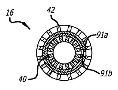
FIG. 6 illustrates an enlarged view of the stent graft 16 in its compacted
state as it
would be found on the balloon, where the balloon has been omitted. The stent
graft 16 of
FIG. 6 is shown as it would appear crimped on a balloon, and FIG. 7 is a cross-
sectional view
of the stent graft 16 of FIG. 6, which shows holes 91a, b aligned and
coincident in each
ePTFE layer, respectively, also passing through the inner and outer stent
walls. This is
similar to what would occur if there were to be a puncture during the crimping
processes of
securing the stent graft to the balloon. The holes 91a,b as may occur during
the crimping
process, the welding process, or in the handling or manufacturing processes.
When in the
crimped state, the holes passes through both layers 46,48 of the ePTFE and are
aligned so that
they appear as a single hole. Without the two layers of ePTFE of the present
invention, when
expanded these holes 91a.b could present a risk of blood leakage or other
structural
deformities of the stent graft.
FIG. 8 depicts the same stent graft after expansion, and FIG. 9 shows the
expanded
stent graft in cross section. When the stent graft 16 is expanded, as for
example by a balloon
or a self-expanding nitinol stent configuration, the inner and outer ePTFE
layers (46, 48
respectively) of the present invention expand in slightly different geometries
due to the
differences in their material properties. As a result, the holes 91a,b that
originally were
aligned are now misaligned so that there is no overlap, and each layer blocks
the hole in the
adjacent layer. That is, hole 91a from the outer layer 48 moves to a different
circumferential
position when compared with the hole 91b of the inner layer 46, best shown in
FIG. 9. This
misalignment of the holes 91a,b provides protection against leakage from holes
developed
during the crimping process, and operates to self-seal the stent graft should
a hole occur.
The relative movement of the two ePTFE layers provides a defense against
leaks, and
also contributes to the overall integrity of the stent graft. That is, the two
layers provide a
back-up to each other in the event one layer has a defect or if one layer is
punctured during
the manufacturing process. The properties of the ePTFE materials can be
adjusted and
manipulated using different sintering processes. For example, the layer to
have the higher
rigidity could be sintered at a higher level than the layer that is to have
the higher plasticity,
which would be sintered at a lower level. Other manufacturing processes could
be used to
alter the properties of the ePTFE film so that one layer would have a higher
rigidity and one
layer would have a higher plasticity.
While particular forms of the invention have been illustrated and described,
it will be
apparent to those skilled in the art that various modifications can be made
without departing
:A 028170572013-05-03
WO 2012/094212 - 8 -
PCT/US2011/067595
from the spirit and scope of the invention. Accordingly, it is not intended
that the invention
be limited except by the appended claims.