Note: Descriptions are shown in the official language in which they were submitted.
CA 02817295 2013-05-30
ABLATION DEVICE WITH DRUG DELIVERY COMPONENT
BACKGROUND
1. Technical Field
[0001] The present disclosure relates to electrosurgical systems and
devices for
performing medical procedures. The present disclosure relates to the
administration of
beneficial agents in general, which include any physiologically,
pharmacologically active
and/or psychotropic substance(s). More particularly, the present disclosure
relates to
ablation devices with drug delivery components, ablation needles with drug
delivery
components, and electrosurgical systems including the same.
2. Discussion of Related Art
[0002] Electrosurgical instruments have become widely used by surgeons.
Electrosurgery involves the application of thermal and/or electrical energy to
cut, dissect,
ablate, coagulate, cauterize, seal or otherwise treat biological tissue during
a surgical
procedure. Electrosurgery is typically performed using a handpiece including a
surgical
instrument (e.g., end effector, ablation probe, or electrode) adapted to
transmit energy
to a tissue site during electrosurgical procedures, an electrosurgical
generator operable
to output energy, and a cable assembly operatively connecting the surgical
instrument
to the generator.
[0003] Treatment of certain diseases requires the destruction of malignant
tissue
growths, e.g., tumors. Electromagnetic radiation can be used to heat and
destroy tumor
cells. Treatment may involve inserting ablation probes into tissues where
cancerous
1
CA 02817295 2013-05-30
tumors have been identified. Once the probes are positioned, electromagnetic
energy
is passed through the probes into surrounding tissue.
[0004]
In the treatment of diseases such as cancer, certain types of tumor cells have
been found to denature at elevated temperatures that are slightly lower than
temperatures normally injurious to healthy cells. Known treatment methods,
such as
hyperthermia therapy, heat diseased cells to temperatures above 41 C while
maintaining adjacent healthy cells below the temperature at which irreversible
cell
destruction occurs. These methods involve applying various forms of energy
(e.g.,
electromagnetic, ultrasonic, etc.) to heat, ablate and/or coagulate tissue.
Microwave or
radio-frequency energy is sometimes utilized to perform these methods. Radio-
frequency (RE) and microwave (MW) energy are electromagnetic radiation in the
frequency ranges of 3 kilohertz (kHz) to 300 Megahertz (MHz), and 300 MHz to
300
gigahertz (GHz), respectively. Other procedures utilizing electromagnetic
radiation to
heat tissue also include coagulation, cutting and/or ablation of tissue.
[0005] Electrosurgical devices utilizing electromagnetic radiation have been
developed for a variety of uses and applications. A number of devices are
available that
can be used to provide high bursts of energy for short periods of time to
achieve cutting
and coagulative effects on various tissues. There are a number of different
types of
apparatus that can be used to perform ablation procedures. Typically,
microwave
apparatus for use in ablation procedures include a microwave generator that
functions
as an energy source, and a microwave surgical instrument (e.g., microwave
ablation
probe) having an antenna assembly for directing the energy to the target
tissue. The
microwave generator and surgical instrument are typically operatively coupled
by a
2
CA 02817295 2013-05-30
cable assembly having a plurality of conductors for transmitting microwave
energy from
the generator to the instrument, and for communicating control, feedback and
identification signals between the instrument and the generator.
[0006] The basic purpose of both monopolar and bipolar electrosurgery is to
produce
heat to achieve the desired tissue/clinical effect. In monopolar
electrosurgery, devices
use an instrument with a single, active electrode to deliver energy from an
electrosurgical generator to tissue, and a patient return electrode (usually a
plate
positioned on the patient's thigh or back) as the means to complete the
electrical circuit
between the electrosurgical generator and the patient. In bipolar
electrosurgery, the
electrosurgical device includes two electrodes that are located in proximity
to one
another for the application of current between their surfaces. Bipolar
electrosurgical
current travels from one electrode, through the intervening tissue to the
other electrode
to complete the electrical circuit.
[0007] The benefits provided by controlled delivery of active agents for
the treatment
of injury or disease are well recognized in the art and various approaches
have been
taken to realize the goal of delivering active agents at desired rates over
predetermined
periods of time. Various different implantable controlled delivery
formulations are
known in the art, and various different mechanisms have been employed for
delivering
active agent from implantable formulations at a controlled rate over time.
[0008] Medical imaging has become a significant component in the clinical
setting
and in basic physiology and biology research, e.g., due to enhanced spatial
resolution,
accuracy and contrast mechanisms that have been made widely available. Medical
imaging now incorporates a wide variety of modalities, e.g., computed
tomography (CT)
3
CA 02817295 2013-05-30
and magnetic resonance imaging (MRO, that noninvasively capture the structure
and/or
function of the human body. Such images are acquired and used in many
different
ways including medical images for diagnosis, staging and therapeutic
management of
malignant disease.
[0009] Medical image processing, analysis and visualization play an
increasingly
useful role in disease diagnosis and monitoring as well as, among other
things, surgical
planning and monitoring of therapeutic procedures. A contrast agent may used
for
enhancement of the contrast of structures or fluids within the body (or region
of interest)
in medical imaging to allow visualization and evaluation of lesions seen
minimally, if at
all, with imaging alone. There is a continuing need for devices capable of
dispensing a
contrast agent to enhance the visualization of the lesion during the
procedure.
[0010] Despite advancements in the use of electrosurgical devices for
treating
biological tissue, there are still concerns for tumor reoccurrence. A
continuing need
exists for devices capable of dispensing a controlled delivery formulation of
a desired
active agent, which may help to reduce or eliminate tumor reoccurrence.
SUMMARY
[0011] There is a need for ablation devices capable of dispensing a
controlled
delivery formulation of a desired active agent. The combination of ablation
(e.g., RF
ablation and/or microwave ablation) and drug delivery may help to reduce or
eliminate
tumor reoccurrence. There is a need for an ablation device that is configured
to
dispense an active agent in a controlled delivery formulation and/or non-
active agent
(e.g., contrast agent) before, during and/or after ablation, e.g., without the
need for
4
CA 02817295 2013-05-30
turther manipulation at the device. A need exists tor ablation needles with a
drug
delivery component.
[0012] Electromagnetic energy is generally classified by increasing energy
or
decreasing wavelength into radio waves, microwaves, infrared, visible light,
ultraviolet,
X-rays and gamma-rays. As it is used in this description, "ablation procedure"
generally
refers to any ablation procedure, such as microwave ablation, radio frequency
(RF)
ablation or microwave ablation-assisted resection.
[0013] As it is used in this description, "energy-delivery device"
generally refers to
any device that can be used to transfer energy from a power generating source,
such as
a microwave or RF electrosurgical generator, to tissue. For the purposes
herein, the
term "ablation device" is interchangeable with the term "energy-delivery
device." As it is
used in this description, "transmission line" generally refers to any
transmission medium
that can be used for the propagation of signals from one point to another. A
transmission line may be, for example, a wire, a two-wire line, a coaxial
wire, and/or a
waveguide.
[0014] For the purposes of this description, the terms "drug," "drug
agent,"
"implantable drug agent," "active agent," "beneficial agent," "therapeutic
agent,"
"therapeutic molecule," and the like are used interchangeably herein, and may
include,
for example, small molecules, proteins, enzymes, hormones, polynucleotides,
nucleoproteins, polysaccharides, glycoproteins, lipoproteins, polypeptides,
steroids,
analgesics, local anesthetics, antibiotic agents, anti-inflammatory
corticosteroids, ocular
drugs and synthetic analogs of these species. Some examples of drug agents
that may
CA 02817295 2013-05-30
be delivered by devices according to embodiments of the present disclosure are
provided later in this description.
[0019 There is a need for an implantable formulation that provides
pharmacokinetic/
pharmacodynamic (PK/PD) appropriate release rate profile of an active agent
without
necessarily requiring the need for further manipulation, post implantation or
surgical
explantation. There is a need for a formulation to facilitate delivery of a
wide range of
active agents and active agents formulations, as well as multiple active
agents and
multiple active agents' formulations, which may also increase the value of
each drug-
based ablation procedure.
[0016] A continuing need exists for systems, devices and methods for
controlling
and/or monitoring real-time tissue effects to improve patient safety, reduce
risk, and/or
improve patient outcomes. There is a need for ablation devices capable of
dispensing
contrast agent to enhance the visualization of the lesion during the treatment
procedure.
[0017] According to an aspect of the present disclosure, an ablation device
is
provided. The ablation device includes a handle assembly, an ablation
electrode
extending from the handle assembly, and one or more delivery needles extending
from
the handle assembly. The ablation electrode includes an ablation needle. The
ablation
needle includes a distal end portion including a drug delivery port defined
therethrough.
[0018] According to an aspect of the present disclosure, an ablation device
is
provided. The ablation device includes a handle assembly and an array of
ablation
electrodes operably associated with the handle assembly. One or more ablation
electrodes of the array of ablation electrodes include a recess defined
therein. A drug is
disposed at least in part within the recess.
6
CA 02817295 2013-05-30
[0019] According to an aspect of the present disclosure, an ablation system
is
provided. The ablation system includes a source of electrosurgical energy, a
source of
coolant fluid, and an ablation electrode assembly operatively connected to the
source of
electrosurgical energy and fluidly-coupled to the source of coolant fluid. The
ablation
electrode assembly includes a hub defining a chamber therein and one or more
electrically-conductive ablation needles extending from the hub. The ablation
system
also includes one or more delivery needles extending from the hub. The one or
more
delivery needles are selectively moveable from a first position, wherein the
distal end of
the delivery needle is disposed proximal to the distal end portion of the
ablation needle,
to at least a second position, wherein at least the distal end of the delivery
needle is
disposed distally beyond the distal end portion of the ablation needle.
BRIEF DESCRIPTION OF THE DRAWINGS
[0020] Objects and features of the presently-disclosed ablation devices
with drug
delivery (and/or contrast agent) components, ablation needles with drug
delivery
components, and electrosurgical systems including the same will become
apparent to
those of ordinary skill in the art when descriptions of various embodiments
thereof are
read with reference to the accompanying drawings, of which:
[0021] FIG. 1A is a schematic diagram of an ablation system including an
ablation
electrode assembly in accordance with an embodiment of the present disclosure;
[0022] FIG. 1B is a schematic diagram of an ablation electrode assembly
including
the needle assembly of the ablation electrode assembly shown in FIG. 1A in
accordance with an embodiment of the present disclosure;
7
CA 02817295 2013-05-30
[0023] FIG. 2 is a schematic diagram of an ablation system including an
energy
applicator in accordance with an embodiment of the present disclosure;
[0024] FIG. 3 is an enlarged, perspective view of an ablation system
including an
electrode array in accordance with an embodiment of the present disclosure;
[0025] FIG. 4 is an enlarged, perspective view of an ablation system
including an
electrode array that includes a drug delivery component in accordance with an
embodiment of the present disclosure;
[0026] FIG. 5A is an enlarged, perspective view of an ablation system
including an
ablation device that includes ablation electrodes and a delivery needle, shown
with the
delivery needle disposed in a first configuration, in accordance with an
embodiment of
the present disclosure;
[0027] FIG. 5B is an enlarged, perspective view of the ablation device
shown in
FIG. 5A shown with the delivery needle disposed in a second configuration in
accordance with an embodiment of the present disclosure;
[0028] FIG. 6A is an enlarged, perspective view of a portion of an ablation
needle,
with portions removed, shown with an integral delivery needle disposed in a
first
configuration in accordance with an embodiment of the present disclosure;
[0029] FIG. 6B is an enlarged, perspective view of the ablation needle
shown in
FIG. 6A shown with the integral delivery needle disposed in a second
configuration in
accordance with an embodiment of the present disclosure;
8
CA 02817295 2013-05-30
[0030] FIG. 7A is an enlarged, perspective view of a portion of an ablation
needle,
with portions removed, shown with an integral delivery needle disposed in a
first
configuration in accordance with another embodiment of the present disclosure;
[0031] FIG. 7B is an enlarged, perspective view of the ablation needle
shown in
FIG. 7A shown with the integral delivery needle disposed in a second
configuration in
accordance with an embodiment of the present disclosure;
[0032] FIG. 8 is an enlarged, perspective view of a portion of an ablation
needle
assembly that includes a shaft portion and an interchangeable sleeve member
disposed
around the shaft portion, the sleeve member including a plurality of drug
reservoir divots
associated therewith, in accordance with an embodiment of the present
disclosure;
[0033] FIG. 9 is an enlarged, perspective view of a portion of an ablation
needle that
includes a shaft portion including a plurality of drug reservoir divots
associated therewith
in accordance with an embodiment of the present disclosure;
[0034] FIG. 10 is an enlarged, perspective view of a portion of an ablation
needle
assembly that includes an interchangeable sleeve member including a plurality
of drug
reservoir divots associated therewith in accordance with an embodiment of the
present
disclosure;
[0035] FIG. 11 is an enlarged, perspective view of a portion of an ablation
needle
assembly that includes an interchangeable sleeve member including a plurality
of drug
reservoir divots associated therewith and a plurality of apertures defined
therethrough in
accordance with an embodiment of the present disclosure;
9
CA 02817295 2013-05-30
[0036] FIG. 12 is an enlarged, perspective view of a portion of an ablation
needle
assembly that includes an interchangeable sleeve member including a plurality
of drug
reservoir divots associated therewith and a plurality of elongated apertures
defined
therethrough in accordance with an embodiment of the present disclosure;
[0037] FIG. 13 is an enlarged, perspective view of an RF ablation device
including a
drug reservoir and a moveable tip portion containing deployable tendrils,
shown with the
tip portion disposed in a first configuration, in accordance with an
embodiment of the
present disclosure;
[0038] FIG. 14A is an enlarged, perspective view of the RF ablation device
shown in
FIG. 13 shown with the tip portion disposed in a second configuration, showing
the
transfer of drugs from the drug reservoir to tissue, in accordance with an
embodiment of
the present disclosure;
[0039] FIG. 14B is an enlarged, perspective view of the RF ablation device
shown in
FIG. 14A showing deployment of tendrils from the tip portion into tissue in
accordance
with an embodiment of the present disclosure;
[0040] FIG. 15 is a schematic diagram of a distal portion of an ablation
device
including deployable tendrils in accordance with an embodiment of the present
disclosure;
[0041] FIG. 16A is an enlarged, perspective view of an ablation device
including a
shaft portion that includes a moveable member and deployable tendrils, shown
with the
tendrils disposed in a first configuration, in accordance with an embodiment
of the
present disclosure;
CA 02817295 2013-05-30
[0042] FIG. 16B is an enlarged, perspective view of the ablation device
shown in
FIG. 16A, shown with the tendrils disposed in a second configuration, in
accordance
with an embodiment of the present disclosure;
[0043] FIG. 17A is a schematic diagram of a portion of an RF ablation
device
including a barrel portion configured to contain antenna filaments deployable
therefrom
and a moveable tip portion, shown with the tip portion disposed in a first
configuration,
in accordance with an embodiment of the present disclosure;
[0044] FIG. 17B is a schematic diagram of the RF ablation device shown in
FIG. 16
shown with the tip portion disposed in a second configuration, showing
deployment of
the antenna filaments from the barrel portion into tissue, in accordance with
an
embodiment of the present disclosure;
[0045] FIG. 18A is a schematic diagram of the RF ablation device shown in
FIG. 16
shown with the tip portion disposed in a third configuration, showing the
deployed
antenna filaments attached in part to the tip portion, in accordance with an
embodiment
of the present disclosure;
[0046] FIG. 18B is a schematic diagram of the RF ablation device shown in
FIG. 18A
shown with the tip portion disposed in a fourth configuration, showing the
antenna
filaments separated from the tip portion in accordance with an embodiment of
the
present disclosure;
[0047] FIG. 19 is an enlarged, cross-sectional view of an ablation device
that
includes an antenna assembly including a porous-metal radiating section
suitable for
drug delivery in accordance with an embodiment of the present disclosure;
11
CA 02817295 2013-05-30
[0048] FIG. 20 is an enlarged, perspective view of an ablation device that
includes
an antenna assembly in accordance with an embodiment of the present
disclosure;
[0049] FIG. 21 is an enlarged view of the indicated area of detail of FIG.
20 showing
a distal portion of the antenna assembly disposed in a first configuration in
accordance
with an embodiment of the present disclosure;
[0050] FIG. 22 is an enlarged view of the indicated area of detail of FIG.
20 showing
a distal portion of the antenna assembly disposed in a second configuration in
accordance with an embodiment of the present disclosure;
[0051] FIG. 23 is an enlarged view of a portion of an ablation needle
including drug-
delivery tines, the ablation needle is shown disposed in a first configuration
within target
tissue, in accordance with an embodiment of the present disclosure;
[0052] FIG. 24 is an enlarged view of the ablation needle including drug-
delivery
tines shown in FIG. 23 shown with the ablation needle disposed in a second
configuration within target tissue in accordance with an embodiment of the
present
disclosure;
[0053] FIG. 25 is an enlarged view of the ablation needle including drug-
delivery
tines shown in FIG. 23 shown with the ablation needle disposed in a third
configuration
within target tissue in accordance with an embodiment of the present
disclosure;
[0054] FIG. 26 is an enlarged view of a portion of an ablation needle
including a
delivery needle that includes a micro-needle adapted to be slideably moveable
within
the delivery needle in accordance with an embodiment of the present
disclosure;
12
CA 02817295 2013-05-30
[0055] FIG. 27 is an enlarged view of the delivery needle shown in FIG. 26
shown
with the micro-needle shown in a deployed configuration in accordance with an
embodiment of the present disclosure;
[0056] FIG. 28 is an enlarged view of a portion of a delivery needle, such
as the
delivery needle of the ablation needle shown in FIG. 7, shown with a micro-
needle
shown in a deployed configuration in accordance with an embodiment of the
present
disclosure;
[0057] FIG. 29 is an enlarged view of a portion of a delivery needle, such
as the
delivery needle of the ablation needle shown in FIG. 7, shown with a micro-
needle array
shown in a deployed configuration in accordance with an embodiment of the
present
disclosure;
[0058] FIG. 30 is an enlarged view of the indicated area of detail of FIG.
29 showing
a string of micro-needle elements of the micro-needle array in accordance with
an
embodiment of the present disclosure;
[0059] FIG. 31 is an enlarged view of the string of micro-needle elements
shown in
FIG. 30 in accordance with another embodiment of the present disclosure;
[0060] FIG. 32 is an enlarged view of a string of micro-needle elements of
a micro-
needle array shown with a drug attached to the micro-needle elements in
accordance
with an embodiment of the present disclosure;
[0061] FIG. 33 is an enlarged view of a configuration of micro-needle
elements of a
micro-needle array shown with a drug attached to the micro-needle elements in
accordance with another embodiment of the present disclosure;
13
CA 02817295 2013-05-30
[0062] FIG. 34 is an enlarged view of a portion of a delivery needle, such
as the
delivery needle of the ablation needle shown in FIG. 7, shown with a partially
deployed,
heat-sensitive material coated, micro-needle array in accordance with an
embodiment
of the present disclosure;
[0063] FIG. 35 is an enlarged view of the indicated area of detail of FIG.
34 showing
micro-needle elements of the micro-needle array in accordance with an
embodiment of
the present disclosure;
[0064] FIG. 36A is a enlarged, perspective view of an electrosurgical
system
including an ablation device that includes an array of ablation electrodes, a
delivery
needle, and an antenna assembly including a delivery needle in accordance with
an
embodiment of the present disclosure;
[0065] FIG. 36B is an enlarged, perspective view of the indicated area of
detail of
FIG. 36A in accordance with an embodiment of the present disclosure;
[0066] FIG. 37A is a enlarged, perspective view of the electrosurgical
system of
FIG. 36A shown with the antenna assembly including another embodiment of a
delivery
needle in accordance with the present disclosure;
[0067] FIG. 37B is an enlarged, perspective view of the indicated area of
detail of
FIG. 37A in accordance with an embodiment of the present disclosure; and
[0068] FIG. 38 is an enlarged, perspective view of an ablation device that
includes
an array of ablation electrodes including a plurality of drug reservoir divots
associated
therewith in accordance with an embodiment of the present disclosure.
14
CA 02817295 2013-05-30
DETAILED DESCRIPTION
[0069] Hereinafter, embodiments of the presently-disclosed ablation devices
with
drug delivery and/or contrast agent components, ablation needles with drug
delivery
and/or contrast agent components (e.g., suitable for use with CooltipTM RF
ablation
devices), and electrosurgical systems including the same are described with
reference
to the accompanying drawings. Like reference numerals may refer to similar or
identical
elements throughout the description of the figures. As shown in the drawings
and as
used in this description, and as is traditional when referring to relative
positioning on an
object, the term "proximal" refers to that portion of the device, or component
thereof,
closer to the user and the term "distal" refers to that portion of the device,
or component
thereof, farther from the user.
[0070] This description may use the phrases "in an embodiment," "in
embodiments,"
"in some embodiments," or "in other embodiments," which may each refer to one
or
more of the same or different embodiments in accordance with the present
disclosure.
[0071] Various embodiments of the present disclosure provide energy-
delivery
devices including ablation needles with drug delivery and/or contrast agent
components.
Embodiments may be suitable for use with cooltipTM RF ablation devices.
Embodiments may be suitable for utilization in open surgical applications.
Embodiments may be suitable for utilization with endoscopic and laparoscopic
surgical
procedures. Embodiments may be implemented using electromagnetic radiation at
microwave frequencies, RF frequencies or at other frequencies.
[0072] Various embodiments of the present disclosure provide
electrosurgical
system including an energy delivery device provided with one or more ablation
needles
CA 02817295 2013-05-30
with drug delivery (and/or contrast agent) components. Various embodiments of
the
presently-disclosed electrosurgical systems may be suitable for microwave
ablation and
for use to pre-coagulate tissue for microwave ablation assisted surgical
resection.
Various embodiments of the presently-disclosed electrosurgical systems
including an
ablation device may include any feature or combination of features of the
ablation
device embodiments disclosed herein.
[0073] Various embodiments of the presently-disclosed ablation needle
assembly
include an elongated body or shaft portion configured to facilitate delivery
of one or
more drug agents which may be temperature sensitive into tissue, wherein one
or more
drug agents may be releaseably disposed over at least a portion of body or
shaft portion
of the ablation needle assembly itself, and/or one or more drug agents may be
releaseably disposed over at least a portion of a sleeve member disposed
coaxially
around the body or shaft portion of the ablation needle assembly.
[0074] Drug agents which may be delivered by devices according to
embodiments of
the present disclosure include drugs which act on the peripheral nerves,
adrenergic
receptors, cholinergic receptors, the skeletal muscles, the cardiovascular
system,
smooth muscles, the blood circulatory system, synoptic sites, neuroeffector
junctional
sites, endocrine and hormone systems, the immunological system, the
reproductive
system, the skeletal system, autacoid systems, the alimentary and excretory
systems,
the histamine system and the central nervous system. Some examples of
implantable
drug agents which may be delivered by devices according to embodiments of the
present disclosure are provided later in this description.
16
CA 02817295 2013-05-30
[0075] In accordance with various embodiments, the combination of tissue
ablation
and drug delivery may help to reduce and/or eliminate tumor reoccurrence. In
accordance with various embodiments, the combination ablation devices with
drug
delivery and/or contrast agent components may help to reduce procedure times
and/or
eliminate the need for a separate drug-delivery device.
[0076] FIG. 1A shows an electrosurgical system (shown generally as 100) in
accordance with an embodiment of the present disclosure that includes an
ablation
electrode assembly 110 and a hub 130 configured to support the ablation
electrode
assembly 110. Ablation electrode assembly 110 is operatively connected to an
electrosurgical power generating source 28, e.g., a microwave or radio
frequency (RF)
electrosurgical generator. Ablation electrode assembly 110 is disposed in
fluid
communication with a coolant source 48. Ablation electrode assembly 110 may
include
additional, fewer, or different components than shown in FIG. 1A, depending
upon a
particular purpose or to achieve a desired result.
[0077] An embodiment of an ablation electrode assembly 101, similar to the
ablation
electrode assembly 110 of the electrosurgical system 100 shown in FIG. 1A, in
accordance with the present disclosure, is shown in FIG. 1B. It is to be
understood,
however, that other ablation device (e.g., ablation system 200 shown in FIG.
2, ablation
system 300 shown in FIG. 3, ablation device 400 shown in FIG. 4, ablation
device 510
shown in FIGS. 5A and 5B, ablation needle 600 shown in FIGS. 6 and 7, ablation
needle assembly 800 shown in FIG. 8, ablation needle 900 shown in FIG. 9,
ablation
needle assembly 1000 shown in FIG. 10, ablation needle assembly 1100 shown in
FIG. 11, ablation needle assembly 1200 shown in FIG. 12, ablation device 1300
shown
17
CA 02817295 2013-05-30
in FIGS. 13-14B, ablation device 1500 shown in FIG. 15, ablation device 1400
shown in
FIGS. 16A and 16B, RF ablation device 1600 shown in FIGS. 17A-18B, ablation
device
1900 shown in FIG. 19, ablation device 2000 shown in FIGS. 20-22, ablation
needle
2300 shown in FIGS. 23-25, and combinations thereof) may also be used.
[0078]
In some embodiments, electrosurgical system 100 (also referred to herein as
ablation system 100) may include a controller 26 for controlling and/or
monitoring the
operating parameters of the ablation system 100. In some embodiments, as shown
in
FIG. 1A, the controller 26 is communicatively-coupled to the electrosurgical
power
generating source 28.
Controller 26 may additionally, or alternatively, be
communicatively-coupled to the fluid source 48. In some embodiments, the
controller
26 may receive user-inputs from one or more user-input devices, such as
without
limitation, a keyboard, a pointing device, e.g., a mouse, joystick or
trackball, a
touchscreen, and/or other device communicatively-coupled to the controller 26.
In
some embodiments, electrosurgical system 100 includes an imaging system (not
shown) capable of generating image data, and the controller 26 may be
communicatively-coupled to the imaging system. Controller 26 may include any
type of
computing device, computational circuit, or any type of processor or
processing circuit
capable of executing a series of instructions that are stored in a memory (not
shown)
associated with the controller 26. Functions of the controller 26 may be
performed in
hardware and/or software, as desired. Controller 26 may include logic,
circuitry and/or
code adapted to control the electrosurgical power generating source 28 and/or
the
coolant source 48 responsive to one or more electrical signals received from
one or
more user-input devices. Functions of the controller 26 may be integrated with
those of
18
CA 02817295 2013-05-30
the electrosurgical power generating source 28, may be integrated with other
components of the electrosurgical system 100, and/or may be in the form of
stand-alone
units coupled among components of the electrosurgical system 100.
[0079] As seen in FIGS. 1A and 1B, ablation electrode assembly 110 includes
an
elongated ablation needle 112. Ablation needle 112 includes a substantially
cylindrically-shaped body or shaft portion 114 defining a cavity or 116
therein. Ablation
needle 112 includes a distal end portion 118 including a tapered portion,
which may
terminate in a sharp tip 118a to allow for insertion into tissue with minimal
resistance.
Ablation needle 112 includes a proximal end portion 120, which may be
configured for
connection to a hub 130, which is described in more detail later in this
description.
Ablation needle 112 is fabricated from an electrically-conductive material,
e.g., stainless
steel, titanium, etc. The shape and size of the ablation needle 112 may be
varied from
the configuration depicted in FIGS. 1A and 1B.
[0080] Ablation electrode assembly 110 includes an insulative coating 122
over at
least a portion of the length of the ablation needle 112. In some embodiments,
the
insulative coating 122 is disposed over substantially the length of the
ablation needle
112. In some embodiments, as shown in FIGS. 1A and 1B, the insulative coating
122
extends from the hub 130 to the distal end portion 118 of the ablation needle
112, such
that the distal end portion 118 of the ablation needle 112 is exposed or non-
insulated.
lnsulative coating' 122 is used to prevent the flow of electrical current from
the shaft
portion 114 of the ablation needle 112 into surrounding tissue. lnsulative
coating 122
shields the intervening tissue from RF current, so that such tissue is not
substantially
19
CA 02817295 2013-05-30
heated along the length ot shaft portion 114 except by the heating effect from
the distal
end portion 118 which is exposed.
[0081] In some embodiments, as shown in FIGS. 1A and 1B, an ablation
electrode
assembly 110 includes one or more heat sinks, in the form of a heat strap or
heat pipe
124 extending through the chamber 116, or portion thereof, of the ablation
needle 112.
It is to be understood, however, that in ablation electrode embodiments (e.g.,
electrode
211 of the ablation system 200 shown in FIG. 2) coolant fluid (and/or drug
agent) may
circulate to a tip portion for cooling of the electrode without the use of
heat sinks.
[0082] Referring to FIGS. 1A and 1B, heat strap 124 includes a distal end
124a
operatively secured to the ablation needle 112, and a proximal end 124b
extending into
a chamber 132 formed in the hub 130. In some embodiments, the distal end 124a
of
the heat strap 124 is operatively connected or secured to the distal end
portion 118 of
the ablation needle 112. In some embodiments, the distal end 124a of the heat
strap
124 is bonded to the distal end portion 118 of the ablation needle 112 with a
thermally-
conductive adhesive or the like. Although a single heat strap 124 is shown in
FIGS. 1A
and 1B, a plurality of heat straps 124 may be provided. It is to be
understood, however,
that in ablation electrode embodiments (e.g., electrode 211 of the ablation
system 200
shown in FIG. 2) coolant fluid (and/or drug agent) may circulate to a tip
portion for
cooling of the ablation electrode without the use of heat sinks.
[0083] Heat strap 124 is fabricated from a highly heat-conductive
anisotropic
material, e.g., graphite fiber. Accordingly, in use, the heat strap 124 draws
heat away
from distal end portion 118 of the ablation needle 112 and dissipates the heat
along a
CA 02817295 2013-05-30
length thereof. In order to increase the efficiency and the rate of heat
dissipation, a
cooling fluid may be circulated over the proximal end 124b of the heat strap
124.
[0084] Hub 130 may have a variety of suitable shapes, e.g., cylindrical,
rectangular,
etc. Hub 130 generally includes a hub body 145 defining a chamber 132 therein.
In
some embodiments, as shown in FIG. 1A, hub body 145 defines an outlet fluid
port 177
and an inlet fluid port 179 disposed in fluid communication with the chamber
132. Hub
130 may include an inlet conduit 134 for delivering coolant fluid "F" through
the inlet
fluid port 179 into the chamber 132, and may include an outlet conduit 136 for
delivering
coolant fluid "F" through the outlet fluid port 177 from the chamber 132. In
some
embodiments, coolant chamber 132 may include baffles, multiple lumens, flow
restricting devices, or other structures that may redirect, concentrate, or
disperse flow
depending on their shape. Examples of coolant chamber embodiments are
disclosed in
commonly assigned U.S. Patent Application Serial No. 12/350,292 filed on
January 8,
2009, entitled "CHOKED DIELECTRIC LOADED TIP DIPOLE MICROWAVE
ANTENNA", commonly assigned U.S. Patent Application Serial No. 12/401,268
filed on
March 10, 2009, entitled "COOLED DIELECTRICALLY BUFFERED MICROWAVE
DIPOLE ANTENNA", and U.S. Pat. No. 7,311,703, entitled "DEVICES AND METHODS
FOR COOLING MICROWAVE ANTENNAS".
[0085] In operation, coolant fluid "F" is communicated into the chamber 132
through
the inlet conduit 134 and out of the chamber 132 through the outlet conduit
136.
Coolant fluid "F" may be any suitable fluid that can be used for cooling the
ablation
needle 112, e.g., deionized water, or other suitable cooling medium. As
coolant fluid "F"
is circulated through the chamber 132 of the hub 130, heat or energy is
withdrawn from
21
CA 02817295 2013-05-30
the proximal end 124b of the heat strap 124 and carried away with fluid flow,
e.g., to the
fluid source 48 for re-cooling and the like.
[0086] In some embodiments, as shown in FIG. 1A, the hub 130 may include a
proximal connector (e.g., a luer connector) including a tapered hole 140 or
the like. into
female luer connector 140, a hub of a high-frequency or thermo-sensing
electrode 142
may be inserted and sealed by its male luer connection. A probe 144 of the
thermo-
sensing electrode 142 may be connected to the ablation needle 112, which may
sense
the temperature of ablation needle 112 at that point, or alternatively, may
sense the
temperature of the distal end portion 118.
[0087] Connected to or within the hub of the high-frequency and/or thermo-
sensing
electrode 142 are connections, indicated by dashed lines in FIG. 1A, which
connect to
the electrosurgical power generating source 28 and/or a thermal-sensing
circuit "TO".
In some embodiments, the thermal-sensing circuit "TO" may be of a thermocouple
type,
and the temperature sensor may a bi-metal junction thermocouple. The
temperature
sensor may be sensor capable of generating a signal indicative of a
temperature of a
medium in contact therewith.
[0088] Coolant source 48 may include any suitable housing containing a
reservoir of
coolant fluid "F". Coolant source 48 stores coolant fluid "F", and may
maintain coolant
fluid "F" at a predetermined temperature. For example, the coolant source 48
may
include a cooling unit (not shown) that cools the returning coolant fluid "F"
from the
ablation electrode assembly 110. Ablation system 100 may include a coolant
supply
system (not shown) adapted to provide the coolant fluid "F", e.g., from the
coolant
source 48, to the ablation electrode assembly 110. In some embodiments, one or
more
22
CA 02817295 2013-05-30
components ot a coolant supply system may be integrated fully or partially
into the
electrosurgical power generating source 28.
[0089] During ablation, e.g., using the electrosurgical system 100, the
ablation
electrode assembly 110 is inserted into or placed into the body of a patient,
e.g.,
percutaneously or intraoperatively. A plurality of ablation electrodes 110 may
be placed
in variously arranged configurations to substantially simultaneously ablate a
target
tissue region, making faster procedures possible. Ultrasound or computed
tomography
(CT) guidance may be used to accurately guide the ablation electrode assembly
110
into the area of tissue to be treated. Electrosurgical power generating source
28 may
be the source of high-frequency voltage which produces the high-frequency
current that
emanates from the distal end portion 118 of ablation needle 112. Following
treatment
or ablation of the target tissue, ablation electrode assembly 110 may be
withdrawn from
the target site and introduced into another target site, into the same target
site from a
different angle or approach, or in substantially the same location.
[0090] Examples of electrosurgical generators that may be suitable for use
as the
electrosurgical power generating source 28 include generators sold by Covidien
Surgical Solutions of Boulder, CO, e.g., FORCE EZTM electrosurgical generator,
FORCE FXTM electrosurgical generator, and FORCE TRIADTm electrosurgical
generator
FORCE 1CTM generator, FORCE 2TM generator, SurgiStatTM II, or other generators
which may perform different or enhanced functions.
[0091] In alternative embodiments not shown, the ablation electrode
assembly 110
may include an inflatable balloon member which may be connected to walls of
the
electrode assembly 110 using any fastening technique, e.g., adhesive, sonic
welding, or
23
CA 02817295 2013-05-30
by any other suitable process. The inflatable balloon member (not shown) may
be
operated in conjunction with the delivery of a drug agent. The walls of the
electrode
assembly 110 may be provided with an opening or port disposed and configured
to
place a inflation lumen in fluid communication with the inflatable balloon
member, e.g.,
to allow drug-delivery flow supplied via the inflation lumen to be used to
operate the
inflatable balloon member, e.g., drug-eluting balloon. In some embodiments,
the
electrode assembly 110 may be adapted to allow user control of operational
characteristics of the drug-eluting balloon, e.g., rate of inflation,
inflation volume, and
pressure exerted by the inflatable balloon member on the tissue surrounding
the
inflatable balloon member.
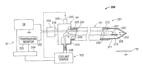
[0092] FIG. 2 shows an ablation system 200 including an energy applicator
201
according to an embodiment of the present disclosure. Energy applicator 201
includes
an elongated shaft or cannula body "C" for positioning in tissue, e.g.,
percutaneously or
intraoperatively into an open wound site. in some embodiments, the cannula
body "C"
is integral with a hub "H" coupled to remote support components, collectively
designated "S". Energy applicator 201 is operatively connected to an
electrosurgical
power generating source 28, e.g., a microwave or radio frequency (RF)
electrosurgical
generator.
[0093] Cannula body "C" includes an elongated ablation electrode 211 formed
of
conductive material, e.g. metal such as stainless steel, titanium, etc.
Electrode 211 may
include a substantially hollow tubular body sized in length and diameter to
fit within the
cannula body "C". At the distal end of the cannula body "C", the electrode 211
defines
24
CA 02817295 2013-05-30
a tip 212. In operation when using an RE power supply 216, electrical current
spreads
from the tip 212 to pass through the surrounding tissue causing the tissue to
heat up.
[0094] Electrode 211 carries an insulative coating 213 over a portion of
its length for
selectively preventing the flow of electrical current from the shaft 215 of
electrode 211
into surrounding tissue. lnsulative coating 213 shields the intervening tissue
from RF
current, so that tissue along the length of the shaft 215 is not substantially
heated
except by the heating effect from the exposed tip 212.
[0095] The proximal end of the electrode 211 is integral with an enlarged
housing
214 of the hub "H", which carries electrical and coolant connections as
described below.
In the portion disposed outside the patient's body, the housing 214 is of
cylindrical
configuration, defining ports for connections to the support components "S",
e.g.,
electrical and fluid couplings. Housing 214 may be integral with the electrode
211,
formed of metal, or it may constitute a separate subassembly as described
below.
Housing 214 may be formed of plastic, and may accommodate separate electrical
connections. In that regard, a plastic housing 214 is amenable to low artifact
imaging
by X-rays CT, MRI, etc. as may be desirable in some situations.
[0096] The housing 214 mates with a block 218 defining a luer taper lock
219 sealing
the block 218 to the housing 214. Connection to a regulated RE power source
216 may
take the form of a standard cable connector, a leader wire, a jack-type
contact or other
designs. The temperature-sensing and radiofrequency electrical connections may
be
made through the housing 214 and extend to the region of the tip 212, where an
RE line
225 is connected by junction 221, e.g., a weld, braze, or other secure
electrical
connection. In some embodiments, sensor lines 224 extend to a thermo-sensor
223,
CA 02817295 2013-05-30
e.g., a thermistor, or a thermocouple, or any other type of temperature
sensing device
capable of sending a signal indicative of a temperature. Thermo-sensor 223 may
be
fused or in thermal contact with the wall of the tip 212 to sense the
temperature of the
tip 212.
[0097] RF power source 216 may be referenced to reference potential, as
illustrated
FIG. 2, and coupled through the block 218 affixed to the hub "H". In
embodiments, the
RF power source 216 provides RF voltage through the block 218 with an
electrical
connection to the electrode 211 as indicated by the line 225, to the
connection junction
221. RF power source 216 may take the form of an RF generator. Examples of RF
generators that may suitably be used as the RF power source 216 may include
the
RFG-3C RF Lesion Generator System available from Radionics, Inc., Burlington,
Massachusetts.
[0098] As indicated above and in accordance with common practice, when the
ablation electrode 211 is in a patient's body, an electrical circuit is
completed through
the body to a reference or dispersive electrode R (symbolically represented in
FIG. 2)
that is connected elsewhere to the body. Energy transmitted from the RF power
source
216 heats body tissue by current from the tip 212. In that regard, a
temperature monitor
220 may be electrically connected by lines 222 and 224 to a temperature sensor
223
disposed within or contacting the tip 212. Temperature sensor 223 may be a
thermocouple, thermistor, or other temperature sensing device. In an
embodiment, the
sensor 223 is connected to the tip 212. The sensed temperature may be utilized
to
control either or both of the flow of RF energy or the flow of coolant to
attain the desired
ablation while maintaining the maximum temperature substantially below a
26
CA 02817295 2013-05-30
predetermined temperature, e.g., 100 C. One or more sensors may be utilized
to
measure temperatures at various locations in the proximity of the tip 212. One
or more
sensor devices, or components thereof, may be disposed outside the distal end
portion
118 of the ablation needle 212. Examples of temperature monitoring devices
that may
suitably be used as the temperature monitor 220 may include the TC
thermocouple
temperature monitoring devices available from Radionics, Inc., Burlington,
Massachusetts.
[0100] In accordance herewith, temperatures at, or near the tip 212 (e.g.,
manifest
by the temperature monitor 220) may be controlled by controlling the flow of
coolant
fluid through the ablation electrode 211. In this manner, the temperature of
the surface
area of the tip 212 in contact with tissue is controllable. In an embodiment,
fluid from a
fluid source "FS" is carried the length of the ablation electrode 211 through
a tube 226
extending from the housing 214 to the distal end of the electrode 211
terminating in an
open end 228 at the tip 212. At the proximal end of the electrode 211, within
the
housing 214, the tube 226 is connected to receive coolant fluid. Fluid flow
may be
regulated in accordance with the sensed temperature sensed at the tip 212,
allowing
increased flow of RF energy.
[0101] The fluid coolant may take the form of water or saline for the
convection
removal of heat from the tip 212. A reservoir or source unit for supplying
coolant fluid
may be a large reservoir of cooled water, saline or other fluid. As an
illustrative
example, a tank of water with ice cubes can function to maintain the coolant
at a
temperature of approximately 0 C. As another example, the fluid source "FS"
could
27
CA 02817295 2013-05-30
incorporate a peristaltic pump or other fluid pump, or could merely be a
gravity feed for
supplying fluid from a bag or rigid tank.
[0102] Flow from the tip 212 to the hub "H" exits the hub "H" through an
exit port 240
as illustrated by arrows 242 and 243. The port 240 may take the form of
couplings, rigid
units or may include flexible tubular couplings to reduce torque transmission
to the
electrode 211. The coolant flow members may take the form of PVC tubes with
plastic
luer connectors for ease of use.
[0103] As a result of the coolant flow, the interior of the electrode 211,
e.g., the
electrode tip 212, can be maintained at a temperature near that of the fluid
source "FS".
The coolant may circulate in a closed system as illustrated in FIG. 2. In some
situations,
it may be desirable to reverse the direction of fluid flow from that depicted
in the FIG. 2.
Coordinated operation involving RF heating along with the cooling may be
accomplished by a controller, e.g., microprocessor 244. In some embodiments,
the
microprocessor 244 may be coupled to the RF power source 216, the temperature
monitor 220 and the fluid source "FS" to receive data on flow rates and
temperatures
and adapted to control operating parameters of support components "S". An
integrated
operation may be provided with feedback from the temperature monitor 20 in a
controlled format and various functions can be concurrently accomplished. Such
controlled operation may effectively reduce the temperature of tissue near the
tip 212 to
accomplish an equilibrium temperature distribution tailored to the size of the
targeted
tumor.
[0104] The temperature distribution in the tissue near the tip 212
generally depends
on the RF current from the radiating section "R" and/or tip 212 and on the
temperature
28
CA 02817295 2013-05-30
of the tissue which is adjacent to the radiating section "R" and/or tip 212.
The tip
temperature can be controlled to approach the temperature of the fluid from
the source
"FS". In this manner, a thermal boundary condition may be established, holding
the
temperature of the tissue near the radiating section "R" and/or tip 212 to
approximately
the temperature of the tip itself, e.g., the temperature of the coolant fluid
inside the tip
212. Accordingly, by temperature control, a surgeon may impose a defined
temperature
at the boundary of the electrode radiating section "R" and/or tip 212 which
can be
somewhat independent of the RF heating process and may significantly modify
the
temperature distribution in the tissue.
[0105] FIG. 3 shows an ablation system 300 in accordance with an embodiment
of
the present disclosure that includes an electrode array "E". Electrode array
"E" may
include one or more ablation electrodes 110. Electrode array "E" is
operatively
connected to the electrosurgical power generating source 28, and may be
disposed in
fluid communication with the coolant source 48. Power generating source 28 may
be
any generator suitable for use with electrosurgical devices and may be
configured to
provide various frequencies of energy.
[0106] In some embodiments, as shown in FIG. 3, the electrode array "E'
includes
three ablation electrodes 110 supported on and/or operatively connected to a
hub
element 330. Hub 330 may be similar to the hub "H" shown in FIG. 2 (or hub 130
shown in FIGS. 1A and 1B) and further description thereof is omitted in the
interests of
brevity. lnsulative coating 122 extends from the hub 330 to the distal end
portion 118 of
the ablation needle 112 of each ablation electrode assembly 110. The shape,
size and
29
CA 02817295 2013-05-30
number of ablation electrodes 110 of the electrode array "E" may be varied
from the
configuration depicted in FIG. 3.
[0107] FIG. 4 shows an ablation system 400 in accordance with another
embodiment
of the present disclosure that includes an electrode array "E". Electrode
array "E" may
include one or more ablation electrodes 110 adapted to allow drug delivery
through the
ablation needles 112 thereof to tissue. Electrode array "E" is operatively
connected to
an electrosurgical power generating source 28, and may be disposed in fluid
communication with a drug reservoir 448. In some embodiments, as shown in FIG.
4,
the distal end portion 418 of the ablation needles 112 of the ablation
electrodes 110
include one or more drug delivery ports 430. In other embodiments, the
radiating
section of an ablation device (e.g., ablation device 1900 shown in FIG. 19)
may include
an antenna assembly including a porous-metal radiating section (e.g.,
radiating section
1904 shown in FIG. 19) suitable for drug delivery to tissue.
[0108] FIGS. 5A and 5B show an electrosurgical system (shown generally as
500) in
accordance with an embodiment of the present disclosure that includes an
ablation
device 510 including a plurality of the ablation electrodes 110 and a delivery
needle 401.
Ablation device 510 includes a handle assembly 450. Ablation device 510 is
adapted to
allow the user to selectively position the delivery needle 401 in tissue. For
ease of
explanation and understanding, the delivery needle 401 is described below as
selectively positionable with respect to fixed structures, or portions
thereof, of the
ablation device 510, e.g., in relation to the distal end portion 118 of the
ablation needles
112 and/or in relation to the distal end 417 (FIG. 5B) of the handle assembly
450.
CA 02817295 2013-05-30
[0109] In some embodiments, as shown in FIGS. 5A and 5B, ablation device
510 is
adapted to allow the user to selectively position at least the distal end 423
of the
delivery needle 401 from at least a first configuration, wherein the distal
end 423 of the
delivery needle 401 is positioned proximal to the distal end portion 118 of
the ablation
needles 112, to at least a second configuration, wherein at least the distal
end 423 of
the delivery needle 401 is positioned distally beyond the distal end portion
118 of the
ablation needles 112.
[0110] Handle assembly 450 generally includes a handle body 451 configured
to
support the ablation electrodes 110 and the delivery needle 401 at the distal
end 417
thereof. Handle assembly 450 includes a slideably moveable member 460 adapted
to
allow the user to selectively move the delivery needle 401, e.g., from at
least the first
configuration to at least the second configuration. Slideably moveable member
460
may include a button 461 having a desired ergonomic form operably associated
with the
handle body 451. The button 461 may be configured to allow the user to
selectively
initiate/activate the delivery of drug and/contrast agent from the supply line
414 to the
integral needle 401.
[0111] Handle assembly 450 may have various configurations.
In some
embodiments, the handle body 451 defines therein a handle-body chamber 476
having
an interior space configured to accommodate one or more components of the
ablation
device 510, e.g., a hub (e.g., hub "H" shown in FIG. 2, or hub 330 shown in
FIG. 3).
Handle body 451 may include one or more internal walls (not shown) configured
to
partition the handle-body chamber 476 into one or more compartments. Handle
assembly 450 may be formed of any suitable material or combination of
materials by
31
CA 02817295 2013-05-30
any suitable process. In some embodiments, the ablation device 510 may be
adapted
to be a reusable device. Autoclavable materials may be used to form the
housing 451,
and/or other components of the ablation device 510, to provide for a
sterilizable device.
[0112] Handle assembly 450 or portions thereof, may be formed from two
housing
halves (not shown). Each half of the housing may include a series of
mechanical
interfacing components (not shown) configured to matingly engage with a
corresponding series of mechanical interfaces (not shown) to align the two
housing
halves about the inner components and assemblies of the ablation device 510.
It is
contemplated that the housing halves (as well as other components described
herein)
may be assembled together with the aid of alignment pins, snap-like
interfaces, tongue
and groove interfaces, locking tabs, adhesive ports, etc., utilized either
alone or in
combination for assembly purposes.
[0113] Ablation electrodes 110 are operatively connected to the
electrosurgical
power generating source 28, and may be disposed in fluid communication with
the
coolant source 48. A transmission line 15 may be provided to electrically-
couple the
ablation device 510 to an electrosurgical power generating source (e.g.,
electrosurgical
power generating source 28). Transmission line 15 may additionally provide a
conduit
(not shown) configured to provide coolant from a coolant source, e.g.,
deionized water,
or other suitable cooling medium, for cooling one or more components of the
ablation
device 510, such as the ablation electrodes 110. In some embodiments, as shown
in
FIGS. 5A and 5B, a coolant supply line 18 leads from the handle assembly 450
to a
coolant source (e.g., coolant source 48 shown in FIG. 1A).
32
CA 02817295 2013-05-30
LUY14.1 frk urug anwor contrast agent supply line 4114 may De provided to
rummy-
couple the ablation device 510 to a source of the drug and/or contrast agent
delivery
supply for supplying drugs and/or contrast agent to the handle assembly 450
and/or the
integral needle 401. Handle assembly 450 may include one or more fluid
conduits (not
shown) associated with the handle body 451 configured to provide fluid
communication
between the supply line 414 and the integral needle 401. Transmission line 15
may
additionally, or alternatively, provide a conduit (not shown) configured to
provide drugs
and/or contrast agent from a source of the supply line 414 to the handle
assembly 450
and/or the integral needle 401.
[0115] In some embodiments, handle-body chamber 476 may include an interior
space configured to accommodate a housing (not shown) containing a reservoir
of
drugs. In such case, handle body 451 may be provided with an opening covered
by a
removable cover plate, e.g., to allow removal of the housing containing a
reservoir of
the drug delivery supply.
[0116] in some embodiments, electrosurgical system 500 (also referred to
herein as
ablation system 500) may include a controller 26 for controlling and/or
monitoring the
operating parameters of the ablation system 500. In some embodiments, as shown
in
FIGS. 5A and 5B, the controller 26 is communicatively-coupled to the
electrosurgical
power generating source 28. In alternative embodiments not shown, ablation
device
510 may include a user interface, e.g., configured to provide user-input
capabilities
and/or capabilities for simplified use and/or programming of the ablation
device 510
and/or the electrosurgical power generating source 28. Some examples of
operating
parameters associated with the power generating source 28 that may be adjusted
33
CA 02817295 2013-05-30
include temperature, impedance, power, current, voltage, mode ot operation,
and
duration of application of electromagnetic energy. The user interface may be
adapted
to enable a user to selectively configure one or more operating parameters of
the
ablation device 510, or component thereof, e.g., depending upon a particular
purpose
and/or to achieve a desired surgical outcome.
[0117] FIGS. 6A and 6B show a portion of an ablation needle (shown
generally as
600) in accordance with an embodiment of the present disclosure that includes
a
substantially cylindrically-shaped body or shaft portion 614 with an integral
needle
passageway 611 and a delivery needle 601, e.g., for the delivery of active
pharmaceutical ingredients (APIs) and/or contrast agent, etc. Ablation needle
600 is
configured to allow the user to selectively position the delivery needle 601,
or portion
thereof, from within the body or shaft portion 614 of the ablation needle 600
to outside
the body or shaft portion 614.
[0118] Shaft portion 614 defines therein a first fluid-flow path 636, a
second fluid-flow
path 634 fluidly-coupled to the first fluid-flow path 636, and the needle
passageway 611
of generally tubular shape configured to receive the delivery needle 601
slideably
moveably therein. Although the passageway 611 is generally tubular-shaped,
other
shapes can be used depending on the configuration of the delivery needle 601.
[0119] As seen in FIGS. 6A and 6B, delivery needle 601 is selectively
moveable
from at least a first configuration, wherein the distal end 623 of the
delivery needle 601
is positioned proximal to the distal end portion 618 of the ablation needle
600 (FIG. 6A),
to at least a second configuration, wherein at least the distal end 623 of the
delivery
needle 601 is positioned distally beyond the distal end portion 618 of the
ablation
34
CA 02817295 2013-05-30
needle 600 (FIG. 6B). In some embodiments, the delivery needle 601 may be
advanced and retracted by way of a thumb-slide actuator, or the like, which
may be
adapted to keep the delivery needle retracted during the ablation portion of
the
procedure, then extended post ablation to administer the drug and/or contrast
agents to
the target site.
[0120] First fluid-flow path 636, e.g., leading to the distal end portion
618 of the
ablation needle 600, and the second fluid-flow path 634, e.g., leading away
from the
distal end portion 118, are configured to provide fluid flow of a coolant
fluid "F", e.g.,
deionized water, or other suitable cooling medium, for cooling at least the
distal end
portion 618 of the ablation needle 600. In some embodiments, first fluid-flow
path 636
and/or the second fluid-flow path 634 are fluidly-coupled to a hub (e.g., hub
130 shown
in FIGS. 1A and 1B, hub "H" shown in FIG. 2, or hub 330 shown in FIG. 3)
providing at
least one coolant connection to the ablation device 600 for providing fluid
flow of the
coolant fluid "F" for cooling the body or shaft portion 614 and/or other
components of
the ablation device 600.
[0121] Ablation needle 600 may be configured to be operatively coupleable
to a
handle assembly of an ablation device, which may be configured to support the
ablation
needle 600. In some embodiments, the ablation device (e.g., ablation device
510
shown in FIGS. 5A and 5B) may include one or more components, such as without
limitation, the handle body 451 and the slideably moveable member 460 to allow
the
user to selectively move the delivery needle 612.
[0122] The delivery needle 601 may include micro-needle arrays disposed in
association with a surface of the delivery needle 601. Micro-needle arrays may
be
CA 02817295 2013-05-30
made from electrical and/or temperature sensitive materials, and may be
oriented in any
suitable manner.
[0123] FIGS. 7A and 7B show a portion of an ablation needle (shown
generally as
700) in accordance with an embodiment of the present disclosure that includes
a
substantially cylindrically-shaped body or shaft portion 614 with an integral
passageway
611 and an antenna assembly 2014 adapted to be slideably moveable within the
passageway 611. Antenna assembly 2014 includes a delivery needle 2023 adapted
to
be slideably moveable within the antenna assembly, e.g., for the delivery of
active
pharmaceutical ingredients (APIs) and/or contrast agent, etc.
[0124] As seen in FIGS. 7A and 7B, antenna assembly 2014 is selectively
moveable
from at least a first configuration, wherein the distal end of the antenna
assembly 2014
is positioned proximal to the distal end portion 618 of the ablation needle
600 (FIG. 7A),
to at least a second configuration, wherein at least the distal end of the
antenna
assembly 2014 is positioned distally beyond the distal end portion 618 of the
ablation
needle 600 (FIG. 7B). In some embodiments, the antenna assembly 2014 and/or
the
delivery needle 2023 may be advanced and retracted by way of a thumb-slide
actuator,
or the like, which may be adapted to keep the antenna assembly 2014 and/or the
delivery needle 2023 retracted during the ablation portion of the procedure,
then
extended post ablation to administer the drug and/or contrast agents to the
target site.
[0125] FIG. 8 shows a portion of an ablation needle assembly (shown
generally as
800) in accordance with an embodiment of the present disclosure that includes
an
interchangeable sleeve member 870 including a plurality of drug reservoir
divots 831
associated therewith. Ablation needle assembly 800 includes a substantially
36
CA 02817295 2013-05-30
cylindrically-shaped body or shaft portion 814. Sleeve member 870 includes a
generally
tubular-shaped sleeve body 817 defining a plurality of recesses 830 therein.
The
recesses 830 may be formed in any suitable shape, and may define receptacles
of any
suitable volume to contain one or more drugs. The recesses 830 may have any
suitable depth less than a through-penetration depth. Sleeve member 870 may be
configured to be disposed coaxially around the body or shaft portion 814, or
portion
thereof. Sleeve member 870 may be either disposable or reusable.
[0126] The recesses 830 are provided with one or more drugs, which may be
temperature-sensitive, therein. In some embodiments, the recesses 830 are
provided
with microspheres, e.g., API or CTA microspheres and/or microparticles, and
may be
provided with a thermo-sensitive binding agent, e.g., wax. In some
embodiments, the
recesses 830 are provided with one or more chemotherapeutic agents, and may be
provided with a thermo-sensitive binding agent. A thermo-sensitive binding
agent may
be combined with the microspheres, API, or CTA disposed in the recesses 830. A
thermo-sensitive binding agent may additionally, or alternatively, be formed
as layered
coating to protect and/or postpone delivery of the microspheres, API, or CTA.
[0127] In some embodiments, as shown in FIG. 8, a first drug 834 is
disposed within
one or more of the recesses 830, and a second drug 836 may be disposed within
one or
more of the recesses 830. Any suitable number of the same or different drug
reservoir
divots 831 may be utilized, e.g., depending upon a particular purpose and/or
to achieve
a desired surgical outcome. in some embodiments, one or more drug reservoir
divots
831, e.g., containing the first drug 834, may be configured to be released at
a first
temperature (or first temperature range), and one or more drug reservoir
divots 831,
37
CA 02817295 2013-05-30
e.g., containing the second drug 836, may be configured to be released at a
second
temperature (or second temperature range). The shape, size, position and
number of
the recesses 830 may be varied from the configuration depicted in FIG. 8.
[0128] FIG. 9 shows a portion of an ablation needle (shown generally as
900) in
accordance with an embodiment of the present disclosure that includes a
plurality of
drug reservoir divots 931 associated therewith. Any suitable number of the
same or
different drug reservoir divots 931 may be utilized. Ablation needle 900
includes a
substantially cylindrically-shaped body or shaft portion 914 defining a
plurality of
recesses 930 therein.
[0129] The recesses 930 are provided with one or more drugs 834 therein,
such as
without limitation, microspheres, chemotherapeutic agents, and/or a thermo-
sensitive
binding agent, e.g., wax. Recesses 930 are similar to the recesses 830 shown
in FIG. 8
and further description thereof is omitted in the interests of brevity. The
shape, size,
position and number of the recesses 930 may be varied from the configuration
depicted
in FIG. 9.
[0130] FIG. 10 shows a portion of an ablation needle assembly (shown
generally as
1000) in accordance with an embodiment of the present disclosure that includes
an
interchangeable sleeve member 1070 including a plurality of drug reservoir
divots 1031
associated therewith. Ablation needle assembly 1000 includes a substantially
cylindrically-shaped body or shaft portion 1001 formed of an electrically-
conductive
material, e.g., stainless steel, titanium, etc. Body or shaft portion 1001 is
operatively
connected to an electrosurgical power generating source (e.g., electrosurgical
power
generating source 28 shown in FIG. 1A).
38
CA 02817295 2013-05-30
[0131] Sleeve member 1070 is configured to be disposed coaxially around at
least a
portion of the body or shaft portion 1001. Recesses 1030 may be provided with
one or
more drugs 834 therein. Any suitable number of the same or different drug
reservoir
divots 1031 may be utilized.
[0132] FIG. 11 shows a portion of an ablation needle assembly (shown
generally as
1100) in accordance with an embodiment of the present disclosure that includes
an
interchangeable sleeve member 1170 including a plurality of drug reservoir
divots 1131
associated therewith. Ablation needle assembly 1100 includes an elongated
substantially cylindrically-shaped body or shaft portion 1001 formed of an
electrically-
conductive material, e.g., stainless steel.
[0133] The body or shaft portion 1001 is operatively connected to an
electrosurgical
power generating source (e.g., electrosurgical power generating source 28
shown in
FIG. 1A). The power generating source may include any generator suitable for
use with
electrosurgical devices, and may be configured to provide various frequencies
of
electromagnetic energy. In some embodiments, the electrosurgical power
generating
source is configured to provide microwave energy at an operational frequency
from
about 300 MHz to about 10 GHz.
[0134] Sleeve member 1170 may be configured to be disposed coaxially around
the
body or shaft portion 1001, or portion thereof. Recesses 1130 may be provided
with
one or more drugs 834 therein. Any suitable number of the same or different
drugs may
be utilized, e.g., depending upon a particular purpose and/or to achieve a
desired
surgical outcome.
39
CA 02817295 2013-05-30
[0135] Sleeve member 1170 additionally, or alternatively, includes one or
more
apertures 1190 defined therethrough. Apertures 1190 are configured to allow
electromagnetic energy, e.g., microwave energy, to be delivered to tissue. In
some
embodiments, as shown in FIG. 11, the apertures 1190 are axially aligned along
the
longitudinal axis of the body or shaft portion 1001, e.g., to provide a
directional radiation
pattern. The shape, size, position and number of the recesses 1130 and the
apertures
1190 may be varied from the configuration depicted in FIG. 11.
[0136] FIG. 12 shows a portion of an ablation needle assembly (shown
generally as
1112) in accordance with an embodiment of the present disclosure that includes
an
interchangeable sleeve member 1270 including a plurality of drug reservoir
divots 1231
associated therewith. Ablation needle assembly 1112 includes a substantially
cylindrically-shaped body or shaft portion 1001 formed of an electrically-
conductive
material. Body or shaft portion 1001 is electrically-coupled to an
electrosurgical power
generating source (e.g., electrosurgical power generating source 28 shown in
FIG. 1A).
[0137] Sleeve member 1270 may be configured to be disposed coaxially around
the
body or shaft portion 1001, or portion thereof. Recesses 1230 may be provided
with
one or more drugs 834 therein. Any suitable number of the same or different
drugs may
be utilized.
[0138] As seen in FIG. 12, sleeve member 1270 includes one or more
elongated
apertures or slots 1290 defined therethrough. Slots 1290 are configured to
allow
electromagnetic energy, e.g., microwave energy, to be delivered to tissue. The
shape,
size, position and number of the recesses 1230 and the slots 1290 may be
varied from
the configuration depicted in FIG. 12.
CA 02817295 2013-05-30
[0139] FIGS. 13, 14A and 14B show an RF ablation device (shown generally as
1300) in accordance with an embodiment of the present disclosure that includes
a drug
reservoir 1348 and a moveable tip portion 1323 containing one or more
deployable
tendrils and/or projections 1385. Ablation device 1300 includes an elongated
body or
shaft portion 1340 and a rod member 1307 disposed therein. Rod member 1307
defines a lumen configured to allow one or more deployable tendrils and/or
projections
1385 to pass therethrough. Tip portion 1323 includes one or more apertures
1390
defined therethrough, e.g., configured to allow the tendrils to pass
therethrough. The
lumen defined by the rod member 1307 may be disposed in communication with the
one or more apertures 1390 defined by the tip portion 1323.
[0140] Rod member 1307 is coupled at its distal end to the tip portion 1323
and
configured to be slideably moveable within the shaft portion 1340. This
arrangement
permits relative longitudinal movement of the rod member 1307 to effect
movement of
the tip portion 1323. As seen in FIGS. 14A and 14B reverse axial movement of
the rod
member 1307 into the body or shaft portion 1340 moves at least a portion of
the tip
portion 1323 into the drug reservoir 1348.
[0141] Although five tendrils and/or projections 1385 are shown in FIG. 13,
the
number of tendrils and/or projections 1385 may vary, e.g., depending upon a
particular
purpose and/or to achieve a desired surgical outcome. In some embodiments, the
tendrils and/or projections 1385 may be configured to inject API, CTA, and/or
suspended microspheres. The tendrils and/or projections 1385 may additionally,
or
alternatively, have a coating thereon to facilitate penetration into the
tissue and/or tumor.
41
CA 02817295 2013-05-30
[0142] In some embodiments, the tendrils and/or projections 1385 may
additionally,
or alternatively, be configured to break off and become implanted into target
tissue, e.g.,
tumor. The tendrils and/or projections 1385 may be implanted permanently, or
until
degraded, dissolved and/or absorbed. In some embodiments, the tendrils and/or
projections may be made of biodegradable material to degrade over time while
releasing one or drugs. In some embodiments, the tendrils and/or projections
1385 may
have a micro-needle array on the surface of the tendrils and/or projections to
further
penetrate the tissue and/or tumor with drug (e.g., API, CTA, and/or suspended
microspheres).
[0143] As seen in FIGS. 13, 14A and 14B, the tip portion 1323 is
selectively
moveable from a first configuration (FIG. 13) to at least a second
configuration, wherein
at least a portion of the tip portion 1323 is disposed within the drug
reservoir 1348
during the delivery of drugs from the drug reservoir 1348 to tissue (FIG. 14).
In some
embodiments, the tendrils 1385 may be controlled by a center braided cord
1385, either
electrically or mechanically. One or more tendrils 1385 may be deployed before
the
drug is released from the reservoir 1348. One or more tendrils 1385 may
additionally,
or alternatively, be deployed as the drug is released from the reservoir 1348.
One or
more tendrils 1385 may additionally, or alternatively, be deployed after the
drug has
been released. For example, after the drug is released as shown in FIG. 14,
the device
can move back to the first configuration (FIG. 13) to deploy the tendrils
1385.
[0144] FIG. 15 shows a distal portion of an ablation device 1500 in
accordance with
an embodiment of the present disclosure that includes a tip portion 1523
including a
plurality of openings or ports 1590 defined therethrough and a configuration
of
42
CA 02817295 2013-05-30
deployable tendrils and/or projections 1585 disposed within the tip portion
1523. The
tendrils and/or projections 1585 are configured to be deployable through the
ports 1590
into tissue. In some embodiments, the ports 1590 are configured to allow the
tendrils
and/or projections 1585 to be deployable at about 900 relative to the surface
of the tip
portion 1523.
The configuration shown in FIG. 15 provides all access to the
tumor/lesion, because the tendrils are deployed at different heights of the
tip portion
1523 and the tendrils exit perpendicular to each other at 90 degree angles
around the
circumference of the tip portion 1523. In some embodiments, four tendrils are
connected to a center braided cord (e.g., 1385 shown in FIGS. 13-14B) that
activates
the deployment, either electrically or mechanically.
[0145]
FIG. 16A shows a distal portion of an ablation device 1400 in accordance with
an embodiment of the present disclosure that includes a body or shaft portion
1414
defining a cavity or chamber 1416 therein and including an open end 1415 in
communication with the chamber 1416. A moveable member 1407, e.g.,
cylindrically-
shaped, is disposed within the body or shaft portion 1414 and adapted to be
longitudinally-moveable therein. A plurality of tendrils 1485 is disposed in
association
with the moveable member 1407. In some embodiments, as shown in FIG. 16A, at
least a portion of the tendrils 1485 extend outwardly of the distal end 1408
of the
moveable member 1407.
[0146]
Tendrils 1485 may be made of biodegradable material to degrade over time
while releasing one or drugs. In some embodiments, the tendrils 1485 may be
configured to inject API, CTA, and/or suspended microspheres. Tendrils 1485
may
additionally, or alternatively, have a coating thereon to facilitate
penetration into the
43
CA 02817295 2013-05-30
tissue and/or tumor. In some embodiments, the tendrils 1485 may include one or
more
micro-needle arrays associated therewith to further penetrate the tissue
and/or tumor
with drug (e.g., API, CTA, and/or suspended microspheres). Tendrils 1485 may
additionally, or alternatively, be configured to break off and become
implanted into
target tissue, e.g., tumor.
[0147] Moveable member 1407 is selectively moveable from at least a first
configuration, as seen in FIG. 16A, wherein the distal end 1408 of moveable
member
1407 is positioned proximal to the distal end portion 1418 of the body or
shaft portion
1414, wherein the tendrils 1485 are contained within the shaft portion 1414,
to at least a
second configuration, as seen in FIG. 16B, wherein the distal end 1408 of
moveable
member 1407 is positioned adjacent or near the distal end portion 1418 of the
body or
shaft portion 1414, wherein at least a portion of the tendrils 1485 extend
outwardly of
the open end 1415 of the body or shaft portion 1414 into tissue.
[0148] FIGS. 17A, 17B, 18A and 18B show of a portion of an RF ablation
device
(shown generally as 1600 in FIG. 17A) in accordance with an embodiment of the
present disclosure that includes deployable RF antenna filaments 1612. RF
ablation
device 1600 includes an inner conductor 1610 formed of any suitable
electrically-
conductive material, a dielectric material 1640 coaxially disposed around the
inner
conductor 1610, and a moveable tip portion 1623 coupled to the distal end of
the inner
conductor 1610. RF ablation device 1600 includes a barrel portion 1627
configured to
house the RF antenna filaments 1612, which are selectively deployable
therefrom. One
or more antenna filaments 1612 may be adapted to release one or more drugs
into
tissue. In some embodiments, one or more antenna filaments 1612 may be coated
with
44
CA 02817295 2013-05-30
one or more drugs. In some embodiments, one or more antenna filaments 1612 may
include a plurality of drug reservoir divots (e.g., drug reservoir divots 931
shown in
FIG. 9) associated therewith.
[0149] Barrel portion 1627 defines a chamber 1630 therein. The chamber 1630
is
configured to house the RF antenna filaments 1612. The antenna filaments 1612
can
break off to act as implantable filaments. The mechanism of the antenna
filaments
breaking off may be done in two steps: (1) the inner conductor 1610 is moved
distally
from the moveable tip portion 1623; and (2) the antenna filaments 1612 break
off
because of the tension that is created between the top edge of the moveable
tip 1623
and the inner conductor 1610. The top edge of the moveable tip 1623 may
additionally,
or alternatively, be machined to have a razor sharp edge to help facilitate
the separation
of the antenna filaments 1612.
[0150] Referring to FIG. 17A, the tip portion 1623 is disposed in a second
configuration, during deployment of the RF antenna filaments 1612 into tissue.
In the
second configuration shown in FIG. 17B, the tip portion 1623 is spaced apart
from the
barrel portion 1627 to allow the RF antenna filaments 1612, which are attached
in part
to the tip portion 1623, to deploy from the chamber 1630.
[0151] Referring to FIG. 18A, the tip portion 1623 is disposed in a third
configuration
wherein the antenna filaments 1612 are deployed with the proximal ends thereof
expanded outward and the distal ends thereof attached to the tip portion 1623.
[0152] Referring to FIG. 186, the tip portion 1623 is disposed in a fourth
configuration wherein one or more of the antenna filaments 1612 are separated
from
the tip portion 1623, e.g., embedded in the tumor.
CA 02817295 2013-05-30
[0153] FIG. 19 shows an ablation device (shown generally as 1900) in
accordance
with an embodiment of the present disclosure that includes an antenna assembly
1914
including a porous-metal radiating section 1904 suitable for delivery of drugs
(as
indicated by arrowed lines) to tissue. Radiating section 1904 is disposed in
fluid
communication with a drug reservoir (e.g., drug reservoir 448 shown in FIG. 4)
via a
drug delivery conduit 1980. Antenna assembly 1914 is operatively connected to
an
electrosurgical power generating source (not shown) e.g., a microwave or RF
electrosurgical generator, and may be disposed in fluid communication with a
coolant
source (e.g., coolant source 48 shown in FIG. 1A).
[0154] In some embodiments, ablation device 1900 may include a hub 1930.
Hub
1930 may provide electrical and/or coolant connections to the antenna assembly
1914,
and may be configured to support the antenna assembly 1914. Hub 1930 may have
a
variety of suitable shapes, e.g., cylindrical, rectangular, etc. Hub 1930
generally
includes a hub body 1945 defining a chamber 1932 therein. In some embodiments,
as
shown in FIG. 19, hub body 1945 defines an outlet fluid port 1977 and an inlet
fluid port
1979 disposed in fluid communication with the chamber 1932.
[0155] FIG. 20 shows an ablation device (shown generally as 2000) in
accordance
with an embodiment of the present disclosure that includes an antenna assembly
2014
having a distal end portion 2005. As shown in FIGS. 21 and 22, the antenna
assembly
2014 includes a delivery needle 2023 adapted to be slideably moveable within
the
antenna assembly 2014, e.g., for the delivery of active pharmaceutical
ingredients (APIs)
and/or contrast agent, etc.
46
CA 02817295 2013-05-30
[0156] FIG. 21 shows the distal end portion 2005 of the antenna assembly
2014
including the delivery needle 2023 shown in a first configuration in
accordance with an
embodiment of the present disclosure. Antenna assembly 2014 includes a distal
end
2018 including a tapered portion, which may terminate in a sharp tip to allow
for
insertion into tissue with minimal resistance. The shape and size of the
antenna
assembly 2014 may be varied from the configuration depicted in FIGS. 1A and
1B.
[0157] FIG. 22 shows the distal portion 2005 of the antenna assembly 2014
including
the delivery needle 2023 shown in a second configuration in accordance with an
embodiment of the present disclosure. Delivery needle 2023 may be formed of
any
suitable material, and may include one or more portions formed of a flexible
material. In
some embodiments, as shown in FIG. 22, at least the distal portion of the
delivery
needle 2023 is formed of a flexible material configured to bend in a
curvilinear fashion.
[0158] FIGS. 23, 24 and 25 show a portion of an ablation needle 2300
including a tip
portion 2323 configured to support a plurality of drug-delivery tines 2385.
Referring to
FIG. 23, the ablation needle 2300 is shown disposed in a first configuration,
wherein the
tip portion 2323 has been inserted through tissue "T1" and positioned into the
periphery
of the tumor "T21'
.
[0159] Referring to FIG. 24, the ablation needle 2300 is shown disposed in
a second
configuration, wherein the tip portion 2323 is disposed within a central
region of the
tumor "12". Referring to FIG. 25, the ablation needle 2300 is shown disposed
in a third
configuration, wherein the tip portion 2323 is inserted to the distal edge of
the tumor "T2".
The drug-delivery tines 2385 may be configured as polymeric controlled-release
drug-
47
CA 02817295 2013-05-30
delivery systems, which may be capable of deployment into tissue, e.g.,
dislodged and
implanted into tissue.
[0160] FIGS. 26 and 27 show a portion of an ablation needle 2600 including
a
delivery needle 2601 in accordance with an embodiment of the present
disclosure that
includes a micro-needle 2602 adapted to be slideably moveable within the
delivery
needle 2601. Micro-needle 2602 may be configured to deliver any suitable drug
2681
into a medium. In FIG. 27, the delivery needle 2601 is shown positioned a
first medium
"M1", e.g., tissue, with the micro-needle 2602 shown extending distally from
the delivery
needle 2601 into a second medium "M2", e.g., tumor.
[0161] FIG. 28 shows a portion of a delivery needle, such as the delivery
needle 601
of the ablation needle 600 shown in FIGS. 6 and 7, shown with a micro-needle
2887
extending distally from the delivery needle 601 into tissue "T".
[0162] FIG. 29 shows a portion of a delivery needle, such as the delivery
needle 601
of the ablation needle 600 shown in FIGS. 6 and 7, shown with a micro-needle
array
2990 including a plurality of micro-needle elements 2991 extending distally
from the
delivery needle 601 into tissue "T".
[0163] FIG. 30 shows three micro-needle elements 2991 of the micro-needle
array
2990 shown in FIG. 29 disposed in a first configuration 3000. FIG. 31 shows a
second
configuration 3100 of micro-needle elements 2991 of the micro-needle array
2990 of
FIG. 29. As seen in FIGS. 30 and 31, each of the micro-needle elements 2991
defines
a lumen 2991 therein suitable for delivery of one or more drugs. It is to be
understood
that the micro-needle elements 2991 may take any of a variety of suitable
configurations.
48
CA 02817295 2013-05-30
[0164] FIG. 32 shows a portion of a micro-needle array 3200 including micro-
needle
elements 3291 shown with a drug 3231 attached to the micro-needle elements
3291 in
accordance with an embodiment of the present disclosure.
[0165] FIG. 33 shows a portion of a micro-needle array 3300 including the
micro-
needle elements 3291 shown in FIG. 32 shown with a drug 3331 attached to the
micro-
needle elements 3291 in accordance with an embodiment of the present
disclosure.
[0166] FIG. 34 shows a portion of a delivery needle, such as the delivery
needle 601
of the ablation needle 600 shown in FIGS. 6 and 7, shown with a micro-needle
array
3490 partially deployed from the delivery needle 601 into tissue "T". Micro-
needle array
3490 includes a plurality of micro-needle elements 3491, which may be coated
with a
heat-sensitive material 3406. FIG. 35 a configuration of the micro-needle
elements
3491 with a drug 3531 attached in accordance with an embodiment of the present
disclosure.
[0167] FIG. 36A shows an electrosurgical system (shown generally as 3600)
in
accordance with an embodiment of the present disclosure that includes an
ablation
device 10 including a delivery needle 101 and an antenna assembly 2014
including a
delivery needle 2023 for use with various surgical procedures. Ablation device
10
generally includes a handle assembly 150 and an array of ablation electrodes
110.
Ablation electrodes 110 are operatively connected to an electrosurgical power
generating source 28. Ablation device 10 may include additional, fewer, or
different
components than shown in FIG. 36A, depending upon a particular purpose or to
achieve a desired result. In some embodiments, as shown in FIGS. 36A and 36B,
the
49
CA 02817295 2013-05-30
delivery needle 2023 is configured to be slideably moveable within the antenna
assembly 2014.
[0168] In some embodiments, electrosurgical system 3600 (also referred to
herein
as ablation system 3600) may include a controller 26 for controlling and/or
monitoring
the operating parameters of the ablation system 3600. In some embodiments, as
shown in FIG. 36A, the controller 26 is communicatively-coupled to the
electrosurgical
power generating source 28. Controller 26 may additionally, or alternatively,
be
communicatively-coupled to a fluid source (e.g., coolant source "FS" shown in
FIG. 2).
Functions of the controller 26 may be integrated with those of the
electrosurgical power
generating source 28, may be integrated with other components of the
electrosurgical
system 36000, and/or may be in the form of stand-alone units coupled among
components of the electrosurgical system 3600.
[0169] Ablation electrode assembly 110 includes an elongated ablation
needle 112.
In some embodiments, coolant fluid (and/or drug agent) may circulate to a tip
portion for
cooling of the ablation needle 112. One or more sensors may be utilized to
measure
temperatures at various locations in the proximity of the tip portion. One or
more sensor
devices, or components thereof, may be disposed outside the distal end portion
118 of
the ablation needle 112. The sensed temperature may be utilized to control the
flow of
energy and/or the flow of coolant to attain the desired ablation while
maintaining the
maximum temperature substantially below a predetermined temperature, e.g., 100
C.
[0170] In some embodiments, ablation device 10 is adapted to allow the user
to
selectively position the delivery needle 101 from one or more first
configurations,
wherein the distal end 123 of the delivery needle 101 is positioned proximal
to the distal
CA 02817295 2013-05-30
end portion 118 ot the ablation needles 112, to one or more second
configurations,
wherein at least the distal end 123 of the delivery needle 101 is positioned
distally
beyond the distal end portion 118 of the ablation needles 112. Ablation device
10 may
additionally, or alternatively, be adapted to allow the user to selectively
position the
antenna assembly 2014 from one or more first configurations, wherein the
distal end of
the antenna assembly 2014 and/or the delivery needle 2023 is positioned
proximal to
the distal end portion 118 of the ablation needles 112, to one or more second
configurations, wherein at least the distal end of the antenna assembly 2014
and/or the
delivery needle 2023 is positioned distally beyond the distal end portion 118
of the
ablation needles 112.
[0171]
Handle assembly 150 generally includes a handle body 151 configured to
support the ablation electrodes 110, the delivery needle 101, and the antenna
assembly
2014 at the distal end 117 of the handle body 151. Handle assembly 150
includes a
slideably moveable member 160 adapted to allow the user to selectively move
the
delivery needle 101 and/or the antenna assembly 2014 and/or the delivery
needle 2023.
Slideably moveable member 160 may include one or more buttons having a desired
ergonomic form operably associated with the handle body 151. In some
embodiments,
as shown in FIG. 36A, slideably moveable member 160 includes a first member
161,
e.g., adapted to allow the user to selectively move the delivery needle 101,
and a
second member 162, e.g., adapted to allow the user to selectively move the
antenna
assembly 2014, wherein the first member 161 and the second members 162 are
independently, slideably moveable. In some embodiments, slideably moveable
member
160 may additionally, or alternatively, be configured to allow the user to
selectively
51
CA 02817295 2013-05-30
initiate/activate tne delivery ot drug and/or contrast agent trom the supply
line 14 to the
delivery needle 101. Handle assembly 150 may additionally, or alternatively,
include a
slideably moveable member 190, e.g., thumb-slide actuator, adapted to allow
the user
to selectively position the delivery needle 2023 from one or more first
configurations,
wherein the delivery needle 2023 is positioned within the antenna assembly
2014, to
one or more second configurations, wherein the delivery needle 2023, or
portion thereof,
is positioned distally beyond the distal end of the antenna assembly 2014
(FIG. 36B).
[0172] In some embodiments, transmission line 15 may provide a conduit (not
shown)
configured to provide coolant from a coolant source, e.g., deionized water, or
other
suitable cooling medium, for cooling one or more components of the ablation
device 10,
such as the ablation electrodes 110. Transmission line 15 may additionally, or
alternatively, provide a conduit (not shown) configured to provide drugs
and/or contrast
agent to the handle assembly 150 and/or the delivery needle 101 and/or the
delivery
needle 2023.
[0173] A drug and/or contrast agent supply line 14 may be provided to
fluidly-couple
the ablation device 10 to a source of the drug and/or contrast agent delivery
supply for
supplying drugs and/or contrast agent to the handle assembly 150 and/or the
delivery
needle 101 and/or the delivery needle 2023. Handle assembly 150 may include
one or
more fluid conduits (not shown) associated with the handle body 151 configured
to
provide fluid communication between the supply line 14 and the delivery needle
101
and/or the delivery needle 2023.
[0174] During ablation, e.g., using the electrosurgical system 3600, the
ablation
electrodes 110 and the antenna assembly 2014 are inserted into or placed into
the
52
CA 02817295 2013-05-30
body of a patient, e.g., percutaneously or intraoperatively. Ultrasound or
computed
tomography (CT) guidance may be used to accurately guide the ablation
electrodes 110
and the antenna assembly 2014 into the area of tissue to be treated.
Electrosurgical
power generating source 28 may be the source of high-frequency voltage which
produces the high-frequency current that emanates from the distal end portion
118 of
ablation needles 112. Following treatment or ablation of the target tissue,
ablation
electrodes 110 and the antenna assembly 2014 may be withdrawn from the target
site
and introduced into another target site, into the same target site from a
different angle or
approach, or in substantially the same location.
[0175]
FIG. 37A shows the eiectrosurgical system 3600 of FIG. 36A, wherein at least
a portion" of the delivery needle 2023 of the antenna assembly 2014 is formed
of a
flexible material. In some embodiments, as shown in FIG 22 and FIGS. 37A and
37B,
at least the distal portion of the delivery needle 2023 is formed of a
flexible material
configured to bend in a curvilinear fashion. Controlled delivery of a drug
agent from an
implantable formulation over periods of time (e.g., periods of time measured
in days or
weeks) may help to assure patient compliance, as implantable formulations are
not
easily tampered with by the patient and can be designed to provide therapeutic
doses of
beneficial agent over periods of days or weeks without patient input. In
accordance with
embodiments of the present disclosure wherein an implantable formulation may
be
placed during an ablation procedure, possible concerns over intrusive access
may be
markedly mitigated. Other potential benefits of the use of implantable
formulation
placed during an ablation procedure according to embodiments of the present
disclosure include non-permanent functional life obviating extraction, reduced
site
53
CA 02817295 2013-05-30
irritation, fewer occupational hazards for patients and practitioners, reduced
waste
disposal hazards, decreased costs, and increased efficacy as compared to other
parenteral administration techniques, such as injections, which may require
multiple
administrations over relatively short time intervals.
[0176] FIG. 38 shows an ablation device 3800 in accordance with an
embodiment of
the present disclosure that includes a plurality of the ablation electrodes
3110 including
a plurality of drug reservoir divots associated therewith. Any suitable number
of the
same or different drug reservoir divots may be utilized. Ablation device 3800
includes a
handle assembly 3850. In some embodiments, as shown in FIG. 38, the ablation
device 3800 includes three ablation electrodes 3110 supported on and/or
operatively
connected to the handle assembly 3850. The shape, size, and number of the
electrodes 3110 may be varied from the configuration depicted in FIG. 38.
Ablation
device 3800 may include any feature or combination of features of the ablation
device
embodiments disclosed herein.
[0177] As shown in FIG. 38, each ablation electrode 3110 includes an
ablation
needle 112. Ablation electrodes 3110 include an insulative coating 122 over at
least a
portion of the length of the ablation needle 112 thereof. In some embodiments,
the
insulative coating 122 is disposed over substantially the length of the
ablation needle
112. In some embodiments, as shown in FIG. 38, the insulative coating 122
extends
toward the distal end of the ablation needle 112, such that the distal end
portion 3818 of
the ablation needle 112 is exposed or non-insulated.
[0178] A plurality of recesses 3830 is defined in the distal end portion of
the
insulative coating 122, e.g., proximate to the distal end portion 3818 of the
ablation
54
CA 02817295 2013-05-30
needle 112. The recesses 3830 may be formed in any suitable shape, and may
define
receptacles of any suitable volume to contain one or more drugs.
In some
embodiments, as shown in FIG. 38, the recesses 3830 are configured as annular
grooves disposed surrounding a portion of the ablation needles 112. The
number, size,
and position of the recesses 3830 may be varied from the configuration
depicted in
FIG. 38.
[0179]
One or more of the recesses 3830 are provided with one or more drugs 3834
therein, such as without limitation, microspheres, chemotherapeutic agents,
and/or a
thermo-sensitive binding agent, e.g., wax. In some embodiments, the recesses
3830
are provided with one or more chemotherapeutic agents, and may be provided
with a
thermo-sensitive binding agent. A thermo-sensitive binding agent may be
combined
with the microspheres, API, or CTA disposed in the recesses 3830. A thermo-
sensitive
binding agent may additionally, or alternatively, be formed as layered coating
to protect
and/or postpone delivery of the microspheres, API, or CTA.
[0180]
A variety of drug agents may be delivered by devices according to
embodiments of the present disclosure. Some examples of drug agents which may
be
delivered by devices according to embodiments of the present disclosure
include
chemotherapeutic agents such as without limitation cisplatin, paclitaxel,
doxorubicin,
fluorouracil, as well as other compounds such as without limitation
prochlorperzine
edisylate, ferrous sulfate, aminocaproic acid, mecamylamine hydrochloride,
procainamide hydrochloride, amphetamine sulfate, methamphetamine
hydrochloride,
benzamphetamine hydrochloride, isoproterenol sulfate, phenmetrazine
hydrochloride,
bethanechol chloride, methacholine chloride, pilocarpine hydrochloride,
atropine sulfate,
CA 02817295 2013-05-30
scopolamine bromide, isopropaniide iodide, tridihexethyl chloride, phenformin
hydrochloride, methylphenidate hydrochloride, theophylline cholinate,
cephalexin
hydrochloride, diphenidol, meclizine hydrochloride, prochlorperazine maleate,
phenoxybenzamine, thiethylperzine maleate, anisindone, diphenadione erythrityl
tetranitrate, digoxin, isofluorophate, acetazolamide,
methazolamide,
bendroflumethiazide, chloropromaide, tolazamide, chlormadinone acetate,
phenaglycodol, allopurinol, aluminum aspirin, methotrexate, acetyl
sulfisoxazole,
erythromycin, hydrocortisone, hydrocorticosterone acetate, cortisone acetate,
dexamethasone and its derivatives such as betamethasone, triamcinolone,
methyltestosterone, 17-S-estradiol, ethinyl estradiol, ethinyl estradiol 3-
methyl ether,
prednisolone, 17-oc-hydroxyprogesterone acetate, 19-nor-progesterone,
norgestrel,
norethindrone, norethisterone, norethiederone, progesterone, norgesterone,
norethynodrel, aspirin, indornethacin, naproxen, fenoprofen, sulindac,
indoprofen,
nitroglycerin, isosorbide dinitrate, propranolol, timolol, atenolol,
aiprenolol, cimetidine,
clonidine, imipramine, levodopa, chlorpromazine, methyldopa,
dihydroxyphenylalanine,
theophylline, calcium gluconate, ketoprofen, ibuprofen, cephalexin,
erythromycin,
haloperidol, zomepirac, ferrous lactate, vincamine, diazepam,
phenoxybenzamine,
diltiazem, mitrinone, capropril, mandol, quanbenz, hydrochlorothiazide,
ranitidine,
flurbiprofen, fenufen, fluprofen, tolmetin, alciofenac, mefenamic, flufenamic,
difiuinal,
nimodipine, nitrendipine, nisoldipine, nicardipine, felodipine, lidoflazine,
tiapamil,
gallopamul, amlodipine, mioflazine, lisinoipril, enalapril, enalaprilat,
captopril, ramipril,
famotidine, nizatidine, sucralfate, etintidine, tetratolol, minoxidil,
chlordazepoxide,
diazepam, amitriptyline, and imipramine; opioids such as meperidine,
hydrocodone,
56
CA 02817295 2013-05-30
oxycodone, and semi-synthetic opioids such as oxymorphone, hydromorphone,
opiates
such as morphine and codeine, opioid antagonists such as without limitation
naltrexone,
nalbuphine, naloxone as well as opioid agonist/antagonist compounds such as
buprenorphine, and synthetic analgesics such as methadone, tramadol, fentanyl
and
sufentanil.
[0181]
Some other examples of drug agents which may be delivered by devices
according to embodiments of the present disclosure include vitamin and
supplements
such as vitamins B-12 (cyanocobalamin) and D2, anti-virals such as without
limitation
acyclorvir and zidovudine; proteins and peptides such as without limitation
insulin,
colchicine, glucagon, thyroid stimulating hormone, parathyroid and pituitary
hormones,
calcitonin, renin, prolactin, corticotrdphin, thyrotropic hormone, follicle
stimulating
hormone, chorionic gonadotropin, gonadotropin releasing hormone, bovine
somatotropin, porcine somatotropin, oxytocin, vasopressin, GRE, prolactin,
somatostatin, lypressin, pancreozymin, luteinizing hormone, LHRH, LHRH
agonists and
antagonists, leuprolide, interferons, interleukins, growth hormones such as
human
growth hormone, bovine growth hormone and porcine growth hormone, fertility
inhibitors
such as the prostaglandins, fertility promoters, growth factors, coagulation
factors,
human pancreas hormone releasing factor, analogs and derivatives of these
compounds, and pharmaceutically acceptable salts of these compounds, or their
analogs or derivatives. On the molecular level, the various forms of the
beneficial agent
may include uncharged molecules, molecular complexes, and pharmaceutically
acceptable acid addition and base addition salts such as hydrochlorides,
hydrobromides,
acetate, sulfate, laurylate, oleate, and salicylate. Examples of acidic
compounds which
57
CA 02817295 2013-05-30
may be delivered by devices according to embodiments of the present disclosure
include salts of metals, amines or organic cations. Derivatives such as
esters, ethers
and amides may also be used.
[0182] A drug agent for delivery by devices according to embodiments of the
present
disclosure may be used alone or mixed with other agents. A drug agent for
delivery by
the presently-disclosed devices may include pharmaceutically acceptable
excipients,
polymeric carriers and/or additional ingredients, such as antioxidants,
stabilizing agents,
permeation enhancers, polysaccharides, proteins, nucleotides like aptamers,
and fatty
acids, etc., and fabricated into different forms, such as solution,
suspension, gel,
colloidal dispersion like liposome, or micro- and nano-particles for
controlled delivery of
the drug agent. A drug agent for delivery by the presently-disclosed devices
may
include a thermo-sensitive metal depositor or any such compound that increases
the
sensitivity of the target tissue, e.g., tumor, to ablation.
[0183] A drug agent for delivery by the presently-disclosed devices may
include a
cryoablation agent, e.g., liquid nitrogen, and may prove complementary to
thermal
ablation that uses electrosurgical energy at RF or microwave frequencies.
[0184] The above-described systems and ablation devices may offer improved
anti-
cancer efficacy with RF ablation (or microwave ablation) and localized drug
delivery
capabilities integrated into a single medical device. In accordance with the
above-
described systems and ablation devices, an approach is taken to deliver drug
formulation(s) locally when the anatomical access has already been obtained
for the
purpose of RF or microwave ablation, which, in turn, presents the prospect of
reduced
side-effects associated with systemic administration of the same drug
molecule(s).
58
CA 02817295 2013-05-30
[0185] In accordance with the above-described systems and ablation devices,
heat
activated drugs may be delivered to the periphery of the tumor, which may not
get as
hot as the center of the tumor, to ensure adequate margins. The above-
described
systems and ablation devices may be used to kill tumors from the inside out,
wherein
the temperature at the periphery may not be high enough to destroy the tumor
through
ablation (e.g., in some cases, requiring temperatures of at least 55 C), but
at high
enough temperature (e.g., in some cases, temperatures of about 45 C) to
activate one
or more drugs delivered by the above-described ablation devices, which may
take care
of killing the tumor edges.
[0186] Although embodiments have been described in detail with reference to
the
accompanying drawings for the purpose of illustration and description, it is
to be
understood that the inventive processes and apparatus are not to be construed
as
limited thereby. It will be apparent to those of ordinary skill in the art
that various
modifications to the foregoing embodiments may be made without departing from
the
scope of the disclosure.
59