Note: Descriptions are shown in the official language in which they were submitted.
DEVICES AND METHODS FOR FORMING A FISTULA
100011
FIELD
100021 The current invention relates to devices and methods for forming a
fistula. The devices
and methods may be used to form a fistula between two blood vessels.
BACKGROUND
100031 A fistula is generally a passageway formed between two internal organs.
Forming a
fistula between two blood vessels can have one or more beneficial functions.
For example, the
formation of a fistula between an artery and a vein may provide access to the
vasculature for
hemodialysis patients. Specifically, forming a fistula between an artery and a
vein allows blood
to flow quickly between the vessels while bypassing the capillaries. Needles,
catheters, or other
cannulas may then be inserted into the blood vessels near the fistula to draw
blood from the
circulatory system, pass it through a dialysis machine, and return it to the
body. The quickened
flow provided by the fistula may provide for effective hemodialysis. In a
mature fistula, the flow
rate through the fistula may be on the order of 300-500 ml/min, or may be on
the order of 300-
1500 ml/min, or more.
100041 In other instances, a fistula may be formed between two veins to
form a veno-venous
fistula. Such a veno-venous fistula may be used to help treat portal venous
hypertension.
Specifically, cirrhosis or other liver diseases may cause increased resistance
to flow through the
portal veins draining from the intestine to the liver. This increased
resistance may cause massive
dilation of blood vessels, which may rupture spontaneously. To help prevent
this undesirable
outcome, a fistula may be formed between a portal vein and one of the major
branches, thereby
lowering venous pressure in the portal vein. As such, it may be useful to find
improved ways to
form a fistula between two blood vessels.
BRIEF SUMMARY
[0005] Described here are devices and methods for forming a fistula between
two or more
blood vessels. Generally, the devices described here comprise one or more
catheters. Each
catheter generally comprises a distal end, an intermediate portion, and a
proximal end. The
proximal end of the catheter may comprise one or more handles or adaptors,
which may be used
1
CA 2817552 2018-05-23
CA 02817552 2013-05-09
WO 2012/068273 PCT/US2011/061026
to control or manipulate the catheter. The handle or adaptor may comprise one
or more ports for
introducing devices (e.g., electrical leads, guidewires) or substances (e.g.,
contrast fluid,
perfusion fluid, or the like) into the catheter. The handle or adaptor may
additionally comprise
one or more alignment projections that may be used to help align one catheter
relative to another
catheter.
[0006] In some variations of the catheters described here, the catheters may
comprise one or
more alignment elements to help align one catheter relative to another or
relative to an anatomical
structure. The alignment elements may be any suitable element or structure
that may help align
one or more catheters in one Or more blood vessels. In some variations, one or
more of the
alignment elements may comprise one or more magnetic alignment elements. The
magnetic
alignment elements may be used to help advance a catheter through the
vasculature, may be used
to draw two or more catheters closer together within the vasculature, or may
be used to axially
and/or rotationally align two or more catheters. Magnetic alignment elements
may or may not be
organized into one or more arrays, and each magnetic alignment element may
have any suitable
size or shape. In some variations, one or more magnetic alignment elements may
be semi-
cylindrical, cylindrical, or annular-shaped. In other variations, one or more
alignment elements
may be bar-, billet-, or box-shaped.
[0007] In other variations, the catheters may comprise one or more markers. In
some of these
variations, the marker may be directly visualized. In some of these
variations, the catheters may
comprise one or more marker bands along a portion thereof. In other
variations, the marker may
be indirectly visualized (e.g., via fluoroscopy, x-ray, or ultrasound
visualization). In some of
these variations, the device may comprise one or more marker bands that may
allow for rotational
alignment of one or more catheters.
[0008] In some variations of the devices and methods described here, a
catheter may comprise
one or more elements for forming a fistula between vessels. The fistula-
forming element may be
any mechanism suitable for forming a perforation between two blood vessels.
For example, in
some variations the catheter may comprise one or inure mechanical cutting
elements, such as, for
example, a blade, a needle, a lancet, or the like. In other variations, the
catheter may comprise
one or more electrodes for ablating or otherwise vaporizing tissue between two
blood vessels. In
some variations, the electrodes comprise one or more ablation surfaces for
ablating tissues. In
some variations the ablation surface is flush with the surface of the
catheter. In other variations,
the ablation surface may project from the surface of the catheter. In still
other variations, the
ablation surface may be recessed relative to the surface of the catheter. In
still other variations,
the ablation surface may be adjustable relative to the surface of the
catheter.
2
CA 02817552 2013-05-09
WO 2012/068273 PCT/US2011/061026
[0009] In some variations of the device and methods described here, a catheter
may comprise
one or more expandable structures. The catheter may comprise any number of
expandable
structures (e.g., zero, one, two, or three or more), and each expandable
structure may be any
suitable expandable structure (e.g., a balloon, an expandable cage, mesh, or
the like). The
expandable structure or expandable structures may be used to help place the
catheter in
apposition with a tissue wall. In other variations, one or more expandable
structures may be used
to dilate one or more portions of a blood vessel. In still other variations,
one or more expandable
structures may be used to expand or otherwise modify the size of a fistula. In
yet other
variations, an expandable structure may comprise one or more electrodes that
may be activated to
deliver RE' energy to one or more blood vessels, which may restrict blood flow
therethrough.
Additionally or alternatively, an expandable structure may help to anchor a
catheter at least
temporarily at certain position within the vasculature.
[0010] In some variations a catheter may comprise one or more components for
joining or
otherwise fixing a portion of a first blood vessel to a second blood vessel.
In some variations, a
catheter may comprise one or more components (e.g., an electrode) configured
to supply
electrical, ultrasound, or laser energy to tissue. In other variations, a
catheter may comprise one
or more needles configured to deliver an adhesive between a first blood vessel
and a second
blood vessel. In still other variations, a catheter may be configured to
deploy one or more barbs,
staples, or other implants into tissue of the first and second blood vessels.
BRIEF DESCRIPTION OF THE DRAWINGS
[0011] FIGS. 1A-1C depict different perspective views of the distal portion of
one variation of
the catheters described here.
[0012] FIGS. 2, 3, 4, 5, 6A, 6B, 7A, 7B, 8 depict distal portions of
variations of the catheters
described here.
[0013] FIGS. 9A-9D, 10A-10C, 11, and 12 depict variations of catheters
described here
comprising one or more expandable members.
[0014] FIGS. 13A and 13B depict the proximal portions of two variations of the
catheters
described here.
[0015] FIG. 14A shows a perspective view of one variation of a catheter
comprising a marker
band. FIGS. 14B depicts a perspective view of a marker band, while FIGS. 14C
and 14D depict
side views of a marker band.
3
CA 02817552 2013-05-09
WO 2012/068273 PCT/US2011/061026
[0016] FIGS. 15A and 15B depict two variations of proximal portions of the
catheters
described here.
[0017] FIGS. 16A and 16B depict another variation of the catheters described
here.
[0018] FIGS. 17A and 17B illustrate a method by which an external magnet may
be used to
help advance a catheter through the vasculature.
[0019] FIGS. 18A and 18B depict two variations of catheters comprising
electrodes with flat
ablation surfaces.
[0020] FIGS. 19, 20, 21A, 21B, 22, 23, 24A, and 24B depict distal portions of
variations of the
catheters described here.
[0021] FIG. 25A shows a partial cross-sectional view of a distal portion of a
variation of the
catheters described here. FIGS. 25B-25D depict perspective views of the
catheter of FIG. 25A.
[0022] FIG. 26A depicts a distal portion of a variation of the catheters
described here. FIG.
26B depicts the catheter of FIG. 26A with another variation of the catheters
described here.
[0023] FIGS. 27A and 27B depict two perspective views of a variation of the
catheters
described here. FIGS. 27C and 27D depict two variations of the catheters
described here placed
in blood vessels.
[0024] FIGS. 28A and 29A depict two variations of electrodes suitable for use
with the
catheters described here. FIGS. 28B and 29B depict two variations of catheters
that include the
electrodes of FIGS. 28A and 29A.
[0025] FIGS. 30, 31A-31B, 32, 33A-3B, 34, 35A-35B, and 36 depict several
variations of the
catheters described here.
[0026] FIGS. 37A and 37B show cross-sectional views of a variation of a
catheter comprising
a blade.
[0027] FIGS. 38A and 38B depict a perspective view and a cross-sectional side
view,
respectively, of a variation of a catheter comprising a blade.
[0028] FIG. 39A depicts a perspective view of a variation of a catheter
comprising a blade.
FIGS. 39B and 39C depict cross-sectional side views of the catheter shown in
FIG. 39A.
4
CA 02817552 2013-05-09
WO 2012/068273 PCT/US2011/061026
[0029] FIGS. 40A-40B, 41, and 42 depict variations of devices and methods for
joining a first
blood vessel to a second blood vessel.
[0030] FIGS. 43 and 44 depict variations of catheters comprising optical
fibers.
DETAILED DESCRIPTION
[0031] Described here are devices and methods for forming a fistula. In some
variations, the
devices and methods may be used to form a fistula between two blood vessels
(e.g., an
arteriovenous fistula between an artery and a vein or a veno-venous fistula
between two veins).
Generally, to form such a fistula between two blood vessels, one or more
catheters are advanced
in a minimally invasive fashion through the vasculature to a target location.
In some instances, a
single catheter may be placed in a blood vessel to form a fistula with an
adjoining blood vessel.
In other instances, a system comprising multiple catheters may be used to form
a fistula. For
example, in some instances a catheter may be placed in each of the two blood
vessels. In these
instances, it should be appreciated that each catheter may or may not have the
same configuration
of elements, and that some catheters may be different from and/or
complementary to other
catheters, as will be described in more detail below.
[0032] One or a combination of the catheters described here may be used to
form a fistula, as
will be described in more detail below. Generally, each catheter will have a
proximal end, a
distal end, and an intermediate portion connecting the proximal and distal
ends. The proximal
end may comprise one or more adaptors or handles, which may be utilized to
help aid in
advancement, positioning and control of the catheter within the vasculature,
and may further be
used to actuate one or more components of the catheter and/or introduce one or
more fluids or
substances into and/or through the catheter. The catheter may comprise one or
more elements
that may aid in fistula formation. In some variations, one or more portions
(e.g., the distal end
and/or the intermediate portion) of the catheter may comprise one or more
alignment elements
(e.g., one or more magnets) that may help align the catheter with another
catheter positioned in a
related blood vessel and/or bring the catheters (and blood vessels) in closer
approximation.
Additionally or alternatively, one or more portions (e.g., the distal end
and/or an intermediate
portion) of the catheter may comprise one or more mechanisms for forming a
fistula.
[0033] The catheters may additionally comprise one or more lumens or
passageways extending
at least partially along or through the catheter, and may be used to pass one
or more guidewires,
one or more drugs or fluids (e.g., contrast agents, perfusion fluids),
combinations thereof, or the
like at least partially along or through the catheter. The distal tip of the
catheter may be
configured to aid in advancement of the catheter and/or to be atraumatic. In
some variations, the
CA 02817552 2013-05-09
WO 2012/068273 PCT/US2011/061026
tip may comprise one or more rapid exchange portions or other lumens for
advancement of the
catheter over a guidewire. In still other variations, the tip portion may have
a guidewire attached
to or otherwise integrally formed with the catheter.
[0034] Additionally, in some variations the catheters may further comprise one
or more
external expandable elements (e.g., a balloon, expandable cage, mesh, or the
like) that may help
position a catheter within a blood vessel. Additionally or alternatively, the
one or more
expandable elements may affect the flow of blood through one or more blood
vessels (e.g., by
temporarily occluding blood flow through the blood vessel, dilating one or
more portions of a
blood vessel, constricting one or more portions of a blood vessel, or the
like). In some instances,
one or more expandable elements may act to temporarily anchor a portion of the
catheter relative
to a blood vessel. In variations where the catheter comprises one or more
shape-changeling
elements, as will be described in more detail below, the use of an expandable
element to
temporarily anchor a portion of the catheter relative to a blood vessel may
aid in altering the
shape of the catheter. It should be appreciated that the catheters described
here may have any
combination of the aforementioned elements, each of which will be described in
more detail
below.
[0035] FIGS. 1A-1C depict an illustrative variation of a catheter (100)
suitable for use in
forming a fistula. Specifically, FIG. lA depicts a perspective view of distal
portion (108) of
catheter (100) with sleeve (106) covering at least a portion of the catheter
(100). FIG. 1B depicts
a partially-transparent view of catheter (100) with sleeve (106) illustrated
as partially transparent.
FIG. 1C depicts a partially-perspective view of catheter (100) with sleeve
(106) and the catheter
body illustrated as partially transparent. As shown in these figures, catheter
(100) may comprise
electrode (102) having an exposed ablation surface (105) and a lead wire (104)
attached thereto.
Also shown there are proximal anchoring magnet (116), distal anchoring magnet
(118), and rapid
exchange portion (110) including first and second apertures ((112) and (114)
respectively), each
of which will be described in more detail below. To form a fistula using
catheter (100), ablation
surface (105) of electrode (102) may be placed in electrical contact with a
target tissue, and a
current may be supplied to the electrode (102) to ablate or vaporize tissue.
Individual catheter
components and methods will be described in more detail below.
Fistula Formation
[0036] As mentioned above, the catheters described here may comprise one or
more elements
for forming a fistula. These fistula-forming elements may utilize any
structure or mechanism
capable of cutting, ablating, vaporizing, dissolving, or otherwise removing
tissue between
adjoining vessels, such as, for example, one or more electrical mechanisms
(e.g., one or more
6
CA 02817552 2013-05-09
WO 2012/068273 PCT/US2011/061026
electrodes or electrocautery devices), one or more mechanical mechanisms
(e.g., one or more
cutting blades, lancets, needles, or the like), one or more chemical
mechanisms (e.g., one or more
enzyme-releasing devices), cryogenic-cautery devices, laser ablation devices
(e.g., one or more
fiber-optic laser light sources), combinations thereof or the like. A catheter
may have any
suitable number (e.g., zero, one, two, three, or four or more) and combination
of these fistula-
forming elements, and these fistula-forming elements may be located in or on
any suitable portion
of the catheter (e.g., the distal end, an intermediate portion, combinations
thereof). In variations
where a catheter comprises two or more fistula-forming elements, multiple
fistula-fomiing
elements may form multiple fistulas, simultaneously or sequentially. In other
variations, multiple
fistula-forming elements may interact to form a single fistula.
[0037] In variations where a system comprising multiple catheters is used to
create a fistula
between two blood vessels, each catheter may comprise a fistula-forming
element, but need not.
Indeed, in some of these variations, only one catheter may comprise a fistula-
forming element. In
some of these instances, the other catheter may still help align the catheters
and/or approximate
the blood vessels, but may not directly contribute to tissue removal. In
variations where multiple
catheters each comprises a fistula-forming element, the catheters may have
complimentary
fistula-forming elements. For example, in variations where two or more
catheters comprise
electrodes, as explained in more detail below, one catheter may comprise an
electrode that acts as
an active electrode, while another catheter may comprise an electrode that
acts as a passive or
ground electrode.
Electrodes
[0038] As mentioned above, in some variations of the catheters described here,
a catheter may
comprise one or more electrodes for use in forming a fistula. Generally, in
these variations, a
catheter may comprise an electrode body and at least one lead wire or other
conductor attached
thereto for connecting the electrode to an electrosurgical generator. In some
variations, one or
more portions of a lead wire may act as an electrode to ablate tissue. A
catheter may have any
suitable number of electrodes (e.g., zero, one, two, or three or more), and
each electrode may be
positioned at any suitable point along the catheter's length (i.e., the distal
end, an intermediate
portion, etc.), and may have any suitable size and shape, as discussed in more
detail below. It
should be appreciated that when used with a direct current generator, an
electrode may either act
as an active electrode (e.g., in which current is supplied to the electrode to
ablate tissue) or a
passive ground electrode (e.g., in which current is carried away from the
electrode to a grounded
location), depending on the manner in which it is used. When a catheter having
an active
electrode is used in conjunction with a catheter having one or more passive
ground electrodes,
7
CA 02817552 2013-05-09
WO 2012/068273 PCT/US2011/061026
electrical energy may have a tendency to flow from the active electrode
through intervening
tissue and to the passive electrode. In this way, the electrode pair may help
prevent energy loss to
surrounding tissue.
[0039] In some instances one or more electrodes may be connected to an
electrosurgical
generator, power supply, or other waveform generator that is configured to
generate an
alternating current. In some of these variations, two or more electrodes may
be connected to the
bipolar outputs of a generator. In other variations, one or more electrodes
may be connected to a
monopolar output of a generator. In some of these variations, a first
electrode is attached to the
active output of the generator, and a return electrode (e.g., a large metal
plate or flexible
metalized pad) may be temporarily attached or affixed to the patient and
connected to the return
output of the generator. In others of these variations, two or more electrodes
may be attached to
an active output of the generator, and a return electrode may be temporarily
attached or affixed to
the patient and connected to the return output of the generator. In still
other variations, a first
electrode may be attached to the active output of the generator, and a second
electrode may be
attached to the return output of the generator in a "focus monopolar"
configuration.
[0040] Generally, at least a portion of each electrode may be exposed to the
surrounding
environment (e.g., through one or more apertures or openings in the catheter
body). This exposed
surface may be configured to contact surrounding tissue (e.g., a blood vessel
wall) or fluids, and
may act as an ablation surface such that current may be supplied to and/or
carried from tissue via
the ablation surface to facilitate ablation or vaporization of tissue. In some
variations, the
ablation surface may be temporarily covered (e.g., by a sheath or tubing) such
that the ablation
surface does not contact tissue. In these instances, the temporary covering
may be moved or
removed to expose the ablation surface to the surrounding environment. In
other variations, the
ablation surface may be temporarily recessed or held within the catheter, and
in some of these
instances may be advanced out of the catheter to contact tissue. The ablation
surface need not be
movable, and may instead be fixed relative to the catheter. Additionally or
alternatively, in some
variations an exposed electrode surface may comprise a porous coating that
allows conduction of
current thereto or therefrom while preventing direct contact between two
electrodes, as will be
described in more detail below. The electrodes may be made from any suitable
material or
combination of materials. In some variations the electrode may comprise one or
more refractory
metals. For example, an electrode may comprise tungsten, molybdenum, niobium,
tantalum,
rhenium, combinations or alloys thereof.
[0041] The electrode ablation surface may have any shape or size suitable for
ablating tissue.
For example, the ablation surface may be oval-shaped, circular, rectangular,
triangular,
8
CA 02817552 2013-05-09
WO 2012/068273 PCT/US2011/061026
pentagonal, hexagonal, polygonal, irregularly shaped, or the like.
Alternatively or additionally,
the ablation surface may be roughened or otherwise patterned, as will be
described in more detail
below. In variations where the ablation surface is exposed through one or more
apertures or
openings in the catheter body, these apertures or openings may at least
partially define the size
and shape of the ablation surface. In variations where the catheter comprises
a nesting material,
as will be described in more detail below, the nesting material may at least
partially define the
size and shape of the ablation surface. The size and shape of the ablation
surface may help
determine the size and shape of the resulting fistula. The ablation surface
may have any suitable
length (e.g., about 0.0625 in, about 0.1875 in, between about 0.05 in and
about 0.2 in, between
about 0.05 in and about 0.075 in, between about 0.15 in and about 0.2 in, and
the like) and any
suitable width (e.g., about 0.0313 in., about 0.0625 in, between about 0.025
in and about 0.075 in,
between about 0.025 and about 0.05 in, between about 0.05 and about 0.075 in,
and the like). In
variations where the ablation surface is circular, cylindrical, or semi-
spherical, the ablation
surface may have any suitable radius (e.g., about 0.03 in, about 0.04 in,
about 0.05 in, and the
like). In variations where a portion of the electrode extends out of a portion
of the catheter, as
will be described in more detail below, the ablation surface may have any
suitable height (e.g.,
about .25 mm, about .5 mm, about .75 mm, about 1 mm, between about .1 and
about 1.5 mm,
between about .25 and about 1 mm, between about .25 and about .75 mm, greater
than about 1.5
mm, or the like).
[0042] When two or more electrodes are used in conjunction to form a fistula,
the two or more
electrodes may have different sizes. For examples, in some variations, a first
electrode having a
larger ablation surface (e.g., a rectangular ablation surface of about 0.2
inches by about 0.05
inches) may be placed in an artery, and a second electrode having a smaller
ablation surface (e.g.,
a rectangular ablation surface of about 0.1 inches by about 0.05 inches) may
be placed in a vein.
In these variations, when an RF signal (e.g., a sinusoidal waveform, or the
like) of a certain
power (e.g., 40 W) is applied to the electrodes to form a fistula between the
artery and the vein,
the second electrode may have a larger current density than the first
electrode by virtue of its
smaller ablation surface. This may cause the formation of the fistula to begin
in the vein, and
propagate through the artery. Directional formation of fistula may help
prevent extravasation
(e.g., blood loss to surrounding tissue) in instances where a fistula is not
fully formed between an
artery and a vein (as partial fistula formation beginning in an artery may
have a greater risk of
extravasation than partial fistula formation beginning an a vein).
[0043] In some variations, the ablation surface may be flush with an outer
surface of the
catheter body. FIG. 2 illustrates one such variation of catheter (200)
comprising an electrode
body (202) having ablation surface (205). Also shown there are lead wire
(204), proximal
9
CA 02817552 2013-05-09
WO 2012/068273 PCT/US2011/061026
anchoring magnet (206), and distal anchoring magnet (208). As shown in FIG. 2,
ablation
surface (205) may be exposed through catheter (200), and may be substantially
flush with the
outer surface of catheter (200). While shown in FIG. 2 as having a cylindrical
electrode body
(202) with a rounded rectangular ablation surface (205), it should be
appreciated that electrode
body (202) may have any suitably-shaped ablation surface (205), such as those
mentioned above.
While catheter (200) is shown in FIG. 2 as comprising a proximal (206) and a
distal (208)
anchoring magnet, it should be appreciated that catheter (200) may have any
alignment elements
or combinations of alignment elements as described in more detail below, or
may not comprise
any alignment elements.
[0044] As shown in FIG. 2, ablation surface (205) may be flush with catheter
(200), and thus
may have a rounded surface. In other variations of the device described here,
a catheter may
comprise an electrode where one or more portions of the ablation surface may
be flat. For
example, FIGS. 18A and 18B illustrate end views two variations of catheters
having flat ablation
surfaces. FIG. 18A shows a first variation of catheter (1800) comprising a
catheter body (1802)
and an electrode (1804) comprising a flat ablation surface (1806). A flat
ablation surface (1806)
may help provide better tissue apposition between the electrode (1804) and
tissue (not shown).
Specifically, when two catheters, each comprising an electrode having a flat
ablation surface
(such as ablation surface (1806)), are placed in different blood vessels and
are brought in closer
approximation (e.g., via one or more of the alignment elements or shape-
changing members
described in more detail below), the two ablation surfaces may cause vessel
tissue to at least
temporarily flatten therebetween. This may increase the electrical isolation
of the flattened tissue
(e.g., current supplied to an active electrode will be more likely to pass
through the flattened
tissue as it travels to the ground electrode, rather than be lost to other
fluids or surrounding
tissue), which may aid in fistula formation.
[0045] Although the variation of ablation surface (1806) shown in FIG. 18A may
not be
completely flush with the outer surface of the catheter body (1802), the plane
of the flat ablation
surface (1806) shown there does not protrude beyond the edge of catheter body
(1802). In other
variations, however, a flat ablation surface may be recessed into the catheter
body, or may
protrude therefrom. For example, FIG. 18B shows another variation of catheter
(1808)
comprising a catheter body (1810) and an electrode (1812) comprising a flat
ablation surface
(1814). As shown there, the plane of the ablation surface (1814) may protrude
a distance (x)
from the catheter body (1810). This distance (x) may be any suitable distance,
such as, for
example about .25 mm, about .5 mm, about .75 mm, about 1 mm, between about .1
and about 1.5
mm, between about .25 and about 1 mm, between about .25 and about .75 mm, or
the like. A
protruding ablation surface (1814) may press into tissue as catheter (1808) is
brought toward
CA 02817552 2013-05-09
WO 2012/068273 PCT/US2011/061026
another catheter, which may help increase tissue apposition with the ablation
surface, which may
aid in tissue ablation. In some variations, the electrode may be configured
such that distance (x)
is adjustable. For example, in these variations, one or more portions of the
device (e.g., a rod,
lead wire, or other actuation mechanism) may adjust the protrusion of the
device. For example,
in some variations, distance (x) may be adjustable between about 0 mm and
about 1.5 mm,
between about 0 mm and about 1.0 mm, between about 0 mm and about 0.5 mm,
between about
0.25 mm and about 0.75 mm and the like. It should also be appreciated that the
ablation surface
may be configured to move from a recessed position and a protruding position.
[0046] In some variations, one or more ablation surfaces of an electrode may
be patterned, but
need not be. FIG. 28A shows a first variation of electrode (2800) comprising a
surface (2802).
As shown there, surface (2802) may be flat, and may be made from a conductive
material.
Surface (2802) may act as an ablation surface when electrode (2800) is used
with one or more of
the catheters described here. For example, FIG. 28B shows a variation of
catheter (2804)
comprising electrode (2800) at least partially housed in a nesting material
(2806) within catheter
body (2808). As shown there, surface (2802) may act as an ablation surface. It
should be
appreciated while shown in FIG. 28B as comprising a plurality of coupling
magnets (2810)
(which will be described in more detail below) located both proximally and
distally of electrode
(2800), it should be appreciated that catheter (2804) may comprise any
suitable alignment
elements or combination of alignment elements as described in more detail
below.
[0047] FIG. 29A shows a second variation of an electrode (2900) comprising a
patterned
surface (2902). As shown there, electrode (2900) may comprise a body (2904)
made from a
conductive material. A first side of body (2904) may comprise a plurality of
channels (2906)
which may define a plurality of projections (2908), each having a raised
surface (2910).
Channels (2906) may be at least partially filled with a non-conductive
encapsulant material (not
shown) such as, for example, one or more ceramic materials, parylene, one or
more polymeric
resins (e.g., polyetherimide, polyetheretherketone, one or more phenolic
resins, or the like), silica,
one or more metal oxides (e.g., aluminum oxide), combinations thereof, and the
like. For
example, FIG. 29B shows a variation of catheter (2912) comprising electrode
(2900) at least
partially housed in a nesting material (2913) within catheter body (2915). As
shown there, an
encapsulant material (2914) may fill the channels of the electrode (2900) such
that the
encapsulant material (2914) and raised surfaces (2910) form a flat patterned
surface (2902).
Patterned surface (2902) may be exposed through catheter body (2915), and may
act as an
ablation surface, as will be described immediately below. It should be
appreciated while shown
in FIG. 29B as comprising a plurality of coupling magnets (2916) (which will
be described in
more detail below) located both proximally and distally of electrode (2900),
it should be
11
CA 02817552 2013-05-09
WO 2012/068273
PCT/US2011/061026
appreciated that catheter (2912) may comprise any suitable alignment elements
or combination of
alignment elements as described in more detail below.
[0048] When patterned surface (2902) is used as an ablation surface, the
raised surfaces (2910)
of projections (2908) may be capable of conducting electrical energy to
tissue, while the
encapsulant material (2914) may prevent or resist the flow of energy
therethrough. Since only a
portion of the patterned surface (2902) may be conductive via raised surfaces
(2910), the
effective electrode area provided by patterned surface (2902) is decreased.
When a power output
is applied to an electrode, the decreased effective electrode area may
increase the current density
on the conductive portions of the electrode (e.g., the conductive raised
surfaces (2910) of
electrode (2900). Current may build on the edges of the raised surfaces
(2910), and the increased
current density may promote current arcing between electrodes, which may aid
in the ablation or
vaporization of tissue.
[0049] While shown in FIGS. 29A and 29B as being either triangular or
squared in cross-
sectional area, the projections (2908) (and their raised surfaces (2910)) may
have any suitable
cross-section shape or shapes, such as, for example, rectangles, trapezoids,
circles, ovals,
polygons, shapes with irregular geometry, or the like. Projections (2908) and
channels (2906)
may be formed in any suitable manner. In some variations, one or more channels
may be formed
(e.g., by cutting, etching, carving, or the like) in a block of material (such
as electrode (2800)
described above in regards to FIGS. 28A and 28B) to define projections (2908).
In other
variations, one or more projections may be formed separately from a base
member, and then may
be attached to the base member.
[0050] In variations where a catheter comprises a flat ablation surface, the
flat ablation surface
may have any suitable cross-sectional shape (e.g., a circle, oval, triangle,
square, rectangle,
pentagon, hexagon, other polygon, irregular shape, or the like). Additionally
or alternatively, the
ablation surface may be patterned, such as described in more detail above.
FIG. 3 shows one
variation of catheter (300) comprising an electrode body (310) with a
hexagonal ablation surface
(311) protruding from catheter (300). Also shown there are proximal anchoring
magnet (302),
distal anchoring magnet (304), lumen (308), and concentric electric conductor
(314), each of
which will be described in more detail below. Additionally, while flat
ablation surfaces (1806)
and (1814) are shown in FIGS. 18A and 18B respectively as being parallel to
the catheter bodies
((1802) and (1804), respectively), it should be appreciated that a flat
ablation surface may be
angled relative to a catheter body. It should also be appreciated that
electrode may have an
ablation surface that protrudes from the catheter body but does not comprise a
flat surface. For
12
CA 02817552 2013-05-09
WO 2012/068273 PCT/US2011/061026
example, in some variations, such as ablation surface (103) of catheter (100)
illustrated in FIGS.
1A-1C and described in more detail above, the ablation surface may be
hemispherical.
[0051] In variations where one or more portions of an electrode body protrudes
from the
catheter body, one or more portions of the catheter may taper to help reduce
trauma that may be
caused by the protruding ablation surface (or edges thereof) as a catheter is
advanced through a
blood vessel. In some variations, the ablation surface itself may be tapered.
In other variations,
one or more additional components, such as the catheter body or an electrode
nesting material (as
will he described in more detail below), may taper to the ablation surface.
For example, FIG. 4
depicts a variation catheter (400) comprising an electrode (402) having an
ablation surface (404)
protruding from the surface of the catheter (400). Also shown there is nesting
material (406)
partially covering the electrode (402). As shown in FIG. 4, nesting material
(406) may taper from
the surface of catheter (400) to ablation surface (404), which may help
minimize tissue trauma.
[0052] As mentioned immediately above, in some variations one or more portions
of an
electrode may be at least partially covered or housed by a nesting material.
Indeed, in some of
these variations the entire electrode body except for the ablation surface is
covered by a nesting
material. The nesting material may serve a number of useful purposes. As
described
immediately above, the nesting material may help prevent damage done by the
electrode to tissue
as the catheter is advanced through a blood vessel. In some variations, the
nesting material may
hold an electrode in place relative to one or more other elements of a
catheter (e.g., one or more
alignment elements, or the like). Additionally, in some instances, the nesting
material may
insulate the electrode body from surrounding tissue or other portions of the
catheter, which may
protect or shield the other portions of the catheter. For example, thermal
insulation provided by
the nesting material may protect other catheter components from heat that may
be generated by
the electrode. Additionally or alternatively, electrical insulation provided
by the nesting material
may help minimize current loss to other parts of the catheter or surrounding
tissue. The nesting
material may be made of any heat and/or electrically resistant materials.
Examples of suitable
nesting materials include, but are not limited to, ceramic materials,
parylene, one or more
polymeric resins (e.g., polyetherimide, polyetheretherketone, one or more
phenolic resins, or the
like), silica, one or more metal oxides (e.g., aluminum oxide), combinations
thereof, or the like.
In some instances, the nesting material may be a machined solid or may be
molded. In other
instances, the nesting material may be plasma-sprayed, coated or otherwise
deposited on one or
more portions of the electrode. It should also be appreciated that in
variations where one or more
portions of the electrode is moveable relative to the catheter, the nesting
material may or may not
also be movable relative to the catheter. The nesting material and electrode
may move in concert,
but need not. In some of these variations, one or more pieces/portions of
nesting material may
13
CA 02817552 2013-05-09
WO 2012/068273 PCT/US2011/061026
move with the electrode, while one or more pieces/portions of nesting material
remain fixed
relative to the catheter. Additionally, the nesting material may be configured
to house or
otherwise hold one or more alignment elements (e.g., one or more magnets), as
will be described
in more detail below.
[0053] In some variations, a nesting material may provide directed heat
dissipation, such that
heat is directed towards the center of the ablation surface away from the edge
of the ablation
surface. For example, the nesting material may be made of various materials
with different heat-
transfer properties, where the nesting material near the edge of the ablation
surface may be made
of a material that is resistant to heat-transfer, while the nesting material
near the center of the
ablation surface may be made of a material that has efficient heat-transfer
properties.
Intermediate positions between the edge and center of the ablation surface may
have intermediate
heat-transfer properties. Alternatively, the nestinL, material may be made of
a single material
whose density varies from the edge to the center of the ablation surface,
e.g., the material of the
edge region may have a density that is greater than the density of the
material at the center region.
Any suitable heat and/or current nesting materials and configurations may be
used to direct or
otherwise regulate the temperature and/or current that may be a result of
activating the electrode.
[0054] As mentioned above, a nesting material may help shield or insulate one
or more
portions of an electrode body from surrounding tissue. Although the catheter
body may cover
one or more portions of a nesting material, the catheter body need not. For
example, FIG. 19
shows one variation of catheter (1900). Shown there are distal catheter body
(1902), proximal
catheter body (1904), and nesting material (1906) housing electrode (1908) and
coupling magnets
(1910). In these variations, proximal (1904) and distal (1902) catheter bodies
may be attached to
the nesting material (1906) such that a circumference of at least a portion of
the nesting material
(1906) is not covered by a catheter body. In these instances, the diameter of
the nesting material
(1906) and the electrode (1908) may be increased, which may allow the size of
ablation surface
of electrode (1908) to be increased without increasing the overall diameter of
the catheter
(1900)).
[0055] In other variations of the catheters described here, an ablation
surface may be at least
partially recessed into a surface of the catheter. In some instances, direct
contact between an
electrode surface and a blood vessel wall may yield carbon deposition on the
surface during tissue
ablation. As such, a recessed ablation surface may help ablate tissue while
minimizing carbon
build-up on the ablation surface by providing spacing between the ablation
surface and the blood
vessel wall. Specifically, when a catheter is placed against a blood vessel
wall, blood or other
fluid may be temporarily trapped within the recessed portion. The blood may
provide an efficient
14
CA 02817552 2013-05-09
WO 2012/068273 PCT/US2011/061026
conduction medium to help transfer the ablation energy to a blood vessel wall
without carbon
build-up on the ablation surface, which may help prevent or otherwise reduce
degradation of the
electrode. HG. 5 shows a variation of catheter (500) comprising an electrode
(502) with a
recessed electrode ablation surface (504). Also shown there is nesting
material (506) at least
partially covering electrode (502). As noted above, nesting material (506) may
help separate and
insulate ablation surface (504) and electrode (502) from the remaining
components of catheter
(500). The size, shape, and depth of the aperture may be determined in part by
the desired
volume of blood that is to be held or otherwise trapped in the recessed
portion of the catheter
(500).
[0056] It should be appreciated that although shown in the variations above as
having a single
ablation surface, the electrodes described here may have more than one
ablation surface. Each
electrode may have one, two, three, or four or more ablation surfaces, and
each ablation surface
may have any suitable placement relative to the catheter body. For example, in
some variations
an electrode may have a first ablation surface on a first side of the catheter
and second ablation
surface located distal or proximal to the first ablation surface along the
first side of the catheter.
Depending upon the spacing between the first and second ablation surfaces,
this may contribute
to the formation of two fistulas, or one enlarged fistula. In other
variations, the two or more
ablation surfaces may be on different sides of the catheter, e.g., a first
ablation surface may be on
one portion of the catheter, and a second ablation surface may be located
about 10 , about 20 ,
about 30 , about 45 , about 60 , about 90 , about 120 , about 150 , about 180
, about 200 , about
300', etc. from the first ablation surface.
[0057] Additionally, in some variations, at least a portion of the electrode
body may be housed
inside of the nesting material. In these variations, the housed portion of the
electrode may have
any suitable size or shape. For example, in the variation of catheter (200)
shown in FIG. 2,
electrode body (202) comprises a cylindrical portion (204) housed within the
catheter body.
Alternatively, the housed portion may be an elongate shape having a
rectangular, triangular,
elliptical, ovoid, polygonal or irregular cross-section. In still other
variations, the housed portion
of the electrode may be a semi-cylinder, a quarter-cylinder, or another
suitable fractional portion
of a cylinder. For example, FIG. 6A shows one such variation of catheter (600)
comprising an
electrode body (602) having an ablation surface (603), lead wire (605),
proximal anchoring
magnet (604), distal anchoring magnet (606), and lumen (608). In this
variation, the housed
portion of electrode body (602) may be semi-cylindrical. Variations having a
semi-cylindrical
electrode body may allow for lumen (608) to pass thereby, as will be described
in more detail. In
other variations, the electrode may have an aperture through it, such that a
lumen of the catheter
may pass therethrough. For example, in the variation of catheter (300) shown
in FIG. 3 and
CA 02817552 2013-05-09
WO 2012/068273 PCT/US2011/061026
described in more detail below, electrode body (310) is shown as having an
aperture defined
therein, such that lumen (308) may pass through the electrode body (310).
[0058] While many of the catheter variations described above are illustrated
as having an
electrode or electrodes that are fixedly attached relative to the catheter
body, it should be
appreciated that the electrodes (or one or more portions thereof) described
here may also be
adjustable or otherwise moveable relative to the catheter body. For example,
an electrode may be
positioned such that an ablation surface thereof may be substantially flush
with or recessed within
the catheter as the catheter is advanced through a blood vessel to the target
site, and may
subsequently be adjusted to protrude from the catheter body. In some
instances, the entire
electrode body may be adjustable, while in other instances only a portion of
the electrode is
adjustable. Any suitable mechanism may be used to adjust the electrode, such
as, for example, a
spring mechanism.
[0059] FIGS. 7A and 7B illustrate a variation of a catheter (700) comprising a
movable
electrode (702) and sleeve (704). As shown there, electrode (702) may comprise
a spring wire
electrode, which may be movable between a retracted configuration, in which
electrode (702) is
retained within the catheter (as shown in FIG. 7A), and a protruding
configuration, in which
electrode (702) projects from the surface of catheter (700) (as shown in FIG.
7B). The electrode
(702) may or may not be naturally biased to project from the catheter. When
the electrode (702)
is naturally biased to project from the catheter, such as in the variation
shown in FIGS. 7A and
7B, a structure may be used to hold or maintain the electrode (702) in a
retracted configuration.
For example, sleeve (704) may be used to control the protrusion of electrode
(702). Sleeve (704)
may be advanced distally to hold electrode (702) in a retracted configuration,
as shown in FIG.
7A. Sleeve (704) may then be withdrawn proximally to expose electrode (702),
which may then
naturally move to a protruding configuration, as illustrated in FIG. 7B.
Electrode (702) may
protrude any suitable amount from the surface of the catheter (700) (e.g.,
between about 0.1 mm
to about 1 mm, about 0.25 mm, about 0.5 mm, about 0.75 mm, about 1.0 mm, and
the like).
[0060] While shown in FIGS. 7A and 7B as being naturally biased into a
protruding
configuration, electrode may be manually adjustable between a retracted
configuration and a
protruding configuration. For example, FIG. 8 depicts one such variation of a
catheter (800) with
a leaf spring electrode (802) that may be actuated by wire (804). As shown
there, wire (804) may
be slidably disposed within rod (806). Movement of wire (804) may transition
electrode (802)
between a retracted configuration and a protruding configuration. The amount
of protrusion of
the electrode (802) may be determined at least in part by the amount of
movement of the wire
(806) , allowing for additional user control in deployment of the electrode.
In other variations,
16
CA 02817552 2013-05-09
WO 2012/068273 PCT/US2011/061026
wire (804) may be attached to rod (806), and rod (806) may be movable within
catheter (800) to
advance or retract wire (804). In these variations, the amount of protrusion
of the electrode (802)
may be determined at least in part by the amount of movement of the rod (806).
[0061] In variations where the electrode comprises a spring electrode or
another deployable
electrode, one or more portions of the electrode may be covered by a nesting
material, such as
those described above. FIG. 20 shows one such variation of a catheter (2000)
comprising a
deployable electrode (2002). As shown there, catheter (2000) may comprise
catheter body
(2001), electrode (2002) and a distal coupling magnet array (2004). At least a
portion of
electrode (2002) may be covered/coated with an insulating material (2006),
such that the
uncovered portion (2008) of the electrode (2002) may act as an ablation
surface. The insulated
portion of the electrode (2002) may be coated in any suitable manner (e.g.,
plasma spraying,
flame spring, dip coating, or the like), and insulating material (2006) may be
any suitable
material, such as one or more of the nesting materials described above. A rod,
stiffened lead
wire, or other actuation mechanisms (not shown) may be used to move the
electrode (2002)
between a low-profile configuration (not shown), in which the electrode (2002)
is housed within
or flush with the catheter body (2001), and a deployed configuration, as shown
in FIG. 20. To
move electrode (2002) to a deployed configuration, the actuation mechanism may
compress the
electrode (2002) such that it bends, flexes, or otherwise deforms away from
the catheter body
(2001). It should also be appreciated that in some instances, the electrode
(2002) may naturally
bend or flex away from catheter body (2001), and an actuation mechanism (or a
sleeve) may be
used to move the electrode (2002) to a low-profile configuration.
[0062] In variations in which a catheter comprises a deployable electrode, it
should be
appreciated that one or more ablation surfaces of the electrode may be
patterned, as described in
more detail above. FIG. 30 shows one such variation of a catheter (3000)
comprising a
deployable electrode (3002). As shown there, catheter may comprise an
electrode (3002) having
a first electrode portion (3003) and a second patterned electrode portion
(3004), catheter body
(3006), and nesting material (3008) housing coupling magnets (3010) and having
a track (3012).
Electrode (3002) may be advanced from a retracted position, in which electrode
(3002) is
contained within track (3012) of nesting material (3008). To deploy electrode
(3002), first
electrode portion (3003) may be configured to bend or flex away from the
catheter body (3006),
similar to electrode (2002) described above in relation to FIG. 20. Second
electrode portion
(3004) may be attached to first electrode portion (3003), such that second
electrode portion
(3004) extends from catheter body (3006) when first electrode portion (3003)
bends or flexes
away from catheter body (3006). Second electrode portion (3004) may comprise
one or more
patterned surfaces, such as patterned surface (2902) of electrode (2900)
described above with
17
CA 02817552 2013-05-09
WO 2012/068273 PCT/US2011/061026
respect to FIGS. 29A and 29B. In some variations, at least a portion of first
electrode portion
(3003) may be covered or otherwise coated with one or more insulating
materials, such as one or
more of the nesting materials described above. While shown in PIG. 30 as
having two coupling
magnets (3010) located distally from electrode (2900), it should be
appreciated that catheter
(3000) may comprise any alignment elements or combination of alignment
elements, such as
those described in more detail below.
[0063] As mentioned above, in variations where a catheter comprises an
electrode, the catheter
may additionally comprise a wire or other conductive structure which may
electrically join the
electrode to a current or ground source to carry current to or from the
electrode. In some
variations, as will be described in more detail below, one or more portions of
the wire or
conductive structure may act as an electrode for ablating tissue. A wire may
be disposed inside
the catheter, outside the catheter, or a combination thereof. In some
variations where the wire is
disposed externally to the catheter, the wire may be embedded in the wall of
the catheter, attached
along an external surface of the catheter, and/or at least partially covered
by a sheath or another
non-conductive material (such as one or more nesting materials as described in
more detail
above). For example, in the variation of catheter (100) shown in FIGS. 1A-1C
and described in
more detail below, wire (104) may at least partially be located along the
external surface of the
catheter. As shown there, wire may further be shielded from surrounding tissue
by sleeve (106).
[0064] In other variations, the wire may be at least partially disposed within
the catheter. In
some of these variations, the wire may comprise a concentric electric
conductor which may be
disposed around one or more portions of the device. For example, in the
variation of catheter
(300) shown in FIG. 3 and described in more detail above, concentric electric
conductor (314)
may be connected to electrode (310). As shown there, concentric electric
conductor (314) may be
disposed around a portion of lumen (308). Concentric electric conductor (314)
may or may not
be a braided material, and may be made of any suitable conductive material,
such as copper, gold,
platinum, and the like.
[0065] In some variations, the wire may be electrically insulated by a non-
conductive material,
such as parylene, ceramic, polytetrafluroethylene, polyetheretherketone,
fluorinated ethylene-
propylene, or the like. Electric insulation may serve a number of useful
purposes. In some
instances, the insulation may help prevent current loss from the wire. In
other instances, the
insulation may protect the wire from inadvertently contacting tissue or other
components of the
device. It should be appreciated that any of the catheters described here may
comprise any
electrode or combination of electrodes, any wire or conductive material,
and/or any insulating or
nesting materials as described above.
18
CA 02817552 2013-05-09
WO 2012/068273 PCT/US2011/061026
[0066] The wire may be operatively connected to one or more generators for
supplying RF
energy to the electrode. The generator may supply any suitable current to the
electrodes that is
capable of ablating tissue. In some variations, the generator may be
configured to supply power
between about 10 W and about 300 W. In other variations, the generator may be
configured to
supply power between about 100 W and about 200 W. In some variations, the
generator may be
configured to generate a pulsed current. In some of these variations, the
amplitude of the pulsed
current may vary between pulses. In other variations the generator may be
configured to generate
an alternating current. In these variations, one or more electrodes may be
attached to the bipolar
or monopolar outputs of the generator, as described in more detail above. In
variations where the
generator is configured to generate an alternating current, the current may
have any suitable
frequency range, such as, for example about 300 kHz to about 9.5 MHz. It
should also be
appreciated that the generator may be configured to provide a plurality of
power outputs. For
example, in some variations a generator may be configured to supply a first
output to fuse blood
vessel tissue (as will be described in more detail below), and may be
configured to supply a
second output to ablate or vaporize tissue.
[0067] As mentioned above, one or more portions of a lead wire may act as an
electrode for
ablating or vaporizing tissue. For example, FIGS. 21A and 21B show one such
variation of
catheter (2100). As shown there, catheter (2100) comprises a distal catheter
body (2102),
proximal catheter body (2104), nesting material (2106) comprising coupling
magnets (2108) and
track (2110), and lead wire (2112). In these variations, at least a portion of
lead wire (2112) may
be uncovered (e.g., not electrically isolated via one or more insulating
coatings, nesting materials,
or other non-conductive materials), such that the exposed portion of the lead
wire (2112) may act
as an ablation surface from which current may be delivered to ablate, vaporize
or otherwise
remove tissue. Additionally, a distal portion of lead wire (2112) may be
biased away from the
catheter (2100), and may be moveable between three positions. In a first
position (not shown),
the lead wire (2112) may be held or otherwise housed within the catheter
(2100), which may
allow for low-profile advancement of the catheter (2100) through the
vasculature. The lead wire
(2112) may then be advanced (or in some instances, withdrawn) such that the
bias of the lead
wire (2112) causes a distal portion of the lead wire (2112) to project out of
catheter (2100)
through track (2110), as shown in FIG. 21A. In some instances, this bias may
urge or otherwise
press the lead wire (2112) against blood vessel tissue (not shown). A current
may then be
supplied to lead wire (2112) to ablate blood vessel tissue. As blood vessel
tissue is ablated, the
bias of the lead wire (2112) may continue to urge the distal portion of the
lead wire (2112)
through tissue, where it may come into contact with one or more portions of a
second catheter
(such as, for example, an electrode comprising a flat ablation surface such as
those described
above) in an adjoining blood vessel. Additionally, the lead wire (2112) may be
further advanced
19
CA 02817552 2013-05-09
WO 2012/068273 PCT/US2011/061026
(or withdrawn) during ablation to move the lead wire (2112) to a second
position, as shown in
FIG. 21B. As the lead wire (2112) is moved, it may move across blood vessel
tissue to ablate a
tract or path in the tissue, which may facilitate formation of the fistula.
Following ablation, the
lead wire (2112) may then be returned to its original low-profile
configuration (or a different low-
profile configuration), and the catheter may be repositioned or removed.
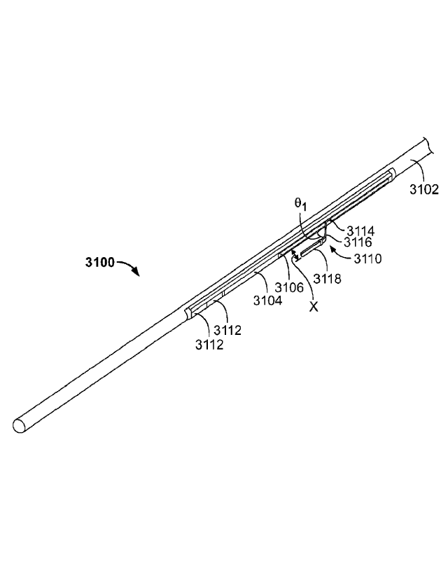
[0068] FIGS. 31A and 31B illustrate another variation of catheter (3100).
Specifically, FIG.
31A shows a perspective view of catheter (3100), comprising catheter body
(3102), nesting
material (3104) with track (3106), coupling magnets (3108), and shaped lead
wire (3110). FIG.
31B shows catheter (3100) with catheter body (3102) removed. Additionally
shown in FIG. 31B
are anchoring magnets (3112). Similar to the lead wire (2112) described above
in relation to
FIGS. 21A and 21B, at least a portion of lead wire (3110) may be uncovered and
thus may act as
an ablation surface to ablate or vaporize tissue. Additionally, the distal
portion of lead wire
(3110) may be configured to bias away from the catheter (3100), and may be
moveable between
three positions. In the first position (not shown), the lead wire (3110) may
be held or otherwise
housed within the catheter (3100) (e.g., within nesting material (3104) and/or
catheter body
(3102)), which may allow for low-profile advancement of the catheter (3100)
through the
vasculature. The lead wire (3110) may then be withdrawn (or in some instances,
advanced) such
that the bias of the lead wire (3110) may cause the distal portion of lead
wire (3110) to bias away
from catheter body (3102), as shown in FIGS. 31A and 31B. As illustrated
there, lead wire
(3110) may comprise a first segment (3114) housed at least partially within
catheter body (3102),
a first angled segment (3116) extending from a distal end of the first segment
(3114), and a
second angled segment (3118) extending from a distal end of the first angled
segment (3116).
First angled segment (3116) may extend from first segment (3114) at a first
angle (OA such that
when lead wire (3110) biases away from catheter body (3102), first angled
segment (3116) angles
away from catheter body (3102) at first angle (01). First angle (0]) may be
any suitable angle
(e.g., about 30 degrees, about 45 degrees, about 60 degrees, between about 30
degrees and about
60 degrees, between about 15 degrees and about 75 degrees, or the like).
Second angled segment
(3118) may be angled relative to first angled segment (3116) at a second angle
(02). Second angle
(02) may be any suitable angle (e.g., about 100 degrees, about 135 degrees,
about 170 degrees,
between about 100 degrees and about 170 degrees, or the like). In the
variation shown in FIGS.
31A and 31B, lead wire (3110) may be configured such that when lead wire
(3110) biases second
angled portion (3118) is approximately parallel to the longitudinal axis of
catheter body (3102),
and separated from the catheter body (3102) by a distance (x). Distance (x)
may be any value
suitable to extend at least partially through vascular tissue during ablation
(e.g., less than 1 mm,
between about 1 mm and about 2 min, between about 1 mm and about 3 mm, greater
than about 4
mm, and the like).
CA 02817552 2013-05-09
WO 2012/068273 PCT/US2011/061026
[0069] When catheter (3100) is placed inside of a blood vessel (not shown) and
lead wire
(3110) extends out from catheter (3100), the first (3116) and second (3118)
angled sections of the
lead wire (3110) may be biased into tissue of the blood vessel. When lead wire
(3110) is used to
ablate tissue, this bias may cause lead wire (3110) to press through or
otherwise ablate blood
vessel tissue. As lead wire (3110) passes through blood vessel tissue, it may
come into contact
with one or more portions of a second catheter (not shown) placed in an
adjoining blood vessel,
as will be described in more detail below. In some variations, the lead wire
(3110) may be
further withdrawn (or advanced) during ablation to slide the lead wire (3110)
relative to the
catheter into a third position (not shown). As the lead wire (3110) is moved,
it may move across
blood vessel tissue to ablate a tract or path in the tissue, which may
facilitate formation of the
fistula. Following ablation, the lead wire (3110) may then be returned to a
low-profile (e.g., by
withdrawing the lead wire (3110) relative to the catheter body (3102)), and
the catheter may be
repositioned or removed.
[0070] One or more portions of lead wire (3110) may be coated over otherwise
covered with
one or more insulating materials. For example, as shown in FIGS. 31A and 31B,
an insulating
material (3122) may at least partially cover lead wire (3110). Insulating
material may cover any
suitable portion or portions of lead wire. For example, in the variation shown
in FIGS. 31A and
31B, an insulating material (3122) may cover first segment (3114) and first
angled segment
(3116), but not second angled segment (3118). In other variations, the
insulating material (3122)
may cover the first segment (3114) and only partially cover the first angled
segment (3116), such
that the second angled segment (3118) and a portion of the first angled
segment (3116) remain
uncovered. In these variations, the second angled segment (3118) and uncovered
portion of the
first angled segment (3116) may act as an ablation surface. When insulating
material (3122)
covers multiple segments of lead wire (3110), the same material may cover each
segment, or
different insulating materials may cover the different segments. Insulating
material (3122) may
comprise any suitable material or materials, such as those described above. In
some variations,
insulating material (3122) may comprise polyetheretherketone.
[0071] FIG. 32 shows another variation of catheter (3200) comprising a lead
wire (3202)
having a first segment (3204), a first angled segment (3206), and a second
angled segment
(3208). As shown there, catheter (3200) may comprise a catheter body (3210)
having a recessed
region (3212). Catheter (3200) may comprise a lumen (3214) or other passageway
extending
through catheter body (3210). Lumen (3214) may extend through catheter body
(3210) both
proximally and distally of recessed region (3212), or may only extend through
catheter body
(3210) only proximally of recessed region (3212). As with lead wire (3110)
described above in
relation to FIGS. 31A and 31B, at least a portion of lead wire (3202) be
uncovered, and lead wire
21
CA 02817552 2013-05-09
WO 2012/068273 PCT/US2011/061026
(3202) may be moveable from a low-profile configuration and a biased
configuration in which
first angled segment (3206) angles away from first segment (3204) and catheter
body (3210).
When in a low-profile configuration, the first (3206) and second (3208) angled
segments may be
at least partially constrained within lumen (3214). In some variations, at
least a portion of first
angled segment (3206) and/or second angled segment (3208) may be temporarily
housed in a
portion of lumen (3214) distally of recessed region (3212). In these
variations, lead wire (3202)
may be withdrawn relative to catheter body (3210) to release first angled
segment (3206) and
second angled segment (3208) from lumen (3214), which may allow these segments
to bias away
from catheter body (3210) as described above. In other variations, at least a
portion of first
angled segment (3206) and/or second angled segment (3208) may be temporarily
housed in a
portion of lumen (3214) proximally of recessed region (3212). In these
variations, the lead wire
(3202) may be withdrawn to release first angled segment (3206) and second
angled segment
(3208) from lumen (3214).
[0072] As shown in FIG. 32, an insulating material (3216) (such as one or more
of the
insulating materials described above) may cover first segment (3204) and may
partially cover
first angled segment (3206), leaving second angled segment (3208) and a
portion of first angled
segment (3206) exposed. In some variations, one or more insulating materials
may also partially
cover second angled segment (3208), but need not. The exposed portions of
first (3206) and
second (3208) angled segments may act as an ablation surface to ablate or
vaporize tissue.
Catheter body (3210) may also comprise one or more insulating nesting
materials (not shown) or
coatings which may help protect the catheter body (3210) from and in some
instances redirect
heat and energy produced by lead wire (3202) during ablation.
[0073] Additionally, in some variations, the lead wire (3202) may be further
withdrawn (or
advanced) during ablation to slide the lead wire (3202) relative to the
catheter. As the lead wire
(3202) is moved, it may move across blood vessel tissue to ablate a tract or
path in the tissue,
which may facilitate formation of the fistula. Following ablation, the lead
wire (3202) may then
be returned to a low-profile, for example, by withdrawing the lead wire (3202)
such that first
angled segment (3206) and second angled segment (3208) are pulled into lumen
(3214).
[0074] As mentioned above, in some variations one or more portions of an
ablation surface of
an electrode of a first catheter may extend or otherwise be advanced through
blood vessel tissue
during ablation. When a second catheter is placed in an adjoining blood
vessel, this advancement
through blood vessel tissue may cause the ablation surface to contact one or
more portions of the
second catheter. When the second catheter comprises an electrode having an
exposed conductive
surface, direct contact between the electrodes of each catheter may cause the
energy source (e.g.,
22
CA 02817552 2013-05-09
WO 2012/068273 PCT/US2011/061026
an electrosurgical generator) to shut off or otherwise cease tissue ablation.
In other instances,
contact between the electrode of the first catheter and the second catheter
may damage one or
more components of the second catheter. Accordingly, in some variations it may
be desirable to
configure a catheter to include one or more sections that may accommodate
contact with an active
electrode without ceasing ablation or otherwise damaging one or more portions
of the catheter.
[0075] FIGS. 33A and 33B show one such variation of a catheter (3300). As
shown there in
FIG. 33A, catheter (3300) may comprise a catheter body (3302), nesting
material (3304) with
pocket (3306), coupling magnets (3308), and electrodes (3310). FIG. 33B shows
catheter (3300)
with catheter body (3302) removed. Additionally shown there are anchoring
magnets (3312).
Generally, pocket (3306) may be configured to receive a portion of an
electrode from a second
catheter. For example, when catheter (3300) is placed within a blood vessel
(not shown), and a
second catheter is placed in an adjoining blood vessel, catheter (3300) may be
positioned relative
to the second catheter such that pocket (3306) may be aligned with an
electrode (not shown) of
the second catheter. Alignment may result from attraction between alignment
elements of
catheter (3300) (e.g., coupling magnets (3308) and/or anchoring magnets (3312)
and
corresponding alignment elements of the second catheter, as will be described
in more detail
below. During ablation, the electrode of the second catheter may pass between
the blood vessels,
where it may be received by pocket (3306). Nesting material (3304) may be
formed from or
coated with an insulating material, such that energy delivered by the
electrode does not damage
catheter (3300) as electrode is received by pocket (3306).
[0076] Pocket (3306) may be configured to receive any suitable electrode, as
described in more
detail above. For example, in some variations, pocket (3306) may be configured
to receive a
portion of a lead wire, such as wire (2112) of catheter (2100) describe above
in relation to FIGS.
21A and 21B, lead wire (3110) of catheter (3100) described above with respect
to FIGS. 31A and
31B, lead wire (3202) described above in relation to FIGS. 32A and 32B, or the
like. For
example, in some variations, the coupling magnets and anchoring magnets of
catheters (3300)
and (3100) may be configured such that when catheters (3300) and (3100) are
placed in adjoining
blood vessels, the pocket (3306) of catheter (3300) may be substantially
aligned relative to track
(3106). When lead wire (3110) is advanced (or withdrawn) such that a distal
portion of the lead
wire (3110) is biased out of track (3106), lead wire (3110) may be activated
to ablate vessel
tissue, as described in more detail below. As lead wire (3110) ablates through
tissue, one or more
portions of the lead wire (3110) (e.g., second angled portion (3118))) may
enter or otherwise be
received by the pocket (3306).
23
CA 02817552 2013-05-09
WO 2012/068273 PCT/US2011/061026
[0077] While shown in FIGS. 33A and 33B as having electrodes (3310), catheter
(3300) need
not comprise any electrodes. In variations that do include electrodes (3310),
electrodes (3310)
may act as a passive ground electrode for an active electrode of a second
catheter (e.g., lead wire
(3110) of catheter (3100) described above) or vice versa, which may aid in
tissue ablation. While
shown in FIGS. 33A and 33B as having two electrodes (3310), it should be
appreciated that the
catheters described here may comprise any suitable number of electrodes (e.g.,
zero, one, two, or
three or more electrodes). For example, FIG. 34 illustrates one such variation
of a catheter (3400)
comprising a single electrode (3402). Also shown there are catheter body
(3404) and nesting
material (3406) with pocket (3408) and housing electrode (3402) and coupling
magnets (3410).
Pocket (3408) may be configured to receive one or more portions of an
electrode of a second
catheter, as described in more detail above. While electrode (3402) is
proximal to pocket (3408)
in the variation of catheter (3400) shown in FIG. 34, in other variations
electrode (3402) may be
positioned distal to pocket (3408).
[0078] In some variations, a catheter may comprise a pocket formed in an
electrode. FIGS.
35A and 35B show one such variation of catheter (3500). As shown in FIG. 35A,
catheter may
comprise a catheter body (3501) and nesting material (3502). Nesting material
(3502) may house
electrode (3504) and coupling magnets (3506) therein. FIG. 35B shows catheter
(3500) with
catheter body (3501) removed, and further shows anchoring magnets (3510).
Pocket (3508) may
be formed in electrode (3504), and may be configured to receive a portion of
an electrode from a
second catheter. In some variations, pocket (3508) may be electrically and/or
thermally insulated
by depositing one or more insulating coatings (e.g., a refractory metal oxide
coating) onto the
surfaces of pocket (3508), which may allow pocket (3508) to receive and
contact at least a
portion of an electrode without pocket (3508) providing a direct electrical
connection. In other
variations, pocket (3508) may be configured to allow for electrical conduction
therethrough
without direct physical contact with an external electrode. For example, in
some of these
variations, pocket (3508) may be covered or otherwise coated with a porous
insulating coating
(e.g., a porous metal oxide coating). When pocket (3508) receives an electrode
(e.g., one or more
of the lead wire electrodes described above), the porous coating may allow for
electrical
conduction through the pocket (3508) without direct electrode-to-electrode
physical pocket,
which may prevent short-circuiting or interruption of ablation.
[0079] Additionally, while the variations of catheters described immediately
each comprise a
pocket for receiving an electrode from a second catheter, it should be
appreciated that the
catheters described here need not comprise a pocket. Indeed, in some
variations one or more
portions of the device may be electrically insulated or partially electrically
insulated to allow for
direct contact with one or more electrodes of a second catheter. For example,
FIG. 36 illustrates
24
CA 02817552 2013-05-09
WO 2012/068273 PCT/US2011/061026
one such variation of catheter (3600). As shown there, catheter (3600) may
comprise a catheter
body (3602) and a nesting material (3604). Nesting material (3604) may house
an electrode
(3606) and coupling magnets (3608) therein. Electrode (3606) may further
comprise one or more
coated segments (3610). Coated segment (3610) may comprise an insulating
coating (as
described in more detail above) or a partially-insulating coating (e.g., a
porous coating as
described immediately above). Catheter (3600) may interact with a second
catheter (not shown),
such that when the catheters are placed in adjoining blood vessels, an
electrode of the second
catheter may extend through vessel tissue during ablation and contact coated
segment (3610)
without damaging or short-circuiting the device. While the coated segment
(3610) of electrode
(3606) shown in FIG. 36 may be recessed relative to the remainder of the
electrode, it should also
be appreciated that in some variations the coated segment (3610) may be flush
relative to the
uncoated portions of the electrode (3606)
Mechanical Cutting Elements
[0080] In some variations, a catheter may comprise one or more mechanical
cutting elements.
For example, in some variations a catheter may comprise a blade that may be
advanced or
otherwise extended from the catheter to cut or otherwise sever tissue. FIG. 22
shows one such
variation of catheter (2200) comprising a nesting material (2201) comprising
track (2202) and a
blade (2204). Blade (2204) may have any suitable shape and configuration
(e.g., single-edge,
double-edge, pointed, rounded, or the like). Blade (2204) may be rotatably,
translatably, or
otherwise coupled to catheter (2200) such that it may be deployed through
track (2202) to cut or
otherwise sever tissue. In some variations, the blade (2204) may be configured
to oscillate
relative to catheter (2200) to cut or otherwise sever tissue. Blade (2204) may
be deployed by any
suitable mechanism (e.g., one or more mechanical actuators, magnet-based
actuators, electronic
actuators, or the like), and may be withdrawn into track (2202) to allow for
low-profile
advancement or withdrawal of the catheter. In some variations, as will be
described in more
detail below, the blade (2204) may be used to pierce or puncture one or more
balloons in a
corresponding catheter in another blood vessel. Additionally, in some
variations, the blade
(2204) may electrically connected to an electrosurgical generator such that
blade (2204) may act
as an electrode, like those electrodes described in more detail above.
[0081] FIGS. 37A and 37B show cross-sectional perspective views of a variation
of catheter
(3700), and illustrate a mechanism by which a blade (3702) may be advanced out
of catheter
(3700). Catheter (3700) may comprise a recess (3704) in catheter body (3705).
Blade (3702)
may be moveable from a low-profile configuration, in which blade (3702) is
housed in recess
(3704) (as shown in FIG. 37A) to a cutting configuration, in which blade
(3702) is advanced out
CA 02817552 2013-05-09
WO 2012/068273 PCT/US2011/061026
of recess (3704) (as shown in FIG. 37B). Catheter (3700) may comprise a
rotation arm (3706)
and an activation wire (3708), which may help move blade (3702) between the
retracted and
cutting configurations, as will be described in more detail below. Also shown
in FIGS. 37A and
37B are coupling magnets (3710) located proximal and distal of blade (3702),
although it should
be appreciated that catheter (3700) need not comprise any alignment element or
may comprise
any suitable alignment elements or combinations of alignment elements as
described in more
detail below.
[0082] As shown in FIGS. 37A and 37B, rotation arm (3706) may be pivotally
connected to
blade (3702) at a first pivot point (3712) at or near a first end of rotation
arm (3706), and may
also be pivotally connected to the catheter body (3705) at a second pivot
point (3714) at or near a
second end of rotation arm (3706). The pivot points described here may
comprise one or more
pins, projections, other structures that allow for rotational movement between
two members. For
example, as shown in FIGS. 37A and 37B, second pivot point (3714) may comprise
a pin (3716).
In some variations, the second pivot point (3714) may additionally be
configured to move along
the longitudinal axis of the catheter body (3705). For example, pin (3716) of
second pivot point
(3714) may be slidably disposed in a track (3718) within catheter body (3705),
such that pin
(3716) may both rotate and slide relative to track (3718) and catheter body
(3705). Blade (3702)
may further be pivotally connected to the catheter body (3705) at a third
pivot point (3720).
Additionally, activation wire (3708) may be connected to rotation arm (3706)
at or near its
second end. For example, in the variation of catheter (3700) shown in FIGS.
37A and 37B
activation wire (3708) may be attached to a portion of pin (3716).
[0083] Activation wire (3708) may be manipulated to move blade (3702) between
a retracted
position (as shown in FIG. 37A) and an extended cutting position (as shown in
FIG. 37B).
Activation wire (3708) may be pulled proximally relative to the longitudinal
axis of the catheter
(3702), which may cause second pivot point (3714) to slide proximally relative
to the catheter
body. As second pivot point (3714) moves proximally toward third pivot point
(3720), the
rotation arm (3706) and blade (3702) may each rotate away from catheter body
(3705), as shown
in FIG. 37B. When catheter (3700) is placed in a blood vessel, rotation of
blade (3702) into a
cutting position may cause blade (3702) to cut or otherwise sever vessel
tissue. To return blade
(3702) to a retracted position, the activation wire (3708) may be advanced
distally relative to the
catheter (3700), which may move second pivot point (3714) away third pivot
point (3720), which
may cause rotation arm (3706) and blade (3702) to rotate back toward the
catheter body. It
should also be appreciated that in some variations, catheter (3700) may be
configured such that
distal advancement of the activation wire causes rotation arm (3706) and blade
(3702) to rotate
26
CA 02817552 2013-05-09
WO 2012/068273 PCT/US2011/061026
blade to an extended position, while proximal withdrawal of the activation
wire causes rotation
arm (3706) and blade (3702) to rotate blade (3702) to a retracted position.
[0084] FIGS. 38A and 38B depict another variation of a catheter (3800)
comprising a blade
(3802). As shown in a perspective view in FIG. 38A, catheter (3800) may
comprise a catheter
body (3804), and a recess (3806) in catheter body (3804) through which blade
(3802) may
extend. Also shown there are guide plates (3810) on both sides of recess
(3806), and activation
wire (3814). FIG. 38B shows a cross-sectional side view taken along the
longitudinal axis of
catheter (3800). As shown there, blade (3802) may be pivotally attached to one
or more of guide
plates (3810) at pivot point (3812). In some variations, the pivot point
(3812) may comprise one
or more pins or projections, as described immediately above.
[0085] As shown in FIG. 38B, a distal portion of activation wire (3814) may be
attached to
blade (3802), and may extend through a lumen (3816) or other passageway in the
catheter body
(3804). A proximal portion of activation wire (3814) may be manipulated to
withdraw or
advance activation wire (3814) within lumen (3816), and this movement may
cause blade (3802)
to rotate relative to pivot point (3812). In the variation shown in FIGS. 38A
and 38B, withdrawal
of the activation wire (3814) may cause the blade (3802) to rotate outwardly
from catheter body
(3804) (as shown in FIG. 38A), while advancement of activation wire (3814) may
cause blade
(3802) to rotate to a retracted position (such as shown in FIG. 38B).
[0086] When catheter (3800) is advanced into a blood vessel (not shown), the
catheter (3800)
may be advanced with blade (3802) in a retracted position within the catheter
body (3804). When
catheter (3800) is positioned within the blood vessel, a user may withdraw or
otherwise retract
pull wire to rotate the blade (3802) to an extended position, whereby blade
(3802) may cut or
otherwise sever tissue. When blade (3802) is in an extended position, catheter
may optionally be
moved relative to the blood vessel to further cut or otherwise sever tissue.
Additionally or
alternatively, pivot point (3812) may be moveable relative to the catheter
body (3804), such that
pivot point (3812) and blade (3802) may be translated along the longitudinal
axis of the catheter
body (3804). Following the cutting action by the blade (3802), the activation
wire (3814) may be
advanced to return blade (3802) to a retracted position. Catheter (3800) may
optionally be
repositioned and reactivated to cut or sever tissue at another location, or
catheter (3800) may be
removed from the blood vessel. While catheter (3800) is illustrated above as
being configured
such that withdrawal of activation wire (3814) extends blade (3802) from
catheter body (3804)
and advancement retracts blade (3802) into catheter body (3804), it should be
appreciated that
catheter (3800) may be configured such that advancement of activation wire
(3814) may extend
27
CA 02817552 2013-05-09
WO 2012/068273 PCT/US2011/061026
blade (3802) from catheter body (3804) and withdrawal of the activation wire
(3814) may retract
blade (3802) into the catheter body (3804).
[0087] FIGS. 39A-39C illustrate yet another variation of a catheter (3900)
comprising a blade
(3902). FIG. 39A shows a perspective view of a portion of catheter (3900) with
blade (3902) in
an extended position, extending from a recess (3904) in catheter body (3906).
FIGS. 39B and
39C show cross-sectional side views of catheter (3900) along its longitudinal
axis. As shown
there, blade (3902) may be attached to a first wire portion (3908) and a
second wire portion
(3910). First wire portion may he attached to or otherwise engage a
translation wire (3912) at
connection point (3914). In some of these variations, first wire portion
(3908) and second wire
portion (3910) may comprise a shape-memory material, and may be configured
such that first
wire portion (3908) and second wire portion (3910) bias blade (3902) away from
translation wire
(3912) and toward an extended position, as shown in FIG. 39C. To move blade
(3902) from an
extended position to a retracted position, as shown in FIG. 39B, second wire
portion (3910) may
be pulled away from connection point (3914) in the direction of arrow (3916).
This may cause
first (3908) and second (3910) wire portions to at least partially straighten,
which may withdraw
blade (3902) into catheter body (3906). Second wire portion (3910) may be
locked or otherwise
fixed relative to translation wire (3912) to hold blade (3902) in a retracted
position.
[0088] To use blade (3902) to aid in forming a fistula, catheter (3900) may be
advanced into a
blood vessel (not shown) with blade (3902) in a retracted position. Once
positioned (e.g., using
one or more alignment elements, visualization methods, or the like), blade
(3902) may be moved
to an extended position. To do this, second wire portion (3910) may be
unlocked relative to
translation wire (3912), which may allow first (3908) and second (3910) wire
portions to return to
their outwardly biased positions, thereby extending blade (3902) to an
extended position, as
shown in FIG. 39C. In some variations, a user may advance or otherwise move
second wire
portion (3910) toward connection point (3914) to help bias blade (3902) in an
extended position.
As blade (3902) extends from catheter body (3906) it may cut or otherwise
sever tissue. In some
variations, second wire portion (3910) may be locked or otherwise fixed
relative to translation
wire (3912) to hold blade (3902) in an extended position. Once extended from
catheter body
(3906), translation wire (3912) may be advanced or withdrawn relative to
catheter body (3906) to
translate blade (3902) along the longitudinal axis of the catheter, which may
allow blade (3902)
to cut a larger tract of tissue. Additionally or alternatively, catheter
(3900) may be advanced or
withdrawn relative to the blood vessel with blade (3902) extended to cut a
larger tract of tissue.
The second wire portion (3910) may then be withdrawn relative to translation
wire (3912) and
connection point (3914) to return blade (3902) to a retracted position, and
the catheter (3900)
may be repositioned or removed.
28
CA 02817552 2013-05-09
WO 2012/068273 PCT/US2011/061026
[0089] It should be appreciated that the above-described variations of
catheters comprising
blades may include any of the additional device features described
hereinthroughout. For
example, the catheters may comprise one or more alignment elements. In these
variations, the
catheters may comprise one or more anchoring magnets and/or one or more
coupling magnets.
Additionally or alternatively, the catheter may comprise one or more shape-
changing elements
and/or one or more markers, as will be described in more detail below.
Laser Energy
[0090] In some variations, the catheters described here may be configured to
deliver laser
energy to tissue to vaporize or otherwise remove tissue during fistula
formation. Generally,
variations of these catheters may comprise an optical fiber which may run from
a proximal
portion of the catheter to a distal portion of the catheter. A proximal
portion of the optical fiber
may operatively connected (e.g., via a SMA connector, or the like) to a laser
generator. Laser
energy produced by the laser generator may propagate or otherwise pass through
the optical fiber,
and may be delivered from optical fiber to tissue to vaporize tissue. In some
variations the
catheter may comprise one or more lenses, mirrors, diffuser, and/or other
components which may
redirect light from the optical fiber toward tissue.
[0091] The laser generator may be configured to produce any suitable laser
energy. In some
variations, it may be desirable to produce light energy having a wavelength
with high water
absorption, which may promote energy absorption by vessel tissue. In some
variations, the laser
generator may be configured to generate infrared energy. Examples of suitable
wavelengths
include, but are not limited to, about 730 nanometers, between about 680
nanometers and about
780 nanometers, about 820 nanometers, between about 750 nanometers and about
870
nanometers, about 930 nanometers, between about 880 nanometers and about 980
nanometers,
about 970 nanometers, between about 920 nanometers and about 1020 nanometers,
about 1200
nanometers, between about 1150 nanometers and about 1250 nanometers, about
1450
nanometers, between about 1400 nanometers and about 1500 nanometers, about
1950
nanometers, between about 1900 nanometers and about 2000 nanometers, about
2900
nanometers, between about 2850 nanometers and about 2950 nanometers or the
like. Examples
of suitable laser generators include, but are not limited to, diode lasers,
diode-pumped lasers, Nd-
YAG lasers, and the like.
[0092] FIG. 43 shows a distal portion of one variation of catheter (4300)
which may be
configured to deliver laser energy to tissue. As shown there, catheter (4300)
may comprise
catheter body (4302), optical fiber (4304), and irrigation lumen (4306). As
shown there, optical
fiber (4304) may run along the longitudinal axis (4310) of catheter body
(4302), and a distal
29
CA 02817552 2013-05-09
WO 2012/068273 PCT/US2011/061026
portion of the optical fiber (4304) may bend to direct the distal end of the
optical fiber (4304) out
of a side of the catheter body (4302). The distal portion of the optical fiber
(4304) may bend at
any angle (0) relative to the longitudinal axis (4310) of the catheter body
(4302). In some
variations, angle (0) may be about 45 degrees. In other variations, angle (0)
may be about 90
degrees. In still other variations, angle (0) may be between about 45 degrees
and about 90
degrees. In yet other variations, angle (0) may be less than about 45 degrees,
or greater than
about 90 degrees.
[0093] When the distal end of the optical fiber (4304) is directed toward the
side of the catheter
body (4302), laser energy may be passed through optical fiber (4304) and out
the side of catheter
body (4302), where it may vaporize, ablate, or otherwise remove tissue. In
some variations, it
may be desirable to pass a gas (e.g., carbon dioxide) or fluid (e.g., saline)
between the distal end
of the optical fiber (4304) and tissue during tissue vaporization.
Accordingly, in some variations,
one or more fluids may be passed through catheter body (4302) via irrigation
lumen (4306) and
be delivered between the optical fiber (4304) and tissue (not shown). The gas
or fluid may be
introduced continuously or intennittently during tissue vaporization, and may
help to minimize or
otherwise prevent excessive heating or damage to surrounding tissue.
[0094] Additionally, in some variations, it may be desirable to space the
output of the optical
fiber (4304) from tissue. In some instances, spacing the output of the optical
fiber from tissue
may affect the power density of the laser energy provided and/or the size of
the fistula formed. In
some variations, the catheter may comprise a space (4308) between the end of
the optical fiber
(3404) and the side wall of the catheter body (4302). Space (4308) may
separate the end of the
optical fiber (3404) from the side wall of the catheter body (4302) by any
suitable amount (e.g.,
about 0.5 mm, about 1 mm, about 1.5 mm, between about 0.5 mm and about 1.5 mm,
greater than
about 1.5 mm, and the like). Additionally, in variations where an irrigation
lumen (4306) is used
to deliver a gas or fluid between the output of the optical fiber (4304) and
tissue, gas or fluid may
be delivered into or through space (4308).
[0095] As mentioned above, in some variations, a catheter may comprise one or
more lenses,
diffusers, mirrors, or the like, for altering or otherwise redirecting light
passing through an optical
fiber. For example, FIG. 44 shows one variation of a catheter (4400)
comprising a diffuser
(4410) which may redirect laser energy provided through an optical fiber
(4404). As shown
there, catheter (4400) may comprise a catheter body (4402), an irrigation
lumen (4406), optical
fiber (4404), and diffuser (4410). Diffuser (4410) may be attached at or near
the distal end of the
optical fiber (4404), and may redirect light from optical fiber (4404) out of
the side of catheter
body (4402). In some variations, a space (4408) within catheter body (4402)
may separate an
CA 02817552 2013-05-09
WO 2012/068273 PCT/US2011/061026
output of diffuser (4410) from tissue. Additionally, irrigation lumen (4406)
may be positioned to
pass fluid between diffuser (4410) and tissue (not shown), as described
immediately above.
Alignment Elements
[0096] In some variations, the catheters described here may comprise one or
more alignment
elements to help align or otherwise reposition the catheters when placed in
the vasculature. For
example, in some instances alignment elements may help bring two or more
catheters (and with
them, two or more blood vessels) in closer approximation. In other instances,
the alignment
elements may help ensure that one or more catheters are in proper axial or
rotational alignment
relative to another catheter (or catheters). Ensuring proper position of
catheters and blood vessels
may help facilitate formation of a fistula with one or more of the fistula-
forming elements
described above. In some variations, catheters may comprise mechanical
alignment features,
such as protrusions, grooves, flat surfaces, and the like, that may or may not
interact with one or
more alignment features on another catheter. Additionally or alternatively, a
catheter may have
one or more magnetic components that may interact with one or more magnetic
components of
another catheter or one or more magnets positioned externally from the body.
In still other
variations, the catheter may comprise one or more markers that may help a user
to align one or
more catheters. In still other variations, a catheter may comprise one or more
shape-changing
members for adjusting the positioning of a catheter. It should be appreciated
that each catheters
described here may comprise any alignment element or combination of alignment
elements
described below, and in variations where the catheter comprises a fistula-
forming element, may
comprise any fistula forming element or combination of elements described in
more detail above.
Magnets
[0097] As mentioned above, a catheter may comprise one or more magnetic
alignment
components. These magnetic alignment components may be attracted to one or
more additional
elements (e.g., one or more portions of a second catheter, one or more magnets
or other
components placed externally from the body) to help position or align the
catheter within a
vessel. For example, one or more magnets placed outside of the body may
interact with the
magnetic alignment components of a catheter to help facilitate advancement of
the catheter
through the vasculature, as will be described in more detail below. In other
instances, a catheter
may comprise one or more "anchoring" magnetic alignment elements that act to
attract the
catheter toward one or more portions of a second catheter, thereby bringing
the catheters in closer
approximation. In other variations, a catheter may comprise one or more
"coupling" magnetic
alignment elements, which may act to rotationally (and/or axially) orient
and/or mate a surface of
the catheter with one or more surfaces or portions of a second catheter.
31
CA 02817552 2013-05-09
WO 2012/068273 PCT/1JS2011/061026
[0098] A catheter may comprise any number of individual magnets (e.g., zero,
one, two, three,
four, five, six, seven, or eight or more, etc.). Each magnetic component may
be any suitable
magnet or magnetic material. For example, in some variations, a catheter may
comprise one or
more rare-earth magnets (e.g., neodymium magnets or samarium-cobalt magnets)
and/or one or
more selectively-activated electromagnets. In variations where a catheter
comprises a plurality of
magnets, these magnets may be grouped into one or more arrays. These magnetic
arrays may
located inside or outside of a catheter (or a combination thereof), and may be
positioned
anywhere along the length of the catheter. When two or more catheters comprise
magnets or
magnet arrays, each magnet or magnet array may be configured or arranged to
align with one or
more magnets or magnet arrays from a second catheter. Each magnet may be fixed
in or on a
catheter by any suitable method. For example, in some variations one or more
magnets may be
embedded in, adhered to or friction fit within a catheter. Each magnet may
have any suitable
diameter (e.g., about 0.075 in., about 0.080 in., about 0.029 inch, about
0.110 inch, or the like) or
length (e.g., about 5 mm, about 10 mm, about 15 mm, about 20 mm, or the like),
and may be
separated from adjoining magnets by any suitable distance (e.g., about 1 mm,
about 5 mm, and
the like). In some variations, the magnets of an array may have alternating
polarity (e.g., each
magnet will have the opposite polarity as any adjacent magnets), matching
polarity, or
combinations thereof. In other variations, one or more portions of the
catheter may be made from
a magnetic material, and/or may be embedded with one or more magnetic
particles/materials.
[0099] Each magnet may have any suitable shape for placement inside or outside
of the
catheter. Magnets may be cylindrical, semi-cylindrical, tube-shaped, box-
shaped, or the like. For
example, in the variation of catheter (200) shown in FIG. 2 and described in
more detail above,
catheter (200) may comprise a proximal anchoring magnet array (206) and a
distal anchoring
magnet array (208), the magnets of each of which are cylindrical.
Alternatively, in the variation
of catheter (600) shown in FIG. 6A, catheter (600) may comprise a proximal
anchoring magnet
array (604) and a distal anchoring magnet array (606), the magnets of each of
which are semi-
cylindrical. In these variations, a lumen and/or lead wire (such as lumen
(608) and lead wire
(605)) may pass by or along the anchoring magnet arrays, because the semi-
cylindrical magnets
only take up a portion of the interior of catheter (600).
[0100] While the magnets of proximal (604) and distal (606) magnet assemblies
are shown in
FIG. 6A as being configured such that the apex of each semi-cylinder is
aligned with ablation
surface (603), it should be appreciated that the magnets may be positioned in
any manner relative
to a fistula forming component. For example, FIG. 6B shows another variation
of catheter (610)
comprising electrode body (612) having ablation surface (613), proximal
anchoring magnet array
(614), distal anchoring magnet array (616), lead wire (615) and lumen (618).
In this variation,
32
CA 02817552 2013-05-09
WO 2012/068273 PCT/US2011/061026
the apex of each magnet of the proximal (614) and distal (616) anchoring
magnet arrays may be
perpendicular to ablation surface (613). Altering the orientation of the
magnets relative to the
ablation surface (613) may affect the strength of the magnetic force between
catheter (610) and
another catheter (not shown) when placed in a blood vessel. It should be
appreciated that each
individual magnet or anchoring magnet array may have any rotational
positioning relative to
ablation surface (613), which may or may not be the same as the rotational
position as another
magnet or array of magnets.
[0101] In some variations, one or more magnets may have one or more lumens or
passageways
therethrough, which may allow one or more other components (e.g., a lead wire,
actuation
mechanism, lumen, combinations thereof and the like) of catheter to pass
through the magnets.
For example, in the variation of catheter (300) shown in FIG. 3, catheter
(303) comprises
proximal and distal anchoring magnet arrays ((302) and (304) respectively)
having tube-shaped
magnets. As shown there, concentric electrical conductor (314) may pass
through the magnets of
proximal anchoring magnet array (302), and lumen (308) may pass through the
magnets of the
proximal and distal anchoring magnet arrays ((302) and (304) respectively).
[0102] In some variations, one or more magnetic alignment components may
comprise one or
more box-shaped magnets (e.g., a magnet with a substantially rectangular cross-
section). FIG. 23
shows one such variation of catheter (2300). Shown there is tip (2302), distal
anchoring magnet
array (2304), proximal anchoring magnet array (2306), and nesting material
(2308) comprising
coupling magnets (2310), electrode body (not shown) with ablation surface
(2312) and marker
(2316). Catheter (2300) further comprises a catheter body (or a plurality of
catheter segments,
such as those described above), but the catheter body is not illustrated in
FIG. 23 so as to
highlight the internal components of catheter (2300). Additionally, while
shown in FIG. 23 as
comprising coupling magnets (2310) and marker (2316) (each of which will be
described in more
detail below), catheter (2300) need not.
[0103] As shown in FIG. 23, distal anchoring magnet array (2304) comprises a
cylindrical
anchoring magnet (2316) and a box-shaped anchoring magnet (2314), while
proximal anchoring
magnet array (2306) comprises a box-shaped anchoring magnet (2314). It should
be appreciated
that the anchoring magnet arrays may have any suitable combination of magnets,
such as one or
more of the magnets described above. In variations that comprise one or more
box-shaped
magnets, such as box-shaped magnets (2314) of catheter (2300), the box-shaped
magnets may
help bring the catheter in closer approximation with a second catheter, but
may also help
rotationally orient the catheter relative to the second catheter.
Specifically, when two box-shaped
magnets are associated with separate catheters, the attractive strength
between the two magnets
33
CA 02817552 2013-05-09
WO 2012/068273 PCT/US2011/061026
may be greatest when the magnets are aligned. For example, in the variation of
catheter (2300)
shown below, a front surface (2320) of box-shaped magnet (2314) may align with
a front surface
of another box-shaped magnet (not shown) of a catheter in another blood
vessel. Specifically, the
attractive force between the magnets may be greatest when the front surfaces
are aligned with
each other, and thus the magnets may naturally rotate or facilitate rotation
to the aligned position.
[0104] In variations where a catheter comprises a nesting material, the
nesting material may
house one or more coupling magnets for temporarily magnetically coupling a
surface or portion
of the nesting material to one or more portions of another catheter or device.
Specifically, the
coupling magnets may be configured such that the attractive force between two
catheters is
greatest when a surface of each catheter is aligned with the other. For
example, in the variation
of catheter (2300) shown in FIG. 23 above, nesting material (2308) comprises
coupling magnets
(2310). In these instances, the longitudinal axis of the coupling magnets
(2310) may be
substantially transverse to the longitudinal axis of the catheter.
Additionally, coupling magnets
(2310) may have a flat mating surface which may attract a flat mating surface
of a coupling
magnet of another catheter (not shown). As described in more detail above in
regards to flat
ablation surfaces, the flat mating surfaces of a coupling magnet may act to
flatten tissue between
two catheters, which may aid in ablation by the ablation surface.
[0105] While nesting material (2308) is shown in FIG. 23 as housing a single
coupling magnet
(2310) on either side of ablation surface (2314), it should be appreciated
that a catheter may
comprise any suitable number of coupling magnets. FIGS. 24A and 24B show two
such
variations of catheters comprising coupling magnets. FIG. 24A shows a first
variation of catheter
(2400). Shown there is catheter body (2402) and nesting material (2406)
housing an electrode
(2408) and a distal coupling magnet array (2412), wherein the electrode (2408)
comprises
ablation surface (2410). While shown in FIG. 24A as comprising two coupling
magnets (2414),
distal coupling magnet array (2412) may comprise any suitable number of
coupling magnets
(2414) (e.g., one, two, or three or more). In other variations, a nesting
material may comprise a
plurality of coupling magnet arrays. For example, FIG. 24B shows a variation
of catheter (2416)
comprising catheter body (2418), nesting material (2420) housing electrode
(2422) with ablation
surface (2424), proximal coupling magnet array (2426) and distal coupling
magnet array (2428).
Each of the coupling magnet arrays may comprise any suitable number of
coupling magnets
(2430), as mentioned immediately above.
[0106] In some variations, such as catheters (2400) and (2416) described above
in relation to
FIGS. 24A and 24B, the catheter may be configured that a fistula-forming
element may be placed
in close proximity to the distal end of the catheter body. These variations
may find particular
34
CA 02817552 2013-05-09
WO 2012/068273 PCT/US2011/061026
utility when it is desirable to form a fistula near a tissue structure,
blockage, or other impediment
that limits the ability of the blood vessels to be brought in closer
approximation.
[0107] In variations where a catheter comprises an array of electromagnets,
the electromagnets
may be independently activated or may be activated as a group. For example,
electromagnets of
a magnet array may be activated one at a time to help ensure a certain
alignment orientation with
respect to another magnetic device, e.g., proximal magnets may be activated
prior to activating
distal magnets, every other magnet may be activated in sequence, etc.
Alternatively, two or more
magnets may be activated simultaneously to promote secure attachment to
another magnetic
device.
Shape-Changing Elements
[0108] In some variations, the catheter may comprise one or more shape-
changing elements for
approximating two or more blood vessels. In these variations, the shape-
changing element may
have a first configuration during advancement of the catheter through the
vasculature. Once the
catheter reaches a target location, the shape-changing element may be altered
to a second
configuration, which may alter the overall shape of the catheter. As the
catheter changes shape,
the catheter may move or reconfigure one or more portions of the blood vessel,
which may help
bring that portion or portions of the blood vessel in closer approximation to
one or more portions
of a second blood vessel. The shape of a catheter may be altered in any
suitable manner. In some
variations, a catheter may comprise one or more pull wires, which may pulled
or push to deflect
or otherwise alter the shape of the catheter. In other variations, a catheter
may comprise one or
more shaped wires which may alter the shape of the catheter, as will be
described in more detail
below.
[0109] FIGS. 25A-25D illustrate one variation of catheter (2500).
Specifically, FIG. 25A
shows a partial cross-sectional area of catheter (2500). Shown there is
catheter body (2502),
nesting material (2504) housing coupling magnets (2508) and an electrode body
(not shown)
comprising ablation surface (2506), shaped lead wire (2510), straightening
cannula (2512), and
torque-transmitting sheath (2514). A portion of catheter body (2502) is not
shown in FIG. 25A to
help illustrate the other elements of catheter (2500). While shown in FIG. 25A
as having an
electrode housed within nesting material (2504) to define ablation surface
(2506), catheter (2500)
may comprise any suitable fistula-forming element, such as those described in
more detail above.
Furthermore, while shown in FIG. 25A as comprising a plurality of coupling
magnets (2508),
catheter (2500) need not. In variations where the catheter does comprise one
or more magnets,
the catheter may comprise any magnets or combination of magnets, such as those
described
CA 02817552 2013-05-09
WO 2012/068273 PCT/US2011/061026
above. Finally, catheter (2500) may or may not comprise a torque-transmitting
sheath (2514),
which may help rotate the catheter, as will be described in more detail below.
[0110] Shaped lead wire (2510) may be used to alter the shape of catheter
(2500). Specifically,
shaped lead wire (2510) may be pre-formed with one or more bends or curves. A
straightening
cannula (2512) may be advanced over lead wire (2510) to temporarily straighten
or otherwise
constrain the bends and curves of the shaped lead wire (2510), thereby
rendering the distal
portion of the catheter (2500) substantially straight, as shown in FIG. 25B.
Catheter (2500) may
be advanced into a blood vessel (not shown), at which point the straightening
cannula (2512) may
be withdrawn. Once withdrawn, shaped lead wire (2510) may return to its
original configuration,
which may cause catheter (2500) to change shape, as shown in FIG. 25C. When
catheter (2500)
is placed in a blood vessel, this shape change may alter the shape of one or
more blood vessels.
For example, FIG. 25D shows one variation in which two catheters (2518) and
(2520) are placed
in adjoining blood vessels (2522). As shown there, catheters (2518) and (2520)
may comprise the
components of catheter (2500) described immediately above. When the
straightening cannulas
(not shown) for each of catheters (2518) and (2520) have been withdrawn, the
lead wires of each
catheter may take on a bent/curved configuration, which may cause the distal
portions of each of
catheters to bend or flex toward each other, thereby bringing a portion of the
blood vessels (2522)
closer together as shown in FIG. 25D. One or more fistula-forming elements may
then be
activated to form a fistula between the adjoining blood vessels (2522).
[0111] Shaped lead wire (2510) may act as a lead wire to carry current to or
from an electrode,
but need not. Indeed, in some variations, a device may comprise a shaped wire
and a separate
lead wire. In variations where a catheter does not comprise an electrode, the
catheter may
comprise a shaped wire but no lead wire. It should also be appreciated that in
some variations, a
shaped member may be located outside of catheter. In some of these variations,
the shaped
member may be at least partially covered by one or more sheaths or other
coverings, such as
those described above. It should also be appreciated that any suitable shaped
structure may be
used to alter the shape of the catheters described here.
[0112] In variations where a catheter comprises one or more expandable
structures (e.g., a
balloon or the like), as will be described in more detail below, the
expandable structure may be
used in conjunction with a shape-changing member to help position a catheter
within a vessel.
For example, in the variation of catheter (2500) described above with respect
to FIGS. 25A-25D,
catheter (2500) may further comprise one or more expandable structures (not
shown), such as one
or more inflatable balloons. These expandable structures may be expanded
inside of a blood
vessel to temporarily hold catheter (2500) in place relative to the vessel.
When shaped lead wire
36
CA 02817552 2013-05-09
WO 2012/068273 PCT/US2011/061026
(2510) and straightening cannula (2512) are used to move catheter body (2502)
between a bent
and a straight configuration, contact between the expandable structure and the
surrounding tissue
may help to move the blood vessel with the catheter body (2502). In some
variations, catheter
(2500) may comprise a single expandable structure, which may be located
proximal or distal to
the bend of the shaped lead wire (2510). In other variations, catheter (2500)
may comprise one or
more expandable structures on either side of the bend of the shaped lead wire
(2510). In
variations where a shaped lead wire comprises multiple bends, expandable
structures may be
positioned proximal and/or distal all, some, or none of the bends. It should
also be appreciated
that the expandable structures may be used in conjunction with any suitable
shape-changing
element (e.g., a shaped wire, a pull wire, or the like), and may be used to
provide temporary
attachment and/or fixation between the catheter body and vessel tissue in any
suitable manner.
Markers
[0113] The catheters described here may comprise one or more markers that may
allow for
visualization of one or more portions of a catheter during positioning and/or
orientation thereof.
In some variations, the marker may be directly visualized. In other
variations, the marker may be
indirectly visualized (e.g., via ultrasound, fluoroscopy and/or X-ray
visualization). Markers may
be located anywhere relative to the catheter, e.g., one or more surfaces of
the catheter, inside of
the catheter. In some variations, one or more portions of the catheter may be
made from an
echogenic or radiographic material. A marker may be attached to the catheter
by any suitable
method, for example, by mechanical attachment (e.g., embedded in a portion of
the catheter,
circumferential circumscription, or the like), adhesive bonding, welding,
soldering, combinations
thereof or the like.
[0114] FIGS. 14A-14D illustrate one variation of catheter (1400) comprising
marker bands
(1402). Also shown in FIG. 14A are anchoring magnets (1410), and electrode
(1404) partially
covered by nesting material (1406) and comprising ablation surface (1408).
Catheter (1400) may
comprise any suitable number of marker bands (1402), and each marker band
(1402) may be
positioned at any suitable location in or on catheter (1400). Marker bands
(1402) may comprise
cut-out regions that may help a practitioner determine the position of a
catheter via one or more
imaging techniques. Specifically, FIG. 14B shows a perspective view of one
marker band (1402)
comprising a first cut-out region (1412) and a second cut-out region (1414).
First (1412) and
second (1414) cut-out regions arc shown in FIGS. 14B-14D as having the same
shape, but need
not. When marker band (1402) is visualized (e.g., via ultrasound or
fluoroscopy), a user may be
able to see the negative space formed by the overlapping segment (1416) of
first (1412) and
second (1414) cut-out regions. The shape of this overlapping segment (1416)
may change as
37
CA 02817552 2013-05-09
WO 2012/068273 PCT/US2011/061026
catheter (1400) (and with it, marker band (1402)) is rotated. Eventually,
rotation of marker band
(1402) may reach a point where first (1412) and second (1414) cut-out regions
completely or
substantially overlap, as shown in FIG. 14D. When marker band (1402) reaches
this "aligned"
configuration (or when two markers on associated catheters are each in an
"aligned"
configuration), a user may know that the catheter is in a rotational
orientation suitable for
activating a fistula-forming element. For example, in the variation of
catheter shown in FIG.
14A, the ablation surface (1408) may be positioned relative to marker bands
(1402) such that the
ablation surface (1408) faces a direction perpendicular to the cut-out
regions. When used in
conjunction with a second catheter (not shown) having a second set of marker
bands (not shown),
the marker bands of each catheters may be used to rotationally and/or axially
position the
catheters such that that one or more fistula forming elements are properly
positioned to form a
fistula.
[0115] While shown in FIGS. 14A-14D as having bi-lobular shapes, the cut-out
regions may
be any shapes or combination of shapes, e.g., rectangular, circular,
elliptical, multi-lobular
shapes, alphanumeric symbols, any shape with one or more axes of symmetry
(e.g., bilateral
symmetry), and the like. In some variations, the cut-out regions may have a
directional shape,
which may have a tapered portion that indicates the location of the ablation
surface of an
electrode, e.g., a polygon with a vertex at an acute angle, arrow, and the
like. First and second
cut-out regions may have the same shape as each other, or may each have
different shapes. Other
orientation markers or indicators may be provided on the catheter and/or
electrode as desired and
described below. While shown in FIGS. 14A-14D as comprising marker bands, it
should be
appreciated that the catheters described here may comprise any marker that is
capable of indirect
visualization.
[0116] In other variations, the catheter may comprise one or more visual
markers that may help
align two or more catheters relative to each other. For example, FIGS. 15A and
15B illustrate
one variation of catheter (1500) comprising a lateral stripe (1504) which may
help orient catheter
(1500). Specifically lateral stripe (1504) may be a visual marker with a known
location relative
to a fistula-forming element (1508). For example, in the variation shown in
FIGS. 15A and 15B,
lateral stripe (1504) is longitudinally aligned with fistula-forming element
(1508). When the
distal end of catheter (1500) is placed in the body (and thus cannot be
directly visualized), the
lateral stripe (1504) (which may at least partially remain outside of the
body) may give a visual
indication as to the rotational orientation of the fistula-forming element
(1508). When two
catheters (1500) are placed in two blood vessels (not shown), as illustrated
in FIG. 15B, the
relative positioning of laterals stripes (1504) may give an indication of the
relative positioning of
the two catheters. For example, as shown in FIG. 15B, when the lateral stripes
(1504) of each
38
CA 02817552 2013-05-09
WO 2012/068273 PCT/US2011/061026
catheter (1500) are directly across from each other, the fistula-forming
elements (1508) of each
catheter (1500) may be in an appropriate orientation for activation of the
fistula-forming
elements. Lateral stripe (1504) may be applied to catheter (1500) in any
suitable manner (e.g.,
via ink marking, texturing, application of one or more colored adhesives,
etc.).
External Positioning
[0117] In some variations of the catheters described here, a catheter may
comprise one or more
balloons or other expandable structures. These expandable structures may serve
one or more
functions. In some instances, an expandable structure may help appose an
electrode surface (or
other fistula-forming element) against one or more vessel walls. This
apposition may help
temporarily flatten or otherwise relocate tissue, and may act to displace
blood from the area.
Additionally, during fistula formation, the expandable member may continue to
urge the fistula
forming element against tissue as it is removed from the vessel wall. In some
variations, the
expandable structure may be configured to help provide apposition between the
catheter and a
vessel wall, while still allowing for blood flow through the blood vessel. In
some instances, one
or more expandable structures may help modify or otherwise alter the size or
shape of a fistula.
In still other instances, the expandable structures may be used to dilate,
contract, or otherwise
displace a portion of one or more blood vessels. In some of these variations,
this displacement
may help bring a portion of the blood vessel closer to a skin surface. In
still other variations, as
mentioned above, one or more expandable structures may be used to hold a
catheter in place
relative to a blood vessel, and may aid in repositioning the blood vessel.
[0118] As mentioned above, in some variations of the catheters described here,
the catheter
may comprise one or more balloons. For example, FIGS. 9A-9D depict various
illustrations of
catheters comprising a balloon. In some variations, the balloon may be
configured to push a
portion of a catheter (e.g., an ablation surface or other fistula forming
element) into contact with a
blood vessel wall. For example, FIG. 9A depicts one variation of catheter
(900) comprising
balloon (902), and an electrode body (not shown) having an exposed ablation
surface (904).
Balloon (902) may have an undeployed collapsed configuration (not shown) for
low-profile
advancement and a deployed expanded configuration (as shown in FIG. 9A). In
the variation
shown in FIG. 9A, balloon (902) may be non-concentrically mounted on the
catheter (900) away
from ablation surface (904) such that expansion of balloon (902) within a
blood vessel may bias,
press, or otherwise push ablation surface (904) against a blood vessel wall.
In variations where
the catheter has a flat ablation surface, expansion of the balloon (902) may
help flatten tissue
against the ablation surface. Additionally, the balloon (902) may aid in
fistula formation by
continuing to urge the ablation surface (904) against and through the blood
vessel wall as tissue is
39
CA 02817552 2013-05-09
WO 2012/068273 PCT/US2011/061026
ablated, vaporized or otherwise removed. In still other instances, expansion
of the balloon (902)
may help displace blood from the vicinity of ablation surface (904), which in
turn may minimize
current loss to blood during ablation. In instances where a catheter comprises
a recessed
electrode, as described in more detail above, expansion of a balloon may
displace some blood
from the area while causing other blood to be trapped within the recessed
portion. In instances
where catheter (900) comprises one or more shape-changing elements, engagement
between the
balloon (902) and the surrounding blood vessel (not shown) may help to hold
the catheter (900)
in place and may further aid in repositioning the vessel tissue when the
catheter (900) changes
shape.
[0119] It should be appreciated that while shown in HG. 9A as having a balloon
(902), the
catheters described here may achieve one or more of these functions using any
suitable
expandable structure or structures (e.g., one or more expandable cages,
meshes, scaffolds, struts,
or the like). The balloons described here may have any suitable shape or
shapes (e.g., cylindrical,
semi cylindrical, circular, trapezoidal, rectangular, fractional portions
thereof, and the like), and
may be made of any suitable material or combination of materials (e.g., one or
more non-elastic,
elastic, or semi-elastic materials).
[0120] Additionally, while the balloon (902) shown in FIG. 9A as being mounted
on an
opposite side of catheter (900) from the ablation surface (904), it should be
appreciated that the
balloon (902) may be positioned in any manner relative to the catheter (900).
For example, in
some variations a balloon (or other expandable structure) may be positioned
such that expansion
thereof creates a directional distension of a blood vessel. For example, FIG.
9B depicts one such
variation of a catheter (910) comprising a balloon (912) and ablation surface
(914) of an electrode
(not shown). As shown there, balloon (912) may be positioned on catheter (910)
such that the
balloon (912) expands in a direction approximately orthogonal relative to the
direction in which
the ablation surface (914) faces. When catheter (910) is placed in a blood
vessel and ablation
surface (914) is aligned with another catheter an adjoining blood vessel,
expansion of balloon
(912) may cause directional distension of a blood vessel toward the skin
overlying the blood
vessel, which in turn may cause that skin to distend. This distension may give
a user a visual
indication of placement of the balloon (912), thereby allowing a user or
operator to locate the
balloon and blood vessel from outside the body. This visualized location may
provide a site
through which a user may externally access the blood vessel (e.g., by
puncturing with a needle or
the like).
[0121] In some instances, a balloon or other expandable structure may help
alter and/or
regulate the flow of blood through a blood vessel. For example, in some
variations, expansion of
CA 02817552 2013-05-09
WO 2012/068273 PCT/US2011/061026
a balloon may dilate one or more portions of a blood vessel, which may
encourage increased
blood flow through that portion of the blood vessel. In other variations, an
expandable element
may temporarily occlude a blood vessel, or may reduce blood flow therethrough.
In some of
these variations, the expandable element may comprise one or more electrodes,
which may be
used to help reduce blood flow through a portion of a blood vessel. FIG. 9C
shows one such
variation of catheter (930) comprising an ablation surface (932) and balloon
(934). As shown
there, balloon (934) is positioned concentrically around catheter (930), and
may comprise a
plurality of electrodes (936) disposed on the balloon (934). Although shown in
FIG. 9C as being
located distally on catheter (900) relative to ablation surface (932), it
should be appreciated that
balloon (934) may be placed proximally relative to ablation surface (932)
and/or may be non-
concentrically mounted away from ablation surface (932). Indeed, in some
variations (as
described in more detail below), catheter may comprise balloon both proximal
to and distal to the
ablation surface, each of which may comprise one or more electrodes. While
shown in FIG. 9C
as having a plurality of circumferentially disposed electrodes (936), balloon
(934) may comprise
any suitable number of electrodes (e.g., zero, one, two, three, or four or
more) and each electrode
may fully or partially circumscribe balloon (934).
[0122] Balloon (934) may be expanded within a blood vessel to temporarily
occlude the vessel.
Additionally, one or more of the electrodes (936) may be activated to
partially constrict the blood
vessel and reduce flow through at least a portion of the vessel. Specifically,
electrical energy
may be delivered to the vessel wall to induce necrosis and/or a proliferative
cellular response,
which may reduce the inner diameter of the blood vessel, thereby reducing the
blood flow
therethrough.
[0123] The balloons described immediately above may be used to alter or
otherwise regulate
blood flow relative to a fistula. FIG. 9D illustrates one example of how
catheter (930) may be
used to affect blood flow relative to a fistula. As shown there, catheter
(930) may be advanced in
an arterial vessel (940) that is in close proximity to a corresponding venous
vessel (942). A
second catheter (not shown) may be placed in venous vessel (942), and one or
more alignment
elements (not shown) may be used help approximate the arterial (940) and
venous (942) vessels.
Ablation surface (932) may be activated (alone, or in conjunction with one or
more electrodes of
the other catheter) to form an arterio-venous fistula, through which blood may
flow (as
represented by arrow (932)). Arrow (941) indicates the direction of blood flow
in arterial vessel
(940) and arrow (943) indicates the direction of blood flow in venous vessel
(942). As shown in
FIG. 9D, catheter (930) may be advanced in a retrograde (i.e., against the
blood stream) direction
into an arterial vessel (940), such that balloon (934) is located upstream of
the ablation surface
(932) and the resulting fistula. In these instances, balloon (934) may be
expanded in arterial
41
CA 02817552 2013-05-09
WO 2012/068273 PCT/US2011/061026
vessel (940) to at least partially occlude the vessel and temporarily prevent
or reduce arterial
blood flow therethrough, which in turn may help prevent current loss during
fistula formation.
Additionally, electrodes (936) may be activated to damage or scar surrounding
tissue, which may
reduce flow therethrough. This may be used to help prevent one or more
potential complications
with fistula formation, such as steal syndrome. For example, steal syndrome
may occur when a
fistula is formed between an artery and a vein, and blood flows through the
resulting fistula at a
rate the results in insufficient blood flowing distally/downstream of the
fistula in the artery. This
can result in tissue necrosis, and may necessitate an additional surgical
procedure to prevent the
loss of a limb. Accordingly, electrodes (936) may be activated to reduce flow
through a vein,
which may reduce flow through the fistula, and may thereby reduce the
likelihood of steal
syndrome.
[0124] While shown in FIG. 9D as being advanced in a retrograde fashion and
located
upstream of the ablation surface (932), balloon (934) may alternatively be
located downstream of
ablation surface (932) and the resulting fistula. In some of these variations,
catheter (930) may be
advanced in an anterograde (i.e., with the flow of blood) fashion into
arterial vessel (940). In
other variation, a catheter may be advanced in a retrograde fashion, hut a
balloon may instead be
located proximally on catheter relative to the ablation surface. In still
other variations, a catheter
may be moved within the blood vessel to change the positioning of a balloon
between an
upstream position and a downstream position, and vice versa. When a balloon is
placed
downstream in an arterial vessel (940), the balloon may be expanded to dilate
the vessel (940) to
increase blood flow therethrough or may be constricted using one or more
electrodes to decrease
blood flow therethrough. Dilating the downstream portion of the arterial
vessel (940) may divert
blood flow away from the fistula, while constriction of the downstream portion
may encourage
increased blood flow through the fistula. It should be appreciated that one or
more balloons
and/or electrodes may be placed in a venous vessel upstream or downstream
relative to a fistula
to dilate and/or constrict portions of the venous vessel. Arterial and/or
venous dilation or
constriction may help aid fistula maturation and/or prevent venous
hypertension, as will be
described in more detail below.
[0125] While shown in FIGS. 9A-9D as having a single balloon, it should be
appreciated that
the catheters described here may have any suitable number of balloons,
expandable members, or
combinations thereof. Indeed, the catheters described may comprise a plurality
of expandable
members for positioning the catheter within a blood vessel, urging an
electrode ablation surface
against a blood vessel wall, and/or regulating the blood flow in the vicinity
of the targeted
vascular site. FIG. 10A depicts one such variation of a catheter (1000)
comprising an ablation
surface (1006), a first balloon (1002) located proximal to the ablation
surface and comprising
42
CA 02817552 2013-05-09
WO 2012/068273 PCT/US2011/061026
circumferential electrodes (1008), and a second balloon (1004) located distal
to the ablation
surface. While only the first balloon (1002) is shown in FIG. 10A as having
electrodes (1008), it
should be appreciated that any of balloons (e.g., none of the balloons, only
the first balloon
(1002), only the second balloon (1004), or both the first (1002) and second
(1004) balloons) may
comprise any suitable number of electrodes (e.g., one, two, three, or four or
more electrodes).
The first (1002) and second balloons (1004) may be independently actuated to
regulate blood
flow within a blood vessel. For example, catheter (1000) may be inserted into
a vein (not
shown), in which the direction of blood flow is represented by arrow (1001).
Second balloon
(1004) may be expanded to dilate the downstream portion of the vein, while the
circumferential
electrodes (1008) of first balloon (1002) may be activated to constrict the
vein. In other instances
the catheter (1000) may be inserted in the opposite direction (or the balloons
may be otherwise
positioned) such that the second balloon (1004) may be used to dilate the
upstream portion, and
first balloon (1002) may constrict the downstream portion. In still other
instances, the balloon
may be configured to dilate both the upstream and downstream portions, or may
be configured to
constrict both the upstream and downstream portions. Again, it should be
appreciated that any
expandable structure or stmctures may be utilized by the catheters.
[0126] FIG. 10B shows another variation of catheter (1010) comprising an
electrode with
ablation surface (1016), a proximal balloon (1012) and a distal balloon
(1014). Proximal balloon
(1012) may comprise a fixed volume body and may further comprise a
circumferential band
electrode (1118) around a portion of the balloon (1012). Circumferential band
electrode (1118)
may expand or collapse with an expandable member. FIG. 10C illustrates still
another variation
of a catheter (1020) with a distal balloon (1024), a proximal expandable wire
loop (1022), and an
ablation surface (1026). Wire loop (1022) may be movable between an
undeployed, low-profile
configuration (not shown), and a deployed, expanded configuration (as shown
HG. 10C). Wire
loop (1022) may be moved between the undeployed and deployed configuration
using any
suitable mechanism (e.g., hinged radial struts, a coiling mechanism, etc.),
and RF energy may be
applied to the wire loop (1022) to induce tissue necrosis, as described above.
[0127] In some variations, once a fistula has been formed, one or more
balloons or other
expandable structures may be used to modify the size or shape of a fistula.
For example, FIG. 11
illustrates one such variation of catheter (1100). As shown there, catheter
(1100) may comprise
an electrode ablation surface (1102) and a lateral extension balloon (1104).
Lateral extension
balloon (1104) may be configured to expand in two or more directions, and may
be used to alter
the size or shape of a fistula. Specifically, ablation surface (1102) (or
another suitable fistula-
forming element) may be used to form a fistula (not shown) between two vessels
(not shown),
and catheter (1100) may be subsequently moved to position the lateral
extension balloon (1104)
43
CA 02817552 2013-05-09
WO 2012/068273 PCT/US2011/061026
adjacent or near the fistula. Balloon (1104) may be then be expanded, and a
portion of balloon
(1104) may push into the fistula, thereby altering its size and/or shape.
Balloon (1104) may be a
fixed volume structure, or may be made from one or more elastic or semi-
elastic materials. It
should also be appreciated that the lateral extension balloon (1104) may
comprise two or more
separate balloons. Additionally, lateral extension balloon (1104) may comprise
pleats (1106) or
other surface modification to regulate the degree of expansion, which may also
provide traction
or a frictional attachment to the blood vessel wall at the target vascular
site. For example, in the
variation shown in FIG. 11, pleats (1106) may engage tissue surrounding the
fistula to help move
or adjust that tissue.
[0128] In other variations of the catheters described here, a catheter may
comprise one or more
balloons that may be configured to allow blood flow therethrough. For example,
FIG. 12 shows
another variation of catheter (1200) comprising a plurality of ring-shaped
balloons (1202). Also
shown there is electrode ablation surface (1204). In these variations,
expansion of the balloons
(1202) may allow for the increased apposition and blood displacement between
ablation surface
(1204) and a vessel wall (not shown), as described in more detail above.
Additionally, the
lumens (1208) within each of the ring-shaped balloons (1202) may allow for
blood to pass
therethrough. In this way, catheter (1200) may be left inside of a blood
vessel (not shown) for an
extended period of time without substantially affecting blood flow
therethrough. For example, in
some instances it may be necessary to leave catheter (1200) in a blood vessel
for an extended
period of time, durinL, which it may not be feasible to block all blood flow
through the blood
vessel. It should be appreciated that one or more of the balloons (1202) may
also be configured
to dilate one or more portions of a blood vessel, and/or may comprise one or
more electrodes to
help constrict a blood vessel, as described in more detail above.
[0129] In some variations, one or more balloons of a catheter may carry or be
inflated with a
contrast material to help in visualization of the catheter. In some
variations, one or more of these
balloons may be pierced or otherwise punctured to release the contrast agent
into the blood
vessel, which may be used to evaluate whether a fistula has been properly
formed. For example,
FIGS. 26A and 26B illustrates one such variation of catheter (2600). As shown
there, catheter
(2600) may comprise a catheter body (2602) with distal balloon (2604), central
balloon (2606),
and proximal balloon (2608). Catheter (2600) may additionally comprise one or
more shape-
changing elements or alignment elements, such as those described above.
Additionally, while
shown FIGS. 26A and 26B as having three balloons, the catheter (2600) may
comprise any
suitable number of balloons (e.g., one, two, three, or four or more). Each of
balloons (2604),
(2606) and (2608) are shown in FIGS. 26A and 26B as being concentrically
mounted around
44
CA 02817552 2013-05-09
WO 2012/068273 PCT/US2011/061026
catheter body (2602), but they may be mounted in any suitable manner or
manners, such as
described in more detail above.
[0130] When catheter (2600) is placed in a blood vessel (not shown), distal
(2604), central
(2606), and proximal (2608) balloons may be inflated. These balloons may be
inflated using any
suitable fluid or fluids (e.g., saline, water, one or more contrast solutions,
or the like). In some
variations, all three balloons are inflated with the same fluid. In other
variations, the distal (2604)
and proximal (2608) balloons are inflated with a first fluid (e.g., a first
contrast solution), while
the central balloon (2606) is inflated with a second fluid (e.g., a second
contrast solution having a
higher or lower contrast level). In yet other variations, each balloon is
inflated with a different
solution. When the balloons are inflated, proximal (2608) and/or distal
balloon (2604) may
engage an interior surface of the blood vessel to prevent fluid flow thereby.
For example, in
instances where proximal balloon (2608) is placed in a blood vessel upstream
relative to the flow
of blood, inflation of proximal balloon (2608) may temporarily stop blood flow
through the blood
vessel.
[0131] In some instances, a second catheter (2610) may be placed in an
adjoining blood vessel
(not shown), as illustrated in FIG. 26B. Second catheter (2610) may comprise a
catheter body
(2611), and a nesting material (2614) housing a lead-wire electrode (2612),
such as described in
more detail above). While shown in FIG. 26B as comprising a lead-wire
electrode (2612), it
should be appreciated that second catheter (2610) may comprise any suitable
fistula-forming
element such as those described in more detail above. In some of these
instances, the catheter
(2600) and the second catheter (2610) may be aligned (e.g., using one or more
alignment
elements such as those described above) such that the nesting material (2614)
may be in axial and
rotational alignment with the central balloon (2606), and current may he
applied to lead-wire
electrode (2612) to ablate vessel tissue between the catheters, thereby
forming a fistula (not
shown). During or after fistula formation, one or more portions of the lead-
wire electrode (2612)
may puncture, pierce, or otherwise penetrate the central balloon (2606) to
release one or more
fluids therefrom. When this fluid comprises one or more contrast solutions,
this fluid may be
viewed (e.g., fluoroscopically) as it passes through the fistula. In this way,
as the contrast fluid
passes between the blood vessels, a user may be able to determine that the
fistula has been
formed in a manner that allows fluid flow therethrough. While central balloon
(2606) is
discussed above as being punctured, it should be appreciated that any balloon
or balloons of the
catheter (2600) may be pierced or punctured by a fistula-forming element.
CA 02817552 2013-05-09
WO 2012/068273 PCT/US2011/061026
Catheter Body
[0132] The catheters described hereinthroughout may be any elongate body
suitable for
advancement through at least a portion of the vasculature. The catheters may
be hollow, partially
hollow, and/or partially solid. One or more portions of the catheter may be
flexible or semi-
flexible, one or more portions may be rigid or semi-rigid, and/or one or more
portions of the
catheter may be changed between flexible and rigid configurations. Flexible
portions of the
catheter may allow the catheter to be navigated through tortuous blood vessels
to reach a desired
target site. The catheters described here may be made of any material or
combination of
materials. For example, the catheters may comprise one or more metals or metal
alloys (e.g., e.g.,
nickel titanium alloys, copper-zinc-aluminum-nickel alloys, copper-aluminum-
nickel alloys, and
the like) and/or one or more polymers (e.g., silicone, polyvinyl chloride,
latex, polyurethane,
polyethylene, PTFE, nylon, and the like). Catheters may have any suitable
dimensions. For
example, catheters may have any suitable length that allows the catheter to be
advanced from a
point external to the body to a target location. Catheters may have any
diameter suitable for
intravascular use, such as, for example, about 5.7 French, about 6.1 French,
about 7 French, about
8.3 French, between about 5 French and about 9 French, between about 5 French
and about 7
French, between about 6 French and about 9 French, or the like.
[0133] Some variations of the catheters described here may have a lumen, slit,
or passageway
extending at least partially through the length of the catheter. Lumens may be
used to pass one or
more devices (e.g., a guidewire) and/or one or more substances (e.g., contrast
solution, perfusion
fluid, one or more drug-containing solutions, etc.) through a portion of the
device. For example,
the variation of catheter (300) shown in FIG. 3 and described in more detail
above comprises a
lumen (308) passing therethrough. Although shown in FIG. 3 as passing
concentrically through
magnets, it should be appreciated that a lumen may have any positioning
relative to other
components of the device. For example, in the variation of catheter (600)
shown in FIG. 6A,
lumen (608) may extend next to one or more magnets of the proximal (604) and
distal (606)
anchoring magnets. In still other variations, a lumen may be attached to or
otherwise run along
an exterior surface of the catheter.
[0134] When housed at least partially inside of the catheter body, a lumen may
pass between
any portion or portions of the catheter. For example, in the variations
described immediately
above, the lumens may exit out the distal-most tip of the catheter. In other
variations, one end of
a lumen may be located at an intermediate portion of the catheter. In some
variations, a lumen
may be divided into a number of sub-lumens. For example, FIGS. 16A and 16B
show a
perspective view and a partially-transparent view, respectively, of one
variation of catheter
46
CA 02817552 2013-05-09
WO 2012/068273 PCT/US2011/061026
(1600). Shown there is catheter (1600) with main lumen (1610) which subdivides
into first
(1602), second (1604), third (1606), and fourth (1608) lumens at a distal tip
(1601) of the
catheter. Main lumen (1610) may be divided into any suitable number of lumens
(e.g., two,
three, four, or five or more), and each of these lumens may have ends at any
suitable points on the
catheter. In variations where a main lumen is split into two or more lumens,
the two or more
lumens may provide one or more fluids or other substances to two or more
points simultaneously.
[0135] The catheters described herein throughout may have any suitable tip
portion. In some
variations, the tip may be rounded or otherwise blunted so as to help minimize
tissue trauma
during advancement of the catheter. Additionally or alternatively, the distal
tip may be at least
partially tapered. A tapered tip may assist the catheter in navigating through
the vasculature
and/or may help dilate the blood vessels during advancement. The tip of the
catheter may be
integral with the rest of the catheter body, or may be a separate component
joined to the catheter
body. In some of these variations, the tip of the catheter may be releasably
attached (e.g., via
screw-fit, snap-fit, friction-fit or the like) to the catheter, which may
allow a user to select a tip
portion that is appropriate for a given patient or blood vessel.
[0136] The tip portion of the catheter may help guide a catheter through the
vasculature. In
some variations, a lumen may run through the catheter to a tip of the
catheter. In these variations,
a guidewire may be threaded through the lumen, and the catheter may be
advanced over the
guidewire to a target location. In other variations, a guidewire may be
fixedly attached to the tip
of the catheter. For example, in the variation of catheter (1600) shown in
FIGS. 16A and 16B
and described in more detail above, tip (1601) may comprise a guidewire (1612)
that is attached
to and extends from tip (1601). Guidewire (1612) may be advanced into the
vasculature to help
guide the catheter to a target location. In still other variations, a tip may
comprise a rapid
exchange portion. For example, in the variation of catheter (100) shown in
FIGS. 1A-1C above,
the tip of catheter (100) comprises rapid exchange portion (100) having first
and second apertures
((112) and (114) respectively) that are in communication with one another. A
guidewire (not
shown) may be threaded through the first (112) and second (114) apertures,
such that the rapid
exchange portion (100) (and with it, catheter (100)) may be advanced along the
guidewire to a
target location. While shown in FIGS. 1A-1C as being located at the tip of
catheter (100), it
should be appreciated that the rapid exchange portion (100) may be located at
any suitable point
along the length of the catheter (100).
[0137] Some variations of the catheters described here may comprise a torsion-
transmitting
sheath. As the length of a catheter is increased, one or more rotational
forces applied to a
proximal end may have a reduced ability to rotate the distal end of the
catheter. To help prevent
47
CA 02817552 2013-05-09
WO 2012/068273 PCT/US2011/061026
torsion-transmission problems, the catheter may comprise a torque-transmitting
sheath disposed
on or in the catheter. A torsion-transmitting sheath may be made from any
stiff or stiffened
materials that may resist rotational forces (e.g., stainless steel, shape
memory allows, and various
plastics), which may allow the practitioner to adjust the location and the
rotational orientation of
the distal portion of the catheter when it is inserted into a blood vessel.
[0138] In some variations, the catheter may comprise one or more suctions
ports. In these
variations, the suction ports may be used to remove blood or other fluids from
a portion of a
blood vessel. For example, FIGS. 27A-27D illustrate one variation of a
catheter (2700)
comprising suction ports (2702). FIG. 27A shows a perspective view of catheter
(2700),
comprising suction ports (2702), sleeve (2704), nesting material (2706)
housing a spring
electrode (2708) and coupling magnets (2710), proximal balloon (2712), and
distal balloon
(2714). While shown in FIG. 27A as having spring wire electrode (2708) and
coupling magnets
(2710), catheter (2700) may comprise any suitable combination of fistula-
forming elements
and/or alignment elements, such as those described in more detail above.
Sleeve (2704) may be
advanced to cover one or more components of the catheter (2700), as
illustrated in FIG. 27B,
which may help facilitate low-profile advancement of catheter (2700) through
tissue.
[0139] When catheter (2700) is placed in a blood vessel, suction ports (2702)
may be used in
conjunction with proximal (2712) and distal (2714) balloons to temporarily
remove blood and/or
any other fluids from a portion of a blood vessel prior to or during fistula
formation. For
example, FIGS. 27C and 27D illustrate one method by which catheter (2700) may
be used to
form a fistula (not shown). As shown in FIG. 27C, catheter (2700) may be
advanced into vein
(2716), while a second catheter (2718) may be advanced into artery (2720).
Second catheter
(2718) may comprise a flat electrode ablation surface (not shown), may
comprise another suitable
fistula-forming element such as those described in more detail above, or may
not comprise any
fistula-forming element. During advancement of catheter (2700), sleeve (2704)
may be in an
advanced position in which sleeve (2704) covers one or more elements of the
catheter (2700).
For example, sleeve (2704) may cover spring wire electrode (2708) (not shown
in FIGS. 27C and
27D) to hold spring wire electrode in a low-profile configuration.
[0140] Once the catheters have been advanced, sleeve (2704) may be withdrawn
to reveal one
or more components of the catheter. For example, when sleeve (2704) is
withdrawn, spring wire
electrode (2708) (not shown in FIGS. 27C and 27D) may extend from catheter
toward second
catheter (2718) (and in some instances, toward one or more fistula-forming
elements of the
second catheter (2718)). Additionally, one or more alignment elements may help
position
catheter (2700) relative to second catheter (2718). For example, coupling
magnets (2710) may be
48
CA 02817552 2013-05-09
WO 2012/068273 PCT/US2011/061026
attracted to and align with one or more coupling magnets (not shown) of the
second catheter
(2718) to orient the spring wire electrode (not shown in FIGS. 27C and
27D) relative to the
second catheter (2718), as shown in FIG. 27C. Once the catheters are in place
and properly
aligned, the proximal (2712) and distal (2714) balloons may be inflated, as
shown in FIG. 27C.
In some instances, the balloons (2712) and (2714) may hold catheter (2700) in
place relative to
the blood vessel. Additionally, each balloon may temporarily seal that portion
of the blood vessel
relative to the rest of the blood vessel.
[0141] Once the proximal (2712) and distal (2714) balloons have been inflated,
vacuum or
other suction may be applied to the suction ports (2702) such that any fluid
between the proximal
(2712) and distal (2714) balloons are removed from the vein (2716). In
instances where the
proximal (2712) and distal (2714) balloons create a seal within vein (2716),
the suction may also
cause a portion of the vein (2716) to collapse around catheter (2700), as
shown in FIG. 27D. At
this point, current may be supplied to the sprinL, wire electrode (2708) (and
in some instances
carried away by a ground electrode of the second catheter (2718) to ablate
and/or vaporize tissue
between the catheters. In other instances, current may be supplied to an
active electrode (not
shown) of the second catheter (2718) and carried away by the spring wire
electrode (2708).
Additionally, because any blood or other fluids have been removed from the
vein (2716) via
suction portions (2702), there may be a reduction in current loss to
surrounding fluids during
tissue ablation. While catheters (2700) and (2718) are shown in FIGS. 27C and
27D as being
placed in a vein (2716) and artery (2720) respectively, it should be
appreciated that these
catheters may be placed in any suitable blood vessels. In some variations,
catheter (2700) may be
placed in an artery while second catheter (2718) may be placed in a vein. In
still other variations,
both catheters are placed in veins. It should also be appreciated that both
catheters may comprise
suction ports.
49
CA 02817552 2013-05-09
WO 2012/068273
PCT/US2011/061026
Proximal Adaptors
[0142] The catheters described here may comprise one or more proximal adaptors
and/or
handles at a proximal end thereof. These elements may help advance Or align
the catheters,
activate one or more fistula-forming elements, and/or deliver one or more
fluids or substances
into or through the catheter. FIGS. 13A and 13B show two variations of
adaptors suitable for use
with the catheters described here. FIG. 13A shows one variation of catheter
(1300) comprising
an adaptor (1302). Catheter (1300) may comprise any suitable fistula-forming
element(s) and/or
alignment feature(s), such as those described above. As shown there, adaptor
(1302) comprises a
first port (1306), a second port (1308), and a third port (1312). Although
shown in FIG. 13A as
having three ports, adaptor may comprise any suitable number of ports (e.g.,
zero, one, two, three,
or four or more), and each port may serve any useful function (e.g., the
introduction of one or
more elements or substances into or through the catheter). For example, in the
variation shown in
FIG. 13A, first port (1306) may be used to introduce a fluid or substance
(e.g., contrast agents,
flush agents, therapeutic agents, and/or intravenous fluids) into a lumen (not
shown), and may be
connected to a liquid or gaseous fluid source (e.g., a fluid pump, a syringe,
etc.). Similarly,
second port (1308) may allow for the introduction of an electrosurgical lead
(1320) for driving an
electrical current to an electrode (not shown). In variations where the
catheter (1300) does not
comprise an electrode, any suitable control element (e.g., a pushrod, pull-
wire, or the like) may
enter the catheter via a port to control fistula formation. Finally, third
port (1312) may allow for
one or more devices (e.g., a guidewire) to pass through the catheter via
hemostasis valve (1316).
While shown in FIG. 13A as having a hemostasis valve (1316), third port (1312)
need not have
such a valve. It should be appreciated that each of the ports of a proximal
adaptor may converge
into a single lumen, or may provide access to different lumens. Additional
ports may be provided
as desired for other functions, such as a visualization port, an actuator
port, a suction port, and the
like. Ports may have any suitable connection form factor, such as a threaded
connector, luer
connector, or the like.
[0143] FIG. 13B shows another variation of catheter (1318). As shown there,
catheter (1318)
comprises the same proximal adaptor as the variation of catheter (1300) shown
in FIG. 13A, and
thus the same reference labels are used for the variation shown in FIG. 13B.
Additionally shown
in FIG. 13B is sleeve (1322) which may be provided over a portion catheter,
and may be used to
regulate the contact between an electrode ablation surface (1304) and a vessel
wall (not shown).
The position of sleeve (1322) may be controlled at least in part by a hub
(1324). A user may
manipulate hub (1324) to move sleeve (1322) proximally or distally relative to
catheter (1300).
This in turn may cause sleeve (1322) to either cover or expose electrode
ablation surface (1304).
CA 02817552 2013-05-09
WO 2012/068273 PCT/1JS2011/061026
[0144] Some variations of adaptors comprise one or more alignment features
that may help the
practitioner to orient one catheter with respect to another. For example, the
variation of adaptor
(1502) shown in FIGS. 15A and 15B and described in more detail above may
comprise an
alignment projection (1506), where the rotational orientation of the alignment
projection (1506)
maps to a corresponding rotational orientation of the electrode ablation
surface of the fistula-
forming assembly. For example, when two catheters (1500) are placed in two
adjoining blood
vessels (not shown), the alignment projections (1506) of each catheter (1500)
may be aligned
with each other to align the respective fistula-forming components (1508) on
each catheter.
[0145] It should be appreciated that any of the catheters described here
comprise any
combination of fistula-forming elements, alignment elements, catheter bodies,
proximal adaptors,
and/or expandable structures as described above, and catheter or combination
of catheters may be
used to form a fistula in any suitable manner.
Systems
[0146] Also described here are systems for forming a fistula between blood
vessels. Generally,
the system may comprise a first catheter, which may comprise one or more
fistula-forming
elements. The first catheter may comprise any of the fistula-forming elements
or combination of
fistula-forming elements as described in more detail above. For example, in
some variations, the
first catheter may comprise one or more electrodes, which may be comprise any
of the electrode
structures described in more detail above. In some variations, the first
catheter may comprise one
or more mechanical cutting elements, such as one or more of the blades
described in more detail
above. Additionally or alternatively, the first catheter may comprise one or
more optical fibers
which may be used to deliver laser energy to tissue. In variations where the
first catheter
comprises an electrode-based fistula-forming element, the system may comprise
one or more
ground electrodes, which may in some variations may be positioned externally
of a patient.
[0147] In some variations, the first catheter may comprise one or more
alignment elements. In
some variations, the first catheter may comprise one or more shape-changing
elements which
may be used to alter the shape of the first catheter. In some of these
variations, the first catheter
may comprise a shaped wire and/or one or more pull wires, as described in more
detail above.
Additionally or alternatively, the first catheter may comprise one or more
markers, such as those
described in more detail above. Additionally or alternatively, the first
catheter may comprise one
or more magnets. In these variations, the first catheter may comprise any
combination of
alignment magnets and/or coupling magnets. In some variations, the first
catheter may comprise
one or more magnet arrays proximal to a fistula-forming element. Additionally
or alternatively,
the first catheter may comprise one or more magnet arrays distal to a fistula
forming element.
51
CA 02817552 2013-05-09
WO 2012/068273 PCT/US2011/061026
[0148] The first catheter may comprise any suitable catheter body, as
described in more detail
above. In some variations, the first catheter may comprise one or more lumens
extending at least
partially through the catheter body. In some variations, the first catheter
may be configured to be
advanced over or along a guidewire. In some variations, the first catheter may
comprise a lumen
through a guidewire may pass. In other variations, the first catheter may
comprise a rapid
exchange portion. Additionally, in some variations the first catheter may
comprise one or more
expandable elements, as described in more detail above. In some variations,
the first catheter
may comprise one or more balloons. In some of these variations, the first
catheter may comprise
one or more balloons proximal to the fistula-forming element, and/or may
comprise one or more
balloons distal to the fistula-forming element.
[0149] In some variations, the system may further comprise a second catheter.
In some
variations, the second catheter may comprise a fistula-forming element, but
need not. In
variations where the second catheter does comprise a fistula-forming element,
the second catheter
may comprise any of the fistula-forming elements or combination of fistula-
forming elements as
described in more detail above. For example, in some variations, the second
catheter may
comprise one or more electrodes, which may be comprise any of the electrode
structures
described in more detail above. In some variations, the second catheter may
comprise one or
more mechanical cutting elements, such as one or more of the blades described
in more detail
above. Additionally or alternatively, the second catheter may comprise one or
more optical fibers
which may be used to deliver laser energy to tissue. The fistula-forming
element of the second
catheter may be the same as or different from the fistula-forming element of
the first catheter.
[0150] In some variations, the first catheter may comprise an electrode which
is configured to
extend through vessel tissue during fistula formation (e.g., one or more of
the wire electrodes or
other deployable electrodes described in more detail below), the second
catheter may be
configured to receive or otherwise contact one or more portions of the first
catheter's electrode
during ablation. In some variations, the second catheter may comprise one or
more recesses or
pockets for receiving a portion of the first catheter's electrode, as
described in more detail above.
In some variations, an electrode of the second catheter may be configured to
receive an electrode
of the first catheter during fistula formation. In some variations, the
electrode or other receiving
surface may comprise one or more insulating coatings, such as those described
in more detail
above.
[0151] In some variations, the second catheter may comprise one or more
alignment elements.
In some variations, the second catheter may comprise one or more shape-
changing elements
which may be used to alter the shape of the second catheter. In some of these
variations, the
52
CA 02817552 2013-05-09
WO 2012/068273 PCT/US2011/061026
second catheter may comprise a shaped wire and/or one or more pull wires, as
described in more
detail above. Additionally or alternatively, the second catheter may comprise
one or more
markers, such as those described in more detail above. Additionally or
alternatively, the second
catheter may comprise one or more magnets. In these variations, the second
catheter may
comprise any combination of alignment magnets and/or coupling magnets. In some
variations,
the second catheter may comprise one or more magnet arrays proximal to a
fistula-forming
element. Additionally or alternatively, the second catheter may comprise one
or more magnet
arrays distal to a fistula forming element. In variations where both the first
and the second
catheter comprise alignment elements, the catheters may comprise the same
configuration of
alignment elements, or may comprise different configurations of alignment
elements.
[0152] The second catheter may comprise any suitable catheter body, as
described in more
detail above. In some variations, the second catheter may comprise one or more
lumens
extending at least partially through the catheter body. In some variations,
the second catheter
may be configured to be advanced over or along a guidewire. In some
variations, the second
catheter may comprise a lumen through a guidewire may pass. In other
variations, the second
catheter may comprise a rapid exchange portion. Additionally, in some
variations the second
catheter may comprise one or more expandable elements, as described in more
detail above. In
some variations, the second catheter may comprise one or more balloons. In
variations where the
second catheter comprises a fistula-forming element, the second catheter may
comprise one or
more balloons proximal to the fistula-forming element, and/or may comprise one
or more
balloons distal to the fistula-forming element.
Methods
[0153] The methods described here may be utilized to create a fistula between
two closely-
associated blood vessels (e.g., between a vein and an artery, between two
veins, etc.). Generally,
in these methods one or more fistula-forming elements may be activated to bore
through,
perforate, or otherwise create a passageway between the two blood vessels such
that blood may
flow directly between the two adjoining blood vessels. When such a fistula is
formed, hemostasis
may be created without the need for a separate device or structure (e.g., a
suture, stent, shunt, or
the like) connecting or joining the blood vessels.
[0154] Generally, the methods described here comprise accessing a first blood
vessel with a
first catheter, and advancing the first catheter to a target location within a
blood vessel. In some
of these methods, a second blood vessel is accessed with a second catheter,
and advanced to a
target location within the second vessel. In some of these methods, a first
catheter is advanced
into an artery, and the second catheter is advanced into a vein. In other
methods, a first catheter
53
CA 02817552 2013-05-09
WO 2012/068273 PCT/US2011/061026
is advanced into a first vein, and a second catheter is advanced into a second
vein. In still other
methods, a first catheter is advanced into a first artery and a second
catheter is advanced into a
second artery. The first and/or second catheters may be advanced in any
suitable manner, such as
using a Seldinger technique or other similar technique. Advancement may or may
not occur
under indirect visualization (e.g., via fluoroscopy, x-ray, or ultrasound).
The first and second
catheters may be advanced in the same manner, or may be advanced in different
manners. In
variations where one of the catheters is configured for advancement over a
guidewire (e.g.,
catheter (100) described above in relation to FIGS. 1A-1C) the catheter may be
advanced along a
guidewire. In variations where one of the catheters has a guidewire fixedly
attached to its tip
(e.g., catheter (1600) described above in relation to FIGS. 16A and 16B), the
guidewire may be
advanced through the vasculature to a target location. In other variations,
one or more external
magnets may help advance or position a catheter at a target site. For example,
FIGS. 17A and
17B show a perspective view and a side view, respectively, of an external
magnet (1700) that
may be used to help advance catheter (1702) within a blood vessel (1704).
External magnet
(1700) may interact with any suitable portion of the catheter (e.g., a fixed
guidewire (1706), one
or more magnetic alignment elements, etc.) to create an attractive force
between the catheter
(1702) and the external magnet (1700). This attractive force may be used to
pull, push, or
otherwise manipulate the catheter during advancement.
[0155] Once the first and/or second catheters have been advanced into the
respective blood
vessels, the catheters may be adjusted to affect the positioning of the
catheters within the blood
vessels and/or the positioning of the blood vessels relative to each other. In
variations where a
first catheter has been advanced into a first blood vessel and a second
catheter has been advanced
into a second blood vessel, the first and second catheters may be adjusted to
bring at least a
portion of the first and second catheters toward each other, which may act to
bring blood vessels
in closer approximation. In some variations, each of the first or second
catheters may comprise
one or more magnetic alignment elements, such as those described in more
detail above. The
magnetic alignment elements may result in an attractive force between the
first and second
catheters, which may pull the catheters toward each other. In some instances,
this attractive force
may be sufficient to compress tissue between the first and second catheters.
For example, in
variations where the first and second catheters comprise flat ablation
surfaces, as described
above, the attractive force may flatten and/or compress vessel tissue between
the ablation
surfaces. In other variations, the first and/or second catheter may comprise
one or more shape-
changing member, such as those described in relation to catheter (2500) in
FIGS. 25A-25D, and
the method comprises changing the shape of the first and/or second catheters
using the shape-
changing members. Changing the shape of the first and/or second catheters may
help
approximate the first and second blood vessels, as described above.
Additionally, the shape
54
CA 02817552 2013-05-09
WO 2012/068273 PCT/US2011/061026
change may also act to compress tissue between the first and second blood
vessels, as mentioned
above.
[0156] In some variations, adjusting the first and second catheters may
comprise aligning the
catheters axially and/or rotationally. For example, the catheters may be
oriented such that a
fistula-forming element of either the first or second catheter is positioned
to form a fistula in a
certain location. In variations where both the first and second catheters
comprise fistula-forming
elements (e.g., an active electrode and a ground electrode), the catheters may
be oriented to align
these fistula-forming elements. The catheters may be aligned in any suitable
manner. In
variations where the first and/or second catheters comprise one or more
markers, such as those
described above, the markers may be viewed (e.g., via fluoroscopy, x-ray, or
the like) to ensure
that the catheters have the proper axial and/or radial orientation relative to
each other.
Additionally, in variations where the first and/or second catheters comprise
one or more magnetic
alignment elements (e.g., one or more coupling magnets, as described in more
detail above), the
magnetic alignment elements may be used to axially and/or rotationally orient
the first catheter
relative to the second catheter.
[0157] Additionally, in some variations, one or more balloons or expandable
members, such as
those described above, may be used to help position the first and/or second
catheters, or may act
to hold the first and/or second catheters in place within the blood vessels.
For example, in some
variations, expansion of a balloon or expandable member of one of the
catheters may engage the
interior of a blood vessel, which may hold that catheter in place within the
blood vessel. In other
methods, the expansion of the balloon or expandable member can bias or
otherwise press a
fistula-forming element against blood vessel tissue, which may aid fistula
formation.
[0158] Once the catheter or catheters have been positioned and adjusted, one
or more fistula-
forming elements may be used to create a fistula between the two blood
vessels. For example, in
some variations, one of the first and second catheters comprises a fistula-
forming element (e.g.,
an electrode, a cutting blade, or the like), while the other catheter does not
comprise a fistula-
forming element. In other variations, both catheters comprise a fistula-
forming element. In some
of these variations, the fistula-forming elements of the first and second
catheters act to form
different fistulas. In other variations, the fistula-forming elements of the
first and second
catheters interact to form the same fistula. For example, in some variations
the first and second
catheters each comprises at least one electrode. In these methods, current may
be supplied to the
electrode or electrodes of one of the catheters, may be carried away by the
electrode or electrodes
of the other catheter, and may ablate or otherwise vaporize tissue as the
current passes
therethrough. Any suitable combination of electrodes as described above may be
utilized to
CA 02817552 2013-05-09
WO 2012/068273 PCT/US2011/061026
form the fistula. In other methods, such as those described above, formation
of a fistula
comprises puncturing or piercing a balloon of the first or second catheter,
which may release one
or more contrast solutions into the blood vessels. Additionally, in some
variation, a balloon may
be used to modify a fistula after the fistula has been formed.
[0159] Additionally, one or more balloons may be activated to affect the blood
flow relative to
the fistula. For example, in variations where an arterio-venous fistula is
formed, it may be
beneficial to dilate one or more portions of the artery and/or veins.
Specifically, the portion of
the artery upstream of an arterio-venous fistula may be expanded to increase
flow through the
fistula. Alternatively or additionally, a portion of a vein downstream from a
fistula may be
dilated to help increase flow through the fistula. In some variations, one or
more portions
expandable members may comprise an electrode for inducing necrosis or swelling
in a portion of
a blood vessel to decrease flow therethrough. For example, in some variations
a portion of a vein
upstream from a fistula may be at least partially occluded to minimize venous
hypertension.
[0160] It should be appreciated that any of the catheters described above may
be used to form a
fistula using the methods above. For example, in some variations, a first
catheter may be
advanced into a first blood vessel, and the first catheter may comprise one or
more fistula forming
elements, such as those described in more details above. For example, in some
variations, the
first catheter may comprise one or more blades or other mechanical cutting
elements. In some of
these variations, the first catheter may comprise one or more of the blade
mechanisms of
catheters (2200), (3700), (3800) and/or (3900) described above in relation to
FIGS. 22, 37, 38,
and 39 respectively. In other variations, the first catheter may comprise one
or more electrodes.
The electrode may comprise one or more ablation surfaces, such as those
described in more detail
above. In some variations, the electrode may comprise a lead wire, wherein a
portion of the lead
wire acts as an ablation surface. For example, the first catheter may comprise
one or more of the
lead wire electrodes of catheters (2100), (3100), and/or (3200) described
above in relation to
FIGS. 21, 31 and 32, respectively. In still other variations, the first
catheter may comprise one or
more optical fibers or other members for delivering laser energy to blood
vessel tissue. It should
be appreciated that in some variations the first catheter may comprise a
combination of two or
more fistula forming elements. The fistula forming member of the first
catheter may be activated
or otherwise used to form a fistula between the first blood vessel and a
second adjoining blood
vessel.
[0161] In some variations, a second catheter may be placed in the second blood
vessel. In
some variations, the second catheter may comprise a fistula forming element
(such as one or
more of the fistula forming elements described in more detail above), but need
not. In variations
56
CA 02817552 2013-05-09
WO 2012/068273 PCT/US2011/061026
where the first catheter comprises one or more electrodes, second catheter may
also comprise one
or more electrodes. In some of these variations, current may be passed between
the electrodes of
the first catheter and the electrode of the second catheter during tissue
ablation. In variations
where the fistula forming element of the first catheter is configured to
extend or otherwise move
through blood vessel tissue during tissue fistula formation (e.g., a blade or
other mechanical
cutting device, one or more of the electrodes described above), the second
catheter may comprise
one or more sections or elements for contacting or otherwise receiving the
fistula forming
element of the first catheter as is passes through tissue. For example, in
some variations, the
second catheter may comprise one or more pockets or coated portions, such as
those described
above in relation to catheters (3300), (3400), (3500), and (3600) and FIGS.
33A-33B, 34, 35A-
35B, and 36, respectively. In some of these variations, the pocket or coated
portion may be
configured to receive or otherwise contact an electrode of the first catheter
as it passes through
vessel tissue. In some variations, the electrode of the first catheter may be
positioned such that it
comes into contact with one or more electrodes of the second catheter. In some
of these
variations, the electrode may comprise one or more coated portions. In some of
these variations,
the coated portions may comprise a porous coating, such that current may pass
through the
porous coating between the electrodes, but direct physical contact between the
two electrodes
may be prevent. Additionally or alternatively, in some variations the second
catheter may
comprise one or more balloons (e.g., such as distal balloon (2604), central
balloon (2606), and
proximal balloon (2608) of catheter (2600) described above in relation to
FIGS. 26A and 26B),
such that advancement of a fistula forming element (e.g. an electrode, a
mechanical cutting blade)
may puncture or otherwise pierce one or more of the balloons. In some
variations, this may
release one or more fluids (e.g., a contrast solution) therefrom.
[0162] In some variations, it may be desirable to directionally form a fistula
such that an
opening is formed in a first blood vessel prior to formation of a second blood
vessel. For
example, in variations where a fistula is formed between an artery and a vein,
it may be desirable
to begin fistula formation in the vein. In these variations, an opening may be
formed in a vein
before an opening may be formed in the artery. If dining fistula formulation
one or more
catheters malfunctions such that a complete fistula is not formed, this
directional fistula formation
may prevent the formation of an opening being formed in the artery without a
corresponding
opening being formed in the vein. When an opening is formed in an artery
without completely
forming a fistula, the arterial pressure may push blood into the extravascular
space around the
blood vessels, which in some instances may require a surgical procedure to
fix. Conversely,
formation of an opening in a vein without fully forming a fistula may result
in some extravascular
bleeding, but the venous pressure may be low enough such that significant
bleeding does not
occur, which may allow the blood vessel to heal itself. While described above
as being used to
57
CA 02817552 2013-05-09
WO 2012/068273 PCT/US2011/061026
directionally form a fistula from a vein to an artery, it should also be
appreciated that in some
instances it may be desirable to directionally form a fistula from an artery
to a vein, from a first
vein to a second vein, or from a first artery to a second artery. In still
other variations, the
catheters may be configured to form the fistula through the first and second
blood vessels
substantially simultaneously.
[0163] In order to directionally form a fistula from a first blood vessel
(e.g., a vein) to a second
blood vessel (e.g., an artery), a first catheter comprising a fistula forming
element may be placed
in the first blood vessel. The fistula forming element may be any suitable
fistula forming
elements as described in more detail above. In some variations, a second
catheter may be placed
in the second blood vessel. In variations where the fistula forming element
comprises a blade or
other mechanical cutting mechanism, the blade may activated to pierce,
puncture, or otherwise
through tissue of the first blood vessel. As the blade passes through tissue
of the first blood
vessel, it may also cut tissue of the second blood vessel. In variations where
the first catheter
comprises one or more electrodes, the electrodes may directionally form a
fistula from the first
blood vessel to the second blood vessel. In some variations, the electrode may
be connected to a
current generator (e.g., via the monopolar output of the current generator),
and an ablation surface
may be directed toward the second blood vessel. In some of these variations, a
ground electrode
may be placed external to the patient, and current may be applied to the
tissue via the electrode of
the first catheter. The tissue of the first blood vessel, being located closer
to the electrode, may
be ablated or vaporized more quickly than tissue of the second blood vessel.
Additionally, in
variations where the electrode is configured to extend through tissue, the
electrode may first
contact and ablate tissue of the first blood vessel prior to contacting and
ablating tissue of the
second blood vessel. Additionally, in some variations this directional fistula
formation may form
a larger opening in the first blood vessel than the opening formed in the
second blood vessel.
This may be useful in instances where the first blood vessel is a vein and the
second blood vessel
is an artery. Because a larger opening may provide less resistance to blood
flow than a smaller
opening, forming a larger opening in the vein may promote flow from the artery
to the vein,
which may reduce the likelihood the blood extravasates through fistula into
the extravascular
space.
[0164] As mentioned above, when a first catheter is placed in a first blood
vessel and a second
catheter is placed in a second catheter, first and second catheters may be
aligned using one or
more alignment elements. The first and second catheters may comprise any
alignment elements
or combination of alignment elements as described in more detail above. In
some variations, the
first and/or second catheters may comprise one or more coupling magnets
proximal to a fistula
forming element. Additionally or alternatively, the first and/or second
catheters may comprise
58
CA 02817552 2013-05-09
WO 2012/068273 PCT/US2011/061026
one or more coupling magnets distal to a fistula forming element. Additionally
or alternatively,
the first and/or second catheters may comprise one or more anchoring magnets
proximal to a
fistula forming element. Additionally or alternatively, the first and/or
second catheters may
comprise one or more anchoring magnets distal to a fistula forming element.
When the first
catheter is placed in a first blood vessel and the second catheter is placed
in a second blood
vessel, the alignment elements of the first and second catheters may interact
to help bring the first
and second blood vessels in closer approximation. In other instances, the
alignment elements
may be used to direct a fistula forming element (such as those describe above)
of the first catheter
toward tissue of the second vessel and/or one or more portions (e.g., a
fistula forming element, a
pocket, or the like) of the second catheter.
[0165] In some instances, it may be desirable to hold a first blood vessel in
place relative to a
second blood vessel. Accordingly, in some methods described here, at least a
portion of a first
blood vessel may be joined or fixed relative to at least a portion of a second
blood vessel. In
some variations, the first and second blood vessels may be joined prior to
formation of the fistula.
In other variations, a portion of a first blood vessel may be joined to a
second blood vessel during
fistula formation. In yet other variations, the first and second blood vessels
may be joined
following fistula formation. When a first blood vessel is joined to or fixed
relative to a second
blood vessel prior to fistula formation, this connection may help to minimize
relative movement
between the first and second blood vessels during fistula formation.
Additionally, a connection
between a first and second blood vessel may help prevent relative movement
between the first
and second blood vessels following fistula formation, which may reduce the
likelihood that blood
may extravasate out of fistula and into the extravascular space.
[0166] In methods where a first blood vessel is joined or otherwise fixed
relative to a second
blood vessel, the blood vessels may be joined in any suitable manner. In some
variations, one or
more catheters may be configured to deliver electrical, ultrasonic, or laser
energy to the blood
vessels to fuse a portion of a first blood vessel with a portion of a second
blood vessel. In some
instances, this application of energy may result in denaturization of proteins
in the vessel walls,
and the denatured proteins from each vessel wall may intertwine after
application of the energy,
which may act to fuse the blood vessels together.
[0167] FIGS. 40A and 40B show one method by which a first blood vessel (4000)
may be
joined to a second blood vessel (4002). First blood vessel (4000) may be an
artery or a vein, and
second blood vessel (4002) may be an artery or a vein. As shown in FIG. 40A, a
first catheter
(4004) may be advanced into the first blood vessel (4000) and a second
catheter (4006) may be
advanced into the second blood vessel (4002). First (4004) and second (4006)
catheters may each
59
CA 02817552 2013-05-09
WO 2012/068273 PCT/US2011/061026
comprise electrodes (4008). In some variations, once advanced into the blood
vessels, the first
(4004) and second (4006) catheters may be manipulated to bring the first blood
vessel (4000) in
closer approximation to the second blood vessel (4002). In some variations,
the first and second
catheters one or more alignment elements (not shown), such as described in
more detail above,
may help to bring the blood vessels in closer approximation. Once positioned,
energy may be
delivered to the vessel tissue via one or more of the electrodes (4008), which
may create a fused
region (4010) of vessel tissue. Fused region (4010) may act to hold first
blood vessel (4000) in
place relative to the second blood vessel (4002). Electrodes (4008) may form a
fused region
(4010) of any suitable size or shape. In some variations, electrodes (4008)
may be configured to
form a rectangular fused region (4010). In other variations, electrodes may be
configured to form
a circular or oval fused region (4010).
[0168] In other variations, one or more biocompatible adhesives may be applied
to a first blood
vessel and a second blood vessel. In some variations, a needle or other
delivery device may be
introduced through the skin to a position near the first and second blood
vessels, and may inject
the adhesive to connect the first blood vessel and second blood. In these
variations, a first
catheter comprising one or more alignment elements may be placed in the first
blood vessel, a
second catheter comprising one or more alignment elements may be placed in the
second blood
vessel, and the alignment elements (e.g., one or more magnets and/or one or
more shape-
changing portions) may act to bring the first and second blood vessels in
closer approximation,
such that the adhesive bonds with the first and second blood vessels to hold
them in an
approximated position.
[0169] In other variations, a catheter placed in one of the blood vessels may
be used to deliver
one or more biocompatible adhesives. FIG. 41 shows one such method, in which a
first catheter
(4100) may be introduced into a first blood vessel (4102). In some variations,
a second catheter
(4104) may be introduced into a second blood vessel (4106). In these
variations, first (4100) and
second (4104) catheters may each comprise one or more alignment elements,
which may act to
bring the blood vessels in closer approximation as described in more detail
above. First catheter
(4100) may be comprise a needle (4106), which may be advanced from first
catheter (4100) to
puncture through tissue of the first blood vessel (4102). When a distal end of
needle (4106) is
advanced out of blood vessel (4102), an adhesive (4108) may be delivered out
of needle (4106)
between first (4102) and second (4106) blood vessels to join the blood vessels
together.
[0170] In still other variations, a catheter may deliver one or more barbs,
staples, or other
implants to connect a first blood vessel to a second blood vessel. FIG. 42
shows one such
method, in which a first catheter (4200) may be introduced into a first blood
vessel (4202). In
CA 02817552 2013-05-09
WO 2012/068273 PCT/US2011/061026
some variations, a second catheter (4204) may be introduced into a second
blood vessel (4206).
In these variations, first (4200) and second (4204) catheters may each
comprise one or more
alignment elements, which may act to bring the blood vessels in closer
approximation as
described in more detail above. First catheter (4200) may be configured to
deploy one or more
barbs (4108), staples (4110), or other implants therefrom. The barbs (4108),
staples (4110) or
other implants may be delivered at least partially through tissue of the first
blood vessel (4202)
and at least partially through tissue of the second blood vessel (4206) and
may act to hold the
tissue of the first blood vessel (4202) in place relative to the tissue of the
second blood vessel
(4206). While shown in FIG. 42 as being used to deliver both barbs (4108) and
staple (4110),
catheter (4200) may be configured to deliver one or more barbs, one or more
staples, one or more
additional implants, or a combination thereof.
[0171] When one or more catheters are used to join or otherwise connect a
first blood vessel to
a second blood vessel, it should be appreciated that the one or more of the
same catheters may
also be used to form a fistula between the first and second blood vessels. In
some variations, the
same mechanism that is used to join the first and second blood vessels may
also be used to form a
fistula. For example, in variations where a catheter comprises an electrode,
the same electrode
may be used to both fuse vessel tissue (e.g., when a first power output is
applied to electrode) and
to create a fistula between the two blood vessels (e.g., when a second power
output is applied to
the electrode). In other variations, a catheter may comprise a first component
for joining two
blood vessels and a separate fistula forming element, such as those described
in more detail
above.
61