Note: Descriptions are shown in the official language in which they were submitted.
CA 02817851 2013 05 13
WO 2012/065178
PCT/US2011/060642
ALBUMIN-BOUND PROTEIN/PEPTIDE COMPLEX AS A BIOMARKER FOR
DISEASE
STATEMENT OF GOVERNMENT RIGHTS
[0001] This invention was made with Government support of an NHLBI
proteomic grant,
awarded by the National Institutes of Health. The Government has certain
rights in this
invention.
CROSS-REFERENCE TO RELATED APPLICATIONS
[0002] This application claims priority to U.S. Provisional Application No.
61/412,931 filed
November 12, 2010, the entire contents of which are hereby incorporated by
reference.
FIELD OF INVENTION
[0003] The invention relates to methods of diagnosis using biomarkers
comprising unique
albumin-bound protein/peptide complex(es) (ABPPC).
BACKGROUND
[0004] Serum albumin is the most abundant protein in serum and plasma,
typically present
at 45-50 mg/ml. Albumin functions as a "molecular sponge" binding proteins,
lipids, and small
molecules in the intracellular space (Millea, K., Krull, I. Journal of Liquid
Chromatography and
Related Technologies 2003, 26, 2195-2224; Anderson, N. L., Anderson, N. G. Mol
Cell
Proteomics 2002, 1, 845-867; Carter, D. C., Ho, J. X. Adv Protein Chem 1994,
45,153-203) and
has been found to form associations with peptide hormones, serum amyloid A,
interferons,
glucagons, bradykinin, insulin, and Streptococcal Protein G (Peters, T., Jr.
All About Albumin;
Academic Press: San Diego, 1996; Baczynskyj, L., Bronson, G. E., Kubiak, T. M.
Rapid
Commun Mass Spectrom 1994, 8, 280-286; Carter, W. A. Methods Enzymol 1981, 78,
576-582;
Sjobring, U., Bjorck, L., Kastern, W. JBiol Chem 1991, 266, 399-405) but an
extensive list of
binding partners, and whether these partners change with disease, has not been
investigated.
Previous studies have shown a higher recovery of low molecular weight species
when removing
high molecular weight species under denaturing conditions, further confirming
that larger
proteins, such as albumin, are binding peptides (Tirumalai, R. S., Chan, K.
C., Prieto, D. A.,
Issaq, H. J., Conrads, T. P., Veenstra, T. D. Mol Cell Proteomics 2003, 2,
1096-1103).
Furthermore, albumin has been reported to bind to a small number of specific
proteins such as
paraoxonase 1 (Ortigoza-Ferado, J., Richter, R. J., Hornung, S. K., Motulsky,
A. G., Furlong, C.
E. Am J Hum Genet 1984, 36, 295-305), alpha-l-acid glycoprotein (Krauss, E.,
Polnaszek, C. F.,
CA 02817851 2013 05 13
WO 2012/065178
PCT/US2011/060642
Scheeler, D. A., Halsall, H. B., Eckfeldt, J. H., Holtzman, J. L. JPharmacol
Exp Ther 1986, 239,
754-759), and clusterin (Kelso, G. J., Stuart, W. D., Richter, R. J., Furlong,
C. E., Jordan-Starck,
T. C., Harmony, J. A. Biochemistry 1994, 33, 832-839) (indirect interaction
through
paraoxonase 1) and apolipoprotein E in serum. Although albumin binding
peptides (below 30
kDa) in serum have been studied, the extent of their binding is currently
unknown (Zhou, M.,
Lucas, D. A., Chan, K. C.; Issaq, H. J., Petricoin, E. F., 3rd, Liotta, L. A.,
Veenstra, T. D.,
Conrads, T. P. Electrophoresis 2004, 25, 1289-1298). To date, a comprehensive
study of the
proteins/peptides bound to albumin in ischemic disease has not been carried
out.
[0005] Albumin has been found to change with disease which alters its
binding to metals
and currently functions as a biomarker for ischemia. A modification of albumin
that has
previously been identified as a biomarker for myocardial ischemia is the N-
terminus N-
acetylation of albumin, which decreases the binding affinity of albumin for
cobalt and nickel
(Bar-Or, D., Curtis, G., Rao, N., Bampos, N., Lau, E. EurJBiochem 2001, 268,
42-47;
Takahashi, N., Takahashi, Y., Putnam, F. W. Proc Natl Acad Sci USA 1987, 84,
7403-7407;
Chan, B., Dodsworth, N., Woodrow, J., Tucker, A., Harris, R. Eur JBiochem
1995, 227, 524-
528). Current patents applications (Crosby, P. A. M., Deborah L in PCT Int
AppL: USA, 2002;
Bar-or, D. L., Edward; Winkler, James V In PCT Int: US, 2004) disclose the
usage of this N-
terminal modification of albumin for ischemia and have led to a clinical assay
for albumin cobalt
binding (ACB assay). In addition to the N-terminal modification, the oxidation
of albumin has
been proposed to be a marker for oxidative stress (Mera, K., Anraku, M.,
Kitamura, K.,
Nakajou, K., Maruyama, T., Tomita, K., Otagiri, M. Hypertens Res 2005, 28, 973-
980).
MALDI-TOF analysis (Matrix Assisted Laser Desorption/Ionization Time-of-
Flight) of the
albumin in patients with renal impairment and end-stage renal disease show an
increase in the
molecular weight (MW) of albumin with disease (Thornalley, P. J., Argirova,
M., Ahmed, N.,
Mann, V. M., Argirov, 0., Dawnay, A. Kidney Int 2000, 58, 2228-2234). Finally,
the fatty acid
transport function of albumin is modified in atherosclerosis and diabetes
(Muravskaya, E. V.,
Lapko, A. G., Muravskii, V. A. Bull Exp Biol Med 2003, 135, 433-435). In
patients with
diabetes, the binding capacity of albumin for fatty acids is increased, and in
patients with
atherosclerosis the capacity is decreased. In conclusion, the evidence that
albumin is changing
with disease is clear. The altered binding of albumin with particular
protein/peptide complexes
(ABPPC) in ischemic disease has not been identified. Identification of such
novel ABPPC
complexes in ischemic disease will result in new biomarkers for methods of
diagnosing ischemic
disease.
2
CA 02817851 2013 05 13
WO 2012/065178
PCT/US2011/060642
[0006] Altered binding of proteins and/or peptides to albumin in serum or
plasma or other
body fluids in ischemic events has not been used to diagnose ischemic disease.
The current
work is unique because it includes the analysis of intact proteins, degraded
proteins, and
peptides, without eliminating any mass range in patients with ischemia.
Furthermore, the
current work focuses on the changes in the proteins and peptides that bind to
albumin, in an
ischemic disease state.
SUMMARY
[0007] A method of diagnosing ischemia is provided, comprising determining
the level of
specific albumin-bound protein/peptide complex(es) (ABPPC) in a subject
suspected of having
ischemia, and quantifying the level determined to a control level from a
normal subject
population. It has been found that variations in the levels of specific
ABPPCs, and variations in
ABPPC profile are indicative ischemia.
BRIEF DESCRIPTION OF THE DRAWINGS
[0008] Figure 1. Size exclusion chromatograms for standard proteins with
molecular
weights and retention times (in minutes) listed in the table. The red trace if
for an album inome
sample taken from a control patient at baseline.
[0009] Figure 2. Size exclusion chromatograms of the ABPPC for patients
undergoing
PTCA.
[0010] Figure 3. One-dimensional SDS-PAGE for SEC fractions of albuminome
taken from
control and diseased patients.
[0011] Figure 4. A) Comparison of log10 spectral counts for proteins in
control and
diseased group at time-point 1, baseline. B) Comparison of log10 spectral
counts for proteins in
control and diseased group at time-point 8, 24 hr post PTCA. Analysis was run
using the Stata
software.
3
CA 02817851 2013 05 13
WO 2012/065178
PCT/US2011/060642
DETAILED DESCRIPTION
[0012] We examined an albumin-enriched fraction of human serum in order to
determine the
albumin binding proteins in healthy and diseased individuals.
[0013] Accordingly, a method of diagnosing ischemia is provided, comprising
determining
the level of specific albumin-bound protein/peptide complex(es) (ABPPC) in a
subject suspected
of having myocardial ischemia, and quantifying the level determined to a
control level from a
normal subject population. It has been found that variations in the levels of
specific ABPPCs,
and variations in ABPPC profile are indicative ischemia.
[0014] The aim is to characterize proteins/protein fragments/peptides that
are differentially
bound to albumin in ischemic and healthy patients in a cost effective, rapid
and sensitive manner
that is compatible with current blood collection protocols. This is based on
the hypothesis that
albumin changes with disease, and therefore the complex of albumin with its
bound proteins and
peptides changes, although the inventors are not bound by any particular
hypothesis. The
ABPPC assay may measure a modification of albumin or a change in ABPPC
composition (i.e.
the presence or absence of one or more proteins), altered concentration (or
stoichiomery or
molar ratio) of one or more proteins, change in a protein's PTM
(postranslational modification)
(e.g. proteolysis fragment vs. intact protein including albumin). The post-
translational
modification can include oxidation, citrullination, phosphorylation and
glycosylation.
[0015] Findings have shown that the ABPPC is altered in patients with
myocardial ischemia
(prior to cell necrosis) and with myocardial infarction and the ABPPC differs
in patients with
vasculitis and those with ischemia, myocardial infarction and healthy
individuals. However, the
actual proteins and peptides involved have not been previously identified.
Identification of the
actual proteins and peptides will improve diagnosis of ischemia by assaying
for albumin-bound
protein/peptide complex(es) with particular proteins/peptides in mind. Herein
lies the
advancement in the field of ischemia diagnostics.
[0016] The inventors have analyzed the ABPPC obtained from patients with
stable angina
(SA, control group) and patients with myocardial necrosis or myocardial
infarction (MI, diseases
group, based on cell necrosis and detection of cTnI or cTnT in blood) who
underwent
angioplasty (inducing a degree of myocardial ischemia). The ABPPC proteins
were quantified
using mass spectrometry. The total spectral counts was determined and compared
between the
4
CA 02817851 2013 05 13
WO 2012/065178
PCT/US2011/060642
SA and MI patients. Certain proteins or peptides increase or decrease in the
MI patients
compared to the SA patients and these proteins are potential biomarkers for
ischemic as well as
non-ischemic diseases that change the ABPPC. The findings appear in Table I.
[0017] Table I:
Proteins detected in the albumin-binding protein/peptide complex
ISCHEMIA -
SA - Average Average Spectral
Spectral Count count
Protein Accession
# Number Protein Name MW TP1 TP7
TP8 TP1 TP7 TP8
1 1P100027462 Protein 5100-A9 # 13 kDa 20.0
31.7 29.3 31.0 19.0 75.7
3 1P100007047 Protein 5100-A8 # 11 kDa 11.3
14.3 16.3 15.0 9.7 34.0
4 1P100025753 Desmoglein-1 # 114 kDa 13.7
14.3 12.3 12.7 13.7 25.7
Glyceraldehyde-3-
phosphate Dehydrogenase
1P100795257 # 32 kDa 6.3 9.0 7.7 10.0 4.7
31.7
6 1P100219806 Protein 5100-A7 # 11 kDa 8.3 8.7 9.0 9.0
8.7 25.3
7 1P100455315 Annexin A2 # 39 kDa 3.3 10.2 2.2 6.8 4.3 29.5
8 1P100554711 Plakoglobin # 82 kDa 1.2 6.8 1.8 5.3
1.0 22.0
Gamma-
glutamylcyclotransferase*
9 1P100031564 @ 21 kDa 1.3 1.5 0.7 2.5 4.0
6.0
1P100017987 Cornifin-A @ 10 kDa 0.5 0.5 1.2 0.8 2.0 4.3
11 1P100000874 Peroxiredoxin-1 # 22 kDa _ 0.5 0.5 0.7 1.0
0.5 3.2
12 1P100646687 Protein POF1B* # 68 kDa 0.5 0.5 0.5 1.3
0.5 5.8
13 1P100218528 Plakophilin-1* # 80 kDa 0.5 0.5 0.5 0.5
0.5 4.2
14 1P100162735 Attractin* a 141 kDa 3.0 4.0 7.7 5.5 8.5
9.5
1P100465436 Catalase # 60 kDa 0.7 0.5 0.7 0.5 0.5 2.5
17 1P100022463 Serotransferrin 77 kDa 54.3
67.0 73.3 74.7 74.0 59.3
IP100784985 IGK@ protein 26 kDa 18.0 31.0 44.7
36.3 30.0 46.3
22 1P100013885 Caspase-14 # 28 kDa 21.0
22.0 19.3 24.3 29.7 40.0
23 IP100478003 Alpha-2-macroglobulin 163 kDa 36.8 28.3 36.7
5.0 18.2 31.2
1P100021440 Actin, cytoplasmic 2 % 42 kDa 6.0
18.0 6.8 17.7 5.8 34.3
26 1P100009650 Lipocalin-1 19 kDa 15.0
16.0 15.3 17.0 16.3 12.3
IP100978930
27 Ig alpha-1 chain C region 53 kDa 9.0
14.7 19.0 14.7 16.0 18.3
29 IP100019038 Lysozyme C 17 kDa 10.3 7.7
11.3 16.3 12.0 12.3
1P100032325 Cystatin-A 11 kDa 11.7
13.3 11.3 11.3 13.0 13.0
Serine protease inhibitor
31 1P100022204 B3* % 45 kDa 3.3 9.0 3.7 11.7
3.7 34.0
32 IP100027547 Dermcidin 11 kDa 7.3 11.3 12.3 12.0 13.7
9.3
Ubiquitin and ribosomal
36 IP100456429 protein L40 precursor 15 kDa
10.3 11.0 11.7 I 10.7 10.3 11.0
37 IP100397801 Filaggrin-2 248 kDa 9.7 11.3 12.0 8.7
9.0 14.3
38 IP100022974 Prolactin-inducible protein 17 kDa 10.7 10.7
10.7 8.7 10.3 8.3
Putative uncharacterized
protein
39 1P100423463 D1CFZp686001196 @ 53 kDa 1.7 2.8
12.7 11.0 7.0 24.3
Fatty acid-binding protein,
42 IP100007797 epidermal 15 kDa 3.0 4.3 4.0 4.3 4.3
5.3
44 IP100871372 E3 ubiquitin-protein ligase 289 kDa 4.0 1.5 2.0
2.0 2.7 2.0
5
CA 02817851 2013-05-13
WO 2012/065178
PCT/US2011/060642
ISCHEMIA -
SA - Average Average Spectral
Spectral Count count
Protein Accession
# Number Protein Name MW TP1 TP7 TP8 TP1 TP7 TP8
HECTD I
Calmodulin-like protein 5
45 1P100021536 # 16 kDa 4.5 5.7 2.2 7.7 3.7
13.5
46 IP100219221 Galectin-7 # 15 kDa 1.8 6.7 2.3 6.3 4.3
12.3
Protein-glutamine
gamma-
47 IP100300376 glutamyltransferase E a 77 kDa 4.3 3.7 5.0
5.2 7.3 5.7
Dynein heavy chain 8,
48 IP100552749 axonemal 478 kDa 3.0 3.0 3.2 6.2 4.2
6.2
Elongation factor 1-alpha
49 1P100396485 1 % 50 kDa 4.5 6.0 1.0 6.0 1.7
11.0_
L-lactate dehydrogenase
50 IP100219217 B chain @ 37 kDa 3.5 3.3 3.0 6.2 6.7
7.5
51 IP100006662 Apolipoprotein D a 21 kDa 1.2 2.7 2.0 1.2
6.3 2.3
Plasma protease CI
52 IP100291866 inhibitor ft 55 kDa 0.7 3.8 4.7 1.0 1.7
0.8
53 IP100216298 Thioredoxin 12 kDa 4.0 3.3 5.0 3.3 5.3
5.0
Excitatory amino acid
55 IP100908330 transporter 1 54 kDa 2.8 2.7 1.7 2.5 3.7
2.0
56 IP100643202 SERPINB12 protein 48 kDa 3.7 4.0 3.0 2.2 _
3.5 5.0
57 IP100453473 Histone H4 1)/0 11 kDa 1.3 5.5 2.7 4.5 _
0.5 9.7
Ephrin type-A receptor 5
58 1P100008290 a 115 kDa 0.5 0.7 1.5 5.2 4.8
2.2
59 1P100903112 Lactotransferrin ô 77 kDa 0.8 0.5 16.8 1.5
1.0 0.8
61 1P100383347 PR02194 a 14 kDa 0.8 1.5 2.0 2.3 3.3
2.3
62 'P100154742 IGLg protein 25 kDa 1.3 2.0 2.8 2.5 3.2
5.0
65 IP100019502 Myosin-9* # 227 kDa 0.5 0.5 0.5 1.0 0.5
15.7
66 IP100011692 Involucrin # 70 kDa 0.5 0.5 0.5 4.3 0.5
12.3
67 1P100218343 Tubulin alpha-1C chain # 50 kDa 0.5 0.7 0.5
1.3 , 0.5 12.3
68 IP100411765 14-3-3 protein sigma* ')/0 24 kDa 0.5 4.0 , 0.5
5.7 1.3 5.8
69 IP100026256 Filaggrin # 435 kDa 0.5 2.2 0.5 2.0 1.2
7.3
70 IP100019884 Alpha-actinin-2 c./0 104 kDa 0.5 12.3 0.5
0.5 0.5 2.3
ATP synthase subunit
71 IP100303476 beta, mitochondrial % 57 kDa 0.7 8.7 0.5
0.5 0.5 5.3
Ras-related protein Rab-
72 IP100008964 1B # 22 kDa 0.5 0.5 0.5 5.7 0.5
8.3
74 IP100291560 Arginase-1* % 35 kDa 0.5 2.0 0.5 0.5 0.5
10.2
75 IP100217966 L-lactate dehydrogenase # 40 kDa 0.5 0.7 0.5 3.3
0.5 9.3
ADP/ATP translocase 3
77 1P100291467 % 33 kDa 1.5 4.0 0.7 0.5 1.0
3.8
78 1P100514201 Myosin-6y 224 kDa 4.3 8.0 0.5 0.5 0.5
0.5
Heat shock protein beta-1
79 1P100025512 % 23 kDa 0.5 1.8 0.5 2.0 0.5
4.3
Calmodulin-like protein 3
80 1P100216984 g 17 kDa 0.5 0.5 0.5 3.7 2.0
6.3
81 IP100013895 Protein S100-A11 ')/0 12 kDa 0.5 1.7 0.7
3.0 0.5 6.0
82 IP100011654 Tubulin beta chain # 50 kDa 0.5 0.5 0.5
0.5 0.5 11.0
83 IP100032294 Cystatin-S # 16 kDa 1.2 2.5 0.7 1.5 2.2
1.5
ATP synthase subunit
84 IP100908963 alpha % 58 kDa 0.5 6.7 0.5 0.5 0.5
4.7
85 1P100479186 Pyruvate kinase isozymes 58 kDa 0.7 1.3 0.7
1.3 0.5 6.0
6
CA 02817851 2013-05-13
WO 2012/065178
PCT/US2011/060642
ISCHEMIA -
SA - Average Average Spectral
Spectral Count count
Protein Accession
# Number Protein Name MW TP1 TP7
TP8 TP1 TP7 TP8
Ml/M2* #
Lamin-A/C - (Progerin)*
86 1P100216952 # 65 kDa 0.5 0.5 0.7 1.0 0.5
7.5
87 IP100304621 Zinc finger protein 518B # 120 kDa 0.5 0.5 0.7
1.2 2.0 2.2
88 IP100218918 Annexin Al % 39 kDa 0.5 4.5 0.8 0.7 _
2.0 2.3
Alpha-2-macroglobulin-
89 1P100419215 like protein 1 # 161 kDa 0.5 0.5 0.7 0.5
0.5 8.8
90 1P100011229 Cathepsin D # 45 kDa 0.5 1.0 1.0 1.3 1.3
5.2
Fructose-bisphosph ate
91 1P100796333 aldolase A ')/0 45 kDa 0.5 2.2 0.5 3.0 0.5
2.3
92 1P100020101 _ Histone H2B % 14 kDa 0.5 1.5 1.0 0.5 0.5
7.0
Polymeric immunoglobulin
93 1P100004573 receptor 6 83 kDa 0.5 0.5 6.7 0.5 1.3
0.5
94 IP100386975 Desmocollin-1* 94 kDa 1.5 1.0
1.3 0.5 1.5 1.7
95 IP100022426 Protein AMBP 39 kDa 0.5 2.0 3.0 0.5 1.5
2.8
Small proline-rich protein
96 1P100017992 2B @ 8 kDa 2.0 0.7 0.5 2.2
1.5 , 1.0
Actin, alpha cardiac
97 1P100023006 muscle 1 % 42 kDa 0.5 2.8 1.3 1.3 0.5
3.0
Putative uncharacterized
98 IP100930072 protein DKFZp686E23209 52 kDa _ 0.5 0.5 2.3
1.3 0.7 3.8
99 1P100465248 Alpha-enolase* # 47 kDa 0.5 0.7 0.5 2.7
0.5 3.7
Heat shock protein HSP
100 1P100414676 90-beta # 83 kDa 0.5 0.5 0.5 1.7 0.5
6.0
Tropomyosin alpha-1
101 1P100296039 chain* y 33 kDa 0.5 5.7 0.5 0.5 0.5
1.0
102 1P100909570 Elongation factor 2 # 63 kDa 0.5 0.5 0.5
1.0 0.5 6.0
103 IP100013808 Alpha-actinin-4 # 105 kDa 0.5 0.5 0.5 1.7
0.5 5.0
Neutrophil gelatinase-
104 1P100643623 associated lipocalin # 23 kDa 0.5 0.5 2.0
0.5 0.5 5.0
Protein-glutamine
gamma-
105 1P100305622 glutamyltransferase K # 90 kDa 0.5 0.5 0.5
0.5 0.5 6.0
Myosin regulatory light
chain 2, ventricular/cardiac
106 1P100216798 muscle isoform* y 19 kDa 0.5 5.7 0.5 0.5
0.5 0.5
Isoform Non-muscle of
Myosin light polypeptide
107 1P100335168 6* % 17 kDa 0.5 2.3 0.5 0.5 0.5
2.7
Aconitase 2, mitochondrial
108 1P100790739 y 88 kDa 0.5 6.3 0.5 0.5 0.5
0.5
Guanine nucleotide-
binding protein subunit
109 1P100848226 beta-2-like 1 # 35 kDa 0.5 0.5 0.5 0.5
0.5 6.0
78 kDa glucose-regulated
110 IP100003362 protein # 72 kDa 0.5 0.5 0.5 1.3 0.5
4.7
111 1P100412407 Serpin B4 A 42 kDa 0.5 2.0 0.5 , 1.0
0.5 3.0
Creatine Kinase type mu,
mitochondrial* #
112 1P100877726 50 kDa 0.5 0.5 0.5 0.5 0.5
5.0
Putative uncharacterized
113 1P100426051 protein 51 kDa 0.5 0.5 1.0 1.7 0.5
2.7
7
CA 02817851 2013-05-13
WO 2012/065178
PCT/US2011/060642
ISCHEMIA -
SA- Average Average Spectral
Spectral Count count
Protein Accession
# Number Protein Name MW TP1 TP7
TP8 TP1 TP7 TP8
DKFZp686C15213 #
Peptidyl-prolyl cis-trans
114 1P100419585 isomerase A # 18 kDa 0.5 0.7 0.5 1.3 0.5
3.3
Heat shock 70kDa protein
115 1P100893099 1-like variant # 70 kDa 0.5 0.5 0.5 1.7
0.5 3.3
116 0100794543 Calmodulin # 17 kDa 0.5 0.5 0.5 1.0 0.5
4.3
Glutathione S-transferase
117 1P100219757 P # 23 kDa 0.5 0.5 0.5 1.7 0.5
3.3
118 1P100021828 Cystatin-B % 11 kDa 0.5 1.7 0.5 0.5 0.5
3.0
Proteasome subunit alpha
119 1P100291922 type-5 # 26 kDa 0.5 0.5 0.5 0.5 0.5
4.0
Neuroblast
differentiation-associated
120 1P100021812 protein # 629 kDa 0.5 0.5 0.5 0.5 0.5
3.3
Zymogen granule protein
121 1P100060800 16 homolog B 6 23 kDa 0.5 0.5 2.0 2.0 0.5
0.5
Malate dehydrogenase,
123 1P100291006 mitochondrial % 36 kDa 0.5 2.0 0.5 0.5
0.5 1.0
ADP-ribosylation factor 3
124 1P100215917 # 21 kDa 0.7 0.5 0.5 0.5 0.5
3.7
125 1P100009856 Protein Plunc 27 kDa 0.5 0.5 3.7 0.5 _
0.5 0.7
Heterogeneous nuclear
126 1P100215965 ribonucleoprotein A1* # 39 kDa 0.5 0.5 0.5
0.5 _ 0.5 3.3
Voltage-dependent anion-
selective channel protein
127 1P100216026 2* # 32 kDa 0.5 0.5 0.5 0.5 0.5
3.3
128 1P100873099 Protein S100A2 # 11 kDa 0.5 0.5 0.5 0.5
0.5 2.3
129 1P100414684 Semenogelin-1* ô 45 kDa 0.5 0.5 3.7 0.5
0.5 0.5
Triosephosphate
130 1P100797270 isomerase % 27 kDa 0.5 1.5 0.5 1.7 _
0.5 1.3
131 IP100022990 Statherin 7 kDa 2.0 0.5 0.5 1.7 0.5
0.5
Transitional endoplasmic
132 1P100022774 reticulum ATPase # 89 kDa 0.5 0.5 0.5
0.7 0.5 2.3
40S ribosomal protein S9
134 1P100879238 # 17 kDa 0.5 0.5 0.5 0.5 0.5
2.3
Tropomyosin alpha-4
136 1P100216975 chain* # 33 kDa 0.5 0.5 0.7 0.5 0.5
2.0
Tripartite motif-
137 1P100232492 containing protein 29* # 64 kDa 0.5 0.5 0.5
0.5 _ 0.5 3.0
Purine nucleoside
138 1P100017672 phosphorylase # 33 kDa 0.5 0.5 0.5 0.5
0.5 3.0
139 1P100007188 ADP/ATP translocase 2 # 33 kDa 0.5 0.5 _ 0.5 0.5
0.5 3.0
140 1P100243742 Myosin light chain 3 y 22 kDa 0.5 2.7 0.5
0.5 0.5 0.5
Long palate, lung and nasal
epithelium carcinoma-
141 1P100291410 associated protein 1* 6 52 kDa 0.5 0.5 1.7
1.0 0.7 0.5
143 1P100216691 Profilin-16 15 kDa 0.5 0.5 2.3 0.7 0.5
0.5
Voltage-dependent anion-
selective channel protein 1
144 1P100790304 # 20 kDa 0.5 0.5 0.5 0.5 0.5
2.7
Creatine kinase, sarcomeric
145 1P100015141 mitochondrial y 48 kDa 0.5 2.7 0.5 0.5
0.5 0.5
8
CA 02817851 2013-05-13
WO 2012/065178
PCT/US2011/060642
ISCHEMIA -
SA - Average Average Spectral
Spectral Count count
Protein Accession
# Number Protein Name MW _ TP1
TP7 TP8 TP1 TP7 TP8
Troponin I, cardiac muscle
146 1P100244346 y 24 kDa 0.5 2.7 0.5 0.5 0.5
0.5
60S acidic ribosomal
147 IP100556485 protein PO # 27 kDa 0.5 0.5 0.5 0.5 0.5
2.7
148 IP100012011 Conlin-1 # 19 kDa 0.5 0.5 0.5 0.7 0.5
2.0
60 kDa heat shock
149 IP100915941 protein, mitochondrial # 25 kDa 0.5 0.7 0.5
0.5 0.5 1.7
150 IP100186711 P1ectin-1* # 518 kDa 0.5 0.5 0.7 0.5
0.5 1.7
151 IP100455383 Clathrin heavy chain 1* # 188 kDa 0.5 0.5 0.5
0.5 0.5 2.0
Eukaryotic initiation
152 IP100328328 factor 4A-II* # 46 kDa 0.5 0.5 0.5 0.5
0.5 2.3
Proteasome subunit beta
153 IP100479306 type-5 # 28 kDa 0.5 0.5 0.5 0.5 0.5
2.3
154 IP100926685 Tubulin beta-4 chain # 41 kDa 0.5 0.5 0.5
0.5 0.5 2.0
155 IP100010214 Protein S100-A14 # 12 kDa 0.5 0.5 0.5
0.5 0.5 2.3
Ig heavy chain V-III
156 IP100382482 region CAM # 14 kDa 0.5 0.5 0.5 0.5 0.8
1.2
157 IP100219575 Bleomycin hydrolase # 53 kDa 0.5 0.5 0.5
0.5 0.5 1.7
Myosin-binding protein C,
158 1P100798035 cardiac-type y 141 kDa 0.5 2.3 0.5 0.5
0.5 0.5
Eukaryotic initiation
159 IP100025491 factor 4A-I # 46 kDa 0.5 0.5 0.5 0.5 0.5
1.7
160 IP100329389 60S ribosomal protein L6# 33 kDa 0.5 0.5 0.5
0.5 0.5 1.7
161 IP100645201 Ribosomal protein S8 # 22 kDa 0.5 0.5 0.5
0.5 0.5 1.7
Prostatic acid
162 IP100289983 phosphatase* # 48 kDa 0.5 0.5 0.5 0.5
0.5 1.3
NADH-ubiquinone
oxidoreductase 75 kDa
163 1P100925023 subunit, mitochondrial y 74 kDa 0.5 2.0 0.5
0.5 0.5 0.5
Hemoglobin subunit delta
164 1P100473011 # 16 kDa 0.5 0.7 0.5 0.5 0.5
1.7
165 IP100018146 14-3-3 protein theta # 28 kDa 0.5 0.5 0.5
0.5 0.5 2.0
Proteasome subunit alpha
166 IP100154509 type-7-like # 29 kDa 0.5 0.5 0.5 0.5 0.5
2.0
167 IP100759776 Actinin, alpha 1* i # 106 kDa 0.5 0.5 0.5
0.5 0.5 2.0
Elongation factor 1-
168 1P100909534 gamma # 24 kDa 0.5 0.5 0.5 0.5 0.5
2.0
Heterogeneous nuclear
ribonucleoproteins
169 1P100414696 A2/B1* # 36 kDa 0.5 0.5 0.5 0.5 0.5
1.8
170 1P100916818 Phosphoglycerate kinase 35 kDa 0.5 0.7 0.5
1.7 0.5 0.5
14-3-3 protein beta/alpha*
172 1P100216318 # 28 kDa 0.5 0.5 0.5 0.5 0.5
1.3
173 IP100550363 Transgelin-2 # 22 kDa 0.5 0.5 0.5 0.5
0.5 1.7
Translocon-associated
174 IP100301021 protein subunit alpha* # 32 kDa 0.5 0.5 0.5
0.5 0.5 1.7
Similar to 40S ribosomal
175 IP100871956 protein S2 # 20 kDa 0.5 0.5 0.5 0.5 0.5
1.7
177 IP100220740 Nucleophosmin* # 29 kDa 0.5 0.5 0.5 0.5
0.5 1.7
Inositol monophosphatase
178 1P100023635 2* # 31 kDa 0.5 0.5 0.5 0.5 0.5
1.3
179 IP100031549 Desmocollin-3* # 100 kDa 0.5 0.5 0.5 0.5
0.5 1.0
9
CA 02817851 2013 05 13
WO 2012/065178 PCT/US2011/060642
ISCHEMIA -
SA - Average Average Spectral
Spectral Count count
Protein Accession
# Number Protein Name MW TP1 TP7 TP8 TP1 TP7 TP8
Proteasome subunit beta
180 1P100555956 type-4 # 29 kDa 0.5 0.5 0.5 0.5 0.5
1.0
Putative uncharacterized
protein
181 1P100478287 ENSP00000352132 # 22 kDa 0.5 0.5 0.5 0.5
0.5 1.3
182 1P100219038 Histone H3.3 # 15 kDa 0.5 0.5 0.5 0.5 0.5
1.0
184 1P100941747 Calnexin # 68 kDa _ 0.5 0.5 0.5 0.5 0.5
1.0
Proteasome subunit alpha
185 1P100219622 type-2 # 26 kDa 0.5 0.5 0.5 0.5 0.5
1.0
Guanine aminohydrolase
186 1P100873506 # 53 kDa 0.5 0.5 0.5 0.5 0.5
1.0
Footnote - All isoforms are covered for proteins marked with an asterisk (*)
TP1 - Baseline before surgery
TP7 - 1 hr post PTCA
TP8 -24 hr Post PTCA
Proteins in bold are elevated in diseased group at either TP7 or TP8
Proteins in italics are decreased in diseased group based at either TP7 or TP8
# elevated by at least two fold in diseased at TP8 only
@ elevated by at least two fold at in diseased at TP7 and remain elevated at
TP8
a- Elevated by at least two fold in diseased at TP7 and return to baseline at
TP8
% decreased by at least two fold in diseased at TP7 and increase by at least
two fold in diseased at TP8
p- decreased by at least two fold in diseased at TP7 and remain decreased at
TP8
y- decreased by at least two fold in diseased at TP7 and return to baseline at
TP8
8- decreased by at least two fold in diseased at TP8 only
[0018] The particular proteins/peptides which are elevated or decreased in
the ischemic
group appears in Table 2.
[0019] Table 2: Changes in Proteins in Diseased Individuals
ISCHEMIA -
SA - Average Average Spectral
Spectral Count count
Protein Accession
# Number Protein Name MW TP1 TP7 TP8 TP1 TP7 TP8
1 1P100027462 Protein S100-A9 # 13 kDa 20.0 31.7 29.3 31.0 19.0 75.7
3 1P100007047 Protein S100-A8 # 11 kDa 11.3 14.3 16.3 15.0 9.7 34.0
4 1P100025753 Desmoglein-1 # 114 kDa 13.7 14.3 12.3 12.7 13.7 25.7
Glyceraldehyde-3-
phosphate Dehydrogenase
1P100795257 II 32 kDa 6.3 9.0 7.7 10.0 4.7
31.7
6 1P100219806 Protein S100-A7 # 11 kDa 8.3 8.7 9.0
9.0 8.7 25.3
7 1P100455315 Annexin A2 # 39 kDa 3.3 10.2 2.2 6.8 4.3 29.5
8 1P100554711 Plakoglobin # 82 kDa 1.2 6.8 1.8 5.3
1.0 22.0
9 1P100031564 Gamma- 21 kDa 1.3 1.5 0.7 2.5 4.0
6.0
CA 02817851 2013-05-13
WO 2012/065178 PCT/US2011/060642
ISCHEMIA -
SA - Average Average Spectral
Spectral Count count
Protein Accession
# Number Protein Name MW TP1 TP7
TP8 TP1 TP7 TP8
glutamylcyclotransferase*
IP100017987 Cornifin-A @ 10 kDa 0.5 0.5 1.2 0.8 2.0 4.3
11 IP100000874 Peroxiredoxin-1 # 22 kDa 0.5 0.5 0.7 1.0
0.5 3.2
12 IP100646687 Protein POF1B* # 68 kDa 0.5 0.5 0.5 1.3
0.5 5.8
13 IP100218528 Plakophilin-1* # 80 kDa 0.5 0.5 0.5 0.5
0.5 4.2
14 IP100162735 Attractin* a 141 kDa 3.0 4.0 7.7 5.5
8.5 9.5
IP100465436 Catalase # 60 kDa 0.7 0.5 0.7 0.5 0.5 2.5
22 1P100013885 Caspase-14 # 28 kDa 21.0
22.0 19.3 24.3 29.7 40.0
IP100021440 Actin, cytoplasmic 2 A 42 kDa 6.0
18.0 6.8 17.7 5.8 34.3
Serine protease inhibitor
31 1P100022204 B3* % 45 kDa 3.3 9.0 3.7 11.7
3.7 34.0
Putative uncharacterized
protein
39 1P100423463 DKFZp686001196 @ 53 kDa 1.7 2.8 12.7
11.0 7.0 24.3
Calmodulin-like protein 5
45 1P100021536 # 16 kDa 4.5 5.7 2.2 7.7
3.7 13.5
46 IP100219221 Galectin-7 # 15 kDa 1.8 6.7 2.3 6.3
4.3 12.3
Protein-glutamine
gamma-
47 IP100300376 glutamyltransferase E a 77 kDa 4.3 3.7 5.0
5.2 7.3 5.7
Dynein heavy chain 8,
48 1P100552749 axonemal 478 kDa 3.0 3.0 3.2 6.2
4.2 6.2
Elongation factor 1-alpha
49 1P100396485 1 % 50 kDa 4.5 6.0 1.0 6.0
1.7 11.0
L-lactate dehydrogenase
50 1P100219217 B chain @ 37 kDa 3.5 3.3 3.0 6.2 6.7
7.5
51 IP100006662 Apolipoprotein D a 21 kDa 1.2 2.7 2.0 1.2
6.3 2.3
Plasma protease Cl
52 1P100291866 inhibitor 13 55 kDa 0.7 3.8 4.7 1.0 1.7
0.8
57 IP100453473 Histone H4 % 11 kDa 1.3 5.5 2.7 4.5 0.5
9.7
Ephrin type-A receptor 5
58 1P100008290 a 115 kDa 0.5 0.7 1.5 _ 5.2
4.8 2.2
59 1P100903112 Lactotransferrin ö 77 kDa 0.8 0.5 16.8 1.5
1.0 0.8
61 1P100383347 PR02194 a 14 kDa 0.8 1.5 2.0 2.3 3.3
2.3
65 IP100019502 Myosin-9* # 227 kDa 0.5 0.5 0.5 1.0
0.5 15.7
66 IP100011692 Involucrin # 70 kDa 0.5 0.5 0.5 4.3
0.5 12.3
67 IP100218343 Tubulin alpha-1C chain # 50 kDa 0.5 0.7 0.5
1.3 0.5 12.3
68 IP100411765 14-3-3 protein sigma* 'Yo 24 kDa 0.5 4.0 0.5
5.7 1.3 5.8
69 IP100026256 Filaggrin # 435 kDa 0.5 2.2 0.5 2.0
1.2 7.3
70 IP100019884 Alpha-actinin-2 'Yo 104 kDa 0.5 12.3 0.5
0.5 0.5 2.3
ATP synthase subunit
71 IP100303476 beta, mitochondrial % 57 kDa 0.7 8.7 0.5
0.5 0.5 5.3
Ras-related protein Rab-
72 IP100008964 1B # 22 kDa 0.5 0.5 0.5 5.7 0.5
8.3 ,
74 IP100291560 Arginase-1* /0 35 kDa 0.5 2.0 0.5 0.5
0.5 10.2
75 IP100217966 L-lactate dehydrogenase # 40 kDa 0.5 0.7 , 0.5
3.3 0.5 9.3
ADP/ATP translocase 3
77 1P100291467 % 33 kDa 1.5 4.0 0.7 0.5 1.0
3.8
78 1P100514201 Myosin-6 y 224 kDa 4.3 8.0 0.5 0.5
0.5 0.5
79 IP100025512 Heat shock protein beta-1 23 kDa 0.5 1.8 0.5
2.0 0.5 4.3
11
CA 02817851 2013-05-13
WO 2012/065178
PCT/US2011/060642
ISCHEMIA -
SA - Average Average Spectral
Spectral Count count
Protein Accession
# Number Protein Name MW TP1 TP7
TP8 TP1 TP7 TP8
Calmodulin-like protein 3
80 1P100216984 @ 17 kDa 0.5 0.5 0.5 3.7
2.0 6.3
81 IP100013895 Protein S100-A11 % 12 kDa 0.5 1.7 0.7 3.0
0.5 6.0
82 IP100011654 Tubulin beta chain # 50 kDa 0.5 0.5 0.5
0.5 0.5 11.0
83 IP100032294 Cystatin-S # 16 kDa 1.2 2.5 0.7 _ 1.5 2.2
1.5
ATP synthase subunit
84 IP100908963 alpha % 58 kDa 0.5 6.7 0.5 0.5
0.5 4.7
Pyruvate kinase isozymes
85 IP100479186 M1/M2* # 58 kDa 0.7 1.3 0.7 1.3
0.5 6.0
Lamin-A/C - (Progerin)*
86 1P100216952 # 65 kDa 0.5 0.5 0.7 1.0
0.5 7.5
87 IP100304621 Zinc finger protein 518B # 120 kDa 0.5 0.5 0.7
1.2 2.0 _ 2.2
88 IP100218918 Annexin Al % 39 kDa 0.5 _ 4.5 0.8 0.7
2.0 2.3
Alpha-2-macroglobulin-
89 IP100419215 like protein 1 # 161 kDa 0.5 0.5 0.7 0.5
0.5 8.8
90 IP100011229 Cathepsin D # 45 kDa 0.5 1.0 1.0 1.3
1.3 5.2
Fructose-bisphosph ate
91 IP100796333 aldolase A % 45 kDa 0.5 2.2 0.5 3.0
0.5 2.3
92 IP100020101 Histone H2B % 14 kDa 0.5 1.5 1.0 0.5
0.5 7.0
Polymeric immunoglobulin
93 1P100004573 receptor o 83 kDa 0.5 0.5 6.7 0.5
1.3 0.5
94 IP100386975 Desmocollin-1* 94 kDa 1.5 1.0 1.3 0.5 1.5
_ 1.7
95 IP100022426 Protein AMBP 39 kDa 0.5 2.0 _ 3.0 0.5 1.5
2.8
Small proline-rich protein
96 1P100017992 2B * 8 kDa 2.0 0.7 0.5 2.2
1.5 1.0
Actin, alpha cardiac
97 IP100023006 muscle 1 % 42 kDa 0.5 2.8 _ 1.3 1.3 0.5
3.0 _
99 IP100465248 Alpha-enolase* # 47 kDa 0.5 0.7 0.5 2.7
0.5 3.7
Heat shock protein HSP
100 1P100414676 90-beta # 83 kDa 0.5 0.5 0.5 1.7
0.5 6.0
Tropomyosin alpha-1
101 1P100296039 chain* y 33 kDa 0.5 5.7 0.5 0.5
0.5 1.0
102 IP100909570 Elongation factor 2 # 63 kDa 0.5 0.5 0.5
1.0 0.5 6.0
103 IP100013808 Alpha-actinin-4 # 105 kDa 0.5 0.5 0.5 1.7
0.5 5.0
Neutrophil gelatinase-
104 IP100643623 associated lipocalin # 23 kDa 0.5 0.5 2.0
0.5 0.5 5.0
Protein-glutamine
gamma-
105 IP100305622 glutamyltransferase K # 90 kDa 0.5 0.5 0.5
0.5 0.5 6.0
Myosin regulatory light
chain 2, ventricular/cardiac
106 1P100216798 muscle isoform* y 19 kDa 0.5 5.7 0.5 0.5
0.5 0.5
Isoform Non-muscle of
Myosin light polypeptide
107 1P100335168 6* % 17 liDa 0.5 2.3 0.5 0.5
0.5 2.7
Aconitase 2, mitochondrial
108 1P100790739 y 88 kDa 0.5 6.3 0.5 0.5
0.5 0.5
Guanine nucleotide-
binding protein subunit
109 IP100848226 beta-2-like 1 # 35 kDa 0.5 0.5 0.5 0.5
0.5 6.0
110 IP100003362 78 kDa glucose-regulated 72 kDa 0.5 0.5 0.5
1.3 0.5 4.7
12
CA 02817851 2013-05-13
WO 2012/065178
PCT/US2011/060642
ISCHEMIA -
SA - Average Average Spectral
Spectral Count count
Protein Accession
# Number Protein Name MW TP1 TP7 TP8 TP1 TP7 TP8
protein #
111 1P100412407 Serpin B4 % 42 kDa 0.5 2.0 0.5 1.0 0.5
3.0
Creatine Kinase type mu,
mitochondrial* #
112 1P100877726 50 kDa 0.5 0.5 0.5 0.5 0.5
5.0 _
Putative uncharacterized
protein
113 1P100426051 DK1FZp686C15213 # 51 kDa 0.5 0.5 1.0 1.7
0.5 2.7
Peptidyl-prolyl cis-trans
114 1P100419585 isomerase A # 18 kDa 0.5 0.7 0.5 1.3 0.5
3.3
Heat shock 70kDa protein
115 1P100893099 1-like variant # 70 kDa 0.5 0.5 0.5 1.7
0.5 3.3
116 1P100794543 Calmodulin # 17 kDa 0.5 0.5 0.5 1.0 0.5
4.3
Glutathione S-transferase
117 1P100219757 P # 23 kDa 0.5 0.5 0.5 1.7 0.5
3.3
118 1P100021828 Cystatin-B (Yo 11 kDa 0.5 1.7 0.5 0.5 0.5
3.0
Proteasome subunit alpha
119 1P100291922 type-5 # 26 kDa 0.5 0.5 0.5 0.5 0.5
4.0
Neuroblast
differentiation-associated
120 1P100021812 protein # 629 kDa 0.5 0.5 0.5 0.5 0.5
3.3
Zymogen granule protein
121 1P100060800 16 homolog B 6 23 kDa 0.5 0.5 2.0 2.0 0.5
0.5
Malate dehydrogenase,
123 1P100291006 mitochondrial % 36 kDa 0.5 2.0 0.5 0.5
0.5 1.0
ADP-ribosylation factor 3
124 1P100215917 # 21 kDa 0.7 0.5 0.5 0.5 0.5
3.7
125 1P100009856 Protein Plunc 27 kDa 0.5 0.5 3.7 0.5 0.5
0.7
Heterogeneous nuclear
126 1P100215965 ribonucleoprotein A1* # 39 kDa 0.5 0.5 0.5
0.5 0.5 3.3
Voltage-dependent anion-
selective channel protein
127 1P100216026 2* # 32 kDa 0.5 0.5 0.5 0.5 0.5
3.3
128 1P100873099 Protein S100A2 # 11 kDa 0.5 0.5 0.5 0.5
0.5 2.3
129 1P100414684 Semenogelin-1* 6 45 kDa 0.5 0.5 3.7 0.5
0.5 0.5
Triosephosphate
130 1P100797270 isomerase A) 27 kDa 0.5 1.5 0.5 1.7 0.5
1.3
Transitional endoplasmic
132 1P100022774 reticulum ATPase # 89 kDa 0.5 0.5 0.5
0.7 0.5 2.3
40S ribosomal protein S9
134 1P100879238 # 17 kDa 0.5 0.5 0.5 0.5 0.5
2.3
Tropomyosin alpha-4
136 1P100216975 chain* # 33 kDa 0.5 0.5 0.7 0.5 0.5
2.0
Tripartite motif-
137 1P100232492 containing protein 29* # 64 kDa 0.5 0.5 0.5
0.5 0.5 3.0
Purine nucleoside
138 1P100017672 phosphorylase # 33 kDa 0.5 0.5 0.5 0.5
0.5 3.0
139 1P100007188 ADP/ATP translocase 2 # 33 kDa 0.5 0.5 0.5 0.5
0.5 3.0
140 1P100243742 Myosin light chain 3 y 22 kDa 0.5 2.7 0.5
0.5 0.5 0.5
Long palate, lung and nasal
epithelium carcinoma-
141 1P100291410 associated protein 1* 6 52 kDa 0.5 0.5 1.7
1.0 0.7 0.5
13
CA 02817851 2013-05-13
WO 2012/065178
PCT/US2011/060642
ISCHEMIA -
SA - Average Average Spectral
Spectral Count count
Protein Accession
# Number Protein Name MW TP1 TP7 TP8 TP1 TP7 , TP8
143 1P100216691 Profilin-16 15 kDa 0.5 0.5 2.3 0.7 0.5
0.5
Voltage-dependent anion-
selective channel protein 1
144 1P100790304 # 20 kDa 0.5 0.5 0.5 0.5 0.5
2.7
Creatine kinase, sarcomeric
145 1P100015141 mitochondrial y 48 kDa 0.5 2.7 0.5 0.5
0.5 0.5
Troponin I, cardiac muscle
146 1P100244346 y 24 kDa 0.5 2.7 0.5 0.5 0.5
0.5
60S acidic ribosomal
147 IP100556485 protein PO # 27 kDa 0.5 0.5 0.5 0.5 0.5
2.7
148 IP100012011 Cofilin-1 # 19 kDa 0.5 0.5 0.5 0.7 0.5
2.0
60 kDa heat shock
149 IP100915941 protein, mitochondrial # 25 kDa 0.5 0.7 0.5
0.5 0.5 1.7
150 IP100186711 Plectin-1* # 518 kDa 0.5 0.5 0.7 0.5 0.5
1.7
151 IP100455383 Clathrin heavy chain 1* # 188 kDa 0.5 0.5 0.5
0.5 0.5 2.0
Eukaryotic initiation
152 1P100328328 factor 4A-II* # 46 kDa 0.5 0.5 0.5 0.5
0.5 2.3
Proteasome subunit beta
153 IP100479306 type-5 # 28 kDa 0.5 0.5 0.5 0.5 0.5
2.3
154 IP100926685 Tubulin beta-4 chain # 41 kDa 0.5 0.5 0.5
0.5 0.5 2.0
155 IP100010214 Protein S100-A14 # 12 kDa 0.5 0.5 0.5 0.5
0.5 2.3
Ig heavy chain V-III
156 1P100382482 region CAM # 14 kDa 0.5 0.5 0.5 0.5 0.8
1.2
157 IP100219575 Bleomycin hydrolase # 53 kDa 0.5 0.5 0.5 0.5
0.5 1.7
Myosin-binding protein C,
158 1P100798035 cardiac-type y 141 kDa 0.5 2.3 0.5 0.5
0.5 0.5
Eukaryotic initiation
159 1P100025491 factor 4A-I # 46 kDa 0.5 0.5 0.5 0.5 0.5
1.7
161 IP100645201 Ribosomal protein S8 # 22 kDa 0.5 0.5 0.5
0.5 0.5 1.7
Prostatic acid
162 1P100289983 phosphatase* # 48 kDa 0.5 0.5 0.5 0.5 0.5
1.3
NADH-ubiquinone
oxidoreductase 75 kDa
163 1P100925023 subunit, mitochondrial y 74 kDa 0.5 2.0 0.5
0.5 0.5 0.5
Hemoglobin subunit delta
164 1P100473011 # 16 kDa 0.5 0.7 0.5 0.5 0.5
1.7
165 IP100018146 14-3-3 protein theta # 28 kDa 0.5 0.5 0.5
0.5 0.5 2.0
Proteasome subunit alpha
166 IP100154509 type-7-like # 29 kDa 0.5 0.5 0.5 0.5 0.5
2.0
167 IP100759776 Actinin, alpha 1* i # 106 kDa 0.5 0.5 0.5
0.5 0.5 2.0
Elongation factor 1-
168 IP100909534 gamma # 24 kDa 0.5 0.5 0.5 0.5 0.5
2.0
Heterogeneous nuclear
ribonucleoproteins
169 1P100414696 A2/B1* # 36 kDa 0.5 0.5 0.5 0.5 0.5
1.8
14-3-3 protein beta/alpha*
172 1P100216318 # 28 kDa 0.5 0.5 0.5 0.5 0.5
1.3
174 IP100301021 protein subunit alpha* # 32 kDa 0.5 0.5 0.5
0.5 0.5 1.7
175 IP100871956 Similar to 40S ribosomal 20 kDa 0.5 0.5 0.5
0.5 0.5 1.7
14
CA 02817851 2013 05 13
WO 2012/065178 PCT/US2011/060642
ISCHEMIA -
SA - Average Average Spectral
Spectral Count count
Protein Accession
# Number Protein Name MW TP1 TP7 TP8 TP1 TP7 TP8
protein S2 #
177 IP100220740 Nucleophosmin* # 29 kDa 0.5 0.5 0.5 0.5
0.5 1.7
Inositol monophosphatase
178 1P100023635 2* # 31 kDa 0.5 0.5 _ 0.5 0.5 0.5
1.3
179 IP100031549 Desmoco1lin-3* # 100 kDa 0.5 0.5 0.5 0.5
0.5 1.0
Proteasome subunit beta
180 IP100555956 type-4 # 29 kDa 0.5 0.5 0.5 0.5 0.5
1.0
Putative uncharacterized
protein
181 1P100478287 ENSP00000352132 # 22 kDa 0.5 0.5 0.5 0.5
0.5 1.3
182 IP100219038 Histone H3.3 # 15 kDa 0.5 0.5 0.5 0.5 0.5
1.0
184 IP100941747 Calnexin # 68 kDa 0.5 0.5 _ 0.5 0.5 0.5
1.0
Proteasome subunit alpha
185 IP100219622 type-2 # 26 kDa 0.5 0.5 0.5 0.5 0.5
1.0
Guanine aminohydrolase
186 1P100873506 # 53 kDa 0.5 0.5 0.5 0.5 0.5
1.0
Footnote - All isoforms are covered for proteins marked with an asterisk (*)
TP1 - Baseline before surgery
TP7 - 1 hr post PTCA
TP8 - 24 hr Post PTCA
Proteins in bold are elevated in diseased group at either TP7 or TP8
Proteins in italics are decreased in diseased group based at either TP7 or TP8
# elevated by at least two fold in diseased at TP8 only
@ elevated by at least two fold at in diseased at TP7 and remain elevated at
TP8
a- Elevated by at least two fold in diseased at TP7 and return to baseline at
TP8
% decreased by at least two fold in diseased at TP7 and increase by at least
two fold in diseased at TP8
I- decreased by at least two fold in diseased at TP7 and remain decreased at
TP8
y- decreased by at least two fold in diseased at TP7 and return to baseline at
TP8
8- decreased by at least two fold in diseased at TP8 only
[0020] The method disclosed herein can be used alone, or in conjunction
with other
diagnostic tests to improve the accuracy and specificity of the diagnosis.
These include
commonally used myocardial injury biomarkers like cTnI, cTnT, myoglobin, CKMB.
The
method can also be used for screening purposes, to identify individuals who
appear to be "at
risk" for further testing by this or other means.
[0021] Accordingly, in one aspect, the method comprises (a) determining the
level of at least
one biomarker in a biological sample obtained from said subject, wherein said
biomarker
comprises a protein or peptide identified in Table 2, and (b) an elevation or
decrease in the level
of the biomarker, compared to control level of certain proteins or peptides,
is indicative of a
disease or disorder. In an embodiment, the disease is ischemia. In another
embodiment, the
CA 02817851 2013 05 13
WO 2012/065178
PCT/US2011/060642
disease is myocardial ishemia. In another embodiment, the disease is renal
ischemia. In another
embodiment, the disease is skeletal muscle ischemia. In another embodiment,
the disease is
brain ischemia. In another embodiment, the disease is organ ischemia.
[0022] In another aspect, the method comprises assaying a subject sample
for the presence
of at least one biomarker comprising a protein/peptide of Table 2; wherein the
detection of said
biomarker(s) is correlated with a diagnosis of the disease or disorder, the
correlation taking into
account the presence and level of biomarker(s) in the subject sample as
compared to normal
subjects.
[0023] The biomarkers can be detected by any suitable means known to those
of skill in the
art, for example, using a protein or peptide assay, binding assay, or an
immunoassay.
Biomarkers may also be identified as peaks using Mass Spectroscopy (MS) of the
intact or
digested peptide(s), or as gel bands using, for example size exclusion
chromatography (SEC),
optionally after appropriate initial treatment of the sample after isolation
of ABBPC. For a
positive diagnosis, the biomarkers are elevated or lowered as compared to
values in normal
healthy controls or changes in the same individual over time can be used.
Multiple reaction
monitoring (MRM) is a mass spectrometry technique that allows monitoring of
selected ions
which is useful in another embodiment. Using this technique one can monitor
very specific
chemical or biological species and can obtain absolute quantitation. For
example, you can
determine the concentration of a protein based on the monitoring of one or
more peptides unique
to that protein.
[0024] The subject sample may be selected, for example, from the group
consisting of
blood, blood plasma, serum or other body fluids. Preferably, the sample is
albumin-enriched
serum or plasma.
[0025] The diagnostic assay can be used, for example, to evaluate patients
presenting to an
emergency room, or for ongoing care within a hospital setting, or in a medical
practitioner's
office or in emergency transit (eg ambulance), during or following surgery or
theurepetic
treatment (e.g. during or following angioplasty or thrombylsis treatment). The
assay has the
advantage that it can be easily and reproducibly obtained from individuals
since albumin is
highly abundant in serum (40-50 mg/ml). Specific antibodies to albumin are
available and the
ABPPC can be enriched or captured easily without a complicated assay. Other
biochemical
methods can be used as well, including liquid chromatography, affinity
chromatography, and gel
16
CA 02817851 2013 05 13
WO 2012/065178
PCT/US2011/060642
based methods. Capturing this naturally-occurring sub-proteome reduces sample
complexity
and avoids the problems associated with assay sensitivity at low protein
concentrations. Since
some proteins in the ABPPC have not been observed in albumin depleted serum,
it appears that
some biomarkers are unique to the ABPPC.
[0026] Also provided is a kit for carrying out the method described herein.
In one
embodiment, the kit may comprise any of: an antibody (or a chemical moiety) to
specifically
capture or enrich for the endogenous albumin, a secondary antibody (or
chemical moiety) to one
or more of the specific protein (or peptide or modified protein) bound to
albumin and
components for detection and/or quantification of the amount of secondary
antibody bound. In
one embodiment, the secondary antibody would be against protein(s) listed in
Table 1 or Table 2
that change in ischemia with the specific protein so that one is quantifying
the change in protein
content of the ABPPC.
[0027] In an embodiment, endogenous ABPPC is captured (with an antibody or
chemical
moiety) followed by a direct detection of the protein(s) of interest using
mass spectrometry (MS)
of the intact or enzymatically degraded protein. In this embodiment the kit
may contain the anti-
albumin antibody coupled to a matrix (for example, in a small column or packed
into an end of a
pipette tip) where the ABPPC would be enriched following elution into MS for
intact mass or
eluted for digestion and subsequent MS analysis (of all peptides or specific
signature peptide for
the analyte(s)). The kit may further comprise a labeled internal protein
standard. Kits of the
invention may contain a plurality of antibodies so that more than one ABPPC
component could
be assessed simultaneously.
[0028] It is also believed that the ratio of bound to free (circulating)
ABPPC may be
important. Methods and kits may be modified so that specific proteins are
measured as bound to
serum albumin or free. For example, a number of proteins have been observed to
be both bound
to albumin, but also observed in the albumin-depleted fraction of serum,
indicating that they
could be present in their free form. Examples of these proteins include
antithrombin III,
apolipoprotein All, AIV, CIL clusterin, transthyretin, and vitamin D binding
protein, for
example. Practitioners will be able to determine through routine
experimentation how the ratio
is altered in particular disease states.
17
CA 02817851 2013 05 13
WO 2012/065178
PCT/US2011/060642
[0029] Diseases or disorders for which the methods and compositions of the
invention are
expected to be useful include ischemia. Different forms of ischemia may be
detectable
including myocardial ischemia, organ ischemia, renal ischemia, and brain
ischemia.
Definitions
[0030] The following terms are used as defined below throughout this
application, unless
otherwise indicated.
[0031] "Marker" or "biomarker" are used interchangeably herein, and in the
context of the
present invention refer to an ABPPC (of a particular specific identity or
apparent molecular
weight) which is differentially present in a sample taken from patients having
a specific disease
or disorder as compared to a control value, the control value consisting of,
for example, average
or mean values in comparable samples taken from control subjects (e.g., a
person with a
negative diagnosis, normal or healthy subject). Biomarkers may be determined
as specific
peptides or proteins (Table 1 or Table 2), either presently bound or cleaved
from albumin, or as
specific peaks, bands, fractions, etc. in a mass spectroscopy, size exclusion
chromatography, or
other separation process or antibody detection. In some applications, for
example, a mass
spectroscopy or other profile or multiple antibodies may be used to determine
multiple
biomarkers, and differences between individual biomarkers and/or the partial
or complete profile
may be used for diagnosis.
[0032] The phrase "differentially present" refers to differences in the
quantity and/or the
frequency of a marker present in a sample taken from patients having a
specific disease or
disorder as compared to a control subject. For example, a marker can be a
ABPPC which is
present at an elevated level or at a decreased level in samples of patients
with the disease or
disorder compared to a control value (e.g. determineed from samples of control
subjects).
Alternatively, a marker can be an ABPPC which is detected at a higher
frequency or at a lower
frequency in samples of patients compared to samples of control subjects. A
marker can be
differentially present in terms of quantity, frequency or both. It may also be
a physical
change/modification of the protein that is the marker, rather than just an
increase or decrease in
the amount present/detected. For example, it may be the post-translational
modification,
cleavage, or isoform of the protein that is changing, and it is this change
that is detected by the
assay. This is separate from determining a different quantity in diseased vs.
control.
18
CA 02817851 2013 05 13
WO 2012/065178
PCT/US2011/060642
[0033] A marker, compound, composition or substance is differentially
present in a sample
if the amount of the marker, compound, composition or substance in the sample
is statistically
significantly different from the amount of the marker, compound, composition
or substance in
another sample, or from a control value. For example, a compound is
differentially present if it
is present at least about 120%, at least about 130%, at least about 150%, at
least about 180%, at
least about 200%, at least about 300%, at least about 500%, at least about
700%, at least about
900%, or at least about 1000% greater or less than it is present in the other
sample (e.g. control),
or if it is detectable in one sample and not detectable in the other.
[0034] Alternatively or additionally, a marker, compound, composition or
substance is
differentially present between samples if the frequency of detecting the
marker, etc. in samples
of patients suffering from a particular disease or disorder, is statistically
significantly higher or
lower than in the control samples or control values obtained from healhty
individuals. For
example, a biomarker is differentially present between the two sets of samples
if it is detected at
least about 120%, at least about 130%, at least about 150%, at least about
180%, at least about
200%, at least about 300%, at least about 500%, at least about 700%, at least
about 900%, or at
least about 1000% more frequently or less frequently observed in one set of
samples than the
other set of samples. These exemplary values notwithstanding, it is expected
that a skilled
practitioner can determine cut-off points, etc. that represent a statistically
significant difference
to determine whether the marker is differentially present.
[0035] "Diagnostic" means identifying the presence or nature of a
pathologic condition and
includes identifying patients who are at risk of developing a specific disease
or disorder.
Diagnostic methods differ in their sensitivity and specificity. The
"sensitivity" of a diagnostic
assay is the percentage of diseased individuals who test positive (percent of
"true positives").
Diseased individuals not detected by the assay are "false negatives." Subjects
who are not
diseased and who test negative in the assay, are termed "true negatives." The
"specificity" of a
diagnostic assay is 1 minus the false positive rate, where the "false
positive" rate is defined as
the proportion of those without the disease who test positive. While a
particular diagnostic
method may not provide a definitive diagnosis of a condition, it suffices if
the method provides a
positive indication that aids in diagnosis.
[0036] The terms "detection", "detecting" and the like, may be used in the
context of
detecting biomarkers, or of detecting a disease or disorder (e.g. when
positive assay results are
obtained). In the latter context, "detecting" and "diagnosing" are considered
synonymous.
19
CA 02817851 2013 05 13
WO 2012/065178
PCT/US2011/060642
[0037] By "at risk of' is intended to mean at increased risk of, compared
to a normal subject,
or compared to a control group, e.g. a patient population. Thus a subject
carrying a particular
marker may have an increased risk for a specific disease or disorder, and be
identified as
needing further testing. "Increased risk" or "elevated risk" mean any
statistically significant
increase in the probability, e.g., that the subject has the disorder. The risk
is preferably
increased by at least 10%, more preferably at least 20%, and even more
preferably at least 50%
over the control group with which the comparison is being made.
[0038] A "test amount" of a marker refers to an amount of a marker present
in a sample
being tested. A test amount can be either in absolute amount (e.g., 1.1g/m1)
or a relative amount
(e.g., relative intensity of signals).
[0039] A "diagnostic amount" of a marker refers to an amount of a marker in
a subject's
sample that is consistent with a diagnosis of a particular disease or
disorder. A diagnostic
amount can be either in absolute amount (e.g., gimp or a relative amount
(e.g., relative
intensity of signals).
[0040] A "control amount" of a marker can be any amount or a range of
amount which is to
be compared against a test amount of a marker. For example, a control amount
of a marker can
be the amount of a marker in a person who does not suffer from the disease or
disorder sought to
be diagnosed. A control amount can be either in absolute amount (e.g., ug/m1)
or a relative
amount (e.g., relative intensity of signals).
[0041] The terms "polypeptide," "peptide" and "protein" are used
interchangeably herein to
refer to a polymer of a-amino acid residues, in particular, of naturally-
occuring a-amino acids.
The terms apply to amino acid polymers in which one or more amino acid residue
is an analog
or mimetic of a corresponding naturally-occurring amino acid, as well as to
naturally-occurring
amino acid polymers. Polypeptides can be modified, e.g., by the addition of
carbohydrate
residues to form glycoproteins, phosphorylation to form phosphoproteins, and a
large number of
chemical modifications (oxidation, deamidation, amidation, methylation,
formylation,
hydroxymethylation, guanidination, for example) as well as degraded, reduced,
or crosslinked.
The terms "polypeptide," "peptide" and "protein" include all unmodified and
modified forms of
the protein.
CA 02817851 2013 05 13
WO 2012/065178
PCT/US2011/060642
[0042] "Detectable moiety" or a "label" refers to a composition detectable
by spectroscopic,
photochemical, biochemical, immunochemical, or chemical means. For example,
useful labels
include 32P, 35S, fluorescent dyes, electron-dense reagents, enzymes (e.g., as
commonly used in
an ELISA), biotin-streptavidin, dioxigenin, haptens and proteins for which
antisera or
monoclonal antibodies are available, or nucleic acid molecules with a sequence
complementary
to a target. The detectable moiety often generates a measurable signal, such
as a radioactive,
chromogenic, or fluorescent signal, that can be used to quantify the amount of
bound detectable
moiety in a sample. Quantitation of the signal is achieved by, e.g.,
scintillation counting,
densitometry, flow cytometry, or direct anlaysis by mass spectreometry of
intact or
subsequentally digested peptides (one or more peptide can be assessed.)
[0043] "Antibody" refers to a polypeptide ligand substantially encoded by
an
immunoglobulin gene or immunoglobulin genes, or fragments thereof, which
specifically binds
and recognizes an epitope (e.g., an antigen). The recognized immunoglobulin
genes include the
kappa and lambda light chain constant region genes, the alpha, gamma, delta,
epsilon and mu
heavy chain constant region genes, and the myriad immunoglobulin variable
region genes.
Antibodies exist, e.g., as intact immunoglobulins or as a number of well
characterized fragments
produced by digestion with various peptidases. This includes, e.g., Fab' and
F(ab)12 fragments.
The term "antibody," as used herein, also includes antibody fragments either
produced by the
modification of whole antibodies or those synthesized de novo using
recombinant DNA
methodologies. It also includes polyclonal antibodies, monoclonal antibodies,
chimeric
antibodies, humanized antibodies, or single chain antibodies. "Fc" portion of
an antibody refers
to that portion of an immunoglobulin heavy chain that comprises one or more
heavy chain
constant region domains, CHI, CH2 and CH3, but does not include the heavy
chain variable
region.
[0044] By "binding assay" is meant a biochemical assay wherein the
biomarkers are detected
by binding to an agent, such as an antibody, through which the detection
process is carried out.
The detection process may involve radioactive or fluorescent labels, and the
like. The assay
may involve immobilization of the biomarker, or may take place in solution.
[00451 "Immunoassay" is an assay that uses an antibody to specifically bind
an antigen (e.g.,
a marker). The immunoassay is characterized by the use of specific binding
properties of a
particular antibody to isolate, target, and/or quantify the antigen.
21
CA 02817851 2013 05 13
WO 2012/065178
PCT/US2011/060642
[0046] The phrase "specifically (or selectively) binds" to an antibody or
"specifically (or
selectively) immunoreactive with," when referring to a protein or peptide,
refers to a binding
reaction that is determinative of the presence of the protein in a
heterogeneous population of
proteins and other biologics. Thus, under designated immunoassay conditions,
the specified
antibodies bind to a particular protein at least two times the background and
do not substantially
bind in a significant amount to other proteins present in the sample. Specific
binding to an
antibody under such conditions may require an antibody that is selected for
its specificity for a
particular protein. A variety of immunoassay formats may be used to select
antibodies
specifically immunoreactive with a particular protein. For example, solid-
phase ELISA
immunoassays are routinely used to select antibodies specifically
immunoreactive with a protein
(see, e.g., Harlow & Lane, Antibodies, A Laboratory Manual (1988), for a
description of
immunoassay formats and conditions that can be used to determine specific
immunoreactivity).
[0047] The terms "subject", "patient" or "individual" generally refer to a
human, although
the methods of the invention are not limited to humans, and should be useful
in other animals
(e.g. birds, reptiles, amphibians, mammals), particularly in mammals, since
albumin is
homologous among species.
[0048] "Sample" is used herein in its broadest sense. A sample may comprise
a bodily fluid
including blood, serum, plasma, tears, aqueous and vitreous humor, spinal
fluid; a soluble
fraction of a cell or tissue preparation, or media in which cells were grown;
a, aorganelle, or
membrane isolated or extracted from a cell or tissue; polypeptides, or
peptides in solution or
bound to a substrate; a cell; a tissue; a tissue print; a fingerprint, skin or
hair; fragments and
derivatives thereof. Subject samples usually comprise derivatives of blood
products, including
blood, plasma and serum.
[0049] By "albumin-enriched serum or plasma" is meant serum or plasma that
has been
treated to reduce or remove components other than albumin and associated
peptides and proteins
which are bound thereto.
EXAMPLES
[0050] There are two primary methods available for isolating albumin from
serum or
plasma: affinity-based (e.g., antibody, Cibacron blue) and chemical-based
methods (e.g.,
NaCl/Et0H (Fu, Q., Garnham, C. P., Elliott, S. T., Bovenkamp, D. E. et al.,
Proteomics 2005, 5,
22
CA 02817851 2013 05 13
WO 2012/065178
PCT/US2011/060642
2656-2664. Colantonio, D. A., Dunkinson, C., Bovenkamp, D. E., Van Eyk, J. E.,
Proteomics
2005, 5, 3831-3835.) TCA/acetone (Chen, Y. Y., Lin, S. Y., Yeh, Y. Y., Hsiao,
H. H. etal.,
Electrophoresis 2005, 26, 2117-2127)). Many of the affinity-based methods have
been
compared and shown to effectively remove albumin (Zolotarjova, N., Martosella,
J., Nicol, G.,
Bailey, J. etal., Proteomics 2005, 5, 3304-3313; Bjorhall, K., Miliotis, T.,
Davidsson, P.,
Proteomics 2005, 5, 307-317; Chromy, B. A., Gonzales, A. D., Perkins, J.,
Choi, M. W. etal., J.
Proteome Res. 2004, 3, 1120-1127). However, these methods are vulnerable to
non-specific
binding of proteins/peptides to the ligand and column materials and carryover
between
experiments in the case of LC columns (Zolotarj ova, N., Martosella, J.,
Nicol, G., Bailey, J. et
al., Proteomics 2005, 5, 3304-3313; Colantonio, D. A., Dunkinson, C.,
Bovenkamp, D. E., Van
Eyk, J. E., Proteomics 2005, 5, 3831-3835; Bjorhall, K., Miliotis, T.,
Davidsson, P., Proteomics
2005, 5, 307-317; Chromy, B. A., Gonzales, A. D., Perkins, J., Choi, M. W.
etal., J. Proteome
Res. 2004, 3, 1120-1127; Steel, L. F., Trotter, M. G., Nakajima, P. B., Mattu,
T. S. et al., Mol.
Cell. Proteomics 2003, 2, 262-270; Stanley, B. A., Gundry, R. L., Cotter, R.
J., Van Eyk, J. E.,
Dis. Markers 2004, 20, 167-178). Alternatively, albumin has been purified
using NaCl/Et0H
precipitation since the 1940s (Cohn, E. J., Strong, L. E., Hughes, W. L.,
Mulford, D. J. et al., J.
Am. Chem. Soc. 1946, 68, 459-475) and this method is routinely used for
isolating
pharmaceutical grade albumin. Recently, this process was optimized for the
proteomics field to
minimize the steps required for effective purification and removal of albumin
(Fu, Q., Gamham,
C. P., Elliott, S. T., Bovenkamp, D. E. et al., Proteomics 2005, 5, 2656-
2664), but
copurification of other proteins may still be an issue.
Example 1
[0051] Cohort: Human serum was obtained from patients undergoing elective
angioplasty
(PTCA). Serum was drawn from the femoral artery at various time points
throughout the
procedure. The patient samples were classified as non-diseased (control) or
diseased
(myocardial infarction, MI) based on the absence or presence of cardiac
troponin I (cTnI),
respectively. Three time points from each group were chosen for analysis, To ¨
baseline, T7 ¨
11-ir post PTCA, and Tg ¨ 24hr post PICA.
[0052] Materials: All reagents and solvents were of the highest grade
available. Size
exclusion standards were all purchased from Sigma Aldrich and were at least
90% pure.
[0053] Size Exclusion Chromatography: Human serum albumin (HSA) was removed
from
the serum samples by chemical depletion, in which non-HSA associated proteins
are precipitated
23
CA 02817851 2013 05 13
WO 2012/065178
PCT/US2011/060642
using a NaC1/Et0H solvent system and HSA and its associated proteins/peptides
remain in the
supernatant. The HSA containing supernatant was then subjected to non-
denaturing size
exclusion chromatography (SEC) performed on a ProteomeLab PF2D HPLC system
(Beckman
Coulter, Fullerton, CA, USA) using a BioSep-SEC-S2000 300 x 7.8mm column
(Phenomenex,
Torrance, CA, USA). The mobile phase was 50 mM sodium phosphate buffer, pH
6.8, which
was run isocratically at a flow rate of 0.25 mL/min. For each sample, 200 g
of total protein
was loaded onto the SEC column two times and fractions from both runs were
combined.
Fractions were collected every 0.5 minutes and fractions that contained HSA
with associated
proteins/peptides bound were collected and pooled together in 2-minute
fraction pools over 10
minutes (fractions labeled A4E). Fractions A and B were then combined to give
fraction AB,
so there were four total pooled SEC fractions for each sample. Total protein
concentration for
each pooled fraction (AB, C, D, and E) was determined using a micro BCA assay
kit (Sigma
Aldrich, St. Louis, MO, USA) according to the manufacturer's protocol. Six
molecular weight
standards were also run using the same experimental conditions (Beta-
galactosidase from
Aspergillus oryzae 116.3 kDa, human serum albumin 67 kDa, chicken ovalbumin 45
kDa,
carbonic anhydrase from bovine erythrocytes 30 kDa, myoglobin from equine
heart 16.7 kDa,
and bovine oxidized insulin beta-chain 3.5 kDa).
[0054] 1-D SDS-PAGE and tryptic digestion: Three hundred and seventy five
nanograms of
total protein from each fraction pool was then lyophilized and protein was
resuspended in a 3:1
mixture of 20 mM DTT:4X Invitrogen Loading buffer. Samples were then boiled at
95 C for 5
min and loaded onto Invitrogen 4-12% Bis-Tris gels. Gels were run in IX MES
running buffer
at 140V for 20 min then at 200V until tracking dye reached the bottom of the
gel. Gels were
silver stained according to the protocol of Shevehenko et al. (Shevchenko et
al. Analytical
Chemistry 1996, 68:850-858). The bands corresponding to albumin and the
albumin dimer were
excised from the gels and discarded. The remaining gel from each lane was then
placed in a 2.0
mL eppendorf tube and digested with trypsin.
[0055] Mass Spectrometry: Peptide solutions for each pooled fraction were
desalted using
Omix C18 ZipTips (Varian, Santa Clara, CA, USA) according to the
manufacturer's protocol
and eluted with 30 L of 70% acetonitrile (MeCN), 0.1% formic acid (FA). Two
microliters of
fractions AB and C were combined and 2 L of fractions D and E were combined
before LC-
MS/MS analysis. Two technical replicates of each combination were analyzed on
an Agilent
1200 nano-LC system (Agilent, Santa Clara, CA, USA) connected to an LTQ-
Orbitrap mass
24
CA 02817851 2013 05 13
WO 2012/065178
PCT/US2011/060642
spectrometer (Thermo, Waltham, MA, USA) equipped with a nanoelectrospray ion
source.
Peptides were separated on a C18 RP-HPLC column (75 pm x 10 cm self-packed
with 5 pm, 200
A Magic C18; Michrom BioResources, Auburn, CA, USA) at a flow rate of 300 nil
min where
mobile phase A was 0.1% v/v formic acid in water and mobile phase B was 90%
acetonitrile,
0.1 % formic acid in water. The linear gradient was 10-45% B in 40 minutes.
Each MS1 scan
followed by collision induced dissociation (CID, acquired in the LTQ part) of
the seven most
abundant precursor ions with dynamic exclusion for 24 seconds. Only MS1
signals exceeding
1000 counts triggered the MS2 scans. For MS1, 2x10⁵ ions were accumulated in
the Orbitrap
over a maximum time of 500 ms and scanned at a resolution of 60,000 FWFIM
(from 375-2000
m/z). MS2 spectra (via collision induced dissociation (CID)) were acquired in
normal scan mode
in the LTQ, with a target setting of 104 ions and accumulation time of 30 ms.
The normalized
collision energy was set to 35%, and one microscan was acquired for each
spectrum. An
exclusion list of 134 m/z values corresponding to human serum albumin and
bovine pancreatic
trypsin peptides was generated based on previous MS runs, which excluded these
values from
being selected for MS2 analysis.
[0056] Database Searching. Raw MS data were searched against the
International Protein
Index human v.3.62 database was performed using Sorcerer fm-SEQUEST (Sage-N
Research, Milpitas, CA, USA) with post-search analysis performed using
Scaffold 3 (Proteome
Software, Inc., Portland, OR, USA). All raw data peak extraction was performed
using Sorcerer
2-SEQUEST default settings. Database search parameters were as follows: semi-
enzyme digest
using trypsin (after Lys or Arg) with up to 2 missed cleavages; monoisotopic
precursor mass
range of 400-4500 amu; differential oxidation of methionine and static
carbamidomethylation of
cysteine were allowed. Peptide mass tolerance was set to 50 ppm, fragment mass
type was set to
monoisotopic, and maximum number of modifications set to 4 per peptide.
Advanced search
options that were enabled included: XCorr score cutoff of 1.5; isotope check
using mass shift of
1.003355 amu; keep the top2000 preliminary results for final scoring; display
up to 200 peptide
results in the result file; display up to 5 full protein descriptions in the
result file; display up to 1
duplicate protein references in the result file. Error rates (false discovery
rates) and protein
probabilities (p) were calculated by Scaffold. The raw data from each AB-C and
D-E duplicate
for each sample were combined into a single database search.
CA 02817851 2013 05 13
WO 2012/065178
PCT/US2011/060642
Results
[0057] The serum of six patients (3 control, 3 diseased) undergoing
elective angioplasty
(PTCA) was collected at three time-points as described above. The ABPPC from
each of these
samples was analyzed by size exclusion chromatography (SEC), 1-D
electrophoresis and LC-
MS/MS. Molecular weight standards were run on the SEC column before analysis
of the
ABPPC samples and the chromatogram is shown in Figure 1. Size exclusion
chromatograms for
each ABPPC sample are shown in Figure 2.
[0058] Looking at the SEC chromatograms in Figure 2 it is clear that the
ABPPC is indeed
different between individuals, which show that there is biological variation
in the ABPPC
between patients. Additionally, the ABPPC is also changing within patients, as
can be seen by
the change in the small peaks between 22 and 28 minutes for some patients. The
1-DE gel
profiles for the SEC fractions are also different between patients (Figure 3),
especially in the AB
and C fractions, which were collected between 22.5-26 and 26.5-28 minutes,
respectively. These
fractions correspond to the small peaks that are eluting in the MW range of
greater than 66 kDa,
as shown in Figure 1. This region of the SEC is most likely where the majority
of the ABPPC is
located so this is further evidence that there is biological variation between
individuals.
[0059] The large peak between 45-50 minutes (corresponding to a MW of about
3,500 Da,
determined from the chromatogram of MW standards) seen in some of the SEC
chromatograms
has yet to be identified. The fractions ranging from 44-50 minutes were
collected and, although
these pooled fractions reported an absorbance at 595 nm when assayed by BCA
method, the 1-
DE SDS-PAGE did not show any bands for this fraction when they were silver
stained (results
not shown). In addition, trypsin digestion and LC-MS/MS analysis of these
fractions did not
show the presence of any human protein or peptide.
[0060] The gel pieces (minus albumin) for each fraction were digested with
trypsin and
analyzed by LC-MS/MS. A search of the human IPI database returned 187 total
proteins that
were distributed throughout the samples. A majority of these proteins were
present only in the
disease #2, 24 hr post PTCA sample. Proteins reporting a zero spectral count
were arbitrarily
assigned a value of 0.5. For data analysis, the average spectral counts were
used for each protein
at all three time-points for patients from each group. The log10 of the
average spectral count for
each protein in the control group was then calculated and plotted against the
log10 of the
average spectral count for each protein in the diseased group for time-points
1 and 8, Figures 4a
and 4b, respectively. Proteins falling above the upper red-dashed line are
proteins that are
26
CA 02817851 2013 05 13
WO 2012/065178
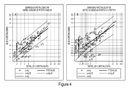
PCT/US2011/060642
elevated in the diseased group and proteins falling below the lower red-dashed
line are proteins
that are elevated in the control group. Proteins falling between the two red
dashed lines are not
significantly different between the two groups, although proteins in this area
may still be of
interest upon further evaluation.
[0061] Looking at Figure 4A, there are not many proteins that fall outside
the dashed lines,
which can be expected since this is the baseline time-point. However, when
looking at Figure
4b the number of proteins that are outside the dashed lines increases
dramatically. There are
three proteins that are increased in the diseased group at time-point 8 that
are considered of
"proteins of high interest" and they are proteins 7, 8, and 31, which
correspond to annexin A2,
plakoglobin, and serpin B3, respectively. These proteins appear in boldface in
Table 1. The
proteins that are decreased in the diseased group at time-point 8 are also
proteins of interest and
they appear in italics in Table 1. Proteins that are not in boldface or
italics are not excluded
from further investigation and may be of importance. In particular, proteins
1, 3, and 6 are of
interest because they have been seen free in serum and the ratio of free vs
bound for these
proteins, as well as for any of the other proteins listed, may be indicative
of the disease process.
Ultimately, any protein listed in the supplemental table may be a protein that
could have
potential clinical use.
[0062] The three proteins of "high interest" are particularly intriguing
because they are
implicated in known diseases and are elevated in diseased patients at time-
point 8. Plakoglobin
is intriguing because it is a component of the desmosomes, which are major
intracellular
adhesive junctions that anchor intermediate filaments to the plasma membrane
(Green et al.
Nature Reviews Molecular Cell Biology 2000, 1:208-216). Mutations in genes
encoding for
cardiac desmosomal proteins is prevalent in patients with arrhythmogenic right
ventricular
dysplasia/cardiomyopathy (ARVD/C), an inherited heart disease that is
clinically defined by the
presence of particular electrical, functional, and structural right
ventricular abnormalities and
histologically by replacement of cardiomyocytes with fibrous or fibrofatty
tissue (Basso et al.
Lancet 2009, 373:1289-1300; McKenna et al. British Heart Journal 1994, 71:215-
218). Work
over the past decade has shown that ARVC is an autosomal dominant trait
frequently caused by
mutations in genes that encode important structural proteins found within the
desmosome (Awad
et al. Nat Clin Pract Cardiovasc Med 2008, 5:258-267). Recent work has shown
that mutations
in the genes encoding for desmosomal proteins are also prevalent in patients
with dilated
cardiomyopathy (Elliott et al. Circulation Cardiovascular Genetics 2010, 3:314-
322).
27
CA 02817851 2013 05 13
WO 2012/065178
PCT/US2011/060642
[0063] The fact that an increase is observed in the amount of plakoglobin
bound to the
ABPPC in diseased patients indicates that there is degradation of the
desmosomes in these
patients and therefore loss of structural integrity of the cell-cell
interactions within the
myocardium, which is highly probable since the patients in this group are
showing elevated
levels of cTnI. Albumin could be serving as a sponge to bind these proteins
that are released
from degraded desmosomes. If this is the case and these and other desmosomal
proteins, such
as the plakophilins, desmogleins, and desmocollins (all of which are
represented in the ABPPC)
would be elevated in the ABPPC as a result of myocardial ischemia, then it
stands to reason that
they would also be elevated in the ABPPC for patients with other cardiac
disorders and could be
used as powerful biomarkers in cardiovascular medicine.
[0064] SERPINB3 is a peptidase inhibitor that is implicated in the survival
of squamous
carcinoma cells (Ahmed et al. Biochem Biophys Res Commun 2009, 378:821-825)
and in
chronic liver disease through its modulation of TGF-13 (Turato et al.
Laboratory Investigation
2010, 90:1016-1023). Annexin A2 is a member of the annexin family, which is a
family of
calcium-dependent phospholipid-binding proteins that play a role in the
regulation of cellular
growth and in signal transduction pathways. Annexins have been shown to be
involved in a
variety of cellular processes, including trafficking and organization of
vesicles, exo- and
endocytosis, and in calcium ion channel formation (Gerke et al. Nat Rev Mol
Cell Biol 2005,
6:449-461) and annexin A2 has been proposed as a differential diagnostic
marker of
hepatocellular tumors (Ji et al. Inter J Mol Med 2009, 24:765-771; Longrich et
al. Pathol Res
Pract 2010, Article in Press doi:10.1016/j .prp.2010.09.007). The implication
of the free form of
these proteins in disease makes the fact that they are observed in the ABPPC
very intriguing and
the ABPPC bound forms of these proteins (or any of the proteins observed in
the ABPPC) could
have significant diagnostic potential.
28