Note: Descriptions are shown in the official language in which they were submitted.
CA 02818556 2013-05-17
WO 2012/071344
PCT/US2011/061701
NUCLEOTIDE-BASED PROBES AND METHODS FOR THE DETECTION AND
QUANTIFICATION OF MACROMOLECULES AND OTHER ANALYTES
CROSS-REFERENCE TO RELATED APPLICATIONS
This application claims the benefit of priority under 35 U.S.C. 119(e) to
U.S.
Provisional Patent Application No. 61/417,141, filed November 24, 2010, the
disclosure of
which is hereby incorporated by reference in its entirety.
REFERENCE TO GOVERNMENT SUPPORT
This invention was made in part with government support under grants from the
National
Institutes of Health (Grant Nos. R01EB007689 and 1R01A1076899). The government
has
certain rights in this invention.
INTRODUCTION
Existing bio-analytical assays, including ELISAs, western blots and PCR, are
typically
multistep, washing-intensive and reagent-intensive processes. As such, these
approaches are not
well suited for use outside the laboratory, or for real-time or in situ
applications. In order to
overcome this limitation, a number of sensors have been developed that detect
binding in real
time by monitoring a change in mass, charge or optical properties that occurs
when the target
binds a biomolecule-coated surface (e.g., surface plasmon resonance, field-
effect transistor,
quartz crystal microbalance and microcantilevers). However, these approaches
detect adsorption
to the sensor head rather than a specific binding per se, and thus cannot
distinguish between the
binding of the correct, authentic target and the non-specific binding of
contaminants. Thus,
these approaches are not suitable for detection of targets in complex samples,
such as whole
blood or blood serum.
SUMMARY
Provided are unimolecular oligonucleotide probes for the detection of a target
in a
sample. The probes use target binding-induced structural changes to detect the
presence of the
target in the sample. Also provided are methods of using the probes to detect
a target in a
sample.
1
CA 02818556 2013-05-17
WO 2012/071344
PCT/US2011/061701
Probes that use binding-induced segregation of two target binding moieties
In some embodiments, the probes use target binding-induced structural changes
to detect
the presence of the target in the sample by utilizing binding-induced
segregation of two target
binding moieties as a signaling mechanism.
Accordingly, in some embodiments, a system for detecting one or more targets
in a
sample is provided. The system includes an oligonucleotide probe configured to
produce a
detectable signal when contacted by the one or more targets. The probe
includes: (a) a first
target binding moiety and a second target binding moiety; (b) a first
hybridization sequence and
a second hybridization sequence, where the first hybridization sequence and
the second
hybridization sequence are configured to form a duplex in the absence of the
target binding to
both the first target binding moiety and the second target binding moiety such
that the first target
binding moiety is positioned adjacent the second target binding moiety; and
(c) a first signaling
moiety and a second signaling moiety configured such that the position of the
first signaling
moiety is changed relative to the second signaling moiety upon binding of the
one or more
targets to both the first target binding moiety and the second target binding
moiety. In addition,
in the presence of the binding of the one or more targets to both the first
target binding moiety
and the second target binding moiety, formation of the duplex is inhibited
such that the probe is
configured to position the first signaling moiety relative to the second
signaling moiety such that
the probe produces a detectable change in a signal from the first and second
signaling moieties.
Embodiments of the system may also include that the probe includes a stem-loop
structure in the absence of the one or more targets binding to the first
target binding moiety and
the second target binding moiety.
Embodiments of the system may also include that the first target binding
moiety and the
second target binding moiety are bound directly to the probe.
Embodiments of the system may also include that the first target binding
moiety and the
second target binding moiety are bound indirectly to the probe.
Embodiments of the system may also include that the first target binding
moiety and the
second target binding moiety are bound to the probe through a linker moiety.
Embodiments of the system may also include that the probe further includes a
third
hybridization sequence and a fourth hybridization sequence. In these
embodiments, the first
target binding moiety may be bound to a fifth hybridization sequence
complementary to the
third hybridization sequence and the second target binding moiety may be bound
to a sixth
hybridization sequence complementary to the fourth hybridization sequence.
Embodiments of the system may also include that the third hybridization
sequence and
the fourth hybridization sequence are substantially the same, the fifth
hybridization sequence
2
CA 02818556 2013-05-17
WO 2012/071344
PCT/US2011/061701
and the sixth hybridization sequence are substantially the same. In these
embodiments, the
probe may include a frame inversion between the third hybridization sequence
and the fourth
hybridization sequence.
Embodiments of the system may also include that the frame inversion is a 3' to
3' or a 5'
to 5' frame inversion.
Embodiments of the system may also include that the first target binding
moiety and the
second target binding moiety include antigens, and that the target includes an
antibody specific
for the antigens.
Embodiments of the system may also include that the first target binding
moiety and the
second target binding moiety include polypeptides that specifically bind to a
macromolecule,
and that the target includes the macromolecule.
Embodiments of the system may also include that the first target binding
moiety and the
second target binding moiety include aptamers that specifically bind to a
macromolecule, and
that the target includes the macromolecule.
Embodiments of the system may also include that the first target binding
moiety and the
second target binding moiety include DNA or RNA sequences that specifically
bind to a
macromolecule, and that the target includes the macromolecule.
Embodiments of the system may also include that the target has a concentration
ranging
from 10 pM to 300 pM.
Embodiments of the system may also include that the first signaling moiety
includes a
fluorophore and the second signaling moiety includes a quencher.
Embodiments of the system may also include that the first signaling moiety
includes a
first fluorophore and the second signaling moiety includes a second
fluorophore.
Embodiments of the system may also include that the first signaling moiety
includes a
nanoparticle and the second signaling moiety includes a quencher.
Embodiments of the system may also include that the first signaling moiety
includes a
first nanoparticle and the second signaling moiety includes a second
nanoparticle.
Embodiments of the system may also include that the first signaling moiety
includes an
electrochemical reporter and the second signaling moiety is an electrode.
Embodiments of the system may also include that the probe is immobilized on
the
surface of the electrode.
Embodiments of the system may also include that the first signaling moiety
includes a
macromolecule having a catalytic activity and the second signaling moiety
includes an inhibitor
or an activator of the catalytic activity.
Embodiments of the system may also include that the system includes an array
of probes.
3
CA 02818556 2013-05-17
WO 2012/071344
PCT/US2011/061701
Aspects of the present disclosure also include a method of detecting a target
in a sample.
The method includes contacting a unimolecular oligonucleotide probe with the
sample, whereby
the target selectively binds to both the first target binding sequence and the
second target
binding sequence to form a target-probe hybrid. The method further includes
detecting the
presence or absence of the target-probe hybrid.
Embodiments of the method may also include that the sample includes a complex
sample.
Embodiments of the method may also include that the sample includes whole
blood.
Aspects of the present disclosure also include a method of detecting a second
target in a
sample. The method includes contacting an oligonucleotide probe with a first
with the sample,
whereby the target selectively binds to both the first target binding sequence
and the second
target binding sequence to form a target-probe hybrid. The method further
includes contacting
the target-probe hybrid with a second target, whereby the second target
selectively binds the
target and inhibits formation of the target-probe hybrid. The method further
includes detecting
the presence or absence of the target-probe hybrid.
Probes that utilize binding-induced reconstitution of a recognition element
In some embodiments, the probes use target binding-induced structural changes
to detect
the presence of the target in the sample by utilizing binding-induced
reconstitution of a
recognition element as a signaling mechanism. For example, the probe may use
binding-
induced reconstitution of a specific DNA binding sequence as signaling
mechanism.
Aspects of the present disclosure include a system for detecting a DNA binding
protein
in a sample. The system includes a unimolecular oligonucleotide probe
configured to produce a
detectable signal when contacted with the DNA binding protein. The probe
includes: (a) a first
recognition sequence and a second recognition sequence, where the first and
second recognition
sequences are configured to form a recognition duplex specifically bound by
the DNA binding
protein in the sample; (b) a first hybridization sequence and a second
hybridization sequence,
where the first and second hybridization sequences are configured to form a
second duplex in
the absence of binding of the DNA binding protein to the recognition duplex;
(c) a third
hybridization sequence and a fourth hybridization sequence, where the third
and fourth
hybridization sequences are configured to form a third duplex in the absence
of binding of the
DNA binding protein to the recognition duplex; and (d) a first signaling
moiety and a second
signaling moiety, where in the absence of binding of the DNA binding protein
to the recognition
duplex, the first signaling moiety is positioned adjacent the second signaling
moiety such that
the probe does not produce a detectable signal. In addition, in the presence
of binding of the
4
CA 02818556 2013-05-17
WO 2012/071344
PCT/US2011/061701
DNA binding protein to the recognition duplex, formation of the second and
third duplexes is
inhibited such that the probe is configured to position the first signaling
moiety away from the
second signaling moiety such that the probe produces a detectable signal.
Embodiments of the system may also include that the first recognition sequence
is
positioned between the first and second hybridization sequences and the second
recognition
sequence is positioned between the third and fourth hybridization sequences.
Embodiments of the system may also include a fifth hybridization sequence and
a sixth
hybridization sequence, where fifth and sixth hybridization sequences are
configured to form a
fourth duplex in the absence of binding of the DNA binding protein to the
recognition duplex.
Embodiments of the system may also include that at least a portion of the
first
recognition sequence is positioned between the second and third hybridization
sequences and at
least a portion of the second recognition sequence is positioned between the
fourth and fifth
hybridization sequences.
Embodiments of the system may also include that the probe is configured to be
in an
equilibrium between formation of the second and third duplexes and formation
of the
recognition duplex.
Embodiments of the system may also include that in the absence of binding of
the DNA
binding protein to the recognition duplex, the equilibrium is shifted towards
the formation of the
second and third duplexes.
Embodiments of the system may also include that in the presence of binding of
the DNA
binding protein to the recognition duplex, a DNA binding protein-probe hybrid
is formed and
the equilibrium is shifted towards the formation of the recognition duplex.
Embodiments of the system may also include that in the presence of a single-
stranded
DNA sequence that stabilizes the DNA binding protein-probe hybrid, the
equilibrium is shifted
towards the formation of the recognition duplex.
Embodiments of the system may also include that the DNA binding protein has a
concentration ranging from 1 nM to 1 it.M.
Embodiments of the system may also include that the first signaling moiety
includes a
fluorophore and the second signaling moiety includes a quencher.
Embodiments of the system may also include that the first signaling moiety
includes a
fluorophore and the second signaling moiety includes a second fluorophore.
Embodiments of the system may also include that the first signaling moiety
includes an
electrochemical reporter and the second signaling moiety includes an
electrode.
5
CA 02818556 2013-05-17
WO 2012/071344
PCT/US2011/061701
Embodiments of the system may also include that the first signaling moiety
includes a
nanoparticles (gold, silver or diamonds) and the second signaling moiety
includes a quencher or
a second nanoparticles.
Embodiments of the system may also include that the probe is immobilized on
the
surface of the electrode.
Embodiments of the system may also include that the first signaling moiety
includes a
macromolecule that display a catalytic activity and the second signaling
moiety includes an
inhibitor or an activator of this catalytic activity.
Embodiments of the system may also include that the system includes an array
of probes.
Aspects of the present disclosure also include a method of detecting a DNA
binding
protein in a sample. The method includes contacting a unimolecular
oligonucleotide probe with
the sample, whereby the DNA binding protein selectively binds to the
recognition duplex to
form a DNA binding protein-probe hybrid. The method further includes detecting
the presence
or absence of the DNA binding protein-probe hybrid.
Embodiments of the method may also include that the detecting includes
quantifying the
concentration of the DNA binding protein-probe hybrid by comparing the signal
from the
sample to: (1) a saturating concentration of a competitive DNA binding
sequence; (2) a
saturating concentration of a transcription factor; or (3) a saturating
concentration of a single-
stranded DNA configured to stabilize the DNA binding protein-probe hybrid.
Embodiments of the method may also include that the sample includes a complex
sample.
Embodiments of the method may also include that the sample includes whole
blood.
Embodiments of the method may also include that the sample includes a crude
nuclear
extract.
BRIEF DESCRIPTION OF THE FIGURES
FIG. 1(a) shows a unimolecular oligonucleotide probe (top: with stem; bottom:
without
stem) configured to produce a detectable signal upon target binding, according
to embodiments
of the present disclosure. FIG. 1(b) shows a chart of stem nucleotide sequence
vs. stem stability
for probes according to embodiments of the present disclosure. FIG. 1(c) shows
a graph of
fluorescence intensity vs. temperature for probes according to embodiments of
the present
disclosure.
FIG. 2(a) shows a unimolecular oligonucleotide probe configured to produce a
detectable
fluorescent signal upon target binding, according to embodiments of the
present disclosure. The
fluorescent signaling moieties are FAM-6 for the fluorophore and BHQ-1 for the
quencher. FIG.
6
CA 02818556 2013-05-17
WO 2012/071344
PCT/US2011/061701
2(b) shows graphs of fluorescence vs. wavelength for anti-Dig antibody and
anti-DNP antibody
probes according to embodiments of the present disclosure. The stem sequence
used was 1MM
(see FIG. 1(b)). FIG. 2(c) shows graphs of signal vs. target concentration for
anti-Dig antibody
and anti-DNP antibody probes according to embodiments of the present
disclosure. FIG. 2(d)
shows graphs of signal vs. time for anti-Dig antibody and anti-DNP antibody
probes according
to embodiments of the present disclosure.
FIG. 3 shows the detection of a target (e.g., the antigen DNP) in a sample
using the anti-
DNP antibody probe in a competition assay, according to embodiments of the
present disclosure.
FIG. 4(a) shows a modular oligonucleotide probe configured to produce a
detectable
fluorescent signal upon target binding, according to embodiments of the
present disclosure. The
fluorescent signaling moieties are FAM-6 for the fluorophore and BHQ-1 for the
quencher. In
this particular probe, the first and second binding moieties (X) are attached
to the probe via
hybridization to third and fourth hybridization sequences. FIG. 4(b) shows
graphs of fluorescent
signal vs. target concentration for modular anti-Dig antibody and anti-DNP
antibody probes
according to embodiments of the present disclosure. FIG. 3(c) shows graphs of
the fluorescent
signal vs. time for modular anti-Dig antibody and anti-DNP antibody probes
according to
embodiments of the present disclosure.
FIG. 5 shows a modular oligonucleotide probe configured to produce a high
detectable
fluorescent signal upon target binding, according to embodiments of the
present disclosure. FIG.
5 also shows graphs of fluorescence vs. temperature and fluorescence vs.
wavelength (nm) for
modular probes, according to embodiments of the present disclosure.
FIG. 6 (top) shows graphs of fluorescence vs. wavelength (nm) and fluorescence
vs.
target size (kDa) for modular oligonucleotide probes, according to embodiments
of the present
disclosure. FIG. 6 (bottom) shows a schematic of the signaling of the modular
probe (e.g., the
probe stem opening) in the presence of two targets binding to a single probe,
according to
embodiments of the present disclosure.
FIG. 7 (top) shows a graph of fluorescence vs. wavelength (nm) for probes for
the
detection of targets directly in blood serum, according to embodiments of the
present disclosure.
FIG. 7 (bottom) shows graphs of fluorescence vs. wavelength (nm) in buffer and
whole blood
(panels a and b), according to embodiments of the present disclosure.
FIG. 8(a) shows a modular unimolecular oligonucleotide probe that uses an
electrochemical reporter (e.g., methylene blue) and an electrode as the two
signaling moieties,
according to embodiments of the present disclosure. FIG. 8(b) shows graphs of
square wave
voltammograms for anti-Dig antibody and anti-HIV antibody probes, according to
embodiments
of the present disclosure (results were obtained in 80% whole blood using a
probe stem with
7
CA 02818556 2013-05-17
WO 2012/071344
PCT/US2011/061701
1MM and 2GC and 2AT). FIG. 8(c) shows graphs of current signal vs. target
concentration for
anti-Dig antibody and anti-HIV antibody probes, according to embodiments of
the present
disclosure (results were obtained in 80% whole blood). FIG. 8(d) shows graphs
of current signal
vs. time for anti-Dig antibody and anti-HIV antibody probes, according to
embodiments of the
present disclosure. FIG. 8(e) shows graphs of square wave voltammograms for
anti-Dig
antibody and anti-HIV antibody probes in buffer and 80% whole blood, according
to
embodiments of the present disclosure.
FIG. 9 shows a graph of non-specific signal degradation vs. time for an
electrochemical
probe in whole blood and blood serum, according to embodiments of the present
disclosure.
FIG. 10(a) shows a probe that works in absence of a stem region in which the
target
binding moieties can be positioned at various locations, according to
embodiments of the present
disclosure. FIG. 10(b) shows a schematic of how target binding changes the
distance between
the two signaling moieties, according to embodiments of the present
disclosure. FIG. 10(c)
shows graphs of square wave voltammograms for anti-Flag antibody probes,
according to
embodiments of the present disclosure. FIG. 10(d) shows a graph of current
signal vs. target
concentration for anti-Flag antibody probes, according to embodiments of the
present disclosure.
FIG. 11(a) shows a unimolecular oligonucleotide probe for detecting a DNA
binding
protein, according to embodiments of the present disclosure. The fluorescent
signaling moieties
used were FAM-6 for the fluorophore and BHQ-1 for the quencher. FIG. 11(b)
shows a chart of
probe variants displaying various switching equilibrium constants, Ks, and
dissociation
constants, KD, for TATA Binding Protein, according to embodiments of the
present disclosure.
FIG. 11(c) shows a graph of fluorescence of these different variants vs. the
concentration of a
TBP target, according to embodiments of the present disclosure.
FIG. 12 shows graphs of fluorescence signal vs. target concentration for
probes for
detecting DNA binding proteins (e.g., TATA Binding Protein, Myc-Max, and
NFkB), and their
binding kinetics, according to embodiments of the present disclosure.
FIG. 13 (top) shows graphs of fluorescence signal vs. target concentration for
probes for
detecting DNA binding proteins directly in crude nuclear extracts (250 it.g/mL
of HeLa nuclear
extracts) (+TBP), according to embodiments of the present disclosure. FIG. 13
(bottom) shows
a schematic (FIG. 13(a)) and graph (FIG. 13(b)) for the quantification of
transcription factors in
crude nuclear extracts using a probe, according to embodiments of the present
disclosure.
FIG. 14 (top) shows a unimolecular oligonucleotide probe for the detection of
a DNA
binding protein, that uses an electrochemical reporter (e.g., methylene blue)
and an electrode as
the two signaling moieties, according to embodiments of the present
disclosure. FIG. 14(a)
shows a graph of square wave voltammograms for TATA binding protein probe in
the presence
8
CA 02818556 2013-05-17
WO 2012/071344
PCT/US2011/061701
of various concentration of TATA binding protein, according to embodiments of
the present
disclosure (results wereobtained in buffer). FIG. 14(b) shows a graph of
current signal vs. target
concentration for TATA binding protein probes, according to embodiments of the
present
disclosure (results were obtained in buffer and in 250 pg/mL of HeLa nuclear
extracts).
Before the present invention is described in greater detail, it is to be
understood that this
invention is not limited to the particular embodiments described, and as such
may, of course,
vary. It is also to be understood that the terminology used herein is for the
purpose of describing
particular embodiments only, and is not intended to be limiting, since the
scope of the present
invention is embodied by the appended claims.
Where a range of values is provided, it is understood that each intervening
value, to the
tenth of the unit of the lower limit unless the context clearly dictates
otherwise, between the
upper and lower limit of that range and any other stated or intervening value
in that stated range,
is encompassed within the invention. The upper and lower limits of these
smaller ranges may
independently be included in the smaller ranges and are also encompassed
within the invention,
subject to any specifically excluded limit in the stated range. Where the
stated range includes
one or both of the limits, ranges excluding either or both of those included
limits are also
included in the invention.
Unless defined otherwise, all technical and scientific terms used herein have
the same
meaning as commonly understood by one of ordinary skill in the art to which
this invention
belongs. Although any methods and materials similar or equivalent to those
described herein
can also be used in the practice or testing of the present invention,
representative illustrative
methods and materials are now described.
It is noted that, as used herein and in the appended claims, the singular
forms "a", "an",
and "the" include plural referents unless the context clearly dictates
otherwise. It is further
noted that the claims may be drafted to exclude any optional element. As such,
this statement is
intended to serve as antecedent basis for use of such exclusive terminology as
"solely," "only"
and the like in connection with the recitation of claim elements, or use of a
"negative" limitation.
As will be apparent to those of skill in the art upon reading this disclosure,
each of the
individual embodiments described and illustrated herein has discrete
components and features
which may be readily separated from or combined with the features of any of
the other several
embodiments without departing from the scope or spirit of the present
invention. In addition, it
will be readily apparent to one of ordinary skill in the art in light of the
teachings herein that
certain changes and modifications may be made thereto without departing from
the spirit and
9
CA 02818556 2013-05-17
WO 2012/071344
PCT/US2011/061701
scope of the appended claims. Any recited method can be carried out in the
order of events
recited or in any other order which is logically possible.
All publications and patents cited in this specification are herein
incorporated by
reference as if each individual publication or patent were specifically and
individually indicated
to be incorporated by reference and are incorporated herein by reference to
disclose and describe
the methods and/or materials in connection with which the publications are
cited. To the extent
such publications may set out definitions of a term that conflicts with the
explicit or implicit
definition of the present disclosure, the definition of the present disclosure
controls. The citation
of any publication is for its disclosure prior to the filing date and should
not be construed as an
admission that the present invention is not entitled to antedate such
publication by virtue of prior
invention. Further, the dates of publication provided may be different from
the actual
publication dates which may need to be independently confirmed.
DETAILED DESCRIPTION OF EXEMPLARY EMBODIMENTS
Provided are unimolecular oligonucleotide probes for detecting a target in a
sample. The
probes use target binding-induced structural changes to detect the presence of
the target in the
sample. Also provided are methods of using the probes to detect a target in a
sample.
Below, the subject systems that include the oligonucleotide probes are
described first in
greater detail, followed by a review of the various methods in which the
probes may find use, as
well as a discussion of various representative applications in which the
subject probes and
methods find use.
SYSTEMS
Systems of the present disclosure include one or more oligonucleotide probes
described
in more detail below. The term "probe" as used herein refers to a unimolecular
biopolymer that
undergoes a structural change upon its specific binding to a target (e.g.,
molecule,
macromolecule, or analyte). Probes may include, but are not limited to,
nucleic acids (DNA or
RNA), non-natural oligonucleotide analogs such as PNA, LNA, aptamers, peptides
and proteins,
etc. In some instances, the probes are oligonucleotides that may be of any
length, but may be
short oligonucleotides ranging from 20 to 100 nucleotides, or 25 to 90
nucleotides, such as 30 to
80 nucleotides. The particular use of terms "nucleic acid," "oligonucleotide,"
and
"polynucleotide" should in no way be considered limiting and may be used
interchangeably
herein. "Oligonucleotide" is used when the relevant nucleic acid molecules
include less than
about 100 bases. "Polynucleotide" is used when the relevant nucleic acid
molecules include
CA 02818556 2013-05-17
WO 2012/071344
PCT/US2011/061701
more than about 100 bases. Both terms are used to denote DNA, RNA, modified or
synthetic
DNA or RNA (including but not limited to nucleic acids comprising synthetic
and naturally-
occurring base analogs, dideoxy or other sugars, thiols or other non-natural
or natural polymer
backbones), or other nucleobase containing polymers. Accordingly, the terms
should not be
construed to define or limit the length of the nucleic acids referred to and
used herein.
Oligonucleotides of the present disclosure may be single-stranded, double-
stranded,
triple-stranded, or include a combination of these conformations. Generally
oligonucleotides
contain phosphodiester bonds, although in some cases, as outlined below,
nucleic acid analogs
are included that may have alternate backbones, comprising, for example,
phosphoramide,
phosphorothioate), phosphorodithioate, 0-methylphosphoroamidite linkages, and
peptide
nucleic acid backbones and linkages. Other analog nucleic acids include those
with positive
backbones, non-ionic backbones, and non-ribose backbones. Nucleic acids
containing one or
more carbocyclic sugars are also included within the definition of nucleic
acids. These
modifications of the ribose-phosphate backbone may be done to facilitate the
addition of
additional moieties such as labels, or to increase the stability and half-life
of such molecules in
physiological environments. The term "nucleic acid sequence" or
"oligonucleotide sequence"
refers to a contiguous string of nucleotide bases and in particular contexts
also refers to the
particular placement of nucleotide bases in relation to each other as they
appear in an
oligonucleotide.
In certain embodiments, the probes may recognize their targets by specific
binding of the
target to the probe at, for example, a target binding moiety included on the
probe. "Target"
refers to any molecule that specifically binds to a probe of the present
disclosure. These include,
but are not limited to, macromolecules (e.g., proteins, carbohydrates, nucleic
acids, lipids, etc.),
small molecules (e.g., peptides, aptamers, etc.), and the like. While not an
exhaustive list, in
certain embodiments, the target may be an antibody, a DNA binding protein, a
receptor, or an
enzyme that specifically binds the probe. One of skill in the art will
recognize that the important
aspect of probe-target binding is not the particular mechanism involved but
the fact that the
binding is specific, as in specifically binding as defined in this disclosure.
Oligonucleotide probes of the present disclosure may be unimolecular. By
"unimolecular" is meant that the probe includes a single moiety that binds to
the target.
Unimolecular probes do not include probes that include two or more separate
probe elements
that associate with each other during formation of the target-probe hybrid.
Unimolecular probes
may include single-stranded oligonucleotide probes, as well as single-stranded
oligonucleotide
probes that are directly or indirectly bound to target binding moieties as
described in detail
herein.
11
CA 02818556 2013-05-17
WO 2012/071344
PCT/US2011/061701
In certain embodiments, the target is a bidentate target. As used herein,
"denticity"
refers to the number of distinct binding sites included in a target molecule.
A polydentate target
may bind to two or more target binding moieties, with each target binding
moiety binding to
different binding sites on the target. For example, a bidentate target
includes two target binding
sites with each binding site capable of specifically binding to a target
binding moiety. Bidentate
targets may include, but are not limited to, antibodies which may include two
antigen binding
sites that each specifically bind to one copy of a specific antigen. In
certain embodiments, the
target is a non-bidentate target, for example a target that includes one
binding site capable of
specifically binding to a target binding moiety.
Aspects of the present disclosure include oligonucleotide probes for detecting
a target in
a sample. The probes can be made as oligonucleotide strands constructed using
techniques well-
known to those of skill in the art, and include internal sequences allowing
the oligonucleotide
strand to undergo intramolecular hybridization when one internal hybridization
sequence
specifically hybridizes to a complementary internal hybridization sequence.
The terms "complementary" or "complementarity" refer to polynucleotides (i.e.,
a
sequence of nucleotides) related by base-pairing rules. For example, the
sequence "5'-AGT-3',"
is complementary to the sequence "5'-ACT-3'. Complementarity may be "partial,"
in which
only some of the nucleic acids' bases are matched according to the base
pairing rules, or there
may be "complete" or "total" complementarity between the nucleic acids. The
degree of
complementarity between nucleic acid strands can have effects on the
efficiency and strength of
hybridization between nucleic acid strands under defined conditions.
As used herein, the term "hybridization" is used in reference to the pairing
of
complementary nucleic acids. Hybridization and the strength of hybridization
(i.e., the strength
of the association between the nucleic acids) is influenced by such factors as
the degree of
complementary between the nucleic acids, stringency of the conditions
involved, and the thermal
melting point, Tm, of the formed hybrid. Hybridization methods involve the
annealing of one
nucleic acid to another, complementary nucleic acid, e.g., a nucleic acid
having a
complementary nucleotide sequence.
Hybridization is carried out in conditions permitting specific hybridization.
The length
of the complementary sequences and GC content affects the thermal melting
point, Tm, of the
hybridization conditions necessary for obtaining specific hybridization of the
target site to the
target nucleic acid. Hybridization may be carried out under stringent
conditions. The phrase
"stringent hybridization conditions" refers to conditions under which a probe
will hybridize to
its target subsequence, typically in a complex mixture of nucleic acid, but to
no other sequences
at a detectable or significant level. Stringent conditions are sequence-
dependent and may be
12
CA 02818556 2013-05-17
WO 2012/071344
PCT/US2011/061701
different in different circumstances. The phrase "selectively (or
specifically) hybridizing" refers
to the binding, duplexing, or hybridizing of a molecule only to a particular
nucleotide sequence
under stringent hybridization conditions when that sequence is present in a
complex mixture
(e.g., total cellular, library DNA or RNA, complex samples such as whole blood
samples and the
like). Those of ordinary skill in the art will readily recognize that
alternative hybridization and
wash conditions can be utilized to provide conditions of similar stringency
and will recognize
that the combination of parameters may be more important than the measure of
any single
parameter.
Intramolecular hybridization of the oligonucleotide probes can result in the
probe taking
a stem-loop secondary conformation in the absence of target binding to the
probe. The probes
are configured to use target binding-induced structural changes to detect the
presence of the
target in the sample. As used herein, the different oligonucleotide probe
structures, such as
those that exist in the presence or absence of a target, may be as referred to
as "conformations."
In certain embodiments, internal hybridization sequence lengths range from 5
to 25 nucleotides,
for example 5 to 20 nucleotides, such as 10 to 20 nucleotides per internal
hybridization
sequence. The "loop" structures of each probe may be of any length suitable to
the application,
but may range from 3 to 30 nucleotides in length, for example 5 to 25
nucleotides, such as 10 to
nucleotides in length.
In some embodiments the hybridization leads to two double-stranded
oligonucleotides
20 separated by a single-stranded region. The single-stranded region of
each probe may be of any
length suitable to the application, but may range from 3 to 30 nucleotides in
length, for example
5 to 25 nucleotides, such as 10 to 20 nucleotides in length.
The probes may be provided in solution. In these cases, the probes are free to
diffuse
through the solution and are not attached to a surface. In certain
embodiments, the probes are
attached to the surface of a substrate. The probes may be attached to the
surface of the substrate
at predetermined locations, such that the probes are arranged in an array
formation. An "array,"
includes any one-dimensional, two-dimensional or substantially two-dimensional
(as well as a
three-dimensional) arrangement of addressable regions bearing a particular
probe associated
with that region. The probes may be covalently attached to the arrays at any
point along the
nucleic acid chain. In certain cases, the probes are attached at one of their
termini (e.g., the 3' or
5' terminus). In some cases, the probes are attached to the array at an
internal site of the probe.
An "addressable array" includes any one or two dimensional arrangement of
discrete regions (or
"features") bearing particular probes associated with that region and
positioned at particular
predetermined locations on the substrate (each such location being at a known
"address").
These regions may or may not be separated by intervening spaces.
13
CA 02818556 2013-05-17
WO 2012/071344
PCT/US2011/061701
Any given substrate may carry one, two, four or more arrays disposed on a
front surface
of the substrate. Depending upon the use, any or all of the arrays may be the
same or different
from one another and each may contain multiple spots or features. A typical
array may contain
more than ten, more than one hundred, more than one thousand, more than ten
thousand
features, or even more than one hundred thousand features, in an area of less
than 20 cm2, such
as less than 10 cm2. For example, features may have widths (that is, diameter,
for a round spot)
in the range from a 10 um to 1.0 cm. In other embodiments each feature may
have a width in
the range of 1.0 um to 1.0 mm, such as 5.0 um to 500 um, including 10 um to
200 um. Non-
round features may have area ranges equivalent to that of circular features
with the foregoing
width (diameter) ranges. In certain embodiments, the arrays are formed by
processes involving
drop deposition of reagents, for example, photolithographic array fabrication
processes may be
used.
With arrays that are read by detecting fluorescence, the substrate may be of a
material
that emits low fluorescence upon illumination with the excitation light.
Additionally in this
situation, the substrate may be relatively transparent to reduce the
absorption of the incident
illuminating light (e.g., laser light) and subsequent heating if the focused
light travels too slowly
over a region.
Oligonucleotide Probes
In certain embodiments, the oligonucleotide probes are configured to produce a
detectable signal when a target specifically binds to the probe to form a
target-probe hybrid.
The target may specifically recognize and bind to particular portions of the
probe at, for
example, a target binding moiety included on the probe. As used herein, the
term "target
binding moiety" refers to any molecule that specifically binds a target of the
present disclosure.
These include, but are not limited to, proteins, peptides, carbohydrates,
nucleic acids, lipids,
small molecules, and the like. For instance, the target binding moiety may be
an antigen. In
some cases, the probe includes two target binding moieties, such as a first
target binding moiety
and a second target binding moiety. The first target binding moiety may be
different from the
second target binding moiety, for example in embodiments where the target is
capable of
binding to two or more different target binding moieties. In certain
instances, the first target
binding moiety and the second target binding moiety are substantially the
same, for example in
embodiments where the target is capable of binding two or more of the same
target binding
moiety, such as where the target is an antibody.
The target binding moiety may be bound to the probe. In some cases, the target
binding
moiety is directly bound to the probe. For example, the target binding moiety
may be directly
14
CA 02818556 2013-05-17
WO 2012/071344
PCT/US2011/061701
bound to the probe by modification of a nucleotide in the oligonucleotide
strand that makes up
the probe, such as, but not limited to, covalent attachment of the target
binding moiety to a
nucleotide in the oligonucleotide sequence, insertion of the target binding
moiety between two
nucleotides in the oligonucleotide sequence through the introduction of
additional
phosphodiester bonds, and the like.
In some cases, the target binding moiety is indirectly bound to the probe,
such as, but not
limited to, attachment of the target binding moiety to the probe through a
linker moiety. The
linker moiety can be any linker moiety suitable for the attachment of the
target binding moiety
to one or more nucleotides in the oligonucleotide probe. The linker moiety may
include 1 to 25
carbons, such as 2 to 20 carbons, including 5 to 15 carbons. In certain
embodiments, the target
binding moiety is indirectly bound to the probe by hybridization of an
oligonucleotide to the
probe. In these embodiments, the target binding moiety may be attached either
directly or
indirectly to a hybridization sequence, which specifically hybridizes to a
complementary
sequence on the probe to form a duplex. As indicated above, certain
embodiments of the probe
include two target binding moieties. In some cases, the first target binding
moiety and the
second target binding moiety are attached to hybridization sequences that have
different
nucleotide sequences. In these cases, the hybridization sequences specifically
hybridize to
different complementary sequences on the probe. In other instances, the first
target binding
moiety and the second target binding moiety are attached to hybridization
sequences that have
substantially the same nucleotide sequence. In these instances, the
hybridization sequences
specifically hybridize to the same complementary nucleotide sequence. The
probe may include
one or more, such as two or more repeats of the complementary nucleotide
sequence, such that a
corresponding number of hybridization sequences may be hybridized to the
probe, and thus a
corresponding number of target binding moieties may be attached to the probe.
In certain
embodiments, the probe includes two hybridization sequences that are
complementary to the
hybridization sequences bound to the target binding moieties, such that two
target binding
moieties are attached to the probe. In some cases, the probe includes a frame
inversion between
the hybridization sequences. The frame inversion may be a 3' to 3' or a 5' to
5' frame inversion.
Inclusion of a frame inversion may facilitate attachment of the target binding
moieties to the
probe in a configuration that facilitates the structural change induced by the
binding of the target
to both of the target binding moieties.
In some embodiments, the probe also includes two or more hybridization
sequences
(e.g., intramolecular hybridization sequence, IHS) configured to allow the
oligonucleotide strand
to undergo intramolecular hybridization. For instance, the probe may include a
first
hybridization sequence (e.g., a first IHS) and a second hybridization sequence
(e.g., a second
CA 02818556 2013-05-17
WO 2012/071344
PCT/US2011/061701
IHS). In embodiments that include two target binding moieties, as described
herein, the first
hybridization sequence and the second hybridization sequence may be configured
to form a
duplex in the absence of target binding to both of the target binding
moieties. The first
hybridization sequence and the second hybridization sequence may be separated
by a loop
structure formed by the oligonucleotide sequence of the probe that is between
the first
hybridization sequence and the second hybridization sequence. As such, in the
absence of target
binding to the target binding moieties, the probe may adopt a stem-loop
conformation.
In some embodiments, the probe may include two double-stranded regions
separated by
a single-stranded region. The single-stranded region may facilitate an
increase in the flexibility
of the probe in the absence of target binding.
The probes also include one or more signaling moieties. In some cases, the
probe
includes two signaling moieties, such as a first signaling moiety and a second
signaling moiety.
In certain embodiments, the first signaling moiety is held at distance in
close proximity to the
second signaling moiety, such as adjacent the second signaling moiety, by
complementary base-
pairing within the probe. In some embodiments, the probe is flexible in the
absence of target
binding, allowing the signaling moieties to approach one another transiently
or intermittently. In
embodiments of the probe configured to produce a detectable change in signal
in the presence of
target binding to the target binding moieties, under conditions in the absence
of target, the
distance the first signaling moiety is held from the second signaling moiety
is sufficient to
minimize, suppress, or prevent the first signaling moiety from emitting a
detectable signal. In
some embodiments, this proximity instead enhances or maximizes the detectable
signal from the
first signaling moiety. In some embodiments, collisions between the two
signaling moieties
increase or decrease the signal or signals associated with them. When target
is present and binds
to the target binding moieties of the probe, the internal hybridization of the
probe is disrupted.
Disruption of the internal hybridization allows the end of the nucleotide
chain to which the first
signaling moiety is attached to move to a distance further away from the
second signaling
moiety. Under conditions in the presence of target, the distance the first
signaling moiety moves
away from the second signaling moiety is sufficient to lead to a detectable
change in the signal
from the first signaling moiety. In some embodiments, this change in distance
leads to a
detectable decrease in signal. In other embodiments, target binding prevents
collisions between
the two signaling moieties, leading to a detectable change in their signal.
As described above, in the absence of target binding to the target binding
moieties, the
probe may be in a stem-loop configuration. In these cases, the probe may adopt
a conformation
where the first signaling moiety is positioned adjacent the second signaling
moiety, such that the
probe does not produce a detectable signal. For example, the first signaling
moiety may be a
16
CA 02818556 2013-05-17
WO 2012/071344
PCT/US2011/061701
fluorophore and the second signaling moiety may be a quencher. In these
instances, under
conditions in the absence of target, the distance the fluorophore is held from
the quencher is
sufficient to minimize, suppress, or prevent the fluorophore from emitting a
detectable signal.
Alternatively, this proximity may increase the signal from the first signaling
moiety. When
target is present and binds to the target binding moieties of the probe, the
internal hybridization
of the probe is disrupted such that the fluorophore is able to move to a
distance further away
from the quencher. Under conditions in the presence of target, the distance
the fluorophore
moves away from the quencher is sufficient to allow the signal emitted by the
fluorophore to
change detectably. In some instances, the detectable change in signal is an
increase in the signal
emitted by the fluorophore.
As described above, in the absence of target binding to the target binding
moieties, the
probe may have a flexible conformation. In these cases, the first signaling
moiety can
transiently collide with or bind to the second signaling moiety, such the
signal from the
signaling moieties is changed. For example, the first signaling moiety may be
a fluorophore and
the second signaling moiety may be a quencher. In these instances, under
conditions in the
absence of target, collisions between the fluorophore and the quencher are
sufficient to
minimize, suppress, or prevent the fluorophore from emitting a detectable
signal. When target is
present and binds to both the target binding moieties of the probe, contact
between the
fluorophore and the quencher may be inhibited or reduced such that the
quencher does not
approach the fluorophore as readily or as frequently. Under conditions in the
presence of target,
the distance the fluorophore moves away from the quencher detectably changes
the signal that
the fluorophore emits. In certain cases, the detectable change in signal is an
increase in the
signal emitted by the fluorophore.
The term "fluorophore" refers to any molecular entity that is capable of
absorbing energy
of a first wavelength and re-emit energy at a different second wavelength. In
certain
embodiments, the oligonucleotide probe includes a fluorophore attached to one
end of the probe
or at a central position in the probe sequence, so long as the position of the
fluorophore allows
the fluorophore to be positioned adjacent the quencher in the absence of
target binding to the
target binding moieties and away from the quencher when target binds to the
target binding
moieties. In some embodiments, as discussed in more detail below, the
fluorophore may be
attached to one end of the probe. The fluorophore attached to the probe need
not be a single
molecule, but may include multiple molecules. In some embodiments, the
fluorophore is a
fluorescent moiety, such as but not limited to, a fluorescent nanoparticle,
such as gold, silver or
diamond nanoparticles, and the like. The "end" of the oligonucleotide probe
possessing the
fluorophore includes any nucleotide within one quarter of the total number of
nucleotides in the
17
CA 02818556 2013-05-17
WO 2012/071344
PCT/US2011/061701
probe from the terminal nucleotide. Alternatively, the end possessing the
fluorophore includes
the terminal 10, 9, 8, 7, 6, 5, 4, 3 or 2 nucleotides of the probe. Attachment
may also be on the
terminal nucleotide alone. The attachment of the fluorophore to the
oligonucleotide probe
allows the fluorophore to be positioned in an alternate configuration at a
distance away from the
quencher in response to target specifically binding the probe, thereby
generating a detectable
signal.
The fluorophore may be synthetic or biological in nature, as known to those of
skill in
the art. More generally, any fluorophore can be used that is stable under
assay conditions and
that can be sufficiently suppressed when in close proximity to the quencher
such that a
significant change in the intensity of fluorescence of the fluorophore is
detectable in response to
target specifically binding the probe. Examples of suitable fluorophores
include, but are not
limited to CAL Fluor Red 610 (FR610; Biosearch Technologies, Novato, CA),
fluorescein
isothiocyanate, fluorescein, 6-carboxyfluorescein (6-FAM), rhodamine and
rhodamine
derivatives, coumarin and coumarin derivatives, cyanine and cyanine
derivatives, Alexa Fluors
(Molecular Probes, Eugene, OR), DyLight Fluors (Thermo Fisher Scientific,
Waltham, MA),
and the like.
The term "quencher" may refer to a substance that absorbs excitation energy
from a
fluorophore and dissipates that energy as heat. The quencher may also absorb
excitation energy
from a fluorophore and dissipate that energy as re-emitted light at a
different wavelength.
Quenchers are used in conjunction with fluorophores, such that when the
quencher is positioned
adjacent the fluorophore or at a distance sufficiently close to the
fluorophore, the emission of the
fluorophore is suppressed. However, when the quencher is positioned away from
the
fluorophore or at a distance sufficiently far from the fluorophore, the
emission of the
fluorophore is not suppressed, such that a signal of the fluorophore is
detectable. Alternatively,
the quencher may include moieties that reduce the emission of the fluorophore
via photoelectron
transfer, resonance energy transfer or other quenching mechanisms. The
quencher may also be
replaced by a second fluorophore capable of resonance energy transfer, by a
second fluorophore
capable of forming an excimer or exiplex or, in general, by any other group
that modulates the
fluorescence of the first fluorophore.
The oligonucleotide probes may include a quencher attached at a central
position away
from the ends of the probe (e.g., at a position in the central portion of the
probe sequence) or at
one end of the probe, as long as the position of the fluorophore allows the
fluorophore to be
positioned adjacent to the quencher in the absence of target binding to the
target binding
moieties and away from the quencher when target binds to the target binding
moieties. The
quencher attached to the probe need not be a single molecule, but may include
multiple
18
CA 02818556 2013-05-17
WO 2012/071344
PCT/US2011/061701
molecules. The attachment position of the quencher includes any nucleotide
within the probe
that positions the quencher in close proximity to the fluorophore in the
absence of target
specifically binding to the target binding moieties. The attachment of the
quencher to the
oligonucleotide probe allows the quencher to be positioned in an alternate
configuration at a
distance away from the fluorophore in response to target specifically binding
the probe, thereby
detectably changing the signal emitted by the fluorophore. In certain
instances, the detectable
change in the signal is an increase in the signal emitted by the fluorophore.
The quencher may be synthetic or biological in nature, as known to those of
skill in the
art. More generally, any quencher can be used that is stable under assay
conditions and that can
sufficiently suppress the fluorescence of the fluorophore when in close
proximity to the
fluorophore such that a significant change in the intensity of fluorescence of
the fluorophore is
detectable in response to target specifically binding the probe. Examples of
quenchers include,
but are not limited to, Black Hole Quencher (BHQ; Biosearch Technologies,
Novato, CA),
Dabsyl (dimethylaminoazosulphonic acid), Qxl quenchers (AnaSpec Inc., San
Jose, CA), Iowa
black FQ, Iowa black RQ, and the like. In another embodiment the quencher may
also be
fluorescent, leading to emission at a second wavelength when the quencher is
in proximity to the
first fluorophore. Examples of such fluorophore/quencher pairs include
A1exa488-A1exa555,
A1exa488-Cy3, Cy3-Cy5. In other embodiments, the quencher is a second
fluorophore that
forms an excimer or an exciplex with the first fluorophore, leading to a
change in fluorescence
upon their segregation. An example would include an embodiment in which both
the
fluorophore and the quencher are pyrene.
In certain embodiments, the probes of the present disclosure are
oligonucleotides that
include a first signaling moiety that includes a macromolecule having a
catalytic activity and a
second signaling moiety that includes an inhibitor (or an activator) of the
catalytic activity. In
certain embodiments, the catalytic macromolecule is held at distance in close
proximity to the
inhibitor, such as adjacent the inhibitor, by complementary base-pairing
within the probe. In
embodiments of the probe configured to produce a detectable change in signal
in the presence of
target binding to the target binding moieties, under conditions in the absence
of target, the
distance the catalytic macromolecule is held from the inhibitor is sufficient
to minimize,
suppress, or prevent the catalytic macromolecule from performing its catalytic
activity. In some
embodiments, such as where the second signaling moiety is an activator, this
proximity instead
enhances or maximizes the catalytic activity of the catalytic macromolecule.
When target is
present and binds to the target binding moieties of the probe, the internal
hybridization of the
probe is disrupted. Disruption of the internal hybridization allows the end of
the nucleotide
chain to which the catalytic macromolecule is attached to move to a distance
further away from
19
CA 02818556 2013-05-17
WO 2012/071344
PCT/US2011/061701
the inhibitor. Under conditions in the presence of target binding, the
distance the catalytic
macromolecule moves away from the inhibitor is sufficient to lead to a
detectable change in the
catalytic activity of the catalytic macromolecule. In some embodiments, this
change in distance
leads to a detectable increase in signal.
In certain embodiments, the target may be removed and the probe regenerated
using mild
conditions that retain the integrity of the probe and allow the probe to re-
establish the internal
base pair hybridization pattern that suppresses the fluorescence of the
fluorophore. In these
embodiments, the probes are reusable, such that the probes may be regenerated
as described
above and reused any number of times, such as 2 or more times, including 3 or
more times, for
instance 5 or more times, or 10 times or more, while maintaining substantially
the same ability
to detect a target in a sample.
In certain embodiments, the probes are capable of specifically identifying
nanomolar or
picomolar concentrations of targets in a sample. For example, the probes may
be configured to
detect a target in a sample, where the target has a concentration ranging from
1 pM to 100 nM,
such as from 1 pM to 750 pM, including from 5 pM to 500 pM, or from 10 pM to
300 pM. In
some instances the probes may be configured to detect a target in a sample,
where the target has
a concentration ranging from 1 nM to 1 uM, such as from 1 nM to 750 nM,
including from 1 nM
to 500 nM, or from 1 nM to 250 nM, for instance from 1 nM to 100 nM.
The phrase binding "specifically" or "selectively," refers to the interaction
of an
oligonucleotide probe, as described herein, with a specific target in a manner
that is
determinative of the presence of the target in the presence or absence of a
heterogeneous
population of molecules that may include nucleic acids, proteins, and other
biological molecules.
Thus, under designated conditions, a specified oligonucleotide probe binds to
a particular target
and does not bind in a significant manner to other molecules in the sample.
Probes do not bind
to a molecule in a detectable or significant manner when the interaction does
not disrupt the
intramolecular hybridization of the probe resulting in no significantly
detectable signal or no
significantly detectable change in signal from the probe.
Moreover, "specific binding" results in a disruption of intramolecular
hybridization
between probe nucleotide sequences resulting in a conformational change in the
probe such that
the probe produces a detectable signal or a detectable change in a signal.
Thus, specific binding
may be determined by titration of the probe with a target. Specific binding
will allow an
increase (or decrease) in signal with increasing amount of target contacted
with the probe.
CA 02818556 2013-05-17
WO 2012/071344
PCT/US2011/061701
Probes that use binding-induced segregation of two target binding moieties
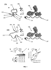
An example of an oligonucleotide probe 100a configured to produce a detectable
signal
upon target binding-induced segregation of two target binding moieties is
depicted in FIG. 1.
An aspect of the oligonucleotide probe of FIG. 1(a, top) is that the probe
100a has a stem-loop
structure formed by intramolecular hybridization of a single-stranded
oligonucleotide. In other
embodiments, the probe 100b may be stemless (FIG. 1(a, bottom)). In these
embodiments, the
probe 100b may have a lower gain than a probe that includes a stem due to an
increase in the
fluorescence background in the absence of target (FIG.1(a, bottom)). In
embodiments that
include a step-loop structure, the stem-loop structure of the probe 100a is
formed through
intramolecular hybridization between a first hybridization sequence 101 and a
second
hybridization sequence 102. Internal hybridization between first hybridization
sequence 101 and
second hybridization sequence 102 forms a duplex. First hybridization sequence
101 and
second hybridization sequence 102 are separated by a loop structure 103 formed
by the
oligonucleotide sequence between first hybridization sequence 101 and second
hybridization
sequence 102.
Regardless of whether the probe does or does not include a stem-loop
structure, the
oligonucleotide probe also includes a first target binding moiety 104 and a
second target binding
moiety 105. The first target binding moiety 104 and the second target binding
moiety 105 may
be directly or indirectly attached to the probe as described above. The
oligonucleotide probe
further includes a fluorophore 106 and a quencher 107. In FIG. 1, the
fluorophore 106 is
coupled to one end of the oligonucleotide strand of the probe and the quencher
107 is coupled to
the other end of the oligonucleotide strand of the probe. As described herein,
the fluorophore
and/or the quencher may be coupled to the oligonucleotide strand of the probe
at an internal site.
As shown in FIG. 1(a), in the absence of target binding to the target binding
moieties (104 and
105), the internal hybridization between the first and second hybridization
sequences (101 and
102) positions the fluorophore 106 adjacent the quencher 107, such that the
quencher 107
substantially suppresses detectable emissions from the fluorophore 106 (see
FIG. 2). As shown
in FIG. 1(a), binding of the target 108 (e.g., an antibody) to the first and
second target binding
moieties (104 and 105) causes a conformational change in the probe that
positions the
fluorophore 106 at a distance away from the quencher 107, such that the
fluorophore 106
produces a detectable signal (see FIG. 2).
In certain examples, the length of each stem duplex structure may be
different, as is also
the case with loop structures. Limits on the size of each duplex, each loop,
and the single-
stranded linear probe length are not contemplated as being rigidly limited but
are rather
application dependent. Optimal lengths for each of the probe components
described herein may
21
CA 02818556 2013-05-17
WO 2012/071344
PCT/US2011/061701
be determined without undue experimentation by one of skill in the art through
the teachings of
this specification. Lengths provided herein are examples only.
In certain embodiments, the probe is configured to have switching
thermodynamics (or
equilibrium) between the non-bound state (e.g., stem-loop structure) and the
target bound state
where the equilibrium is shifted to the non-bound state (e.g., stem-loop
structure) in the absence
of target binding (see FIG. 1(b)), without over-stabilizing this structure.
Over-stabilization of
the stem-loop may favor the binding of two targets, one on each of the target
binding moieties
on a probe, thus precluding opening of the stem and the signaling of the
probe. FIGS. 1(b) and
1(c) present different variants of probes with various switching
thermodynamics that are
optimized for different temperatures.
Another example of an oligonucleotide probe 400 configured to produce a
detectable
signal upon target binding is depicted in FIG. 4. Embodiments of probe 400
include the
modular attachment of the first target binding moiety 404 and the second
target binding moiety
405 to the probe. An aspect of the modular oligonucleotide probe of FIG. 4 is
that the probe has
a stem-loop structure formed by intramolecular hybridization of a single-
stranded
oligonucleotide. This stem-loop structure is formed through intramolecular
hybridization
between a first hybridization sequence 401 and a second hybridization sequence
402. Internal
hybridization between first hybridization sequence 401 and second
hybridization sequence 402
forms a duplex. First hybridization sequence 401 and second hybridization
sequence 402 are
separated by a loop structure 403 formed by the oligonucleotide sequence
between first
hybridization sequence 401 and second hybridization sequence 402.
The oligonucleotide probe also includes a first target binding moiety 404 and
a second
target binding moiety 405. The probe 400 also includes third hybridization
sequence 406 and
fourth hybridization sequence 407. The first target binding moiety 404 is
attached to a fifth
hybridization sequence 408, and the second target binding moiety 405 is
attached to a sixth
hybridization sequence 409. As depicted in FIG. 4(a), the first target binding
moiety 404 and
the second target binding moiety 405 are indirectly attached to the probe by
hybridization
between third hybridization sequence 406 and fifth hybridization sequence 408,
and by
hybridization between fourth hybridization sequence 407 and sixth
hybridization sequence 409,
respectively.
The oligonucleotide probe 400 further includes a fluorophore 410 and a
quencher 411.
In FIG. 4, the fluorophore 410 and the quencher 411 are coupled to the
oligonucleotide strand of
the probe at internal sites. As described herein, the fluorophore and/or the
quencher may be
coupled to the oligonucleotide strand of the probe at other sites, such as at
or near the end of the
oligonucleotide strand of the probe. As shown in FIG. 4(a), in the absence of
target binding to
22
CA 02818556 2013-05-17
WO 2012/071344
PCT/US2011/061701
the target binding moieties (404 and 405), the internal hybridization between
the first and second
hybridization sequences (401 and 402) positions the fluorophore 410 adjacent
the quencher 411
such that the quencher 411 substantially suppresses detectable emissions from
the fluorophore
410 (see FIG. 4(b)). Binding of the target to the first and second target
binding moieties (404
and 405) causes a conformational change in the probe 400 that positions the
fluorophore 410 at a
distance away from the quencher 411, such that the fluorophore 410 produces a
detectable signal
(see FIG. 4(b)).
Probes that utilize binding-induced reconstitution of recognition elements
Aspects of the present disclosure also include an oligonucleotide probe for
the detection
and quantification of a DNA binding protein in a sample. In some cases, the
probe includes two
or more recognition sequences, such as a first recognition sequence and a
second recognition
sequence. The first recognition sequence and the second recognition sequence
may be
complementary hybridization sequences. In certain instances, the first
recognition sequence and
the second recognition sequence are configured to form a recognition duplex by
intramolecular
hybridization of the first recognition sequence to the second recognition
sequence. The target
DNA binding protein may specifically recognize and bind to particular portions
of the probe, at
for example the recognition duplex.
The probe also includes two or more hybridization sequences (e.g.,
intramolecular
hybridization sequence, IHS) configured to allow the oligonucleotide strand to
undergo
intramolecular hybridization. For instance, the probe may include a first
hybridization sequence
(e.g., a first IHS) and a second hybridization sequence (e.g., a second IHS).
The first
hybridization sequence and the second hybridization sequence may be configured
to form a
second duplex in the absence of binding of the DNA binding protein to the
recognition duplex.
The first hybridization sequence and the second hybridization sequence may be
separated by a
loop structure formed by the oligonucleotide sequence of the probe that is
between the first
hybridization sequence and the second hybridization sequence. As such, in the
absence of
binding of the DNA binding protein to the recognition duplex, the probe may
adopt a stem-loop
conformation.
In addition, the probe includes a third hybridization sequence and a fourth
hybridization
sequence (e.g., third and fourth intramolecular hybridization sequences; a
third HIS and a fourth
IHS). The third hybridization sequence and the fourth hybridization sequence
may be
configured to allow the oligonucleotide strand to undergo an additional
intramolecular
hybridization. For instance, the third hybridization sequence and the fourth
hybridization
sequence may be configured to form a third duplex in the absence of binding of
the DNA
23
CA 02818556 2013-05-17
WO 2012/071344
PCT/US2011/061701
binding protein to the recognition duplex. The third hybridization sequence
and the fourth
hybridization sequence may be separated by a second loop structure formed by
the
oligonucleotide sequence of the probe that is between the third hybridization
sequence and the
fourth hybridization sequence. As such, in the absence of binding of the DNA
binding protein
In certain embodiments, the probe is configured to produce a detectable signal
when
contacted with the target DNA binding protein. In some instances, the probe
includes one or
more signaling moieties. For example, the probe may include two signaling
moieties, such as a
first signaling moiety and a second signaling moiety. In certain embodiments,
the first signaling
moiety is held at distance in close proximity to the second signaling moiety,
such as adjacent the
second signaling moiety, by complementary intramolecular base-pairing within
the probe as
described above (e.g., by formation of the second and third duplexes to
produce a probe with a
double stem-loop conformation). Under conditions in the absence of the DNA
binding protein,
the distance the first signaling moiety is held from the second signaling
moiety is sufficient to
minimize, suppress, or prevent the first signaling moiety from emitting a
detectable signal.
When the DNA binding protein is present and binds to the recognition duplex of
the probe, the
internal hybridization of the probe is disrupted (e.g., the double stem-loop
conformation of the
probe is disrupted). Disruption of the internal hybridization allows the end
of the nucleotide
chain to which the first signaling moiety is attached to move to a distance
further away from the
second signaling moiety. Under conditions in the presence of the DNA binding
protein, the
distance the first signaling moiety moves away from the second signaling
moiety is sufficient to
allow the first signaling moiety to emit a detectable signal.
As described above, in the absence of binding of the DNA binding protein to
the
recognition duplex, the probe may be in a double stem-loop configuration. In
these cases, the
probe may adopt a conformation where the first signaling moiety is positioned
adjacent the
second signaling moiety, such that the probe does not produce a detectable
signal. For example,
the first signaling moiety may be a fluorophore and the second signaling
moiety may be a
quencher. In these instances, under conditions in the absence of the DNA
binding protein, the
distance the fluorophore is held from the quencher is sufficient to minimize,
suppress, or prevent
the fluorophore from emitting a detectable signal. When the DNA binding
protein is present and
binds to the recognition duplex of the probe, a conformational change in the
probe is produced
such that the fluorophore is able to move to a distance further away from the
quencher. Under
conditions in the presence of the DNA binding protein, the distance the
fluorophore moves away
from the quencher is sufficient to allow the fluorophore to emit a detectable
signal.
24
CA 02818556 2013-05-17
WO 2012/071344
PCT/US2011/061701
DNA binding proteins that may be detected using the oligonucleotide probes may
include, but are not limited to, proteins or peptides that specifically bind
to DNA, such as
transcription factors (e.g., TATA binding protein (TBP), Myc-Max, NF-KB,
etc.), and the like.
An example of an oligonucleotide probe 1100 for detecting a DNA binding
protein is
depicted in FIG. 11. An aspect of the oligonucleotide probe of FIG. 11 is that
the probe has a
double stem-loop structure formed by intramolecular hybridization of a single-
stranded
oligonucleotide. This stem-loop structure is formed through intramolecular
hybridization
between a first hybridization sequence 1101 and a second hybridization
sequence 1102 to form a
first duplex, and intramolecular hybridization between a third hybridization
sequence 1103 and a
fourth hybridization sequence 1104 to form a second duplex. First
hybridization sequence 1101
and second hybridization sequence 1102 are separated by a first loop structure
1105 formed by
the oligonucleotide sequence between first hybridization sequence 1101 and
second
hybridization sequence 1102. Third hybridization sequence 1103 and fourth
hybridization
sequence 1104 are separated by a second loop structure 1106 formed by the
oligonucleotide
sequence between third hybridization sequence 1103 and fourth hybridization
sequence 1104.
In the absence of target binding to the probe, the probe may adopt a double
stem-loop
conformation that includes formation of the first and second duplexes as
described above. The
probe also includes a first recognition sequence 1107 and a second recognition
sequence 1108.
The first recognition sequence 1107 is formed by a portion of the first loop
structure 1105, and
the second recognition sequence 1108 is formed by a portion of the second loop
structure 1106.
The first recognition sequence 1107 and the second recognition sequence 1108
are
complementary sequences that may hybridize to each other to form a recognition
duplex. The
conformation of the probe is in equilibrium between formation of the double
stem-loop
conformation and formation of the recognition duplex. In the absence of target
1109 binding to
the probe 1100, the probe 1100 favors formation of the double stem-loop
configuration, where
the first recognition sequence 1107 is not hybridized to the second
recognition sequence 1108.
In the presence of target 1109, the equilibrium may be shifted towards the
formation of the
recognition duplex by target 1109 binding to the recognition duplex.
The oligonucleotide probe 1100 further includes a fluorophore 1110 and a
quencher
1111. In FIG. 11, the fluorophore 1110 is coupled to one end of the
oligonucleotide strand of
the probe and the quencher 1111 is coupled to an internal site of the
oligonucleotide strand of
the probe. As described herein, the fluorophore may be coupled to the
oligonucleotide strand of
the probe at an internal site, and the quencher may be coupled to one end of
the oligonucleotide
strand of the probe. As shown in FIG. 11(a), in the absence of target 1109
binding to the
recognition duplex, the internal hybridization between the first and second
hybridization
CA 02818556 2013-05-17
WO 2012/071344
PCT/US2011/061701
sequences (1101 and 1102) and the internal hybridization between the third and
fourth
hybridization sequences (1103 and 1104) positions the fluorophore 1110
adjacent the quencher
1111 such that the quencher 1111 substantially suppresses detectable emissions
from the
fluorophore 1110 (see also FIGS. 12 and 13). As shown in FIG. 11(a), binding
of the target
1109 (e.g., a DNA binding protein, such as a transcription factor) to the
recognition duplex
causes a conformational change in the probe 1100 that positions the
fluorophore 1110 at a
distance away from the quencher 1111, such that the fluorophore 1110 produces
a detectable
signal (see also FIGS. 12 and 13).
In certain embodiments, the probe is configured to have switching
thermodynamics (or
equilibrium constant, Ks) between the non-bound state (e.g., double-stem-loop
structure) and the
target bound state (e.g., single stem-loop structure) where the equilibrium is
shifted to the non-
bound state (e.g., double-stem-loop structure) in absence of target binding
(see FIG. 11(a)),
without over-stabilizing this structure. Over-stabilization of the double-stem-
loop may lead to a
reduction of the affinity of the probe for the target (see FIGS. 11(b) and
11(c)). FIGS. 11(b) and
11(c) present different variants of probes with various switching
thermodynamics that display
various gain and affinity for the target. In certain embodiments, the Ks
ranges from 0.001 to 10,
such as from 0.01 to 5, including from 0.1 to 2, or from 0.1 to 1.
Probes Configured to Produce Other Types of Signals upon Target Binding
In certain embodiments, the probe is configured to produce a signal change
through
different signal output mechanisms. In some cases, the probe includes two
target binding
moieties, such as a first target binding moiety and a second target binding
moiety. Various
signaling moieties may be used, where the first and second signaling moieties
produce a
detectable signal change upon target binding. The detectable change in signal
includes, but is
not limited to, a detectable signal decrease when a target specifically binds
to the probe to form a
target-probe hybrid, or a detectable increase in signal when a target
specifically binds to the
probe to form a target-probe hybrid. In certain embodiments, the first
signaling moiety is a
detectable reporter and the second signaling moiety is a detector configured
to detect the
reporter. Suitable reporters may include reporters that are detectable by the
detector, such as,
but not limited to, electrochemical reporters, magnetic reporters, and the
like.
In certain embodiments, the first signaling moiety is an electrochemical
reporter and the
second signaling moiety is an electrode. In some cases, the use of an
electrochemical reporter
and an electrode as the signaling moieties may facilitate target detection
directly in whole blood
or other complex clinical, food and environmental samples. In certain
instances, under
conditions in the absence of target, the distance the electrochemical reporter
is held from the
26
CA 02818556 2013-05-17
WO 2012/071344
PCT/US2011/061701
electrode is sufficient to produce a detectable signal (see e.g., FIG. 8). In
certain embodiments,
when target is present and binds to both the target binding moieties of the
probe, the internal
hybridization of the probe is disrupted such that the electrochemical reporter
is able to move to a
distance further away from the electrode (see e.g., FIG. 8). Under these
conditions, in the
presence of target binding, the distance the electrochemical reporter moves
away from the
electrode is sufficient to produce a detectable change in the signal, such as
a detectable decrease
in the signal (see e.g., FIG. 8). In other embodiments, under conditions in
the absence of target,
the electrochemical reporter is held a distance away from the electrode, such
that a detectable
signal is not produced or a low detectable signal is produced (see e.g., FIG.
14). In certain
embodiments, when target is present and binds to the target binding moiety of
the probe, the
electrochemical reporter is positioned adjacent to the electrode (see e.g.,
FIG. 14). Under these
conditions, in the presence of target binding, positioning the electrochemical
reporter adjacent to
the electrode is sufficient to produce a detectable change in the signal, such
as a detectable
increase in the signal (see e.g., FIG. 14).
In some instances, the probe may be attached to the surface of a substrate. As
described
above, the first signaling moiety may be an electrochemical reporter and the
second signaling
moiety may be an electrode. As such, the probe may be attached to the surface
of the electrode.
The probe may be attached by any convenient attachment method suitable for
attachment of the
oligonucleotide probe to the surface of the substrate. For example, the probe
may include
modified nucleotides configured to be attached to the surface of the
substrate, such as, but not
limited to nucleotides modified to include a thiol group. The probe may be
attached to the
surface of the substrate directly, such as by covalent attachment of the probe
to the surface of the
substrate, or indirectly, such as through a linker moiety or by affinity
binding (e.g., through
streptavidin-avidin complex formation, and the like).
An example of an oligonucleotide probe 800 configured to produce a detectable
electrochemical signal change upon target binding is depicted in FIG. 8. An
aspect of the
oligonucleotide probe of FIG. 8 is that the probe has a stem-loop structure
formed by
intramolecular hybridization of a single-stranded oligonucleotide. This stem-
loop structure is
formed through intramolecular hybridization between a first hybridization
sequence 801 and a
second hybridization sequence 802. Internal hybridization between first
hybridization sequence
801 and second hybridization sequence 802 forms a duplex. First hybridization
sequence 801
and second hybridization sequence 802 are separated by a loop structure 803
formed by the
oligonucleotide sequence between first hybridization sequence 801 and second
hybridization
sequence 802.
27
CA 02818556 2013-05-17
WO 2012/071344
PCT/US2011/061701
The oligonucleotide probe also includes a first target binding moiety 804 and
a second
target binding moiety 805. The probe 800 also includes third hybridization
sequence 806 and
fourth hybridization sequence 807. The first target binding moiety 804 is
attached to a fifth
hybridization sequence 808, and the second target binding moiety 805 is
attached to a sixth
hybridization sequence 809. As depicted in FIG. 8(a), the first target binding
moiety 804 and
the second target binding moiety 805 are indirectly attached to the probe by
hybridization
between third hybridization sequence 806 and fifth hybridization sequence 808,
and by
hybridization between fourth hybridization sequence 807 and sixth
hybridization sequence 809,
respectively.
The oligonucleotide probe 800 further includes an electrochemical reporter 810
(e.g.,
methylene blue). The probe 800 is attached to an electrode 811. In FIG. 8, the
electrochemical
reporter 810 is coupled to the probe at an internal site. In addition, the
probe is attached to the
electrode 811 at an internal site. As described herein, the electrochemical
reporter and/or the
electrode may be coupled to the oligonucleotide strand of the probe at other
sites, such as at or
near the end of the oligonucleotide strand of the probe. As shown in FIG.
8(a), in the absence of
target binding to the target binding moieties (804 and 805), the internal
hybridization between
the first and second hybridization sequences (801 and 802) positions the
electrochemical
reporter 810 adjacent the electrode 811 such that the probe produces a
detectable signal (see
FIGS. 8(c) and 8(d)). As shown in FIG. 8(a), binding of the target 812 (e.g.,
an antibody) to the
first and second target binding moieties (804 and 805) causes a conformational
change in the
probe 800 that positions the electrochemical reporter 810 at a distance away
from the electrode
811, such that the probe produces a detectable signal decrease (see FIGS. 8(c)
and 8(d)).
As described above, in the absence of target binding to the target binding
moieties, the
probe may also be in a flexible conformation (e.g., without a stem-loop
structure) (FIG. 10 (a)).
In these cases, the first signaling moiety can transiently move into proximity
with or bind to the
second signaling moiety, such that the signal from these signaling moieties
changes (FIG. 10
(b)). For example, the first signaling moiety may be an electrode and the
second signaling
moiety may be an electrochemical reporter. In these instances, under
conditions in the absence
of target, positioning the electrochemical reporter proximal to the electrode
is sufficient to
produce a detectable signal. When target is present and binds to the target
binding moieties of
the probe, the probe may be in a conformation where the electrochemical
reporter is positioned
such that does not approach the electrode as readily or as frequently. Under
conditions in the
presence of target binding, the distance the electrochemical reporter moves
away from the
electrode may produce a detectable current signal change (FIGS. 10(c) and
10(d)).
28
CA 02818556 2013-05-17
WO 2012/071344
PCT/US2011/061701
Different signal output mechanisms may be also applied for the detection and
quantification of a DNA binding protein in a sample. An example of an
oligonucleotide probe
1400 for detecting a DNA binding protein through electrochemical detection is
depicted in FIG.
14(top). The oligonucleotide probe 1400 of FIG. 14 has a double stem-loop
structure formed by
intramolecular hybridization of a single-stranded oligonucleotide. This double
stem-loop
structure is formed through intramolecular hybridization between a first
hybridization sequence
1401 and a second hybridization sequence 1402 to form a first duplex, and
intramolecular
hybridization between a third hybridization sequence 1403 and a fourth
hybridization sequence
1404 to form a second duplex. First hybridization sequence 1401 and second
hybridization
sequence 1402 are separated by a first loop structure 1405 formed by the
oligonucleotide
sequence between first hybridization sequence 1401 and second hybridization
sequence 1402.
Third hybridization sequence 1403 and fourth hybridization sequence 1404 are
separated by a
second loop structure 1406 formed by the oligonucleotide sequence between
third hybridization
sequence 1403 and fourth hybridization sequence 1404. In the absence of target
binding to the
probe, the probe may adopt a double stem-loop conformation that includes
formation of the first
and second duplexes as described above. The probe also includes a first
recognition sequence
1407 and a second recognition sequence 1408. The first recognition sequence
1407 is formed
by at least a portion of the first loop structure 1405, and the second
recognition sequence 1408 is
formed by at least a portion of the second loop structure 1406. The first
recognition sequence
1407 and the second recognition sequence 1408 are complementary sequences that
may
hybridize to each other to form a recognition duplex. The conformation of the
probe is in
equilibrium between formation of the double stem-loop conformation and
formation of the
recognition duplex. In the absence of target 1409 binding to the probe 1400,
the probe 1400
favors formation of the double stem-loop configuration, where the first
recognition sequence
1407 is not hybridized to the second recognition sequence 1408. In the
presence of target 1409,
the equilibrium may be shifted towards the formation of the recognition duplex
by target 1409
binding to the recognition duplex. The oligonucleotide probe 1400 further
includes an
electrochemical reporter 1410 (e.g., methylene blue). In FIG. 14(top), the
electrochemical
reporter 1410 is coupled to the probe at an internal site. In addition, the
probe is attached to the
electrode 1411 at one end. As described herein, the electrochemical reporter
and/or the
electrode may be coupled to the oligonucleotide strand of the probe at other
sites, such as at or
near the end of the oligonucleotide strand of the probe. As shown in FIG.
14(top), in the
absence of target 1409 binding to the recognition duplex, the internal
hybridization between the
first and second hybridization sequences (1401 and 1402) and the internal
hybridization between
the third and fourth hybridization sequences (1403 and 1104) positions the
electrochemical
29
CA 02818556 2013-05-17
WO 2012/071344
PCT/US2011/061701
reporter 1410 at a distance away from the electrode 1411, such that the
electrochemical reporter
1410 shows a decreased electron transfer rate (e.g., a low or not
significantly detectable
electrochemical signal) (see FIG. 14(top)). As shown in FIG. 14(top), binding
of the target 1409
(e.g., a DNA binding protein, such as a transcription factor, TF) to the
recognition duplex causes
a conformational change in the probe 1400 that positions the electrochemical
reporter 1410
adjacent the electrode 1411, such that the electrochemical reporter 1410
produces a greater
detectable electrochemical signal compared to the signal in the absence of
target 1409 (see
FIGS. 14(a) and 14(b)). The probe 1400 may also include a fifth hybridization
sequence 1412
and a sixth hybridization sequence 1413 configured to hybridize to each other
to form a third
duplex. The third duplex may be adjacent the electrochemical reporter 1410. In
the absence of
target 1409 binding to the recognition duplex, the fifth hybridization
sequence 1412 and the
sixth hybridization sequence 1413 are hybridized and position the
electrochemical reporter 1410
at a distance away from the electrode 1411 as described above. When target
1409 is bound to
the recognition duplex, a fourth duplex 1414 may be formed adjacent the
electrochemical
reporter 1410 that positions the electrochemical reporter adjacent the
electrode 1411 as
described above.
METHODS
Detection of Targets Using Oligonucleotide Probe-Based Detectors
Provided are methods for detecting the presence of a target in a sample using
unimolecular oligonucleotide probe-based detectors. Aspects of the methods
include contacting
a sample suspected of containing a target with a probe of the present
disclosure under conditions
that allow target that may be present in the sample to specifically bind to
the target binding
moieties of the probe. Binding of the target to the probe causes a
conformational change in the
probe, which in turn produces a detectable signal or a detectable change in a
signal. For
example, binding of the target to the probe may position a fluorophore at a
distance away from a
quencher sufficient to allow a signal of the fluorophore to be detectable. In
other embodiments,
binding of the target to the probe may position a reporter moiety at a
distance away from a
detector sufficient to allow a change in a signal from the detector to be
detectable.
The detectable signal or the detectable change in signal may be compared to
control
readouts from control samples that do not contain target or to results from
samples that contain
targets that do not specifically bind to the target binding moieties of the
probe (e.g., negative
controls). In other embodiments, the signal detected by the detector may be
optionally
compared to control readouts for control samples that contain target or a
known amount of target
(e.g., positive controls). Numerous alternative controls may be performed
individually or in
CA 02818556 2013-05-17
WO 2012/071344
PCT/US2011/061701
combination, as is known to those of skill in the art. For example, the
control may be to
challenge the probe with a surrogate solution absent the sample, and thus
lacking target.
Alternatively, the control may be a solution containing a target derivative
that may have
similarity to the actual target, but is normally not recognized and
specifically bound by the probe
under specific binding or "stringent" conditions.
Suitable samples include bodily fluids (e.g., blood, urine, interstitial
fluid, lachrymal
fluid, sweat, saliva, and the like), water, cell extracts, cell suspensions,
secretions, solvents, and
other aqueous and organic liquid solutions, suspension or emulsions capable of
including the
target of the probe of the detector. Samples may also include complex samples,
such as, but not
limited to, whole blood, crude nuclear extracts, and the like. In certain
embodiments, the
probes of the present disclosure are oligonucleotides that include target
binding moieties, such
as antigens, that specifically bind to target antibodies. In other
embodiments, the probes are
oligonucleotides that include complementary intramolecular hybridization
sequences that
hybridize to form a recognition duplex, which is specifically bound by a
target DNA binding
protein. In certain embodiments, the probes of the present disclosure are
oligonucleotides that
include target binding moieties, such as polypeptides, that specifically bind
to target
macromolecules. In certain embodiments, the probes of the present disclosure
are
oligonucleotides that include target binding moieties, such as aptamers, that
specifically bind to
target macromolecules.
Reaction Conditions and Detection Methods
The methods disclosed herein may be carried out in any reaction medium that
allows
specific binding between probe and, if present, target as defined herein. In
cases where the
sample contains target that specifically binds to the target binding moieties
of the probe, specific
binding between the target binding moieties of the probe and the target is
favored over
intramolecular hybridization between the internal hybridization sequences of
the probe. In cases
where the sample contains targets that do not specifically bind to the target
binding moieties of
the probe, intramolecular hybridization between the internal hybridization
sequences of the
probe is favored over binding between the probe and mismatched target.
Binding reactions involving the probes disclosed herein may be carried out in
the
presence of agents and additives that promote the desired specific binding,
diminish nonspecific
background interactions, inhibit the growth of microorganisms, or increase the
stability of the
probe and/or target. Binding reactions of the disclosure may be carried out at
ambient
temperature, although any temperature in the range allowing specific binding
may be used. For
instance in some embodiments, the temperature range is from 5 C to 45 C,
such as from 10 C
31
CA 02818556 2013-05-17
WO 2012/071344
PCT/US2011/061701
to 40 C, or from 20 C to 30 C. In addition, in some embodiments, the pH of
the binding
reaction medium is about physiological pH. For example, the pH may range from
4 to 10, such
as 5 to 9, including 6 to 8. In certain cases, the pH may be 7. For
convenience, reaction
conditions may be chosen to allow specific binding to occur as rapidly as
possible. Binding
times as short as seconds (e.g., 1 to 60 seconds), or minutes (e.g. 1 to 30
minutes) may be
employed. By way of example, times of 1 to 60 seconds, such as 10 to 60
seconds, including 20
to 60 seconds may be used. In other embodiments, times of 1 to 30 minutes,
such as 5 to 20
minutes, including 10 to 20 minutes may be used.
Multiplexing
In certain embodiments, the methods may include multiplex detection of
targets. The
terms "multiplex" or "multiplexing" as used herein refer to using multiple
distinct signaling
moieties, such that a single assay may be used to detect the presence of
different targets in a
single sample.
For example, in embodiments that include fluorescent signaling moieties, the
system
may include multiple fluorescently distinct fluorophores, such that a single
assay may include
multiple probes each with different fluorophores. Fluorophores of these
embodiments emit
detectable signals at different wavelengths. Multiplexing facilitates the
detection of different
targets of interest within a single sample (e.g., targets that specifically
bind to different target
binding moieties). In these embodiments, a mixture of differentially labeled
probes (e.g., a first
probe with a first fluorophore and a second probe with a second fluorophore)
may be contacted
with a sample that includes one or more different targets of interest. For
example, a first target
may bind to a first target binding moiety and a second target binding moiety
of a first probe, as
described above. A second target may bind to a third target binding moiety and
a fourth target
binding moiety of a second probe. Upon binding of the first target to the
first and second target
binding moieties of the first probe, a conformational change is induced in the
first probe such
that the first probe produces a detectable first signal. In addition, upon
binding of the second
target to the third and fourth target binding moieties of the second probe, a
conformational
change is induced in the second probe such that the second probe produces a
second detectable
signal that is distinct from the first signal. Both the first signal and the
second signal may be
detected, thus indicating the presence (or absence) of the first target and
the second target in the
sample. In certain embodiments, multiplexing may be used in reactions that
include unbound
probes in solution. In other embodiments, multiplexing may be used in systems
comprising
arrays or addressable arrays of probes attached to a substrate surface.
32
CA 02818556 2013-05-17
WO 2012/071344
PCT/US2011/061701
Similarly, multiplexing may be applied to embodiments that include
electrochemical
signaling moieties. In these embodiments, two or more different probes may be
contacted with
a sample that includes one or more different targets of interest. For example,
the system may
include a first probe that includes a first and a second target binding
moieties and a second probe
with a third and a fourth target binding moieties. The first and the second
target binding
moieties may be different from the third and the fourth target binding
moieties, such that a first
target binds to the first and second target binding moieties and a second
target binds to the third
and fourth target binding moieties. Upon binding of the first target to the
first and second target
binding moieties of the first probe, a conformational change is induced in the
first probe such
that the first probe produces a detectable change in a first signal. In
addition, upon binding of
the second target to the third and fourth target binding moieties of the
second probe, a
conformational change is induced in the second probe such that the second
probe produces a
detectable change in a second signal that is distinct from the first signal.
The changes in the first
and second signals may be detected, thus indicating the presence (or absence)
of the first target
and the second target in the sample.
UTILITY
The subject systems and methods find use in a variety of different
applications where
determination of the presence or absence, and/or quantification of one or more
targets in a
sample is desired. In certain embodiments, the methods are directed to the
detection of proteins,
carbohydrates, nucleic acids, lipids, peptides, enzymes or other biomolecules
in a sample.
Samples may include, but are not limited to, blood, plasma, serum, or other
bodily fluids or
excretions, such as but not limited to, urine, saliva, semen, prostatic fluid,
nipple aspirate fluid,
lachrymal fluid, perspiration, feces, cheek swabs, cerebrospinal fluid, cell
lysate samples,
amniotic fluid, gastrointestinal fluid, biopsy tissue, and the like.
The presence or absence of a target in a sample or significant changes in the
concentration of a target over time can be used to diagnose disease risk,
presence of disease in
an individual, or to tailor treatments for the disease in an individual. For
example, the presence
of a particular target or panel of targets may influence the choices of drug
treatment or
administration regimes given to an individual. In evaluating potential drug
therapies, the
presence, absence, or concentration of a target may be used as a surrogate for
a natural endpoint
such as survival or irreversible morbidity. If a treatment alters the target,
which has a direct
connection to improved health, the target can serve as a surrogate endpoint
for evaluating the
clinical benefit of a particular treatment or administration regime. Thus,
personalized diagnosis
and treatment based on the particular target or panel of targets detected in
an individual are
33
CA 02818556 2013-05-17
WO 2012/071344
PCT/US2011/061701
facilitated by the subject systems and methods. Furthermore, the early
detection of targets
associated with diseases is facilitated by the high sensitivity of the subject
systems and methods,
as described above. Due to the multiplex capability of detecting multiple
targets in a single
assay, combined with selectivity, sensitivity and ease of use, the presently
disclosed systems
and methods find use in quantitative, point-of-care or near-patient bio-
molecular assays.
The subject systems and methods find use in diagnostic assays, such as, but
not limited
to, the following: detecting and/or quantifying targets, as described above;
screening assays,
where samples are tested at regular intervals for asymptomatic subjects;
prognostic assays,
where the presence and or quantity of a target is used to predict a likely
disease course;
stratification assays, where a subject's response to different drug treatments
can be predicted;
efficacy assays, where the efficacy of a drug treatment is monitored; and the
like.
The subject devices, systems and methods also find use in validation assays.
For
example, validation assays may be used to validate or confirm that a potential
disease biomarker
is a reliable indicator of the presence or absence of a disease across a
variety of individuals. The
short assay times for the subject systems and methods may facilitate an
increase in the
throughput for screening a plurality of samples in a minimum amount of time.
In certain embodiments, the subject systems and methods find use in detecting
antibodies
in a sample. In some cases, the subject systems and methods may be used to
detect the presence
or absence of particular antibodies, as well as an increase or decrease in the
concentration of
particular antibodies in a sample.
In certain embodiments, the subject systems and methods find use in detecting
DNA
binding proteins, such as transcription factors. The subject systems and
methods may be used to
detect the presence or absence of particular DNA binding proteins, as well as
an increase or
decrease in the concentration of particular DNA binding proteins in a sample.
For example,
given their nanoscale size and their ability to provide a readout signal
without the need of
additional reagents, transcription factor probes could be transferred into a
cell nucleus (e.g., by
transfection, coupled with an appended nuclear localization peptide) allowing
fluorescent
microscopy to track the concentration of active transcription factor during
cell growth or upon
administration of a drug compound. Resonance energy transfer-based signaling
fluorophores
may be used, as the ratiometric nature of these types of fluorophores corrects
for varying probe
concentrations. Transcription factor probes may also find use in drug
screening assays, for
example by ensuring that the optical signal changes obtained due to the
presence of a specific
drug are specifically linked to the DNA-protein target interaction and not
solely attributable to
off-target drug interactions. Transcription factor probes may also be amenable
to other signaling
34
CA 02818556 2013-05-17
WO 2012/071344
PCT/US2011/061701
mechanisms, such as, but not limited to, electrochemical signaling moieties as
described above,
which may facilitate use of these probes in complex samples.
KITS
Also provided are kits that find use in practicing the subject methods, as
described
above. For example, kits and systems for practicing the subject methods may
include one or
more systems of the present disclosure, which may include one or more probes.
As such, in
certain embodiments the kits may include a solution or suspension of the
probes in an aqueous
or other compatible solution. The one or more probes may be provided in
separate containers
with each container including a single type of probe, or may be provided in a
container that
includes a mixture of two or more types of probes. In other embodiments, the
kits may include
one or more probes immobilized on the surface of a substrate forming an
addressable array of
probes as described above.
In addition to the above components, the subject kits may further include
instructions for
practicing the subject methods. These instructions may be present in the
subject kits in a variety
of forms, one or more of which may be present in the kit. One form in which
these instructions
may be present is as printed information on a suitable medium or substrate,
e.g., a piece or
pieces of paper on which the information is printed, in the packaging of the
kit, in a package
insert, etc. Another means would be a computer readable medium, e.g.,
diskette, CD, DVD,
Blu-ray, computer-readable memory, etc., on which the information has been
recorded or stored.
Yet another means that may be present is a website address which may be used
via the Internet
to access the information at a removed site. Any convenient means may be
present in the kits.
As can be appreciated from the disclosure provided above, the present
disclosure has a
wide variety of applications. Accordingly, the following examples are offered
for illustration
purposes and are not intended to be construed as a limitation on the invention
in any way. Those
of skill in the art will readily recognize a variety of noncritical parameters
that could be changed
or modified to yield essentially similar results. Thus, the following examples
are put forth so as
to provide those of ordinary skill in the art with a complete disclosure and
description of how to
make and use the present invention, and are not intended to limit the scope of
what the inventors
regard as their invention nor are they intended to represent that the
experiments below are all or
the only experiments performed. Efforts have been made to ensure accuracy with
respect to
numbers used (e.g. amounts, temperature, etc.) but some experimental errors
and deviations
should be accounted for. Unless indicated otherwise, parts are parts by
weight, molecular
CA 02818556 2013-05-17
WO 2012/071344
PCT/US2011/061701
weight is weight average molecular weight, temperature is in degrees Celsius,
and pressure is at
or near atmospheric.
EXAMPLES
Fluorescent Probes
Fluorescent probes for antibody detection
The first probe was tested by using a DNA sequence that includes a stem-loop
with a
relatively weak stem-loop conformation with switching thermodynamics (Ks) of
about 1. A
stable, GC-rich stem was successively destabilizing by 1 kcal/mol increments
until a stem
sequence was identified that favored the stem-loop conformation without over
stabilizing the
stem-loop conformation (see FIG 1(b), construct 1MM with one "T-T" miss match
in the middle
of the stem) at room temperature, thus facilitating the binding-induced
opening of the stem-loop
structure of the probe. Over-stabilization of the probe may facilitate
undesired binding of two
molecules of the target to a single probe.
A pair of fluorescent antibody probes employing the haptens 2,4-dinitrophenol
(DNP)
and digoxigenin (Dig) as target binding moieties, and thus targeting anti-Dig
and anti-DNP
antibodies respectively were prepared (FIG. 2). The target binding moieties
were covalently
attached to the stems of the probe using two different strategies: Dig was
attached via amine
linkers on the C-5 positions of the two thymines in the middle of the double-
stranded stem, and
DNP was inserted within the stem via the introduction of additional
phosphodiester bonds (see
Material and Methods below). To facilitate antibody-driven conformational
changes in the
probe, the two strands of the stem were connected via an 18-base loop that
spanned the 12 nm
distance between the two antigen binding sites present on the target antibody.
6-
carboxyfluorescein (6 FAM) and Black Hole Quencher 1 (BHQ-1) were attached to
the 5' and
3' termini of the stem to produce a detectable fluorescent signal upon opening
of the stem-loop
structure of the probe.
The anti-Dig and anti-DNP antibody probes produced 65% and 130% increases in
fluorescence, respectively, at saturating target concentrations (FIG. 2(b)).
Both probes
responded to their respective target antibodies at 30 nM (FIG. 2(b)), and no
increase in
fluorescence was observed when the two antibodies were interchanged (FIG.
2(b), dotted line).
The two probes displayed low-nanomolar affinities and equilibration times of
one minute or less
(FIG. 2(c)), and achieved detection limits of 100 pM and 300 pM (e.g., 15 and
45 ng/ml) in less
than one minute for anti-Dig and anti-DNP antibody probes, respectively (FIG.
2(d)). The
probes were specific for their respective target antibodies as no
statistically significant increase
36
CA 02818556 2013-05-17
WO 2012/071344
PCT/US2011/061701
in fluorescence was observed when either probe was incubated with 30 nM of the
other's target
(FIG. 2(d), black dots) or when challenged with a 1000-fold higher
concentration (e.g., 3 uM) of
non-specific pooled human antibodies (FIG. 2(d)). The titrations and kinetic
traces shown in
FIG. 2 represent the average of at least three independent measurements, with
error bars
reflecting the average absolute deviation.
Probes were also tested in a competitive assay format in order to detect small
molecules
such as DNP. The DNP-modified probe (10 nM) loaded with a minimal
concentration of anti-
DNP antibodies (10 nM) was used to detect the presence of free DNP but not
free Dig (FIG. 3).
An anti-DNP antibody probe was first bound to its specific anti-DNP antibody.
In the presence
of free DNP in the sample (and not free Dig), the antibody bound to the free
DNP and was
released from the probe, thus leading to a decrease in fluorescence signal.
The apparent
inhibition constant (IC50%) of the free DNP (or apparent affinity of the probe-
bound antibody for
free DNP) was significantly higher (4-fold) than the dissociation constant of
the antibody-probe
complex itself (see FIG. 2(c), bottom), which reflected the cooperative
binding of a single
antibody to the two antigens on the probe. In FIG. 3, the "free" DNP and
"free" Dig were
covalently linked to small poly-thymine constructs (TTTT-antigen-TTT) in order
to insure that
the increased affinity observed between the antibody and its probe was not
attributable to the
presence of the chemical bond linking the antigen to the probe.
Another embodiment of the probe is shown in FIG. 4, which depicts a modular
unimolecular probe according to embodiments of the present disclosure. In this
modular probe,
the target binding moieties were indirectly attached to the probe by
hybridization to the probe of
two copies of a 17-base DNA strand modified with the target binding moiety
(FIG. 4(a)). The
stem-loop contained a frame inversion near one end to allow for symmetrical
labeling of two
copies of the same modified hybridization sequence. This may facilitate a
reduction in
fabrication cost and complexity. The modular probes were made using both Dig
and an 11-
residue polypeptide epitope from the HIV-1 protein gp41 as target binding
moieties (FIG. 4).
Specific detection of anti-Dig antibodies (FIG. 4(b), top) and anti-HIV
antibodies (FIG. 4(b),
bottom) were tested. Experimental results indicated that their gain, affinity,
specificity and
kinetics (FIGS. 4(b) and (c)) compare closely to those of the non-modular
probes described
above. The anti-HIV antibody probe, for example, achieved a 300 pM detection
limit (e.g., 45
ng/ml), a 12 second response time constant, and a dissociation constant of 4
2 nM.
Modular unimolecular probes were configured to produce a high detectable
fluorescent
signal upon target binding (see FIG.5). The optimal gain of the modular switch
at 37 C was
obtained when using a 3GC stem (e.g., a stem containing five Watson-Crick base-
pairs with 3
GC and 2 AT base pairs). This stem stability facilitated a minimization in the
background
37
CA 02818556 2013-05-17
WO 2012/071344
PCT/US2011/061701
fluorescence in the absence of antibody, while still allowing stem opening
upon binding to the
antibody.
Probes were also tested for the detection of targets of different sizes (see
FIG. 6 (top)).
The fluorescence signal of the modular oligonucleotide probe was correlated to
the size of the
binding moieties (X) and the target bound to them. FIG. 6 (bottom) shows a
schematic of the
signaling of the modular probe (e.g., the probe stem opening) in the presence
of two targets
binding to a single probe. In some instances, the probe stem opening may be
attributable to a
steric-hindrance mechanism linked to the binding of two targets to a single
probe (e.g., one
target bound at each binding moiety, X). In certain cases, binding of one
target to each binding
moiety facilitated detection of non-bidentate targets (for example PDGF),
e.g., a target that only
contains one binding site.
Experiments were also performed to test fluorescent probes for the detection
of targets
directly in blood serum. FIG. 7 (top) shows the detection of Dig-antibodies
directly in blood
serum at 37 C using a modular oligonucleotide probe with the 3GC stem (see
e.g., FIG. 5). The
fluorescent signaling moieties used for FIG. 7 (top) were Alexa Fluor-680 for
the fluorophore
and BHQ-2 for the quencher (blood serum has a low fluorescence in the
infrared). The
fluorescent probe showed a lower detectable signal in whole blood as compared
to blood serum
(FIG. 7(b)). FIG 7 (a) shows the detection of 10 nM Dig antibody using 10 nM
of probe in:
buffer (FIG 7(a)); whole blood (FIG 7(b), left); 1:10 whole blood (FIG. 7(b),
middle); and 1:100
whole blood (FIG 7(b), right). Fluorescence signal from the probe was not
significantly
detectable in whole blood or in a 1:10 whole blood sample due to high
absorbance of the
sample. Whole blood samples were diluted 100-fold or more to produce a
detectable
fluorescence signal (e.g., a final concentration of 10 nM of antibody after a
1:100 dilution
corresponds to an antibody concentration of 1 it.M in whole blood). For FIG.
7, panels a and b,
the fluorescent signaling moieties used were FAM-6 for the fluorophore and BHQ-
1 for the
quencher.
Fluorescent probes for DNA-binding protein detection
A DNA-based probe that binds to TATA binding protein (TBP) was made. The probe
included a DNA sequence that adopts both a "dark" double stem-loop structure
and a "bright"
fluorescent single stem-loop conformation, the latter of which includes a TBP
recognition
duplex (FIG. 11). The gain and affinity, and thus sensitivity, of the probes
may depend on the
thermodynamics of the conformational change between the double stem-loop
structure and the
single stem-loop structure. Probes were made that had various stabilities when
in the unbound
conformation (FIG. 11(b)). As predicted by the population-shift model of
conformational
38
CA 02818556 2013-05-17
WO 2012/071344
PCT/US2011/061701
change, probes with high equilibrium constants (e.g., >9.5, such that the
probes are >90% in the
binding competent state even in absence of their target) do not respond
appreciably after
addition of TBP (FIG 11(c)). Probes with equilibrium constants near unity, in
contrast, exhibit a
detectable difference in fluorescence in the presence of target (about twice
as intense as the
fluorescence observed in the absence of saturating target) and still retain
high target affinity (KD
= 36 6 nM). Further decreases in the equilibrium constant produce greater
fluorescence
changes upon target binding, but, as predicted by the population shift model,
affinity for the
target is reduced. For example, according to the model, a switching
equilibrium constant of
0.006 produces a dissociation constant of 1.1 p M. In certain embodiments, the
switching
equilibrium constant ranges from 1 to 0.1, depending on whether the probe is
configured for
achieving a low detection limit or for achieving a high signal gain (e.g., for
applications such as
in vivo imaging).
DNA-based switches for the quantitative detection of DNA binding proteins were
tested.
FIG. 11(a) shows DNA sequences recognized by transcription factors
(recognition sequences
1007 and 1008) that can be engineered into structure-switching probes by
stabilizing an
alternative double stem-loop conformation (FIG. 11(a), left). By adding a
quencher and
fluorophore at locations that experienced the largest distance separation upon
switching, binding
of the transcription factor to its specific double stranded DNA recognition
duplex shifts the
equilibrium of the sensor towards the fluorescent binding state. A Ks ranging
from 1 to 0.1
produced a probe configured to populate its darker state in absence of target
binding without
over-stabilizing this nonbinding state, which results in decrease affinity of
the probe as predicted
by the population-shift model. Predicted Ks were evaluated from the difference
in energy
predicted between both states using mfold while experimental Ks were
determined from the
fluorescence signal of the probe in absence of target.
The TBP-detecting transcription factor probe shown in FIG. 12 had a switching
equilibrium constant of 0.72 and produced a 300% increase in fluorescence in
the presence of
100 nM concentrations of TBP, but no significant increase in fluorescence when
in the presence
of similar concentrations of other transcription factors such as Myc-Max and
NF-KB. The TBP
sensor was sensitive and rapid, able to detect 10 nM target in 5 minutes or
less (FIG. 12, right).
Probes were also designed for the detection of the transcription factors Myc-
Max and
NFkB. Probe structures similar to those employed in the detection of TBP were
used (FIG. 12,
graphs in the middle and bottom rows). Using Myc-Max and NFkB-binding probes
with
switching thermodynamics similar to those used for TBP detection (Ks ¨ 0.3),
resulted in signal
gains, affinities, specificities, and kinetics similar to those achieved for
the detection of TBP
(FIG. 12, middle and bottom rows). Both the Myc-Max and NFkB probes showed
signal gains
39
CA 02818556 2013-05-17
WO 2012/071344
PCT/US2011/061701
of 300%, with KD of 53 12 nM and 134 41 nM, respectively. The probes were
specific and
exhibited only very low cross-reactivities (FIG. 12).
The ability of the TBP transcription factor probe to function in HeLa nuclear
cell extract
was also tested (FIG. 13). A probe directed against TBP was titrated with its
target protein
against a background of 250 ,g/mL crude HeLa nuclear extract (FIG. 13, top),
producing a
binding curve similar to that observed in buffer (see e.g., FIG. 12). The
probe had an apparent
dissociation constant that decreased from 45 3 nM in simple buffers to 36
6 nM in the
extract. Without being limited to any particular theory, a possible
explanation for this shift is
that the true TBP concentration in this experiment was 9 7 nM higher than
the concentration of
exogenous TBP added at each point in the titration due to the presence of
endogenous TBP. To
characterize this further, an unlabeled, double-stranded TBP recognition
oligonucleotide was
introduced as a competitor (DNAcomp) and a dose-dependent decrease in
fluorescence signal was
observed, which was consistent with the presence of endogenous TBP (FIG. 13,
top). The
introduction of an unlabeled, double-stranded TBP recognition oligonucleotide
as a competitor
(DNAcomp) generated a loss of probe fluorescence signal consistent with the
presence of
endogenous TBP.
Transcription factor probes were tested to determine the detection and
quantification of
transcription factors in crude nuclear extracts (FIG. 13, bottom). Such
quantification required
measurement of the fluorescence of the probe-containing extract: (1) in
equilibrium with the
endogenous transcription factor population contained in the sample, Fsmp; (2)
in the background
when no transcription factor was bound to the switch, Fbkg; and (3) when the
switch was fully
bound (e.g., saturated) with its transcription factor, Fõ,. Fsmp was
determined by adding 10 nM
of the relevant transcription factor probe to the sample, and then measuring
the fluorescence at
the relevant emission maxima of the fluorophore after the system had
substantially reached
equilibrium. To determine Fbkg and Fsw, the sample was spilt into two samples.
Fbkg was
determined in one sample by adding a saturating concentration (> 500 nM) of
unlabeled, double-
stranded recognition site that served as a competitor, liberating the free
probe. Fsa, was
determined via two strategies. First, by adding exogenous transcription factor
to saturation (e.g.,
until all of the probe was in the emissive state). Alternatively, a single-
stranded oligonucleotide
complementary to the two tails of a probe in its binding competent state was
added to drive the
equilibrium of the probe into its fully emissive conformation (e.g., the
target-bound state) (FIG.
13(a)). After Fbkg, Fsa, and Fsmp were determined, the concentration of the
target protein, C, was
determined by using the known dissociation constant of the probe, KD, (42 nm)
using the
following formula:
CA 02818556 2013-05-17
WO 2012/071344
PCT/US2011/061701
KD(Fsmp ¨ Fbkg)
C = ______________________________________________________________________
(1)
Fsat ¨ Fsmp
Using this approach, the endogenous TBP concentration in crude, 250 g/mL HeLa
nuclear extracts was 5.8 0.2 nM in 25% nuclear extract (250 it.g/mL). This
value was in
reasonable agreement with both our estimate above (based on the shift in
apparent dissociation
constant (FIG. 13, top) and with the expected concentration (¨ 3 nM) estimated
from the number
of copies of TBP per cell and the known number of cells in a given amount of
nuclear extract
(Borggrefe, T., et al., J. Biol. Chem. 2001, 276, pp. 47150).
Electrochemical Probes
Electrochemical probes for antibody detection
Electroactive contaminants are generally rare (FIG. 8), which may facilitate
electrochemical detection of targets in whole blood and other complex samples.
For example, in
the absence of target antibodies, the electrochemical readouts of probes
modified with either Dig
antigen or HIV peptide antigen increased only by 15-30% when transferred from
buffer to whole
blood (FIG. 8(e)). Electrochemical probes in which the fluorophore of the
fluorescent probe was
replaced with a thiol group for surface attachment, and the quencher of the
fluorescent probe
was replaced with a methylene blue redox reporter (FIG. 8(a)) were prepared.
In the absence of
target, the stem of the probe positioned the methylene blue electrochemical
reporter in proximity
to the electrode, promoting electron transfer and producing an increase in the
faradaic current.
Upon target binding, the methylene blue was positioned away from the
electrode, decreasing
electron transfer and generating a detectable current signal change (FIGS.
8(a) and 8(b)).
Electrochemical probes detected nanomolar concentrations of their targets
directly in
whole blood (FIGS. 8(b)-(d)). Blood doped with the relevant antibody targets
(30 nM) produced
a detectable decrease in the current signal from anti-Dig and anti-HIV
antibody sensors (FIG.
8(b)). Blood doped with a mixture of 30 nM of the other sensor's target
antibody and a 100-fold
higher concentration of random, pooled human IgGs produced no significant
change in the
observed current (FIG. 8(b), dotted line). The anti-Dig and anti-HIV probes
achieved 1 nM and
10 nM detection limits (e.g., 0.15 to 1.5 jig/ml), respectively (FIG. 8(c)),
which was well below
the serum concentrations typical of antibodies. These electrochemical probes
achieved
equilibration time constants of 5 mm or less (FIG. 8(d)). The electrochemical
probes did not
measurably respond to non-targeted monoclonal or polyclonal antibodies at 30
nM even when
mixed with a 3 !AM mixture of random human antibodies (FIG. 8(d)). The
titrations and kinetic
traces shown in FIG. 8 represent the average of measurements conducted with at
least four
41
CA 02818556 2013-05-17
WO 2012/071344
PCT/US2011/061701
independently fabricated sensors, with error bars reflecting the average
absolute deviation.
Shown in FIG. 8(e) are square wave voltammograms for anti-Dig antibody and
anti-HIV
antibody probes in buffer and 80% whole blood. The graphs shown in FIG. 8(e)
show how
electrochemical probes were relatively insensitive to the presence of whole
blood.
Stability of the electrochemical probe (e.g., a unimolecular probe indirectly
bound with
Dig antigen) in whole blood and blood serum was tested (FIG. 9). The
background of faradaic
current of the sensor was reduced linearly by 6.6 % and 8.4 % per hour in
whole blood and
serum, respectively. The decrease in the background of faradaic current may
have been due to
the activity of DNA nucleases.
Stem-less probes with the target binding moieties at various locations (FIG.
10(a)) were
also made and tested. The stem-less probes were labeled with an antigenic
peptide epitope
sequence and included an electrochemical reporter and an electrode as the
signaling moieties. In
the absence of target binding, the electrochemical reporter was able to be
positioned proximal to
the electrode, and thus transfer electrons with the electrode surface, thus
generated a relatively
large current (FIG. 10(b)). Specific target binding of, for example the anti-
Flag antibody to the
epitope recognition moieties, caused the flexible probe to switch to a
substantially rigid, target-
bound structure, thus producing a decrease in the signal (FIG. 10(c)) by about
80% with
saturating antibody concentrations (FIG. 10(d)).
Electrochemical probes for DNA-binding protein detection
A DNA-based electrochemical probe for the detection of TATA binding protein
(TBP)
was also made. The probe included a DNA sequence that adopts both a "low-
current-signal"
double stem-loop structure and a "high-current-signal" single stem-loop
conformation, the latter
of which includes a TBP recognition duplex (FIG. 14). FIG. 14(top) shows DNA
sequences
recognized by the TBP transcription factor (recognition sequences 1407 and
1408) that can be
engineered into structure-switching probes by stabilizing an alternative
double stem-loop
conformation (FIG. 14(top), left). The probe was also internally modified with
an
electrochemical reporter (e.g., methylene blue) and at one end with a thiol
group for
immobilization to a gold electrode surface. In the absence of the target DNA
binding protein
TBP the double stem-loop structure (FIG. 14(top), left) positioned the
methylene blue
electrochemical reporter at a distance away from the electrode surface, thus
decreasing the
electron transfer rate and resulting in a low current signal. Upon TBP binding
(FIG. 14(top),
right), the DNA probe was shifted towards the single stem-loop conformation
and the
electrochemical reporter was positioned in close proximity with the electrode
surface. This, in
turn, resulted in an increase in the current signal proportional to the
concentration of the TBP
42
CA 02818556 2013-05-17
WO 2012/071344
PCT/US2011/061701
target (FIG. 14(b). The TBP electrochemical sensor was sensitive and rapid,
able to detect
nanomolar concentrations of target in 5 minutes or less (FIG. 14(b)).
The ability of the TBP transcription factor electrochemical probe to function
in HeLa
nuclear cell extract was also tested (FIG. 14(b). The probe directed against
TBP was titrated
with its target protein against a background of 250 ug/mL crude HeLa nuclear
extract (FIG.
14(b), producing a binding curve similar to that observed in buffer (FIG.
14(b). This binding
curve was slightly offset from the curve obtained in buffer. Without being
limited to any
particular theory, a possible explanation for this shift was that this was a
contribution from the
endogenous TBP present in cell extract. The TBP transcription factor
electrochemical probe
provided the convenient quantification of transcription factors in cell
extract. The sensor's
response in buffer, cell extract and, finally, extract to which a large excess
of TBP had been
added were measured. These measurements were used to calculate the
concentration of TF in
the sample, C, via the following relationship:
C = K D (S õmp Smin )
smax ¨ Ssamp
where Smin is the minimum signal response, Smaõ is the maximum signal
response, and
Sõ,,p is the signal response seen in the sample of interest. By performing a
sequential addition
and electrochemical measurement series in one sensor, measuring buffer,
extract, and after
further addition of 1 TBP, the endogenous TBP concentration was determined
to be 4 2
nM, which was in close agreement with the results of prior studies.
Material and methods
Fluorescent probes: HPLC purified DNAs modified with 5' -FAM, 3'-BHQ-1 and
either
Dig or DNP were purchased from IBA (Goettingen, Germany) and Biosearch
Technologies
(Novato, CA), respectively. All constructs possessed an additional adenine
base after the FAM-
and guanine nucleotide before the BHQ-1. Dig was inserted on a thymine
modified nucleotide
in the middle of the stem using a C8 linker (about 1 nm): 5' -FAM-
ACTT(Dig)TGTTTTTTTGCGTTTTTTTTCA-T(Dig)AGG-BHQ-3'. DNP was inserted
between two nucleotides at a similar location using a C12 linker (about 1.5
nm): 5' -FAM-
ACTT-DNP-TGTTTTTTTTTTTTTTTTTTCA-DNP-TAGG-3'.
Modular fluorescent probes: HPLC purified DNA containing a frame inversion and
modified internally with thymine-labeled FAM and BHQ-1; purchased from IBA
(Goettingen,
Germany): 5' -TGGATCGGCGTTTTATTTT(FAM)-
CCTTGTTTTTTTTTTTTTTTTTTCATGGT(BHQ)T-3' -3'TTATTTTGCGGCTAGGT-5'.
43
CA 02818556 2013-05-17
WO 2012/071344
PCT/US2011/061701
HPLC purified antigen-modified 17-base DNA sequences were obtained from IBA
(Goettingen,
Germany) (Dig-DNA): T(Dig)AATAAAACGCCGATCCA; and Bio-synthesis (Lewisville,
USA) (peptide-linker-DNA): ELLELDKWASLWNC-(SMCC-NH-(CH2)6P03)-AATAAAAC-
GCCGATCCA (HIV-1 gp41 epitope21 with a 4 amino acids linker -SMCC).
Fluorescent DNA binding protein probes: HPLC purified DNAs modified with 5'-
FAM, and internal BHQ-1 inserted on a thymine residue were purchased from IBA
(Goettingen,
Germany) and Biosearch Technologies (Novato, CA). DNA binding proteins TBP and
Myc-
Max were recombinantly expressed, purified, and characterized as previously
described
(Bonham 2009; Martinez 2004). Recombinant NFkB (p50 homo-dimer) was purchased
from
Electrochemical probes for antibody detection: HPLC purified DNA with a frame
inversion, and modified with internal C6-thiol and methylene blue (MB) was
purchased from
Biosearch Technologies (Novato, CA): 5' -TGGATCGGCGTTTTATTTTT(C6-Thiol)CCTTGT-
TTTTTTTTTTTTTTTTT-CATGG-T(MB)TT-3'-3'-TTATTTTGCGGC-TAGGT-5'.
Antibodies were purchase from Roche Diagnostic Corporation (Mannheim, Germany)
(Sheep polyclonal anti-digoxigenin and its Fab fragments), Sigma-Aldrich (St.
Louis, MO)
(Mouse Monoclonal Anti-DNP), and Polymun Scientific (Vienna, Austria)
(antibody 2F5
specific for gp41). Heparinized whole blood (bovine calf) was purchase from
Innovative
Electrochemical probes for DNA-binding protein detection: HPLC purified DNA
modified with terminal C6-thiol and internal methylene blue (MB) was purchased
from
Biosearch Technologies (Novato, CA): 5'- (C6-Thiol) GAATAGGTTCC-TATAAAA-
GGTTGG-TTTTATA-AACCTAT (MB) CCTATTC -3'.
All fluorescent experiments were conducted at pH 7 in 50 mM sodium phosphate
buffer,
150 mM NaC1, 10 mM MgC12 at 20 C, unless otherwise indicated. This buffer was
supplemented with 5 mM MgC12 for all experiments with TBP. Equilibrium
fluorescence
measurements were obtained using a Cary Eclipse Fluorimeter with excitation at
480 ( 5) nm
and acquisition at 517 ( 5) nm. Fluorescence spectra were obtained using 10
nM solutions of
44
CA 02818556 2013-05-17
WO 2012/071344
PCT/US2011/061701
that the true dissociation constant of this system was lower than the 4 nM
probe concentration
employed. Kinetic fluorescence data were obtained using an SM-18 Applied-
Photophysics
stopped-flow instrument by excitation at 480 ( 10) nm and monitoring the
total fluorescence
above 495 nM using a cut-off filter.
Electrochemical measurements in buffer (1M NaC1, 0.05% Tween, 0.1% BSA), in
nuclear extract (250 ug/mL HeLa nuclear cell extract), or in whole blood
(similarly buffered
with a 5x stock solution) were performed at room temperature using a CHI630C
potentiostat
with a CHI684 Multiplexer (CH Instruments, Austin, TX) and a standard three-
electrode cell
containing a platinum counter electrode (BAS) and a Ag/AgC1 (3M NaC1)
reference electrode
(BAS). Electrodes were fabricated as described in the literature using a low
probe density of 2 x
1011 molecules/cm2 to insure that antibodies did not bind antigens located on
two different
probes. The electrode-bound modular DNA-probes were modified with their
respective antigens
by incubating the electrodes 30 minutes in a solution containing 100 nM of
antigen-modified 17-
base DNA. Square wave voltammograms were collected at 60 Hz from -0.05 to -
0.45 in
increments of 0.001 V vs. Ag/AgC1 with an amplitude of 50 mV. Peak currents
were fit using
the manual fit mode in the CH Instruments software. With the exception of
kinetic
measurements, all measurements were obtained after 20 mm incubations following
an initial 20
minutes incubation of the probe in buffered whole blood. Gains represent
difference in peak
currents obtained before and after target addition divided by initial peak
current.
The preceding merely illustrates the principles of the disclosure. All
statements herein
reciting principles, aspects, and embodiments of the disclosure as well as
specific examples
thereof, are intended to encompass both structural and functional equivalents
thereof.
Additionally, it is intended that such equivalents include both currently
known equivalents and
equivalents developed in the future, e.g., any elements developed that perform
the same
function, regardless of structure. The scope of the present disclosure,
therefore, is not intended
to be limited to the exemplary embodiments shown and described herein. Rather,
the scope and
spirit of present disclosure is embodied by the appended claims.