Note: Descriptions are shown in the official language in which they were submitted.
C 0261W164 201S 21
WO 2012/071411
PCT/US2011/061840
1 PATENT
APPLICATION
44292.118602
NK CELL MODULATING TREATMENTS AND METHODS FOR TREATMENT OF
HEMATOLOGICAL MALIGNANCIES
CROSS-REFERENCE TO RELAthll APPLICATIONS
This application claims priority to provisional application Serial No.
61/415,973,
filed on November 22,2010.
FIELD OF THE INVENTION
This invention relates to the modulation of NK cell activity for the treatment
of
hematological malignancies.
BACKGROUND OF THE INVENTION
Natural killer (NK) cells are a subset of large granular lymphocytes that act
as
cytotoxic immune cells. The cytotoxic activity mediated by NK cells naturally
against target
cells (e.g., cancer cells, virally infected cells) is generally expressed a
being the result of a
"balance" of positive and negative signals transmitted respectively by
activating and
inhibitory cell surface receptors.
NK cells can be identified by any number of known cell surface markers which
vary
between species (e.g., in humans CD56, CD16, NKp44, NKI)46, and NKp30 are
often used;
in mice NK1.1, Ly49A-W, CD49b are often used). In an active state, NK cells
are capable of
killing certain autologous, allogeneic, and even xenogeneic tumor cells, virus-
infected cells,
certain bacteria (e.g., Salmonella typhi), and other target cells. NK cells
appear to
preferentially kill target cells that express little or no Major
Histocompatibility Class I
("MHCI" or "Marc-r) molecules on their surface. NK cells also kill target
cells to which
antibody molecules have attached, a mechanism known as antibody-dependent
cellular
cytotoxicity (ADCC). In action against target cells, NK cells can release pore-
forming
proteins called perforins, proteolytic enzymes called granzymes, and
cytokines/chemokines
(e.g., TNFa, IFNy, etc.) that directly lead to target cell apoptosis or lysis,
or that regulate
other immune responses. Upon activation, NK cells also may express Fas ligand
(FasL),
enabling these cells to induce apoptosis in cells that express Fas.
SUBSTITUTE SHEET (RULE 26)
CA 2818684 2018-08-09
CA 028186842013-05-21
WO 2012/071411 PCT/IJS2011/061840
2 PATENT
APPLICATION
44292.118602
Sufficient NK cell activity and NK cell count typically are both necessary to
mounting an adequate NK cell-mediated immune response. NK cells may be present
in
normal numbers in an individual, but if not activated these cells will be
ineffective in
performing vital immune system functions, such as eliminating abnormal cells.
Decreased
NK cell activity is linked to the development and progression of many
diseases. For
example, research has demonstrated that low NK cell activity causes greater
susceptibility to
diseases such as chronic fatigue syndrome (CFS), viral infections, and the
development of
cancers.
NK cell activity is regulated by NK cell activity-modulating receptors
("NKCAMRs"
or simply "AMRs"), which may be specific for various ligands such as MHC-I
molecules,
MHC-I homologs, or other biological molecules expressed on target cells. NK
cells in an
individual typically present a number of activating and inhibitory receptors.
The activity of
NK cells is regulated by a balance of signals transduced through these
activating and
inhibitory receptors. Each type of NKCAMR is briefly discussed in turn below.
Most
NKCAMRs appear to belong to one of two classes of proteins: the immunoglobulin
(Ig)-like
receptor superfamily (IgSF) or the C-type lectin-like receptor (CTLR) super
family (see, e.g.,
Radaev and Sun, Annu. Rev. Biomol. Struct. 2003 32:93-114). However, other
forms of
NKCAMRs are known.
Antibodies against NKCAMR, such as killer immunoglobulin-like receptors (MR),
have been previously described and there also has been at least some
suggestion of
combining anti-NK receptor antibodies, such as anti-MR antibodies, with other
anti-cancer
agents in the prior art. For example, W02004056392 describes anti-NKp30 and/or
anti-
NKp46 antibodies used in admixture with interleukin-2 (IL-2). W02005009465
describes
the combination of a therapeutic antibody (e.g., Rituxan) in combination with
a compound
that blocks an inhibitory receptor or stimulates an activating receptor of an
NK cell (e.g., an
anti-MR mAb, such as the mAb DF200, or an anti-NKp30 mAb) in order to enhance
the
efficiency of the treatment with therapeutic antibodies in human subjects (see
also US
20050037002). W02008/084106 describes anti-KIR formulations, dosages and dose
regimens. W02005079766 also describes combinations of antibodies (e.g., anti-
tissue factor
antibodies) including anti-MR antibodies for use in cancer therapies.
W02005003168 and
W02005003172 describe combinations of a number of anti-MR antibodies with a
variety of
SUBSTITUTE SHEET (RULE 26)
CA 028186842013-05-21
WO 2012/071411 PCMJS2011/061840
3 PATENT
APPLICATION
44292.118602
agents, including IL-2 and IL-21. W02005037306 similarly describes
combinations of IL-
21, IL-21 derivatives, and IL-21 analogues in combination with anti-KIR
antibodies.
While NK cells have received a great deal of attention in the scientific
literature for
their potential contribution to anti-tumor responses mediated by antibodies
that bind tumor
antigens, few studies have been directed to examining the in vivo efficacy or
potentiating NK
cell cytotoxicity directly by modulating NK cell receptors. Treatments with NK
cell
modulating compounds have to date generally been envisaged as potentially
restoring the
ability of NK cells to kill target cells. Such treatments have not been used
in patients without
advanced disease, possibly in view of evidence that NK cell immunosurveillance
is impaired
with significant disease (e.g., tumor burden). For example, in myeloma,
aggressive multiple
myeloma (MM) parallels with a quantitative decline and functional exhaustion
of NK cells.
NK cell count also declines and NK cells become hyporesponsive to stimulation
in patients
with advanced MM.
Consequently, there is a need in the art for methods of using NK cell
modulation to
provide improved benefit to patients. Compounds that modulate NK cell
activity, e.g., anti-
+NKCIR antibodies and fragments thereof, may be particularly useful in the
treatment of
cancer.
SUMMARY OF THE INVENTION
The present invention provides methods for treating an individual having or
previously having had a hematological malignancy or pre-malignancy. The
methods
comprise administering to the individual a therapeutically active amount of a
compound that
inhibits a NK cell inhibitory receptor (NKCIR). The compound is preferably
administered to
the individual at a time when the individual has minimal or non-detectable
disease.
Additionally, the invention contemplates use of a compound that inhibits a
NKCIR (Natural
Killer Cell Inhibitory Receptor), for preparing a pharmaceutical composition
for treating an
individual having or previously having had a hematological pre-malignancy or
hematological
malignancy, for administration to an individual at a time when the individual
has minimal or
non-detectable disease, said composition comprising a therapeutically active
amount of a
compound that inhibits a NKCIR (Natural Killer Cell Inhibitory Receptor).
In one embodiment of the invention, the individual has a hematological pre-
malignancy. In a particular embodiment, the individual has SMM (smoldering
myeloma),
SUBSTITUTE SHEET (RULE 26)
CA 028186842013-05-21
WO 2012/071411 PCT/US2011/061840
4 PATENT
APPLICATION
44292.118602
MGUS (monoclonal gammopathy of undetermined significance), or MDS
(myelodysplastic
syndrome).
In another embodiment of the invention, the individual has or previously has
had a
hematological malignancy or a genetic mutation that correlates to an increased
risk of the
onset of a hematological malignancy. In a particular embodiment, the
individual has or
previously has had leukemia, lymphoma, myeloma, or a lymphoid malignancy. In a
preferred embodiment, the individual has or previously has had AML (acute
myeloid
leukemia), MM (multiple myeloma), SMM (smoldering myeloma), CML (chronic
myelogenous leukemia), or CLL (chronic lymphocytic leukemia).
In one embodiment, the individual has been treated with a first treatment for
the
hematological malignancy or hematological pre-malignancy prior to
administering the
compound. The first treatment may be selected from treatment with a
chemotherapeutic
agent, an immunomodulatory agent, radiotherapy, surgery, an anti-hormone
agent, or an anti-
angiogenic agent or a combination of any of the foregoing. Preferably, the
individual
experienced a partial response or a complete response to treatment with the
first treatment.
As a result of the first treatment, the individual may be in remission, have a
non-detectable
disease, is asymptomatic, and/or have low number of abnormal cells.
In one embodiment, the hematological malignancy is a leukemia, namely acute
myeloid leukaemia (AML). Preferably, the individual is in remission, is
asymptomatic, has a
non-detectable disease, and/or has a low number of abnormal cells, optionally
following
treatment with the first treatment. In a particular embodiment, the individual
has total body
leukaemia burden below approximately 109 cells and/or less than 5% blasts in
the marrow
and/or no signs or symptoms of leukemia.
In one embodiment, the hematological malignancy is a myeloma, namely multiple
myeloma (MM). Preferably, the individual has experienced a partial or complete
response, is
in remission, is asymptomatic, has a non-detectable disease, and/or has a low
number of
abnormal cells, optionally following treatment with the first treatment. In a
particular
embodiment, the individual has experienced a greater than 25% reduction in the
serum
protein M level. Preferably, the individual has experienced a greater than 50%
reduction in
the serum protein M level.
In one embodiment, the hematological malignancy is smoldering multiple myeloma
(SMM). Preferably, the individual has experienced a partial or complete
response, is in
SUBSTITUTE SHEET (RULE 26)
CA 028186842013-05-21
WO 2012/071411 PCMJS2011/061840
PATENT APPLICATION
44292.118602
remission, is asymptomatic, has a non-detectable disease, and/or has a low
number of
abnormal cells, optionally following treatment with the first treatment. In a
particular aspect
of the invention, the individual has 10% or more plasma cells in the bone
marrow but does
not meet the criteria for multiple myeloma (MM). In another aspect of the
invention, the
5 individual has
serum M protein > 3 g/dL. In yet another aspect of the invention, the
individual has 10% or more plasma cells in the bone marrow with no evidence of
end-organ
damage (CRAB). In a further embodiment, the individual has serum M protein? 3
g/dL and
also has 10% or more plasma cells in the bone marrow, optionally further with
no evidence of
end-organ damage.
In one embodiment, the hematological malignancy is asymptomatic monoclonal
gammopathy of unknown significance (MGUS). In such an embodiment, the
individual
preferably has less than 10% plasma cells in the bone marrow.
The invention also contemplates methods comprising:
(a) determining whether an individual having or having had a
hematological malignancy has minimal or non-detectable disease ; and
(b) if the individual has minimal or non-detectable disease, treating the
individual with a therapeutically active amount of a compound that inhibits a
NKCIR.
Moreover, the invention includes methods comprising:
(a) determining whether an individual has a smoldering multiple myeloma
(SMM), an asymptomatic monoclonal gammopathy of unknown significance (MGUS) or
a
myelodysplastic syndrome (MDS);
(b) if the individual has SMM, MGUS or MDS, treating the individual with
a therapeutically active amount of a compound that inhibits a NKCIR.
Furthermore, the invention includes methods, comprising:
(a) treating an
individual having a hematological malignancy with a first
treatment (e.g., one or more induction therapies and optionally one or more
consolidation
therapies), optionally wherein the first treatment is a chemotherapeutic agent
or an
immunomodulatory agent, e.g., an Imid, such that the individual has minimal or
non-
detectable disease (e.g., disease is in remission and/or the individual
experiences a response
to the first treatment);
(b) treating
the individual having minimal or non-detectable disease with a
therapeutically active amount of a compound that inhibits a NKCIR. Optionally,
step (a)
SUBSTITUTE SHEET (RULE 26)
CA 028186842013-05-21
WO 2012/071411
PCMJS2011/061840
6 PATENT APPLICATION
44292.118602
further includes determining whether an individual having or having had a
hematological
malignancy has minimal or non-detectable disease.
Additionally, the invention contemplates the use of a compound in preparing a
composition containing a moiety that detects whether an individual has or
previously had had
a hematological malignancy has minimal or non-detectable disease and if the
individual has
minimal or non-detectable disease, treating the individual with a
therapeutically active
amount of a compound that inhibits a NKCIR.
The invention also contemplates the use of a compound in preparing a
composition
containing a moiety that detects whether an individual has a smoldering
multiple myeloma
(SMM), an asymptomatic monoclonal gammopathy of unknown significance (MGUS) or
a
myelodysplastic syndrome (MDS), and if the individual SMM, MGUS, or MDS,
treating the
individual with a therapeutically active amount of a compound that inhibits a
NKCIR.
Moreover, the invention includes the use of a compound in preparing a
composition
for treating an individual having a hematological malignancy, treating the
individual with a
first treatment, such that the individual has minimal or non-detectable
disease, and treating
the individual having minimal or on-detectable disease with a therapeutically
active amount
of a compound that inhibits a NKCIR.
In one embodiment, determining whether an individual having or having had a
hematological malignancy has minimal or non-detectable disease, is in
remission, has a
partial or complete response, ancUor has a particular pathology (e.g., SMM,
MGUS, AML,
CML, MDS, MM, etc.) is made according to standard medical guidelines.
In one embodiment, determining whether an individual having or having had a
hematological malignancy has minimal or non-detectable disease, is in
remission or has a
partial or complete response comprises identifying a population of abnormal
cells or
abnormal numbers of cells (e.g., percentage of plasma cells in bone marrow).
Optionally,
said identification is by flow cytometry. Optionally, the method further
comprises sorting or
isolating the population of abnormal cells.
In one embodiment, determining whether an individual having or having had a
hematological malignancy has minimal or non-detectable disease, is in
remission and/or has a
.. complete response comprises detecting cytogenetic aberrations (e.g.,
assessing karyotype).
In one embodiment, detection of minimal disease comprises sorting the
population of
abnormal cells; and contacting nucleic acid isolated from the sorted cells
with one or more
SUBSTITUTE SHEET (RULE 26)
CA 028186842013-05-21
WO 2012/071411
PCMJS2011/061840
7 PATENT
APPLICATION
44292.118602
nucleic acids that target a genetic rearrangement that correlates to increased
likelihood of the
onset of a hematological malignancy, wherein the contacting determines the
presence of
cytogenetic aberrations; thereby detecting the presence of minimal disease.
In one
embodiment, the genetic marker is a mutation in FLT3 or NpM1 that correlates
to poor
prognosis for survival in individuals having AML. In another embodiment, the
genetic
marker is a rearrangement in the Immunoglobulin (Ig) and/or T cell receptor
gene.
In one embodiment, determining whether an individual having or having had a
hematological malignancy has minimal or non-detectable disease, is in
remission and/or has a
partial or complete response (e.g., in MM) comprises assessing the levels of
serum
monoclonal protein (M protein) in the individual.
In one embodiment, determining whether an individual has SMM or MGUS
comprises assessing the levels of serum monoclonal protein (M protein) in the
individual;
optionally wherein the patient is determined to have SMM if the levels of M
protein are at
least 3g/dL. In one embodiment, determining whether an individual has SMM or
MGUS
comprises assessing bone marrow plasma cells in the individual; optionally
wherein the
patient is determined to have SMM if the individual has at least 10% bone
marrow plasma
cells.
As discussed above, a patient has a poor disease prognosis, e.g., is at a
higher risk of
progression, based on one or more predictive factors. In one embodiment, the
patient has
SMM and is within Group 1, according to the classification in Table 2. In one
embodiment,
the patient has a poor prognosis based on gene mutations, e.g., the patient
has AML and has a
mutation in FLT3 or NpM1 associated with a poor prognosis.
In one embodiment, the compound that inhibits a NKCIR is used as a single
agent. In
another embodiment, the compound that inhibits a NKCIR is administered in
combination
with at least one other therapeutic agent.
The compound that inhibits a NKCIR may modulate NK cell cytoxicity as a result
of
inhibiting said NKCIR. Preferably, the compound that inhibits a NKCIR is an
anti-NKCIR
antibody or antibody fragment having the ability to block or neutralize NKCIR-
mediated NK
inhibition and thereby potentiate NK cell activity against otherwise blocked
target cells. In
one embodiment, the antibody or antibody fragment is an antibody against a
killer
immunoglobulin-like receptor (ICIR) or a fragment thereof. In another
embodiment, the
antibody or antibody fragment is a chimeric, human, or humanized antibody or
antibody
SUBSTITUTE SHEET (RULE 26)
CA 028186842013-05-21
WO 2012/071411 PCT/IJS2011/061840
8 PATENT APPLICATION
44292.118602
fragment. In yet another embodiment, the antibody or antibody fragment
comprises an IgG1 ,
IgG2, IgG3, IgG4, IgD, IgA, IgE, or IgM. Preferably, the antibody or antibody
fragment
comprises an igG1 or IgG4. In one embodiment, the antibody or antibody
fragment
comprises a Fe domain that comprises at least one mutation that affects one or
more of
effector function, half-life, proteolysis, FcR binding, or glycosylation.
In a particular embodiment, the antibody or antibody fragment is an anti-KIR
antibody or antibody fragment that binds KIR2DL1 and KIR2DL2/3. Preferably,
the anti-
KIR antibody or antibody fragment competes with 1-7F9. More preferably, the
anti-KIR
antibody or antibody fragment is 1-7F9 or a fragment thereof. It is also
contemplated that the
anti-MR antibody fragment is a fragment of 1-7E9 that has the same binding
properties as 1-
7F9. In one aspect, the anti-MR antibody or antibody fragment comprises VL and
VH
domains which are at least 90% identical to those of I-7F9. In another aspect,
the anti-KIR
antibody or antibody fragment comprises the VL and VH domains of 1-7F9. In yet
another
aspect, the VL of the anti-MR antibody or antibody fragment comprises the VL
CDRs of 1-
7F9. In a further aspect, the VH of the anti-MR antibody or antibody fragment
comprises the
VH CDRs of 1-7F9.
In one embodiment, the anti-MR antibody or antibody fragment comprises a
polypeptide whose amino acid sequence has at least 80% sequence identity to 1-
7F9, at least
90% sequence identity to 1-7F9, at least 95% sequence identity to 1-7F9, or at
least 98%
sequence identity to 1-7F9. In another embodiment, the anti-MR antibody or
antibody
fragment specifically binds to the same linear or conformational epitope on an
intact
KIR2DL1 or KIR2DL2/3 as does 1-7F9, andior competes with 1-7F9 for binding to
the same
linear or conformation epitope on an intact KIR2DL1 or KIR2DL2/3.
In another embodiment, the antibody of antibody fragment is an antibody
against an
NKCIR selected from the group consisting of CD94, NKG2 (e.g., NKG2A and NKG2E)
and
LIR (e.g., LIERB1 to B5), or a fragment thereof.
In one embodiment of the invention, the anti-NKCIR antibody is administered as
a
pharmaceutically acceptable composition comprising a therapeutically effective
amount of
the anti-NKCIR antibody. In one aspect, the NKCIR antibody is administered in
an amount
resulting in substantially complete saturation of the NKCIR on NK cells for a
period of at
least about 1 week, at least about 2 weeks, or at least about one month.
SUBSTITUTE SHEET (RULE 26)
CA 028186842013-05-21
WO 2012/071411 PCMJS2011/061840
9 PATENT APPLICATION
44292.118602
In one aspect, antibody is dosed in amount and at a frequency that results in
substantially complete saturation of the NKCIR on NK cells for a period of at
least about 1
week, at least about 2 weeks, or at least about 1 month without a significant
"de-saturation"
during the treatment period. In one embodiment, a therapeutically active
amount of one or
more NKCIR antibodies is an amount of such antibody that results in
substantially complete
NKCIR saturation on NK cells for a period of at least about 1 week, about 2
weeks, or about
1 month, following administration of the antibody, where the antibody is
administered several
times at a dosing frequency of once about every 2 weeks, once about every
month, or once
about every 2 months or longer and the subsequent doses are separated by about
2 weeks or
about 1 month.
In one aspect, antibody is dosed in amount and at a frequency that results in
substantially complete saturation of the NKCIR on NK cells for a period of at
least about 1
week, at least about 2 weeks, or at least about 1 month and that permits a
significant "de-
saturation" during the treatment period. In one embodiment, a therapeutically
active amount
of one or more NKCIR antibodies is an amount of such antibody that results in
substantially
complete NKCIR saturation on NK cells for a period of at least about 1 week,
about 2 weeks,
or about one month, following administration of the antibody, where the
antibody is
administered several times at a dosing frequency of one about every 2 weeks,
about once
every month, or about once every two months and subsequent doses are separated
by about 2
weeks or about 1 month.
In another embodiment, the anti-NKCIR . antibody or antibody fragment is
administered in a dosage range of about 0.1 mg/kg to about 3.0 mg/kg, about
0.3 mg/kg to
about 3.0 mg/kg, about 0.1 mg/kg to about 1.0 mg/kg, or about 1.0 mg/kg to
about 3.0 mg/kg.
Preferably, the anti-NKCIR antibody or antibody fragment is administered about
once every
2 months.
In another aspect, any one of the various above-described methods may further
optionally be modified by application of a chemotherapy treatment with one or
more
additional anti-cancer agents, e.g., chemotherapy agents.
In another embodiment, pharmaceutical compositions for human therapy are
provided
that contain an anti-NKCIR antibody or antibody fragment according to the
invention and a
pharmaceutically acceptable carrier or excipient, which d upon administration
to an average
human subject (about 45-90 kg in weight) result in a dosage range of about 0.1
mg/kg to
SUBSTITUTE SHEET (RULE 26)
CA 028186842013-05-21
WO 2012/071411 PCMJS2011/061840
PATENT APPLICATION
44292.118602
about 3.0 mg/kg, about 0.3 mg/kg to about 3.0 mg/kg, about 0.1 mg/kg to about
1.0 mg/kg, or
about 1.0 mg/kg to about 3.0 mg/kg. In specific embodiments composition upon
administration to an average human subject results in a dosage range of about
0.1-0.3 mg/kg,
and more specifically 0.2 mg/kg or about 0.3 mg/kg.
The invention also contemplates methods for treating an individual having a
disease
and/or for potentiating NK cell activity in an individual in need thereof. The
method
comprising administering to the individual an anti-NKCIR antibody or antibody
fragment in
an amount that provides for a dosage of about 0.1 mg/kg to about 0.3 mg/kg in
a human
patient, and a pharmaceutically acceptable carrier, wherein the anti-NKCIR
antibody or
antibody fragment is administered no more than once per month. Additionally,
the invention
contemplates the use of an anti-NKCIR antibody or antibody fragment in an
amount that
provides for a dosage of about 0.1 mg/kg to about 0.3 mg/kg in a human patient
and a
pharmaceutically acceptable carrier for the preparation of a pharmaceutical
composition for
human therapy. In one embodiment, the anti-NKCIR antibody or antibody fragment
is
administered no more than once every two months. In another embodiment, the
anti-NKCIR
antibody or antibody fragment is administered between once per month and once
every two
months. In yet another embodiment, the anti-NKCIR antibody or antibody is
provided in a
dosage of about 0.1 mg/kg to about 0.2 mg/1<g in a human patient.
These aspects are more fully described in, and additional aspects, features,
and
advantages of the invention will be apparent from, the description of the
invention provided
herein.
5 BRIEF DESCRIPTION OF THE FIGURES
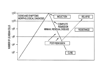
Figure 1 shows the therapeutic strategy for most patients with AML which is
divided
into two general phases: induction therapy and post-remission therapy.
Figure 2 (Figure 12 of W02006/003179) provides a comparative alignment of the
amino acid sequences of the light chain variable regions, and light chain CDRs
of antibodies
10 DF200 and Pan2D
(NKVSF1). (A) Alignment of anti-KIR variable light (VL) regions of
DF200 (SEQ ID NO:1) and Pan-2D (SEQ ID NO:2). Numbers above amino acid
sequences
indicate position respective to initiation of translation Met (+1) in the
immature (non-
secreted) immunoglobulin. (B) Alignment of CDR-L1 sequences. Residue before:
Normally
Cys. Residues after: Trp. Typically Trp-Tyr-Leu. Length: 10-17 aa. (C)
Alignment of CDR-
SUBSTITUTE SHEET (RULE 26)
CA 028186842013-05-21
WO 2012/071411
PCT/US2011/061840
11 PATENT
APPLICATION
44292.118602
L2 sequences. Residues before: Generally Ile-Tyr. Length: 7 aa. Start:
approximately 16 aa
after the end of CDR-Ll. Start: approximately 24 aa from the beginning of
secreted protein.
(D) Alignment of CDR-L3 sequences. Residues before: Cys. Residues after: Phe-
Gly-XXX-
Gly. Length: 7-11 aa. Start: approximately 33 aa after the end of CDR-L2.
Figure 3 (Figure 13 of W02006/003179)provides the heavy chain variable region,
and
the heavy-chain CDRs of antibody DF200. (A) DF-200 VH region, immature
protein. The
secreted, mature VH starts at position 20: residue Q. The VH region ends with
residue S and
thereafter the constant region (not shown ) continues, (B) CDR-Hl. Residues
before: Cys-
XXX-XXX-XXX. Residues after: Trp. Generally Trp-Val or Trp-Ile. Length: 10-14
aa. Start:
Approximately 22-26 aa from the beginning of the secreted protein. (C) CDR-H2.
Residues
before: Leu-GIu-Trp-He-GIy but other variations possible. Residues after: Lys
or Arg / Leu
or He or Val or Phe or Thr or Ala / Thr or Ser or lie or Ala. Length: 16-20
aa. Start:
Approximately 15 aa after the end of CDR-HI. (D) CDR-H3. Residues before: Cys-
XXX-
XXX (Typically Cys-Ala-Arg). Residues after: Trp-G Iy-XX.XvG Iy. Length: 3-25
aa. Start:
Approximately 33 after the end of CDR-H2.
Figure 4 (Figure 14 of W02006/003179) depicts the nucleotide and amino acid
sequences of the VH and VL sequence of human antibody 1-7F9. (A) Translation
of HuKIR
1-7F9 mature variable light chain. (B) Nucleotide sequence encoding HuKIR 1-
7F9 mature
variable light chain. (C) Translation of HuKIR I -7F9 mature variable heavy
chain. (D)
Nucleotide sequence encoding HuKIR 1-7F9 mature heavy chain.
Figure 5 (Figure 15 of W02006/003179)shows the amino acid sequences of the VH
and VL sequences of monoclonal antibodies 1-7F9, DF200 (VH sequence: SEQ ID
NO:19;
VL sequence: SEQ ID NO:21 ), and Pan2D (NKVSFI ; VH sequence: SEQ ID NO:20; VL
sequence: SEQ ID NO:22). The CDRs are boxed.
Figure 6 (Figure 20 of W02006/003179) shows the binding epitope of 1-7F9 on
KIR2DL1, as indicated in the KIR2DL1 sequence. Amino acids within 4.0 A
distance from
1-7F9 are highlighted in grey and black background. Amino acids highlighted by
a black
background are involved in hydrogen-bonding to 1-7F9. The sequence ID No's
listed in
Figures 2-6 correspond to SEQ ID NO's in the Sequence Listing filed in
W02006/003179
that is contained in the pages that immediately precede the claims of this
application.
SUBSTITUTE SHEET (RULE 26)
CA 028186842013-05-21
WO 2012/071411
PCT/IJS2011/061840
12 PATENT
APPLICATION
44292.118602
DESCRIPTION OF THE INVENTION
This invention provides methods for treating an individual having or
previously
having had a hematological malignancy or pre-malignancy. The methods comprise
administering to the individual a therapeutically active amount of a compound
that inhibits a
NK cell inhibitory receptor (NKCIR). The compound is administered to the
individual at a
time when the individual has minimal or non-detectable disease.
Human clinical trials described herein showed that treatment with a compound
that
blocks an NK cell inhibitor receptor involved in NK cell cytotoxicity, e.g.,
anti-NKCIR
antibodies, greatly prolonged disease-free survival in patients who had
suffered from
hematological malignancy but were in remission and/or had minimal or
undetectable disease
when treated with the compound.
ANTIBODIES =
Unless otherwise stated or clearly contradicted by context, the term antibody
in the
context of this invention refers to an immunoglobulin (Ig) molecule, a
fragment of an Ig
molecule, or a derivative of either thereof that has the ability to (a)
specifically bind to at least
one target antigen under typical physiological conditions for significant
periods of time
and/or (b) modulate a physiological response associated with its target NKCIR,
such as
modulating KIR-modulated NK cell activity. A significant period of time in
this respect
means any period suitable for detection of the antibody-antigen complex in a
standard
immunological assay, such as an enzyme-linked immunosorbent assay (ELISA).
Typically, a
significant period of time is a period of at least about 30 minutes, at least
about 45 minutes, at
least about one hour, at least about two hours, at least about four hours, at
least about 8 hours,
at least about 12 hours, about 24 hours or more, about 48 hours or more, etc.
Immunoglobulins are a class of structurally related proteins comprising heavy
chains
(e.g., a, A, a, y, and IA chains) and light chains (e.g., ic and chains).
In humans,
imrnunoglobulins may be divided into five major classes (IgA, IgD, IgE, IgG,
and IgM)
according to which heavy chains are contained in the Ig molecule.
The structure of immunoglobul ins is well characterized. See, e.g.,
Fundamental
Immunology (Paul, W., ed., 2nd ed. Raven Press, N.Y. (1989)). IgG molecules,
the most
common type of immunoglobulin, comprise two pairs of polypeptide chains, one
pair of light
(L), low molecular weight chains and one pair of heavy (H) chains, all four
inter-connected
by disulfide bonds. Briefly, each heavy chain typically is comprised of a
heavy chain
SUBSTITUTE SHEET (RULE 26)
CA 028186842013-05-21
WO 2012/071411
PCT/US2011/061840
13 PATENT
APPLICATION
44292.118602
variable region (abbreviated herein as HCVR or VH) and a heavy chain constant
region. The
heavy chain constant region typically is comprised of three domains, CHI, CH2,
and CH3.
Each light chain typically is comprised of a light chain variable region
(abbreviated herein as
LCVR or VL) and a light chain constant region. The light chain constant region
typically is
comprised of one domain, CL. The VH and VL regions can be further subdivided
into
regions of hypervariability (or hypervariable regions, which can be
hypervariable in sequence
and/or form of structurally defined loops), also termed complementarity
determining regions
(CDRs), interspersed with regions that are more conserved, termed framework
regions (FR).
In full length, naturally produced antibodies, each VH and VL typically is
composed of three
CDRs and four FRs, arranged from amino-terminus to carboxy-terminus in the
following
order: FR1, CDR1, FR2, CDR2, FR3, CDR3, FR4 (which also may be referred to as
FR Ll ,
CDR Li, etc. or loop Li, L2, L3 in the light chain variable domain and loop
H1, H2, and H3
in the heavy chain domain in the case of hypervariable loop regions (see,
e.g., Chothia and
Lesk J. Mol. Biol. 196:901-917 (1987)). Typically, the numbering of amino acid
residues in
this region is performed by the method described in Kabat et al., Sequences of
Proteins of
Immunological Interest, 5th Ed. Public Health Service, National Institutes of
Health,
Bethesda, MD. (1991) (phrases such as "variable domain residue numbering as in
Kabat" and
"according to Kabat" herein refer to this numbering system for heavy chain
variable domains
or light chain variable domains). Using this numbering system, the actual
linear amino acid
sequence of a peptide may contain fewer or additional amino acids
corresponding to a
shortening of, or insertion into, a FR or CDR of the variable domain. For
example, a heavy
chain variable domain may include a single amino acid insert (residue 52a
according to
Kabat) after residue 52 of CDR H2 and inserted residues (e.g., residues 82a,
82b, and 82c,
etc. according to Kabat) after heavy chain FR residue 82. The Kabat numbering
of residues
may be determined for a given antibody by alignment at regions of homology of
the sequence
of the antibody with a "standard" Kabat numbered sequence.
As indicated above, an anti-NKCIR antibody can be in the form of (or comprise)
an
antibody "fragment" that retains the ability to specifically bind to a NKCIR.
Such antibody
fragments can be characterized by possessing any one or combination of the
aforementioned
features associated with full length antibodies, discussed elsewhere herein,
to the extent
appropriate (e.g., many antibody fragments lack an Fc domain and, accordingly,
do not
induce or promote antibody-associated complement functions). The antigen-
binding function
SUBSTITUTE SHEET (RULE 26)
CA 028186842013-05-21
WO 2012/071411
PCMJS2011/061840
14 PATENT
APPLICATION
44292.118602
of antibodies can be performed by any number of suitable fragments thereof.
Examples of
antibody fragments include (i) a Fab fragment, a monovalent fragment
consisting essentially
of the VL, VH, CL and CH I domains; (ii) F(ab)2 and F(ab')2 fragments,
bivalent fragments
comprising two Fab fragments linked by a disulfide bridge at the hinge region;
(iii) a Fd
fragment consisting essentially of the VH and CHI domains; (iv) a Fv fragment
consisting
essentially of the VL and VH domains of a single arm of an antibody, (v) a dAb
fragment
(Ward et al., (1989) Nature 341:544-546), which consists essentially of a VH
domain; and
(vi) an isolated complementarity determining region (CDR). Furthermore,
although the two
domains of the Fv fragment, VL and VH, are coded for by separate genes, they
can be joined,
using recombinant methods, by a synthetic linker that enables them to be made
as a single
protein chain in which the VL and VH regions pair to form monovalent molecules
(known as
single chain antibodies or single chain Fv (scFv); see e.g., Bird et al.
(1988) Science 242:423-
426: and Huston et al. (1988) Proc. Natl. Acad. Sci. USA 85:5879-5883). Such
single chain
antibodies also are encompassed within terms such as antibody fragment and
antibody-like
peptide/molecule, unless otherwise noted or clearly indicated by context.
Other forms of
single chain antibodies, such as diabodies also are intended be encompassed by
these terms.
Diabodies are bivalent, bispecific antibodies in which VH and VL domains are
expressed on
a single polypeptide chain, but using a linker that typically is too short to
allow for pairing
between the two domains on the same chain, thereby forcing the domains to pair
with
complementary domains of another chain and creating two antigen binding sites
(see e.g.,
Holliger, P., et at. (1993) Proc. Natl. Acad. Sci. USA 90:6444-6448; Poljak,
R. J., et al.
(1994) Structure 2:1121-1123; and Cao et al. (1998), Bioconjugate Chem. 9, 635-
644).
Although having similar binding properties as full-length antibodies, such
antibody fragments
collectively and each independently are unique features of the invention,
exhibiting different
biological and/or physiochemical properties and utilities than antibodies.
These and other
useful antibody fragments and antibody-like molecules provided by this
invention are
discussed further herein. It should be generally understood that any suitable
antibody
fragment can be used as a surrogate for an antibody in inventive compositions
and methods
described herein, and visa versa, unless otherwise stated or clearly
contradicted by context.
In a general sense, the term antibody includes polyclonal antibodies and
monoclonal
antibodies (mAbs). The term "monoclonal antibody" refers to a composition
comprising a
homogeneous antibody population having a uniform structure and specificity.
Polyclonal
SUBSTITUTE SHEET (RULE 26)
CA 028186842013-05-21
WO 2012/071411
PCMJS2011/061840
15 PATENT
APPLICATION
44292.118602
antibodies typically are derived from the serum of an animal that has been
immunogenically
challenged, but they can also be derived by recombinant technology. Anti-K1R
antibodies
can be considered monoclonal antibodies, regardless of the manner in which
they are
produced.
An antibody as generated can possess any isotype and the antibody can be
isotype
switched thereafter using conventional techniques that are well known in the
art. Such
techniques include the use of direct recombinant techniques (see, e.g., US
Patent 4,816,397),
cell-cell fusion techniques (see e.g., US Patent 5,916,771), and other
suitable techniques
known in the art. Thus, for example, the effector function of multispecific
multivalent
antibodies provided by the invention may be "changed" with respect to the
isotype of one or
both parent antibodies by isotype switching to, e.g., an IgGI, IgG2, IgG3,
IgG4, IgD, IgA,
IgE, or IgM antibody for various therapeutic uses.
NK CELL ACTIVITY-MODULATING RECEPTORS (NKCAMRS)
NK cell activity is regulated by NK cell activity-modulating receptors
("NKCAMRs"
or simply "AMRs''), which may be specific for various ligands such as MHC-I
molecules,
MHC-I homologs, or other biological molecules expressed on target cells. NK
cells in an
individual typically present a number of activating and inhibitory receptors.
The activity of
NK cells is regulated by a balance of signals transduced through these
activating and
inhibitory receptors. Each type of NKCAMR is briefly discussed in turn below.
When somatic cells are either under stress, such in cancer progression or
infection,
various molecules, such as MICA and MICB, are typically displayed on the
surface of the
stressed cells and normally displayed MHC-I molecules are "lost" from the cell
surface
(reduced in number and/or glycosylated such that they are not "seen" as
"foreign" by the
immune system). NKCAMRs are sensitive to these and other changes in potential
NK target
cells associated with cellular stress, disease, and disorder.
Most NKCAMRs appear to belong to one of two classes of proteins: the
immunoglobulin (Ig)-like receptor superfamily (IgSF) or the C-type lectin-like
receptor
(CUR) super family (see, e.g., Radaev and Sun, Annu. Rev. Biomol. Struct. 2003
32:93-
114). However, other forms of NKCAMRs are known. The structures of a number of
NKCAMRs have been elucidated (Id.). To better illustrate the invention, types
of well
understood NKCAMRs, with reference to particular examples thereof, are
described here.
However, several additional NKCAMRs are known besides those receptors
explicitly
SUBSTITUTE SHEET (RULE 26)
CA 028186842013-05-21
WO 2012/071411
PCMJS2011/061840
16 PATENT
APPLICATION
44292.118602
described here (see, e.g., Farag et al., Expert Opin. Biol. Ther. 3(2):237-
250) and the
inventive compositions and methods described herein typically will also be
applicable to
these and other NKCAMRs.
NK Cell Activating Receptors (NKCARs)
Many NK cell activating receptors (NKCARs) belong to the Ig superfamily (IgSF)
(such receptors also may be referred to as Ig-like receptors or "ILRs"
herein). Activating ILR
NK receptors (AILRs) include, e.g., CD2, CD16, CD69, DNAX accessory molecule-1
(DNAM-1), 2B4, NK1.1; killer immunoglobulin (Ig)-like activating receptors
(KARs);
ILTs/LIRs; and natural cytotoxicity receptors (NCRs) such as NKp44, NKp46, and
NKp30.
Several other NKCARs belong to the CLTR superfamily (e.g., NKRP-1, CD69;
CD94/NKG2C and CD94/NKG2E heterodimers, NKG2D homodimer, and in mice,
activating isoforms of Ly49 (such as Ly49A-D)). Still other NKCARs (e.g., LFA-
1 and
VLA-4) belong to the integrin protein superfamily and other activating
receptors may have
even other distinguishable structures. Many NKCARs possess extracellular
domains that
bind to MHC-I molecules, and cytoplasmic domains that are relatively short and
lack the
inhibitory (ITIM) signaling motifs characteristic of inhibitory NK receptors.
The
transmembrane domains of these receptors typically include a charged amino
acid residue
that facilitates their association with signal transduction-associated
molecules such as
CD3zeta, FcERIy, DAP12, and DAP10 (2B4, for example, appears to be an
exception to this
general rule), which contain short amino acid sequences termed an
Immunoreceptor
tyrosine-based activating motif (ITAMs) that propagate NK cell-activating
signals.
Receptor 2B4 contains 4 so-called Irnmunoreceptor Tyrosine-based Switch Motif
(ITSM) in
its cytoplasmic tail; ITSM motifs can also be found in NKCARs CS1/CRACC and
NTB-A.
The cytoplasmic domains of 2B4 and SLAM contain two or more unique tyrosine-
based
motifs that resemble motifs presents in activating and inhibitory receptors
and can recruit the
SH2-domain containing proteins SHP-2 and SAP (SLAM-associated protein).
Stress-induced molecules, such as MIC-A, MIC-B, and ULBPs in humans, and Rae-1
and H-60 in mice, can serve as ligands for NKCARs, such as the NKG2D
homodimer.
Cellular carbohydrates, pathogenic antigens, and antibodies can also be NKCAR
ligands. For
example, NKR-P1 may bind to carbohydrate ligands and trigger NK cell
activation,
particularly against tumor cells which exhibit aberrant glycosylation
patterns. Viral
SUBSTITUTE SHEET (RULE 26)
CA 028186842013-05-21
WO 2012/071411
PCT/IJS2011/061840
17 PATENT
APPLICATION
44292.118602
hemagglutinins may serve as ligands for natural cytotoxic receptors (NCRs),
such as ILR
NKCARs NKp30, NKp44, NKp46, and NKp80.
NKCARs can either directly transduce activating signals or can act in
connection with
adaptor molecules or other receptors (either in the context of a coordinated
response between
receptors that are sometimes singularly effective or in the context of
coreceptor-receptor
pairings). For example, NKCAR NCRs typically lack ITAMs and, accordingly, bind
to
adaptor molecules through a charged residue in their transmembrane domains
(e.g., NKp30
associates with the CD3 zeta chain; NKp44 associates with DAP12 and/or KARAP;
NKp46
is coupled to the CD3 zeta chain and FcRly chain), which are, in turn, able to
recruit protein
tyrosine kinases (PTKs) in order to propagate NK cell-activating signals.
CD16, which is a
NKCAR important to NK cell-mediated ADCC and cytokine production, associates
with
homodimers or heterodimers formed of CD3 zeta and/or gamma chains. NKG2D
appears to
play a complementary and/or synergistic role with NCRs and NKCARs in NK cell
activation.
Activation of NK cells against particular targets may require coordinated
activation of
multiple NKCARs or NCRs, or only action of a single receptor. Other triggering
surface
molecules including 2B4 and NKp80 appear to function as coreceptors for NK
cell activation.
Activating isoforms of human KIRs (e.g., KIR2DS and KIR3DS) and murine Ly-49
proteins (e.g., Ly-49D and Ly-49H) are expressed by some NK cells. These
molecules differ
from their inhibitory counterparts (discussed below) by lacking inhibitory
motifs (ITIMs) in
their relatively shorter cytoplasmic domains and possessing a charged
transmembrane region
that associates with signal-transducing polypeptides, such as disulfide-linked
dimers of
DAP12.
NKClRs NK Cell Inhibitory Receptors
ILR (IgSF) NK cell inhibitory receptors (NKCIRs) (I) include a number of
different
human KIRs, specific for HLA-A, -B, or -C allotypes. KIRs may recognize
multiple alleles
within a particular allotype, e.g., KIR2DL1 recognizes HLA-Cw2, 4, and 6
allotypes. CTLR
superfamily inhibitory receptors include members of the CD94/NKG2 protein
family, which
comprise receptors formed by lectin-like CD94 with various members of the NKG2
family,
such as NKG2A, and recognize the nonclassical class I molecules HLA-E and Qa-1
in
humans and mice, respectively, and the murine Ly49 molecules that recognize
the classical
class I MHC molecules in mice. In even further contrast, NKRP1A, Nkrplf and
Nkrpld are
inhibitory receptors whose ligands are not MHC-related but are CTLR family
members
SUBSTITUTE SHEET (RULE 26)
CA 028186842013-05-21
WO 2012/071411
PCMJS2011/061840
18 PATENT
APPLICATION
44292.118602
expressed on various cell types, such as dendritic cells, macrophages, and
lymphocytes.
MHC class I-specific NKCIRs include CTLR Ly-49 receptors (in mice); the IgSF
receptors Leukocyte Immunoglobulin-like Receptors (LIRs) (in humans), KIRs
(e.g., p58 and
p70 Killer-cell Immunoglobulin-like Receptors, in humans), and CTLR CD94/NKG2
receptors (in mice and humans). All MHC-I-specific NKCIRs appear to use a
common
inhibitory mechanism apparently involving phosphorylation of ITIMs in their
cytoplasmic
domains in the course of MHC-I binding and recruitment of tyrosine
phosphatases (e.g.,
SHP-1 and SHP-2) to the phosphorylated ITIMs, resulting in the inhibition of
proximal
protein tyrosine kinases (PTKs) involved in NK activation through NKCARs.
Inhibitory CD94/NKG2 heterodimers formed from CTLR glycoproteins, comprise an
ITIM-bearing NKG2 molecule (e.g., NKG2A) and bind to non-classical MHC-I
molecules
(e.g., HLA-E in humans and Qa-1 in mice).
Leukocyte Immunoglobulin-like Receptors include several members, containing
two
or four Ig-domains and structurally related to KIR polypeptides. See, e.g.,
Fanger et al. 1999
J. Leukocyte Biol. 66:231-236. LIR include subfamilies A and B and include,
e.g., LIR-1 to
LIR-8 (several of which are also referred to ILT polypeptides, including ILT-
1, 1LT-2, 1LT-3,
1LT-4, ILT-5, and ILT-6. The polypeptides LIR-1, LIR-2, LIR-3, LIR-5, and LIR-
8 all
contain two or more ITIM inhibitory signaling domains.
Inhibitory Ly-49 receptors are murine type H membrane disulfide-linked
homodimer
CUR glycoproteins, which bind to various MHC-I molecules and deliver typically
dominant
inhibitory (negative) signals to NK cells. Ly-49A, for example, binds to
alphal/alpha2
domains of MHC-I molecule H-2Dd, whereas Ly-49C binds H-2Kb. Human NK cells
appear
to lack homologs of the murine Ly-49 receptors. Instead, human NK cells
express KIRs,
which are not found in mouse NK cells. Although human KIRs and mouse Ly-49
receptors
lack structural homology, they are functionally orthologous: Both types of
receptors bind to
HLA class I on target cells, resulting in inhibition of NK-mediated
cytotoxicity.
Killer-cell immunoglobutin-like receptors MR
An important type of NKCIRs is the KIRs. Generally, KIRs are cell surface
glycoproteins, comprising one to three extracellular immunoglobulin-like
domains, which are
expressed by some T cells as well as most human NK cells. A number of KIRs are
well
characterized (see, e.g., Carrington and Norman, The IC1R Gene Cluster, May
28, 2003,
available through the National Center for Biotechnology Information (NCBI) web
site at
SUBSTITUTE SHEET (RULE 26)
CA 028186842013-05-21
WO 2012/071411
PCMJS2011/061840
19 PATENT APPLICATION
44292.118602
http://www.ncbi.nlm.nih.gov/books/bookres.fcgi/mono_003/chldl.pdf). Human KIRs
include KIR2DL and KIR3DL. KIRs also may be referred to by various other names
such as
CD158e1, CD158k, CD158z, p58 MR CD158e1 (p70), CD244, etc. (see, e.g., US
Patent
Application 20040038894, Radaev et al., Annu. Rev, Biophys. Biomol. Struct.,
32:93-114
(2003), Cerweknka et al., Nat. Rev. Immunol. 1:41-49 (2001); Farag et al.,
Expert Opin. Biol.
Ther., 3(2):237-250 (2003); Biassoni et al., J. Cell. Mol. Med., 7(4):376-387
(2003); and
Warren et al., British J. Haematology, 121:793-804 (2003), each of which being
hereby
incorporated into this application in their entirety). The structure of a
number of KIRs has
been elucidated and reveals remarkable structural similarity between these
proteins. See,
e.g., Radaev et al., supra.
KIRs can be classified structurally as well as functionally. For example, most
KIRs
have either two Ig domains (58 kDa KIR2D KIRs), whereas others have three Ig
domains (70
kDa KIR3D KIRs), which may sometimes be respectively referred to as p58 and
p70
molecules. KIRs vary also in cytoplasmic tail length. Typically, KIRs with a
relatively long
cytoplasmic tail (L) deliver an inhibitory signal, whereas MR with a short
cytoplasmic tail
(S) can activate NK or T cell responses. Nomenclature for KIRs accordingly can
be based
upon the number of extracellular domains (KIR2D or KIR3D) and whether the
cytoplasmic
tail is long (KIR2DL or KIR3DL) or short (KIR2DS or KIR3DS). Additional
nomenclature
information for KIRs is provided in the following Detailed Description of the
Invention.
Some members of the "MR family" are NKCARs, or more particularly "KARs" (e.g.,
MR2DS2 and KIR2DS4); they typically comprise one or more charged transmembrane
residues (e.g., Lys) that associate with an adapter molecule having an
immunostimulatory
motif (ITAM) (e.g., DAP12). The intracytoplasmic portion of inhibitory KIRs
typically
comprises one or more ITIMs that recruit phosphatases. Inhibitory KIRs bind to
alphal/a1pha2 domains of HLA molecules. Inhibitory KIRs do not appear to
typically require
adaptor-molecule association for activity. Unless otherwise stated, terms such
as "MR",
"KIRs", and the like refer to NKCIR members of the "KIR family" and terms such
as "KAR",
"KARs", and the like refer to NKCAR members of the "MR family."
KIRs can bind MHC-I molecules (e.g., certain HLA class I allotypes), typically
resulting in the transmission of a negative signal that counteracts, and may
override
stimulatory, activating signal(s) to the NK cell, thereby preventing the NK
cell from killing
the associated potential target cell (apparently via ITLM phosphorylation and
tyrosine
SUBSTITUTE SHEET (RULE 26)
CA 028186842013-05-21
WO 2012/071411
PCMJS2011/061840
20 PATENT APPLICATION
44292.118602
phosphatase (e.g., SH2-domain containing protein tyrosine phosphatases such as
SHP-1 arid
SHP-2) recruitment, leading to PTK (e.g., Syk, TcR and/or ZAP70)
dephosphorylation and/or
LAT/PLC complex formation inhibition and associated disruption of ITAM
cascade(s)).
Because viruses often suppress class I MHC expression in cells they infect,
such virus-
infected cells become susceptible to killing by NK cells. Because cancer cells
also often have
reduced or no class I MHC expression, these cells, too, can become susceptible
to killing by
NK cells. Infected cells can also change the proteins bound in the MHC in
terms of
glycosylation. If this occurs, the MHC-I:protein complex the cell expresses
will be altered.
If NK-associated KIRs cannot bind to these "foreign" complexes, no inhibitory
signal can be
generated, and lysis will proceed.
All confirmed inhibitory KIRs appear to interact with different subsets of
HLA/MHC
antigens depending upon the MR subtype. In humans, KIRs having two Ig domains
(KIR2D)
recognize HLA-C allotypes: KIR2DL2 (formerly designated p58.2) and the closely
related
gene product KIR2DL3 both recognize an epitope shared by group 1 HLA-C
allotypes (Cwl,
3, 7, and 8), whereas KIR2DL1 (p58.1) recognizes an epitope shared by the
reciprocal group
2 HLA-C allotypes (Cw2, 4, 5, and 6). The specificity of KIR2DL1 appears to be
dictated by
the presence of a Lys residue at position 80 of group 2 HLA-C alleles. KIR2DL2
and
KIR2DL3 recognition appears to be dictated by the presence of an Asn residue
at position 80.
A substantial majority of HLA-C alleles have either an Asn or a Lys residue at
position 80.
One MR with three Ig domains, MR3DL1 (p70), recognizes an epitope shared by
HLA-Bw4
alleles. Finally, a homodimer of molecules with three Ig domains, KIR3DL2
(p140),
recognizes HLA-A3 and -Al 1.
Individual MHC-I-specific NK cell receptors of either type (activating or
inhibitory)
typically do not interact with all MHC class I molecules, but specifically
bind to certain
allotypes (proteins encoded by different variants of a single genetic locus).
Also, an
individual NK cell may express several different inhibitory and/or activating
receptors which
function independently of each other. For example, in humans the presence or
absence of a
given KIR is variable from one NK cell to another within a single individual.
There also is
relatively high level of polymorphism of KIRs in humans, with certain KIR
molecules being
present in some, but not all individuals. Although KIRs and other MHC-
recognizing
inhibitory receptors may be co-expressed by NK cells, in any given
individual's NK
repertoire there are typically cells that express a single KIR; accordingly,
the corresponding
SUBSTITUTE SHEET (RULE 26)
CA 028186842013-05-21
WO 2012/071411
PCMJS2011/061840
21 PATENT
APPLICATION
44292.118602
NK cell activity in this latter type of NK cells is inhibited only by cells
expressing a specific
MHC-I allele group. In fact, recent estimates of the extent of MR genotype
diversity within
the population suggest that < 0.24% of unrelated individuals can expect to
have identical
genotypes. The most common Caucasian haplotype, the "A" haplotype (frequency
of ¨ 47-
59%), contains only one activating MR gene (KIR2DS4) and six inhibitory KIR
loci
(KIR3DL3, -2DL3, -2DLI, -2DL4, -3DL1, and -3DL2). The remaining "B" haplotypes
are
very diverse and contain 2-5 activating KIR loci (including KIR2DS1, -2DS2, -
2DS3, and-
2DS5).
It should be noted that KIRs are known by several aliases, as reflected here
in Table I,
which includes information obtained from the Hugo Gene Nomenclature Committee
web site
(http://www.gene.ucl.ac.uk/nomenclatureigenefamily/kir.html) and Andre et al.,
Nature
Immunol. 2(8):661 (2001).
SUBSTITUTE SHEET (RULE 26)
CA 028186842013-05-21
WO 2012/071411 PCMJS2011/061840
22 PATENT APPLICATION
44292.118602
Table I ¨ KIR Nomenclature
KIR Full name Aliases Accession ID
KIR2DL1 killer cell immunoglobulin-like c1-42, nkatl, L41267
receptor, two domains, long 47.11, p58.1,
cytoplasmic tail, 1 CD158a
KIR2DL2 killer cell immunoglobulin- c1-43, nkat6, L76669
like receptor, two domains, long CD158b1,
cytoplasmic tail, 2 p58.2
KIR2DL3 killer cell immunoglobulin- c1-6, nkat2, L41268
like receptor, two domains, long nkat2a, nkat2b,
cytoplasmic tail, 3 p58.3,
CD158b2
KIR2DL4 killer cell immunoglobulin- 103AS, 15.212, X97229
like receptor, two domains, long CD158d, p70
cytoplasmic tail, 4
killer cell immunoglobulin-
KIR2DL5.1,
KIR2DL5A like receptor, two domains, long AF217485
CD158f
cytoplasmic tail, 5A
killer cell immunoglobulin-like KIR2DL5.2,
KIR2DL5B receptor, two domains, long KIR2DL5.3, kF217486
cytoplasmic tail, 5B KIR2DL5.4
KIR2DS1 killer cell immunoglobulin-like EB6ActI, X89892
receptor, two domains, short EB6ActII,
cytoplasmic tail, 1 CD158h, p50.1
KIR2DS2 killer cell immunoglobulin-like c1-49, nkat5, L76667
receptor, two domains, short 183ActI,
cytoplasmic tail, 2 CD158j, p50.2
SUBSTITUTE SHEET (RULE 26)
CA 028186842013-05-21
WO 2012/071411
PCMJS2011/061840
23 PATENT APPLICATION
44292.118602
KIR Full name Aliases Accession ID
KIR2DS3 killer cell immunoglobulin-like nkat7 L76670
receptor, two domains, short
cytoplasmic tail, 3
KIR2DS4 killer cell immunoglobulin-like c1-39, KKA3, L76671
receptor, two domains, short nkat8, CD158i,
cytoplasmic tail, 4 p50.3
KIR2DS5 killer cell immunoglobulin-like nkat9, CD158g L76672
receptor, two domains, short
cytoplasmic tail, 5
killer cell immunoglobulin-like KIRZ, KIRY,
KIR2DP1 receptor, two domains, pseudogene KIR15, AF204908
1 KIR2DL6
KIR3DL1 killer cell immunoglobulin-like c1-2, NICB1, cl- L41269
receptor, three domains, long 11, nkat3,
cytoplasmic tail, 1 NKB1B,
AMB11, KIR,
CD158e1
KIR3DL2 killer cell immunoglobulin-like c1-5, nkat4, L41270
receptor, three domains, long nkat4a, nkat4b,
cytoplasmic tail, 2 CD158k, p140
killer cell immunoglobulin-like ICIRC1,
KIR3DL3 receptor, three domains, long KIR3DL7, AF352324
cytoplasmic tail, 3 KIR44, CD158z
KIR3DS1 killer cell immunoglobulin-like nkatl 0, L76661
receptor, three domains, short CD158e2
cytoplasmic tail, 1
= SUBSTITUTE SHEET (RULE 26)
CA 028186842013-05-21
WO 2012/071411 PCMJS2011/061840
24 PATENT
APPLICATION
44292.118602
KIR Full name Aliases Accession ID
KIRX, KIR48,
killer cell immunoglobulin-like AF204919,
KIR2DS6,
KIR3DP1 receptor, three domains, AF204915
KIR3DS2P,
pseudogene 1 AF204917
CD158c
NEUTRALIZATION OF NKCIR-ASSOCIATED NK CELL INHIBITION
Anti-NKCIR antibodies also or alternatively can be characterized on the basis
of their
ability to block or neutralize NK inhibition and thereby potentiate NK cell
activity against
otherwise blocked target cells. As indicated above, anti-NKCIR antibodies that
bind to at
least one NKCIR for a sufficient amount of time to neutralize NKCIR -mediated
inhibition of
NK cell cytotoxicity in NK cells can be used in the context of this invention.
Such anti-
NKCIR antibodies may be used directly as therapeutic agents in a native form
(e.g., without
conjugation to a cytotoxic agent). A more particular advantageous feature of
the invention is
anti-NKCIR antibodies that cross-react with two or more NKCIRs and neutralize
the
inhibitory activity associated with some or all (typically preferably all) of
such associated
NKCIRs.
Neutralizing anti-NKCIR antibodies may partially or fully neutralize the NKCIR
-
mediated inhibition of NK cell cytotoxicity. Neutralization refers to any
substantial blocking
of otherwise present inhibitory signals. Neutralization can be measured by any
suitable
method. In one aspect, neutralization of inhibition is reflected in that the
neutralizing anti-
KIR antibody (ies) cause(s) an least about 20%, preferably at least about 30%,
at least about
40%, at least about 50%, at least about 60%, at least about 75% or more (e.g.,
about 25-
100%) increase in NK cell-mediated specific lysis in a particular mixture of
NK and NK
target cells compared to the amount of specific lysis that typically occurs in
a substantially
identical setting without the presence of the anti-NKCIR antibody (ies). The
percentage
increase in this aspect can be determined when considering anti-NKCIR or other
antibodies
by, e.g., comparison with the results of chromium release toxicity test assays
obtained from a
mixture of NK target cells and NK cells not blocked their associated NKCIR(s)
(100%) and a
mixture of NK cells and NK target cells, in which the NK target cells present
a ligand for the
SUBSTITUTE SHEET (RULE 26)
CA 028186842013-05-21
WO 2012/071411 PCMJS2011/061840
25 PATENT
APPLICATION
44292.118602
NKCIR (0%). In the case of anti-ICIR antibodies, comparison can be with the
results of
chromium release toxicity test assays obtained from a mixture of NK target
cells and NK
cells not blocked their associated KIR(s) (100%) and a mixture of NK cells and
NK target
cells, in which the NK target cells present the cognate MHC class I molecule
for the
inhibitory KIR on the NK cells (0%). In an advantageous aspect, the invention
provides anti-
NKCIR antibodies that induce lysis of cell(s) that would not be effectively
lysed without the
presence of such anti-NKCIR antibody. Alternatively, neutralization of NKCIR
inhibitory
activity can be indicated by, e.g., the results of a chromium assay using an
NK cell clone or
transfectant expressing one or several inhibitory NKCIRs (e.g., KIR, NKG2,
NKG2A, and
LIR (e.g., LILRB1, LILRB5)) and a target cell expressing only one ligand
(e.g., HLA
polypeptide or allele, HLA-E, etc.) that is recognized by one of the NKCIRs on
the NK cell,
where the level of cytotoxicity obtained with the antibody is at least about
20%, such as at
least about 30%, at least about 40%, at least about 50%, at least about 60%,
at least about
70% or more (e.g., about 25-100%) of the cytotoxicity observed with a known
blocking
antibody to the ligand of the NKCIR. For example, when testing an anti-KIR
antibody, an
anti-MHC class I molecule is administered in a substantially identical
setting, such as W6/32
anti-MHC class I antibody that is currently available from, e.g., Research
Diagnostics,
Flanders, NJ, USA and described in, e.g., Shields et al., Tissue Antigens.
1998
May ;51(5):567-70.
Chromium release assays and other methods of assessing NK cell cytolytic
activity
are known in the art. Conditions suitable for such assays also are well known.
A typical
chromium release assay is performed by labeling target cells (e.g., Cw3 and/or
Cw4 positive
cell lines ¨ at about, e.g., 5000 cells per well in a microtitration plate)
with Na251Cr04 (such
that 5ICr is taken up and retained by viable target cells), washing to remove
excess
radioactivity, thereafter exposed to NK cells for a period of about 4 hours in
the presence or
absence of anti-NKCIR antibody(s) at a suitable effector:target ratio (e.g.,
about 4:1), and
measuring for subsequent 51Cr levels reflecting target cell death and lysis.
An example of
such an assay is described in, e.g., Moretta et al., 1993, J Exp Med 178, 597-
604. In a similar
assay, proliferating target cells can be labeled with 3H-thymidine, which is
incorporated into
the replicating DNA. Upon cytolytic action by NK cells, the DNA of the target
cells is
rapidly fragmented and retained in a filtrate, while large, unfragmented DNA
can be collected
on a filter, such that one can measure either the release of these fragments
or the retention of
SUBSTITUTE SHEET (RULE 26)
CA 028186842013-05-21
WO 2012/071411
PCMJS2011/061840
26 PA ______________ l'hNT
APPLICATION
44292.118602
3H-thymidine in cellular DNA. Other examples and relevant discussion related
to such
assays can be found in, e.g., PCT application no. W02006/072625.
In another aspect, the invention provides anti-NKCIR antibodies characterized
by the
ability to compete with cross-reactive and/or neutralizing anti-NKCIR
antibodies for binding
to cognate NKCIRs and/or to bind to the same antigenic determinant
region/epitope as such
known antibodies. The phrase "competes with when referring to a particular
monoclonal
antibody (e.g., 1-7F9, etc.) means that the anti-NKCIR antibody competes with
the
referenced antibody or other molecule in a binding assay using either
recombinant NKCIR
molecules or surface expressed NKCIR molecules. For example, if an anti-KIR
antibody
detectably reduces binding of 1-7F9 to a MR molecule normally bound by 1-7F9
in a binding
assay, the anti-MR antibody can be said to "compete" with 1-7F9. An anti-MR
antibody
that "competes" with 1-7F9 may compete with 1-7F9 for binding to the KIR2DL1
human
receptor, the KIR2DL2/3 human receptor, or both KIR2DLI and KIR2DL2/3 human
receptors.
Although often related, describing a protein in terms of competition with a
reference
binding protein versus the ability of the protein to bind to the same or
substantially similar
epitope as a reference protein in some cases imply significantly different
biological and
physiochemical properties. Competition between binding proteins implies that
the test anti-
NKCIR antibody binds to an epitope that at least partially overlaps with an
epitope bound by
an anti-NKCIR antibody or is located near enough to such an epitope so that
such an anti-
MR antibody competes with known anti-NKCIR antibodies due to steric hindrance.
An anti-
NKCIR antibody may compete with a reference anti-NKCIR antibody, without
binding to the
same or similar epitope due to the large size of the antibodies. Such a
competing anti-
NKCIR antibody can be useful in blocking interactions associated with the same
antigenic
determining region as the reference anti-NKCIR antibody even though it binds a
different
antigenic determinant.
In another exemplary aspect, the invention provides an anti-NKCIR antibody
that
binds to substantially the same antigenic determinant region as an anti-NCKIR
antibody, such
as 1-7F9, DF200 and/or NKVSF1 (for MR), or antibody Z199 (for NKG2A, available
from
Beckman Coulter, CA), etc.
Competition refers to any significant reduction in the propensity for a
particular
molecule to bind a particular binding partner in the presence of another
molecule that binds
SUBSTITUTE SHEET (RULE 26)
CA 028186842013-05-21
WO 2012/071411 PCMJS2011/061840
27 PATENT
APPLICATION
44292.118602
the binding partner. Typically, competition means an at least about 15%
reduction in
binding, such as an at least about 20% reduction in binding (e.g., a reduction
in binding of
about 25% or more, about 30% or more, about 15-35%, etc.) between, e.g., an
anti-KIR
antibody and at least one KIR in the presence of the competing molecule, e.g.,
an anti-KIR
antibody. In certain situations, such as in cases where epitopes belonging to
competing
antibodies are closely located in an antigen, competition can be marked by
greater than about
40% relative inhibition of receptor (e.g., KIR) binding, at least about 50%
inhibition, at least
about 55% inhibition, at least about 60% inhibition, at least about 75%
inhibition, or a higher
level of inhibition, e.g., such as a level of inhibition of about 45-95%.
Assessing competition typically involves an evaluation of relative inhibitory
binding
using a first amount of a first molecule (e.g., an anti-MR antibody); a second
amount of a
second molecule (e.g., a known anti-MR antibody); and a third amount of a
third molecule
(e.g., a KIR), wherein the first, second, and third amounts all are sufficient
to make a
comparison that imparts information about the selectivity and/or specificity
of the molecules
at issue with respect to the other present molecules. Usually, for ELISA
competition assays,
about 5-50 [kg (e.g., about 10-50 g, about 20-50 p.g, about 5-20 1.tg, about
10-20 It g, etc.) of
an anti-MR antibody, a known anti-MR antibody, and at least one KIR are used
to assess
whether competition exists. Conditions also should be suitable for binding of
the competing
molecules to their putative/known target. Physiological or near-physiological
conditions
(e.g., temperatures of about 20-40 C, pH of about 7-8, etc.) can typically be
suitable for anti-
KIR antibody:KIR.
Determination of competition (or relative inhibition of binding) between two
or more
molecules can be made by use of immunoassays in which the control NKCIR-
binding
molecule (e.g., 1-7F9) and test anti-NKCIR antibody are admixed (or pre-
adsorbed) and
applied to a sample containing relevant KIRs, such as both KIR2DL1 and
KIR2DL2/3, each
of which is known to be bound by DF200. Protocols
based upon ELISAs,
radioimmunoassays, Western blotting, and the like are suitable for use in such
competition
studies. Competition FLISAs are typically performed under conditions suitable
for binding
of the molecules (e.g., physiological conditions, particularly in the case of
antibodies that
bind conformational/nonlinear epitopes). Competition also can be assessed by,
for example, a
flow cytometry test, SPR analysis and other techniques found in, e.g., Harlow,
et al.,
Antibodies: A Laboratory Manual, Cold Spring Harbor Laboratory Press, Cold
Spring
SUBSTITUTE SHEET (RULE 26)
CA 028186842013-05-21
WO 2012/071411
PCMJS2011/061840
28 PATENT
APPLICATION
44292.118602
Harbor, N.Y., 1988), Colligan et al., eds., Current Protocols in Immunology,
Greene
Publishing Assoc. and Wiley Interscience, N.Y., (1992, 1993), Ausubel et al.,
Eds., Short
Protocols in Molecular Biology, (5th edition), John Wiley & Sons (2002), and
Muller, Meth.
Enzymol. 92:589-601 (1983)).
An antigenic determinant region or epitope can be identified by a number of
known
techniques. For example, an antigenic determinant region can be identified
quickly by "foot
printing" assays, such as through a chemical modification of the exposed
amines/carboxyls in
target NKCIR proteins. One specific example of such a foot-printing technique
is the use of
hydrogen-deuterium exchange detected by mass spectrometry (HXMS), wherein a
hydrogen/deuterium exchange of receptor and ligand protein amide protons,
binding, and
back exchange occurs, wherein the backbone amide groups participating in
protein binding
are protected from back exchange and therefore will remain deuterated.
Relevant regions can
be identified at this point by peptic proteolysis, fast microbore high-
performance liquid
chromatography separation, and/or electrospray ionization mass spectrometry.
See, e.g.,
Ehring H, Analytical Biochemistry, Vol. 267 (2) pp. 252-259 (1999) and/or
Engen, J.R. and
Smith, D.L. (2001) Anal. Chem. 73, 256A-265A.
Another example of a suitable epitope identification technique is nuclear
magnetic
resonance (NMR) epitope mapping, where typically the position of the signals
in two-
dimensional NMR spectres of the free antigen and the antigen complexed with
the antigen-
binding peptide, such as an antibody, are compared. The antigen typically is
selectively
isotopically labeled with 15N so that only signals corresponding to the
antigen and no signals
from the antigen binding peptide are seen in the NMR-spectrum. Antigen signals
originating
from amino acids involved in the interaction with the antigen binding peptide
typically will
shift position in the spectres of the complex compared to the spectres of the
free antigen, and
the amino acids involved in the binding can be identified that way. See, e.g.,
Ernst Schering
Res Found Workshop. 2004;(44):149-67; Huang et al, Journal of Molecular
Biology, Vol.
281 (1) pp. 61-67 (1998); and Saito and Patterson, Methods. 1996 Jum9(3):516-
24.
Epitope mapping/characterization also can be performed using mass spectrometry
methods. See, e.g., Downward, J Mass Spectrom. 2000 Apr;35(4):493-503 and
Kiselar and
.. Downard, Anal Chem. 1999 May 1;71(9):1792-801.
Protease digestion techniques also can be useful in the context of epitope
mapping
and identification. Antigenic determinant-relevant regions/sequences can be
determined by
SUBSTITUTE SHEET (RULE 26)
CA 028186842013-05-21
WO 2012/071411 PCMJS2011/061840
29 PATENT
APPLICATION
44292.118602
protease digestion, e.g. by using trypsin in a ratio of about 1:50 to NKCIR in
an overnight
digestion at 37 C and pH 7-8, followed by mass spectrometry (MS) analysis for
peptide
identification. The peptides protected from trypsin cleavage by the anti-NKCIR-
binder can
subsequently be identified by comparison of samples subjected to trypsin
digestion and
samples incubated with antibody and then subjected to digestion by e.g.,
trypsin (thereby
revealing a foot print for the binder). Other enzymes like chymotrypsin,
pepsin, etc., also or
alternatively can be used in similar epitope characterization methods,
Moreover, enzymatic
digestion can provide a quick method for analyzing whether a potential
antigenic determinant
sequence is within a region of the NKCIR in the context of an anti-NKCIR
polypeptide that is
not surface exposed and, accordingly, most likely not relevant in terms of
antigenicity. See,
e.g., Manca, Ann 1st Super Sanita. 1991;27(1):15-9 for a discussion of similar
techniques.
Various phage display techniques also can be used to identify epitopes. See,
e.g.,
Wang and Yu, Curr Drug Targets. 2004 Jan;5(1):1-15; Burton, Immunotechnology.
1995
Aug;1(2):87-94; Cortese et al., Immunotechnology. 1995 Aug;1(2):87-94; and
Irving et al.,
Curr Opin Chem Biol. 2001 Jun;5(3):314-24. Consensus epitopes also can be
identified
through modified phage display-related techniques (see, Mumey et al., J.
Comput. Biol.
10:555-567 and Mumey, Proceedings of the Sixth Annual International Conference
on
Computational Molecular Biology (RECOMB-02), pp. 233-240 (ACM Press, New
York)) for
discussion (see also Bailey et al., Protein Science (2003), 12:2453-2475;
Dromey et al., J
Immunol. 2004 Apr 1;172(7):4084-90; Parker et al., Mol Biotechnol. 2002
Jan;20(1):49-62;
and Czompoly et al., Biochem Biophys Res Commun. 2003 Aug 8;307(4):791-6).
Epitope mapping by competitive binding to a MR with two KIR-binding molecules
where one is biotinylated (e.g., a known anti-MR antibody) or otherwise
similarly labeled is
another method for identifying relevant antigenic determinant regions.
Other methods potentially helpful in mapping epitopes include crystallography
techniques, X-ray diffraction techniques (such as the X-ray
diffraction/sequence study
techniques developed by Poljak and others in the 1970s-1980s), and the
application of
Multipin Peptide Synthesis Technology.
Computer-based methods such as sequence analysis and three dimensional
structure
analysis and docking also can be used to identify antigenic determinants. For
example, an
epitope also can be determined by molecular modeling using a structure of a
NKCIR or
portion thereof with docking of the structure of the Fab fragment of an
individual mAb.
SUBSTITUTE SHEET (RULE 26)
CA 028186842013-05-21
WO 2012/071411 PCMJS2011/061840
30 PATENT
APPLICATION
44292.118602
Where necessary, models of NKCIRs can be produced by homology modeling with
structure-
characterized NKCIRs using programs such as Molecular Operating Environment
(MOE),
which is available from Chemical Computing Group (Montreal, Quebec, Canada -
www.chemcomp.com). These and other mapping methods are discussed in Epitope
Mapping
A Practical Approach (Westwood and Hay Eds.) 2001 Oxford University Press (see
also,
Cason, J Virol Methods. 1994 Sep;49(2):209-19).
CHARACTERISTICS OF ANTI-KM ANTIBODIES
Advantageous anti-KIR antibodies may be classified based on functional
characteristics, particularly with respect to their ability to cross-react or
cross-bind more than
one MR, such as more than one type of inhibitory KIR, and/or the ability to
effectively
neutralize NK inhibitory signals. The invention contemplates treatment using
an anti-MR or
a combination of anti-MR antibodies. Exemplary anti-MR antibodies include, but
are not
limited to, an anti-KIR2DLI antibody and an anti-KIR2DL2 antibody, or an anti-
KIR2DL1
antibody and an anti-KIR2DL3 antibody, or an anti-K1R2DL1 antibody and an anti-
KIR2DL2 antibody and an anti-KIR2DL3 antibody, or an anti-KIR antibody that
binds at
least two different human inhibitory KIR receptor gene products and is capable
of
neutralizing MR-mediated inhibition of NK cell cytotoxicity in NK cells
expressing at least
one of the two different human inhibitory MR receptors, or an anti-KIR2D
antibody and an
anti-ICIR3D antibody.
Anti-MR antibodies that effectively bind to more than one type of MR are a
particularly advantageous feature of the invention. In a particular exemplary
aspect, the
invention provides anti-MR antibodies that bind to at least two inhibitory MR
receptors at
the surface of NK cells. In an even more particular illustrative aspect, the
invention provides
anti-MR antibodies that bind a common antigenic determinant region of human
KIR2DL
receptors. In a yet even further specific aspect, the invention provides an
anti-MR antibody
that binds to ICIR2DL1, KIR2DL2, and KIR2DL3 receptors.
The term "KIR2DL2/3" can be used to refer to either or both of the KIR2DL2 and
KIR2DL3 receptors. These two receptors have a very high homology, are allelic
forms of the
same gene, and are considered by the art to be interchangeable in many
respects.
Accordingly, KIR2DL2/3 can be considered in certain respects to be a single
inhibitory KIR
molecule. While anti-KIR antibodies that cross-react with KIR2DL2/3 are within
the
SUBSTITUTE SHEET (RULE 26)
CA 028186842013-05-21
WO 2012/071411 PCMJS2011/061840
31 PATENT APPLICATION
44292.118602
invention, anti-KIR antibodies that have a MR-binding profile that only
included KIR2DL2
and KIR2DL3 are not considered "cross-reactive."
Because at least one of KIR2DL1 or KID2DL2/3 is present in at least about 90%
of
the human population, KIR2DL1 ¨ KIR2DL2/3 cross-reactive anti-MR antibodies
can
promote or enhance NK activity against most of the HLA-C allotype-associated
cells,
respectively group 2 HLA-C allotypes and group 1 HLA-C allotypes. A
composition
comprising a single KIR antibodies having such cross-reactivity may be used in
treatment
and/or diagnosis of most human subjects, thereby eliminating the necessity of
genetic
profiling of the patient and reducing the amount of different antibodies that
need to be
administered to a patient to ensure an effective result.
Cross-reacting anti-MR antibodies can have any suitable composition and can be
obtained by a number of suitable techniques. For example, a cross-reactive
anti-KIR
antibody can comprise a number of MR ligand and/or anti-KIR antibody sequences
that bind
to different KIRs, which may be associated by conjugation, multimerization, or
(in the case
of peptide ligands) by being comprised in a fusion protein. In another aspect,
an anti-KIR
antibody is provided that comprises anti-MR antibody sequences from a cross-
reacting anti-
KIR antibody.
Cross-reacting anti-KIR antibodies, from which KIR-binding sequences can be
obtained or derived, are known. An example of such an antibody is described
in, e.g., Watzl
et al., Tissue Antigens, 56, p. 240 (2000). Another example is antibody NKVSF1
(also
referred to as pan2D mAb; recognizing a common epitope of CD158a (KIR2DLI),
CDI58b
(KIR2DL2) and p50.3 (KIR2DS4)) having the variable region and CDR sequences
shown in,
e.g., Figure 15, of PCT patent application W02006/003179 (Innate Pharma; Novo
Nordisk;
University of Genoa). The monoclonal antibody DF200, which reacts with various
members
of the MR family including KIR2DL1 and KIR2DL2/3 is another example of such a
cross-
reacting antibody. A hybtidoma that produces DF200 has been deposited at the
CNCM
culture collection, as Identification no. "DF200", registration no. CNCM 1-
3224, registered
10 June 2004, Collection Nationale de Cultures de Microorganismes, Institut
Pasteur, 25, Rue
du Docteur Roux, F-75724 Paris Cedex 15, France. Several additional monoclonal
antibodies can be generated and demonstrated to be cross-reactive anti-KIR
antibodies. Yet
other examples are antibodies 1-7F9 and 1-4F1, described in W02006/003179.
SUBSTITUTE SHEET (RULE 26)
CA 028186842013-05-21
WO 2012/071411
PCMJS2011/061840
32 PATENT
APPLICATION
44292.118602
A cross-reactive anti-KIR antibody can have any suitable affinity and/or
avidity for
the two or more KIRs to which it binds. Affinity refers to the strength of
binding of an anti-
MR antibody or other antigen-binding protein to an epitope or antigenic
determinant.
Typically, affinity is measured in terms of a dissociation constant KD,
defined as [Ab] x [Ag]
/ [Ab-Ag] where [Ab-Ag] is the molar concentration of the antibody-antigen
complex, [Ab] is
the molar concentration of the unbound antibody and [Ag] is the molar
concentration of the
unbound antigen. The affinity constant Ka is defined by 1/K0. Suitable methods
for
determining binding peptide specificity and affinity by competitive
inhibition, equilibrium
dialysis, and the like can be found in, e.g., Harlow, et al., Antibodies: A
Laboratory Manual,
Cold Spring Harbor Laboratory Press, Cold Spring Harbor, N.Y., 1988); Colligan
et al., eds.,
Current Protocols in Immunology, Greene Publishing Assoc. and Wiley
Interscience, N.Y.,
(1992, 1993), and Muller, Meth. Enzymol. 92:589-601 (1983).
Typically, an anti-MR antibody provided by the invention has an affinity for
at least
one MR in the range of about 104 to about 1010 M-I (e.g., about 107 to about
109 M-1). The
term immunoreact herein typically refers to binding of an anti-MR antibody to
a KIR with a
dissociation constant KE1 lower than about 10-4 M. For example, in a
particular aspect the
invention provides anti-MR antibody that have an average disassociation
constant (KO of
about 7 x 10⁻⁹ M or more with respect to KIR2DL1 and KIR2DL2/3, as determined
by, e.g.,
surface plasmon resonance (SPR) screening (such as by analysis with a
BIAcoreTM SPR
analytical device). In a more particular exemplary aspect, the invention
provides anti-MR
antibodies that have a KD of about 2 x 10⁻⁹ M (e.g., about 0.1 ¨ 4 x 10⁻⁹ M)
or more for
KIR2DL2/3 and about 11 x leM (e.g., about 7-15 x 10 M) or more for KIR2DLl.
Affinity can be determined by any of the methods described elsewhere herein or
their
known equivalents in the art. An example of one method that can be used to
determine
affinity is provided in Scatchard analysis of Munson & Pollard, Anal. Biochem.
107:220
(1980). Binding affinity also may be determined by equilibrium methods (e.g.,
ELISA or
radioimmunoassay (RIA)) or kinetics analysis (e.g., BlAcoreTM analysis).
Anti-KIR antibodies also or alternatively can be characterized by exhibiting
KIR
binding with a disassociation constant of less than about 100 nM, less than
about 50 nM, less
than about 10 nM, about 5 nM or less, about 1 nM or less, about 0.5 nM or
less, about 0.1 nM
or less, about 0.01 nM or less, or even about 0.001 nM or less.
SUBSTITUTE SHEET (RULE 26)
CA 028186842013-05-21
WO 2012/071411 PCMJS2011/061840
33 PATENT
APPLICATION
44292.118602
Avidity refers to the overall strength of the total interactions between a
binding
protein and antigen (e.g., the total strength of interactions between an anti-
KIR antibody and
a KIR). Affinity is the strength of the total noncovalent interactions between
a single
antigen-binding site on an antibody or other binding peptide and a single
epitope or antigenic
determinant. Avidity typically is governed by three major factors: the
intrinsic affinity of the
binding protein for the epitope(s) or antigenic determinant(s) to which it
binds, the valence of
the antibody or binding protein and antigen (e.g., an anti-MR antibody with a
valency of
three, four, or more will typically exhibit higher levels of avidity for an
antigen than a
bivalent antibody and a bivalent antibody can will have a higher avidity for
an antigen than a
univalent antibody, especially where there are repeated epitopes in the
antigen), and/or the
geometric arrangement of the interacting components. Avidity typically is
measured by the
same type of techniques used to assess affinity.
In another aspect, the invention provides an anti-KIR antibody that cross-
reacts with
KIRs from two or more species. For example, in one aspect, the invention
provides an anti-
KIR antibody that cross-reacts with KIRs of humans and cynomolgus monkeys. In
a
particular aspect, the invention provides an anti-KIR antibody that cross-
reacts with at least
two human KIRs and also binds to NK cells of cynomolgus monkeys. Such an anti-
KIR
antibody can comprise sequences from or that are derived from antibody NKVSF1,
which
exhibits such a cross-reactivity profile. Such anti-MR antibodies can be
subjected to toxicity
testing and other useful studies in cynomolgus monkeys, if needed.
Antibodies that are cross-reactive with a variety of KIRs can be used in the
combination compositions and methods of the invention. Exemplary cross-
reactivity profiles
of such antibodies include antibodies that cross-react with KIRs 2DL1 plus
2DL2/3, 3DL1
plus 3DL2, 2DL1 (and 2DL2/3) plus 2D54, and 2DL1 (and 2DL2/3) but not 2DS4.
Thus, for example, the inventive methods or compositions can comprise an anti-
KIR
antibody that binds KIR2DL1, KIR2DL2, and KIR2DL3 and reduces or blocks
inhibition of
MR-mediated NK cell cytotoxicity, as described in, e.g., W02005003168.
Exemplary anti-MR antibodies useful in the combination methods and
compositions
of the invention include anti-KIR antibodies comprising a VL region that
corresponds to that
of anti-MR antibody DF200, or consists essentially of such a VL region (by
being
substantially similar and retaining a similar binding profile and affinity),
or a VL
sequence/domain that is highly similar (e.g., at least about 90% identical or
95% identical) to
SUBSTITUTE SHEET (RULE 26)
CA 028186842013-05-21
WO 2012/071411 PCMJS2011/061840
34 PATENT
APPLICATION
44292.118602
the VL sequence of DF200. The VL sequence of DF200 is shown in W02006/3179.
Such
anti-KIR antibodies also may alternatively be defined by comprising the set of
light variable
CDRs of DF200 (also shown in W02006/3179). Such an antibody typically also
will
comprise either the VH domain of DF200 or a highly similar sequence (e.g., a
sequence
having high identity to the DF200 VH domain or otherwise consisting
essentially of such a
sequence) or at least the heavy variable CDRs of DF200 (shown in W02006/3179).
As used herein, the term "percent sequence identity" or "sequence identity"
refers to
the percent identity between two sequences, which is a function of the number
of identical
positions shared by the sequences, i.e., % homology = # of identical
positions/total # of
positions x 100, taking into account the number of gaps, and the length of
each gap, which
need to be introduced for optimal alignment of the two sequences. The
comparison of
sequences and determination of percent identity between two sequences can be
accomplished
using a mathematical algorithm, as described in the non-limiting examples
below.
The percent identity between two amino acid sequences can be determined using
the
algorithm of E. Myers and W. Miller (Comput. Appl. Biosci., 4:11-17 (1998))
which has
been incorporated into the ALIGN program (version 2.0), using a PAM120 weight
residue
table, a gap length penalty of 12 and a gap penalty of 4. In addition, the
percent identity
between two amino acid sequences can be determined using the Needleman and
Wunsch (J.
Mol. Biol. 48:444-453 (1970)) algorithm which has been incorporated into the
GAP program
in the GCG software package (available at www.gcg.com), using either a Blossum
62 matrix
or a PAM250 matrix, and a gap weight of 16, 14, 12, 10, 8, 6, or 4 and a
length weight of 1,
2, 3,4, 5, or 6.
In certain instances, the protein sequences of the present disclosure can be
further
used as a "query sequence" to perform a search against public databases to,
for example,
identify related sequences. Such searches can be performed using the XBLAST
program
(version 2.0) of Altschol, et al. (1990) J. Mol. Biol. 215:403-10. BLAST
protein searches can
be performed with the XBLAST program, score=50, wordlength=3 to obtain amino
acid
sequences homologous to the antibodies of the invention. To obtain gapped
alignments for
comparison purposes, Gapped BLAST can be utilized as described in Altschul et
al., (1997)
Nucleic Acids Res. 25(17):3389-3402. When utilizing BLAST and Gapped BLAST
programs, the default parameters of the respective programs (e.g., XBLAST and
NBLAST)
can be used. See, www.ncbi.nlm.nih.gov.
SUBSTITUTE SHEET (RULE 26)
CA on 1E604 2013-Kn21
WO 2012/071411
PCT/US2011/061840
35 PATENT
APPLICATION
44292.118602
In another exemplary aspect, the combination composition or method of the
invention
includes an anti-KIR antibody comprising VH and VL sequences that correspond
to or are
highly similar to (e.g., consists essentially of) the VH and VL sequences of
antibody 1-7F9
(shown in W02006/3179) or at least comprises the VL and VH CDRs of 1-7F9.
Competition with Cross-Reactive and/or Neutralizing Anti-KIR Antibodies
In another aspect, the inventive methods or compositions are characterized by
comprising an anti-KIR antibody that competes with one of these antibodies or
one of the
other anti-KIR antibodies described in the references incorporated herein
(e.g., 1-7F9).
Antibodies that compete with exemplary anti-KIR antibodies, such as DF200, 1-
7F9,
and/or NKVSF1, can be identified using known screening assays. A number of
such assays
are routinely practiced and well known in the art (see, e.g., U.S. Pat. No.
5,660,827.
Protocols based on, e.g., ELISAs, radio-
immunoassays, Western blotting, and the use of B1ACORE analysis are suitable
for use in
such competition studies.
One can, e.g., pre-mix the control antibody (e.g., DF200, NKVSFI, or 1-7F9)
with
varying amounts of the test antibody (e.g., in ratios of about 1:1, 1:2, 1:10
or about 1:100) for
a period of time prior to applying to a KIR antigen sample. Alternatively, the
control and
varying amounts of test antibody can simply be added separately and admixed
during
exposure to the KIR antigen sample. As long as one can distinguish bound from
free
antibodies (e.g., by using separation or washing techniques to eliminate un-
bound antibodies)
and control anti-body from the test antibody (e.g., by using species specific
or isotype
specific secondary antibodies or by specifically labeling the control antibody
with a
detectable label) one will be able to determine if the test antibody reduce
the binding of the
control antibody to the different KIR2DL antigens, indicating that the test
antibody
recognizes substantially the same epitope as the control. The binding of the
(labeled) control
antibody in the presence of a completely irrelevant antibody (that does not
bind KIR) can
serve as the control high value. The control low value can be obtained by
incubating the
labeled control antibody with the same but unlabelled control antibody, where
competition
would occur and reduce binding of the labeled antibody. In a test assay, a
significant
reduction in labeled antibody reactivity in the presence of a test antibody is
indicative of a
test antibody that recognizes substantially the same epitope, i.e., one that
competes with the
labeled control antibody. For example, any test antibody that reduces the
binding of control
SUBSTITUTE SHEET (RULE 26)
CA 2818684 2018-08-09
CA 028186842013-05-21
WO 2012/071411
PCT/US2011/061840
36 PATENT
APPLICATION
44292.118602
antibody to one or both of KIR2DL1 and KIR2DL3 antigens by at least about 50%,
such as at
least about 60%, or more preferably at least about 70% (e.g., about 65-100%),
at any ratio of
control:test antibody between about 1:1 or 1:10 and about 1:100 is considered
to be an
antibody that competes with the control.
Competition can also be assessed by, for example, flow eytometry. In such a
test,
cells bearing a given MR can be incubated first with a control antibody, and
then with the
test antibody labeled with a fluorochrome or biotin. The antibody is said to
compete with
control antibody if the binding obtained upon pre-incubation with saturating
amount of
control antibody is about 80%, preferably about 50%, about 40% or less (e.g.,
about 30%) of
the binding (as measured by mean of fluorescence) obtained by the test
antibody without
preincubation with control antibody. Alternatively, an antibody is said to
compete with the
control antibody if the binding obtained with a labeled control antibody (by a
fluorochrome
or biotin) on cells preincubated with saturating amount of test antibody is
about 80%,
preferably about 50%, about 40%, or less (e.g., about 30%) of the binding
obtained without
preincubation with the test antibody.
A simple competition assay in which a test antibody is pre-adsorbed and
applied at
saturating concentration to a surface onto which either KIR2DL1 or KIR2DL2/3,
or both, are
immobilized also may be advantageously employed. The surface in the simple
competition
assay is preferably a BIACORE chip (or other media suitable for surface
plasmon resonance
analysis). The binding of a control antibody to the MR-coated surface is
measured. This
binding to the KIR-containing surface of the control antibody alone is
compared with the
binding of the control antibody in the presence of a test antibody. A
significant reduction in
binding to the KIR2DL1 and KIR2DL2/3-containing surface by the control
antibody in the
presence of a test antibody indicates that the test antibody recognizes
substantially the same
epitope as the control antibody such that the test antibody "competes" with
the control
antibody. Any test antibody that reduces the binding of control antibody to
both of KIR21DL1
and KIR2DL2/3 antigens by at least about 20% or more, at least about 40%, at
least about
50%, at least about 70%, or more, can be considered to be an antibody that
competes with the
control antibody. Preferably, such test antibody will reduce the binding of
the control
antibody to each of at least the KIR2DLI, 2, and 3 antigens by at least about
50% (e.g., at
least about 60%, at least about 70%, or more). It will be appreciated that the
order of control
and test antibodies can be reversed; i.e., the control antibody can be first
bound to the surface
SUBSTITUTE SHEET (RULE 26)
CA 02818604 2913 05-21
WO 2012/071411
PCT/US2011/061040
37 PATENT
APPLICATION
44292.118602
and then the test antibody is brought into contact with the surface thereafter
in a competition
assay. Preferably, the antibody having higher affinity for KIR2DL1 and
KIR2DL2J3 antigens
is bound to the KIR2DL1 and KIR2DL2/3-containing surface first, as it will be
expected that
the decrease in binding seen for the second antibody (assuming the antibodies
are competing)
will be of greater magnitude. Further examples of such assays are provided in
the Examples
herein, and in e.g., Sauna! and Regenmottel, (1995) J. Immunol. Methods 183:
33-41.
In another aspect, the inventive method or composition is characterized by
inclusion
of only antibodies that are not cross-reactive with more than one KIR. For
example,
monoclonal antibodies specific only for KIR2DL1 have been shown to block the
interactions
between KIR2DL1 and HLA-Cw4 allotypes, as well as similar HLA-C allotypes
belonging to
the same group as Cw4 (Moretta et al., J Exp Med. 1993;178(2):597-604).
In another example, monoclonal antibodies
against KIR2DL2/3 have also been described that block the interactions of
KIR2DL2/3 with
HLACw3 (or the like) allotypes (Moretta et al., 1993, supra). Optionally, the
antibody can be
selected from the group consisting of GL183 (UR2DL2/3/S2-specific, available
from
Immunotech, France and Beckton Dickinson, USA); EB6 (KIR2DL1/sl-specific,
available
from Immunotech, France and Beckton Dickinson, USA); AZ138 (KIR3DL1-specific,
available from Moretta et al, Univ. Genova, Italy); Q66 (KIR3DL2-specific,
available from
Immunotech, France); and DX9, Z27 (KIR3DL1-specific, available from
Immunotech,
France and Beckton Dickinson, USA).
Epitopes
In additional aspects, the invention provides anti-KIR antibodies that are
directed to
particular antigenic regions and/or epitopes presented on various KIRs. In one
exemplary
aspect, the invention provides anti-KIR antibodies that specifically bind
ICIR2DL1 within a
region defined by one or more of the amino acid residues selected from 105,
106, 107, 108,
109, 110, 111, 127, 129, 130, 131, 132, 133, 134, 135, 152, 153, 154, 155,
156, 157, 158,
159, 160, 161, 162, 163, 181, and 192. In another embodiment the invention
provides anti-
KIR antibodies that specifically bind to KIR2DL1 and KIR 2DL2/3 in a region
defined by
one or more of amino acid residues 105, 106, 107, 108, 109, 110, 111, 127,
129, 130, 131,
132, 133, 134, 135, 152, 153, 154, 155, 156, 157, 158, 159, 160, 161, 162,
163, 181, and 192
thereof.
SUBSTITUTE SHEET (RULE 26)
CA 2818684 2018-08-09
CA 0281t9e4 2,31346.21
WO 2012/071411 PCT/1JS2011/061840
=
38 PATENT
APPLICATION
44292.118602
In a further aspect, the invention provides anti-KIR antibodies that bind to
KIR2DL1,
but that bind to a mutant of KIR2DL1 in which R131 is Ala with significantly
reduced
binding affinity relative thereto (about 20% or less, about 30% or less, about
40% or less,
about 50% or less, about 60% or less, about 70% or less, etc., of the affinity
exhibited for
KIR2DL1). In another aspect, the invention provides anti-ICIR antibodies that
bind to
KIR2DL1 but that which bind,to a mutant of KIR2DL1 in which R157 is Ala with
relatively
reduced binding affinity (about 20% or less, about 30% or less, about 40% or
less, about 50%
or less, about 60% or less, about 70% or less, etc., of the affinity exhibited
for KIR2DL1). In
another aspect, the invention provides anti-KIR antibodies that bind to
KIR2DL1 and which
binds a mutant of KIR2DL1 in which R158 is Ala with relatively reduced binding
affinity
(about 20% or less, about 30% or less, about 40% or less, about 50% or less,
about 60% or
less, about 70% or less, etc., of the affinity exhibited for KIR2DL1).
In a further aspect, the invention provides anti-KIR antibodies that bind to
KIR2DL1
residues 131, 157, and 158.
In an additional aspect, the invention provides anti-ICIR antibodies that bind
to
KIR2DS3(R131W), but not to wild type KIR2DS3. In yet another aspect, the
invention
provides anti-MR antibodies that bind to KIR2DL1 and ICIR2DL2/3 as well as
KIR2DS4. In
still another aspect, the invention provides anti-KIR antibodies that bind to
both KIR2DL1
and KIR2DL2/3, but not to KIR2DS4.
To illustrate the use of anti-KIR antibody sequences in the composition and
construction of anti-MR antibodies, exemplary anti-KW antibody sequences and
antibody
sequence variants will be described here. Amino acid and nucleic acid
sequences of variable
regions and CDRS of exemplary MR antibodies DF200 and 1-7F9 are also disclosed
in PCT
application no. W02006/003179.
In one exemplary aspect, the invention provides an anti-KIR antibody
comprising a
CDR-LI sequence that consists or consists essentially of the sequence Lys Ala
Ser Gin Asn
Val Val Thr Tyr Val Ser (SEQ ID NO:43). In another aspect, the invention
provides an anti-
ICIR antibody that comprises a CDR-L1 that consists or consists essentially of
the sequence
Thr Ala Ser Ser Ser Val Ser Ser Ser Tyr Leu Tyr (SEQ ID NO:44).
In another illustrative aspect, the invention provides an anti-KIR antibody
that also or
alternatively comprises a CDR-L2 sequence that consists or consists
essentially of the
sequence Gly Ala Ser Asn Arg Tyr Thr (SEQ ID NO:45). In a further aspect, the
invention
SUBSTITUTE SHEET (RULE 26)
CA 2818684 2018-08-09
CA 028186842013-05-21
WO 2012/071411
PCMJS2011/061840
39 PATENT
APPLICATION
44292.118602
provides an anti-MR antibody that also or alternatively comprises a CDR-L2
that consists or
consists essentially of the sequence Ser Thr Ser Asn Leu Ala Ser (SEQ ID
NO:46).
In another demonstrative facet, the invention provides an anti-MR antibody
that also
or alternatively comprises a CDR-L3 that consists or consists essentially of
the sequence Gly
Gln Gly Tyr Ser Tyr Phe Tyr Thr (SEQ ID NO:47). In yet another aspect, the
invention
provides an anti-MR antibody that also or alternatively comprises a CDR-L3
that consists or
consists essentially of the sequence His Gin Tyr His Arg Ser Pro Pro Thr (SEQ
ID NO:48).
As a further exemplary feature, the invention provides an anti-MR antibody
that
comprises a CDR-H1 that consists or consists essentially of the sequence Gly
Phe Ser Phe
Thr Phe 'I'yr Gly Val His (SEQ ID NO:49).
In still another exemplary aspect, the invention provides an anti-MR antibody
that
comprises a CDR-H2 that consists or consists essentially of the sequence Val
Ile Trp Ser Gly
Gly Asn Thr Asp Tyr Asn Ala Ala Phe Ile Ser (SEQ ID NO:50).
In yet another exemplary aspect, the invention provides an anti-MR antibody
that
comprises a CDR-H3 that consists or consists essentially of the sequence Asn
Pro Arg Pro
Gly Asn Tyr Pro Tyr Gly Met Asp Tyr (SEQ ID NO:51).
In a different aspect, the invention provides an anti-MR antibody that
comprises a
CDR-H1 that consists or consists essentially of the sequence Gly Tyr Thr Phe
Thr Ser Tyr
Trp Met His (SEQ ID NO:52).
In an additional aspect, the invention provides an anti-MR antibody that
comprises a
CDR-H2 that consists or consists essentially of the sequence Thr Ile Tyr Pro
Gly Asn Ser Asp
Thr Asn Tyr Asn Gin Lys Phe Lys Gly (SEQ ID NO:53).
Another aspect of the invention is embodied by an anti-MR antibody that
comprises a
CDR-H3 that consists or consists essentially of the sequence Pro Thr Thr Ala
Thr Arg Ser Ser
Ala Met Asp Tyr (SEQ ID NO:54).
The basic and novel properties of these CDR sequences is the ability to, in
combination with other necessary CDR and FR sequences, bind to epitope(s)
presented on
one or more KIRs. As indicated above, it may be the case that certain residues
in such
sequences contribute little or nothing to MR epitope binding. Moreover, it
also or
alternatively may be the case that such CDR sequences may tolerate one or a
few insertions
without impacting their epitope binding characteristics substantially
(specificity and/or
SUBSTITUTE SHEET (RULE 26)
CA 028186842013-05-21
WO 2012/071411
PCMJS2011/061840
40 PATENT
APPLICATION
44292.118602
affinity). However, in another aspect of the invention significant changes can
be made in
such sequences to produce useful variants. Such changes are discussed further
below.
These exemplary CDR sequences can be combined with one another, variant CDR
sequences described below, or other anti-KIR CDRs (typically from KIR-binding
anti-KIR
antibodies). In one exemplary aspect, the invention provides an anti-MR
antibody that
comprises most or all of the CDR sequences selected from SEQ ID NOS:43, 45,
47, 49, 50,
and 51.
In another illustrative aspect, the invention provides an antibody comprising
a light
variable (VL) sequence consisting essentially of the sequence Met Glu Ser Gin
Thr Leu Val
Phe Ile Ser Ile Leu Leu Trp Leu Tyr Gly Ala Asp Gly Asn Ile Val Met Thr Gln
Ser Pro Lys
Ser Met Ser Met Ser Val Gly Glu Arg Val Thr Leu Thr Cys Lys Asn Ser Glu Asn
Val Val
Thr Tyr Val Ser Trp Tyr Gin Gin Lys Pro Glu Gin Ser Pro Lys Leu Leu Ile Tyr
Gly Ala Ser
Asn Arg Tyr Thr Gly Val Pro Asp Arg Phe Thr Gly Ser Gly Ser Ala Thr Asp Phe
Thr Leu
Thr Ile Ser Ser Val Gin Ala Glu Asp Leu Ala Asp Tyr His Cys Gly Gin Gly Tyr
Ser Tyr Pro
Tyr Thr Phe Gly Gly Thr Lys Leu Asp Ile Lys Arg (SEQ ID NO:55). The basic and
novel
properties of such a sequence are its contribution to MR binding. It may be
possible that
some amino acids can be deleted, add, or substituted in this sequence without
substantial
impact to such properties.
In an additional exemplary aspect, the invention provides an antibody
comprising a
VL sequence consisting essentially of the sequence Met Asp Phe Gin Val Gln Ile
Phe Ser Phe
Leu Leu Ile Ser Ala Ser Val Ile Met Ser Arg Gly Gin Ile Val Leu Thr Gin Ser
Pro Ala Ser
Met Ser Ala Ser Leu Gly Glu Arg Val Thr Met Thr Cys Thr Ala Ser Ser Ser Val
Ser Ser Ser
Tyr Leu Tyr Trp Tyr Gin Gin Lys Pro Gly Ser Ser Pro Lys Leu Trp Ile Tyr Ser
Thr Ser Asn
Leu Ala Ser Gly Val Pro Ala Arg Phe Ser Gly Ser Gly Ser Gly Thr Ser Tyr Ser
Leu Thr Ile
Ser Ser Met Gin Ala Glu Asp Ala Ala Thr Tyr Tyr Cys His Gln Tyr His Arg Ser
Pro Pro Thr
Phe Gly Gly Gly Thr Lys Leu Glu Ile Lys Arg (SEQ ID NO:56).
The N-terminal portion of both SEQ ID NO:55 and SEQ ID NO:56 may be cleaved in
a suitable host cell if the sequence is presented in an appropriate context
(e.g., the first about
23 amino acids of SEQ ID NO:55 may be cleaved after acting as a signal
sequence for the VL
sequence where it is the entire content of the peptide at issue or represents
an exposed N-
terminal portion of a peptide). Accordingly, the invention also provides anti-
MR antibodies
comprising VL sequences consisting essentially of N-terminal truncated
versions of SEQ ID
SUBSTITUTE SHEET (RULE 26)
CA 028186842013-05-21
WO 2012/071411
PCMJS2011/061840
41 PATENT APPLICATION
44292.118602
NO:55 and SEQ ID NO:56 (e.g., where about 20 amino acids of the N-terminal
portion
thereof have been removed).
In another aspect, the invention provides an anti-MR antibody that also or
alternatively comprises a variable heavy (VH) sequence consisting essentially
of the
sequence Met Ala Val Leu Gly Leu Leu Phe Cys Leu Val Thr Phe Pro Ser Cys Val
Leu Ser
Gin Val Gin Leu Glu Gin Ser Gly Pro Gly Leu Val Gin Pro Ser Gin Ser Leu Ser
Ile Thr Cys
Thr Val Ser Gly Phe Ser Phe Thr Pro Tyr Gly Val His Trp Val Arg Gin Ser Pro
Gly Lys Gly
Leu Glu Trp Leu Gly Val Ile Trp Ser Gly Gly Asn Thr Asp Tyr Asn Ala Ala Phe
Ile Ser Arg
Leu Ser Ile Asn Lys Asp Asn Ser Lys Ser Gin Val Phe Phe Lys Met Asn Ser Leu
Gin Val
Asn Asp Thr Ala Ile Tyr Tyr Cys Ala Arg Asn Pro Arg Pro Gly Asn Tyr Pro Tyr
Gly Met
Asp Tyr Trp Gly Gin Gly Thr Ser Val Thr Val Ser Ser (SEQ ID NO:57). The first
20 amino
acid residues of this sequence can act as a signal sequence for a peptide
consisting or
consisting essentially of this sequence or a protein chain comprising this
sequence positioned
in an appropriate context. Accordingly, the invention also provides an anti-MR
antibody that
comprises a VH sequence that consists essentially of a fragment of SEQ ID
NO:57 that lacks
the first about 1-20 residues thereof.
In further aspect, the invention provides an anti-MR antibody that also or
alternatively comprises a variable heavy (VH) sequence consisting essentially
of the
sequence Met Glu Cys Asn Trp Ile Leu Pro Phe Ile Leu Ser Val Thr Ser Gly Val
Tyr Ser Glu
Val Gin Leu Gin Gin Ser Gly Thr Val Leu Ala Arg Pro Gly Ala Ser Val Lys Met
Ser Cys
Lys Ala Ser Gly Tyr Thr Phe Thr Ser Tyr Trp Met His Trp Met Lys Gin Arg Pro
Gly Gin Gly
Leu Glu Tip Ile Gly Thr Ile Tyr Pro Gly Asn Ser Asp Thr Asn Tyr Asn Gin Lys
Phe Lys Gly
Lys Ala Lys Leu Thr Ala Val Thr Ser Thr Asn Thr Ala Tyr Met Glu Leu Ser Ser
Leu Thr
Asn Glu Asp Ser Ala Val Tyr Tyr Cys Ser Arg Pro Thr Thr Ala Thr Arg Ser Ser
Ala Met Asp
Tyr Trp Gly Gin Gly Thr Ser Val Thr Val Ser Ser (SEQ ID NO:58). The first 20
amino acid
residues of this sequence can act as a signal sequence for a peptide
consisting or consisting
essentially of this sequence or a protein chain comprising this sequence
positioned in an
appropriate context. Accordingly, the invention also provides an anti-MR
antibody that
comprises a VH sequence that consists essentially of a fragment of SEQ ID
NO:58 that lacks
about the first 1-20 residues thereof.
In one aspect, the invention provides an anti-MR antibody that comprises a VL
sequence that consists essentially of SEQ ID NO:55 or an N-terminal truncated
portion
SUBSTITUTE SHEET (RULE 26)
CA 028186842013-05-21
WO 2012/071411
PCMJS2011/061840
42 PATENT APPLICATION
44292.118602
thereof and a VH sequence that consists essentially of SEQ ID NO:57 or an N-
terminal
truncated portion thereof.
As already mentioned, suitable sequence variants of antigen-binding antibody
sequences, such as anti-KIR antibody sequences, can be incorporated into
antibodies of the
invention. Variations in most types of antibody sequence may be suitable.
Thus, for
example, an anti-KIR antibody can comprise variant constant sequences and/or
variant
framework sequences.
In one aspect, the invention provides an anti-MR antibody that comprises one
or more
variant CDR sequences (i.e., a CDR sequence that differs from similar wild-
type CDR
sequence by one or more amino acid insertions, deletions, additions, and/or
substitutions that
impact the biological and/or physiochemical properties of the sequence with
respect to its
wild-type relative sequence). See e.g., techniques disclosed in PCT
application no.
W02006/072625. CDR, VH, and VL sequence variants can exhibit any suitable
level of
identity to one or more "parent" CDR, VH, and VL sequences, respectively, such
as the CDR,
VH, and VL sequences of anti-MR mAb DF200 and/or anti-MR mAb NKVSF1.
Typically,
a variant sequence that binds to an essentially identical antigenic
determinant region as a
parent will retain at least about 40% amino acid sequence identity to the
parent sequence,
such as about 50% or more, about 60% or more, about 70% or more, about 75% or
more,
about 80% or more, about 85% or more, about 90% or more, or at least about 95%
(e.g.,
about 45-99%, about 55-99%, or about 65-99%) identity to the parent sequence.
However, in
some cases, particularly with respect to CDR sequences targeted to an
essentially identical
epitope, variants with even lower levels of identity can be suitable.
CDR, VH, and VL sequence variants that bind to different antigenic determinant
regions or a different set (or "profile") of antigenic determinant regions
also can be generated
by any of the techniques described elsewhere herein (rational design,
mutagenesis, directed
evolution, etc.). In such instances, significantly lower levels of amino acid
sequence identity
to a parent sequence can be expected. For example, in the context of a CDR-L1,
CDR-H1,
CDR-H2, or CDR H3 variant having a different epitope binding profile from a
parent
sequence, as little as about 20-30% amino acid sequence identity to a parent
CDR sequence
may be exhibited in variants that contribute to binding of NKCAMRs, such as
MRs.
SUBSTITUTE SHEET (RULE 26)
CAOMAIG$42013 0541
WO 2012/071411
PCT/US2011/061840
43 PATENT
APPLICATION
44292.118602
PCT application no. W02006/072625 further provides variants of anti-KIR
antibody
sequences, including specific formulae for CDR and variable region sequences.
Typically, variants differ from "parent" sequences mostly through conservative
substitutions; e.g., at least about 35%, about 50% or more, about 60% or more,
about 70% or
more, about 75% or more, about 80% or more, about 85% or more, about 90% or
more, about
95% or more (e.g., about 65-99%) of the substitutions in the variant are
conservative amino
acid residue replacements. In the context of this invention, conservative
substitutions can be
defined by substitutions within the classes of amino acids reflected in one or
more of tables 4,
5 and 6 of PCT patent application no. W02006/072625 (Novo Nordisk AS and
Innate
Pharma SA). PCT application no. W02006/072625, also
describes additional conservative substitutions groupings; making substantial
changes in
function by selecting substitutions that are less conservative; principles
useful in the design
and selection of peptide variants; conservation in terms of
hydropathic/hydrophilic properties;
maintaining a structure of the variant peptide substantially similar to the
structure of the
parent peptide, including methods for assessing similarity of peptides in
terms of
conservative substitutions, hydropathic properties, weight conservation,
secondary structure
comparisons or similarity score, as determined by use of a BLAST program;
other points of
variation/divergence between a variant and a parent can be acceptable;
advantageous
sequence changes in CDRs; sequence variations that result in an altered
glycosylation;
hypervariable region insertions and to generate a variant antibody and more
generally, CDR
variants.
Identity in the context of amino acid sequences of the invention can be
determined by
any suitable technique, typically by a Needleman-Wunsch alignment analysis
(see
Needleman and Wunsch, J. Mol. Biol. (1970) 48:443-453), such as is provided
via analysis
with ALIGN 2.0 using the BLOSUM50 scoring matrix with an initial gap penalty
of -12 and
an extension penalty of -2 (see Myers and Miller, CABIOS (1989) 4:11-17 for
discussion of
the global alignment techniques incorporated in the ALIGN program). A copy of
the ALIGN
2.0 program is available, e.g., through the San Diego Supercomputer (SDSC)
Biology
Workbench. Because Needleman-Wunsch alignment provides an overall or global
identity
measurement between two sequences, it should be recognized that target
sequences which
may be portions or subsequences of larger peptide sequences may be used in a
manner
SUBSTITUTE SHEET (RULE 26)
CA 2818684 2018-08-09
CA 028186842013-05-21
WO 2012/071411 PCT/US2011/061840
44 PATENT APPLICATION
44292.118602
analogous to complete sequences or, alternatively, local alignment values can
be used to
assess relationships between subsequences, as determined by, e.g., a Smith-
Waterman
alignment (J. Mol. Biol. (1981) 147:195-197), which can be obtained through
available
programs (other local alignment methods that may be suitable for analyzing
identity include
programs that apply heuristic local alignment algorithms such as FastA and
BLAST
programs). Further related methods for assessing identity are described in,
e.g., International
Patent Application WO 03/048185. The Gotoh algorithm, which seeks to improve
upon the
Needleman-Wunsch algorithm, alternatively can be used for global sequence
alignments.
See, e.g., Gotoh, J. Mol. Biol. 162:705-708 (1982).
PRODUCTION OF ANTIBODIES
Monoclonal antibodies in particular may be made using the hybridoma method
first
described by Kohler et al., Nature, 256:495 (1975), or by other well-known,
subsequently-
developed methods (see, e.g., Coding, Monoclonal Antibodies: Principles and
Practice,
pp.59-103 (Academic Press, 1986)). Hybridomas and other fusion cells may be
formed by
chemical fusion, electrical fusion, or any other suitable technique, with any
suitable type of
myelomas, heteromyelomas, phoblastoid cells, plasmacytomas or similar
immortalized cell
and any suitable type of antibody-expressing cell(s).
Transformed immortalized B cells also can be used to efficiently produce
antibodies.
Transformed B cells can be produced by standard techniques, such as
transformation with an
Epstein Barr Virus, or a transforming gene. (See, e.g., "Continuously
Proliferating Human
Cell Lines Synthesizing Antibody of Predetermined Specificity," Zurawaki, V.
R. et al, in
Monoclonal Antibodies, ed. by Kennett R. H. et al, Plenum Press, N.Y. 1980, pp
19-33.).
Thus, stable and continuous and/or immortalized anti-NKCIR antibody-expressing
cells and
cell lines are another feature of the invention. A step of a method for
producing anti-NKCIR
antibodies can include, for example, a step of producing immortalized B cells
producing an
anti-AMR antibody and/or anti-STM antibody which are fused to appropriate
partners to
produce anti-NKCIR antibody (s) or which are sequenced and such sequences used
to
produce a recombinant anti-NKCIR antibody.
Cell lines available as hosts for recombinant protein expression are well
known in the
.. art and include many immortalized cell lines available from the American
Type Culture
Collection (ATCC). These include, inter alia, Chinese hamster ovary (CHO)
cells, NSO, SP2
cells, HeLa cells, baby hamster kidney (BHK) cells, monkey kidney cells (COS),
human
SUBSTITUTE SHEET (RULE 26)
CA 028186842013-05-21
WO 2012/071411
PCMJS2011/061840
45 PA ______________ l'ENT
APPLICATION
44292.118602
hepatocellular carcinoma cells (e.g., Hep G2), A549 cells, and a number of
other cell lines.
Other cell lines that may be used are insect cell lines, such as Sf9 cells.
When nucleic acids
(or nucleic acid-containing vectors) encoding antibody genes are introduced
into mammalian
host cells, antibodies can be produced by culturing the host cells for a
period of time
sufficient to allow for expression of the antibody in the host cells or, more
preferably,
secretion of the antibody into the culture medium in which the host cells are
grown.
Antibodies can be recovered from the culture medium using standard protein
purification
methods. Antibodies may also be recovered from host cell lysates when directly
expressed
without a secretory signal.
The purification of antibodies from cell cultures, cell lysates, and
transgenic animals
or biological materials obtained therefrom (e.g., from the ascites fluid of a
transgenic animal
producing antibodies) can be achieved by application of any number of suitable
techniques
known in the art including, e.g., immunoaffinity column purification; sulfate
precipitation;
. chromatofocusing; preparative SDS-PAGE, and the like.
Anti-NKCIR antibodies also can be produced in bacterial cells and eukaryotic
unicellular microorganisms, such as yeast. Bacterial cell produced antibodies
lack normal
glycosylation and accordingly may be deficient in terms of ADCC functions and
other
aspects of the immune response that may otherwise be associated with
essentially identical
antibodies produced in mammalian cells and/or animals.
Suitable methods for purifying, screening and selection of antibodies can be
used,
including those described in WO 2006/072625. Screening and selection of anti-
NKCIR
antibodies can be accomplished by any suitable technique or combination of
techniques. For
example, a variety of immunoassay formats may be used to select antibodies
that selectively
bind with a particular protein, variant, or fragment. For example, solid-phase
ELISA
immunoassays are routinely used to select antibodies selectively
immunoreactive with a
protein, protein variant, or fragment thereof. See Harlow and Lane, supra. The
binding
affinity of a monoclonal antibody can, for example, be determined by the
Scatchard analysis
of Munson et al., Anal. Biochem., 107:220(1980).
Anti-NKCIR antibodies typically are screened for the ability to modulate NK
cell
activity, such as by inhibiting NKCIR-mediated signals, promoting activation
of NK cells
through NKCAR-mediated signals. A number of NK cell assays have been developed
that
can be useful in such contexts including, for example, flow cytometric
screening methods.
SUBSTITUTE SHEET (RULE 26)
CA 02818684 2013-05-21
WO 2012/071411
PCMJS2011/061840
46 PATENT
APPLICATION
44292.118602
See, e.g., McGinnes et at, J Immunol Methods 80 1984 70-85. Methods relevant
to culturing
NK cells, assessing NK cells, and the like are known in the art. See, e.g.,
Campbell and
Colonna, Natural Killer Cell Protocols (Methods in Molecular Biology Series
vol. 121)
(2000).
In the context of anti-NKCIR antibodies, NK cell neutralizing activity can be
demonstrated by the capacity of an anti-NKCIR Antibody to reconstitute lysis
of target cells
by NKCIR-positive NK cells. Anti-NKCIR antibody-associated NK cell modulation
(e.g.,
MR inhibition) can also be assessed by various cell based cytotoxicity assays.
Redirected
killing is one experimental system for determining the capacity of a NK-cell
receptor to
induce cytotoxicity. NK cells coated with antibody specific for a candidate
receptor are
assessed for their ability to kill target cells that express an Fc receptor to
which the antibody
binds. In another variant, the NK cell activity modulation associated with an
anti-KIR
antibody can be assessed in a cytokine-release assay. Other biological
activities associated
with various anti-NKCIR antibodies also can be used to evaluate anti-NKCIR
antibodies.
For example, anti-NKCIR antibodies can be evaluated for their ability to
induce, promote,
and/or enhance antibody-dependent cellular cytotoxicity (ADCC) induced by
IgG2a, IgG3,
and some IgGi subclass antibodies that mediate ADCC. The ability to induce
ADCC can be
assessed using a chromium release assay.
Anti-NKCIR antibodies typically are used in and provided in an at least
substantially
pure form. A substantially pure molecule is a molecule that is the predominant
species in the
composition wherein it is found with respect to the class of molecules to
which it belongs
(e.g., a substantially pure antibody is the predominant protein species in the
composition
wherein it is found. A substantially pure species makes up at least about 50%
of the type of
molecule in the composition and typically will make up at least about 70%, at
least about
80%, at least about 85%, at least about 90%, at least about 95%, or greater
percentage of the
species in the composition by weight. Commonly, a composition comprising an
anti-NKCIR
antibody will exhibit at least about 98%, 98%, or 99% homogeneity for the anti-
NKCIR
antibody in the context of all present peptide species in the composition or
at least with
respect to substantially active peptide species in the context of proposed
use. For example, a
peptide stabilizer/buffer such as an albumin may be intentionally included in
a final
pharmaceutical formulation, without impeding the activity of the anti-NKCIR
antibodies,
and, accordingly, may be excluded from such purity calculations. The presence
of impurities
SUBSTITUTE SHEET (RULE 26)
CA 028186842013-05-21
WO 2012/071411
PCMJS2011/061840
47 PATENT
APPLICATION
44292.118602
that do not interfere with the fundamental activity also may be acceptable in
the context of a
substantially pure composition. Purity can be measured by methods appropriate
for the given
compound (e.g., chromatographic methods; agarose and/or polyacrylamide gel
electrophoresis; HPLC analysis; etc.).
An isolated molecule refers to a molecule that is not associated with
significant levels
(e.g., more than about 1%, more than about 2%, more than about 3%, or more
than about 5%)
of any extraneous and undesirable biological molecules, such as non-anti-NKCIR
antibody
biological molecules contained within a cell, cell culture, chemical media, or
animal in which
the anti-NKCIR antibody was produced. An isolated molecule also refers to any
molecule
that has passed through such a stage of purity due to human intervention
(whether automatic,
manual, or both) for a significant amount of time (e.g., at least about 10
minutes, at least
about 20 minutes, at least about one hour, or longer). In many of the various
compositions
provided by the invention, such as in a composition comprising one or more
pharmaceutically
acceptable carriers, an anti-NKCIR antibody can be present in relatively small
amounts in
terms of numbers of total molecular species in the composition (e.g., in the
case of a
composition comprising a large amount of a pharmaceutically acceptable
carrier, stabilizer,
and/or preservative). In some cases additional peptides, such as BSA, can be
included in
=
such a composition with a previously purified anti-NKCIR antibody. However,
provided that
such additional constituents of the composition are acceptable for the
intended application of
the anti-NKCIR antibody, such a composition can still be described as
comprising an isolated
anti-NKCIR antibody. In other words, the term "isolated" is not meant to
exclude artificial or
synthetic mixtures with other compounds or materials, such as may form part of
a
pharmaceutically acceptable preparation.
PHARMACEUTICALLY ACCEPTABLE CARRIERS
An anti-NKCIR antibody can be combined with one or more carriers (diluents,
excipients, and the like) and/or adjuvants appropriate for one or more
intended routes of
administration to provide compositions that are pharmaceutically acceptable.
Anti-NKCIR antibodies may be, for example, admixed with lactose, sucrose,
powders
(e.g., starch powder), cellulose esters of alkanoic acids, stearic acid, talc,
magnesium stearate,
magnesium oxide, sodium and calcium salts of phosphoric and sulphuric acids,
acacia,
gelatin, sodium alginate, polyvinylpyrrolidine, and/or polyvinyl alcohol, and
optionally
further tabletted or encapsulated for conventional administration.
Alternatively, an anti-
SUBSTITUTE SHEET (RULE 26)
CA 028186842013-05-21
WO 2012/071411 PCT/ES2011/061840
48 PATENT
APPLICATION
44292.118602
NKCIR antibody may be dissolved in saline, water, polyethylene glycol,
propylene glycol,
carboxymethyl cellulose colloidal solutions, ethanol, corn oil, peanut oil,
cottonseed oil,
sesame oil, tragacanth gum, and/or various buffers. Other carriers, adjuvants,
and modes of
administration are well known in the pharmaceutical arts. A carrier or diluent
may include
time delay material, such as glyceryl monostearate or glyceryl distearate
alone or with a wax,
or other functionally similar materials.
Pharmaceutically acceptable carriers generally include any and all suitable
solvents,
dispersion media, coatings, antibacterial and antifungal agents, isotonic and
absorption
delaying agents, and the like that are physiologically compatible with an anti-
NKCIR
antibody. Examples of pharmaceutically acceptable carriers include water,
saline, phosphate
buffered saline, dextrose, glycerol, ethanol, and the like, as well as
combinations of any
thereof. In many cases, it can be desirable to include isotonic agents, for
example, sugars,
polyalcohols such as mannitol, sorbitol, or sodium chloride in such a
composition.
Pharmaceutically acceptable substances such as wetting agents or minor amounts
of auxiliary
substances such as wetting agents or emulsifying agents, preservatives or
buffers, which
desirably can enhance the shelf life or effectiveness of the anti-KIR
antibody, related
composition, or combination. Suitability
for carriers and other components of
pharmaceutical compositions is determined based on the lack of significant
negative impact
on the desired biological properties of the antibody.
Anti-N KCIR antibody compositions, related compositions, and combinations
according to the invention may be in a variety of suitable forms. Such forms
include, for
example, liquid, semi-solid and solid dosage forms, such as liquid solutions
(e.g., injectable
and infusible solutions), dispersions or suspensions, emulsions,
microemulsions, tablets, pills,
powders, liposomes, dendiimers and other nanoparticles (see, e.g., Back et
al., Methods
Enzymol. 2003;362:240-9; Nigavekar et al., Pharm Res. 2004 Mar;21(3):476-83),
microparticles, and suppositories. Formulations and salts are further
described in PCT
application no. W02006/072625.
Typically, compositions in the form of injectable or infusible solutions, such
as
compositions similar to those used for passive immunization of humans with
other
antibodies, are used for delivery of anti-NKCIR antibodies of the invention. A
typical mode
for delivery of anti-NKCIR antibody compositions is by parenteral
administration (e.g.,
intravenous, subcutaneous, intraperitoneal, and/or intramuscular
administration). In one
SUBSTITUTE SHEET (RULE 26)
CA 028186842013-05-21
WO 2012/071411 PCMJS2011/061840
49 PATENT
APPLICATION
44292.118602
aspect, an anti-NKCIR antibody is administered to a human patient by
intravenous infusion
or injection.
A composition for pharmaceutical use also can include various diluents,
fillers, salts,
buffers, detergents (e.g., a nonionic detergent, such as Tween-80),
stabilizers (e.g., sugars or
protein-free amino acids), preservatives, tissue fixatives, solubilizers,
and/or other materials
suitable for inclusion in a composition for pharmaceutical use. Examples of
suitable
components also are described in, e.g., Berge et al., J. Pharm. Sci., 6661), 1-
19 (1977); Wang
and Hanson, J. Parenteral. Sci. Tech: 42, S4-S6 (1988);US Patents 6,165,779
and 6,225, 289;
and other documents cited herein. Such a pharmaceutical composition also can
include
preservatives, antioxidants, or other additives known to those of skill in the
art. Additional
pharmaceutically acceptable carriers are known in the art (see e.g. references
in
W02006/072625.
TREATMENT OF HEMATOLOGICAL MALIGNANCIES
The invention provides therapeutic methods for treating individuals having or
having
had a hematological malignancy or a hematological pre-malignancy. The
treatment involves
anti-NKCIR antibodies, anti-NKCIR antibody compositions, and/or related
compositions,
which are administered to an individual having minimal or non-detectable
disease. The
invention also provides preferred therapeutic regimens for administering the
anti-NKCIR
antibodies for such treatment of hematological malignancies, including
leukemias (e.g.,
AML, CML, MDS) and myelomas (e.g., MM, SMM), and hematological pre-
malignancies,
such as MDS, SMM and MGUS.
The term "treatment" herein refers to the delivery of an effective amount of
such a
formulation with the purpose of preventing any symptoms or disease state to
develop or with
the purpose of preventing or postponing progression, easing, ameliorating, or
eradicating
such symptoms or disease states already developed. The term "treatment" is
thus meant to
include treatment of minimal or non-detectable disease in an individual, who
(i) experienced
a partial response or complete response after a first treatment, (ii) is in
remission, (iii)
suffered from a detectable disease (e.g., AML, MM, MDS), or (iv) has a pre-
malignancy.
Thus, the treatments contemplated include treatment of an individual having
SMM or MGUS,
but not yet having MM. Additionally, the treatments include treatment of an
individual
having MDS, but not having AML. The term "treatment" includes induction
therapy and
consolidation therapy.
SUBSTITUTE SHEET (RULE 26)
CA 028186842013-05-21
WO 2012/071411 PCMJS2011/061840
50 PATENT APPLICATION
44292.118602
The term "non-detectable disease" as used herein refers to a disease state in
an
individual where the biological and/or medical markers of the disease have
fallen below the
cytologically detectable level. For example, a patient having leukemia is said
to have a "non-
detectable disease" when the patient has a total body leukemia burden below
the
cytologically detectable level, i.e., approximately 109 cells. By way of
further example, a
patient having myeloma is said to have a "non-detectable disease" when there
is a substantial
absence of bone marrow or blood findings of multiple myeloma and/or there is
no evidence
of serum and urine M protein components. The biological and/or medical markers
of disease
may be asses using standard procedures. For example, serum and urine M protein
levels may
be assess using electrophoresis and/or immunofixation.
As used herein, the term "remission" refers to the partial or complete
disappearance of
the clinical and subjective characteristics of a chronic or malignant disease.
The terms "hematological pre-malignancy" and/or "hematological pre-
malignancies"
as used herein refer to pre-cancerous cells. These pre-cancerous cells are not
yet malignant,
but are posed to become malignant. Pre-cancerous cells may look different then
normal cells,
but they have not yet invaded surrounding tissue. Exemplary pre-malignancies
include, but
are not limited to, MDS, SMM, and MGUS.
The term "hematological malignancies" herein includes lymphoma, leukemia,
myeloma or lymphoid malignancies, as well as cancers of the spleen and the
lymph nodes.
Exemplary lymphomas include both B cell lymphoma and T cell lymphoma. Non-
limiting
examples of B cell lymphomas include low grade/NHL follicular cell lymphoma
(FCC),
mantle cell lymphoma (MCL), diffuse large cell lymphoma (DLCL), small
lymphocytic (SL)
NHL, intermediate grade/follicular NHL, intermediate grade diffuse NHL, high
grade
immunoblastic NHL, high grade lymphoblastic NHL, high grade small non-cleaved
cell
NHL, bulky disease NHL, Waldenstrom's Macroglobulinemia, lymhoplasmacytoid
lymphoma (LPL), mantle cell lymphoma (MCL), follicular lymphoma (FL), diffuse
large cell
lymphoma (DLCL), Burkitt's lymphoma (BL), AIDS-related lymphomas, monocytic B
cell
lymphoma, angioimmunoblastic lymphoadenopathy, small lymphocytic, follicular,
diffuse
large cell, diffuse small cleaved cell, large cell immunoblastic
lymphoblastoma, small, non-
cleaved, Burkitt's and non-Burkitt's, follicular, predominantly large cell;
follicular,
predominantly small cleaved cell; and follicular, mixed small cleaved and
large cell
lymphomas. Non-limiting examples of T cell lymphomas include extranodal T cell
SUBSTITUTE SHEET (RULE 26)
CA 028186842013-05-21
WO 2012/071411 PCMJS2011/061840
51 PATENT APPLICATION
44292.118602
lymphoma, cutaneous T cell lymphomas, anaplastic large cell lymphoma, and
angioimmunoblastic T cell lymphoma. Hematological malignancies also include
leukemia,
such as, but not limited to, secondary leukemia, chronic lymphocytic leukemia
(CLL), acute
myelogenous leukemia (AML), chronic myelogenous leukemia (CML), B-cell
prolymphocytic leukemia (B-PLL), acute lymphoblastic leukemia (ALL) and
myelodysplasia
(MDS). Hematological malignancies further include myelomas, such as, but not
limited to,
multiple myeloma (MM) and smoldering multiple myeloma (SMM). Other
hematological
and/or B cell- or T-cell-associated cancers are encompassed by the term
hematological
malignancy. For example, hematological malignancies also include cancers of
additional
hematopoietic cells, including dendritic cells, platelets, erythrocytes,
natural killer cells, and
polymorphonuclear leukocytes, e.g., basophils, eosinophils, neutrophils and
monocytes.
It should be clear to those of skill in the art that these pre-malignancies
and
malignancies will often have different names due to changing systems of
classification, and
that patients having lymphomas classified under different names may also
benefit from the
combined therapeutic regimens of the present invention.
In one exemplary aspect, the invention provides a method of reducing a
hematological
malignancy's progression in a mammalian host, such as a human patient, having
a detectable
level of cancer cells comprising administering an anti-NKCIR antibody, an anti-
NKCIR
antibody composition, or a related composition (e.g., a nucleic acid encoding
an anti-NKCIR
antibody), in an amount sufficient to detectably reduce the progression of the
hematological
malignancies in the host.
Disease or cancer progression can be defined by standard criteria for the
particular
type of disease. Progression is optionally determined by assessing the
selective clonal
expansion of initiated cells. Methods for detecting cancers and cancer
progression can be
achieved by any suitable technique, several examples of which are known in the
art.
Examples of suitable techniques include PCR and RT-PCR (e.g., of cancer cell
associated
genes or "markers"), biopsy, imaging techniques, karyotyping and other
chromosomal
analysis, immunoassay/immunocytochemical detection techniques, histological
and/or
histopathologic assays, cell kinetic studies and cell cycle analysis, flow
cytometry, and
physical examination techniques (e.g., for physical symptoms). Exemplary
methods for
detecting cancer and cancer progression include detecting cytogenetic
aberrations, e.g.,
neoplastic genetic markers, by isolating a population of abnormal cells,
isolating nucleic acid
SUBSTITUTE SHEET (RULE 26)
CA 028186842013-05-21
WO 2012/071411
PCMJS2011/061840
52 PATENT
APPLICATION
44292.118602
from the abnormal cells, and contacting the isolated nucleic acid with one or
more
oligonucleotides that target a genetic rearrangement. The contact detects the
presence of a
cytogenetic aberration, such as Immunoglobulin (Ig) and/or T cell receptor
gene
rearrangements.
Delivering anti-NKCIR antibodies to a subject (either by direct administration
or
expression from a nucleic acid therein, such as from a pox viral gene transfer
vector
comprising anti-NKCIR antibody-encoding nucleic acid sequence(s)) and
practicing the other
methods of the invention can be used to reduce, treat, prevent, or otherwise
ameliorate any
suitable aspect of cancer progression. The methods of the invention can be
particularly
useful in the reduction and/or amelioration of tumor growth (e.g., percentage
of plasma cells
in bone marrow, number of tumor cells in circulation), and any parameter or
symptom
associated therewith (e.g., M protein levels). Methods that reduce, prevent,
or otherwise
ameliorate such aspects of cancer progression, independently and collectively,
are
advantageous features of the invention.
A reduction of cancer progression can include, e.g., any detectable decrease
in (1) the
rate of normal cells transforming to neoplastic cells (or any aspect thereof),
(2) the rate of
proliferation of pre-neoplastic (e.g., SMM or MGUS) or neoplastic cells, (3)
the number of
cells exhibiting a pre- neoplastic and/or neoplastic phenotype, (4) the
physical area of a cell
media (e.g., a cell culture, tissue, or organ (e.g., an organ in a mammalian
host)) comprising
pre-neoplastic and/or neoplastic cells, (5) the probability that normal cells
and/or
preneoplastic cells will transform to neoplastic cells, (6) the probability
that cancer cells will
progress to the next aspect of cancer progression (e.g., a reduction in
metastatic potential), or
(7) any combination thereof. Such changes can be detected using any of the
above-described
techniques or suitable counterparts thereof known in the art, which typically
are applied at a
suitable time prior to the administration of a therapeutic regimen so as to
assess its
effectiveness.
In another aspect, the invention provides a method of reducing the risk of
cancer
progression, reducing the risk of further cancer progression in a cell
population that has
undergone initiation, and/or providing a therapeutic regimen for reducing
cancer progression
in a human patient, which comprises administering to the patient one or more
first treatments
(e.g., induction therapy, such as a chemotherapeutic agent) in an amount and
regimen
sufficient to achieve a partial or complete response, and administering an
amount of an Anti-
SUBSTITUTE SHEET (RULE 26)
CA 028186842013-05-21
WO 2012/071411
PCMJS2011/061840
53 PA ______________ fh,NT
APPLICATION
44292. 118602
NKCIR antibody or related composition (or applying a combination
administration method)
to the patient. The anti-NKCIR antibody is administered while the patient's
response to the
first treatment remains ongoing, e.g., when the patient is in remission or has
minimal disease.
The anti-NKCIR compounds may be administered as a monotherapeutic agent or in
combination with other therapeutic agents. The term "monotherapeutic agent",
as used
herein, refers to the medicament comprising the anti-NKCIR compound as being
free of any
other pharmaceutically active agents and/or no additional pharmaceutically
active agents are
used to treat the individual for the particular disease condition.
Alternatively, in some
embodiments of the invention, administration of the anti-NKCIR antibody or
antibody
fragment may be combined with other therapeutic agents. For example, a number
of
therapeutic agents are available for the treatment of cancers. The antibody
compositions and
methods of the present invention may be combined with any other methods
generally
employed in the treatment of the particular disease, particularly a tumor,
cancer disease, or
other disease or disorder that the patient exhibits. So long as a particular
therapeutic
approach is not known to be detrimental to the patient's condition in itself,
and does not
significantly counteract the activity of the antibody in a pharmaceutical
composition of this
invention, its combination with the present invention is contemplated.
In connection with cancer treatment, the pharmaceutical compositions of the
present
invention may be used in combination with classical approaches, such as
surgery,
radiotherapy, chemotherapy, and the like. The invention therefore provides
combined
therapies in which a pharmaceutical composition of this invention is used
simultaneously
with, be fore, or after surgery; or is administered to patients with, before,
or after
conventional chemotherapeutic, radiotherapeutic or anti-angiogenic agents, or
targeted
immunotoxins or coaguligands. Other anti-cancer agents may be given prior to,
at the same
time as, or following, administration of an anti-MR antibody composition of
this invention.
In some situations, it may even be desirable to extend the time period for
treatment
significantly, where several days (e.g., 2, 3, 4, 5, 6 or 7), several weeks
(e.g., 2, 3, 4, 5, 6, 7 or
8) or even several months (e.g., 1, 2, 3, 4, 5, 6, 7 or 8) lapse between the
respective
administration of the anti-cancer agent or anti-cancer treatment and the
administration of an
antibody composition of this invention. This might be advantageous in
circumstances where
the anti-cancer treatment was intended to substantially destroy the tumor,
such as surgery or
chemotherapy, and administration of an antibody composition of this invention
was intended
SUBSTITUTE SHEET (RULE 26)
CA 028186842013-05-21
WO 2012/071411
PC111_182011/061840
54 PATENT
APPLICATION
44292.118602
to prevent micrometastasis or tumor re-growth. It also is envisioned that more
than one
administration of either an anti-MR antibody-based composition of this
invention or the anti-
cancer agent will be utilized. These agents may be administered
interchangeably, on alternate
days or weeks; or a cycle of treatment with an anti-MR antibody composition of
this
invention, followed by a cycle of anticancer agent therapy. In any event, to
achieve tumor
regression using a combined therapy, all that is required is to deliver both
agents in a
combined amount effective to exert an antitumor effect, irrespective of the
times for
administration.
To practice combined anti-cancer therapy, one would simply administer to a
patient
an antibody composition of this invention in combination with another anti-
cancer agent in a
manner effective to result in their advantageous combined anti-cancer actions
within the
patient. When one or more agents are used in combination with an antibody-
containing
composition of this invention in a therapeutic regimen, there is no
requirement for the
combined results to be additive of the effects observed when each treatment is
conducted
separately. Although at least additive effects are generally desirable, any
increased anti-
cancer effect above one of the single therapies would be of benefit. Also,
there is no
particular requirement for the combined treatment to exhibit synergistic
effects, although this
is possible and advantageous.
The combined administration includes co-administration of separate
formulations or a
single pharmaceutical formulation, and consecutive administration of the anti-
NKCIR
antibody or antibody fragment and first treatment in either order. Preferably,
all administered
active agents simultaneously exert their biological activities.
Depending on the patient and the stage of the cancer, the first treatment may
involve
one or more of the following agents and/or therapies: surgery, radiotherapy,
immunomodulatory agents, chemotherapeutic agents, hormone therapy, and anti-
angiogenic
agents. It may also be desirable to combine administration of the anti-NKCIR
antibody or
antibody fragments with administration of another antibody, e.g., an antibody
directed against
another tumor antigen associated with the particular cancer. For example, the
anti-NKCIR
antibody or antibody fragments may be administered in combination with
Rituxan.
In terms of surgery, any surgical intervention may be practiced in combination
with
the present invention.
SUBSTITUTE SHEET (RULE 26)
CA 028186842013-05-21
WO 2012/071411 PCMJS2011/061840
55 PATENT APPLICATION
44292.118602
In connection with radiotherapy, any mechanism for inducing DNA damage locally
within cancer cells is contemplated, such as gamma-irradiation, X-rays, UV
irradiation,
microwaves and even electronic emissions and the like. The directed
delivery of
radioisotopes to cancer cells is also contemplated, and this may be used in
connection with a
targeting antibody or other targeting means.
In other aspects, immunomodulatory agents or regimens may be administered in
combination with or as part of the antibody compositions of the present
invention. Preferred
examples of immunomodulatory agents include cytokines. Various cytokines may
be
employed in such combined approaches. Examples of cytokines useful in the
combinations
contemplated by this invention include IL-alpha, IL-1 beta, IL-2, IL-3, IL-4,
IL-5, IL-6, IL-7,
IL-8, IL-9, IL-10, IL-11, IL-12, IL-13, IL-15, IL-21, TGF-beta, GM-CSF, M-CSF,
G-CSF,
TNF-alpha, TNF-beta, LAF, TCGF, BCGF, TRF, BAF, BDG, MP, LIF, OSM, TMF, PDGF,
IFN-alpha, MN-beta, IFN-gamma. Cytokines used in the combination treatment or
compositions of this invention are administered according to standard
regimens, consistent
.. with clinical indications, such as the condition of the patient and
relative toxicity of the
cytokine. Other immunomodulatory compounds that may be administered in
combination
with, or, as part of, the antibody compositions of the present invention
include antibodies that
bind specifically to other inhibitory receptors on lymphocytes, including
without limitation
antibodies such as anti-CTLA4 antibodies, or anti-CD94JNKG2A antibodies (see,
for
example, U.S. published patent application 20030095965). Variants and
derivatives of these
molecules that are known in the art also or alternatively can be used in such
methods, and
incorporated into compositions of the invention, as appropriate.
In certain embodiments, the anti-MR antibody-comprising therapeutic
compositions
of the present invention may be administered in combination with or may
further comprise a
chemotherapeutic or hormonal therapy agent. A variety of hormonal therapy and
chemotherapeutic agents may he used in the combined treatment methods
disclosed herein.
Exemplary chemotherapeutic agents include, but are not limited to, acetogenins
(e.g.,
bullatacin and bullatacinone), adriamycin, alkylating agents, alkyl
sulfonates, aziridines (e.g.,
benzodopa, carboquone, meturedopa, and uredopa), bisphosphonates (e.g.,
clodronate,
etidronate, NE-58095, zoledronic acid/zoledronate, alendronate, pamidronate,
tiludronate,
and risedronate); dactinomycin, delta-9-tetrahydrocannabinol (dronabinol),
ethylenimines and
methylamelamines (e.g., altretamine, triethylenemelamine,
trietylenephosphoramide,
SUBSTITUTE SHEET (RULE 26)
CA 028186842013-05-21
WO 2012/071411
PCMJS2011/061840
56 PATENT
APPLICATION
44292.118602
triethiylenethiophosphoramide and trimethylolomelamine), beta-lapachone;
lapachol;
colchicines; betulinic acid; camptothecin (e.g., topotecan, irinotecan,
acetylcamptothecin,
scopolectin, and 9-aminocamptothecin); bryostatin; callystatin; CC-1065 (e.g.,
adozelesin,
carzelesin and bizelesin synthetic analogues); podophyllotoxin; podophyllinic
acid;
teniposide; cryptophycins (e.g., cryptophycin 1 and cryptophycin 8);
dolastatin; duocarmycin
(e.g., KW-2189 and CBI -TM1); eleutherobin; pancratistatin; a sarcodictyin;
spongistatin;
nitrogen mustards (e.g., chlorambucil, chlornaphazine, cholophosphamide,
estramustine,
ifosfamide, mechlorethamine, mechlorethamine oxide hydrochloride, melphalan,
novembichin, phenesterine, prednimustine, trofosfamide, and uracil mustard);
nitrosureas
(e.g., carmustine, chlorozotocin, fotemustine, lomustine, nimustine, and
ranimnustine);
antibiotics such as the enediyne antibiotics (e.g., calicheamicin); dynemicin;
esperamicin;
neocarzinostatin chromophore and chromoprotein enediyne antibiotics (e.g.,
aclacinomysins,
actinomycin, authramycin, azaserine, bleomycins, cactinornycin, carabicin,
caminomycin,
carzinophilin, chromomycinis, dactinomycin, daunorubicin, detorubicin, 6-diazo-
5-oxo-L-
norleucine, doxorubicin, epirubicin, esorubicin, idarubicin, marcellomycin,
mitomycins,
mycophenolic acid, nogalamycin, olivomycins, peplomycin, potfiromycin,
puromycin,
quelamycin, rodorubicin, streptonigrin, streptozocin, tubercidin, ubenimex,
zinostatin, and
zorubicin); anti-metabolites (e.g., methotrexate and 5-fluorouracil (5-FU));
folic acid
analogues (e.g., denopterin, methotrexate, pteropterin, trimetrexate); purine
analogs (e.g.,
fludarabine, 6-mercaptopurine, thiamiprine, thioguanine; pyiimidine analogs
such as
ancitabine, azacitidine, 6-azauridine, carmofur, cytarabine, dideoxyuridine,
doxifluridine,
enocitabine, and floxuridine); androgens (e.g., calusterone, dromostanolone
propionate,
epitiostanol, mepitiostane, and testolactone); anti-adrenals (e.g.,
aminoglutethimide, mitotane,
and trilostane); folic acid replenisher (e.g., frolinic acid); aceglatone;
aldophosphamide
glycoside; arninolevulinic acid; eniluracil; amsacrine; bestrabucil;
bisantrene; edatraxate;
defofamine; demecolcine; diaziquone; elformithine; elliptinium acetate; an
epothilone;
etoglucid; gallium nitrate; hydroxyurea; lentinan; lonidainine; maytansinoids
(e.g.,
maytansine and ansamitocins); mitoguazone; mitoxantrone; mopidanmol;
nitraerine;
pentostatin; phenamet; pirarubiin; losoxantrone; 2-ethylhydrazide;
procarbazine;
polysaccharide K; razoxane; rhizoxin; sizofuan; spirogermanium; tenuazonic
acid;
triaziquone; 2,2',2''-trichlorotriethylamine; trichothecenes (e.g., T-2 toxin,
verracurin A,
roridin A and anguidine); urethan; vindesine ; dacarbazine; mannomustine;
mitobronitol;
SUBSTITUTE SHEET (RULE 26)
CA 028186842013-05-21
WO 2012/071411
PCMJS2011/061840
57 PATENT
APPLICATION
44292.118602
mitolactol; pipobroman; gacytosine; arabinoside ("Ara-C"); thiotepa; taxoids
(e.g., taxol,
taxotere, paclitaxel, and doxetaxel); chloranbucil; gemcitabine; 6-
thioguanine;
mercaptopurine; methotrexate; platinum analogs (e.g., cisplatin and
carboplatin); vinblastine;
platinum; etoposide (VP-16); ifosfamide; mitoxantrone; vincristine;
oxaliplatin; leucovovin;
vinorelbine; novantrone;. edatrexate; daunomycin; aminopterin; ibanronate;
topoisomerase
inhibitor RFS 2000; difluoromethylornithine (DMF0); retinoids (e.g., retinoic
acid);
capecitabine; and pharmaceutically acceptable salts, acids or derivatives of
any of the above.
As will be understood by those of ordinary skill in the art, the appropriate
doses of
chemotherapeutic agents used in combination with the NKCIR inhibitory agent,
e.g., anti-
MR antibody, will approximate those already employed in clinical therapies
involving
administration of the chemotherapeutic alone or in combination with other
chemotherapeutics. Variation in dosage will likely occur depending on the
condition being
treated. The physician administering treatment will be able to determine the
appropriate dose
for the individual subject.
Anti-hormonal agents act to regulate, reduce, block, or inhibit the effects of
hormones
that can promote the growth of cancer. As mentioned above, the NKCIR
inhibitory agents,
e.g,, anti-MR antibodies, of the present invention may be used in combination
with anti-
hormonal agents. Exemplary anti-hormonal agents include, but are not limited
to, LHRH
agonists (e.g., leuprorelin, goserelin, triptorelin, and buserelin); anti-
estrogens and selective
estrogen modulators (SERMs) (e.g., tamoxifen, raloxifene, droloxifene, 4-
hydroxytamoxifen,
trioxifene, keoxifene, LY I 17018, onapristone, and toremifene); estrogen
receptor
downregulators (ERDs); anti-androgens (e.g., flutamide, nilutamide,
cyproterone and
bicalutamide); aromatase inhibitors (e.g., anastrozole, exemestane, letrozole,
4(5)-imidazoles,
aminoglutethimide, megestrol acetate, formestanie, vorozole, and fadrozole);
and
progestagens (e.g., medroxy, chlormadinone and megestrol).
The present NKCIR inhibitory agents, e.g., anti-KIR antibodies, of this
invention may
be used in combination with any one or more anti-angiogenic therapies or may
further
comprise anti-angiogenic agents. Non-limiting examples of anti-angiogenie
agents include
neutralizing antibodies, antisense RNA, siRNA, RNAi, RNA aptamers and
ribozymes,
particularly those that inhibit expression of genes in signalling pathways
implicated in
aberrant cell proliferation, such as, for example, PKC-alpha, Raf, H-Ras, EGF-
R, VEGF or
VEGF-R.
SUBSTITUTE SHEET (RULE 26)
CA 028186842013-05-21
WO 2012/071411
PCMJS2011/061840
58 PATENT APPLICATION
44292.118602
The dosing regimen and dosages of these aforementioned chemotherapeutic drugs
that
are therapeutically effective will depend on the particular cancer being
treated, the extent of
the disease and other factors familiar to the physician of skill in the art
and can be determined
by the physician.
In a further aspect, the invention provides a method of promoting remission of
a
cancer in a mammalian host, such as a human patient, comprising administering
a
composition comprising an anti-NKCIR antibody, such as an anti-KIR antibody,
to the host,
so as to promote cancer remission in the host.
In an even further aspect, the invention provides a method for reducing the
risk of
developing a cancerous condition, reducing the time to onset of a cancerous
condition,
reducing the severity of a cancer diagnosed in the early stages, and/or
reducing the affected
area of a cancer upon development thereof in a mammalian host, comprising
administering to
a host a prophylactically effective amount of an anti-NKCIR antibody or
related composition
of the invention so as to achieve the desired physiological effect(s).
Preferably the host has
MDS, SMM or MGUS, and the cancerous condition is MM, wherein the anti-NKCIR
antibody reduces the risk of developing MM and/or reduces the time to onset of
MM.
NK cell count and activation markers are markedly increased in patients with
SMM.
Typically, the individual having SMM will not have been treated with a first
treatment prior
to treatment with the compound, e.g., an antibody that binds a NKCIR; however,
the
invention also envisages SMM patients having had a prior treatment with a non-
NKCIR
agent.
Without being bound by theory, it is believed that the inventive methods are
most
efficacious when used in an individual having minimal disease, as compared to
a patient
having a high tumor burden, as the methods are not particularly suitable for
restoring NK cell
function in individuals with signs and symptoms of disease. Consequently, in
some
embodiments involving pathologies other than pre-malignant conditions, such as
SMM or
MGUS, the patient is preferentially treated with a first treatment, such that
the individual has
minimal or non-detectable disease. For example, treatment with an induction
therapy and
optionally a consolidation therapy may result in disease remission or a
complete response,
enhancing the efficacy of the inventive methods.
In a further aspect, the invention provides a method of increasing the
likelihood of
survival over a relevant period in a human patient diagnosed with cancer. In
another aspect,
SUBSTITUTE SHEET (RULE 26)
CA 028186842013-05-21
WO 2012/071411 PCT/US2011/061840
59 PATENT
APPLICATION
44292.118602
the invention provides a method for improving the quality of life of a cancer
patient
comprising administering to the patient a composition of the invention in an
amount effective
to improve the quality of life thereof. In a further aspect, inventive methods
described herein
can be applied to significantly reduce the number of cancer cells in a
vertebrate host, such
that, for example, the total number of tumor cells is reduced. In a related
sense, the invention
provides a method for killing preneoplastic and/or neoplastic cells in a
vertebrate, such as a
human cancer patient. Optionally, the cancer is a hematological malignancy
selected from
the group consisting of AML, CML, MM, SMM and MGUS.
In another aspect, the invention provides a use of a compound that inhibits a
NKCIR
for the preparation of a pharmaceutical composition for treating an individual
having or pre-
viously having had a hematological pre-malignancy or hematological malignancy.
In one
embodiment of this use, the individual has minimal or non-detectable disease.
In another
embodiment, the individual has a hematological pre-malignancy. In a further
embodiment,
the individual has SMM (smoldering myeloma), MGUS (monoclonal gammopathy of
unde-
termined significance), or MDS (Myelodysplastic Syndrome).
The invention also provides a compound that inhibits a NKCIR for use in
treating an
individual having or previously having had a hematological pre-malignancy or
hematological
malignancy.
Acute myeloid leukaemia (AML)
Acute myeloid leukaemia (AML) is, one of the most common types of leukaemia
among adults in the United States and Europe. Approximately 11,930 new cases
of AML are
estimated to be diagnosed in the US in 2006, accounting for less than 1 % of
all cancers and
34% of all leukemias. The incidence of AML is low below the age of 40 but
increases
progressively with age, from approximately 1 per 100,000 at age 40 to more
than 15 per
100,000 at age 75 and older. The median patient age for presentation of AML is
65 to 70
years. Successful treatment is far less common in elderly patients with AML
than in younger
patients. For elderly patients, 55-65 years or older, the median time from
treatment (induction
chemotherapy) to death is 5 to 10 months. Although complete remission rates
are about 50%,
the remissions are usually transient, and rarely last more than 12 months. The
probability of
remaining in remission 3 years after induction chemotherapy is less than 10%.
Age-associated differences in outcomes are related to co-morbidities and
prognostic
factors. Many elderly patients are unable to tolerate standard treatment with
cytotoxic agents
SUBSTITUTE SHEET (RULE 26)
CA 028186842013-05-21
WO 2012/071411
PCT/US2011/061840
60 PATENT
APPLICATION
44292.118602
and their complications. Patients with age-related chronic cardiac, pulmonary,
hepatic, or
renal disorders are at greater risk of acute toxicity from chemotherapy.
Abnormal karyotypic features are common in AML. The importance of karyotype in
defining the pathophysiology, natural history, and response to therapy in
acute leukaemia is a
key concept. The cytogenetic aberrations most often associated with treatment
failure in
young patients with AML (e.g. abnormalities of chromosome 5 or 7 or complex
karyotypes)
are considerably more common in the elderly, occurring in 32% to 57% of
patients. In
contrast, "favorable" cytogenetic abnormalities, such as t(8;21), t(15;17), or
inv(16), are more
common in younger patients and are responsible in part for their better
disease-free survival.
Certain biomarkers of disease progression that are commonly used in AML
include the
mutations in FLT3 and/or NPM1 mutations - the two most important prognostic
biomarkers
for karyotype normal AML. FLT3 is generally the single most important
prognostic factor in
AML. Approximately 25-35% of AML patients have a FLT3 mutation. Patients with
FLT3
mutations have a worse outcome and response to standard chemotherapy.
Current Treatment of AML
The therapeutic strategy for most patients with AML has been divided into two
general phases: induction therapy and post-remission therapy, shown in Figure
1.
Induction therapy
The first goal of therapy in AML is to induce complete remission (CR). Adult
AML
in remission is defined as a normal peripheral blood cell count and
normocellular bone
marrow with less than 5% blasts in the marrow and no signs or symptoms of the
disease. In
addition, there are no signs or symptoms of central nervous system leukaemia
or other
extramedullary infiltration.
Induction therapy aims to reduce the total body leukaemia burden to below the
cytologically detectable level of approximately 109 cells. A prerequisite for
the achievement
of complete remission is the attainment of marrow aplasia lasting typically 1
or 2 weeks after
induction chemotherapy. However, at the time of complete remission, patients
still have a
significant however barely detectable leukemic burden remaining (minimal
residual disease),
requiring some form of post-remission therapy.
For more than 20 years, standard induction chemotherapy has included an
anthracycline and cytarabine. The most common regimen combines 3 days of
daunorubicin
with 7 days of continuous infusion of cytarabine (3+7 regimen). In most
prospective studies
SUBSTITUTE SHEET (RULE 26)
CA 028186842013-05-21
WO 2012/071411
PC7'4182011/061840
61 PATENT APPLICATION
44292.118602
using this regimen, CR is achieved in 65% to 75% of patients younger than 60
years and in
approximately 50% of those older than 60 years. The reduced likelihood of
obtaining CR
among older patients is the result of an increased risk of resistant disease
as well as an
increased risk of death from complications of pancytopenia. Other factors
associated with the
lower rate of CR after induction therapy includes the presence of adverse
cytogenetics,
preceding hematologic disorders, and poor performance status at diagnosis.
Given the relatively low response rate to standard therapy in elderly
patients, dose-
intensification induction therapy regimens have been attempted. To date,
however, no
induction regimen has been proven superior to the 3+7 regimen with respect to
remission or
mortality rates.
Post-remission Therapy
Post-remission therapy aims at further reducing the residual leukemic cell
number,
which may be as high as 108 to 109 cells at initial CR. This may be achieved
by either
cytotoxic chemotherapy, causing significant myelosuppression, or by
replacement of a
patient's stem cells through allogeneic transplantation.
In elderly patients, representing the largest proportion of the AML
population,
chemotherapy with curative intent does not constitute a treatment option with
a favourable
risk-benefit profile, however consolidation chemotherapy is normally offered
to elderly
patients. Intensified consolidation or maintenance chemotherapy regimens for
elderly AML
patients have been tested in clinical trials, but have not proven beneficial.
This is mainly due
to chemotherapy-related toxicities in combination with age-related patient co
morbidities.
Therefore, patients who suffer from significant decreases in their performance
status or
significant organ toxicity in correlation to induction chemotherapy will not
be candidates for
intensified post-remission therapy.
Treatment of AML with anti-NKCIR Compounds
Anti-NKCIR compounds can be administered advantageously as post-remission
therapy. For example, patients having achieved a CR to prior therapy, e.g.
following
induction therapy and optionally consolidation therapy, can be treated with
anti-NKCIR
antibodies according to the doses and dosing schemes disclosed herein. In
embodiment, the
patient has a poor prognostic (e.g. is in a group having high risk of
progression), for example
the patient has a FLT3 or NpM1 mutation associated with poor prognostic. Anti-
NKCIR
treatment may be as monotherapeutic agent treatment or in combination with
other agents
SUBSTITUTE SHEET (RULE 26)
CA 028186842013-05-21
WO 2012/071411
PCT/IJS2011/061840
62 PATENT
APPLICATION
44292.118602
used in the treatment of the disease. Preferably, however, anti-NKCIR
antibodies will be
administered without concomitant use of chemotherapeutic agents that have a
negative effect
on NK cell activity.
Smoldering Multiple Myeloma (SMM)
Smoldering Multiple Myeloma is defined since 2003 by using international
consensus
criteria (International Myeloma Working Group. Criteria for the classification
of monoclonal
gammopathies, multiple myeloma and related disorders: a report from the
International
Myeloma Working Group. Br J Haematol 2003; 121: 749-57):
o a serum monoclonal protein of 3 g/dL or higher,
o and/or 10% or more plasma cells in the bone marrow,
o but no evidence of related organ or tissue impairment (no end organ
damage including bone lesions) as well as related symptoms.
The Mayo Clinic group has refined these criteria to clarify that patients with
only
serum free chains should be excluded and that the plasma cells need to be
clonal (Kyle RA,
Rajkumar SV. Criteria for diagnosis, staging, risk stratification and response
assessment of
multiple myeloma. Leukemia 2009; 23: 3-9).
Current treatment of SMM
No therapy of SMM is currently available. Current management of this medical
condition is limited to a close follow-up enabling to diagnose early
progression to an active
MM deserving to be treated. Patients should be followed every 3 months during
the first year
in order to establish the pattern of evolution. A less frequent follow-up
could be considered in
non evolving patients with a stable M protein and, a low risk of progression,
according to
Kyle's prognostic criteria (Blad6 J et al. J Clin Oncol 2010 28: 690-97).
SMM prevalence increases with age, the median age of the patients at diagnosis
ranges from 65 to 70 years. SMM accounts for approximately 15% of all cases
with newly
diagnosed MM. Given the estimated incidence of MM in the US, among 20.000 new
cases /
year, no more than 3,000 new cases of SMM would be diagnosed in the US each
year.
However, most of SMM are currently not diagnosed, virtually all MM being
likely preceded
by SMM, as discussed below.
Convergent evidence suggest that NK cell is involved in MM immunosurveillance,
including ex vivo data showed that activated NK cells are cytotoxic against
malignant cells
derived from MM. At an early stage of the disease myeloma cells widely express
the above
SUBSTITUTE SHEET (RULE 26)
CA 028186842013-05-21
WO 2012/071411 PCMJS2011/061840
63 PATENT
APPLICATION
44292.118602
mentioned activating NK ligands (MICA and B, ULBP) and down-regulate the
inhibitory
HLA class-1 ligand (Carbone E et al. Blood 2005; 105: 251-58). Also,
progression of low
tumor burden to aggressive MM parallels with a quantitative decline and
functional
exhaustion of NK cells. NK cell count and activation markers are markedly
increased in
patients with MGUS as well as smoldering or early MM. In contrast NK cell
count declines
and NK cell become hyporesponsive to stimulation in patients with advanced MM
(Carbone
et al 2005; Sawanobori Metal. Acta Haematol 1997; 98: 150-54.
Two recent studies suggest that all MM are preceded by an asymptomatic
monoclonal
gammopathy of unknown significance (MGUS) characterized by both a serum M
protein < 3
g/dI and bone marrow plasma cell < 10%. However, only half of the patients
with MGUS
progresses and, in this case, increase in the M protein is gradual, leading
likely to SMM
before overt MM. Patho-physiology of these plasma cell proliferations and
mechanisms
involved in the progression from MGUS to SMM and SMM to symptomatic MM are
fare to
be fully understood. However, the 2 main steps of a several hit pathogenic
process have been
well characterized:
o The initially limited transformation of clonal plasma cells, which
results from acquired genetic events. Plasma cells of MGUS, SMM have genetic
and phenotypic profiles similar to myelomatous cells, distinguishing them
clearly
from their normal counterpart.
o The gradual progression of MGUS to SMM and SMM to MM which
seems to be linked not only to genetic events occurring in the neoplastic
plasma
cells, but also to an accumulation of changes in the bone marrow
microenvironment.
Natural history was well described in a cohort of 276 patients with SMM
followed by
Kyle et al. group, concluding:
o The cumulative probability of progression to symptomatic and
incurable malignancies was 73% at 5 years. Most of the patients developed a
MM,
while only 2 % progressed to a primary amyloidosis (AL).
o The overall risk of progression in SMM was greatly influenced by the
time since diagnosis. It was approximately 10% per year in the first 5 years
but
only 3% per year in the next 5 years with a decrease to 1% per year
thereafter. The
median time to progression (TTP) has ranged between 2 and 3 years.
SUBSTITUTE SHEET (RULE 26)
CA 028186842013-05-21
WO 2012/071411
PCT/1JS2011/061840
64 PATENT
APPLICATION
44292.118602
o The risk of progression was significantly affected by the level of
monoclonal protein, the proportion of bone marrow plasma cells, or both. As
shown, below, there were substantial differences, in the median time of
progression between the 3 prognostic groups created by using these 2
variables. In
the high risk group, 87% progressed to overt malignancies at 15 years, and
median
time of progression was as short as 2 years. In contrast, in the low risk
group, only
39% of the persons progressed with a median time of 19 years.
SUBSTITUTE SHEET (RULE 26)
CA 028186842013-05-21
WO 2012/071411
PCMJS2011/061840
65 PATENT
APPLICATION
44292.118602
Table 2
Time to progression Number of Median Progression at
patients (n %) years 15 years (%)
Group 1
Serum M spike > 3 g/dl 106 (38%) 2 87
BMPC > 10%
Group 2
Serum M spike < 3 g/dl 143 (52%) 8 70
BMPC > 10%
Group 3
Serum M spike 3 g/dl 27(10%) 19 39
BMPC < 10%
Total population
P < 0.001 n multivariate analysis 276 (100%) 5 73
Table 2 shows the Probability of progression to Active Multiple myeloma (from
Kyle
RA et al. N Engl J Med 2007; 356: 2582-90)
Other factors of progression have been identified in additional studies and
are
mentioned in the below table. Recent data especially highlight the importance
of 2
immunological predictors, which may improve the prognostication of SMM:
o An abnormal free light chain ratio (FLC), at breakpoints lower than
0.126 or higher than 8, appeared to be an independent risk factor. As shown by
working in the same Mayo cohort, incorporation of FLC resulted in an improved
classification with a more balanced distribution of patients, as compared to
the
initial classification which only took into account levels of serum M protein
and
percentage of bone marrow plasma cell.
o An aberrant phenotype of bone marrow plasma cells BMPC, and, as
also shown in older studies, a so called immunoparesis, defined as decrease in
one
or two of the uninvolved Ig isotypes. Based on these 2 parameters, a scoring
system was proposed with accumulative progression of SMM to MM of 4%, 46%
and 72%, when none, one or two factors, respectively, were present. However,
SUBSTITUTE SHEET (RULE 26)
CA 028186842013-05-21
WO 2012/071411 PCMJS2011/061840
66 PATENT APPLICATION
44292.118602
characterization of BMPC phenotype by flow cytometry remains cumbersome,
and potentially difficult to reproduce from one team to the other.
Finally, patients with a so called evolving SMM, i.e. a progressive increase
in the
serum M protein value have a shorter time to progression to symptomatic MM
than patients
with a stable M protein (median 1.3 versus 3.9 years).
Table 3 shows the main factors of progression. Treatment with anti-NKCIR
antibodies may be:
Table 3: Main factors of progression
Predictors of SMM progression
Before the IMWG consensus criteria
= M protein levels
= BMPC percentage
= Light chain proteinuria (> 50 mg/24h)
= IgA isotype
= MRI abnormalities of the spine
= Labeling index of bone marrow plasma cell
(BMPC)
Using IMWG consensus criteria
= M protein levels
= BMPC percentage
= Abnormal free light chain ratio
= Percentage of phenotypically abnormal
BMPC
= Imunoparesis
= Evolution pattern
Treatment of SAM with anti -NKCIR compounds
Anti-NKCIR compounds can be administered advantageously to patients having
SMM or MGUS e.g. as defined by standard International Myeloma Working Group
definitions. In embodiment, the patient has a poor prognostic (e.g., is in a
group having high
SUBSTITUTE SHEET (RULE 26)
CA 028186842013-05-21
WO 2012/071411 PCMJS2011/061840
67 PATENT
APPLICATION
44292.118602
risk of progression), for example the patient has a mutation associated with
poor prognostic
or is in Group 1 according to the grouping in Table 2. Optionally, the
patients having one or
more defined risk factors of progression to MM can be treated, for example
patients falling
within Groups 1, 2 or 3 can be identified or selected based on probability of
progression to
active multiple myeloma (see, e.g. criteria of Kyle RA et al. N Engl J Med
2007; 356: 2582-
90) and treated with an anti-NKCIR compound. Patients can be treated with anti-
NKCIR
antibodies according to the doses and dosing schemes disclosed herein. Anti-
NKCIR
treatment may be as monotherapeutic agent treatment or in combination with
other agents
used in the treatment of the disease. Preferably, however, anti-NKCIR
antibodies will be
administered in without concomitant use of chemotherapeutic agents that have a
negative
effect on NK cell activity.
Multiple myeloma (MM)
Multiple myeloma is the second most frequent hematological cancer (19,900 new
cases in the US in 2007, and a comparable number in Western Europe). MM
results from the
malignant proliferation of plasma cells which produces in most but not all the
cases a clonal
immunoglobulin, the so called M-protein. MM is characterized by skeletal
destruction,
hypocalcaemia, bone marrow and renal failure. Accurate consensual criteria are
used
internationally to define MM Kyle and Rajkumar, 2009.
Current Treatment of MM
Historical treatments consist of cytoreductive therapies, the so called
"induction" by
high doses corticosteroids and conventional anti mitotic chemotherapies
including alkylating
agents or the less potent (but less myelotoxic) combination of adriamycin and
vineristine.
The most potent treatments were followed, after induction, of an
"intensification" step
(also called high dose chemotherapy), usually by the administration of high
doses of the
melphalan alkylator (200 mg/m2). Their myelotoxicity requires a hematological
rescue by
transplantation of autologous hematopoietic cells, which shortens the aplastic
phase.
Haematopoietic cells are usually mobilized from bone marrow towards peripheral
blood by
GCSE and/or cyclophosphamide administered at the end of the induction phase.
Intensification is however only acceptable, for safety reason, in patients <
65 years old
without major co-morbidity.
Such intensified treatments enabled with historical induction treatments to
achieve
VGPR and PR in no more than 10-20 % of the patients. Without intensification,
and thus in
SUBSTITUTE SHEET (RULE 26)
CA 028186842013-05-21
WO 2012/071411
PCMJS2011/061840
68 PATENT
APPLICATION
44292.118602
patients > 65 years old, complete response (CR) and very good partial response
(VGPR) were
rarely achieved. Five large randomised studies enabled to demonstrate the
superiority of
intensified based regimens, as compared to chemotherapies, in terms of
response, progression
free survival, and in 3 cases of overall survival (reviewed in Attal et al.,
2007.
Two new classes of immunomodulating drugs, "Imids", e.g., thalidomide and
lenalidomid, and proteasome inhibitors, e.g., bortezomib, emerged within the
last years and
are mainly used as part of combined therapies with corticosteroids or
cytotoxic
chemotherapies. Both drug classes combine at least 2 effects: a cytoreduction
and a
modulation of plasma cell microenvironment. Combination of these new drugs
with steroids
and conventional chemotherapies, including high dose chemotherapies, enabled
to improve
dramatically the response rate and especially the rate of VGPR or CR. However,
molecular
remission with an undetectable minimal residual disease appears, when it can
be documented,
to be very rare, achieved in less than 10% of the patients in these
conditions, patients who
achieve CR ineluctably relapse after a few years.
First relapse responds at least in 50% of the cases to treatment, but second
or
subsequent relapses become ultimately refractory to any available treatment.
Thus, the
disease remains incurable, except, in the few young patients who are
successfully
transplanted with allogenic hematopoietic cells. The toxicity of the procedure
limits however
drastically its indications.
Current treatments remain limited to symptomatic patients. The benefit of any
treatment on progression or survival of the asymptomatic patients has not yet
been
demonstrated. Indications of treatments are consensually defined:
In patients > 65 years old or with major co-morbidity, the historical
treatment
consisted of a dual induction therapy: melphalan + prednisone (MP). Several
studies showed
that a combination of MP with any of the 3 new agents (thalidomide, bortezomib
or
lenalidomid) is superior to standard MP. Other combinations are currently
tested including
lenalidomid + dexamethasone (Dex), and lenalidomid +bortezomib + Dex. which
may even
lead to better results. In patients < 65 years old and without major co-
morbidity, treatment
begins as previously by an induction, enabling to reduce tumour burden before
stem cell
harvesting and intensification with add back of the autologous hematopoietic
cells.
Consolidation by the repetition of cytoreductive chemotherapies combining
Imids and
Protease Inhibitors after autologous HCT has just begun to be explored.
SUBSTITUTE SHEET (RULE 26)
CA 028186842013-05-21
WO 2012/071411 PCT/US2011/061840
69 PATENT
APPLICATION
44292.118602
Assessment of disease response
International uniform response criteria of the IMWG (International MM Working
Group), as published in Leukemia in 2006 by a panel of leaders in the field
should now be
used in all trials.
Responses can be assessed by various methods, including molecular remission
(for
instance defined as < 1 malignant cell / 10,000 BM cells), typically involving
detecting and
quantify the minimal residual disease of patients in CR using real time PCR
from bone
marrow samples with allelic specific oligonucleotides. Multiparametric flow
cytometry is
also used. Other methods include quantifying free light chains can be
quantified in serum,
and immunological assessment of bone marrow.
Complete and partial responses to induction, consolidation (including
intensification)
therapies can be assessed according to standard guidelines (e.g., IMWG
guidelines). Patients
with a CR, PR or VGPR may be treated with the anti-NKCIR antibodies of the
invention.
Treatment of MM with anti-NKCIR compounds
Anti-NKC1R compounds can be administered advantageously as post-induction
(and/or consolidation and/or intensification) therapy following treatment
using chemotherapy
and/or treatment with an immunomodulator (e.g. Imid or proteosome inhibitor),
in patients
that have experienced a partial or complete response to such induction and/or
optionally
consolidation therapy and thus have minimal disease. Patients having achieved
response or
remission following therapy, e.g. following induction therapy and optionally
consolidation
therapy, can be treated with anti-NKCIR antibodies according to the doses and
dosing
schemes disclosed herein. Anti-NKCIR treatment may be as monotherapeutic agent
treatment
or in combination with other agents used in the treatment of the disease.
Preferably, however,
anti-NKCIR antibodies will be administered in without concomitant use of
chemotherapeutic
agents that have a negative effect on NK cell activity.
DOSING AND DOSAGE REGIMENS OF ANTI-NKCIR ANTIBODIES
In one aspect, the methods of treatment the invention provides comprise
administering
to an individual a composition comprising an anti-NKCIR antibody in a
therapeutically
effective amount. A therapeutically effective amount may be for example a
dosage of about
0.0003 mg (antibody)/kg (patient weight) to about 3 mg/kg (e.g., about 0.003
mg/kg to about
3 nag/kg, such as about 0.015 to about 3 mg/kg, e.g., any of about 0.075 mg to
about 3 mg/kg,
about 0.3 mg/kg to about 3 mg/kg, and about 1 mg/kg to about 3 mg/kg, or any
of about
SUBSTITUTE SHEET (RULE 26)
029,10=4 2.3,30STI
WO 2012/071411
PCT/US2011/061840
70 PATENT
APPLICATION
44292.118602
0.0003 mg/kg, about 0.003 mg/kg, about 0.015 mg/kg, about 0.075 mg/kg, about
0.3 mg/kg,
about 1 mg/kg, and about 3 mg/kg). Doses and formulations of anti-MR
antibodies are
described in PCT application No. W02008/084106.
In one embodiment, the method comprises repeating the administration
at least once, for example with a dosing frequency in the range of 3 times per
day to once per
2 months. The dose may also be administered, e.g., at least 3 times, at least
6 times, or at
least 10 times. In one embodiment, the antibody is administered intravenously.
In another
embodiment, binding of the antibody to an inhibitory MR on the surface of an
NK cell
potentiates the cytotoxic activity of the NK cell. In yet another embodiment,
the antibody is a
cross-reactive anti-ICIR antibody. For example, the antibody may be antibody 1-
7F9 in a
formulation as described in PCT application no. W02008/084106.
In one preferred embodiment, the dose is selected to provide substantially
complete
saturation in human patients. As used herein, the term "substantially complete
saturation"
refers to at least 90% occupancy of the targeted NKCIR, and preferably at
least 95% receptor
occupancy. The method optionally includes assessing the patient for NK cell
potentiation
and/or anti-tumor activity (which may be performed by use of any suitable
technique, several
of which being known in the art, including, e.g., NKCIR occupancy level,
CD107a marker,
etc., as described herein). The formulation is typically administered by i.v.
administration
over a suitable period of time, such as about 1 hour.
For example, an anti-NKCIR antibody can be administered at a dose and a dosing
frequency achieving at least about 90%, preferably at least about 95% NKCIR
occupancy on
NK cells in plasma for at least about one, two, three or six months, thereby
having sustained
saturation for an extended period of time (e.g., at least 3 months, 6 months).
In separate
embodiments, the dose is in the range from about 0.1 to about 3 mg/kg, from
about 0.3 to
about 3 mg/kg, from about 0.1 to about 1 mg/kg and from about I to about 3
mg/kg, further
preferably wherein the antibody is an anti-KIR antibody, further preferably
wherein the
antibody is 1-7F9. The dosing frequency may be in the range of once per day to
once per 2
months, from about once per week to about once per 2 months; or about once per
month.
Alternatively, the dosing frequency can be selected from about three times,
about twice, and
about once per day; about five times, about four times, about three times, and
about twice per
week; and about once every two, four, and six weeks.
SUBSTITUTE SHEET (RULE 26)
CA 2818684 2018-08-09
CA 028186842013-05-21
WO 2012/071411 PCMJS2011/061840
71 PATENT
APPLICATION
44292.118602
In one preferred embodiment, a dose of anti-NKCIR antibody resulting in
substantially complete receptor saturation (e.g., at least about 90% or 95%
receptor
occupancy) is administered from about 2 times per week to about once per
month, or from
about once per month to about once per 2 months. The dose can be, e.g.,
administered at least
3 times, at least 6 times, or more. For example, the method may comprise
administering an
anti-NKCIR antibody at a dose and a dosing frequency achieving at least about
90% or 95%
NKCIR occupancy on NK cells for at least about two weeks, one month, 6 months,
9 months
or 12 months.
In one preferred embodiment, a regimen results in sustained substantially
complete
receptor saturation. A dose of anti-NKCIR antibody resulting in substantially
complete
receptor saturation for a period of at least about 1 week, 2 weeks or 1 month
is administered.
When the dose results in substantially complete receptor saturation (e.g., at
least about 90%
or 95% receptor occupancy) for about one week, the dose may be administered
for example
between once per week and once every two weeks; when the dose results in
substantially
complete receptor saturation for about two weeks, the dose may be administered
for example
between once every two weeks and once per month. When the dose results in
substantially
complete receptor saturation for about two weeks to about one month, the dose
may be
administered for example about once per month. In each regimen, the dose can
be, e.g.,
administered at least 3 times, at least 6 times, or more. For example, the
method may
comprise administering an anti-NKCIR antibody at a dose and a dosing frequency
achieving
at least about 95% KIR occupancy on NK cells for at least about 6 months, 9
months or 12
months.
In another preferred embodiment, a regimen results in intermittent
substantially
complete receptor saturation. A dose of anti-NKCIR antibody resulting in
substantially
complete receptor saturation (e.g. at least about 90% or 95% receptor
occupancy) for a period
of at least about 1 week, 2 weeks or 1 month is administered. When the dose
results in
substantially complete receptor saturation for about one to two weeks, the
dose may be
administered for example about once per month or once per period of at least
two months
(e.g., once every two months). When the dose results in substantially complete
receptor
saturation for about two weeks to about one month, the dose may be
administered for
example about once per period of at least two months (e.g., once every two
months). In
separate embodiments, the dose is in the range from about 0.1 to about 0.3
mg/kg,
SUBSTITUTE SHEET (RULE 26)
CA 028186842013-05-21
WO 2012/071411 PCMJS2011/061840
72 PATENT
APPLICATION
44292.1 I 8602
administered about once per month; in one embodiment, the dose is in the range
of about 0.1
to about 3 mg/kg, preferably 1 to about 3 mg/kg, administered about once every
about two
months (or once per period of more than two months, that is, less than once
per two month
period), further preferably wherein the antibody is an anti-K1R antibody,
further preferably
wherein the antibody is 1-7E9. The treatment can be repeated such that the
treatment
regimen results in intermittent substantially complete receptor saturation for
a period of at
least 6 months, 9 months or 12 months.
The antibody is typically administered intravenously, but other suitable
administration
modes are known, and also described in, e.g., W02008/084106.
While anti-KIR antibody 1-7E9 or its S241P variant is a preferred antibody for
modulating NK cell activity and/or treatment of disease, other anti-NKCIR and
anti-K1R
antibodies may also be used in the methods according to the invention. Such
antibodies
should, however, have similar KD values, similar clearance in a patient, and a
similar volume
of distribution, as anti-KIR antibody 1-7E9, where "similar" means within
about 50%,
preferably within about 30% of the corresponding anti-MR antibody 1-7E9
parameter. Anti-
MR antibody 1-7E9 has a high affinity KD of about 4 ng/ml, and low affinity KD
of about 20
ng/ml for doses up to 0.015 mg/kg; a clearance of about 0.5 mll/h/kg, and a
volume of
distribution of about 115 ml/kg (see W02008/084106). An exemplary anti-NKCIR
antibody
useful in one or more methods of the invention may have the following
properties: (a)
reduces or blocks the signalling of an inhibitory NKCIR on NK cells; (b) a
high affinity KD
from about 2 to about 6 ng/ml; (c) a low affinity KD from about 10 to about 30
ng/ml; (d) a
clearance of from about 0.25 to about 0.75 ml/h/kg, (e) a volume of
distribution of from
about 50 mlVkg to about 175 ml/kg. Anti-NKCIR antibodies' receptor occupancy
can be
determined using assays as described in the present invention adapted to the
particular
NKCIR bound by the antibody (see, e.g., Example 2). Anti-NKCIR antibodies'
pharmacokinetic properties can be determined using assays as described in the
present
invention adapted to the particular anti-NKCIR antibody (see, e.g., Example
1).
EXAMPLES
EXAMPLE 1¨ PHARMACOKINETICS IN PATIENTS
Plasma concentrations of anti-MR antibody 1-7E9 are determined by ELISA as
briefly described below.
SUBSTITUTE SHEET (RULE 26)
CA 0281804 2013-0541
WO 2012/071411
PCT/US2011/061840
73 PATENT APPLICATION
44292.118602
The plates are coated with 1GR2DL3 coating solution (1000/wel1) and incubated
overnight at about +4 C. The plates are then washed 3 times with wash buffer
using an
automated plate washer (4000/well). Blocking buffer is added (2000 per well)
and plates
are incubated for approximately 2 hours on a plate shaker at room temperature.
After this, the
plates are once again washed 3 times with wash buffer (4001.d/we1l).
Standards, quality controls and samples are added to the plates (100111/well)
before
incubation for approximately 2 hours on the plate shaker at room temperature.
Before adding
mouse anti-human IgG4:peroxidase working solution (1000/well) the plates are
washed
another 3 times (as above). The plates are then again incubated for
approximately 2 hours on
a plate shaker at room temperature, after which they are washed once again.
TMB is added to the plates (1000/well), which are then incubated for
approximately
30 minutes on a plate shaker at room temperature. The enzymatic reaction is
terminated with
addition of stop solution (500/well). Absorbances are read at 450 rim
(reference filter 650
nm). The lower limit of quantification for this study is 5.000 ng/mL and the
upper limit of
quantification for this study is 110.0 ng/mL.
EXAMPLE 2 ¨ICIR OCCUPANCY ASSAY
Receptor occupancy is evaluated on human whole blood samples by four-color
fluorescence analysis. Briefly, free and bound KIR2D receptor levels are
assessed on T and
NK lymphocytes in EDTA anti-coagulated peripheral blood. Free site assay will
Assess
unbound KIR2D by staining with PE ¨conjugated 1-7F9, which binds to the KIR2D
molecule. Bound site assay will assess KIR2D receptors occupied by 1-7F9 by
staining with
a PE-conjugated mouse anti-human IgG4 monoclonal antibody that recognizes the
1-7F9
bound to the KIR2D receptors. The Free and Bound Assays will allow for
assessment of both
percentage positive staining as well as the fluorescence intensity [MESF] for
I -7F9-PE or
anti-hIgG4-PE. The following combinations of conjugated antibodies are used in
the
following two assays:
Free Site Assay: CD3/1-7F9/CD45/CD56
Bound Assay: CD3/hIgG4/CD45/CD56
Samples are analyzed on a Becton Dickinson FACScalibuusing the Becton
Dickinson Cellquest software. T cells are defined as CD45+CD3+ lymphocytes and
NK cells
are defined as CD45+CD3-CD56+ cells.
SUBSTITUTE SHEET (RULE 26)
CA 2818684 2018-08-09
CA 028186842013-05-21
WO 2012/071411
PCMJS2011/061840
74 PATENT
APPLICATION
44292.118602
EXAMPLE 3- CLINICAL AML STUDY
A single dose escalation trial was conducted in elderly AML patients (> 60
years),
who are in first complete remission following induction and consolidation
chemotherapy, and
not eligible for bone-marrow transplantation. A standard 3+3 design is
applied, and a total of
7 dose levels were explored: Doses range from 0.0003 mg/kg to 3 mg/kg.
Following dosing,
the patients were monitored for safety, PK and KIR occupancy until MR
occupancy was no
longer detectable.
An extension trial was also conducted. AML patients who had completed the dose-
escalation trial and who were still in complete remission could participate in
the extension
trial, in which the patients were dosed up to 6 times on a monthly basis. The
patients are
dosed with the same dose as they received in the previous trial.
Patients, materials and methods
In both trials, elderly AML patients (> 60 years of age) in their first
complete
remission (CR) and not eligible for transplantation were eligible for the
studies. AML was
according to WHO Criteria. (Brunning RD, Matutes E, Harris NL et al.: Acute
myeloid
leukaemia: Introduction. In Jaffe ES, Harris NL, Stein H, et al.
Eds.:Pathology and Genetics
of Tumors of Haematopoietic and Lymphoid Tissues. Lyon, France: IARC Press,
2001.
World Health Organization Classification of Tumors, 3, pp 77-80). Remission
was
morphological complete remission (CR) defined according to NCI criteria
(Cheson et al.
JCO, Vol 21, no. 24, pp 4642-4649 (2003)), or CRi with incomplete platelet
count recovery
only after 1 or 2 cycles of induction chemotherapy, and at least 1, and
maximally 6 cycles of
consolidation chemotherapy.
At screening in the dose-escalation trial, the time since last dose of
chemotherapy was
at least 30 days and no more than 120 days. Other eligibility criteria
included (but were not
limited to) expression of KIR2DL1 and 2/3 on NK-cells, ECOG (Oken, M.M., et
al. Toxicity
And Response Criteria Of The Eastern Cooperative Oncology Group. Am J Clin
Oncol
5:649-655, 1982) status 0-2 and recovery from all toxicities from previous
treatment.
For the extension trial, completion of the dose-escalation trial with an
acceptable
safety profile was an additional eligibility criterion.
Additional criteria included absolute neutrophil count > lx 109/L, Platelets >
80x109/L, less than 5% blasts in bone-marrow, no Auer rods, no symptoms of
disease,
recovery from acute toxicities of all previous anti-leukemic therapies, MR-
expression on
SUBSTITUTE SHEET (RULE 26)
CA 028186842013-05-21
WO 2012/071411
PCMJS2011/061840
75 PATENT
APPLICATION
44292.118602
patient NK-cells (ability to bind anti-KIR antibody 1-7E9), no major relevant
organ
dysfunction as judged by the Investigator, and clinical laboratory values as
follows: (a) A
Serum creatinine < 2 mg/dL, (b) Total bilirubin < 1.5 x the upper limit of
normal and (c)
AST < 3x the upper limit of normal.
Study design
The dose-escalation trial is a multi-centre, open-label, single dose-
escalation safety
and tolerability trial. Seven dose levels are planned to be explored; 0.0003
mg/kg, 0.003
mg/kg, 0.015 mg/kg, 0.075 mg/kg, 0.3 mg/kg, 1 mg/kg and 3 mg/kg. A general
(3+3) design
is chosen for this trial. Each patient is allocated to one dose, and is
monitored for safety,
pharmacokinetics and pharmacodynamics until there is no detectable MR-
occupancy on the
patients NK-cells. Safety, PK and KIR-occupancy are analysed on an on-going
basis, and the
data obtained during the first 4 weeks post dosing from each dose group
generally forms the
foundation of the dose-escalation decision.
The extension trial is designed as a repeated dosing, multi-centre, open-
label, safety
and tolerability. The dose given to the individual patient is the same as the
patient received in
the single dose trial. The patient can receive 6 administrations at 4 week
interval i.e. 6 dosing
cycles with a maximal to duration of 6 months. Each dosing cycle consists of a
dosing visit
and a safety monitoring visit. Following the last dosing, the patient is
monitored for safety
until there is no detectable KIR-occupancy on the patients NK-cells. The
duration of this
safety follow-up period likely depends on the dose received, and is expected
to be maximally
24 weeks post the last dosing.
Safety (i.e. any observed toxicity) to anti-KIR antibody 1-7E9 administration
is
assessed using the US National Cancer Institute Common Terminology Criteria
for Adverse
Events (CTCAE) version 3Ø Pharmacokinetic endpoints, MR-occupancy, markers
of NK-
and T-cell activation, WT-1 tumour marker, progression-free survival and
overall survival is
also evaluated.
Results of AML study
Receptor saturation was evaluated in the dose escalation trial among the
patients
receiving each dose level of 0.0003 mg/kg, 0.003 mg/kg, 0.015 mg/kg, 0.075
mg/kg, 0.3
mg/kg, 1 mg/kg and 3 mg/kg. In summary, dose 0.0003 mg/kg resulted in partial
KIR
saturation (50 % occupancy) for a period of about 2 hours; dose 0.003 mg/kg
resulted in full
KIR saturation (90 % occupancy) for a period of less than 24 hours; dose 0.015
mg,/kg
SUBSTITUTE SHEET (RULE 26)
CA 028186842013-05-21
WO 2012/071411
PCMJS2011/061840
76 PA __ IENT APPLICATION
44292.118602
resulted in full MR saturation for a period of less than 7days; dose 0.075
mg/kg resulted in
full KIR saturation for a period of almost 7 days; dose 0.3 mg/kg resulted in
full KIR
saturation for a period of greater than 7 days and less than 14 days;dose 1
mg/kg resulted in
full MR saturation for a period of less than 3 weeks (between about 2 weeks
and 3 weeks);
dose 3 mg,/kg resulted in full MR saturation for a period of more than 4
weeks.
Patients treated at dose levels of 0.0003 mg/kg, 0.003 mg/kg, 0.015 mg/kg,
0.075
mg/kg, 0.3 mg/kg, 1 mg/kg and 3 mg/kg were evaluated for disease free survival
(DFS) from
the time of beginning of treatment. Results are shown in Table 4. Patients
receiving dose
levels of 1 mg/kg and 3 mg/kg experienced significantly greater DFS than
patients at lower
doses. Additionally, there was an even stronger suggestion of a dose
relationship when time
to relapse was calculated from the time of initiation of IPH21 therapy, with a
median of 11
weeks (range 3 to 112 weeks) at lower doses vs. 43 weeks (range 36 to 71
weeks) at higher
doses Receptor saturation, including continued saturation for prolonger
periods of time
therefore appears not to induce significant hyporeactivity or deficiencies in
NK cell education
and appears to be more effective than repeated stimulation with doses that
produce do not
produce receptor saturation. Additionally, DFS for patients receiving 1 mg/kg
and 3 mg/kg
(73 weeks and ongoing for patients remaining free of disease) appears to be
much higher than
expected for patients not receiving treatment. Consequently, modulation of NK
cells with
anti-MR antibody has a significant beneficial effect when administered to
patients in
remission. The effect if particularly high when antibody is dosed to achieve
complete
receptor saturation of at least 2 weeks and in particular with continued
complete receptor
saturation, during the course of repeated cycles of dosing (here 6
administrations of the 1
mg/kg and 3 mg/kg dose levels that result in about one-month saturation,
administered once
per month).
SUBSTITUTE SHEET (RULE 26)
CA 028186842013-05-21
WO 2012/071411
PCMJS2011/061840
77 PATENT APPLICATION
44292.118602
Table 4
Delay CR-
DFS=Relapse-CR PFS=Relapse-
IPH2101
(weeks) LPH21(weeks)
(weeks)
All the pts
Median 51 35 20
(21)
Mean 67 47 21
Group = 0.3
Median 42 10 19
(15)
Mean 55 36 20
Group 1-3 mg
Median 92 55 26
(6)
Mean 97 73 24
EXAMPLE 4¨ CLINICAL STUDY IN SMOLDERING MULTIPLE MYELOMA
A two-arm trial using two different doses of anti-MR antibody 1-7F9 resulting
in
continued or intermittent saturation is conducted in patients having SMM. A
first group A is
treated with: 0.2 mg/kg, leading to a full (>90%) but transient saturation
over at least about 7
days after each injection, and a group B receives 2 mg/kg, leading to a full
and sustained
saturation between two consecutive injections. The patents are dosed up to 6
times on a
monthly basis and examined for safety and criteria of efficacy, including
criteria indicating
progression of disease toward MM.
Patients, materials and methods
Patients eligible for the study have SMM of any risk level according to a
definition
derived of the International Myeloma Working Group definition ( Br J Haematol
2003; 121:
749) : Serum M protein > 3 g/dl , AND/OR Bone Marrow plasma cells > 10 % with
no
evidence of end-organ damage (CRAB):
SUBSTITUTE SHEET (RULE 26)
CA 028186842013-05-21
WO 2012/071411 PCMJS2011/061840
78 PATENT
APPLICATION
44292.118602
(C)Absence of hypercalcemia : Ca < 10.5 mg/di
(R)Absence of renal failure : creatinine < 2mg/d1 (177 urno1/1) or
calculated creatinine clearance (according to MDRD) > 50 ml/min
(A)Absence of anemia : Hb > Ii g/dl
(B)Absence of lytic bone lesion on standard skeletal survey (MRI
could be used if clinically indicated).
Patients also must have measurable disease defined as a disease with a serum M
protein? 1 g/dl, and no evidence of fatigue, recurrent infections or any
clinical suspicion of
MM.
Study design
Two doses are assessed, with patients assigned by randomization:
In group A: 0.2 mg/kg, leading to a full (>90%) but transient saturation over
at least 7 days after each injection.
In group B: 2 mg/kg, leading to a full and sustained saturation between two
consecutive injections.
In both groups, anti-MR antibody 1-7F9 is administered every 4 weeks by
intravenous route over 1 hour for 6 times. The same dose will be used during
the entire study
in all the patients. Anti-KIR antibody 1-7F9 will be administered every 4
weeks for 6 cycles.
A patient whose disease achieves at least minimal response to study treatment
after 6 cycles,
will be treated with an additional period of treatment of 6 cycles. Patients
will be followed in
study up to 12 or 18 months i.e. 6 months after the completion of the
treatment (or longer if
KIR saturation was still > 30% 6 months after treatment completion).
Evaluation criteria
Responses is classified according to the IMWG uniform response criteria (Dune
BGM et al; Leukemia 2006; 20: 1467) modified in order to include minimal
response as
derived from the EBMT criteria (Blade et al; Br J Haematol 1998; 102: 115.
Definition of
minimal response is derived from EBMT criteria (Blad6 et al; Br J Haematol
1998; 102: 115)
and required both: (a) a 25-49% reduction, in the level of the serum protein
and (b) a 50-89%
reduction in 24 h urinary protein M excretion which still exceeds 200 mg/24h.
Immunofixation and bone marrow examination is performed in all patients whose
serum and urine electrophoresis becomes negative; serum free light chains are
measured at
baseline and in all patients whose disease achieve CR criteria and
immunophenotyping of
SUBSTITUTE SHEET (RULE 26)
CA 028186842013-05-21
WO 2012/071411 PCMJS2011/061840
79 PATENT
APPLICATION
44292.118602
bone marrow is performed in all patients whose disease achieve CR criteria. M
protein is
quantified using dosimetry on serum and urine protein electrophoresis. When
urine samples
are missing, response will be evaluated on serum level only.
Additionally, DOR (Response duration), PFS (Progression Free Survival) and
Time
to progression (TPP) will be documented during the study period.
EXAMPLE 5¨ CLINICAL STUDY IN MULTIPLE MYELOMA FOLLOWING
RESPONSE TO FIRST-LINE THERAPY
An open label, randomised two independent arms, multi-centre study, with a
Gehan's
one-stage phase II design is conducted to evaluate the response on M-protein
levels in serum
to two different dose regimen of a human monoclonal anti-KIR antibody 1-7F9.
Patients will
receive 4 injections of 1-7E9, at the dose of either 0.2 mg,/kg or 2 mg,/kg
(according to their
randomisation) administered over one hour infusion at four weeks intervals.
Patients, materials and methods
Eligible for the study are patients with MM who initially required a systemic
therapy
and received a first line treatment, conventional doses of chemotherapies or
high dose
chemotherapy and an autologous transplantation of hematopoietic cells,
followed or not by a
consolidation treatment.
Patients may have residual disease or responses to prior treatment. Residual
disease is
disease having (a) quantifiable serum M-protein of > 3 g/I, except for spike
in the beta
globulin area in which case serum M-protein is considered quantifiable if >
10g/1; or (b)
serum M-protein is < 3g/1, measurable involved Free Light Chains > 100 mg/I
and an
abnormal Free light chains ratio (< 0.26 or > 1.65).
For patients with responses which are partial (PR and VGPR) and in plateau,
partial
response should meet the IMWG uniform response criteria: a > 50% reduction
from value of
serum M-protein before the first line chemotherapy treatment and a reduction
in 24h urinary
M-protein by > 90% or to < 200 mg / 24h. Very good partial response are
defined according
to the IMWG uniform response criteria with 90% or greater reduction in serum M-
protein
plus urine M-protein level < 100 mg/24h. Plateau phase for patients with serum
M-protein >
3g/1: stable levels of M-protein in serum during at least 2 months, and for
patients with serum
M-protein < 3g/1: stable levels of Free Light Chains in serum.
SUBSTITUTE SHEET (RULE 26)
02B1B414 20134421
WO 2012/071411
PCT/US2011/061840
80 PATENT
APPLICATION
44292.118602
Patients further have an ECOG (Eastern Cooperative Oncology Group) performance
status of 0, 1 or 2.
Study design
One infusion of antibody 1-7F9 is administered every 4 weeks at the dose of
either 0.2
mg/kg or 2mg/kg, according to the randomisation group, by intravenous route
over 1 hour,
for 4 cycles. Patients responding at 4 months (decrease in serum M-protein)
will be allowed
to receive an additional period of treatment of 4 monthly administrations. The
same dose will
be used during the whole study in all the patients of one arm.
The first dose, 0.2 mg/kg, is expected to lead to complete receptor saturation
for nor
more than about 1 week.
The second dose, 2 mg/kg is slightly above the dose saturating the receptors
for a
period of at least 1 month.
Evaluation criteria
Efficacy is evaluated based on levels of M-protein quantified using dosimetty
in
serum and 24h urine electrophoresis, and levels of free Light Chains,
quantified using
nephelometry with a binding site Freelite assay. Survival endpoints, including
TIP (time to
progression), PFS (progression free survival), DOR (duration of response), and
OS (overall
survival) are evaluated.
Results
Of the first 7 patients in each arm treated with of 0.2 mg/kg or 2 mg/kg for 4
doses,
one response to treatment was observed in the 2 mg/kg treatment arm (sustained
receptor
saturation), as assessed by decrease in M-protein (M protein which decreased
from 25%,
confirmed at 2 consecutive visits).
The use of the terms "a" and "an" and "the" and similar referents in the
context of
describing the invention are to be construed to cover both the singular and
the plural, unless
otherwise indicated herein or clearly contradicted by context.
SUBSTITUTE SHEET (RULE 26)
CA 2818684 2018-08-09
CA 028186842013-05-21
WO 2012/071411
PCMJS2011/061840
81 PATENT
APPLICATION
44292.118602
Unless otherwise stated, all exact values provided herein are representative
of
corresponding approximate values (e.g., all exact exemplary values provided
with respect to a
particular factor or measurement can be considered to also provide a
corresponding
approximate measurement, modified by "about," where appropriate).
The description herein of any aspect or embodiment of the invention using
terms such
as "comprising", "having," "including," or "containing" with reference to an
element or
elements is intended to provide support for a similar aspect or embodiment of
the invention
that "consists of", "consists essentially of", or "substantially comprises"
that particular
element or elements, unless otherwise stated or clearly contradicted by
context (e.g., a
composition described herein as comprising a particular element should be
understood as also
describing a composition consisting of that element, unless otherwise stated
or clearly
contradicted by context).
The use of any and all examples, or exemplary language (e.g., "such as")
provided
herein, is intended merely to better illuminate the invention and does not
pose a limitation on
the scope of the invention unless otherwise claimed. No language in the
specification should
be construed as indicating any non-claimed element as essential to the
practice of the
invention.
SUBSTITUTE SHEET (RULE 26)