Note: Descriptions are shown in the official language in which they were submitted.
:A 02818945 2013 05 23
WO 2012/083159
PCT/US2011/065480
SURGICAL TOOLS, SYSTEMS, AND RELATED IMPLANTS AND
METHODS
PRIORITY CLAIM
The present application claims priority to United States Provisional
Application serial no. 61/423,810, filed December 16, 2010, and entitled
"INCONTINENCE SLING AND DELIVERY SYSTEM AND METHOD," is
incorporated herein by reference in its entirety.
FIELD OF THE INVENTION
The following description relates generally to surgical tools, systems of
tools, and related methods, including those that involve placing an implant
using a
multi-tool delivery system, for treating a pelvic condition such as
incontinence,
prolapse, or the like.
BACKGROUND
Pelvic conditions such as urinary incontinence, fecal incontinence, and
prolapse are a significant health concern worldwide. Men, women, and children
of
all ages can suffer from urinary incontinence or involuntary loss of urinary
control.
The lives of those who suffer urinary incontinence are perpetually interrupted
by
thoughts of ensuring ready access to a restroom. Everyday activities such as
attending a theater or sporting event can become unpleasant. Sufferers often
begin
to avoid social situations in an effort to reduce the stress associated with
their
condition.
A variety of treatment options are currently available, but improvements are
continually desired. Some current treatments include external devices,
behavioral
therapy (such as biofeedback, electrical stimulation, or Kegel exercises),
prosthetic
devices, and surgery. Depending on the age, medical condition, and personal
preference of a patient, surgical procedures can be used to completely restore
continence.
In the urology field, needles, suture passers and ligature carriers are used
in a
variety of procedures, many of which are designed to treat incontinence. A
pubomedial sling procedure involves placement of a surgical implant in the
form of
a urethral sling to stabilize or support the bladder neck or urethra, to treat
incontinence. Descriptions of various sling procedures are included at U.S.
Pat.
1
:A 02818945 2013 05 23
WO 2012/083159
PCT/US2011/065480
Nos. 5,112,344; 5,611,515; 5,842,478; 5,860,425; 5,899,909; 6,039,686;
6,042,534;
6,110,101; 6,478,727; 6,638,211; U.S. Publication Nos. 2010/0256442 and
2011/0034759; PCT Publication Nos. WO 02/39890; WO 2011/106419 and WO
02/069781.
Some pubomedial sling procedures extend a sling from the rectus fascia in
the abdominal region to a position below the urethra and back again to the
rectus
fascia. Other procedures, used in particular to treat male stress urinary
incontinence
(SUI), can include introducing and deploying a mesh sling implant via multiple
incisions. Namely, a first medial (e.g., perineal) incision can be made to
expose the
bulb of the urethra, which provides the first sling fixation point. Following
that
incision, two smaller incisions can be made in the creases where the patient's
thighs
join the pelvis to allow introducer needles to pass through the skin into the
perineal
incision. The sling can then be connected to the needles and pulled into
position,
with the ends of the sling drawn outside of the body to allow for tensioning
before
being trimmed at skin level.
While many of the above-identified methods and systems currently provide
efficacious options for treating pelvic conditions including but not limited
to
prolapse and urinary incontinence in male and female patients, improved
methods,
devices, tools, and systems are continuously pursued.
SUMMARY OF THE INVENTION
The invention relates generally to tools, implants, and systems that involve
an implant and a multi-tool delivery system, and related methods. The implant
can
be for treating a pelvic condition in a male or female patient, and can
include a
support portion, multiple extension portions, and an anchor to secure the
implant to
supportive tissue. Certain embodiments of implants for treating urinary
incontinence or vaginal prolapse can include a tissue support portion for
placement
below a urethra or bladder, and two opposing extension portions that can be
placed
at tissue paths extending from a location to support the urethra or bladder,
to
opposing (a left and a right) obturator foramen. A tissue path may extend
toward
and end at pelvic fascia without reaching or passing into or through the
obturator
foramen. Alternately, a tissue path may extend to the obturator foramen. In
still
other embodiments the tissue path may extend through an obturator foramen. The
2
:A 02818945 2013 05 23
WO 2012/083159
PCT/US2011/065480
methods can involve two opposing tissue paths, as described, one on each of a
left
and a right side of the patient. The implant can include or consist of a
single integral
strip (e.g., mesh strip) or two or three pieces that can be assembled to
produce an
implant that includes a support portion and two extension portions.
A multi-tool delivery system can include multiple tools selected from a
tunneler tool (e.g., a stylet), an insertion tool, and an optional core tool.
The tunneler tool can extend from a proximal end external to a patient, to a
distal end internal to the patient and adjacent supportive tissue. The
tunneler tool
can include a shaft that contains a passage lumen (e.g., an open inner
channel) and
that is adapted for insertion into the pelvic region of the patient through an
incision
in the patient that may be a vaginal incision or another medial (perineal) or
otherwise external incision. The tunneler tool can be inserted into the
incision and
create a tissue path by pushing the distal end of the tunneler tool through
tissue,
toward the supportive tissue. To avoid excessive trauma, the distal end of the
tunneler tool, which includes a distal end opening, can be filled or plugged
during
insertion of the tunneler tool to produce the tissue path. The distal end
opening can
be plugged by a distal end of a separate tool, such as a distal end of a core
tool, or a
distal end of an insertion tool. Alternately, an anchor of the implant can be
used to
plug the distal end opening, whereby the anchor is engaged at a distal end of
the
insertion tool, and the assembly of the insertion tool and the engaged anchor
is
inserted within the tunneler tool to place the anchor within the distal end
opening.
Once the tunneler tool is passed through tissue to create a tissue path
between an incision and a region of supportive tissue, the core tool,
insertion tool, or
insertion tool-and-anchor assembly used to plug the distal end opening of the
tunneler tool, can be removed, leaving full access along the length of the
open
internal channel of the tunneler tools to the region of supportive tissue. The
insertion tool can the be connected to a portion of the sling implant, e.g.,
the sling
anchor or self-fixating tip, and the distal end of the insertion tool, engaged
with the
anchor, can be inserted into the open internal channel to place the implant or
its
respective anchor at or near the supportive tissue. The tunneler tool can be
removed
before or after final placemen of the anchor within supportive tissue.
3
:A 02818945 2013 05 23
WO 2012/083159
PCT/US2011/065480
A tunneler tool can completely enclose the open inner channel, or can
include an exterior channel or slot (i.e., longitudinal opening) extending
along a
length of the tunneler tool, such as along a length of the distal end of the
tunneler
tool shaft. In embodiments of tunneler tools that include a longitudinal
channel or
slot, a portion of the implant (e.g., a mesh portion) can be adapted to ride
or travel
on the outside of the tunneler tool, while the insertion tool shaft (or at
least a distal
portion of the insertion tool shaft), and an anchor of the sling implant, ride
or travel
within the inner open channel. In other embodiments, the insertion tool shaft,
implant (in its entirety), and anchor, can all travel within the open internal
channel
of the tunneler tool during placemen of the implant.
Once desired deployment position and tension for the implant are achieved,
the insertion tool is generally held in place while the tunneler tool is
withdrawn,
thereby exposing the sling anchor. The anchor can then be fixated to desired
target
tissue, or later anchored upon similarly positioning an opposing anchor of the
sling
implant at an opposite side of the patient.
Certain described embodiments allow physicians to adjust tension within an
implant, prior to anchoring the implant to target tissue (supportive tissue)
at
opposing sides of the patient. A single incision (e.g., perineal in males,
vaginal in
females) can be used to facilitate an open and easily visualized surgical
field.
Further, needle (insertion tool shaft) placement and maneuvering are
simplified
relative to other known surgical systems and procedures, by allowing the
physician
to focus on first establishing a correct needle path (one on each side of the
patient),
before separately addressing placement and anchoring of the mesh. Conventional
methods require the physician to focus on establishing the path and placement
of the
mesh at the same time, which can introduce unsafe and imprecise procedural
complexities.
Advantageously, embodiments of a tools, systems, and methods as described
allow a step of forming a tissue path using a tunneler tool, to be a separate
step
relative to a step of placing an end of an implant at supportive tissue. In
specific,
after formation of a tissue path using a tunneler tool, an insertion tool can
position
an end of an implant at a location near supportive tissue. The tunneler tool
can be
separated from the insertion tool and withdrawn from the tissue path and. the
patient,
4
:A 02818945 2013 05 23
WO 2012/083159
PCT/US2011/065480
and freed from the implant at the distal end of the insertion tool. According
to
certain preferred embodiments, the insertion tool can be used to place the
distal end
of the implant (e.g., a self-fixating tip) at supportive tissue, after the
tunneler tool
has been used to create the tissue path and subsequently removed from the
tissue
path and the patient.
In one aspect, the invention relates to a delivery tool system that includes:
a tunneler tool comprising a tunneler shaft comprising a proximal end, a
distal end,
and an internal channel; a longitudinal opening along a length between the
proximal
end and the distal end; and a distal end opening in communication with the
internal
channel and in communication with the longitudinal opening. The system also
includes an insertion tool comprising a proximal end, a distal end, and an
elongate
shaft between the proximal end and the distal end. At least the distal end of
the
insertion tool can be located within the internal channel of the tunneler
tool.
In another aspect the invention relates to a delivery tool system. The
delivery tool system includes a first tunneler tool and a second tunneler
tool, each
including: a tunneler shaft comprising a proximal end, a distal end, and an
internal
channel; a longitudinal opening along a length between the proximal end and
the
distal end; and a distal end opening in communication with the internal
channel and
in communication with the longitudinal opening. The system also includes a
first
and a second insertion tool, each comprising a proximal end, a distal end, and
an
elongate shaft between the proximal end and the distal end. At least the
distal end of
the each insertion tool can be located within the internal channel of a
tunneler tool.
In another aspect the invention relates to delivery tool system. The system
includes a tunneler tool that includes: a tunneler shaft having a proximal
end, a distal
end, and an internal channel; a longitudinal opening along a length between
the
proximal end and the distal end; and a distal end opening in communication
with the
internal channel and in communication with the longitudinal opening. The
system
also includes an insertion tool having a proximal end, a distal end, and an
elongate
shaft between the proximal end and the distal end. The insertion tool can be
located
within the internal channel of the tunneler tool. The system includes a plug
for the
distal end opening. The system includes an implant having a support portion,
two
5
:A 02818945 2013 05 23
WO 2012/083159
PCT/US2011/065480
extension portions, and an anchor at an end of each extension portion. At
least one
anchor is adapted to engage a distal end of at least one of the two insertion
tools.
In another aspect, the invention relates to a method of treating a pelvic
condition in a patient. The method includes: providing a tunneler tool, an
insertion
tool, and an implant; creating an incision in the patient; using the tunneler
tool to
form a tissue path between the incision and a region of supportive tissue;
engaging
an end of the implant at a distal end of the insertion tool; with the tunneler
tool in the
tissue path, advancing the end of the implant through an internal channel of
the
tunneler tool from a proximal end of the tunneler tool to a distal end of the
tunneler
tool at the region of supportive tissue; removing the tunneler tool from the
tissue
path; and before or after removing the tunneler tool, using the insertion tool
to place
the distal end of the implant in the supportive tissue.
In another aspect, the invention relates to method of assembling a system.
The method includes: providing a tunneler tool having a proximal end, a distal
end,
an internal channel, and a longitudinal opening; providing an insertion tool
having a
proximal end, a distal end, and a shaft; providing an implant having a tissue
support
portion, a first extension portion, and a first anchor at an end of the first
extension
portion; engaging the distal end of the insertion tool with the anchor;
advancing the
anchor through the internal channel of the tunneler tool; and separating the
tunneler
tool from the shaft of the insertion tool by passing the shaft through the
longitudinal
opening of the tunneler tool.
BRIEF DESCRIPTION OF THE DRAWINGS
Figures lA and IB illustrate examples of implants as described.
Figure 2 illustrates an exemplary tunneler tool as described.
Figure 3 illustrates exemplary tunneler tools as described.
Figures 4A and 4B (top view and side view, respectively), 4C and 4D (top
= view and side view, respectively), 4E and 4F(top view and side view,
respectively),
= and 40 and 4H (top view and side view, respectively), illustrate examples
of
insertion tools as described.
Figure 5A illustrates a system as described, including a distal end of an
insertion tool (top view), and a tunneler tool (in cross section).
Figure 5B illustrates a top view of the tunneler tool of figure 5A.
6
:A 02818945 2013 05 23
WO 2012/083159
PCT/US2011/065480
Figure 5C illustrates an end view of the tunneler tool of figure 5B.
Figure 6A illustrates a side view of a system as described, including an
insertion tool and a tunneler tool.
Figures 6B and 6C are end view illustrations of alternate embodiments of a
tunneler tool and insertion tool of figure 6A.
Figure 6D illustrates a side view and an end view of an alternate embodiment
of tunneler tool and insertion tool of figure 6A.
Figure 6E illustrates a side view and an end view of an alternate embodiment
of tunneler tool and insertion tool of figure 6A.
Figure 6F illustrates an embodiment of an insertion tool as described.
Figure 7 illustrates an exemplary system as described including an insertion
tool (including cross sectional or end views of a proximal end and a distal
end), a
tunneler tool (in cross section), and a core tool (side view).
Figures 8A and 8B illustrate an exemplary tunneler tool (including a cross
sectional view and an end view), having an anchor disposed therein.
Figures 9, 10, 11, 12, and 13 show examples of anchors as described.
Figure 15 shows an example of an insertion tool and implant combination as
described.
Figures 16A, 16B, and 16C show a system that includes the insertion tool of
figure 15 and a tunneler tool, in use.
Figures 17A (top view), 17B (top view), 17C (top view), 17D (end view),
and 17E (side views), illustrate insertion tools as described.
Figures 18A, 18B, 18C, 18D, 18E, and 18F, all side views, illustrate
insertion tools as described.
Figures 19 and 20 illustrate a system as described, including two tunneler
tools and two insertion tools, along with an implant.
All figures are not to scale.
DETAILED DESCRIPTION
Described are surgical instruments, assemblies, systems, and implantable
articles for treating disorders such as urinary incontinence (e.g., stress
urinary
incontinence (SUI)) and other pelvic conditions. In various embodiments, the
described instruments, assemblies, systems, etc., can be specifically directed
to uses
7
:A 02818945 2013 05 23
WO 2012/083159
PCT/US2011/065480
in treating urinary incontinence in men. However, these and other embodiments
of
described instruments, assemblies, systems, etc., will also be useful to treat
urinary
incontinence, fecal incontinence, prolapse, and other pelvic conditions in a
female
anatomy as well (e.g., via a vaginal incision). Exemplary devices, systems,
and
methods as described can be applied to treat pelvic conditions such as
incontinence
(various forms such as fecal incontinence, stress urinary incontinence, urge
incontinence, mixed incontinence, etc.), vaginal prolapse (including various
forms
such as enterocele, cystocele, rectocele, apical or vault prolapse, uterine
descent,
etc.), levator defects, and other conditions caused by muscle and ligament
weakness,
hysterectomies and the like.
Various tools, device structures, components, methods and techniques described
and
depicted in U.S. Patent Nos. 7,686,760, 7,070,556 are envisioned for use, in
whole
or in part, with the present invention. As such, the entire disclosures of the
above-
referenced patents are incorporated herein by reference in their entirety. See
also,
e.g., U.S. Publication Nos. 2010/0256442 and 2011/0034759, and PCT Publication
No. W02011!106419.
Certain embodiments involve surgical instruments, assemblies, combinations
(e.g., of implants and tools), and implantable articles for treating pelvic
floor
disorders such as prolapse (e.g., vaginal prolapse), incontinence (urinary and
fecal
incontinence), conditions of the pelvic floor such as the perineal body,
conditions of
levator muscle (such as a component of levator muscle), conditions of the
levator
hiatus, and combinations of two or more of these. According to various
embodiments, a surgical implant can be used to treat a pelvic condition,
wherein the
method includes placing an implant in a manner to support tissue of the pelvic
region in a male or female. Methods involve the use of an implant and one or
more
tools of a multi-component assembly, the implant including at least one self-
fixating
tip that becomes implanted into supportive tissue of the pelvic region.
An implant can include a tissue support portion (or "support portion") that
can be used to support pelvic tissue such as the bladder or urethra (which
includes
any location of the bladder, urethra, bladder neck, mid-urethra, or proximal
end of
the urethra), vaginal tissue, tissue of the perineum, coccygeus, levator ani,
levator
hiatus, rectum, etc., as discussed herein. During use, the tissue support
portion can
8
:A 02818945 2013 05 23
WO 2012/083159
PCT/US2011/065480
be placed in contact with tissue to be supported, or adjacent tissue, and
optionally
attached or secured to that tissue by use of one or more of a suture,
biological
adhesive, mechanical attachment, or another mode of attachment.
An implant can additionally include one or more extension portion
(otherwise known as "end" portions or "arms") attached to the tissue support
portion. Examples of pelvic implants are described in the following exemplary
documents: United States patent number 7,070,556; United States patent
publication
numbers 2005/0245787; 2006/0195011; 2006/0195010; 2006/0235262;
2006/0287571; 2006/0195007; 2006/0260618; 2006/0122457; 2005/0250977; and
International patent application number PCT/US2006/028828, having an
International Filing Date of July 25, 2006; International patent application
number
PCT/US2007/016760, having an International Filing Date of July 25, 2007;
International patent application number PCT/US2007/014120, having an
International Filing Date of June 15, 2007; and International patent
publication WO
2007/097994, the entireties of each of these disclosures being incorporated
herein by
reference. Extension portions are elongate pieces of material that extend from
the
tissue support portion and either are or can be connected to the tissue
support
portion, and are useful to attach to an anatomical feature of the pelvic
region (e.g.,
using a self-fixating tip) to thereby provide support for the tissue support
portion and
the supported tissue. One or multiple (e.g., one, two, or four) extension
portions can
extend from the tissue support portion as elongate "ends," "arms," or
"extensions,"
useful to attach to tissue in the pelvic region.
An implant may include portions or sections that are synthetic or of
biological material (e.g., porcine, cadaveric, etc.). Extension portions may
be, e.g., a
synthetic mesh such as a polypropylene mesh. The tissue support portion may be
synthetic (e.g., a polypropylene mesh) or biologic. Examples of implant
products
that may be similar to those useful according to the present description,
include
those sold commercially by American Medical Systems, Inc., of Minnetonka MN,
under the trade names Apogee and Perigee for use in treating pelvic prolapse
(including vaginal vault prolapse, cystocele, enterocele, etc.), and Sparc ,
Bioarc ,
Monarc , Advance , and Miniarc for treating urinary incontinence.
9
:A 02818945 2013 05 23
WO 2012/083159
PCT/US2011/065480
An example of a particular type of pelvic implant is the type that includes
supportive portions including or consisting of a central support portion and
either
two, four, or six elongate extension portions extending from the central
support
portion. An implant that has exactly two extension portions can be of the type
useful
for treating, e.g., urinary incontinence, anterior vaginal prolapse, or
posterior vaginal
prolapse. An implant having four or six extension portions can be useful for
treating
combinations of these conditions. The term "supportive portions" refers to
extension portions and tissue support portions and does not include optional
or
appurtenant features of an implant or implant system such as a sheath, self-
fixating
tip or other type of connector for attaching the implant to an insertion tool,
guide,
etc.
Examples of implants for treating urinary incontinence, e.g., urethral slings,
can include a central support portion (e.g. "support portion" or "tissue
support
portion") and only two extension portions, and may take the form of an
integral
mesh strip. An exemplary urethral sling can be an integral mesh strip with
supportive portions consisting of or consisting essentially of a central
support
portion and two extension portions. Examples of urethral slings for treating
male
urinary incontinence can have a widened central support portion, as discussed,
for
example, in Assignee's copending United States patent publication numbers
2006/0287571 and 2006/0235262. Other exemplary urethral sling implants are
described in Assignee's United States patent number 7,070,556; United States
publication numbers 2006/0195010, 2006/0195007, 2010/0256442 and
2011/0034759; and International application numbers WO 2007/097994, WO
2007/014120 and WO 2011/106419; among others.
Examples of implants for treating vaginal prolapse can comprise a central
support portion and from two to four to six extension portions, and may take
the
form of an integral piece of mesh or multiple pieces of mesh or mesh and
biologic
material, attached in a modular fashion. See, e.g., Assignee's copending
United=
States patent publication numbers 2006/0260618; 2005/0245787; 2006/0122457;
2005/0250977; and International patent application number PCT/2006/028828;
among others.
:A 02818945 2013 05 23
WO 2012/083159
PCT/US2011/065480
Examples of implants for treating conditions of the pelvic floor, such as to
support tissue of the perineal body, to treat levator avulsion, to treat
levator
ballooning, to support or repair levator ani muscle, to tighten or reduce the
size of
levator hiatus, to treat vaginal prolapse, or to treat fecal incontinence, may
take the
form of an integral piece of mesh or multiple pieces of mesh or mesh and
biologic
material, attached in a modular fashion. See, e.g., International patent
application
number PCT/US2007/016760, filed July 25, 2007, by Kimberly Anderson, entitled
SURGICAL ARTICLES AND METHODS FOR TREATING PELVIC
CONDITIONS.
In use, an implant can be placed to support tissue of a pelvic region by
placing the tissue support portion in a position to support that tissue, and
by placing
each extension portion or an end of each extension portion at supportive
tissue, in a
manner to secure the extension portion (such as a self-fixating tip) to the
supportive
tissue, also in the pelvic region. In exemplary uses, each extension portion
can
extend from the location of attachment with the tissue support portion,
through
pelvic tissue, and optionally be attached to supportive tissue within the
pelvic
region. For certain procedures, the supportive tissue can be tissue adjacent
to the
urethra such as pelvic fascia; tissue between the urethra and an obturator
foramen
such as pelvic fascia; or tissue of an obturator foramen such as obturator
fascia,
obturator internus muscle, obturator membrane, obturator externus muscle, etc.
Alternate supportive tissues, for use in supporting an implant for treating a
different
condition, e.g., prolapse, may include a ligament (sacrospinous ligament),
tendon, or
muscle in the pelvic region such as an arcus tendineus, sacrospinous ligament,
or
levator muscle. Dimensions, shapes, and overall designs of implants and tools
(tunneler tool, insertion tool, and core tool) as described herein can be
designed to
allow access to such supportive tissue and placement of an implant to that
supportive
tissue, through a single incision in a patient such as a single medial or
vaginal
incision.
Dimensions of an implant can be as desired and useful for any particular
installation procedure, treatment, patient anatomy, and to support or repair a
specific
tissue or type of tissue. Exemplary dimensions can be sufficient to allow the
tissue
support portion to contact tissue to be repaired or supported, and to allow
extension
11
:A 02818945 2013 05 23
WO 2012/083159
PCT/US2011/065480
portions to extend from the tissue support portion to a desired anatomical
location to
allow the extension portion be secured to anatomy of the pelvic region (e.g.,
supportive tissue), to support the tissue support portion.
Dimensions of extension portions can allow an extension portion to reach
between a tissue support portion placed to support pelvic tissue (at an end of
the
extension portion connected to the tissue support portion) and a location at
which the
distal end of the extension portion attaches to pelvic tissue (e.g.,
supportive tissue).
A distal end of an extension portion can include a self-fixating tip that can
be
attached directly to pelvic tissue such as pelvic muscle, ligament, or tendon,
bone, or
in other supportive tissue. The length of the extension portion, therefore,
can be in a
range that allows placement of a tissue support portion as desired to support
pelvic
tissue, while the self-fixating tip is placed in pelvic tissue such as
supportive tissue.
A length of an extension portion can optionally be fixed (i.e., the extension
portion need not include, and according to certain embodiments may
specifically
exclude, any form of length-adjusting mechanism), as can a length of an
implant
spanning from opposite self-fixating tips and including extension portions and
a
length or segment of tissue support portion. Alternate implants may include
adjustment or tensioning mechanisms that allow a physician to alter the length
of an
extension portion before, during, or after implantation. See, e.g.,
International
application number PCT/US2007/014120, filed June 15, 2007, by Dockendorf et
al.,
titled "SURGICAL IMPLANTS, TOOLS, AND METHODS FOR TREATING
PELVIC CONDITIONS"; and International application number
PCT/US2011/025917, filed February 23, 2011, by Wirbisky et al., titled
"SURGICAL ARTICLES AND METHODS."
Alternately, adjustment and tensioning mechanisms can also be excluded
from embodiments of implants of the invention by selecting the length of
extension
portions and tissue support portion, and by adjusting for tensioning or
positioning of
extension portions and tissue support portions based on placement of the self-
fixating tip within the pelvic tissue, selected placement including selection
of the
point of insertion of a self-fixating tip and depth of insertion of the self-
fixating tip.
An extension portion of an implant can include an anchor (e.g., self-fixating
tip) at an end of the extension portion that is distal from a tissue support
portion.
12
:A 02818945 2013 05 23
WO 2012/083159
PCT/US2011/065480
The anchor in general can be a structure connected to a distal end of an
extension
portion and that can be implanted into supportive tissue in a manner that will
maintain the position of the anchor and the attached implant. Optionally, a
self-
fixating tip can also be designed to engage a distal end of an insertion tool
so the
insertion tool can be used to push the self-fixating tip into supportive
tissue for
implantation, then optionally adjust the placement. The anchor may engage the
insertion tool at an internal channel within a base of the anchor, at a
location external
to a base, or at a lateral extension, as desired.
A self-fixating tip can be made out of any useful material, generally
including materials that can be molded or formed to a desired structure and
connected to or attached to an end of an extension portion of an implant.
Useful
materials can include plastics such as polyethylene, polypropylene, and other
thermoplastic or thermoformable materials, as well as metals, ceramics, and
other
types of biocompatible and optionally bioabsorbable or bioresorbable
materials.
Exemplary bioabsorbable materials include, e.g., polyglycolic acid (PGA),
polylactide (PLA), copolymers of PGA and PLA, and the like.
A self-fixating tip also, preferably, includes one or more lateral extension
that
can increase the force required to remove the self-fixating tip from
supportive tissue
after insertion into the tissue, i.e. the "pullout force." At the same time, a
lateral
extension can be designed to exhibit a reduced or relatively low "insertion
force,"
which is the amount of force used to insert the self-fixating tip into tissue.
Exemplary self-fixating tips described herein include a cylindrical base or
tapered cylindrical base, with a hollow or solid interior. Other shapes for a
base may
also be useful, such as blocks having square or rectangular forms when viewed
in
cross section along a longitudinal axis extending from a proximal base end to
a
distal base end. For those types of self-fixating tips, dimensions of a square
or
rectangular cross section can be of a range similar to the described range of
diameters of a cylindrical base, such as from about 2 to about 5 millimeters
in either
dimension when viewed in cross section.
As examples of specific ranges of lengths of exemplary self-fixating tips,
lengths (measured from the proximal base end to the distal base end along a
longitudinal axis of the self-fixating tip) in the range from 0.4 to 1.0
centimeter, e.g.,
13
:A 02818945 2013 05 23
WO 2012/083159
PCT/US2011/065480
from 0.4 to 0.8 centimeters, or from 0.4 to 0.7 centimeters, have been found
to be
useful. These ranges are specifically useful for self-fixating tips that can
be inserted
into tissue of the obturator intemus, because the relatively short length can
allow the
self-fixating tip to be inserted into the muscle tissue a desired depth, i.e.,
over a
range of depths, optionally without penetrating the obturator membrane. More
generally, the self-fixating tip can be of a length dimension that is less
than the
thickness of muscle or other supportive (pelvic) tissue into which the self-
fixating
tip is to be inserted, so the self-fixating tip can be inserted a desired
distance into the
tissue.
A lateral extension can be rigid or "fixed" relative to a base so the lateral
extension does not substantially move or deflect during or after implantation.
For
example, a fixed lateral extension can be a lateral extension that is not
substantially
moveable relative to the base in a manner that certain types of known soft
tissue
anchor extensions are moveable, for instance between a non-deployed or non-
extended position that places an extension against the base to allow insertion
of the
anchor into tissue with a reduced size or shape profile, and a deployed or
extended
position that places the extension away from the base to engage tissue and
prevent
movement of the self-fixating tip in a direction opposite of the direction of
insertion.
Alternate embodiments of lateral extensions can be moveable or deflectable,
if desired, such as to allow a reduced insertion profile, and insertion force,
and to
allow placement of an anchor within a tunneler tool. A lateral extension may
deflect
backward (toward the proximal base end or against the base) when a self-
fixating tip
is being pushed through a tunneler tool, or through tissue. Upon exiting the
tunneler
tool and upon entry into tissue, the moveable lateral extension may extend
away
from the base to produce a larger cross-sectional profile of the self-fixating
tip, and
increase pullout force.
A self-fixating tip can be connected to an extension portion of an implant in
any fashion, directly by any attachment mechanism, or indirectly such as
through an
attachment structure such as a suture. A connection can be based on a
mechanical
structure, by adhesive, by a connecting suture, or by an integral connection
such as
by injection molding or "insert" molding (also, "overmolding") as described
U.S.
Publication No. 2006/0260618-A1, incorporated herein by reference. According
to
14
:A 02818945 2013 05 23
WO 2012/083159
PCT/US2011/065480
that description a thermoplastic or thermosetting polymer material can be
insert
molded or injection molded at an end of a mesh extension portion of an
implant,
e.g., directly to the mesh. By this method, a molded polymer can form an
anchor
(e.g., self-fixating tip)at an end of an extension portion. The anchor (e.g.,
self-
fixating tip) can be as described herein, for example, including lateral
extensions and
an internal channel.
Referring to figures IA and 1B, portions of exemplary implant embodiments
(12) are shown, 12a through 12k. Each of implants 12a through 12k includes a
support portion, one or two extension portions, and one or multiple tissue
fixation
devices such as a soft tissue anchor or self-fixating tip 18. Each implant 12a
through
12k includes a mesh (or biologic) portion 16 that includes the support portion
(boundaries of which are not specifically demarcated), and one or two
extension
portions that each include one or more anchor or anchors (e.g., self-fixating
tip) 18.
As illustrated 12a through 12f, an extension portion can include a non-mesh
elongate
structure such as a suture, filament, polymeric rod, or other non-mesh
elongate
extension upon which one or more anchor 18 can be located, at a location along
a
length of the extension portion or at an end of the extension portion distal
from the
support portion. Alternate embodiments of implants can include extension
portions
made of another material such as a mesh other film or porous material, or a
biologic
material. See implants 12g through 12k.
Illustrated implants 12a through 12k include a mesh portion 16 (support
portion 16 of implant 12k is illustrated as cadaveric but may alternately be
mesh)
and one or more anchors 18 provided at an end of an extension portion of
implant
12. As illustrated, mesh portion 16 and anchors 18 are adapted for insertion
and
anchoring within a pelvic anatomy of a patient to treat urinary incontinence
in a
male or female (also optionally female vaginal prolapse, as with figure 12k)
by
supporting tissue of the patient's bladder, bladder neck, urethra or like
tissue
structure.
Each anchor 18 can be of any design, e.g., having features as specified for a
self-fixating tip as described herein. An anchor located at a distal end or
along a
length of an extension portion can be adapted to engage and be pushed by an
insertion tool, and can include multiple lateral extensions that can be either
:A 02818945 2013 05 23
WO 2012/083159
PCT/US2011/065480
extendable or fixed relative to a base of the anchor. According to certain
specific
embodiments, an anchor can optionally serve as a plug that closely fits a
distal end
opening of a tunneler tool.
Referring to implant 12a, the illustration shows one anchor, 18, e.g., a self-
fixating tip, at an end of the illustrated extension portion distal from the
support
portion. The anchor can be of any design and may include a base, an internal
channel extending longitudinally from the proximal base end for engaging an
insertion tool, and one or multiple lateral extensions, which may be fixed or
extendable. Anchor 18 can fit within an open internal channel of a shaft of a
tunneler tool, and preferably can be inserted at a proximal end of a tunneler
tool
shaft and advanced to a distal end of the tunneler tool shaft by pushing
anchor 18 at
a distal end of an insertion tool. As illustrated, anchor 18 is in the form of
one
pointed, "dart"-style soft tissue anchor or self-fixating tip. A proximal end
of a base
of anchor 18, as illustrated, can engage a distal end of a shaft of an
insertion tool to
allow the insertion tool to push anchor 18.
Implant 12b includes features of implant 12a, including features of anchor
181 but differs in that implant 12b includes multiple anchors 18 along a
length of an
extension portion of implant 12.
Implants 12c and 12d include features of implants 12a and 12b, including
certain features of anchor 18, but with certain differences in anchor shape.
Implant
12c includes multiple anchors 18 placed along a length of an extension portion
of
implant 12c. Each anchor includes a circular cross-section when viewed from a
side, or a spherical shape. Each anchor 18 can fit within an open internal
channel of
a shaft of a tunneler tool, and preferably can be inserted at a proximal end
of a
tunneler tool shaft and advanced to a distal end of the tunneler tool shaft by
pushing
anchor 18 at a distal end of an insertion tool. Optionally, the most distal
anchor can
be useful as a plug to fill a distal end opening of a tunneler tool, when
placed at a
distal end of an insertion tool. Each anchor 18 may have one or more fixed or
extendable lateral extensions. Each anchor may optionally be radiopaque.
Implant 12d is similar to implant 12c, but anchors 18 of implant 12d have a
rectangular or square profile when viewed from a side (as illustrated). A
longitudinal cross section (not shown) of anchors 18 may be an useful shape
adapted
16
:A 02818945 2013 05 23
WO 2012/083159
PCT/US2011/065480
to fit within an open internal channel of a shaft of a tunneler tool, e.g.:
square,
rectangular, circular, hexagonal, octagonal.
Implants 12e and 12f include anchors 18 as identified for figures 12c and
12d, respectively, but each only includes a single anchor at an end of the
extension
portion distal from the support portion of the implant.
Implants 12g and 12h include a support portion and extension portion made
of a single, integral mesh material 16. Implant 12g includes multiple anchors
18 (of
any specific or general design described herein), along a length of an
extension
portion. Implant 12h includes a single anchor 18 (of any specific or general
design
described herein), at an end of an extension portion distal from a support
portion.
Implant 12i includes a mesh support portion and two mesh extension
portions. Each mesh extension portion is in the form of a mesh tube or wound
(when viewed along the longitudinal axis of the implant) mesh. Each anchor 18
can
be secured to a mesh extension portion by bonding an inner surface of the
wound
mesh extension portion to an outer surface of each anchor 18, by injection
molding,
or by any other useful securing mechanism.
Implant 12j can be useful for treating urinary incontinence in a male or
female patient. Implant 12j has a support portion, two extension portions, and
two
self-fixating tips, one at an end of each extension portion. Implant 12k can
be useful
for treating anterior female vaginal prolapse such as cystocele, along with
urinary
incontinence. Implant 12k has a support portion, two superior extension
portions,
two inferior extension portions, and four self-fixating tips, one at an end of
each
extension portion. At least two of the extension portions can be placed (via a
transvaginal incision) at a patient's opposing obturator foramen with the
support
portion being placed in contact with anterior tissue of a vagina, or to
support a
urethra, bladder, or bladder neck. The lengths (L1 and L2) of implants 12j and
12k
between distal ends of extension portions can be sufficient to place opposing
self-
fixating tips at positions and depths of tissue of the obturator foramen,
preferably
without penetrating the obturator membrane, with the implant reaching between
the
opposing obturator foramen while supporting urethra or vaginal tissue.
Exemplary
lengths of an implant or implant portion for extension below the urethra,
between
opposing obturator foramen, from distal end to distal end of the extensions
while
17
:A 02818945 2013 05 23
WO 2012/083159
PCT/US2011/065480
laying flat, can be in the range from about 6 to 15 centimeters, e.g., from 7
to 10
centimeters or from 8 to 9 centimeters or about 8.5 centimeters. (Lengths Li
and L2
of implants 12j and 12k can be within these ranges.) The lengths are for male
and
female urethral slings, and are for anterior portions of implants for treating
female
anterior prolapse or combined female prolapse and incontinence, which include
an
anterior portion that has a length between ends of anterior extensions
portions within
these same ranges. A width of the extension portion can be as desired, such as
within the range from about 1 to 1.5 centimeters.
A tunneler tool or insertion tool can include a rigid elongate shaft that
includes a distal end, a proximal end, an open internal channel, a proximal
end
opening in communication with the open internal channel, and a distal end
opening
in communication with the open internal channel. According to certain
embodiments, the shaft can include a longitudinal opening along a length of
the
shaft, either along an entire length or a portion of the length that include a
portion of
length at the distal end of the shaft.
The shaft can be made of a hollow tube comprising or consisting of a narrow
sidewall, optionally including a longitudinal opening (e.g., slot or channel)
extending from the proximal end to the distal end, optionally from the
proximal end
opening to the distal end opening. The shaft can be elongate and straight or
curved
in two or three dimensions, and can be considered to include a straight or
curved
longitudinal axis extending lengthwise and tangentially through a center of
the shaft
when viewed in cross section. The cross section of the shaft may be uniform
along
the length, or non-uniform, and may be circular or optionally non-circular
(e.g.,
oval, square, rectangular, angled, cornered, etc.). The proximal end of the
shaft may
optionally connect to a handle. The distal end, at the terminus of the distal
end (e.g.,
the distal end tip), can include a distal end opening (in cross section) in
communication with the open internal channel and optionally in connection with
the
optional longitudinal opening. A distal end terminus can also include an
angled or
beveled end that defines a plane or surface that is not orthogonal to a
longitudinal
axis extending through the shaft at the distal end tip.
As shown at figure 2, tunneler tool 20 can include a straight (alternately
curved) shaft 8 extending from a proximal end adjacent to handle 42, to a
distal end
18
:A 02818945 2013 05 23
WO 2012/083159
PCT/US2011/065480
that includes distal end tip 9. Shaft 8 is hollow (i.e., includes an open
internal
channel) to allow passage through shaft 8 of a component of an implant along
with
an insertion tool. Shaft 8 includes a proximal end opening 44 at the proximal
end of
the shaft, in communication with the open internal channel, and a distal end
opening
at the distal end of the shaft, also in communication with the open internal
channel.
The longitudinal opening along a portion or the entire length of shaft 8 can
be useful
to allow lateral separation of the tunneler tool and an insertion tool (or
distal portion
thereof) contained therein. Optional markers 7 at the distal end of shaft 8
are
radiopaque markers. Alternately, the entire shaft 8 may be radiopaque.
Figure 3 shows alternate tunneler tools 20a through 20f, each including a
handle 42 and elongate shaft 8 having an internal open channel (not shown).
Distal
end tip 9 can be beveled, as illustrated. As shown, a tunneler tool shaft 8
and handle
42 may be configured according to any of various designs, including: a
straight shaft
8 and a straight handle 42 aligned together along a longitudinal axis (tool
20d), a
straight shaft 8 and a straight handle 42 connected at a corner (tool 20e), a
shaft 8
having a single radius curve (tool 20c or 20f) or a multi-radius curve (tool
20b), or a
tool having a proximal straight shaft portion connected to a distal curved
shaft
portion, and straight handle aligned along a longitudinal axis with the
proximal
straight shaft portion (tool 10a). Various other shapes and sizes can also be
employed without deviating from the spirit and scope of the present invention.
Each
of tunneler tools 20a through 20f includes a proximal end opening (44, not
specifically illustrated) on shaft 8. The proximal end opening can be at any
useful
location of each tunneler tool, e.g., at a proximal end of shaft 8, and may
extend into
handle 42 (also not illustrated).
A system as described can also include an optional removable core tool
adapted to fit within an open internal channel of a tunneler tool, to fit,
fill, or plug
the distal end opening during use of the tunneler tool to create a path
through tissue
(a tissue path). A core tool may completely fill the open internal channel of
the
tunneler tool, and may optionally include a bearing on a proximal end to
engage the
tunneler tool at the proximal end opening, to allow the proximal end of the
core tool
to be used to push the tunneler tool. A proximal end of the core tool may also
include a handle.
19
:A 02818945 2013 05 23
WO 2012/083159
PCT/US2011/065480
During insertion of a tunneler tool, the distal end tip of the tunneler tool,
including the distal end opening, will be pressed against, into, and through
intact
tissue of a patient to create an open tissue path. To prevent undue trauma to
the
tissue during the creation of the tissue path, a distal end opening of the
tunneler tool
can be plugged by a removable core tool or other structure such as a distal
end of an
inserter tool or anchor. For example, a core tool can be inserted through a
proximal
end opening of a shaft of a tunneler tool, to advance a distal end of the core
tool to
reach and plug and closely match the space, size, and shape of the distal end
opening
of the tunneler tool. The distal end of the core tool is adapted to fit
closely to the
size and shape of the distal end opening, i.e., to plug the opening, so when
the distal
end of the core tool is placed within the distal end opening of the tunneler
tool, the
distal end of the core tool will plug or fill the distal end opening and
inhibit or
prevent the distal end opening from cutting tissue. With the core tool (or
another
structure, as mentioned) installed in the tunneler tool to plug the open
distal end, the
tunneler tool can be used to create a tissue path in a patient without causing
undue
tissue trauma. After the tissue path is created, the core tool (or other
structure) can
be removed by withdrawing the core tool (or other structure) from the proximal
end
of the shaft of the tunneler tool in a proximal direction. The shaft of the
core tool
can be rigid or flexible, and either straight or curved, to allow the shaft of
the core
tool to adapt to a straight or curved open internal channel of the tunneler
tool.
An insertion tool can be used to pass an implant or a portion of an implant
(e.g., extension portion, self-fixating tip, or the like) through a tunneler
tool to a
distal end of the tunneler tool shaft and to a location at which the implant
will be
secured to tissue of a patient. Various types of insertion tools are known,
and these
types of tools and modifications thereof can be used according to the present
description.
Certain embodiments of insertion tools can include a relatively flexible shaft
having an asymmetrical cross section, or asymmetrical indexers. Such a tool
may be
useful with a tunneler tool that is straight or curved, that include an open
internal
channel that may or may not have an asymmetrical cross section, and that
includes a
longitudinal opening along the full length of the tunneler tool shaft. The
insertion
tool may be inserted into the open internal channel at one rotation, to match
a cross-
:A 02818945 2013 05 23
WO 2012/083159
PCT/US2011/065480
section of the open internal channel, allowing the asymmetrical shaft to be
advanced
along an optionally curved open internal channel without becoming displaced
through the longitudinal opening. When contained within the open internal
channel,
the asymmetrical shaft can be turned and oriented (e.g., rotated, e.g.,
approximately
ninety degrees) to allow the tunneler tool to be removed from (slid off of)
the
insertion tool by moving the tunneler tool laterally; a narrow dimension of
the
insertion tool shaft (or one or more indexer located on the shaft) can be
aligned with
the longitudinal opening of the tunneler tool, allowing the shaft (or one or
more
indexer) to pass through the longitudinal opening.
Certain embodiments of useful insertion tools include those types of tools
that generally includes a shaft (e.g., a thin elongate, rigid needle) that
attaches at a
proximal end to a handle; a handle attached to one end (a proximal end) of the
shaft;
and a distal end of the shaft adapted to engage a self-fixating tip (or other
engagement) of an implant to allow the insertion tool shaft to push the self-
fixating
tip through a tunneler tool and insert the self-fixating tip within tissue of
a patient's
pelvic region. This class of tool can be used with a self-fixating tip that
includes an
internal channel designed to be engaged by a distal end of an insertion tool.
Other
general types of insertion tools will also be useful, but may engage a self-
fixating tip
in a manner that does not involve an internal channel of a self-fixating tip.
For
example, alternate insertion tools may include a relatively larger shaft (in
cross-
section). See, for example, systems as illustrated at figures 6B, 6C, 6D, 6E,
and 7.
Exemplary insertion tools for treatment of incontinence and vaginal prolapse
are described, e.g., in United States patent application serial numbers
10/834,943,
10/306,179; 11/347,553; 11/398,368; 10/840,646; United States patent
publication
numbers 2010/0256442; 2011/0034759; PCT application numbers 2006/028828;
2006/0260618; and PCT Publication No. WO 2011/106419; among others. Tools
described in those patent documents are designed for placement of an implant
in a
pelvic region for the treatment of prolapse, male or female incontinence, etc.
The
insertion tools of the above-referenced patent documents and for use as
described
herein may include a shaft (e.g., metal or polymeric needle) that is rigid and
curved
in two or three dimensions and that can extend through a medial incision in a
male
or female (e.g., a perineal incision or a vaginal incision, respectively),
laterally past
21
:A 02818945 2013 05 23
WO 2012/083159
PCT/US2011/065480
a urethra, and to an obturator foramen. A length of a straight or curved
insertion
tool shaft can be sufficient to reach from a medial (vaginal or perirectal)
incision to
an obturator foramen, for example. Alternately, for placing an end of an
implant at a
location other than an obturator foramen, the length of the insertion tool
shaft may
be sufficient to reach from a medial (vaginal or perirectal) incision to a
different
muscle or tissue (supportive tissue) such as a levator ani, coccygeous muscle,
iliococcygeous muscle, arcus tendineus, sacrospinous ligament, etc., to place
a self-
fixating tip at one of those supportive tissues.
Exemplary insertion tools for use according to this description can be similar
to or can include features of tools described in the above-referenced patent
documents. For use according to methods described herein, those insertion
tools
may be modified to allow the insertion tool to be used to place an implant or
portion
of an implant (e.g., an extension portion or a self-fixating tip) through a
tunneler
tool, and then allow the tunneler tool and the shaft of the insertion tool be
separated
(inside of the patient), such as by moving the tunneler tool laterally such
that the
insertion tool shaft passes through a longitudinal channel located along a
length of
the tunneler tool shaft, e.g., at a distal end of the tunneler tool shaft or
along a length
between the distal end and the proximal end of the tunneler tool shaft.
Figures 4A, 4C, 4E, and 4G (top view) and 4B, 4D, 4F, and 4H (side view)
show examples of insertion tools 31a through 31d having handle 30 at a
proximal
end of tool 31, a shaft 22 having a distal end 23 and a proximal end 21
(attached to
handle 30). Each distal end 23 includes a distal end tip adapted to engage a
portion
of an implant, to use the distal end tip to advance the portion of implant
through an
open internal channel of a shaft of a tunneler tool. According to these
embodiments,
a tunneler tool can be used to create a tissue passage in a patient, an
insertion tool
can engage an implant or a portion of the implant, and the insertion tool and
attached
implant or portion of implant can be inserted into the tunneler tool at a
proximal end
of the tunneler tool shaft. The assembly of the insertion tool and attached
implant or
implant portion can then be advanced along the length of the tunneler tool
shaft,
within the open internal channel, to the distal end of the tunneler tool
shaft. The
implant and the insertion tool shaft (e.g., 22) can fit together within the
open internal
22
:A 02818945 2013 05 23
WO 2012/083159
PCT/US2011/065480
channel of the tunneler tool, with the implant being held against the
insertion tool
shaft to fit against or within the recessed region 32.
A shaft 22 of insertion fool 31, as illustrated, may be straight or curved,
and
can preferably be rigid. As illustrated at figures 4A through 411, each shaft
includes
a recessed region 32 or space adapted to fit an implant or a portion of
implant. For
example, shaft 22 of insertion tool 31a is relatively straight, substantially
rigid (e.g.,
stainless steel), and includes recessed region 32 extending along one side of
shaft 22.
Recessed region 32 includes a relatively wider portion (32a) along a major
length of
shaft 22 that is adapted to engage a mesh or similar extension portion of an
implant.
Recessed region 32 also includes a relatively narrow portion (32b) located at
or near
the distal end tip of shaft 22, adapted to engage a portion of an implant that
has
relatively smaller dimensions such as an elongate suture or similar structure.
Shaft 22 and recessed region 32 of insertion tool 3 lb are similar to those of
insertion tool 32a, other than different relative sizes, lengths, and
locations, of the
wider and narrower portions 32a and 32b of recessed region 32. Insertion tool
3 lb
may be adapted for use with one or more of implants 12a, 12e, and 12f of
figures lA
and 1B, for example.
Shaft 22 and recessed region 32 of insertion tool 31c are somewhat similar to
those of insertion tool 31b, other than multiple larger recessed regions 32c
extending
along a length of narrower portion 32c. Insertion tool 31c may he adapted for
use
with one or more of implants 12b, 12c, and 12d of figures 1A and 1B, for
example.
Shaft 22 and recessed region 32 of insertion tool 31d are somewhat similar to
those of insertion tool 31a, or may alternately be similar to those of
insertion tool
3 lb or 31c. Shaft 22 of tool 31d includes a curved proximal portion and a
straight
distal portion. Curved shaft 22 of insertion tool 31d may be substantially
rigid (e.g.,
made of stainless steel, rigid plastic or polymer, or a comparable material),
and
when used in combination with a rigid tunneler tool, can include a distal
straight
portion that is of sufficient length to match a straight length of a straight
elongate
opening of a distal portion of a tunneler tool. That is, the distal straight
portion of
shaft 22 can preferable be at least as long as a straight distal portion of a
tunneler
tool shaft.
23
:A 02818945 2013 05 23
WO 2012/083159
PCT/US2011/065480
Figures 5A (cross-sectional view), 5B (top view), and 5C (end view) show
insertion tool shaft 22 of insertion tool 31a of figure 1A, with implant 12b
engaged
along a length at a distal portion of shaft 22, and positioned against
recessed region
31 Figure 5A is a cross-sectional view of a tunneler tool 20, having handle 42
at a
proximal end, and a beveled distal end tip 9. The side view at figure 5A also
shows
open internal channel 3 of tunneler tool 20. The top view at figure 5B shows
shaft 8
and longitudinal opening 5 represented by dashed lines. The end view at figure
5C
shows open internal channel 3 and longitudinal opening 5. Figure 5C also
denotes a
dimension (width) of longitudinal opening 5; an insertion tool shaft 22 can be
passed
through longitudinal opening 5 if the shaft has a cross-sectional dimension
that is
smaller than the width (w) of longitudinal opening 5.
Generally, an implant (12) as described herein can be used in combination
with a tunneler tool 20 and an insertion tool 31. The implant 12 can include
the
anchor 18 adapted as an end cap for communication with and guidance by the
insertion tool 31. The insertion tool 31 can take on the form of a push rod to
pass
through a tunneler tool 20, and can be constructed of various metal or polymer
materials known to those of ordinary skill in the art. A shaft 22 can be
straight,
curved, and substantially rigid, and can include a distal end that engages an
implant,
such as anchor or self-fixating tip of an implant. Optionally, a shaft 22 of
insertion
tool 31 can include a bend, corner, or other features to facilitate guidance
and use in
conjunction with an implant and tunneler tool 20. For instance, a bend can
permit
maneuvering of the shaft 22 through an elongate opening (slot) 5 of a tunneler
tool
20. See figures 16B, 17, 18A through 18C, 21, and 22. Other embodiments of an
insertion tool 31, as shown in figure 8E, can include a cutting blade 50 or
device
incorporated for use during deployment. The cutting blade 50 can be fixed,
selectively deployable/retractable or otherwise provided to facilitate its
use.
Referring to figure 6A, an embodiment of a delivery tool system 14 is
illustrated to include insertion tool 31 having handle 30 and shaft 22, and
tunneler
tool 20 having shaft 8, distal end tip 9, open internal channel 3 extending
between a
proximal shaft end and a distal shaft end, and a longitudinal opening 5
extending
along a length of shaft 8. An implant (not shown) can be engaged at a distal
end 23
of shaft 22 and inserted into open internal channel 3 of tunneler tool 20. in
use, a
24
:A 02818945 2013 05 23
WO 2012/083159
PCT/US2011/065480
plug (e.g., as part of a tissue anchor or as a distal end of a core tool or
insertion tool)
can be placed at a distal end opening of shaft 8, and tunneler tool 20 can be
used to
create a tissue path in a patient. A core tool, if used, or the insertion
tool, can then
be removed from tunneler tool 20. A distal end 23 of insertion tool 31 can
then be
placed in engagement with an implant (or portion thereof) and the insertion
tool can
be used to pass the implant (or portion thereof) through the open internal
channel 3
of tunneler tool 20.
Figure 6B shows an example of a cross-sectional or end view of features of a
delivery tool system 14 such as that shown at figure 6A. In particular, shaft
22 can
have a non-circular cross-section (shape and dimension) with opposed flat
sides and
radiused ends (in cross section) that, when properly oriented within open
internal
channel 3 allows shaft 22 to be disengaged with (i.e., removed from) open
internal
channel 3, by shaft 22 passing through longitudinal opening 5.
Figure 6C shows another example of a cross-sectional or end view of
features of a delivery tool system such as that shown at figure 6A. In
particular,
shaft 22 can have a non-circular cross section (shape and dimension) including
notch
2. When shaft 22 is twisted, notch 2 can engage an edge or lip of a wall of
shaft of 8
of tunneler tool 20 at longitudinal opening 5, and shaft 22 can be removed
from
open internal channel 3 by twisting shaft 22, and moving shaft 22 through
longitudinal opening 5.
Figure 6D shows another example of features of a delivery tool system such
as that shown at figure 6A. In particular, in the side view, shaft 22 of
insertion tool
31 can be seen to have a narrow proximal portion and a widened indexing
feature (or
"indexer") 44 toward a distal end of shaft 22. Indexing feature 44 extends
laterally
relative to the narrow portion of shaft 22 (see end view). Also in this
embodiment,
shaft 8 and open internal channel 3 of tunneler tool 20 can include a non-
circular
cross section with a width dimension (w) adapted to provide a close fit with
the
width of indexing feature 44. With insertion tool 31 inserted into tunneler
tool 20,
the width (w) of indexing feature 44 causes insertion tool 31 to become
oriented
with the width of indexing feature 44 aligned with a width dimension (w) of
tunneler
tool 20. When shaft 22 is rotated ninety degrees, indexing feature 44 can
align with
longitudinal opening 5 and shaft 22 with indexing feature 44 can be removed
from
:A 02818945 2013 05 23
WO 2012/083159
PCT/US2011/065480
open internal channel 3 of tunneler tool 20 by moving shaft 22 laterally
through
longitudinal opening 5.
Figure 6E shows another example of a delivery tool system such as that
shown at figure 6D, and including multiple indexing features 44. In the side
view,
shaft 22 of insertion tool 31 can be seen to include narrow extending shaft 22
and
multiple indexing features 44 located at intervals along the length of shaft
22.
Indexing features 44 extend laterally relative to shaft 22 (see end views).
Also in
this embodiment, shaft 8 and open internal channel 3 of tunneler tool 20 can
include
a non-circular cross section with a width dimension (w) adapted to provide a
close
fit with indexing feature 44 (see end views). With insertion tool 31 inserted
into
tunneler tool 20, the extended width (w) of indexing feature 44 causes
insertion tool
31 to become oriented with the width of indexing feature 44 aligned with a
width
dimension (w) of tunneler tool 20. When shaft 22 is rotated ninety degrees
(see side
view rotated ninety degrees and end view rotated ninety degrees), indexing
features
44 can align with longitudinal opening 5, and shaft 22 with indexing feature
44 can
be removed from open internal channel 3 of tunneler tool 20 by moving shaft 22
laterally through longitudinal opening 5 (see end view rotated ninety
degrees).
Referring to figure 6F, illustrated is an embodiment of insertion tool 31 for
use as part of a delivery tool system described herein, in conjunction with an
implant
and a tunneler tool 20 as also described. As illustrated, in addition to other
features
described herein for use in an insertion tool 31, the illustrated insertion
tool 31 can
optionally also include a cutting blade 50 or device incorporated at distal
end 23,
which can also be useful to engage an anchor 18, such as a self-fixating tip.
Cutting
blade 50 can be fixed, selectively deployable and retractable, or otherwise
provided
to facilitate use during a surgical procedure. At figure 6F, insertion tool 31
is
configured with a retracted cutting blade 50 at the upper illustration, and is
configured with an extended cutting blade 50 at the lower illustration.
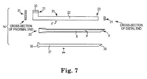
As shown at figure 7, a delivery system 14 can include a multi-component
assembly comprising a tunneler tool (or stylet) 20, an optional core tool 36,
and an
insertion tool 31. Tunneler tool 20 is adapted for receiving at least a
portion, such as
an end portion, of insertion tool (pusher tool) 31 (and also at least a
portion, such as
an end portion, of core tool 36).
26
:A 02818945 2013 05 23
WO 2012/083159
PCT/US2011/065480
In use, systems shown at figures 6B through 6E, and 7, can be used by
making an incision and inserting the tunneler tool into the incision, while a
core tool
or insertion tool is contained in the open inner channel, and a distal end of
the core
tool or insertion tool plugs and blocks the distal end opening of the tunneler
tool.
The assembly of the tunneler tool and core tool or insertion tool can be
advanced
through the incision and through tissue to place a distal end of the tunneler
tool at a
region of supportive tissue on one side of a patient. Plugging the distal open
end
prevents the open distal end of the tunneler tool from cutting a plug of
tissue or
otherwise producing undue trauma. After insertion of the assembly of the
tunneler
tool and the core tool or insertion tool, the core tool or insertion tool can
be removed
to expose and open the lumen (open internal channel) of the tunneler tool. A
distal
end of the insertion tool can then be connected to an anchor of an implant,
and the
insertion tool can be used to push the anchor through the open internal
channel. The
anchor passes within the open internal channel, and the balance of the implant
can
be located within the open internal channel alongside the shaft of the
insertion tool,
or may extend through a longitudinal opening in the tunneler tool to be
located
externally alongside the tunneler tool. When a desired location of the anchor
is
achieved, the insertion tool can be used to insert the anchor into supportive
tissue.
Before or after inserting the anchor into supportive tissue, the tunneler tool
can be
separated laterally from the insertion tool, and then withdrawn from the
tissue path.
These steps can be repeated on an opposite side of the patient, using the same
or a
new set of tools, to place a second anchor of the implant at an opposing
supportive
tissue location.
According to certain embodiments, a distal end of tool insertion tool 31 fits
a
distal end opening of tunneler tool 20 to plug the distal end opening during
use of
the tunneler tool to pass through an incision in a patient and create a tissue
path
using distal end tip 9. With insertion tool 31 inserted and extended into open
internal channel 3 of tunneler tool 20, the assembly of the insertion tool 31
and
tunneler tool 20 can be inserted through an incision of the patient and into
the
interior pelvic region to form a tissue path extending to a location for
placement of
an end of an implant. As shown at figure 7, embodiments of insertion tool 31
can
include a proximal end handle 30, a shaft 22, and a distal end tip 23 that
fits or plugs
27
:A 02818945 2013 05 23
WO 2012/083159
PCT/US2011/065480
a distal end opening of tunneler tool 20. Distal end tip 23 can additionally
be useful
to engage an implant, such as at an anchor 18, to allow insertion tool 31 to
pass the
implant or a portion thereof through tunneler tool 20. Shaft 22 is sized and
shaped
for insertion into tunneler tool 20. Upon insertion of the assembly into the
pelvis of
the patient through the incision, insertion tool 31 can be removed to leave
tunneler
tool 20 positioned within the patient to provide a pathway for inserting the
implant
12 using the same insertion tool 31.
Optionally, a system 14 can additionally include a core tool (or "plug") 36,
or other like device, along with tunneler 20 and insertion tool 31. A core
tool can be
used to plug a distal end opening of tunneler tool 20 (instead of the
insertion tool 31)
to prevent tunneler tool 20 from dissecting or "plugging" tissue of the
patient during
insertion and positioning of tunneler tool 20. As shown at figure 7, a core
tool 36
can include a proximal end handle 35, a shaft 37, and a distal end tip 39 that
fits or
plugs a distal end opening of tunneler tool 20. Shaft 37 is sized and shaped
for
insertion into tunneler tool 20. In use, with core tool 36 inserted and
extended into
open internal channel 3 of tunneler tool 20, the assembly of the core tool 36
and
tunneler tool 20 can be inserted through an incision of the patient and into
the
interior pelvic region to form a tissue path to a location for placement of an
end of
an implant. Upon insertion of the assembly into the pelvis of the patient
through the
incision, core tool 36 can be removed to leave tunneler tool 20 positioned
within the
patient to provide a pathway for inserting implant 12 using insertion tool 31.
Tunneler tool 20, core tool 36, and insertion tool 31 can be straight, curved,
or take on a myriad of other advantageous shapes and configurations. In
various
embodiments, core tool 36 and insertion tool 31 can be the same tool and
operate to
position tunneler tool 30, as well as insert the implant 12.
As described in detail elsewhere herein, and as shown at figure 7, tunneler
tool 20 can include an external communication channel or slot (i.e.,
longitudinal
opening) 5 in communication with open internal channel 3 and also in
communication with a distal end opening of tunneler tool 20. In certain
embodiments, an anchor 18 of a sling 12 can be adapted to be positioned within
internal channel 3, with a portion of the anchor 18 or sling 12 (e.g., mesh
16)
28
2802818945 2013-05-23
WO 2012/083159
PCT/US2011/065480
extending through longitudinal opening 5, to place a portion of the implant
outside
of tunneler tool 20 during use.
Referring now to figures 8A and 8B, illustrated is an example of a tunneler
tool and implant that can be arranged with an anchor of the implant being
passed
through an open internal channel of the tunneler tool, while another portion
of the
implant such as an extension portion, extends through a longitudinal opening
of the
tunneler tool. As shown, anchor 18 is located within open internal channel 3
of
tunneler tool 20, and can slide distally or proximally within open internal
channel 3,
such as by being pushed by a distal end of an insertion tool (not shown) also
located
within open inner channel 3. Anchor 18 is connected to implant 12 by fin 19,
which
extends through longitudinal opening 5. This embodiment of tunneler tool 20,
implant 12, and anchor 18, allows a major portion of implant 12 to be located
outside of tunneler tool 20 during placement of the implant, as insertion tool
31 is
within tunneler tool 20 in engagement with anchor 18. Other embodiments allow
a
major portion of implant 12, or the entire implant, to be located within open
internal
channel 3 of tunneler tool 20 during placement of the implant, alongside a
distal end
of an insertion tool 31 engaged with anchor 18 (see, e.g., figures 4A through
4H, and
figure 5A).
As shown, e.g., at figures 9 through 13, a tunneler tool 20 that includes an
elongate opening (slot) 5 can be adapted such that an anchor 18 can be
positioned
within open internal channel 3 for traversal along the length of tunneler tool
20,
while an implant or portion thereof remains outside of open internal channel
3. In
various embodiments, the anchor 18 can serve as a plug within the tunneler
tool 20,
including to plug a distal end opening during insertion of tunneler tool 20,
until
exiting a distal end. (In other embodiments, a separate plug in the form of a
core
tool 36 can be used to fill the distal end opening of tunneler tool 20 during
insertion
of tunneler tool 20 into a patient.) With anchor 18 disposable within the open
internal channel 3 of tunneler tool 20, a mesh portion 16 of an implant 12 can
be
connected to anchor 18 (e.g., via a fin 19) and extend out of tunneler tool 20
through
longitudinal opening 5. The insertion tool 31 can be coupled with or otherwise
engaged with anchor 18 or another portion of implant 12 such that pushing or
advancing insertion tool 31 likewise advances anchor 18 along open internal
channel
29
2802818945 2013-05-23
WO 2012/083159
PCT/US2011/065480
3 of the tunneler 20. Any desired connector, such as a fin, post, block,
plate, etc., or
other useful structure, can be used to connect anchor 18, with sling 12 (e.g.
mesh
16), with the connector extending through and slidable along longitudinal
opening 5.
The anchors 18 and sling can be separately attached or integrally molded or
joined
together.
Figures 9 through 13 show various embodiments of anchors 18. Certain
embodiments include extendable barbs 40, which can be fixed or adapted to
retract
or expand as detailed herein. Any of the anchors 18 can include various
members,
tabs, connectors, lateral extensions, a base, or like features to facilitate
the
11:1 placement, optional expansion, and holding strength of the anchor
within tissue, as
well as guidance and traversal along or within the open internal channel 3 a
tunneler
tool 20 during surgical placement of the anchor. Further, the anchors 18 can
include
various aperture or other mating features at a proximal end to facilitate
engagement,
or selective engagement, with a distal end 23 of an insertion tool 31.
Referring to figures 9 and 10, illustrated are various embodiments (side
views and end views) of useful anchors (18). Each of illustrated anchors 18
can
include one or more tines or barbs 40 adapted to retract or lay back against
an
anchor base 41, while anchor 18 is located within open internal channel 3 of
tunneler
tool 20. The illustrated anchors 18 also include a fin 19 that can extend
through a
longitudinal opening 5 of a tunneler tool 20, to connect to an implant located
outside
of the tunneler tool 20. Upon exiting the distal end opening of tunneler tool
20, the
bias of retracted barbs 40, while located within open internal channel 3, will
cause
the barbs 40 to extend outward and increase the cross-sectional size of the
anchor
18. Barbs 40 are adapted for tissue penetration while in the non-extended
positions,
and tissue fixation upon expansion to the extended or expanded positions. Each
exemplary anchor 18, at base 411 can include recesses configured to receive
retracted barbs 40. Barbs 40 can be constructed of memory shaped plastic or
metal
materials, include mechanical or living hinges, or can be constructed in other
ways
to facilitate the operable retraction and expansion features described herein.
Figure 11 shows an embodiment of an anchor that includes a distal end 43,
and one or more tines or barbs (or extendable lateral extensions) 40 connected
to the
distal end and adapted to retract or lay back in a retracted position while
anchor 18 is
:A 02818945 2013 05 23
WO 2012/083159
PCT/US2011/065480
located within open internal channel 3 of tunneler tool 20. Anchor 18 does not
include a fin (e.g., 19) or other connecting device capable of extending
through a
longitudinal opening 5 of a tunneler tool 20 to connect to an implant located
outside
of the tunneler tool 20. Anchor 18 as illustrated also does not include a base
(41).
Instead of a base 41, tines or barbs 40 can be supported by distal end 43.
And, when
barbs 40 are in their retracted positions, within an open internal channel 3,
the
retracted barbs 40 can perform the function of a base and can be used to
engage a
distal end of an insertion tool to push anchor 18.
Figure 12 shows an embodiment of an anchor that includes a distal end 43, a
base 41, and a frictional outer surface that includes multiple small ridges or
extensions, e.g., teeth 45. Anchor 18, as illustrated, does not include a fin
(e.g., 19),
but a fin or other connecting device capable of extending through a
longitudinal
opening of a tunneler tool 20 to connect to an implant located outside of the
tunneler
tool 20, could be included. Anchor 18 as illustrated also does not include
elongate,
extendable barb features 40 or other elongate lateral extensions, but instead
includes
circumferential ridges, teeth, or other frictional structure 45 to allow
anchor 18 to be
inserted into tissue and subsequently hinder, resist, or prevent removal from
the
tissue.
Figure 13 shows an embodiment of an anchor 18 that includes a distal end
43, base 41, and one or more tines or barbs 40 connected to base 41 and distal
end
43. Anchor 18 does not include a fin (e.g., 19) or other connecting device
capable of
extending through a longitudinal opening of a tunneler tool 20 to connect to
an
implant located outside of the tunneler tool 20, but a fin or other such
structure could
be included. Barbs 40 are adapted to retract in a retracted position while
anchor 18
is located within open internal channel 3 of tunneler tool 20. Upon exiting
the distal
end opening of tunneler tool 20, the bias of the retracted barbs 40, while
located
within open internal channel 3, will cause the barbs 40 to extend outward and
increase the cross-sectional size of anchor 18. Barbs 40 are adapted for
tissue
penetration while in the non-extended positions, and tissue fixation upon
expansion
to the extended or expanded positions. Barbs 40 can be constructed of metal or
polymeric memory shaped materials, include mechanical or living hinges, or can
be
31
:A 02818945 2013 05 23
WO 2012/083159
PCT/US2011/065480
constructed in other ways to facilitate the operable retraction and expansion
features
described herein.
Figures 14A and 14B illustrate a delivery tool system, including an implant,
that includes a tunneler tool 20, a core tool 36, and an insertion tool 31. As
shown
in figure 14A, tunneler tool 20 can include a handle portion 42, curved shaft
8 (of
constant or varied radius) that includes distal end tip 9, open internal
channel 3, and
longitudinal opening 5. Longitudinal opening 5 is located on the outer curved
surface of shaft 8. A communication port or opening (proximal end opening) 44
can
be included at a proximal end portion of shaft 8, at a location adjacent to
handle 42
meeting a proximal end of shaft 8. This location of proximal end opening 42
facilitates introduction of the insertion tool 31 during a surgical procedure.
For
example, after placement of shaft 8 through an incision in a patient, proximal
end
opening 44 can remain external to the patient and accessible to a surgeon or
other
User.
Figure 14B illustrates tunneler tool 20 and insertion tool 31 in use, wherein
shaft 22 of insertion tool 20, engaged with anchor 18 of implant 12, is
introduced
through proximal end opening 44 and advanced along open internal channel 3 of
tunneler tool 20. Mesh portionl 6 is connected to anchor 18 by fin 19 or
another
connecting structure, and rides outside of and along the outer surface length
of
tunneler tool 20. Portions or all of tunneler tool 20 and insertion tool 31
can be
constructed of a generally flexible material, or a rigid material, to
facilitate traversal
and component introduction.
Figure 15 shows an embodiment of insertion tool 31, having shaft 22 that
includes one or more angle or bend 48. Also shown is implant 12 having a
central
mesh portion 16 with integral support portion and extension portions, and two
self-
fixating tips 18 at opposing ends of the two extension portions. Shaft 22 can
be a
relatively stiff metal (stainless steel or nitinol) or stiff plastic, attached
at proximal
end 21 to handle 30, and having distal end 23 adapted to engage a proximal end
(e.g., base) of self-fixating tip 18. Shaft 22 includes two bends, 48a and
48b. Each
bend is approximately a 90 degree angle, but other angles may also be useful.
The
combined angles of the bends allow the longitudinal axis of shaft 22 at distal
end tip
23 to be parallel with a longitudinal axis of handle 30 (i.e., the sum of the
angles of
32
:A 02818945 2013 05 23
WO 2012/083159
PCT/US2011/065480
the two bends may be approximately 180 degrees, e.g., from 160 degrees to 200
degrees.
In use, as illustrated at figures 16A, 16B, and 16C, shaft 22, having bends
48a and 48b, allows a distal portion of shaft 22 (distal to both bends, i.e.,
between
distal end 23 and bend 48b) to be positioned within open internal channel 3 of
an
tunneler tool 20, while a proximal portion of shaft 22 (proximal to both
bends, i.e.,
between handle 30 and bend 48a) is located externally of open inner channel 3.
in
this manner, bends 48a and 48b may facilitate guidance and use of insertion
tool 31
with implant 12 and tunneler tool 20. For instance, bends 48a and 48b can
permit
maneuvering of a distal portion of shaft 22 through a distal portion of
elongate
opening (slot) 5 of tunneler tool 20. Figure 16A shows a distal portion of
insertion
tool shaft 22, including distal end 23 engaged with anchor 18, entering
proximal end
opening 44 of tunneler tool 20. The portion of shaft 22 between distal end 23
and
bend 48b enters open internal channel 3, and the remaining length of shaft 22
remains exterior to open internal channel 3. Implant 12 and the portion of
shaft 22
that extends between bend 48a and bend 48b, pass through longitudinal opening
5 to
be located exterior to open internal channel 3. Figure 16B shows that the
assembly
of insertion tool 31 and implant 12 can be advanced longitudinally and
distally along
a length of open internal channel 3, to a distal end of shaft 8 of tunneler
tool 20.
With implant 12 and anchor 18 located at a desired location at the distal end
of
tunneler tool 20, and either before or after placement of anchor 18 within
supportive
tissue, tunneler tool 20 can be withdrawn from the patient by movement of
tunneler
tool 20 laterally or proximally relative to insertion tool 31 (see arrows), to
cause
distal end 23 and anchor 18 to separate from the tunneler tool 20, e.g., to
pass
through longitudinal opening 5.
Accordingly, as illustrated and described herein, the tools, systems, and
implants can be useful for treating a pelvic condition by steps that include
the
following. A tunneler tool can be used to make a tissue path from an incision
of a
patient to a location of a pelvic region at which an end of an implant is to
be secured
to supportive tissue. The incision may be an external incision or a vaginal
incision,
and may be a medial incision (e.g., vaginal or perineal), or otherwise. A
distal end
of the tunneler tool can be advanced through the incision and advanced through
33
:A 02818945 2013 05 23
WO 2012/083159
PCT/US2011/065480
internal tissue of the patient to supportive tissue. Optionally and
preferably, a distal
end opening of the tunneler tool can be filled (e.g., plugged) to prevent
undue
trauma to the patient during formation of the tissue path, by the distal end
tip and
distal end opening of the tunneler tool. The distal end opening may be plugged
by
placement of a distal end of a core tool at the location of the distal end
opening.
Alternately, the distal end opening may be plugged by placement of a distal
end of
an insertion tool at the location of the distal end opening. As yet another
alternative,
the distal end opening may be plugged by placement of an anchor (e.g., 18,
such as a
self-fixating tip) at the location of the distal end opening, the anchor being
engaged
with a distal end of an insertion tool placed within the tunneler tool.
Upon desired placement of the tunneler tool within a patient, a core tool, if
used to fill the distal end opening of the tunneler tool, can be removed from
the
tunneler tool. If an insertion tool (without an anchor engaged at the distal
end) was
used to fill the distal end opening of the tunneler tool, the insertion tool
can be
removed from the tunneler tool. (If an insertion tool (with an anchor engaged
at the
distal end) was used to fill the distal end opening of the tunneler tool, a
next step can
be to remove the tunneler tool from the patient. See below.)
After placement of the tunneler tool and removal of a core tool or insertion
tool used to fill the distal end opening during use of the tunneler tool to
produce a
tissue path, an implant (or a portion thereof) may be introduced at a proximal
end
opening of the tunneler tool using an insertion tool. The insertion tool can
advance
the implant (or a portion thereof) along the length of the tunneler tool
shaft, to the
distal end of the tunneler tool shaft. The implant may be contained entirely
within
the tunneler tool, or a portion of the implant may extend through a
longitudinal
opening to a location external to the tunneler tool. Likewise, a shaft of the
insertion
tool may be contained entirely within the tunneler tool, or a portion of the
shaft may
extend through a longitudinal opening to a location external to the tunneler
tool and
remain external to the tunneler tool.
After placement of the implant (or a portion thereof) at the distal end of the
tunneler tool shaft, and at a location for securing to supportive tissue, the
tunneler
tool can be removed. For example, the tunneler tool may be laterally withdrawn
away from any portion of the implant and tunneler tool positioned with the
tunneler
34
2802818945 2013-05-23
WO 2012/083159
PCT/US2011/065480
tool, by aligning the insertion tool with a longitudinal channel of the
tunneler tool
and laterally separating the tunneler tool from the insertion tool. The
insertion tool
and implant pass from the open internal channel of the tunneler tool, through
the
longitudinal opening, and become free of the tunneler tool. The tunneler tool
can
then be withdrawn in. a proximal direction and removed from the patient.
The insertion tool can be used to place the anchor at supportive tissue, e.g.,
at
tissue of an obturator foramen. This part of the method can be performed
either
before or after removal of the tunneler tool from the tissue path and the
patient, but
can preferably be performed according to the latter option, after removal of
the
tunneler tool from the patient. Advantageously, the latter option allows for
the step
of placing an anchor at supportive tissue to be separate from a step of
creating a
tissue path extending to the supportive tissue, from an incision, such as an
external
incision (e.g., a medial incision).
In use, a system (e.g., 14) described herein can be used bilaterally, to place
two ends of an implant in a patient, one end at a right side of the patient
and the
other end at a left side of the patient, preferably through a single medial
incision. A
method and techniques described immediately above can be performed on one side
of a patient to place a first end of an implant, then on another (opposite)
side of a
patient to place a second end of an implant. One end or anchor 18 of an
implant 12
will be attached at each side of the interior pelvic region of the patient
such that a
support portion (e.g. mesh 16) extends under tissue or an organ in need of
physical
support (e.g., urethra, bladder, bladder neck, vaginal tissue, etc.).
According to certain specific embodiments of delivery tool systems, an
insertion tool 31 can include two separate but joinable devices 31a, 3 1 b,
i.e., a
system can include two separate insertion tools. Each separate insertion tool
31a
and 31 b can include a handle 30, shaft 22, distal end 23, or other features
and
components described herein as a feature of an insertion tool 31. However, as
shown in figures 19 through 22, a portion of each of insertion tool 31a, 31b
can be
adapted to link or otherwise connect to the other insertion tool. Joining
surfaces 51a
and 51b can be located at a proximal end of an insertion tool 31, such as at
opposing
and joinable surfaces of opposing handles, and can include complementary
joining
:A 02818945 2013 05 23
WO 2012/083159
PCT/US2011/065480
surfaces that when engaged will limit, inhibit, or prevent relative movement
between
the surfaces in at least one direction.
Joining surfaces 51a and 51b may include complementary and opposing
engagement surfaces having opposing gears, teeth, interacting surfaces, pins,
members, slots, frictional features, and other like features to facilitate a
frictional
engagement, linkage, joining, selective connectivity, and optional locking of
the two
insertion tools 31a and 32b. See figures 17A, 17B, 17C, 17D, and 17E, and
figures
18A, 18B, 18C, 18D, 18E, and 18F. Figures 17B and 17C show top views of
insertion tools 31a and 31b having joinable surfaces 51a and Sib, wherein the
joinable surfaces comprise a series of teeth or ridges, with the joinable
surfaces
disengaged (figure 17B) or engaged (figure 17C). Figure 17E shows side views
(each insertion tool is rotated ninety degrees) of insertion tools 31a and 31b
having
joinable surfaces 51a and 51b. Figures 18A and 18B show top and side views,
respectively, of insertion tools 31a and 31b having joinable surfaces 51a and
51b,
wherein the joinable surfaces comprise an elongate peg and an opposing
elongate
channel, allowing for some relative longitudinal movement when surfaces 51a
and
5 lb are joined. Figures 18C and 18D show top and side views, respectively, of
insertion tools 31a and 31b having joinable surfaces 51a and 51b comprising a
series
of peg extensions and opposing complementary holes. Figures 18E and 18F show
top and side views, respectively, of insertion tools 31a and 31b having
joinable
surfaces 51a and 51b comprising a series of gear extensions and opposing
complementary channels.
In use, each of the two individual insertion tools 31a and 31b can be used
separately, without joining their respective joining surfaces 51a and 51b,
with each
tool being used, via a single incision, to place an end of an implant at two
desired
supportive tissue locations on two opposing sides a patient. See, e.g.,
figures 19 and
20. For example, one insertion tool can be useful to place an end of an
implant,
through an incision, at or near supportive tissue on a left side of a patient,
e.g., at a
left obturator foramen. Using the same medial or external incision, the other
insertion tool can be useful to place a second end of the implant at or near
opposing
supportive tissue on a right side of the patient, e.g., at a right obturator
foramen.
Figure 19 shows two tunneler tools, 20a and 20b, one for a right side of a
patient and
36
2802818945 2013-05-23
WO 2012/083159
PCT/US2011/065480
one for a left side of the patient. Figure 19 also shows two insertion tools,
31a and
31b. Each set of tools can be the same, and can include a shaft that is either
straight
or curved, or one tunneler tool and one insertion tool can be specifically
designed for
use on a right side of a patient and the other tunneler tool and the other
insertion tool
can be specifically designed for use on the left side of the patient.
Insertion tools 31a and 3ab can be used independently of each other, as
stated, during insertion, until the user desires to manipulate ends or anchors
18 of
implant 12 together. Figure 19 shows insertion tools 31a and 31b in a pre-
deployment configuration with handles 30a and 30b separated. Figure 20 shows
the
same insertion tools 31a and 31b after formation of a tissue path, during
placement
of implant 12 (i.e., anchors 18) through tunneler tools 20, such that barbs 40
of
anchors 18 can expand upon exiting the distal end openings of the respective
tunneler tools 20. During deployment, before or after removal of the two
tunneler
tools 20, handles 30a and 30b can be contacted and joined, to likewise join
insertion
tools 31a and 31b. With insertion tools 31a and 31b joined at handles 20a and
30b,
and optionally with removal of the two tunneler tools 20, the user can grasp
both
insertion tools (31a and 3 lb), to allow the user to manipulate and control
both
handles with a single hand. The user can have combined control of both the
left side
anchor 18 and the right side anchor 18 of implant 12, to improve control of
the
placement and tensioning of the implant being surgically installed.
Various shapes and configurations for the tunnelers 20, insertion tools 31 and
other components and tools are envisioned depending on the particular surgical
application or anatomy of the patient (male or female).
A variety of materials may be used to form portions or components of the
system 14, including nitinol, polymers, elastomers, thermoplastic elastomers,
metals,
ceramics, springs, wires, plastic tubing, and the like. The system 14 and its
components and methods may have a number of suitable configurations known to
one of ordinary skill in the art.
All patents, patent applications, and publications cited herein are hereby
incorporated by reference in their entirety as if individually incorporated,
and
include those references incorporated within the identified patents, patent
applications and publications.
37
:A 02818945 2013 05 23
WO 2012/083159
PCT/US2011/065480
Numerous modifications and variations of the present invention are possible
in light of the teachings herein. It is therefore to be understood that within
the scope
of the appended claims, the invention may be practiced other than as
specifically
described herein.
38