Note: Descriptions are shown in the official language in which they were submitted.
CA2819038
1
Humanized Antibodies to LIV-1 and Use of Same to Treat Cancer
[0001] <deleted>
BACKGROUND
[0002] LIV-1 is a member of the LZT (LIV-1-ZIP Zinc Transporters) subfamily of
zinc
transporter proteins. Taylor etal., Biochim. Biophys. Acta 1611:16-30 (2003).
Computer
analysis of the LIV-1 protein reveals a potential metalloprotease motif,
fitting the consensus
sequence for the catalytic zinc-binding site motif of the zinc
metalloprotease. LIV-1 mRNA is
primarily expressed in breast, prostate, pituitary gland and brain tissue.
[0003] The LIV-I protein has also been implicated in certain cancerous
conditions, e.g. breast
cancer and prostate cancer. The detection of LIV-1 is associated with estrogen
receptor-positive
breast cancer, McClelland et al., Br. J. Cancer 77:1653-1656 (1998), and the
metastatic spread of
these cancers to the regional lymph nodes. Manninget al., Eur. J. Cancer
30A:675-678 (1994).
SUMMARY OF THE CLAIMED INVENTION
100041 The invention provides a humanized antibody comprising a mature heavy
chain variable
region having an amino acid sequence at least 90% identical to SEQ ID NO:53
provided that
position H27 is occupied by L, position H29 is occupied by I, H30 by E and H94
by V and a
mature light chain variable region at least 90% identical to SEQ ID NO:60
provided position L36
is occupied by Y and position L46 by P. Optionally, the humanized antibody
comprises three
CDRs of SEQ ID NO:53 and three CDRs of SEQ ID NO:60. Those CDRs are shown in
Figure
16. Optionally, position H76 is occupied by N. Optionally, the humanized
comprises a mature
heavy chain variable region having an amino acid sequence at least 95%
identical to SEQ ID
NO:53 and a mature light chain variable region at least 95% identical to SEQ
ID NO:60.
Optionally, the mature heavy chain variable region is fused to a heavy chain
constant region and
the mature light chain constant region is fused to a light chain constant
region. Optionally, the
heavy chain
Date Recue/Date Received 2020-04-15
CA 02110038 20%0544
WO 2012/078688 PCT/US2011/063612
2
constant region is a mutant form of natural human constant region which has
reduced
binding to an Fcgamma receptor relative to the natural human constant region.
Optionally, the heavy chain constant region is of IgG1 isotype. Optionally,
the heavy
chain constant region has an amino acid sequence comprising SEQ ID NO:44 and
the
light chain constant region has an amino acid sequence comprising SEQ ID
NO:42.
Optionally, the heavy chain constant region has an amino acid sequence
comprising SEQ
ID NO:46 (5239C) and the light chain constant region has an amino acid
sequence
comprising SEQ ID NO:42. In some such humanized antibodies, any differences in
CDRs of the mature heavy chain variable region and mature light variable
region from
SEQ ID NOS. 52 and 60 respectively reside in positions H60-H65. In some such
humanized antibodies, the mature heavy chain variable region has an amino acid
sequence designated SEQ ID NO:52 or 53 and the mature light chain variable
region has
an amino acid sequence designated SEQ ID NO: 59 or 60. In some such humanized
antibodies, the mature heavy chain variable region has an amino acid sequence
designated SEQ ID NO:53 and the mature light chain variable region has an
amino acid
sequence designated SEQ ID NO:60. Some such humanized antibodies are
conjugated to
a cytotoxic or cytostatic agent. Some such humanized antibodies have an
association
constant for human or cynomulgus monkey LIV-1 of 0.5 to 2 x 10⁹ .
[0005] The invention also provides a humanized antibody comprising a mature
heavy
chain variable region comprising the three Kabat CDRs of SEQ ID NO:52, wherein
position 1-127 is occupied by L, position 1-129 is occupied by 1, 1-130 by E,
H76 by N, and
H94 by V and a mature light chain variable region comprising the three Kabat
CDRs of
SEQ ID NO:60 provided position L36 is occupied by Y and position L46 by P.
The invention also provides a nucleic acid encoding a mature heavy chain
variable region
and/or a mature light chain variable region of any of the above defined
humanized
antibodies.
[0006] The invention further provides a method of treating a patient having or
at risk of
cancer, comprising administering to the patient an effective regime of any of
the above
defined humanized antibodies. The cancer can be for example a breast cancer,
cervical
cancer, melanoma, or a prostate cancer.
CA 02810038 2013-0544
[0007] The invention further provides a pharmaceutical composition comprising
a
humanized antibody as defined above.
[0012]The invention further provides methods of treating a subject afflicted
with a
melanoma that expresses the LIV-1 protein by administering to the subject a
LIV-1
specific antibody or a LIV-1 antibody drug conjugate, in an amount sufficient
to inhibit
growth of the melanoma cancer cells.
[0012]The invention further provides methods of treating a subject afflicted
with a
cervical cancer that expresses the LIV-1 protein by administering to the
subject a LIV-1
specific antibody or a LIV-1 antibody drug conjugate, in an amount sufficient
to inhibit
growth of the cervical cancer cells.
[0008] The invention further provides a humanized antibody comprising a mature
heavy
chain variable region having an amino acid sequence at least 90% identical to
HB (SEQ
ID NO:10) and a mature light chain variable region at least 90% identical to
LB (SEQ ID
NO:15). Optionally, the antibody comprises a mature heavy chain variable
region having
an amino acid sequence at least 95% identical to HB and a mature light chain
variable
region at least 95% identical to LB. Optionally, in any such antibody,
positions H29,
H30 and H76 are occupied by I, E and N, and L36 is occupied by Y. Optionally,
any
difference in the variable region frameworks of the mature heavy chain
variable region
and SEQ ID NO:10 is/are selected from the group consisting of H27 occupied by
F, 1128
occupied by N, H48 occupied by I, H66 occupied by K, H67 occupied by A, H71
occupied by A, H76 occupied by N, H93 occupied by N, H94 occupied by V, L37
occupied by L, L39 occupied by K, L45 occupied by K, and L46 occupied by L.
Optionally, the 3 CDRs of the mature heavy chain variable region are those of
SEQ ID
NO. 10 and the 3 CDRs of the mature light chain variable region are those of
SEQ ID
NO:15. The CDRs are shown in Fig. 1. Optionally, the mature heavy chain
variable
region is fused to a heavy chain constant region and the mature light chain
constant
region is fused to a light chain constant region. Optionally, the heavy chain
constant
region is a mutant form of natural human constant region which has reduced
binding to
an Fcgamma receptor relative to the natural human constant region. Optionally,
the
heavy chain constant region is of IgG1 isotype. Optionally, the heavy chain
constant
region has an amino acid sequence comprising SEQ ID NO:6 and the light chain
constant
3
CA 02110038 20%0544
WO 2012/078688
PCT/US2011/063612
4
region has an amino acid sequence comprising SEQ ID NO:4. Optionally, the
heavy
chain constant region has an amino acid sequence comprising SEQ ID NO:8
(S239C) and
the light chain constant region has an amino acid sequence comprising SEQ ID
NO:4.
Optionally, any differences in CDRs of the mature heavy chain variable region
and
mature light variable region from SEQ ID NOS. 10 and 15 respectively reside in
positions H60-H65. Optionally, the mature heavy chain variable region has an
amino
acid sequence comprising SEQ ID NO:10 and the mature light chain variable
region has
an amino acid sequence comprising SEQ ID NO:15. Optionally, the antibody is
conjugated to a cytotoxic or cytostatic agent. Preferred humanized antibodies
having
greater affinity for LIV-1 than the antibody BR2-14a In another embodiment,
the
humanized antibody has an association constant for human or cynomolgus monkey
LIV-
1 of 0.5 to 2 x 10⁹M-1.
[0009] The invention further provides a humanized antibody comprising a mature
heavy
chain variable region comprising the 3 CDRs of SEQ ID NO:10 and wherein
positions
H29, H30 and H76 are occupied by I, E and N respectively, and a mature light
chain
variable region comprising the 3 CDRs of SEQ ID NO:15, and wherein position
L36 is
occupied by Y.
[0010] The invention further provides a nucleic acid encoding a mature heavy
chain
variable region and/or a mature light chain variable region of any of the
humanized
antibodies described above.
[0011] The invention further provides a method of treating a patient having or
at risk of
cancer, comprising administering to the patient an effective regime of a
humanized
antibody as described above. Optionally, the cancer is breast cancer, cervical
cancer,
melanoma, or a prostate cancer.
[0012] The invention further provides a pharmaceutical composition comprising
a
humanized antibody as described above.
[0013] The invention further provides a method of treating a patient having or
at risk of
triple negative breast cancer, comprising administering to the patient an
effective regime
of an antibody that specifically binds to LIV-1. Optionally, in such methods,
the
antibody is conjugated to a cytotoxic or cytostatic agent.
CA2819038
4a
[0013a] Various embodiments of the claimed invention relate to a humanized
antibody specifically
binding LIV-1 comprising a mature heavy chain variable region comprising a
heavy chain CORI
comprising the amino acid sequence DYYMH, a heavy chain CDR2 comprising the
amino acid
sequence WIDPENGDTEYGPKFQG, and a heavy chain CDR3 comprising the amino acid
sequence
HNAHYGTWFAY, wherein the mature heavy chain variable region comprises an amino
acid sequence
at least 95% identical to SEQ ID NO:53 provided that position H27 is occupied
by L, position H29 is
occupied by I, position H30 is occupied by E and position H94 is occupied by V
and a mature light
chain variable region comprising a light chain CDR1 comprising the amino acid
sequence
RSSQSLLHSSGNTYLE, a light chain CDR2 comprising the amino acid sequence
KISTRFS, and a
light chain CDR3 comprising the amino acid sequence FQGSHVPYT, wherein the
mature light chain
variable region comprises an amino acid sequence at least 95% identical to SEQ
ID NO:60 provided
position L36 is occupied by Y and position L46 is occupied by P, wherein the
amino acids are
numbered according to the Kabat numbering scheme.
10013b1 Various embodiments of the claimed invention also relate to nucleic
acid encoding the mature
heavy chain variable region and the mature light chain variable region of the
humanized antibody as
claimed.
[0013c] Various embodiments of the claimed invention also relate to a vector
comprising the nucleic
acid as claimed.
[0013d] Various embodiments of the claimed invention also relate to a host
cell comprising the nucleic
acid as claimed.
[0013e] Various embodiments of the claimed invention also relate to a method
of producing the
humanized anti-LW-1 antibody of any one of claims 1-10 comprising culturing
the host cell of claim 26
or 27 under a condition suitable for production of the anti-LIV-1.
[0013f1 Various embodiments of the claimed invention also relate to a method
of producing an anti-
LIV-1 antibody-drug conjugate comprising culturing the host cell of claim 26
or 27 under a condition
suitable for production of the anti-L1V-1 antibody of any one of claims 1-10;
isolating the anti-L1V-1
antibody produced from the host cell; and conjugating the anti-LIV-1 antibody
to a cytotoxic or
cytostatic agent.
10013g] Various embodiments of the claimed invention also relate to use of the
humanized antibody
according as claimed, for the manufacture of a medicament for treating cancer
in a subject having or at
risk of cancer, wherein the cancer expresses LIV-1.
10013h1 Various embodiments of the claimed invention also relate to use of the
humanized antibody as
claimed, for treating cancer in a subject having or at risk of cancer, wherein
the cancer expresses LIV-1.
Date Recue/Date Received 2020-04-15
CA 02110038 20%0544
WO 2012/078688
PCT/US2011/063612
BRIEF DESCRIPTION OF THE FIGURES
[0014] Figure 1 shows an alignment of the amino acid sequences of the parental
murine mAb (referred to as BR2-14a) with the humanized LIV-1 heavy (upper two
panesl) and light chain variable (lower two panels) regions.
[0015] Figure 2 shows the binding curves for the humanized LIV-1 mAbs and the
parental murine antibody (referred to as BR2-14a).
[0016] Figure 3 shows the results of competition binding studies of the
humanized
LIV-1 mAbs and the parental murine antibody (referred to as BR2-14a). The
numbers in
parentheses after each variant indicate the number of back mutations.
[0017] Figure 4 shows the results of saturation binding studies on MCF7 cells.
BR2-
14a-AF refers to AF-labeled parental murine antibody. hLIV-14 refers to AF-
labeled
HBLB antibody, a humanized antibody that specfically binds to LIV-1.
[0018] Figure 5 shows the results of competition binding studies on CHO cells
expressing recombinant LIV-1 protein. BR2-14a refers to the parental murine
antibody.
hLIV-14 HBLB WT refers to the HBLB antibody. hLIV-14 HBLB S239C refers to the
HBLB antibody having serine to cysteine substititions at each position in the
heavy chain.
[0019] Figure 6 shows an analysis of LIV-1 protein expression by IHC on post-
hormone treated breast cancer patient samples.
[0020] Figure 7 shows an analysis of LIV-1 protein expression by 11-IC on
hormone-
refractory metastatic prostate cancer patient samples.
[0021] Figure 8 shows an analysis of LIV-1 protein expression by IHC on triple
negative breast cancer patient samples.
[0022] Figure 9 shows the results of cytotoxicity assays on hLIV-14 antibody
drug
conjugates, i.e., the HBLB mAb conjugated to veMMAE (1006) or mcMMAF (1269),
as
well as conjugates of control murine (mIgG) and human (hIgG) antibodies. hLIV-
14-
SEA-1006 refers to a non-fucosylated form of the HBLB mAb conjugated to veMMAE
(1006).
[0023] Figure 10 shows the results of an in vitro ADCC assay on MCF7 cells
using
human NK cells (donor 1; WV). hLIV-14 WT refers to the HBLB mAb. hLIV-14 SEA
refers to the non-fucosylated form of the HBLB mAb. hLIV-14 mcMMAF refers to
an
CA 02110038 20%0544
WO 2012/078688
PCT/US2011/063612
6
antibody drug conjugate of the HBLB mAb conjugated to mcMMAF. hLIV-14
veMMAE refers to an antibody drug conjugate of the HBLB mAb conjugated to
veMMAE. hLIV-14 SEA veMMAE refers to a non-fucosylated form of the HBLB mAb-
veMMAE antbody drug conjugate.
[0024] Figure 11 shows the results of an in vitro ADCC assay on MCF7 cells
using
human NK cells (donor 2). hLIV-14 WT refers to the HBLB mAb. hLIV-14 SEA
refers
to the non-fucosylated form of the HBLB mAb. cLIV-14 SEA refers to the non-
fucosylated form of the chimeric parental murine antibody. hLIV-14 mcF(4)
refers to an
antibody drug conjugate of the HBLB mAb with an average of 4 mcMMAF drug
linker
molecules per antibody. hLIV-14 vcE(4) refers to an antibody drag conjugate of
the
HBLB mAb with an average of 4 veMMAE drug linker molecules per antibody. hL1V-
14 vcE(4) SEA refers to a non-fucosylated form of the HBLB mAb-vcMMAE antibody
drug conjugate having an average of four veMMAE drug linker molecules per
antibody.
hIgG refers to control human IgG. HOO-mcF(4) refers to a control antibody drug
conjugate of a nonbinding antibody with an average of 4 mcMMAF drug linker
molecules per antibody. HOO-vcE(4) refers to a control antibody drug conjugate
of a
nonbinding antibody with an average of 4 veMMAE drug linker molecules per
antibody.
100251 Figure 12 shows the results of a xenograft study of the MCF7 breast
cancer line
in nude mice. cLIV-14-mcMMAF(4) refers to an antibody drug conjugate of the
chimeric form of the parental murine antibody having an average of 4 mcMMAF
drug
linker molecules per antibody_ cLIV-14-vcMMAE(4) refers to an antibody drug
conjugate of the chimeric form of the parent murine antibody having an average
of 4
veMMAE drug linker molecules per antibody. HOO-mcMMAF(4) refers to an antibody
drug conjugate of a nonbinding control antibody having an average of 4 mcMMAF
drug
linker molecules per antibody. HOO-veMMAE(4) refers to an antibody drug
conjugate of
a nonbinding control antibody having an average of 4 vcMMAE drug linker
molecules
per antibody. The dose and time of administration of indicated on the figure.
[0026] Figure 13 shows the results of a xenograft study of the PC3 prostate
cancer line
in male nude mice. cLIV-14-vcMMAE(4) refers to an antibody drug conjugate of
the
chimeric form of the parent murine antibody having an average of 4 veMMAE drug
linker molecules per antibody. hBU12- vcMMAE(4) refers to an antibody drug
CA 02110038 20%0544
WO 2012/078688
PCT/US2011/063612
7
conjugate of an anti-CD19 antibody having an average of 4 vcMMAE drug linker
molecules per antibody. The dose and time of administration of indicated on
the figure.
[0027] Figure 14 shows the results of a xenograft study of the MCF7 breast
cancer line
in nude mice. hLIV-14-veMMAE (4) refers to an antibody drug conjugate of the
HBLB
antibody having an average of 4 vcMMAE drug linker molecules per antibody.
hLIV-
14d-veMMAE (2) refers to an antibody drug conjugate of the HBLB antibody
having an
average of 2 vcMMAE drug linker molecules per antibody, each conjugated at the
S239C
position of each heavy chain. HOO-vcMMAE(4) refers to an antibody drug
conjugate of a
nonbinding control antibody having an average of 4 vcMMAE drug linker
molecules per
antibody. The dose and time of administration of indicated on the figure.
[0028] Figure 15 shows the results of a xenograft study of the PC3 prostate
cancer line
in male nude mice. hLIV-14-vcM1vIAE (4) refers to an antibody drug conjugate
of the
HBLB antibody having an average of 4 vcMMAE drug linker molecules per
antibody.
hLIV-14-mcMMAF(4) refers to an antibody drug conjugate of the HBLB antibody
having an average of 4 mcMMAF drug linker molecules per antibody. hLIV-14d-
vcMMAE(2) refers to an antibody drug conjugate of the HBLB antibody having an
average of 2 vcMMAE drug linker molecules per antibody, each conjugated at the
S239C
position of each heavy chain. hLIV-14d-mcMMAF(2) refers to an antibody drug
conjugate of the HBLB antibody having an average of 2 mcMMAF drug linker
molecules
per antibody, each conjugated at the S239C position of each heavy chain. HOO-
vcMMAE(4) refers to an antibody drug conjugate of a nonbinding control
antibody
having an average of 4 vcMMAE drug linker molecules per antibody. HOO-
mcMMAF(4)
refers to an antibody drug conjugate of a nonbinding control antibody having
an average
of 4 mcMMAF drug linker molecules per antibody. The dose and time of
administration
of indicated on the figure.
[0029] Figures 16A and 16B show alignments of humanized heavy chain (Figure
16A) and light chain (Figure 16B) mature variable regions with those of the
mouse BR2-
22a.
[0030] Figure 17 shows competition binding assays of different permutations of
humanized heavy chains HA-HF and humanized light chains LA-LF derived from the
murine monoclonal anti LIV-1 antibody BR2-22a. The total number of murine back
CA 02110038 20%0544
WO 2012/078688
PCT/US2011/063612
8
mutations in each light or heavy chain is shown in parentheses. Only HELF
showed
sufficient retention of binding.
[0031] Figure 18 shows systematic variation of the HE and LF chains to test
contribution
of individual backmutations to antigen binding. Sites of potential somatic
hypermutation
are in parentheses. Mouse residues are underlined. The remaining residues are
human
germline residues.
[0032] Figure 19 shows competition binding of the LF variants on the top of
the figure.
The tested back mutations are shown in the bottom of the figure. Mouse
residues are
underlined. The remaining residues are human germline residues.
[0033] Figure 20 shows competition binding of the HE variants on the top of
the figure.
The tested back mutations are shown in the bottom of the figure. Mouse
residues are
underlined. The remaining residues are human germline residues.
[0034] Figure 21 shows competition binding of different permutations of HE,
HF, HG
and LF and LG.
[0035] Figure 22 shows saturation binding of humanized LIV14 antibody and
humanized LIV22 antibody on human and cynomolgus LIV-1 expressed from CHO
cells.
[0036] Figure 23 shows cytotoxic activity of humanized LIV22-veMMAE on MCF-7
cells after 144 hr of treatment. h00-1006 is a control drug-conjugated
antibody.
[0037] Figure 24 shows cytotoxic activity of hLIV22-mcMMAF on MCF-7 cells
after
144 hr of treatment. h00-1269 is a control drug-conjugated antibody.
[0038] Figure 25 shows the activity of hL1V22 antibody on PC3 (DSMZ) prostate
carcinoma model in nude female mice. Dose days are indicated by triangles on
the X-
axis.
[0039] Figure 26 shows that activity of hLIV22 antibody on MCF7 (NCI) breast
carcinoma tumors in nude mice.
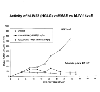
[0040] Figure 27 compares the activity of hLIV22 and hLIV14 in the same model
as
Figure 26.
[0041] Figure 28 shows an analysis of LIV-1 protein expression by IHC on
melanoma
cancer patient samples.
CA 02119038 20%0544
WO 2012/078688
PCT/US2011/063612
9
DEFINITIONS
[0042] Monoclonal antibodies are typically provided in isolated form. This
means that
an antibody is typically at least 50% w/w pure of interfering proteins and
other
contaminants arising from its production or purification but does not exclude
the
possibility that the monoclonal antibody is combined with an excess of
pharmaceutical
acceptable carrier(s) or other vehicle intended to facilitate its use.
Sometimes
monoclonal antibodies are at least 60%, 70%, 80%, 90%, 95 or 99% w/w pure of
interfering proteins and contaminants from production or purification.
[0043] Specific binding of a monoclonal antibody to its target antigen means
an affinity
of at least 106, 107, 108, 109, or 1010 M-1. Specific binding is detectably
higher in
magnitude and distinguishable from non-specific binding occurring to at least
one
unrelated target. Specific binding can be the result of formation of bonds
between
particular functional groups or particular spatial fit (e.g., lock and key
type) whereas
nonspecific binding is usually the result of van der Waals forces. Specific
binding does
not however necessarily imply that a monoclonal antibody binds one and only
one target.
[0044] The basic antibody structural unit is a tetramer of subunits. Each
tetramer
includes two identical pairs of polypeptide chains, each pair having one
"light" (about 25
kDa) and one "heavy" chain (about 50-70 lcDa). The amino-terminal portion of
each
chain includes a variable region of about 100 to 110 or more amino acids
primarily
responsible for antigen recognition. This variable region is initially
expressed linked to a
cleavable signal peptide. The variable region without the signal peptide is
sometimes
referred to as a mature variable region. Thus, for example, a light chain
mature variable
region, means a light chain variable region without the light chain signal
peptide. The
carboxy-terminal portion of each chain defines a constant region primarily
responsible
for effector function.
[0045] Light chains are classified as either kappa or lambda. Heavy chains are
classified as gamma, mu, alpha, delta, or epsilon, and define the antibody's
isotype as
1gG, IgM, IgA, 1gD and IgE, respectively. Within light and heavy chains, the
variable
and constant regions are joined by a "J" region of about 12 or more amino
acids, with the
heavy chain also including a "D" region of about 10 or more amino acids. (See
generally,
CA2819038
Fundamental Immunology (Paul, W., ed., 2nd ed. Raven Press, N.Y., 1989, Ch.
7).
100461 The mature variable regions of each light/heavy chain pair form the
antibody binding
site. Thus, an intact antibody has two binding sites. Except in bifunctional
or bispecific
antibodies, the two binding sites are the same. The chains all exhibit the
same general structure
of relatively conserved framework regions (FR) joined by three hypervariable
regions, also
called complementarity determining regions or CDRs. The CDRs from the two
chains of each
pair are aligned by the framework regions, enabling binding to a specific
epitope. From N-
terminal to C-terminal, both light and heavy chains comprise the domains FR1,
CDR1, FR2,
CDR2, FR3, CDR3 and FR4. The assignment of amino acids to each domain is in
accordance
with the definitions of Kabat, Sequences of Proteins of Immunological Interest
(National
Institutes of Health, Bethesda, MD, 1987 and 1991), or Chothia & Lesk, J. Mot
Biol. 196:901-
917 (1987); Chothia et al., Nature 342:878-883 (1989). Kabat also provides a
widely used
numbering convention (Kabat numbering) in which corresponding residues between
different
heavy chains or between different light chains are assigned the same number.
100471 The term "antibody" includes intact antibodies and binding fragments
thereof.
Typically, antibody fragments compete with the intact antibody from which they
were derived
for specific binding to the target including separate heavy chains, light
chains Fab, Fab', F(ab1)2,
F(ab)c, diabodies, Dabs, nanobodies, and Fv. Fragments can be produced by
recombinant
DNA techniques, or by enzymatic or chemical separation of intact
immunoglobulins. The term
"antibody" also includes a diabody (homodimeric Fv fragment) or a minibody (VL-
VH-CH3), a
bispecific antibody or the like. A bispecific or bifunctional antibody is an
artificial hybrid
antibody having two different heavy/light chain pairs and two different
binding sites (see, e.g.,
Songsivilai and Lachmann, Clin. Exp. Immunol., 79:315-321 (1990); Kostelny et
al., J.
Immunol., 148:1547-53 (1992)). The term "antibody" includes an antibody by
itself (naked
antibody) or an antibody conjugated to a cytotoxic or cytostatic drug.
100481 The term "epitope" refers to a site on an antigen to which an antibody
binds. An
epitope can be formed from contiguous amino acids or noncontiguous amino acids
juxtaposed
by tertiary folding of one or more proteins. Epitopes formed from contiguous
Date Recue/Date Received 2020-04-15
CA 02119038 20%0544
WO 2012/078688
PCT/US2011/063612
11
amino acids are typically retained on exposure to denaturing solvents whereas
epitopes
formed by tertiary folding are typically lost on treatment with denaturing
solvents. An
epitope typically includes at least 3, and more usually, at least 5 or 8-10
amino acids in a
unique spatial conformation. Methods of determining spatial conformation of
epitopes
include, for example, x-ray crystallography and 2-dimensional nuclear magnetic
resonance. See, e.g., Epitope Mapping Protocols, in Methods in Molecular
Biology, Vol.
66, Glenn E. Morris, Ed. (1996).
[0049] Antibodies that recognize the same or overlapping epitopes can be
identified in
a simple immunoassay showing the ability of one antibody to compete with the
binding
of another antibody to a target antigen. The epitope of an antibody can also
be defined by
X-ray crystallography of the antibody bound to its antigen to identify contact
residues.
Alternatively, two antibodies have the same epitope if all amino acid
mutations in the
antigen that reduce or eliminate binding of one antibody reduce or eliminate
binding of
the other. Two antibodies have overlapping epitopes if some amino acid
mutations that
reduce or eliminate binding of one antibody reduce or eliminate binding of the
other.
[0050] Competition between antibodies is determined by an assay in which an
antibody
under test inhibits specific binding of a reference antibody to a common
antigen (see,
e.g., Junghans et al., Cancer Res. 50:1495, 1990). A test antibody competes
with a
reference antibody if an excess of a test antibody (e.g., at least 2x, 5x, 10;
20x or 100x)
inhibits binding of the reference antibody by at least 50% but preferably 75%,
90% or
99% as measured in a competitive binding assay. Antibodies identified by
competition
assay (competing antibodies) include antibodies binding to the same epitope as
the
reference antibody and antibodies binding to an adjacent epitope sufficiently
proximal to
the epitope bound by the reference antibody for steric hindrance to occur.
[0051] The term "patient" includes human and other mammalian subjects that
receive
either prophylactic or therapeutic treatment.
[0052] For purposes of classifying amino acids substitutions as conservative
or
nonconservative, amino acids are grouped as follows: Group I (hydrophobic side
chains): met, ala, val, leu, ile; Group 11 (neutral hydrophilic side chains):
cys, ser, thr;
Group III (acidic side chains): asp, glu; Group IV (basic side chains): asn,
gln, his, lys,
arg; Group V (residues influencing chain orientation): gly, pro; and Group VI
(aromatic
CA 02110038 20%0544
WO 2012/078688
PCT/US2011/063612
12
side chains): trp, tyr, phe. Conservative substitutions involve substitutions
between
amino acids in the same class. Non-conservative substitutions constitute
exchanging a
member of one of these classes for a member of another.
[0053] Percentage sequence identities are determined with antibody sequences
maximally aligned by the Kabat numbering convention. After alignment, if a
subject
antibody region (e.g., the entire mature variable region of a heavy or light
chain) is being
compared with the same region of a reference antibody, the percentage sequence
identity
between the subject and reference antibody regions is the number of positions
occupied
by the same amino acid in both the subject and reference antibody region
divided by the
total number of aligned positions of the two regions, with gaps not counted,
multiplied by
100 to convert to percentage.
[0054] Compositions or methods "comprising" one or more recited elements may
include other elements not specifically recited. For example, a composition
that
comprises antibody may contain the antibody alone or in combination with other
ingredients.
[0055] Designation of a range of values includes all integers within or
defining the
range.
[0056] An antibody effector function refers to a function contributed by an Fe
domain(s) of an Ig. Such functions can be, for example, antibody-dependent
cellular
cytotoxicity, antibody-dependent cellular phagocytosis or complement-dependent
cytotoxicity. Such function can be effected by, for example, binding of an Fc
effector
domain(s) to an Fe receptor on an immune cell with phagocytic or lytic
activity or by
binding of an Fc effector domain(s) to components of the complement system.
Typically,
the effect(s) mediated by the Fc-binding cells or complement components result
in
inhibition and/or depletion of the LIV-1 targeted cell. Fc regions of
antibodies can
recruit Fc receptor (FcR)-expressing cells and juxtapose them with antibody-
coated target
cells. Cells expressing surface FcR for IgGs including FcyRIII (CD16), FcyRII
(CD32)
and FcyRIII (CD64) can act as effector cells for the destruction of IgG-coated
cells. Such
effector cells include monocytes, macrophages, natural killer (NK) cells,
neutrophils and
eosinophils. Engagement of FcyR by IgG activates antibody-dependent cellular
cytotoxicity (ADCC) or antibody-dependent cellular phagocytosis (ADCP). ADCC
is
CA 02119038 20%0544
WO 2012/078688
PCT/US2011/063612
13
mediated by CD16+ effector cells through the secretion of membrane pore-
forming
proteins and proteases, while phagocytosis is mediated by CD32+ and CD64+
effector
cells (see Fundamental Immunology, 4th ed., Paul ed., Lippincott-Raven, N.Y.,
1997,
Chapters 3, 17 and 30; Uchida etal., 2004, J. Exp. Med. 199:1659-69; Akewanlop
etal.,
2001, Cancer Res. 61:4061-65; Watanabe et al., 1999, Breast Cancer Res. Treat.
53:199-
207). In addition to ADCC and ADCP, Fe regions of cell-bound antibodies can
also
activate the complement classical pathway to elicit complement-dependent
cytotoxicity
(CDC). Clq of the complement system binds to the Fe regions of antibodies when
they
are complexed with antigens. Binding of Clq to cell-bound antibodies can
initiate a
cascade of events involving the proteolytic activation of C4 and C2 to
generate the C3
convertase. Cleavage of C3 to C3b by C3 convertase enables the activation of
terminal
complement components including C5b, C6, C7, C8 and C9. Collectively, these
proteins
form membrane-attack complex pores on the antibody-coated cells. These pores
disrupt
the cell membrane integrity, killing the target cell (see Immunobiology, 6th
ed., Janeway
et al., Garland Science, N. Y., 2005, Chapter 2).
[0057] The term "antibody-dependent cellular cytotoxicity", or ADCC, is a
mechanism
for inducing cell death that depends upon the interaction of antibody-coated
target cells
with immune cells possessing Lytle activity (also referred to as effector
cells). Such
effector cells include natural killer cells, monocytes/macrophages and
neutrophils. The
effector cells attach to an Fe effector domain(s) ofig bound to target cells
via their
antigen-combining sites. Death of the antibody-coated target cell occurs as a
result of
effector cell activity.
[0058] The term "antibody-dependent cellular phagocytosis", or ADCP, refers to
the
process by which antibody-coated cells are internalized, either in whole or in
part, by
phagocytic immune cells (e.g., macrophages, neutrophils and dendritic cells)
that bind to
an Fe effector domain(s) of1g.
[0059] The term "complement-dependent cytotoxicity", or CDC, refers to a
mechanism
for inducing cell death in which an Fe effector domain(s) of a target-bound
antibody
activates a series of enzymatic reactions culminating in the formation of
holes in the
target cell membrane. Typically, antigen-antibody complexes such as those on
antibody-
coated target cells bind and activate complement component Clq which in turn
activates
CA 02119038 20%0544
WO 2012/078688
PCT/US2011/063612
14
the complement cascade leading to target cell death. Activation of complement
may also
result in deposition of complement components on the target cell surface that
facilitate
ADCC by binding complement receptors (e.g., CR3) on leukocytes.
[0060] A "cytotoxic effect" refers to the depletion, elimination and/or the
killing of a
target cell. A "cytotoxic agent" refers to an agent that has a cytotoxic
effect on a cell.
Cytotoxic agents can be conjugated to an antibody or administered in
combination with
an antibody.
[0061] A "cytostatic effect" refers to the inhibition of cell proliferation. A
"cytostatic
agent" refers to an agent that has a cytostatic effect on a cell, thereby
inhibiting the
growth and/or expansion of a specific subset of cells. Cytostatic agents can
be
conjugated to an antibody or administered in combination with an antibody.
[0062] The term "pharmaceutically acceptable" means approved or approvable by
a
regulatory agency of the Federal or a state government or listed in the U.S.
Pharmacopeia or other generally recognized pharmacopeia for use in animals,
and more
particularly in humans. The term "pharmaceutically compatible ingredient"
refers to a
pharmaceutically acceptable diluent, adjuvant, excipient, or vehicle with
which an anti-
LIV-1 antibody.
[0063] The phrase "pharmaceutically acceptable salt," refers to
pharmaceutically
acceptable organic or inorganic salts of an anti-LIV-1 antibody or conjugate
thereof or
agent administered with an anti-LIV-1 antibody. Exemplary salts include
sulfate, citrate,
acetate, oxalate, chloride, bromide, iodide, nitrate, bisulfate, phosphate,
acid phosphate,
isonicotinate, lactate, salicylate, acid citrate, tartrate, oleate, tannate,
pantothenate,
bitartrate, ascorbate, succinate, maleate, gentisinate, fumarate, gluconate,
glucuronate,
saccharate, formate, benzoate, glutamate, methanesulfonate, ethanesulfonate,
benzenesulfonate, p toluenesulfonate, and pamoate (i.e., 1,1' methylene bis -
(2 hydroxy 3
naphthoate)) salts. A pharmaceutically acceptable salt may involve the
inclusion of
another molecule such as an acetate ion, a succinate ion or other counterion.
The
counterion may be any organic or inorganic moiety that stabilizes the charge
on the
parent compound. Furthermore, a pharmaceutically acceptable salt may have more
than
one charged atom in its structure. Instances where multiple charged atoms are
part of the
pharmaceutically acceptable salt can have multiple counter ions. Hence, a
CA 02110038 20%0544
WO 2012/078688
PCT/US2011/063612
pharmaceutically acceptable salt can have one or more charged atoms and/or one
or more
counterion.
[0064] Unless otherwise apparent from the context, the term "about"
encompasses
values within a standard deviation of a stated value.
DETAILED DESCRIPTION
I. General
[0065] The invention provides monoclonal antibodies that specifically bind to
LIV-1.
The antibodies are useful for treatment and diagnoses of various cancers as
well as
detecting LIV-1.
Target molecules
[0066] Unless otherwise indicated, LIV-1 means a human LIV-1. An exemplary
human sequence is assigned Swiss Prot accession number Q13433. Q13433 is
included
herein as SEQ ID NO:83. Three variant isoforms and one polymorphism are known.
A
second version of the human LIV-1 protein, accession number AAA96258.2, is
included
herein as SEQ ID NO:84. Four extracellular domains are bounded by residues 29-
325,
377-423, 679-686 and 746-755 of Q13433 respectively.
[0067] Unless otherwise apparent from the context reference LIV-1 means at
least an
extracellular domain of the protein and usually the complete protein other
than a
cleavable signal peptide (amino acids 1-28 of Q13433).
III. Antibodies of the invention
A. Binding specificity and functional properties
[0068] The invention provides humanized antibodies derived from two mouse
antibodies,
BR2-14a and BR2-22a. Unless specifically indicated otherwise, the present
disclosures
relate to both antibodies. The two mouse antibodies show 94% and 91% sequence
identity to one another in the mature heavy and light chain variable regions.
The two
antibodies bind to the same or overlapping epitopes on human LIV-1. However,
the
BR2-22a antibody has about ten-fold higher affinity for human LIV-1 and about
3 ¨fold
higher affinity for cynomolgus monkey LIV-1 than BR2-14a as shown in Fig. 22.
[0069] The affinity of humanized forms of the mouse BR2-14a antibody (i.e.,
Ka) is
preferably within a factor of five or or a factor of two of that of the mouse
antibody BR2-
14a for human LIV-1. Humanized BR2-14a antibodies specifically bind to human
LIV-1
in native form and/or recombinantly expressed from CHO cells as does the mouse
CA 02110038 20%0544
WO 2012/078688
PCT/US2011/063612
16
antibody from which they were derived. Preferred humanized BR2-14a antibodies
have
an affinity the same as or greater than (i.e., greater than beyond margin of
error in
measurement) that of BR2-14a for human LIV-1 (e.g., 1.1-5 fold, 1.1 to 3 fold,
1.5 to 3-
fold, 1.7 to 2.3-fold or 1.7-2.1-fold the affinity or about twice the affmity
of BR2-14a).
Preferred humanized BR2-14a antibodies bind to the same epitope and/or compete
with
BR2-14a for binding to human LIV-1. Preferred humanized BR2-14a antibodies
also
bind to the cyno-homolog of LIV-1 thus permitting preclinical testing in
nonhuman
primates.
[0070] The affinity of humanized forms of the mouse BR2-22a antibody (i.e.,
Ka) for
human LIV-1, natively expressed or expressed from CHO cells, is preferably
within a
factor of five or a factor of two of that of the mouse antibody BR2-22. Some
humanized
BR2-22a antibodies have an association constant that is essentially the same
as that of
BR2-22a (i.e., within experimental error). Some humanized BR2-22a antibodies
have an
association constant within a range of 0.5 to 1 or 0.5-1.5 that of the
association constant
of the BR2-22a antibody. Preferred humanized BR2-22a antibodies have an
association
constant greater than 5 x10⁸ M', or in a range of 0.5 to 2 x 10⁹M1 or about
0.8 x10⁹ M-1
(+/- error in measurement) for human LIV-1 expressed from CHO cells. Here as
elsewhere in this application, affinities can be measured in accordance with
the methods
of the Examples. Preferred humanized BR2-22a antibodies bind to the same
epitope
and/or compete with BR2-22a for binding to human LIV-1. Humanized BR2-22a
antibodies bind to the cyno-homolog of LIV-1 as well as human LIV-1. Preferred
humanized BR2-22a antibodies bind with essentially the same association
constant to
human and cynomolgus monkey LIV-1 both expressed from CHO cells (within
experimental error) thus permitting and increasing the predictive accuracy of
preclinical
testing in nonhuman primates.
[0071] Preferred antibodies (both humanized BR2-14a and humanized BR2-22a)
inhibit cancer (e.g., growth of cells, metastasis and/or lethality to the
organisms) as
shown on cancerous cells propagating in culture, in an animal model or
clinical trial.
Animal models can be formed by implanting LIV-1-expressing human tumor cell
lines
into appropriate immunodeficient rodent strains, e.g., athymic nude mice or
SCID mice.
These tumor cell lines can be established in immunodeficient rodent hosts
either as solid
CA 02110038 20%0544
WO 2012/078688
PCT/US2011/063612
17
tumor by subcutaneous injections or as disseminated tumors by intravenous
injections.
Once established within a host, these tumor models can be applied to evaluate
the
therapeutic efficacies of the anti-LIV-1 antibodies or conjugated forms
thereof as
described in the Examples.
B. Humanized Antibodies
100721 A humanized antibody is a genetically engineered antibody in which the
CDRs
from a non-human "donor" antibody are grafted into human "acceptor" antibody
sequences (see, e.g., Queen, US 5,530,101 and 5,585,089; Winter, US 5,225,539;
Carter,
US 6,407,213; Adair, US 5,859,205; and Foote, US 6,881,557). The acceptor
antibody
sequences can be, for example, a mature human antibody sequence, a composite
of such
sequences, a consensus sequence of human antibody sequences, or a germline
region
sequence. A preferred acceptor sequence for the heavy chain is the germline VH
exon
V111-2 (also referred to in the literature as HV1-2) (Shin et aL, 1991, EMBO 1
10:3641-
3645) and for the hinge region (JH), exon JH-6 (Mattila et aL, 1995, Eur. J.
ImmunoL
25:2578-2582). For the light chain, a preferred acceptor sequence is exon VK2-
30 (also
referred to in the literature as KV2-30) and for the hinge region exon .1K-4
(Hieter et aL,
1982,1 Biol. Chem. 257:1516-1522), Thus, a humanized antibody is an antibody
having
some or all CDRs entirely or substantially from a donor antibody and variable
region
framework sequences and constant regions, if present, entirely or
substantially from
human antibody sequences. Similarly a humanized heavy chain has at least one,
two and
usually all three CDRs entirely or substantially from a donor antibody heavy
chain, and a
heavy chain variable region framework sequence and heavy chain constant
region, if
present, substantially from human heavy chain variable region framework and
constant
region sequences. Similarly a humanized light chain has at least one, two and
usually all
three CDRs entirely or substantially from a donor antibody light chain, and a
light chain
variable region framework sequence and light chain constant region, if
present,
substantially from human light chain variable region framework and constant
region
sequences. Other than nanobodies and dAbs, a humanized antibody comprises a
humanized heavy chain and a humanized light chain. A CDR in a humanized
antibody is
substantially from a corresponding CDR in a non-human antibody when at least
60%,
85%, 90%, 95% or 100% of corresponding residues (as defined by Kabat) are
identical
CA 02110038 20%0544
WO 2012/078688
PCT/US2011/063612
18
between the respective CDRs. The variable region framework sequences of an
antibody
chain or the constant region of an antibody chain are substantially from a
human variable
region framework sequence or human constant region respectively when at least
85%,
90%, 95% or 100% of corresponding residues defined by ICabat are identical.
[0073] Although humanized antibodies often incorporate all six CDRs
(preferably as
defined by Kabat) from a mouse antibody, they can also be made with less than
all CDRs
(e.g., at least 3, 4, or 5) CDRs from a mouse antibody (e.g., Pascalis et al.,
J. Immunol.
169:3076,2002; Vajdos et al., Journal of Molecular Biology, 320: 415-428,
2002;
Iwahashi et al., Mol. Immunol. 36:1079-1091, 1999; Tamura et al, Journal of
Immunology, 164:1432-1441, 2000).
[0074] Certain amino acids from the human variable region framework residues
can be
selected for substitution based on their possible influence on CDR
conformation and/or
binding to antigen. Investigation of such possible influences is by modeling,
examination
of the characteristics of the amino acids at particular locations, or
empirical observation
of the effects of substitution or mutagenesis of particular amino acids.
[0075] For example, when an amino acid differs between a murine variable
region
framework residue and a selected human variable region framework residue, the
human
framework amino acid can be substituted by the equivalent framework amino acid
from
the mouse antibody when it is reasonably expected that the amino acid:
(1) noncovalently binds antigen directly,
(2) is adjacent to a CDR region,
(3) otherwise interacts with a CDR region (e.g. is within about 6 A of a
CDR
region); or
(4) mediates interaction between the heavy and light chains.
[0076] The invention provides humanized forms of the mouse BR2-14a antibody
including five exemplified humanized heavy chain mature variable regions (HA-
BE) and
six exemplified humanized light chain mature variable regions (LA-LF). The
permutations of these chains having the strongest binding (lowest EC50) are
HBLB,
HBLF, HCLB, HCLF, HDLB, HDLF, HELE and HELF. Of these permutations, HBLB
(also known as hLIV14) is preferred because it has the strongest binding,
about 2 fold
stronger than the mouse donor antibody, and has the fewest back mutations
(four).
CA 02110038 20%0544
WO 2012/078688
PCT/US2011/063612
19
[0077] The invention provides variants of the HBLB humanized antibody in which
the
humanized heavy chain mature variable region shows at least 90%, 95% or 99%
identity
to SEQ ID NO:10 and the humanized light chain mature variable region shows at
least
90%, 95% or 99% sequence identity to SEQ ID NO:15. Preferably, in such
antibodies
some or all of the backmutations in HBLB are retained. In other words, at
least 1, 2 or
preferably all 3 of heavy chain positions H29, H30 and H76 are occupied by I
and E and
N, respectively. Likewise position L36 is preferably occupied by Y. The CDR
regions
of such humanized antibodies are preferably substantially identical to the CDR
regions of
HBLB, which are the same as those of the mouse donor antibody. The CDR regions
can
be defined by any conventional definition (e.g., Chothia) but are preferably
as defined by
Kabat. In one embodiment, the humanized antibody comprises a heavy chain
comprising
the 3 CDRs of SEQ ID NO:10 and variable region frameworks with at least 95%
identity
to the variable region frameworks of SEQ ID NO:10. In another embodiment, the
humanized antibody comprises a light chain comprising the 3 CDRs of SEQ ID
NO:15
and variable region frameworks with at least 95% identity to variable region
frameworks
of SEQ ID NO:15. In a further embodiment, the humanized antibody comprises a
heavy
chain comprising the 3 CDRs of SEQ ID NO:10 and variable region frameworks
with at
least 95% identity to the variable region frameworks of SEQ ID NO:10, and a
light chain
comprising the 3 CDRs of SEQ ID NO:15, and variable region frameworks with at
least
95% identity to the variable region frameworks of SEQ ID NO:15.
[0078] Insofar as humanized antibodies show any variation from the exemplified
HBLB humanized antibody, one possibility for such additional variation is
additional
backmutations in the variable region frameworks. Any or all of the positions
backmutated in other exemplified humanized heavy or light chain mature
variable
regions can also be made (i.e., 1, 2, 3, 4, 5, 6, 7, 8 or all 9 of H27
occupied by F, H28
occupied by N, H48 occupied by I, H66 occupied by K, F167 occupied by A, H71
occupied by A, H76 occupied by N, H93 occupied by N and H94 occupied by V in
the
heavy chain and 1, 2, 3,4 or all 5 of L37 occupied by L, L39 occupied by K,
L45
occupied by K, and L46 occupied by L in the light chain. However, such
additional
backmutations are not preferred because they in general do not improve
affinity and
introducing more mouse residues may give increased risk of immunogenicity.
CA 02110038 20%0544
WO 2012/078688
PCT/US2011/063612
100791 The invention provides humanized forms of the mouse BR2-22a antibody
including three exemplified humanized heavy chain mature variable regions (HE,
HF and
HG) and two exemplified humanized light chain (LF and LG) which can be
combined in
different permutations with adquate binding (see Figure 21). Of these
permutations,
I-IGLG (also known as hLIV22) is preferred because it has the best combination
of
binding properties (essentially the same as the mouse BR2-22a antibody within
experimental error) and fewest back mutations (seven).
100801 The invention provides variants of the HGLG humanized antibody in which
the
humanized heavy chain mature variable region shows at least 90%, 95%, 98% or
99%
identity to SEQ ID NO:53 and the humanized light chain mature variable region
shows at
least 90%, 95%, 98% or 99% sequence identity to SEQ ID NO:60. Preferably, in
such
antibodies some or all of the backmutations in HGLG are retained. In other
words, at
least 1, 2, 3, 4 or preferably all 5 of heavy chain positions H27, H29, H30,
H76, and H94
are occupied by L, I, E, N and V (here, as elsewhere in this application Kabat
numbering
is used to describe positions in the mature variable heavy and light chain
variable
regions). Of these backmutations, H94 contributes the most to retention of
binding
affinity and H76 the least. Likewise positions L36 and L46 are preferably
occupied by Y
and P respecitvely. The CDR regions of such humanized antibodies are
preferably
substantially identical to the CDR regions of HGLG, which are the same as
those of the
mouse donor antibody. The CDR regions can be defined by any conventional
definition
(e.g., Chothia) but are preferably as defined by Kabat. In one embodiment, the
humanized antibody comprises a heavy chain comprising the 3 CDRs of SEQ ID
NO:53
and variable region frameworks with at least 95% identity to the variable
region
frameworks of SEQ ID NO:53. In another embodiment, the humanized antibody
comprises a light chain comprising the 3 CDR's of SEQ ID NO:60 and variable
region
frameworks with at least 95% identity to the variable region frameworks of SEQ
ID
NO:60. In a further embodiment, the humanized antibody comprises a heavy chain
comprising the 3 CDRs of SEQ ID NO:53 and variable region frameworks with at
least
95% identity to the variable region frameworks of SEQ ID NO:53, and a light
chain
comprising the 3 CDRs of SEQ ID NO:60, and variable region frameworks with at
least
95% identity to the variable region frameworks of SEQ ID NO:60.
CA 02110038 20%0544
WO 2012/078688
PCT/US2011/063612
21
100811 Insofar as humanized BR2-22a antibodies show any variation from the
exemplified HGLG humanized antibody, one possibility for such additional
variation is
additional bacicmutations in the variable region frameworks. Any or all of the
positions
backmutated in other exemplified humanized heavy or light chain mature
variable
regions can also be made (i.e., 1, 2, 3, 4, 5, or all 6, of H28 occupied by N,
H48 occupied
by I, H66 occupied by K, H67 occupied by A, H71 occupied by A, H93 occupied by
T in
the heavy chain and 1 or , 2 of L37 occupied by L37 occupied by Lõ and L45
occupied
by K. However, such additional baclunutations are not preferred because they
in general
do not improve affinity and introducing more mouse residues may give increased
risk of
immunogenicity.
100821 Another possible variation is to substitute certain residues in the
CDRs of the
mouse antibody with corresponding residues from human CDRs sequences,
typically
from the CDRs of the human acceptor sequences used in designing the
exemplified
humanized antibodies. In some antibodies only part of the CDRs, namely the
subset of
CDR residues required for binding, termed the SDRs, are needed to retain
binding in a
humanized antibody. CDR residues not contacting antigen and not in the SDRs
can be
identified based on previous studies (for example residues H60-H65 in CDR H2
are often
nut required), from regions of Kabul CDRs lying outside Chothia hypervktriable
loops
(Chothia, J. Mol. Biol. 196:901, 1987), by molecular modeling and/or
empirically, or as
described in Gonzales et al., Mol. Immunol. 41: 863 (2004). In such humanized
antibodies at positions in which one or more donor CDR residues is absent or
in which an
entire donor CDR is omitted, the amino acid occupying the position can be an
amino acid
occupying the corresponding position (by Kabat numbering) in the acceptor
antibody
sequence. The number of such substitutions of acceptor for donor amino acids
in the
CDRs to include reflects a balance of competing considerations. Such
substitutions are
potentially advantageous in decreasing the number of mouse amino acids in a
humanized
antibody and consequently decreasing potential irnmunogenicity. However,
substitutions
can also cause changes of affinity, and significant reductions in affinity are
preferably
avoided. In a further variation, one or more residues in a CDR of a humanized
BR2-22a
antibody (which would otherwise be the same as the CDR of the mouse BR2-22a
antibody) can be replaced by corresponding residues from a CDR from the mouse
BR2-
CA 02110038 20%0544
WO 2012/078688
PCT/US2011/063612
22
14a antibody (or vice versa). Positions for substitution within CDRs and amino
acids to
substitute can also be selected empirically.
[0083] Although not preferred other amino acid substitutions can be made, for
example, in framework residues not in contact with the CDRs, or even some
potential
CDR-contact residues amino acids within the CDRs. Often the replacements made
in the
variant humanized sequences are conservative with respect to the replaced HBLB
amino
acids (in the case of humanized BR2-14a) or HGLG amino acids (in the case of
humanized BR2-22). Preferably, replacements relative to HBLB or HGLG (whether
or
not conservative) have no substantial effect on the binding affinity or
potency of the
humanized mAb, that is, its ability to bind human LTV-1 and inhibit growth of
cancer
cells.
[0084] Variants typically differ from the heavy and light chain mature
variable region
sequences of HBLB (hLIV14) or HGLG (hLIV22) by a small number (e.g., typically
no
more than 1, 2, 3, 5 or 10 in either the light chain or heavy chain mature
variable region,
or both) of replacements, deletions or insertions.
C. Selection of Constant Region
[0085] The heavy and light chain variable regions of humanized antibodies can
be
linked to at least a portion of a human constant region. The choice of
constant region
depends, in part, whether antibody-dependent cell-mediated cytotoxicity,
antibody
dependent cellular phagocytosis and/or complement dependent cytotoxicity are
desired.
For example, human isotopes IgG1 and IgG3 have strong complement-dependent
cytotoxicity, human isotype IgG2 weak complement-dependent cytotoxicity and
human
IgG4 lacks complement-dependent cytotoxicity. Human IgG1 and IgG3 also induce
stronger cell mediated effector functions than human IgG2 arid IgG4. Light
chain
constant regions can be lambda or kappa. Antibodies can be expressed as
tetramers
containing two light and two heavy chains, as separate heavy chains, light
chains, as Fab,
Fab', F(a131)2, and Fv, or as single chain antibodies in which heavy and light
chain
variable domains are linked through a spacer.
[0086] Human constant regions show allotypic variation and isoallotypic
variation
between different individuals, that is, the constant regions can differ in
different
individuals at one or more polymorphic positions. Isoallotypes differ from
allotypes in
CA 02119038 20%0544
WO 2012/078688
PCT/US2011/063612
23
that sera recognizing an isoallotype binds to a non-polymorphic region of a
one or more
other isotypes.
[0087] One or several amino acids at the amino or carboxy terminus of the
light and/or
heavy chain, such as the C-terminal lysine of the heavy chain, may be missing
or
derivatized in a proportion or all of the molecules. Substitutions can be made
in the
constant regions to reduce or increase effector function such as complement-
mediated
cytotoxicity or ADCC (see, e.g., Winter et al., US Patent No. 5,624,821; Tso
et al., US
Patent No. 5,834,597; and T Azar et al., Proc. Natl. Acad. Sci. USA 103:4005,
2006), or to
prolong half-life in humans (see, e.g., Hinton et al., J. Biol. Chem.
279:6213, 2004).
[0088] Exemplary substitution include the amino acid substitution of the
native amino
acid to a cysteine residue is introduced at amino acid position 234, 235, 237,
239, 267,
298, 299, 326, 330, or 332, preferably an S239C mutation in a human IgG1
isotype (US
20100158909). The presence of an additional cysteine residue allows interchain
disulfide
bond formation. Such interchain disulfide bond formation can cause steric
hindrance,
thereby reducing the affinity of the Fc region-Fc7R binding interaction. The
cysteine
residue(s) introduced in or in proximity to the Fe region of an IgG constant
region can
also serve as sites for conjugation to therapeutic agents (i.e., coupling
cytotoxic drugs
using thiul specific reagents such as maleimide derivatives of drugs. The
presence of a
therapeutic agent causes steric hindrance, thereby further reducing the
affinity of the Fe
region-FcyR binding interaction. Other substitutions at any of positions 234,
235, 236
and/or 237 reduce affinity for Fcy receptors, particularly FcyRI receptor
(see, e_g_, US
6,624,821, US 5,624,821.)
[0089] The in vivo half-life of an antibody can also impact on its effector
functions.
The half-life of an antibody can be increased or decreased to modify its
therapeutic
activities. FcRn is a receptor that is structurally similar to MHC Class I
antigen that non-
covalently associates with 02-microglobulin. FcRn regulates the catabolism of
IgGs and
their transcytosis across tissues (Ghetie and Ward, 2000, Annu. Rev. Immunol.
18:739-
766; Ghetie and Ward, 2002, Immunol. Res. 25:97-113). The IgG-FcRn interaction
takes
place at pH 6.0 (pH of intracellular vesicles) but not at pH 7.4 (pH of
blood); this
interaction enables IgGs to be recycled back to the circulation (Ghetie and
Ward, 2000,
Ann. Rev. Immunol. 18:739-766; Ghetie and Ward, 2002, Immunol. Res. 25:97-
113). The
CA 02110038 20%0544
WO 2012/078688
PCT/US2011/063612
24
region on human IgG1 involved in FcRn binding has been mapped (Shields et aL,
2001,
1 Biol. Chem. 276:6591-604). Alanine substitutions at positions Pro238,
Thr256,
Thr307, Gln311, Asp312, Glu380, Glu382, or Asn434 of human IgG1 enhance FcRn
binding (Shields et al., 2001,1 BioL Chem. 276:6591-604). IgG1 molecules
harboring
these substitutions have longer serum half-lives. Consequently, these modified
IgG1
molecules may be able to carry out their effector functions, and hence exert
their
therapeutic efficacies, over a longer period of time compared to unmodified
IgGI. Other
exemplary substitutions for increasing binding to FcRn include a Gln at
position 250
and/or a Leu at position 428. EU numbering is used for all position in the
constant
region.
100901 Oligosaccharides covalently attached to the conserved Asn297 are
involved in
the ability of the Fc region of an IgG to bind FcyR (Lund et al., 1996,1
ImmunoL
157:4963-69; Wright and Morrison, 1997, Trends BiotechnoL 15:26-31).
Engineering of
this glycoform on IgG can significantly improve IgG-mediated ADCC. Addition of
bisecting N-acetylglucosamine modifications (Umana etal., 1999, Nat.
BiotechnoL
17:176-180; Davies et al., 2001, Biotech. Bioeng. 74:288-94) to this glycoform
or
removal of fucose (Shields etal., 2002, J. Biol. Chem. 277:26733-40; Shinkawa
et al.,
2003, Biol. Chem. 278:6591-604; Niwa et al., 2004, Cancer Res. 64:2127-33)
from this
glycoform are two examples of IgG Fc engineering that improves the binding
between
IgG Fc and FcyR, thereby enhancing Ig-mediated ADCC activity.
[0091] A systemic substitution of solvent-exposed amino acids of human IgG1 Fc
region has generated IgG variants with altered FcyR binding affinities
(Shields etal.,
2001,1 Biol. Chem. 276:6591-604). When compared to parental IgGl, a subset of
these
variants involving substitutions at Thr256/Ser298, Ser298/G1u333,
Ser298/Lys334, or
Ser298/G1u333/Lys334 to Ala demonstrate increased in both binding affinity
toward
FcyR and ADCC activity (Shields etal., 2001,1 Biol. Chem. 276:6591-604;
Okazaki et
aL, 2004, J. MoL Biol. 336:1239-49).
100921 Complement fixation activity of antibodies (both Cl q binding and CDC
activity) can be improved by substitutions at Lys326 and Glu333 (Idusogie et
aL, 2001,1
ImmunoL 166:2571-2575). The same substitutions on a human IgG2 backbone can
convert an antibody isotype that binds poorly to Cl q and is severely
deficient in
CA 02119038 20%0544
WO 2012/078688
PCT/US2011/063612
complement activation activity to one that can both bind Clq and mediate CDC
(Idusogie
et aL, 2001,1 ImmunoL 166:2571-75). Several other methods have also been
applied to
improve complement fixation activity of antibodies. For example, the grafting
of an 18-
amino acid carboxyl-terminal tail piece of IgM to the carboxyl-termini of IgG
greatly
enhances their CDC activity. This is observed even with IgG4, which normally
has no
detectable CDC activity (Smith et al., 1995, 1 Immunot 154:2226-36). Also,
substituting Ser444 located close to the carboxy-terminal of IgG1 heavy chain
with Cys
induced tail-to-tail dimerization of IgG1 with a 200-fold increase of CDC
activity over
monomeric IgG1 (Shopes etal., 1992,1 Immunol 148:2918-22). In addition, a
bispecific diabody construct with specificity for Clq also confers CDC
activity
(Kontermann et al., 1997, Nat. Biotech. 15:629-31).
[0093] Complement activity can be reduced by mutating at least one of the
amino acid
residues 318, 320, and 322 of the heavy chain to a residue having a different
side chain,
such as Ala. Other alkyl-substituted non-ionic residues, such as Gly, Ile,
Leu, or Val, or
such aromatic non-polar residues as Phe, Tyr, Trp and Pro in place of any one
of the three
residues also reduce or abolish Clq binding. Ser, Thr, Cys, and Met can be
used at
residues 320 and 322, but not 318, to reduce or abolish Clq binding activity.
Replacement of the 318 (Glu) residue by a polar residue may modify but not
abolish Clq
binding activity. Replacing residue 297 (Asn) with Ala results in removal of
lytic activity
but only slightly reduces (about three fold weaker) affinity for Clq. This
alteration
destroys the glycosylation site and the presence of carbohydrate that is
required for
complement activation. Any other substitution at this site also destroys the
glycosylation
site. The following mutations and any combination thereof also reduce Clq
binding:
D270A, K322A, P329A, and P31 IS (see WO 06/036291).
[0094] Reference to a human constant region includes a constant region with
any
natural allotype or any permutation of residues occupying polymorphic
positions in
natural allotypes. Also, up to 1, 2, 5, or 10 mutations may be present
relative to a natural
human constant region, such as those indicated above to reduce Fcgamma
receptor
binding or increase binding to FcRN.
CA 02119038 20%0544
WO 2012/078688
PCT/US2011/063612
26
D. Expression of Recombinant Antibodies
[0095] Humanized antibodies are typically produced by recombinant expression.
Recombinant polynucleotide constructs typically include an expression control
sequence
operably linked to the coding sequences of antibody chains, including
naturally-
associated or heterologous promoter regions. Preferably, the expression
control
sequences are eukaryotic promoter systems in vectors capable of transforming
or
transfecting eukaryotic host cells. Once the vector has been incorporated into
the
appropriate host, the host is maintained under conditions suitable for high
level
expression of the nucleotide sequences, and the collection and purification of
the
crossreacting antibodies.
[0096] Mammalian cells are a preferred host for expressing nucleotide segments
encoding immunoglobulins or fragments thereof. See Winnacker, From Genes to
Clones,
(VCH Publishers, NY, 1987). A number of suitable host cell lines capable of
secreting
intact heterologous proteins have been developed in the art, and include CHO
cell lines
(e.g., DG44), various COS cell lines, HeLa cells, 14E1(293 cells, L cells, and
non-
antibody-producing myelomas including Sp2/0 and NSO. Preferably, the cells are
nonhuman. Expression vectors for these cells can include expression control
sequences,
such as an origin of replication, a promoter, an enhancer (Queen et al.,
Immunol. Rev.
89:49 (1986)), and necessary processing information sites, such as ribosome
binding
sites, RNA splice sites, polyadenylation sites, and transcriptional terminator
sequences.
Preferred expression control sequences are promoters derived from endogenous
genes,
cytomegalovirus, SV40, adenovirus, bovine papillomavirus, and the like. See Co
et al.,
Immunol. 148:1149 (1992).
[0097] Once expressed, antibodies can be purified according to standard
procedures of
the art, including HPLC purification, column chromatography, gel
electrophoresis and
the like (see generally, Scopes, Protein Purification (Springer-Verlag, NY,
1982)).
IV. Nucleic Acids
[0098] The invention further provides nucleic acids encoding any of the
humanized
heavy and light chains described above. Typically, the nucleic acids also
encode a signal
peptide fused to the mature heavy and light chains. Coding sequences on
nucleic acids
can be in operable linkage with regulatory sequences to ensure expression of
the coding
sequences, such as a promoter, enhancer, ribosome binding site, transcription
termination
CA 02119038 20%0544
WO 2012/078688
PCT/US2011/063612
27
signal and the like. The nucleic acids encoding heavy and light chains can
occur in
isolated form or can be cloned into one or more vectors. The nucleic acids can
be
synthesized by for example, solid state synthesis or PCR of overlapping
oligonucleotides.
Nucleic acids encoding heavy and light chains can be joined as one contiguous
nucleic
acid, e.g., within an expression vector, or can be separate, e.g., each cloned
into its own
expression vector.
V. Antibody Drug Conjugates
[0099] Anti-LIV-1 antibodies can be conjugated to cytotoxic or cytostatic
moieties
(including pharmaceutically compatible salts thereof) to form an antibody drug
conjugate
(ADC). Particularly suitable moieties for conjugation to antibodies are
cytotoxic agents
(e.g., chemotherapeutic agents), prodrug converting enzymes, radioactive
isotopes or
compounds, or toxins (these moieties being collectively referred to as a
therapeutic
agent). For example, an anti-LIV-1 antibody can be conjugated to a cytotoxic
agent such
as a chemotherapeutic agent, or a toxin (e.g., a cytostatic or cytocidal agent
such as, e.g.,
abrin, ricin A, pseudomonas exotoxin, or diphtheria toxin).
[0100] An anti-LIV-1 antibody can be conjugated to a pro-drug converting
enzyme.
The pro-drug converting enzyme can be recombinantly fused to the antibody or
chemically conjugated thereto using known methods. Exemplary pro-drug
converting
enzymes are carboxypeptidase G2, beta-glucuronidase, penicillin-V-amidase,
penicillin-
G-arnidase, 13-lactamase,13-glucosidase, nitroreductase and carboxypeptidase
A.
[0101] Techniques for conjugating therapeutic agents to proteins, and in
particular to
antibodies, are well-known. (See, e.g., Anton et al., "Monoclonal Antibodies
For
Immunotargeting Of Drugs In Cancer Therapy," in Monoclonal Antibodies And
Cancer
Therapy (Reisfeld etal. eds., Alan R. Liss, Inc., 1985); Hellstrom etal.,
"Antibodies For
Drug Delivery," in Controlled Drug Delivery (Robinson et al. eds., Marcel
Dekker, Inc.,
2nd ed. 1987); Thorpe, "Antibody Carriers Of Cytotoxic Agents In Cancer
Therapy: A
Review," in Monoclonal Antibodies '84: Biological And Clinical Applications
(Pinchera
et al. eds., 1985); "Analysis, Results, and Future Prospective of the
Therapeutic Use of
Radiolabeled Antibody In Cancer Therapy," in Monoclonal Antibodies For Cancer
Detection And Therapy (Baldwin et al. eds., Academic Press, 1985); and Thorpe
etal.,
1982, Immunol. Rev. 62:119-58. See also, e.g., PCT publication WO 89/12624.)
CA 02119038 20%0544
WO 2012/078688
PCT/US2011/063612
28
[0102] The therapeutic agent can be conjugated in a manner that reduces its
activity
unless it is cleaved off the antibody (e.g., by hydrolysis, by antibody
degradation or by a
cleaving agent). Such therapeutic agent is attached to the antibody with a
cleavable
linker that is sensitive to cleavage in the intracellular environment of the
LIV-1-
expressing cancer cell but is not substantially sensitive to the extracellular
environment,
such that the conjugate is cleaved from the antibody when it is internalized
by the LIV-1-
expressing cancer cell (e.g., in the endosomal or, for example by virtue of pH
sensitivity
or protease sensitivity, in the lysosomal environment or in the caveolear
environment).
[0103] Typically the ADC comprises a linker region between the therapeutic
agent and
the anti-LIV-1 antibody. As noted supra, typically, the linker is cleavable
under
intracellular conditions, such that cleavage of the linker releases the
therapeutic agent
from the antibody in the intracellular environment (e.g., within a lysosome or
endosome
or caveolea). The linker can be, e.g., a peptidyl linker that is cleaved by an
intracellular
peptidase or protease enzyme, including a lysosomal or endosomal protease.
Typically,
the peptidyl linker is at least two amino acids long or at least three amino
acids long.
Cleaving agents can include cathepsins B and D and plasmin (see, e.g.,
Dubowchik and
Walker, 1999, Pharm. Therapeutics 83:67-123). Most typical are peptidyl
linkers that
are cleavable by enzymes that are present in LIV-1-expressing cells. For
example, a
peptidyl linker that is cleavable by the thiol-dependent protease cathepsin-B,
which is
highly expressed in cancerous tissue, can be used (e.g., a linker comprising a
Phe-Leu or
a Gly-Phe-Leu-Gly peptide). Other such linkers are described, e.g., in U.S.
Patent No.
6,214,345. In specific embodiments, the peptidyl linker cleavable by an
intracellular
protease comprises a Val-Cit linker or a Phe-Lys dipeptide (see, e.g., U.S.
patent
6,214,345, which describes the synthesis of doxorubicin with the Val-Cit
linker). One
advantage of using intracellular proteolytic release of the therapeutic agent
is that the
agent is typically attenuated when conjugated and the serum stabilities of the
conjugates
are typically high.
[0104] The cleavable linker can be pH-sensitive, le., sensitive to hydrolysis
at certain
pH values. Typically, the pH-sensitive linker is hydrolyzable under acidic
conditions.
For example, an acid-labile linker that is hydrolyzable in the lysosome (e.g.,
a hydrazone,
semicarbazone, thiosemicarbazone, cis-aconitic amide, orthoester, acetal,
ketal, or the
CA 02119038 20%0544
WO 2012/078688
PCT/US2011/063612
29
like) can be used. (See, e.g., U.S. Patent Nos. 5,122,368; 5,824,805;
5,622,929;
Dubowchik and Walker, 1999, Pharm. Therapeutics 83:67-123; Neville et al.,
1989, Biol.
Chem. 264:14653-14661.) Such linkers are relatively stable under neutral pH
conditions,
such as those in the blood, but are unstable at below pH 5.5 or 5.0, the
approximate pH of
the lysosome. In certain embodiments, the hydrolyzable linker is a thioether
linker (such
as, e.g., a thioether attached to the therapeutic agent via an acylhydrazone
bond (see, e.g.,
U.S. Patent No. 5,622,929)).
[0105] Other linkers are cleavable under reducing conditions (e.g., a
disulfide linker).
Disulfide linkers include those that can be formed using SATA (N-succinimidyl-
S-
acetylthioacetate), SPDP (N-succinimidy1-3-(2-pyridyldithio)propionate), SPDB
(N-
succinimidy1-3-(2-pyridyldithio)butyrate) and SMPT (N-succinimidyl-oxycarbonyl-
alpha-methyl-alpha-(2-pyridyl-dithio)toluene), SPDB and SMPT. (See, e.g.,
Thorpe et
al., 1987, Cancer Res. 47:5924-5931; Wawrzynczak et al., In Immunoconjugates:
Antibody Conjugates in Radioimagery and Therapy of Cancer (C. W. Vogel ed.,
Oxford
U. Press, 1987. See also U.S. Patent No. 4,880,935.)
[0106] The linker can also be a malonate linker (Johnson et al., 1995,
Anticancer Res.
15:1387-93), a maleimidobenzoyl linker (Lau etal., 1995, Bioorg-Med-Chem.
3(10):1299-1304), or a 3'-N-amide analog (Lau el al., 1995, Bioorg-Med-Chem.
3(10):1305-12).
[0107] The linker also can be a non-cleavable linker, such as an maleimido-
alkylene- or
maleimide-aryl linker that is directly attached to the therapeutic agent
(e.g., a drug). An
active drug-linker is released by degradation of the antibody.
[0108] Typically, the linker is not substantially sensitive to the
extracellular
environment meaning that no more than about 20%, typically no more than about
15%,
more typically no more than about 10%, and even more typically no more than
about 5%,
no more than about 3%, or no more than about 1% of the linkers in a sample of
the ADC
is cleaved when the ADC present in an extracellular environment (e.g., in
plasma).
Whether a linker is not substantially sensitive to the extracellular
environment can be
determined, for example, by incubating independently with plasma both (a) the
ADC (the
"ADC sample") and (b) an equal molar amount of unconjugated antibody or
therapeutic
agent (the "control sample") for a predetermined time period (e.g., 2, 4, 8,
16, or 24
CA 02110038 20%0544
WO 2012/078688
PCT/US2011/063612
hours) and then comparing the amount of unconjugated antibody or therapeutic
agent
present in the ADC sample with that present in control sample, as measured,
for example,
by high performance liquid chromatography.
[0109] The linker can also promote cellular internalization. The linker can
promote
cellular internalization when conjugated to the therapeutic agent (i.e., in
the milieu of the
linker-therapeutic agent moiety of the ADC or ADC derivative as described
herein).
Alternatively, the linker can promote cellular internalization when conjugated
to both the
therapeutic agent and the anti-LIV-1 antibody (i.e., in the milieu of the ADC
as described
herein).
[0110] A variety of linkers that can be used with the present compositions are
described
in WO 2004-010957 and have the form
(H)
wherein:
-A- is a stretcher unit;
a is 0 or 1;
each -W- is independently an amino acid unit;
w is independently an integer ranging from 0 to12;
-Y- is a spacer unit; and
y is 0, 1 or 2.
[0111] Representative stretcher units are depicted within the square brackets
of
Formulas (Ia) and (lb; see infra), wherein A-, -W-, -Y-, -D, w and y are as
defined above
and R1 is selected from -C1-C10 alkylene-, -C3-C8 carbocyclo-, -
arylene-, -C1-C10 alkylene-arylene-, -arylene-Ci-C10alkylene-, -C1-C10
alkylene-(C3-CR
carbocyclo)-, -(C3-C8 carbocyclo)-Ci-Cio alkylene-, -C3-C8 heterocyclo-, -CI-
Cio
alkylene-(C3-C8 heterocyclo)-, -(C3-C8 heterocyclo)-Ci-Clo alkylene-, -
(CH2CH20)r-, and
-(CH2CH20)r-CH2-; and r is an integer ranging from 1-10. Ab is antibody.
AU ___________________
N¨R1-C(0)¨Ww¨Yy¨D
0 P
CA 02119038 20%0544
WO 2012/078688
PCT/US2011/063612
31
Ab ________________ CH2¨COHN¨R1¨C(0)¨Ww¨Yy¨ D
/13
(lb)
[0112] The drug loading is represented by p, the number of drug-linker
molecules per
antibody. Depending on the context, p can represent the average number of drug-
linker
molecules per antibody, also referred to the average drug loading. P ranges
from 1 to 20
and is preferably from 1 to 8. In some preferred embodiments, when p
represents the
average drug loading, p ranges from about 2 to about 5. In some embodiments, p
is about
2, about 3, about 4, or about 5. The average number of drugs per antibody in a
preparation may be characterized by conventional means such as mass
spectroscopy,
ELISA assay, and HPLC.
The Amino Acid unit (-W-), if present, links the Stretcher unit (-A-) to the
Spacer unit (-
Y-) if the Spacer unit is present, and links the Stretcher unit to the
cytotoxic or cytostatic
agent (Drug unit; D) if the spacer unit is absent.
[0113] If present,-W- is preferably a dipeptide, tripeptide, tetapeptide,
pentapeptide,
hexapeptide, heptapeptide, octapeptide, nonapeptide, decapeptide,
undecapeptide or
dodecapeptide unit.
[0114] The Spacer unit (-Y-), when present, links an Amino Acid unit to the
Drug unit.
Spacer units are of two general types: self-immolative and non self-
immolative. A non
self-immolative spacer unit is one in which part or all of the Spacer unit
remains bound to
the Drug unit after enzymatic cleavage of an amino acid unit from the anti-LIV-
1
antibody-linker-drug conjugate or the drug-linker compound. Examples of a non
self-
immolative Spacer unit include a (glycine-glycine) spacer unit and a glycine
spacer unit.
When an anti-LW-1 antibody-linker-drug conjugate containing a glycine-glycine
spacer
unit or a glycine spacer unit undergoes enzymatic cleavage via a tumor-cell
associated-
protease, a cancer-cell-associated protease or a lymphocyte-associated
protease, a
glycine-glycine-drug moiety or a glycine-drug moiety is cleaved from Ab-A.-W,-
. To
liberate the drug, an independent hydrolysis reaction should take place within
the target
cell to cleave the glycine-drug unit bond.
CA 02119038 20%0544
WO 2012/078688
PCT/US2011/063612
32
[0115] Alternatively, an anti-LIV-1 antibody drug conjugate containing a self-
immolative spacer unit can release the drug (D) without the need for a
separate hydrolysis
step. In some of these embodiments, -Y- is ap-aminobenzyl alcohol (PAB) unit
that is
linked to -W- via the nitrogen atom of the PAB group, and connected directly
to -D via
a carbonate, carbarnate or ether group. Other examples of self-immolative
spacers
include aromatic compounds that are electronically equivalent to the PAB group
such as
2-aminoimidazol-5-methanol derivatives (see Hay et aL, 1999, Bioorg. Med.
Chem. Lett.
9:2237 for examples) and ortho or para-aminobenzylacetals. Spacers can be used
that
undergo facile cyclization upon amide bond hydrolysis, such as substituted and
unsubstituted 4-aminobutyric acid amides (Rodrigues et al., 1995, Chemistry
Biology
2:223), appropriately substituted bicyclo[2.2.1] and bicyclo[2.2.2] ring
systems (Storm et
al., 1972,1 Amer. Chem. Soc. 94:5815) and 2-aminophenylpropionic acid amides
(Amsberry et aL, 1990, .1. Org. Chem. 55:5867). Elimination of amine-
containing drugs
that are substituted at the a-position of glycine (Kingsbury, et al., 1984,
./. Med. Chem.
27:1447) are also examples of self-immolative spacer strategies that can be
applied to the
anti-LIV-1 antibody-linker-drug conjugates. Alternatively, the spacer unit is
a branched
bis(hydroxymethyl)styrene (BEMS) unit, which can be used to incorporate
additional
drugs.
[0116] Useful classes of cytotoxic agents to conjugate to anti-LIV-1
antibodies include,
for example, antitubulin agents, DNA minor groove binding agents, DNA
replication
inhibitors, chemotherapy sensitizers, or the like. Other exemplary classes of
cytotoxic
agents include anthracyclines, auristatins, camptothecins, duocarmycins,
etoposides,
maytansinoids and vinca alkaloids. Some exemplary cytotoxic agents include
auristatins
(e.g., auristatin E, AFP, MMAF, MMAE), DNA minor groove binders (e.g.,
enediynes
and lexitropsins), duocarmycins, taxanes (e.g., paclitaxel and docetaxel),
vinca alkaloids,
doxorubicin, morpholino-doxorubicin, and cyanomorpholino-doxorubicin.
[0117] The cytotoxic agent can be a chemotherapeutic such as, for example,
doxorubicin, paclitaxel, melphalan, vinca alkaloids, methotrexate, mitomycin C
or
etoposide. The agent can also be a CC-1065 analogue, calicheamicin,
maytansine, an
analog of dolastatin 10, rhizoxin, or palytoxin.
CA 02110038 20%0544
WO 2012/078688
PCT/US2011/063612
33
[0118] The cytotoxic agent can also be an auristatin. The auristatin can be an
auristatin
E derivative is, e.g., an ester formed between auristatin E and a keto acid.
For example,
auristatin E can be reacted with paraacetyl benzoic acid or benzoylvaleric
acid to produce
AEB and AEVB, respectively. Other typical auristatins include APP, MMAF, and
MMAE. The synthesis and structure of various auristatins are described in, for
example,
US 2005-0238649 and US2006-0074008.
[0119] The cytotoxic agent can be a DNA minor groove binding agent. (See,
e.g., U.S.
Patent No, 6,130,237.) For example, the minor groove binding agent can be a
CBI
compound or an enediyne (e.g., calicheamicin).
[0120] The cytotoxic or cytostatic agent can be an anti-tubulin agent.
Examples of
anti-tubulin agents include taxanes (e.g., Taxol (paclitaxel), Taxotere
(docetaxel)),
T67 (Tularik), vinca alkyloids (e.g., vincristine, vinblastine, vindesine, and
vinorelbine),
and auristatins (e.g., auristatin E, AFP, MMAF, MMAE, AEB, AEVB). (Exemplary
auristatins are shown below in formulae III-XIII. Other suitable antitubulin
agents
include, for example, baccatin derivatives, taxane analogs (e.g., epothilone A
and B),
nocodazole, colchicine and colcimid, estramustine, cryptophysins, cemadotin,
maytansinoids, combretastatins, discodermolide, and eleutherobin.
0 N HO
/411
HN 111
OCH3 0
OCH3 0
NH2
0
N
0 OCH3 0
OCH3 0
(IV)
CA 02110038 20%0544
WO 2012/078688 PCT/US2011/063612
34
.%.õ.....õ-----
0
H II
HN r N N
I 0 0,,,,,,,i OCH3 0 H
OCH3 0
(V)
1
H3C,,,...CH3
0 CH3
11 .,.L CH
I I N
CH3 0 ,= -..,õ C113 0013 0 1 H
I CH3 OCH3 0 u
CH3
(Y1)
\_./ 44*=-õ--- HI
H
1 I
0 cia t 6 YL 1 H 11101
CCH1 0
(VII)
=
1 1
(vim
H3CCH3 0H3C.,--043 0 N112
H CH3 0
H3C., oe,y Nõ,,.
CH3 0 _.....- CH3 0C113 0 H
113C CH3 OCH3 0
(IX)
CA 02110038 201S05.24
WO 2012/078688
PCT/US2011/063612
H3Cõõ,..CH3
0 CH3 4111
N, 0.--....õ..____õ,
H I CH3
HN
I I I N CH3 0 ,--.....õ CH3 0C113 0 H
H3C CH3 OCH3 0 N)
(X)
./. 0
H
N N
I 0 I 0 0
(XI)
0
0 0
H
N
0 ---,_ I OCH, 0 H
OCH3 0
(X11)
0
0
0 0 41
H
NI
1=14f--y ',"'
I 1 0 OCH3 0 N
H
OCH3 0
(XIII)
101211 The cytotoxic agent can be a maytansinoid, another group of anti-
tubulin agents.
For example, the maytansinoid can be maytansine or a maytansine containing
drug linker
such as DM-1 or DM-4 (ImmunoGen, Inc.; see also Chari et al., 1992, Cancer
Res.
52:127-131).
CA 02110038 20%0544
WO 2012/078688
PCT/US2011/063612
36
Exemplary antibody drug conjugates include veMMAE and meMMAF antibody
drug conjugates as follows wherein p and Ab are as previously described
herein:
H2Ny:
NH
0 CH3
0
H3C CH3 H3c....)
Ab CH3 FIC)"
0 1 0 0 fi.H
H3c 01- H3 IFY NYLNIMEN
Nar",CH3
0 CH 3 0 C H3CH3 0CH30 H
H3C OC H3"
veMMAE
Ab 0
0 H 9
to I 0, 0 (!) I
0, 0
0 04w)
P
mcMMAF
or a pharmaceutically acceptable salt thereof.
VI. Other Antibodies to LIV-1
[0122] As well as humanized forms of the BR2-14a and BR2-22a antibodies
discussed
above, other antibodies binding to an extracellular domain of LIV-1 can be
used in some
of the methods of the invention, particularly the treatment of triple negative
breast
cancers. A collection of mouse antibodies to LIV-1 is described in
US20080175839.
These antibodies include 1.1F10, 1.7A4, BR2-10b, BR2-11a, BR2-13a, BR2-14a,
BR2-
15a, BR2-16a, BR2-17a, BR2-18a, BR2-19a, BR2-20a, BR2-21a, BR2-22a, BR2-23a,
BR2-24a, and BR2-25a, of which BR2-19a produced by the hybridoma ATCC
Accession
No. PTA-5706 or BR2-23a produced by the hybridoma ATCC Accession No. PTA-5707
in addition to BR2-14a and BR2-22a are preferred. Humanized, chimeric or
veneered
forms of these antibodies can be made by conventional methods summarized
below.
[0123] Other antibodies to LIV-1 can be made de novo by immunizing with LIV-1
or
one or more extracellular domains thereof. The production of other non-human
monoclonal antibodies, e.g., murine, guinea pig, primate, rabbit or rat,
against an
CA2819038
37
immunogen can be performed by as described by Harlow & Lane, Antibodies, A
Laboratory
Manual (CSHP NY, 1988). Such an immunogen can be obtained from a natural
source, by
peptide synthesis or by recombinant expression.
[0124] Humanized, chimeric or veneered forms of non-human antibodies can be
made.
General methodology for producing humanized antibodies is described by Queen,
US
5,530,101 and 5,585,089; Winter, US 5,225,539; Carter, US 6,407,213; Adair, US
5,859,205;
and Foote, US 6,881,557). A chimeric antibody is an antibody in which the
mature variable
regions of light and heavy chains of a non-human antibody (e.g., a mouse) are
combined with
human light and heavy chain constant regions. Such antibodies substantially or
entirely retain
the binding specificity of the mouse antibody, and are about two-thirds human
sequence. A
veneered antibody is a type of humanized antibody that retains some and
usually all of the
CDRs and some of the non-human variable region framework residues of a non-
human
antibody but replaces other variable region framework residues that may
contribute to B- or T-
cell epitopes, for example exposed residues (Padlan, Mol. Immunol. 28:489,
1991) with
residues from the corresponding positions of a human antibody sequence. The
result is an
antibody in which the CDRs are entirely or substantially from a non-human
antibody and the
variable region frameworks of the non-human antibody are made more human-like
by the
substitutions.
[0125] Human antibodies against LIV-1 can be provided by a variety of
techniques described
below. Methods for producing human antibodies include the trioma method of
Oestberg et al.,
Hybridoma 2:361-367 (1983); Oestberg, U.S. Patent No. 4,634,664; and Engleman
et al., US
Patent 4,634,666; use of transgenic mice including human immunoglobulin genes
(see, e.g.,
Lonberg et al., W093/12227 (1993); US 5,877,397, US 5,874,299, US 5,814,318,
US
5,789,650, US 5,770,429, US 5,661,016, US 5,633,425, US 5,625,126, US
5,569,825, US
5,545,806, Nature 148, 1547-1553 (1994), Nature Biotechnology 14, 826 (1996),
Kucherlapati, WO 91/10741 (1991) and phage display methods (see, .e.g. Dower
et al., WO
91/17271 and McCafferty et al., WO 92/01047, US 5,877,218, US 5,871,907, US
5,858,657,
US 5,837,242, US 5,733,743 and US 5,565,332.
Date Recue/Date Received 2020-04-15
CA 02119038 20%0544
WO 2012/078688
PCT/US2011/063612
38
101261 Any of the antibodies can be selected to have the same or overlapping
epitope
specificity as an exemplar antibody, such as the BR2-14a antibody, by a
competitive
binding assay or otherwise.
VII. Therapeutic Applications
101271 The humanized antibodies of the invention, alone or as LIV-1 antibody
drug
conjugates thereof, can be used to treat cancer. Some such cancers show
detectable
levels of LIV-1 measured at either the protein (e.g., by immunoassay using one
of the
exemplified antibodies) or mRNA level. Some such cancers show elevated levels
of
LIV-1 relative to noncancerous tissue of the same type, preferably from the
same patient.
An exemplary level of LIV-1 on cancer cells amenable to treatment is 5000-
150000 LIV-
1 molecules per cell, although higher or lower levels can be treated.
Optionally, a level
of LIV-1 in a cancer is measured before performing treatment.
101281 Examples of cancers associated with LW-1 expression and amenable to
treatment include breast cancer, prostate cancer, ovarian cancer, endometrial
cancer,
cervical, liver, gastric, kidney, and squamous cell carcinomas (e.g., bladder,
head, neck
and lung), skin cancers, e.g., melanoma, small lung cell carcinoma or lung
carcinoid.
The treatment can be applied to patients having primary or metastatic tumors
of these
kinds. The treatment can also be applied to patients who are refractory to
conventional
treatments (e.g., hormones, tamoxifen, herceptin), or who have relapsed
following a
response to such treatments. The methods can also be used on triple negative
breast
cancers. A triple negative breast cancer is a term of art for a cancer lacking
detectable
estrogen and progesterone receptors and lacking overexpression of HER2/neu
when
stained with an antibody to any of these receptors, such as described in the
examples.
Staining can be performed relative to an irrelevant control antibody and lack
of
expression shown from a background level of straining the same or similar to
that of the
control within experimental error. Likewise lack of overexpression is shown by
staining
at the same or similar level within experimental error of noncancerous breast
tissue,
preferably obtained from the same patient. Alternatively or additionally,
triple native
breast cancers are characterized by lack of responsiveness to hormones
interacting with
these receptors, aggressive behavior and a distinct pattern of metastasis.
CA 02110038 20%0544
WO 2012/078688
PCT/US2011/063612
39
hLIV14 antibodies can be used to treat cancers that express LIV-1. In one
embodiment,
an hLIV14 antibody is used treat a subject with a LIV-1-expressing breast
cancer. In
another embodiment, an hLIV14 antibody is used treat a subject with a LIV-1-
expressing
prostate cancer. In another embodiment, an hLIV14 antibody is used treat a
subject with
a LIV-1-expressing melanoma. In another embodiment, an hLIV14 antibody is used
treat
a subject with a LIV-1-expressing ovarian cancer. In another embodiment, an
hLIV14
antibody is used treat a subject with a LIV-1-expressing endometrial cancer.
In another
embodiment, an hLIV14 antibody is used treat a subject with a LIV-1-expressing
cervical
cancer. In another embodiment, an hLIV14 antibody is used treat a subject with
a LIV-1-
expressing liver cancer. In another embodiment, an hLIV14 antibody is used
treat a
subject with a LIV-1-expressing gastric cancer. In another embodiment, an
hLIV14
antibody is used treat a subject with a LIV-1-expressing kidney cancer. In
another
embodiment, an hLIV14 antibody is used treat a subject with a LIV-1-expressing
squamous cell carcinomas (e.g., bladder, head, neck and lung cancer). In
another
embodiment, an hLIV14 antibody is used treat a subject with a LIV-1-expressing
breast
cancer. In another embodiment, an hLIV14 antibody is used treat a subject with
a LIV-1-
expressing skin cancer. In another embodiment, an hLIV14 antibody is used
treat a
subject with a LIV-1-expressing small lung cell carcinoma or lung carcinoid.
hLIV22 antibodies can be used to treat cancers that express LIV-1. In one
embodiment,
an hLIV22 antibody is used treat a subject with a LIV-1-expressing breast
cancer. In
another embodiment, an hLIV22 antibody is used treat a subject with a LIV-1 -
expressing
prostate cancer. In another embodiment, an hLIV22 antibody is used treat a
subject with
a LW-1-expressing melanoma. In another embodiment, an hLIV22 antibody is used
treat
a subject with a LIV-1-expressing ovarian cancer. In another embodiment, an
hLIV22
antibody is used treat a subject with a LIV-1-expressing endometrial cancer.
In another
embodiment, an hLIV22 antibody is used treat a subject with a LIV-1-expressing
cervical
cancer. In another embodiment, an hLIV22 antibody is used treat a subject with
a LIV-1-
expressing liver cancer. In another embodiment, an hLIV22 antibody is used
treat a
subject with a LIV-1-expressing gastric cancer. In another embodiment, an
hLIV22
antibody is used treat a subject with a LIV-1-expressing kidney cancer. In
another
embodiment, an hLIV22 antibody is used treat a subject with a LIV-1-expressing
CA 02119038 20%0544
WO 2012/078688
PCT/US2011/063612
squamous cell carcinomas (e.g., bladder, head, neck and lung cancer). In
another
embodiment, an hLIV22 antibody is used treat a subject with a LIV-1-expressing
breast
cancer. In another embodiment, an hLIV22 antibody is used treat a subject with
a LIV-1-
expressing skin cancer. In another embodiment, an hLIV22 antibody is used
treat a
subject with a LIV-1-expressing small lung cell carcinoma or lung carcinoid.
This application provides the first disclosure that LIV-1 protein is expressed
on the
surface of melanoma cells. Thus, antibodies that bind to LIV-1 can be used to
treat
patients that are afflicted with melanomas that express LIV-1. Such antibodies
include
antibodies disclosed herein, e.g., hLIV14 and hLIV22, but are not limited to
the
antibodies disclosed herein.
[0129] Humanized antibodies, alone or as conjugates thereof, are administered
in an
effective regime meaning a dosage, route of administration and frequency of
administration that delays the onset, reduces the severity, inhibits further
deterioration,
and/or ameliorates at least one sign or symptom of cancer. If a patient is
already
suffering from cancer, the regime can be referred to as a therapeutically
effective regime.
If the patient is at elevated risk of the caner relative to the general
population but is not
yet experiencing symptoms, the regime can be referred to as a prophylactically
effective
regime. In some instances, therapeutic or prophylactic efficacy can be
observed in an
individual patient relative to historical controls or past experience in the
same patient. In
other instances, therapeutic or prophylactic efficacy can be demonstrated in a
preclinical
or clinical trial in a population of treated patients relative to a control
population of
untreated patients.
[0130] Exemplary dosages for a monoclonal antibody are 0.1 mg/kg to 50 mg/kg
of the
patient's body weight, more typically 1 mg/kg to 30 mg/kg, 1 mg/kg to 20
mg/kg, 1
mg/kg to 15 mg/kg, 1 mg/kg to 12 mg/kg, or 1 mg/kg to 10 mg/kgl, or 2 mg/kg to
30
mg/kg, 2 mg/kg to 20 mg/kg, 2 mg/kg to 15 mg/kg, 2 mg/kg to 12 mg/kg, or 2
mg/kg to
10 mg/kg, or 3 mg/kg to 30 mg/kg, 3 mg/kg to 20 mg/kg, 3 mg/kg to 15 mg/kg, 3
mg/kg
to 12 mg/kg, or 3 mg/kg to 10 mg/kg. Exemplary dosages for a monoclonal
antibody or
antibody drug conjugates thereof are 1 mg/kg to 7.5 mg/kg, or 2 mg/kg to 7.5
mg/kg or 3
mg/kg to 7.5 mg/kg of the subject's body weight, or 0.1-20, or 0.5-5 mg/kg
body weight
(e.g., 0.5, 1, 2, 3,4, 5, 6, 7, 8, 9 or 10 mg/kg) or 10-1500 or 200-1500 mg as
a fixed
CA 02119038 20%0544
WO 2012/078688
PCT/US2011/063612
41
dosage. In some methods, the patient is administered a dose of at least 1.5
mg/kg, at least
2 mg/kg or at least 3 mg/kg, administered once every three weeks or greater.
The dosage
depends on the frequency of administration, condition of the patient and
response to prior
treatment, if any, whether the treatment is prophylactic or therapeutic and
whether the
disorder is acute or chronic, among other factors.
[0131] Administration can be parenteral, intravenous, oral, subcutaneous,
intra-arterial,
intracranial, intrathecal, intraperitoneal, topical, intranasal or
intramuscular.
Administration can also be localized directly into a tumor. Administration
into the
systemic circulation by intravenous or subcutaneous administration is
preferred.
Intravenous administration can be, for example, by infusion over a period such
as 30-90
mm or by a single bolus injection.
[0132] The frequency of administration depends on the half-life of the
antibody or
conjugate in the circulation, the condition of the patient and the route of
administration
among other factors. The frequency can be daily, weekly, monthly, quarterly,
or at
irregular intervals in response to changes in the patient's condition or
progression of the
cancer being treated. An exemplary frequency for intravenous administration is
between
twice a week and quarterly over a continuous course of treatment, although
more or less
frequent dosing is also possible. Other exemplary frequencies for intravenous
administration are between weekly or three out of every four weeks over a
continuous
course of treatment, although more or less frequent dosing is also possible.
For
subcutaneous administration, an exemplary dosing frequency is daily to
monthly,
although more or less frequent dosing is also possible.
[0133] The number of dosages administered depends on the nature of the cancer
(e.g.,
whether presenting acute or chronic symptoms) and the response of the disorder
to the
treatment. For acute disorders or acute exacerbations of a chronic disorder
between 1 and
doses are often sufficient. Sometimes a single bolus dose, optionally in
divided form,
is sufficient for an acute disorder or acute exacerbation of a chronic
disorder. Treatment
can be repeated for recurrence of an acute disorder or acute exacerbation. For
chronic
disorders, an antibody can be administered at regular intervals, e.g., weekly,
fortnightly,
monthly, quarterly, every six months for at least 1, 5 or 10 years, or the
life of the patient.
CA 02119038 20%0544
WO 2012/078688
PCT/US2011/063612
42
101341 Pharmaceutical compositions for parenteral administration are
preferably sterile
and substantially isotonic and manufactured under GMP conditions.
Pharmaceutical
compositions can be provided in unit dosage form (i.e., the dosage for a
single
administration). Pharmaceutical compositions can be formulated using one or
more
physiologically acceptable carriers, diluents, excipients or auxiliaries. The
formulation
depends on the route of administration chosen. For injection, antibodies can
be
formulated in aqueous solutions, preferably in physiologically compatible
buffers such as
Hank's solution, Ringer's solution, or physiological saline or acetate buffer
(to reduce
discomfort at the site of injection). The solution can contain formulatory
agents such as
suspending, stabilizing and/or dispersing agents. Alternatively antibodies can
be in
lyophilized form for constitution with a suitable vehicle, e.g., sterile
pyrogen-free water,
before use. The concentration of antibody in a liquid formulation can be e.g.,
1-100
mg/ml, such as 10 mg/ml.
101351 Treatment with antibodies of the invention can be combined with
chemotherapy, radiation, stem cell treatment, surgery other treatments
effective against
the disorder being treated. Useful classes of other agents that can be
administered with
humanized antibodies to LIV-1 include, for example, antibodies to other
receptors
expressed on cancerous cells, antitubulin agents (e.g., auristatins), DNA
minor groove
binders, DNA replication inhibitors, alkylating agents (e.g., platinum
complexes such as
cis-platin, m on o(platinum), bis(platinum) and tri-nuclear platinum complexes
and
carboplatin), anthracyclines, antibiotics, anti folates, antimetabolites,
chemotherapy
sensitizers, duocarmycins, etoposides, fluorinated pyrimidines, ionophores,
lexitopsins,
nitrosoureas, platinols, pre-forming compounds, purine antimetabolites,
puromycins,
radiation sensitizers, steroids, taxanes, topoisomerase inhibitors, vinca
alkaloids, and the
like.
101361 Treatment with the humanized anti-LIV-1 antibody, optionally in
combination
with any of the other agents or regimes described above alone or as an
antibody drug
conjugate, can increase the median progression-free survival or overall
survival time of
patients with tumors (e.g., breast, prostate, melanoma), especially when
relapsed or
refractory, by at least 30% or 40% but preferably 50%, 60% to 70% or even 100%
or
longer, compared to the same treatment (e.g., chemotherapy) but without an
anti-L1V-1
CA 02119038 20%0544
WO 2012/078688
PCT/US2011/063612
43
antibody alone or as a conjugate. In addition or alternatively, treatment
(e.g., standard
chemotherapy) including the anti-LIV-1 antibody alone or as a conjugate can
increase the
complete response rate, partial response rate, or objective response rate
(complete +
partial) of patients with tumors by at least 30% or 40% but preferably 50%,
60% to 70%
or even 100% compared to the same treatment (e.g., chemotherapy) but without
the anti-
LIV-1 antibody.
[0137] Typically, in a clinical trial (e.g., a phase II, phase II/III or phase
III trial), the
aforementioned increases in median progression-free survival and/or response
rate of the
patients treated with standard therapy plus the humanized anti-LIV-1 antibody,
relative to
the control group of patients receiving standard therapy alone (or plus
placebo), are
statistically significant, for example at the p = 0.05 or 0.01 or even 0.001
level. The
complete and partial response rates are determined by objective criteria
commonly used
in clinical trials for cancer, e.g., as listed or accepted by the National
Cancer Institute
and/or Food and Drug Administration.
VIII. Other Applications
[0138] The anti-LIV-1 humanized antibodies can be used for detecting LIV-1 in
the
context of clinical diagnosis or treatment or in research. Expression of LIV-1
on a cancer
provides an indication that the cancer is amenable to treatment with the
antibodies of the
present invention. The antibodies can also be sold as research reagents for
laboratory
research in detecting cells bearing LIV-1 and their response to various
stimuli. In such
uses, monoclonal antibodies can be labeled with fluorescent molecules, spin-
labeled
molecules, enzymes or radio isotypes, and can be provided in the form of kit
with all the
necessary reagents to perform the assay for LIV-1. The antibodies described
herein,
BR2-14a, 13R2-22a and humanized versions thereof, e.g., hLIV14 and hLIV22, can
be
used to detect LIV-1 protein expression and determine whether a cancer is
amenable to
treatment with LIV-1 ADCs. As an example, BR2-14a, BR2-22a and humanized
versions thereof, e.g., hLIV14 and hLIV22 can be used to detect LIV-1
expression on
breast cancer cells, melanoma cells, cervical cancer cells, or prostate cancer
cells. The
antibodies can also be used to purify LIV-1, e.g., by affinity chromatography.
CA2819038
44
IX. Cynomolgus monkey LIV-1
[0139] The invention further provides an amino acid sequence for LIV-1 (CY LIV-
1) from
cynomolgus monkeys at SEQ ID NO:85 with or without a signal peptide, which
occupies
approximately residues 1-28 of SEQ ID NO:85, as well as nucleic acids that
encode that amino
acid sequences. Variants differing by up to 1, 2,3, 4, or 5 substitutions,
deletions or insertions
are also included provided CY variants do not include a natural human LIV-1
sequence. Analogous to human LIV-1, reference to CY-LIV-1 means at least an
extracellular
domain of the protein and usually the complete protein other than a cleavable
signal peptide
(amino acids 1-28). The invention further provides antibodies that
specifically bind to SEQ ID
NO:85 with or without specifically binding to human LW-1 (i.e., binding to
human LIV-1 at
level of negative control irrelevant antibody). The invention further provides
antibodies that
preferentially bind CY-LIV-1 over human LIV-1 and vice versa. Preferential
binding means an
association higher beyond experimental error and preferably at least 2, 3 or 4
fold higher. The
invention further provides antibodies that show the same binding profile to
human and CY
LIV-1 within experimental error as any of the exemplified antibodies described
below. The
invention further provides methods of analyzing binding of an antibody to CY
LIV-1. Such
methods involve contacting an antibody with CY LTV-1, determining whether the
antibody
specifically binds to CY LIV-1 and optionally determining a measure of binding
strength, such
as an association constant.
[0140] If different versions of a sequence are associated with an accession
number at
different times, the version associated with the accession number at the
effective filing date of
this application is meant. The effective filing date means the earlier of the
actual filing date or
filing date of a priority application referring to the accession number if
applicable. Likewise if
different versions of a publication, website or the like are published at
different times, the
version most recently published at the effective filing date of the
application is meant unless
otherwise indicated. Any feature,
Date Recue/Date Received 2020-04-15
CA 02110038 20%0544
WO 2012/078688
PCT/US2011/063612
step, element, embodiment, or aspect of the invention can be used in
combination with
any other unless specifically indicated otherwise. Although the present
invention has
been described in some detail by way of illustration and example for purposes
of clarity
and understanding, it will be apparent that certain changes and modifications
may be
practiced within the scope of the appended claims.
EXAMPLES
I. Humanization of BR2-14a
Materials
101411 Cell lines described in the following examples were maintained in
culture
according to the conditions specified by the American Type Culture Collection
(ATCC),
the National Cancer Institute (NCI) or the Deutsche Sammlung von
Mikroorganismen
und Zellkulturen GmbH, Braunschweig, Germany (DMSZ). Cell culture reagents
were
obtained from Invitrogen Corp. (Carlsbad, CA.) or other suppliers.
Methodologies:
Saturation binding assays
[0142] 13(105 antigen expressing cells (either MCF7 cells (ATCC) expressing
human
LW-I, a transfected CHO cell line expressing human LIV-1 or a transfected CHO
cell
line expressing cyno LIV-1) were aliquoted per well of a 96-well v-bottom
plates.
AlexaFluor-647 labeled murine LIV-1 mAb, e.g., BR2-14a, was added in
concentrations
ranging from 0.66 pM to 690 nM and incubated on ice for 30 minutes. Cells were
pelleted and washed 3X with PBS/BSA. The cells were then pelleted and
resuspended in
125 iLL of PBS/BSA. Fluorescence was analyzed by flow cytometry, using percent
of
saturated fluorescent signal to determine percent bound and to subsequently
calculate
apparent Kd.
Competition binding assays
101431 1x10⁵ CHO cells expressing recombinant human LIV-1 in PBS/BSA were
aliquoted into each well of a 96-well v-bottom plates on ice. The cells were
incubated for
1 hour with 5 nM AlexaFluor-647 (AF) labeled parental murine L1V-1 mAb and
increasing concentrations (from 0.038 nM to 600 nM) of unlabeled humanized LIV-
1
mAb, combinations of humanized light chains LA-LF and humanized heavy chains
HA-
HE. Cells were pelleted and washed 3 times with PBS/BSA. The cells were
pelleted and
resuspended in 125 1_, of PBS/BSA. Fluorescence was analyzed by flow
cytometry,
CA 02110038 20%0544
WO 2012/078688
PCT/US2011/063612
46
using percent of saturated fluorescent signal to determine percent labeled
murine LIV-1
mAb bound and to subsequently extrapolate the EC50 by fitting the data to a
sigmoidal
dose-response curve with variable slope.
[0144] 1x10⁵ MCF7 cells expressing LIV-1 in PBS/BSA were aliquoted in each
well of a
96-well v-bottom plates on ice. The cells were incubated for 1 hour with 5 nM
AlexaFluor-647 labeled murine LIV-1 mAb and increasing concentrations (from
0.038
nM to 600 nM) of unlabeled humanized LIV-1 mAb, combinations of humanized
light
chains LA-LF and humanized heavy chains HA-HE. Cells were pelleted and washed
3
times with PBS. The cells were pelleted and resuspended in 125 L of PBS/BSA.
Fluorescence was analyzed by flow cytometery, using percent of saturated
fluorescent
signal to determine percent labeled murine LIV-1 mAb bound and to subsequently
extrapolate the EC50 by fitting the data to a sigmoidal dose-response curve
with variable
slope.
[0098] 1x10⁵ CHO cells expressing recombinant cyno LIV-1 in PBS were aliquoted
in
each well of a 96-well v-bottom plates on ice. The cells were incubated for 1
hour with 5
nM AlexaFluor-647 labeled murine LIV-1 mAb and increasing concentrations (from
0.038 nM to 600 nM) of unlabeled humanized LIV-1 mAb, combinations of
humanized
light chains LA-LF and humanized heavy chains HA-HE. Cells were pelleted and
washed 3 times with PBS. The cells were pelleted and resuspended in 125 L of
PBS/BSA. Fluorescence was analyzed by flow cytometry, using percent of
saturated
fluorescent signal to determine percent labeled murine LIV-1 mAb bound and to
subsequently extrapolate the EC50 by fitting the data to a sigmoidal dose-
response curve
with variable slope.
Quantitative Flow cytometric analysis
[0145] Quantification of LIV-1 copy number on the cell surfaces was determined
using
murine LIV-1 mAb as primary antibody and the DAKO QiFiKit flow cytometric
indirect
assay as described by the manufacturer (DAKO A/S, Glostrup, Denmark) and
evaluated
with a Becton Dickinson FACSOcan flow cytometer.
Cytotoxicity assay
[0146] Tumor cells were incubated with LIV-1 antibody drug conjugates for 96-
144
hours at 37 C. A non-binding (H00) ADC was used as a negative control. Cell
viability
was measured by resazurin (Sigma) at the final concentration of 50 M. Cells
were
CA2819038
47
incubated for four to six hours at 37 . Fluorescent signal was measured on a
Fusion HT
fluorescent plate reader (Perkin Elmer, Waltham, MA). Results are reported as
IC50, the
concentration of compound needed to yield a 50% reduction in viability
compared to vehicle-
treated cells (control = 100%).
Production of antibody drug conjugates
[0147] Antibody drug conjugates of the LIV-1 antibodies were prepared as
described in
US20050238649. The drug linkers vcMMAE (also referred to as 1006) and mcMMAF
(referred to as 1269) are both described in US20050238649. Preparation of
cysteine mutants of
IgG1 antibodies is generally described in US20100158919. US20050238649 and
US20100158919.
Production of non-fucosylated anti-LIV-1 mAb
[0148] A CHO DG44 cell line producing the humanized IgG1 anti-LIV-1 monoclonal
antibody, HBLB mAb (hLIV-14), was cultured at 3.0 x 10⁵ cells per mL in 30 mL
of CHO
culture media at 370, 5% CO2 and shaking at 100 RPM in a 125 mL shake flask.
Media was
supplemented with insulin like growth factor (IGF), penicillin, streptomycin
and 65 [IM 2-
fluorofucose peracetate (SGD-2084) (see US20090317869). Cultures were fed on
day 3 with
2% volume of feed media. On day four, the culture was split 1:4 into fresh
culture media.
Cultures were fed with a 6% volume of production feed media on days 5, 7, 9
and 10.
Conditioned media was collected on day 13 by passing the culture through a 0.2
Rm filter.
Antibody purification was performed by applying the conditioned media to a
protein A column
pre-equilibrated with IX phosphate buffered saline (PBS), pH 7.4.
[0149] After washing column with 20 column volumes of 1X PBS, antibodies were
eluted
with 5 column volumes of Immunopure IgG elution buffer (Pierce Biotechnology,
Rockford,
IL). A 10% volume of 1M Tris pH 8.0 was added to eluted fraction. Sample was
dialyzed
overnight into lx PBS.
Antibody-dependent Cellular Cytotoxicity (ADCC)
[0150] ADCC activity was measured using the standard 51Cr-release assay.
Briefly, the
MCF-7 target tumor cells were labeled with 100 [tCi Na51Cr04, washed, and pre-
incubated
with test antibodies prior to addition of effector (natural killer, NK) cells.
NI( (CD16+ CD56+)
cells were prepared from non-adherent peripheral blood mononuclear
Date Recue/Date Received 2020-04-15
CA 02110038 20%0544
WO 2012/078688
PCT/US2011/063612
48
cells (PBMCs) obtained from normal FcyRIIIA 158VN donors (Lifeblood, Memphis,
TN) with immunomagnetic beads (EasySep, StemCell Technologies, Vancouver, BC,
Canada). Viable NK cells were added to target cells at an effector to target
cell ratio of
10:1. A human IgGlic (Ancell, Bayport, MN) was used as negative control in
this assay.
After 4 hours of incubation, supernatants were collected and dried overnight
on Luma
plates. Gamma radiation emitted from lysed MCF-7 cells was then detected using
the
TopCount Microplate Scintillation and Luminescence Counter (Perkin Elmer,
Waltham,
Massachusetts). ADCC activity is reported as % specific lysis.
In Vivo Activity Study
[0151] Nude (nu/nu) mice (7-8 animals/group) were implanted with tumor cells
grown in
culture: MCF'-7 from NCI (5x10⁶ cells in 25% matrigel), PC3 from ATCC (2.5x
106 cells
in 25% matrigel), and PC3 from DSMZ (5 x 10⁵ in 25% matrigel). For in vivo
growth of
MCF-7 cells, female mice also received estrogen supplementation by implanting
a slow-
release estrogen pellet (90 day release). Dosing with either chimeric or
humanized LIV-1
ADC or nonbinding control ADC (3 mg/kg) started when tumors reached 100 mm3
(q4d
x 4 intraperitoneal injections). Tumor volumes were monitored using calipers
and
animals were euthanized when tumor volume reached ¨800 mm3. Median tumor
volume
plots were continued for each group until one or more animals were euthanized.
All
animal procedures were performed under a protocol approved by the
Institutional Animal
Care and Use Committee in a facility accredited by the Association for
Assessment and
Accreditation of Laboratory Animal Care.
LIV-1 immunohistochemical (IHC) staining
Method
[0152] [0153] Tumor microarrays (TMAs) and individual tumor samples were
obtained
from commercial sources. Tissue microarrays from normal or tumor formalin
fixed and
paraffin embedded (FFPE) tissues were purchased either from US Biomax Inc. or
Cybrdi.
A frozen array was purchased from BioChain. Single sections were purchased
from
NDRI, Asterancl, Tissue Solution or CHIN. A set of 25 paraffin-embedded
samples of
metastatic hormone refractory prostate cancer (corresponding bone and soft
tissue
metastatic sites) was provided by Dr. R. Vessella, University of Washington,
Genitourinary Cancer Department. All samples were processed on Bond-MaxTm auto-
stainer (Leica).
CA 02110038 20%0544
WO 2012/078688
PCT/US2011/063612
49
IHC staining of }TYE tissues:
[0154] FFPE slides or TMAs sectioned on glass slides were de-paraffinized
using
BondTM Dewax solution (Leica, cat # AR9222) at 72 C and rehydrated. Antigen
retrieval
was performed using EDTA based BondTM Epitope Retrieval Solution 2 (Leica, cat
#
AR9640) for 20 min at 95-100 C before incubation with the primary murine LTV-1
mAb
(1-2 ug/ml for 30-45 minutes at 25 C). Isotype-matched murine IgG1 (Sigma; cat
#
M5284) was used as negative control for background staining. For automated IHC
staining we used either a Refine DAB kit or an alkaline phosphatase based
detection kit:
BondTM Polymer AP Red Detection kit (Leica, cat # DS9305). Slides were
incubated
with murine monoclonal primary antibodies against murine L1V-1 mAb for 45 mm
at 1
g/ml with a preliminary 30 min protein block (DAKO cat #X0909). After
chromogen
development, sections were counterstained with hematoxylin and coverslipped.
Slides
were evaluated and scored by a pathologist and images were taken using a Zeiss
Axiovert
200M microscope (Carl Zeiss, Inc., Thomwood, NY).
IHC of frozen tissues:
[0155] 5 um sections of frozen/OCT samples were acetone fixed for 10 min., air
dried
for 30 min, and pretreated 20 min with 1xMorphosave at room temperature. The
slides
were loaded into Bond-MaxTm auto-stainer (Leica) and stained for 45 min with
primary
antibody with preliminary 30 min protein block (DAKO cat# X0909). Mouse IgG1
(BD
Pharmingen cat #550878) was used as negative control For detection we used DAB-
based Bond Polymer Refine kit (Leica, cat # DS9800). After chromogen
development,
sections were counterstained with hematoxylin and coverslipped. Slides were
evaluated
and scored by pathologist.
Results
1. Binding of mouse antibody
[0156] The KD for the murine LIV-1 monoclonal antibody BR2-14a antibody
(US2004141983) was determined for human LIV-1 expressed as an endogenous
protein
in a human breast cancer cell line or as a recombinant protein in a CHO cell
line. The KD
for the murine L1V-1 antibody BR2-14a was also determined for cyno L1V-1
expressed
as a recombinant protein in a CHO cell line. MCF7 is a human breast cancer
cell line.
293F is a human embryonic kidney cell line. Table 1 shows that the antibody
had about
5-fold lower dissociation constant for non-recombinant LIV-1 expressed from a
human
CA 02110038 20%0544
WO 2012/078688
PCT/US2011/063612
cell line than recombinant LIV-1, whether human (hLIV-1) or from cynomolgus
monkeys (cyLIV-1).
Table 1
Cell line Antigen Kd (nM)
MCF-7 (ATCC) hLIV-1 2.4
293F (hLIV-1) hLIV-1 2.7
CHO (hLIV-1) hLIV-1 12.5
CHO (cyLIV-1) cLIV-1 14.0
2. Design and testing of humanized antibodies
[0157] The starting point or donor antibody for humanization in this Example
is the
mouse antibody BR2-14a produced by the hybridoma having ATCC Accession No. PTA-
5705A and described in US2004141983. Suitable human acceptor sequences are
genomic sequences provided by VH1-02 and JH5 for the heavy chain and by VIC2-
30 and
Jk4 for the light chain. The human acceptor sequences show 68 and 85
percentage
identity to the donor sequences in the variable region frameworks. The light
chain CDRs
of the human acceptor sequences are of the same canonical type as the CDRs of
the donor
sequences. In contrast, the heavy chain CDRs of the human acceptor sequences
differed
in their canonical type (the germline was 1-3 versus 1-2 for the murine
donor).
[0158] Alignment of the donor sequences identified eleven positions in the
heavy chain
(H27,H28, H29, H30, H48, H66, H67, H71, H76, H93 and H94) and five positions
in the
light chain (L36, L37, L45, L46 and L39) at which the human acceptor sequence
differed
from the donor sequence and that may affect antibody binding as a result of
contacting
antigen directly, affecting conformation of CDRs or affecting packing between
heavy and
light chains. Five humanized heavy chains and six humanized light chains were
made
incorporating back mutations at different permutations of these positions
(Figure 1
(sequence alignment) and Table 2).
CA 02110038 20%0544
WO 2012/078688 PCT/US2011/063612
51
Table 2: Backmutations
VH variant VII exon acceptor donor framework residues
sequence
hVH A VH1 -02 none
hVII B V111-02 1129, H30, H76
hVH C VH1-02 1166, H67, H71
D VH1-02 1127, H93, H94
hVH E VH1-02 H27, H28, H29, H30, H48, H76, H66, H67,
1171, H93, H94
VI, variant VL exon acceptor donor framework residues
sequence
hVK A VK2-30 none
hVK B VIC2-30 L36
hVKC VK2-30 L37
hVK D V1C2-30 L45
hVK E V1C2-30 L46
hVK F VIC2-30 L36, L37, L39, L45, L46
[0159] Humanized antibodies were then expressed representing every permutation
of
these chains (30 possibilities) of the humanized heavy and light chains. The
binding
curves for recombinant human LIV-1 expressed from CHO cells are shown in
Figure 2.
The EC50's are summarized in the Table 3 below.
CA 02110038 20%0544
WO 2012/078688
PCT/US2011/063612
52
Table 3: EC50s for humanized LIV-1 mAb antibodies, derived from BR2-14a, on
human
LIV-1 expressed in CHO cells
Ab EC50 ( g/mL)
HALA DNB
HALB 37.8
HALC 25.5
HALD 4.9
HALE DNB
HALF 8.8
HBLA 19.9
HBLB 0.3
HBLC 44.0
HBLD 17.4
HBLE DNB
HBLF 0.7
HCLA DNB
HCLB 1.8
HCLC DNB
HCLD 66.6
HCLE DNB
HCLF 1.3
HDLA DNB
HDLB 2.3
HDLC DNB
HDLD 67.9
HDLE DNB
HDLF 1.4
HELA 12.5
HELB 173.3
HELC DNB
HELD 24.2
HELE 0.3
HELF 1.5
DNB means "did not bind"
101601 These data indicate considerable variation of EC50 between the 30
humanized
antibodies tested with HBLB and HELE showing at least two fold better binding
that the
next humanized antibody 1-IBLF and larger margins over most of the humanized
CA 02110038 20%0544
WO 2012/078688
PCT/US2011/063612
53
antibodies. The binding curves of Figure 2 show that both HBLB and HELE had
stronger binding than the original mouse antibody.
[0161] The HBLB antibody was selected as the best of the humanized antibodies
because
it has (together with HELE) the strongest binding but has fewer backmutations
versus
HELE, there being four back mutations in HBLB and twelve in HELE.
[0162] The EC5Os for the humanized LIV-1 mAb which bound human LIV-1 expressed
on CHO cells were determined for human LIV-1 expressed as a native protein in
an
MCF7 cell line (Figure 3). Again, LIV-1 mAb FIBLB and HELE were determined to
be
the tightest binders.
101631 The Kd for HBLB to human LIV-1 on the MCF7 cell line was determined
from
the average of several saturation binding curves as 1.5 nM whereas that for
the mouse
antibody is 2.9 nM. In other words, the HBLB antibody has about twice the
affinity for
native human LIV-1 as the mouse antibody. The saturation binding curve shown
in
Figure 4 is a representative example.
[0164] Two forms of the HBLB were compared for binding to human LW-1
recombinantly expressed from CHO cells. One form was expressed with wildtype
human
IgG1 and kappa constant regions. The other form was the same except for an
S239C
mutation (EU numbering) in the IgG1 heavy chain (referred to as LIV-14d or
HBLB
S239C), which reduces binding of the antibody to Fc gamma receptors. The
binding
curves and EC50's of these antibodies compared with the mouse donor antibody
are
shown in Figure 5. The EC50's of both forms of HBLB were similar to one
another
(within the error of the study), and both were stronger than the mouse
antibody.
[0165] The ECSOs for the humanized LIV-1 mAb HBLB and HBLB S239C were also
determined for cyno LIV-1 expressed as a recombinant protein in a CHO cell
line. Both
antibodies bound with equal affinity (better than murine LIV-1 mAb).
CA 02119038 20%0544
WO 2012/078688
PCT/US2011/063612
54
Expression Data for LIV-1
[0166] Murine LIV-1 tnAbs (at least 2 for concordance) were used for
immunohistochemical analysis of various tumor types using formalin-fixed
paraffin
embedded tissues.
Table 4: Summary of the expression data for LIV-1 in tumor samples
Origin Type LIV-1+ # cases
Breast Primary & metastatic (TMA) 28-46
Primary tumors 12 12 100
Metastatic tumors 17 19 89
Post-hormone treatment 19 22 86
Triple Negative 13 20 65
Prostate Metastatic hormone refractory: bone mets 15 25 60
soft tissue mets 21 25 84
Ovarian Primary ('TMA) 9 72 13
Metastatic (TMA) 4 11 36
Post-chemo treated 5 17 29
Endometri al 7 56 12
Squamous cell
carcinoma (uterine and
multiple organs) Primary tumors 8 114 7
Pancreatic Primary tumors 9 95 9
Lung Primary tumors (TMA) 3 192 2
[0167] We observed lower LIV-1 IHC positivity in studies done using tissue
microarrays
compared to large tissue sections. The difference in expression is highly
significant
suggesting analysis of LIV-1 expression in larger tissue sections is
preferred. There was
good concordance of expression using at least 2 different anti-LIV-1 mAbs.
Figures 6
and 7 show a high level of LIV-1 expression in post-hormone (tamoxifen or
aromatase
inhibitors) treated breast and prostate tumors providing a strong rationale to
target these
tumors using a LIV-1 ADC. Figure 8 shows detectable LIV-1 expression in triple
negative (ER-,PgR-, Her2-) breast cancer tissues. The L1V-1 level of
expression in triple
negative breast cancer by immunohistochemistry staining was comparable to the
level in
the PC3 animal model, where we demonstrated anti-tumor activity of LIV-1 ADC.
Triple negative breast cancers are therefore a potential target population,
particularly
triple negative breast cancers which have been found to express LIV-1.
CA 02110038 20%0544
WO 2012/078688
PCT/US2011/063612
In Vitro Anti-tumor Activity of hLIV-14 mAb as ADC and Effector Function
Enhanced mAb (SEA)
101681 Anti-tumor activity of LIV-1 ADCs in vitro was measured using both
cytotoxicity
assays (Figure 9) and antibody dependent cell cytotoxicity (ADCC) (Figures 10
and 11).
First, we performed a survey of LIV-1 expression in various cell lines by
quantitative
FACS analysis. The breast cancer cell line MCF-7 from ATCC had the highest
level of
L1V-1 binding sites/cell, as compared to the MCF-7 cell line from other
sources (data not
shown). We used this cell line for both assays in vitro. Referring to Figure
9, various
hLIV-14 ADCs (the HBLB antibody conjugated with veMMAE (referred to as 1006)
or
mcMMAF (referred to as 1269) (both small molecules and/or linkers described in
US20050238649)) were highly effective in killing MCF-7 cells, as compared with
the
nonbinding and murine control conjugates (mIgG-1006, mIgG-1269, hIgG-1006 and
hIgG-1269). In addition, cysteine mutant LIV-14d ADCs, having an average of
two drug
linkers per antibody were also highly effective in killing MCF-7 cells as
measured by the
cytotoxic assay. Referring to Figures 10 and 11, in ADCC assays the activity
of the
fucosylated/wild-type (WT) mAb and ADCs were compared with the effector-
function
enhanced versions (non-fucosylated mAbs and ADCs, referred to as SEA). The
results
dcmonstratcd that cffcctor function cnhanccd LIV-1 mAbs and ADCs have good
ADCC
activity against MCF-7 cells, as compared to non-effector function enhanced
mAbs or
ADCs (compare, for example, Figure 10 hLIV-1 SEA voMMAE with hLIV-1
voMMAE). Referring again to Figure 9, an effector function enhanced LIV-1 ADC
(indicated as SEA) also had a similar level of cytotoxic activity as wildtype
(non-
fucosylated) ADCs (compare hLIV-1 SEA 1006 (veMMAE) with hLIV-1 1006
(vcMMAE)). Thus cytotoxicity can be affected by both effector function and
conjugate
action.
In Vivo Anti-tumor Activity of hLIV-14 ADC
101691 Using breast cancer (MCF-7) and prostate cancer (PC-3) models, we
determined
the anti-tumor activity of LIV-1 ADCs (chimeric and humanized (HBLB) mAbs with
an
average of 4 drugs per antibody) in vivo (Figs. 12-15). LIV-1 ADCs conjugated
to
veMMAE showed significant tumor delay compared to untreated and control ADCs.
At
CA 02110038 20%0544
WO 2012/078688
PCT/US2011/063612
56
least one complete regression (CR) was observed in all the studies using LIV-1-
vcMMAE at 3 mg/kg with a number of animals having tumors that were static or
grew
slowly compared to controls. Referring to Figure 12, a chimeric form of the
parental
murine antibody conjugated to vcMMAE resulted in complete regressions in 3 out
of 7
mice. Referring to Figure 13, the same chimeric ADC produced a complete
regression in
1 out of 8 mice. Referring to Figure 14, a humanized ADC (HBLB) conjugated to
vcMMAE (hLIV-14-vcMMAE(4)) produced a complete regression in 1 out of 8 mice.
In
addition, a cysteine mutant form of the HBLB antibody, a vcMMAE drug linker
conjugated to each heavy chain at position 239, producing a conjugate with an
average
drug load of 2 drug linkers per antibody; designated hLIV-14d-vcMMAE(2))
exhibited
similar activity as the 4-loaded form. Referring to Figure 15, the humanized
ADC
(HBLB) conjugated to vcMMAE (hLIV-14-va4MAE(4)) produced a complete
regression in 1 out of 8 mice in a prostate carcinoma model. In contrast, the
activity of
the two loaded cysteine mutants was not as pronounced in this model (compare
hLIV-14-
veMMAE(4) with hLIV-14d-vcMMAE(2), and hLIV-14-mcMMAF(4) with hLIV-14d-
mcMMAF(2). In summary, these studies demonstrate that LIV-1 ADC can stop or
delay
growth of LIV-1 expressing cancers including breast and prostate.
II. Humanization of BR2-22a
101701 BR2-22a, sometimes also referred to as mAb2, is a mouse monoclonal
antibody
of isotype IgG1 Kappa.
Methodologies:
[0171] Unless otherwise stated below, methods described for humanization and
testing of
BR2-14a are also applicable to BR2-22.
Saturation binding assays
[0172] 1 x 10⁵ antigen expressing cells (either MCF7 cells expressing human
LIV-1,
293F cells, a transfected CHO cell line expressing human LIV-1 or a
transfected CHO
cell line expressing cyno LIV-1) were aliquoted per well of a 96-well v-bottom
plates.
AlexaFluor-647 labeled murine BR2-22a was added in concentrations ranging from
0.66
pM to 690 nM and incubated on ice for 30 minutes. Cells were pelleted and
washed 3X
with PBS/BSA. The cells were then pelleted and resuspended in 125 l.LL of
PBS/BSA.
CA 02110038 20%0544
WO 2012/078688
PCT/US2011/063612
57
Fluorescence was analyzed by flow cytometery, using percent of saturated
fluorescent
signal to determine percent bound and to subsequently calculate apparent Kd.
Competition binding assays
[0173] 1x10⁵ CHO cells expressing recombinant LIV-1 in PBS were aliquoted into
each
well of a 96-well v-bottom plates on ice. The cells were incubated for 1 hour
with 5 nM
AlexaFluor-647 (AF) labeled parental BR2-22a and increasing concentrations
(from
0.038 nM to 600 nM) of unlabeled humanized BR2-22a antibody in all
combinations of
humanized light Chains LA-LG and humanized heavy chains HA-HG. Cells were
pelleted and washed 3 times with PBS. The cells were then pelleted and
resuspended in
125 L of PBS/BSA. Fluorescence was analyzed by flow cytometery, using percent
of
saturated fluorescent signal to determine percent labeled humanized BR2-22a
antibody
bound and to subsequently extrapolate the EC50 by fitting the data to a
sigmoidal dose-
response curve with variable slope.
In Vivo Activity Study
[0174] Nude (nu/nu) mice (7-8 animals/group) were implanted with tumor cells
grown in
culture: MCF-7 (NCI) at 5x10⁶ in 25% matrigel, PC3 from ATCC (2.5x 10⁶ cells
in 25%
matrigel), and PC3 from DSMZ (5 x 10⁵ in 25% matrigel). For in vivo growth of
MCF-7
cells, female mice also received estrogen supplementation by implanting a slow-
release
estrogen pellet (90 day release). Dosing with either chimeric or humanized LIV-
1 ADC
or nonbinding control ADC (3 mg/kg) started when tumors reached 100 mm3 (q4d x
4
intraperitoneal injections). Tumor volumes were monitored using calipers and
animals
were euthanized when tumor volume reached ¨800 mm3. Median tumor volume plots
were continued for each group until one or more animals were euthanized. All
animal
procedures were performed under a protocol approved by the Institutional
Animal Care
and Use Committee in a facility accredited by the Association for Assessment
and
Accreditation of Laboratory Animal Care.
Summary of Results and Discussion
Saturation binding
[0175] BR2-22a shows 94% identity to BR2-14a in the mature heavy chain
variable
region and 91% identity in the mature light chain variable region. The KD for
the murine
Livl of BR2-22a (Table 5) was determined for human LIV-1 expressed as an
endogenous
CA 02110038 20%0544
WO 2012/078688
PCT/US2011/063612
58
protein in a human breast cancer cell line, in 293F cells or as a recombinant
protein in a
CHO cell line. The KD for BR2-22a was also determined for cyno UV-1 expressed
as a
recombinant protein in a CHO cell line.
Table 5: Affinity measurements of BR2-22a for human (hLIV-1) and cyno LIV-1
(cyLIV-1).
Cell line Antigen Kd (nM)
MCF7 (ATCC) -hLIV-1 1.1
293F (hLIV-1) hLIV-1 0.5
Cho hLIV-1 hLIV-1 1.5
Cho cyLIV-1 cLIV-1 4.2
Humanization strategy
[0176] The BR2-22a antibody was humanized using a VH1-02 JI-15 germline
acceptor
sequence for the heavy chain and a VK2-30 JK4acceptor sequence for the light
chain.
These acceptor sequences were chosen based on their having the highest
sequence
identity to the mature variable region frameworks of BR2-22A heavy and light
chains.
Initially five variant heavy chains were constructed. Each included the three
Kabat CDRs
from the heavy chain of BR2-22a, the chains differing in having from zero (VA)
to 11
(YE) backmutations. Initially six variant light chains were constructed. Each
included
the three Kabat CDRs from the light chain of BR2-22a and from zero (LA) to
four
backmutations (LF). These backmutations were chosen as a result of modeling
the BR2-
22A antibody to identify positions with potential to interact with antigen
directly, affect
CDR conformation or affect the interface between heavy and light chains and
based on
prior experience in humanizing BR2-14a because of the high sequence identity
between
BR2-14a and BR2-22a. In fact, the same eleven positions in the heavy chain and
same
four positions in the light chain were considered for backmutation in both BR2-
14a and
BR2-22a (L39 was not considered in BR2-22a because the mouse residue is the
same as
the human residue). The back mutations present in each variant of humanized
BR2-22a
CA 02819038 201345.24
WO 2012/078688
PCT/US2011/063612
59
are shown in Tables 6 and 7 below.
Table 6
VH VH exon acceptor donor framework residues
variant sequence
hVH A VH1-02 None
hVH B 'VH1-02 H29, H30, H76
hVH C VH1-02 H66, H67, H71
hVH D VH1-02 H27, H93, H94
hVH E VH1-02 H27, H28, H29, 1130, H48, H66, H67,
1171, H76,1193, H94
hVH F VH1-02 1427,1429, H30, H94
hVH G VH1-02 H27, 1429, H30, H76, H94
101771 Table 7
VL exon acceptor donor framework residues
variant sequence
hVK A V'K2-30 None
hVK B VK2-30 L36
hVK C VK2-30 L37
hVK D VK2-30 L45
hVK E V1C2-30 L46
hVK F VK2-30 L36, L37, L45, L46
hVK G VK2-30 L36, L46
CA 02110038 20%0544
WO 2012/078688
PCT/US2011/063612
[0178] The full sequence of the mature variable region of each variant is
shown in
Figures 16A and 16B.
[0179] All permutations of these five heavy chains and six light chains were
then tested
in a competition assay compared with BR2-22a (see Figure 17). Surprisingly, in
view of
the experience with the BR2-14a antibody in which improved binding relative to
the
mouse antibody was obtained with only four backmutations and further
backmutations
did not necessarily improve binding affinity, the only combination of
humanized chains
that showed binding affinity approximating that of BR2-22a was HELP with 15
backmutations. The other permutations showed poor or no significant binding to
LIV-1.
The ECSOs of the different permutations are shown in Table 8 below.
Table 8: ECSOs for humanized BR2-22a antibodies
Ab EC50
04/1111-4
HALA DNB
HALB DNB
HALC DNB
HALD DNB
HALE DNB
HALF 33.2
HBLA DNB
HBLB 4.9
HBLC DNB
HBLD DNB
FIBLE DNB
HBLF 6.5
HCLA DNB
HCLB >100
HCLC DNB
HCLD DNB
HCLE DNB
HCLF >100
HDLA DNB
HDLB DNB
HDLC DNB
HDLD DNB
HDLE DNB
HDLF 14.4
HELA 68.2
CA 02110038 20%0544
WO 2012/078688
PCT/US2011/063612
61
HELB >100 ,
HELC 65.7
HELD >100
HELE 25.1
HELF 0.3 ,
HELG 0.2 ,
HFLF 0.8
HFLG 0.8
HGLF 0.4
HGLG 0.5
DNB means did not bind
[0180] Although HELF shows satisfactory binding, the antibody contains a total
of 15
backmutations, a number larger than ideal with respect to potential
immunogenicity.
Therefore, the HE and LF chains were systematically varied to test the effect
of removing
individual backmutations. Figure 18 shows the variants tested. LF-1 through LF-
4
differ from LF in each lacking a different bacicmutation present in LF.
Similarly, HE-1
through HE-11 lack one of the backmutations present in HE. Figure 19 compares
LF-1
through LF-4 (each paired with HE). Figure 19 shows that LF-2 and LF-3 lose
substantial binding affinity relative to LF (indicated as HELF historic
control in the
graph), whereas LF-1 and LF-4 do not. It is concluded that backmutations L36
and L46
contribute substantially to retention of binding affinity, while backmutations
at positions
L37 and L45 can be dispensed without significant effect on binding. Figure 20
shows
similar binding curves for the HE variants. Figure 20 shows that HE-11 lost
most of its
binding indicating that backmutation at position H94 has the greatest effect
on binding
affinity of the backmutations tested. Loss of backmutations at positions H27,
H29 and
H30 also caused significant loss of affinity. The role of H30 can be
rationalized by the
mouse residue being the result of somatic mutation. Loss of a back mutation at
position
H76 caused some loss of affinity. The other back mutations at positions H28,
H48, H66,
1-167, H71 and H93 could be dispensed with little or no effect on binding
affinity.
[0181] In light of these experiments, heavy chains HF and HG were constructed
as was
light chain LG. FIF included backmutations at H27, H29, H30 and H94 and HG
included
these mutations and a backmutation at H76. LG contains backmutations at L36
and L46.
CA 02110038 20%0544
WO 2012/078688
PCT/US2011/063612
62
Several permutations of HF, HG, LE and LF were tested for competition binding
as
shown in Figure 21 and all showed binding within a factor of three of that of
mouse BR2-
22a.
[0182] In light of this experiment, HGLG was selected for further experiments
as
representing the best combination of binding affinity and fewest
bacicmutations. This
antibody is hereafter referred to as hLIV22. The saturation binding affinity
of hLIV22
for human and cyno LIV-1 expressed from CHO cells is shown in Figure 22
compared
with that of hLIV14. Figure 22 shows that hLIV22 has about four fold higher
affinity
(inverse of dissociation constant) for human LIV-1 than does hLIV14.
Furthermore, the
affinity of hLIV22 for human LIV-1 is the same within experimental error as
its affinity
for cynomolgus LIV-1, whereas hL1V14 shows twice the affinity for human L1V-1
as for
cynomolgus LIV-1. The affinity of hLIV22 for human LIV-1 is the same within
experimental error as that of the parent mouse antibody, BR2-22a.
In Vitro Anti-tumor Activity of hLIV22 ADCs
[0183] Anti-tumor activity of hLIV22 ADC in vitro was measured using
cytotoxicity
assays. First, we performed a survey of LIV-1 expression in various cell lines
by
quantitative FACS analysis. The breast cancer cell line MCF-7 from ATCC had
the
highest level of LIV-1 binding sites/cell, as compared to the MCF-7 cell line
from other
sources (data not shown). We used this cell line for in vitro assays. We
observed that
various hLIV22 ADCs (conjugated with veMMAE (referred to as 1006) or mcMMAF
(referred to as 1269) (both small molecules described in US 2005-0238649))
were highly
effective in killing MCF-7 cells as measured by the in vitro cytotoxic assay.
Figures 23
and 24 compare hLIV22-conjugated to 1006 or 1269 with a nonbinding control
antibody
conjugated to 1006 or 1269.
In Vivo Anti-tumor Activity of LIV-1 ADC
[0184] Using prostate cancer (PC-3) and breast cancer (MCF-7) models as shown
in
Figures 25 and 26, we determined the anti-tumor activity of hLIV22 ADCs (with
an
average of 4 drugs per antibody) in vivo. hLIV22 ADCs conjugated to veMivIAE
showed
significant tumor delay compared to untreated and control ADCs. There were
multiple
complete regressions was observed in the MCF-7 study using hLIV22-vcMMAE at 3
mg/kg. Additionally, in all studies there were a number of animals that had
tumors that
CA 02110038 20%0544
WO 2012/078688
PCT/US2011/063612
63
were static or grew slowly compared to controls. These studies demonstrate
that hLIV22
ADC can stop or delay growth of LIV-1 expressing cancers, including breast and
prostate. Figure 27 compares the activity of hLIV22 and hLIV14 ADCs in the MCF-
7
model. Although both antibodies were effective, hLIV22 was slightly more
effective.
hLIV22 ADCs were also tested in a model of cervical cancer. A HeLA cell
xenograft
model was used for the assay. After tumors grew to an appropriate sixe, hLIV22
conjugated to veMMAE was administered to animals at 3 mg/kg and 1 mg/kg. A
control
antibody conjugate was administered at 3 mg/kg. Complete and partial
regression were
observed in animals that received 3 mg/kg hLIV22 vc MMAE conjugate. (Data not
shown.) Thus, LW-1 antibodies and antibody drug conjugates can be used to
treat LIV-1
expressing cervical cancers.
III. Treatment of skin cancer using anti-LW-1 antibodies
Expression of LIV-1 Protein on Melanoma Tumor Samples
[0185] Melanoma samples from patients were assessed for LW-1 expression,
using IHC staining. FFPE slides were de-paraffinized using BondTM Dewax
solution
(Leica, cat # AR9222) at 72 C. Antigen retrieval was performed using EDTA
based
BondTM Epitope Retrieval Solution 2 (Leica, cat AR9640) for 20 min at 100 C.
For
IHC staining we used alkaline phosphatase based detection kit: BondTM Polymer
Refine
Red Detection kit (Leica, cat # DS9390). Slides were incubated with murine
monoclonal
primary antibodies against LIV-1 (13R2-14a) for 45 mm at 1 g/m1 with
preliminary 30
mm protein block (DAKO cat #X0909). Mouse IgG (Sigma, cat # M5284) was used as
negative control. After chromogen development, sections were counterstained
with
hematoxylin and coverslipped. Slides were evaluated and scored by pathologist.
[0186] Results are shown in Figure 28. Seventy-two percent of the tested
melanoma patient samples (21/29) were positive for LIV-1 expression. This
indicates
that LIV-1 inhibitors, e.g., anti-LIV-1 antibodies, can be used to treat
melanoma cancers.
In vivo anti-melanoma activity of LW-I ADC
[0187] Nude (nu/nu) mice (7-8 animals/group) are implanted with 10x106 SK-
MEL-5 cells (a melanoma tumor-derived cell line) grown in culture. Tumors are
allowed
CA 02110038 20%0544
WO 2012/078688
PCT/US2011/063612
64
to grow in vivo until they are 100 mm3, as measured using a caliper. Humanized
LIV-1
ADCs, e.g., hLIV14 or hLIV22, are administered at 3 mg/kg. Drug conjugates
are, e.g.,
veMMAE or mcMMAF. Control ADC's are also administered to control animals at 3
mg/kg. ADC's are given as q4d x 4 intraperitoneal injections. Tumor volumes
are
monitored using calipers and animals are euthanized when tumor volume reaches
¨800
mm3. Administration of hLIV14 ADC or hLIV22 ADC greatly reduces tumor growth
in
animals as compared to those animals that received control ADC's.
CA 02819038 201345.24
WO 2012/078688
PCT/US2011/063612
Sequence listing
SEQ ID NO:1 <LIV-1 mAb light chain leader;PRT/1;mus
musculus>
MKLPVRLLVLMFWIPVSTS
SEQ ID NO:2<LIV-1 mAb heavy chain leader;PRT/1;mus
musculus>
MKCSWVIFFLMAVVLGINS
SEQ ID NO:3<replacement heavy chain leader
sequence;PRT/1;mus musculus>
MAWVWTLLFLMAAAQSAQA
SEQ ID NO:4<Light chain constant region;PRT/1;homo
sapiens>
TVAAPSVFIFPPSDEQLKSGTASVVCLLNNFYPREAKVQWKVDNALQSGNS
QESVTEQDSKDSTYSLSSTLTLSKADYEKHKVYACEVTHQGLSSPVTKSFN
RGEC
SEQ ID NO:5<CH1-CH3;PRT/1;homo sapiens>
ASTKGPSVFPLAPSSKSTSGGTAALGCLVKDYFPEPVTVSWNSGALTSGVH
TFPAVLUSGLYSLSSVVTVPSSSLGTQTYICNVNHKPSNTKVDKKVEPKS
CDKTHTCPPCPAPELLGGPSVFLFPPKPKDTLMISRTPEVTCVVVDVSHED
PEVKFNWYVDGVEVHNAKTKPREEQYNSTYRVVSVLTVLHQDWLNGKEYKC
KVSNKALPAPIEKTISKAKGQPREPQVYTLPPSRDELTKNQVSLTCLVKGF
YPSDIAVEWESNGQPENNYKTTPPVLDSDGSFFLYSKLTVDKSRWQQGNVF
SCSVMHEALHNHYTQKSLSLSPGK
SEQ ID NO:6<heavy chain CH1 - CH3 (no c-term
K);PRT/1;homo sapiens>
ASTKGPSVFPLAPSSKSTSGGTAALGCLVKDYFPEPVTVSWNSGALTSGVH
TFPAVLQSSGLYSLSSVVTVPSSSLGTQTYICNVNHKPSNTKVDKKVEPKS
CDKTHTCPPCPAPELLGGPSVFLFPPKPKDTLMISRTPEVTCVVVDVSHED
PEVKFNWYVDGVEVHNAKTKPREEQYNSTYRVVSVLTVLHQDWLNGKEYKC
KVSNKALPAPIEKTISKAKGQPREPQVYTLPPSRDELTKNQVSLTCLVKGF
YPSDIAVEWESNGQPENNYKTTPPVLDSDGSFFLYSKLTVDKSRWQQGNVF
SCSVMHEALHNHYTQKSLSLSPG
WO 2012/078688
PCT/US2011/063612
66
SEQ ID NO:7<5239C heavy chain CH1 - CH3;PRT/1;homo
sapiens>
ASTKGPSVFPLAPSSKSTSGGTAALGCLVKDYFPEPVTVSWNSGALTSGVH
TFPAVLQSSGLYSLSSVVTVPSSSLGTQTYICNVNHKPSNTKVDKKVEPKS
CDKTHTCPPCPAPELLGGPCVFLFPPKPKDTLMISRTPEVTCVVVDVSHED
PEVKFNWYVDGVEVHNAKTKPREEQYNSTYRVVSVLTVLHQDWLNGKEYKC
KVSNKALPAPIEKTISKAKGQPREPQVYTLPPSRDELTKNQVSLTCLVKGF
YPSDIAVEWESNGQPENNYKTTPPVLDSDGSFFLYSKLTVDKSRWQQGNVF
SCSVMHEALHNHYTQKSLSLSPGK
SEQ ID NO:8<S239C heavy chain CH1 - CH3 (no c-term
K);PRT/1;homo sapiens>
ASTKGPSVFPLAPSSKSTSGGTAALGCLVKDYFPEPVTVSWNSGALTSGVH
TFPAVLQSSGLYSLSSVVTVPSSSLGTQTYICNVNHKPSNTKVDKKVEPKS
CDKTHTCPPCPAPELLGGPCVFLFPPKPKDTLMISRTPEVTCVVVDVSHED
PEVKFNWYVDGVEVHNAKTKPREEQYNSTYRVVSVLTVLHQDWLNGKEYKC
KVSNKALPAPIEKTISKAKGQPREPQVYTLPPSRDELTKNQVSLTCLVKGF
YPSDIAVEWESNGQPENNYKTTPPVLDSDGSFFLYSKLTVDKSRWQQGNVF
SCSVMHEALHNHYTQKSLSLSPG
SEQ ID NO:9<hLIV-1 mAb HA;PRT/1;artificial>
QVQLVQSGAEVKKPGASVKVSCKASGYTFTDYYMHWVRQAPGQGLEWMGWI
DPENGDTEYAPTFQGRVTMTRDTSISTAYMELSRLRSDDTAVYYCARHDAH
YGTWFAYWGQGTLVTVSS
SEQ ID NO:10<hLIV-1 mAb HB;PRT/1;artificial>
QVQLVQSGAEVKKPGASVKVSCKASGYTIEDYYMHWVRQAPGQGLEWMGWI
DPENGDTEYAPTFQGRVTMTRDTSINTAYMELSRLRSDDTAVYYCARHDAH
YGTWFAYWGQGTLVTVSS
SEQ ID NO:11<hLIV-1 mAb HC;PRT/1;artificial>
QVQLVQSGAEVKKPGASVKVSCKASGYTFTDYYMHWVRQAPGQGLEWMGWI
DPENGDTEYAPTFQGKATMTADTSISTAYMELSRLRSDDTAVYYCARHDAH
YGTWFAYWGQGTLVTVSS
SEQ ID NO:12<hLIV-1 mAb HD;PRT/1;artificial>
QVQLVQSGAEVKKPGASVKVSCKASGFTFTDYYMHWVRQAPGQGLEWMGWI
DPENGDTEYAPTFQGRVTMTRDTSISTAYMELSRLRSDDTAVYYCARHDAH
YGTWFAYWGQGTLVTVSS
CA 02819038 201345.24
WO 2012/078688
PCT/US2011/063612
67
SEQ ID NO:13<hLIV-1 mAb HE;PRT/1;artificial>
QVQLVQSGAEVKKPGASVKVSCKASGFNIEDYYMHWVRQAPGQGLEWIGWI
DPENGDTEYAPTFQGKATMTADTSINTAYMELSRLRSDDTAVYYCNVHDAH
YGTWFAYWGQGTLVTVSS
SEQ ID NO:14<hLIV-1 mAb LA;PRT/1;artificial>
DVVMTQSPLSLPVTLGQPASISCRSSQSIIRNDGNTYLEWFQQRPGQSPRR
LIYRVSNRFSGVPDRFSGSGSGTDFTLKISRVEAEDVGVYYCFQGSHVPYT
FGGGTKVEIKR
SEQ ID NO:15<hLIV-1 mAb LB;PRT/1;artificial>
DVVMTQSPLSLPVTLGQPASISCRSSQSIIRNDGNTYLEWYQQRPGQSPRR
LIYRVSNRFSGVPDRFSGSGSGTDFTLKISRVEAEDVGVYYCFQGSHVPYT
FGGGTKVEIKR
SEQ ID NO:16<hLIV-1 mAb LC;PRT/1;artificial>
DVVMTQSPLSLPVTLGQPASISCRSSQSIIRNDGNTYLEWFLQRPGQSPRR
LIYRVSNRFSGVPDRFSGSGSGTDFTLKISRVEAEDVGVYYCFQGSHVPYT
FGGGTKVEIKR
SEQ ID NO:17<hLIV-1 mAb LD;PRT/1;artificial>
DVVMTQSPLSLPVTLGQPASISCRSSQSIIRNDGNTYLEWFQQRPGQSPKR
LIYRVSNRFSGVPDRFSGSGSGTDFTLKISRVEAEDVGVYYCFQGSHVPYT
FGGGTKVEIKR
SEQ ID NO:18<hLIV-1 mAb LE;PRT/1;artificial>
DVVMTQSPLSLPVTLGQPASISCRSSQSIIRNDGNTYLEWFQQRPGQSPRL
LIYRVSNRFSGVPDRFSGSGSGTDFTLKISRVEAEDVGVYYCFQGSHVPYT
FGGGTKVEIKR
SEQ ID NO:19<hLIV-1 mAb LF;PRT/1;artificial>
DVVMTQSPLSLPVTLGQPASISCRSSQSIIRNDGNTYLEWYLQKPGQSPKL
LIYRVSNRFSGVPDRFSGSGSGTDFTLKISRVEAEDVGVYYCFQGSHVPYT
FGGGTKVEIKR
WO 2012/078688
PCT/US2011/063612
68
DNA sequences:
SEQ ID NO:20<LIV-1 mAb heavy chain leader;DNA;mus
musculus>
atgaaatgcagctgggtcatcttcttcctgatggcagtggttctaggaatc
aattca
SEQ ID NO:21<LIV-1 mAb light chain leader;DNA;mus
musculus>
atgaagttgcctgttaggctgttggtgctgatgttctggattcctgtttct
accagt
SEQ ID NO:22<replacement heavy chain leader
sequence;DNA;mus musculus>
atggcttgggtgtggaccttgctattcctgatggcagctgcccaaagtgcc
caagca
SEQ ID NO:23<light chain constant region;DNA;mus
musculus>
acggtggctgcaccatctgtcttcatcttcccgccatctgatgagcagttg
aaatctggaactgcctctgttgtgtgcctgctgaataacttctatcccaga
gaggccaaagtacagtggaaggtggataacgccctccaatcgggtaactcc
caggagagtgtcacagagcaggacagcaaggacagcacctacagcctcagc
agcaccctgacgctgagcaaagcagactacgagaaacacaaagtctacgcc
tgcgaagtcacccatcagggcctgagctcgcccgtcacaaagagcttcaac
aggggagagtgt
SEQ ID NO:24<CI1-CH3;DNA;homo sapiens>
gctagcaccaagggcccatctgtcttccccctggcaccctcctccaagagc
acct ctgggggcacagctgccctgggctgcctggtcaaggactacttccct
gaacctgtgacagtgtcctggaactcaggcgccctgaccagcggcgtgcac
accttcccggctgtcctacagtcctcaggactctactccctcagcagcgtg
gtgaccgtgccctccagcagcttgggcacccagacctacatctgcaacgtg
aatcacaagcccagcaacaccaaggtggacaagaaagttgagcccaaatct
tgtgacaaaactcacacatgcccaccgtgcccagcacctgaactcctgggg
ggaccgtcagtcttcctcttccccccaaaacccaaggacaccctcatgatc
tcccggacccctgaggtcacatgcgtggtggtggacgtgagccacgaagac
cctgaggtcaagttcaactggtacgtggacggcgtggaggtgcataatgcc
aagacaaagccgcgggaggagcagtacaacagcacgtaccgtgtggtcagc
gtcctcaccgtcctgcaccaggactggctgaatggcaaggagtacaagtgc
aaggtctccaacaaagccctcccagcccccatcgagaaaaccatctccaaa
WO 2012/078688
PCT/US2011/063612
69
gccaaagggcagccccgagaaccacaggtgtacaccctgcccccatcccgg
gatgagctgaccaagaaccaggtcagcctgacctgcctggtcaaaggcttc
tatcccagcgacatcgccgtggagtgggagagcaatgggcagccggagaac
aactacaagaccacgcctcccgtgctggactccgacggctccttcttcctc
tacagcaagctcaccgtggacaagagcaggtggcagcaggggaacgtcttc
tcatgctccgtgatgcatgaggctctgcacaaccactacacacagaagagc
ctctccctgtctccgggtaaa
SEQ ID NO:25<CH1-CH3 (w/o c-term K);DNA;homo
sapiens>
gctagcaccaagggcccatctgtcttccccctggcaccctcctccaagagc
acct ctgggggcacagctgccctgggctgcctggtcaaggactacttccct
gaacctgtgacagtgtcctggaactcaggcgccctgaccagcggcgtgcac
accttcccggctgtcctacagtcctcaggactctactccctcagcagcgtg
gtgaccgtgccctccagcagcttgggcacccagacctacatctgcaacgtg
aatcacaagcccagcaacaccaaggtggacaagaaagttgagcccaaatct
tgtgacaaaactcacacatgcccaccgtgcccagcacctgaactcctgggg
ggaccgtcagtcttcctcttccccccaaaacccaaggacaccctcatgatc
tcccggacccctgaggtcacatgcgtggtggtggacgtgagccacgaagac
cctgaggtcaagttcaactggtacgtggacggcgtggaggtgcataatgcc
aagacaaagccgcgggaggagcagtacaacagcacgtaccgtgtggtcagc
gtcctcaccgtcctgcaccaggactggctgaatggcaaggagtacaagtgc
aaggtctccaacaaagccctcccagcccccatcgagaaaaccatctccaaa
gccaaagggcagccccgagaaccacaggtgtacaccctgcccccatcccgg
gatgagctgaccaagaaccaggtcagcctgacctgcctggtcaaaggcttc
tatcccagcgacatcgccgtggagtgggagagcaatgggcagccggagaac
aactacaagaccacgcctcccgtgctggactccgacggctccttcttcctc
tacagcaagctcaccgtggacaagagcaggtggcagcaggggaacgtcttc
tcatgctccgtgatgcatgaggctctgcacaaccactacacacagaagagc
ctctccctgtctccgggt
SEQ ID NO:26<S239C CH1-CH3;DNA;artificial>
gctagcaccaagggcccatctgtcttccccctggcaccctcctccaagagc
acct ctgggggcacagctgccctgggctgcctggtcaaggactacttccct
gaacctgtgacagtgtcctggaactcaggcgccctgaccagcggcgtgcac
accttcccggctgtcctacagtcctcaggactctactccctcagcagcgtg
gtgaccgtgccctccagcagcttgggcacccagacctacatctgcaacgtg
aatcacaagcccagcaacaccaaggtggacaagaaagttgagcccaaatct
tgtgacaaaactcacacatgcccaccgtgcccagcacctgaactcctgggg
qbbbooqpqbqoDogpqo
obebeebRoepeoeqpeopeepeobqoqobbebqeobqefyilooqobgeog
Dqqa4boppbE6beo5po5b-466po5efipeop65-463Deo4D5eeD5eDe4
oqopqqa4.4=4.066oe600qopbbqob.453poqop6peop262eoe4pee
pettebboobtobbbqePobtbebbbqbebbqboobo4eoebobepoo4e;
3.443E6ePeoqbbqopbqpoubqopbeoqbbeopuebeepoubqobebquI)
bboo3Teopoo3b4o33pop-46.41.6p3eoppp5e5ooD35eD566eee336
eeepoqoquopeeeebeboqepoppobeopoqopobeeepeepo4o466ee
obgbeepeqbEbbeRobbqeebqobbqoEbbeopeob4=46opeog33qb
DbuDqbelqbqbpoeqbouDbuDueDuqbuDbebbebb6ob=bueeoubee
DD54ee4eDb4bbe554535bDeb54E.DE-411)4DeeD445eeD4E6e54DD
Debpe6oppobefq.Boe66.4664684booqeopoqB6e6qopope663=4
oqebqeDqoppeoebbeRoppeeeepoopoogqoqopqqoqbgbqbooebb
BEELqopqouubqopeoficepoobqLopuppobquoupeoqoureeepubqfig
4o4eeeD3Dbe544beeebeeDebb4bbeeDDepeep6eDDDbeeDeD4ee
bqboeeobqoqepeqoppbeoppeobbbqgobpobpooqopooqbooebqb
bqbobeobeoqopoqouqoqoubbeoqopqbepeqopqbqobboopqqope
peofiabobbobeopubqoopbobbuolouebbqoaablbeoebbqopeee,
4DDD44De4DebbeeD4b54DDEI4D6bb4D3D64DbeDuDb6bbEI4D4DDE,
obpbeepoqopqop3eobbqoppooggoq5qoqeoop6b5eeppeo5e4D8
<TuTDT4T4ae!VNG!(M
wipq-o oim) E113-TH3 36EZS>LZ:ON GI OHS
eepqbbbooqoqfq.poogoqo
obubeebuoppepegoupopepeobqpqpbbpbgeobgebgboogobgeog
oggogbouubbbb2obeobbqbbeobubeepebbgbopepqpbuupbepeq.
oqooqqoqqooqottoubooqoubb4ob4b000qoobouppubueoe4peu
puebebboobeobbbqeepbebebbbqbeffq.Boobo4eoebobeopp4e4
aigobbeepoqbbqoobqoopbqopbpoqbfipopeebepopebqp6pbqeb
bboopqeoppoob4opoeouqbqbbuoupouebuboopobuDbbbeeupob
eppooqoqeopeeepbeboqepoopobeopogoop5peepeepo43;56ee
obqbeeouqbebbeeobbqeebqobbqoubbeopeob4oDqlopeo4=45
obealLbqfiqbooeqbppobeopeopqbpofiebbebbboboobppeopbee
pobquequobqbbebbgbobboubbgboeqbbqoueo4gbuuogbbeb4op
opbpvbppoobvblbopbbqbbqbbqboblpopoqbbebqoppopbboopq
D-4-264eo4pooeoe55ppopoeeppoDoopoqqa4=44o45-4546o3e56
OL
Z1990/IIOZS11LL3d 8898L0/Z 10Z
OM
PV10110Z 8C0011r0 VO
PO4
3oqoqbeoeo.4.5b4o3oepfibeeoob6bb43eqqofr44-416.4.Doe6554e4
peoqob.42Egeoebeoobqbqo2T4E-111.46.4ob2oeopb.425.4a4pbetqo
bbeobebqobubbgeoeqoobeoupbuoqeopqooepebbbeopebgepoe
oqbbbeobeibepoqqopeoo333bgeqeebqoeqe&46bgeebebqooqeb
4-4-2664p66bqeb6qbefrigo6E6ppDe66-400DoMeo6BeE,45654Deo
6.4eqeqopqp2Egeo2oqqopeoqqe664DT4DEBeep6qopqa456ee646
epqopbbbeiqopbeebeebqbbeeqobbbbqoqbeob4bbqobeo6gbeieo
<TeT0T3T4Je!VNG!GH T-AIT-1>TE:ON
GI 03S
ep4
33goqbepeogbEqoppeebbeepobbbb43eqqp&444Mqopebbbqe;
3eDqpbqueqeDeflepobqb4DeqqeqbqbqobeDuDebqeelqoqebebqo
bbeDbeb4Dbebb4eDe4Dpbepeobeo4=43DeDebeD64De64e4De
opMeeDBEZpooqqopeoppoo6qpqee6qopqe&4664eebe5qopqe6
qqebb4ebbbgebbqbebqqobbbeepebbqoppobbeobbefy45,65gpeo
EququqopqoubuoupqqopuouTe5qoqqoBEcep36qopqoq66ue646
eo4DDELM4Dpbeebeeb4bbe.64DE6M4D4beDb4b84DbeD6466eD
<TeToTTPIP!VNG!OH qVul T-AIT4>OE:ON GI OES
Do4D4beDeD.4164=DeebbeeoDfibbb4De44Db444554DDe6654e4
peogo6qp6qppebpoo.6.464peqqpqbqfy4obeopoebqe6qoqebe6qo
bbeobebqobebbqepeqopbeoepeepqeopqppeoebbbepoebqeDoe
pqabbeobbbeopqqopepoppobgequiebqpegebqbbgeebeeqopqab
44ubequbbEqubb4bubT4DbEibupDubbqD=DbbeDbbub4b6b4DeD
bqeqeq3pq3pbeebqqpo3epeqe.66q3qq36beeo6qo3q3qbbee6q8
eogoobbbbqopbeebeubgbbebqobbbbqpqbeobqbbqpbeobqbbeo
<TeIDTTPle'VNG!SH qvui T-AIrILD-6Z:ON GI OHS
ep4
poqoqbeopoqbbqpoopebbeeopbbbbqopqqobqqqbbqooebbbqpq
pEogobqubgeoebEopbqbqougq2gbqbqpbupepebgebqp4ebebqo
bbeobebqobebb4epegoobepeobeogepogoopoebbbeope6qeope
3-41bEieobbbeoo4.43oeDoopobqeqPefgoeqe5.46bqeebuti4DD4e5
qqebbq2bE6.42bbqbebqqobbb22oebbqopoobbeobbebqb6.64Dep
bgequ4ougoubuppoqqopeougebbqpqqobbeepbqopqpqbbeebqb
voloobbbbloobppbepbqbEmblobbbbqoqbeobqbbqobeobqbeieo
<TeT3TJT41-2!VNG!,a1qtuT-AIrILI>H:ON GI OES
IL
Z1990/IIOZS11LL3d 8898L0/ZI0Z
OM
PV10110Z 8C0011r0 VO
WO 2012/078688
PCT/US2011/063612
72
SEQ ID NO:32<hLIV-1 mAb HE;DNA;artificial>
Caggtgcagctggtgcagtctggggctgaggtgaagaagcctggggcctca
gtgaaggtctcctgcaaggcttctggattcaacattgaagactactatatg
cactgggtgaggcaggcccctggacaagggcttgagtggattggatggatt
gatcctgagaatggtgatactgaatatgcccccaccttccagggcaaggcc
actatgactgcagacacctccatcaacacagcctacatggagctgagcagg
ctgagatctgatgacacagctgtgtattactgtaatgtccatgatgctcac
tatgggacctggtttgcttactggggccaaggaaccctggtcacagtctcc
tca
SEQ ID NO:33<hLIV-1 mAb LA;DNA;artificial>
gatgttgtgatgactcagtctccactctccctgcctgtcacccttggacag
cctgcctccatctcctgcagatctagtcagagcattataaggaatgatgga
aacacctatttggaatggtttcagcagaggccaggccaatctccaaggagg
ctaatttatagagtttccaacaggttttctggggtcccagacagattctct
ggcagtgggtcaggcactgatttcacactgaaaatcagcagggtggaggct
gaggatgttggggtttattactgctttcaaggttcacatgttccctacacc
tttggaggagggaccaaggtggagatcaaacgt
SEQ ID NO:34<hLIV-1 mAb LB;DNA;artificial>
gatgttgtgatgactcagtctccactctccctgcctgtcacccttggacag
cctgcctccatctcctgcagatctagtcagagcattataaggaatgatgga
aacacctatttggaatggtaccagcagaggccaggccaatctccaaggagg
ctaatttatagagtttccaacaggttttctggggtcccagacagattctct
ggcagtgggtcaggcactgatttcacactgaaaatcagcagggtggaggct
gaggatgttggggtttattactgctttcaaggttcacatgttccctacacc
tttggaggagggaccaaggtggagatcaaacgt
SEQ ID NO:35<hLIV-1 mAb LC;DNA;artificial>
gatgttgtgatgactcagtctccactctccctgcctgtcacccttggacag
cctgcctccatctcctgcagatctagtcagagcattataaggaatgatgga
aacacctatttggaatggtttctgcagaggccaggccaatctccaaggagg
ctaatttatagagtttccaacaggttttctggggtcccagacagattctct
ggcagtgggtcaggcactgatttcacactgaaaatcagcagggtggaggct
gaggatgttggggtttattactgctttcaaggttcacatgttccctacacc
tttggaggagggaccaaggtggagatcaaacgt
WO 2012/078688
PCT/US2011/063612
73
SEQ ID NO:36<hLIV-1 mAb LD;DNA;artificial>
gatgttgtgatgactcagtctccactctccctgcctgtcacccttggacag
cctgcctccatctcctgcagatctagtcagagcattataaggaatgatgga
aacacctatttggaatggtttcagcagaggccaggccaatctccaaagagg
ctaatttatagagtttccaacaggttttctggggtcccagacagattctct
ggcagtgggtcaggcactgatttcacactgaaaatcagcagggtggaggct
gaggatgttggggtttattactgctttcaaggttcacatgttccctacacc
tttggaggagggaccaaggtggagatcaaacgt
SEQ ID NO:37<hLIV-1 mAb LE;DNA;artificial>
gatgttgtgatgactcagtctccactctccctgcctgtcacccttggacag
cctgcctccatctcctgcagatctagtcagagcattataaggaatgatgga
aacacctatttggaatggtttcagcagaggccaggccaatctccaaggctc
ctaatttatagagtttccaacaggttttctggggtcccagacagattctct
ggcagtgggtcaggcactgatttcacactgaaaatcagcagggtggaggct
gaggatgttggggtttattactgctttcaaggttcacatgttccctacacc
tttggaggagggaccaaggtggagatcaaacgt
SEQ ID NO:38<hLIV-1 mAb LF;DNA;artificial>
gatgttgtgatgactcagtctccactctccctgcctgtcacccttggacag
cctgcctccatctcctgcagatctagtcagagcattataaggaatgatgga
aacacctatttggaatggtacctgcagaaaccaggccaatctccaaagctc
ctaatttatagagtttccaacaggttttctggggtcccagacagattctct
ggcagtgggtcaggcactgatttcacactgaaaatcagcagggtggaggct
gaggatgttggggtttattactgctttcaaggttcacatgttccctacacc
tttggaggagggaccaaggtggagatcaaacgt
SEQ ID NO:39<Livl mAb2 light chain leader;PRT/1;mus
musculus>
MKLPVRLLVLMFWIPVATSS
SEQ ID NO:40<Livl mAb2 heavy chain leader;PRT/1;mus
musculus>
MKCSWVIFFLMAVVIGINS
SEQ ID NO:41<replacement heavy chain leader
sequence;PRT/1;mus musculus>
MAWVWTLLFLMAAAQSAQA
WO 2012/078688
PCT/US2011/063612
74
SEQ ID NO:42<Light chain constantregion;PRT/1;homo
sapiens>
TVAAPSVFIFPPSDEQLKSGTASVVCLLNNFYPREAKVQWKVDNALQSGNS
QESVTEUSKDSTYSLSSTLTLSKADYEKHKVYACEVTHQGLSSPVTKSFN
RGEC
SEQ ID NO:43<CH1-CH3;PRT/1;homo sapiens>
ASTKGPSVFPLAPSSKSTSGGTAALGCLVKDYFPEPVTVSWNSGALTSGVH
TFPAVLQSSGLYSLSSVVTVPSSSLGTQTYICNVNHKPSNTKVDKKVEPKS
CDKTHTCPPCPAPELLGGPSVFLEPPKPKDTLMISRTPEVTCVVVDVSHED
PEVKFNWYVDGVEVHNAKTKPREEQYNSTYRVVSVLTVLHQDWLNGKEYKC
KVSNKALPAPIEKTISKAKGQPREPQVYTLPPSRDELTKNQVSLTCLVKGF
YPSDIAVEWESNGQPENNYKTTPPVLDSDGSFFLYSKLTVDKSRWQQGNVF
SCSVMHEALHNHYTQKSLSLSPGK*
SEQ ID NO:44<heavy chain CH1 - CH3 (no c-term
K);PRT/1;homo sapiens>
ASTKGPSVFPLAPSSKSTSGGTAALGCLVKDYFPEPVTVSWNSGALTSGVH
TFPAVLQSSGLYSLSSVVTVPSSSLGTQTYICNVNHKPSNTKVDKKVEPKS
CDKTHTCPPCPAPELLGGPSVFLFPPKPKDTLMISRTPEVTCVVVDVSHED
PEVKFNWYVDGVEVHNAKTKPREEQYNSTYRVVSVLTVLHQDWLNGKEYKC
KVSNKALPAPIEKTISKAKGQPREPQVYTLPPSRDELTKNQVSLTCLVKGF
YPSDIAVEWESNGQPENNYKTTPPVLDSDGSFFLYSKLTVDKSRWQQGNVF
SCSVMHEALHNHYTQKSLSLSPG
SEQ ID NO:45<S239C heavy chain CH1 - CH3;PRT/1;homo
sapiens>
ASTKGPSVFPLAPSSKSTSGGTAALGCLVKDYFPEPVTVSWNSGALTSGVH
TFPAVLQSSGLYSLSSVVTVPSSSLGTQTYICNVNHKPSNTKVDKKVEPKS
CDKTHTCPPCPAPELLGGPCVFLFPPKPKDTLMISRTPEVTCVVVDVSHED
PEVKFNWYVDGVEVHNAKTKPREEQYNSTYRVVSVLTVLHQDWLNGKEYKC
KVSNKALPAPIEKTISKAKGQPREPQVYTLPPSRDELTKNQVSLTCLVKGF
YPSDIAVEWESNGQPENNYKTTPPVLDSDGSFFLYSKLTVDKSRWQQGNVF
SCSVMHEALHNHYTQKSLSLSPGK
SEQ ID NO:46<S239C heavy chain CH1 - CH3 (no c-term
K);PRT/1;homo sapiens>
AS
TFPAVLQSSGLYSLSSVVTVPSSSLGTQTYICNVNHKPSNTKVDKKVEPKS
CA 02819038 201345.24
WO 2012/078688
PCT/US2011/063612
CDKTHTCPPCPAPELLGGPCVFLFPPKPKDTLMISRTPEVTCVVVDVSHED
PEVKFNWYVDGVEVHNAKTKPREEQYNSTYRVVSVLTVLHQDWLNGKEYKC
KVSNKALPAPIEKTISKAKGQPREPQVYTLPPSRDELTKNQVSLTCLVKGF
YPSDIAVEWESNGQPENNYKTTPPVLDSDGSFFLYSKLTVDKSRWQQGNVF
SCSVMHEALHNHYTQKSLSLSPG
SEQ ID NO:47<hLiv1 mAb2 HA;PRT/1;artificial>
QVQLVQSGAEVKKPGASVKVSCKASGYTFTDYYMHWVRQAPGQGLEWMGWI
DPENGDTEYGPKFQGRVTMTRDTSISTAYMELSRLRSDDTAVYYCARHNAH
YGTWFAYWGQGTLVTVSS
SEQ ID NO:48<hLiv1 mAb2 HB;PRT/1;artificial>
QVQLVQSGAEVKKPGASVKVSCKASGYTIEDYYMHWVRQAPGQGLEWMGWI
DPENGDTEYGPKFQGRVTMTRDTSINTAYMELSRLRSDDTAVYYCARHNAH
YGTWFAYWGQGTLVTVSS
SEQ ID NO:49<hLiv1 mAb2 HC;PRT/1;artificial>
QVQLVQSGAEVKKEIGASVKVSCKASGYTFTDYYMHWVRQAPGQGLEWMGWI
DPENGDTEYGPKFQGKATMTADTSISTAYMELSRLRSDDTAVYYCARHNAH
YGTWFAYWGQGTLVTVSS
SEQ ID NO:50<hLivl mAb2 HD;PRT/1;artificial>
QVQLVQSGAEVKKPGASVKVSCKASGFTFTDYYMHWVRQAPGQGLEWMGWI
DPENGDTEYGPKFQGRVTMTRDTSISTAYMELSRLRSDDTAVYYCTVHNAH
YGTWFAYWGQGTLVTVSS
SEQ ID NO:51<hLivl mAb2 HE;PRT/1;artificial>
QVQLVQSGAEVKKPGASVKVSCKASGFTIEDYYMHWVRQAPGQGLEWIGWI
DPENGDTEYGPKFQGKATMTADTSINTAYMELSRLRSDDTAVYYCTVHNAH
YGTWFAYWGQGTLVTVSS
SEQ ID NO:52<hLiv1 mAb2 HF;PRT/1;artificial>
QVQLVQSGAEVKKPGASVKVSCKASGLTIEDYYMHWVRQAPGQGLEWMGWI
DPENGDTEYGPKFQGRVTMTRDTSISTAYMELSRLRSDDTAVYYCAVHNAH
YGTWFAYWGQGTLVTVSS
CA 02819038 201345.24
WO 2012/078688
PCT/US2011/063612
76
SEQ ID NO:53<hLiv1 mAb2 HG;PRT/1;artificial>
QVQLVQSGAEVKKPGASVKVSCKASGLTIEDYYMHWVRQAPGQGLEWMGWI
DPENGDTEYGPKFQGRVTMTRDTSINTAYMELSRLRSDDTAVYYCAVHNAH
YGTWFAYWGQGTLVTVSS
SEQ ID NO:54<hLivl mAb2 LA;PRT/1;artificial>
DVVMTQSPLSLPVTLGQPASISCRSSULLHSSGNTYLEWFQQRPGQSPRR
LIYKISTRFSGVPDRFSGSGSGTDFTLKISRVEAEDVGVYYCFQGSHVPYT
FGGGTKVEIKR
SEQ ID NO:55<hLiv1 mAb2 LB;PRT/1;artificial>
DVVMTQSPLSLPVTLGQPASISCRSSQSLLHSSGNTYLEWYQQRPGQSPRR
LIYKISTRFSGVPDRFSGSGSGTDFTLKISRVEAEDVGVYYCFQGSHVPYT
FGGGTKVEIKR
SEQ ID NO:56<hLiv1 mAb2 LC;PRT/1;artificial>
DVVMTQSPLSLPVTLGUASISCRSSQSLLHSSGNTYLEWFLQRPGQSPRR
LIYKISTRFSGVPDRFSGSGSGTDFTLKISRVEAEDVGVYYCFQGSHVPYT
FGGGTKVEIKR
SEQ ID NO:57<hLiv1 mAb2 LD;PRT/1;artificial>
DVVMTQSPLSLPVTLGQPASISCRSSQSLLHSSGNTYLEWFQQRPGQSPKR
LIYKISTRFSGVPDRFSGSGSGTDFTLKISRVEAEDVGVYYCFQGSHVPYT
FGGGTKVEIKR
SEQ ID NO:58<hLiv1 mAb2 LE;PRT/1;artificial>
DVVMTQSPLSLPVTLGQPASISCRSSQSLLHSSGNTYLEWFQQRPGQSPRL
LIYKISTRFSGVPDRFSGSGSGTDFTLKISRVEAEDVGVYYCFQGSHVPYT
FGGGTKVEIKR
SEQ ID NO:59<hLiv1 mAb2 LF;PRT/1;artificial>
DVVMTQSPLSLPVTLGQPASISCRSSQSLLHSSGNTYLEWYLQKPGQSPKL
LIYKISTRFSGVPDRFSGSGSGTDFTLKISRVEAEDVGVYYCFQGSHVPYT
FGGGTKVEIKR
SEQ ID NO:60<hLivl mAb2 LG;PRT/1;artificial>
DVVMTQSPLSLPVTLGQPASISCRSSQSLLHSSGNTYLEWYQQRPGQSPRP
LIYKISTRFSGVPDRFSGSGSGTDFTLKISRVEAEDVGVYYCFQGSHVPYT
FGGGTKVEIKR
WO 2012/078688
PCT/US2011/063612
77
DNA sequences:
SEQ ID NO:61<Livl mAb2 heavy chain leader;DNA;mus
musculus>
atgaaatgcagctgggtcatcttcttcctgatggcagtggttataggaatc
aattca
SEQ ID NO:62<Livl mAb2 light chain leader;DNA;mus
musculus>
atgaagttgcctgttaggctgttggtgctgatgttctggattcctgctacc
agcagt
SEQ ID NO:63<replacement heavy chain leader
sequence;DNA;mus musculus>
atggcttgggtgtggaccttgctattcctgatggcagctgcccaaagtgcc
caagca
SEQ ID NO:64<light chain constant region;DNA;homo
sapiens>
acgacggtggctgcaccatctgtcttcatcttcccgccatctgatgagcag
ttgaaatctggaactgcctctgttgtgtgcctgctgaataacttctatccc
agagaggccaaagtacagtggaaggtggataacgccctccaatcgggtaac
tcccaggagagtgtcacagagcaggacagcaaggacagcacctacagcctc
agcagcaccctgacgctgagcaaagcagactacgagaaacacaaagtctac
gcctgcgaagtcacccatcagggcctgagctcgcccgtcacaaagagcttc
aacaggggagagtgttag
SEQ ID NO:65<CH1-CH3;DNA;homo sapiens>
gctagcaccaagggcccatctgtcttccccctggcaccctcctccaagagc
acct ctgggggcacagctgccctgggctgcctggtcaaggactacttccct
gaacctgtgacagtgtcctggaactcaggcgccctgaccagcggcgtgcac
accttcccggctgtcctacagtcctcaggactctactccctcagcagcgtg
gtgaccgtgccctccagcagcttgggcacccagacctacatctgcaacgtg
aatcacaagcccagcaacaccaaggtggacaagaaagttgagcccaaatct
tgtgacaaaactcacacatgcccaccgtgcccagcacctgaactectgggg
ggaccgtcagtcttcctcttccccccaaaacccaaggacaccctcatgatc
tcccggacccctgaggtcacatgcgtggtggtggacgtgagccacgaagac
cctgaggtcaagttcaactggtacgtggacggcgtggaggtgcataatgcc
aagacaaagccgcgggaggagcagtacaacagcacgtaccgtgtggtcagc
gtcctcaccgtcctgcaccaggactggctgaatggcaaggagtacaagtgc
qp4eueoopbely44fieeebueoubb4.6beepoeoueo6uDDDbeeoep4ee
bqbpeepbqogeoegooRbeoppeobbb.44obRobRooqopobgbooefiqb
.5-41obeobeoqoopqoeqDqoebbeoqop4Bepeqop4b-4DE6Doo44poe
peobqbobbobeooebqpoobobbeal.oeefibqooqbqbeDebqbqoppeb
qopoqqopqoubbeeoqbbqopeqobbbqpoobqobuDeobbbbbqp4Doe
obebeepoqopqoppeobbqoppooqqoqbqogeopp6bbeeppeobeqpb
<IPT3TJT41-2!VNG!EHD-THD D6E3S>L9:0N GI OES
qbbboogally4opogogo
obpbee6popoeoeqpeooppoeo6.43-4o6bp6qeo64e645DD4D64e34
oqqa4topebbbbeobeobbqbbeobeb2epebbqbopeoqp62eobeoe4
oqopq4pqqopqobboebooqoebbqobgb3opqopboeopebeepeg3ee
DeebebeopbepbbbqeeDbutebbbqbebbqbpoba4eDeeobeDoDqeq
Dq4Dbbeeeo4bb4DDB4Dpe64DobeD465eDpeebeeDpe5436e64e6
bboopqeoppoofqoppepeq6468ppeopee6e6opoobeo665eeepo6
eeepo4pqeopeeeRbeb3qeoppoobep33go33bReepeepoqoqbbee
obqLueopqbabbeupfiLquu6.40ELqoubbspoupoqopq6opuogooq6
DbeD4Ely41.4b3De4E.DepbeDeeDe4beD6ebbebb6DbpDbeeeDebee
pobqeeqpobqbbebbqbobboebbqbopqbbqoepoqqbeeoqbbebqop
pubeebouopbebgboebbqbbqbbqbobgepeoqbbebqopopeffoopq
3qufiaeol3ope3ebbeuo3peeuup3op33qqoqoplqolbeD16o3ebb
bbb64DD4Depb4DDeDbeDDDEriloDuDDDb4eDeDeD4DeeeeDeb4b4
qoqpeepoobpbqqbeepbeepe6.6.46bpeoppoepo6epoofmeoeo4ee
bqbpeepbqoqepeqopebeoDoeobbbqqobeDbeopqDpobqbopebqt,
bqbobeobeoqopoqoeqoqoebbeoqopqbeopqopqbqpbbDooq4Doe
DuDb4bDbbDbuDDpbqDDDEobbuD4Duebb4DD464bupub4b4DDeut,
qopoqqopqopbbepoq.6.6qopbqa66.6qopobqobppeo66588qoqope
obubeepoqopqoppeobbqoppooqqoqbqoqeopobbbeeopeobeqpb
<supTdes
owog fVNG:(M wap4-0 0/m) H3-THD>99:0N GI 03S
ppeqbbbooqoqbqpooqoqo
obebeebupepeoeqpepoueouobqoqobbebqeob4ebqloD4Dbgeog
oqqoq6opebbbbeobeobbgbbeobebpeoeb5qEopeoqobeepbepeg
oqooqqa4qooqobboebDa4DebbqobqboopqopbaPDDebeeoe4pee
peebebboobeobbbqeepbebebbbqbebfiqbooboqeopEobeopoqeq
oqqobbepuoqbb4pobqooebqopbuoqbbepouebeeppubqobeb4eb
bboopqepoopoblopoppelbqbbppeopeefmboopobRobbbpppoob
eepoD4o4pooepepbefoqpoD000beopoqoDo6peeoeeDo4D466ee
8L
Z1990/IIOZS11LL3d 8898L0/ZI0Z
OM
qbbbooqpqbqpooqoqo
obebuebuoeppoeqoepoueouobqa4obbebquobqebqboD4Dbgeog
3.4.4pqeoPebbbbeobeobbgbbeobebeepebbqbopeoqobeepbepeg
Dqoo44o4qoa4o66Defooqoebfq.Dbqopoo4000DeopebeeDe4Dee
Deebebboob2a666.4e2o6e52.6b6qbe56.4booboqeD26oBeopoqe4
oqqobbPPeoqbeqopbqopPbqopbeoqbbeopeebeeopebqp6eb4eb
beopoqepoppobqoppepegeqMPoupoeebeboopobeobbbeeepob
eepop4o4pooeeepbefoqpop000beopoqoDo6peeDeeDD4D466ee
obqbeeppqb2bbeeobbqeebqobbqp2bbeopeobqopq6opeo4poqb
obRoq6bqbqbooeqboRobepeepeqbEobebbebbboboobeeeoebee
DoelqeequpbqbbebbqbDbeoebbqboe4bec4DeeD4qbeeD4O6eb1.00
DebeebDepabeb4bDebb-41164M4ba64eDeD46,6eb4DDDDe65Dpo4
D.4.26qeDqoppeoe55eepopeeee3ooppoqqoqo3qqpq6.46q6ope66
bbbbqopqpeRbqopeobRopobqlopepoobqeoepeogoeeeepebqbq
qoqueepoobubqq&eurebureoubLqffceepopoupoEceopobeepeoque
biLDeeDb4D4eDe4DDebeDDDeDbE6443beDbeDD4D3D546DDe645
bqbobeobeoqopoqoeqoqoebbpoqopqbepeqopqbqobb000ggooe
peobqbobbobepoubqopobobbeoqpeebbqopqbqbeoebqbqpoeeb
lopoqqouloubbeupqbbloobqobbbqopoblobupeobbbbe.qoppe
DbebeeDD4DD4DDDeDM4DDDDD44D484D4eDDD6bbeeDDeD5e4Db
<TPT0T7T4.7e!VNG!N
maaq-o o/m) EHD-THD 36US>89:0N GI OES
uup4bbb=43.4b4DDD4DqD
obebeebppeopoeqpepoeepeobqoqobbebqeofgebgbooqobgeog
oggaitoupbbbbeobuobbqbbeobebeeppbbgEopeogpbeepbepeq
ogooggoggpogobboeboogoebbqpbgbppogooboeppeb2epegoee
oupbubboDbuoMbquuobutubbbqbubbqbpobo4upubobeopo4e4
oqqobbepeoqbfqopbqpoebqoobeoqbbeopeebeeopebgobebqeb
bb000qeopopobq000popqbqbbeoeopppbeboopobeobbbeepoob
uEepogoquopueeEtebogeopopobeopoqopobeeepeepp4pqbbee
obgbeeopqbpbbepobbqeebqobbqop5beopeo5400.46opeoqopq5
obeoqbbqbqbooeqboeDbeDeeoeqbeDbebbeff)6o5DobeeeDebee
pob.42E-42obqbbebbqbobboEtbqbo2q6b4oe23.4.482epqbbebqop
pebueboupobubgboebbqbbqbbgbobquoupqbbebqppopebboppq
plebqpoq000vopbbeppooevRepooppoqqoqopqqoqbqblbooebb
E66640043ppEqooeo5p0006-46oDeopoLqeop3eoqoeeeeDe6454
6L
Z1990/IIOZS11LL3d 8898L0/Z 10Z
OM
PV10110Z 8C0011r0 VO
WO 2012/078688
PCT/US2011/063612
SEQ ID NO:69<hLiv1 mAb2 HA;DNA;artificial>
caggtgcagctggtgcagtctggggctgaggtgaagaagcctggggcctca
gtgaaggtctcctgcaaggcttctggatacaccttcacagactactatatg
cactgggtgaggcaggcccctggacaagggcttgagtggatgggatggatt
gatcctgaaaatggtgatactgaatatggcccgaagttccagggcagggtc
accatgaccagggacacctccatcagcacagcctacatggagctgagcagg
ctgagatctgatgacacagctgtgtattactgtgccagacataatgctcac
tacgggacctggtttgcttactggggccaaggaaccctggtcacagtctcc
tca
SEQ ID NO:70<hLivl mAb2 HB;DNA;artificial>
caggtgcagctggtgcagtctggggctgaggtgaagaagcctggggcctca
gtgaaggtctcctgcaaggcttctggatacaccattgaagactactatatg
cactgggtgaggcaggcccctggacaagggcttgagtggatgggatggatt
gatcctgaaaatggtgatactgaatatggcccgaagttccagggcagggtc
accatgaccagggacacctccatcaacacagcctacatggagctgagcagg
ctgagatctgatgacacagctgtgtattactgtgccagacataatgctcac
tacgggacctggtttgcttactggggccaaggaaccctggtcacagtctcc
tca
SEQ ID NO:71<hLiv1 mAb2 HC;DNA;artificial>
caggtgcagctggtgcagtctggggctgaggtgaagaagcctggggcctca
gtgaaggtctcctgcaaggcttctggatacaccttcacagactactatatg
cactgggtgaggcaggcccctggacaagggcttgagtggatgggatggatt
gatcctgaaaatggtgatactgaatatggcccgaagttccagggcaaggcc
accatgaccgcagacacctccatcagcacagcctacatggagctgagcagg
ctgagatctgatgacacagctgtgtattactgtgccagacataatgctcac
tacgggacctggtttgcttactggggccaaggaaccctggtcacagtctcc
tca
SEQ ID NO:72<hLivl mAb2 HD;DNA;artificial>
Caggtgcagctggtgcagtctggggctgaggtgaagaagcctggggcctca
gtgaaggtctcctgcaaggcttctggactcaccttcacagactactatatg
cactgggtgaggcaggcccctggacaagggcttgagtggatgggatggatt
gatcctgaaaatggtgatactgaatatggcccgaagttccagggcagggtc
accatgaccagggacacctccatcagcacagcctacatggagctgagcagg
ctgagatctgatgacacagctgtgtattactgtactgtccataatgctcac
tacgggacctggtttgcttactggggccaaggaaccctggtcacagtctcc
tca
WO 2012/078688
PCT/US2011/063612
81
SEQ ID NO:73<hLivl mAb2 HE;DNA;artificial>
caggtgcagctggtgcagtctggggctgaggtgaagaagcctggggcctca
gtgaaggtctcctgcaaggcttctggactcaacattgaagactactatatg
cactgggtgaggcaggcccctggacaagggcttgagtggattggatggatt
gatcctgaaaatggtgatactgaatatggcccgaagttccagggcaaggcc
accatgaccgcagacacctccatcaacacagcctacatggagctgagcagg
ctgagatctgatgacacagctgtgtattactgtactgtccataatgctcac
tacgggacctggtttgcttactggggccaaggaaccctggtcacagtctcc
tca
SEQ ID NO:74<hLivl mAb2 HF;DNA;artificial>
caggtgcagctggtgcagtctggggctgaggtgaagaagcctggggcctca
gtgaaggtctcctgcaaggcttctggactcaccattgaagactactatatg
cactgggtgaggcaggcccctggacaagggcttgagtggatgggatggatt
gatcctgaaaatggtgatactgaatatggcccgaagttccagggcagggtc
accatgaccagggacacctccatcagcacagcctacatggagctgagcagg
ctgagatctgatgacacagctgtgtattactgtgccgtccataatgctcac
tacgggacctggtttgcttactggggccaaggaaccctggtcacagtctcc
tca
SEQ ID NO:75<hLivl mAb2 HG;DNA;artificial>
caggtgcagctggtgcagtctggggctgaggtgaagaagcctggggcctca
gtgaaggtctcctgcaaggcttctggactcaccattgaagactactatatg
cactgggtgaggcaggcccctggacaagggcttgagtggatgggatggatt
gatcctgaaaatggtgatactgaatatggcccgaagttccagggcagggtc
accatgaccagggacacctccatcaacacagcctacatggagctgagcagg
ctgagatctgatgacacagctgtgtattactgtgccgtccataatgctcac
tacgggacctggtttgcttactggggccaaggaaccctggtcacagtctcc
tca
SEQ ID NO:76<hLivl mAb2 LA;DNA;artificial>
gatgttctggattcctgctaccagcagtgatgttgtgatgactcagtctcc
actctccctgcctgtcacccttggacagcctgcctccatctcctgcagatc
tagtcagagccttttacacagtagtggaaacacctatttagaatggtttca
gcagaggccaggccaatctccaaggaggctaatttataaaatttccacccg
attttctggggtcccagacagattctctggcagtgggtcaggcactgattt
cacactgaaaatcagcagggtggaggctgaggatgttggggtttattactg
ctttcaaggttcacatgttccctacacctttggaggagggaccaaggtgga
gatcaaacgtacg
WO 2012/078688
PCT/US2011/063612
82
SEQ ID NO:77<hLivl mAb2 LB;DNA;artificial>
gatgttctggattcctgctaccagcagtgatgttgtgatgactcagtctcc
actctccctgcctgtcacccttggacagcctgcctccatctcctgcagatc
tagtcagagccttttacacagtagtggaaacacctatttagaatggtacca
gcagaggccaggccaatctccaaggaggctaatttataaaatttccacccg
attttctggggtcccagacagattctctggcagtgggtcaggcactgattt
cacactgaaaatcagcagggtggaggctgaggatgttggggtttattactg
ctttcaaggttcacatgttccctacacctttggaggagggaccaaggtgga
gatcaaacgtacg
SEQ ID NO:78<hLivl mAb2 LC;DNA;artificial>
gatgttctggattcctgctaccagcagtgatgttgtgatgactcagtctcc
actctccctgcctgtcacccttggacagcctgcctccatctcctgcagatc
tagtcagagccttttacacagtagtggaaacacctatttagaatggtttct
gcagaggccaggccaatctccaaggaggctaatttataaaatttccacccg
attttctggggtcccagacagattctctggcagtgggtcaggcactgattt
cacactgaaaatcagcagggtggaggctgaggatgttggggtttattactg
ctttcaaggttcacatgttccctacacctttggaggagggaccaaggtgga
gatcaaacgtacg
SEQ ID NO:79<hLivl mAb2 LD;DNA;artificial>
gatgttctggattcctgctaccagcagtgatgttgtgatgactcagtctcc
actctccctgcctgtcacccttggacagcctgcctccatctcctgcagatc
tagtcagagccttttacacagtagtggaaacacctatttagaatggtttca
gcagaggccaggccaatctccaaagaggctaatttataaaatttccacccg
attttctggggtcccagacagattctctggcagtgggtcaggcactgattt
cacactgaaaatcagcagggtggaggctgaggatgttggggtttattactg
ctttcaaggttcacatgttccctacacctttggaggagggaccaaggtgga
gatcaaacgtacg
SEQ ID NO:80<hLivl mAb2 LE;DNA;artificial>
gatgttctggattcctgctaccagcagtgatgttgtgatgactcagtctcc
actctccctgcctgtcacccttggacagcctgcctccatctcctgcagatc
tagtcagagccttttacacagtagtggaaacacctatttagaatggtttca
gcagaggccaggccaatctccaaggcccctaatttataaaatttccacccg
WO 2012/078688
PCT/US2011/063612
83
attttctggggtcccagacagattctctggcagtgggtcaggcactgattt
cacactgaaaatcagcagggtggaggctgaggatgttggggtttattactg
ctttcaaggttcacatgttccctacacctttggaggagggaccaaggtgga
gatcaaacgtacg
SEQ ID NO:81<hLivl mAb2 LF;DNA;artificial>
gatgttgtgatgactcagtctccactctccctgcctgtcacccttggacag
cctgcctccatctcctgcagatctagtcagagccttttacacagtagtgga
aacacctatttagaatggtacctgcagaggccaggccaatctccaaagccc
ctaatttataaaatttccacccgattttctggggtcccagacagattctct
ggcagtgggtcaggcactgatttcacactgaaaatcagcagggtggaggct
gaggatgttggggtttattactgctttcaaggttcacatgttccctacacc
tttggaggagggaccaaggtggagatcaaacgt
SEQ ID NO:82<hLiv1 BR2-22a LG;DNA;artificial>
gatgttgtgatgactcagtctccactctccctgcctgtcacccttggacag
cctgcctccatctcctgcagatctagtcagagccttttacacagtagtgga
aacacctatttagaatggtaccagcagaggccaggccaatctccaaggccc
ctaatttataaaatttccacccgattttctggggtcccagacagattctct
ggcagtgggtcaggcactgatttcacactgaaaatcagcagggtggaggct
gaggatgttggggtttattactgctttcaaggttcacatgttccctacacc
tttggaggagggaccaaggtggagatcaaacgt
SEQ ID NO:83<Q13433; protein
MARKLSVILI LTFALSVTNP LHELKAAAFP QTTEKISPNW
ESGINVDLAI STRQYHLQQL FYRYGENNSL SVEGFRKLLQ
NIGIDKIKRI HIHHDHDHHS DHEHHSDHER HSDHEHHSEH
EHHSDHDHHS HHNHAASGKN KRKALCPDHD SDSSGKDPRN
SQGKGAHRPE HASGRRNVKD SVSASEVTST VYNTVSEGTH
FLETIETPRP GKLFPKDVSS STPPSVTSKS RVSRLAGRKT
NESVSEPRKG FMYSRNTNEN PQECFNASKL LTSHGMGIQV
PLNATEFNYL CPAIINQIDA RSCLIHTSEK KAEIPPKTYS
LQIAWVGGFI AISIISFLSL LGVILVPLMN RVFFKFLLSF
CA 02819038 201345.24
WO 2012/078688
PCT/US2011/063612
84
LVALAVGTLS GDAFLHLLPH SHASHHHSHS HEEPAMEMKR
GPLFSHLSSQ NIEESAYFDS TWKGLTALGG LYFMFLVEHV
LTLIKQFKDK KKKNQKKPEN DDDVEIKKQL SKYESQLSTN
EEKVDTDDRT EGYLRADSQE PSHFDSQQPA VLEEEEVMIA
HAHPQEVYNE YVPRGCKNKC HSHFHDTLGQ SDDLIHHHHD
YHHILHHHHH QNHHPHSHSQ RYSREELKDA GVATLAWMVI
MGDGLHNFSD GLAIGAAFTE GLSSGLSTSV AVFCHELPHE
LGDFAVLLKA GMTVKQAVLY NALSAMLAYL GMATGIFIGH
YAENVSMWIF ALTAGLFMYV ALVDMVPEML HNDASDHGCS
RWGYFFLQNA GMLLGFGIML LISIFEHKIV FRINF
SEQ ID NO : 8 4 < AAA96258.2; protein
MARKLSVILI LTFALSVTNP LHELKAAAFP QTTEKISPNW
ESGINVDLAI STRQYHLQQL FYRYGENNSL SVEGFRKLLQ
NIGIDKIKRI HIHHDHDHHS DHEHHSDHER HSDHEHHSDH
EHHSDHNHAA SGKNKRKALC PDHDSDSSGK DPRNSQGKGA
HRPEHASGRR NVKDSVSASE VTSTVYNTVS EGTHFLETIE
TPRPGKLFPK DVSSSTPPSV TSKSRVSRLA GRKTNESVSE
PRKGFMYSRN TNENPQECFN ASKLLTSHGM GIQVPLNATE
FNYLCPAIIN QIDARSCLIH TSEKKAEIPP KTYSLQIAWV
GGFIAISIIS FLSLLGVILV PLMNRVFFKF LLSFLVALAV
GTLSGDAFLH LLPHSHASHH HSHSHEEPAM EMKRGPLFSH
LSSQNIEESA YFDSTWKGLT ALGGLYFMFL VERVLTLIKQ
FKDKKKKNQK KPENDDDVEI KKQLSKYESQ LSTNEEKVDT
DDRTEGYLRA DSQEPSHFDS QQPAVLEEEE VMIAHAHPQE
VYNEYVPRGC KNKCHSHFHD TLGQSDDLIH HHHDYHHILH
HHHHQNHHPH SHSQRYSREE LKDAGVATLA WMVIMGDGLH
CA 02819038 201345.24
WO 2012/078688
PCT/US2011/063612
NFSDGLAIGA AFTEGLSSGL STSVAVFCHE LPHELGDFAV
LLKAGMTVKQ AVLYNALSAM LAYLGMATGI FIGHYAENVS
MWIFALTAGL FMYVALVDMV PEMLHNDASD HGCSRWGYFF
LQNAGMLLGF GIMLLISIFE HKIVFRINF
SEQ ID NO:85>Cyno LIV-1
MARKLSVILILTFTLSVTNPLHELKSAAAFPQTTEKISPNWESGINVDLAI
TTRQYHLQQLFYRYGENNSLSVEGFRKLLQNIGIDKIKRIHIHHDHDHHSD
HEHRSDHEHHSDHEHHSHRNHAASGKNKRKALCPEHDSDSSGKDPRNSQGK
GAHRPEHANGRRNVKDSVSTSEVTSTVYNTVSEGTHFLETIETPKLFPKDV
SSSTPPSVTEKSLVSRLAGRKTNESMSEPRKGFMYSRNTNENPQECFNASK
LLTSHGMGIQVPLNATEFNYLCPAIINQIDARSCLIHTSEKKAEIPPKTYS
LQIAWVGGFIAISIISFLSLLGVILVPLMNRVFFKFLLSFLVALAVGTLSG
DAFLHLLPHSHASHHHSHSHEEPAMEMKRGPLFSHLSSQNIEESAYFDSTW
KGLTALGGLYFMFLVEHVLTLIKQFKDKKKKNQKKPENDDDVEIKKQLSKY
ESQLSTNEEKVDTDDRTEGYLRADSQEPSHFDSQQPAILEEEEVMIAHAHP
QEVYNEYVPRGCKNKCHSHFHDTLGQSDDLIHHHHDYHHILHHHHHQNHHP
HSHSQRYSREELKDAGIATLAWMVIMGDGLHNFSDGLAIGAAFTEGLSSGL
STSVAVFCHELPHELGDFAVLLKAGMTVKQAVLYNALSAMLAYLGMATGIF
IGHYAENVSMWIFALTAGLFMYVALVDMVPEMLHNDASDHGCSRWGYFFLQ
NAGMLLGFGIMLLISIFEHKIVFRINF