Note: Descriptions are shown in the official language in which they were submitted.
CA 02819353 2013 05 29
WO 2012/037359
PCT/US2011/051776
DETECTING OR VALIDATING A DETECTION OF A STATE CHANGE FROM A
TEMPLATE OF HEART RATE DERIVATIVE SHAPE OR HEART BEAT WAVE
COMPLEX
1. FIELD OF THE DISCLOSURE
This disclosure relates to medical device systems and methods capable of
detecting,
validating a detection, and/or treating an occurring or impending state
change.
2. DESCRIPTION OF THE RELATED ART
Approximately 60 million people worldwide are affected with epilepsy, of whom
roughly 23 million suffer from epilepsy resistant to multiple medications. In
the USA alone,
the annual cost of epilepsy care is USD 12 billion (in 1995 dollars), most of
which is
attributable to subjects with pharmaco-resistant state changes. Pharmaco-
resistant state
changes are associated with an increase mortality and morbidity (compared to
the general
population and to epileptics whose state changes are controlled by
medications) and with
markedly degraded quality of life for patients. State changes may impair motor
control,
responsiveness to a wide class of stimuli, and other cognitive functions. The
sudden onset of
a patient's impairment of motor control, responsiveness, and other cognitive
functions
precludes the performance of necessary and even simple daily life tasks such
as driving a
vehicle, cooking, or operating machinery, as well as more complex tasks such
as acquiring
knowledge and socializing.
Therapies using electrical currents or fields to provide a therapy to a
patient
(electrotherapy) are beneficial for certain neurological disorders, such as
epilepsy.
Implantable medical devices have been effectively used to deliver therapeutic
electrical
stimulation to various portions of the human body (e.g., the vagus nerve) for
treating
epilepsy. As used herein, "stimulation," "neurostimulation," "stimulation
signal,"
"therapeutic signal," or "neurostimulation signal" refers to the direct or
indirect application of
Page 1 of 78
CA 02819353 2013 05 29
WO 2012/037359
PCT/US2011/051776
an electrical, mechanical, magnetic, electro-magnetic, photonic, acoustic,
cognitive, and/or
chemical signal to an organ or a neural structure in the patient's body. The
signal is an
exogenous signal that is distinct from the endogenous electro-
chemical,activity inherent to
the patient's body and also from that found in the environment. In other
words, the
stimulation signal (whether electrical, mechanical, magnetic, electro-
magnetic, photonic,
acoustic, cognitive, and/or chemical in nature) applied to a cranial nerve or
to other nervous
tissue structure in the present disclosure may be a signal applied from a
medical device.
A "therapeutic signal" refers to a stimulation signal delivered to a patient's
body with
the intent of treating a medical condition through a suppressing (blocking) or
modulating
effect to neural tissue. The effect of a stimulation signal on neuronal
activity may be
suppressing or modulating; however, for simplicity, the terms "stimulating",
suppressing, and
modulating, and variants thereof, may be sometimes used interchangeably
herein. In general,
however, the delivery of an exogenous signal itself refers to "stimulation" of
an organ or a
neural structure, while the effects of that signal, if any, on the electrical
activity of the neural
structure may be properly referred to as suppression or modulation.
Depending upon myriad factors such as the history (recent and distant) of the
nervous
system, stimulation parameters and time of day, to name a few, the effects of
stimulation
upon the neural tissue may be excitatory or inhibitory, facilitatory or
disfacilitatory and may
suppress, enhance, or leave unaltered neuronal activity. For example, the
suppressing effect
of a stimulation signal on neural tissue would manifest as the blockage of
abnormal activity
(e.g., epileptic state changes) see Osorio et al., Ann Neurol 2005; Osorio &
Frei IJNS 2009)
The mechanisms thorough which this suppressing effect takes place are
described in the
foregeoing articles. Suppression of abnormal neural activity may be generally
a threshold or
suprathreshold process and the temporal scale over which it occurs may be
usually in the
order of tens or hundreds of milliseconds. Modulation of abnormal or
undesirable neural
2
CA 02819353 2013 05 29
WO 2012/037359
PCT/US2011/051776
activity may be typically a "sub-threshold" process in the spatio-temporal
domain that may
summate and result under certain conditions, in threshold or suprathreshold
neural events.
The temporal scale of modulation may be usually longer than that of
suppression,
encompassing seconds to hours, even months. In addition to inhibition or
dysfacilitation,
modification of neural activity (wave annihilation) may be exerted through
collision with
identical, similar or dissimilar waves, a concept borrowed from wave
mechanics, or through
phase resetting (Winfree).
In some cases, electrotherapy may be provided by implanting an electrical
device, i.e.,
an implantable medical device (IMD), inside a patient's body for stimulation
of a nervous
tissue, such as a cranial nerve. Generally, electrotherapy signals that
suppress or modulate
neural activity may be delivered by the IMD via one or more leads. When
applicable, the
leads generally terminate at their distal ends in one or more electrodes, and
the electrodes, in
turn, may be coupled to a target tissue in the patient's body. For example, a
number of
electrodes may be attached to various points of a nerve or other tissue inside
a human body
for delivery of a neurostimulation signal.
Although non-contingent, programmed periodic stimulation (also referred to as
"open-loop," "passive," or "non-feedback" stimulation (i.e., electrotherapy
applied without
reference to sensed information)) is the prevailing modality, contingent (also
referred to as
"closed-loop," "active," or "feedback" stimulation (i.e., electrotherapy
applied in response to
sensed information)) stimulation schemes have been proposed. Included in such
proposed
stimulation schemes are electrotherapy applied in response to an indication of
an impending,
occurring, or occurred state change, with the intent of reducing the duration,
the severity, or
both of a state change or a post-state change recovery period. However, such
stimulation
schemes would require reasonably sensitive techniques for indicating an
impending,
occurring, or occurred state change.
3
CA 02819353 2013 05 29
WO 2012/037359
PCT/US2011/051776
Even if closed-loop neurostimulation, or any other therapy for epilepsy, were
not
performed, reasonably sensitive and/or specific techniques for indicating an
impending,
occurring, or occurred state change would be desirable for warning of state
changes to
minimize risk of injuries and for logging to assess the state of the disease
and assess the
efficacy of therapies. Numerous studies have shown that self-reporting by
patients, such as in
state change diaries, generally only captures about half of all state changes
having both
electroencephalographic (EEG) and clinical signatures. Roughly a third of all
patients do not
identify any of their state changes. Detection of brain state changes may be
accomplished
using different body signals, but cortical electrical signals may be most
commonly used for
this purpose., For multiple reasons (e.g., signal to noise ratio, stability of
signals, etc.)
intracranial and not scalp recordings are the modality of choice for prolonged
(e.g., weeks to
years) recording of cortical signals. However, since use of intracranial
signals requires costly
and burdensome surgical procedures that may be associated with certain
potentially serious
complications, they are neither accessible nor acceptable to the majority of
hundreds of
thousands of patients that could benefit from them. Use of non-cerebral or
extra-cerebral
signals has emerged as a viable, useful, and highly cost-effective alternative
to electrical
cortical signals for the detection, warning, and logging of brain state
changes, such as
epileptic seizures.
SUMMARY OF THE DISCLOSURE
In one aspect of the present disclosure, a method for indicating an occurrence
of a
state change may be provided. In one embodiment, the method comprises
obtaining data
relating to at least a portion of a heart beat complex from a patient;
comparing said at least
said portion of said heart beat complex with a corresponding portion of a
first reference heart
beat complex template of said patient; and indicating an occurrence of a state
change based
4
CA 02819353 2013 05 29
WO 2012/037359
PCT/US2011/051776
upon a determination that said heart beat complex fails to match said first
reference heart beat
complex template.
In yet another aspect of the present disclosure, a computer readable program
storage
device may be provided that may be encoded with instructions that, when
executed by a
computer, perform a method described above.
In one aspect of the present disclosure, a medical device may be provided
comprising
a computer readable program storage device and/or capable of implementing the
method as
described above.
BRIEF DESCRIPTION OF THE DRAWINGS
The disclosure may be understood by reference to the following description
taken in
conjunction with the accompanying drawings, in which like reference numerals
identify like
elements, and in which:
Figure 1 provides a stylized diagram of a medical device implanted into a
patient's
body for providing a therapeutic electrical signal to a neural structure of
the patient's body, in
accordance with one illustrative embodiment of the present disclosure;
Figure 2A is a block diagram of a medical device system that includes a
medical
device and an external unit, in accordance with one illustrative embodiment of
the present
disclosure;
Figure 2B is a block diagram of a medical device system that includes a
medical
device and an external unit, in accordance with one illustrative embodiment of
the present
disclosure;
Figure 3A is a stylized block diagram of a cardiac data collection module of a
medical
device, in accordance with one illustrative embodiment of the present
disclosure;
5
CA 02819353 2013 05 29
WO 2012/037359
PCT/US2011/051776
Figure 3B is a stylized block diagram of an heart beat/interval determination
module
of a medical device, in accordance with one illustrative embodiment of the
present disclosure;
Figure 3C is a stylized block diagram of a HR derivative/complex module of a
medical device, in accordance with one illustrative embodiment of the present
disclosure;
Figure 3D is a stylized block diagram of a template match module of a medical
device, in accordance with one illustrative embodiment of the present
disclosure;
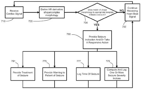
Figure 4 illustrates a flowchart depiction of a method for detecting a state
change and
taking one or more responsive actions, in accordance with an illustrative
embodiment of the
present disclosure;
Figure 5 shows basic shapes of a heart rate plot, from which more complex
shapes can
be produced by deformation in accordance with an illustrative embodiment of
the present
disclosure;
Figure 6 shows a graph of heart rate (BPM) vs. time (hr), with an epileptic
event
identified by electrocorticography (ECoG) indicated by vertical lines, from
which a triangle
pattern may be discernible, in accordance with an illustrative embodiment of
the present
disclosure;
Figure 7A-C shows three graphs of heart rate vs. time, with epileptic events
identified
by ECoG indicated by vertical lines, from each of which a notched triangle
pattern may be
discernible, in accordance with an illustrative embodiment of the present
disclosure;
Figure 8A-C shows three graphs of heart rate vs. time, with epileptic events
identified
by ECoG indicated by vertical lines, from each of which an "M' pattern may be
discernible,
in accordance with an illustrative embodiment of the present disclosure;
Figure 9 shows a graph of heart rate vs. time, with an epileptic event
identified by
ECoG indicated by vertical lines, from which a "W" pattern may be discernible,
in
accordance with an illustrative embodiment of the present disclosure;
6
CA 02819353 2013 05 29
WO 2012/037359
PCT/US2011/051776
Figure 10 shows a graph of heart rate vs. time, with an epileptic event
identified by
ECoG indicated by vertical lines, from which a fused "M' and "W" pattern may
be
discernible, in accordance with an illustrative embodiment of the present
disclosure;
Figure 11A-B shows two graphs of heart rate vs. time, with epileptic events
identified
by ECoG indicated by vertical lines, from which a pattern of periodic
oscillations may be
discernible, in accordance with an illustrative embodiment of the present
disclosure;
Figure 12 shows a graph of heart rate vs. time, with an epileptic event
identified by
ECoG indicated by vertical lines, from which a pattern of periodic
oscillations, specifically
forming a sawtooth pattern, may be discernible, in accordance with an
illustrative
embodiment of the present disclosure;
Figure 13A-D shows four graphs of heart rate vs. time, with epileptic events
identified
by ECoG indicated by vertical lines, from which a pattern of periodic
oscillations overlaid on
a longer-timescale triangle pattern may be discernible, in accordance with an
illustrative
embodiment of the present disclosure;
Figure 14 shows a graph of heart rate vs. time, with an epileptic event
identified by
ECoG indicated by vertical lines, from which periodic oscillations forming a
"comb" pattern
may be discernible, as well as a pattern of lower amplitude periodic
oscillations overlaid on a
longer-timescale triangle pattern may be discernible, in accordance with an
illustrative
embodiment of the present disclosure;
Figure 15 shows a graph of heart rate vs. time, with an epileptic event
identified by
ECoG indicated by vertical lines, from which a pattern of periodic
oscillations overlaid on a
longer-timescale parabola pattern may be discernible, in accordance with an
illustrative
embodiment of the present disclosure;
7
CA 02819353 2013 05 29
WO 2012/037359
PCT/US2011/051776
Figure 16 shows a graph of heart rate vs. time, with an epileptic event
identified by
ECoG indicated by vertical lines, from which a triphasic pattern may be
discernible, in
accordance with an illustrative embodiment of the present disclosure;
Figure 17A-B shows two graphs of heart rate vs. time, with epileptic events
identified
by ECoG indicated by vertical lines, from which multiple "M' and/or "W"
patterns may be
discernible, in accordance with an illustrative embodiment of the present
disclosure;
Figure 18 shows exemplary heart beat complex changes detectable by use of the
P
wave and the R wave of a heart beat, in accordance with an illustrative
embodiment of the
present disclosure; and
Figure 19 A-B shows a first heart beat complex derived from data collected
over an
entire period of EKG monitoring of a patient (A) and a second heart beat
complex derived
from EKG data collected from the same patient during circumictal periods only
(B).
While the disclosure may be susceptible to various modifications and
alternative
forms, specific embodiments thereof have been shown by way of example in the
drawings
and are herein described in detail. It should be understood, however, that the
description
herein of specific embodiments is not intended to limit the disclosure to the
particular forms
disclosed, but on the contrary, the intention is to cover all modifications,
equivalents, and
alternatives as defined by the appended claims.
DETAILED DESCRIPTION OF SPECIFIC EMBODIMENTS
Illustrative embodiments of the disclosure are described herein. In the
interest of
clarity, not all features of an actual implementation may be described in this
specification. In
the development of any such actual embodiment, numerous implementation-
specific
decisions must be made to achieve the design-specific goals, which will vary
from one
implementation to another. It will be appreciated that such a development
effort, while
8
CA 02819353 2013 05 29
WO 2012/037359
PCT/US2011/051776
possibly complex and time-consuming, would nevertheless be a routine
undertaking for
persons of ordinary skill in the art having the benefit of this disclosure.
This document does not intend to distinguish between components that differ in
name
but not function. In the following discussion and in the claims, the terms
"including" and
"includes" are used in an open-ended fashion, and thus should be interpreted
to mean
"including, but not limited to." Also, the term "couple" or "couples" may mean
either a
direct or an indirect electrical connection. "Direct contact," "direct
attachment," or providing
a "direct coupling" indicates that a surface of a first element contacts the
surface of a second
element with no substantial attenuating medium there between. The presence of
small
quantities of substances, such as bodily fluids, that do not substantially
attenuate electrical
connections does not vitiate direct contact. The word "or" is used in the
inclusive sense (i.e.,
"and/or") unless a specific use to the contrary is explicitly stated.
The term "electrode" or "electrodes" described herein may refer to one or more
stimulation electrodes (i.e., electrodes for delivering a therapeutic signal
generated by an
IMD to a tissue), sensing electrodes (i.e., electrodes for sensing a
physiological indication of
a state of a patient's body), and/or electrodes that may be capable of
delivering a therapeutic
signal, as well as performing a sensing function.
In one embodiment, the present disclosure provides a method of detecting a
state
change based upon data derivable from cardiac signals. The state change can
be, for
example, at least one of an unstable brain state, a brain state indicative of
an elevated
probability of a state change, a brain state indicative of an impending state
change, or a state
change, among others.
In one embodiment, the present disclosure provides a method for indicating an
occurrence of a state change. In one embodiment, the method comprises
obtaining a time
series of cardiac data from a patient; determining a reference heart rate
parameter from said
9
CA 02819353 2016-02-23
WO 2012/037359
PCT/US2011/051776
cardiac data; determining a heart rate derivative shape from said time series
of cardiac data,
wherein said heart rate derivative shape comprises at least one characteristic
selected from a
number of phases relative to said reference heart rate parameter, a number of
extrema of said
heart rate derivative, a number of directions of change of said heart rate
derivative, a number of
positive phases, or a number of negative phases; and indicating an occurrence
of a state
change based upon a determination that said heart rate derivative shape
matches a state
change template in said at least one characteristic.
The cardiac data can be gathered by any of a number of techniques. For
example, the
cardiac data may be gathered by an electrocardiogram (EKG) device. For another
example, the
cardiac data may be gathered by a cranial nerve stimulator device. In one
embodiment, the
cardiac data may be related to the R-waves of the beat sequence, such as a
time series of R-
waves or a series of R-R intervals. Those skilled in the art having benefit of
the present
disclosure would appreciate that other time series of cardiac waves and/or
their fiducial points
(e.g., P waves, T waves, etc.) may be used and still remain within the scope
of the present
disclosure and the claims, which are construed following a purposive
construction according to
Canadian Law.
Data relating to R-waves may be gathered by an EKG device or, in one
embodiment, by
a vagus nerve stimulator, such as described in U.S. Patent 5,928,272.
Obtaining the cardiac data may comprise sensing a time of beat sequence of a
patient's
heart and generating a time series data stream from the time of the beat
sequence. In a further
embodiment, receiving the cardiac data of the patient's heart may comprise
sensing and time-
stamping a plurality of R waves, and generating the time series data stream
may comprise
determining a series of R-R intervals from the time stamps of the sensed R
waves.
In one embodiment, the fiducial time marker may be an R wave peak or threshold
crossing. The amplitude or height of one or more representative R waves may be
used to set
CA 02819353 2013 05 29
WO 2012/037359
PCT/US2011/051776
a threshold that, when reached or crossed, may be registered as a fiducial
time marker of a
heart beat.
In one embodiment, a heart rate derivative may be determined from the time
series of
cardiac data. As defined herein, a "heart rate derivative" may be a value
derivable, directly or
indirectly, from the time series of cardiac data, wherein the value relates to
a feature, property
or relationship between two or more heart beats. Although a first or higher-
order derivative,
as understood from calculus, may be a "heart rate derivative" under the above
definition, a
heart rate derivative may be not necessarily a first or higher-order calculus
derivative.
Exemplary heart rate derivatives include, but are not limited to, heart rate
and heart rate
variability (HRV). A "shape" may be used herein to refer to a feature apparent
to the person
of ordinary skill in the art upon viewing a graph of the heart rate or of one
of its derivative
over a period of time. In one embodiment, a heart rate derivative shape
comprises at least
one characteristic selected from a number of phases relative to a reference
heart rate
parameter, a number of extrema of the heart rate derivative, a number of
directions of change
of the heart rate derivative, a number of positive phases, or a number of
negative phases.
By "heart rate shape" may be meant one or more characteristics or features of
a time
series of cardiac data that may be reflective of the appearance of that time
series if plotted on
a graph ( on the y-axis and time on the x-axis). For example, one
characteristic of heart rate
shape may be a number of phases relative to the reference heart rate
parameter. A "phase"
may be a period between two consecutive deviations from, crossings of, or
returns to the
reference heart rate parameter. A phase may be positive (having a value
greater than the
reference heart rate parameter) or negative (having a value less than the
reference heart rate
parameter). Yet another exemplary characteristic of heart rate shape may be a
number of
extrema of heart rate. An "extremum" (plural, "extrema") may be a point where
the slope of
heart rate changes sign, or phrased alternatively, a point that may be a
highest high or lowest
11
CA 02819353 2013 05 29
WO 2012/037359
PCT/US2011/051776
low of heart rate for some length of time or number of beats before and after.
Still another
exemplary characteristic of heart rate shape may be a number of directions of
heart rate
change, which can be defined as the number of changes of the sign of the slope
of heart rate,
plus one. Yet another exemplary characteristic of heart rate shape may be the
steepness of
one or more ascending or descending slopes.
Though not to be bound by theory, we have found that heart activity during
normal
states (exercise, anger, etc.) and abnormal states (e.g., epileptic seizures)
as displayed or
graphed over various time scales take on distinctive shapes which may be used
to identify the
various states as well as changes from one state to another, such as from non-
seizure to
seizure. Said shapes may be considered and treated herein as templates, given
their
stereotypical nature, and may be used in several ways (to be described below)
to detect states,
state changes, state and/or state change onsets, and/or other features, such
as duration,
intensity or magnitude, and/or other relevant characteristics, such as type of
state or state
change.
Another heart rate derivative that may be considered may be a heart rate
volatility
(non-stationarity) parameter, a measure of dispersion which may be defined as
a change in
the standard deviation or variance of heart rate over a moving window.
Commonly, the
higher the volatility, the higher appears to be the probability of state
changes. Volatilty, a
metric often found in financial contexts, may be used here to obtain certain
information about
the state of a system regardless of the similarities or dissimilarities
between financial and
biological time series and consideration for the underlying systems' dynamics.
t-20-1 0+1
For example, let .,===
be a stochastict
process. Its terms
¨ 0 ., Q
represent heart rates as components of a vector or a matrix. The volatility of
the process at
12
CA 02819353 2013-05-29
WO 2012/037359
PCT/US2011/051776
time (1 may be defined as the standard deviation of the time t return.
Typically, log returns
are used, so the definition becomes
....= ..... t µ...µ,.
Q
volatility = std log _______
[1]
where log denotes a natural logarithm.
If heart rate time series are conditionally homoskedastic, definition [1] is
precise.
However, if they are conditionally heteroskedastic, measure [1] requires
modification.
Volatility at time (1 represents in this case, the standard deviation of the
time t log return
conditional on information available at time (1 as defined below
volatility = t-1 std log t_i __
..., ..... Q..... i
where the preceding superscript t-1 indicates that the standard deviation is
conditional
on information available at time t-1.
Transitions from homoskedasticity (defined herein as approximately constant
standard
deviations over a certain time window) to heteroskedasticity (inconstant
standard deviation)
also provide information about the probability of being in or near a state
change of interest
and may be used for automated detection, warning, delivery of therapy and
logging (of
events, warnings and therapy) purposes.
Volatility will be measured using time scales (seconds to days) based on
temporal
(e.g., duration) and other properties of the state change on interest and of
the reference state.
The method also comprises indicating an occurrence of a state change based
upon a
determination that said heart rate derivative shape matches a state change
template in said at
least one characteristic.
13
CA 02819353 2013 05 29
WO 2012/037359
PCT/US2011/051776
A "state change template" may be a template known or discovered by the
practitioner
to be associated with the state change, wherein the template can be used in
the analysis of the
heart rate derivative shape.
Plots of instantaneous heart rate (y-axis) as a function of time (x-axis) in
subjects with
-- epilepsy reveal consistent changes before, during and after seizures,
referred herein to as
circum-ictal changes. ("Circum-ictal" or "circumictal," as used herein,
encompasses pre-
ictal, ictal, and post-ictal subperiods. The circumictal period can be
considered the time
window (e.g., in min) preceding and following a seizure during which cardiac
activity differs
from that observed during interictal conditions, normal physical activity
(including exercise),
-- intense emotions (fear, anger, etc.), and physiological functions such as
defecation, urination
or coitus). The curves described by these circum-ictal changes in heart rate,
approximate
triangles or parabolae, and may have indentations of varying sizes. See the
discussion of
Figures 5-17 below for more information. Visual review of a large human
database of
instantaneous heart rate plots reveal that over a certain window length
(referred herein as the
-- mesoscopic scale) their circum-ictal shapes may be limited to the triangles
and parabolae and
to "deformations" of these two shapes (see Figure 5). These "deformations"
appear to have
temporal and magnitude dependencies, in that the longer the duration of the
change in heart
rate and the larger its magnitude, the more likely they may be to occur. The
behavior of these
shapes likely reflect fluctuations in the strength of sympathetic and
parasympathetic inputs to
-- the heart. For example, transient, rapid drops in heart rate may be caused
by either a
withdrawal in sympathetic tone or by an increase in parasympathetic tone
resulting from
differential activation or inhibition by epileptiform activity of brain
regions involved in
autonomic control.
The shape (i.e., all the geometrical information that may be invariant to
position
-- (including rotation) and scale) of these curves may be used for detection
of changes in brain
14
CA 02819353 2013 05 29
WO 2012/037359
PCT/US2011/051776
state such as epileptic seizures and their properties may be characterized
through use of
statistical shape analysis (e.g., Procrustes analysis), of the different
embodiments of
"matched filtering" or of other geometrical (Euclidian and non-Euclidian)
methods. Other
approaches such as computing the area of the triangles and parabolae and
comparing the
results to a reference value outside the circum-ictal state, may be used. In
the case of
triangles, there area may be calculated using for example Heron's formula:
Area ............. 'S(S a)( S b)(8
,where
5' ............ ( 1) + c)
2 \ = ' ' and a, b, and c are the sides of the triangle.
Similarly the area of parabolae (Area = 2/3 b x h, where b is the base and the
height, may be computed and used to detect seizures.
Other attributes not captured by the concept of shape may be applied as need
to the
sign al for detecting state changes such as epileptic seizures.
In one embodiment, the at least one characteristic of the state change
template
comprises two or more phases relative to the reference heart rate parameter,
two or more
extrema of the heart rate derivative, three or more directions of change of
the heart rate
derivative or its slope, a number of positive phases, or a number of negative
phases, provided
the total number of positive phases and negative phases may be two or more.
In another embodiment, the at least one characteristic of the state change
template
comprises at least one phase relative to the reference heart rate parameter,
at least one
extremum of the heart rate derivative or its slope, two or more directions of
change of the
heart rate derivative, a number of positive phases, or a number of negative
phases, provided
the total number of positive phases and negative phases may be at least one.
In another embodiment, the at least one characteristic of the state change
template
comprises at least one of the amplitude of at least one phase, the duration of
at least one
CA 02819353 2013 05 29
WO 2012/037359
PCT/US2011/051776
phase, the valence (positive or negative) of at least one phase, at least one
slope of at least
one phase, the arc length (which may be used interchangeably with line length)
of at least
one phase, the number of extrema in at least one phase, and the sharpness of
the extrema of at
least one phase.
A reference heart rate parameter, as used herein, may be a reference value
obtained
during a state that may be deemed of no or little interest for automated
detection, warning,
treatment or logging purposes. The reference heart rate parameter may be a
single value, a
series of values, or a statistic selected from the group consisting of a
shape, a vector, a vector
space, a matrix, and two or more thereof
For example, heart activity during a non-seizure state may be considered as a
reference state. The reference heart rate parameter may be calculated from a
time series of
value over any particular window, such as a window haying a length from 30 sec
to 24 hr,
although longer or shorter windows may be used. The window may be a simple
window or
an exponentially-forgetting window. The reference heart rate parameter may be
calculated as
any measure of any tendency of the time series, such as the central tendency
of the time
series. For example, the reference heart rate parameter may be calculated as a
mean, median,
nth percentile (where n can be from 30 to 70), or exponential moving average
of the time
series, among other measures of central tendency. Other mathematical or
statistical
measures, including, but not limited to, correlation dimension, entropy,
Lyapunoy exponents,
and fractal or multifractal dimensions, may be also applied to any of the
parameters or their
templates.
The reference heart rate parameter may be determined from previously recorded
data,
or from "normative" values obtained from normal or abnormal cohorts of
subjects or
populations or it may be determined from the time series of cardiac data
referred to above.
16
CA 02819353 2013 05 29
WO 2012/037359
PCT/US2011/051776
An exemplary state change template can be derived from the pattern shown in
Figure
8, wherein the changes in heart rate during a seizure form a readily
discernible "M' between
0.88 hr and 0.92 hr, having one positive phase relative to a reference heart
rate parameter
(calculated as the median value from about 0.85 hr to 0.89 hr and from about
0.93 hr to about
1.00 hr), three extrema (two maxima and one minimum, each being an extremum
relative to
about 20 seconds before and 20 seconds after), and four directions of heart
rate change.
The state change template may be the "raw" pattern (analog or digitized) or it
can be
derived by smoothing, averaging, or otherwise mathematically processing
subseries of
cardiac data obtained during state changes. A "matched filter" may be a type
of filter matched
to the known or assumed characteristics of a target signal, to optimize the
detection of that
signal in the presence of noise. A matched filter may be the filter with
impulse response equal
to the time reversed, complex conjugate impulse response of the input.
One skilled in the art will appreciate that when applying matched filter
techniques to
attempt to detect a pattern in a signal, the raw signal may first be
transformed so that it has
zero mean on a timescale of interest when the pattern may be absent. Such
transformation
may include, but not be limited to, detrending or subtracting a background
reference value (or
time-varying reference signal) from the raw signal and may be used to remove
bias in the
matched filter output and improve its signal-to-noise ratio.
Seizure detection may be performed over multiple time scales or window lengths
listed in no particular order:
a) "Mesoscopic" corresponding to an scale of observation of several seconds to
tens
of seconds (e.g., 10-300 s) to capture at least in part, a change in the shape
of heart rate plot
representative of a state change.
b) "Microscopic" corresponding to the scale of observation of at least part of
a heart
beat such as that represented by an EKG's P-QRS-T complex.
17
CA 02819353 2013 05 29
WO 2012/037359
PCT/US2011/051776
c) "Macroscopic" corresponding to an scale of observation longer than 300 s to
encompass more than the information contained in the mesoscopic scale or
window as
defined in a).
Seizure detection at a macroscopic scale provides information not obtainable
with the
two other scales (micro- and mesoscopic) allowing for the identification of
certain patterns
(defined herein as the occurrence of more than one triangle or parabola or
combinations
thereof within a macroscopic window).
A shape deformation (e.g., a deformed "M") may show local and global extrema
that
may be used for detection and validation purposes.
In one embodiment, the method comprises matched filtering. Matched filtering
may
be a theoretical framework and not the name of a specific filter. A matched
filter may be a
type of filter matched to the known or assumed characteristics of a target
signal and may be
designed to optimize the detection of that signal in the presence of noise as
it maximizes S/N.
A matched filter's impulse response may be equal to the time reversed, complex
conjugate
impulse response of the input.
The output response of a "matched" filter derived from meso-, micro- or
macroscopic
patterns, as it may be passed through any of these patterns may be
characteristic (it forms a
spatio-temporal pattern) and in turn may be used not only to validate
detections but to allow
detections before the convolution may be completed ('early" detection).
A second filter matched to the first matched filter's output response may be
run
simultaneously with the first matched filter and its output response may be
used for early
detection and second level validation of the detection.
The pattern formed by any of the cardiac activity parameters may used as a
matched
filter. Other realizations such as the orthogonal and projected orthogonal
matched filter
detection (Eldar YC. Oppenheim A, Egnor D. Signal Processing 2004; 84: 677 ¨
693),
18
CA 02819353 2013 05 29
WO 2012/037359
PCT/US2011/051776
adaptive matched filter and parametric adaptive matched filter (Dong Y.
Parametric adaptive
filter and its modified version DSTO-RR-0313 My 2006 Australian Government,
Dept. oif
Defence); the nearest matched filter fpr classification of spatio-temporal
patterns (Hecht-
Nielsen R. Applied Optics 1987; 26:1892-98), an outlier resistant matched
filter (Gerlach K.
IEEE Trans Aerospace Electronic Syst 2002; 38:885-901), a phase-only matched
filter
(Horner JL, Gianino PD. Applied Optics 1984; 23:812-16) may be also used for
detection
and validation of state changes such epileptic seizure.
The detection and validation of states based on the morphology or shape of
signals
may be performed at various time scales (micro-, meso-, or macroscopic)
through estimation
of the autocorrelation function of said shapes or patterns. Furthermore,
estimation of the
autocorrelation function of a reference state may also be used for detection
and validation of
state changes alone or in combination with the autocorrelation estimates of
the state change
shapes or patterns. Autocorrelation may be considered as an equivalent method
to matched
filtering.
Other methods such as non-linear detectors (Theiler J, Foy BR, Fraser AM.
Beyond
the adaptive matched filter: Non-linear detectors for weak signals in high
dimensional clutter.
Proc SPIE 6565 (2007) 6565-02: 1-12) and maximum likelihood estimation (Forney
GD,
Maximum-likelihood estimation of digital sequences in the presence of
intersymbol
interference. IEEE Trans Information Theory 1972; 18:363-76) may be also
applied in this
disclosure.
Matching a heart rate shape to a state change template can be performed by any
appropriate mathematical technique. For example, pattern matching may be by
use of a
matched filter. In one embodiment, the state change template comprises at
least one matched
filter. In one embodiment, a "match" refers to a match score found by a
matched filter
analysis of greater than about 0.75, such as greater than about 0.80, greater
than about 0.85,
19
CA 02819353 2013 05 29
WO 2012/037359
PCT/US2011/051776
greater than about 0.90, greater than about 0.95, greater than about 0.98, or
greater than about
0.99. A "failure to match" refers to a match score found by a matched filter
analysis of less
than about 0.75, such as less than about 0.80, less than about 0.85, less than
about 0.90, less
than about 0.95, less than about 0.98, or less than about 0.99. However, these
values may be
changed as needed.
In one embodiment, the state change template comprises at least a state change
matched filter and a reference parameter matched filter. A "match" can be
defined as a match
to the state change matched filter not accompanied by a match to the reference
parameter
filter.
Regardless of the type of filter, in one embodiment, the heart rate derivative
shape has
a matched filter score to said state change template greater than a value
threshold for at least
a duration threshold. For example, any of the values set forth above may be
used as the value
threshold and the duration threshold may be selected as any appropriate number
of seconds or
heart beats, such as 1 to 10 sec, or 1 to 10 beats, such as 3 beats.
In one embodiment, the state change template exists in a first timescale and
said heart
rate derivative shape may be present in said first timescale. For example, the
heart rate
derivative shape may be present over a first timescale not typically found in
a reference heart
rate derivative shape observed during rising from lying to sitting, rising
from sitting to
standing, minor physical exertion, exercise, or emotionally-intense
experiences. This allows
distinction between heart rate derivative shapes associated with a state
change of interest,
e.g., an epileptic seizure, and heart rate derivative shapes associated with
normal daily
activities.
In one embodiment, the state change template comprises at least one positive
phase
and at least one negative phase. In a further embodiment, the at least one
positive phase may
be a period of elevated heart rate. In an even further embodiment, the period
of elevated
CA 02819353 2013 05 29
WO 2012/037359
PCT/US2011/051776
heart rate may be a period of tachycardia. In people fifteen years of age and
older,
tachycardia may be defined as a heart rate greater than 100 bpm. In another
further
embodiment, the at least one negative phase may be a period of decreased heart
rate. In an
even further embodiment, the period of decreased heart rate may be a period of
bradycardia.
Bradycardia may be defined in adults as a heart rate less than 60 bpm.
In one embodiment, the state change template comprises at least two extrema of
heart
rate. In a further embodiment, the state change template can also comprise at
least two
phases.
The state change template may comprise one or more shapes readily discernible
to the
human eye. For example, the state change template may comprise a triangle,
such as that
shown in Figure 6. Although in many cases, state change templates that appear
more
complex than a triangle may be useful, they can generally be understood as
involving one or
more triangles or parabolas and/or deformations thereof
Figure 5 illustrates the metamorphosis or transformation of circumictal heart
rate
shapes or patterns at a mesoscopic scale. The simplest shape may be that of a
parabola (left
upper panel). In certain seizures a short-lived withdrawal or reduction of
sympathetic
influences or an increase in parasympathetic ones early in the course of a
seizure causes a
notch or indentation in the parabola (right upper panel). In other seizures
(in the same subject
or in a different subject), a later, more pronounced and prolonged withdrawal
or reduction of
sympathetic influences or an increase in parasympathetic ones (compared to
that seen in the
right upper panel) leads to a prominent indentation or notch (right lower
panel), resembling
the letter "M". A later, briefer, and less pronounced withdrawal or reduction
of sympathetic
influences or an increase in parasympathetic ones (compared to that seen in
the right lower
panel) causes an indentation in the parabola.
21
CA 02819353 2013 05 29
WO 2012/037359
PCT/US2011/051776
The relative balance of sympathetic and parasympathetic influences can be
assayed at
multiple timescales. As can be seen with reference to at least some of the
figures discussed
below, the relative balance of sympathetic and parasympathetic influences can
oscillate on
multiple timescales.
While a parabola is shown in Figure 5 as an example, this may be replaced by a
triangle or by any other topologically equivalent shape.
We have discovered a number of specific patterns or shapes occurring in at
least some
circumictal periods of at least some patients, which patterns or shapes may be
used as the
basis for a state change template as discussed herein.
Generally, the specific patterns or shapes can be considered as belonging to
one of
three categories:
Simple patterns, including the parabola shown in Figure 5 or the triangle
shown in
Figure 6, among others;
Complex patterns, including the notched triangle pattern of Figure 7, the "M"
pattern
of Figure 8, and the "W" pattern of Figure 9, among others;
Polymorphic patterns, containing two or more simple and/or complex patterns,
including fused simple and/or complex patterns, periodic or quasiperiodic
oscillations,
periodic or quasiperiodic oscillations overlaid on a longer term simple and/or
complex
patterns, and multiple simple and/or complex patterns, such as those shown in
Figures 10-17,
among others.
Exemplary patterns or shapes are shown in Figures 6-17. In each of these
figures, a
relevant portion of a graph of a patient's heart rate in beats per minute
(BPM) vs. time in
hours from the onset of ECoG monitoring of his or her seizure activity may be
shown.
Vertical lines mark the electrographic onset and electrographic termination of
a seizure.
22
CA 02819353 2013 05 29
WO 2012/037359
PCT/US2011/051776
The reader will have noticed that some patterns notable in Figures 6-17 as
being
closely correlated in time with a seizure also occur at times when no seizure
was detected by
ECoG. It should be pointed out that since monitoring of brain activity with
intracranial
electrodes may be limited to certain regions, seizures may occur and go
undetected if they
originate in regions not monitored by the available electrodes. This may
explain the presence
of multiple heart rate patterns in the circumictal period when only one
seizure was recorded.
In other words, the cardiac data may indicate the occurrence of seizures that
intracranial
electrodes failed to detect. The use of cardiac information, such as the uses
described and
claimed herein, may supplement the inherent limitations of brain-based seizure
detection.
Figure 6 shows what may be termed a simple pattern, viz., a triangle, in
accordance
with an illustrative embodiment of the present disclosure. Herein, when
discussing shapes,
the words "triangle" and "parabola" can be used interchangeably. Generally,
"triangle" will
be used for convenience only.
Figure 7A-C shows three graphs of what may be termed a notched triangle.
In various examples, the state change template may comprise one or more shapes
that
can be considered as comprising a plurality of triangles. For example, the
state change
template may comprise one or more shapes resembling letters of the Latin
alphabet.
Figure 8A-C shows three graphs of what may be termed an "M" pattern, formed by
two contiguous triangles or parabolae. The "M" pattern may be monophasic (the
heart rate
does not drop below the reference value or baseline) or multiphasic (after
raising above the
reference value, the heart rate drops below it). An "M" can be considered as
distinct from a
"notched triangle" in that the indentation of the M generally returns
substantially to a baseline
value and generally divides the M into substantially symmetrical halves.
The "M" patterns shown in Figures 8A-8C have total durations of about 60-90
sec,
beginning anywhere from about 15 sec before electrographic onset to about 90
sec after
23
CA 02819353 2013 05 29
WO 2012/037359
PCT/US2011/051776
electrographic onset. However, other total durations and beginning times
relative to
electrographic onset may occur in other "M" patterns.
Figure 9 shows a graph of what may be termed a "W" pattern, discernible from
about
15 sec after electrographic onset to about 20 sec after electrographic
termination. Though not
to be bound by theory, the "W " pattern may reflect differences (compared to
the "M"
pattern) in the timing of changes in autonomic influences during seizures.
The triangle, notched triangle, "M," and "W" patterns of Figures 6-9 can be
considered to occur on a mesoscopic timescale. However, the same patterns may
be
discerned at shorter or longer timescales.
Figures 10-17 show patterns that can be considered to occur at long mesoscopic
and/or macroscopic timescales. As can be seen and will be discussed below, the
patterns of
Figures 10-17 can generally be considered as polymorphic patterns comprising
two or more
of the basic shapes, simple patterns, or complex patterns discussed above.
Figure 10 shows a fused "M" and "W" pattern. The "W" can be considered as
starting
at about 30 sec before electrographic onset and ending at about 60-75 sec
after electrographic
onset in the region of highest heart rate during the seizure event. The "M"
can be considered
as starting a few seconds before electrographic onset and ending about at
electrographic
termination. One may also discern a "W" occurring at a microscopic or short
mesoscopic
timescale at the notch of the "M."
Alternatively or in addition, a person of ordinary skill in the art, having
the benefit of
the present disclosure, may discern an "M" beginning at about 45-60 sec before
electrographic onset and ending at about the middle of the seizure, with a "W"
beginning
about 30 sec after electrographic onset and ending about 15-30 sec after
electrographic
termination.
24
CA 02819353 2013 05 29
WO 2012/037359
PCT/US2011/051776
Figure 11A-B shows two graphs of patterns of periodic or quasiperiodic
oscillations.
(For convenience, we will use the term "periodic," although it must be borne
in mind that the
frequency and the amplitude of the oscillations associated with a single
seizure in one patient
may vary over the course of about 10 min, as shown in Figures 11A-B. In other
words, the
term "periodic" may be not limited herein to refer to series of oscillations
with fixed
frequency and amplitude).
The pattern of periodic oscillations may be deformed by a seizure event (e.g.,
Figure
11B). In instances where this may be not the case, a dysfunction of the
patient's autonomic
nervous system may be indicated. For example, Figure 11A shows a rapid
oscillation of the
patient's heart rate by as much as 40 BPM in a short time.
Detecting a pattern in a preictal period in a time series of heart rate data
may be
considered, at least in some patients, as a "prediction" of a seizure and/or
an indication of a
period of greater risk of a seizure. Alternatively or in addition, it may be
used to aid
detection of seizures originating in brain regions not surveyed by
intracranial electrodes.
Multiple triangles with a certain degree of periodicity and either monophasic
or
biphasic nature can form what may be viewed as a "sawtooth" pattern in the
circumictal
period. Figure 12 shows a graph of another pattern of periodic oscillations.
The periodic
oscillations from about 15-30 sec after the seizure to about 3 min after the
seizure can be
considered a sawtooth pattern.
Figure 13A-D shows four graphs of patterns of periodic oscillations overlaid
on a
longer-timescale triangle pattern. For example, the pattern in Figure 13A
shows an
asymmetric triangle with a trailing slope lasting about 5 min, on which may be
overlaid a
pattern of periodic oscillations having an average wavelength of about 20 sec
may be
discernible from about 90 sec after the seizure until the end of the window
shown.
CA 02819353 2013 05 29
WO 2012/037359
PCT/US2011/051776
Figure 14 shows, in addition to a pattern of periodic oscillations overlaid in
the post-
ictal period on a longer-timescale triangle pattern, a comb pattern in the
preictal period. For a
duration of about 2.5 min starting about 3.5 min before electrographic onset,
a pattern of
periodic oscillations may be shown with pronounced negative amplitudes
(relative to the
average heart rate over the first 30-45 sec of the window) and an average
wavelength of about
sec. Again, detecting a pattern in a preictal period in a time series of heart
rate data may
be considered, at least in some patients, as a "prediction" of a seizure
and/or an indication of a
period of greater risk of a seizure. Alternatively or additionally, the
presence of one pattern
of long duration or more than one pattern of any duration in the circumictal
period may be
10
indicative of cardiac or autonomic instability. This information may be used
to warn the
patient or his caregiver(s) of an increased risk of a serious outcome and/or
institute
therapeutic measures.
Figure 15 shows another comb pattern, this one with pronounced positive
amplitudes,
overlaid on a longer-timescale parabola.
15 Figure
16 shows a triphasic pattern relative to the preictal baseline, in which a
first
positive phase forms a notched triangle from just before electrographic onset
until late in the
seizure; a second, negative phase follows until about 30-45 sec after the
seizure; and a third,
positive phase ensues with a duration of about 4 min until the end of the
window.
Figure 17A-B shows two graphs from which multiple "M' and/or "W" patterns may
be discernible in all three of the preictal, ictal, and postictal time
periods. These multiple "M"
and/or "W" patterns can be considered as part of a macroscopic pattern
comprising a plurality
of complex shapes.
In addition, very rapid oscillations in heart rate may also occur, and along
with lower
frequency oscillations, may provide useful insight into the behavior of heart
rate variability
circum-ictally and of its usefulness for seziure detection, given its
differences from those
26
CA 02819353 2013 05 29
WO 2012/037359
PCT/US2011/051776
observed outside the circum-ictal period. That is, oscillations at two
frequencies (e.g., slow
and fast) or more than two frequencies (e.g., very fast, slow, and very slow)
may overlap to
form a pattern that may be commonly associated with a circumictal period.
Any one or more of the patterns shown in Figures 6-17, among others, can be
taken as
the basis for a state change template. Also, HRV values can be derived from
the time series
of heart rates depicted in Figures 6-17, and one or more distinctive patterns
discernible from
the HRV values can be used as the basis for a state change template. Such
distinctive
patterns would generally be expected to be distinct from HRV changes resulting
from
exercise or normal exertion.
Regardless of how HRV values may be determined, in one embodiment, the pattern
or
shape of heart rate variability (as distinct from heart rate) measured at any
or all of the
timescales (micro-, meso-, or macroscopic) may be used as a template for
detection and
quantification of state changes using matched filtering or its autocorrelation
function.
In a particular embodiment, the state change template comprises one phase
relative to
the reference heart rate parameter, three extrema, four directions of heart
rate change, and two
periods of increased heart rate relative to the reference heart rate
parameter. This state
change template may be considered to be the "M" pattern shown in Figure 8.
Multiple state change templates, including but not limited to multiple
templates at
different timescales, may be used for various purposes. For example, a first
template found
to have a particularly high sensitivity, specificity, or both can be used as a
primary detection
technique, with other templates used to validate detections made by the first
template. For
another example, a template found to have high sensitivity but low specificity
(i.e., giving
detections with a relatively high false positive rate) can be paired with
another template found
to have high specificity to be used in detections with higher sensitivity and
specificity than
either alone. For still another example, a first template can be used to
identify a state change
27
CA 02819353 2013 05 29
WO 2012/037359
PCT/US2011/051776
e.g., from a non-circumictal state to a preictal state, and this
identification can be used to
trigger use of a second template to identify a second state change, e.g., from
a preictal state to
an ictal state. For a particular example, a comb pattern can be used to
identify a state change
from a non-circumictal state to a preictal state, and an "M" pattern can be
used to identify a
state change from a preictal state to an ictal state.
In one embodiment, a plurality of matched filters (and/or the output of one or
more of
the matched filters as another matched filter or filters) can be used. For
example, two or
three matched filters, each on a separate one of the macroscopic, mesoscopic,
and
microscopic timescales can be run simultaneously on the time series of heart
rate derivative
data. After adequate analysis, comparisons of the results of matched filtering
at the three
times scales can be made to find the matched filter/timescale combination(s)
giving highest
sensitivity, highest specificity, fastest detection, or two or more thereof
Depending on the
intended use, the most useful matched filter/timescale can then be used and
run continuously
and its output (detection) used to run the other matched filters/timescales
for detection of
changes (at longer or shorter time scales) and validation of detected changes.
Alternatively or in addition to the state change detections discussed herein,
circumictal changes at various times scales may be used for assessment of
disease state, both
among circumictal changes monitored over long time periods (such as months or
years) and
between circumictal and non-circumictal states. In one embodiment, such
disease state
assessment may include assessment of the patient's risk of epilepsy-related
sudden death
(SUDEP).
Regardless of the desired use of circumictal data, circumictal changes may be
quantified in one or more dimensions. In one embodiment, the output value of a
detection, a
disease state assessment, or the like can be monitored as a function of time
(days, month
years), both inter-circumictally and circumictally vs. non-circumictally, with
the results
28
CA 02819353 2013 05 29
WO 2012/037359
PCT/US2011/051776
analyzed for the presence of changes and trends. In another embodiment,
circumictal
changes can be classified as a function of pattern type (e.g., simple,
complex, or
polymorphic) and their temporal evolution tracked. In another embodiment, the
temporal
density of the circumictal period can be defined as percent time spent in a
pattern(s).
Quantification of the match between the heart rate derivative shape and the
state
change template can also provide information about the duration of a seizure.
In one
embodiment, the method further comprises indicating the termination of the
state change
based upon a determination that the heart rate derivative shape fails to match
the state change
template, after an indication of an occurrence of a state change.
In one embodiment, the state change template further comprises at least one
second
characteristic selected from a magnitude of heart rate change relative to the
reference heart
rate parameter, a slope of heart rate change, a duration of one or more
phases, a duration from
a heart rate excursion from the reference heart rate parameter to a peak or a
trough heart rate,
a total duration of all the phases, or a duration of a constant slope of heart
rate change; and
indicating an occurrence of a state change may be based upon a determination
that the heart
rate shape matches a state change template in both the at least one
characteristic and in the at
least one second characteristic.
The slope can be measured on any time scale, though for cardiac data, it may
be
smoother if taken over multiple beats, such as five or fifteen beats, or over
a length of time,
such as five to fifteen seconds. The term "constant slope" may be used herein
to refer to a fit,
such as a least-squares fit or other fit, of the data series in question that
has a sufficiently high
fit to a straight line as to commend itself to the person of ordinary skill in
the art as being a
constant. For example, a region of a data series having a linear least-squares
fit with an R2
value of at least 0.9 can be considered to have a constant slope.
29
CA 02819353 2013 05 29
WO 2012/037359
PCT/US2011/051776
As stated above, a state change can be indicated by quantifying the match of
the heart
rate shape to the state change template. This state change indication can be
considered as the
sole indication of a state change, it can be validated by other techniques of
state change
identification, or it can be used to verify state changes indicated by other
techniques. Such
other techniques include those described elsewhere herein, as well as others
known to the
person of ordinary skill in the art or others the subject of one or more
patent applications,
such as United States patent applications 12/770,562, filed April 29, 2010;
12/771,727, filed
April 30, 2010; and 12/771,783, filed April 30, 2010.
In one embodiment, the determination comprises using a first matched filter to
yield a
first output, building a second matched filter from the first output, and
using the second
matched filter to detect the state change. In other words, because the passage
of a first
matched filter over a data window will produce a stereotypical output when it
begins passing
over a shape which it matches, the stereotypical output itself can be used to
detect a state
change prior to, or as a validation of, a detection by the first matched
filter.
Thus, in one embodiment, the method further comprises identifying an
occurrence of
a state change; and wherein said determining said heart rate derivative shape
and said
indicating may be performed in response to said identifying, to validate said
identifying.
In another embodiment, the method further comprises identifying an occurrence
of a
state change in response to said indicating, to validate said indicating. In a
further
embodiment, the method further comprises obtaining data relating to at least a
portion of a
heart beat complex from said patient; comparing said at least said portion of
said heart beat
complex with a corresponding portion of a reference heart beat complex
template of said
patient, wherein the reference heart beat complex template may be not
indicative of a state
change; and validating said indicating an occurrence of a state change,
wherein said
CA 02819353 2013 05 29
WO 2012/037359
PCT/US2011/051776
validating may be based upon a determination that said heart beat complex
fails to match said
reference heart beat complex template.
In one embodiment, the reference heart beat complex template may be selected
from a
normal template (e.g., a reference heart beat complex template not indicative
of a state
change from a patient with healthy heart activity) or an abnormal template
(e.g., a reference
heart beat complex template not indicative of a state change from a patient
with current or
past unhealthy heart activity).
For example, a heart rate derivative shape present over a first timescale not
typically
found in a reference heart rate derivative shape observed during rising from
lying to sitting,
rising from sitting to standing, minor physical exertion, exercise, or
emotionally-intense
experiences can be used to indirectly validate an identification of a seizure
made from a rise
in heart rate, or vice versa.
Alternatively or in addition, in another embodiment, the method comprises
determining a second reference heart rate parameter; determining a second
heart rate
derivative shape from said time series of cardiac data, wherein said second
heart rate
derivative shape comprises at least one second characteristic selected from a
number of
phases relative to said reference heart rate parameter, a number of positive
phases relative to
said reference heart rate parameter, a number of negative phases relative to
said reference
heart rate parameter, a number of extrema of said second heart rate
derivative, or a number of
directions of change of said second heart rate derivative; and validating said
indicating an
occurrence of a state change, wherein said validating may be based upon a
determination that
said second heart rate derivative shape matches a second state change template
in said at least
one second characteristic.
The present disclosure also provides a method for indicating an occurrence of
a state
change, comprising obtaining data relating to at least a portion of a heart
beat complex from a
31
CA 02819353 2013 05 29
WO 2012/037359
PCT/US2011/051776
patient; comparing the at least the portion of the heart beat complex with a
corresponding
portion of a reference heart beat complex template of the patient; and
indicating an
occurrence of a state change based upon a determination that the heart beat
complex fails to
match the reference heart beat complex template.
In one embodiment, some state change characteristics may be attributed to
epileptic
events, e.g., seizure, while other state change characteristics may be
indicative to a
physiological event that is not an epileptic event. Body data, such as brain
signals, cardiac
signals (e.g., heartbeat data), etc., may be used to determine whether state
change
characteristics are attributable to epileptic events or other physiological
events that are not
epileptic events. Accordingly, state change characteristic(s) may be used to
determine
whether an epileptic event is occurring, has occurred, or is imminent.
A heart beat complex may be used herein to refer to a PQRST complex from a
single
heart beat, including both the relative and absolute magnitudes of the P-, Q-,
R-, S-, and T-
waves, and all of the intervals P-Q, P-R, P-S, P-T, Q-R, Q-S, Q-T, R-S, R-T,
and S-T. A
portion of the heart beat complex may be then any one or more of the relative
and/or absolute
magnitudes of the waves, their shapes, and/or one or more of the intervals
between waves. A
relative magnitude may be defined according to any one or more of the waves of
the
complex, e.g., an R-wave amplitude can be defined as r times the P-wave
amplitude. Figure
18 shows exemplary heart beat complexes with P- and R-waves identified by
name. The
horizontal lines may be drawn for convenience, to point out plausible
deviations between the
various waves of different beat complex.
Although the term "a heart beat complex" may be used above, a plurality, such
as, but
not necessarily, a sequential plurality, of heart beat complexes can be used,
with the
comparing being done for one or more of the plurality of heart beat complexes.
The plurality
32
CA 02819353 2013 05 29
WO 2012/037359
PCT/US2011/051776
may be a fixed set of beats or a moving window over a predetermined time or
number of
beats.
In one embodiment, the portion of the heart beat complex comprises at least
one of an
amplitude of a P wave, a polarity of a P wave, at least one of an amplitude of
an R wave, a
polarity of a Q wave, a polarity of an R wave, an amplitude of an S wave, a
polarity of an S
wave a polarity of an S waveõ an amplitude of a T wave, a polarity of a T
wave, an area
under the curve of a P wave, an area under the curve of a Q wave, an area
under the curve of
an R wave, an area under the curve of an S wave, an area under the curve of a
T wave, a
width of a P wave, a width of a Q wave, a width of an n R wave, a width of an
S wave, a
width of a T wave, a morphology of a P wave, a morphology of a Q wave, a
morphology of
an R wave, a morphology of a T wave, a magnitude of a change in the distance
from a P
wave to a Q wave, a magnitude of a change in the distance from a P wave to an
R wave, a
magnitude of a change in the distance from a Q wave to an R wave. a magnitude
of a change
in the distance from an R wave to an S wave, a magnitude of a change in the
distance from an
R wave to a T wave, a magnitude of a change in the distance from an S wave to
a T wave, a
magnitude of an S-T segment elevation, a magnitude of an S-T segment
depression, a
magnitude of a Q-T segment elevation, a magnitude of a Q-T segment depression,
a P-R
interval, an R-S interval, an S-T interval, an R-T interval, and a Q-T
interval.
The reference heart beat complex template can be derived from any non-state
change
heart beats. Such beats may be one, some, or all the same beats used to define
the reference
heart rate parameter and/or reference HRV described above, but need not be any
of the same
beats. In one embodiment, the reference heart beat complex template comprises
at least one
matched filter. In a further embodiment, the heart beat complex fails to match
the reference
heart beat complex template if a matched filter score for the heart beat
complex to the at least
one matched filter may be less than a heart beat complex value threshold.
33
CA 02819353 2013 05 29
WO 2012/037359
PCT/US2011/051776
Although one reference heart beat complex template may be referred to above, a
plurality of reference heart beat complexes may be used. For example, a
plurality of
reference heart beat complexes can be used on the same heart beats, or one or
more of the
plurality can be used at different times of day, under different states of
exertion or arousal, in
view of changes in heart health histories or differences in heart health
between patients,
among other possibilities. In one embodiment, a second reference heart beat
complex
template comprises at least one of T wave depression, P-Q segment elongation,
another
abnormality, or two or more thereof, relative to the canonical "normal" heart
beat complex.
Alternatively, one or more heart beat complex templates derived from heart
beat
complexes observed during one or more periods of state change may be used,
with a state
change declared if the heart beat complex(es) match(es) the state change heart
beat complex
template(s).
Figure 19A shows an exemplary heart beat complex derived from data collected
over
an entire period of EKG monitoring of a patient, which may be used as a
reference heart beat
complex template. Figure 19B shows an exemplary heart beat complex derived
from EKG
data collected from the same patient during circumictal periods only, which
may be used as a
state change heart beat complex template.
In the event a plurality of reference heart beat complex templates may be
used, one or
more of the templates may be modified over time, based on observed changes in
the patient's
heart beat complexes, such as during non-state-change periods.
The at least portion of the heart beat complex and the corresponding portion
of the
reference heart beat complex template can be compared using any of the pattern
matching
techniques described herein. Because the reference heart beat complex template
may be
taken from non-seizure heart beats, a failure to match between the at least
portion of the heart
34
CA 02819353 2013 05 29
WO 2012/037359
PCT/US2011/051776
beat complex and the corresponding portion of the reference heart beat complex
template
may be an indirect indication of a seizure.
Quantification of the match between a portion of a heart beat complex and the
corresponding portion of the reference heart beat complex template can also
provide
information about the duration of a seizure. In one embodiment, the method
further
comprises obtaining a time series of data relating to a plurality of heart
beat complexes from
the patient; comparing at least a portion of each of a sequential plurality of
heart beat
complexes with a corresponding portion of the first reference heart beat
complex template;
and indicating the termination of the state change based upon a determination
that at least one
heart beat complex of the sequential plurality matches the reference heart
beat complex
template, after an indication of an occurrence of a state change. Matched
filters can be used
in this determination, as described elsewhere herein.
In one embodiment, the determination further comprises analyzing one or more
of a
pulse shape, an R wave amplitude, an apex cardiogram, or a pressure wave, to
validate or
classify the state change.
In one embodiment, a heart beat complex fails to match a reference heart beat
complex template if a matched filter output for said heart beat complex may be
less than a
first matched filter threshold, or differs from a second matched filter
threshold by at least a
predetermined magnitude.
Also similarly to the heart rate derivatives described above, a state change
can be
indicated by quantifying the match of the portion of the heart beat complex to
the reference
heart beat complex template. This state change indication can be considered as
the sole
indication of a state change, it can be validated by other techniques of state
change
identification, or it can be used to verify state changes indicated by other
techniques. Such
other techniques include those described elsewhere herein, as well as others
known to the
CA 02819353 2013 05 29
WO 2012/037359
PCT/US2011/051776
person of ordinary skill in the art or others the subject of one or more
patent applications,
such as United States patent applications 12/770,562, filed April 29, 2010;
12/771,727, filed
April 30, 2010; and 12/771,783, filed April 30, 2010.
Thus, in one embodiment, the method further comprises identifying an
occurrence of
a state change; wherein the obtaining, the comparing, and the indicating may
be performed in
response to the identifying, to validate the identifying.
Particularly, the prior indicating can be performed using heart rate or HRV
data, and
in one embodiment, one or more heart beats taken from the reference heart rate
parameter of
the heart rate or HRV data can be used to define the reference heart beat
complex template
and one or more heart beats taken from the excursion of the heart rate or HRV
data from its
reference heart rate parameter can be used to as the heart beat complex from
which a portion
may be matched with a corresponding portion from the reference heart beat
complex
template. By "zooming" from the heart rate or HRV shape into one or more
individual heart
beats giving rise to the heart rate or HRV shape, a state change indication
from HRV data can
be validated. For example, if the heart rate or HRV shape gives an indication
of a state
change, but one or more heart beat complexes from the putative state change
match the
reference heart beat complex template, the excursion of heart rate or HRV from
the reference
heart rate parameter may be considered to result from exercise or another non-
seizure-event
source.
In another embodiment, the method further comprises identifying an occurrence
of a
state change in response to said indicating, to validate said indicating. For
example,
identifying an occurrence of a state change to validate an indication can be
performed by
using a prior detection algorithm, using a second characteristic of the state
change template,
or matching at least a portion of a heart beat complex with a corresponding
portion from a
reference heart beat complex template, among other techniques.
36
CA 02819353 2013 05 29
WO 2012/037359
PCT/US2011/051776
The present disclosure also provides a method for identifying a state change
template
from cardiac data, comprising obtaining a time series of cardiac data from a
patient during a
first time window; determining a time of occurrence of at least one state
change suffered by
-- the patient during the first time window; and either (i) determining at
least one state change
template in the time series of cardiac data within the first time window and
timewise
correlated with the at least one state change, wherein the at least one state
change template
comprises at least one characteristic selected from a number of phases
relative to a reference
heart rate parameter, a number of extrema, a number of directions of change, a
number of
-- positive phases relative to said reference heart rate parameter, or a
number of negative phases
relative to said reference heart rate parameter, or (ii) determining at least
one reference heart
beat complex template in the time series of cardiac data within the first time
window and not
timewise correlated with the at least one state change.
In a particular embodiment, the at least one characteristic comprises at least
one of the
-- amplitude of at least one phase, the duration of at least one phase, the
valence (positive or
negative) of at least one phase, at least one slope of at least one phase, the
arc length of at
least one phase, the number of extrema in at least one phase, and the
sharpness of the extrema
of at least one phase.
The cardiac data can comprise one or more of heart rate data, HRV data, or
heart beat
-- complex data, such as data from at least a portion of each of a plurality
of heart beat
complexes, among others. The cardiac data can be derived from signals
collected from or
related to EKGõ heart sounds (such as can be collected by a microphone mounted
on the
skin of the chest), blood pressure, apex cardiography, echocardiograpohy,
thermography, or
blood flow velocities estimated by Doppler imaging, among other techniques
known to the
-- person of ordinary skill in the art.
37
CA 02819353 2013 05 29
WO 2012/037359
PCT/US2011/051776
The time of occurrence of the at least one state change can be determined by
any
appropriate technique, such as EEG, cardiac-based seizure detection (such as
that disclosed in
United States patent applications 12/770,562, filed April 29, 2010;
12/771,727, filed April 30,
2010; and 12/771,783, filed April 30, 2010), testing of the patient's
responsiveness (such as
that disclosed in United States patent application 12/756,065, filed April 7,
2010), among
other techniques known to the person of ordinary skill in the art or otherwise
available.
The finding of a timewise correlation of at least one state change template
with a state
change, or the finding of a non-timewise correlation of at least one reference
heart beat
complex template with a state change, can be performed by any appropriate
technique.
"Timewise correlation" refers to any substantially repeated duration between a
putative
template and a state change, and includes putative templates taking place
before a state
change, during a state change, or after a state change.
The state change template can be further defined according to at least one
second
characteristic selected from a magnitude of cardiac data value change relative
to the reference
heart rate parameter cardiac data series, a slope of cardiac data value
change, a duration of
one or more phases, a duration from a cardiac data excursion from the
reference heart rate
parameter cardiac data series to a peak or a trough cardiac data series, a
total duration of a
cardiac data excursion from the reference heart rate parameter cardiac data
series, or a
duration of a constant slope of cardiac data series change.
In another embodiment, the present disclosure relates to a method for
determining at
least one property of a pattern indicative of an occurrence of a state change.
In one
embodiment, this method comprises obtaining a time series of cardiac data from
a patient;
determining if at least one heart rate derivative shape forms at least one
pattern; and
determining at least one property of the pattern.
38
CA 02819353 2013 05 29
WO 2012/037359
PCT/US2011/051776
For example, in one embodiment, the at least one property of the pattern
comprises a
shape of the pattern, a time of occurrence of the pattern, a time elapsed
between occurrences
of the pattern, and an association of the pattern with a state change of a
body organ.
Any state change of any body organ may be considered. In one embodiment, the
at
least one property of the pattern may be an association of the pattern with a
state change of
the brain. In a further embodiment, the state change of the brain may be a
epileptic seizure.
The state change template or reference heart beat complex template produced by
the
present method can be used in a method as described above.
However the state change may be identified, and regardless of the state change
template, the timescale, and the subperiod of the circumictal period in which
state changes
may be detected, in some embodiments, an indication of a state change can be
used as the
basis for taking a responsive action selected from warning, logging the time
of a state change,
computing and storing one or more state change severity indices, treating the
state change, or
two or more thereof In one embodiment, quantification of one or more state
change severity
indices can be performed through comparisons of matched filtering outputs,
although scaling
and/or other appropriate transformation may be required when the shapes may be
similar but
their sizes may be not.
A state change warning may be given as, for example, a warning tone or light,
vibration, pressure, or scent implemented by a medical device or a device
adapted to receive
indications of the state change; as an automated email, text message,
telephone call, or video
message sent from a medical device or a unit in communication with a medical
device to the
patient's cellular telephone, PDA, computer, television, 911 or another
emergency contact
number for paramedic/EMT services, etc. Such a warning may allow the patient
or his or her
caregivers to take measures protective of patient's well-being and those of
others, e.g., pulling
out of traffic and turning off a car, when the patient is driving; stopping
the use of machinery,
39
CA 02819353 2013 05 29
WO 2012/037359
PCT/US2011/051776
contacting another adult if the patient is providing childcare, removing the
patient from a
swimming pool or bathtub, lying down or sitting if the patient is standing,
etc.
The time may be logged by receiving an indication of the current time and
associating
the indication of the current time with an indication of the state change.
State change severity indices may be calculated and stored by appropriate
techniques
and apparatus.
In an exemplary embodiment of the present disclosure, any method of indicating
a
seizure can further comprise taking a responsive action based upon the
identifying the state
change. The responsive action may include providing a warning and/or notifying
the patient
or a caregiver, logging the time of a state change, computing and storing one
or more state
change severity indices, or treating the state change.
In one embodiment of the present disclosure, treating the state change
comprises
providing a neurostimulation therapy. The neurostimulation therapy may involve
applying an
electrical, mechanical, magnetic, electro-magnetic, photonic, acoustic,
cognitive, sensori-
perceptual and/or chemical signal to a neural structure of the body. The
neural structure may
be a brain, a spinal cord, a peripheral nerve, a cranial nerve, or another
neural structure. In a
particular embodiment, the responsive action comprises treating the state
change by
providing a cranial nerve stimulation therapy. Cranial nerve stimulation has
been proposed to
treat a number of medical conditions pertaining to or mediated by one or more
structures of
the nervous system, including epilepsy, movement disorders, depression,
anxiety disorders
and other neuropsychiatric disorders, dementia, traumatic brain injury, coma,
migraine
headache, obesity, eating disorders, sleep disorders, cardiac disorders (such
as congestive
heart failure and atrial fibrillation), hypertension, endocrine disorders
(such as diabetes and
hypoglycemia), and pain (including neuropathic pain and fibromyalgia), among
others. See,
e.g., U.S. Pats. Nos. 4,867,164; 5,299,569; 5,269,303; 5,571,150; 5,215,086;
5,188,104;
CA 02819353 2013 05 29
WO 2012/037359
PCT/US2011/051776
5,263,480; 6,587,719; 6,609,025; 5,335,657; 6,622,041; 5,916,239; 5,707,400;
5,231,988;
and 5,330,515.
In some embodiments, electrical neurostimulation may be provided by implanting
an
electrical device underneath the skin of a patient and delivering an
electrical signal to a nerve
such as a cranial nerve. In another alternative embodiment, the signal may be
generated by
an external pulse generator outside the patient's body, coupled by an RF or
wireless link to an
implanted electrode. In one embodiment, the treatment comprises at least one
of applying an
electrical signal to a neural structure of a patient; delivering a drug to a
patient; or cooling a
neural structure of a patient. When the treatment comprises applying an
electrical signal to a
portion of a neural structure of a patient, the neural structure may be at
least one of a portion
of a brain structure of the patient, a portion of a cranial nerve of a
patient, a portion of a
spinal cord of a patient, a portion of a sympathetic nerve structure of the
patient, a portion of
a parasympathetic nerve structure of the patient, and/or a portion of a
peripheral nerve of the
patient.
The above methods can be performed alone. In one embodiment, the above methods
can be performed in combination with a continuous or open-loop therapy for
epilepsy. In one
embodiment, the above methods may be performed to take action in response to
an indication
of a state change, and at all or most other times, a chronic therapy signal
may be applied to a
target structure in the patient's body. In one embodiment, the target
structure may be a
cranial nerve, such as the vagus nerve.
Although not limited to the following, an exemplary system capable of
implementing
embodiments of the present disclosure may be described below. Figure 1 depicts
a stylized
implantable medical system (IMD) 100 for implementing one or more embodiments
of the
present disclosure. An electrical signal generator 110 may be provided, having
a main body
112 comprising a case or shell with a header 116 for connecting to an
insulated, electrically
41
CA 02819353 2013 05 29
WO 2012/037359
PCT/US2011/051776
conductive lead assembly 122. The generator 110 may be implanted in the
patient's chest in a
pocket or cavity formed by the implanting surgeon just below the skin
(indicated by a dotted
line 145), similar to the implantation procedure for a pacemaker pulse
generator.
A nerve electrode assembly 125, preferably comprising a plurality of
electrodes
having at least an electrode pair, may be conductively connected to the distal
end of the lead
assembly 122, which preferably comprises a plurality of lead wires (one wire
for each
electrode). Each electrode in the electrode assembly 125 may operate
independently or
alternatively, may operate in conjunction with the other electrodes. In one
embodiment, the
electrode assembly 125 comprises at least a cathode and an anode. In another
embodiment,
the electrode assembly comprises one or more unipolar electrodes.
Lead assembly 122 may be attached at its proximal end to connectors on the
header
116 of generator 110. The electrode assembly 125 may be surgically coupled to
the vagus
nerve 127 in the patient's neck or at another location, e.g., near the
patient's diaphragm or at
the esophagus/stomach junction. Other (or additional) cranial nerves such as
the trigeminal
and/or glossopharyngeal nerves may also be used to deliver the electrical
signal in particular
alternative embodiments. In one embodiment, the electrode assembly 125
comprises a
bipolar stimulating electrode pair 126, 128 (i.e., a cathode and an anode).
Suitable electrode
assemblies are available from Cyberonics, Inc., Houston, Texas, USA as the
Model 302
electrode assembly. However, persons of skill in the art will appreciate that
many electrode
designs could be used in the present disclosure. In one embodiment, the two
electrodes may
be wrapped about the vagus nerve, and the electrode assembly 125 may be
secured to the
vagus nerve 127 by a spiral anchoring tether 130 such as that disclosed in
U.S. Pat. No.
4,979,511 issued Dec. 25, 1990 to Reese S. Terry, Jr.. Lead assembly 122 may
be secured,
while retaining the ability to flex with movement of the chest and neck, by a
suture
connection to nearby tissue (not shown).
42
CA 02819353 2013 05 29
WO 2012/037359
PCT/US2011/051776
In alternative embodiments, the electrode assembly 125 may comprise
temperature
sensing elements, blood pressure sensing elements, and/or heart rate sensor
elements. Other
sensors for other body parameters may also be employed. Both passive and
active
stimulation may be combined or delivered by a single IMD according to the
present
disclosure. Either or both modes may be appropriate to treat a specific
patient under
observation.
The electrical pulse generator 110 may be programmed with an external device
(ED)
such as computer 150 using programming software known in the art. A
programming wand
155 may be coupled to the computer 150 as part of the ED to facilitate radio
frequency (RF)
communication between the computer 150 and the pulse generator 110. The
programming
wand 155 and computer 150 permit non-invasive communication with the generator
110 after
the latter may be implanted. In systems where the computer 150 uses one or
more channels
in the Medical Implant Communications Service (MICS) bandwidths, the
programming wand
155 may be omitted to permit more convenient communication directly between
the
computer 150 and the pulse generator 110.
Turning now to Figure 2A, a block diagram depiction of a medical device 200
may be
provided, in accordance with one illustrative embodiment of the present
disclosure.
In some embodiments, the medical device 200 may be implantable (such as
implantable electrical signal generator 110 from Figure 1), while in other
embodiments the
medical device 200 may be completely external to the body of the patient.
The medical device 200 (such as generator 110 from Figure 1) may comprise a
controller 210 capable of controlling various aspects of the operation of the
medical device
200. The controller 210 may be capable of receiving internal data or external
data, and in one
embodiment, may be capable of causing a stimulation unit 220 (Figure 2B) to
generate and
deliver an electrical signal to target tissues of the patient's body for
treating a medical
43
CA 02819353 2013 05 29
WO 2012/037359
PCT/US2011/051776
condition. For example, the controller 210 may receive manual instructions
from an operator
externally, or may cause the electrical signal to be generated and delivered
based on internal
calculations and programming. In other embodiments, the medical device 200
does not
comprise a stimulation unit 220 (Figure 2A). In either embodiment, the
controller 210 may
be capable of affecting substantially all functions of the medical device 200.
The controller 210 may comprise various components, such as a processor 215, a
memory 217, etc. The processor 215 may comprise one or more microcontrollers,
microprocessors, etc., capable of performing various executions of software
components.
The memory 217 may comprise various memory portions where a number of types of
data
(e.g., internal data, external data instructions, software codes, status data,
diagnostic data,
etc.) may be stored. The memory 217 may comprise one or more of random access
memory
(RAM), dynamic random access memory (DRAM), electrically erasable programmable
read-
only memory (EEPROM), flash memory, etc.
As stated above, in one embodiment, the medical device 200 may also comprise a
stimulation unit 220 capable of generating and delivering electrical signals
to one or more
electrodes 126, 128 via leads 201 (Figure 2B). A lead assembly such as lead
assembly 122
(Figure 1) may be coupled to the medical device 200. Therapy may be delivered
to the leads
201 comprising the lead assembly 122 by the stimulation unit 220 based upon
instructions
from the controller 210. The stimulation unit 220 may comprise various
circuitry, such as
electrical signal generators, impedance control circuitry to control the
impedance "seen" by
the leads, and other circuitry that receives instructions relating to the
delivery of the electrical
signal to tissue. The stimulation unit 220 may be capable of delivering
electrical signals over
the leads 201 comprising the lead assembly 122. As should be apparent, in
certain
embodiments, the medical device 200 does not comprise a stimulation unit 220,
lead
assembly 122, or leads 201.
44
CA 02819353 2013 05 29
WO 2012/037359
PCT/US2011/051776
In other embodiments, a lead 201 may be operatively coupled to an electrode,
wherein
the electrode may be adapted to couple to at least one of a portion of a brain
structure of the
patient, a cranial nerve of a patient, a spinal cord of a patient, a
sympathetic nerve structure of
the patient, or a peripheral nerve of the patient.
The medical device 200 may also comprise a power supply 230. The power supply
230 may comprise a battery, voltage regulators, capacitors, etc., to provide
power for the
operation of the medical device 200, including delivering the therapeutic
electrical signal.
The power supply 230 comprises a power source that in some embodiments may be
rechargeable. In other embodiments, a non-rechargeable power source may be
used. The
power supply 230 provides power for the operation of the medical device 200,
including
electronic operations and the electrical signal generation and delivery
functions. The power
supply 230 may comprise a lithium/thionyl chloride cell or a lithium/carbon
monofluoride
(LiCFx) cell if the medical device 200 may be implantable, or may comprise
conventional
watch or 9V batteries for external (i.e., non-implantable) embodiments. Other
battery types
known in the art of medical devices may also be used.
The medical device 200 may also comprise a communication unit 260 capable of
facilitating communications between the medical device 200 and various
devices. In
particular, the communication unit 260 may be capable of providing
transmission and
reception of electronic signals to and from a monitoring unit 270, such as a
handheld
computer or PDA that can communicate with the medical device 200 wirelessly or
by cable.
The communication unit 260 may include hardware, software, firmware, or any
combination
thereof
The medical device 200 may also comprise one or more sensor(s) 212 coupled via
sensor lead(s) 211 to the medical device 200. The sensor(s) 212 may be capable
of receiving
signals related to a physiological parameter, such as the patient's heart
beat, blood pressure,
CA 02819353 2013 05 29
WO 2012/037359
PCT/US2011/051776
and/or temperature, and delivering the signals to the medical device 200. In
one
embodiment, the sensor(s) 212 may be the same as implanted electrode(s) 126,
128 (Figure
1). In other embodiments, the sensor(s) 212 may be external structures that
may be placed on
the patient's skin, such as over the patient's heart or elsewhere on the
patient's torso.
In one embodiment, the medical device 200 may comprise a cardiac data
collection
module 265 that may be capable of collecting cardiac data comprising fiducial
time markers
of each of a plurality of heart beats. The cardiac data collection module 265
may also process
or condition the cardiac data. The cardiac data may be provided by the
sensor(s) 212. The
cardiac data collection module 265 may be capable of performing any necessary
or suitable
amplifying, filtering, and performing analog-to-digital (A/D) conversions to
prepare the
signals for downstream processing. The cardiac data collection module, in one
embodiment,
may comprise software module(s) that may be capable of performing various
interface
functions, filtering functions, etc., to process fiducial time markers of each
of a plurality of
heart beats. In another embodiment the cardiac data collection module 265 may
comprise
hardware circuitry that may be capable of performing these functions. In yet
another
embodiment, the cardiac data collection module 265 may comprise hardware,
firmware,
software and/or any combination thereof A more detailed illustration of the
cardiac data
collection module 265 may be provided in Figure 3A and accompanying
description below.
The cardiac data collection module 265 may be capable of collecting cardiac
data
comprising fiducial time markers of each of a plurality of candidate heart
beats and providing
the collected cardiac data to a heart beat/interval determination module 275.
Based upon the
signals processed by the cardiac data collection module 265, the heart
beat/interval
determination module 275 may calculate an interbeat interval from a
consecutive pair of the
fiducial time markers and store such interbeat interval or forward it on for
further
processing/analysis. The heart beat/interval determination module 275 may
comprise
46
CA 02819353 2013 05 29
WO 2012/037359
PCT/US2011/051776
software module(s) that may be capable of performing various interface
functions, filtering
functions, etc., to calculate interbeat intervals. In another embodiment the
heart beat/interval
determination module 275 may comprise hardware circuitry that may be capable
of
performing these functions. In yet another embodiment, the heart beat/interval
determination
module 275 may comprise hardware, firmware, software and/or any combination
thereof
Further description of the heart beat/interval determination module 275 may be
provided in
Figure 3B and accompanying description below.
The heart beat/interval determination module 275 may be capable of calculating
an
interbeat interval and providing the interbeat interval to the heart
rate/heart rate variability
(HRV)/complex module 297. Based upon one or more interbeat intervals received
from the
heart beat/interval determination module 275, and/or signals of sufficient
sampling rate to
provide information regarding the heart beat complex received from the cardiac
data
collection module 265, the HR derivative/complex module 297 determines at
least one or
more of an heart rate (such as from an interbeat interval determined from a
consecutive pair
of fiducial time markers), a heart rate variability (such as from two
consecutive interbeat
intervals determined from fiducial time markers), or at least a portion of a
heart beat
complex.
The HR derivative/complex module 297 may comprise software module(s) that may
be capable of performing various interface functions, filtering functions,
etc., to calculate the
various values. In another embodiment the HR derivative/complex module 297 may
comprise hardware circuitry that may be capable of performing these functions.
In yet
another embodiment, the HR derivative/complex module 297 may comprise
hardware,
firmware, software and/or any combination thereof Further description of the
HR
derivative/complex module 297 may be provided in Figure 3E and accompanying
description
below.
47
CA 02819353 2013 05 29
WO 2012/037359
PCT/US2011/051776
The HR derivative/complex module 297 may be capable of forwarding the
calculated
information to template match module 299. Based upon the information received
by the
template match module 299, it performs any operations desired to indicate a
state change.
For example, the template match module 299 may indicate a state change based
on one or
more of a heart rate shape matching an appropriate state change template, an
HRV shape
matching an appropriate state change template, a portion or more of a heart
beat complex
failing to match a reference heart beat complex template, or two or more of
the foregoing.
The template match module 299 may comprise software module(s) that may be
capable of
performing various interface functions, filtering functions, etc., to indicate
a state change. In
another embodiment the template match module 299 may comprise hardware
circuitry that
may be capable of performing these functions. In yet another embodiment, the
template
match module 299 may comprise hardware, firmware, software and/or any
combination
thereof Further description of the template match module 299 may be provided
in Figure 3F
and accompanying description below.
In addition to components of the medical device 200 described above, an
implantable
medical system may comprise a storage unit to store an indication of at least
one of state
change or an increased risk of a state change. The storage unit may be the
memory 217 of the
medical device 200, another storage unit of the medical device 200, or an
external database,
such as the local database unit 255 or a remote database unit 250. The medical
device 200
may communicate the indication via the communications unit 260. Alternatively
or in
addition to an external database, the medical device 200 may be adapted to
communicate the
indication to at least one of a patient, a caregiver, or a healthcare
provider.
In various embodiments, one or more of the units or modules described above
may be
located in a monitoring unit 270 or a remote device 292, with communications
between that
unit or module and a unit or module located in the medical device 200 taking
place via
48
CA 02819353 2013 05 29
WO 2012/037359
PCT/US2011/051776
communication unit 260. For example, in one embodiment, one or more of the
cardiac data
collection module 265, the heart beat/interval determination module 275, the
HR
derivative/complex module 297, or the template match module 299 may be
external to the
medical device 200, e.g., in a monitoring unit 270. Locating one or more of
the cardiac data
collection module 265, the heart beat/interval determination module 275, the
HR
derivative/complex module 297, or the template match module 299 outside the
medical
device 200 may be advantageous if the calculation(s) is/are computationally
intensive, in
order to reduce energy expenditure and heat generation in the medical device
200 or to
expedite calculation.
The monitoring unit 270 may be a device that may be capable of transmitting
and
receiving data to and from the medical device 200. In one embodiment, the
monitoring unit
270 may be a computer system capable of executing a data-acquisition program.
The
monitoring unit 270 may be controlled by a healthcare provider, such as a
physician, at a base
station in, for example, a doctor's office. In alternative embodiments, the
monitoring unit
270 may be controlled by a patient in a system providing less interactive
communication with
the medical device 200 than another monitoring unit 270 controlled by a
healthcare provider.
Whether controlled by the patient or by a healthcare provider, the monitoring
unit 270 may be
a computer, preferably a handheld computer or PDA, but may alternatively
comprise any
other device that may be capable of electronic communications and programming,
e.g., hand-
held computer system, a PC computer system, a laptop computer system, a
server, a personal
digital assistant (PDA), an Apple-based computer system, a cellular telephone,
etc. The
monitoring unit 270 may download various parameters and program software into
the
medical device 200 for programming the operation of the medical device, and
may also
receive and upload various status conditions and other data from the medical
device 200.
Communications between the monitoring unit 270 and the communication unit 260
in the
49
CA 02819353 2013 05 29
WO 2012/037359
PCT/US2011/051776
medical device 200 may occur via a wireless or other type of communication,
represented
generally by line 277 in Figure 2. This may occur using, e.g., wand 155
(Figure 1) to
communicate by RF energy with an implantable signal generator 110.
Alternatively, the
wand may be omitted in some systems, e.g., systems in which the MD 200 may be
non-
implantable, or implantable systems in which monitoring unit 270 and MD 200
operate in the
MICS bandwidths.
In one embodiment, the monitoring unit 270 may comprise a local database unit
255.
Optionally or alternatively, the monitoring unit 270 may also be coupled to a
database unit
250, which may be separate from monitoring unit 270 (e.g., a centralized
database wirelessly
linked to a handheld monitoring unit 270). The database unit 250 and/or the
local database
unit 255 may be capable of storing various patient data. These data may
comprise patient
parameter data acquired from a patient's body, therapy parameter data, state
change severity
data, and/or therapeutic efficacy data. The database unit 250 and/or the local
database unit
255 may comprise data for a plurality of patients, and may be organized and
stored in a
variety of manners, such as in date format, severity of disease format, etc.
The database unit
250 and/or the local database unit 255 may be relational databases in one
embodiment. A
physician may perform various patient management functions (e.g., programming
parameters
for a responsive therapy and/or setting thresholds for one or more detection
parameters) using
the monitoring unit 270, which may include obtaining and/or analyzing data
from the medical
device 200 and/or data from the database unit 250 and/or the local database
unit 255. The
database unit 250 and/or the local database unit 255 may store various patient
data.
One or more of the blocks illustrated in the block diagram of the medical
device 200
in Figure 2A or Figure 2B, may comprise hardware units, software units,
firmware units, or
any combination thereof Additionally, one or more blocks illustrated in Figure
2A-B may be
combined with other blocks, which may represent circuit hardware units,
software
CA 02819353 2013 05 29
WO 2012/037359
PCT/US2011/051776
algorithms, etc. Additionally, any number of the circuitry or software units
associated with
the various blocks illustrated in Figure 2A-B may be combined into a
programmable device,
such as a field programmable gate array, an ASIC device, etc.
Turning now to Figure 3A, a more detailed stylized depiction of the cardiac
data
collection module 265 of Figure 2, in accordance with one illustrative
embodiment of the
present disclosure may be depicted. In one embodiment, the cardiac data
collection module
265 comprises a cardiac data signal receiver 410, an analog-to-digital
converter (A/D
Converter) 420, and a cardiac data forwarding unit 425. The cardiac data
signal receiver 410
may be capable of receiving the signals from the sensor(s) 212 via receiver
circuit 412. The
signal that may be received by the receiver circuit 412 may be processed and
filtered to
enable the data to be further analyzed and/or processed for determining
cardiac data, such as
that described above.
The cardiac data signal receiver 410 may comprise amplifier(s) 414 and
filter(s) 416.
The amplifiers 414 may be capable of buffering and amplifying the input
signals received by
the receiver circuit 412. In many cases, the heart beat signal may be
attenuated and may be
characterized by significantly low amplitude responses and signal noise. The
amplifier(s)
414 may be capable of buffering (amplification by unity) and amplifying the
signals for
further processing. In one embodiment, the amplifier 414 may comprise op amp
circuit(s),
digital amplifier(s), buffer amplifiers, and/or the like.
The cardiac data signal receiver 410 may also comprise one or more filters
416. The
filters 416 may comprise analog filter(s), digital filter(s), filters
implemented by digital signal
processing (DSP) means or methods, etc. The amplified and buffered signal may
be filtered
to remove various noise signals residing on the signal. The filter 416, for
example, may be
capable of filtering out various noise signals caused by external magnetic
fields, electrical
51
CA 02819353 2013 05 29
WO 2012/037359
PCT/US2011/051776
fields, noise resulting from physiological activity, etc. Signal noise due to
breathing or other
signals produced by the patient's body may be filtered.
The cardiac data signal receiver 410 provides amplified, filtered signals to
the A/D
converter 420. The A/D converter 420 performs an analog-to-digital conversion
for further
processing. The A/D converter 420 may be one type of a plurality of converter
types with
various accuracies, such as an 8-bit converter, a 12-bit converter, a 24-bit
converter, a 32-bit
converter, a 64-bit converter, a 128-bit converter, a 256-bit converter, etc.
The converted
digital signal may be then provided to a cardiac data forwarding unit 425. In
an alternative
embodiment, the A/D conversion may be performed prior to filtering or signal
processing of
the heart beat signal. The converted digital signal may be then provided to a
cardiac data
forwarding unit 425.
The cardiac data forwarding unit 425 may be capable of organizing,
correlating,
stacking, and otherwise processing the digitized, buffered, and filtered
cardiac data and
forwarding it to the heart beat/interval determination module 275, and/or
directly to the HR
derivative/complex module 297.
Turning now to Figure 3B, a more detailed stylized depiction of the heart
beat/interval
determination module 275 of Figure 2, in accordance with one illustrative
embodiment of the
present disclosure, is depicted. The heart beat/interval determination module
275 may
comprise a cardiac data receiving module 430, for receiving a time stamp
sequence of
candidate heart beats, a heart beat/interval determination module 440, and a
heart
beat/interval time series storage unit 450. The heart beat/interval
determination module 275
may determine interbeat intervals for adjacent candidate heart beats as they
appear in the time
series of signals via the cardiac data receiving module 430. For example,
cardiac data
receiving module 430 may characterize certain data points in the time series
of signals as
52
CA 02819353 2013 05 29
WO 2012/037359
PCT/US2011/051776
being fiducial time markers corresponding to the start, the peak, or the end
of an R-wave of a
patient's cardiac cycle.
Once fiducial time markers may be determined from the time series of signals,
the
heart heart beat/interval determination module 440 may determine the interval
between
consecutive beats ("interbeat interval") and forward this information to heart
beat/interval
time series storage 450, which may store one or both of a time stamp series
associated with
fiducial markers indicating of an individual heart beat and a time stamp
series of adjacent
interbeat intervals. In some embodiments, heart beat/interval determination
module 440 may
calculate an heart rate, heart rate variability (HRV), or at least a portion
of a heart beat
complex. In other embodiments, heart beat/interval determination module 440
may calculate
a heart rate, heart rate variability (HRV), or both.
Turning now to Figure 3C, a more detailed stylized depiction of the HR
derivative/complex module 297 of Figure 2, in accordance with one illustrative
embodiment
of the present disclosure, may be depicted. In one embodiment, the HR
derivative/complex
module 297 may receive various cardiac data indicative from the cardiac data
collection
module 265 or the heart beat/interval determination module 275. In the
embodiment depicted
in Figure 3C, the HR derivative/complex module 297 comprises units that
perform various
calculations, for example, an heart rate calculation unit 569 may determine a
heart rate from
some or all interbeat intervals and/or pairs of heart beats collected and/or
identified by
modules 265 or 275. Certain embodiments of the disclosure may also include a
heart rate
variability unit 571 which determines an HRV value from some or all interbeat
intervals
and/or pairs of heart beats collected and/or identified by modules 265 or 275,
and/or a heart
beat complex unit 572 which analyzes one or more portions of a heart beat
complex, e.g.,
relative R-wave and P-wave amplitudes, P-wave to R-wave temporal separations,
or the like.
Of course, one or more of units 569, 571, and 572 may be omitted, if desired.
53
CA 02819353 2013 05 29
WO 2012/037359
PCT/US2011/051776
The HR derivative/complex module 297 need not perform all steps 569-572. Any
steps the HR derivative/complex module 297 performs may be in any order, not
necessarily
that shown.
Although the heart rate calculation unit 569, the heart rate variability unit
571, and the
heart beat complex unit 572 may be shown in Figure 3C as components of HR
derivative/complex module 297, in various other embodiments, one or more of
these units
can be included in other modules.
Turning now to Figure 3D, a more detailed stylized depiction of the template
match
module 299 of Figure 2, in accordance with one illustrative embodiment of the
present
disclosure, is depicted. The template match module 299 may receive various
data from the
HR derivative/complex module 297, including, for example, one or more a heart
rate shape
characteristics, one or more HRV shape characteristics, information regarding
one or more
portions of a heart beat complex, etc. Based upon data from the HR
derivative/complex
module 297, the template match module 299 may be capable of indicating a state
change,
such as described above.
In the exemplary depiction shown in Figure 3D, data received from the HR
derivative/complex module 297 may be forwarded to a template comparison unit
587, which
determines whether one or more of the heart rate shape, HRV shape, or portion
of the heart
beat complex matches a relevant template. The determination of a match can be
performed
by known mathematical techniques, such as matched filtering, or the like. A
signal indicative
of the occurrence of a state change may be provided by state change indication
unit 589 if the
template comparison may be indicative of a state change, such as a seizure.
If a state change may be identified by template match module 299, in one
embodiment, a response may be implemented, such as those described by United
States
54
CA 02819353 2013 05 29
WO 2012/037359
PCT/US2011/051776
patent applications 12/770,562, filed April 29, 2010; 12/771,727, filed April
30, 2010; and
12/771,783, filed April 30, 2010.
Turning now to Figure 4, a stylized flowchart depiction of detecting one
particular
type of state change, namely, a seizure, in accordance with one illustrative
embodiment of the
present disclosure, is provided. The medical device 200 receives a cardiac
signal (block
710). In specific embodiments, the cardiac data collection module 265 (Figures
2 and 3A) of
the medical device 200 receives the cardiac signal.
After performing buffering,
amplification, filtering, and A/D conversion of the cardiac signal, the heart
beat/interval
determination module 275 and/or HR derivative/complex module 297 process the
heart beat
signal to derive HR derivative shapes or heart beat complex morphology (block
720). From
the derived shapes or characteristics, it may be decided from one or more
template matching
operations if a state change may be indicated (block 730). This decision may
be performed
by template match module 299.
Based upon the decision (block 730), if no state change is indicated, the
medical
device 200 continues to receive the heart beat signal (block 750, returning
flow to block 710).
However, if a state change may be indicated in block 730, the medical device
200 or
an external unit 270 may provide an indication of the state change occurrence
and/or take a
responsive action (block 760), such as providing a warning to the patient or
his or her
caregivers, physician, etc. (block 775); logging a time of state change (block
777); computing
and optionally logging one or more state change severity indices (block 779);
and/or
providing treatment of the state change (block 781). More details on logging,
warning,
computing seizure severity, and providing treatment are provided in United
States patent
applications 12/770,562, filed April 29, 2010; 12/771,727, filed April 30,
2010; 12/771,783,
filed April 30, 2010; and 12/756,065, filed April 7, 2010.
CA 02819353 2013 05 29
WO 2012/037359
PCT/US2011/051776
The above methods may be performed by a computer readable program storage
device encoded with instructions that, when executed by a computer, perform
the method
described herein.
All of the methods and apparatuses disclosed and claimed herein may be made
and
executed without undue experimentation in light of the present disclosure.
While the
methods and apparatus of this disclosure have been described in terms of
particular
embodiments, it will be apparent to those skilled in the art that variations
may be applied to
the methods and apparatus and in the steps, or in the sequence of steps, of
the method
described herein. It should be especially apparent that the principles of the
disclosure may be
applied to selected cranial nerves other than, or in addition to, the vagus
nerve to achieve
particular results in treating patients having epilepsy, depression, or other
medical conditions.
In various embodiments, the present disclosure relates to the subject matter
of the
following numbered paragraphs:
2. The computer readable program storage unit of claim 20, wherein said at
least
one characteristic of said state change template further comprises at least
one of the
amplitude of at least one phase, the area under the curve of at least one
phase, the duration of
at least one phase, the valence (positive or negative) of at least one phase,
at least one slope
of at least one phase, the arc length of at least one phase, the number of
extrema in at least
one phase, the sharpness of the extrema of at least one phase, and the
sharpness of at least one
phase.
3. The computer readable program storage unit of claim 20, wherein said
state
change template comprises at least one matched filter.
4. The computer readable program storage unit of numbered paragraph 3,
further
comprising a reference parameter filter, wherein said indicating said
occurrence of said state
change is based upon both said determination that said heart rate derivative
shape matches
56
CA 02819353 2013 05 29
WO 2012/037359
PCT/US2011/051776
said state change template in said at least one characteristic and a second
determination that
said heart rate derivative shape fails to match said reference parameter
filter.
5. The computer readable program storage unit of numbered paragraph 3,
wherein said heart rate derivative shape has a matched filter output to said
state change
template equal to or greater than a value threshold for at least a duration
threshold.
6. The computer readable program storage unit of claim 20, wherein said
state
change template comprises at least one positive phase relative to said
reference heart rate
parameter and at least one negative phase relative to said reference heart
rate parameter.
7. The computer readable program storage unit of claim 20, wherein the
number
of positive phases relative to said reference heart rate parameter in said
heart rate derivative
shape is at least one and said at least one positive phase relative to said
reference heart rate
parameter is a period of increased heart rate.
8. The computer readable program storage unit of claim 20, wherein the
number
of negative phases relative to said reference heart rate parameter in said
heart rate derivative
shape is at least one and said at least one negative phase relative to said
reference heart rate
parameter is a period of decreased heart rate.
9. The computer readable program storage unit of claim 20, wherein said
state
change template comprises at least two extrema of said heart rate derivative.
10. The computer readable program storage unit of numbered paragraph 9,
wherein said state change template further comprises at least two phases.
11. The computer readable program storage unit of claim 20, wherein said
state
change template comprises a notched triangle pattern, an M pattern, a W
pattern, a fused M-
W pattern, a pattern of periodic oscillations, a sawtooth pattern, a pattern
of periodic
oscillations overlaid on a longer-timescale triangle pattern, a comb pattern,
a triphasic pattern
a multiple "M's and/or " W"s pattern, or two or more thereof
57
CA 02819353 2013 05 29
WO 2012/037359
PCT/US2011/051776
12. The computer readable program storage unit of claim 20, wherein said
method
further comprises:
identifying an occurrence of a state change prior to said determining step and
said
indicating step, wherein said identifying is not based upon a determination
that a heart rate
derivative shape matches a state change template in at least one
characteristic;
and wherein said determining said heart rate derivative shape and said
indicating are
performed in response to said identifying, to validate said identifying.
13. The computer readable program storage unit of claim 20, wherein said
heart
rate derivative is selected from heart rate, heart rate variability, or heart
rate volatility.
14. The computer
readable program storage unit of claim 20, wherein said method
further comprises:
validating an occurrence of a state change in response to said indicating,
wherein said
validating is not based upon a determination that a heart rate derivative
shape matches a state
change template in at least one characteristic.
15. The computer
readable program storage unit of claim 20, wherein said
method further comprises:
obtaining data relating to at least a portion of a heart beat complex from
said patient;
comparing said at least said portion of said heart beat complex with a
corresponding
portion of a reference heart beat complex template of said patient, wherein
said reference
heart beat complex template is not indicative of a state change of interest;
and,
validating said indicating an occurrence of said state change, wherein said
validating
is based upon a determination that said heart beat complex fails to match said
reference heart
beat complex template.
58
CA 02819353 2013 05 29
WO 2012/037359
PCT/US2011/051776
16. The computer readable program storage unit of numbered paragraph 15,
wherein said reference heart beat complex template is selected from a normal
template or an
abnormal template.
17. The computer readable program storage unit of numbered paragraph 14,
wherein said method comprises:
determining a second reference heart rate parameter;
determining a second heart rate derivative shape from said time series of
cardiac data,
wherein said second heart rate derivative shape comprises at least one second
characteristic
selected from a number of phases relative to said reference heart rate
parameter, a number of
positive phases relative to said reference heart rate parameter, a number of
negative phases
relative to said reference heart rate parameter, an area under the curve of at
least one phase, a
number of extrema of said second heart rate derivative, or a number of
directions of change
of said second heart rate derivative; and,
validating said indicating an occurrence of a state change, wherein said
validating is
based upon a determination that said second heart rate derivative shape
matches a second
state change template in said at least one second characteristic.
18. The computer readable program storage unit of claim 20, wherein said
determination comprises using a first matched filter to yield a first output,
building a second
matched filter from said first output, and using said second matched filter to
detect said state
change.
19. The computer readable program storage unit of claim 20, wherein said
reference heart rate parameter is selected from the group consisting of a
shape, a vector, a
vector space, a matrix, and two or more thereof
59
CA 02819353 2013 05 29
WO 2012/037359
PCT/US2011/051776
21. The computer readable program storage unit of claim 20, wherein the
state
change template exists in a first timescale and said heart rate derivative
shape is present in
said first timescale.
22. The computer readable program storage unit of claim 20, wherein the
method
-- further comprises taking an action in response to said indicating, wherein
said action is
providing a warning of said state change, logging a time of said state change,
computing one
or more state change indices, logging one or more computed state change
indices, providing
at least one treatment of said state change, or two or more thereof
24. The computer readable program storage unit of claim 20, wherein the
state
-- change template exists in a first timescale, a second state change template
exists in a second
timescale other than the first timescale, said heart rate derivative shape is
present in said first
timescale, and a second heart rate derivative shape is present in said second
timescale,
wherein a state change is indicated if both said heart rate derivative shape
matches said state
change template in said at least one characteristic and said second heart rate
derivative shape
-- matches said second state change template in at least one said
characteristic.
25. The computer readable program storage unit of claim 20, wherein the
heart
rate derivative shape occurs before said state change.
34. A
method for identifying a state change template from cardiac data,
comprising:
obtaining a time series of cardiac data from a patient during a first time
window;
determining a time of occurrence of at least one state change suffered by said
patient
during said first time window; and,
either
(i) determining at least one state change template in the time series of
cardiac data
-- within the first time window and timewise correlated with the at least one
state change,
CA 02819353 2013 05 29
WO 2012/037359
PCT/US2011/051776
wherein the at least one state change template comprises at least one
characteristic selected
from a number of phases relative to a reference heart rate parameter, a number
of extrema,
area under the curve of at least one phase, a number of directions of change,
a number of
positive phases relative to said reference heart rate parameter, or a number
of negative phases
relative to said reference heart rate parameter, or
(ii) determining at least one reference heart beat complex template in said
time series
of cardiac data within said first time window and not timewise correlated with
said at least
one state change.
35. The method of numbered paragraph 34, wherein said cardiac data
comprises
heart rate data, heart rate variability data, or heart rate volatility data.
36. The method of numbered paragraph 34, wherein said cardiac data
comprises at
least a portion of each of a plurality of heart beat complexes.
37. The method of numbered paragraph 34, wherein said at least one
characteristic
comprises at least one of the amplitude of at least one phase, the duration of
at least one
phase, the valence (positive or negative) of at least one phase, the area
under the curve of at
least one phase, at least one slope of at least one phase, the arc length of
at least one phase,
the number of extrema in at least one phase, and the sharpness of the extrema
of at least one
phase.
38. A method for obtaining a state change template indicative of an
occurrence of
a state change of interest, comprising:
obtaining a first time series of cardiac data from a patient, the first time
series not
associated with said state change of interest;
determining at least one reference heart rate parameter from said first time
series of
cardiac data;
61
CA 02819353 2013 05 29
WO 2012/037359
PCT/US2011/051776
obtaining a second time series of cardiac data from said patient, the second
time series
being associated with said state change of interest;
determining at least one property of said heart rate derivative, said property
comprising at least one of a number of phases relative to said reference heart
rate parameter,
the perimeter of at least one phase, a number of extrema of said heart rate
derivative, the
sharpness of said extrema, a number of directions of change of said heart rate
derivative, an
area under the curve of at least one phase, a number of positive phases, or a
number of
negative phases; and
determining that the at least one property of said heart rate derivative of
the state of
interest is different from the same at least one property of the heart rate
derivative not
associated with the state of interest
obtaining a state change template associated with said state change of
interest and
comprising said at least one property, from said heart rate derivative and
using it as a
matched filter to detect said state change.
39. The method of
numbered paragraph 38, wherein the at least one property of
said pattern comprises a shape of said pattern, a time of occurrence of said
pattern, a time
elapsed between occurrences of said pattern, and an association of said
pattern with a state
change of a body organ.
40. The method of numbered paragraph 39, wherein said at least one property
of
said pattern is an association of said pattern with a state change of the
brain.
41. The method of numbered paragraph 40, wherein said state change of the
brain
is a epileptic seizure.
42. The method of numbered paragraph 38, wherein said heart rate derivative
is
heart rate.
62
CA 02819353 2013 05 29
WO 2012/037359
PCT/US2011/051776
43. The method of numbered paragraph 38, wherein said heart rate derivative
is
heart rate variability or heart rate volatility.
44. A method for indicating an occurrence of a state change, comprising:
providing a first template comprising at least one of a microscopic state
change
template, a mesoscopic state change template, and a macroscopic state change
template;
obtaining a time series of cardiac data from a patient;
determining a first cardiac data derivative shape from said time series of
cardiac data;
and,
indicating an occurrence of a state change based upon a determination that
said first
cardiac data derivative shape matches said first template.
45. The method of numbered paragraph 44, further comprising:
providing a second template comprising at least one of said microscopic state
change
template, said mesoscopic state change template, and said macroscopic state
change template,
wherein said second template is not based upon a state change template
included in said first
template;
determining a second cardiac data derivative shape from said time series of
cardiac
data;
and wherein said indicating is based upon a determination that said first
cardiac data
derivative shape matches said first template and said second cardiac data
derivative shape
matches said second template.
46. The method of numbered paragraph 44, wherein said determination
comprises
using a matched filter on a moving window of said first cardiac data
derivative, calculating a
time series of outputs of said matched filter, and declaring said match if
said time series of
outputs is substantially equal to a time series of expected output values.
101. A method for indicating an occurrence of a state change, comprising:
63
CA 02819353 2013 05 29
WO 2012/037359
PCT/US2011/051776
obtaining a time series of cardiac data from a patient;
selecting at least one parameter from said cardiac data time series;
determining the magnitude, duration, direction and rate of change of said
parameter
during a reference state wherein said parameter comprises at least one of a
heart rate, a heart
rate variability, a heart rate volatility, a characteristic of the heart's
electrical beat, a
characteristic of the heart's beat sounds, a characteristic of the heart's
beat contractility and a
characteristic of the heart's beat generated pressure
indicating the occurrence of a state change when at least one of said values
is greater
or lower than at least one reference state parameter value, e.g., for a
certain time period.
102. The method of numbered paragraph 101 wherein the parameters' values are
treated as phases and extremae endowed with shape, curvature, arc length and
inflection
points
indicating the occurrence of a state change when at least one of the
parameters' values
is greater or lower than at least one reference state parameter values, e.g.,
for a certain time
period.
103. The method of numbered paragraph 101 wherein the cardiac's data parameter
values's temporal scale is macroscopic.
104. The method of numbered paragraph 101 wherein the cardiac's data parameter
values's temporal scale is mesoscopic.
105. The method of numbered paragraph 101 wherein the cardiac's data parameter
values's temporal scale is microscopic.
106. A method for indicating an occurrence of a state change, comprising:
obtaining a time series of cardiac data from a patient during a reference
state;
selecting at least one parameter from said cardiac data during said reference
state
wherein said refrence parameter comprises at least one of a heart rate, a
heart rate variability,
64
CA 02819353 2013 05 29
WO 2012/037359
PCT/US2011/051776
a heart rate volatility, a characteristic of the heart's electrical beat, a
characteristic of the
heart's beat sounds, a characteristic of the heart's beat contractility and a
characteristic of the
heart's beat generated pressure
constructing a reference template using said at least one reference parameter
value
and using said template as a reference matched filter
indicating an occurrence of a state change based upon a determination that the
output
of said at least one reference matched filter reaches a value outside the
range of values
characteristic of the reference state
107. The method of numbered paragraph 106 wherein the reference matched
filter's
scale is macroscopic.
108. The method of numbered paragraph 106 wherein the reference matched
filter's
scale is mesoscopic.
109. The method of numbered paragraph 106 wherein the reference matched
filter's
scale is microscopic.
110. A method for indicating an occurrence of a state change, comprising:
obtaining a time series of cardiac data from a patient during a non-reference
state;
selecting at least one parameter from said cardiac data during said non-
reference state
wherein said non-reference parameter comprises at least one of a heart rate, a
heart rate
variability, a heart rate volatility, a characteristic of the heart's
electrical beat, a characteristic
of the heart's beat sounds, a characteristic of the heart's beat contractility
and a characteristic
of the heart's beat generated pressure
constructing a non-reference template using said at least one non-reference
parameter
value and using said non-reference template as a non-reference matched filter
CA 02819353 2013 05 29
WO 2012/037359
PCT/US2011/051776
indicating an occurrence of a state change based upon a determination that the
output
of said at least one non-reference matched filter reaches a value
characteristic of the non-
reference state values.
111. The method of numbered paragraph 110 wherein the non-reference matched
filter's scale is macroscopic.
112. The method of numbered paragraph 110 wherein the non-reference matched
filter's scale is mesoscopic.
113. The method of numbered paragraph 110 wherein the non-reference matched
filter's scale is microscopic.
114. A method for indicating an occurrence of a state change, comprising:
obtaining a time series of cardiac data from a patient;
selecting at least one reference parameter and at least one non-reference
parameter
from said cardiac data wherein said parameters comprise at least one of a
heart rate, a heart
rate variability, a heart rate volatility,a characteristic of the heart's
electrical beat, a
characteristic of the heart's beat sounds, a characteristic of the heart's
beat contractility and a
characteristic of the heart's beat generated pressure
constructing a reference template using said at least one reference parameter
value
and using said reference template as a reference matched filter
constructing a non-reference template using said at least one non-reference
parameter
value and using said non-reference template as a non-reference matched filter
indicating an occurrence of a state change based upon a determination that the
output
of said at least one reference matched filter reaches a value outside the
values characteristic
of the reference state values and the output of said at least one non-
reference matched filter
reaches a value characteristic of the non-reference state values.
66
CA 02819353 2013 05 29
WO 2012/037359
PCT/US2011/051776
115. The method of numbered paragraph 114 wherein the scales of the reference
and
of the non-reference matched filters are macroscopic.
116. The method of numbered paragraph 114 wherein the scales of the reference
and
of the non-reference matched filters are mesoscopic.
117. The method of numbered paragraph 114 wherein the scales of the reference
and
of the non-reference matched filters are microscopic.
118. A method for obtaining a state change template indicative of an
occurrence of
a state change of interest, comprising:
obtaining a first time series of cardiac data from a patient, the first time
series not
associated with said state change of interest;
determining at least one parameter from said first time series of cardiac data
wherein
said parameters comprise at least one of a heart rate, a heart rate
variability, a heart rate
volatility,a characteristic of the heart's electrical beat, a characteristic
of the heart's beat
sounds, a characteristic of the heart's beat contractility and a
characteristic of the heart's beat
generated pressure
obtaining a second time series of cardiac data from said patient, the second
time series
being associated with said state change of interest;
determining at least one parameter from said second time series of cardiac
data
wherein said parameters comprise at least one of a heart rate, a heart rate
variability, a heart
rate volatility, a characteristic of the heart's electrical beat, a
characteristic of the heart's beat
sounds, a characteristic of the heart's beat contractility and a
characteristic of the heart's beat
generated pressure
determining that the at least one parameter from said second time series of
cardiac
data associate with a state change of interest is different from the same at
least one parameter
of the first time series of cardiac data not associated with a state change of
interest
67
CA 02819353 2013 05 29
WO 2012/037359
PCT/US2011/051776
obtaining a state change template associated with said state change of
interest and
comprising said at least one property,
using said state change template as a matched filter to detect similar state
changes.
119. The method of numbered paragraph 118 wherein the scale of said template
and
matched filter associated with a state change of interest is macroscopic.
120. The method of numbered paragraph 118 wherein the scale of said template
and
matched filter associated with a state change of interest is mesoscopic.
121. The method of numbered paragraph 118 wherein the scale of said template
and
matched filter associated with a state change of interest is microscopic.
68