Note: Descriptions are shown in the official language in which they were submitted.
CA 02819482 2013-05-30
FLAT DEVICE FOR FACILITATING THE TRANSPLANT OF BIOLOGICAL
MATERIAL
DESCRIPTION OF THE INVENTION
Technical Field of the Invention
This invention is related to the field of biomedicine, biotechnology,
biomaterials and other subjects related to the use of medical devices
particularly with respect to cells implants for the production of biological
factors
for the treatment of chronic degenerative diseases or diseases caused by any
deficiency of biological factors. More specifically, this invention is related
to a
device generator of an immunologically privileged site for the implant of any
kind of cell line either in vitro or in vivo.
Background of the Invention
The deficiency of a biological factor in an individual is the main cause of
appearance of chronic degenerative diseases like diabetes mellitus,
Parkinson's
disease, hypothyroidism, hormonal disease and others.
The traditional treatments of some of these diseases have consisted in
the application administration of the deficient biological factors individuals
or
substances that stimulate their production, generally by means of injections
of
products obtained from chemical synthesis or by biotechnology. This type of
treatment shows several disadvantages especially related to the frequency of
doses required to maintain the factor at an optimum level, which is
practically
impossible to achieve. This nevertheless is still the method that is most
frequently used, since it is the easiest and cheapest option to maintain the
factor
at the required limits.
In order to enhance the bioavailability of the factor, attempts have been
conducted to develop methods, devices and apparatus to control its release.
An alternative refers to pumps to control the dosage of the biological
factor based on the required or demanded dose, fact which has been simplified
with the use of these apparatuses, their cost makes them unaffordable for all
the
population. In addition, a commitment from the user is required in order to
optimize its operation.
CA 02819482 2013-05-30
Another alternative that has been tried is the implant of cells that
produce the biological factor. The direct contact of cells with the body of
the
receptor however causes a implant rejection reaction which is evident by means
of the formation of tissues that prevents the flow of nutrients with the
consequent destruction of cells so that the life of the implanted cells is
relatively
short and the transfer of biological factors is limited. Consequently their
therapeutic effect is deficient. The tissues that appear to reject the
implants are
constituted by cells called lymphocytes, plasmatic cells and antibodies.
Fibrocollagen is the means to cover foreign bodies even when those bodies are
positives.
In order to try to avoid the problems of implant rejection derived from
the direct implant of cells, a variety of devices has been designed that
generally consist of a chamber or capsule where the cells are placed, in
such a way that these cells are isolated and do not have contact with the
individual's immune system thus preventing the formation of antibodies. The
devices for implant that contain the cells are generally made of natural
polymers
like collagen and alginates or synthetic polymers such as polyacrylates, vinyl
acrylonitrile, and poly-xylene.
In US Patent No 5'614,205 (Usala) for instance, a matrix is described
consisting of a poly-para-xylene membrane and a cell culture that produces
insulin for the treatment of diabetes mellitus. The membrane has certain
porosity
that allows the passage of nutrients and biological factors but prevents the
passage of immune agents. The patent mentions that the biocompatible material
does not produce rejection.
US Patent No 5,569,462 (Martison et al.) describes that the mortality of
the cells producing the biological factor of interest occurs due to the fact
that the
flow of nutrients and waste products are not adequate during the ischemic
period of the implant. The alternative al consists of the use of a device with
a
chamber for cells, where said chamber is immuno-isolated with biocompatible
material such as polytetrafluoroethylene (PTFE) 15-micron in width and 5-
micron
porosity. Additionally the uses of immuno-modulatory agents such as
immunosuppressive agents like mycophenolic acid, cyclosporine, rapamacyn,
2
CA 02819482 2013-05-30
=
etc., or like anti- inflammatory agents such as corticosteroids are required.
Furthermore, it is well known that the use of products to suppress the immune
response and to inhibit the recognition and rejection of transplants and/or
implants such as cyclosporine has negative effects on neovascularization, so
that increases the probability of an unsuccessful transplant or implant. The
above mentioned devices do not solve satisfactorily the various disadvantages
of implants, because despite these materials are biocompatible, there is still
tissues are formation and inadequate vascularization around the device in
relatively short periods of time after the implant, so that the bloodstream
supplied to the tissues in that region is very low and therefore the
availability of
nutrients is also low.
The construction materials of the devices despite being permeable,
constitute an additional barrier for the exchange of nutrients and biological
factors between the implanted cells and the patient body. The US patent No
5'725,854 (Selawry) claims a method for the treatment of diseases that
comprises the administration of Sertoli cells together with cells that produce
the biological factor an attempt is made to create an immunologically
privileged site. It is well known that the Sertoli cells promote the
immunological
tolerance and contain a high amount of elements for protecting the cells
responsible for the production of biological factor and to maintain their
functioning for an indefinite period of time However since this alternative
does
not suppress totally the rejection, it is therefore necessary to continue the
administration of immunosuppressive or immunomodulatory drugs, which in turn
has negative effect on the neovascularization.
A better alternative is the device referred in the US Patent No 6,716,246
(Valdes). Process and device for facilitating the implant of biological
material,
which describes a device that use fibrocollagen produced by the receiving
body,
in order to create an immunologically privileged site for receiving biological
factor
producing cells or primed cells for the treatment of malignant diseases
(W02009/075556 (Valdes). (Procedure for priming cells and their use for the
treatment of tumors). The density of the fibrocollagen produced by this device
is relatively high, so that the device can be maintained isolated thus forming
a
3
CA 02819482 2013-05-30
thick layer of fibrocollagen composed of neoformed vessels, thus creating a
natural reservoir to generate immunological tolerance.
A variant of the device referred in the US patent No 6,716,246 (Valdes) is
the denominated Cell Pouch SystemTM of Semova Corporation that proposes
the same circular device but instead of having only one tube, they propose 8
tubes horizontally aligned one next to the other hold with a band in one end.
This device however still has the same problems with the circular devices.
On the other hand, in the last years the xenoimplantation has gained
importance for clinical use, examples of this are the experiments that were
successful to implant neuronal cells of fetal pig in the brains of 24 patients
affected by Parkinson's or Huntington diseases (Fetal pig neuronal cells for
Parkinson's Disease, Diacrin, Inc. started in April 1995 the phase I clinical
trials.
The initial clinical center was Lahey Hitchcock Clinic, Mass.) The purpose was
to
attain that the implanted cells would behave in a similar way to the brain
implants of human fetal cells (http://www.genvec.com/). On the other side
scientific experience has been developed for the implantation of porcine
adrenals cells in the spinal cord of 36 terminal cancer patients with
intractable
pain, with the purpose of the implanted cells to produce specific substances
capable of blocking the pain signals (fetal adrenals cells (encapsulated)
implanted in the space of the spinal cord to relief pain in terminal cancer,
Cytotherapeutics, Rhode Island). The success of both treatments was partial
but
very promising.
In the same way, a scientific group encapsulated porcine pancreatic cells
with the purpose of stimulating the production of insulin in 50 diabetic
patients
by trying to avoid the patient immune response that eventually would destroy
the
implanted cells. (Porcine pancreatic islet (encapsulated) implanted for the
treatment of insulin-dependent diabetes mellitus VivoRx, Minnesota and
California). The short term result was fairly good and it was not successful
in
the long term.
Nowadays a considerable number of scientific groups have the purpose
of using hepatic cells, genetically modified pig livers (The genetically
modified pig liver is used for ex vivo perfusion for the treatment of
fulminant
4
CA 02819482 2013-05-30
,
'
hepatic failure, Dr. Platt, Duke University, North Carolina) or bioartificial
livers
composed of porcine hepatocytes (HepatAssist System 2000) for treating
patients with hepatic failure. An example of this are the experiments
conducted
in which the blood of 54 patients was passed extracorporeally through
bioartificial livers of porcine hepatocytes, used as temporary bridges to keep
them alive until receiving the implant or until recovering their hepatic
function
(Pless G, Sauer IM. Bioartificial liver: current status. Implant Proc. 2005
Nov:37(9):3893-5). Dr. Suzanne Ildstad, from the University of Allesheny
achieved scientific relevance when she implanted bone marrow from a baboon
monkey to a patient suffering from AIDS; the patient remained alive during 13
days (Michaels MG, Kaufman C, Volberding PA, Gupta P, Switzer WM, Heneine
W, Sandstrom P, Kaplan L, Swift P, Damon L, Ildstad ST. Baboon bone-marrow
xenoimplantat in a patient with advanced HIV disease: case report and 8- year
follow-up. Implantation 2004 Dec 15;78(11):1582-9.) (Baboon bone marrow
implant for HIV, Dr. Ildstand, UCSF General Hospital, single-patient IND, and
FDA allowed IND to proceed August 1995. Implant took place Dec. 14, 1995.
The absence of evidence of baboon cell engraftment was reported Februaty
1996.)
One of the most promising results of the xenoimplantation use for the
treatment of diseases is the one achieved by the group of Dr. Rafael Valdes
(Valdes-Gonzalez RA, Dorantes LM, Garibay GN, Bracho-Blanchet E, Mendez
AJ, Davila-Perez R, Elliot RB, -reran L, White DJ. Xenoimplantation of porcine
neonatal islets of Langerhans and Sertoli cells: a 4-year study. Eur J
Endocrinol. 2005 Sep;153(3):419-27) who could maintain a group of patients
diagnosed with type I Diabetes mellitus under control without the
administration
of insulin for a period longer than 10 years, by using the implant of porcine
pancreatic cells in a device to facilitate the implant of cells (US Patent No
US
6,716,246 Valdes). Results showed that the survival of the implanted cells was
attained with the consequent production of insulin.
The clinical future of implants and particularly of the xenoimplants is
very promising for concrete applications with cells and tissues for the
treatment
CA 02819482 2013-05-30
of complex diseases deficient of a biological factor and it is being assessed
as a
complementary method for the treatment of cancer.
Although promising results have been obtained with the device referred
in the US patent No 6,716,246 (Valdes), this device still has some
deficiencies.
Circular devices have the inconvenience that the contact area between the new
fibrocollagen tissue and the biological material implanted for the desired
production of biological factors is limited. A possible solution to the
limited
contact area in the circular devices is the use of multiple devices like that
denominated Cell Pouch SystemTM of Semova Corporation. However, each of
these cylindrical elements must be filled with the biological material to be
implanted and this process does not warrant homogeneity among cylinders nor
the cell survival. On the other hand, according to the provided description of
the circular devices, the implant of the biological material producer of the
desired biological factors leaves a dead space because only the implanted
cells
that are in contact with the neovascularized fibrocollagen survive in the
short
time, fact which limits the function of the implanted device. So, the circular
devices composed either of a single or by several cylindrical structures have
the
inconvenience that the cell viability and particularly the dead space
generated at
the center of the device cannot be accurately determined. It is a factor of
death
because cells are left conglomerated in the inner chamber bringing about that
a
large number of implanted cells do not survive because they do not receive
nutrients. Besides the number of surviving implanted cells cannot be
quantified
or controlled.
Other inconvenience of the referred devices deals with the method of
filling of the biological material to be implanted into the device. The cells
to be
implanted are suspended in an aqueous medium and when the medium is
introduced in the orifices either into one or more circular tubes of the
device it is
inevitable the death of certain amount of cells to be implanted. In addition,
the
number of cells that will to adhere to the surface of the neovascularized
tissue is
uncertain.
One of the objectives of this invention therefore, is to provide an improved
device, generator of immunologically privileged sites, that can be used for
6
CA 02819482 2013-05-30
receiving the implant of cells producer of biological factors for the
treatment of
diseases, like the implant of cells for the treatment of diseases like
Parkinson,
CNS and cancer. This device should minimize to the utmost the inconveniences
that circular devices have particularly that related with the dead space
created
in the interior and the lack of control on the viability of the implanted
cells.
Another objective of the present invention is to provide an
immunologically isolated site with particular characteristics to allow a good
neovascularization for the adequate transfer of nutrients and biological
factors in
such a way that allows a larger number of cells to have contact with the blood
vessels therefore favoring the survival of a larger number of cells and as
consequence, a better performance for the production of biological factors.
Another purpose of this invention is to provide a safer and more
effective method for the implant of biological material that allows a more
certain
quantification of the implanted material and also that allows a higher
survival of
the material after the implant thus maintaining the functionality of the
device in
the long term.
This and other purposes will be described with more details in the next
detailed description of the invention.
Brief description of the drawings
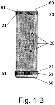
Figure 1 represents a general view of the device where in part 1-A, it can
be seen the device with the plunger inserted (30). The device has in one end
(31) a wedge (32) that facilitates the plunger withdrawal when the
fibrocollagen
is already formed. Part 1-B represents a general view of the device with the
cell
culture tray (60) inserted.
Figure 2 represents a sagital section of the device over its midline. Part 2
¨A shows with more detail the plunger (30) inserted in the body of the device
(20), with a wedge in one end of the device (32). Part 2-B. shows the body of
the device (20) with the cell culture tray (60) inserted. As it can be seen
there is
a minimum space around its two sides, fact which allows the contact of all the
implanted cells with the neoformed vessels of the fibrocollagen.
7
CA 02819482 2013-05-30
Figure 3 shows a front view of the tray, which shows its gridded base that
allows the adhesion of the implanted cells.
Figure 4 corresponds to a side view of the cell culture tray (60), that
shows in the ends, the abutments/supports (61) which function is such that
when the tray is inserted in the body (20), it leaves a space enough so that
the
cells fixed to both sides of the cell culture tray culture (60), be in contact
directly
with the neoformation vessels.
Figure 5 shows a preferred embodiment in the present invention with
different plungers (30) inserted, these plungers will be exchanged by culture
trays. The advantage is that with only one incision, various cell culture
trays can
be inserted.
Detailed description of the invention
The present invention refers to an improved device which is inserted in
the subcutaneous cell tissue, in order to facilitate the implant of cells.
This
device differs from those devices currently known because it is flat shaped,
form which decreases the dead space in it inner chamber, as it occurs with the
circular devices, thus allowing an extensive contact of the implanted cells
with
the neovascularized surface in such a way that cells in liquid media can also
be
implanted directly. In addition, it comprises a novel tray whose design and
shape, allows the cells can be seeded in both anterior and posterior sides,
thus
increasing even more the useful space so that every cell to be implanted, will
be seeded in such a way that will be adhered to the sides and will contact
immediately the neoformation vessels, thus allowing the cells to interact with
the media so that there is no lack of nutrients thus increasing the cell
survival
rate and therefore the device functionality. This novel flat or flattened
device,
with its culture tray, allows the cells to implant to be seeded in vitro in
both sides
of the tray before being inserted in the body of the device covered by
fibrocollagen. This fact allows to know accurately the amount of seeded cells
and to determine their viability before being inserted. In addition, in a
preferred
embodiment and as a variation of this invention, the device is composed of
several plungers resembling a comb, one next to the other, held in one of its
8
CA 02819482 2013-05-30
. .
ends, that when the plungers are replaced by the cell culture trays it
facilitates
the implanting procedure. The number of culture trays depends on the number of
cells to be implanted. Although it could be thought that the use of a widest
culture tray would be simpler, this invention has proved that the device
described in the present invention allows a better control of the number of
cells
to be implanted, by handling the cells in independent trays in the comb
arrangement. In addition another important aspect is that the dimensional
integrity of the porous body is maintained.
With the present invention any kind of cell line can be cultured for the
treatment of chronic degenerative diseases, diseases generated by the
deficiency of a biological factor or for the treatment of malignant tumors,
among
other diseases.
In accordance with Figure 1, the device object of the present invention
consists of an improved device which contains one hollow section or body (20)
preferably with a porous surface, and has a cavity inside it that houses a
plunger
(30). At the ends of the porous body (20) it can be found sealing sets or
mechanisms (21) one of which connects to the one sealing element (50) while
the other end (21) connects to the second sealing element (31) one end of
which is joined to the plunger or to a second sealing element (61) which is
integrated to the cell culture tray (60), in such a way that, when the device
is
closed by means of the sealing plugs (31) or (61) it keeps rigidly inside the
porous body (20) said plunger (30) or the cell culture tray (60) as can be
seen
with more detailed in Figure 2.
The porous body (20) is preferably composed of a rectangular or square
grid with rounded corners and edges that can be made of biocompatible
stainless steel, biodegradable inert polymer or any other material capable of
providing with dimensional stability to the intermediate part of the set of
the
device thus allowing the contact of the neoformed blood vessels with the
implanted cells by means of the cell culture tray (60). In accordance with the
purposes of the present invention, the degree of porosity of the device porous
body (20) in order to achieve the goals the present invention must have a mesh
size of 40 to 150-mesh or higher if the implant of primed antitumor cells is
9
CA 02819482 2013-05-30
required. On other side, the length of this intermediate porous section or
porous
body (20) can be the adjusted according with therapeutic needs in order to
favor
appropriately the production of the biological factor needed. The preferred
length
is 10 to 600 millimeters with an inner space preferably between 0.1 mm and 3
mm. The device has two ends having each the sealing elements (21). One of
this is coupled to the plunger (30) sealing element (31) or to the cell
culture tray
(60) sealing element (61) in such a way that, when the sealing elements are
closed, the inner cavity of the porous body (20) becomes sealed. In its other
end, the sealing element (21) is coupled to the plug (50) sealing element (51)
plug which has a shape and design that when coupled to the porous body (20)
sealing element (21) seals the end and gives continuity to the mesh in the
bottom of the porous body. If it is used under the modality to receive the
comb
plunger, the porous body (20) is divided into two or more sections, although 4
are preferred, which are attached to the arms of the comb-shaped plunger.
Figure 5.
The plunger (30) has a size, shape and design such that it can be
introduced in the cavity of the porous body (20) and since it has a sealing
element (31) in the end which has the wedge (32), the plunger allows the
inner of the porous body to be sealed (20) aimed at preventing the
contamination of the inner cavity by adventitious agents. In the exterior part
of its end the plunger has a wedge (32) whose main function is to hold firmly
the plunger to the porous body when withdrawing the plunger from the porous
body so as to withdraw with higher strength. The plunger (30) is made of any
biomaterial which can be introduced into the organism of a mammal, without
being rejected but that at the same time it should produce an important
response
from the organism to a foreign body in order to promote the coating with
fibrocollagen. Teflon is the material preferred for this invention The details
of the
plunger (30) can be seen with more detail in Figure 2A or in Figure 5, if the
device corresponds to the comb-shaped one.
The culture tray (60) has a design and shape such that it can be
introduced into the porous body (20). Its sealing element (61) when coupled
to the sealing element of the porous body seals the inner cavity thus
promoting
CA 02819482 2013-05-30
the growth of the seeded and implanted cells. Its anterior (60a) and posterior
(60b) sides are made of stainless steel medical degree mesh or of any
other material accepted for implant with such porosity that allows the cells
to be
seeded to adhere to both surfaces of the anterior (60a) and posterior (60b)
sides so that in a subsequent step, to be inserted into the body (20) of the
device. In each of its edges, the culture tray (60) has an abutment/support
(62) with shape and dimensions such that the tray becomes adapted to the
inner space of the porous body. The function of such abutment/support is to
keep fix the cell culture tray within the porous body (20) and when the tray
is
fixed in this position it provides with the necessary space between the sides
(60a and 60b) of the culture tray (60) and the inner anterior (20a) and
posterior (20b) sides of the porous body (20), in order to favor the immediate
contact between the seeded cells in the culture tray (60) and implanted into
the
inner chamber of the porous body (20) covered by fibrocollagen and the
neoformed vessels. In a preferred embodiment of this invention, this space is
0.1 mm. to 3mm. Figures 3 and 4 show more details of the culture tray (60).
In a preferred embodiment of the present invention various culture trays
(60) are used one next to the other as it can be seen in Figure 5. The
number of culture trays will be the one required to implant the number of
cells
required by the by the receiving organism. Several cell culture trays (60) may
be used instead of only one, wider, in order to avoid that the wider porous
body
(20) collapses.
In accordance with the procedure of the present invention, the improved
device for the implant of cells, once that is introduced within the
subcutaneous
tissue of any mammal, the first sealing element (31) that is fitted to the
plunger
(30) coupled to the sealing element (21) of the porous body (20), will produce
a
foreign body reaction thus bringing about the device to be coated by
fibrocollagen of the mammal in which the device was implanted. Once the
plunger (30) is withdrawn withdrawal which is facilitated by its wedge (32),
produces an inner site immunologically isolated or privileged, which is
appropriate to house the tray containing the cell culture (60) either
previously
cultured or in vitro. The novel tray for cell culture (60) has design such
that
11
CA 02819482 2013-05-30
cells can be cultured on both sides and that thanks to its abutment/support
(62)
allows that the cell culture tray (60) remains fixed in the inner chamber of
the
porous body (20) of the device thus leaving an inner space within the chamber
enough for the seeded cells in one or both anterior and posterior sides (60a)
(60b) to contact immediately the neoformed vessels of the fibrocollagen. In a
preferred embodiment of the present invention, this space ranges between 0.1
and 3 mm.
It has been determined that the degree of porosity of the grid composing
the intermediate porous body (20), in accordance with the procedure herein
explained and also with the functions of the patient's organism, is
determinant of
the size of the neoformed vessels in the fibrocollagen. Therefore the size of
the
mesh or pore is determined according to the type of application to be given to
the tube of fibrocollagen recently formed.
In accordance with one of the preferred modalities the plunger (30) or
plug (50) sealing elements (31) or (51) respectively, consist preferentially
of a
press- activated sealing element with a latch as fastening element that
generates a hook click so that the user can ensure that it has been sealed.
The
sealing elements are inserted in one of the sealing elements of the porous
body
(21), thus holding the porous body to the sealing element (31) or (51).
The applicant has found that the thickness of the of the device porous
body (20) of the porosity, is related to the size of the neoformed vessels.
Therefore porosity is defined in accordance with the most appropriate
conditions
for the survival and growth of the cells to be implanted in maintain an
effective
therapeutic action.
The device is manufactured with medical degree biocompatible materials.
These materials for instance, can be made of some kind of stainless steel, of
virgin polytetrafluoroethylene (PTFE), titanium, biodegradable polymers,
biopolymers,
etc.
As it can be inferred from the description of the flat or improved
flattened device for favoring the implant of biological material, its
composing
parts can be made by means of machining or by means of mold injection. The
12
CA 02819482 2013-05-30
chosen process will be determined depending on the composing materials to be
used.
In accordance with the present invention, the procedure for the implant
of biological materials under the modality of reservoir, product of the
formation
of the biologic fibrocollagen tube by using the already mentioned device,
consists of the implantation in the body of any specie of mammal including
humans, the device together with the plunger (30) inserted in the inner
chamber of the porous body (20), in such a way that, when the device is
implanted, the mammal organism naturally coats the porous body (20) with
fibrocollagen. Then once that the layer of fibrocollagen is formed, a partial
incision is made, with the purpose of exposing the part of the device that has
the
sealing element (31) coupled to the sealing element (21) of the porous body
(20), with the purpose of withdrawing the plunger by facilitating this process
by
means of the wedge (32). Once the plunger (30) has been withdrawn there is a
chamber made of neovascularized fibrocollagen appropriate for the implant of
cells seeded on the culture tray (60). The tray can then be inserted through
the opening appearing when the plunger or flat tablet is removed (30). The
space remains sealed due to the coupling of the sealing elements of the porous
body (21) and of the culture tray (61). In these terms, the cells producer of
the
biological factor, start producing when entering in contact with the neoformed
and vascularized collagen fiber tissues thus being the biological factor
absorbed
by the blood stream. In another modality, if the device was chosen with
several
trays of cell culture (60), the same procedure is followed but only the
necessary
trays of cell culture are inserted (60).
In order to increase the effectiveness of the treatment, cells genetically
manipulated by known techniques can be used to produce the biological factor.
The culture medium to be used is selected in terms of the cells to be
implanted. With the aid of cytoprotective agents like niacin or allopurinol
The
culture medium can be placed directly in the immunologically privileged space
generated by the device, without the need of the tray Thanks to the flatted-
shape of the device, the implanted cells are not compressed.
13
CA 02819482 2013-05-30
. .
The applicant has found that with the use of the device described in the
present invention, it has been possible to obtain semi-isolated sites with
good
appropriate neovascularization and therefore, conditions exist for a better
cell
viability of cells. Similarly, a better interchange rate of biological factors
is
obtained in, comparison with the circular devices.
The amount of cells, for the case of the treatment of diabetes referred in
the literature is about 6,000 to 12,000 Islets of Langerhans per kilogram of
the
patient's weight. In the case of the present invention, it has been seen that
these
can be combined or not with Sertoli cells in order to immunologically protect
them from the rejection. These devices can also be used for the implant of any
cellular culture required for the treatment of different diseases. This
includes
primed cells against tumors, since it has been confirmed that the implanted
cells
in the device in the US patent No 6,716,246 migrated from the device without
being rejected by the recipient organism fact that has opened the possibility
for
this kind of treatments as referred in the patent application No
W02009/075556. Procedure to prime cells and its use for the treatment of
tumors.
Examples
Under the preferred embodiment of favoring the implant of cells producers
of a biological factor, the flat or flattened improved device object of the
present
invention was implanted in the dorsal part of a sample of Long Evans rats
weighing between 180 and 200 grams. In a second group of rats with the same
characteristics the circular device referred in the US patent No 6,716,246
(Valdes) "Process and device to facilitate implantation of biological
material" was
implanted. Diabetes was induced by means of an intravenous application of 65
mg/kg of streptozotocin to the groups of ten rats with the device object of
this
invention and to a control group. The level of glucose in both groups showed
no
important differences, being in the order of 337mg/dL.
An implant of islets of Langerhans was performed with both groups and
the islets of Langerhans were isolated and conserved using known conventional
techniques, with the difference that the group of rats with the flat or
flattened
14
CA 02819482 2013-05-30
improved device received the Islets of Langerhans in the site formed by the
cell culture tray culture (60). Whereas the group in which the circular device
was
implanted, the cells were implanted, directly in the formed fibrocollagen
tubes.
Both groups were implanted with the same number of islets, which was in the
order of 50 to 100 thousand IEQs. Glucose levels were measured daily
during the first week and subsequently once a week. The animals with the
circular device group showed in the first three days a glycemia of over 250
mg/dL for two consecutive days in a row, fact which describes by itself the
functionality of the circular device.
The animals with the flat or improved flattened device showed a more
significant decrease of glucose levels to 150 mg/dL, fact which shows a 40%
improvement in comparison with the circular device. This fact can be
interpreted
as a better performance of the flat or improved flattened device as a result
of a
higher number of surviving cells and functionally active.
In a preferred embodiment of this invention, instead of inserting various
independent flat devices to obtain the number of cells required by each
receptor
the flat or improved flattened device is wider, with the plunger combed- shape
one next to the other and/or one above the other in the central porous body,
whose dimensions will be adjusted to the plungers width. Once the device has
been covered by fibrocollagen with the neoformed vessels; the combed-shape
plunger is replaced by the cell culture trays or the cells are implanted
directly
in the liquid medium into the immunologically privileged spaces left by the
plungers. With this, the performance of the improved device is increased even
more and facilitates the insertion of the culture trays, because instead of
making
various incisions depending on the number of devices inserted in the recipient
organism, only one incision is made, locating the combed-shape plunger which
is replaced by the cell culture trays. In the preferred embodiment four
culture
trays are used as it can be seen with more details in Figure 5.
It is herein stated that up to this date, the best known method by the
applicant to implement said invention, is the one resulting from the
description of
this invention.