Note: Descriptions are shown in the official language in which they were submitted.
CA 02819635 2013-05-31
WO 2012/075337 PCT/US2011/062958
DIRECTED DELIVERY OF AGENTS TO NEURAL ANATOMY
CROSS REFERENCE TO RELATED APPLICATIONS
[0001] This application claims priority under 35 U.S.C. 119(e) of U.S.
Provisional Patent
Application Serial No: 61/418,721 filed on December 1, 2010, the contents of
which are incorporated
herein by reference in their entirety.
FIELD OF THE INVENTION
[0002] The present invention is directed to methods, devices and systems
for neurostimulation of
target neural anatomies, particularly the dorsal root and dorsal root
ganglion. Such methods, devices and
systems include agent delivery alone or in combination with electrical
stimulation for the treatment of
various conditions, particularly pain and pain-related disorders.
BACKGROUND OF THE INVENTION
[0003] Pain affects more Americans than heart disease, diabetes and
cancer combined. In fact,
about 50 million Americans suffer from chronic pain and spend about $100
billion for treatments per year.
Unfortunately, many of the strongest available analgesics have serious side-
effects including addiction,
dependence and increased risk of heart attack and stroke. Moreover, many
chronic pain conditions cannot
be effectively treated with existing medications. Considering the revenue of
drugs like CELEBREXO
($2.8 billion in 2004; G.D.Searle & Co., Skokie, Ill., United States of
America) and VIOXXO ($1.4
billion in 2004, Merck & Co., Inc., Whitehouse Station, N.J., United States of
America), a safe and
effective treatment for chronic pain would significantly benefit human health.
Accordingly, there is an
unmet need for effective pain treatments. The present invention aims to meet
at least some of these
objectives.
SUMMARY OF THE INVENTION
[0004] The present invention is directed generally to systems, devices
and methods for direct
delivery of agents, e.g., drugs, directly to the spinal anatomy of humans,
particularly to at least one region
or a combination of regions selected from the dorsal root (DR) and/or dorsal
root ganglia (DRG) and/or
dorsal root entry zone (DREZ) and/or intrathecal space. Such delivery can be
used to treat a variety of
conditions, including, for example, the treatment of pain and pain related
disorders, including but not
limited to neuropathic pain, chronic itch, puritis, sensory disorders,
multiple sclerosis, post-herpetic
neuralgia and the like.
[0005] In some embodiments, the system and devices as disclosed herein
can be used to deliver
at least one agent alone to the target spinal anatomy, or alternatively in
combination with electrical
stimulation. In some embodiments, the delivery of an agent to the target
anatomy using the devices as
disclosed herein is in a temporal pattern which is coordinated with a temporal
pattern of electrical
stimulation of the target anatomy, such that an agent is delivered to the
target spinal anatomy in
-1-
CA 02819635 2013-05-31
WO 2012/075337 PCT/US2011/062958
combination with electrical stimulation. In some embodiments, the delivery of
the agent can be
simultaneous with the electrical stimulation, or alternatively the delivery of
the agent may be before or
after the electrical stimulation of the target spinal anatomy.
[0006] The present invention has numerous advantages over existing
methods and devices for the
treatment of pain. In particular, one advantage is that selected target spinal
anatomies undergo pathological
changes, referred to as "neuronal plasticity" in certain pain pathologies,
e.g., during inflammatory pain and
neuronal injury. If the changes occur in the peripheral nervous system, this
is referred to as peripheral
sensitization. For example, without wishing to be bound by theory, nociceptors
have a characteristic
thresholds or sensitivity that distinguish them from other sensory nerve
fibers. Depending on the
nociceptor type, the can be excited by intense noxious heat, intense pressure
or irritant chemicals, but not
innocuous stimuli such as warming or light touch. In particular, alternations
in pain pathways lead to
hypersensitivity, such that pain outlives its usefulness as an acute warning
system and instead becomes
chronic and debilitating. This may be seen, at some level, as an extension of
the normal healing process,
whereby tissue or nerve damage elicits hyperactivity to promote guarding of
the injured area. For example,
sunburn produces temporary sensitization of the affected area. As a result
normally innocuous stimuli,
such as light touch or warmth, are perceived as painful (a phenomenon referred
to as allodynia), or
normally painful stimuli elicit pain of greater intensity (referred to as
hyperalgesia). At its extreme, the
sensitization does not resolve. Indeed, individuals who suffer from arthritis,
postherpetic neuralgia (after a
bout of shingles), or bone cancer experience intense and often unremitting
pain that is not only
physiologically and psychologically debilitating, but may also hamper
recovery. Chronic pain may even
persist long after an acute injury. Accordingly, in many pain instances, e.g.,
inflammatory pain or nerve
injury, the expression of certain receptors and ion channels can be
upregulated and downregulated in
dorsal root ganglion cells (DRG), the cell body of the of primary sensory
neurons, which can decrease the
threshold of activation of these nociceptor neurons, resulting in an increased
pain sensation in the subject
by a stimuli which would not normall cause pain. As an example, under
sustained peripheral
inflammation, pro-longed c-fiber activation alters the pattern of gene
transcription from the DRG and the
dorsal horn neurons. Additionally, some components of the inflammatory soup
(e.g., protons, ATP,
serotonin, lipids) alter the excitability of neurons directly by interacting
with ion-channels on the sensory
neuron cell surface. For example, NGF activates TrkA on neurons, bradykinin
activates BK2 receptor,
serotonin activates 5-HT3 receptors, ATP activates P2X3 receptor, protons (H+)
activate ASIC3/VR1
receptors, lipids activate the PGE2, CB1 and VR1 receptors, and heat activate
the VR1NRL-1 receptors,
belonging to the TRPV family of ion channels, thus sensitizing (e.g., lowing
the threshold of activation) or
exciting the terminals of the nociceptors. Accordingly, a subject suffering
from inflammatory pain could
be treated with a specific pharmacological agent at the target spinal anatomy,
e.g., DRG to ameliorate the
effects of the inflammatory mediators on the activation of ion channels and
other receptors in the DRG.
- 2 -
CA 02819635 2013-05-31
WO 2012/075337 PCT/US2011/062958
[0007] Accordingly, an agent delivered with the devices and systems as
disclosed herein for the
treatment of clinical pain would not have an effect on normal patients not
experiencing chronic pain where
such pathological changes have not occurred. Another advantage of the devices,
systems and methods as
disclosed herein is that it allows for specific and localized delivery of
agents to specific target spinal
anatomies, such as but not limited to the DRG. Accordingly, lower doses can be
used and therefore avoids
any off-target and/or systemic side affects associated with the delivered
agent. Another advantage of the
present system allows for the combination of electrical stimulation to be used
in combination with
delivering the agent to the target spinal anatomy. For example, the
combination of electrical stimulation
and concurrent agent delivery is useful for activating channels present in
specific cells present at the target
spinal antatomy, e.g., DRG, and thus allowing entry of the agents into
specific cells of interest, which
increases the specificity and selectivity of the agents delivered.
Additionally, the electrical stimulation can
be used to activate certain agents, e.g., from a pro-drug to a biologically
active agent at the target spinal
anatomy location.
[0008] Any agent can be delivered using the device and system as disclosed
herein, including but not
limited to ion channel agonists and antagonists, sodium channel blockers,
biologics, neuroinflammatory
modulators, toxins etc., to selectively neuromodulate or inhibit the
electrical impulses from the neurons. In
some embodiments to selectively destroy the neurons. For example, in some
embodiments, a toxin which
binds to and is specific for a specific neuronal cell-type can be used to
selectively ablate certain pain-
transmitting neuronal types, e.g., neurons firing ectopically or spontaneously
as a result of innocuous
stimuli.
[0009] In particular, in some embodiments, the agent delivered to the target
spinal anatomy using the
devices, systems and methods as disclosed herein is selected based on the
particular pain indication to be
treated. For example and without wishing to be limited to theory, inflammatory
mediators such as, but not
limited to Prostaglandin E2 (PGE2) increase the excitability of DRG neurons in
part by reducing the
extent of membrane depolarization needed to activate TTX-R Na+ channels.
Accordingly, sensory neurons
have increased spontaneous firing and repetitive spiking, resulting in
increased intense pain sensation by
the subject. Additionally, other pro-inflammatory agents, such as bradykinin
and capsaicin increase
activation of the Vanilloid Receptor [VR1D and increase the effect the TTX-R
Na+ channel. Embodiments
of the present invention advantageously utilize aspects of the pain pathway
and neurochemistry to modify
electrophysiological excitability of the DRG neurons where electrical
stimulation is coupled with
pharmacological agents (electrical stimulation alone or in combination with a
pharmacological agent) to
optimize the efficacy of the stimulation system.
[0010] Other aspects of the present invention relate to a combination of agent
delivery and/or electrical
stimulation to the target anatomy, which can be automated and/or can be "on
demand" for example,
controlled by a patient controlled analgesia (PCA) pump. In particular, the
present invention is different
from the neurostimulation methods and systems as disclosed in International
Application
- 3 -
CA 02819635 2013-05-31
WO 2012/075337 PCT/US2011/062958
W02006/029257, and U.S Application US 2008/0167698 (which are incorporated
herein in their entirety
by reference), in that the delivery device as disclosed herein allows for
controlled and precise delivery of
one or more pharmacological agents to a target anatomy, which can be
specifically tailored to a particular
delivery regimine by the physician and/or patient.
[0011] Additionally, the present application provides for coordinated delivery
of one or more
pharmacological agents with electrical stimulation so that the delivery of the
agent and/or electrical
stimulation can be timed with resepect to each other, e.g., temporally
regulated (e.g., an agent is delivered
(e.g. "on") or not delivered (e.g., "off')) according to a particular
electrical stimulation patterning or
treatment regimine and/or can be delivered "on demand" by the patient using a
patient controlled analgesia
(PCA) pump.
[0012] Additionally, the delivery device as disclosed herein allows the
delivery of one or more agents
in a coordinated manner, or in concert with the electrical stimulation, for
example, where the coordinated
delivery allows the pharmacological agent to act synergistically with the
electrical stimulation, such that
the efficacy of the agent is enhanced by the electrical stimulation. For
instance, but not wishing to be
bound by theory, an agent to be delivered using the device as disclosed herein
selected to be delivered
based on its ability for its activity or efficacy to be enhanced by the
electrical stimulation. Such electrical-
stimulation induced enhancement of the agent can be by a variety of
mechanisms, e.g., the agent becomes
activated on electrical stimulation, or the target receptors or ion channels
that the agent modulates
becomes activated or open by the electrical stimulation such that the agent
only acts on activated receptors
and/or open ion channels etc, or migration of an agent to particular cell
subtype on electrical stimulation
etc. Furthermore, in some embodiments of the present delivery device as
disclosed herein, the electrodes
and the agent delivery structure, such as an outlet port, are close together
(in some embodiments, the
electrodes are interdispursed between outlet ports) such that the electrical
stimulation can activate the
agent being delivered by the device, therefore enabling a better control of
electrical stimulation with agent
delivery such that the electrical stimulation and delivery of the agent
function synergistically to reduce the
pain sensation in the subject.
[0013] Accordingly, in some embodiments, the devices, methods and systems as
disclosed herein
provide improvements over existing systems in that they allow delivery of
selected agents which are
enhanced by electrical stimulation. Some additional advantages of the delivery
device and methods as
disclosed herein include but are not limited to the temporal pattern of the
delivery of the agent alone, or in
conjunction with a coordinated temporal electrical stimulation, such that the
stimulation parameters
specifically activate the agent.
[0014] Additional advantages of the delivery device and methods as disclosed
herein include but are
not limited to the delivery of the agent by the device in a delivery agent,
e.g., a vector or carrier particle,
such that the delivered agent remains in the location it was delivered for a
period of time for effective
therapeutic effect (e.g., reduction in pain sensation by the subject).
- 4 -
CA 02819635 2013-05-31
WO 2012/075337 PCT/US2011/062958
[0015] In some embodiments, the electrical stimulation can be used to deliver
the agent to the target
spinal anatomy. For example, in some embodiments, the present invention can be
adapted so that the
electrical stimulation is used for electrophoritic (also referred to as
"iontophoretic flux" or
"iontrophoretic") agent delivery, where an electrically conducting wire in the
delivery lumen 140 can be
used to charge the agent (e.g., either a positive or negative charge) within
the lumen, and as the charge is
greater than the charge in the subjects body, the charged agent is driven out
of the lumen and through the
outlet ports 40 and into close proximity of the target site, such as the DRG.
[0016] Accordingly, the present invention relates to the combination
neurostimulation and
pharmacological agent delivery element, where the inventors have discovered
that a pre-determined
temporal pattern of neurostimulation and agent delivery surprisingly results
in a greater efficacy of the
agent and reduced pain sensation in the subject as compared to either delivery
of the agent alone, or the
electrical stimulation alone, thus being able to obtain the desired
stimulation or modulation level.
[0017] Other aspects of the disclosure relate to methods for treating chronic
pain. For example, in one
embodiment, the disclosure relates to a method for treating chronic nerve pain
in a subject, e.g., a mammal
such as a human. In some embodiments, in accordance with the method, the area
of pain in the subject is
identified, and the spinal level within the mammal that is associated with the
chronic pain is determined. A
delivery device as disclosed herein is provided for introducing an agent at
the location of the DRG
associated with the chronic pain.
[0018] Other aspects of the present invention relate to a method for targeted
treatment of pain and pain
related disorders and/or conditions with minimal deleterious side effects,
such as undesired side effects as
a result of off-target non-specific effects of an agent as well as undesired
motor responses or stimulation of
unaffected body regions,. In some embodiments, the system and devices as
disclosed herein achive
minimal deleterious side effects by directly delivering the agent to the
target anatomy in combination with
selectively neuromodulating the target anatomy, e.g., the DRG to modulate or
decrease pain and a pain
related disorder or condition, while minimizing or excluding undesired side
effects by avoiding non-
specific or systemic administration of a pain agent or analgesic, or
generalized neuromodulation of other
anatomies. In most embodiments, delivery of the agent to the target anatomy
can be alone or in
combination with neurostimulation, such as electrical stimulation, however it
may be appreciated that
neurostimulation may include a variety of forms of altering or modulating
nerve activity by at least one
agent and optionally, delivering electrical stimulation directly to the target
anatomy. For illustrative
purposes, descriptions herein will be provided in terms of agent delivery to
the DRG in combination with
electrical stimulation, with exemplary stimulation parameters as well as
temporal patterning of the agent
delivery with the electrical stimulation, however, it may be appreciated that
such descriptions are not so
limited and may include a combination or variety of agent delivery methods,
e.g., continuous, on-demand,
intermittent based on a predefined temporal pattern of delivery to the DRG,
and in combination with
electrical stimulation, using a variety of different parameters, such as
intermittent and in a temporal
- 5 -
CA 02819635 2013-05-31
WO 2012/075337 PCT/US2011/062958
regulated pattern so the electrical stimulation of the DRG works
synergistically with the delivery of the
agent to the DRG.
[0019] In particular, the combining of direct delivery of an agent to the DRG
with electrical stimulation
of the DRG as disclosed herein provides provide several advantages. For
example, the delivered agent and
electrical stimulation can function synergistically to decrease pain sensation
in a subject, and/or enhance
the therapeutic effect of the agent and the electrical stimulation as compared
to their use alone.
Alternatively, in some embodiments, the electrical stimulation increases the
selectivity of an agent to
target DRG cell bodies. Alternatively, in some embodiments, the electrical
stimulation enables targeted
activation of an agent delivered to the DRG. In another embodiment, the
electrical stimulation causes
differential enhancement of an agent to delivered target DRG cell bodies.
[0020] Typically, the agent-neurostimulatory systems and delivery devices as
disclosed herein are used
to neuromodulate portions of neural tissue of the pairs of nerves along the
spinal cord which are known as
spinal nerves. The spinal nerves include both dorsal and ventral roots that
integrate near the intravertebral
foramen to create a mixed nerve which is part of the peripheral nervous
system. At least one dorsal root
ganglion (DRG) is disposed along each dorsal root prior to the point of
mixing. Thus, the neural tissue of
the central nervous system is considered to include the dorsal root ganglions
and exclude the portion of the
nervous system beyond the dorsal root ganglions, such as the mixed nerves of
the peripheral nervous
system. Typically, the agent-neurostimulatory systems and delivery devices as
disclosed herein are used to
neuromodulate one or more spinal anatomy, for example, but not limited to one
or more dorsal root
ganglia, dorsal roots, dorsal root entry zones, or portions thereof, while
minimizing or excluding undesired
stimulation of other tissues, such as surrounding or nearby tissues, ventral
root and portions of the
anatomy associated with body regions which are not targeted for treatment.
However, it may be
appreciated that stimulation of other tissues are contemplated. In some
embodiments, it is also envisioned
that a system or device can neuromodulate different neural anatomies in the
same subject, for example, for
illustration purposes only but by no way a limitation, the device or system
may be configured and
positioned in a subject so that an agent and electrical stimulation is
delivered to a spinal anatomy such as
DRG, and can also be delivered to a different spinal anatomy of the subject,
such as dorsal root. Or, the
device or system may be configured and positioned in a subject so that an
agent and electrical stimulation
is delivered to a spinal anatomy such as DRG, and can also be delivered to a
different spinal anatomy of
the subject, such as a spinal cord. Or, the device or system may be configured
and positioned in a subject
so that an agent and electrical stimulation is delivered to a spinal anatomy
such as DRG, and can also be
delivered to a different neural anatomy of the subject, such as a sympathetic
ganglion or peripheral nerve.
Accordingly, any combination of different neural anatomies can be targeted for
agent delivery and
electrical stimulation by the methods, systems and devices as disclosed
herein. It is also encompassed that
any combination of different neural anatomies at different spinal cord levels
can be targeted in a subject by
the devices and systems as disclosed herein.
- 6 -
CA 02819635 2013-05-31
WO 2012/075337 PCT/US2011/062958
[0021] Accordingly, as the devices, systems and methods to treat various
disorders as disclosed herein
enable both an agent and in some embodiments, electrical stimulation, to be
delivered at a specific dose
and specific stimulation energy levels to a defined anatomical location, e.g.,
in proximity to the dorsal
root, in particular, the dorsal root ganglion (DRG), the devices, systems and
methods have numerous
advantages including reduced side effects associated with systematic delivery
agents, and/or adverse side
effects from spinal cord electrical stimulation (SCS). Additionally, as the
localized delivery of the agent to
the DRG can be coordinated with the specific electrical stimulation of the
DRG, it provides a superior
level of control and specificity of agent efficacy and/or the electrical
simulation effect which is not easily
achievable with other systems.
[0022] Accordingly, the present invention relates generally to devices,
systems and methods for direct
delivery of agents, e.g., analgesics and pain medicine to the DRG. Herein, in
one embodiment, the device
for direct delivery of agents to a target neural anatomy, e.g., DRG is
referred to as delivery device (DD)
10, which comprises a agent release module 20 connected to a delivery element
30 for transporting the
agents from the agent release module, where they are stored and released in a
controlled manner, to the
delivery site at the anatomical target spinal anatomy location, which can be
any of, but not limited to one
or more dorsal root ganglia, dorsal roots, dorsal root entry zones and other
spinal anatomies. In some
embodiments, the delivery element 30 is configured as a catheter comprising a
lumen for delivery of at
least one agent to at least one target neural anatomy. In alternative
embodiments, the delivery element 30
is configured as a lead comprising at least one electrode which is connected
to a pulse generator for
electrical stimulation of at least one target spinal anatomy.
[0023] Accordingly, the agent release module of the delivery device is placed
in the subject's body in
an anatomically convenient location, such as in the back or buttocks, and the
agent or drug formulation is
transported along fluidly connected agent delivery elements such that the
agent or the drug formulation is
released at least one target spinal anatomy, e.g., a DRG delivery site. A
target delivery site is in close
proximity to at least one target spinal anatomy, e.g., a DRG, and in some
embodiments the released drug
formulation functions on the cell bodies in the DRG to modulate the pain
response.
[0024] In some embodiments, the delivery device is further configured for
combining the delivery of a
drug formulation in close proximity to the DRG with electrical stimulation of
the DRG. In such
embodiments, the agent release module further comprises a pulse generator and
a battery and is connected
to leads which comprise electrodes near its distal end which are positioned in
close proximity to the DRG,
allowing for a combined electrical stimulation and agent delivery either
simultaneously (e.g., at the same
time) or in a pre-determined temporal pattern of electrical stimulation and
agent delivery.
[0025] In some embodiments, an agent or drug formulation is stored within an
agent release module
(e.g., contained in a reservoir or impregnated within a matrix within the
agent release module). The drug
formulation comprises an amount of drug sufficient for treatment and is stable
at body temperatures (i.e.,
no unacceptable degradation) for the entire pre-selected treatment period. The
agent delivery devices store
- 7 -
CA 02819635 2013-05-31
WO 2012/075337 PCT/US2011/062958
the drug formulation safely (e.g., without dose dumping), provide sufficient
protection from bodily
processes to prevent unacceptable degradation of the formulation, and release
the drug formulation in a
controlled fashion at a therapeutically effective rate to treat pain
[0026] One object of the invention is to provide a method for convenient, long-
term management of
pain.
[0027] One advantage of the invention is that the delivery devices, systems
and methods described
herein provide effective management of pain by administration of an agent,
e.g., a drug formulation,
directly to the DRG, providing adequate pain relief and a reduction in adverse
side effects relative to
systematic or delivery of agents to other locations. Another advantage is that
the present invention relates
to a combined used of electric stimulation of the target anatomy, e.g., DRG
concurrently with, or in a
temporal pattern with direct delivery of an agent to the DRG. This provides
several advantages, which
include a synergistic effect of the electrical stimulation to increase the
efficacy of the agent and vice versa
(e.g., synergistic analgesia), such that the therapeutic effect of the agent
and the electrical stimulation are
each enhanced when they are used together, as compared to their use alone, or
an increase of the
selectivity of an agent (e.g., agent targeting) to target anatomy, e.g., DRG
cell bodies in the presence of
electrical stimulation, or an increased targeted activation of an agent (e.g.,
compound activation) delivered
to the target anatomy, e.g., DRG in the presence of electrical stimulation.
Another advantage of concurrent
or temporal delivery of agents and electrical stimulation is that e-fields can
result in differential
enhancement of an agent (e.g., cell specific target enhancement) delivered to
the target anatomy, e.g.,
DRG cell bodies.
[0028] Given the adverse effects of many pain drugs, e.g., opioid analgesics,
one of the advantages of
the delivery device as disclosed herein is lower doses which still provide
considerable benefit to those
desiring pain relief, particularly in relatively long term (e.g., 1-4 months)
pain situations. Furthermore, the
delivery devices can also be more cost-effective, and thus may make pain
management available to a
broader population. Such target specific delivery can also reduce escalating
tolerance, dependence and
incomplete effectiveness due to localized concentrations of the effective
agent delivered at a concentration
sufficient to achieve the desired effect at the target spinal anatomy, e.g.,
DRG.
[0029] Another advantage of the invention is that the invention can be used to
deliver relatively small
quantities of pain drugs to a subject accurately and precisely and thus safely
delivering such agents and
pain drugs despite the extreme potency of these agents. Thus, the invention
allows for the convenient use
of a variety of different pain drugs for treatment of pain ranging in severity
from mild to severe.
[0030] Another surprising advantage of the systems and devices as disclosed
herein relates to the
combined use of electrical stimulation of the DRG in combination with direct
delivery of agents to the
DRG, which enables tailoring the pain treatment to the patients needs, as well
as providing sufficiently
effective therapy over a relatively long duration of therapy.
- 8 -
CA 02819635 2013-05-31
WO 2012/075337 PCT/US2011/062958
[0031] One notable advantage of the agent-neurostimulatory systems as
disclosed herein avoids the
need for placement of external needles and/or catheters in the subject, which
might provide sites
susceptible to infection. In addition, use of delivery devices in a subject
increases the patient compliance
with a prescribed therapeutic regimen, substantially decreases or completely
avoids the risk of abuse of the
agent by the patient or others in contact with the patient, and affords
greater mobility and easier outpatient
management.
[0032] Another advantage of the agent-neurostimulatory systems and delivery
devices as disclosed
herein is that a selective agent, or combination of agents can be delivered
directly to the DRG with such
accuracy and precision and at such low quantities of agent is required, and
allows long-term use of such
agents to treat pain. Additionally, the agent-neurostimulatory systems as
disclosed herein also allows for
effective pain management by the subject via the patient programmer 60
allowing treatment of
breakthrough pain episodes, as well as tailoring the delivery of the agent
with electrical stimulation of the
DRG for tailoring the pain treatment in real-time to meet the needs of the
subject for pain relief at that
particular time period.
[0033] Another advantage of the agent-neurostimulatory systems as disclosed
herein allows the
delivery of agents which marked potency, e.g., delivery of agents such as
opioids, Na+ channel blockers,
etc., in small amounts and volumes directly to the DRG, avoiding undesired
side-effects of systemic
administration or risks of subject addiction and the like.
[0034] Yet another advantage is that the invention provides for precise
delivery of an agent to the DRG,
thus allowing delivery of lower doses and/or for delivery of precisely metered
doses of a particular agent
at a consistent delivery volume rates (e.g., on the order of microliters to
milliliters per hour) which can be
controlled precisely and maintained for pre-determined periods of time.
[0035] Micro-electrode and stimulation system embodiments of the present
invention may be placed in
close proximity to a single nerve root ganglion (e.g., DRG) utilizing method
as disclosed herein. In some
embodiments, the distal end of the delivery element, which comprises the agent
delivery structure, such as
outlet ports, (and optionally the electrodes) are placed in close proximity,
or in contact with the dorsal root
ganglion epinurium, or just below the surface of the dorsal root ganglion
epinurium. In some
embodiments, the distal end of the delivery element does not penetrate or is
not implanted into the DRG
(e.g, see embodiments shown in Figures 3, 5, 12, 13, 22 and 26).
[0036] The methods as described herein provide numerous advantages, including
but not limited to: low
risk percutaneous access route similar to other procedures, direct delivery of
localized quantities of
pharmacological agents at the specific target spinal anatomy, e.g., DRG or
nerve root when using an
embodiment of the device having electrodes, and electrode placement that
enables preferential, selective
nerve fiber stimulation along with pharmacological agent delivery.
[0037] One aspect of the present invention relates to a neuromodulation system
comprising (a) a delivery
element having a distal end and at least one outlet port disposed near the
distal end, wherein the distal end
- 9 -
CA 02819635 2013-05-31
WO 2012/075337 PCT/US2011/062958
is configured for positioning at least one of the at least one outlet ports
near a dorsal root ganglion; (b) an
agent release module connectible with the delivery element, the agent release
module having an agent
release mechanism; and (c) an agent releaseable from the agent release
mechanism so as to be delivered
from the at least one outlet port according to a controlled release pattern to
at least assist in
neuromodulating the dorsal root ganglion. In some embodiments, the agent is
chargeable and the agent
release mechanism includes a mechanism for charging the agent so that the
agent is delivered by
iontophoretic flux according to the controlled release pattern.
[0038] In some embodiments of all aspects of the invention as disclosed
herein, an agent which is
delivered can be, for example, but is not limited to, one or more or a
combination of: lidocaine,
epinephrine, fentanyl, fentanyl hydrochloride, ketamine, dexamethasone,
hydrocortisone, peptides,
proteins, Angiotension II antagonist, Antriopeptins, Bradykinin, Tissue
Plasminogen activator,
Neuropeptide Y, Nerve growth factor (NGF), Neurotension, Somatostatin,
octreotide, Immunomodulating
peptides and proteins, Bursin, Colony stimulating factor, Cyclosporine,
Enkephalins, Interferon, Muramyl
dipeptide, Thymopoietin, TNF, growth factors, Epidermal growth factor (EGF),
Insulin-like growth
factors I & II (IGF-I & II), Inter-leukin-2 (T-cell growth factor) (II -2),
Nerve growth factor (NGF),
Platelet-derived growth factor (PDGF), Transforming growth factor (TGF) (Type
I or 6) (TGF), Cartilage-
derived growth factor, Colony-stimulating factors (CSFs), Endothelial-cell
growth factors (ECGFs),
Erythropoietin, Eye-derived growth factors (EDGF), Fibroblast-derived growth
factor (FDGF), Fibroblast
growth factors (FGFs), Glial growth factor (GGF), Osteosarcoma-derived growth
factor (ODGF),
Thymosin, or Transforming growth factor (Type II or 13)(TGF). In some
embodiments, an agent delivered
is selected from one or more or a combination of: opioids, COX inhibitors,
PGE2 inhibitors, Na+ channel
inhibitors.
[0039] In some embodiments of all aspects of the invention as disclosed
herein, an agent which is
delivered can be, for example, an agonist or antagonist of a receptor or ion
channel expressed by a dorsal
root ganglion, for example, an agonist or antagonist of a receptor or ion
channel which is upregulated in a
dorsal root ganglion in response to nerve injury, inflammation, neuropathic
pain, and/or nociceptive pain.
In some embodiments, an ion channel expressed by the dorsal root ganglion is
selected from any one of, or
a combination of: voltage gated sodium channels (VGSC), voltage gated Calcium
Channels (VGCC),
voltage gated potassium channel (VGPC), acid-sensing ion channels (ASICs). In
some embodiments, a
voltage-gated sodium channel (VGSC) includes TTX-resistant (TTX-R) voltage
gated sodium channels,
such as, but not limited to, Nav1.8 and Nav1.9. In some embodiments, a voltage-
gated sodium channel
(VGSC) is a TTX-sensitive (TTX-S) voltage gated sodium channel, for example,
but not limited to, Brain
III (Nav1.3). In some embodiments, a receptor is selected from any one of, or
a combination of, ATP
receptor, NMDA receptors, EP4 recetors, metrix metalloproteins (MMPs), TRP
receptors, neurtensin
receptors.
- 10-
CA 02819635 2013-05-31
WO 2012/075337 PCT/US2011/062958
[0040] In some embodiments of all aspects of the invention as disclosed
herein, a delivery element
further comprises at least one electrode which is capable of delivering
electrical energy, for example, to
provide electrical energy to assists in creating the iontophoretic flux of the
agent, amoung other effects of
the electrical stimulation, such as activating or opening specific ion
channels and/or receptors on the soma
of the sensory neurons. In some embodiments, least one electrode in close
proximity to the at least one
agent delivery structure, e.g., an agent outlet port, and in some embodiments,
the electrodes can be
intermittent between one or more agent delivery structures.
[0041] In some embodiments, an agent release module further comprises a pulse
generator which
provides the electrical energy in a manner which impacts the effect of the
agent on at least a portion of the
dorsal root ganglion. In some embodiments, the electrical energy is provided
once the agent has targeted at
least a portion of the dorsal root ganglion. In some embodiments, the
electrical energy is provided in a
manner that targets at least one particular type of cell within the dorsal
root ganglion, for example the cell
body of a sensory neuron, e.g., but not limited to the soma of a c-fiber
sensory neuron.
[0042] In some embodiments, the controlled release pattern of the electrical
release pattern and/or agent
release is determined to impact an effect of the electrical energy on at least
a portion of the dorsal root
ganglion, or alternatively, where the agent and/or the controlled release
pattern is determined to enhance
the ability of the electrical energy to excite or inhibit a primary sensory
neuron in the dorsal root ganglion.
In some embodiments, the agent and/or the controlled release pattern is
determined to cause a change in
the open probability of at least one sodium channel.
[0043] In some embodiments, the agent release mechanism delivers the agent to
assist in
neuromodulating the dorsal root ganglion over time. In some embodiments, the
agent release mechanism
comprises a matrix impregnated with the agent so that the matrix releases the
agent over time according to
the controlled release pattern, for example, an erodible material matrix.
[0044] In some embodiments, the agent is delivered in conjunction with a
carrier particle, for example,
but not limited to one or more or any combination of: a macromolecule complex,
nanocapsule,
microsphere, bead or lipid-based system, micelle, mixed micelle, liposome or
lipid:oligonucleotide
complex of uncharacterized structure, dendrimer, virosome, nanocrystal,
quantum dot, nanoshell or
nanorod. In further embodiments, an agent can also be conjugated or associated
with a targeting molecule
which targets the dorsal root ganglion, for example, but not limited to, a
targeting molecule which has a
specific affinity for a cell surface marker expressed on at least one cell
within the dorsal root ganglion, for
example, expressed on at least one cell body of a c-fiber.
[0045] In some embodiments, the agent can be delivered in conjunction with a
gellable material which
retains the agent near the dorsal root ganglion after delivery, for example, a
gellable material which gells
upon delivery (e.g., release from the agent delivery structure).
[0046] In some embodiments of all aspects of the invention as disclosed
herein, the positioning the distal
end of the delivery element comprises positioning at least one of the at least
one outlet port on or in
- 11-
CA 02819635 2013-05-31
WO 2012/075337 PCT/US2011/062958
contact with the dorsal root ganglion epinurium. In some embodiments, the
delivery element is not
implanted or does not penetrate into the dorsal root ganglion.
[0047] Another aspect of the present invention relates to an intrathecal agent
delivery system comprising:
(a) a delivery element having a distal end and at least one outlet port
disposed near the distal end, wherein
the delivery element is configured for advancement within an intrathecal space
along a spinal cord and
then along a dorsal root to position at least one of the at least one outlet
ports near an associated dorsal root
ganglion; (b) an agent release module connectible with the delivery element,
the agent release module
having an agent release mechanism; and (c) an agent releaseable from the agent
release mechanism so as
to be delivered from the at least one outlet port to at least assist in
neuromodulating the dorsal root
ganglion.
[0048] In some embodiments, an intrathecal delivery system comprises a
delivery element which
includes a stylet, wherein the stylet has a curved distal end configured to
assist in guiding the delivery
element along a root sleeve angulation of the dorsal root during advancement.
In some embodiments, the
intrathecal delivery system can be used to deliver an agent to the DRG, and in
some embodiments, the
agent comprises a targeting molecule which targets the agent to the dorsal
root ganglion, as disclosed
herein, where a targeting molecule has a specific affinity for a cell surface
marker expressed on at least
one cell within the dorsal root ganglion, such as but not limited to a c-fiber
cell body.
[0049] In some embodiments of the intrathecal delivery system, an agent
delivered is selected from any
or a combination of a benzodiazepine, clonazepam, morphine, baclofen and/or
ziconotide. In some
embodiments, the agent comprises a genomic agent or biologic. In some
embodiments, an agent delivered
by the intrathecal delivery system is activatable by electrical stimulation.
In alterantive embodiments, an
agent delivered by the intrathecal delivery system enhances the ability of
electrical stimulation to excite or
inhibit a primary sensory neuron in the dorsal root ganglion, or
alternatively, can enhance the ability of
electrical stimulation to target at least one specific cell within the dorsal
root ganglion.
[0050] In some embodiments of all aspect of the present invention, an agent
release module includes
electronic circuitry capable of generating stimulation energy for delivery of
the agent to the delivery
element. In such embodiments, an electronic circuitry includes memory
programmable with an electrical
stimulation parameter set and an agent delivery parameter set, for example,
where the set parameters cause
the agent and the stimulation energy to be delivered in a predetermined
coordinated manner.
[0051] Another aspect of the present invention relates to an agent delivery
system comprising: (a) a
delivery element having a distal end, at least one agent delivery structure
disposed near the distal end and
at least one electrode disposed near the distal end, wherein the distal end is
configured for positioning at
least one of at least one agent delivery structures and at least one of the at
least one electrodes near a dorsal
root ganglion; (b) a pulse generator connectable with the delivery element,
wherein the pulse generator
includes memory programmable with an electrical stimulation parameter set that
controls delivery of
- 12-
CA 02819635 2013-05-31
WO 2012/075337 PCT/US2011/062958
electrical energy from the at least one electrode in a predetermined manner
dependent on the delivery of an
agent from the at least one of the at least one agent delivery structures.
[0052] In some embodiments of all aspect of the present invention, an agent
delivery structure comprises
an agent-eluting coating or an agent-eluting structure, for example where the
agent delivery structure
comprises an agent outlet port. In some embodiments, an agent delivery system
as disclosed herein
comprises a pulse generator which comprises an agent release mechanism which
releases agent from the at
least one agent outlet port. In some embodiments, a pulse generator includes
memory programmable with
an agent delivery parameter set that controls delivery of the agent from the
agent release mechanism. In
some embodiments, the delivery of the electrical energy is controlled to
impact the effect of the agent on at
least a portion of the dorsal root ganglion, and can be optionally timed to
maximize the effect of the agent
on the at least a portion of the dorsal root ganglion. In some embodiments,
the delivery of the electrical
energy is controlled based on an impact the delivery agent has on the effect
of the electrical energy on at
least a portion of the dorsal root ganglion. In some embodiments, the delivery
of the electrical energy is
reduced during delivery of the agent.
[0053] Another aspect of the present invention relates to a neuromodulation
system comprising: (a) an
agent delivery system including a delivery element having a distal end, at
least one agent delivery structure
disposed near the distal end and at least one electrode disposed near the
distal end, wherein the distal end
is configured for positioning at least one of the at least one agent delivery
structure and at least one of the
at least one electrodes near a dorsal root ganglion; (b) an agent releaseable
from the at least one agent
delivery structure, wherein electrical energy provided by the at least one
electrode assists in
neuromodulating the dorsal root ganglion by activating a cell body within the
dorsal root ganglion so that
the cell body is preferentially targeted by the agent.
[0054] In some embodiments, activating the cell body comprises depolarizing
the cell body, for example,
but not limited to, a cell body selected based on its size and/or membrane
properties.
[0055] In some embodiments of all aspects of the invention as disclosed
herein, an agent can be a toxin,
for example, for selectively ablating a particular neuronal subtype or non-
neuronal subtype. In some
embodiments, toxin agents can be associated with targeting molecules to
increase the selectivity and
specificity to targeting a particular neuronal subtype, e.g., c-fibers and the
like.
[0056] Another aspect of the present invention relates to a neuromodulation
system comprising: (a) an
agent delivery system including a delivery element having a distal end, at
least one agent delivery structure
disposed near the distal end and at least one electrode disposed near the
distal end, wherein the distal end
is configured for positioning at least one of the agent delivery structures
and at least one of the one
electrodes near a dorsal root ganglion; (b) an agent releaseable from the at
least one agent delivery
structure, wherein electrical energy provided by the at least one electrode
selectively activates the agent in
a first cell type within the dorsal root ganglion while not activating the
agent in a second cell type within
the dorsal root ganglion.
- 13 -
CA 02819635 2013-05-31
WO 2012/075337 PCT/US2011/062958
[0057] In some embodiments of all aspects of the invention as disclosed
herein, an agent can be a pro-
drug. In some embodiments of all aspects of the invention as disclosed herein,
an agent can be selected
from one or any combination of agents, for example, but not limited to
opioids, COX inhibitors, PGE2
inhibitors, Na+ channel inhibitors. In some embodiments, agent can be an
agonist or antagonist of a
receptor or ion channel which is upregulated in a dorsal root ganglion in
response to nerve injury,
inflammation, neuropathic pain, and/or nociceptive pain.
[0058] Another aspect of the present invention relates to a method for
administering a pharmacological
agent to a target spinal anatomy of a subject, the method comprising: (a)
positioning a distal end of a
delivery element in proximity to the subjects target spinal anatomy, wherein
the delivery element
comprises at least one outlet port near the distal end, at least one lumen
having a distal end and a proximal
end, and wherein the lumen proximal end is connected to a first reservoir, and
wherein the lumen distal
end is connected to the at least one outlet port; and (b) delivering at least
one pharmacological agent to the
target spinal anatomy from the at least one outlet port, wherein the
pharmacological agent is administered
in a controlled manner to the target spinal anatomy. In some embodiments, the
pharmacological agent is in
a composition comprising a delivery agent, e.g., for example, a delivery
vehicle such as a nanoparticle,
vector, gel, or the like as disclosed herein to facilitate the delivery of the
agent at the target spinal
anatomy.
[0059] In some embodiments, the target spinal anatomy is at least one dorsal
root ganglion (DRG),
within the intrathecal space and/or within the epidural space. In some
embodiments, the positioning the
distal end of the delivery element comprises advancing the delivery element
within an intrathecal space of
the subject. In some embodiments, the positioning the distal end of the
delivery element comprises
advancing the distal end of the delivery element within an epidural space of
the subject. In some
embodiments, the positioning of the distal end of the delivery element
comprises placing the outlet ports
and/or the electrodes in close proximity, or in contact with the dorsal root
ganglion epinurium, (but where
the distal end of the delivery element is not implanted into, or penetrating
the DRG) (e.g. see embodiments
shown in herein in Figures 3, 5, 12, 13, 22, 26). In some embodiments, the
distal end of the delivery
element is positioned so that at least one of the outlet ports is adjacent to
a portion of a dorsal root.
[0060] In many aspects of the embodiments as disclosed herein, the
pharmacological agent modulates a
pain sensation in the subject, for example, a human subject. In some
embodiments, at least one outlet port
includes any one of: a void, opening, hole or side wall aperture in the tube
wall of the shaft, or in
alternative embodiments, at least one outlet port includes a permeable portion
of the delivery element. In
some embodiments, the permeable portion extends around a circumference of a
portion of the delivery
element.
[0061] In some embodiments, the delivery device further comprises a tensile
element.
[0062] In many aspects of the embodiments as disclosed herein, the delivery
element can further
comprise at least one electrode disposed near the distal end of the delivery
element, and can be used in a
- 14-
CA 02819635 2013-05-31
WO 2012/075337 PCT/US2011/062958
method to provide electrical stimulation energy to the at least one electrode
so to stimulate at least a
portion of the target spinal anatomy. In some embodiments, the electrical
stimulation can be used to
charge an agent, to allow, for example iontophoretic flux of the agent out of
the outlet ports at the target
spinal anatomy. In some embodiments, at least one agent is delivered at the
same time as the occurrence
of the electrical stimulation energy. In alternative embodiments, at least one
agent is delivered
intermittently with providing electrical stimulation. In some embodiments, the
electrical stimulation can be
used to activate specific neuronal cell types and/or non-neuronal cells, e.g.,
glial cells or satellite cells
and/or astrocytes so that it will enhance the efficacy of the pharmacological
agent. For example, but not
being limited to, the electrical stimulation can open ion channels, activate
receptors present on specific
neuronal cell types in the DRG, allowing the pharmacological agent to modulate
said ion channels or
receptors. In some embodiments, the electrical stimulation energy is generated
by a pulse generator, for
example, a pulse generator controlled by a controller. In some embodiments, a
controller can additionally
control the output of agent from the reservoir, thus control the output of the
agent from the outlet port in
the delivery element.
[0063] In some embodiments, the controller can control the generation of
stimulation energy and/or the
output of agent from the reservoir using a preset program, e.g., a program
regimen determined by the
physician and/or the patient, such that the release of the agent from the
outlet ports is in a controlled
manner, and can in some embodiments, be temporally regulated in a coordinated
manner with the
electrical stimulation. In some embodiments, a controller can controls the
signal generator and/or output of
agent from the reservoir and thus its release form the outlet ports of the
delivery element using at least one
of a plurality of predetermined programs selected by the physician. In an
alternative embodiments, a
controller can control the signal generator and/or output of agent from the
reservoir, and thus release of the
agent from the outlet ports on the delivery element in an "on demand" manner,
as determined by the
subject, for example, when the subject is experiencing breakthrough pain.
[0064] In some embodiments, the output from the reservoir is controlled by a
controller, for example,
where a controller controls the output of agent from the reservoir, and thus
its release from the outlet port
of the delivery agent using a preset program, or alternatively, "on demand" by
the subject.
[0065] In all aspects of the embodiments as disclosed herein, an agent can be
an agonist or antagonist
of a receptor or ion channel expressed by a dorsal root ganglion, for example,
an agonist or antagonist of a
receptor or ion channel which is upregulated in a dorsal root ganglion in
response to nerve injury,
inflammation, neuropathic pain, and/or nociceptive pain. In some embodiments,
an ion channel expressed
by the dorsal root ganglion is selected from the group consisting of: voltage
gated sodium channels
(VGSC), voltage gated Calcium Channels (VGCC), voltage gated potassium channel
(VGPC), acid-
sensing ion channels (ASICs). In some embodiments, a voltage-gated sodium
channel includes TTX-
resistant voltage gated sodium channels, such as, but not limited to, Nav1.8
and Nav1.9. In some
embodiments, a voltage-gated sodium channel is a TTX-sensitive voltage gated
sodium channel, such as,
- 15-
CA 02819635 2013-05-31
WO 2012/075337 PCT/US2011/062958
but not limited to, Brain III (Nav1.3). In some embodiments, a receptor is
selected from one or any
combination of an ATP receptor, a NMDA receptor, a EP4 receptor, a matrix
metalloproteins (MMPs), a
TRP receptor, a neurtensin receptor, VR1 and the like.
[0066] Another aspect of the present invention relates to a system for
delivering at least one agent to a
target spinal anatomy in a subject, such as the DRG, DR, DREZõ comprising: (a)
a delivery element with
a distal and proximal end, and at least one outlet port near the distal end;
and at least one lumen disposed
within the delivery element, having a distal end and a proximal end, wherein
the lumen proximal end is
connected to a first reservoir, and wherein the lumen distal end is connected
to the at least one outlet port;
(b) a reservoir comprising an agent; and (c) a controller to control the
output of the agent from the
reservoir, and thus controlling the release of the agent from the outlet port
of the delivery element.
[0067] In some embodiments, each delivery element can comprise at least one,
or at least two lumens,
for delivery of multiple agents to the target spinal anatomy. In such an
embodiment, the proximal end of
the second lumen can be connected to a second reservoir, and the distal end is
connected to a second outlet
port on the delivery element. In some embodiments, the delivery agent
comprises at least one electrode
disposed near the end of the delivery element, and where the controller can
control output of electrical
stimulation to the target spinal anatomy via the at least one electrode. In
some embodiments, the electrode
is located between (e.g., interspersed) between one or more outlet ports on
the delivery element. In some
embodiments, a controller can control the output of the pharmacological agent
and/or electrical stimulation
in a controlled manner to treat pain in a subject.
[0068] Additional objects and advantages of the disclosure will be set forth
in part in the description
which follows, and/or can be learned by practice of the disclosure. The
objects and advantages of the
disclosure will be realized and attained by means of the elements and
combinations particularly pointed
out in the appended claims.
BRIEF DESCRIPTION OF THE DRAWINGS
[0069] The accompanying drawings illustrate an embodiment of the invention and
depict the above-
mentioned and other features of this invention and the manner of attaining
them.
[0070] Figure 1 shows an illustration of an embodiment of an agent-
neurostimulation system 1000
comprising a delivery device 10, a patient programmer 60 and a clinical
programmer 65.
[0071] Figure 2 is a prospective view illustration of various embodiments of
the agent release module
20 showing at least two delivery elements 30 connected to the outputs 120 of
the agent release module.
[0072] Figure 3 is a schematic illustration showing an embodiment of the
placement of the distal ends
of the delivery elements and the associated agent outlet ports 40 and the
electrodes 50 within a subject's
anatomy.
- 16 -
CA 02819635 2013-05-31
WO 2012/075337 PCT/US2011/062958
[0073] Figure 4 shows an antegrade approach to a target DRG wherein the
delivery element is
positioned along a nerve root sleeve angulation so that at least one of the
outlet ports 40 and electrodes 50
are positioned within a clinically effective distance to the target anatomy,
such as the target DRG.
[0074] Figure 5 illustrates a cross-sectional view of an individual spinal
level showing an embodiment
of the distal end of a delivery element of the agent-neurostimulatory system
positioned near a target DRG.
Also shown is an example area of the agent release and e-field of electrical
stimulation 180.
[0075] Figure 6 is a schematic illustration showing how the delivery element
connects to the agent
release module 20.
[0076] Figure 7A-7B is a cross-sectional view to illustrate various
embodiments of the delivery
element, with Figure 7A showing a lumen 145 for transporting the agent, and at
least four conductor
cables 150. Figure 7B shows an embodiment of the delivery element comprising a
solid, multi-lumen
shaft having a deliverylumen 145 and conductor cables 150, among other
features.
[0077] Figures 8A-8D illustrate an embodiment of an agent delivery element
comprising a lead and
components of a delivery system for use in placing the delivery element within
the subject's anatomy.
Figure 8A shows an embodiment of the lead having a plurality of electrodes 50,
Figure 8B shows an
embodiment of the sheath 30, Figure 8C shows an embodiment of a stylet 140.
Figure 8D shows the
combination of the sheath, stylet and lead during delivery.
[0078] Figures 9A-9C show illustrations of various embodiments of the delivery
element 30
comprising a lead having at least one electrode 50. Figure 9A shows an
illustration of an agent delivery
lumen 140 which is fluidly connected to at least one outlet port 40. Disposed
also in the element is a
conductor cable 150 connected to the at least one electrode 50. Figure 9B
shows a variation of the
embodiment of Figure 9A, where disposed within the element is a plurality of
lumens (140(i), 140(ii))
each connected to at least one outlet port 40(i), 40(i'), 40(fi) and 40(ii'),
and a plurality of conductor cable
150 each connected to an electrode 50. Figure 9C shows a variation of the
embodiment of Figure 9B,
where disposed within the element is a plurality of lumens (140(i), 140(ii))
each connected to at least two
outlet ports 40(i), 40(i'), 40(fi) and 40(ii'), and a plurality of conductor
cables 150 each connected to an
electrode 50.
[0079] Figures 10A-10C show illustrations of various embodiments of a delivery
element 30. Figure
10A shows an illustration of an agent delivery lumen 140 which is fluidly
connected to at least one (two
are shown) outlet port 40 in the element. Figure 10B shows a variation of the
embodiment of Figure 10A,
showing a plurality of outlet ports 40 connected to the lumen 140. Figure 10C
shows a variation of the
embodiment of Figure 10A, showing a plurality of lumens (140(i), 140(fi)) each
connected to at least one
outlet port 40(i), 40(i'), 40(fi) and 40(ii').
[0080] Figure 11 shows an illustration of an embodiment of the agent release
module 20.
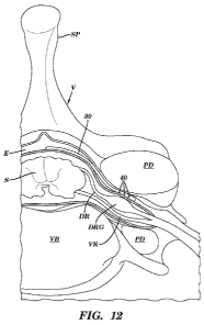
[0081] Figure 12 shows an illustration of one embodiment of the delivery
element 30 advanced within
the epidural space so that several outlet ports 40 are positioned in close
proximity to the dorsal root
- 17 -
CA 02819635 2013-05-31
WO 2012/075337 PCT/US2011/062958
ganglia (DRG). In this embodiment, the delivery element 30 is advanced along
the spinal cord S within the
epidural space E to a desired spinal level and advanced at least partially
through a foramen, between the
pedicles PD. VR = ventral root, DR = dorsal root, E = epidural space, S =
spinal cord, VB = vertebral
body.
[0082] Figure 13 shows an illustration of one embodiment of the position of a
gel 200 delivery vehicle
delivered to the epidural space E adjacent to the target DRG.
[0083] Figure 14 shows an illustration of one embodiment of a delivery element
30 having electrodes
50 and an agent-eluting coating 250 covering its distal end.
[0084] Figures 15A-15B show an illustration of embodiments of the delivery
element 30 having an
agent-eluting structure 260 disposed on the surface of the distal end of a
delivery element 30, where the
structure 260 comprises circumferential stripes or strips 262 that extend
around the shaft of the delivery
element 30. Figure 15A shows an embodiment of the delivery element 30
comprises a catheter and the
strips 262 are spaced apart along the distal end of the delivery element 30.
Figure 15B shows an
embodiment of the delivery element 30 which comprises a lead having electrodes
50, with the structures
260 as circumferential stripes or strips 262 that are disposed between the
electrodes. Thus, the agent is
eluted near the electrodes 50, such as for use in combination with electrical
stimulation.
[0085] Figure 16 shows an illustration of an embodiment of the delivery
element 30 having agent-
eluting structures 260 disposed as longitudinal stripes or strips along
specific portions of the delivery
element 30.
[0086] Figure 17 shows an illustration of an embodiment of the delivery
element 30 having agent-
eluting structures 260 disposed as dots longitudinally and circumferentially
around the delivery agent 30.
[0087] Figure 18 shows an illustration of an embodiment of the delivery
element 30 having an agent-
eluting structure 260 extending along a portion of the distal end of the
delivery agent 30, wherein the
structure 260 extends at least partially around the shaft of the delivery
element 30 and includes an opening
for at least one outlet port 40.
[0088] Figure 19A-19B shows an illustration of embodiments of the delivery
element 30 having agent-
eluting structures 260 as protrusions such as flexible hair-like protrusions
264. Figure 19A shows an
illustration of an embodiment of a delivery element 30 comprising a catheter
having protrusions 264
extending radially outwardly from the shaft of the delivery element 30. Figure
19B shows an illustration
of an embodiment of a delivery element 30 comprising a lead having at least
one electrode 50, at least one
outlet port 40 and at least one protrusion 264.
[0089] Figure 20 shows an illustration of an embodiment of the placement of a
sheet 300 positioned
adjacent the DRG, wrapping partially around the DRG, where the sheet 300 is
positioned within the
epidural space E at least partially within a foramen between the pedicles PD.
[0090] Figure 21 shows an illustration of an embodiment of the placement of a
tube 350 positioned
within a foramen, between the pedicles PD, so that the tube 350 extends around
the DRG. Since the tube
- 18 -
CA 02819635 2013-05-31
WO 2012/075337 PCT/US2011/062958
350 is positioned within the epidural space E, the tube 350 extends along the
surface of the dura layer D
which surrounds both the DRG and the nearby ventral root YR.
[0091] Figure 22 shows an illustration of an embodiment of the position of the
delivery device 30
placed intrathecally or into the subarachnoid or intrathecal space. In this
embodiment, the delivery element
30 is inserted into the intrathecal space and advanced in an antegrade
direction within the intrathecal
space along the spinal cord S, where the delivery element 30 comprises a
catheter having at least one
outlet port 40, and is advanced through the patient anatomy so that at least
one of the outlet ports 40 is
within a clinically effective distance to the DRG.
[0092] Figure 23 shows an embodiment of e-fields radiating from the electrodes
50 at the distal end of
the delivery element. The electrodes 50 are positioned either side of two
outlet ports 40 allowing both a
combination of electrical stimulation and agent delivery to the DRG, either
concurrently, or in a temporal
pattern of electrical stimulation and agent delivery.
[0093] Figure 24A-24C provides schematic illustrations of treatment to a
target DRG. Figure 24A
shows an embodiment of electrical stimulation 402 only of the DRG. Figure 24B
shows an embodiment
of agent delivery 400 only of the DRG. The combination of electrical
stimulation 402 and agent delivery
400 to the DRG can be concurrently, or in a pre-defined temporal pattern of
electrical stimulation 402 and
agent delivery 400.
[0094] Figure 25A-25B shows various embodiments using the agent-
neurostimulation system. Figure
25A shows distribution of an agent 400, e.g., a toxin, administered around a
DRG cell. Figure 25B shows
that when the DRG is activated, for example, using neurostimulation of the DRG
from the electrodes, the
DRG cell becomes activated, allowing agent binding and/or entry 402 into the
cell, and where the agent is
a toxin, results in selective molecular neuroablation of the activated cell.
[0095] Figure 26A-26B shows another embodiment of using the agent-
neurostimulation system.
Figure 26A shows delivery of an agent 400, e.g., a prodrug to cells within the
DRG using the DRG
delivery device. Figure 26B shows activation of the prodrug agent 400 by the
electrical stimulation 402
to render the agent active, resulting in no activation (e.g., no effect) or
activation or selective cell ablation
of specific cell subtypes in the DRG on electrical stimulation (A), whereas
other cell subtypes are
activated by the active agent (B), resulting in modulation of the cell
activity and/or cell death.
[0096] Figure 27A-27B shows another embodiment of using the agent-
neuromodulation of the system.
Figure 27A shows an agent 400 is delivered and has specificity or selectivity
for some cell types (e.g., cell
A) in the DRG and not other cell types cells (e.g., cell B). An agent can be
selective for one cell-type by
having a higher binding affinity for that cell type, and/or bind to cell-
surface receptors on that cell-type, or
be ligand for a channel and/or receptor on that particular cell type. Figure
27B shows when electrical
stimulation 402 is applied to all the DRGs, the cells that are sensitive to
the agent 400 (e.g., where the
agent is selective to that cell type) are activated and have altered activity
as compared to the cells subjected
to electrical stimulation 402 in the absence of the agent, or cells which are
not sensitive to the agent 400.
- 19 -
CA 02819635 2013-05-31
WO 2012/075337 PCT/US2011/062958
[0097] Figures 28A-28E shows an embodiment of altered input and output
electrical excitement
dynamics using the agent-neuromodulation system.
DETAILED DESCRIPTION OF THE INVENTION
[0098] The present invention generally relates to devices, systems and methods
for delivering an agent
to various levels of the spinal anatomy, particularly to various dorsal roots
(DR), more particularly to
various dorsal root ganglia (DRG), in a subject. For example, one aspect
relates to a device for direct
delivery of an agent, e.g., a drug formulation to at least target spinal
anatomy, for example, to at least one
DRG, where the agent is stored in an agent release module and is transported
via an agent delivery element
to the target anatomy, e.g., to at least one target DRG. In another aspect,
the present invention relates to a
device for direct delivery of an agent, e.g., a drug formulation to at least
target spinal anatomy, for
example, via the intrathecal space and/or the epidural space.
[0099] In some embodiments, the device, method and system can be further
configured to enable direct
and specific electrical stimulation, e.g., neurostimulation of the target
anatomy, e.g., DRG, in combination
with delivery of the agent to the DRG.
[00100] In some embodiments, electrical stimulation of the DRG is in a
temporal pattern which is
coordinated with a temporal pattern of delivery of the agent to the DRG. In
some embodiments, the device
allows delivery of an agent to a spinal nerve ganglion which is a dorsal root
ganglion (DRG), while in
alternative embodiments, the device enables delivery of an agent to a nerve
root ganglion in the
sympathetic nervous system, e.g., delivery of an agent to a sympathetic chain
ganglion. The following
examples will illustrate embodiments of specific temporal patterns of delivery
of agents to the DRG alone,
or in combination with temporal patterns of electrical stimulation of the DRG,
however, the invention is
not limited to such embodiments. Also described are a delivery device for
delivering agents to the DRG,
and where the delivery device is configured to enable electrical stimulation
of the DRG in combination
(e.g., concurrently) or intermittently, e.g., substantially simultaneously,
before or after, delivery of an
agent to the DRG. It may be appreciated that other elements, such as different
agent release modules, and
pulse generators may be used alternatively or in addition to the modules of
the delivery device for delivery
of agents to the DRG, alone, or in combination with electrical stimulation of
a DRG at one or more various
spinal cord levels.
[00101] The devices, systems and methods of the present invention allow for
targeted delivery of an
agent to at least one spinal anatomy, such as, but not limited to a DRG, and
enables targeted treatment of
such desired spinal anatomies. Accordingly, such targeted delivery of agents
alone or in combination with
electrical stimulation provides targeted treatment which minimizes deleterious
side effects, such as
undesired motor responses or undesired stimulation of unaffected body regions.
This is achieved by
directly delivering an agent to the DRG and, in some embodiments,
neuromodulating a target anatomy
associated with the condition while minimizing or excluding undesired
neuromodulation of other
- 20-
CA 02819635 2013-05-31
WO 2012/075337 PCT/US2011/062958
anatomies. For example, this may include stimulating the dorsal root ganglia,
dorsal roots, dorsal root
entry zones, or portions thereof while minimizing or excluding undesired
stimulation of other tissues, such
as surrounding or nearby tissues, portions of the ventral root and portions of
the anatomy associated with
body regions which are not targeted for treatment. Such stimulation is
typically achieved with the agent
delivery device as disclosed herein which has been adapted to include at least
one lead having at least one
electrode thereon. The distal end of the delivery device is advanced through
the patient anatomy so that the
delivery element, comprising at least one agent delivery structure, such as an
outlet port, for agent release
and at least one electrode, is positioned on, near or about the target DRG. In
some embodiments, the lead
and electrode(s) are sized and configured so that the electrode(s) are able to
minimize or exclude undesired
stimulation of other anatomies. In other embodiments, the stimulation signal
or other aspects are
configured so as to minimize or exclude undesired stimulation of other
anatomies. In addition, it may be
appreciated that stimulation of other tissues are also contemplated.
[00102] Embodiments of the present invention provide novel stimulation systems
and methods that
enable direct and specific neurostimulation techniques. For example, there is
provided a method of
delivering an agent to the DRG and simultaneously stimulating a nerve root
ganglion, comprising placing
an electrode of a delivery element in close proximity, or near to, the target
spinal anatomy, e.g., a nerve
root ganglion or DRG or spinal cord and delivering an agent and also
activating the electrode to stimulate
the nerve root ganglion. As discussed in greater detail below, the nerve root
ganglion may be a dorsal root
ganglion in some embodiments while in other embodiments, the nerve root
ganglion may be a nerve root
ganglion in the sympathetic nervous system or other ganglion, e.g.,
sympathetic chain ganglion.
[00103] Another aspect of the present invention provides an agent delivery
device to deliver agents to
the intrathecal space near the target DRG, combined with an electrical
stimulation systems and methods of
use. For example, provided herein is a method of delivering an agent to the
intrathecal space near the
target DRG and simultaneously stimulating the target DRG with the use of
another delivery device
positioned within the epidural space. Thus, an agent is delivered
intrathecally in conjunction with also
activating the electrode placed epidurally.
A Definitions
[00104] For convenience, certain terms employed herein, in the specification,
examples and appended
claims are collected here. Unless stated otherwise, or implicit from context,
the following terms and
phrases include the meanings provided below. Unless explicitly stated
otherwise, or apparent from
context, the terms and phrases below do not exclude the meaning that the term
or phrase has acquired in
the art to which it pertains. The definitions are provided to aid in
describing particular embodiments, and
are not intended to limit the claimed invention, because the scope of the
invention is limited only by the
claims. Unless otherwise defined, all technical and scientific terms used
herein have the same meaning as
commonly understood by one of ordinary skill in the art to which this
invention belongs.
- 21 -
CA 02819635 2013-05-31
WO 2012/075337 PCT/US2011/062958
[00105] The term "pain" as used herein, unless specifically noted otherwise,
is meant to encompass pain
of any duration and frequency, including, but not limited to, acute pain,
chronic pain, intermittent pain,
and the like. Causes of pain may be identifiable or unidentifiable. Where
identifiable, the origin of pain
may be, for example, of malignant, non-malignant, infectious, non-infectious,
or autoimmune origin. Of
particular interest is the management of pain associated with disorders,
diseases, or conditions that require
long-term therapy, e.g., chronic and/or persistent diseases or conditions for
which therapy involves
treatment over a period of several days (e.g., about 3 days to 10 days), to
several weeks (e.g., about 2
weeks or 4 weeks to 6 weeks), to several months or years, up to including the
remaining lifetime of the
subject. Subjects who are not presently suffering from a disease or condition,
but who are susceptible to
such may also benefit from prophylactic pain management using the devices and
methods of the invention,
e.g., prior to traumatic surgery. Pain amenable to therapy according to the
invention may involve
prolonged episodes of pain alternating with pain-free intervals, or
substantially unremitting pain that varies
in severity. Pain includes all types of clinical pain, including but not
limited to, nociceptive pain,
pathological pain, neuropathic pain, somatic pain, cutaneous pain, chronic
pain syndrome, referred pain,
radicular pain, breakthrough pain or incidence pain, phantom limb pain,
intractable pain and idiopathic
pain, as defined in Hawthorn and Redmond, Pain: causes and managements,
(Blackwell Science, Ed).
[00106] The term "breakthrough pain" is also referred to as incident pain,
refers to short periods of
sharper more intense pain that "breaks through" a background or constant
discomfort from pain.
Breakthrough pain can be caused by movement, pressure or treatment
interventions.
[00107] The term "nociceptive pain" refers to pain produced from an
identifiable cause.
[00108] The term "pathological pain" refers to pain felt due to activity in
the nociceptive pathway which
may be due to an identifiable cause or due to a disruption in the normal
sensory mechanisms. Pathological
pain is usually disproportionate to the causative factors and can be
inappropriate and can outlast the
original trauma, due to neuronal plasticity, including peripheral
sensitization and/or central sensitization.
[00109] The term "idiopathic pain" refers to pain from an unknown origin or
has not apparent
underlying cause, or pain which is excessive in comparison to the underlying
cause. Idiopathic pain is not
nociceptive, neuropathic or even psychogenic. Idiopathic pain may be made
worse by psychological
distress, and is more common in people who already have a pain disorder such
as TMJ and fibromyalgia.
Idiopathic pain, like psychogenic pain, is often more difficult to treat than
nociceptive or neuropathic pain.
A person who has back pain with no apparent cause may be diagnosed as having
idiopathic back pain.
[00110] The term "chronic pain" refers to long-lasting pain or pain
disproportionate to the cause.
Chronic pain may be pathological pain associated with changes in the central
or peripheral nervous system
or may be due to a constant stimulus.
[00111] The term "chronic pain syndrome" refers to a syndrome induced by long-
term pain where pain
and responses to pain are not well correlated with the underlying condition.
In some embodiments,
- 22-
CA 02819635 2013-05-31
WO 2012/075337 PCT/US2011/062958
subjects with chronic pain may experience changes in personality, behavior and
changes to functional
ability.
[00112] The term "referred pain" refers to pain felt in a different area to
the source of the pain. Referred
pain is commonly referred from organs and deep tissue to muscles and skin.
[00113] The term "pain-related disorder" as used herein refers to any disease,
condition or malady
where the subject is experiencing pain.
[00114] The term "delivery site as used herein is meant to refer to an area of
the body to which the
agent or drug is delivered. A delivery site can be in close proximity to the
target spinal anatomy, which
means that the delivery site is in a close enough proximity or location to
deliver the agent and/or electrical
stimulation to the target spinal anatomy, and includes but is not limited to
dorsal root ganglia (DRGs),
dorsal roots, dorsal root entry zones, or portions thereof.
[00115] The term "implantation" or "implant" or "implanted" are used
interchangeably herein, and refer
to the penetration or insertion of an element into a tissue, e.g., penetration
of the distal end of the delivery
element into neuronal tissue. In some aspects, implantation can also refer to
inserting the device or pumps
into cavities in the body.
[00116] The term "proximity to" as used herein refers to placing an element
next to, or in a position near
to, or in contact with another element or anatomical structure or tissue.
[00117] The term "drug delivery device" or "agent delivery device" includes
the implantable agent
release module and means, e.g., delivery elements to transport the agent or
drug formulations to the
desired target area or target anatomy in the subject.
[00118] The term "drug release module" also referred herein as a "controlled
drug pump device" or
"agent release module" refers to any device suitable for placing
subcutaneously or in desired location in a
subject for the storage and controlled release of an agent or drug formulation
for pain management
according to the method of the invention. The agent or drug or other desired
substance contained in the
pump is released in a controlled manner (e.g., rate, timing of release), which
is controlled by or determined
by the device itself, which in turn can be controlled by the subject user, or
the clinician according to a
predetermined program or treatment protocol. In some embodiments, the release
of the agent or drug or
other substance can be an osmotic pump where the agent is released according
to the environmental use,
e.g., where diffusion and osmotic concentrations control the release of the
agent or drug from the pump in
a controlled manner. The term "drug release module" or "agent release module"
also encompasses any
device with any mechanism of action including diffusive, erodible, or
convective systems, e.g., osmotic
pumps, biodegradable implants, electrodiffusion systems, electroosmosis
systems, vapor pressure pumps,
electrolytic pumps, effervescent pumps, piezoelectric pumps, erosion-based
systems, or electromechanical
systems.
- 23-
CA 02819635 2013-05-31
WO 2012/075337 PCT/US2011/062958
[00119] The term "controlled drug pump device" is meant to encompass any
device wherein the release
(e.g., rate, timing of release) of a drug or other desired substance contained
therein is controlled by or
determined by the device itself and not the environment of use.
[00120] The term "patterned" or "temporal" as used in the context of drug
delivery and/or electronic
stimulation refers to the delivery of the agent and/or electrical stimulation
in a pattern, generally a
substantially regular pattern, over a pre-selected period of time (e.g., other
than a period associated with,
for example a bolus injection). The term "patterned" or "temporal" drug
delivery is meant to encompass
delivery of drug at an increasing, decreasing, substantially constant, or
pulsatile, rate or range of rates
(e.g., amount of drug per unit time, or volume of drug formulation for a unit
time), and further
encompasses delivery that is continuous or substantially continuous, or
chronic.
[00121] By "substantially continuous" as used in, for example, the context of
"substantially continuous
subcutaneous infusion" or "substantially continuous delivery" is meant to
refer to delivery of an agent or
drug in a manner that is substantially uninterrupted for a pre-selected period
of drug delivery (other than a
period associated with, for example, a bolus injection). Furthermore,
"substantially continuous" drug
delivery can also encompass delivery of drug at a substantially constant, pre-
selected rate or range of rates
(e.g., amount of drug per unit time, or volume of drug formulation for a unit
time) that is substantially
uninterrupted for a pre-selected period of drug delivery.
[00122] The term "systemic delivery" is meant to encompass all parenteral
routes of delivery which
permit drug to enter into the systemic circulation, e.g., intravenous, intra-
arterial, intramuscular,
subcutaneous, intra-adipose tissue, intra-lymphatic, etc.
[00123] The term "block" or "blockade" or "blocking" are used interchangeably
herein and refers to
inhibition, disruption, prevention or inhibition of the conduction or
propagation of action potentials and
nerve impulse transmission along the axons of the target nerves, either
partially or completely. The terms
"block" or "blockade" or "blocking" also refer to the blockage of electrical
signals along non-neuronal cell
types, e.g., glial cells and astrocytes and the like, as well as refers to
inhibition of intracellular signaling
from cell surface markers and increases in soma size.
[00124] The term "nerve ablation" or "nerve lesioning" as used herein refers
to the destruction of one or
more axons of a target nerve so as to result in a nerve blockade in which
conduction or propagation of
action potentials in the target nerve is attenuated or abolished, either
reversibly or permanently, as
evidenced by the attenuation or abolition of sensation normally mediated by
the nerve or weakness or
paralysis of the body tissue innervated by the target nerve lasting more than
a week, more than two weeks,
or more than a month.
[00125] As used herein, the term "neurodegenerative disease or disorder"
includes any disease disorder
or condition that affects neuronal homeostasis, e.g., results in the
degeneration or loss of neuronal cells.
Neurodegenerative diseases include conditions in which the development of the
neurons, i.e., motor or
brain neurons, is abnormal, as well as conditions in which result in loss of
normal neuron function.
- 24-
CA 02819635 2013-05-31
WO 2012/075337 PCT/US2011/062958
Examples of such neurodegenerative disorders include Alzheimer's disease and
other tauopathies such as
frontotemporal dementia, frontotemporal dementia with Parkinsonism,
frontotemporal lobe dementia,
pallidopontonigral degeneration, progressive supranuclear palsy, multiple
system tauopathy, multiple
system tauopathy with presenile dementia, Wilhelmsen-Lynch disease,
disinhibition-dementia-park-
insonism-amytrophy complex, Pick's disease, or Pick's disease-like dementia,
corticobasal degeneration,
frontal temporal dementia, Parkinson's disease, Huntington's disease,
amyotrophic lateral sclerosis (ALS),
multiple sclerosis, Friedreich's ataxia, Lewybody disease, spinal muscular
atrophy, and parkinsonism
linked to chromosome 17.
[00126] As used herein, the term "inflammation" refers to any cellular
processes that lead to the
activation of caspase-1, or caspase-5, the production of cytokines IL-I and IL-
8, and/or the related
downstream cellular events resulting from the actions of the cytokines thus
produced, for example, fever,
fluid accumulation, swelling, abscess formation, and cell death. As used
herein, the term "inflammation"
refers to both acute responses (i.e., responses in which the inflammatory
processes are active) and chronic
responses (i.e., responses marked by slow progression and formation of new
connective tissue). Acute and
chronic inflammation may be distinguished by the cell types involved. Acute
inflammation often involves
polymorphonuclear neutrophils; whereas chronic inflammation is normally
characterized by a
lymphohistiocytic and/or granulomatous response.
[00127] As used herein, the term "inflammation" includes reactions of both the
specific and non-specific
defense systems. A specific defense system reaction is a specific immune
system reaction response to an
antigen (possibly including an autoantigen). A non-specific defense system
reaction is an inflammatory
response mediated by leukocytes incapable of immunological memory. Such cells
include granulocytes,
macrophages, neutrophils and eosinophils. Examples of specific types of
inflammation include, but are
not limited to, diffuse inflammation, focal inflammation, croupous
inflammation, interstitial inflammation,
obliterative inflammation, parenchymatous inflammation, reactive inflammation,
specific inflammation,
toxic inflammation and traumatic inflammation.
[00128] The term "agent" as used herein means any compound or substance such
as, but not limited to, a
small molecule, nucleic acid, polypeptide, peptide, drug, ion, etc. An "agent"
can be any chemical, entity
or moiety, including without limitation synthetic and naturally-occurring
proteinaceous and non-
proteinaceous entities. In some embodiments, an agent is nucleic acid, nucleic
acid analogues, proteins,
antibodies, peptides, aptamers, oligomer of nucleic acids, amino acids, or
carbohydrates including without
limitation proteins, oligonucleotides, ribozymes, DNAzymes, glycoproteins,
siRNAs, lipoproteins,
aptamers, and modifications and combinations thereof etc. In certain
embodiments, agents are small
molecule having a chemical moiety. For example, chemical moieties included
unsubstituted or substituted
alkyl, aromatic, or heterocyclyl moieties including macrolides, leptomycins
and related natural products or
analogues thereof. Compounds can be known to have a desired activity and/or
property, or can be selected
from a library of diverse compounds.
- 25-
CA 02819635 2013-05-31
WO 2012/075337 PCT/US2011/062958
[00129] As used herein, the term "small molecule" refers to a chemical agent
which can include, but is
not limited to, a peptide, a peptidomimetic, an amino acid, an amino acid
analog, a polynucleotide, a
polynucleotide analog, an aptamer, a nucleotide, a nucleotide analog, an
organic or inorganic compound
(e.g., including heterorganic and organometallic compounds) having a molecular
weight less than about
10,000 grams per mole, organic or inorganic compounds having a molecular
weight less than about 5,000
grams per mole, organic or inorganic compounds having a molecular weight less
than about 1,000 grams
per mole, organic or inorganic compounds having a molecular weight less than
about 500 grams per mole,
and salts, esters, and other pharmaceutically acceptable forms of such
compounds.
[00130] The term "drug" or "compound" as used herein refers to a chemical
entity or biological product,
or combination of chemical entities or biological products, administered to a
subject to treat or prevent or
control a disease or condition. The chemical entity or biological product is
preferably, but not necessarily a
low molecular weight compound, but may also be a larger compound, for example,
an oligomer of nucleic
acids, amino acids, or carbohydrates including without limitation proteins,
oligonucleotides, ribozymes,
DNAzymes, glycoproteins, siRNAs, lipoproteins, aptamers, and modifications and
combinations thereof.
[00131] As use herein, the term "ion-channel" refers to a transmembrane pore
that presents a
hydrophilic channel for specific ions to cross a lipid bilayer down their
electrochemical gradients. There
are over 300 types of ion-channels in a living cell (Gabashvili, et al., "Ion-
channel gene expression in the
inner ear", J. Assoc. Res. Otolaryngol. 8 (3): 305-28 (2007). The ion-channels
are classified upon their
ion specificity, biological function, regulation or molecular structure, and
nature of their gating. Examples
of ion-channels are voltage gated ion-channels, Gap-junction ion-channels,
ligand-gated ion-channels,
ATP-gated ion-channels, heat-activated ion-channels, intracellular ion-
channels, ion-channels gated by
intracellular ligands such as cyclic nucleotide-gated channels or calcium-
activated ion-channels. As used
herein the term "gated ion-channel" is defined as an ion-channel the passage
of ions through which is
dependent on the presence of an analyte. As used herein, the term "voltage
gated ion-channel" as used
herein refers to an ion-channel where the passage of ions through which is
dependent on the presence of
voltage activation of the channel, where activation above a certain threshold
level for the ion channel
allows ions to pass through the ion channel. As used herein "ion channel" also
encompasses transporters
which transport ions and other charged molecules across a membrane, including
but not limited to Na+/K+
channels, Na+/Ca2+ and other ion transporters.
[00132] As used herein, the term "ion-channel modulator" refers to a
compound that modulates at
least one activity of an ion-channel, for example, but not limited to, and
agent which increases or
decreases the opening frequency and/or duration of the ion-channel, or an
agent which increases and/or
reduces the sensitivity of opening and/or closing of the ion-channel from the
normal threshold of
activation (opening) or deactivation (closing). In some embodiments, an ion-
channel modulator is an
agent which alters the selectivity of the ion channel to allow or prevent
different ions from entering the ion
channel, as well as an agent which alters (increases or decreases) the
activation of the ion-channel by
- 26-
CA 02819635 2013-05-31
WO 2012/075337 PCT/US2011/062958
different receptor activation. The term "ion-channel modulator" as used herein
is intended to include
agents that interact with the channel pore itself, or that may act as an
allosteric modulator of the channel
by interacting with a site on the channel complex. The term "ion-channel
modulator" as used herein is also
intended to include agents that modulate activity of an ion-channel
indirectly. By "indirectly, "as used in
reference to modulator interactions with ion-channel, means the ion-channel
modulator does not directly
interact with the ion-channel itself, i.e., ion-channel modulator interacts
with the ion-channel via an
intermediary. Accordingly, the term "indirectly" also encompasses the
situations wherein the ion-channel
modulator requires another molecule in order to bind or interact with the ion-
channel.
[00133] As used herein, the term "modulate" refers to a change or
alternation in at least one
biological activity of the ion-channel or receptor, e.g., cell-surface
receptor. Modulation may be an
increase or decrease the ion-channel or receptor activity, change the binding
characteristics, or any other
change in the biological, functional, or immunological properties of the ion-
channel or receptor.
Modulation can include, for example, a decreased or increased threshold of
activation, an increased or
decreased sensitivity to activation or deactivation, an increased or decreased
selectivity for particular ions
and/or ligands (e.g., endogenous or exogenous ligands or biologics) of the ion
channel and/or receptor,
respectively. Modulation can also include an alteration in the mode of action
of the ion channel or the
receptor, e.g., for example, an agent can modulate an ion channel to efflux
ions rather than influx ions into
the cell, or alternatively, alter the ion which is transmitted through the ion
channel. In some embodiments
of the aspects described herein, the ion-channel modulator modulates the
passage of ions through the ion-
channel.
[00134] The term "analgesic" as used herein refers to any member of the
diverse group of drugs used to
relieve pain. Analgesic drugs include, but are not limited to, the
nonsteroidal anti-inflammatory drugs
(NSAIDs) such as the salicylates, narcotic drugs such as morphine, and
synthetic drugs with narcotic
properties such as tramadol. Other classes of drugs not normally considered
analgesics are used to treat
neuropathic pain syndromes; these include tricyclic antidepressants and
anticonvulsants. Without wishing
to be bound by theory, NSAIDs including aspirin, naproxen, and ibuprofen not
only relieve pain but also
reduce fever and inflammation. Narcotic analgesics, including opiates and
opioids, including Oxycodone,
(also known as brand names DAZIDOXTM, ETH-OXYDOSETm, ENDOCODONETM, OXYIRTM,
OXYCONTINTm, OXYFASTTm, PERCOLONETM, ROXICODONETM) and and a
hydrocodone/paracetamol (or acetaminophen) mix (also known as brand names
VICODINTM,
ANEXSIATM, ANOLOR DH5TM, BANCAP HCTM, ZYDONETM, DOLACETTm, LORCETTm,
LORTABTm, AND NORCOTm), largely work through specific opioid receptors in the
peripheral and
central nervous system and alter the perception of pain (nociception).
Analgesics can be used in
combination, and can also be used in combination with vasoconstrictor drugs
such as pseudoephedrine for
sinus-related preparations, or with antihistamine drugs for allergy sufferers.
- 27-
CA 02819635 2013-05-31
WO 2012/075337 PCT/US2011/062958
[00135] The term "agonist" as used herein refers to any agent or entity
capable of increasing the
expression and/or the biological activity of a protein, polypeptide portion
thereof, or polynucleotide which
is the target of the agonist agent. Thus, an agonist can operate to increase
the transcription, translation,
post-transcriptional or post-translational processing or otherwise activate
the activity of the protein,
polypeptide or polynucleotide in any way, such as functioning as a ligand to
activate a receptor or via
other forms of direct or indirect action. In some embodiments, an agonist
refers to an agent which
increases the biological activity of the target protein by a statistically
significant amount as compared to in
the absence of the agonist. In some embodiments, an agonist refers to an agent
which increases the
biological activity of the target protein by a clinically statistically
significant amount as compared to in the
absence of the agonist, such that the effect of the agonist on the target
protein produces a clinically
measurable change in outcome. In some embodiments, the term "agonist" as can
refer to an agent which
increases the biological activity of the target protein by about at least
about 5%, e.g., an agonist to a target
ion channel increases the activity of the ion channel expressed on the DRG by
at least 5% or more than
5%. By way of example only, an agonist which activates an ion channel, e.g., a
sodium channel can be any
entity or agent which functions as a co-factor or ligand which opens a sodium
channel or decreases the
threshold of activation of a sodium channel, or promotes beta-subunit
association with the sodium channel,
or alternatively any agent which interacts with the sodium channel to increase
its opening or decrease its
threshold of activation (if the sodium channel is a voltage gated sodium
channel). An agonist can be, for
example a nucleic acid, peptide, or any other suitable chemical compound or
molecule or any combination
of these. Additionally, it will be understood that in indirectly activating
the activity of a protein,
polypeptide of polynucleotide, an agonist may affect the activity of the
cellular molecules which may in
turn act as regulators or the protein, polypeptide or polynucleotide itself.
Similarly, an agonist may affect
the activity of molecules which are themselves subject to the regulation or
modulation by the protein,
polypeptide of polynucleotide. An agonist is also referred to herein as an
"activating agent".
[00136] The term "antagonist" as used herein refers to any agent or entity
capable of inhibiting the
expression and/or the biological activity of a protein, polypeptide portion
thereof, or polynucleotide. Thus,
the antagonist may operate to prevent transcription, translation, post-
transcriptional or post-translational
processing or otherwise inhibit the activity of the protein, polypeptide or
polynucleotide in any way, such
as functioning as a ligand to activate a receptor or via other forms of direct
or indirect action. In some
embodiments, an antagonist refers to an agent which decreases the biological
activity of the target protein
by a statistically significant amount as compared to in the absence of the
antagonist. In some
embodiments, an antagonist refers to an agent which decreases the biological
activity of the target protein
by a clinically statistically significant amount as compared to in the absence
of the antagonist, such that
the effect of the antagonist on the target protein produces a clinically
measurable change in outcome. In
some embodiments, an "antagonist" refers to an agent that can decrease the
biological activity of the target
protein by at least about 5%, e.g., an antagonist to a target ion channel
decreases the activity of the ion
- 28-
CA 02819635 2013-05-31
WO 2012/075337 PCT/US2011/062958
channel expressed on the DRG by at least 5% or greater than 5%. By way of
example only, an antagonist
which inhibits a sodium channel, for example a sodium channel blocker can be
any entity or agent which
functions as a to competitively block the channel pore of the sodium channel,
or alternatively any agent
which is a non-competitive inhibitor of a sodium channel which interacts at a
region of a sodium channel
which is not the pore) to inhibit channel opening or increase the threshold of
activation (e.g., if the sodium
channel is a voltage gated sodium channel). An antagonist may for example, be
any agent, such as but not
limited to a nucleic acid, peptide, or any other suitable chemical compound or
molecule or any
combination of these. Additionally, it will be understood that in indirectly
impairing the activity of a
protein, polypeptide of polynucleotide, the antagonist may affect the activity
of the cellular molecules
which may in turn act as regulators or the protein, polypeptide or
polynucleotide itself. Similarly, the
antagonist may affect the activity of molecules which are themselves subject
to the regulation or
modulation by the protein, polypeptide of polynucleotide.
[00137] The term "treating", as used herein, refers to altering the disease
course of the subject being
treated. Therapeutic effects of treatment include, without limitation,
preventing occurrence or recurrence
of disease, alleviation of symptom(s), diminishment of direct or indirect
pathological consequences of the
disease, decreasing the rate of disease progression, amelioration or
palliation of the disease state, and
remission or improved prognosis.
[00138] The term "pain management or treatment" is used here to generally
describe regression,
suppression, or mitigation of pain so as to make the subject more comfortable
as determined by subjective
criteria, objective criteria, or both. In general, pain is assessed
subjectively by patient report, with the
health professional taking into consideration the patient's age, cultural
background, environment, and other
psychological background factors known to alter a person's subjective reaction
to pain.
[00139] The term "therapeutically effective amount" as used herein refers an
amount of an agent, or a
rate of delivery of an agent, effective to facilitate a desired therapeutic
effect or benefit or desired clinical
result upon treatment, e.g., a measurable decrease in the sensation of pain
experienced by the subject. The
precise desired therapeutic effect (e.g., the degree of pain relief, and
source of the pain relieved, etc.) will
vary according to the condition to be treated, the agent and/or drug
formulation to be administered, as well
as the effect in combination with electrical stimulation and a variety of
other factors that are appreciated
by those of ordinary skill in the art. In general, the method of the invention
involves the suppression or
mitigation of pain in a subject suffering from pain that may be associated
with any of a variety of
identifiable or unidentifiable etiologies. The phrase "therapeutically-
effective amount" as used herein
means that amount of a compound, material, or composition comprising an agent,
e.g., an ion-channel
modulator which is effective for producing some desired therapeutic effect in
at least a sub-population of
cells in an animal at a reasonable benefit/risk ratio applicable to any
medical treatment. For example, an
amount of an ion-channel modulator administered to a subject that is
sufficient to produce a clinically
meaningful or statistically significant measurable decrease in the pain
experienced by the subject. A
- 29-
CA 02819635 2013-05-31
WO 2012/075337 PCT/US2011/062958
therapeutically effective amount will vary, as recognized by those skilled in
the art, depending on the
specific disease treated, the excipient selected, and the possibility of
combination therapy, e.g., effect of
the delivery of the agent in combination with electrical stimulation of the
DRG.
[00140] Determination of a therapeutically effective amount is well within the
capability of those skilled
in the art. Generally, a therapeutically effective amount can vary with the
subject's history, age, condition,
sex, as well as the severity and type of the medical condition in the subject,
and administration of other
pharmaceutically active agents. Furthermore, therapeutically effective amounts
will vary, as recognized
by those skilled in the art, depending on the specific disease treated, the
route of administration, the
excipient selected, and the possibility of combination therapy.
[00141] The term "pharmaceutically acceptable excipient", as used herein,
refers to carriers and vehicles
that are compatible with the active ingredient (for example, a compound of the
invention) of a
pharmaceutical composition of the invention (and preferably capable of
stabilizing it) and not deleterious
to the subject to be treated. For example, solubilizing agents that form
specific, more soluble complexes
with the compounds of the invention can be utilized as pharmaceutical
excipients for delivery of the
compounds. Suitable carriers and vehicles are known to those of extraordinary
skill in the art. The term
"excipient" as used herein will encompass all such carriers, adjuvants,
diluents, solvents, or other inactive
additives. Suitable pharmaceutically acceptable excipients include, but are
not limited to, water, salt
solutions, alcohol, vegetable oils, polyethylene glycols, gelatin, lactose,
amylose, magnesium stearate, talc,
silicic acid, viscous paraffin, perfume oil, fatty acid monoglycerides and
diglycerides, petroethral fatty
acid esters, hydroxymethyl-cellulose, polyvinylpyrrolidone, etc. The
pharmaceutical compositions of the
invention can also be sterilized and, if desired, mixed with auxiliary agents,
e.g., lubricants, preservatives,
stabilizers, wetting agents, emulsifiers, salts for influencing osmotic
pressure, buffers, colorings,
flavorings and/or aromatic substances and the like, which do not deleteriously
react with the active
compounds of the invention.
[00142] Thus, as used herein, the term "pharmaceutically acceptable salt," is
a salt formed from an acid
and a basic group of a compound of the invention. Illustrative salts include,
but are not limited, to sulfate,
citrate, acetate, oxalate, chloride, bromide, iodide, nitrate, bisulfate,
phosphate, acid phosphate,
isonicotinate, lactate, salicylate, acid citrate, tartrate, oleate, tannate,
pantothenate, bitartrate, ascorbate,
succinate, maleate, gentisinate, fumarate, gluconate, glucaronate, saccharate,
formate, benzoate, glutamate,
methanesulfonate, ethanesulfonate, benzenesulfonate, p-toluenesulfonate, and
pamoate salts.
[00143] The term "pharmaceutically acceptable salt" also refers to a salt
prepared from a compound of
the invention having an acidic functional group, such as a carboxylic acid
functional group, and a
pharmaceutically acceptable inorganic or organic base. Suitable bases include,
but are not limited to,
hydroxides of alkali metals such as sodium, potassium, and lithium; hydroxides
of alkaline earth metal
such as calcium and magnesium; hydroxides of other metals, such as aluminum
and zinc; ammonia, and
organic amines, such as unsubstituted or hydroxy-substituted mono-, di-, or
trialkylamines;
- 30-
CA 02819635 2013-05-31
WO 2012/075337 PCT/US2011/062958
dicyclohexylamine; tributyl amine; pyridine; N-methyl, N-ethylamine;
diethylamine; triethylamine; mono-
, bis-, or tris-(2-hydroxy-lower alkyl amines), such as mono-, bis-, or tris-
(2-hydroxyethyl)amine, 2-
hydroxy-tert-butylamine, or tris-(hydroxymethyl)methylamine, N,N,-di-lower
alkyl-N-(hydroxy lower
alkyl)-amines, such as N,N-dimethyl-N-(2-hydroxyethyl)amine, or tri-(2-
hydroxyethyl)amine; N-methyl-
D-glucamine; and amino acids such as arginine, lysine, and the like. Other
pharmaceutically acceptable
salts are described in the Handbook of Pharmaceutical Salts. Properties,
Selection, and Use (P. Heinrich
Stahl and C. Wermuth, Eds., Verlag Helvetica Chica Acta, Zurich, Switzerland
(2002)).
[00144] As used here, the term "pharmaceutically acceptable" refers to those
compounds, materials,
compositions, and/or dosage forms which are, within the scope of sound medical
judgment, suitable for
use in contact with the tissues of human beings and animals without excessive
toxicity, irritation, allergic
response, or other problem or complication, commensurate with a reasonable
benefit/risk ratio.
[00145] As used here, the term "pharmaceutically-acceptable carrier" means a
pharmaceutically-
acceptable material, composition or vehicle, such as a liquid or solid filler,
diluent, excipient,
manufacturing aid (e.g., lubricant, talc magnesium, calcium or zinc stearate,
or steric acid), or solvent
encapsulating material, involved in carrying or transporting the subject
compound from one organ, or
portion of the body, to another organ, or portion of the body. Each carrier
must be "acceptable" in the
sense of being compatible with the other ingredients of the formulation and
not injurious to the patient.
Some examples of materials which can serve as pharmaceutically-acceptable
carriers include: (1) sugars,
such as lactose, glucose and sucrose; (2) starches, such as corn starch and
potato starch; (3) cellulose, and
its derivatives, such as sodium carboxymethyl cellulose, methylcellulose,
ethyl cellulose, microcrystalline
cellulose and cellulose acetate; (4) powdered tragacanth; (5) malt; (6)
gelatin; (7) lubricating agents, such
as magnesium stearate, sodium lauryl sulfate and talc; (8) excipients, such as
cocoa butter and suppository
waxes; (9) oils, such as peanut oil, cottonseed oil, safflower oil, sesame
oil, olive oil, corn oil and soybean
oil; (10) glycols, such as propylene glycol; (11) polyols, such as glycerin,
sorbitol, mannitol and
polyethylene glycol (PEG); (12) esters, such as ethyl oleate and ethyl
laurate; (13) agar; (14) buffering
agents, such as magnesium hydroxide and aluminum hydroxide; (15) alginic acid;
(16) pyrogen-free
water; (17) isotonic saline; (18) Ringer's solution; (19) ethyl alcohol; (20)
pH buffered solutions; (21)
polyesters, polycarbonates and/or polyanhydrides; (22) bulking agents, such as
polypeptides and amino
acids (23) serum component, such as serum albumin, HDL and LDL; (22) C2-C12
alchols, such as
ethanol; and (23) other non-toxic compatible substances employed in
pharmaceutical formulations.
Wetting agents, coloring agents, release agents, coating agents, sweetening
agents, flavoring agents,
perfuming agents, preservative and antioxidants can also be present in the
formulation. The terms such as
"excipient", "carrier", "pharmaceutically acceptable carrier" or the like are
used interchangeably herein.
[00146] The term "subject" is used interchangeably herein with "patient" and
refers to a vertebrate,
preferably a mammal, more preferably a primate, still more preferably a human.
Mammals include,
without limitation, humans, primates, wild animals, feral animals, farm
animals, sports animals, and pets.
- 31-
CA 02819635 2013-05-31
WO 2012/075337 PCT/US2011/062958
Mammals include, without limitation, humans, primates, wild animals, rodents,
feral animals, farm
animals, sports animals, and pets. Primates include chimpanzees, cynomologous
monkeys, spider
monkeys, and macaques, e.g., Rhesus. Rodents include mice, rats, woodchucks,
ferrets, rabbits and
hamsters. Domestic and game animals include cows, horses, pigs, deer, bison,
buffalo, feline species, e.g.,
domestic cat, canine species, e.g., dog, fox, wolf, avian species, e.g.,
chicken, emu, ostrich, and fish, e.g.,
trout, catfish and salmon. The terms, "patient" and "subject" are used
interchangeably herein. A subject
can be male or female. A subject can be one who has been previously diagnosed
with or identified as
suffering from a condition, such as pain or having a pain-related disorder. A
subject can also one who is
currently being treated for pain, e.g., chronic pain or a pain-related
disorder. In addition, the methods and
compositions described herein can be used to treat domesticated animals and/or
pets.
[00147] As used herein, a "prodrug" refers to compounds that can be converted
via some chemical or
physiological process (e.g., enzymatic processes and metabolic hydrolysis) to
either S-a-methyl-
hydrocinnamic acid or R-a-methyl-hydrocinnamic acid. A prodrug may be inactive
when administered to
a subject, i.e. an ester, but is converted in vivo to an active compound (e.g.
S-a-methyl-hydrocinnamic acid
or R-a-methyl-hydrocinnamic acid), for example, by hydrolysis to the free
carboxylic acid or free
hydroxyl. The prodrug compound often offers advantages of solubility, tissue
compatibility or delayed
release in an organism. The term "prodrug" is also meant to include any
covalently bonded carriers, which
release the active compound in vivo when such prodrug is administered to a
subject. Prodrugs of an active
compound may be prepared by modifying functional groups present in the active
compound in such a way
that the modifications are cleaved, either in routine manipulation or in vivo,
to the parent active compound.
Prodrugs include compounds wherein a hydroxy, amino or mercapto group is
bonded to any group that,
when the prodrug of the active compound is administered to a subject, cleaves
to form a free hydroxy, free
amino or free mercapto group, respectively. Examples of prodrugs include, but
are not limited to, acetate,
formate and benzoate derivatives of an alcohol or acetamide, formamide and
benzamide derivatives of an
amine functional group in the active compound and the like. See Harper, "Drug
Latentiation" in Jucker,
ed. Progress in Drug Research 4:221-294 (1962); Morozowich et al, "Application
of Physical Organic
Principles to Prodrug Design" in E. B. Roche ed. Design of Biopharmaceutical
Properties through
Prodrugs and Analogs, APHA Acad. Pharm. Sci. 40 (1977); Bioreversible Carriers
in Drug in Drug
Design, Theory and Application, E. B. Roche, ed., APHA Acad. Pharm. Sci.
(1987); Design of Prodrugs,
H. Bundgaard, Elsevier (1985); Wang et al. "Prodrug approaches to the improved
delivery of peptide
drug" in Curr. Pharm. Design. 5(4):265-287 (1999); Pauletti et al. (1997)
Improvement in peptide
bioavailability: Peptidomimetics and Prodrug Strategies, Adv. Drug. Delivery
Rev. 27:235-256; Mizen et
al. (1998) "The Use of Esters as Prodrugs for Oral Delivery of (3-Lactam
antibiotics," Pharm. Biotech.
11,:345-365; Gaignault et al. (1996) "Designing Prodrugs and Bioprecursors I.
Carrier Prodrugs," Pract.
Med. Chem. 671-696; Asgharnejad, "Improving Oral Drug Transport", in Transport
Processes in
Pharmaceutical Systems, G. L. Amidon, P. I. Lee and E. M. Topp, Eds., Marcell
Dekker, p. 185-218
- 32-
CA 02819635 2013-05-31
WO 2012/075337 PCT/US2011/062958
(2000); Balant et al., "Prodrugs for the improvement of drug absorption via
different routes of
administration", Eur. J. Drug Metab. Pharmacokinet., 15(2): 143-53 (1990);
Balimane and Sinko,
"Involvement of multiple transporters in the oral absorption of nucleoside
analogues", Adv. Drug Delivery
Rev., 39(1-3): 183-209 (1999); Browne, "Fosphenytoin (Cerebyx)", Clin.
Neuropharmacol. 20(1): 1-12
(1997); Bundgaard, "Bioreversible derivatization of drugs¨ principle and
applicability to improve the
therapeutic effects of drugs", Arch. Pharm. Chemi 86(1): 1-39 (1979);
Bundgaard H. "Improved drug
delivery by the prodrug approach", Controlled Drug Delivery 17: 179-96 (1987);
Bundgaard H. "Prodrugs
as a means to improve the delivery of peptide drugs",Arfv. Drug Delivery Rev.
8(1): 1-38 (1992); Fleisher
et al. "Improved oral drug delivery: solubility limitations overcome by the
use of prodrugs", Arfv. Drug
Delivery Rev. 19(2): 115-130 (1996); Fleisher et al. "Design of prodrugs for
improved gastrointestinal
absorption by intestinal enzyme targeting", Methods Enzymol. 112 (Drug Enzyme
Targeting, Pt. A): 360-
81, (1985); Farquhar D, et al., "Biologically Reversible Phosphate-Protective
Groups", Pharm. Sci., 72(3):
324-325 (1983); Freeman S, et al., "Bioreversible Protection for the Phospho
Group: Chemical Stability
and Bioactivation of Di(4-acetoxy-benzyl) Methylphosphonate with
Carboxyesterase," Chem. Soc., Chem.
Commun., 875-877 (1991); Friis and Bundgaard, "Prodrugs of phosphates and
phosphonates: Novel
lipophilic alphaacyloxyalkyl ester derivatives of phosphate- or phosphonate
containing drugs masking the
negative charges of these groups", Eur. J. Pharm. Sci. 4: 49-59 (1996);
Gangwar et al., "Pro-drug,
molecular structure and percutaneous delivery", Des. Biopharm. Prop. Prodrugs
Analogs, [Symp.]
Meeting Date 1976, 409-21. (1977); Nathwani and Wood, "Penicillins: a current
review of their clinical
pharmacology and therapeutic use", Drugs 45(6): 866-94 (1993); Sinhababu and
Thakker, "Prodrugs of
anticancer agents", Adv. Drug Delivery Rev. 19(2): 241-273 (1996); Stella et
al., "Prodrugs. Do they have
advantages in clinical practice?", Drugs 29(5): 455-73 (1985); Tan et al.
"Development and optimization
of anti-HIV nucleoside analogs and prodrugs: A review of their cellular
pharmacology, structure-activity
relationships and pharmacokinetics", Adv. Drug Delivery Rev. 39(1-3): 117-
151(1999); Taylor,
"Improved passive oral drug delivery via prodrugs", Adv. Drug Delivery Rev.,
19(2): 131-148 (1996);
Valentino and Borchardt, "Prodrug strategies to enhance the intestinal
absorption of peptides", Drug
Discovery Today 2(4): 148-155 (1997); Wiebe and Knaus, "Concepts for the
design of anti-HIV
nucleoside prodrugs for treating cephalic HIV infection", Adv. Drug Delivery
Rev.: 39(l-3):63-80 (1999);
Waller et al., "Prodrugs", Br. J. Clin. Pharmac. 28: 497-507 (1989).
[00148] In some embodiments of the aspects described herein, the method
further comprising
diagnosing a subject with pain or a pain-related disorder before treatment
with the systems, devices and
methods as described herein. Methods of diagnosing pain, such as chronic pain,
neuropathic and
inflammatory pain are well known in the art.
[00149] In some embodiments, the method further comprising selecting a subject
identified to have pain,
such as chronic pain, before treatment with the systems, devices and methods
as described herein.
- 33 -
CA 02819635 2013-05-31
WO 2012/075337 PCT/US2011/062958
[00150] The terms "decrease", "reduce", "reduction" or "inhibit" are all used
herein generally to mean a
decrease by a statistically significant amount. However, for avoidance of
doubt, "reduced", "reduction" or
"decrease" or "inhibit" means a decrease by at least 5%, or by at least 10% as
compared to a reference
level, for example a decrease by at least about 5% or about 10%, or about 20%,
or at least about 30%, or at
least about 40%, or at least about 50%, or at least about 60%, or at least
about 70%, or at least about 80%,
or at least about 90% or up to and including a 100% decrease (e.g. absent
level as compared to a reference
sample), or any decrease between 10-100% as compared to a reference level.
[00151] The terms "increase", or "enhance" or "activate" are all used herein
to generally mean an
increase by a statistically significant amount. However, for the avoidance of
any doubt, the terms
"increased", "increase" or "enhance" or "activate" means an increase of at
least about 5%, or least 10% as
compared to a reference level, for example an increase of at least about 20%,
or at least about 30%, or at
least about 40%, or at least about 50%, or at least about 60%, or at least
about 70%, or at least about 80%,
or at least about 90% or up to and including a 100% increase or any increase
between 10-100% as
compared to a reference level, or at least about a 2-fold, or at least about a
3-fold, or at least about a 4-fold,
or at least about a 5-fold or at least about a 10-fold increase, or any
increase between 2-fold and 10-fold or
greater as compared to a reference level.
[00152] The term "statistically significant" or "significantly" refers to
statistical significance and
generally means a two standard deviation (2SD) as compared to the other value.
The term refers to
statistical evidence that there is a difference. The decision is often made
using the p-value.
[00153] As used herein the term "comprising" or "comprises" is used in
reference to compositions,
methods, and respective component(s) thereof, that are essential to the
invention, yet open to the inclusion
of unspecified elements, whether essential or not.
[00154] As used herein the term "consisting essentially or refers to those
elements required for a given
embodiment. The term permits the presence of additional elements that do not
materially affect the basic
and novel or functional characteristic(s) of that embodiment of the invention.
[00155] The term "consisting or refers to compositions, methods, and
respective components thereof as
described herein, which are exclusive of any element not recited in that
description of the embodiment.
[00156] As used in this specification and the appended claims, the singular
forms "a," an, and the
include plural references unless the context clearly dictates otherwise. Thus
for example, references to the
method" includes one or more methods, and/or steps of the type described
herein and/or which will
become apparent to those persons skilled in the art upon reading this
disclosure and so forth.
[00157] Other than in the operating examples, or where otherwise indicated,
all numbers expressing
quantities of ingredients or reaction conditions used herein should be
understood as modified in all
instances by the term "about." The term "about" when used in connection with
percentages can mean
+1%.
- 34-
CA 02819635 2013-05-31
WO 2012/075337 PCT/US2011/062958
[00158] In this application and the claims, the use of the singular includes
the plural unless specifically
stated otherwise. In addition, use of "or" means "and/or" unless stated
otherwise. Moreover, the use of
the term "including", as well as other forms, such as "includes" and
"included", is not limiting. Also,
terms such as "element" or "component" encompass both elements and components
comprising one unit
and elements and components that comprise more than one unit unless
specifically stated otherwise.
[00159] Unless otherwise defined herein, scientific and technical terms used
in connection with the
present application shall have the meanings that are commonly understood by
those of ordinary skill in the
art to which this disclosure belongs. It should be understood that this
invention is not limited to the
particular methodology, protocols, and reagents, etc., described herein and as
such can vary. The
terminology used herein is for the purpose of describing particular
embodiments only, and is not intended
to limit the scope of the present invention, which is defined solely by the
claims. Definitions of common
terms in immunology, and molecular biology can be found in The Merck Manual of
Diagnosis and
Therapy, 18th Edition, published by Merck Research Laboratories, 2006 (ISBN 0-
911910-18-2); Robert S.
Porter et al. (eds.), The Encyclopedia of Molecular Biology, published by
Blackwell Science Ltd., 1994
(ISBN 0-632-02182-9); and Robert A. Meyers (ed.), Molecular Biology and
Biotechnology: a
Comprehensive Desk Reference, published by VCH Publishers, Inc., 1995 (ISBN 1-
56081-569-8);
Immunology by Werner Luttmann, published by Elsevier, 2006. Definitions of
common terms in
molecular biology are found in Benjamin Lewin, Genes IX, published by Jones &
Bartlett Publishing,
2007 (ISBN-13: 9780763740634); Kendrew et al. (eds.), The Encyclopedia of
Molecular Biology,
published by Blackwell Science Ltd., 1994 (ISBN 0-632-02182-9); and Robert A.
Meyers (ed.), Maniatis
et al., Molecular Cloning: A Laboratory Manual, Cold Spring Harbor Laboratory
Press, Cold Spring
Harbor, N.Y., USA (1982); Sambrook et al., Molecular Cloning: A Laboratory
Manual (2 ed.), Cold
Spring Harbor Laboratory Press, Cold Spring Harbor, N.Y., USA (1989); Davis et
al., Basic Methods in
Molecular Biology, Elsevier Science Publishing, Inc., New York, USA (1986); or
Methods in
Enzymology: Guide to Molecular Cloning Techniques Vol.152, S. L. Berger and A.
R. Kimmerl Eds.,
Academic Press Inc., San Diego, USA (1987); Current Protocols in Molecular
Biology (CPMB) (Fred M.
Ausubel, et al. ed., John Wiley and Sons, Inc.), Current Protocols in Protein
Science (CPPS) (John E.
Coligan, et. al., ed., John Wiley and Sons, Inc.) and Current Protocols in
Immunology (CPI) (John E.
Coligan, et. al., ed. John Wiley and Sons, Inc.).
[00160] It is understood that the foregoing detailed description and the
following examples are
illustrative only and are not to be taken as limitations upon the scope of the
invention. Various changes
and modifications to the disclosed embodiments, which will be apparent to
those of skill in the art, may be
made without departing from the spirit and scope of the present invention.
Further, all patents, patent
applications, and publications identified are expressly incorporated herein by
reference for the purpose of
describing and disclosing, for example, the methodologies described in such
publications that might be
used in connection with the present invention. These publications are provided
solely for their disclosure
- 35 -
CA 02819635 2013-05-31
WO 2012/075337 PCT/US2011/062958
prior to the filing date of the present application. Nothing in this regard
should be construed as an
admission that the inventors are not entitled to antedate such disclosure by
virtue of prior invention or for
any other reason. All statements as to the date or representation as to the
contents of these documents are
based on the information available to the applicants and do not constitute any
admission as to the
correctness of the dates or contents of these documents.
B. Agent Delivery System Embodiments
[00161] In some embodiments of the invention, the delivery system delivers an
agent or drug
formulation to one or more target spinal anatomies, e.g., a dorsal root (DR)
or dorsal root ganglion (DRG)
of the subject.
1. Delivery Elements Connectable with Agent Delivery Module
[00162] Fig. 1 illustrates an example embodiment of neuromodulation system
1000 comprising an agent
delivery system 10, a clinical programmer 65 and a patient programmer 60. In
this embodiment, the agent
delivery system 10 comprises two main components: 1) an agent release module
20 which stores and
releases an agent, e.g., a drug formulation which is to be delivered to the
target anatomical site of agent
delivery, e.g., the DRG, and 2) at least one delivery element 30 connected to
thereto, where the delivery
element includes at least one delivery lumen which delivers an agent, e.g. a
drug formulation from the
agent release module 20 to the target anatomical site of agent delivery, e.g.,
the DRG. As indicated by
zig-zag arrows, the clinical programmer 65 and/or the patient programmer 60
wirelessly communicate
with the agent delivery module 20 to provide agent delivery program
information, receive data, and/or
perform various other functions as will be described further herein.
[00163] In this embodiment, the delivery elements 30 deliver both an agent and
electrical stimulation to
the target anatomical site. Thus, in this embodiment, each delivery element 30
includes at least one
electrode 50 and at least one outlet port 40 for delivery of the drug or agent
there through. In such
instances wherein a delivery element 30 includes at least one electrode 50 the
delivery element is referred
to as a lead. It is encompassed that the delivery elements 30 may
alternatively not include electrodes,
wherein the delivery elements are for delivery of an agent independent of
electrical stimulation. Such
delivery elements are referred to as catheters, herein. It may be appreciated
that the agent delivery system
may include leads, catheters or leads and catheters.
[00164] Fig. 3 illustrates example placement of delivery elements 30 of the
delivery system 10 of Fig. 1.
The delivery elements 30 are shown positioned along portions of the central
nervous system. Typically,
the delivery system is used to neuromodulate portions of neural tissue of the
central nervous system,
wherein the central nervous system includes the spinal cord and the pairs of
nerves along the spinal cord
which are known as spinal nerves. The spinal nerves include both dorsal and
ventral roots which fuse in
the intravertebral foramen to create a mixed nerve which is part of the
peripheral nervous system. At least
- 36 -
CA 02819635 2013-05-31
WO 2012/075337 PCT/US2011/062958
one dorsal root ganglion (DRG) is disposed along each dorsal root prior to the
point of mixing. Thus, the
neural tissue of the central nervous system is considered to include the
dorsal root ganglions and exclude
the portion of the nervous system beyond the dorsal root ganglions, such as
the mixed nerves of the
peripheral nervous system. In some embodiments, the systems and devices of the
present invention are
used to neuromodulate one or more dorsal root ganglia, dorsal roots, dorsal
root entry zones, or portions
thereof. Fig. 3 illustrates the delivery elements 30 of the delivery system 10
of Fig. 1 positioned so that
the distal end of each of the delivery elements is near a DRG. In particular,
each distal end is positioned
so that at least one electrode thereon and at least one agent delivery port is
within a distance of the target
DRG so as to allow neuromodulation of the target DRG, more particularly
selective neuromodulation of
the DRG
[00165] Accessing these areas is challenging, particularly from an antegrade
epidural approach. Fig. 4
schematically illustrates portions of the anatomy of Fig. 3 including
anatomical placement of the pedicles
PD. As shown, each DRG is disposed along a dorsal root DR and typically
resides at least partially
between the pedicles PD or within a foramen. Each dorsal root DR exits the
spinal cord S at an angle 0.
This angle 0 is considered the nerve root sleeve angulation and varies
slightly by patient and by location
along the spinal column. The average nerve root angulation in the lumbar spine
is significantly less than
90 degrees and typically less than 45 degrees. Therefore, accessing this
anatomy from an antegrade
approach involves making a sharp turn through, along or near the nerve root
sleeve angulation. It may be
appreciated that such a turn may follow the nerve root sleeve angulation
precisely or may follow various
curves in the vicinity of the nerve root sleeve angulation.
[00166] Fig. 4 illustrates an embodiment of a delivery element 30 of Fig. 1
inserted epidurally and
advanced in an antegrade direction within the epidural space along the spinal
cord S. The delivery
element 30 having at least one electrode 50 thereon, is advanced through the
patient anatomy so that at
least one of the electrodes 50 is positioned on a target DRG. Likewise, the
delivery element 30 is
positioned so that at least one of the outlet ports is positioned within a
clinically effective distance to the
target anatomy, such as the target DRG. Such advancement of the lead 100
toward the target DRG in this
manner involves making a sharp turn along the angle 0. A turn of this severity
is achieved with the use of
a variety of delivery tools and design features of the delivery elements 30
which will be described in more
detail herein. In addition, the spatial relationship between the nerve roots,
DRGs and surrounding
structures are significantly influenced by degenerative changes, particularly
in the lumbar spine. Thus,
patients may have nerve root angulations which differ from the normal anatomy,
such as having even
smaller angulations necessitating even tighter turns. The present invention
also accommodates these
anatomies.
[00167] The devices, systems and methods of the present invention allow for
targeted treatment of the
desired anatomies. Such targeted treatment minimizes deleterious side effects,
such as undesired motor
responses or undesired stimulation or neuromodulation of unaffected body
regions. This is achieved by
- 37 -
CA 02819635 2013-05-31
WO 2012/075337 PCT/US2011/062958
directly neuromodulating a target anatomy associated with the condition while
minimizing or excluding
undesired neuromodulation of other anatomies. For example, this may include
stimulating the dorsal root
ganglia, dorsal roots, dorsal root entry zones, or portions thereof while
minimizing or excluding undesired
stimulation of other tissues, such as surrounding or nearby tissues, portions
of the ventral root and portions
of the anatomy associated with body regions which are not targeted for
treatment. In addition, it may be
appreciated that stimulation of other tissues are also contemplated.
[00168] Fig. 5 illustrates an example cross-sectional view of an individual
spinal level showing a
delivery element 30 of Fig. 1 positioned on, near or about a target DRG. The
delivery element 30 is
advanced along the spinal cord S within the epidural space to the appropriate
spinal level wherein the
delivery element 30 is advanced laterally toward the target DRG. In some
instances, the delivery element
30 is advanced through or partially through a foramen. At least one, some or
all of the electrodes 50 and
agent delivery outlet ports 40 are positioned on, about or in proximity to the
DRG. In preferred
embodiments, the delivery element 30 is positioned so that the electrodes 50
and outlet ports 40 are
disposed along a surface of the DRG opposite to the ventral root VR, as
illustrated in Fig. 5. It may be
appreciated that the surface of the DRG opposite the ventral root VR may be
diametrically opposed to
portions of the ventral root VR but is not so limited. Such a surface may
reside along a variety of areas of
the DRG which are separated from the ventral root VR by a distance.
[00169] As mentioned, the delivery elements 30 of Fig. 1 are configured to
include electrodes for
intermittent (e.g., temporally patterned) or simultaneous electrical
stimulation as well as delivery of the
agent and/or drug formulation to the target site. Such configuration may
include a variety of design
features, including agent delivery parameters, electrical signal parameters,
which are able to minimize
undesired delivery or stimulation of other anatomies. Fig. 5 shows an example
area of agent release and e-
field of electrical stimulation 180 as indicated by dashed line. The area 180
extends within the DRG but
does not extent to the ventral root YR. Thus, such placement of the delivery
element 30 may assist in
reducing any possible stimulation of the ventral root VR due to distance.
However, it may be appreciated
that the electrodes 50 and agent outlet ports 40 may be positioned in a
variety of locations in relation to the
DRG and may selectively stimulate the DRG due to factors other than or in
addition to distance, such as
due to stimulation profile shape, stimulation signal parameters, agent
selection, agent concentration,
dosing schedule, to name a few. It may also be appreciated that the target DRG
may be approached by
other methods, such as a retrograde epidural approach. Likewise, the DRG may
be approached from
outside of the spinal column wherein the delivery element 30 is advanced from
a peripheral direction
toward the spinal column, optionally passes through or partially through a
foramen and is positioned so
that at least some of the electrodes 106 are positioned on, about or in
proximity to the DRG.
[00170] It may be appreciated that the delivery elements 30 can be used for
selective electrical
stimulation or neuromodulation in a number of different configurations.
Example configurations include
unilateral (on or in one root ganglion on a level), bi-lateral (on or in two
root ganglion on the same level),
- 38 -
CA 02819635 2013-05-31
WO 2012/075337 PCT/US2011/062958
unilevel (one or more root ganglion on the same level) or multi-level (at
least one root ganglion is
stimulated on each of two or more levels) or combinations of the above
including stimulation of a portion
of the sympathetic nervous system and one or more dorsal root ganglia
associated with the neural activity
or transmission of that portion of the sympathetic nervous system. Likewise,
example configurations
include combinations of the above including stimulation of a portion of the
spinal cord and one or more
dorsal root ganglia associated with the neural activity. As such, embodiments
of the present invention may
be used to create a wide variety of stimulation control schemes, individually
or overlapping, to create and
provide zones of treatment.
[00171] In some embodiments, the delivery device and systems as disclosed
herein are based on
improved versions of the neurostimulation devices as disclosed in
International Application
W02010/083308, and W02006/029257, and US Patent Applications U52010/0137938
and
US2008/0167698, each of which are incorporated herein in its entirety by
reference.
[00172] In alternate embodiments, the identified DRG or numerous DRGs are
identified and are selected
for placement of the distal end of the shaft of the delivery element 30, such
that the plurality of sidewall
apertures, e.g., outlet ports 40 for agent delivery allows delivery of an
agent proximal to the DRG. In such
an embodiment, placement of the device may be achieved through methods as
disclosed in U.S. patent
applications 2010/0137938, 2010/0249875, US2008/0167698 and International
Application,
W02010/083308, W02008/070807, W02006/029257, which are incorporated herein in
their entirety by
reference.
a. Delivery Elements
[00173] As mentioned previously, the delivery elements 30 are connected by
their proximal ends to the
agent release module 20, such as depicted in Fig. 1. In this embodiment, the
agent delivery device 10
includes four delivery elements 30, however, it may be appreciated that any
number of delivery elements
30 may be used including one, two, three, four, five, six, seven, eight, about
8-10, about 10-20, about 20-
30, about 30-50, or about 50 or more, or about 58 or more. In some
embodiments, the delivery element 30
includes at least one agent delivery structure, such as an agent outlet 40,
near its distal end. As mentioned
previously, delivery elements 30 having at least one electrode 50 is
considered a lead. Delivery elements
30 without at least one electrode are considered a catheter.
1) Leads
[00174] As stated above, delivery elements 30 which are considered leads
include at least one electrode
50, typically near its distal end. It may be appreciated that each lead can
comprise any number of
electrodes 50, including one, two, three, four five, six, seven, eight, nine,
ten, eleven, twelve, thirteen,
fourteen, fifteen, sixteen or more. Typically, each electrode can be
configured as off, anode or cathode. In
some embodiments, only one lead is providing stimulation energy at any given
time. In other
embodiments, more than one lead is providing stimulation energy at any given
time, or stimulation by the
- 39 -
CA 02819635 2013-05-31
WO 2012/075337 PCT/US2011/062958
leads is staggered or overlapping. Likewise, it may be appreciated that at any
given time only one
electrode per lead is providing stimulation energy, or more than one electrode
per lead is providing
stimulation energy wherein the more than one electrodes are providing the
stimulation energy
simultaneously, staggered or overlapping. In some embodiments, even though
each electrode is
independently configurable, at any given time the software ensures only one
lead is stimulating at any
time. In other embodiments, more than one lead is stimulating at any time, or
stimulation by the leads is
staggered or overlapping.
[00175] In some embodiments, each lead includes at least one outlet port 40.
The outlet port(s) 40
typically are located near the distal end of the lead and may be located near
one or more electrodes 50. In
some embodiments, the lead includes at least 2 or a least 3 outlet ports 40,
and at least 3 or at least 4
electrodes 50 positioned between the outlet ports 40, as shown in FIG 1.
[00176] Fig. 6 illustrates an agent release module 20 and a single delivery
element 30 having a plurality
of electrodes 50 and a plurality of outlet ports 40. The delivery element 30
has a proximal end 32 which is
connectable with the agent release module 20, particularly insertable into a
header 34 having lead
receptacle 36 or lead connection assembly.
[00177] Fig. 7A shows a cross-sectional view of an embodiment of the delivery
element 30, such as
illustrated in Fig. 6. The delivery element 30 includes a shaft 55 having a
plurality of components
extending therethrough. In this embodiment, the components include a tube 148
having an agent delivery
lumen 140 therethrough. The components also include a plurality of conductor
cables 150, each
connecting with an electrode near the distal end of the delivery element 30.
In this embodiment, the
element 30 has four electrodes 50; therefore, there are four conductor cables
150 shown. In addition, the
delivery element 30 includes a tensile element 170 which provides tensile
strength to the lead. Fig. 7B
shows an alternative embodiment of the delivery element 30. In this
embodiment, the shaft 55 comprises a
multi-lumen extruded tube and each of the components extend through dedicated
lumens. For example,
each conductor cable 150, the tensile element 170 and the tube 148 extend
through separate dedicated
lumens.
[00178] Referring to Figs. 8A-8C, an embodiment of a delivery element 30 (Fig.
8A) and delivery
system including a sheath 122 (Fig. 8B) and stylet 130 (Fig. 8C) are shown.
The delivery system is used
for placing the delivery element within the subject's anatomy. In this
embodiment, the distal end of the
delivery element 30 has a closed-end distal tip 160. The distal tip 160 may
have a variety of shapes
including a rounded shape, such as a ball shape (shown) or tear drop shape,
and a cone shape, to name a
few. These shapes provide an atraumatic tip for the delivery element as well
as serving other purposes.
The delivery element 30 also includes stylet lumen 155 (which in some
embodiments also functions as an
agent delivery lumen) which extends toward the closed-end distal tip 160.
[00179] Fig. 8B illustrates an embodiment of the sheath 122 having a distal
end 128 which is pre-curved
to have an angle a, wherein the angle a is in the range of approximately 80 to
165 degrees. The sheath 122
- 40-
CA 02819635 2013-05-31
WO 2012/075337 PCT/US2011/062958
is sized and configured to be advanced over the shaft of the delivery element
30 until a portion of its distal
end abuts the distal tip 160 of the delivery element 30. Thus, the ball shaped
tip 160 of this embodiment
also prevents the sheath 122 from extending thereover. Passage of the sheath
122 over the delivery
element 30 causes the element 30 to bend in accordance with the precurvature
of the sheath 122. Thus, the
sheath 122 assists in steering the delivery element 30 along the spinal column
S and toward a target DRG,
such as in a lateral direction. It may be appreciated that the angle a may
optionally be smaller, such as less
than 80 degrees, forming a U-shape or tighter bend.
[00180] Referring to Fig. 8C, an embodiment of a stylet 130 is illustrated
having a distal end which is
pre-curved. In some embodiments, the distal end has a radius of curvature is
in the range of approximately
0.1 to 0.5 inches. The stylet 130 is sized and configured to be advanced
within the stylet lumen 155 of the
delivery element 30. Typically the stylet 130 extends therethrough so that its
distal end aligns with the
distal end of the delivery element 30. Passage of the stylet 130 through the
delivery element 30 causes the
delivery element 30 to bend in accordance with the precurvature of the stylet
130. Typically, the stylet
130 has a smaller radius of curvature, or a tighter bend, than the sheath 122.
Therefore, as shown in Fig.
8D, when the stylet 130 is disposed within the delivery element 30, extension
of the delivery element 30
and stylet 130 through the sheath 122 bends or directs the delivery element 30
through a first curvature
123. Further extension of the delivery element 30 and stylet 130 beyond the
distal end of the sheath 122
allows the delivery element 30 to bend further along a second curvature 125.
When approaching a target
DRG, the second curvature allows the laterally directed delivery element 30 to
now curve around toward
the target DRG, such as along the nerve root angulation. This two step
curvature allows the delivery
element 30 to be successfully positioned so that at least one of the
electrodes 50 and agent delivery outlet
ports 40 is on, near or about the target DRG, particularly by making a sharp
turn along the angle 0. In
addition, the electrodes 50 and/or delivery ports 40 are spaced to assist in
making such a sharp turn.
[00181] It may be appreciated some embodiments of the delivery element 30 and
the delivery devices as
disclosed herein are based on the neurostimulation devices as disclosed in US
Patent Application
12/687,737, entitled "Stimulation Leads, Delivery Systems and Methods of Use"
and incorporated herein
in its entirety by reference, therefore sharing similar features and delivery
methods as described therein.
[00182] Figs. 9A-9C illustrate various embodiments of delivery elements 30
having at least one
electrode 50 and at least one agent delivery outlet port 40. Referring to Fig.
9A, the delivery element 30
includes one delivery lumen 140 therein, which has its proximal end in fluid
connection with the reservoir
70 of the agent release module 20, and extends towards a distal tip 160. The
diameter of the delivery
lumen can be of any diameter, for example, 0.1mm, 0.2mm, 0.5mm, 1 mm, 2mm,
5mm, or more than
5mm, and any integer between 0.1mm and 5mm in diameter. In some embodiments,
the distal end of the
delivery element 30 has a closed-end distal tip 160. In such embodiments, the
distal tip 160 may have a
variety of shapes including a rounded shape, a ball shape, a tear-drop shape,
or a cone shape, to name a
few. These shapes provide an atraumatic tip for the delivery element 30 as
well as serving other purposes.
-41 -
CA 02819635 2013-05-31
WO 2012/075337 PCT/US2011/062958
When the distal tip 160 has a closed end, the delivery lumen 140 connects with
at least one outlet port 40
located in the wall of the element 30. It may be appreciated that in some
embodiments the delivery
element 30 has an open-end distal tip 160. Fig. 9A illustrates a closed end
distal tip 160 and a single
delivery lumen 140 extending toward the distal tip 160, connecting with two
agent delivery outlet ports 40,
each disposed between a pair of electrodes 50. The delivery element 30 further
includes a conductor cable
150 extending from the distal end of the delivery element 30 to each electrode
50.
[00183] Fig. 9B illustrates another embodiment of the delivery element 30. In
this embodiment, the
delivery element 30 comprising a plurality of agent delivery lumens 140, each
fluidly connected with an
outlet port 40. This allows for delivery of more than one different agent to
the DRG via each delivery
lumen, or alternatively, delivery of the same agent but at a variety of
different doses via each delivery
lumen. In this embodiment, a first delivery lumen 140(i) is connected to a
first outlet port 40(i) and a
second delivery lumen 140(ii) is connected with a second outlet port 40(ii).
In this embodiment, both the
first outlet port 40(i) and the second outlet port 40(ii) are disposed between
the pair of electrodes 50, each
facing an opposite direction. It may be appreciated that any number of outlet
ports 40 may be present,
including one, two, three, four, five, six, seven, eight, etc., and the outlet
ports 40 may be arranged in any
configuration in relation to each other and to the electrodes 50.
[00184] Fig. 9C illustrates another embodiment of a delivery element 30 having
a plurality of delivery
lumens 140(i), 140(ii) each in fluid connection with at least one reservoir 70
at their proximal end and at
least one outlet port in the element wall near their distal end. In this
embodiment, a first delivery lumen
140(i) is in fluid connection with two outlet ports 40(i), 40(i') and a second
delivery lumen 140(ii) is in
fluid connection with two outlet ports 40(ii), 40 (ii'). Each pair of each
outlet ports, such as the pair of
outlet ports 40(i), 40(i'), is disposed between different pairs of electrodes
50. Again, the plurality of
delivery lumens allows for delivery of more than one different agent to the
DRG via each delivery lumen,
or alternatively, delivery of the same agent but at a variety of different
doses via each delivery lumen. It is
also appreciated that the delivery element 30 can comprise any number of
delivery lumens, including one,
two, three, four five, six, seven, eight, nine, ten, eleven, twelve, thirteen,
fourteen, fifteen, sixteen or more.
Each delivery lumen can be used for delivering the same agent (e.g., the
proximal end of each is connected
to the same reservoir) to the target anatomy, e.g., the DRG, or a different
agent (e.g., the proximal end of
each lumen is connected to a different reservoir) to the target anatomy, e.g.,
the DRG. In some
embodiments, each delivery lumen disposed within the delivery element are
independently configurable.
For example, in some embodiments, software can ensure an agent is delivered
from specific lumens, at
specific rates, at particular times. Thus, agent release can be staggered or
overlapping from different
delivery lumens.
[00185] In regards to the combination neurostimulation and pharmacological
agent delivery element, the
distal tip of the delivery element 30 comprising the electrodes 50 and agent
outlet ports 40 can be placed in
any location near the target spinal anatomy, e.g., the DRG, to obtain the
desired stimulation or modulation
- 42-
CA 02819635 2013-05-31
WO 2012/075337 PCT/US2011/062958
level. Additionally, the distal tip of the delivery element 30 comprising the
electrodes 50 and agent outlet
ports 40 can be placed so that modulation or stimulation energy patterns are
selective to the target tissue,
such as will remain within or dissipate only within the targeted neural
tissue.
2) Catheters
[00186] In some embodiments, the agent or drug formulation is transported to
the target spinal anatomy
delivery site, such as a DRG, dorsal root, dorsal root entry zones etc, from
the reservoir 70 (or agent
holding chamber) in the agent release module 20 via an agent delivery lumen
within a delivery element 30,
which is in fluid communication with the reservoir 70. An agent delivery
element 30 is generally a
substantially hollow elongate member or shaft having a first end (or
"proximal" end) associated with the
agent release module of the delivery device, and a second end (or "distal"
end) for delivery of the agent or
drug formulation to a desired target delivery site. In some embodiments, the
proximal (e.g., first) end of
the agent delivery element is in fluid communication or attached to the agent
release module 20 so that the
lumen of the agent delivery element is in communication with the agent
reservoir in the agent release
module, so that a drug formulation contained in the reservoir can move into
the agent delivery lumen, and
out of an output port which is positioned near the desired target anatomy
delivery site. It may be
appreciated that such a delivery element may be termed a delivery catheter.
[00187] The agent delivery lumen is to have a diameter compatible with
providing leak-proof delivery
of an agent, e.g., a drug formulation from the agent release module. Where the
agent release module
dispenses an agent, e.g., a drug formulation by convection (as in, e.g.,
osmotic drug delivery systems), the
size of the drug delivery lumen leading from the reservoir can be designed as
described by Theeuwes
(1975) J. Pharm. Sci. 64:1987-91.
[00188] The body of the agent delivery element 30 can be of any of a variety
of dimensions and
geometries (e.g., curved, substantially straight, tapered, etc.), that can be
selected according to their
suitability for the flexibility and to withstand physical forces for delivery
of the agent to the DRG. The
distal end of the agent delivery element can provide a distinct opening at an
output port for delivery of an
agent, or as a series of openings or outlet ports which are positioned near
the target anatomy delivery site,
such as the DRG.
[00189] In some embodiments, portions of the agent delivery element can
comprise additional materials
or agents (e.g., coatings on the external or internal catheter body
surface(s)) to facilitate agent delivery
and/or to provide other desirable characteristics to the agent delivery
element. For example, portions of the
agent delivery element inner and/or outer walls can be coated with silver or
otherwise coated or treated
with antimicrobial agents, thus further reducing the risk of infection at the
site of agent release module
implantation and DRG agent delivery.
[00190] In one embodiment, an agent delivery lumen is primed with an agent,
e.g., drug formulation,
e.g., is substantially pre-filled with the agent prior to implantation into
the subject. Priming of the agent
- 43-
CA 02819635 2013-05-31
WO 2012/075337 PCT/US2011/062958
delivery lumen reduces delivery start-up time, i.e., time related to movement
of the agent from the agent
delivery module to the distal end of the agent delivery element. This feature
is particularly advantageous in
the present invention where the agent release module of the agent delivery
device releases an agent at
relatively low flow rates.
[00191] In any of the forgoing embodiments, the delivery element may have a
coating for preventing or
lessening infection or immune response in the adjacent tissue. One can use a
variety of coatings, for
example, but not limited to silver or silver-based coatings.
[00192] In some embodiments, it may be desirable to discourage tissue ingrowth
into the sidewall
apertures, and therefore a suitable coating may be applied to at least a
portion of the distal end of the tube
for deterring tissue ingrowth. Alternatively, the material selected for the
flexible tubing may have an
inherent characteristic of deterring tissue ingrowth. Such materials or
coatings may include coatings
having hyaluronidase inhibitors, coatings having hyaluronidase enzymatic
proteolytic chemistry, or
coatings having a dilute papain enzymatic action.
[00193] Figs. 10A-10C illustrate various embodiments of delivery elements 30
having at least one agent
delivery outlet port 40. Referring to Fig. 10A, the delivery element 30
includes at least one delivery lumen
140 therein, which has its proximal end in fluid connection with the reservoir
70 of the agent release
module 20, and extends towards a distal tip 160. The diameter of the delivery
lumen can be of any
diameter, for example, 0.1mm, 0.2mm, 0.5mm, lmm, 2mm, 5mm, or more than 5mm,
and any integer
between 0.1mm and 5mm in diameter. In some embodiments, the distal end of the
delivery element 30 has
a closed-end distal tip 160. In such embodiments, the distal tip 160 may have
a variety of shapes
including a rounded shape, a ball shape, a tear-drop shape, or a cone shape,
to name a few. These shapes
provide an atraumatic tip for the delivery element 30 as well as serving other
purposes. When the distal
tip 160 has a closed end, the delivery lumen 140 connects with at least one
outlet port 40 located in the
wall of the element 30. It may be appreciated that in some embodiments the
delivery element 30 has an
open-end distal tip 160. In such embodiments, the delivery lumen 140 may
connect to the open-end distal
tip 160 wherein the distal-tip 160 acts as a agent outlet port 40.
[00194] Fig. 10B illustrates one embodiment of the delivery element 30
comprising a plurality of agent
outlet ports 40, each fluidly connected to a delivery lumen 140. In this
embodiment, one delivery lumen
is connected to four outlet ports 40 in the wall of the delivery element. It
may be appreciated that any
number of outlet ports 40 may be present, including one, two, three, four,
five, six, seven, eight, etc.
[00195] Alternatively, as shown in Fig. 10C, a delivery element 30 can
comprise a plurality of delivery
lumens 140(i), 140(ii) each in fluid connection with at least one reservoir 70
at their proximal end and at
least one outlet port in the element wall near their distal end. In this
embodiment, a first delivery lumen
140(i) is in fluid communication with two outlet ports 40(i), 40(i') and a
second delivery lumen 140(ii) is
in fluid communication with two outlet ports 40(ii), 40(ii'). Such an
embodiment allows for delivery of
more than one different agent to the DRG via each delivery lumen, or
alternatively, delivery of the same
- 44-
CA 02819635 2013-05-31
WO 2012/075337 PCT/US2011/062958
agent but at a variety of different doses via each delivery lumen. It is also
appreciated that the delivery
element 30 can comprise any number of delivery lumens, including one, two,
three, four five, six, seven,
eight, nine, ten, eleven, twelve, thirteen, fourteen, fifteen, sixteen or
more. Each delivery lumen can be
used for delivering the same agent (e.g., the proximal end of each is
connected to the same reservoir) to
the target anatomy, e.g., the DRG, or a different agent (e.g., the proximal
end of each lumen is connected
to a different reservoir) to the target anatomy, e.g., the DRG. In some
embodiments, each delivery lumen
disposed within the delivery element are independently configurable. For
example, in some embodiments,
software can ensure an agent is delivered from specific lumens, at specific
rates, at particular times. Thus,
agent release can be staggered or overlapping from different delivery lumens.
[00196] In regards to the combination neurostimulation and pharmacological
agent delivery element, the
distal tip of the delivery element 30 comprising the electrodes 50 and agent
outlet ports 40 can be placed in
any location near the target spinal anatomy, e.g., the DRG, to obtain the
desired stimulation or modulation
level. Additionally, the distal tip of the delivery element 30 comprising the
electrodes 50 and agent outlet
ports 40 can be placed so that modulation or stimulation energy patterns
generated by the electrode will
remain within or dissipate only within the targeted neural tissue.
b. Agent Delivery Module
[00197] Fig. 11 shows a simplified schematic representation of a specific
illustrative embodiment of an
agent release module 20 of the delivery device 10 of Fig. 1. In particular,
Fig. 11 illustrates the
components within this embodiment of the agent release module 20 comprising a
agent holding device or
drug formulation or agent storage well 70, in fluid connection with a pump 80
for controlled release of the
agent from the reservoir to an output 120 of the agent release module 20. In
some embodiments, the
storage well 70 includes a reservoir, or can in alternative embodiments be a
permeable matrix capable of
functioning as a support for an agent which releases the agent in a predefined
controlled manner.
[00198] In some embodiments, the agent release module 20 also comprises a
pulse generator 110 and a
power supply 100, e.g., a battery, such as a rechargeable or non-rechargeable
battery, so the agent delivery
device can operate independently of external power sources. It may be
appreciated that alternatively, the
power supply may be located outside of the housing of the agent release module
20, such as within an
external device which supplies power to the agent release module, such as via
inductive coupling, RF or
photoactivation. The power supply 100 can be used to power the various other
components of the agent
release module 10, including the agent pump and the pulse generator 110.
Accordingly, the power supply
100 can be used to generate electrical stimulation pulses. As such, the power
supply 100 can be coupled to
the pulse generator 110. Example pulse generators 110 for use in the agent
release module 20 are
disclosed in U.S. patent applications 2010/0137938, 2010/0249875,
US2008/0167698 and International
Application, W02010/083308, W02008/070807, W02006/029257, which are all
incorporated herein in
- 45-
CA 02819635 2013-05-31
WO 2012/075337 PCT/US2011/062958
their entirety by reference. In some embodiments a power supply can also be
coupled to controller and a
switch device, as well as memory (not shown) in the agent release module.
[00199] In some embodiments, the agent release module 10 may also comprise a
voltage regulator (not
shown) which can be used to alter the voltage of the electrical pulse, e.g.,
step up or step down a voltage
provided by the power supply 100 to produce one or more predetermined voltages
useful for powering
such components of the agent release module 10. Additional electronic
circuitry, such as capacitors,
resistors, transistors, and the like, can be used to generate stimulation
pulses.
[00200] In some embodiments, a pulse generator 110 is coupled to electrodes 50
of the lead(s) via a
switch device. A pulse generator 110 can be a single- or multi-channel pulse
generator, and can be capable
of delivering a single stimulation pulse or multiple stimulation pulses at a
given time via a single electrode
combination or multiple stimulation pulses at a given time via multiple
electrode combinations. In some
embodiments, a pulse generator 110 and a switch device can be configured to
deliver electrical stimulation
pulses to multiple channels on a time-interleaved basis, in which case the
switch device time division
multiplexes the output of pulse generator 110 across different electrode
combinations at different times to
deliver multiple programs or channels of stimulation energy to the patient.
[00201] As mentioned previously, in some embodiments, the at least one
external programming device
comprises a clinical programmer 65 and/or a patient programmer 60. The
clinical programmer 65 is used
to program the agent release (e.g., controls the agent pump 80) and/or the
electrical stimulation
information from the pulse generator 110, as determined by the clinician or
investigator. The electrical
stimulation information includes signal parameters such as voltage, current,
pulse width, repetition rate,
and burst rates. FIG. 22 illustrates an example of possible parameters of both
agent delivery and electrical
stimulation signal which may be varied. Using embodiments of the present
invention, the amplitude,
current, pulse width and repetition rate (also referred to as frequency) which
provide the optimal
therapeutic result can be determined. It may be appreciated that a constant
current with a constant
amplitude may be used.
[00202] The patient programmer 60 allows the patient to adjust the agent
delivery and stimulation
settings of the agent release module 20 within limits preset by the clinician.
The patient programmer 60
also allows the patient to turn the agent delivery off or increase agent
delivery or dose, and turn on or off
the electrical stimulation, if necessary. The clinical and patient programmers
65, 60 are portable, hand-held
devices that can be plugged into a power outlet or powered by an internal
battery. The battery is typically
rechargeable using a power supply and a power outlet. In some embodiments, the
programmers 65, 60
contain an internal magnet to initiate communication with the agent release
module 20. The patient
programmer 65 is designed to be easy to use and establishes two-way
communication with the agent
release module 20 to control the agent delivery to the DRG and/or electrical
stimulation. Together the
delivery device 10, clinical programmer 65 and patient programmer 60 form an
agent-neurostimulatory
- 46-
CA 02819635 2013-05-31
WO 2012/075337 PCT/US2011/062958
system 1000, which operates to provide personalized treatment for each
patient, as will be described in
more detail herein.
[00203] Referring back to Fig. 11, the controller (not shown) can control the
pulse generator 110 to
generate stimulation pulses, and control the switch device to couple the
stimulation energy to selected
electrodes. More specifically, the controller can also control the pulse
generator 110 and the switch device
to deliver electrical stimulation energy in accordance with parameters
specified by one or more stimulation
parameter sets stored within a memory. Exemplary programmable electrical
stimulation parameters that
can be specified include the pulse amplitude, pulse width, and pulse rate
(also known as repetition rate or
frequency) for a stimulation waveform (also known as a stimulation signal).
Additionally, the controller
can control the switch device to select different electrode configurations for
delivery of stimulation energy
from the pulse generator 110. In other words, additional programmable
electrical stimulation parameters
that can be specified include which electrodes 50 of which lead(s) are to be
used for delivering stimulation
energy and the polarities of the selected electrodes 50. Each electrode 50 can
be connected as an anode
(having a positive polarity), a cathode (having a negative polarity), or a
neutral electrode (in which case
the electrode is not used for delivering stimulation energy, i.e., is
inactive). A set of electrical stimulation
parameters can be referred to as a stimulation parameter set since they define
the stimulation therapy to be
delivered to a patient. One stimulation parameter set may be useful for
treating a condition in one location
of the body of the patient, while a second stimulation parameter set may be
useful for treating a condition
in a second location. It may be appreciated that each of the electrodes on an
individual lead may provide a
signal having the same signal parameters or one or more electrodes on the lead
may provide a signal
having differing signal parameters. Likewise, an individual electrode may
provide a signal having
differing signal parameters over time.
[00204] A controller can include a microprocessor, a microcontroller, a
digital signal processor (DSP),
an application specific integrated circuit (ASIC), a field-programmable gate
array (FPGA), a state
machine, or similar discrete and/or integrated logic circuitry. A switch
device can include a switch array,
switch matrix, multiplexer, and/or any other type of switching device suitable
to selectively couple
stimulation energy to selected electrodes. A memory can include RAM, ROM,
NVRAM, EEPROM or
flash memory, but is not limited thereto. Various agent release programs
and/or electrical stimulation
parameter sets can be stored in the memory, examples of which are discussed
herein.
[00205] Once a desired agent delivery rate and regimen and/or electrical
stimulation parameter set is
determined, the agent release module can be programmed with the optimal
parameters of the set. Thus,
when agent delivery and electrical stimulation is desired, the appropriate
agent pump 80 controlling the
agent delivery and the appropriate electrode(s) 50 on the lead(s) are
activated, to affect the nerve tissue
with the determined neuromodulatory delivery.
[00206] The proximal end of the at least one delivery element 30 is connected
to the agent release
module 20 and is fluidly connected to a source of the drug formulation or
agent, e.g., the reservoir 70. The
- 47-
CA 02819635 2013-05-31
WO 2012/075337 PCT/US2011/062958
agent release module 20 comprises at least one reservoir, each of which is
configured to be fluidly
connected to at least one delivery element. Each reservoir 70 also comprises
in input port, which allows
one-way fluid flow into the associated reservoir for adding a fluid agent into
the reservoir. The input port
for each reservoir can be connected to a septum which is percutaneously
accessible and can be used for
periodically and repeatedly refilling at least one reservoir with fluid agent
through an externally introduced
cannula. The access site of the input port may not be visible after healing of
the incisions, and may only be
detectable by touch, ultrasound, or other medical imaging technique. A cut-
away of the agent release
module is shown in Fig. 11. Fig. 2 provides a perspective view of an exemplary
agent release module 20.
[00207] In some embodiments, the reservoir may be affixed to the reservoir end
of the catheter and then
implanted, septum side out, at the access site. The reservoir of the delivery
device may periodically and
repeatedly filled with an agent for alleviating the chronic nerve pain.
[00208] In order to charge the agent release module 20 with a fluid agent, the
reservoir or agent holding
chamber 70 can be connected to an external cannula, via the reservoir inlet
port 90 so the agent is added to
the reservoir. In one embodiment, the inlet port 90 can be connected to
syringe filled with an agent using a
hypodermic needle. In another embodiment, the cannula may be removably
attached to a source of fluid
agent. In some embodiments, the simple construction and operation of the
delivery device as disclosed
herein also advantageously avoids the need for moving parts that might
malfunction.
[00209] After the agent release module 20 has been charged with a fluid agent,
it may be desirable to
flush the reservoir with a saline solution. Without removing the tip of the
cannula from the reservoir, the
source of fluid agent can be removed from the cannula, and a source of saline
(not shown) may be affixed.
Fluid flow pressure may be applied to the saline to flush any remaining agent
in the reservoir.
1) Agent Delivery Module Size
[00210] In some embodiments, the agent release module 20 has a volume not
exceeding approximately
32 cc, and a thickness not exceeding approximately 1.2 cm or a weight not
exceeding approximately 30 g.
It may be appreciated that in other embodiments, an agent release module 20
has a volume not exceeding
approximately, 0.2, 5, 10, 15, 20, 30, 40, 50, 60 or 70 cc. In some
embodiments, an agent release module
20 can have a variety of shapes, including an oval, circular (as shown in Fig.
11), rounded square or
rounded rectangular shape, as shown in Fig. 1. In some embodiments, an agent
release module 20 has a
height of approximately 61 mm, a width of approximately 48 mm and a thickness
of approximately 11
mm. In some embodiments, the reservoir has a volume from about 100p1 to 100m1,
for example, at least
about lOpl, or about lOpl, or about 100p1, or about 200p1, or about 300p1 , or
about 400p1, or about 500p1,
or about 600p1, or about 700p1, or about 1000p1, or about 2m1, or about 3m1,
or about 4m1, or about 5m1,
or about 10m1, or about 15m1, or about 20m1, or about 25m1, or about 30m1, or
about 40m1, or about 50m1,
or about 60m1, or about 70m1,or more than about 70m1.
- 48-
CA 02819635 2013-05-31
WO 2012/075337 PCT/US2011/062958
2) Agent Delivery Module Materials
[00211] In some embodiments, an agent release module comprises a reservoir
which is capable of
carrying an agent in such quantities and concentration as therapeutically
required, and must provide
sufficient protection to the agent formulation from attack by body processes
for the duration of
implantation and delivery. Accordingly, in some embodiments, the exterior of
an agent release module is
made of a material that has properties to diminish the risk of leakage,
cracking, breakage, or distortion so
as to prevent expelling of its contents in an uncontrolled manner under
stresses it would be subjected to
during use, e.g., due to physical forces exerted upon the agent release module
as a result of movement by
the subject or physical forces associated with pressure generated within the
reservoir associated with
delivery of the agent to the DRG. In alternative embodiments, an agent release
module includes other
means for holding or containing an agent which must also be of such material
as to avoid unintended
reactions with the active agent formulation, and is preferably biocompatible
(e.g., where the agent release
module is implanted, it is substantially non-reactive with respect to a
subject's body or body fluids).
[00212] Example suitable materials for the reservoir or agent holding means
for use in the agent release
module of the delivery device are disclosed herein. For example, a reservoir
material may comprise a non-
reactive polymer or a biocompatible metal or alloy. Suitable polymers include,
but are not necessarily
limited to, acrylonitrile polymers such as acrylonitrile-butadiene-styrene
polymer, and the like;
halogenated polymers such as polytetrafluoroethylene, polyurethane,
polychlorotrifluoroethylene,
copolymer tetrafiuoroethylene and hexafluoropropylene; polyethylene
vinylacetate (EVA), polyimide;
polysulfone; polycarbonate; polyethylene; polypropylene; polyvinylchloride-
acrylic copolymer;
polycarbonate-acrylonitrile-butadiene-styrene; polystyrene; cellulosic
polymers; and the like. Further
exemplary polymers are described in The Handbook of Common Polymers, Scott and
Roff, CRC Press,
Cleveland Rubber Co., Cleveland, Ohio.
[00213] Metallic materials suitable for use in a reservoir of the agent
release module include stainless
steel, titanium, platinum, tantalum, gold and their alloys; gold-plated
ferrous alloys; platinum-plated
titanium, stainless steel, tantalum, gold and their alloys as well as other
ferrous alloys; cobalt-chromium
alloys; and titanium nitride-coated stainless steel, titanium, platinum,
tantalum, gold, and their alloys.
[00214] Exemplary materials for use in polymeric matrices include, but are not
necessarily limited to,
biocompatible polymers, including biostable polymers and biodegradable
polymers. Exemplary biostable
polymers include, but are not necessarily limited to silicone, polyurethane,
polyether urethane, polyether
urethane urea, polyamide, polyacetal, polyester, poly ethylene-
chlorotrifluoroethylene,
polytetrafluoroethylene (PTFE or "TEFLONTm "), styrene butadiene rubber,
polyethylene, polypropylene,
polyphenylene oxide-polystyrene, poly-a-chloro-p-xylene, polymethylpentene,
polysulfone and other
related biostable polymers. Exemplary biodegradable polymers include, but are
not necessarily limited to,
polyanhydrides, cyclodestrans, polylactic-glycolic acid, polyorthoesters, n-
vinyl alcohol, polyethylene
- 49-
CA 02819635 2013-05-31
WO 2012/075337 PCT/US2011/062958
oxide/polyethylene terephthalate, polyglycolic acid, polylactic acid and other
related bioabsorbable
polymers.
[00215] In some embodiments the agent, e.g., a drug formulation, is stored in
a reservoir comprising
metal or a metal alloy. In particular, in some embodiments the reservoir is
comprised of titanium or a
titanium alloy having greater than 60%, often greater than 85%, titanium.
Titanium is preferred for size-
critical applications, for high payload capability and for long duration
applications and for those
applications where the formulation is sensitive to body chemistry at the
implantation site or where the
body is sensitive to the formulation. Typically, the agent release module is
designed for storage with an
agent at room temperature or higher.
3) Controlled Agent Release
[00216] Agent delivery devices suitable for use with the present invention can
take advantage of any of
a variety of controlled agent release devices. In general, the agent release
devices suitable for use a variety
of embodiments of the present invention comprise an agent reservoir for
retaining a drug formulation or
alternatively some substrate or matrix which can hold agent (e.g., polymer,
binding solid, etc.). Controlled
agent release devices suitable for use in the present invention generally can
provide for delivery of the
agent from the device at a selected or otherwise patterned amount and/or rate
to a selected site in the
subject.
[00217] Any of a variety of agent release modules can be used in the delivery
device of the present
invention to accomplish delivery of an agent, e.g., a drug formulation to the
DRG. In general, the agent
release module is connectable with an agent delivery element 30, such as a
catheter or lead, where the
implantation site of the agent release module 20 is distant from the target
DRG delivery site.
[00218] In some embodiments, an agent release module 20 suitable for use with
the present invention
can take advantage of any of a variety of controlled agent release devices. In
general, a agent release
module suitable for use in the invention comprise a agent reservoir for
retaining an agent, e.g., drug
formulation or alternatively some substrate or matrix which can hold the agent
or agent (e.g., polymer,
binding solid, etc.). Controlled agent release devices suitable for use in the
present invention generally can
provide for delivery of the agent from the agent release module at a selected
or otherwise patterned
amount and/or rate to the DRG target site in the subject.
[00219] In some embodiments, the agent release module is an implantable device
based on diffusive,
erodible and/or convective systems, e.g., osmotic pumps, biodegradable
implants, electrodiffusion
systems, electroosmosis systems, vapor pressure pumps, electrolytic pumps,
effervescent pumps,
piezoelectric pumps, erosion-based systems, or electromechanical systems.
[00220] In some embodiments, the pump works by mechanisms such as but not
limited to (i) active
pumping, (ii) passive pumping (e.g., diffusion), and/or (iii) electrophoritic
drug delivery. In some
embodiments, where electrophoritic drug delivery is desired, an electrically
conducting wire is inserted
- 50-
CA 02819635 2013-05-31
WO 2012/075337 PCT/US2011/062958
into the delivery lumen 140 and where the conducting wire is used to charge
the agent (e.g., either a
positive or negative charge) within the lumen, and as the charge is greater
than the charge in the subjects
body, the charged agent is driven out of the lumen and through the outlet
ports 40 and into close proximity
of the target site, such as the DRG. Agents suitable for delivery using such
electrophoritic drug delivery,
also referred to in the art as iontophoretic flux or "iontrophoretic delivery"
include, without limitation,
lidocaine, Epinephrine, fentanyl, fentanyl hydrochloride, ketamine,
dexamethasone, hydrocortisone, as
well as peptides including but not limited to peptides and proteins such as
Angiotension II antagonist,
Antriopeptins, Bradykinin, and Tissue Plasminogen activator, Neuropeptide Y,
and Nerve growth factor
(NGF), Neurotension, Somatostatin and its analogs such as octreotide.
Immunomodulating peptides and
proteins such as Bursin, Colony stimulating factor, Cyclosporine, Enkephalins,
Interferon, Muramyl
dipeptide, Thymopoietin, and TNF, and other growth factors such as Epidermal
growth factor (EGF),
Insulin-like growth factors I & II (IGF-I & II), Inter-leukin-2 (T-cell growth
factor) (I1 -2), Nerve growth
factor (NGF), Platelet-derived growth factor (PDGF), Transforming growth
factor (TGF) (Type I or 6)
(TGF), Cartilage-derived growth factor, Colony-stimulating factors (CSFs),
Endothelial-cell growth
factors (ECGFs), Erythropoietin, Eye-derived growth factors (EDGF), Fibroblast-
derived growth factor
(FDGF), Fibroblast growth factors (FGFs), Glial growth factor (GGF),
Osteosarcoma-derived growth
factor (ODGF), Thymosin, and Transforming growth factor (Type II or 13)(TGF),
as disclosed in Patent
5,494,679 and 6,730,667, which are incorporated herein in their entirety by
reference.
[00221] Release of agent from the agent release module is typically a
controlled release of the agent, and
can be accomplished in any of a variety of ways, e.g., by incorporation of an
agent into a polymer that
provides for substantially controlled diffusion of the agent from within the
polymer, incorporation of an
agent in a biodegradable polymer, providing for delivery of an agent from an
osmotically-driven device,
etc. In some embodiments, an agent can be delivered through the agent delivery
element (e.g., agent
delivery lumen) to the target DRG delivery site as a result of capillary
action, for example, as a result of
pressure generated from the agent release module, by diffusion, by
electrodiffusion or by electroosmosis
through the device and/or the element. Likewise, examples of stimuli that may
be used to bring about
release include pH, enzymes, light, magnetic fields, temperature, ultrasonics,
osmosis and more recently
electronic control of MEMS and NEMS.
[00222] In some embodiments, where the delivery device is configured to
comprise a lead for electrical
stimulation of the DRG, an agent can be delivered through the agent delivery
element, e.g., agent delivery
lumen via iontophoresis, as disclosed in Dixit et al., Current Drug Delivery,
2007; 4; 1-10, which is
incorporated herein in its entirety by reference.
[00223] In some embodiments, an agent release module suitable for use in the
delivery device as
disclosed herein can be based on any of a variety of modes of operation. For
example, an agent release
module can be based upon a diffusive system, a convective system, or an
erodible system (e.g., an erosion-
based system). For example, an agent release module can be an osmotic pump, an
electroosmotic pump, a
-51-
CA 02819635 2013-05-31
WO 2012/075337 PCT/US2011/062958
vapor pressure pump, or osmotic bursting matrix, e.g., where an agent is
incorporated into a polymer and
the polymer provides for release of agent, e.g., a drug formulation
concomitant with degradation of an
agent-impregnated polymeric material (e.g., a biodegradable, drug-impregnated
polymeric material). In
other embodiments, an agent release module can be based upon an
electrodiffusion system, an electrolytic
pump, an effervescent pump, a piezoelectric pump, a hydrolytic system, etc is
used and controls the
release of the agent into the connected delivery lumen for delivery to the
DRG.
[00224] An agent release module useful in the agent delivery device as
disclosed herein can include a
mechanical and/or electromechanical infusion pump. In some embodiments, the
delivery device as
disclosed herein comprises an agent release module which includes any of a
variety of refillable, non-
exchangeable pump systems. In some instances, pumps and other convective
systems are preferred due to
their generally more consistent, controlled release over time. In some
instances, osmotic pumps are
particularly preferred due to their combined advantages of more consistent
controlled release and
relatively small size.
[00225] In one embodiment, an agent release module useful in the agent
delivery device as disclosed
herein is an osmotically-driven device. Osmotically-driven agent release
systems are those that can
provide for release of an agent or drug in a range of rates of from about 0.01
pig/hr to about 200 pig/hr, and
which can be delivered at a volume rate of from about 0.01 pl/day to about 100
p1/day (i.e., from about
0.0004 pl/hr to about 4 p1/hr), preferably from about 0.04 pl/day to about 10
pl/day, generally from about
0.2 pl/day to about 5 pl/day, typically from about 0.5 pl/day to about 1
pl/day.
[00226] Additional details of combination neurostimulation and delivery of
agents to the DRG using the
pulse generator 110 and the agent release module 20 typically use a combined
pump 80 and reservoir 70
with the pulse generator 110, however, it is to be appreciated that the pump
80 for moving the agent from
the reservoir 70 out of agent release module 20 to the DRG and the pulse
generator 110 connected to the
electrodes 40 may be two separate components that operate in a coordinated
fashion. Pumps and reservoirs
may be any of those suited for controlled delivery of the particular
pharmacological agent being delivered.
Suitable pumps include any device adapted for whole implantation in a subject,
and suitable for delivering
the formulations for pain management or other pharmacological agents described
herein. In general, the
pump and reservoir provide for movement of agent from the reservoir (defined
by a housing of the pump
or a separate vessel in communication with the pump) by action of an
operatively connected pump, e.g.,
osmotic pumps, vapor pressure pumps, electrolytic pumps, electrochemical
pumps, effervescent pumps,
piezoelectric pumps, or electromechanical pump systems.
[00227] The present disclosure also provides methods of using a delivery
device as disclosed herein for
providing long-term relief from various conditions, including chronic pain in
a subject, e.g., a human
subject. The long-term treatment, such as pain relief, may be provided by
periodically and repeatedly
refilling the reservoirs in the agent release module with a suitable drug
formulation or agent. The delivery
device can remain fully enclosed within a patient's body during the entire
term of use, which may range
- 52-
CA 02819635 2013-05-31
WO 2012/075337 PCT/US2011/062958
from about 1 day to about the end of the patient's lifespan. A desirable term
of use for the delivery device
as disclosed herein can range from about one week to about 50 years, with a
further suitable example
ranging from about 1 year to about 25 years.
c. DRG as a Target
[00228] The dorsal root ganglion (DRG) is a spinal neural structure that
partly contains the primary
sensory neurons. The primary sensory neurons are fairly unique in that they
are bipolar, or quasiunipolar,
cells. Each sensory neuron comprises a cell body (soma) and two axons, one
carrying sensory information
from the periphery to the soma and one carrying information from the soma to
the spinal cord. The soma
itself is located within the DRG and the axons extend therefrom, such as
through the dorsal root into the
spinal cord and the sensory fiber axon to the peripheral target, e.g., skin.
[00229] Without wishing to be bound by theory, in chronic pain conditions,
neurons in the dorsal root
ganglion that are specific for the transduction of pain are hypersensitized,
as a result of changes in
membrane physiology (induced by in the receptor and ion channel expression
amoung other things),
sensitivity and activation at the central nerve terminal and peripheral nerve
terminal (referred to central
sensitization and peripheral sensitization, respectively). The result of this
hypersensitization is that the
neuron responds to typical nociceptive or non-nociceptive inputs in an
exaggerated way, thus resulting in a
larger perception of pain than would normally be expected for a given input.
This response is called
hyperalgesia. Contributing to the increased excitability of pain neurons in
the DRG is the increased
expression of various sodium channels (NaV) subtypes as well as other ion
channels in the primary
sensory neurons.
[00230] Sodium channels (NaV) are integral membrane proteins involved in the
transport of sodium ion
across the semi-permeable membrane in neurons. These channels form a "family"
of channels, with there
being several different subtypes of sodium channels. Sodium channels, in
essence, provide basic
excitability to neurons. They allow the transduction of sodium ions from the
extracellular space to the
intracellular space thus producing membrane depolarization and action
potentials. Sodium channels are a
critical element in the transduction of nerve signals and impulses. Sodium
channels have been implicated
in the development and maintenance of chronic pain. Since sodium channels are
a primary driver of
neuronal membrane excitability, the increased expression as well as
alterations in the channel kinetics can
significantly contribute to the pathophysiological alterations in cellular
function and, ultimately, in the
contribution to chronic pain conditions.
[00231] Accordingly, local anesthetics predominantly function by blocking
sodium channels, in turn
producing the ability to effectively block the transduction of pain. These
anesthetic agents are currently
used in a variety of ways including local infiltration, epidural anesthesia,
regional anesthesia and also as
diagnostic nerve blocks. Acute blockade of sodium channels can be used for
diagnostic procedures, but
-53-
CA 02819635 2013-05-31
WO 2012/075337 PCT/US2011/062958
the chronic delivery of sodium channel blockers in the treatment of chronic
pain is limited due to
inefficiency and potential side effects.
[00232] Accordingly, the present invention is advantageous in allowing direct
delivery of agents, e.g.,
analgesic agents directly to the target spinal anatomy, e.g., the DRG to
specifically direct their action in a
localized fashion, thus eliminating undesired non-specific effects and
increasing efficiency. The direct
delivery of agents can be used to target the soma (e.g., cell body) of the
primary sensory neurons in the
DRG, as this is primarily the location of the pathophysiological changes which
occur during nociceptive
and neuropathic pain syndromes. In some embodiments, the delivery of agents to
the the target spinal
anatomy, e.g., DRG can act on the cell body membrane and integral membranes ,
cell nucleus and
intranuclear structures, ribosomes, mitochondria, t-junction, as well as
peripheral and central axons
emanating from the biplolar cell.
[00233] In some embodiments, the present invention can be used to delivery
agents to non-
neuronal cells in proximity to the target spinal anatomy, e.g., glial cells
(e.g., satalite cells) and
astrocytes and other non-neuronal supporting cells, and/or inflammatory cells
within the
proximity of the DRG or cell bodies of the sensory neurons.
[00234] When combined agent delivery and neurostimulation are desired, a
delivery element 30
(which comprises the lumen) also comprises a lead. The lead includes at least
one electrode 50
and where the at least one electrode 50 is in place on, in or adjacent the
desired spinal anatomy,
e.g., a nerve root ganglion (DRG), the activating step proceeds by coupling a
programmable
electrical signal to the electrode. In one embodiment, the amount of
stimulation energy provided
into the target anatomy, e.g., a nerve ganglion is sufficient to selectively
stimulate target anatomy,
e.g., the DRG. In such an embodiment, the stimulation energy provided only
stimulates neural
tissue within the targeted DRG and stimulation energy beyond the DRG is below
a level
sufficient to stimulate, modulate or influence nearby neural tissue.
[00235] In an example where the electrode is implanted into a target tissue
which is a dorsal root
ganglion (DRG), the stimulation level may be selected as one that
preferentially activates myelinated,
large diameter fibers and/or soma (such as AI3 and Aa fibers) over
unmyelinated, small diameter fibers
(such as c-fibers). In additional embodiments, the stimulation energy used to
activate an electrode to
stimulate neural tissue remains at an energy level below the level to used
ablate, lesion or otherwise
damage the neural tissue. For example, during a radiofrequency percutaneous
partial rhizotomy, an
electrode is placed into a dorsal root ganglia and activated until a
thermolesion is formed (i.e., at an
electrode tip temperature of about 67 C) resulting in a partial and temporary
sensory loss in the
corresponding dermatome. In one embodiment, the stimulation energy levels
applied to a DRG remain
below the energy levels used during thermal ablation, RF ablation or other
rhizotomy procedures.
- 54-
CA 02819635 2013-05-31
WO 2012/075337 PCT/US2011/062958
[00236] Tissue stimulation is mediated when current flow through the tissue
reaches a threshold, which
causes cells experiencing this current flow to depolarize. Current is
generated when a voltage is supplied,
for example, between two electrodes with specific surface area. The current
density in the immediate
vicinity of the stimulating electrode is an important parameter. For example,
a current of 1 mA flowing
through a 1 mm2 area electrode has the same current density in its vicinity as
10 mA of current flowing
through a 10 mm2 area electrode that is 1 mA/mm2. In this example, cells close
to the electrode surface
experience the same stimulation density. The difference is that the larger
electrode can stimulate a larger
volume of cells and the smaller electrode can stimulate a smaller volume of
cells in proportion to their
surface area.
[00237] In many instances, the preferred effect is to stimulate or reversibly
block nervous tissue. Use of
the term "block" or "blockade" in this application means disruption,
modulation, and inhibition of nerve
impulse transmission. Abnormal regulation can result in an excitation of the
pathways or a loss of
inhibition of the pathways, with the net result being an increased perception
or response. Therapeutic
measures can be directed towards either blocking the transmission of signals
or stimulating inhibitory
feedback. Electrical stimulation permits such stimulation of the target neural
structures and, equally
importantly, prevents the total destruction of the nervous system.
Additionally, electrical stimulation
parameters can be adjusted so that benefits are maximized and side effects are
minimized
[00238] In some embodiments, the neuromodulation system 1000 includes a pulse
generator 110 that
provides stimulation energy in programmable patterns adapted for direct
stimulation of neural tissue using
small area, high impedance microelectrodes. The level of stimulation provided
is selected to preferentially
stimulate the AI3 and Aa fibers over the c-fibers. Stimulation energy levels
used by embodiments of the
present invention utilize lower stimulation energy levels than conventional
non-direct, non-specific
stimulations systems because the electrode 50 is advantageously placed on, in
or about a dorsal root
ganglion (DRG). Without wishing to be bound by theory, one advantage of
stimulating the faster
transmitting AI3 and Aa fibers by the electrical stimulation methods of the
present invention may release
opioids at the junction of the dorsal root and the spinal cord from the
stimulated fibers. This release raises
the response threshold at that junction (elevated junction threshold). The
slower and later arriving c-fiber
action potential signals remains below the elevated junction threshold and
goes undetected.
[00239] Accordingly, some embodiments of the present invention provide
selective electrical
stimulation of the spinal cord, peripheral nervous system and/or one or more
dorsal root ganglia. As used
herein in one embodiment, selective electrical stimulation means that the
stimulation substantially only
neuromodulates or neurostimulates a nerve root ganglion. In one embodiment,
selective stimulation of a
dorsal root ganglion leaves the motor nerves unstimulated or unmodulated. In
addition, in other
embodiments, selective stimulation can also mean that within the nerve sheath,
the A-myelinated fibers are
preferentially stimulated or neuromodulated as compared to the c-unmyelinated
fibers. As such,
embodiments of the present invention advantageously utilize the fact that A-
fibers carry neural impulses
-55-
CA 02819635 2013-05-31
WO 2012/075337 PCT/US2011/062958
more rapidly (almost twice as fast) as c-fibers. Some embodiments of the
present invention are adapted to
provide stimulation levels intended to preferentially stimulate A-fibers over
c-fibers.
[00240] In some embodiments, the pulse generator 110 provides stimulation
energy at a level below a
threshold for AI3 fiber recruitment. In other embodiments, the pulse generator
provides stimulation energy
at a level below a threshold for AI3 fiber cell body recruitment. In other
embodiments, the pulse generator
provides stimulation energy at a level above a threshold for A6 fiber cell
body recruitment. In still other
embodiments, the pulse generator provides stimulation energy at a level above
a threshold for C fiber cell
body recruitment. In some embodiments, the pulse generator provides
stimulation energy at a level above
a threshold for small myelenated fiber cell body recruitment. And, in some
embodiments, the pulse
generator provides stimulation energy at a level above a threshold for
unmyelenated fiber cell body
recruitment.
[00241] In some embodiments, the electrical stimulation signal has a current
amplitude of less than or
equal to approximately 10 mA. In some embodiments, the electrical stimulation
signal has a current
amplitude of between 10-100mA, or between about 100-200mA, or between about
200-300mA, or
between about 300-500mA, or between about 500-800mA, or between about 800-
1000mA, or more than
1000mA. In some instances the at least one of the at least one electrodes has
an average electrode surface
area of less than or equal to approximately 6 mm2. Optionally, the average
electrode surface area is less
than or equal to approximately 4 mm2.
[00242] In some embodiments, the electrical stimulation signal has a
stimulation signal having a current
amplitude which is less than 100p A. Typically, the target spinal neural
tissue comprises a dorsal root
ganglion.
[00243] In some embodiments, the pulse generator 110 provides the stimulation
signal which has an
energy of less than approximately 100 nJ per pulse. In some embodiments, the
stimulation signal has an
energy of less than approximately 50 nJ per pulse. Alternatively, the
stimulation signal can have an energy
of between about 12-24nJ, or less than approximately 10 nJ per pulse.
Typically, the at least a portion of
the target dorsal root comprises a dorsal root ganglion.
[00244] Likewise, in some embodiments, the at least one signal parameter
includes pulse width and the
pulse width is less than 500 p s. In some embodiments, a pulse generator 110
provides the stimulation
signal having a current amplitude which is adjustable in increments of 50 p A
or less.
[00245] Due to variability in patient anatomy, pain profiles, pain perception
and lead placement, to
name a few, signal parameter settings will likely vary from patient to patient
and from lead to lead within
the same patient. Signal parameters include voltage, current amplitude, pulse
width and repetition rate,
pulse waveform shape, to name a few. In some embodiments of the stimulation
system of the present
invention, the voltage provided is in the range of approximately 0-7 volts. In
some embodiments, the
current amplitude provided is less than approximately 4 mA, particularly in
the range of approximately
0.5-2 mA, more particularly in the range of approximately 0.5-1.0 mA, 0.1-1.0
mA, or 0.01-1.0 mA.
-56-
CA 02819635 2013-05-31
WO 2012/075337 PCT/US2011/062958
Further, in some embodiments, the pulse width provided is less than
approximately 2000 is, particularly
less than approximately 1000 is, more particularly less than approximately 500
is, or more particularly
10-120 is. And, in some embodiments, the repetition rate is in the range of
approximately 2-120 Hz, up to
200 Hz or up to 30,000 Hz, or more than 30,000 Hz.
[00246] Typically, stimulation parameters are adjusted until satisfactory
clinical results are reached.
Thus, there is an envelope of stimulation parameter value combinations between
the threshold for DRG
stimulation and ventral root stimulation for any given lead positioned in
proximity to any given DRG per
patient. The specific combinations or possible combinations that could be used
to successfully treat the
patient are typically determined perioperatively and/or postoperatively and
depend on a variety of factors,
such as the placement of the electrodes and the type and severity of the pain
experienced by the subject.
One factor is lead placement. The closer the desired electrodes are to the DRG
the lower the energy
required to stimulate the DRG. Other factors include electrode selection, the
anatomy of the patient, the
pain profiles that are being treated and the psychological perception of pain
by the patient, to name a few.
Over time, the parameter values for any given lead to treat the patient may
change due to changes in lead
placement, changes in impedance or other physical or psychological changes. In
any case, the envelope of
parameter values is exceedingly lower than those of conventional stimulation
systems which require
energy delivery of at least an order of magnitude higher to treat the
patient's pain condition.
[00247] Given the lower ranges of parameter values, the granularity of control
is also smaller in
comparison to conventional stimulation systems. For example, current in a
conventional stimulation
system is typically adjustable in increments of 0.1 mA. In some embodiments of
the present invention, this
increment is larger than the entire range of current amplitude values that may
be used to treat the patient.
Thus, smaller increments are needed to cycle through the signal parameter
values to determine the
appropriate combination of values to treat the condition. In some embodiments,
the system of the present
invention provides control of current amplitude at a resolution of
approximately 25 p A, particularly when
using a current amplitude under, for example, 2 mA, however it may be
appreciated that smaller
increments may be used such as approximately 10 p A, 5 p A or 1 p A. In other
embodiments, control of
current amplitude is provided at a resolution of approximately 50 p A,
particularly when using a current
amplitude of, for example, 2 mA or greater. It may be appreciated that such a
change in resolution may
occur at other levels, such as 1 mA. Similarly, voltage in a conventional
stimulation system is typically
adjustable in increments of 100 mV. In contrast, some embodiments of the
present invention provide
control of voltage at a resolution of 50 mV. Likewise, some embodiments of the
present invention provide
control of pulse width at a resolution of 10 p s. Thus, it may be appreciated
that the present invention
provides a high granularity of control of stimulation parameters due to the
low ranges of parameter values.
[00248] It may be appreciated that in some instances even lower levels of
energy may be used to
successfully treat a patient using the stimulation system of the present
invention. The closer a lead is
positioned to a target DRG, the lower the level of energy that may be needed
to selectively stimulate the
-57-
CA 02819635 2013-05-31
WO 2012/075337 PCT/US2011/062958
target DRG. Thus, signal parameter values may be lower than those stated
herein with correspondingly
higher granularity of control.
[00249] Such reductions in energy allows a reduction in electrode size, among
other benefits. In some
embodiments, the average electrode surface area is approximately 1-6 mm2,
particularly approximately 2-4
mm2, more particularly 3.93 mm2 whereas conventional spinal cord stimulators
typically have a much
larger average electrode surface area, such as 7.5 mm2 for some leads or 12.7
mm2 for traditional paddle
leads. Likewise, in some embodiments an average electrode length is 1.25 mm
whereas conventional
spinal cord stimulators typically have an average electrode length of 3 mm.
Such reduced electrode sizing
allows more intimate positioning of the electrodes in the vicinity of the DRG
and allows for the pulse
generator 110 in the agent release module 20 having different control and
performance parameters for
providing direct and selective stimulation of a targeted neural tissue,
particularly the DRG. In addition, in
some embodiments the overall dimensions of one or more electrodes and the
spacing of the electrodes is
selected to match or nearly match the overall dimensions or size of the
stimulation target.
[00250] Effective treatment of a condition may be achieved by directly
stimulating a target anatomy
associated with the condition while minimizing or excluding undesired
stimulation of other anatomies.
When such a condition is limited to or primarily affects a single dermatome,
the present invention allows
for stimulation of a single dermatome or regions within a dermatome (also
referred to as subdermatomal
stimulation).
[00251] In one aspect of the present invention, a method of treating a
condition associated with a spinal
neural tissue is provided, wherein the treatment is applied substantially
within a single dermatome. In
some embodiments, the method comprises positioning a lead having at least one
electrode so that at least
one of the at least one electrodes is in proximity to the spinal neural tissue
within an epidural space, and
energizing the at least one of the at least one electrodes so as to stimulate
the spinal neural tissue causing a
treatment effect within the single dermatome while maintaining body regions
outside of the single
dermatome substantially unaffected. In some embodiments, energizing the at
least one electrode comprises
energizing the at least one of the at least one electrode so as to stimulate
the spinal neural tissue causing a
treatment affect within a particular body region within the single dermatome
while maintaining body
regions outside of the particular body region substantially unaffected.
Typically, the spinal neural tissue
comprises a dorsal root ganglion and the treatment effect comprises
paresthesia. In some embodiments, the
particular body region comprises a foot.
[00252] In another aspect of the present invention, a method of treating a
condition of a patient is
provided, wherein the condition is associated with a portion of a dorsal root
ganglion and is not
substantially associated with other portions of the dorsal root ganglion. In
some embodiments, the method
comprises positioning a lead having at least one electrode so that at least
one of the at least one electrode
resides in proximity to the portion of a dorsal root ganglion, and providing a
stimulating signal to the at
least one of the at least one electrode so as to stimulate the portion of the
dorsal root ganglion in a manner
-58-
CA 02819635 2013-05-31
WO 2012/075337 PCT/US2011/062958
that affects the condition while not substantially stimulating the other
portions. In some embodiments, the
condition comprises pain. In such embodiments, affecting the condition may
comprise alleviating the pain
without causing a perceptible motor response.
[00253] In some embodiments, the condition is sensed by a patient at a
location within a dermatome,
and the other portions of the dorsal root ganglion are associated with other
locations within the
dermatome. In some embodiments, a stimulating signal may have a current
amplitude of less than or equal
to approximately 4 mA. Optionally, a stimulating signal may have current
amplitude of less than or equal
1 mA. Typically, positioning the lead comprises advancing the lead using an
epidural approach but is not
limited to such a method.
[00254] In another aspect of the present invention, a method of providing
subdermatomal stimulation is
provided comprising positioning a lead having at least one electrode so that
at least one of the at least one
electrode resides near a dorsal root ganglion within a dermatome, and
providing a stimulating signal to the
at least one of the at least one electrode so as to stimulate the dorsal root
ganglion in a manner which
affects a condition in a subdermatomal region of the dermatome.
[00255] In another aspect of the present invention, a system is provided for
stimulating a portion of a
dorsal root ganglion, wherein the portion of the dorsal root ganglion is
associated with a particular region
within a dermatome. In some embodiments, the system comprises a lead having at
least one electrode,
wherein the lead is configured to be positioned so that at least one of the at
least one electrode is able to
stimulate the portion of the dorsal root ganglion, and a pulse generator
connectable with the lead, wherein
the generator provides a stimulation signal to the at least one of the at
least one electrode which stimulates
the portion of the dorsal root ganglion to cause an effect within the
particular region of the dermatome.
[00256] In some embodiments, the combination of the at least one of the at
least one electrode and the
stimulation signal creates an electric field having a shape which allows for
stimulation of the portion of the
dorsal root ganglion while substantially excluding other portions of the
dorsal root ganglion. In some
embodiments, the at least one of the at least one electrode comprises two
electrodes spaced 0.250 inches
apart from approximate center to center of each electrode. In some
embodiments, stimulation signal has a
current amplitude of less than or equal to approximately 4 mA. Optionally, the
stimulating signal may
have a current amplitude of less than or equal 1 mA. In some embodiments, the
stimulation signal has an
energy of less than approximately 100 nJ per pulse
[00257] In some embodiments, the pulse generator 110 provides stimulation
energy at a level which is
capable of modulating glial cell function within the dorsal root ganglion. For
example, in some
embodiments, the pulse generator provides stimulation energy at a level which
is capable of modulating
satellite cell function within the dorsal root ganglion. In other embodiments,
the pulse generator provides
stimulation energy at a level which is capable of modulating Schwann cell
function within the dorsal root
ganglion.
-59-
CA 02819635 2013-05-31
WO 2012/075337 PCT/US2011/062958
[00258] In some instances, the pulse generator provides stimulation energy at
a level which is capable of
causing at least one blood vessel associated with the dorsal root ganglion to
release an agent or send a cell
signal which affects a neuron or glial cell within the dorsal root ganglion.
[00259] A signal of "stimulation on indicates any of a wide variety of
stimulation patterns and degrees
of stimulation. The "stimulation on signal may be an oscillating electrical
signal may be applied
continuously or intermittently. Furthermore, if an electrode is implanted
directly into or adjacent to more
than one ganglion, the oscillating electrical signal may be applied to one
electrode and not the other and
vice versa. One can adjust the stimulating poles, the pulse width, the
amplitude, as well as the frequency of
stimulation and other controllable electrical and signally factors to achieve
a desired modulation or
stimulation outcome.
[00260] The application of the oscillating electrical signal stimulates the
area of the nerve chain where
the electrode 115 is placed. This stimulation may either increase or decrease
nerve activity. The frequency
of this oscillating electrical signal is then adjusted until the symptoms
manifest by physiological disorder
being treated has been demonstrably alleviated. This step may be performed
using patient feedback,
sensors or other physiological parameter or indication. Once identified, this
frequency is then considered
the ideal frequency. Once the ideal frequency has been determined, the
oscillating electrical signal is
maintained at this ideal frequency by storing that frequency in the
controller.
[00261] In one specific example, the oscillating electrical signal is operated
at a voltage between about
0.5 V to about 20 V or more. More preferably, the oscillating electrical
signal is operated at a voltage
between about 1 V to about 30 V or even 40 V. For micro stimulation, it is
preferable to stimulate within
the range of 1 V to about 20 V, the range being dependent on factors such as
the surface area of the
electrode. Preferably, the electric signal source is operated at a frequency
range between about 10 Hz to
about 10,000 Hz. More preferably, the electric signal source is operated at a
frequency range between
about 30 Hz to about 500 Hz. Preferably, the pulse width of the oscillating
electrical signal is between
about 25 microseconds to about 500 microseconds. More preferably, the pulse
width of the oscillating
electrical signal is between about 50 microseconds to about 300 microseconds.
[00262] The application of the oscillating electrical signal may be provided
in a number of different
ways including, but not limited to: (1) a monopolar stimulation electrode and
a large area non-stimulating
electrode return electrode; (2) several monopolar stimulating electrodes and a
single large area non-
stimulating return electrode; (3) a pair of closely spaced bi-polar
electrodes; and (4) several pairs of
closely spaced bi-polar electrodes. Other configurations are possible. For
example, the stimulation
electrode(s) of the present invention may be used in conjunction with another
non-stimulating electrode--
the return electrode--or a portion of the stimulation system may be adapted
and/or configured to provide
the functionality of a return electrode. Portions of the stimulation system
that may be adapted and/or
configured to provide the functionality of the return electrode include,
without limitation, the battery
casing or the pulse generator casing.
- 60-
CA 02819635 2013-05-31
WO 2012/075337 PCT/US2011/062958
[00263] It will be appreciated that embodiments of the present invention can
stimulate specific
dermatome distributions to probe which electrode or group of electrodes or
combination of electrodes
(including agent coated or delivery electrodes) is best positioned or
correlates most closely to one or more
specific areas of pain. As such, a stimulation system according to an
embodiment of the present invention
may be "fine tuned" to a specific area of coverage or type of pain. The
results obtained from such testing
can be used to one or more stimulation or treatment regimes (i.e., series of
stimulations in the presence of
or in combination with a therapeutic agent from a coated electrode) for a
particular patent for a particular
type of pain. These pain treatment regimes may be programmed into a suitable
electronic controller or
computer controller system (described below) to store the treatment program,
control and monitor the
system components execution of the stimulation regime as the desired
therapeutic regime is executed.
[00264] Synergy of electrical and pharmacological modulation may also be
obtained using a number of
other available pharmacological blockers or other therapeutic agents using a
variety of administration
routes in combination with specific, directed stimulation of a nerve root
ganglion, a dorsal root ganglia, the
spinal cord or the peripheral nervous system. Pharmacological blockers
include, for example, Na+ channel
blockers, Ca++ channel blockers, NMDA receptor blockers and opioid analgesics.
As illustrated in FIGS.
12A-16, a combined stimulation and agent delivery electrode results in several
effects, including but not
limited to, (i) synergistic action of the agent and electrical stimulation,
(ii) an increase in the selectivity of
an agent to target DRG cell bodies, (iii) targeted activation of an agent
delivered to the DRG and (iv)
differential enhancement of an agent to delivered target DRG cell bodies. For
example, for (iv) because
the activation potential of the c-fiber has been lowered, the larger diameter
A-fiber is preferentially
stimulated or the response of the A-fiber remains above the threshold of
activation.
[00265] Embodiments of the present invention also provide numerous
advantageous combinational
therapies. For example, a pharmacological agent may be provided that acts
within or influences reactions
within the dorsal root ganglia in such a way that the amount of stimulation
provided by electrode 50 may
be reduced and yet still achieve a clinically significant effect.
Alternatively, a pharmacological agent may
be provided that acts within or influences reactions within the dorsal root
ganglia in such a way that the
efficacy of a stimulation provided is increased as compared to the same
stimulation provided in the
absence of the pharmacological agent. In one specific embodiment, the
pharmacological agent is a channel
blocker that, after introduction, the c-fiber receptors are effectively
blocked such that a higher level of
stimulation may be used that may be used in the presence of the channel
blocking agent. In some
embodiments, the agent may be released prior to stimulation. In other
embodiments, the agent may be
released during or after stimulation, or in combinations thereof. For example,
there may be provided a
treatment therapy where the agent is introduced alone, stimulation is provided
alone, stimulation is
provided in the presence of the agent, or provided at a time interval after
the introduction of the agent in
such a way that the agent has been given sufficient time to introduce a
desired pharmacological effect in
advance of the applied stimulation pattern.
- 61 -
CA 02819635 2013-05-31
WO 2012/075337 PCT/US2011/062958
[00266] Embodiments of the stimulation systems and methods of the present
invention enable fine
tuning of C-fiber and AP-fiber thresholds using the DRG delivered agents
coupled with electrical
stimulation. Representative pharmacological agents include, but are not
limited to: Na+ channel inhibitors,
Phenytoin, Carbamazepine, Lidocaine GDNF, Opiates, Vicodin, Ultram, and
Morphine.
2. Agent Delivery Vehicles and Methods
[00267] The agent is deliverable to the target tissue, e.g. the DRG, by
itself or via an agent or agent
delivery vehicle or method. Example agent delivery vehicles and methods
include nanoparticles, micelles,
dendrimers, liposomes, mists, microdroplets, aerosols, atomizations, gels,
artifical DNA nanostructures
and biologic vectors, to name a few. At least some of these will be described
herein.
[00268] In some embodiments, the agent delivery vehicle comprises a
biodegradable polymer which
requires no follow up surgical removal once the agent supply is depleted. In
some embodiments, aliphatic
polyesters such as poly (lactic acid), poly (glycolic acid), poly (lactide-co-
glycolide), poly (decalactone),
poly E-caprolactone are used. Various other polymers like triblock polymer
systems composed of
poly(D,L-lactide)-block-poly(ethylene glycol)-block-poly(DL-lactide), blends
of low molecular weight
poly(D,L-lactide) and poly(E-caprolactone) may also be used. These polymers
are mainly used for the
injectable in situ formulations. The feasibility of lactide/glycolide polymers
as excipients for the controlled
release of bioactive agents is well proven. These materials have been
subjected to extensive animal and
human trials without evidence of any harmful side effects. When properly
prepared under GMP conditions
from purified monomers, the polymers exhibit no evidence of inflammatory
response or other adverse
effects upon implantation.
[00269] Fig. 12 illustrates example delivery of an agent or agent containing
delivery vehicle with the use
of a delivery element 30. The delivery element 30 is shown advanced along the
spinal cord S within the
epidural space E to the appropriate spinal level and advanced at least
partially through a foramen, between
the pedicles PD. In this example, the delivery element 30 comprises a catheter
having outlet ports 40. The
delivery element 30 is positioned so that the outlet ports 40 are near or in
proximity to the target DRG.
The agent or agent containing delivery vehicle is advanced through one or more
of the outlet ports 40 into
the epidural space. It may be appreciated that in some embodiments the agent
and/or agent containing
delivery vehicle permeates, penetrates or pervades the dura layer D and the
epinurium of the DRG so as to
be delivered to within the DRG. It may also be appreciated that the agent may
be delivered to the epidural
space near the DRG for other purposes, such as to affect neurostimulation, as
will be discussed in later
sections. It may also be appreciated that the delivery element 30 may approach
the target DRG from
outside of the spinal column, such as with an extraforminal approach, wherein
the delivery element 30 is
advanced into the foramen toward the spinal cord S.
a. Nanoparticles
- 62-
CA 02819635 2013-05-31
WO 2012/075337 PCT/US2011/062958
[00270] In some embodiments, an agent delivered to the target spinal anatomy,
e.g., DRG can be
delivered in a carrier particle, wherein the carrier particle comprises an
agent. Carrier particles as
disclosed herein include any carrier particle for transporting an agent
according to the methods as
disclosed herein. In some embodiments, carrier particles include colloidal
dispersion systems, which
include, but are not limited to, macromolecule complexes, nanocapsules,
microspheres, beads and lipid-
based systems including oil-in-water emulsions, micelles, mixed micelles,
liposomes and
lipid:oligonucleotide complexes of uncharacterized structure. In some
embodiments, a carrier particle is a
liposome, a dendrimers, a nanocrystal, a quantum dot, a nanoshell or a
nanorod, or similar structures.
[00271] In some embodiments, a carrier particle as a delivery tool to deliver
a desired agent to the target
spinal anatomy include, for example but are not limited to, a micro-lipid
particle or nano-lipid particle,
e.g., liposomes, spheres, micelles, or nanoparticles. In some embodiments the
carrier particles are
unilammar, (meaning the carrier particles comprise more than one layer or are
multi-layered). In some
embodiments, carrier particles include colloidal dispersion systems, which
include, but are not limited to,
macromolecule complexes, nanocapsules, microspheres, beads and lipid-based
systems including oil-in-
water emulsions, micelles, mixed micelles, liposomes and lipid:oligonucleotide
complexes of
uncharacterized structure. Other carrier particles are cellular uptake or
membrane-disruption moieties, for
example polyamines, e.g. spermidine or spermine groups, or polylysines; lipids
and lipophilic groups;
polymyxin or polymyxin-derived peptides; octapeptin; membrane pore-forming
peptides; ionophores;
protamine; aminoglycosides; polyenes; and the like. Other potentially useful
functional groups include
intercalating agents; radical generators; alkylating agents; detectable
labels; chelators; or the like.
[00272] The term "carrier particle" as used herein refers to any entity with
the capacity to associate with
and carry (or transport) an agent in the body. As discussed herein in some
embodiments, a carrier particle
can carry both an insoluble agent and a soluble agent simultaneously. In
alternative embodiments, a carrier
particle can carry an insoluble agent or a soluble agent. Carrier particles
can be a lipid particle, such as but
not limited to a liposome or a protein or peptide carrier particle. Carrier
particles include but are not
limited to liposomal or polymeric nanoparticles such as liposomes, proteins,
and non-protein polymers.
Carrier particles can be selected according to (i) their ability to transport
the agent of choice and (ii) the
ability to associate with the islet-targeting molecule as disclosed herein.
[00273] The term "nanoparticle" as used herein refers to a microscopic
particle whose size is measured
in nanometers. A carrier particle here can be a nanoparticle.
[00274] In some embodiments, as disclosed herein, the carrier particle can be
a polymer. Soluble non-
protein polymers useful as carrier particles, include, but are not limited to
polyvinylpyrrolidone, pyran
copolymer, polyhydroxypropylrnethacrylamidephenol,
polyhydroxyethylaspartamidephenol, or
polyethyleneoxidepolylysine substituted with palitoyl residues. Furthermore,
the therapeutic agents can be
coupled to a class of biodegradable polymers useful in achieving controlled
release of a drug, for example,
polylactic acid, polepsilon caprolactone, polyhydroxy butyric acid,
polyorthoesters, polyacetals,
- 63 -
CA 02819635 2013-05-31
WO 2012/075337 PCT/US2011/062958
polydihydropyrans, polycyanoacrylates, and cross-linked or amphipathic block
copolymers of hydrogels.
The therapeutic agents can also be affixed to rigid polymers and other
structures such as fullerenes or
Buckeyballs.
[00275] In such embodiments virtually any agent or drug can be encapsulated in
the carriers via
lyophilization and reconstitution with an agent suspended in aqueous solution.
For example, as disclosed
herein, use of the amphiliphic poly (D,L-lactide-co-glycolide)-block-
poly(ethylene glycol) (PLGA-b-PEG-
COOH) co-polymer allows for spontaneous self-assembly into nanoparticles in
aqueous solution.
Accordingly, if the aqueous solution comprise an agent to be delivered to a
particular cell-type within the
target spinal anatomy, e.g., DRG, DR or DREZ a targeting molecule, where an
agent can be automatically
be encapsulated in the carrier particle nanoparticle on spontaneous self-
assembly. Such amphiliphic poly
(D,L-lactide-co-glycolide)-block-poly(ethylene glycol) (PLGA-b-PEG-COOH) co-
polymers which self-
assemble are advantage as it simplifies optimization and large-scale
production of carrier-particles
enaspulating an agent of interest.
[00276] Accordingly, in some embodiments, a polymer carrier particle is a co-
polymer, for example, but
not limited to a PLGA-PEG co-polymer, for example, but not limited to [PLGA-b-
PEG-COOH]n. In some
embodiments, where a block co-polymer is [PLGA-b-PEG-COOH]n, there can be
various blend
composition of PLGA to PEG, for example different ratios such as (75:25, 50:50
etc, and vice versa), and
can in some embodiments, be or include other biodegradable polymers such as
polycaprolactone,
polylactic acid and polyglycolide.
[00277] The term "polymer" as used herein, refers to a linear chain of two or
more identical or non-
identical subunits joined by covalent bonds. A peptide is an example of a
polymer that can be composed
of identical or non-identical amino acid subunits that are joined by peptide
linkages. A co-polymer is a
linking of different non-indentical subunits in a repeated unit form.
[00278] In some embodiments, a co-polymer useful in the compositions and
methods as disclosed
herein is a synthetic biocompatible and biodegradable copolymer, for example,
such as but not limited to
any one or a combination of the following: polylactides, polyglycolides,
polycaprolactones,
polyanhydrides, poly(glycerol sebacate), polyamides, polyurethanes,
polyesteramides, polyorthoesters,
polydioxanones, polyacetals, polyketals, polyorthocarbonates,
polydihydropyrans, polyphosphazenes,
polyhydroxybutyrates, polyhydroxyvalerates, polyalkylene oxalates,
polyalkylene succinates, poly(malic
acid), poly(acrylic acid), polyvinylpyrrolidone, polyhydroxycellulose,
polymethyl methacrylate.
[00279] In some embodiments, a co-polymer useful as a carrier particle for
delivering an agent as
disclosed herein is a synthetic biocompatible and non-degradable copolymer,
for example, such as but not
limited to any one or a combination of the following: polyethylene glycol,
polypropylene glycol, pluronic
(Poloxamers 407, 188, 127, 68), poly(ethylenimine), polybutylene, polyethylene
terephthalate (PET),
polyvinyl chloride, polystyrene, polyamides, nylon, polycarbonates,
polysulfides, polysulfones,
polyacrylonitrile, polyvinylacetate, cellulose acetate butyrate,
nitrocellulose.
- 64-
CA 02819635 2013-05-31
WO 2012/075337 PCT/US2011/062958
[00280] In some embodiments, a co-polymer useful in the compositions and
methods as disclosed
herein is a natural biodegradable polymer, for example, such as but not
limited to any one or a
combination of the following: chitin, chitosan, elastin, gelatin, collagen,
silk, alginate, cellulose, poly-
nucleic acids, poly(amino acids), hyaluronan, heparin, agarose, pullulan.
[00281] In some embodiments, a copolymer useful in the compositions and
methods as disclosed herein
is can be a combination of biodegradable/biocompatible/natural polymers.
[00282] In some embodiments, a nanoparticle can comprise a first layer which
can comprise agents that
facilitate cryoprotection, long half-life in circulation, or both (PEG,
hyaluronan, others). A carrier particle
comprises at least one insoluble agent and/or at least one soluble agent. In
some embodiments, the carrier
particle can also be conjugated to an agent for specifically targeting the
carrier particle to a particular
spinal anatomy location, e.g., the DRG, or to a particular cell type in the
DRG, e.g., cell bodies of c-fibers.
Accordingly, a carrier particle can comprise a targeting molecule which can
binds to (or has specific
affinity for) to a cell surface marker expressed on a particular cell type,
for example, but not limited to C-
fiber cell bodies in the DRG. Such a targeting molecule which binds to (e.g.,
has specific affinity for) a
cell surface marker expressed on a target cell, e.g., a c-fiber cell body in
the DRG can be, for example, but
not limited to, a peptide, an antibody or aptamer, or modified versions
thereof.
[00283] In another embodiment, the carrier particle is a cyclodextrin-based
nanoparticle. Polycation
formulated nanoparticles have been used for agent delivery into the brain and
are useful for delivery of any
agent, such as but not limited to siRNA. A unique cyclodextrin-based
nanoparticle technology has been
developed for targeted gene delivery in vivo. This delivery system consists of
two components. The first
component is a biologically non-toxic cyclodextrin-containing polycation
(CDP). CDPs self-assemble
with siRNA to form colloidal particles about 50 nm in diameter and protects
si/shRNA against degradation
in body fluids. Moreover, the CDP has been engineered to contain imidazole
groups at their termini to
assist in the intracellular trafficking and release of the nucleic acid. CDP
also enables assembly with the
second component. The second component is an adamantane-terminated
polyethylene glycol (PEG)
modifier for stabilizing the particles in order to minimize interactions with
plasma and to increase the
attachment to the cell surface targeting markers on target neuronal cells,
e.g., DRG cells). Thus, the
advantages of this delivery system are: 1) the CDP protects the siRNA from
degradation therefore
chemical modification of the nucleic acid is unnecessary, 2) the colloidal
particles do not aggregate and
have extended life in biological fluids because of the surface decoration with
PEG that occurs via
inclusion complex formation between the terminal adamantane and the
cyclodextrins, 3) cell type-specific
targeted delivery is possible because some of the PEG chains can contain at
least one or more targeting
molecule, 4) it does not induce an immune response, and 5) in vivo delivery
does not produce an interferon
response even when a siRNA is used that contains a motif known to be
immunostimulatory when
delivered in vivo with lipids.
- 65 -
CA 02819635 2013-05-31
WO 2012/075337 PCT/US2011/062958
[00284] The glycosaminoglycan carrier particles disclosed in U.S. Patent Appl.
No. 20040241248 and
the glycoprotein carrier particles in WO 06/017195, which are incorporated
herein in their entirety by
reference, are useful in the methods of the present invention. Similar
naturally occurring polymer-type
carriers are also useful in the methods of the present invention.
[00285] In some embodiments, a carrier particle can be coated with a second
layer containing a
targeting molecule. In particular, in some embodiments, a carrier particle is
selected for its ability to be
modifiable by attachment of at least targeting molecule which can bind to (or
has specificity to) a specific
target cell, e.g., a specific type of neuronal cell or DRG cell, e.g., a c-
fiber cell body in the DRG. Carrier
particles can be selected according to (i) their ability to transport the
agent of choice and/or (ii) their ability
to associate with a targeting moiety as disclosed herein. In some embodiments,
a carrier particle can
comprise at least one, or at least about 2, or at least about 3, or between
about 4-5, or between about 5-10,
or between about 10-20, or between about 20-50, or between about 50-100, or
between about 100-200, or
between about 200-500 or more than 500, or any integer between 1-500 or more
targeting molecules per
carrier particle. It is assumed that multiple targeting molecules per carrier
particle will increase the
efficiency of targeting the carrier particle to a target location or
particular target cells. In some
embodiments, the carrier particles can comprise more than one different target
molecules, thus enabling
the carrier particle (comprising the agent) to be targeted to more than one
target cell-type. One of ordinary
skill in the art should determine the maximum about of targeting molecules
without interfering with the
ability of the effect of an agent attached on the outside of a carrier
particle, or the ability of the carrier
particle to release the agent at the site of the targeted neuronal cell.
[00286] A targeting molecule can be linked to the carrier particle, e.g.,
nanoparticle or other entity via
any suitable means, see for example U.S. Patent Nos. 4,625,014, 5,057,301 and
5, 514,363, which are
incorporated herein in their entirety by reference. Additional methods are
e.g. described by Hermanson
(1996, Bioconjugate Techniques, Academic Press), in U.S. 6,180,084 and U.S.
6,264,914 which are
incorporated herein in their entirety by reference and include e.g. methods
used to link haptens to carriers
proteins as routinely used in applied immunology (see Harlow and Lane, 1988,
"Antibodies: A laboratory
manual", Cold Spring Harbor Laboratory Press, Cold Spring Harbor, NY). It is
recognized that, in some
cases, an islet-targeting molecule or carrier particle can lose efficacy or
functionality upon conjugation
depending, e.g., on the conjugation procedure or the chemical group utilized
therein. However, given the
large variety of methods for conjugation the skilled person is able to find a
conjugation method that does
not or least affects the efficacy or functionality of the entities to be
conjugated.
[00287] In another embodiment, two or more agents can be delivered by carrier
particle, for example a
lipid particle or polymeric nanoparticles. In such embodiments, one agent can
be an insoluble (i.e.
hydrophobic or lipohilic) agent and the other agent a soluble (i.e.
hydrophilic) agent. An insoluble (or
hydrophobic/lipophilic) agent can be added to the lipid particle during
formation of the lipid particle and
can associate with the lipid portion of the lipid particle. The soluble agent
(i.e. hydrophilic agent) is
- 66 -
CA 02819635 2013-05-31
WO 2012/075337 PCT/US2011/062958
associated with the lipid particle by being added in the aqueous solution
during the rehydration of the
lyophilized lipid particle, and therefore encapsulated in the carrier
particle. An exemplary embodiment of
two agent delivery can include a soluble agent, such as a nucleic acid, e.g.,
RNAi, modRNA etc., and/or
another soluble agent, which is encapsulated or entrapped in the aqueous
interior of a carrier particle
liposome, and where an insoluble (hydrophobic) agent and poorly soluble in
aqueous solution is associated
with the lipid portion of the liposome carrier particle. As used herein,
"poorly soluble in aqueous
solution" refers to a composition that is less that 10% soluble in water.
[00288] In one aspect of the method, a targeting molecule: carrier particle
complex can be detectably
labeled, for example it can comprise a carrier particle such as a liposome or
polymeric nanoparticle is
detectably labeled with a label selected from the group including a
radioactive label, a fluorescent label, a
non-fluorescent label, a dye, or a compound which enhances magnetic resonance
imaging (MRI). In one
embodiment, the liposome product is detected by acoustic reflectivity. The
label may be attached to the
exterior of the liposome or may be encapsulated in the interior of the
liposome.
b. Micelles and Dendrimers
[00289] In some embodiments, the carrier particles for use in the present
invention can be a micro-lipid
particle or nano-lipid particle, e.g., spheres, micelles, or dendrimers. In
some embodiments the carrier
particles are unilammar, (meaning the carrier particles comprise more than one
layer or are multi-layered).
[00290] A micelle is an aggregate of surfactant molecules dispersed in a
liquid colloid. A typical micelle
in aqueous solution forms an aggregate with the hydrophilic "head" regions in
contact with surrounding
solvent, sequestering the hydrophobic single tail regions in the micelle
centre. This type of micelle is
known as a normal phase micelle (oil-in-water micelle). Inverse micelles have
the headgroups at the centre
with the tails extending out (water-in-oil micelle).
[00291] The term "micelle" as used herein refers to an arrangement of
surfactant molecules (surfactants
comprise a non-polar, lipophilic "tail" and a polar, hydrophilic "head"). As
the term is used herein, a
micelle has the arrangement in aqueous solution in which the non-polar tails
face inward and the polar
heads face outward. Micelles are typically colloid particles formed by an
aggregation of small molecules
and are usually microscopic particles suspended in some sort of liquid medium,
e.g., water, and are
between one nanometer and one micrometer in size. A typical micelle in aqueous
solution forms an
aggregate with the hydrophilic "head" regions in contact with surrounding
solvent, sequestering the
hydrophobic tail regions in the micelle center. This type of micelle is known
as a normal phase micelle
(oil-in-water micelle). Inverse micelles have the headgroups at the centre
with the tails extending out
(water-in-oil micelle).
[00292] Micelles are typically smaller in diameter and circumference than
liposomes as disclosed
herein, and are disclosed in U.S. Patent 7,763,271, 7,674,478, 5,510,103,
5,925,720 and U.S. Application
2011/0142884, which are incorporated herein in their entirety by reference. A
micelle can be a colloidal
aggregate of amphipathic molecules containing both hydrophilic and hydrophobic
moieties. In polar
- 67 -
CA 02819635 2013-05-31
WO 2012/075337 PCT/US2011/062958
media, such as water, the hydrophobic part of the amphiphile forming the
micelle tends to locate away
from the polar portion, while the polar portion of the molecule also known as
the head group tends to
locate at the polar micelle water (solvent) interface. On the other hand,
micelles may also be formed in
non-polar media, such as non-polar organic solvents, e.g., hexane, whereby the
amphiphilic cluster around
the small water droplets is in the center of the system. In non-polar media,
the hydrophobic moieties are
exposed to the non-polar media, while the hydrophilic portion tends to locate
away from the solvent and
towards the water droplets. Such an assembly is sometimes referred to as a
reversed micelle. These two
aforementioned systems represent water-in-oil and oil-in-water, respectively,
types of systems.
[00293] The process of forming micellae is known as micellisation. A micelle
can be produced where a
suspension of an agent, antibody, antibody fragment, integrin ligand or
integrin ligand fragment or variant
thereof can be encapsulated in micelles to form liposomes by conventional
methods (U.S. Patent No.
5,043,164, U.S. Patent No. 4,957, 735, 15 U.S. Patent No. 4,925,661; Connor
and Huang, (1985) J. Cell
Biol. 101: 581; Lasic D.D. (1992) Nature 355: 279; Novel Drug Delivery (eds.
Prescott and Nimmo,
Wiley, New York, 1989); Reddy et al. (1992) J. Immunol. 148:1585), which are
incorporated herein in
their entirety by reference.
[00294] Micelles are approximately spherical in shape. Other phases, including
shapes such as
ellipsoids, cylinders, and bilayers are also possible, depending on the
conditions and the composition of
the system, as the shape and size of a micelle is a function of the molecular
geometry of its surfactant
molecules and solution conditions such as surfactant concentration,
temperature, pH, and ionic strength.
For example, small micelles in dilute solution at approximately the critical
micelle concentration (CMC)
are generally believed to be spherical. However, under other conditions, they
may be in the shape of
distorted spheres, disks, rods, lamellae, and the like.
[00295] For example, U.S. Pat. No. 5,929,177 to Kataoka, et al. describes a
polymeric molecule which
is usable as, inter alia, a drug delivery carrier. The micelle can be formed
from a block copolymer having
functional groups on both of its ends and which comprises
hydrophilic/hydrophobic segments. The
polymer functional groups on the ends of the block copolymer include amino,
carboxyl and mercapto
groups on the a-terminal and hydroxyl, carboxyl group, aldehyde group and
vinyl group on the .omega.-
terminal. The hydrophilic segment comprises polyethylene oxide, while the
hydrophobic segment is
derived from lactide, lactone or (meth)acrylic acid ester.
[00296] In some embodiments, a carrier particle used to deliver the agent is a
dendrimer. Dendrimers
are precisely defined, synthetic nanomaterials that are approximately 5-10
nanometres in diameter. They
are made up of layers of polymer surrounding a central core. In particular,
dendrimers are branched
macromolecules are constructed around a simple core unit. They have a high
degree of molecular
uniformity, narrow molecular weight distribution, specific size and shape
characteristics, and a highly-
functionalized terminal surface. The manufacturing process is a series of
repetitive steps starting with a
central initiator core. Each subsequent growth step represents a new
"generation" of polymer with a larger
- 68 -
CA 02819635 2013-05-31
WO 2012/075337 PCT/US2011/062958
molecular diameter, twice the number of reactive surface sites, and
approximately double the molecular
weight of the preceding generation.
[00297] Dendrimers attracted as nano carriers due to their size, possible
encapsulation of drugs in the
core of the dendrimer, and chemical conjugation of drugs, solubilizing groups
(including polyoxyethylene
glycol), and ligands to the surface of dendrimers making them ideal
nanocarriers for drug delivery. In
some embodiments, the surface of a dendrimer contains many different sites to
which drugs or agents may
be attached and also attachment sites for materials such as polyethylene
glycol (PEG) which can be used to
modify the way the dendrimer interacts with the body. PEG can be attached to
the dendrimer to 'disguise'
it and prevent the body's defense mechanisms from detecting it, thereby
slowing the process of breakdown.
This allows the delivery system to circulate in the body for an extended time
period, maximizing the
opportunities for the drug to reach the relevant sites.
[00298] Dendrimers for use as carriers of agents to be delivered to the target
spinal anatomies as
disclosed herein are disclosed in U.S Patents, 7,316,845; 7,390,407;
7,405,042; 7,320,963; 7,354,969;
7,384,626; 7,425,582; 7,459,146; and 7,432,239, which are all incorporated
herein in their entirety by
reference.
c. Liposomes
[00299] In some embodiments a carrier particle is a liposome which is used to
capture and deliver an
agent to the targeted spinal anatomy using the methods and devices herein.
Liposomes are microscopic
spheres having an aqueous core surrounded by one or more outer layers made up
of lipids arranged in a
bilayer configuration (see, generally, Chonn et al., Current Op. Biotech.
1995, 6, 698-708). Liposomes are
non-toxic, non-hemolytic and non-immunogenic even upon repeated injections;
they are biocompatible
and biodegradable. Lipid based, ligand coated nanocarriers can store their
payload in the hydrophobic
shell or the hydrophilic interior depending on the nature of the drug/contrast
agent being carried.
[00300] Liposomes are completely closed lipid bilayer membranes containing an
entrapped aqueous
volume. Liposomes may be unilamellar vesicles possessing a single membrane
bilayer or multilameller
vesicles, onion-like structures characterized by multiple membrane bilayers,
each separated from the next
by an aqueous layer. In one preferred embodiment, the liposomes of the present
invention are unilamellar
vesicles. The bilayer is composed of two lipid monolayers having a hydrophobic
"tail" region and a
hydrophilic "head" region. The structure of the membrane bilayer is such that
the hydrophobic (nonpolar)
"tails" of the lipid monolayers orient toward the center of the bilayer while
the hydrophilic "heads" orient
towards the aqueous phase.
[00301] The liposome particles may be of any suitable structure, such as
unilamellar or plurilamellar, so
long as the agent is contained therein. Positively charged lipids such as N4I-
(2,3dioleoyloxi)propyll-
N,N,N-trimethyl- anunoniummethylsulfate, or "DOTAP," are particularly
preferred for such particles and
vesicles. The preparation of such lipid particles is well known. See, e.g.,
U.S. Patents Nos. 4,880,635;
4,906,477; 4,911,928; 4,917,951; 4,920,016; and 4,921,757 which are
incorporated herein by reference.
- 69 -
CA 02819635 2013-05-31
WO 2012/075337 PCT/US2011/062958
Other non-toxic lipid based vehicle components may likewise be utilized to
facilitate uptake of the agent
carried (e.g., encapsulated or on the outside of the carrier particle) by the
pancreatic islet endothelial cell.
[00302] Liposomes useful in the methods and compositions as disclosed herein
can be produced from
combinations of lipid materials well known and routinely utilized in the art
to produce liposomes. Lipids
can include relatively rigid varieties, such as sphingomyelin, or fluid types,
such as phospholipids having
unsaturated acyl chains. "Phospholipid" refers to any one phospholipid or
combination of phospholipids
capable of forming liposomes. Phosphatidylcholines (PC), including those
obtained from egg, soy beans or
other plant sources or those that are partially or wholly synthetic, or of
variable lipid chain length and
unsaturation are suitable for use in the present invention.
[00303] Synthetic, semisynthetic and natural product phosphatidylcholines
including, but not limited to,
distearoylphosphatidylcholine (DSPC), hydrogenated soy phosphatidylcholine
(HSPC), soy
phosphatidylcholine (soy PC), egg phosphatidylcholine (egg PC), hydrogenated
egg phosphatidylcholine
(HEPC), dipalmitoylphosphatidylcholine (DPPC) and
dimyristoylphosphatidylcholine (DMPC) are
suitable phosphatidylcholines for use in this invention. All of these
phospholipids are commercially
available. Further, phosphatidylglycerols (PG) and phosphatic acid (PA) are
also suitable phospholipids
for use in the present invention and include, but are not limited to,
dimyristoylphosphatidylglycerol
(DMPG), dilaurylphosphatidylglycerol (DLPG), dipalmitoylphosphatidylglycerol
(DPPG),
distearoylphosphatidylglycerol (DSPG) dimyristoylphosphatidic acid (DMPA),
distearoylphosphatidic
acid (DSPA), dilaurylphosphatidic acid (DLPA), and dipalmitoylphosphatidic
acid (DPPA).
Distearoylphosphatidylglycerol (DSPG) is the preferred negatively charged
lipid when used in
formulations. Other suitable phospholipids include phosphatidylethanolamines,
phosphatidylinositols,
sphingomyelins, and phosphatidic acids containing lauric, myristic, stearoyl,
and palmitic acid chains. For
the purpose of stabilizing the lipid membrane, it is preferred to add an
additional lipid component, such as
cholesterol. Preferred lipids for producing liposomes according to the
invention include
phosphatidylethanolamine (PE) and phosphatidylcholine (PC) in further
combination with cholesterol
(CH). According to one embodiment of the invention, a combination of lipids
and cholesterol for
producing the liposomes of the invention comprise a PE:PC:Chol molar ratio of
3:1:1. Further,
incorporation of polyethylene glycol (PEG) containing phospholipids is also
contemplated by the present
invention.
[00304] Liposomes useful in the methods and compositions as disclosed herein
can be obtained by any
method known to the skilled artisan. For example, the liposome preparation of
the present invention can
be produced by reverse phase evaporation (REV) method (see U.S. Pat. No.
4,235,871), infusion
procedures, or detergent dilution. A review of these and other methods for
producing liposomes can be
found in the text Liposomes, Marc Ostro, ed., Marcel Dekker, Inc., New York,
1983, Chapter 1. See also
Szoka Jr. et al., (1980, Ann. Rev. Biophys. Bioeng., 9:467). A method for
forming ULVs is described in
Cullis et al., PCT Publication No. 87/00238, Jan. 16, 1986, entitled
"Extrusion Technique for Producing
- 70-
CA 02819635 2013-05-31
WO 2012/075337 PCT/US2011/062958
Unilamellar Vesicles". Multilamellar liposomes (MLV) can be prepared by the
lipid-film method, wherein
the lipids are dissolved in a chloroform-methanol solution (3:1, vol/vol),
evaporated to dryness under
reduced pressure and hydrated by a swelling solution. Then, the solution is
subjected to extensive
agitation and incubation, e.g., 2 hour, e.g., at 37 C. After incubation,
unilamellar liposomes (ULV) are
obtained by extrusion. The extrusion step modifies liposomes by reducing the
size of the liposomes to a
preferred average diameter.
[00305] In some embodiments, liposomes of the desired size can be selected
using techniques such as
filtration or other size selection techniques. While the size-selected
liposomes of the invention should have
an average diameter of less than about 300 nm, it is preferred that they are
selected to have an average
diameter of less than about 200 nm with an average diameter of less than about
100 nm being particularly
preferred. When the liposome of the present invention is a unilamellar
liposome, it preferably is selected to
have an average diameter of less than about 200 nm. The most preferred
unilamellar liposomes of the
invention have an average diameter of less than about 100 nm. It is
understood, however, that
multivesicular liposomes of the invention derived from smaller unilamellar
liposomes will generally be
larger and can have an average diameter of about less than 1000 nm. Preferred
multivesicular liposomes of
the invention have an average diameter of less than about 800 nm, and less
than about 500 nm while most
preferred multivesicular liposomes of the invention have an average diameter
of less than about 300 nm.
[00306] In some embodiments, the outer surface of the liposomes can be
modified with a long-
circulating agent, e.g., PEG, e.g., hyaluronic acid (HA). The modification of
the liposomes with a
hydrophilic polymer as the long-circulating agent is known to enable to
prolong the half-life of the
liposomes in the blood. Examples of the hydrophilic polymer include
polyethylene glycol,
polymethylethylene glycol, polyhydroxypropylene glycol, polypropylene glycol,
polymethylpropylene
glycol and polyhydroxypropylene oxide. In one embodiment, a hydrophilic
polymer is polyethylene glycol
(PEG). Glycosaminoglycans, e.g., hyaluronic acid, can also be used as long-
circulating agents.
[00307] The liposomes can be modified with a cryoprotectant, e.g., a sugar,
such as trehalose, sucrose,
mannose or glucose, e.g., HA. In some embodiments, a liposome is coated with
HA. HA acts as both a
long-circulating agent and a cryoprotectant. The liposome is modified by
attachment of the targeting
moiety. In another embodiment, a targeting molecule, can be covalently
attached to HA, which is bound
to the liposome surface. Alternatively, a carrier particle is a micelle.
Alternatively, the micelle is
modified with a cryoprotectant, e.g., HA, PEG.
[00308] A method for coating the liposomes or other polymeric nanoparticles
with a targeting molecule
are disclosed in U.S. Provisional Application No. 60/794,361 filed April 24,
2006, and International Patent
Application: PCT/US07/10075 filed April 24, 2007 with are incorporated in
their entirety herein by
reference.
- 71 -
CA 02819635 2013-05-31
WO 2012/075337 PCT/US2011/062958
[00309] In one embodiment, the agents can be delivered in carrier particles
which are immunoliposomes
for targeting to particular cell types within the target spinal anatomy, where
a targeting molecule is
associated with a carrier particle and the carrier particle comprises at least
one agent.
[00310] In one embodiment, liposomes may be stored in a lyophilized condition
prior to encapsulation
of drug or agent, or prior to the attachment of at least one a targeting
molecule.
[00311] Any suitable lipid: pharmaceutical agent ratio that is efficacious is
contemplated by the present
invention. In some embodiments, the lipid: pharmaceutical agent molar ratios
include about 2:1 to about
30:1, about 5:1 to about 100:1, about 10:1 to about 40:1, about 15:1 to about
25:1.
[00312] In some embodiments, the loading efficiency of therapeutic or
pharmaceutical agent is a percent
encapsulated pharmaceutical agent of about 50%, about 60%, about 70% or
greater. In one embodiment,
the loading efficiency for a soluble agent is a range from 50 -100%. In some
embodiments, the loading
efficiency of an insoluble agent to be associated with the lipid portion of
the lipid particle, (i.e. a
pharmaceutical agent poorly soluble in aqueous solution), is a percent loaded
pharmaceutical agent of
about 50%, about 60%, about 70%, about 80%, about 90%, about 100%. In one
embodiment, the loading
efficiency for a hydrophobic agent in the lipid layer is a range from 80 -
100%.
[00313] In some embodiments, a liposome can comprise multiple layers that
assembled in a step-wise
fashion, where each layer can comprise at least one agent to be delivered to
the target spinal anatomy. In
one embodiment, the first step is the preparation of empty nano-scale
liposomes. Liposomes may be
prepared by any method known to the skilled artisan. The second step is the
addition of an agent to the
first layer. The first layer is added to the liposome by covalent
modification. In some embodiments, the
first layer comprises hyaluronic acid, or other cryoprotectant
glucosaminoglycan. A liposome
composition may also be lyophilized and reconstituted at any time after the
addition of the first layer. The
third step is to add a second surface modification. The second layer is added
by covalent attachment to the
first layer. The second layer comprises at least one targeting molecule.
Further layers may add to the
liposome and these layers may include additional agents and/or a targeting
molecule. Alternatively, the
second layer may include a heterogeneous mix of a targeting molecule as well
as agents. The liposome
composition can be lyophilized after addition of the final targeting layer. An
agent of interest to be
delivered to the target spinal anatomy can be encapsulated by the liposome by
rehydration of the liposome
with an aqueous solution containing the agent. In one embodiment, agents that
are poorly soluble in
aqueous solutions or agents that are hydrophobic may be added to the
composition during preparation of
the liposomes in step one.
[00314] The term "stabilized liposome" as used herein refers to a liposome
that comprises a
cryoprotectant and/or a long-circulating agent.
[00315] The terms "encapsulation" and "entrapped," as used herein, refer to
the incorporation of an
agent in a lipid particle. An agent can be present in the aqueous interior of
the lipid particle, for example a
hydrophilic agent. In one embodiment, a portion of the encapsulated agent
takes the form of a precipitated
- 72-
CA 02819635 2013-05-31
WO 2012/075337 PCT/US2011/062958
salt in the interior of the liposome. The agent may also self precipitate in
the interior of the liposome. In
alternative embodiments, an agent can be incorporated into the lipid phase of
a carrier particle, for
example a hydrophobic and/or lipophilic agent.
[00316] The term "lipid particle" refers to lipid vesicles such as liposomes
or micelles.
[00317] The term "hydrophilic" as used herein refers to a molecule or portion
of a molecule that is
typically charge-polarized and capable of hydrogen bonding, enabling it to
dissolve more readily in water
than in oil or other hydrophobic solvents. Hydrophilic molecules are also
known as polar molecules and
are molecules that readily absorb moisture, are hygroscopic, and have strong
polar groups that readily
interact with water. A "hydrophilic" polymer as the term is used herein, has a
solubility in water of at least
100 mg/ml at 25 C.
[00318] The term "soluble agent" or "hydrophilic agent" and "hydrophilic
agent" are used
interchangeably herein, refers to any organic or inorganic compound or
substance having biological or
pharmacological activity and adapted or used for a therapeutic purpose having
a water solubility greater
than 10 mg/ml.
[00319] The term "hydrophobic" as used herein refers molecules tend to be non-
polar and prefer other
neutral molecules and non-polar solvents. Hydrophobic molecules in water often
cluster together. Water
on hydrophobic surfaces will exhibit a high contact angle. Examples of
hydrophobic molecules include the
alkanes, oils, fats, and greasy substances in general. Hydrophobic materials
are used for oil removal from
water, the management of oil spills, and chemical separation processes to
remove non-polar from polar
compounds. Hydrophobic molecules are also known as non-polar molecules.
Hydrophobic molecules do
not readily absorb water or are adversely affected by water, e.g., as a
hydrophobic colloid. A
"hydrophobic" polymer as the term is used herein has a solubility in water
less than 10 mg/ml at 250C,
preferably less than 5 mg/ml, less than 1 mg/ml or lower.
[00320] The term "lipophilic" as used herein is used to refer to a molecule
having an affinity for lipid
molecules or fat molecules, pertaining to or characterized by lipophilia.
Lipophilic or fat-liking molecules
refers to molecules with an ability to dissolve in fats, oils, lipids, and non-
polar solvents, for example such
as hexane or toluene. Lipophilic substances tend to dissolve in other
lipophilic substances, while
hydrophilic (water-loving) substances tend to dissolve in water and other
hydrophilic substances.
Lipophilicity, hydrophobic and non-polarity (the latter as used to describe
intermolecular interactions and
not the separation of charge in dipoles) all essentially describe the same
molecular attribute; the terms are
often used interchangeably
[00321] The term "insoluble agent" or "hydrophobic agent" or "hydrophobic
drug" are used
interchangeably herein, refers to any organic or inorganic compound or
substance having biological or
pharmacological activity and adapted or used for a therapeutic purpose having
a water solubility of less
than 10 mg/ml. Typically an insoluble agent is an agent which is water
insoluble, poorly water soluble, or
poorly soluble in such as those agents having poor solubility in water at or
below normal physiological
- 73 -
CA 02819635 2013-05-31
WO 2012/075337 PCT/US2011/062958
temperatures, that is having at least less than 10mg/ml, such as about <5
mg/ml at physiological pH (6.5-
7.4), or about <1 mg/ml, or about <0.1 mg/ml.
[00322] The term "aqueous solution" as used herein includes water without
additives, or aqueous
solutions containing additives or excipients such as pH buffers, components
for tonicity adjustment,
antioxidants, preservatives, drug stabilizers, etc., as commonly used in the
preparation of pharmaceutical
formulations.
d. Virosomes
[00323] In some embodiments, an agent to be delivered using the devices,
systems and methods as
disclosed herein is encapsulated in a virosome. Virosomes are a carrier
particle comprising lipid bilayers
containing viral glycoproteins derived from enveloped viruses. Virosomes (or
virosome-like-particles,
considering that the exact size and shape of the particles) are generally
produced by extraction of
membrane proteins and lipids from enveloped viruses with a detergent, followed
by removal of this
detergent from the extracted lipids and viral membrane proteins, in fact
reconstituting or reforming the
characteristic lipid bilayers (envelopes) that surround the viral core or
nucleocapsid.
[00324] The term "virosome" defines a specific form of virus-like particles
(YLPs). Virosomes are semi-
synthetic complexes derived from viral particles and produced by an in vitro
procedure. They are
essentially reconstituted viral coats, while the viral nucleocapsid is
replaced by a compound of choice.
Virosomes retain their fusogenic activity and thus deliver the incorporated
compound (antigens, agents,
genes) inside the target cell. They can be used for vaccines, agent delivery,
or gene transfer.
[00325] Virus-like particles (VLPs) are particle structures that are in size
and shape reminiscent of or
even indistinguishable from their parental virus but are lacking the
capability to infect and replicate in host
cells. VLPs are multimeric structures composed of viral proteins (authentic or
modified variants of it). In
addition, VLPs may or may not contain nucleic acids, lipids, and include lipid
membrane structures or not.
Two typical but very distinct examples for VLPs derived from a single Virus
(HBV) are HBs and HBc
particles.
[00326] Virosomes are unilamellar phospholipid bilayer vesicles incorporating
virus derived proteins to
allow the virosomes to fuse with target cells. Virosomes are not able to
replicate but are pure fusion-active
vesicles. In contrast to liposomes, virosomes contain functional viral
envelope glycoproteins, for example,
influenza virus hemagglutinin (HA) and neuraminidase (NA) intercalated in the
phospholipid bilayer
membrane. Virosomes typically have a mean diameter of 150 nm, and without
being limited to theory,
virosomes represent reconstituted empty influenza virus envelopes, devoid of
the nucleocapsid including
the genetic material of the source virus.
[00327] The unique properties of virosomes partially relate to the presence of
biologically active
influenza HA in their membrane. This viral protein not only confers structural
stability and homogeneity
- 74-
CA 02819635 2013-05-31
WO 2012/075337 PCT/US2011/062958
to virosome-based formulations, but it significantly contributes to the
immunological properties of
virosomes, which are clearly distinct from other liposomal and proteoliposomal
carrier systems.
[00328] Virosome can be produced by solubilization of viral membranes by short-
chain phospholipids
and purification of the viral membrane components, followed by removal of the
short-chain phospholipids.
Short-chain phospholipids contain acyl chains with less than twelve carbon
atoms each. In one
embodiment a short-chain phospholipid is a phosphatidylcholine, e.g., 1,2-
diheptanoyl-sn-
phosphatidylcholine (DHPC) or 1,2-dicaproyl-sn-phosphatidylcholine (DCPC). A
short-chain
phospholipid can be produced synthetically or semi-synthetically. Virosomes
can also be prepared by the
classical detergent-dialysis method using various different compositions of
naturally occurring (i.e.
medium-chain to long-chain) phospholipids (J. Biochemistry and Molecular
Biology, Vol. 35, No. 5 2002,
pp 459-464. For example, phospholipids used by Kim Hong Sung et al. were egg
PC, having primarily
C16 and C18 fatty acyl chains, and dioleoyl-PE, having two C18:1 fatty acyl
chains).
[00329] Virosomes for use as carriers of agents to be delivered to the target
spinal anatomies as
disclosed herein are disclosed in International Applications, W01992/19267;
W01998/52603, U.S
Patents: 7,901,902; 5,565,203 and U.S. Applications: 2009/0202622;
U52009/0087453, and
2006/0228376, which are incorporated herein in their entirety by reference.
Additionally, commercially
available virosomes can be used, e.g., such as ready-made virosomes (EPAXALTM
or InflexalTm). In some
embodiments, the virosomes comprise viral glycoproteins from viruses with high
trophism for neuronal
cells, e.g., from viruses which have high affinity for and specifically
transfect neuronal cells with high
affinity and efficicy, e.g., adenovirus particles, herpes simplex virus
particles and the like.
e. Mists, Microdroplets, Aerosals, Atomizations
[00330] In some embodiments, an agent delivered to the target spinal anatomy
by the methods, systems
and devices as disclosed herein can be administered in the form of an aerosol
or by nebulization, e.g., in
the form of a mist, microdroplet, aerosols and atomizations. For use as
aerosols, an agent can be present in
a solution or suspension and can be connected to a pressurized aerosol present
in the device, and can be
delivered with a suitable propellant, for example, air, hydrocarbon
propellants like propane, butane, or
isobutane with conventional adjuvants. An agent can also be administered in a
non-pressurized form such
as in a nebulizer or atomizer.
[00331] The term "nebulization" is well known in the art to include reducing
liquid to a fine spray.
Preferably, by such nebulization small liquid droplets of uniform size are
produced from a larger body of
liquid in a controlled manner. Nebulization can be achieved by any suitable
means therefore, including by
using many nebulizers known and marketed today. When the active ingredients
are adapted to be
administered, either together or individually, via nebulizer(s) they can be in
the form of a nebulized
aqueous suspension or solution, with or without a suitable pH or tonicity
adjustment, either as a unit dose
or multidose device.
- 75-
CA 02819635 2013-05-31
WO 2012/075337 PCT/US2011/062958
[00332] Any suitable gas can be used to apply pressure during the
nebulization, with preferred gases to
date being those which are chemically inert to the agent being delivered.
Exemplary gases including, but
are not limited to, air, nitrogen, argon or helium.
In some embodiments, an agent can also be administered as an aerosol in the
form of a dry powder. For
use as a dry powder, a pressure resistant canister or container is filled with
a product such as a
pharmaceutical composition dissolved in a liquefied propellant or micronized
particles suspended in a
liquefied propellant so that the correct dosage of the composition is
delivered to the patient.
[00333] Dry powder aerosols are generally produced with mean diameters
primarily in the range of
<5 m. As the diameter of particles exceeds 3 m, there is increasingly less
phagocytosis by macrophages.
The powder compositions can be administered via an aerosol dispenser or
encased in a breakable capsule
which can punctured to blow the powder out in a steady stream along the
catheter to the target spinal cord
location. The compositions can include propellants, surfactants, and co-
solvents and may be filled into
aerosol containers that are closed by a suitable metering valve.
[00334] Aerosols are known in the art. See for example, Adjei, A. and Garren,
J. Pharm. Res., 1: 565-
569 (1990); Zanen, P. and Lamm, J.-W. J. Int. J. Pharm., 114: 111-115 (1995);
Gonda, I. "Aerosols for
delivery of therapeutic an diagnostic agents to the respiratory tract," in
Critical Reviews in Therapeutic
Drug Carrier Systems, 6:273-313 (1990); Anderson et al., Am. Rev. Respir.
Dis., 140: 1317-1324 (1989))
and have potential for the systemic delivery of peptides and proteins as well
(Patton and Platz, Advanced
Drug Delivery Reviews, 8:179-196 (1992)); Timsina et. al., Int. J. Pharm.,
101: 1-13 (1995); and Tansey,
I. P., Spray Technol. Market, 4:26-29 (1994); French, D. L., Edwards, D. A.
and Niven, R. W., Aerosol
Sci., 27: 769-783 (1996); Visser, J., Powder Technology 58: 1-10 (1989));
Rudt, S. and R. H. Muller, J.
Controlled Release, 22: 263-272 (1992); Tabata, Y, and Y. Ikada, Biomed.
Mater. Res., 22: 837-858
(1988); Wall, D. A., Drug Delivery, 2: 10 1-20 1995); Patton, J. and Platz,
R., Adv. Drug Del. Rev., 8:
179-196 (1992); Bryon, P., Adv. Drug. Del. Rev., 5: 107-132 (1990); Patton, J.
S., et al., Controlled
Release, 28: 1 5 79-85 (1994); Damms, B. and Bains, W., Nature Biotechnology
(1996); Niven, R. W., et
al., Pharm. Res., 12(9); 1343-1349 (1995); and Kobayashi, S., et al., Pharm.
Res., 13(1): 80-83 (1996),
contents of all of which are herein incorporated by reference in their
entirety.
[00335] In some embodiments, the agents delivered by the devices, systems and
methods as disclosed
herein are in the form of microdroplets. Microdroplets, originally called
monolayer vesicles, consist of
spheres of organic liquid phase agent approximately 500 Angstroms in diameter
and range from 200
Angstroms up to at least one micron (10,000 Angstroms) in diameter and are
covered with a monolayer of
a suitable phospholipid. Microdroplets are distinguished from liposomes
(multilamellar-) and unilamellar
phospholipid vesicles, which consist of a spherical lipid bilayer with an
aqueous phase inside.
[00336] Microdroplets can be used to deliver any water-insoluble/oil-soluble
agent compound or agent.
The organic liquid phase may be the drug or agent itself. The advantages of
the microdroplets include a
- 76 -
CA 02819635 2013-05-31
WO 2012/075337 PCT/US2011/062958
relatively slow release of the agent substance to the tissues and allow for a
targeted delivery with lowered
metabolic degradation, first pass effects, and low toxic side-effects in the
liver and other organs.
[00337] Microparticles for use can be phospholipid stabilized aqueous
suspension of submicron sized
particles of the agent (see U.S. Pat. Nos. 5,091,187; 5,091,188 and 5,246,707)
and microdroplets that are
phospholipid stabilized oil in water emulsion by dissolving the agent in a
suitable bio-compatible
hydrophobic carrier (see U.S. Pat. Nos. 4,622,219 and 4,725,442), which are
incorporated herein in their
entirety by reference. Microparticles can be produced using the device as
disclosed in 6,576,264; 5624608;
and 6974593 which are incorporated herein in their entirety by reference.
Microdroplets can form a mist
which is delivered to a target spinal anatomy by the methods and devices as
disclosed herein.
f. Gels
[00338] In some embodiments, the agent is comprised of a gel. A gel is a
substantially dilute cross-
linked system which resembles a solid in steady state. By weight, gels are
mostly liquid, yet they behave
like solids due to a three-dimensional cross-linked network within the liquid.
This internal network
structure may result from physical bonds (physical gels) or chemical bonds
(chemical gels), as well as
crystallites or other junctions that remain intact within the extending fluid.
Virtually any fluid can be used
as an extender including water (hydrogels), oil, and air (aerogel).
[00339] The agent may be delivered to the target tissue site, such as on,
near, about or adjacent to the
DRG, in a gel form or in a liquid form that gels at the target site. Fig. 13
illustrates a gel 200 delivered to
the epidural space E adjacent to the target DRG. In this example, the gel 200
is delivered by the methods
illustrated in Fig. 12. Gelling of the agent may be achieved by a variety of
techniques, including light
activation, electrical activation, temperature activation, and pH activation,
to name a few.
[00340] Typically, light is delivered by the device through which the agent is
delivered, such as the
delivery element. In some embodiments, light is delivered by a separate
device. Likewise, electrical
energy may be delivered by the device through which the agent is delivered or
through a separate device,
such as a needle. Temperature activation may be achieved by a change in
temperature provided by the
natural environment. For example, the agent may be held at a particular
temperature and delivery to the
target site transitions the temperature of the agent to or toward the natural
temperature of the target tissue
thereby gelling the agent. Or, temperature activation may be achieved by
directly heating or cooling the
target site, such as applied by the delivery element. Likewise, pH activation
may be achieved by a change
in pH provided by the natural environment. For example, the agent may have a
particular pH and delivery
to or toward the target site transitions the pH of the agent to the natural pH
of the target tissue thereby
gelling the agent. Or, pH activation may be achieved by directly changing the
pH at the target site, such as
applied by the delivery element.
[00341] Once the gel is delivered to the target tissue site, the network
structure maintains the gel at the
target site while the agent is delivered, such as in a controlled release
manner. Typically, the network
structure is biodegradable over time.
- 77 -
CA 02819635 2013-05-31
WO 2012/075337 PCT/US2011/062958
1) Biogels
[00342] In some embodiments, the gel comprises a biogel gels in vivo and
releases protein agents slowly
over a sustained period of time. In some instances, the biogel is designed
using biocompatible
components, sodium carboxymethylcellulose and polyethyleneimine, that
electrostatically link to form a
gel on exposure to physiological conditions. Typically, the gel is porous
enough to release the agent in a
slow and controlled manner over a period of up to 15 days, while preventing
biological materials from
entering. This slow delivery of protein agents enhances their therapeutic
benefits.
2) Nanofiber Hydrogel Scaffold
[00343] In some embodiments, the agent comprises a nanofiber hydrogel
scaffold. Such a gel is
comprised of small, woven protein fragments which can successfully carry and
release proteins of
different sizes. The rate of release can be controlled by changing the density
of the gel, allowing for
continuous agent delivery over a specific period of time. The proteins are
released from the gel over
hours, days or even months and the gel itself is eventually broken down into
harmless amino acids. Such
peptide hydrogels are ideally suited for agent delivery as they are pure, easy
to design and use, non-toxic,
non-immunogenic, bio-absorbable, and can be locally applied to a particular
tissue. In addition, proteins
carried by the gel emerge unscathed after delivery, with no adverse affect on
their function.
3) Injectable In Situ Forming Gel
[00344] In some embodiments, the agent delivered to the target tissue forms a
gel in situ. The gel is
then able to provide controlled delivery of the agent to the target tissue
over time. Since the agent is
injectable, the agent can be stored in an agent delivery module and delivered
to the target tissue with the
use of delivery elements such as described above. The agent does not form a
gel until it has been injected
from the delivery element to the target tissue area.
[00345] In some embodiments, the agent comprises chitosan. Chitosan is a
biocompatible pH dependent
cationic polymer obtained by alkaline deacetylation of chitin, a natural
component of shrimp and crab
shell. Chitosan remains dissolved in aqueous solutions up to a pH of 6.2.
Neutralization of chitosan
aqueous solution to a pH exceeding 6.2 leads to the formation of a hydrated
gel like precipitate. The pH
gelling cationic polysaccharides solution are transformed into thermally
sensitive pH dependent gel
forming aqueous solutions without any chemical modification or cross linking
by addition of polyol salts
bearing a single anionic head such as glycerol, sorbitol, fructose or glucose
phosphate salts to chitosan
aqueous solution. This transformation causes the chitosan to be biodegradable
and thermosensitive. The
formulation is in the SOL form at room temperature, in which living cells and
therapeutic proteins can be
incorporated. This formulation, when injected in vivo, turns into gel implants
in situ.
- 78 -
CA 02819635 2013-05-31
WO 2012/075337 PCT/US2011/062958
[00346] In other embodiments, the agent comprises an in situ cross linked
system where the polymers
form cross linked networks by means of free radical reactions that may occur
by means of light
(photopolymerizable systems) or heat (thermosetting systems).
Photopolymerizable systems when
introduced to the desired site via injection are photocured in situ with the
use of fiber optic cables (such as
within the delivery element) and then release the agent for prolonged period
of time. The photo-reactions
provide rapid polymerization rates at physiological temperature. Furthermore,
the systems are easily
placed in complex shaped volumes leading to an implant formation. In some
embodiments, the
photopolymerizable, biodegradable hydrogel is comprised of a macromer (PEG-
oligoglycolyl-acrylate), a
photosensitive initiator (eosin dye) and is used with a light source (UV or
visible light). When exposed to
light, the system undergoes photopolymerization to form a network. These
systems can be used to release
water soluble agents and enzymes at a controlled rate. Argon laser can also be
used as a light source.
[00347] In other embodiments, the agent is in the sol form when initially
constituted, but upon heating,
it sets into its final shape. This sol-gel transition is known as curing.
Curing mainly involves the formation
of covalent cross links between polymer chains to form a macromolecular
network. In some
embodiments, the agent comprises biodegradable copolymers of DL-lactide or L-
lactide with E-
caprolactone implant and slow release agent delivery. The agent is liquid
outside the body and is capable
of being injected through a needle or delivery element 30 and once inside the
body it gels. In in situ
precipitating polymeric systems, the polymer precipitation from solution may
lead to gel formation in situ
and this precipitation can be induced by change in temperature
(thermosensitive systems), solvent removal
or by change in pH.
[00348] In some embodiments, the agent comprises sucrose acetate isobutyrate
(SAIB) which is a non
crystalline, viscous compound that gets dissolved in some of the organic
solvents such as
dimethylsulphoxide. SAIB, a sucrose molecule esterified with two acetic acid
and six isobutyric acid
moieties, is a highly lipophilic, water insoluble sugar and exists as a very
viscous liquid. SAIB forms a low
viscosity solution when dissolved in organic solvents such as ethanol, NMP,
triacetin, and propylene
carbonate, which is mixed with active ingredient prior to administration. Once
administered, the solvent
diffuses out leading to the formation of depot for controlled delivery of
active ingredient. The
concentration of SAIB, type of solvent, and additives used affect release rate
of agent from depot formed
in situ.
g. Artificial DNA Nanostructure
[00349] In some embodiments, the agent comprises an artificial DNA
nanostructure. An artificial DNA
nanostructure is DNA that is used as a structural material rather than as a
carrier of genetic information.
DNA nanotechnology makes use of the fact that, due to the specificity of
Watson-Crick base pairing, only
portions of the strands which are complementary to each other will bind to
each other to form duplex
DNA. DNA nanotechnology attempts to rationally design sets of DNA strands so
that desired portions of
- 79 -
CA 02819635 2013-05-31
WO 2012/075337 PCT/US2011/062958
each strand will assemble in the correct positions for some desired target
structure, a process called nucleic
acid design.
[00350] It may be appreciated that the principles of DNA nanotechnology apply
equally well to other
nucleic acids such as RNA and PNA and may be used in a similar fashion as an
agent as described herein.
h. Biologic Vectors
[00351] In some embodiments, an agent which is delivered to the target
spinal anatomies, e.g., the
DRG using the devices, methods and systems as disclosed herein is present in a
biological vector.
Technologies for the administration of agents in a vector which comprises a
nucleic acid encoding a
protein agent are well known in the art.
[00352] A variety of methods of using a biological vector for delivery of
agents such as proteins
and/or nucleic acids can be used for the delivering an agent to the target
spinal anatomy cells, e.g., DRG
cells, in a subject using the devices, systems and methods as disclosed herein
and include without
limitation, cellular transfection, gene therapy, direct administration with a
delivery vehicle or
pharmaceutically acceptable carrier, indirect delivery by providing
recombinant cells comprising a nucleic
acid encoding a polypeptide agent, lipofection, electroporation, biolistics,
chromosome-mediated gene
transfer, microcell-mediated gene transfer, nuclear transfer, and the like.
[00353] A wide variety of gene transfer/gene therapy vectors and
constructs are known in the art.
These vectors are readily adapted for use in the devices, systems and methods
of the present invention. By
the appropriate manipulation using recombinant DNA/molecular biology
techniques to insert an
operatively linked nucleic acid encoding a protein agent or a functional
fragment, or a functional variant or
derivative thereof into the selected expression/delivery vector, many
equivalent vectors for the practice of
the methods described herein can be generated. A vector containing a nucleic
acid molecule of the
invention linked to expression control elements and capable of replicating
inside the cells is prepared.
Alternatively the vector can be replication deficient and can require helper
cells for replication and use in
gene therapy.
[00354] Vectors, recombinant viruses, and other expression systems can
comprise any nucleic acid
which can infect, transfect, transiently or permanently transduce a neuronal
cell or neuronal support cell,
e.g., glia, astrocytes and the like. In one aspect, a vector can be a naked
nucleic acid, or a nucleic acid
complexed with protein or lipid. In one aspect, a vector can comprise viral or
bacterial nucleic acids and/or
proteins, and/or membranes (e.g., a cell membrane, a viral lipid envelope,
etc.). In one aspect, expression
systems can be replicons (e.g., RNA replicons, bacteriophages) to which
fragments of DNA may be
attached and become replicated. In one aspect, expression systems also
include, but are not limited to
RNA, autonomous self-replicating circular or linear DNA or RNA (e.g.,
plasmids, viruses, and the like,
see, e.g., U.S. Pat. No. 5,217,879), and include both the expression and non-
expression plasmids.
- 80-
CA 02819635 2013-05-31
WO 2012/075337 PCT/US2011/062958
[00355] In one aspect, a vector can be an expression vector including both
(or either) extra-
chromosomal circular and/or linear nucleic acid (DNA or RNA) that has been
incorporated into the host
chromosome(s). In one aspect, where a vector is being maintained by a host
cell, the vector may either be
stably replicated by the cells during mitosis as an autonomous structure, or
is incorporated within the host's
genome.
[00356] In one aspect, an expression system can be commercially available,
publicly available on
an unrestricted basis, or can be constructed from available plasmids in accord
with published procedures.
Plasmids that can be used to practice this invention are well known in the
art.
[00357] Another approach is introducing a gene or nucleic acid sequence
into cells by such
methods as electroporation, lipofection, calcium phosphate mediated
transfection, or viral infection. U.S.
Pat. No. 5,676,954 (which is herein incorporated by reference) reports on the
injection of genetic material
such as naked DNA, complexed with cationic liposome carriers, into mice. U.S.
Pat. Nos. 4,897,355,
4,946,787, 5,049,386, 5,459,127, 5,589,466, 5,693,622, 5,580,859, 5,703,055,
and international
publication NO: WO 94/9469 (which are all herein incorporated by reference)
provide cationic lipids for
use in transfecting DNA into cells and mammals. U.S. Pat. Nos. 5,589,466,
5,693,622, 5,580,859,
5,703,055, and international publication NO: WO 94/9469 (which are herein all
incorporated by reference)
provide methods for delivering DNA-cationic lipid complexes to mammals.
Accordingly, in some
embodiments, such cationic lipid complexes or nanoparticles as disclosed
herein can be used to deliver a
nucleic acid encoding a protein agent of interest to the target spinal
anatomy, e.g., DRG in a subject.
[00358] In some embodiments, the electrical stimulation portion of the
device can be used to
introduce naked DNA, e.g., a nucleic acid encoding an agent of interest into
neuronal cells at the target
spinal anatomy location (e.g., DRG) by electroporation, adapting the
parameters for electroporation using
the device of the present invention, based on prior parameters set forth in
Wong and Neumann, Biochem.
Biophys. Res. Commun. 107:584-87 (1982)) and biolistics (e.g., a gene gun;
Johnston and Tang, Methods
Cell Biol. 43 Pt A:353-65 (1994); Fynan et al., Proc. Natl. Acad. Sci. USA
90:11478-82 (1993).
[00359] In certain embodiments, a gene or nucleic acid sequence encoding a
protein agent can also
be introduced into the target spinal anatomy cells, e.g., DRG cells by
transfection or lipofection. Suitable
agents for transfection or lipofection include, for example, calcium
phosphate, DEAE dextran, lipofectin,
lipfectamine, DIMRIE C, Superfect, and Effectin (Qiagen), unifectin,
maxifectin, DOTMA, DOGS
(Transfectam; dioctadecylamidoglycylspermine), DOPE (1,2-dioleoyl-sn-glycero-3-
phosphoethanolamine), DOTAP (1,2-dioleoy1-3-trimethylammonium propane), DDAB
(dimethyl
dioctadecylammonium bromide), DHDEAB (N,N-di-n-hexadecyl-N,N-dihydroxyethyl
ammonium
bromide), HDEAB (N-n-hexadecyl-N,N-dihydroxyethylammonium bromide), polybrene,
poly(ethylenimine) (PEI), and the like. (See, e.g., Banerjee et al., Med.
Chem. 42:4292-99 (1999); Godbey
et al., Gene Ther. 6:1380-88 (1999); Kichler et al., Gene Ther. 5:855-60
(1998); Birchaa et al., J. Pharm.
183:195-207 (1999)).
- 81-
CA 02819635 2013-05-31
WO 2012/075337 PCT/US2011/062958
[00360] In another aspect, constructs encoding the agent can be inserted
into the genome of a host
cell by e.g., a vector. A nucleic acid sequence can be inserted into a vector,
e.g., viral vector by a variety of
procedures. In general, the sequence is ligated to the desired position in the
vector following digestion of
the insert and the vector with appropriate restriction endonucleases.
Alternatively, blunt ends in both the
insert and the vector may be ligated. A variety of cloning techniques are
known in the art, e.g., as
described in Ausubel and Sambrook. Such procedures and others are deemed to be
within the scope of
those skilled in the art.
[00361] In alternative aspects, a vector used to make or practice the
invention can be chosen from
any number of suitable vectors, including cosmids, YACs (Yeast Artificial
Chromosomes), megaYACS,
BACs (Bacterial Artificial Chromosomes), PACs (P1 Artificial Chromosome), MACs
(Mammalian
Artificial Chromosomes), a whole chromosome, or a small whole genome. The
vector also can be in the
form of a plasmid, a viral particle, or a phage. Other vectors include
chromosomal, non-chromosomal and
synthetic DNA sequences, derivatives of 5V40; bacterial plasmids, phage DNA,
baculovirus, yeast
plasmids, vectors derived from combinations of plasmids and phage DNA, viral
DNA such as vaccinia,
adenovirus, fowl pox virus, and pseudorabies. A variety of cloning and
expression vectors for use with
prokaryotic and eukaryotic hosts are described by, e.g., Sambrook. Particular
bacterial vectors which can
be used include the commercially available plasmids comprising genetic
elements of the well known
cloning vector pBR322 (ATCC 37017), pKK223-3 (Pharmacia Fine Chemicals,
Uppsala, Sweden), GEMI
(Promega Biotec, Madison, Wis., USA) pQE70, pQE60, pQE-9 (Qiagen), pD10,
psiX174 pBluescript 11
KS, pNII8A, pN1-116a. pN1118A, pNI-146A (Stratagene), ptrc99a, pKK223-3,
pKK233-3, DR540,
pRIT5 (Pharmacia), pKK232-8 and pCM7. Particular eukaryotic vectors include
pSV2CAT, p0G44,
pXT1, pSG (Stratagene) pSVK3, pBPV, pMSG, and pSVL (Pharmacia). However, any
other vector may
be used as long as it is replicable and viable in the host cell.
[00362] In some embodiments, a nucleic acid encoding a protein agent is
administered to the target
spinal anatomy, e.g., DRG cells present in a vector. In some embodiments, the
concentration of virus or
vector particle comprising a nucleic acid encoding an agent of interest is
formulated at a titer of about at
least 1010, 1011, 1012, 1013, 1014, 1015, 1016, or lu, ,-.17
physical particles per milliliter. In one aspect, a nucleic
acid encoding an agent of interest is administered in about 10, 20, 30, 40,
50, 60, 70, 80, 90, 100, 110, 120,
130, 140 or 150 or more microliter (pi) injections.
[00363] In alternative embodiments, it may be appropriate to administer
multiple applications to
the target spinal neurons, e.g., DRG to ensure sufficient exposure of target
neurons to the nucleic acid
encoding the agent of interest. In some embodiments, multiple applications of
the expression construct
may also be required to achieve the desired effect.
[00364] Doses and dosage regimens can be determined by a variety of range-
finding techniques.
For example, in alternative embodiments, about 106, 107, 108, 109, 1010, 1011,
1012, 1013, 1014, 1015, 1016 or
1017 viral (e.g., Adenovirus) particles are delivered to the individual (e.g.,
a human patient) in one or
- 82-
CA 02819635 2013-05-31
WO 2012/075337 PCT/US2011/062958
multiple doses. In some embodiments, about 2 x 10⁷' or about 2 x 10⁶, or about
2 x 10⁵, particles are
delivered to the individual (e.g., a human patient) in one or multiple doses.
[00365] In other embodiments, the volume of a vector composition encoding
a protein agent can
be administration to the target spinal neurons, e.g., DRG can be from about
0.1p1 to 1.0p1 to about 10p1 or
to about 100p1 or more than 100p1. Alternatively, dosage ranges from about 0.5
ng or 1.0 ng to about
10p g, 100p g to 1000 pg of a nucleic acid encoding an agent of interest is
administered (either the amount
in an expression construct, or as in one embodiment, naked DNA is injected).
Any necessary variations in
dosages and routes of administration can be determined.
[00366] Viral vector systems which can be utilized to express an agent
include, but are not limited
to, (a) adenovirus vectors including serotype type 5, e.g., Ad5; (b)
retrovirus vectors; (c) adeno- associated
virus vectors (AAV), including serotypes AAV5; (d) herpes simplex virus
vectors (HSV); (e) SV 40
vectors; (f) polyoma virus vectors; (g) papilloma virus vectors; (h)
picornavirus vectors; (i) pox virus
vectors such as an orthopox, e.g., vaccinia virus vectors or avipox, e.g.
canary pox or fowl pox; and (j) a
helper-dependent or gutless adenovirus. In one embodiment, the vector is an
adenovirus or an adeno-
associated virus, or a Baculovirus. Replication-defective viruses can also be
advantageous as well as
viruses engineered to bind to or enter neurons. In particular, adenovirus type
5 (Ad-5) as well as viral
vectors with enhancements in cell binding and cell entry properties like
AdF2K, Adf.11D, and Ad.RGD
have demonstrated tropism for DRG cells.
[00367] In some embodiments, a vector encoding an agent may or may not be
incorporated into
the target cells genome. The constructs may include viral sequences for
transfection, if desired.
Alternatively, the construct may be incorporated into vectors capable of
episomal replication, e.g. EPV
and EB V vectors.
[00368] Constructs for the recombinant expression of an agent which
increases the level of the
agent generally comprise regulatory elements, e.g., promoters, enhancers,
etc., to ensure the expression of
the construct in target cells. Other specifics for vectors and constructs are
described in further detail
below. In some embodiments, the nucleic acid encoding an agent is operatively
linked to the regulatory
element.
[00369] As used herein, a "promoter" or "promoter region" or "promoter
element" used
interchangeably herein, refers to a segment of a nucleic acid sequence,
typically but not limited to DNA or
RNA or analogues thereof, that controls the transcription of the nucleic acid
sequence to which it is
operatively linked. The promoter region includes specific sequences that are
sufficient for RNA
polymerase recognition, binding and transcription initiation. This portion of
the promoter region is
referred to as the promoter. In addition, the promoter region includes
sequences which modulate this
recognition, binding and transcription initiation activity of RNA polymerase.
These sequences may be cis-
acting or may be responsive to trans-acting factors. Promoters, depending upon
the nature of the regulation
may be constitutive or regulated.
- 83 -
CA 02819635 2013-05-31
WO 2012/075337 PCT/US2011/062958
[00370] The term "regulatory sequences" is used interchangeably with
"regulatory elements"
herein refers element to a segment of nucleic acid, typically but not limited
to DNA or RNA or analogues
thereof, that modulates the transcription of the nucleic acid sequence to
which it is operatively linked, and
thus act as transcriptional modulators. Regulatory sequences modulate the
expression of gene and/or
nucleic acid sequence to which they are operatively linked. Regulatory
sequence often comprise
"regulatory elements" which are nucleic acid sequences that are transcription
binding domains and are
recognized by the nucleic acid-binding domains of transcriptional proteins
and/or transcription factors,
repressors or enhancers etc. Typical regulatory sequences include, but are not
limited to, transcriptional
promoters, inducible promoters and transcriptional elements, an optional
operate sequence to control
transcription, a sequence encoding suitable mRNA ribosomal binding sites, and
sequences to control the
termination of transcription and/or translation. Regulatory sequences can be a
single regulatory sequence
or multiple regulatory sequences, or modified regulatory sequences or
fragments thereof. Modified
regulatory sequences are regulatory sequences where the nucleic acid sequence
has been changed or
modified by some means, for example, but not limited to, mutation, methylation
etc.
[00371] The term "operatively linked" as used herein refers to the
functional relationship of the
nucleic acid sequences with regulatory sequences of nucleotides, such as
promoters, enhancers,
transcriptional and translational stop sites, and other signal sequences. For
example, operative linkage of
nucleic acid sequences, typically DNA, to a regulatory sequence or promoter
region refers to the physical
and functional relationship between the DNA and the regulatory sequence or
promoter such that the
transcription of such DNA is initiated from the regulatory sequence or
promoter, by an RNA polymerase
that specifically recognizes, binds and transcribes the DNA. In order to
optimize expression and/or in
vitro transcription, it may be necessary to modify the regulatory sequence for
the expression of the nucleic
acid or DNA in the cell type for which it is expressed. The desirability of,
or need of, such modification
may be empirically determined. In some embodiments, it can be advantageous to
direct expression of a
protein agent in a tissue-or cell-specific manner, e.g., in neuronal cells, or
in dorsal root ganglion cells
(DRGs). In some embodiments, a neuron specific promoter can be used, for
example, but not limited to an
enolase promoter or the elongation factor 1a promoter, neuro filament (NF)
gene promoter, Tujl gene
promoter, which have demonstrated effective gene expression in spiral ganglion
cells , or other neuron-
specific promoter known in the art.
[00372] In some embodiments, the heterologous promoter allows controlled
expression of the
agent to be expressed, such as for example, and agent or stress inducible
promoter, such as a Tet-inducible
system and the like. For example, cells can be engineered to express an
endogenous gene encoding the
agent under the control of inducible regulatory elements, in which case the
regulatory sequences of the
endogenous gene can be replaced by homologous recombination. Gene activation
techniques are described
in U.S. Patent No. 5,272,071 to Chappel; U.S. Patent No. 5,578,461 to Sherwin
et al.; PCT/U592/09627
- 84-
CA 02819635 2013-05-31
WO 2012/075337 PCT/US2011/062958
(W093/09222) by Selden et al.; and PCT/US90/06436 (W091/06667) by Skoultchi et
al, which are
incorporated herein in their entirety by reference.
[00373] Any viral vectors that contain nucleic acid sequences encoding the
agent are encompassed
for use herein. For example, a retroviral vector can be used (see Miller et
al., Meth. Enzymol. 217:581-
599 (1993)). These retroviral vectors contain the components necessary for the
correct packaging of the
viral genome and integration into the host cell DNA. The nucleic acid
sequences encoding an agent can be
cloned into one or more vectors, which facilitates delivery of the gene into a
patient. More detail about
retroviral vectors can be found in Boesen et al., Biotherapy 6:291-302 (1994),
which describes the use of a
retroviral vector to deliver the mdr1 gene to hematopoietic stem cells in
order to make the stem cells more
resistant to chemotherapy. Other references illustrating the use of retroviral
vectors in gene therapy are:
Clowes et al., J. Clin. Invest. 93:644-651 (1994); Kiem et al., Blood 83:1467-
1473 (1994); Salmons and
Gunzberg, Human Gene Therapy 4:129-141(1993); and Grossman and Wilson, Curr.
Opin. in Genetics
and Devel. 3:110-114 (1993). Any lentiviruses belonging to the retrovirus
family can be used for
infecting both dividing and non-dividing cells, see e.g., Lewis et al. (1992)
EMBO J. 3053-3058.
[00374] Viruses from lentivirus groups from "primate" and/or "non-primate"
can be used; e.g., any
primate lentivirus can be used, including the human immunodeficiency virus
(HIV), the causative agent of
human acquired immunodeficiency syndrome (AIDS), and the simian
immunodeficiency virus (STY); or a
non-primate lentiviral group member, e.g., including "slow viruses" such as a
visna/maedi virus (VMV),
as well as the related caprine arthritis-encephalitis virus (CAEV), equine
infectious anemia virus (EIAV)
and/or a feline immunodeficiency virus (FIV) or a bovine immunodeficiency
virus (B IV). Details on the
genomic structure of some lentiviruses may be found in the art; e.g., details
on HIV and EIAV may be
found from the NCBI Genbank database, e.g., Genome Accession Nos. AF033819
(HIV) and AF033820
(EIAV). In alternative embodiments, the lentiviral vector of the invention is
an HIV-based lentiviral vector
or an EIAV-based lentiviral vector.
[00375] In alternative embodiments, lentiviral vectors can be pseudotyped
lentiviral vectors. In
one aspect, pseudotyping incorporates in at least a part of, or substituting a
part of, or replacing all of, an
env gene of a viral genome with a heterologous env gene, for example an env
gene from another virus.
Pseudotyping examples may be found in e.g., WO 99/61639, WO 98/05759, WO
98/05754, WO
97/17457, WO 96/09400, WO 91/00047 and Mebatsion et al. (1997) Cell 90:841-
847. In alternative
embodiments, the lentiviral vector of the invention is pseudotyped with VSV.G.
In an alternative
embodiment, the lentiviral vector of the invention is pseudotyped with
Rabies.G.
[00376] Lentiviral vectors used to practice this invention may be codon
optimized for enhanced
safety purposes. Codon optimization has previously been described in e.g., WO
99/41397. Different cells
differ in their usage of particular codons. This codon bias corresponds to a
bias in the relative abundance
of particular tRNAs in the cell type. By altering the codons in the sequence
so that they are tailored to
match with the relative abundance of corresponding tRNAs, it is possible to
increase expression. By the
- 85 -
CA 02819635 2013-05-31
WO 2012/075337 PCT/US2011/062958
same token, it is possible to decrease expression by deliberately choosing
codons for which the
corresponding tRNAs are known to be rare in the particular cell type. Thus, an
additional degree of
translational control is available. Many viruses, including HIV and other
lentiviruses, use a large number
of rare codons and by changing these to correspond to commonly used mammalian
codons, increased
expression of the packaging components in mammalian producer cells can be
achieved. Codon usage
tables are known in the art for mammalian cells, as well as for a variety of
other organisms. Codon
optimization has a number of other advantages. By virtue of alterations in
their sequences, the nucleotide
sequences encoding the packaging components of the viral particles required
for assembly of viral
particles in the producer cells/packaging cells have RNA instability sequences
(INS) eliminated from
them. At the same time, the amino acid sequence coding sequence for the
packaging components is
retained so that the viral components encoded by the sequences remain the
same, or at least sufficiently
similar that the function of the packaging components is not compromised.
Codon optimization also
overcomes the Rev/RRE requirement for export, rendering optimized sequences
Rev independent. Codon
optimization also reduces homologous recombination between different
constructs within the vector
system (for example between the regions of overlap in the gag-pol and env open
reading frames). The
overall effect of codon optimization is therefore a notable increase in viral
titer and improved safety. The
strategy for codon optimized gag-pol sequences can be used in relation to any
retrovirus. This would apply
to all lentiviruses, including EIAV, FIV, BIV, CAEV, VMR, STY, HIV-1 and HIV-
2. In addition this
method could be used to increase expression of genes from HTLV-1, HTLV-2, HFV,
HSRV and human
endogenous retroviruses (HERV), MLV and other retroviruses. In another
embodiment, lentiviral vectors
are used, such as the HIV based vectors described in U.S. Patent Nos.
6,143,520; 5,665,557; and
5,981,276, which are herein incorporated in their entirety by reference.
[00377] Other viral vectors can be used and include, adenoviruses, adeno-
associated viruses,
vaccinia viruses, papovaviruses, lentiviruses and retroviruses of avian,
murine and human origin.
Adenoviruses are other viral vectors that can be used in gene therapy.
Adenoviruses are especially
attractive vehicles for adenovirus-based delivery to the central nervous
system, endothelial cells, and
muscle. Adenoviruses have the advantage of being capable of infecting non-
dividing cells. Kozarsky and
Wilson, Current Opinion in Genetics and Development 3:499-503 (1993) present a
review of adenovirus-
based gene therapy. Bout et al., Human Gene Therapy 5:3-10 (1994) demonstrated
the use of adenovirus
vectors to transfer genes to the respiratory epithelia of rhesus monkeys.
Another preferred viral vector is a
pox virus such as a vaccinia virus, for example an attenuated vaccinia such as
Modified Virus Ankara
(MVA) or NYVAC, an avipox such as fowl pox or canary pox. Other instances of
the use of adenoviruses
in gene therapy can be found in Rosenfeld et al., Science 252:431-434 (1991);
Rosenfeld et al., Cell
68:143-155 (1992); Mastrangeli et al., J. Clin. Invest. 91:225-234 (1993); PCT
Publication W094/12649;
and Wang, et al., Gene Therapy 2:775-783 (1995).
- 86 -
CA 02819635 2013-05-31
WO 2012/075337 PCT/US2011/062958
[00378] Use of Adeno-associated virus (AAV) vectors is also contemplated
(Walsh et al., Proc.
Soc. Exp. Biol. Med. 204:289-300 (1993); U.S. Pat. No. 5,436,146). Recombinant
adeno-associated viral
vector (AAV) are described in U.S. Pat. App. Pub. No. 2002/0194630, and US
patent 6,943,153,; lentiviral
gene therapy vector are described by e.g., Dull et al. (1998) J. Virol.
72:8463-8471; or a viral vector
particle, e.g., a modified retrovirus having a modified proviral RNA genome,
as described, e.g., in U.S.
Pat. App. Pub. No. 2003/0003582; and retroviral or a lentiviral vector as
described in U.S. Pat. Nos.
7,198,950; 7,160,727; 7,122,18; 6,555,107 can be used. Recombinant adeno-
associated virus (rAAV)
vectors are applicable to a wide range of host cells including many different
human and non-human cell
lines or tissues. Because AAV is non-pathogenic and does not ellicit an immune
response, a multitude of
pre-clinical studies have reported excellent safety profiles. rAAVs are
capable of transducing a broad
range of cell types and transduction is not dependent on active host cell
division. High titers, > 108 viral
particle/ml, are easily obtained in the supernatant and 1011 1012
viralparticle/ml with further
concentration. The transgene is integrated into the host genome so expression
is long term and stable.
[00379] The use of AAV serotypes other than AAV-2 (Davidson et al (2000),
PNAS 97(7)3428-
32; Passini et al (2003), J. Virol 77(12):7034-40) has demonstrated different
cell tropisms and increased
transduction capabilities. With respect to brain cancers, the development of
novel injection techniques into
the brain, specifically convection enhanced delivery (CED; Bobo et al (1994),
PNAS 91(6):2076-80;
Nguyen et al (2001), Neuroreport 12(9):1961-4), has significantly enhanced the
ability to transduce large
areas of the brain with an AAV vector. In particular, AAV5 and AAV2 serotype,
as well as the adenovirus
subtype Ad-5 has demonstrated to be highly efficient at transducing neuronal
cells. Accordingly,
convention enhanced delivery (CED), which is continuous injection under
positive pressure can augment
the biological vector delivery mediated transfer of an agent to the target
spinal anatomy by the devices as
disclosed herein.
3. Agent-Eluting Coating or Structure on Delivery Element
[00380] In some embodiments, the agent delivery structure comprises a coating
or agent-eluting
structure. In such embodiments, the agent is delivered from the coating or
agent-eluting structure on the
delivery element 30.
[00381] In some embodiments, the delivery element 30 is coated with an agent
or an agent-eluting
coating. Fig. 14 illustrates an embodiment of a delivery element 30 having
electrodes 50 and an agent-
eluting coating 250 covering its distal end. Typically the agent-eluting
coating 250 is comprised of a
polymer matrix that is thin and conformal so as to withstand significant
deformations of the delivery
element 30. Also, the polymer matrix is typically tailored to incorporate high
concentrations of agent and
to control the elution of the agent. In some embodiments, a polymer blend is
used which offers
advantages not found in single polymer coatings, such as the ability to tune
elution rates and mechanical
properties by varying the ratio of the two polymers.
- 87 -
CA 02819635 2013-05-31
WO 2012/075337 PCT/US2011/062958
[00382] The coating may be applied to the delivery element 30 by a variety of
methods including
dipping, spraying and deposition methods that can apply a coating solution or
dry materials in very
defined, precise patterns. In addition, the coatings may be covalently bonded
to the surface of the delivery
element 30 or simply adhered to the surface. Likewise, the coatings may be
textured to provide various
attributes.
[00383] The coating may be applied to specific portions of the delivery
element 30, such as in
longitudinal or circumferential (including partially circumferential) stripes,
strips, dots, squares, and/or
patches. The coating may be applied between particular electrodes and/or over
particular electrodes. The
coating may cover the entire distal end of the delivery element, the distal
tip or specific portions of the
delivery element. In some embodiments, the coating comprises multiple layers
or multiple coatings may
be used, each containing the same or different agents. The coatings may also
be used in combination with
other agent delivery techniques to deliver the same or different agents.
[00384] In some embodiments, the agent is delivered from a structure on the
delivery element. In some
embodiments, the structure is comprised of a polymer matrix incorporating the
agent and controlling the
elution of the agent. Typically, the structure extends from the surface of the
delivery element, such as
having the form of a raised surface or a protrusion. Figs. 15A-15B illustrate
embodiments having an
agent-eluting structure 260 disposed on the surface of the distal end of a
delivery element 30. In both
embodiments, the structure 260 comprises circumferential stripes or strips 262
that extend around the shaft
of the delivery element 30. In Fig. 15A, delivery element 30 comprises a
catheter and the strips 262 are
spaced apart along the distal end of the delivery element 30. In Fig. 15B, the
delivery element 30
comprises a lead having electrodes 50. In this embodiment, the structure 260
comprises circumferential
stripes or strips 262 that are disposed between the electrodes. Thus, the
agent is eluted near the electrodes
50, such as for use in combination with electrical stimulation. The structure
260 may be disposed along
specific portions of the delivery element 30, such as in longitudinal stripes
or strips (such as Fig. 16),
circumferential (including partially circumferential) stripes or strips, dots
(such as Fig. 17), squares, and/or
patches. Fig. 18 illustrates an embodiment wherein the delivery element 30 has
an agent-eluting structure
260 extending along a portion of its distal end, wherein the structure 260
extends at least partially around
the shaft of the delivery element 30 and includes an opening for at least one
outlet port 40. Thus, the same
or a different agent may be delivered from the delivery element 30 in addition
to the agent delivered from
the structure 260.
[00385] In some embodiments, the agent-eluting structure 260 comprises
protrusions such as flexible
hair-like protrusions 264 as illustrated in Figs. 19A-19B. Such protrusions
264 may be comprised of any
suitable material including polymers, fibers, microfibers, threads, filaments,
or the like. The protrusions
264 may be coated with agent or infused with agent for controlled agent
delivery. Typically the
protrusions 264 have a first end fixed to the delivery element 30 and second
end which is a free end,
however it may be appreciated that the second end may also be fixed to the
delivery element 30 forming a
- 88 -
CA 02819635 2013-05-31
WO 2012/075337 PCT/US2011/062958
loop. In any case, the protrusions 264 may also assist in anchoring the
delivery element 30 to tissue when
implanted, such as near the target tissue, e.g. the DRG. Fig. 19A illustrates
an embodiment of a delivery
element 30 comprising a catheter having protrusions 264 extending radially
outwardly from the shaft of
the delivery element 30. Fig. 19B illustrates an embodiment of a delivery
element 30 comprising a lead
having at least one electrode 50, at least one outlet port 40 and at least one
protrusion 264. Here the at
least one protrusion 264 comprises a plurality of protrusions extending from
the distal tip of the delivery
element 30.
[00386] In some embodiments, the structure 260 is biodegradable. In such
embodiments, the structure
260 may biodegrade in the body over time so that it is eventually eliminated
from the implantation site.
4. Agent-Eluting Scaffold
[00387] In some embodiments, the agent is delivered from an implantable drug
or agent-eluting scaffold
which is positioned near the target tissue. The scaffold is comprised of a
mesh-like framework having any
suitable form, such as a sheet, tube or other shape. In some embodiments, the
scaffold is comprised of an
expandable metal alloy framework. In such embodiments, the framework typically
has a mesh-like design
to allow expansion and flexibility. In some instances the framework is
comprised of a bare polymer or
metal, such as stainless steel, 316L stainless steel, cobalt chrome alloy,
L605 cobalt chrome alloy, or the
like. When comprised of a bare polymer or metal, the scaffold is typically
coated with a controlled release
polymer which delivers the agent, such as by contact transfer. Coatings are
typically spray coated or dip
coated, however any suitable techniques may be used including those described
above in relation to
coatings. In some embodiments, the coating comprises three or more layers,
e.g. a base layer for adhesion,
a main layer for holding the agent, and sometimes a top coat to slow down the
release of the agent and
extend its effect. In other embodiments, the framework itself is formed from a
material containing the
agent, such as a controlled relased polymer. In such instances, the agent is
eluted directly from the
framework.
[00388] It may be appreciated that the scaffold may elute more than one agent
or the same agent at
different rates or concentrations. In some embodiments, the framework elutes
an agent and a coating on
the framework elutes a different agent. In other embodiments, the framework
elutes an agent and a
coating on the framework elutes the same agent at a different rate. In some
embodiments, the scaffolding
has a biodegradable coating which elutes an agent until the coating
biodegrades away leaving behind the
framework which then elutes a different agent upon its exposure.
[00389] In some embodiments, the scaffold is positionable adjacent the DRG,
including in contact with
the DRG. When the scaffold has the form of a sheet, the sheet may be aligned
with the DRG, such as
extending along a surface of the DRG. In some embodiments, the sheet wraps
partially or at least partially
around the DRG. Fig. 20 illustrates example placement of a sheet 300
positioned adjacent the DRG,
wrapping partially around the DRG. In this embodiment, the sheet 300 is
positioned within the epidural
- 89 -
CA 02819635 2013-05-31
WO 2012/075337 PCT/US2011/062958
space E at least partially within a foramen between the pedicles PD as shown.
The sheet 300 may be
delivered with the use of a delivery element 30 approaching the DRG via an
epidural approach through the
epidural space. It may also be appreciated that the delivery element 30 may
approach the target DRG from
outside of the spinal column, such as with an extraforminal approach, wherein
the delivery element 30 is
advanced into the foramen toward the spinal cord S. Alternatively, the sheet
300 may be delivered using
an open procedure or using a variety of minimally invasive devices which
directly access the foramen. In
any case, the sheet 300 elutes the agent to the epidural space near the DRG.
[00390] When the scaffold has the form of a tube, the tube may extend around
the DRG so that the DRG
resides at least partially within the tube. In some embodiments, the scaffold
is positionable within a
foramen. Such positioning may assist in anchoring the scaffolding in place,
such as due to the restricted
confines of the foramen, and/or may ensure predictable delivery to the target
DRG due to a known
anatomical relationship between the foramen and its associated DRG. Fig. 21
illustrates a tube 350
positioned within a foramen, between the pedicles PD, so that the tube 350
extends around the DRG.
Since the tube 350 is positioned within the epidural space E, the tube 350
extends along the surface of the
dura layer D which surrounds both the DRG and the nearby ventral root YR.
However, the agent eluting
from the tube 350 may be designed to target only the DRG, may only have an
affect on the DRG, and/or
may be used in conjunction with stimulation which only affects the DRG, to
name a few scenarios. In
other instances, delivery of the agent to the ventral root VR does not
interfere with successful treatment of
the patient.
[00391] In any case, the agent-eluting scaffolding delivers the agent
according to the specifics of the
agent and controlled release mechanism of the scaffolding. Such agent elution
is tailored for the disease
state or condition of the patient for which the patient is being treated. In
some embodiments, when the
scaffolding is positioned within a foramen, the target tissue for delivery is
the vertebrae of the foramen
itself or tissues lining the foramen. For example, such a scaffolding may be
used to assist in keeping a
foramen patent after a foraminotomy. A foraminotomy is a medical operation
used to relieve pressure on
nerves that are being compressed by a foramen, the passages through the bones
of the vertebrae of the
spine that pass nerve bundles to the body from the spinal cord. A foraminotomy
is often performed to
relieve the symptoms of spinal foraminal stenosis in cases where a nerve root
is being compressed by
bone, disc, scar tissue, or excessive ligament development resulting in a
pinched nerve. The procedure is
often performed as a minimally invasive procedure in which a small hole cut
into the vertebra itself.
Through this hole, using an arthroscope, the foramen can be visualized, and
the impinging bone or
material removed. By positioning the agent-eluting scaffolding at least
partially within the foramen after
the foraminotomy, the agent can be delivered within the foramen to assist in
maintaining the patency of
the foramen, such as by inhibiting tissue growth. Alternatively or in
addition, the scaffolding may provide
a structural support to assist in maintaining patency of the foramen.
- 90-
CA 02819635 2013-05-31
WO 2012/075337 PCT/US2011/062958
5. Intrathecal Agent Delivery
[00392] In some embodiments, the agent is delivered through a delivery element
30 which is placed
intrathecally or into the subarachnoid or intrathecal space, such as
illustrated in Fig. 22. In such an
embodiment, the delivery element 30 is positioned in a manner similar to that
described in relation to
placement in the epidural space. To begin, the intrathecal space is accessed
via traditional methods. The
delivery element 30 is then inserted into the intrathecal space and advanced
in an antegrade direction
within the intrathecal space along the spinal cord S. In this embodiment, the
delivery element 30
comprises a catheter having at least one outlet port 40. The delivery element
is advanced through the
patient anatomy so that at least one of the outlet ports 40 is within a
clinically effective distance to the
target anatomy, such as the target DRG. Such advancement of the delivery
element 30 toward the target
DRG in this manner involves making a sharp turn along the nerve root sleeve
angulation or angle 0, as
illustrated in relation to Fig. 4. A turn of this severity is achieved with
the use of a variety of delivery
tools and design features of the delivery elements 30, such as were described
in relation to Figs. 8A-8D.
In some embodiments, a flexible sheath may be used which is similar to that
which is illustrated and
described in relation to Fig. 8B. However, such a sheath used intrathecally
will typically be more flexible
than such a sheath used epidurally to reduce any risk of damaging the spinal
cord or neural tissues. When
used, the sheath has a distal end which is pre-curved to have an angle a. In
some instances, the angle a is
in the range of approximately 80 to 165 degrees. Passage of the sheath over
the delivery element 30
causes the delivery element 30 to bend in accordance with the precurvature of
the sheath. Thus, the sheath
assists in steering the delivery element 30 along the spinal column S and
toward a target DRG, such as in a
lateral direction. However, steering of the delivery element 30 within the
intrathecal space toward the
target DRG is assisted by the spinal anatomy wherein the dura layer may assist
in directing the delivery
element 30 toward the target DRG. In such instances, a sheath is not needed
and the delivery element 30
may be guided toward the target DRG with the use of an internal stylet, such
as described and illustrated in
relation to Fig. 8C. In some embodiments, the stylet has a distal end which is
pre-curved, such as so that
its radius of curvature is in the range of approximately 0.1 to 0.5 inches.
The stylet is sized and configured
to be advanced within a stylet lumen of the delivery element 30. Typically the
stylet extends therethrough
so that its distal end aligns with the distal end of the delivery element 30.
Passage of the stylet through the
delivery element 30 causes the element 30 to bend in accordance with the
precurvature of the stylet. When
approaching a target DRG, the curvature allows the delivery element 30 to
curve toward the target DRG,
such as along the nerve root angulation. This allows the delivery element 30
to be successfully positioned
so that at least one of the outlet ports 40 is on, near, about or in the
vicinity of the target DRG. In addition,
the outlet ports 40 may be spaced to assist in making such a turn toward the
DRG.
Typically, the internal stylet is removed when the delivery element 30 is
appropriately positioned at the
target anatomy, e.g., DRG.
- 91 -
CA 02819635 2013-05-31
WO 2012/075337 PCT/US2011/062958
[00393] Once the delivery element 30 is positioned such as shown in Fig. 22,
the agent may be delivered
through the delivery element 30 to the target tissue, such as the DRG. The DRG
is bathed in cerebrospinal
fluid which efficiently delivers the agent to the DRG. And, since the dura
layer D of the intrathecal space
ends just outside or distally of the DRG, the cerebrospinal fluid does not
flow outwardly or away through
the foramen. Rather, the spinal nerves are enveloped in a pocket of
cerebrospinal fluid. This allows the
agent to be delivered and substantially remain in the area for a period of
time. Example periods of time
include at least about 1 minute, or at least about 5 minutes, or at least
about 10 minutes or at least about 30
minutes or any integer between 1-minute and 30 minutes or more than 30
minutes. In some embodiments,
depending on the concentration of the agent and the baricity of the vehicle
used to deliver the agent, the
agent can remain in the area it is delivered to for at least 1 or more hours.
In some embodiments, where the
agent is delivered with a particular delivery vehicle, e.g., nanoparticles,
liposomes, gels and the like, the
agent can remain in the area for extended periods of time, e.g., several
hours, to 6 hours or between about
6-12 hours, or between about 12-24 hours or greater than 24 hours, e.gõ at
least 2 days or at least 3 days or
more than 3 days. The agent will remain where it is delivered for extended
periods of time when the
delivery element is outside the main flow of CSF in the intrathecal space. For
example, a DRG can be
transfected with a gene at least 2 days after a single lumbar intrathecal
injection of a vector. In some
instances, this allows for smaller dosages of agent to be used and/or a
reduced schedule of dosages than
would be used elsewhere within the intrathecal space, or elsewhere within the
epidural space or elsewhere
within the body. In some embodiments, the flow of CSF is at least about 10-
fold less in the DRG than in
the epidural space, and therefore in some embodiments, an agent delivered to
the DRG will remain in near
proximity, e.g., in the lateral recess near the ganglia, after it was
delivered for at least about 10-times
longer as compared to if the agent was delivered to the epidural space.
Accordingly, one can use a dose of
the agent delivered to the DRG at least about 10-fold less concentrated than
if the agent was delivered, for
example, to the epidural space.
[00394] A variety of agents may be used. In particular, benzodiazepines,
clonazepam (Klonopin,
Rivotril, Ravotril, Rivatril, Clonex, Paxam, or Kriadex), morphine, baclofen
and/or ziconotide may be
used. It may be appreciated that a variety of other agents may be used, such
as agents presented elsewhere
herein.
[00395] In some embodiments, an agent is delivered to a target DRG through a
delivery element 30
which is placed intrathecally and electrical stimulation is delivered to the
target DRG through a separate
delivery element 30 which is placed epidurally. In such embodiments, the
intrathecal delivery element is a
catheter and the epidural delivery element is a lead. In some instances, the
combination therapy of
intrathecal agent delivery and epidural stimulation delivery provides effects
beyond those achievable by
one type of therapy alone. Examples of such combination therapy are described
below.
[00396] It may be appreciated that in some embodiments a first agent is
delivered to a target DRG
through a delivery element 30 which is placed intrathecally and a second agent
and electrical stimulation is
- 92-
CA 02819635 2013-05-31
WO 2012/075337 PCT/US2011/062958
delivered to the target DRG through a separate delivery element 30 which is
placed epidurally. In such
embodiments, the intrathecal delivery element is a catheter and the epidural
delivery element is a lead
which also has an agent delivery structure such as outlet ports or a coating,
etc. The first and second
agents may be the same or different. In some instances, the combination
therapy of intrathecal agent
delivery and epidural agent delivery and stimulation delivery provides effects
beyond those achievable by
any one type of therapy alone or by any subcombination of these types of
therapy. Examples of such
combination therapy are described below.
C. Neuromodulation Methods
[00397] In some aspects of the present invention, the delivery device can be
used for delivering an agent
directly to a target anatomy, e.g., a DRG as well as in combination with
electric stimulation of the target
anatomy, e.g., the DRG. This combination allows for neuromodulation of the
DRG, where
neuromodulation comprises electrical stimulation as well as a variety of
others forms of altering or
modulating nerve activity by, for example, delivering agents to the DRG target
areas.
[00398] In regards to the combination neurostimulation and pharmacological
agent delivery element, the
distal tip of the delivery element 30 comprising the electrodes 50 and drug or
agent outlet ports 40 can be
placed in any location near the target spinal anatomy, e.g., the DRG to obtain
the desired stimulation or
modulation level. Additionally, the distal tip of the delivery element 30
comprising the electrodes 50 and
agent outlet ports 40 can be placed so that modulation or stimulation energy
patterns generated by the
electrode will remain within or dissipate only within the targeted neural
tissue, as shown in Fig. 23. In this
embodiment, the targeted spinal anatomy neural tissue is a DRG.
[00399] One aspect of the present invention relates to a method of treating
pain in a subject comprising
positioning a delivery element 30 comprising a delivery lumen with an output
port 40 in close proximity to
a DRG, and delivering at least one agent from the distal end of the delivery
element 30 to the DRG. In
some embodiments, the delivery element 30 comprises a lead wherein the lead
has at least one electrode at
the distal end of the element, so that at least one electrode is positionable
in proximity to the DRG, and
providing stimulation energy to at least one electrode so as to stimulate the
dorsal root ganglion. Together
the positioning of the delivery element and the delivering of the agent and
providing stimulation energy
modulate, e.g., decrease pain sensations, without generating substantial
sensations of paresthesia. In some
embodiments, providing stimulation energy comprises providing stimulation
energy at a level below a
threshold for AI3 fiber recruitment. And, in some embodiments, providing
stimulation energy comprises
providing stimulation energy at a level below a threshold for AI3 fiber cell
body recruitment.
[00400] In other embodiments, providing stimulation energy comprises: a)
providing stimulation energy
at a level above a threshold for A6 fiber cell body recruitment, b) providing
stimulation energy at a level
above a threshold for C fiber cell body recruitment, c) providing stimulation
energy at a level above a
- 93 -
CA 02819635 2013-05-31
WO 2012/075337 PCT/US2011/062958
threshold for small myelenated fiber cell body recruitment, or d) providing
stimulation energy at a level
above a threshold for unmyelenated fiber cell body recruitment.
[00401] In still other embodiments, providing stimulation energy comprises
providing stimulation
energy at a level which is capable of modulating glial cell function within
the dorsal root ganglion. For
example, in some embodiments, providing stimulation energy comprises providing
stimulation energy at a
level which is capable of modulating satellite cell function within the dorsal
root ganglion. In other
embodiments, providing stimulation energy comprises providing stimulation
energy at a level which is
capable of modulating Schwann cell function within the dorsal root ganglion.
[00402] In yet other embodiments, providing stimulation energy comprises
providing stimulation energy
at a level which is capable of causing at least one blood vessel associated
with the dorsal root ganglion to
release an agent or send a cell signal which affects a neuron or glial cell
within the dorsal root ganglion.
[00403] In some embodiments, electrical stimulation comprises selectively
stimulating a small fiber cell
body within a dorsal root ganglion of the subject while excluding an AI3 fiber
cell body with the dorsal
root ganglion of the subject. In some embodiments, the small fiber body
comprises an A6 fiber cell body.
In other embodiments, the small fiber body comprises a C fiber cell body.
[00404] Some embodiments of the present invention include direct delivery of
the agent to the DRG in
combination with electrical stimulation of a nerve root ganglion, for example,
electrical stimulation while
delivering an agent to the DRG. In one embodiment, an agent is delivered
before the electrode is activated.
In other embodiments, the agent is delivered to the DRG after, or during the
electrode activation.
[00405] In still other embodiments, an agent delivered to the DRG is
pharmacologically active in the
nerve root ganglion during stimulation of the nerve root ganglion. It is to be
appreciated that embodiments
of the present invention may be altered and modified to accommodate the
specific requirements of the
neural component being stimulated. For example, embodiments of the present
invention may be used to
directly stimulate a dorsal root ganglion or a nerve root ganglion of the
sympathetic system using the
appropriate pharmacological agents, agent release patterns and amounts as well
as stimulation patterns and
levels.
[00406] Some embodiments of the present invention include direct delivery of
the agent to the DRG in
combination with electrical stimulation of a nerve root ganglion, for example,
electrical stimulation while
delivering an agent to the DRG. In one embodiment, an agent is delivered
before the electrode is activated.
In other embodiments, the agent is delivered to the DRG after, or during the
electrode activation.
[00407] In another embodiment, a method is provided for treating a subject
with pain or a pain related
disorder, comprising identifying a dorsal root ganglion associated with a
sensation of pain by the patient,
and delivering an agent to at least one DRG associated with the level of the
pain, and optionally providing
electrical stimulation to at the DRG so as to reduce the sensation of pain
experienced by the subject. In
some embodiments, the agent is also delivered to a non-neuronal cell, e.g., a
glial cell, e.g., at least one
glial cell which includes a satellite cell, or a Schwann cell, or astrocyte
cell.
- 94-
CA 02819635 2013-05-31
WO 2012/075337 PCT/US2011/062958
[00408] In particular, the neuromodulation system as disclosed herein can
provide several advantages of
combined agent delivery and electrical stimulation of the DRG. For example,
the agent and electrical
stimulation can function synergistically to decreased pain sensation in a
subject, and to enhance the
therapeutic effect of the agent (See FIG 24A-24B). Alternatively, in some
embodiments, the electrical
stimulation increases the selectivity of an agent to target DRG cell bodies
(see FIG 25A-25B).
Alternatively, in some embodiments, the electrical stimulation enables
targeted activation of an agent
delivered to the DRG (see FIG 26A-26B). In another embodiment, the electrical
stimulation causes
differential enhancement of an agent to delivered target DRG cell bodies (see
FIG 27A-27B).
[00409] Turning now to FIGS. 28A-28E, various temporal patterns of agent
delivery and electrical
stimulation mechanisms can be used. While these various mechanisms potentate
pain, each of them acts on
the primary sensory neuron.
1. Synergistic action of an agent and electrical stimulation on the DRG
[00410] Referring to FIG 24A-24C, in one embodiment, agent delivery to the DRG
enhances the
therapeutic effects of electrical stimulation of the DRG, and vice versa, the
electrical stimulation enhances
the therapeutic effect of the agent delivered to the DRG. For example,
electrical stimulation 402 of the
DRG without agent delivery can provide a desired level of treatment to a
patient, such as evoking
significant pain relief (Fig. 24A). Agent 400 delivery or pharmacologic
neuromodulation of target tissue or
cells (either neuronal or non-neuronal such as glial cells) can also provide a
desired level of treatment to a
patient, such as pain relief (Fig. 24B). The combination of both electrical
stimulation 402 and agent 400 or
chemical neuromodulation may be able to provide further relief, longer term
relief or relief not achieved
by either electrical stimulation or pharmacologic management alone (Fig. 24C).
Accordingly, an agent 400
and electrical stimulation 402 function synergistically to increase their
therapeutic effect as compared to
their use alone.
2. Electrical stimulation increases the selectivity of an agent
[00411] Referring to Fig. 25A, in some embodiments, delivery of an agent 400,
e.g., toxin or neurotoxin
to a cell body C within a DRG (e.g., a soma cell) in its normal inactive state
causes only mild attraction or
uptake of the agent 400, e.g, toxin by the cell. In such embodiments,
electrical stimulation 402 may be
used to activate the cell body C making it preferentially targeted by the
agent 400 as shown in FIG 25B.
Thus, electrical stimulation acts on the cell body C, not the agent. In some
embodiments, toxins, e.g,
neurotoxins, such as the d-conotoxins, are used to target neurons involved in
the transduction of pain
within the spinal cord. Toxins can be used to directly modulate cell function
or destroy cells. Combination
of electrical stimulation of cells in the DRG coupled with agent delivery,
such as toxins, allows for
selective ablation of certain cell types in the DRG, e.g., c-fibers and may be
able to provide a therapeutic
- 95 -
CA 02819635 2013-05-31
WO 2012/075337 PCT/US2011/062958
advantage. In some embodiments, agents which are toxins that affect other non-
neuronal cells may also be
preferentially targeted to specific tissues.
3. Electrical stimulation enables targeted activation of an agent
[00412] Referring to FIG 26A, in some embodiments, an agent 400 is delivered
to the DRG using a
delivery device targets all cell types, such as cell A and cell B, equally. In
such embodiments, electrical
stimulation 402 may be provided which selectively activates the agent 400 in
at least one cell, such as cell
B, but not at least another cell, such as cell A. FIG 26B illustrates such
activation. Here, a delivery
element 30 is disposed near the DRG so that at least one of the electrodes 50
resides in proximity to the
DRG. The agent 400 is delivered from the outlet ports 40 on the delivery
element 30 and electrical
stimulation 402 provided by at least one of the electrodes 50 selectively
activates the agent 400 in at least
one cell (cell B) but not another (cell A).
[00413] In some embodiments, an agent can be a pro-drug, e.g., a toxin or
other large molecule which is
voltage sensitive and becomes active when placed in an environment with a
voltage differential.
Exemplary agents which are voltage-sensitive include, but are not limited to
certain dyes which are known
to be activated by changing voltages. Use of such prodrugs, e.g., prodrug
toxins which activate on voltage
differential can be used for selectively neuromodulating and/or selectively
destroying specific cell types in
the DRG. For example, a prodrug toxin agent might be generalizable in which
cells they target, adhere to,
or infect, but the nature of the voltage sensitivity for activation of the
toxicity, coupled with the fact that
selective cell types will be preferentially modulated by the electrical
stimulation e-field, allows for
targeted activation of the agent to specific cell types in the DRG.
4. The agent enables differential enhancement of the electrical stimulation
[00414] Referring to FIG 27A-27B, in some embodiments, the therapeutic effect
of an agent 400
delivered to the DRG is enhanced using electrical stimulation 402. For
example, an agent 400 delivered to
a DRG using a delivery device will facilitate or enhance the effect of
electrical stimulation of cell types
within the DRG for analgesia. For example, chemical neuromodulation of a cell
can increase, or makes the
cell type more susceptible to electrical neuromodulation. For example, certain
agents which function as an
ion channel modulating agent can be used to change the membrane biophysics in
a way to make a specific
cell type more susceptible to the effects of an e-field and subsequent
neuromodulation. This differential
enhancement by the agent can provide enhanced pain relief.
[00415] In the case of Figure 27A, an agent is delivered to a cell and can
have a certain excitatory or
inhibitoryeffect on function of the cell. In the case of 27B the drug is now
inducing a specific sensitivity to
stimulation such that the cells can be more efficiently targeted or have an
especially specific and greater
effect on cell function.
5. Temporal patterns of delivery of agents to the DRG and electrical
stimulation of the DRG
- 96 -
CA 02819635 2013-05-31
WO 2012/075337 PCT/US2011/062958
[00416] As shown in Table 1, the agent can be delivered to the DRG without
electrical stimulation, at
the same time (e.g. concurrently with), after or before electrical
stimulation. In some embodiments, the
electrical stimulation is temporally regulated to be coordinated with the
delivery of an agent to the target
spinal anatomy, e.g., DRG, for example, within about 1 second, or about 2
seconds or longer.
[00417] Table 1
Agent Delivery to the spinal Electrical stimulation of the target anatomy
cord anatomy
Constant Constant pulse
Intermittent (pre-defined pulse, frequency parameters)
On-demand (determined by patient)
Intermittent (pre-defined Constant pulse
temporal regulation) Intermittent (pre-defined pulse, frequency
parameters)
On-demand (determined by patient)
On demand (determined by Constant pulse
the subject) Intermittent (pre-defined pulse, frequency
parameters)
On-demand (determined by patient)
[00418] In some embodiments, where both the delivery of the agent and the
electrical stimulation is
intermittent, they can be temporally regulated and coordinated together such
that as an agent is in the
delivery "on phase", electrical stimulation does not occur, and when agent is
in the delivery "off phase",
electrical stimulation pulse occurs. In alternative embodiments, both an agent
delivery and electrical
stimulation, together or individually, can be in "on phase" for a pre-defined
period of time, followed by a
period where agent delivery and electrical stimulation are both in the "off
phase" for a pre-defined period
of time.
[00419] Without wishing to be limited to theory, electrophysiological studies
suggest that Prostaglandin
E2 (PGE2), produced by COX enzymes, increases the excitability of DRG neurons
in part by reducing the
extent of membrane depolarization needed to activate TTX-R Na+ channels. This
causes neurons to have
more spontaneous firing and predisposed them to favor repetitive spiking
(translates to more intense pain
sensation). Also illustrated here is how other pro-inflammatory agents
(Bradykinin, Capsaicin on the
Vanilloid Receptor [VR1]) converge to effect the TTX-R Na+ channel. Opiate
action is also upstream
from the TTX-R Na+ channel modulation. Embodiments of the present invention
advantageously utilize
aspects of the pain pathway and neurochemistry to modify electrophysiological
excitability of the DRG
neurons where electrical stimulation is coupled with pharmacological agents
(electrical stimulation alone
or in combination with a pharmacological agent) to optimize the efficacy of
the stimulation system.
- 97 -
CA 02819635 2013-05-31
WO 2012/075337 PCT/US2011/062958
[00420] Synergy of electrical and pharmacological modulation may also be
obtained using a number of
other available pharmacological blockers or other therapeutic agents using a
variety of administration
routes in combination with specific, directed stimulation of a nerve root
ganglion, a dorsal root ganglia, the
spinal cord or the peripheral nervous system. Pharmacological blockers
include, for example, Na+ channel
blockers, Ca++ channel blockers, NMDA receptor blockers and opioid analgesics.
As illustrated in FIGS.
24-28 herein, encompassed herein are embodiments for a method for combined
stimulation and agent
delivery. As shown herein, the electrodes 40 and agent outlet ports 40 are in
close proximity of the spinal
anatomy, e.g., DRG and positioned to modify and/or influence c-fiber
responsiveness. For example, one
can deliver a sodium channel blocker (such as, for example,
dilantin4phenytoin], tegretoNcarbamazepine]
or other Na+ channel blockers) to the target anatomy, e.g., DRG at the same
time, or subsequent to
electrical stimulation of the target anatomy, e.g., DRG. As an agent is
delivered from the agent outlet port
40, the receptors on c-fibers are blocked thereby decreasing the
responsiveness of c-fibers below the
response threshold. Accordingly, as the activation potential of the c-fiber
has been lowered, the larger
diameter A-fiber neurons are preferentially stimulated or the response of the
A-fiber remains above the
threshold.
[00421] Referring to Fig. 28A, a cell body C within a DRG (e.g., a soma cell)
is illustrated in an
untreated state wherein action potentials 500 indicate sensations of pain by
the subject. Fig. 28B
illustrates pain relief (by reduced number of action potentials 500) with the
application of electrical
stimulation 402, such as with the use of a delivery element 30 as described
herein. Fig. 28C illustrates the
application of Drug 1 (agent 400) to the cell body C when electrical
stimulation is not applied. In this
instance, pain relief is the same as when electrical simulation is used alone
(same pattern of action
potentials 500 as illustrated in Fig. 28B). Fig. 28D illustrates the
application of Drug 2 (agent 400') to the
cell body C when electrical stimulation is not applied. In this instance, pain
relief is increased in
comparison to electrical stimulation alone (Fig. 28B) and application of Drug
1 alone (Fig. 28C). Fig. 28E
illustrates the application of Drug 1 (agent 400) to the cell body C along
with the application of electrical
stimulation 402 to the cell body C. In this instance, pain relief is increased
to the level that was achieved
with Drug 1 (Fig. 28D). Thus, electrical stimulation can alter the pain relief
benefit derived from a
particular drug, such as by increasing the effectiveness of the particular
drug (for example to the level of
another drug). This may be useful in a variety of circumstances, including
when the other drug has other
negative side effects.
[00422] Referring again to Figs. 28A-28E, in the case of drug delivery, some
agents will provide pain
relief unto the direct actions of the drugs on the cells in the DRG (Figure
28D, Drug 2). In other cases,
drugs will be combined with electrical stimulation to induce a pain relieving
effect. Figure 28C, Drug 1
shows a case where the drug is delivered to the DRG and binds to the cells,
but does not have a direct
effect. In Figure 28E, Drug 1 is then administered except this time a
concomitant e-field is placed within
the binding area of the drug to the cell thereby activating mechanisms by
which pain relief can be induced
- 98 -
CA 02819635 2013-05-31
WO 2012/075337 PCT/US2011/062958
and amplified. In this same way, on-demand systems can be developed to combine
agent delivery and
electrical stimulation together in a controlled manner (e.g, temporally
regulated with respect to each other)
to serve as a means by which pain relief can be induced without having to
tonically deliver the agent
and/or electricity. Furthermore, this avoids the risk of resistance or
"tolerance" which can occur with tonic
agent administration, or desensitivity on tonic electrical stimulation.
Accordingly, the present invention
provides a method for phased delivery of agents and/or electrical stimulation
to prevent resistance or
tolerance to the agent and/or electrical stimulation.
[00423] Embodiments of the present invention also provide numerous
advantageous combinational
therapies. For example, a pharmacological agent may be provided that acts
within or influences reactions
within the dorsal root ganglia in such a way that the amount of stimulation
provided by electrode 50 may
be reduced and yet still achieve a clinically significant effect.
Alternatively, a pharmacological agent may
be provided that acts within or influences reactions within the dorsal root
ganglia in such a way that the
efficacy of a stimulation provided is increased as compared to the same
stimulation provided in the
absence of the pharmacological agent. In one specific embodiment, the
pharmacological agent is a channel
blocker that, after introduction, the c-fiber receptors are effectively
blocked such that a higher level of
stimulation may be used that may be used in the presence of the channel
blocking agent. In some
embodiments, the agent may be released prior to stimulation. In other
embodiments, the agent may be
released during or after stimulation, or in combinations thereof. For example,
there may be provided a
treatment therapy where the agent is introduced alone, stimulation is provided
alone, stimulation is
provided in the presence of the agent, or provided at a time interval after
the introduction of the agent in
such a way that the agent has been given sufficient time to introduce a
desired pharmacological effect in
advance of the applied stimulation pattern. Embodiments of the stimulation
systems and methods of the
present invention enable fine tuning of C-fiber and AP-fiber thresholds using
microelectrodes of the
present invention having pharmacological agent coatings coupled with
electrical stimulation.
D. Agents for Delivery
[00424] In some embodiments, suitable agent and medicines for treating chronic
nerve pain via the
delivery devices (DD) 10, systems 1000 and methods as disclosed herein can be
any agent, e.g.,
pharmaceutical agent useful to treat pain. In some embodiments, the agents can
target the neuronal cell
bodies, e.g., sensory neuron cell bodies or somas, the soma neuronal
membranes, intracellular secondary
messenger systems, gene expression systems (e.g., translational modifications,
post-translational,
transcriptional and post-transcriptional mechanisms), epigenetic modifications
and the like. In some
embodiments, the agents can act on the cell body membrane and integral
membranes of the sensory neuron
cell body, as well as the cell nucleus and intranuclear structures, ribosomes,
mitochondria, t-junction, as
well as peripheral and central axons emanating from the biplolar sensory
neuronal cell. In some
embodiments, the agent and/or electrical stimulation targets the t-junction
such that t-junction reduces it's
- 99 -
CA 02819635 2013-05-31
WO 2012/075337 PCT/US2011/062958
ability to act as a "low pass filter" in the conduction of action potentials
from the periphery to the central
nervous system.
[00425] Examples of such agents include, but are not limited to steroids, such
as dexamethasone, and/or
local anesthetics such as bupivicaine, lidocaine, and the like. In some
embodiments, doxepin or opiate-
class drugs can also be used with the delivery device (DD), systems and
methods as disclosed.
[00426] In some embodiments, the delivery device 10 is adapted to comprise
electrodes 50 positioned in
close proximity to the DRG for electrical stimulation from the electrode in
combination with delivery of
the agent to the DRG. In the embodiment illustrated in FIG. 4C, output ports
40 for agent release are
surrounded by electrodes 50. In other embodiments, any combination of agent
delivery structures, e.g.,
agent outlet ports and electrode placement can be configured to achieve a
desired clinical outcome.
[00427] Examples of desired clinical outcomes provided by delivery of an agent
to the DRG before,
during or after electrical simulation include but are not limited to reduction
of inflammation or reduction
in pain sensation or other neurological pathologies.
[00428] In some embodiments, an agent may include other compounds that, when
placed within the
body, allow the agent to be released at a certain level over time (i.e., a
time released agent). In some
embodiments, an agent is an analgesic, or an anti-inflammatory agent, and
representative pharmacological
agents include, but are not limited to: an opioid, a COX inhibitor, a PGE2
inhibitor, a Na+ channel
inhibitor, and combinations thereof and/or another suitable agent to inhibit
nociceptive or neuropathic or
inflammatory pain, such as for example, Phenytoin, Carbamazepine, Lidocaine
GDNF, Opiates, Vicodin,
Ultram, and Morphine.
[00429] In some embodiments, the agent is a prodrug which is activated by
electrical stimulation.
[00430] Exemplary agents which can be delivered to a target spinal anatomy,
e.g., a DRG using the
delivery device include, but are not limited to; receptor Agonists and
Antagonists, including Alpha-
Receptor Blockers I Agonists, Beta-Receptor Blockers I Agonists, CB-1
(cannaboid-1) receptor agonists
and antagonists, Neurotrophic factor receptor (TrkA, TrkB, TrkC) agonists and
antagonists, Opioid
receptor (mu, delta and kappa subtypes) agonists and antagonists, Partial
Opioid Receptor Agonists (e.g.,
buprenorphine, tramadol, etc), Serotonin (5HT) Receptor Agonists (e.g.,
amitriptyline, Amitriptyline) or
Antagonists, including 5-HT1A agonists or antagonists, and 5-HT1A partial
agonists, Norepinephrin
Transporter Blockers, GABA receptor Agonists or Antagonists, Glutamate
Receptor Agonists or
Antagonists, Toll-like Receptors Agonists or Antagonists, NK-1 receptor
agonists or antagonists,
Neuropeptide Y receptor Agonists or Antagonists, Angiotensin receptor Agonists
or Antagonists,
Adenosin receptor Agonists or Antagonists, Neuropeptide Y receptor Agonists or
Antagonists, Leptin
Receptor Agonists or Antagonists, Glycinergic Receptor Agonists or
Antagonists, Orphanin/Nociceptin
receptor Agonists or Antagonists.
[00431] In some embodiments, agents which can be delivered to a target spinal
anatomy, e.g., a DRG
using the delivery device include, but are not limited to; agents which
modulate ion and non-ionic
- 100 -
CA 02819635 2013-05-31
WO 2012/075337 PCT/US2011/062958
conducting membrane channel proteins, such as but not limited to, Transient
Receptor Potential (TRP)
channel agonists or antagonists, Sodium channel Agonists or Antagonists,
Potassium channel Agonists or
Antagonists, Calcium Channel Agonists or Antagonists, Chloride Channel
Agonists or Antagonists,
Transporter Agonists or Antagonists, Aquaporin channel Agonists or
Antagonists.
[00432] In some embodiments, agents which can be delivered to a target spinal
anatomy, e.g., a DRG
using the delivery device include,calcium channel antagonists, sodium channel
antagonists, neurokinin
receptor 1 (NK1) antagonists, selective serotonin reuptake inhibitors (SSRI)
and/or selective serotonin and
norepinephrine reuptake inhibitors (SSNRI), tricyclic antidepressant drugs,
norepinephrine modulators,
lithium, valproate, norepinephrine reuptake inhibitors, monoamine oxidase
inhibitors (MAOIs), reversible
inhibitors of monoamine oxidase (RIMAs), alpha-adrenoreceptor antagonists,
atypical anti-depressants,
benzodiazepines, corticotropin releasing factor (CRF) antagonists, gabapentin
(e.g., NEURONTINTm), and
pregabalin.
[00433] In some embodiments, an agent which can be delivered to a target
spinal anatomy, e.g., a DRG
using the delivery device includes neuroinflammatory modulators, for example,
but are not limited to;
Cytokine receptor agonists and antagonists (including but not limited to type
I, II, TNF receptor family,
chemokine receptor family, immunoglobulin receptor superfamily) Antibodies
targeted for, but not limited
to, IL-1 family, IL2 family, IL-6, TNT-a, IL- 10, IFN-y.
[00434] In other embodiments, an agent which can be delivered to a target
spinal anatomy, e.g., a DRG
using the delivery device includes intracellular signaling and enzyme
modulators, for example, but are not
limited to; antibodies raised against growth factors such as VEGF, BDNF, NGF,
IGFs, e.g., IGF1, IGF2,
NTs (16), GDNF, CNTF, etc., steroidal anti-inflammatory agents, free radical
scavengers, such as Super
oxide dismutase, NOS inhibitors, Calcineurin Inhibitors, Glutamic acid
decarboxylase inhibitors,
Fracktaline inhibitors, Matrix Metalloproteinase Inhibitors, Heme Oxygenase
enhancers and inhibitors,
NF-kappaB inhibitors, C-Jun N-terminal kinase (JNK) inhibitors and the like.
[00435] Other agents which can be delivered to the DRG using the delivery
device as disclosed herein
include N-methyl-D-aspartate (NMDA) receptor agonists or antagonists, for
example, ketamine, which
topically blocks the NMDA Ca2+ channels, and other inhibitors of NMDA
(including inhibitors of NR2B
and NR1 subunits). Gabapentin also is a glutamate antagonist. Carbamazepine is
an AMPA (Na+ channel)
receptor blocker, as is gabapentin. The 10-11 epoxide is the active molecule
that modulates C fiber
afferents at the Langerhans complex. Carbamazepine blocks peripheral
sympathetic nerve receptors via the
voltage-dependent sodium channels, in the same manner as it blocks these
receptors in the dorsal root
ganglion (DRG). Clonidine is an alpha 2 blocker that similarly blocks the
alpha 2 receptor.
Phenoxybenzamine is an alpha 1 agonist. It has much more power to block dorsal
ganglionic afferents that
synapse with the interneurons of the wide range neurons of areas V to IX of
the dorsal horn, before
ascending up Lissauer's spinothalamic tract, carrying afferent painful stimuli
to the thalamus. Nifedipine is
useful for non-NMDA, voltage-sensitive calcium-channel blockade, which down
regulates nitric oxide
- 101 -
CA 02819635 2013-05-31
WO 2012/075337 PCT/US2011/062958
(NO) synthesis. Accordingly, ketamine HC1 USP, gabapentin, and
phenoxybenzamine HC1 can be
delivered, and these can be delivered in combination, or alone.
[00436] In some embodiments, agents which can be delivered to a target spinal
anatomy, e.g., a DRG
using the delivery device include, but are not limited to a mitogen-activated
protein kinase (MAPK)
inhibitor, an a 2 -receptor agonist, a neuronal nicotinic acetylcholine
receptor agonist, a soluble receptor
and mixtures thereof, one or more agents selected from the following classes
of receptor antagonists and
agonists and enzyme activators and inhibitors, each class acting through a
differing molecular mechanism
of action for pain and inflammation inhibition: histamine receptor
antagonists; bradykinin receptor
antagonists; kallikrein inhibitors; tachykinin receptor antagonists, including
neurokinin 1 and neurokinin 2
receptor subtype antagonists; calcitonin gene-related peptide (CGRP) receptor
antagonists; interleukin
receptor antagonists; inhibitors of enzymes active in the synthetic pathway
for arachidonic acid
metabolites, including phospholipase inhibitors, including PLA 2 isoform
inhibitors and PLC 7 isoform
inhibitors, and lipooxygenase inhibitors; prostanoid receptor antagonists
including eicosanoid EP-1 and
EP-4 receptor subtype antagonists and thromboxane receptor subtype
antagonists; leukotriene receptor
antagonists including leukotriene B4 receptor subtype antagonists and
leukotriene D4 receptor subtype
antagonists; and adenosine triphosphate (ATP)-sensitive potassium channel
openers. Each of the above
agents functions either as an anti-inflammatory agent and/or as an anti-
nociceptive, i.e., anti-pain or
analgesic, agent. The selection of agents from these classes of compounds is
tailored for the particular
application.
[00437] In some embodiments, agents which can be delivered to a target spinal
anatomy, e.g., a DRG
using the delivery device include but are not limited to, inhibitor of TrkB,
inhibitor of PGE2 EP receptor,
inhibitor of MMP-2 and MMP-9, inhibitor of potassium channel Kri1.4, inhibitor
of neurotensin receptor-
2, and inhibitor of acid-sensing ion channel (ASIC-3). In some embodiments, an
agent inhibitor can be
using a RNA interferrencing (RNAi) agent, such as a siRNA, as discussed in Tan
et al., "Therapeutic
potential of RNA interference in Pain Medicine, 2009; Open Pain Journal, 2; 57-
63, which is incorporated
herein in its entirety by reference. In some embodiments, agents which can be
delivered to a target spinal
anatomy, e.g., a DRG using the delivery device include antagonists or
inhibitors of channels on the central
terminal of sensory neurons including, but not limited to, MOR (p-opiod
receptor), DOR (6-opiod
receptor), CB1 (cannaboid recetor 1), GABAA/B, Cav2.2õ EP and B2 (bradykinin
receptor), which is
discussed in Woolf et al., Nociceptors ¨ Noxious stimulus detectors, Neuron,
2007; 55; 353-364, which is
incorporated herein in its entirety by reference.
[00438] In alternative embodiments, agents which can be delivered to a target
spinal anatomy, e.g., a
DRG using the delivery device include antagonists or inhibitors of transducer
channels on the peripheral
terminal of sensory neurons including, but not limited to TREK (heat-sensitive
potassium channel),
TASK, or antagonists or inhibitors of voltage-gated channels involved in
generation of action potentials
and/or action potential transduction, which include sodium channels Na 1.6, Na
1.7, Na 1.8, and Na 1.9.
- 102 -
CA 02819635 2013-05-31
WO 2012/075337 PCT/US2011/062958
[00439] In another embodiment, a sodium channel blocker such as QX-314 can be
delivered to a spinal
anatomy, e.g., DRG using the device as disclosed herein, as although QX-314 is
ineffective at blocking
sodium channels extracellularily (because it cannot gain access to the
innerface of the channel), it has been
demonstrated to inhibit sodium channels in TRPV1-expressing nociceptors.
Further, electrical stimulation
of the DRG can cause threshold activation of the sodium channel and therefore
opening of the channel,
and allow entry of QX-314 into the cell and effective inhibition of the sodium
channel.
[00440] In some embodiments, agents which are delivered to a target spinal
anatomy, e.g., DRG using
the delivery device as disclosed herein include, for example, inhibitors or
antagonists which inhibit TRP
(transient receptor potentials channels) or sodium channels which are
modulated during inflammation
(which decrease the pain threshold at the site of inflammation), including
agonists and antagonists of
TRP1-4 (Transient receptor potential channels 1-4), TRPM8 (cold sensing TRP
channel), inhibitor of
TRPV1 (cold sensing channel), TRPA1 (cold sensing TRP channel), ASICs, P2X3,
TREK (heat-sensitive
potassium channel), TASK, TRPV1 agonists and antagonists are well known to
persons of ordinary skill in
the art, and include QX-314, Neuroges X, Anesiva, and TRPV3 antagonists
include GRC 15133 and GRC
17173 (from Glenmark). Other TRPV1 antagonists are known, including compounds
in Phase I clinical
trials: AMG628, AMG517, ABT102, compounds in Phase II clinical trials; GRC
6211, SB-705498, MK-
2295, as well as TRPV1 agonists in Phase III clinical trials, NGX 4010
(capsascin), Zucapsaicin, and
Capsaicin, sustained release, which are disclosed in Patapoutian et al., Nat.
Rev. Drug Discovery, 2009;
8(1); 55-68. Other TRPV1 agonists include, WL-1001, WL-1002, capsazepine,
quinazolone compound 26,
AMG 0347, AMG 8163, A-784168, benzimidazole, GRC 6127. Antagonists of TPRV1
also include A-
425619, BCTC, SB-705498, AMG 9810, A-425619, SB-705498, JNJ-17203212 (4-(3-
trifluoromethyl-
pyridin-2-y1)-piperazine-1-carboxylic acid ( 5-trifluoromethyl-pyridin-2-y1)-
amide), a quinazolone termed
compound 26, A-784168 (N-1H-indazol-4-yl-N'4(1R)-5-piperidin-1-y1-2,3-dihydro-
1H-inden-1-yl]urea)
and JYL1421( N-(4-tert-butylbenzy1)-N'43-fluoro-4-
(methylsulfonylamino)benzyl]thiourea), the
structural formulas of which are disclosed in Jara-Oseguera et al., Curr Mol
Pharmacol, 2008; 1(3); 255-
269,which are effective in reversing the nociceptive behaviours associated
with neuropathic pain, bone
cancer pain, osteoarthritic pain.
[00441] In some embodiments, agents which can be delivered to a target spinal
anatomy, e.g., a DRG
using the delivery device includes immune modulators of neuropathic pain, for
example including, but not
limited to minocycline, phosphodiesterase inhibitors (propentofylline, AV-411,
pentoxifylline),
methotrexate, nucleotide receptor antagonists (activation of P2X and P2Y
receptors modulate the activity
of peripheral immune cells and microglia), p38 MAP kinase inhibitors,
modulators of cytokine synthesis
(e.g., neutralizing antibodies and receptor-trapping stratagies directed
against ILl, IL6, IL10, TNF and
others) and activity, complement inhibitors, cannabinoids. (see Costigan et
al., Annu Rev Neuroscience,
2009, 32; 1-32).
- 103 -
CA 02819635 2013-05-31
WO 2012/075337 PCT/US2011/062958
[00442] In some embodiments, agents which can be delivered to a target spinal
anatomy, e.g., a DRG
using the delivery device can be a purinoceptor agonists and antagonists
including P2X receptor
antagonists and P2Y receptor agonists, and a P2Y2 receptor agonist or a
pharmaceutically acceptable salt
thereof (also sometimes referred to as an "active agent" herein). Suitable
P2Y2 receptor agonists are
described in columns 9-10 of U.S. Pat. No. 6,264,975, U.S. Pat. No. 5,656,256,
and U.S. Pat. No.
5,292,498.
[00443] In some embodiments, the delivery device as disclosed herein is used
to deliver agents which
typically do not have a therapeutically effective effect to reduce pain in the
subject if deliveryed to non-
somatic regions of a sensory neuron (e.g, if delivered to the distal axon or
central axon in the dorsal
column). For example, one advantage of the present delivery device as
disclosed herein is direct delivery
of agents to a target spinal anatomy, and where the target spinal anatomy is
the DRG, the delivered agents
can act directly on the sensory neuron cell body (e.g., soma).
[00444] In some embodiments, agents which can be delivered to a target spinal
anatomy, e.g., a DRG
using the delivery device include, anti-spasm agents, and include include
serotonin receptor antagonists,
tachykinin receptor antagonists, and ATP-sensitive potassium channel openers,
calcium channel
antagonists, endothelin receptor antagonists and the nitric oxide donors
(enzyme activators).
Vanilloid Receptor agonists
[00445] In some embodiments, agents which can be delivered to a target spinal
anatomy, e.g., a DRG
using the delivery device include a vanilloid agonist, which on sustained use
desensitize the vanilloid
receptor-1 (VR-1) which transduces heap pain during inflammation and the like.
Without wishing to be
bound by theory, the Vanilloid receptor-1 (VR1) is a multimeric cation channel
prominently expressed in
nociceptive primary afferent neurons (see, e.g., Caterina et al., Nature
389:8160824, 1997; Tominaga et
al., Neuron 531-543, 1998). Activation of the receptor typically occurs at the
nerve endings via application
of painful heat (VR1 transduces heat pain) or during inflammation or exposure
to vanilloids.
[00446] After an initial activation of VR1, VR1 agonists have been reported to
desensitize VR1 to
subsequent stimuli. This desensitization phenomenon has been exploited in
order to produce analgesia to
subsequent nociceptive challenge. For example, it has been shown that topical
administration of
resinferatoxin (RTX), which is a potent vanilloid receptor agonist, triggers a
long-lasting insensitivity to
chemical pain stimulation. It has recently been reported in U.S. patent
application U52010/0222385,
which is incorporated herein in its entirety by reference, that
intraganglionic or intrathecal administration
of vanilloid agonist, e.g., resinferatoxin (RTX) results in decreased pain
sensation and decreased
neuogenic inflammation and selective ablation of VR1-expressing neurons.
Accordingly, in some
embodiments, agents which can be delivered to a target spinal anatomy, e.g., a
DRG using the delivery
device include, a vanilloid agonist, such as, but not limited to
resinferatoxin (RTX) or a capsaicin such as
ovanil.
-104-
CA 02819635 2013-05-31
WO 2012/075337 PCT/US2011/062958
[00447] VR1 agonists are typically characterized by the presence of a
vanilloid moiety that mediates
binding and activation of the receptor. Any number of VR1 receptor agonists
are useful for delivering to
the target anatomy, e.g., spinal cord using the delivery device of the
invention. Compounds that act as
VR1 receptor agonists include resiniferatoxin and other resiniferatoxin-like
complex polycyclic
compounds such as tinyatoxin, capsaicin and other capsaicin analogs such as
ovanil, and other compounds
that include a vanilloid moiety that mediates binding and activation of VR1.
Naturally occurring or native
RTX is disclosed in U.S. patent application US2010/0222385, which is
incorporated herein in its entirety
by reference, as well as, RTX analog compounds such as tinyatoxin as well
other compounds, e.g., 20-
homovanillyl esters of diterpenes such as 12-deoxyphorbol 13-phenylacetate 20-
homovanillate and
mezerein 20-homovanillate, are described, for example, in U.S. Pat. Nos.
4,939,194; 5,021,450; and
5,232,684. Other resiniferatoxin-type phorboid vanilloids have also been
identified (see, e.g., Szallasi et
al., Brit. J. Phrmacol. 128:428-434, 1999). Often, the C20-homovanillic
moiety, the C3-keto group and the
ortho-ester phenyl group on ring C are important structural elements for
activity of RTX and its analogs.
As used herein, "a resiniferatoxin" or an RTX" refers to naturally occurring
RTX and analogs of RTX,
including other phorbol vanilloids with VR1 agonist activity.
[00448] In some embodiments, a VR1 agonist which can be used includes
capsaicin, which is a natural
product in capsicum peppers that mediates the "hot" sensation characteristic
of these peppers. As used
herein, "a capsaicin" or "capsaicinoids" refers to capsaicin and capsaicin-
related or analog compounds.
Naturally occurring or native capsaicin has the structure as disclosed in U.S.
patent application
US2010/0222385, and can also occur as analogs of capsaicins are known in the
art including
vanillylacylamides, homovanillyl acylamides, carbamate derivatives,
sulfonamide derivatives, urea
derivatives, aralkylamides and thioamides, aralkyl aralkanamides,
phenylacetamides and phenylacetic acid
esters are known in the art. In one embodiment, the capsaicin analog olvanil
(N-vanilly1-9-
octadecenamide) is used in the methods of the invention. Examples of capsaicin
and capsaicin analogs are
described, for example, in the following patents and patent applications: U.S.
Pat. No. 5,962,532; U.S. Pat.
No. 5,762,963; U.S. Pat. No. 5,221,692; U.S. Pat. No. 4,313,958; U.S. Pat. No.
4,532,139; U.S. Pat. No.
4,544,668; U.S. Pat. No. 4,564,633; U.S. Pat. No. 4,544,669; and U.S. Pat.
Nos. 4,493,848; 4,532,139;
4,564,633; and 4,544,668, which are all incorporated herein in their entirety
by reference.
[00449] Other VR1 agonists are well known by persons of ordinary skill in the
art, and can be readily
identified by measuring binding to a compound to VR1 (VR1 binding assays are
described in WO
00/50387, U.S. Pat. No. 5,232,684), or measuring the ability of the compound
to stimulate Ca2+ influx,
and/or the ability of the agent to kill cells that express the vanilloid
receptor. VR1 agonists include, but are
not limited to those disclosed in WO 00/50387, as well as and OLVANILTM,
AM404, Anandamide, and
15-HPETE, which can be delivered to target spinal anatomies, e.g., DRG using
the delivery devices as
disclosed herein. In some embodiments, these agents can also be used to
selectively ablate neuronal cell-
types which express the VR1 receptor, e.g., C-fiber neurons. Preferred VR1
agonists, e.g., RTX, typically
- 105-
CA 02819635 2013-05-31
WO 2012/075337 PCT/US2011/062958
have a 10-fold, often a 100-fold, preferably a 1000-fold higher binding
affinity for VR1 than native, i.e.,
the naturally occurring, capsaicin.
Serotonin receptor antagonists and agonists
[00450] In some embodiments, an agent delivered by the devices and systems as
disclosed herien is a
serotonin receptor antagonist for the treatment of inflammatory pain and
subjects with chronic pain.
Serotonin (5-HT) produces pain by stimulating serotonin2(5-HT2) and/or
serotonin3(5-HT3) receptors on
nociceptive neurons in the periphery. 5-HT 3 receptors on peripheral
nociceptors mediate the immediate
pain sensation produced by 5-HT (Richardson et al., 1985). 5-HT3 and 5-
HT2receptor antagonists inhibit
nociceptor activation and neurogenic inflammation.
[00451] Accordingly, in some embodiments, 5-HT2and 5-HT3receptor antagonists
can be delivered,
either individually or together. In some embodiments, a 5-HT2 receptor
antagonist is amitriptyline
(ELAVILTM) which has beneficial effects in certain chronic pain patients. In
some embodiments, a5-HT3
receptor antagonist is metoclopramide (REGLANTM) which is used clinically as
an anti-emetic drug, can
inhibit the pain due to inhibiting 5-HT release from platelets. Other suitable
5-HT2receptor antagonists
include but are not limted to, imipramine, trazodone, desipramine and
ketanserin. Ketanserin has been
used clinically for its anti-hypertensive effects. Other suitable 5-
HT3receptor antagonists include cisapride
and ondansetron. SerotoninIB receptor antagonists can also be delivered to
target spinal anatomies using
the devices as disclosed herein, and include but are not limited to,
yohimbine, N-[-methoxy-3-(4-methyl-l-
piperanzinyl)pheny11-2'-methy1-4 '-(5-methy1-1,2,4-oxadiazol-3-y1)111,1-
bipheny11-4-carboxam ide
("GR127935") and methiothepin.
[00452] In some embodiments, agents which can be delivered to the DRG using
the delivery device
include agonists to 5-HT 1A , 5-HT m and 5-HT 1D receptors, which are known to
inhibit adenylate cyclase
activity. Thus including a low dose of these serotonin iA , serotonin m and
serotonin 1D receptor agonists in
the solution should inhibit neurons mediating pain and inflammation. The same
action is expected from
serotonin 1E and serotonin iE receptor agonists because these receptors also
inhibit adenylate cyclase.
Buspirone is a suitable 1A receptor agonist for use in the present invention.
Sumatriptan is a suitable 1A,
1B, 1D and 1F receptor agonist. A suitable 1B and 1D receptor agonist is
dihydroergotamine. A suitable
1E receptor agonist is ergonovine.
Bradykinin Receptor Antagonists
[00453] In some embodiments, an agent delivered by the devices and systems as
disclosed herien is a
bradykinin receptor antagonist for the treatment of acute peripheral pain and
inflammatory pain.
Bradykinin receptors generally are divided into bradykinin 1(B 1) and
bradykinin 2(B 2) subtypes. Acute
peripheral pain and inflammation produced by bradykinin is mediated by the B2
subtype whereas
bradykinin-induced pain in the setting of chronic inflammation is mediated via
the Bi subtype.
- 106 -
CA 02819635 2013-05-31
WO 2012/075337 PCT/US2011/062958
[00454] Bradykinin receptor antagonists can be peptides (small proteins).
Antagonists to B2 receptors
block bradykinin-induced acute pain and inflammation. Therefore, depending on
the application, an agent
delivered by the devices as disclosed herein can be either, or both,
bradykinin B land B 2receptor
antagonists. Suitable bradykinin receptor antagonists include, but are not
limited to, Bireceptor
antagonists: kles-Arg 10] derivative of D-Arg-(Hyp 3 -Thi 5 -D-Tic 7 -Ole 8 )-
BK ("the kles-Arg 1 ]
derivative of HOE 140", available from Hoechst Pharmaceuticals); and [Leu 8]
des-Arg 9 -BK. Suitable
bradykinin 2receptor antagonists include: [D-Phe 7 ]-BK; D-Arg-(Hyp 3 -Thi 5'8
-D-Phe 7 )-BK ("NPC
349"); D-Arg-(Hyp 3 -D-Phe 7 )-BK ("NPC 567"); and D-Arg-(Hyp 3 -Thi 5 -D-Tic
7-Ole 8 )-BK ("HOE
140"). These compounds are more fully described in the previously incorporated
Perkins et al. 1993 and
Dray et al. 1993 references.
Kallikrein Inhibitors
[00455] In some embodiments, an agent delivered by the devices and systems as
disclosed herien is a
kallikrenin inhibitors for the treatment of acute peripheral pan and
inflammatory pain. Bradykinin is
produced as a cleavage product by the action of kallikrein on high molecular
weight kininogens in plasma.
Therefore kallikrein inhibitors, such as aprotinin can be used as agents to
inhibit bradykinin production
and resultant pain and inflammation.
Tachykinin (TK) Receptor Antagonists.
[00456] In some embodiments, an agent delivered by the devices and systems as
disclosed herien is a
tachykinin receptor antagonist for the treatment of neurogenic inflammatory
pain. Tachykinins (TKs) are a
family of structurally related peptides that include substance P, neurokinin A
(NKA) and neurokinin B
(NKB), which induce neuronal stimulation, as well as endothelium-dependent
vasodilation, plasma protein
extravasation, mast cell recruitment and degranulation and stimulation of
inflammatory cells. Due to the
above combination of physiological actions mediated by activation of TK
receptors, TK receptor inhibitors
can be used as agents for the treatment of neurogenic inflammation.
Neurokinin 1 Receptor Subtype Antagonists
[00457] In some embodiments, an agent delivered by the devices and systems as
disclosed herien is a
NK1 receptor antagonist for the treatment of inflammatory pain. Substance P
activates the neurokinin
receptor subtype NK 1, to have multiple actions which produce inflammation and
pain in the periphery
after C-fiber activation, including vasodilation, plasma extravasation and
degranulation of mast cells.
Accordingly, agents delivered to the target spinal anatomies as disclosed
herein for the treatment of
inflammatory pain include substance P antagonist, such as but not limited to,
GD-Pro 9 [spiro-gamma-
lactamTheu 10 ,Trp 11 ]physalaemin-(1-11)) ("GR 82334"), and NK 1 receptor
antagonists such as 1-
-107-
CA 02819635 2013-05-31
WO 2012/075337 PCT/US2011/062958
imino-2-(2-methoxy-phenyl)-ethyl)-7,7-diphenyl-4-perhydro-
isoindolone(3aR,7aR) ("RP 67580"); and
2S,3S-cis-3-(2-methoxybenzylamino)-2-benzhydrylquinuclidine ("CP 96,345").
Neurokinin 2 Receptor Subtype Antagonists
[00458] In some embodiments, an agent delivered by the devices and systems as
disclosed herien is a
NK2 receptor antagonist for the treatment of inflammatory pain. Neurokinin A
is a peptide which is co-
localized in sensory neurons with substance P and which also promotes
inflammation and pain.
Neurokinin A activates the specific neurokinin receptor, NK2. NK2 antagonists
which can be delivered to
the target spinal cord antomies using the devices as disclosed herein include,
without limitation, ((S)-N-
methyl-N44-(4-acetylamino-4-phenylpiperidino)-2-(3,4 -
dichlorophenyl)butyl]benzamide ("( )-SR
48968"); Met-Asp-Trp-Phe-Dap-Leu ("MEN 10,627"); and cyc(Gln-Trp-Phe-Gly-Leu-
Met) ("L
659,877").
CGRP Receptor Antagonists
[00459] In some embodiments, an agent delivered by the devices and systems as
disclosed herein is a
CGRP receptor antagonist for the treatment of pain and inflammatory pain.
Calcitonin gene-related
peptide (CGRP) is a peptide which is also co-localized in sensory neurons with
substance P, and which
acts as a vasodilator and potentiates the actions of substance. An example of
a suitable CGRP receptor
antagonist is a-CGRP-(8-37), a truncated version of CGRP. This polypeptide
inhibits the activation of
CGRP receptors.
Cyclooxygenase Inhibitors
[00460] In some embodiments, non-steroidal anti-inflammatory drugs (NSAIDS)
can be delivered to the
target spinal anatomy, e.g., a DRG using the delivery device as disclosed
herein for the treatment of
inflammatory pain. Such NSAID's include, but are not limited to, Aspirin, COX
2 inhibitors such as
CELECOXIB TM, CELEBREXTM, DICLOFENACTM, IBUPROFENTM, KETOPROFENTm,
NAPROXENTM. Other agents which can be delivered to the DRG using the delivery
device as disclosed
herein include, for example, but is not limited to aspirin, acetaminophen
(TYLENOLTm), ibuprofen
(MOTRINTm, ADVILTm), naproxen (ALEVETM, NAPROSYNTm), and narcotic drugs
including morphine,
oxycodone, and hydrocodone (VICODINTm). In some embodiments, any one or a
combination of COX-2
inhibitors disclosed in U.S. Application U52003/02039956, which is incorported
herein in its entirety by
reference, can be delivered using the devices, systems and methods as
disclosed herein.
[00461] NSAID also include, but without limitation, diclofenac, naproxen,
indomethacin, ibuprofen, etc.
and are generally nonselective inhibitors of both isoforms of COX, but may
show greater selectively for
COX-1 over COX-2, although this ratio varies for the different compounds.
Antagonists of the eicosanoid
receptors (EP-1, EP-2, EP-3, EP-4, DP, FP and TP) can also be delivered as
well as antagonists of the
- 108 -
CA 02819635 2013-05-31
WO 2012/075337 PCT/US2011/062958
thromboxane A2 for the treatment of inflammatory pain. In some embodiments,
ketorolac (TORADOLTm)
can be delivered using the devices for the treatment of inflammatory pain.
[00462] In some embodiments, COX-2 inhibitor agents delivered by the devices
as disclosed herein
have increased selectivity for COX-2 vs. COX-I, for example, agents in the
rank order of potency include,
but are not limited to DuP 697>SC-58451,
celecoxib>nimesulide=meloxicam=piroxicam=NS-398=RS-
57067& gt;SC-57666>SC-58125>flosulide >etodolac>L-745,337>DFU-T-614. Suitable
COX-2 inhibitors
which can be delivered using the device as disclosed herein include, without
limitation: CELECOXIB,
MELOXICAM, NIMESULIDE, NIMESULIDE, DICLOFENAC, FLOSULIDE, N42-(cyclohexyloxy)-
4-
nitrophenyll-methanesulfonamide (NS-398), 1-[(4-methylsulfonyl)phenyll-3-
trifluoromethyl-5-[(4-fluoro)
phenyl[pyrazole (SC58125), and the following compounds as described in
Riendeau, D. et al., (1997) Can.
J. PhysioL Pharmacol . 75: 1088-95: DuP 697.
Lipooxygenase Inhibitors
[00463] In some embodiments, an agent delivered by the devices and systems as
disclosed herein is a
lipooxygenase inhibitor for the treatment of inflammatory pain in the subject.
Inhibition of the enzyme
lipooxygenase inhibits the production of leukotrienes, such as leukotriene B4,
which is known to be an
important mediator of inflammation and pain. An example of a 5-lipooxygenase
antagonist is 2,3,5-
trimethy1-6-(12-hydroxy-5,10-dodecadiyny1)-1,4-benzoqu inone ("AA 861").
Prostanoid Receptor Antagonists
[00464] In some embodiments, an agent delivered by the devices and systems as
disclosed herein is a
prostanoid receptor antagonist for the treatment of inflammatory pain in the
subject. Specific prostanoids
produced as metabolites of arachidonic acid mediate their inflammatory effects
through activation of
prostanoid receptors. Examples of classes of specific prostanoid antagonists
are the eicosanoid EP-1 and
EP-4 receptor subtype antagonists and the thromboxane receptor subtype
antagonists. A suitable
prostaglandin E 2 receptor antagonist is 8-chlorodibenz[b,fl[1,4[oxazepine-
10(11H)-carboxylic acid, 2-
acetylhydrazide ("SC 19220"). A suitable thromboxane receptor subtype
antagonist is [1541a,213(5Z),
313,4od-7-11342-(phenylamino)-carbonyl[hydrazino[methyll-7- oxobicyclo-
112,2,1[-hept-2-y1[-5-heptanoic
acid ("SQ 29548").
Opioid Receptor Agonists
[00465] In some embodiments, an agent delivered by the devices and systems as
disclosed herein is an
opioid for the treatment of chronic pain and/or inflammatory pain in the
subject. In particular, opioid
receptor agonists, including -opioid, 6-opioid, and K-opioid receptor subtype
agonists which can be
delivered to a target spinal anatomy, e.g., a DRG using the delivery device,
as small cells in the DRG
expresses MORI (mu-opioid receptor) and the doral horn (DH) expresses DOR
(delta-opioid receptor).
Suitable opiods which can be delivered include, but are not limited
to,Alfentanil, Buprenorphine,
- 109 -
CA 02819635 2013-05-31
WO 2012/075337 PCT/US2011/062958
Carfentanil, Codeine, Dextropropoxyphene, Dihydrocodeine, Diamorphine,
Endorphin, Fentanyl, Heroin,
Hydrocodone, Hydromorphone, Methadone, Morphine, Oxycodone,
Pethidine/meperidine, Remifentanil,
Sufentanil, Tramadol and derivatives and analogues thereof.
[00466] The pt-receptors are located on sensory neuron terminals in the
periphery and activation of these
receptors inhibits sensory neuron activity. 6- and x-receptors are located on
sympathetic efferent terminals
and inhibit the release of prostaglandins, thereby inhibiting pain and
inflammation. Examples of suitable
-opioid receptor agonists are fentanyl and Try-D-Ala-Gly4N-MePhe]-NH(CH 2)¨OH
("DAMGO"). An
example of a suitable 6-opioid receptor agonist is [D-Pen 2 ,D-Pen 5
]enkephalin ("DPDPE"). An example
of a suitable x-opioid receptor agonist is (trans)-3,4-dichloro-N-methyl-N42-
(1-pyrrolidnyl)cyclohexyl ]-
benzene acetamide ("U50,488").
[00467] Other opioids which can be delivered by the devices as disclosed
herein for the treatment of
chronic and inflammatory pain in the subject include, fentanyl, sufentanil and
fentanyl congeners are well
known in the art, see, e.g., sufentanil (e.g., U.S. Pat. No. 3,998,834;
chemical name: ((N44-
(methyoxymethyl)-142-(2-thienyl)ethyl]-4-piperidiny1]-N-phenylprop anamide 2-
hydroxy-1,2,3,-
propanetricarboxylate (1:1); C22 H30 N2 02S), fentanyl (e.g., U.S. Pat. No.
3,141,823; chemical name: N-
phenyl-N-[1-(2-phenylethy1)-4-piperidinyl]propanamide), alfentanil (e.g., U.S.
Pat. No. 4,167,574;
chemical name: N41-[2-(4-ethy1-4,5-dihydro-5-oxo-1H-tetrazol -1-yl)ethy1]-4-
(methoxymethyl) -4-
piperidiny1]-N-phenylpropanamide (C21 H32 N6 03)), lofenatnil (e.g., U.S. Pat.
No. 3,998,834; chemical
name: 3-methy1-4-11(1-oxopropyl)phenylamino]-1-(2-phenylethy1)4-
piperidinecarboxy lic acid methyl
ester), carfentanil (chemical name: methyl-4-[(1-oxopropyl)phenylamino]-1-(2-
phenylethyl)-4-
piperidinecarboxyl ate (C24 H30 N2 03)), remifentanil (chemical name: 344-
methoxycarbony1-44(1-
oxopropyl) phenylamino]l-piperidine]propanoic acid), trefentanil (chemical
name: N-(1-(2-(4-ethy1-4,5-
dihydro-5-oxo-1H-tetrazol-1-y1)ethyl)-4-phenyl-4-pipe ridiny1)-N-(2-
fluoropheny1)-propanamide, and
mirfentanil (chemical name: [N-(2-pyraziny1)-N-(1-phenethy14-piperidiny1)-2-
furamide).
[00468] Fentanyl and fentanyl congeners and other opioids which can be
delivered by the devices as
disclosed herein for the treatment of chronic and inflammatory pain in the
subject are discussed in
Goodman and Gilman's The Pharmacological Basis of Therapeutics, Chapter 23,
"Opioid Analgesics and
Antagonists", pp. 521-555 (9th Ed. 1996); Baly et al. 1991 Med Res. Rev.
11:403-36 (evolution of the 4-
anilidopiperidine opioids); and Feldman et al. 1991 J. Med. Chem. 34:2202-8
(design, synthesis, and
pharmacological evaluation of opioid analgesics). For additional information
on fentanyl and fentanyl
congeners, see, e.g., Scholz et al. 1996 Clin. Pharmacokinet. 31:275-92
(clinical pharmacokinetics of
alfentanil, fentanyl, and sufentanil); Meert 1996 Pharmacy World Sci. 18:1-15
(describing
pharmacotherapy of morphine, fentanyl, and fentanyl congeners); Lemmens et al.
1995 Anesth. Analg.
80:1206-11 (pharmacokinetics of mirfentanil); Minto et al., 1997 Int.
Anesthesiol. Clin. 35:49-65 (review
of recently developed opioid analgesics); James 1994 Expert Opin. Invest.
Drugs 3:331-40 (discussion of
remifentanil); Rosow 1993 Anesthesiology 79:875-6 (discussion of
remifentanil); Glass 1995 Eur. J.
- 110 -
CA 02819635 2013-05-31
WO 2012/075337 PCT/US2011/062958
Anaesthesiol. Suppl. 10:73-4. (pharmacology of remifentanil); and Lemmens et
al. 1994 Clin. Pharmacol.
Ther.56:261-71 (pharmacokinetics of trefentanil).
[00469] Agent delivered by the delivery devices as disclosed herein, such as
fentanyl or a fentanyl
congener can be provided in the formulation as the opioid base and/or the
opioid pharmaceutically
acceptable salt. A pharmaceutically acceptable salt embraces the inorganic and
the organic salt.
Representative salts include a member selected from the group consisting of
hydrobromide, hydrochloride,
mutate, citrate, succinate, n-oxide, sulfate, malonate, acetate, phosphate
dibasic, phosphate monobasic,
acetate trihydrate, bi(heplafluorobutyrate), male ate, bi(methylcarbamate),
bi(pentafluoropropionate),
mesylate; bi(pyridine-3-carboxylate), bi(trifluoroacetate), bitartrate,
chlorhydrate, fumarate and sulfate
pentahydrate.
Purinoceptor Antagonists and Agonists
[00470] In some embodiments, an agent delivered by the devices and systems as
disclosed herein is a
purinoceptor antagonist or agonist for the treatment of inflammatory pain or
nociceptive in the subject.
Extracellular ATP acts as a signaling molecule through interactions with P2
purinoceptors. In particular,
ATP depolarizes sensory neurons and plays a role in nociceptor activation
since ATP released from
damaged cells stimulates P2X receptors leading to depolarization of
nociceptive nerve-fiber terminals. P2X
purinoceptors which are ligand-gated ion channels possessing intrinsic ion
channels permeable to Na , K
, and Ca 2+ . P2X receptors are important for primary afferent
neurotransmission and nociception. The
P2X3receptor has a highly restricted distribution and is selectively expressed
in sensory C-fiber sensory
neurons. Accordingly, antagonists of P2X3 which an be delivered using the
devices as disclosed herein for
the treatment of inflammatory pain, include, by way of example, suramin and
pyridoxylphosphate-6-
azopheny1-2,4-disulfonic acid ("PPADS").
Adenosine Triphosphate (ATP)-Sensitive Potassium Channel Openers (KC0s)
[00471] In some embodiments, an agent delivered by the devices and systems as
disclosed herein is an
ATP-Sensitive Potassium Channel Openers (KC05) for the treatment of
inflammatory pain in the subject.
ATP-sensitive potassium channels are expressed in numerous tissues, including
vascular and non-vascular
smooth muscle and brain, Opening of these channels causes potassium (K + )
efflux and hyperpolarizes the
cell membrane, causing a reduction in intracellular free calcium through
inhibition of voltage-dependent
calcium (Ca 2+) channels and receptor operated Ca 2+ channels. K + channel
openers (KC05) thus inhibit
the effects of ATP-sensitive K+ channels which are typically activated during
to nerve stimulation and
release of inflammatory mediators. Quast, U., et al., Cardiovasc. Res., Vol.
28, pp. 805-810 (1994).
[00472] ATP-sensitive potassium channel openers (KC05) exhibit synergistic
action. Potassium
channels that are ATP-sensitive (K ATP) couple the membrane potential of a
cell to the cell's metabolic
state via sensitivity to adenosine nucleotides. K ATP channels are inhibited
by intracellular ATP but are
- 111 -
CA 02819635 2013-05-31
WO 2012/075337 PCT/US2011/062958
stimulated by intracellular nucleotide diphosphates. The activity of these
channels is controlled by the
electrochemical driving force to potassium and intracellular signals (e.g.,
ATP or a G-protein), but are not
gated by the membrane potential per se. K ATP channels hyperpolarize the
membrane and thus allow them
to control the resting potential of the cell. ATP-sensitive potassium currents
have been discovered in
skeletal muscle, brain, and vascular and nonvascular smooth muscle. Opening of
these channels causes
potassium efflux and hyperpolarizes the cell membrane. This hyperpolarization
(1) induces a reduction in
intracellular free calcium through inhibition of voltage-dependent Ca 2+
channels by reducing the
probability of opening L-type or T-type calcium channels, (2) restrains
agonist induced (at receptor
operated channels) Ca 2+ release from intracellular sources through inhibition
of inositol triphosphate (IP 3 )
formation, and (3)lowers the efficiency of calcium as an activator of
contractile proteins. The combined
actions of these two classes of drugs (ATP-sensitive potassium channel openers
and calcium channel
antagonists) will clamp the target cells into a relaxed state or one which is
more resistant to activation.
[00473] Potassium-channel opener drugs, such as pinacidil, will open these
channels causing K + efflux
and hyperpolarization of the cell membrane. Suitable ATP-sensitive K + channel
openers for the practice of
the present invention include: (¨)pinacidil; cromakalim; nicorandil;
minoxidil; N-cyano-N'41,1-dimethyl-
112,2,3,3- 3 H]propy1]-N"-(3-pyridinyl)guanidine ("P 1075"); and N-cyano-N'-(2-
nitroxyethyl)-3-
pyridinecarboximidamide monomethansulphonate ("KRN 2391")
MAPK inhibitors
[00474] In some embodiments, an agent delivered by the devices and systems as
disclosed herein is a
MAPK inhibitor for the treatment of inflammatory pain in the subject.
Representative examples of MAPK
inhibitor compounds suitable for the invention include, for example, 4-(4-
fluoropheny1)-2-(4-
methylsulfinylpheny1)-5-(4-pyridy1)- 1H-imidazole (5B203580), 4-(3-Iodopheny1)-
2-(4-
methylsulfinylpheny1)-5-(4-pyridy1)-1H -imidazole (5B203580-iodo), 4-(4-
fluoropheny1)-2-(4-
hydroxypheny1)-5-(4-pyridy1)-1H-imid azole (5B202190), 5-(2-amino-4-pyrimidy1)-
4-(4-fluoropheny1)-1-
(4-piperidinyl) imidazole (5B220025), 4-(4-fluoropheny1)-2-(4-nitropheny1)-5-
(4-pyridy1)-1H-imidaz ole
(PD 169316), and 2'-amino-3'-methoxyflavone (PD98059).
Tumor Necrosis Factor (TNF) Receptor Family
[00475] In some embodiments, an agent delivered by the devices and systems as
disclosed herein is an
inhibitor of TNFa or a TNF-receptor for the treatment of inflammatory pain in
the subject. TNF-a is a
cytokine mainly produced by activated macrophages, and plays a central role in
the sequence of cellular
and molecular events underlying the inflammatory response. Among the
proinflammatory actions of TNF,
it stimulates the release of other proinflammatory cytokines including IL-I,
IL-6, and IL-8. TNFa also
induces the release of matrix metalloproteinases from neutrophils, fibroblasts
and chondrocytes, and has
many other biological actions including cytotoxicity, anti-viral activity,
immunoregulatory activities, and
- 112 -
CA 02819635 2013-05-31
WO 2012/075337 PCT/US2011/062958
transcriptional regulation of several genes that are mediated by specific TNF
receptors. 12 different TNF-
related receptors have been identified (TNFR-1, TNFR-2, TNFR-RP, CD27, CD30,
CD40, NGF receptor,
PV-T2, PV-A53R, 4-1BB, OX-40, and Fas) with which eight different TNF-related
cytokines associate.
[00476] The chimeric TNF soluble receptor (also termed the "chimeric TNF
inhibitor" in U.S. Pat. No.
5,447,851) has been shown to bind TNFa with high affinity and is an effective
inhibitor of the biological
activity of TNFa. In addition, a second example is a chimeric fusion construct
comprised of the ligand-
binding domain of a TNF receptor with portions of the Fc antibody (also termed
Fc fusion soluble
receptors) which have been created for TNFa receptors. In another embodiment,
a soluble TNF receptor:
Fc fusion protein, or modified forms thereof (e.g., the monomeric or dimeric
forms), as disclosed in U.S.
Pat. No. 5,605,690 can be delivered to the target anatomy using the devices as
disclosed herein. In some
embodiments, agents which inhibit TNFa for delivery using the devices as
disclosed herein include, but
are not limited to, a soluble TNF receptor: Fc fusion protein (ENBRELTm), SC-
58451, RS-57067, SC-
57666 and L-745,337.
Ion channel blockers
[00477] Ion channel blockers can be delivered using the devices as disclosed
herein for the treatment of
inflammatory pain, chronic pain, nociceptive pain and/or inflammatory pain.
Without wishing to be
limited to theory, ion-channels can be either anion-channels or cation-
channels. Anion-channels are
channels that facilitate the transport of anions (e.g., chloride, bicarbonate,
and organic ions such as bile
acid) across cell membranes. Cation-channels are channels that facilitate the
transport of cations (e.g.,
divalent cations such as Ca+2 or B a+2 or monovalent cations such as Na, K+,
or H+) across cell membranes.
In some embodiments of the aspects described herein, the ion-channel which are
inhibited are a Na, or a
Ca2+ or a K+ ion-channel.
Sodium channels
[00478] In some embodiments, the delivery devices as disclosed herein deliver
a sodium channel
blocker agent to the target spinal anatomy, e.g., DRG, for the treatment of
inflammatory pain, nociceptive
pain or neuropathic pain after nerve injury.
[00479] In some embodiments of the aspects described herein, the ion-channel
modulator is a sodium
pump blocker. As used herein, the terms "sodium pump blocker," "sodium pump
inhibitor," and "sodium
pump antagonist" refer to compounds that inhibit or block the flow of sodium
and/or potassium ions
across a cell membrane.
[00480] As used herein, a "Na + ion-channel" is an ion-channel which displays
selective permeability to
Na + ions. The term "Sodium channel blockers" or "sodium channel blocking
compounds" encompass any
chemicals that bind selectively to a sodium channel and thereby deactivate the
sodium channel. Agents
which function as sodium channel blockers can bind to the SS1 or SS2 subunit
of a sodium channel and
- 113 -
CA 02819635 2013-05-31
WO 2012/075337 PCT/US2011/062958
include without limitation, tetrodotoxin (TTX) and saxitoxin, as disclosed in
U.S. Pat. No. 6,407,088,
(hereby incorporated in its entirety by reference).
[00481] Without wishing to be bound by theory, Nav1.1 is largely expressed by
large neurons, while
Nav1.6 and Nay, is expressed in medium to large neurons. In small, c-fiber or
noceptive neurons, Nav1.7,
Nav1.8 and Nav1.9 are preferentially expressed, and are responsible for rapid
depolarization in action
potential. Nav1.3 and Nay, are increased after spinal cord injury, and TTX-
resistant sodium channel Nav1.8
is decreased in injured neurons, but upregulated in surrounded uninjured, but
sensitized neurons.
[00482] Accordingly, in some embodments, sodium channel blockers which
selectively block Nav1.7,
Nav1.8 and Nav1.9 are useful in the methods and systems for the treatment of
pain, as well as sodium
channel blockers which inhibit any one of Na 1.3, Na 1.8 and Nay, are useful
in the methods of the present
invention to treat neuropathic pain or pain folloing nerve injury.
[00483] In some embodiments, a sodium channel blocker agent delivered to the
target spinal anatomy,
e.g., DRG, can be selected from the group comprising, dilantin4phenytoin],
tegretol4carbamazepine]
Phenytoin, Carbamazepine, Lidocaine, morphine, mexiletine or other Na+ channel
blockers.
[00484] Intravenous application of the sodium channel blocker lidocaine can
suppress the ectopic
activity and reverse the tactile allodynia at concentrations that do not
affect general behavior and motor
function. [Mao, J. and L. L. Chen, Systemic lidocaine for neuropathic pain
relief. Pain, 2000. 87: p. 7-17].
In a placebo-controlled study, continuous infusion of lidocaine caused reduced
pain scores in patients with
peripheral nerve injury, and in a separate study, intravenous lidocaine
reduced pain intensity associated
with postherpetic neuralgia (PHN). [Mao, J. and L. L. Chen, Systemic lidocaine
for neuropathic pain
relief. Pain, 2000. 87: p. 7-17. Anger, T., et al., Medicinal chemistry of
neuronal voltage-gated sodium
channel blockers. Journal of Medicinal Chemistry, 2001. 44 (2): p. 115-137].
LIDODERMO, lidocaine
applied in the form of a dermal patch, is currently the only FDA approved
treatment for PHN.
[00485] A variety of sodium channel blockers can be delivered by the delivery
device , for example as
disclosed in U.S. Patent Application U52010/0144661 and U.S. Patent 6,030,974,
and can include, but is
not limited to, substituted benzodiazepinones, benzoxazepinones and
benzothiazepinones compounds as
disclosed in U.S. Patent Application U52010/0144715. In some embodiments, a
sodium channel is a
tetrodotoxin or saxitoxin, or their analogues/derivatives can be delivered at
a concentration of between
about 0.001-10mM as disclosed in U.S. Patent Application U52010/0215771. In
some embodiments, a
sodium channel is a compound that binds to the SS1 or SS2 extracellular mouth
of the a-subunit thereof,
which include saxitoxin and its derivatives and analogues and tetrodotoxin and
their derivatives and
analogues as disclosed in U.S. Patent Application U52010/0144767 and U.S. Pat.
No. 6,407,088,
6,030,974 (hereby incorporated in their entirety by reference). Adams, et al.,
U.S. Pat. Nos. 4,022,899 and
4,029,793 pertain to a local anesthetic composition of tetrodotoxin or
desoxytetrodotoxin, and another
compound, generally a conventional local anesthetic compound or a similar
compound having nerve-
blocking properties.
-114-
CA 02819635 2013-05-31
WO 2012/075337 PCT/US2011/062958
[00486] Tetrodotoxin can be used as a local anesthetic and is ten thousand
times more powerful than
commonly used local non-narcotics. Tetrodotoxin preparations in combination
with other widely used
anesthetics have been noted in U.S. Pat. No. 4,022,899 and U.S. Pat. No.
4,029,793. Use of tetrodotoxin as
a local anaesthetic and analgesic and its topical administration is described
in U.S. Pat. No. 6,599,906 Ku.
The systemic use of Tetrodotoxin as an analgesic is described in U.S. Pat. No.
6,407,088. Tetrodotoxin
("TTX"), also known as Puffer Fish toxin, maculotoxin, spheroidine,
tarichatoxin, tetrodontoxin, and fugu
poison, is a biological toxin found in puffer fish (Tetradontiae). The
chemical name is octahydro-12-
(hydroxymethyl)-2-imino-5,9:7,10aH41,3]dioxocino[6,5-d]pyrimidine-4,7,10,11,12-
pentol with a
molecular formula C11H17N308 and a molecular weight of 319.27. TTX can be
extracted from marine
organisms (e.g. JP 270719) or synthesized by methods well known to those
skilled in the art, e.g. in U.S.
Pat. No. 6,552,191, U.S. Pat. No. 6,478,966, U.S. Pat. No. 6,562,968 and US
2002/0086997.
[00487] Tetrodoxin's "derivatives and analogues" are disclosed in U.S. Pat.
No. 6,030,974 and
6,846,475 and includes, but is not limited to, amino perhydroquinazoline
compounds having the molecular
formula C11H17N308, anhydro-tetrodotoxin, tetrodaminotoxin,
methoxytetrodotoxin, ethoxytetrodotoxin,
deoxytetrodotoxin and tetrodonic acid, 6 epi-tetrodotoxin, 11-
deoxytetrodotoxin as well as the hemilactal
type TTX analogues (e.g. 4-epi-TTX, 6-epi-TTX, 11-deoxy-TTX, 4-epi-11-deoxy-
TTX, TTX-8-0-
hemisuccinate, chiriquitoxin, 11-nor-TTX-6(S)-ol, 11-nor-TTX-6(R)-ol, 11-nor-
TTX-6,6-diol, 11-oxo-
TTX and TTX-11-carboxylic acid), the lactone type TTX analogues (e.g. 6-epi-
TTX (lactone), 11-deoxy-
TTX (lactone), 11-nor-TTX-6(S)-ol (lactone), 11-nor-TTX-6(R)-ol (lactone), 11-
nor-TTX-6,6-diol
(lactone), 5-deoxy-TTX, 5,11-dideoxy-TTX, 4-epi-5,11-didroxy-TTX, 1-hydroxy-
5,11-dideoxy-TTX,
5,6,11-trideoxy-TTX and 4-epi-5,6,11-trideoxy-TTX) and the 4,9-anhydro type
TTX analogues (e.g. 4,9-
anhydro-TTX, 4,9-anhydro-6-epi-TTX, 4,9-anhydro-11-deoxy-TTX, 4,9-anhydro-TTX-
8-0-
hemisuccinate, 4,9-anhydro-TTX-11-0-hemisuccinate). The typical analogs of TTX
possess only % to
1/40 of the toxicity of endogenous TTX in mice.
[00488] In some embodiments of the aspects described herein, a sodium channel
inhibitor or blocker
does not significantly modulate an amiloride-sensitive sodium channel. An
amiloride-sensitive sodium
channel is a membrane-bound ion-channel that is highly sodium-selective (e.g.,
does not allow the entry or
exit of any potassium ions) and is a constitutively active ion-channel.
Amiloride-sensitive sodium channels
are also referred to as epithelial sodium channel ("ENaC") and sodium channel
non-neuronal 1
("SCNN1") in the art.
Ca 2+ Channel Antagonists.
[00489] In some embodiments, an agent delivered by the devices and systems as
disclosed herein is a
calcium channel antagonist for the treatment of inflammatory pain in the
subject. As used herein, a
ion-channel" is an ion-channel which displays selective permeabiltiy to Ca2+
ions. It is sometimes
synonymous as voltage-dependent calcium channel, although there are also
ligand-gated calcium channels.
- 115-
CA 02819635 2013-05-31
WO 2012/075337 PCT/US2011/062958
See for example, F. Striggow and RE. Ehrlich, "Ligand-gated calcium channels
inside and out", Curr.
Opin. Cell Biol. 8 (4): 490-5 (1996). Exemplary Ca2+ ion-channels include, but
are not limited to, L-type,
P-type/Q-type, N-type, R-type, and T-type. In some embodiments of the aspects
described herein, the
Ca2+ ion-channel is a L-type Ca2+ ion-channel.
[00490] Accordingly, in some embodiments of the aspects described herein, the
ion-channel modulator
delivered by the delivery device as disclosed herein is a calcium channel
blocker. As used herein, the
terms "calcium channel blocker," "calcium channel inhibitor," and "calcium
channel antagonist" refer to
compounds that inhibit or block the flow of calcium ions across a cell
membrane. Calcium channel
blockers are also known as calcium ion influx inhibitors, slow channel
blockers, calcium ion antagonists,
calcium channel antagonist drugs and as class IV antiarrhythmics.
[00491] Calcium channel antagonists can interfere, e.g., block the
transmembrane flux of calcium ions
required for activation of cellular responses mediating neuroinflammation.
Exemplary calcium channel
blocker include, but are not limited to, amiloride, amlodipine, bepridil,
diltiazem, felodipine, isradipine,
mibefradil, nicardipine, nifedipine (dihydropyridines), nickel, nimodinpine,
nisoldipine, nitric oxide (NO),
norverapamil, verapamil, and analogs, derivatives, pharmaceutically acceptable
salts, and/or prodrugs
thereof. Nifedipine can reduce the release of arachidonic acid,
prostaglandins, and leukotrienes that are
evoked by various stimuli.
[00492] In some embodiments of the aspects described herein, the calcium
channel blocker is a beta-
blocker. Exemplary beta-blockers include, but are not limited to, Alprenolol,
Bucindolol, Carteolol,
Carvedilol (has additional a-blocking activity), Labetalol, Nadolol,
Penbutolol, Pindolol, Propranolol,
Timolol, Acebutolol, Atenolol, Betaxolol, Bisoprolol, Celiprolol, Esmolol,
Metoprolol, Nebivolol,
Butaxamine, and ICI-118,551 (3-(isopropylamino)-1-[(7-methy1-4-
indanyl)oxy]butan-2-01), and analogs,
derivatives, pharmaceutically acceptable salts, and/or prodrugs thereof.
[00493] The dihydropyridines, including nisoldipine, act as specific
inhibitors (antagonists) of the
voltage-dependent gating of the L-type subtype of calcium channels. Systemic
administration of the
calcium channel antagonist nifedipine during cardiac surgery previously has
been utilized to prevent or
minimize coronary artery vasospasm. Seitelberger, R., et al., Circulation,
Vol. 83, pp. 460-468 (1991).
[00494] Calcium channel antagonists and ATP-sensitive potassium channel
openers likewise exhibit
synergistic action. Opening of ATP-sensitive potassium channel causes
potassium efflux and
hyperpolarizes the cell membrane. This hyperpolarization (1) induces a
reduction in intracellular free
calcium through inhibition of voltage-dependent Ca 2+ channels by reducing the
probability of opening L-
type or T-type calcium channels, (2) restrains agonist induced (at receptor
operated channels) Ca 2+ release
from intracellular sources through inhibition of inositol triphosphate (IP 3 )
formation, and (3)lowers the
efficiency of calcium as an activator of contractile proteins. The combined
actions of these two classes of
drugs (ATP-sensitive potassium channel openers and calcium channel
antagonists) will clamp the target
cells into a relaxed state or one which is more resistant to activation.
Accordingly, in some embodiments,
- 116 -
CA 02819635 2013-05-31
WO 2012/075337 PCT/US2011/062958
both a calcium channel antagonist and a KCO are delivered indivdually or
together using the devices as
disclosed herein.
[00495] In some embodiments, calcium channel antagonists can be combined with
tachykinin and/or
bradykinin antagonist to provide synergistic effects in mediating
neuroinflammation. Calcium channel
antagonists interfere with a common mechanism involving elevation of
intracellular calcium, part of which
enters through L-type channels. Suitable calcium channel antagonists for
delivery for the treatment of pain
include, but are not limited to, nisoldipine, nifedipine, nimodipine,
lacidipine, isradipine and amlodipine
Potassium Channels
[00496] In some embodiments, an agent delivered by the devices and systems as
disclosed herein is a
potassium (K+) channel antagonist for the treatment of inflammatory pain in
the subject. As used herein, a
"K+ ion-channel" is an ion-channel which displays selective permeabiltiy to K+
ions. There are four major
classes of potassium channels: calcium-activated potassium channel, which
opens in response to presence
of calcium ions or other signaling molecules; inwardly rectifying potassium
channel, which passes current
(positive charge) more easily in the inward direction (into the cell); tandem
pore domain potassium
channels, which are constitutively open or possess high basal activation; and
voltage-gated potassium
channels, which open or close in response to changes in the transmembrane
voltage.
[00497] Exemplary K+ ion-channel include, but are not limited to, BK channel,
SK channel, ROMK
(K11.1.1), GPCR regulated (K1r3.x), ATP-sensitive (K11.6.x), TWIK, TRAAK,
TREK, TASK, hERG
(K,11.1), and KvLQT1 (Ic7.1). In some embodiments the K+ ion-channel is a ATP-
sensitive K+ channel
which is a K+ ion-channel that is that is gated by ATP. ATP-sensitive
potassium channels are composed
of K1r6.x-type subunits and sulfonylurea receptor (SUR) subunits, along with
additional components. See
for example, Stephan, et al., "Selectivity of repaglinide and glibenclamide
for the pancreatic over the
cardiovascular K(ATP) channels", Diabetologia 49 (9): 2039-48 (2006). ATP-
sensitive K+ channels can
be further identified by their position within the cell as being either
sarcolemmal ("sarcKATp"),
mitochondrial ("mitoKATp"), or nuclear ("nucKATp").
[00498] In some embodiments, a potassium channel agonist, which is a K+ ion-
channel modulator which
facilitates ion transmission through K+ ion-channels can be delivered to
target spinal cord anatomies using
the device as disclosed herein. Exemplary potassium channel agonists include,
but are not limited to
diazoxide, minoxidil, nicorandil, pinacidil, retigabine, flupirtine,
lemakalim, L-735534, and analogs,
derivatives, pharmaceutically acceptable salts, and/or prodrugs thereof.
[00499] Additional exemplary K+ ion-channel modulators which can be delivered
using the devices as
disclosed herein for the treatment of pain, e.g, inflammatory pain, include,
but are not limited to, 2,3-
Butanedione monoxime; 3-B enzidino-6-(4-chlorophenyl)pyridazine; 4-
Aminopyridine; 5-(4-
Phenoxybutoxy)psoralen; 5-Hydroxydecanoic acid sodium salt; L-a-Phosphatidyl-D-
myo-inositol; 4,5-
diphosphate, dioctanoyl; Aal; Adenosine 5'-(13,7-imido)triphosphate
tetralithium salt hydrate; Agitoxin-1;
-117-
CA 02819635 2013-05-31
WO 2012/075337 PCT/US2011/062958
Agitoxin-2; Agitoxin-3; Alinidine; Apamin; Aprindine hydrochloride; BDS-I; BDS-
II; BL-1249; BeKm-1;
CP-339818; Charybdotoxin; Charybdotoxin; Chlorzoxazone; Chromanol 293B;
Cibenzoline succinate;
Clofilium tosylate; Clotrimazole; Cromakalim; CyPPA; DK-AH 269; Dendrotoxin-I;
Dendrotoxin-K;
Dequalinium chloride hydrate; DP0-1 needles; Diazoxide; Dofetilide; E-4031;
Ergtoxin; Glimepiride;
Glipizide; Glybenclamide; Heteropodatoxin-2; Hongotoxin-1; ICA-105574; IMID-4F
hydrochloride;
Iberiotoxin; Ibutilide hemifumarate salt; Isopimaric Acid; Kaliotoxin-1;
Levcromakalim; Lq2;
Margatoxin; Mast Cell Degranulating Peptide; Maurotoxin; Mephetyl tetrazole;
Mepivacaine
hydrochloride; Minoxidil; Minoxidil sulfate salt; N-Acetylprocainamide
hydrochloride; N-
Salicyloyltryptamine; NS 1619; NS1643; NS309; NS8593 hydrochloride;
Nicorandil; Noxiustoxin;
Omeprazole; PD-118057; PNU-37883A; Pandinotoxin-Ka; Paxilline; Penitrem A;
Phrixotoxin-2;
Pinacidil monohydrate; Psora-4; Quinine; Quinine hemisulfate salt monohydrate;
Quinine hydrobromide;
Quinine hydrochloride dehydrate; Repaglinide; Rutaecarpine; S(+)-Niguldipine
hydrochloride; SG-209;
Scyllatoxin; Sematilide monohydrochloride monohydrate; Slotoxin; Stromatoxin-
1; TRAM-34; Tamapin;
Tertiapin; Tertiapin-Q trifluoroacetate salt; Tetracaine; Tetracaine
hydrochloride; Tetraethylammonium
chloride; Tityustoxin-Ka; Tolazamide; UCL 1684; UCL-1848 trifluoroacetate
salt; UK-78282
monohydrochloride; VU 590 dihydrochloride hydrate; XE-991; ZD7288 hydrate;
Zatebradine
hydrochloride; a-Dendrotoxin; P-Dendrotoxin; 6-Dendrotoxin; 7-Dendrotoxin; P-
Bungarotoxin; and
analogs, derivatives, pharmaceutically acceptable salts, and/or prodrugs
thereof.
[00500] In some embodiments of the aspects described herein, the ion-channel
is a Na+/K+ pump. The
Na+/K+ pump is also referred to as simply as the sodium pump in the art. The
Na+/K+ pump is an
electrogeneic transmembrane ATPase. It is a highly-conserved integral membrane
protein that is
expressed in virtually all cells of higher organisms. The sodium pump is
responsible for the maintenance
of ionic concentration gradients across the cell membrane by pumping three Na
+ out of the cell and two K+
into the cell. Since this channel requires the expenditure of energy by
hydrolysis of ATP for this action, it
is, therefore, called as Na+/K+-ATPase. It has been estimated that roughly 25%
of all cytoplasmic ATP is
hydrolyzed by sodium pumps in resting humans. In nerve cells, approximately
70% of the ATP is
consumed to drive Na+/K+-ATPase. The Na+/K+-ATPase helps maintain resting
potential, avail transport,
and regulate cellular volume. It also functions as signal
transducer/integrator to regulate MAPK pathway,
ROS, as well as intracellular calcium.
[00501] In some embodiments, an agent delivered by the devices and systems as
disclosed herein is a
Na+/K+ pump antagonist for the treatment of inflammatory pain in the subject.
Na+/K+ pump maintain the
volume of the cell. The pump transports 3 Na + ions out of the cell and in
exchange takes 2 K+ ions into the
cell. As the membrane is far less permeable to Na + ions than K+ ions the
sodium ions have a tendency to
stay there. This represents a continual net loss of ions out of the cell. The
opposing osmotic tendency that
results operates to drive the water molecules out of the cells. Furthermore,
when the cell begins to swell,
this automatically activates the Na+-K+ pump, which moves still more ions to
the exterior.
- 118 -
CA 02819635 2013-05-31
WO 2012/075337 PCT/US2011/062958
[00502] The catalytic subunit of the Na+/KtATPase is expressed in various
isoforms (al, a2, a3).
[00503] In some embodiments of the aspects described herein, an agent which
can be delivered to a
DRG using the delivery device as disclosed herein is a modulator or an
inhibitor or an antagonist of the
ion-channel. As used herein, the term "inhibitor" with respect to "an
inhibitor of an ion channel" refers to
an agent or compounds which inhibit or decrease the flow of ions through an
ion-channel.
[00504] In some embodiments of the aspects described herein, the modulator is
an agonist of the ion-
channel, for example, where it is desirable to increase an ion channel
activation which is present
specifically on an inhibitory neuron. As used herein, the term "agonist" as
used in reference to an "ion
channel agonist" refers to agent and compounds which increase the flow of ions
through an ion-channel.
[00505] In some embodiments of the aspects described herein, an ion channel
modulator modulates at
least one activity of the ion-channel by at least 5%, at least 10%, at least
15%, at least 20%, at least 25%,
at least 30%, at least 40%, at least 50%, at least 60%, at least 70%, at least
80%, at least 90%, or at least
95%, at least 98% or more relative to a control with no modulation.
[00506] In some embodiments of the aspects described herein, at least one
activity of the ion-channel is
inhibited or lowered by at least 5%, at least 10%, at least 15%, at least 20%,
at least 25%, at least 30%, at
least 40%, at least 50%, at least 60%, at least 70%, at least 80%, at least
90%, at least 95%, at least 98%,
or 100% (e.g. complete loss of activity) relative to control with no
modulator.
[00507] In some embodiments of the aspects described herein, the ion-channel
modulator has an IC50,
for inhibiting the activity of an ion channel, of less than or equal to 500nM,
250nM, 100nM, 50nM, lOnM,
1nM, 0.1nM, 0.01M or 0.001M.
[00508] In some embodiments of the aspects described herein, the ion-channel
modulator inhibits the
flow of ions through the ion-channel by at least 5%, at least 10%, at least
15%, at least 20%, at least 25%,
at least 30%, at least 40%, at least 50%, at least 60%, at least 70%, at least
80%, at least 90%, at least 95%,
at least 98%, or 100% (e.g. complete stop of ion flow through the channel)
relative to a control with no
modulator.
[00509] In some embodiments of the aspects described herein, the ion-channel
modulator increases the
flow of ions through the ion-channel by at least 5%, at least 10%, at least
15%, at least 20%, at least 25%,
at least 30%, at least 40%, at least 50%, at least 60%, at least 70%, at least
80%, at least 90%, at least
100%, at least 1.5 fold, at least by 2-fold, at least 3-fold, at least 4-fold,
or at least 5-fold or more relative
to a control with no modulator.
[00510] In some embodiments of the aspects described herein, the ion-channel
modulator increases
concentration of ions, e.g. sodium, in a cell by at least 5%, at least 10%, at
least 15%, at least 20%, at least
25%, at least 30%, at least 40%, at least 50%, at least 60%, at least 70%, at
least 80%, at least 90%, at least
100%, at least 1.5 fold, at least by 2-fold, at least 3-fold, at least 4-fold,
or at least 5-fold or more relative
to a control with no modulator.
- 119 -
CA 02819635 2013-05-31
WO 2012/075337 PCT/US2011/062958
[00511] Without wishing to be bound by a theory, an ion-channel modulator can
modulate the activity of
an ion-channel through a number of different mechanisms. For example, a
modulator can bind with the
ion-channel and physically block the ions from going through the channel. An
ion-channel modulator can
bring about conformational changes in the ion-channel upon binding, which may
increase or decrease the
interaction between the ions and the channel or may increase or decrease
channel opening.
[00512] A modulator can modulate the energy utilizing activity, e.g. ATPase
activity, of the ion-channel.
In some embodiments of the aspects described herein, the ion-channel modulator
inhibits the ATPAse
activity of the ion-channel.
[00513] In some embodiments of the aspects described herein, an ion-channel
modulator can inhibit the
ATPase activity of an ATP-dependent channel, e.g., a Na+/K+-ATPase by at least
5%, at least 10%, at least
15%, at least 20%, at least 25%, at least 30%, at least 40%, at least 50%, at
least 60%, at least 70%, at least
75%, at least 80%, at least 85%, at least 90%, at least 95%, at least 98%, or
100% (complete inhibition)
relative to a control without the modulator. Without wishing to be bound by
theory, ATPase activity can
be measured by measuring the dephosphorylation of adenosine-triphosphate by
utilizing methods well
known to the skilled artisan for measuring such dephosphorylation reactions.
[00514] In some embodiments of the aspects described herein, an ion-channel
modulator inhibits a
sodium channel activation by at least 5%, at least 10%, at least 15%, at least
20%, at least 25%, at least
30%, at least 40%, at least 50%, at least 60%, at least 70%, at least 75%, at
least 80%, at least 85%, at least
90%, at least 95%, at least 98%, or 100% (complete inhibition) relative to a
control without the modulator.
[00515] Without limitation, the ion-channel modulator can be a small organic
molecule, small inorganic
molecule, a polysaccharide, a peptide, a protein, a nucleic acid, an extract
made from biological materials
such as bacteria, plants, fungi, animal cells, animal tissue, and any
combinations thereof.
[00516] In some embodiments, an ion-channel modulator can be an antiarrhytmic
agent. As used
herein, the term "antiarrhythmic agent" refers to compounds that are used to
treat, or control, cardiac
arrhythmias, such as atrial fibrillation, atrial flutter, ventricular
tachycardia, and ventricular fibrillation.
Generally an antiarrhythmic agent's mechanism of action conforms to one or
more of the four Vaughan-
Williams classifications. The four main classes in the Vaughan Williams
classification of antiarrhythmic
agents are as follow: Class I agents interfere with the Na + channel; Class II
agents are anti-sympathetic
nervous system agents, most agents in this class are beta blockers; Class III
agents affect K+ efflux; and
Class IV agents affect Ca2+ channels and the AV node. Since the development of
the original Vaughan-
Williams classification system, additional agents have been used that don't
fit cleanly into categories I
through IV. These agents are also included in the term "antiarrhythmic agent."
Exemplary antiarrhytmic
agents include, but are not limited to, Quinidine, Procainamide, Disopyramide,
Lidocaine, Phenytoin,
Flecainide, Propafenone, Moricizine, Propranolol, Esmolol, Timolol,
Metoprolol, Atenolol, Bisoprolol,
Amiodarone, Sotalol, Ibutilide, Dofetilide, E-4031, Diltiazem, Adenosine,
Digoxin, adenosine, magnesium
sulfate, and analogs, derivatives, pharmaceutically acceptable salts, and/or
prodrugs thereof.
- 120 -
CA 02819635 2013-05-31
WO 2012/075337 PCT/US2011/062958
[00517] In some embodiments of the aspects described herein, the ion-channel
modulator is selected
from the group consisting of bufalin; digoxin; ouabain; nimodipine; diazoxide;
digitoxigenin; ranolazine;
lanatoside C; Strophantin K; uzarigenin; desacetyllanatoside A; actyl
digitoxin; desacetyllanatoside C;
strophanthoside; scillaren A; proscillaridin A; digitoxose; gitoxin;
strophanthidiol; oleandrin; acovenoside
A; strophanthidine digilanobioside; strophanthidin-d-cymaroside; digitoxigenin-
L-rhamnoside;
digitoxigenin theretoside; strophanthidin; digoxigenin-3,12-diacetate;
gitoxigenin; gitoxigenin 3-acetate;
gitoxigenin-3,16-diacetate; 16-acetyl gitoxigenin; acetyl strophanthidin;
ouabagenin; 3-epigoxigenin;
neriifolin; acetyhieriifolin cerberin; theventin; somalin; odoroside;
honghelin; desacetyl digilanide;
calotropin; calotoxin; convallatoxin; oleandrigenin; periplocyrnarin;
strophanthidin oxime; strophanthidin
semicarbazone; strophanthidinic acid lactone acetate; ernicyrnarin;
sannentoside D; sarverogenin;
sarmentoside A; sarmentogenin; proscillariditi; marinobufagenin; Amiodarone;
Dofetilide; Sotalol;
Ibutilide; Azimilide; Bretylium; Clofilium; N44-[[142-(6-Methy1-2-
pyridinyl)ethyl]-4-piperidinyl]
carbonyllphenyl]methanesulfonamide (E-4031); Nifekalant; Tedisamil;
Sematilide; Ampyra; apamin;
charybdotoxin; 1-Ethy1-2-benzimidazolinone (1-EBIO); 3-Oxime-6,7-dichloro-1H-
indole-2,3-dione
(NS309); Cyclohexyl-[2-(3,5-dimethyl-pyrazol-1-y1)-6-methyl-pyrimidin-4-y11-
amine (CyPPA); GPCR
antagonists; ifenprodil; glibenclamide; tolbutamide; diazoxide; pinacidil;
halothane; tetraethylammonium;
4-aminopyridine; dendrotoxins; retigabine; 4-aminopyridine; 3,4-
diaminopyridine; diazoxide; Minoxidil;
Nicorandi; Retigabine; Flupirtine; Quinidine; Procainamide; Disopyramide;
Lidocaine; Phenytoin;
Mexiletine; Flecainide; Propafenone; Moricizine; atenolol; ropranolol;
Esmolol; Timolol; Metoprolol;
Atenolol; Bisoprolol; Amiodarone; Sotalol; Ibutilide; Dofetilide; Adenosine;
Nifedipine; 6-conotoxin; lc-
conotoxin; ii-conotoxin; w-conotoxin; w-conotoxin GVIA; w-conotoxin w-
conotoxin CNVIIA; co-
conotoxin CVIID; w-conotoxin AM336; cilnidipine; L-cysteine derivative 2A; w-
agatoxin IVA; N,N-
dialkyl-dipeptidyl-amines; SNX-111 (Ziconotide); caffeine; lamotrigine; 202W92
(a structural analog of
lamotrigine); phenytoin; carbamazepine; 1,4-dihydro-2,6-dimethy1-5-nitro-
44thieno[3,2-c]-pyridin-3-y1]-
3-pyridinecarboxylic acid, 1-phenylethyl ester; 1,4-dihydro-2,6-dimethy1-5-
nitro-44thieno[3,2-c]-pyridin-
3-y1]-3-pyridinecarboxylic acid, 1-methyl-2-propynyl ester; 1,4-dihydro-2,6-
dimethy1-5-nitro-443,2-
c]pyridin-3-y1]-3-pyridinecarboxylic acid, cyclopropylmethyl ester; 1,4-
dihydro-2,6-dimethy1-5-nitro-4-
[thieno(3,2-c)pyridin-3-y1]-3-pyridinecarboxylic acid, butyl ester; (S)-1,4-
Dihydro-2,6-dimethy1-5-nitro-4-
[thieno[3,2c]pyridin-3-y1]-3-pyridinecarboxylic acid, 1-methylpropyl ester;
1,4-Dihydro-2,6-dimethy1-5-
nitro-4-thieno[3,2-c]pyridin-3-y1]-3-pyridinecarboxylic acid, methyl ester;
1,4-Dihydro-2,6-dimethy1-5-
nitro-44thieno[3,2-c]pyridin-3-y1]-3-pyridinecarboxylic acid, 1-methylethyl
ester; 1,4-Dihydro-2,6-
dimethy1-5-nitro-4-thieno[3,2-c]pyridin-3-y1]-3-pyridinecarboxylic acid, 2-
propynyl ester; 1,4-Dihydro-
2,6-dimethy1-5-nitro-44thieno[3,2-c]pyridin-3-y1]-3-pyridinecarboxylic acid, 1-
methyl-2propynyl ester;
1,4-Dihydro-2,6-dimethy1-5-nitro-44thieno[3,2-c]pyridin-3-y1]-3-
pyridinecarboxylic acid, 2-butynyl este;
1,4-Dihydro-2,6-dimethy1-5-nitro-44thieno[3,2-c]pyridin-3-y1]-3-
pyridinecarboxylic acid, 1-methyl-
2butynyl este; 1,4-Dihydro-2,6-dimethy1-5-nitro-4-[thieno[3,2-c]pyridin-3-y1]-
3-pyridinecarboxylic acid,
- 121 -
CA 02819635 2013-05-31
WO 2012/075337 PCT/US2011/062958
2,2-dimethylpropyl ester; 1,4-Dihydro-2,6-dimethy1-5 -nitro-4- thieno[3,2-
c[pyridin-3-y1[-3-
pyridinecarboxylic acid, 3-butynyl ester; 1,4-Dihydro-2,6-dimethy1-5-nitro-4-
[thieno3,2-c[pyridin-3-y1[-3-
pyridinecarboxylic acid, 1,1-dimethy1-2propynyl ester; 1,4-Dihydro-2,6-
dimethy1-5-nitro-4-[thieno3,2-
c[pyridin-3-y1-3-pyridinecarboxylic acid, 1,2,2-trimethylpropyl ester; R(+)-
1,4-Dihydro-2,6-dimethy1-5-
nitro-4[thieno[3,2-c[pyridin-3-y1[-3-pyridinecarboxylic (2Amethyl-1-
phenylpropyl) ester; S-(-)-1,4-
Dihydro-2,6-dimerhy1-5-nitro-4[thieno[3,2-c[pyridin-3-y1[-3-pyridinecarboxylic
acid, 2-methyl-l-
phenylpropyl ester; 1,4-Dihydro-2,6-dimethy1-5-nitro-44thieno[3,2c[-pyridin-3-
y1[-3-pyridinecarboxylic
acid, 1-methylphenylethyl ester; 1,4-Dihydro-2,6-dimethy1-5-nitro-44thieno[3,2-
c[pyridin-3-y1[-3-
pyridinecarboxylic acid, 1-phenylethyl ester; 1,4-Dihydro-2,6-dimethy1-5-nitro-
44thieno[3,2c[-pyridin-3-
y1]-3-pyridinecarboxylic acid, (1-phenylpropyl)ester; 1,4-Dihydro-2,6-dimethy1-
5-nitro-4-[thieno[3,2c[-
pyridin-3-y1[-3-pyridinecarboxylic acid, (4-methoxyphenyl)methyl ester; 1,4-
Dihydro-2,6-dimethy1-5-
nitro-44thieno[3,2c[-pyridin-3-y1[-3-pyridinecarboxylic acid, 1-methyl-
2phenylethyl ester; 1,4-Dihydro-
2,6-dimethy1-5-nitro-44thieno[3,20-pyridin-3-y1[-3-pyridinecarboxylic acid, 2-
phenylpropyl ester; 1,4-
Dihydro-2,6-dimethy1-5 -nitro-4-[thieno[3,2c[-pyridin-3-y1[-3-
pyridinecarboxylic acid, phenylmethyl ester;
1,4-Dihydro-2,6-dimethy1-5-nitro-4-[thieno[3,2c[-pyridin-3-y1[-3-
pyridinecarboxylic acid, 2-phenoxyethyl
ester; 1,4-Dihydro-2,6-dimethy1-5-nitro-4-thieno3,2-c[pyridin-3-y1[-3-
pyridinecarboxylic acid, 3-phenyl-
2propynyl este; 1,4-Dihydro-2,6-dimethy1-5-nitro-44thieno[3,20-pyridin-3-y1[-3-
pyridinecarboxylic acid,
2-methoxy2-phenylethyl ester; (S)-1,4-Dihydro-2,6-dimethy1-5-nitro-4-
[thieno[3,2-c[pyridin-3-y1[-3-
pyridinecarboxylic acid, lphenylethyl este; (R)-1,4-Dihydro-2,6-dimethy1-5-
nitro-4-[thieno[3,2-c[pyridin-
3-y1[-3-pyridinecarboxylic acid, lphenylethyl este; 1,4-Dihydro-2,6-dimethy1-5-
nitro-44thieno[3,2c[-
pyridin-3-y1[-3-pyridinecarboxylic acid, cyclopropylmethyl ester; 1,4-Dihydro-
2,6-dimethy1-5 -nitro-4-
thieno[3,2-c[pyridin-3-y1]-3-pyridinecarboxylic acid, 1-cyclopropylethyl este;
1,4-Dihydro-2,6-dimethy1-
5-nitro-4-[thieno[3,2c[-pyridin-3-y1[-3-pyridinecarboxylic acid, 2-cyanoethyl
ester; 1,4-Dihydro-4-(2-{ 5-
114-(2-methoxypheny1)- 1 -lpiperazinyl] pentyl I -3 -furany1)-2,6-dimethy1-5 -
nitro3 -pyridinec arboxylic acid,
methyl ester; 4-(4-Benzofurazany1)-1,4-dihydro-2,6-dimethy1-5-nitro-3-
pyridinecarboxylic acid, { 44442-
methoxypheny1)-1-piperazinyl]butyl I ester; 1,4-Dihydro-2,6-dimethy1-5-nitro-4-
(3-pyridiny1)-3-
pyridinecarboxylic acid, { 4-[4-(2-pyrimidiny1)-1-piperazinyl[butyl I ester; 4-
(3-Furany1)-1,4-dihydro-2,6-
dimethy1-5-nitro-3pyridinecarboxylic acid, { 2-114-(2-methoxypheny1)-
lpiperazinyllethyl I ester; 4-(3-
Furany1)-1,4-dihydro-2,6-dimethy1-5-nitro-3pyridinecarboxylic acid, {244-(2-
pyrimidiny1)-
lpiperazinyllethyl I ester; 1,4-Dihydro-2,6-dimethy1-4-(1-methyl-1H-pyrrol-2-
y1)-5 -nitro-3-
pyridinecarboxylic acid, {44442- methoxyphenyl) 1 -piperazinylibutyl I ester;
1,4-Dihydro-2,6-dimethy1-
4-( 1 -methyl-1 H-pyrrol-2y1)-5-nitro-3-pyridinecarboxylic acid, { 4-[4-
(2pyrimidiny1)-1-
piperazinyl[butyl I ester; 1,4-Dihydro-2,6-dimethy1-5-nitro-4-(3-thieny1)-3-
pyridinecarboxylic acid, { 244-
(2-methoxypheny1)-1-piperazinyllethyl I ester; 1,4-Dihydro-2,6-dimethy1-5-
nitro-4-(3-thieny1)-3-
pyridinecarboxylic acid, {244-(2-pyrimidiny1)-1-piperazinyl[ethyl I ester; 4-
(3-Furany1)-1,4-dihydro-2,6-
dimethy1-5-nitro-3-pyridinecarboxylic acid, { 444-(2-pyrimidiny1)-1-
piperazinyljbutyl I ester; (4-(2-
Furany1)-1,4-dihydro-2,6-dimethy1-5-nitro-3-pyridinecarboxylic acid, { 4-[4-(2-
pyrimidiny1)-1-
- 122 -
CA 02819635 2013-05-31
WO 2012/075337 PCT/US2011/062958
piperazinylibutyl I ester; 1,4-Dihydro-2,6-dimethy1-5-nitro-4-(2-thieny1)-3-
pyridinecarboxylic acid, {244-
(2-methoxypheny1)-1-piperazinyll ethyl I ester; 1,4-Dihydro-2,6-dimethy1-4-(1-
methyl-1H-pyrrol-2-y1)-5-
nitro-3-pyridinecarboxylic acid, {244-(2methoxypheny1)- 1-piperazinyll ethyl}
ester; 1,4-Dihydro-2,6-
dimethy1-4-(1-methy1-1H-pyrrol-2-y1)-5-nitro-3-pyridinecarboxylic acid, { 2-[4-
(2pyrimidinyl) 1-
piperazinyllethyl I ester; 5-(4-Chloropheny1)-N-(3,5-dimethoxypheny1)-2-
furancarboxamide (A-803467);
and analogs, derivatives, pharmaceutically acceptable salts, and/or prodrugs
thereof.
[00518] In some embodiments of the aspects described herein, the ion-channel
modulator is bufalin or
analogs, derivatives, pharmaceutically acceptable salts, and/or prodrugs
thereof. Exempalry bufalin
analogs and derivatives include, but are not limited to, 713-Hydroxyl bufalin;
3-epi-713-Hydroxyl bufalin;
113-Hydroxyl bufalin; 15a-Hydroxyl bufalin; 1513-Hydroxyl bufalin;
Telocinobufagin (5-hydroxyl bufalin);
3-epi-Telocinobufagin; 3-epi-Bufalin-3-0-I3-d-glucoside; 1113-Hydroxyl
bufalin; 1213-Hydroxyl bufalin;
113,713-Dihydroxyl bufalin; 16a-Hydroxyl bufalin; 713,16a-Dihydroxyl bufalin;
113,1213-Dihydroxyl bufalin;
re sibufogenin ; norbufalin; 3-hydroxy-14(15)-en-19-norbufalin-20,22-
dienolide; 14-dehydrobufalin;
bufotalin; arenobufagin; cinobufagin; marinobufagenin; proscillaridin;
scillroside; scillarenin; and 14,15-
epoxy-bufalin. Without limitation, analogs and derivatives of bufalin include
those that can cross the
blood-brain barrier. Herein, bufadienolides and analogs and derivatives
thereof are also considered bufalin
analaogs or derivatives thereof. Further bufalin or bufadienolide analogs and
derivatives amenable to the
present invention include those described in U.S. Pat. No. 3,080,362; No.
3,136,753; No. 3,470,240; No.
3,560,487; No. 3,585,187; No. 3,639,392; No. 3,642,770; No. 3,661,941; No.
3,682,891; No. 3,682,895;
No. 3,687,944; No. 3,706,727; No. 3,726,857; No. 3,732,203; No. 3,80,6502; No.
3,812,106; No.
3,838,146; No. 4,001,401; No. 4,102,884; No. 4,175,078; No. 4,242,33; No.
4,380,624; No. 5,314,932;
No.5,874,423; and No. 7,087,590 and those described in Min, et al., J.
Steroid. Biochem. Mol. Biol., 91(1-
2): 87-98 (2004); Kamano, Y. & Pettit, G.R. J. Org. Chem., 38 (12): 2202-2204
(1973); Watabe, et al.,
Cell Growth Differ, 8(8): 871 (1997); and Mahringer et al., Cancer Genomics
and Proteomics, 7(4): 191-
205 (2010).
[00519] In some embodiments, an agent as disclosed herein can be coupled to a
ligand. Ligands can
provide enhanced affinity for a selected target are also termed targeting
ligands. Ligands in general can
include therapeutic modifiers, e.g., for enhancing uptake; diagnostic
compounds; or reporter groups e.g.,
for monitoring distribution. General examples include lipids, steroids,
vitamins, sugars, proteins, peptides,
polyamines, peptide mimics, and oligonucleotides.
[00520] Ligands can include a naturally occurring substance, such as a protein
(e.g., human serum
albumin (HSA), low-density lipoprotein (LDL), high-density lipoprotein (HDL),
or globulin); a
carbohydrate (e.g., a dextran, pullulan, chitin, chitosan, inulin,
cyclodextrin or hyaluronic acid); or a lipid.
The ligand may also be a recombinant or synthetic molecule, such as a
synthetic polymer, e.g., a synthetic
polyamino acid, an oligonucleotide (e.g. an aptamer). Examples of polyamino
acids include polyamino
acid is a polylysine (PLL), poly L-aspartic acid, poly L-glutamic acid,
styrene-maleic acid anhydride
- 123 -
CA 02819635 2013-05-31
WO 2012/075337 PCT/US2011/062958
copolymer, poly(L-lactide-co-glycolied) copolymer, divinyl ether-maleic
anhydride copolymer, N-(2-
hydroxypropyl)methacrylamide copolymer (HMPA), polyethylene glycol (PEG),
polyvinyl alcohol
(PVA), polyurethane, poly(2-ethylacryllic acid), N-isopropylacrylamide
polymers, or polyphosphazine.
Example of polyamines include: polyethylenimine, polylysine (PLL), spermine,
spermidine, polyamine,
pseudopeptide-polyamine, peptidomimetic polyamine, dendrimer polyamine,
arginine, amidine,
protamine, cationic lipid, cationic porphyrin, quaternary salt of a polyamine,
or an alpha helical peptide.
[00521] Ligands can also include targeting groups, e.g., a cell or tissue
targeting agent, e.g., a lectin,
glycoprotein, lipid or protein, e.g., an antibody, that binds to a specified
cell type such as a specific
sensory neuron cell type, e.g., a c-fiber. A targeting group can be a
thyrotropin, melanotropin, lectin,
glycoprotein, surfactant protein A, Mucin carbohydrate, multivalent lactose,
multivalent galactose, N-
acetyl-galactosamine, N-acetyl-gulucosamine multivalent mannose, multivalent
fucose, glycosylated
polyaminoacids, multivalent galactose, transferrin, bisphosphonate,
polyglutamate, polyaspartate, a lipid,
cholesterol, a steroid, bile acid, folate, vitamin B12, biotin, an RGD
peptide, an RGD peptide mimetic, an
antibody or an aptamer.
[00522] Other examples of ligands include dyes, porphyrins (TPPC4, texaphyrin,
Sapphyrin), polycyclic
aromatic hydrocarbons (e.g., phenazine, dihydrophenazine), lipophilic
molecules, e.g, cholesterol, cholic
acid, adamantane acetic acid, 1-pyrene butyric acid, dihydrotestosterone, 1,3-
Bis-0(hexadecyl)glycerol,
geranyloxyhexyl group, hexadecylglycerol, borneol, menthol, 1,3-propanediol,
heptadecyl group, palmitic
acid, myristic acid,03-(oleoyl)lithocholic acid, 03-(oleoyl)cholenic acid,
dimethoxytrityl, or
phenoxazine)and peptide conjugates (e.g., antennapedia peptide, Tat peptide),
PEG (e.g., PEG-40K),
MPEG, [MPEG]2, polyamino, alkyl, substituted alkyl, radiolabeled markers,
enzymes, haptens (e.g.
biotin), transport/absorption facilitators (e.g., aspirin, vitamin E, folic
acid), dinitrophenyl, HRP, or AP.
[00523] Ligands can be proteins, e.g., glycoproteins, or peptides, e.g.,
molecules having a specific
affinity for a co-ligand, or antibodies e.g., an antibody, that binds to a
specified cell type such as a cancer
cell, endothelial cell, or bone cell. Ligands may also include hormones and
hormone receptors. They can
also include non-peptidyl species, such as lipids, lectins, carbohydrates,
vitamins, cofactors, multivalent
lactose, multivalent galactose, N-acetyl-galactosamine, N-acetyl-gulucosamine
multivalent mannose,
multivalent fucose, or aptamers. The ligand can be, for example, a
lipopolysaccharide, an activator of p38
MAP kinase, or an activator of NF-KB.
[00524] In another aspect, the ligand is a moiety, e.g., a vitamin, which is
taken up by a target DRG cell
body. Exemplary vitamins include vitamin A, E, and K. Other exemplary vitamins
include are B vitamin,
e.g., folic acid, B12, riboflavin, biotin, pyridoxal. Also included are HAS,
low density lipoprotein (LDL)
and high-density lipoprotein (HDL).
[00525] In some preferred embodiments, the ligand is a carbohydrate, e.g.,
monosaccharide,
disaccharide, trisaccharide, oligosaccharide, and polysaccharide. Exemplary
carbohydrate ligands include,
but are not limited to, ribose, arabinose, xylose, lyxose, ribulose, xylulose,
allose, altrose, glucose,
- 124 -
CA 02819635 2013-05-31
WO 2012/075337 PCT/US2011/062958
mannose, gulose, idose, galactose, N-Ac-galatose, talose, psicose, fructose,
sorbose, tagatose, fucose,
fuculose, rhamonse, sedoheptulose, octose, nonose (neuraminic acid), sucrose,
lactose, maltose, trehalose,
turanose, cellobiose, raffinose, melezitose, maltotriose, acarbose, stachyose,
fructooligosaccharide,
galactooligosaccharides, mannanoligosaccharides, glycogen, starch (amylase,
amylopectin), cellulose,
beta-glucan (zymosan, lentinan, sizofiran), maltodextrin, inulin, levan beta
(2->6), chitin, wherein the
carbohydrate may be optionally substituted.
[00526] When the carbohydrate ligand comprises two or more sugars, each sugar
can be independently
selected from the group consisting of ribose, arabinose, xylose, lyxose,
ribulose, xylulose, allose, altrose,
glucose, mannose, gulose, idose, galactose, N-Ac-galatose, talose, psicose,
fructose, sorbose, tagatose,
fucose, fuculose, rhamonse, sedoheptulose, octose, and nonose (neuraminic
acid), wherein the sugar may
be optionally substituted. Without limitation each sugar can independently
have the L- or the D-
conformation. Furthermore, the linkage between two sugars can be independently
a or 11
[00527] In alternative embodiments, an agent which is delivered to a DRG using
the delivery device and
system as disclosed herein is a functional genomic agent, including, but not
limited to, RNA interference
(RNAi) Technology (short interfering RNA molecules), Recombinant DNA, nucleic
acid homologues and
analogues, including protein-nucleic acid (PNA), pseudo-complementary-PNA,
locked nucleic acid
(LNA); Viral Vector based gene delivery, Bacterial Vector based gene delivery
and histone modulating
agents.
[00528] In alternative embodiments, an agent which is delivered to a DRG using
the delivery device and
system as disclosed is a biologic, for example, a toxin, for example, for
selective ablation or death of a
target cell in the DRG. In some embodiments, a toxin can be any toxin know to
persons of ordinary skill in
the art, and include, for example, botulinum toxin, conotoxins. In some
embodiments, a toxin is an
immunotoxin. An immunotoxin is typically composed of a targeting moiety, such
as a ligand, growth
factor or antibody that has cell type selectivity linked to a protein toxin or
an antibody with extraordinary
potency (Hall et al, 2001; Cancer Res; 81;93-124). A suitable targeting moiety
for use in an immunotoxin
as disclosed herein would recognize and deliver the whole molecule to a
specific receptor on the surface of
selected sensory neuron in the DRG which is targeted for ablation or selective
killing. Thus, a toxin can
trigger cell death by reaching the cytosol and catalytically inactivating
vital cell process, or by modifying
the sensory neuron cell membrane. Toxins used in immunotoxins are typically
conjugated to a targeting
moiety which can be an antibody that recognizes and binds to a surface
receptor specifically expressed on
the sensory neuron in the DRG, or be a ligand to a receptor which is
specifically expressed on the surface
of sensory neuron selected to be killed. Commonly used immunotoxins employs
ribonucleases conjugated
to monoclonal antibodies (MAb) (Hurset et al, 2002; 43;953-959), often
targeting the surface receptors of
sensory neurons and carrying toxins capable of killing the cell with a single
molecule (Yamaizumi et al,
1978; 15:245-250; Eiklid et al, 1980; 126:321-326).
- 125-
CA 02819635 2013-05-31
WO 2012/075337 PCT/US2011/062958
[00529] In some embodiments, a toxin molecule or fragment thereof, or
alternatively, an immunotoxin
or fragment thereof can be delivered to a DRG using the delivery device as
disclosed herien. In some
embodiments, a toxins (or immunotoxins) includes, but are not limited to;
protein toxin, bacterial toxin
and plant toxin. Examples plant toxins include, but are not limited to, plant
halotoxins, class II ribosome
inactivating protein, plant hemitoxins, class I ribosome inactivating protein,
saporin (SAP); pokeweed
antiviral protein (PAP); bryodin 1; bouganin and gelonin, anthrax toxin;
diphtheria toxin (DT);
pseudomonal endotoxin (PE); streptolysin 0; or naturally occurring variants,
or genetically engineered
variants or fragments thereof. Further examples of plant toxins useful as
effector molecules in the methods
as disclosed herein include, but are not limited to, ricin A chain (RTA);
ricin B (RTB); abrin; mistletoe,
lectin and modeccin or naturally occurring variants, or genetically engineered
variants or fragments
thereof. In some embodiments, a plant toxin is a ribotoxin, for example but
not limited to ricin A chain
(RTA). In further embodiments, a plant toxin can be a nuclease, for example
but not limited to sarcin;
restrictocin. In some embodiments, a cytotoxic molecule is delivered to the
DRG using the delivery
devices as disclosed herein, which include, for example a cytokine, such as,
but not limited to, IL-1; IL-2;
IL-3; IL-4; IL-13; interferon-0; tumor necrosis factor-alpha (TNF0); IL-6;
granulosa colony stimulating
factor (G-CSF); GM-CSF or natural variants or genetically engineered variants
thereof. In some
embodiments, a toxin is a nuclease or has endonucleolytic activity, for
example a DNA nuclease or DNA
endonuclease, for example DNA endonuclease I or natural variants or
genetically engineered variant
thereof. In alternative embodiments, a nuclease can be a RNA nuclease or RNA
endonuclease, for example
but not limited to RNA endonuclease I; RNA endonuclease II; RNA endonuclease
III. In some
embodiments, a RNA nuclease can be for example, but not limited to
angliogenin, Dicer, RNase A or
variants or fragments thereof.
[00530] In alternative embodiments, a toxin agent delivered to the DRG using
the delivery devices as
disclosed herein, include proteolytic enzymes, such as, but not limited to
caspase enzymes; calpain
enzymes; cathepsin enzymes; endoprotease enzymes; granzymes; matrix
metalloproteases; pepsins;
pronases; proteases; proteinases; rennin; trypsin or variants or fragments
thereof.
[00531] In alternative embodiments, a toxin agent delivered to the DRG using
the delivery devices as
disclosed herein, includes a molecule that is capable of inducing a cell death
pathway in the cell. In such
embodiments, a toxin molecule which is capable of inducing cell death
includes, a pro-apoptotic
molecules, such as but not limited to Hsp90; TNF 0 ; DIABLO; BAX; inhibitors
of Bc1-2; Bad; poly ADP
ribose polymerase-1 (PARP-1): Second Mitochondrial-derived Activator or
Caspases (SMAC); apoptosis
inducing factor (AIF); Fas (also known as Apo-1 or CD95); Fas Ligand (FasL) or
variants or fragments
thereof. In alternative embodiments, a toxin agent delivered to the DRG using
the delivery devices as
disclosed herein, tags a target polypeptide for protein degradation, e.g., can
tag a ion channel such as a
voltage gated sodium channel or a receptor expressed on the surface of a DRG
for degradation. In such
embodiments, such toxins that tag a target protein for degredation include,
but are not limited to, ubiquitin;
- 126 -
CA 02819635 2013-05-31
WO 2012/075337 PCT/US2011/062958
Small Ubiquitin-related Modifier (SUMO); DNA methyltransferase (DNA MTase);
Histone acetylation
enzyme (HAT) and variants or fragments thereof.
[00532] In some embodiments, an agent is delivered to the target spinal
anatomy, e.g., DRG is a RNA
interfering (RNAi) agent. As used herein, the term "RNA interference molecule"
or "RNAi molecule" or
"RNAi agent" are used interchangeably herein to refer to an RNA molecule, such
as a double stranded
RNA, which functions to inhibit gene expression of a target gene through RNA-
mediated target transcript
cleavage or RNA interference. Stated another way, the RNA interference
inducing molecule induces gene
silencing of the target gene. The overall effect of an RNA interference
inducing molecule is gene silencing
of the target gene. A double-stranded RNA, such as that used in siRNA, has
different properties than
single-stranded RNA, double-stranded DNA or single-stranded DNA. Each of the
species of nucleic acids
is bound by mostly non-overlapping sets of binding proteins in the cell and
degraded by mostly non-
overlapping sets of nucleases. The nuclear genome of all cells is DNA-based
and as such is unlikely to
produce immune responses except in autoimmune disease (Pisetsky. Clin Diagn
Lab Immunol. 1998
Jan;51:1-6). Single-stranded RNA (ssRNA) is the form endogenously found in
eukaryotic cells as the
product of DNA transcription. Cellular ssRNA molecules include messenger RNAs
(and the progenitor
pre-messenger RNAs), small nuclear RNAs, small nucleolar RNAs, transfer RNAs
and ribosomal RNAs.
Single-stranded RNA can induce interferon and inflammatory immune response via
TLR7 and TLR8
receptors (Proc Natl Acad Sci. 2004. 101:5598-603; Science. 2004. 303:1526-9;
Science. 2004. 303:1529-
3). Double-stranded RNA induces a size-dependent immune response such that
dsRNA larger than 30bp
activates the interferon response, while shorter dsRNAs feed into the cell's
endogenous RNA interference
machinery downstream of the Dicer enzyme. MicroRNAs (miRNAs), including short
temporal RNAs and
small modulatory RNAs, are the only known cellular dsRNA molecules in mammals
and were not
discovered until 2001 (Kim. 2005. Mol Cells. 19:1-15). Response to
extracellular RNA in the bloodstream,
double- or single-stranded of any length, is rapid excretion by the kidneys
and degradation by enzymes
(PLOS Biol. 2004. 2:18-20).
[00533] Accordingly, the RNA interference-inducing molecule referred to in the
specification includes,
but is not limited to, unmodified and modified double stranded (ds) RNA
molecules including, short-
temporal RNA (stRNA), small interfering RNA (siRNA), short-hairpin RNA
(shRNA), microRNA
(miRNA), double-stranded RNA (dsRNA), (see, e.g. Baulcombe, Science 297:2002-
2003, 2002). The
dsRNA molecules, e.g. siRNA, also may contain 3' overhangs, preferably 3'UU or
3'TT overhangs. In one
embodiment, the siRNA molecules of the present invention do not include RNA
molecules that comprise
ssRNA greater than about 30-40 bases, about 40-50 bases, about 50 bases or
more. In one embodiment,
the siRNA molecules of the present invention have a double stranded structure.
In one embodiment, the
siRNA molecules of the present invention are double stranded for more than
about 25%, more than about
50%, more than about 60%, more than about 70%, more than about 80%, more than
about 90% of their
length.
- 127 -
CA 02819635 2013-05-31
WO 2012/075337 PCT/US2011/062958
[00534] As used herein, "gene silencing" induced by RNA interference refers to
a decrease in the
mRNA level in a cell for a target gene by at least about 5%, about 10%, about
20%, about 30%, about
40%, about 50%, about 60%, about 70%, about 80%, about 90%, about 95%, about
99%, about 100% of
the mRNA level found in the cell without introduction of RNA interference. In
one preferred
embodiment, the mRNA levels are decreased by at least about 70%, about 80%,
about 90%, about 95%,
about 99%, about 100%.
[00535] In another embodiment, siRNAs useful according the methods of the
present invention are
found in WO 05/042719, WO 05/013886, WO 04/039957, and U.S. Pat. App. No.
20040248296 which are
incorporated in their entirety herein by reference. Other useful siRNAs useful
in the methods of the
present invention include, but are not limited to, those found in U.S. Pat.
App. Nos. 20050176666,
20050176665, 20050176664, 20050176663, 20050176025, 20050176024, 20050171040,
20050171039,
20050164970, 20050164968, 20050164967, 20050164966, 20050164224, 20050159382,
20050159381,
20050159380,20050159379,20050159378,20050159376,20050158735,20050153916,2005015
3915,
20050153914,20050148530,20050143333,20050137155,20050137153,20050137151,2005013
6436,
20050130181,20050124569,20050124568,20050124567,20050124566,20050119212,2005010
6726,
20050096284,20050080031,20050079610,20050075306,20050075304,20050070497,2005005
4598,
20050054596,20050053583,20050048529,20040248174,20050043266,20050043257,2005004
2646,
20040242518, 20040241854, 20040235775, 20040220129, 20040220128, 20040219671,
20040209832,
20040209831, 20040198682, 20040191905, 20040180357, 20040152651, 20040138163,
20040121353,
20040102389, 20040077574, 20040019001, 20040018176, 20040009946, 20040006035,
20030206887,
20030190635, 20030175950, 20030170891, 20030148507, 20030143732, and WO
05/060721, WO
05/060721, WO 05/045039, WO 05/059134, WO 05/045041, WO 05/045040, WO
05/045039, WO
05/027980, WO 05/014837, WO 05/002594, WO 04/085645, WO 04/078181, WO
04/076623, and WO
04/04635, which are all incorporated herein in their entirety by reference.
[00536] In some embodiments, an agent delivered as disclosed herein increases
gene expression of a
gene, and is a synthetic modified RNAs (herein referred to as "MOD-RNA") to
induce protein expression
in tissues, e.g., in the target spinal anatomy, e.g., DRG . In some
embodiments, the cardiomyocytes are
mammalian cardiomyocytes, for example human cardiomyocytes.
[00537] Admininstration of MOD-RNA results in a very rapid onset of protein
expression, with protein
expression levels significantly higher, e.g., at least about 2-fold higher, as
compared to cells transfected
than non-MOD RNA. In some embodiments, the optimal dose range for transfection
with MOD-RNA is
between 10-30ng per 1000 cells, and that such a dose is non-toxic to cells.
[00538] Synthetic modified RNA's for delivery using the devices and methods as
disclosed herein are are
described in U.S. Provisional Application 61/387,220, filed September 28,
2010, and U.S. Provisional
Application 61/325,003, filed on April 16, 2010, both of which are
incorporated herein in their entirety by
reference. In some embodiments, the synthetic, modified RNA molecule is not
expressed in a vector, and
- 128 -
CA 02819635 2013-05-31
WO 2012/075337 PCT/US2011/062958
the synthetic, modified RNA molecule can be a naked synthetic, modified RNA
molecule. In some
embodiments, a composition can comprises at least one synthetic, modified RNA
molecule present in a
lipid complex.
[00539] In a further embodiment, an agent delivered using the device as
disclosed herein can be a small
activating RNA, which is are disclosed in W006/013559, US2005/0226848A1,
W02009/086428A2,
6,022,863, which are incorporated herein in their entirety by reference.
E. Dosages of an Agent Delivered by the Delivery Device
[00540] In some embodiments, the agent release module is adapted for delivery
of an agent or drug
formulation over extended periods of time. Such agent release modules may be
adapted for administration
of an agent over several hours (e.g., 2 hours, 12 hours, or 24 hours to 48
hours or more), to several days
(e.g., 2 to 5 days or more, from about 100 days or more), to several months or
years. In some of these
embodiments, an agent release module is adapted for delivery for a period
ranging from about 1 month to
about 12 months or more. An agent release module may be one that is adapted to
administer an agent or
drug formulation to a subject for a period of, for example, from about 2 hours
to about 72 hours, from
about 4 hours to about 36 hours, from about 12 hours to about 24 hours, from
about 2 days to about 30
days, from about 5 days to about 20 days, from about 7 days or more, from
about 10 days or more, from
about 100 days or more; from about 1 week to about 4 weeks, from about 1 month
to about 24 months,
from about 2 months to about 12 months, from about 3 months to about 9 months,
from about 1 month or
more, from about 2 months or more, or from about 6 months or more; or other
ranges of time, including
incremental ranges, within these ranges, as needed, e.g., for the treatment or
management of pain of the
subject. In these embodiments, high-concentration formulations of an agent as
described herein are of
particular interest for use in the invention.
[00541] In one embodiment, the volume/time delivery rate of the agent is
substantially constant (e.g.,
delivery is generally at a rate about 5% to 10% of the cited volume over the
cited time period, e.g., a
volume rate of about a range of rates of from about 0.01 pig/hr to about 200
pig/hr, and which can be
delivered at a volume rate of from about 0.01 pl/day to about 100 pl/day
(i.e., from about 0.0004 p1/hr to
about 4 pl/hr), preferably from about 0.04 pl/day to about 10 p1/day,
generally from about 0.2 pl/day to
about 5 pl/day, typically from about 0.5 pl/day to about 1 pl/day.
[00542] In one embodiment, the volume/time delivery rate of the agent is
patterned or a temporal
delivery, for example, delivery of a specific volume can be delivered followed
by a specific time period of
no delivery of an agent, followed by delivery of a specific volume, and
repeating of the cycle. The amount
of an agent delivered to the delivery site in the "on phase" (e.g., drug
delivery phase) can be determined by
the volume of drug delivered or alternatively, by a specific time period for
delivery. For example, without
limitation, an agent can be delivered in an "on" phase for a defined period of
time of about 1 minute, or
about 2 minutes, or about 5 minutes, or about 30 mins or about lhr, or longer
than one hour, or any
- 129 -
CA 02819635 2013-05-31
WO 2012/075337 PCT/US2011/062958
predetermined timeperiod in between, followed by a delivery "off phase" (e.g.,
no drug delivery phase) for
a defined period of time of about 1 minute, or about 2 minutes, or about 5
minutes, or about 30 mins or
about lhr, or about 2hrs or about 3hrs or about 6 hours or about 12 hours, or
longer than 12 hours or any
predetermined timeperiod in between. In alternative embodiments, an agent can
be delivered in an "on"
phase for a defined volume of delivery, for example, of about 0.01p1, or about
0.05p1, or about 0.1p1, or
about 0.2p1, or about 0.5111 or about 1.0p1 or about 2.0p1 more than about
2.0p1, or any integer between
0.01p1 and 2.0p1, or any predetermined volume of agene delivery, followed by a
delivery "off' period
(e.g., no drug delivery phase) for a defined period of time of about 1 minute,
or about 2 minutes, or about
minutes, or about 30 mins or about lhr, or about 2hrs or about 3hrs or about 6
hours or about 12 hours,
or longer than 12 hours or any predetermined timeperiod in between.
[00543] In some embodiments where the delivery elements 30 are leads, the drug
delivery "on" and
"off' phases may be coordinated with the electicical stimulation, e.g.,
electrical simulation can occur when
the drug delivery is "off' but alternatively, depending on the agent
delivered, can also occur when the drug
delivery is in the "on" phase. In some embodiments, even with intermittent or
patterned delivery of an
agent, the rate of delivery of an agent can be delivered at rate from about
0.01 pg/hr to about 200 pg/hr,
and which can be delivered at a volume rate of from about 0.01 1l/day to about
100 1l/day (i.e., from
about 0.0004 pl/hr to about 4 pl/hr), preferably from about 0.04 1l/day to
about 10 1l/day, generally from
about 0.2 1l/day to about 5 1l/day, typically from about 0.5 1l/day to about 1
p1/day.
[00544] In general, an agent release module useful in the agent delivery
device as disclosed herein can
deliver agent at a low dose, e.g., from about 0.01 1g/hr to about 200 1g/hr,
and preferably at a low volume
rate e.g., on the order of nanoliters to microliters per day. In one
embodiment, a volume rate of from about
0.01 1l/day to about 2 ml/day is accomplished by delivery of about 80 pl/hour
over a period of 24 hours,
with the delivery rate over that 24 hours period fluctuating over that period
by about 5% to 10%.
[00545] In some embodiments, the concentration of the agent can be and can be
administered at a flow
rate of at least about least 0.001 mg/mL, or at least about 0.01 mg/mL, or at
least about 0.05 mg/mL or at
least about 0.1 mg/mL, or at least about 0.5 mg/mL, 1 mg/mL, 10 mg/mL, 25
mg/mL, 50 mg/mL, 75
mg/mL, 100 mg/mL, 150 mg/mL, 200 mg/mL, 225 mg/mL, 250 mg/mL, 300 mg/mL, 350
mg/mL, 400
mg/mL, 450 mg/mL, 500 mg/mL, or greater. An agent delivered by a delivery
device can be in solution,
e.g., are dissolved in a liquid.
[00546] An agent delivered by a delivery device can be in a concentration
which is lower than the dose
of the agent delivered systemically or by another normal routine
administration commonly used for
administration of that agent in the art. In some embodiments, an agent is
deliverered to the target spinal
cord location at at least 5-fold, or at least about 10-fold or at least about
20-fold lower dose than the
conventional dose for that agent when administered to the subject systemically
or by another routine
administration commonly used for administration of that agent in the art.
- 130 -
CA 02819635 2013-05-31
WO 2012/075337 PCT/US2011/062958
[00547] In some embodiments, the dose of the agent delivered to the target
anatomy by the device as
disclosed herein is determined by the concentration of the agent and the flow
rate of the delivery of the
agent to the target anatomy. In some embodiments, the concentration of the
agent is lower than the
conventionally used concentrations for that agent administered systemically or
by the conventional
administration route by at least about 5-fold, or at least about 10-fold, or
at least about 50-fold, or at least
about 100-fold, or at least about 200-fold, or at least about 500-fold or at
least about 1000-fold, or any
concentration interger between 5-fold and 1000-fold as compared to the
concentration used when the agent
is administered systemically or by the conventional administration route.
[00548] In some embodiments, the release of the agent from the reservoir (or
agent holding chamber) of
the agent release module is controlled by the subject, and the agent release
module comprises a
controllable pump.
[00549] Suitable amounts of an agent, e.g., a pharmaceutical agent useful to
treat pain, e.g., chronic pain
can range from about 0.5 cc up to a continuous drip for an initial therapeutic
treatment. In some
embodiments, agents can be delivered in concentrations ranging from about 1
nanogram per cc to about 10
g per cc, where the concentration of an agent depends on the type of agent
(e.g. siRNA, small molecule,
toxin, protein or antibody, etc), the potency of the specific agent used and
the severity of the pain
experienced by the subject. In some embodiments, the reservoir may be charged
on a regularly scheduled
basis, or it may be recharged as needed, as determined by the physician
monitoring the patient's pain.
[00550] Abnormal regulation can be a result of excitation of the pathways or
loss of inhibition of the
pathways, a net result being an increase in perception or response. Agents
suitable for use in the systems,
methods and devices as disclosed herein can be directed to either blocking the
transmission of signals or
stimulating the inhibitory feedback. In some embodiments, electrical
stimulation permits such stimulation
of the target neurons. The electrical stimulation parameters can be adjusted
and optimized for maximal
benefit and coordinated effects with the delivered agent at the DRG, and to
minimize side effects.
[00551] In general, an agent delivered to the DRG by the delivery device as
disclosed herein is delivered
at a volume rate that is compatible with delivery of an agent to the DRG, and
at a dose that is
therapeutically effective in reduction of pain (e.g., sufficient to accomplish
substantial management of
pain) while reducing the presence or risk of side effects that can be
associated with administration of such
agents, e.g., for example, where the agent has know side-effects, such as an
opioid drug.
[00552] Subjects suffering from or susceptible to pain can receive alleviation
of pain according to the
method of the invention for any desired period of time. In general,
administration of an agent to the target
anatomy, e.g., DRG according to the methods of the present invention can be
sustained for several hours
(e.g., 2 hours, 12 hours, or 24 hours to 48 hours or more), to several days
(e.g., 2 to 5 days or more), to
several months or years. Typically, delivery can be continued for a period
ranging from about 1 month to
about 12 months or more. An agent delivered to the target anatomy, e.g., a DRG
by the delivery device as
disclosed herein can be administered to an individual for a period of, for
example, from about 2 hours to
- 131 -
CA 02819635 2013-05-31
WO 2012/075337 PCT/US2011/062958
about 72 hours, from about 4 hours to about 36 hours, from about 12 hours to
about 24 hours, from about 2
days to about 30 days, from about 5 days to about 20 days, from about 7 days
or more, from about 10 days
or more, from about 100 days or more, from about 1 week to about 4 weeks, from
about 1 month to about
24 months, from about 2 months to about 12 months, from about 3 months to
about 9 months, from about
1 month or more, from about 2 months or more, or from about 6 months or more;
or other ranges of time,
including incremental ranges, within these ranges, as needed. This extended
period of agent delivery is
made possible by the ability of the invention to provide both adequate pain
relief, while minimizing the
severity of agent side effects (e.g., some agents such as opioids have side
effects of nausea, vomiting,
sedation, confusion, respiratory depression, addition etc.). In particular
embodiments, an agent delivered to
the DRG by the delivery device as disclosed herein can be delivered to the
subject's DRG without the
need for re-accessing the device and/or without the need for re-filling the
device. In these embodiments,
high-concentration formulations of an agent delivered to the DRG are of
particular interest.
[00553] Preferably, an agent delivered to the DRG by the delivery device as
disclosed herein can is
delivered in a patterned fashion, more preferably in a substantially
continuous fashion, e.g., substantially
uninterrupted for a pre-selected period of drug delivery, and more preferably
at a substantially constant,
pre-selected rate or range of rates (e.g., amount of drug per unit time, or
volume of drug formulation for a
unit time). The drug is preferably delivered at a low volume rate of from
about 0.01 pl/day to about 2
ml/day, preferably about 0.04 pl/day to about 1 ml/day, generally about 0.2
pl/day to about 0.5 ml/day,
typically from about 2.0 pl/day to about 0.25 ml/day.
[00554] Specific delivery of an agent to the DRG at a low volume rate is a
preferred embodiment of the
invention. In general, low volume rate agent delivery to the DRG avoids
accumulation of agent at the
delivery site (e.g., depot or pooling effect) by providing for a rate of
administration that is less than, the
same as, or only very slightly greater than the rate of removal of agent from
the delivery site (e.g., by
absorption of agent in tissues and surrounding cells at the delivery site,
movement of agent away from the
delivery site by flow of blood or other bodily fluids, etc.). Thus, in
addition to providing a delivery system
for direct delivery of an agent to the DRG, the system and devices provide for
delivery of highly potent
agents, including, but not limited to, opiates, sodium channel blockers in a
method for treating pain by
balancing the rates of agent absorption and agent delivery to accomplish
administration of a
therapeutically effective amount of agent, while avoiding accumulation of
agent at the delivery site.
[00555] In some embodiments, a DRG agent delivery device as disclosed herein
can release the agent at
the delivery target site in a substantially continuous preselected rate. For
example, in some embodiments,
an agent can be delivered at a rate of from about 0.01 pig/hr to about 200
p.g/hr, usually from about 0.01
pig/hr, 0.25 pig/hr, or 3 pig/hr to about 85 pig/hr, and typically between
about 5p g/hr to about 100 pig/hr. In
some embodiments, an agent is delivered to the DRG at a rate of from about
0.01 pig/hr, 0.1 pig/hr, 0.25
pig/hr, 1 pig/hr, generally up to about 200 pig/hr. Appropriate amounts of the
agent and the rate of delivery
can be readily determined by the ordinarily skilled artisan based upon, for
example, the relative potency of
- 132 -
CA 02819635 2013-05-31
WO 2012/075337 PCT/US2011/062958
the agent or the drug formulation. The actual dose of agent delivered will
vary with a variety of factors
such as the potency and other properties of the selected agent used (e.g.,
lipophilicity, etc.).
[00556] In one embodiment, an agent delivered by a delivery device can be
present in a formulation in a
concentration substantially higher than conventional formulations, e.g.,
current commercially available
formulations. By "substantially higher," it is intended that the agent is
present in the formulation in a
concentration of at least about 2, at least about 5, at least about 10, at
least about 20, at least about 50, at
least about 100, at least about 250, at least about 500, at least about 1000,
at least about 1500, at least
about 2000, at least about 2500, at least about 3000, at least about 3500, at
least about 4000, at least about
5000, at least about 6000, at least about 7000, at least about 8000, at least
about 9000, at least about
10,000 times, or greater, than the solubility of agent in normal aqueous
solution or conventional
formulations for intrathecal or intravenous administration.
[00557] An agent delivered by a delivery device can be in a concentration of
at least about 0.5 mg/mL, 1
mg/mL, 10 mg/mL, 25 mg/mL, 50 mg/mL, 75 mg/mL, 100 mg/mL, 150 mg/mL, 200
mg/mL, 225 mg/mL,
250 mg/mL, 300 mg/mL, 350 mg/mL, 400 mg/mL, 450 mg/mL, 500 mg/mL, or greater.
An agent
delivered by a delivery device can be in solution, e.g., are dissolved in a
liquid.
[00558] In some embodiments, delivery of an agent directly to the DRG using
the delivery device as
disclosed herein is advantageous in instances where delivery of the agent by
other routes has become
undesirable, e.g., the subject has experienced intractable adverse side
effects with oral, intravenous, or
conventional intrathecal delivery of such an agent, or conventionally
administered subcutaneous infusions
(e.g., using a syringe driver system or other delivery system that requires
relatively high volume delivery).
Delivery using a delivery device as disclosed herein is convenient for the
subject, as the implantation is
permanent and also delivers the agent specifically to the DRG, thus minimizing
non-specific side-effects.
Additionally, agent delivery to the delivery device as disclosed herein can
also increase patient
compliance, prevent agent diversion and abuse, reduce the risk of infection
associated with external pumps
or other methods that require repeated breaking of the skin and/or maintenance
of a port for
administration.
[00559] Pharmaceutical grade organic or inorganic carriers and/or diluents
suitable for delivery of an
agent delivered by a delivery device can include any physiologically
acceptable carriers . Exemplary liquid
carriers for use in accordance with the present invention can be sterile non-
aqueous or aqueous solutions
which contain no materials other than the active ingredient. In general,
hydrophobic solvents are generally
preferred due to the lipophilicity of an agent. The formulations can
optionally further comprise buffers,
such as sodium phosphate at physiological pH value, physiological saline or
both (i.e., phosphate-buffered
saline). Suitable aqueous carriers may optionally further comprise more than
one buffer salt, as well as
other salts (such as sodium and potassium chlorides) and/or other solutes.
[00560] In some exemplary embodiments, an agent delivered by a delivery device
can comprise a low
molecular weight (e.g., MW less than about 300 g/mol) alcohol. In these
embodiments, an agent delivered
- 133 -
CA 02819635 2013-05-31
WO 2012/075337 PCT/US2011/062958
by a delivery device can be present in a formulation in a concentration of
from about 0.5 mg/mL to about
500 mg/mL, from about 1 mg/mL to about 450 mg/mL, from about 50 mg/mL to about
400 mg/mL, from
about 75 mg/mL to about 300 mg/mL, or from about 100 mg/mL to about 250 mg/mL.
Suitable low
molecular weight alcohols include those which are pharmaceutically acceptable,
and which preferably
comprise an aromatic moiety, and which are relatively immiscible in water
(e.g., less than about 5, less
than about 4, less than about 3, less than about 2, less than about 1 gram can
dissolve in 25 ml H20),
including, but not limited to, benzyl alcohol, and derivatives thereof. Small
amounts of other
pharmaceutically acceptable substances such as other pharmaceutically
acceptable alcohols, e.g., ethanol,
or water, may also be present, and, if present, are present in an amount of
less than about 10%, less than
about 5%, or less than about 1%.
[00561] In additional exemplary embodiments, an agent delivered by a delivery
device can comprise a
nonionic surfactant, in an alcohol ester, e.g., an ester of a low molecular
weight alcohol as described
above. In these embodiments, an agent delivered by a delivery device can be
present in a formulation in a
concentration of from about 0.5 mg/ml or 1 mg/mL to about 500 mg/mL, from
about 50 mg/mL to about
300 mg/mL, from about 75 mg/mL to about 275 mg/mL, or from about 100 mg/mL to
about 250 mg/mL.
Suitable alcohol esters include those which are pharmaceutically acceptable,
which preferably comprise an
aromatic moiety, and which are insoluble in water, including, but not limited
to, benzyl benzoate, and
derivatives thereof. Small amounts of pharmaceutically acceptable substances
such as pharmaceutically
acceptable alcohols or other pharmaceutically acceptable alcohol esters, or
water, may also be present,
and, if present, are present in an amount of less than about 10%, less than
about 5%, or less than about 1%.
In a particular embodiment, the alcohol ester is 100% benzyl benzoate, with
the agent to be delivered to
the target spinal anatomy of the subject.
[00562] Suitable nonionic surfactants include those which are pharmaceutically
acceptable, including
but not limited to, polysorbate, e.g., polysorbate 20, polysorbate 40,
polysorbate 60; sorbitan trioleate;
polyoxyethylene polyoxypropyleneglycol, e.g., polyoxyethylene(160)glycol, and
polyoxypropylene(30)glycol. Other nonionic surfactants which are suitable for
use in the formulations
include nonionic surfactants of the fatty acid polyhydroxy alcohol ester type
such as sorbitan monolaurate,
monooleate, monostearate or monopalmitate, sorbitan tristearate or trioleate,
adducts of polyoxyethylene
and fatty acid polyhydroxy alcohol esters such as polyoxyethylene sorbitan
monolaurate, monooleate,
monostearate, monopalmitate, tristearate or trioleate, polyethylene glycol
fatty acid esters such as
polyoxyethyl stearate, polyethylene glycol 400 stearate, polyethylene glycol
2000 stearate, in particular
ethylene oxide-propylene oxide block copolymers of the Pluronics (Wyandotte)
or Synperonic (ICI). In
particular embodiments, the nonionic surfactant is polysorbate 20, polysorbate
40, polysorbate 60, or
sorbitan trioleate, or mixtures of one or more of the foregoing.
- 134 -
CA 02819635 2013-05-31
WO 2012/075337 PCT/US2011/062958
[00563] In general, a nonionic surfactant is present in the formulation in a
concentration of from about
50 mg/mL to about 200 mg/mL, from about 75 mg/mL to about 175 mg/mL, or from
about 100 mg/mL to
about 150 mg/mL.
[00564] Delivery of an agent to the DRG by the delivery device as disclosed
herein is useful where
delivery by other routes has become undesirable, e.g., the subject has
experienced intractable adverse side
effects with oral, intravenous, or conventional intrathecal administration of
an agent, or ineffective
treatment of pain. Delivery of an agent using the delivery devices as
disclosed herein is convenient for the
subject, as the delivery device implantation and removal procedures are a one
time intervention. Also the
DRG agent delivery device also allows for increased patient compliance,
prevent agent diversion and
abuse, reduce the risk of infection associated with external pumps or other
methods that require repeated
breaking of the skin and/or maintenance of a port for administration.
[00565] An agent delivered to the DRG by the delivery devices as disclosed
herein at a low volume rate
is a particularly preferred embodiment of the invention. In general, low
volume rate agent delivery avoids
accumulation of agent at the delivery site (e.g., depot or pooling effect) by
providing for a rate of
administration that is less than, the same as, or only very slightly greater
than the rate of removal of agent
from the delivery site (e.g., by absorption of agent in tissues at the site,
movement of agent away from the
site by flow of blood or other bodily fluids, etc.). Thus, in addition to
enabling delivery of an agent to the
target anatomy, e.g., the DRG, it allows delivery of highly potent agents,
such as opioid antagonist, e.g.,
morphine, fentanyl and fentanyl congeners (e.g., sufentanil), and provides a
method for treating pain by
elegantly balancing the rates of agent absorption and agent delivery to
accomplish administration of a
therapeutically effective amount of agent, while avoiding accumulation of
agent at the delivery site.
[00566] Formulations of particular interest for delivery are characterized in
an agent to be delivered by
the delivery device as disclosed herein can be present in a high
concentration, as described above. An
agent delivered by a delivery device can be soluble in the formulation, i.e.,
little or no agent precipitates
form when the formulation comes in contact with an aqueous environment such as
a body fluid.
[00567] The formulations comprising an agent to be delivered by the delivery
device as disclosed herein
can comprise additional active or inert components that are pharmaceutically
acceptable and compatible
with the active ingredient. Suitable excipients can comprise dextrose,
glycerol, alcohol (e.g., ethanol), and
the like, and combinations of one or more thereof with vegetable oils,
propylene glycol, polyethylene
glycol, benzyl alcohol, benzyl benzoate, dimethyl sulfoxide (DMSO), organics,
and the like to provide a
suitable composition. In addition, if desired, the composition can comprise
hydrophobic or aqueous
surfactants, dispersing agents, wetting or emulsifying agents, isotonic
agents, pH buffering agents,
dissolution promoting agents, stabilizers, antiseptic agents and other typical
auxiliary additives employed
in the formulation of pharmaceutical preparations.
[00568] Exemplary additional active ingredients that can be present in the
formulations useful with the
invention can include an opioid antagonist (e.g., to further decrease the
possibility of addiction, or
- 135-
CA 02819635 2013-05-31
WO 2012/075337 PCT/US2011/062958
dependence, see, e.g., an exemplary osmotic dosage formulation comprising an
opioid agonist and an
opioid antagonist is described in U.S. Pat. No. 5,866,164, incorporated herein
by reference.
F. Methods of Implanting the Delivery Device
[00569] The agent release module of the DRG agent delivery device can be
implanted at any suitable
implantation site . As noted infra, an implantation site is a site within the
body of a subject at which an
agent release module is introduced and positioned. Implantation sites include,
but are not necessarily
limited to a subdermal, subcutaneous, intramuscular, or other suitable site
within a subject's body.
Subcutaneous implantation sites are preferred because of convenience in
implantation and removal of the
agent delivery device. In some embodiments, the implantation site is at or
near the DRG delivery site (e.g.,
the delivery site is not distant from the implantation site), and thus should
be a site compatible with DRG
delivery of agent (e.g., a subcutaneous site). Where the implantation site of
the agent release module and
the DRG delivery site are distant, then the agent release module can be
implanted at a subcutaneous site,
and the delivery of agent or drug formulation from a agent release module to
the target DRG delivery site
can be accomplished by transporting the agent or drug formulation via a
catheter or lead, as described
herein.
[00570] The DRG delivery site is an anatomical area of the body to which the
agent or drug formulation
is delivered.
[00571] In the examples herein, the device may be implanted using a variety of
surgical methods.
Methods to implant such devices wherein the distal end of the delivery element
is located in proximity
with the DRG is disclosed in U.S. patent applications 2010/0137938,
2010/0249875, U52008/0167698
and International Application, W02010/083308, W02008/070807, W02006/029257,
each of which are
incorporated herein in their entirety by reference.
[00572] The method may further include monitoring pain experienced by the
mammal and determining
when the pain may be sufficient to indicate the need for additional medicine
to be delivered to the
determined nerve tissue. An agent, e.g., pain agent or analgesic can be
repeatedly introduced into the
reservoir in response to monitored pain experienced by the subject as desired.
[00573] Accordingly, the present disclosure advantageously provides
implantable agent delivery
systems that may be periodically and repeatedly charged with medicine for
treating chronic nerve pain
over an extended period of time, as well as methods suitable for the treatment
of chronic nerve pain. It has
been observed that the systems and methods disclosed herein advantageously
enable treatment of chronic
pain and overcome disadvantages with prior treatment devices and methods. For
example, several
advantages of the systems and devices herein include, without limitation,
direct delivery of an agent to the
DRG thus circumventing any side effect from non-specific or systemic
administration or delivery to the
CSF, also enabling lower doses of an agent to be delivered, thus reducing risk
of unpleasant side effects, a
combination of delivery of an agent to the DRG with electrical stimulation of
the DRG, either
- 136 -
CA 02819635 2013-05-31
WO 2012/075337 PCT/US2011/062958
simultaneously or temporally synchronized, for optimal agent delivery and
therapeutic efficient for the
treatment of pain. Additionally, the present systems, devices and methods of
treatment enable an
integrated system which enable user or patient controlled pain management
which is substantially shielded
from visual observation by a casual observer, as well as a completely internal
system decreasing the risk of
infection.
[00574] In some embodiments, the device and system may remain functional in
the body of a subject for
extended periods of time, such as for at least a year or at least 2 years, or
between 2-5 years, or for 5-10
years or more than 10 years without removal of the system from the subject.
G. Disorders Susceptible to Management with the Delivery Devices, Systems and
Methods.
[00575] Pain is amenable to alleviation using the methods, systems and
delivery device as disclosed
herein and includes, but is not necessarily limited to, various types of acute
or chronic pain, including
cancer pain, inflammatory disease pain, neuropathic pain, nociceptive pain,
postoperative pain, iatrogenic
pain, complex regional pain syndrome, failed-back pain, soft tissue pain,
joint pain, bone pain, central
pain, injury pain, arthritic pain, hereditary disease, infectious disease,
headache, causalgia, hyperesthesia,
sympathetic dystrophy, phantom limb syndrome, and denervation. This invention
is particularly useful in
the treatment of pain of long duration or chronic pain.
[00576] In general, administration of an agent, e.g. drug formulation using
the delivery devices systems
and methods according to the invention can be used to facilitate management of
pain (e.g., palliative care
through, e.g., systemic or centrally mediated analgesia) that is associated
with any of a wide variety of
disorders, conditions, or diseases. "Pain" as used herein, unless specifically
noted otherwise, is meant to
encompass pain of any duration and frequency, including, but not limited to,
acute pain, chronic pain,
intermittent pain, and the like. Causes of pain may be identifiable or
unidentifiable. Where identifiable, the
origin of pain may be, for example, of malignant, non-malignant, infectious,
non-infectious, or
autoimmune origin.
[00577] Of particular interest is the management of pain associated with
disorders, diseases, or
conditions that require long-term therapy, e.g., chronic and/or persistent
diseases or conditions for which
therapy involves treatment over a period of several days (e.g., about 3 days
to 10 days), to several weeks
(e.g., about 2 weeks or 4 weeks to 6 weeks), to several months or years, up to
including the remaining
lifetime of the subject. Subjects who are not presently suffering from a
disease or condition, but who are
susceptible to such may also benefit from prophylactic pain management using
the devices and methods of
the invention, e.g., prior to traumatic surgery. Pain amenable to therapy
according to the invention may
involve prolonged episodes of pain alternating with pain-free intervals, or
substantially unremitting pain
that varies in severity.
[00578] In general, pain can be nociceptive, somatogenic, neurogenic, or
psychogenic. Somatogenic
pain can be muscular or skeletal (i.e., osteoarthritis, lumbosacral back pain,
posttraumatic, myofascial),
- 137 -
CA 02819635 2013-05-31
WO 2012/075337 PCT/US2011/062958
visceral (i.e., pancreatitis, ulcer, irritable bowel), ischemic (i.e.,
arteriosclerosis obliterans), or related to
the progression of cancer (e.g., malignant or non-malignant). Neurogenic pain
can be due to posttraumatic
and postoperative neuralgia, can be related to neuropathies (i.e., diabetes.,
toxicity, etc.), and can be related
to nerve entrapment, facial neuralgia, perineal neuralgia, postamputation,
thalamic, causalgia, and reflex
sympathetic dystrophy.
[00579] Specific examples of pain-related disorders, conditions, diseases, and
origins of pain amenable
to management according to the present invention include, but are not
necessarily limited to, cancer pain
(e.g., metastatic or non-metastatic cancer), inflammatory disease pain,
neuropathic pain, postoperative
pain, iatrogenic pain (e.g., pain following invasive procedures or high dose
radiation therapy, e.g.,
involving scar tissue formation resulting in a debilitating compromise of
freedom of motion and
substantial pain), complex regional pain syndromes, failed-back pain (e.g.,
acute or chronic back pain),
soft tissue pain, joints and bone pain, central pain, injury (e.g.,
debilitating injuries, e.g., paraplegia,
quadriplegia, etc., as well as non-debilitating injury (e.g., to back, neck,
spine, joints, legs, arms, hands,
feet, etc.)), arthritic pain (e.g., rheumatoid arthritis, osteoarthritis,
arthritic symptoms of unknown etiology,
etc.), hereditary disease (e.g., sickle cell anemia), infectious disease and
resulting syndromes (e.g., Lyme
disease, AIDS, etc.), headaches (e.g., migranes), causalgia, hyperesthesia,
sympathetic dystrophy, phantom
limb syndrome, denervation, and the like. Pain can be associated with any
portion(s) of the body, e.g., the
musculoskeletal system, visceral organs, skin, nervous system, etc.
[00580] Cancer pain is an example of one broad category of pain that can be
alleviated according to the
methods of the invention. One of the underlying causes of cancer pain is the
severe local stretching of
tissues by the neoplastic lesion. For example, as the cancer cells proliferate
in an unrestricted manner, the
tissues in the local region of cancer cell proliferation are subjected to
mechanical stress required to
displace tissue and accommodate the increased volume occupied by the tumor
mass. When the tumor
burden is confined to a small enclosed compartment, such as the marrow of a
bone, the resulting pressure
can result in severe pain. Another cause of cancer pain can result from the
aggressive therapies used to
combat the patient's cancer, e.g., radiation therapy, chemotherapy, etc. Such
cancer therapies can involve
localized or widespread tissue damage, resulting in pain.
[00581] Pain associated with any type of cancer is amenable to alleviation
according to the invention.
Specific examples of cancers that can be associated with pain (due to the
nature of the cancer itself or
therapy to treat the cancer) include, but are not necessarily limited to lung
cancer, bladder cancer,
melanoma, bone cancer, multiple myeloma, brain cancer, non-Hodgkins lymphoma,
breast cancer, oral
cancers, cervical cancer, ovarian cancer, colon cancer, rectal cancer,
pancreatic cancer, dysplastic nevi,
endocrine cancer, prostate cancer, head and neck cancers, sarcoma, Hodgkins
disease, skin cancer, kidney
cancer, stomach cancer, leukemia, testicular cancer, liver cancer, uterine
cancer, and aplastic anemia.
Certain types of neuropathic pain can also be amenable to treatment according
to the invention.
- 138 -
CA 02819635 2013-05-31
WO 2012/075337 PCT/US2011/062958
[00582] Back pain, which is also amenable to management using the methods of
the invention, is
another broad category of pain that can be alleviated by application of the
methods of the invention. Back
pain is generally due to one or more of the following six causes: (i) stress
on intervertebral facet joints,
caused by slippage, arthritis, wedging, or scoliosis; (ii) radiculopathy, the
mechanical compression of the
nerve root due to bulging discs or tumors; (iii) tendonitis or tendon sprain;
(iv) muscle spasm or muscle
sprain; (v) ischemia, a local insufficiency in circulatory flow; and (vi)
neuropathy, damage to nervous
tissue of metabolic etiology or arising from cord tumors or central nervous
system disease.
[00583] In some embodiments, the delivery devices, systems and methods as
disclosed herein can be
used to manage pain in patients who are opioid naive or who are no longer
opioid naive, although due to
the potency of the agents administered, patients are preferably not opioid
naive. Exemplary opioid naive
patients are those who have not received long-term opioid therapy for pain
management. Exemplary non-
opioid naive patients are those who have received short-term or long-term
opioid therapy and have
developed tolerance, dependence, or other undesirable side effect. For
example, subjects who have
intractable adverse side effects with oral, intravenous, or intrathecal
morphine, or morphine analogues and
derivatives, e.g., transdermal fentanyl patches, or conventionally
administered subcutaneous infusions of
fentanyl, morphine or other opioid can achieve good analgesia and maintain
favorable side-effects profiles
with delivery of agents and drug formulations when administered using the
methods, delivery devices and
systems as disclosed herein, for example, in the dose ranges and/or low volume
rates described above.
[00584] In some embodiments, a physician can locate the source of the pain
before installing the
delivery device into a subject. It is desirable that the source of pain be
accurately located in order for the
patient to receive the most pain-relief benefit from the implant. A patient
experiencing chronic nerve pain
may verbally identify the location of the pain to the physician. The physician
may also utilize the patient's
prior medical history, imaging diagnostic tests, such as MRI or CT scans, or
any other suitable diagnostic
tests, in order to ascertain the location of the nerve tissue causing the
chronic pain. In some embodiments,
the physician identifies the spinal level associated with the chronic pain,
including a peripheral nerve
bundle including, but not limited to, the brachial plexus.
[00585] In further embodiments, the devices, systems and methods as disclosed
herein may be routinely
used for treatment of post-thoracotomy syndrome and non-entrapped dermatomal
peripheral neuropathy,
as well as any syndrome for chronic pain at the axial skeleton, excluding
intrathecal locations.
[00586] Movement disorders are amenable to alleviation using the methods,
systems and delivery device
as disclosed herein and includes, but is not necessarily limited to,
Akathisia, Akinesia (lack of movement),
Associated Movements (Mirror Movements or Homolateral Synkinesis), Athetosis
(contorted torsion or
twisting), Ataxia, Ballismus (violent involuntary rapid and irregular
movements) and Hemiballismus
(affecting only one side of the body), Bradykinesia (slow movement), Cerebral
palsy, Chorea (rapid,
involuntary movement), including Sydenham's chorea, Rheumatic chorea and
Huntington's disease,
Dystonia (sustained torsion), including Dystonia muscularum, Blepharospasm,
Writer's cramp, Spasmodic
- 139 -
CA 02819635 2013-05-31
WO 2012/075337 PCT/US2011/062958
torticollis (twisting of head and neck), and Dopamine-responsive dystonia
(hereditary progressive dystonia
with diurnal fluctuation or Segawa's disease), Geniospasm (episodic
involuntary up and down movements
of the chin and lower lip), Myoclonus (brief, involuntary twitching of a
muscle or a group of muscles),
Metabolic General Unwellness Movement Syndrome (MGUMS), Multiple Sclerosis,
Parkinson's disease,
Restless Legs Syndrome RLS (WittMaack-Ekboms disease), Spasms (contractions),
Stereotypic
movement disorder, Stereotypy (repetition), Tardive dyskinesia, Tic disorders
(involuntary, compulsive,
repetitive, stereotyped), including Tourette's syndrome, Tremor
(oscillations), Rest tremor (approximately
4-8 Hz), Postural tremor, Kinetic tremor, Essential tremor (approximately 6-8
Hz variable amplitude),
Cerebellar tremor (approximately 6-8 Hz variable amplitude), Parkinsonian
tremors (approximately 4-8
Hz variable amplitude), Physiological tremor (approximately 10-12 Hz low
amplitude), and Wilson's
disease.
[00587] The methods, systems and devices as disclosed herein may be used to
treat movement disorders
as described in U.S. Provisional Patent No. 61/438,895 entitled, "Devices,
Systems and Methods for the
Targeted Treatment of Movement Disorders", incorporated herein by reference.
The targeted treatment of
such conditions is provided with minimal deleterious side effects, such as
undesired motor responses or
undesired stimulation of unaffected body regions. This is achieved by directly
neuromodulating a target
anatomy associated with the condition while minimizing or excluding undesired
neuromodulation of other
anatomies. Tt may be appreciated that neuromodulation may include a variety of
forms of altering or
modulating nerve activity by delivering electrical and/or pharmaceutical
agents directly to a target area,
such as the DRG.
[00588] The present invention may be defined in any of the following numbered
paragraphs:
1. A neuromodulation system comprising:
a delivery element having a distal end and at least one outlet port disposed
near the distal
end, wherein the distal end is configured for positioning at least one of the
at least one outlet ports
near a dorsal root ganglion;
an agent release module connectible with the delivery element, the agent
release module
having an agent release mechanism; and
an agent releaseable from the agent release mechanism so as to be delivered
from the at
least one outlet port according to a controlled release pattern to at least
assist in neuromodulating
the dorsal root ganglion.
2. The neuromodulation system as in paragraph 1, wherein the agent is
chargeable and the agent
release mechanism includes a mechanism for charging the agent so that the
agent is delivered by
iontophoretic flux according to the controlled release pattern.
3. The neuromodulation system as in paragraphs 1 or 2, wherein the agent is
selected from one or more
of the group consisting of: lidocaine, epinephrine, fentanyl, fentanyl
hydrochloride, ketamine,
- 140 -
CA 02819635 2013-05-31
WO 2012/075337 PCT/US2011/062958
dexamethasone, hydrocortisone, peptides, proteins, Angiotension II antagonist,
Antriopeptins, Bradykinin,
Tissue Plasminogen activator, Neuropeptide Y, Nerve growth factor (NGF),
Neurotension, Somatostatin,
octreotide, Immunomodulating peptides and proteins, Bursin, Colony stimulating
factor, Cyclosporine,
Enkephalins, Interferon, Muramyl dipeptide, Thymopoietin, TNF, growth factors,
Epidermal growth factor
(EGF), Insulin-like growth factors I & II (IGF-I & II), Inter-leukin-2 (T-cell
growth factor) (II -2), Nerve
growth factor (NGF), Platelet-derived growth factor (PDGF), Transforming
growth factor (TGF) (Type I
or 6) (TGF), Cartilage-derived growth factor, Colony-stimulating factors
(CSFs), Endothelial-cell growth
factors (ECGFs), Erythropoietin, Eye-derived growth factors (EDGF), Fibroblast-
derived growth factor
(FDGF), Fibroblast growth factors (FGFs), Glial growth factor (GGF),
Osteosarcoma-derived growth
factor (ODGF), Thymosin, Transforming growth factor (Type II or 13)(TGF).
4. The neuromodulation system as in any of paragraphs 1-3, wherein the
agent is selected from one or
more of the group consisting of: opioids, COX inhibitors, PGE2 inhibitors, Na+
channel inhibitors.
5. The neuromodulation system as in any of paragraphs 1-4, wherein the
agent is an agonist or
antagonist of a receptor or ion channel expressed by a dorsal root ganglion.
6. The neuromodulation system as in as in any of paragraphs 1-5, wherein
the agent is an agonist or
antagonist of a receptor or ion channel which is upregulated in a dorsal root
ganglion in response to nerve
injury, inflammation, neuropathic pain, and/or nociceptive pain.
7. The neuromodulation system as in any of paragraphs 1-6, wherein the ion
channel expressed by the
dorsal root ganglion is selected from the group consisting of: voltage gated
sodium channels (VGSC),
voltage gated Calcium Channels (VGCC), voltage gated potassium channel (VGPC),
acid-sensing ion
channels (ASICs).
8. The neuromodulation system as in any of paragraphs 1-7, wherein the
voltage-gated sodium channel
includes TTX-resistant voltage gated sodium channels.
9. The neuromodulation system as in any of paragraphs 1-8, wherein the TTX-
resistant voltage gated
sodium channels include Nav1.8 and Nav1.9.
10. The neuromodulation system as in any of paragraphs 1-9, wherein the
voltage-gated sodium channel
includes TTX-sensitive voltage gated sodium channels.
11. The neuromodulation system as in any of paragraphs 1-10, wherein the
TTX-sensitive voltage gated
sodium channels is Brain III (Nav1.3).
12. The neuromodulation system as in any of paragraphs 1-11, wherein the
receptor is selected from
ATP receptor, NMDA receptors, EP4 recetors, metrix metalloproteins (MMPs), TRP
receptors, neurtensin
receptors.
13. The neuromodulation system as in any of paragraphs 1-12, wherein the
delivery element further
comprises at least one electrode which is capable of delivering electrical
energy.
14. The neuromodulation system as in any of paragraphs 1-13, wherein the
electrical energy at least
assists in creating the iontophoretic flux of the agent.
- 141 -
CA 02819635 2013-05-31
WO 2012/075337 PCT/US2011/062958
15. The neuromodulation system as in any of paragraphs 1-14, wherein the at
least one electrode in
close proximity to the at least one outlet port.
16. The neuromodulation system as in any of paragraphs 1-5, wherein the
agent release module further
comprises a pulse generator which provides the electrical energy in a manner
which impacts the effect of
the agent on at least a portion of the dorsal root ganglion.
17. The neuromodulation system as in any of paragraphs 1-16, wherein the
electrical energy is provided
once the agent has targeted at least a portion of the dorsal root ganglion.
18. The neuromodulation system as in any of paragraphs 1-17, wherein the
electrical energy is provided
in a manner that targets at least one particular type of cell within the
dorsal root ganglion.
19. The neuromodulation system as in any of paragraphs 1-18, wherein the
controlled release pattern is
determined to impact an effect of the electrical energy on at least a portion
of the dorsal root ganglion.
20. The neuromodulation system as in any of paragraphs 1-19, wherein the
agent and/or the controlled
release pattern is determined to enhance the ability of the electrical energy
to excite or inhibit a primary
sensory neuron in the dorsal root ganglion.
21. The neuromodulation system as in any of paragraphs 1-20, wherein the
agent and/or the controlled
release pattern is determined to cause a change in the open probability of at
least one sodium channel.
22. The neuromodulation system as in any of paragraphs 1-21, wherein the
agent release mechanism
delivers the agent to assist in neuromodulating the dorsal root ganglion over
time.
23. The neuromodulation system as in any of paragraphs 1-22, wherein the
agent release mechanism
comprises a matrix impregnated with the agent so that the matrix releases the
agent over time according to
the controlled release pattern.
24. The neuromodulation system as in any of paragraphs 1-23, wherein the
matrix comprises an
erodible material.
25. The neuromodulation system as in any of paragraphs 1-24, wherein the
agent comprises a carrier
particle.
26. The neuromodulation system as in any of paragraphs 1-25, wherein the
carrier particle is selected
from one or more from the group consisting of: a macromolecule complex,
nanocapsule, microsphere,
bead or lipid-based system, micelle, mixed micelle, liposome or
lipid:oligonucleotide complex of
uncharacterized structure, dendrimer, virosome, nanocrystal, quantum dot,
nanoshell, nanorod.
27. The neuromodulation system as in any of paragraphs 1-26, wherein the
agent comprises a targeting
molecule which targets the dorsal root ganglion.
28. The neuromodulation system as in any of paragraphs 1-27, wherein the
targeting molecule has a
specific affinity for a cell surface marker expressed on at least one cell
within the dorsal root ganglion.
29. The neuromodulation system as in any of paragraphs 1-28, wherein the at
least one cell comprises at
least one cell body of a c-fiber.
30. The neuromodulation system as in any of paragraphs 1-29, wherein the
agent comprises a gellable
material which retains the agent near the dorsal root ganglion after delivery.
- 142 -
CA 02819635 2013-05-31
WO 2012/075337 PCT/US2011/062958
31. The neuromodulation system as in any of paragraphs 1-30, wherein the
gellable material is gellable
upon delivery.
32. The neuromodulation system as in any of paragraphs 1-31, wherein
positioning the distal end of the
delivery element comprises positioning at least one of the at least one outlet
port on or in contact with the
dorsal root ganglion epinurium.
33. The neuromodulation system as in any of paragraphs 1-33, wherein the
delivery element is not
implanted into the dorsal root ganglion.
34. An intrathecal agent delivery system comprising:
a delivery element having a distal end and at least one outlet port disposed
near the distal end,
wherein the delivery element is configured for advancement within an
intrathecal space along a spinal cord
and then along a dorsal root to position at least one of the at least one
outlet ports near an associated dorsal
root ganglion;
an agent release module connectible with the delivery element, the agent
release module having an
agent release mechanism; and
an agent releaseable from the agent release mechanism so as to be delivered
from the at least one
outlet port to at least assist in neuromodulating the dorsal root ganglion.
35. The intrathecal delivery system as in paragraph 34, wherein the
delivery element includes a stylet,
wherein the stylet has a curved distal end configured to assist in guiding the
delivery element along a root
sleeve angulation of the dorsal root during advancement.
36. The intrathecal delivery system as in paragraph 34 or 35, wherein the
agent comprises a targeting
molecule which targets the agent to the dorsal root ganglion.
37. The intrathecal delivery system as in any of paragraphs 34-36, wherein
the targeting molecule has a
specific affinity for a cell surface marker expressed on at least one cell
within the dorsal root ganglion.
38. The intrathecal delivery system as in any of paragraphs 34-37, wherein
the agent comprises a
benzodiazepine, clonazepam, morphine, baclofen and/or ziconotide.
39. The intrathecal delivery system as in any of paragraphs 34-39, wherein
the agent comprises a
genomic agent or biologic.
40. The intrathecal delivery system as in any of paragraphs 34-39, wherein
the agent is activatable by
electrical stimulation.
41. The intrathecal delivery system as in any of paragraphs 34-40, wherein
the agent enhances the
ability of electrical stimulation to excite or inhibit a primary sensory
neuron in the dorsal root ganglion.
42. The intrathecal delivery system as in any of paragraphs 34-41, wherein
the agent enhances the
ability of electrical stimulation to target at least one specific cell within
the dorsal root ganglion.
43. The intrathecal delivery system as in any of paragraphs 34-42, wherein
the agent release module
includes electronic circuitry capable of generating stimulation energy for
delivery to a delivery element
having an electrode.
- 143 -
CA 02819635 2013-05-31
WO 2012/075337 PCT/US2011/062958
44. The intrathecal delivery system as in any of paragraphs 34-43, wherein
the electronic circuitry
includes memory programmable with an electrical stimulation parameter set and
an agent delivery
parameter set.
45. The intrathecal delivery system as in any of paragraphs 34-44, wherein
the parameter sets cause the
agent and the stimulation energy to be delivered in a predetermined
coordinated manner.
46. An agent delivery system comprising:
a delivery element having a distal end, at least one agent delivery structure
disposed near the distal
end and at least one electrode disposed near the distal end, wherein the
distal end is configured for
positioning at least one of at least one agent delivery structures and at
least one of the at least one
electrodes near a dorsal root ganglion; and
a pulse generator connectable with the delivery element, wherein the pulse
generator includes
memory programmable with an electrical stimulation parameter set that controls
delivery of electrical
energy from the at least one electrode in a predetermined manner dependent on
the delivery of an agent
from the at least one of the at least one agent delivery structures.
47. The agent delivery system as in paragraph 46, wherein the agent
delivery structure comprises an
agent-eluting coating.
48. An agent delivery system as in paragraph 46 or 47, wherein the agent
delivery structure comprises
an agent-eluting structure.
49. An agent delivery system as in any of paragraphs 46-48, wherein the
agent delivery structure
comprises an agent outlet port.
50. An agent delivery system as in any of paragraphs 46-49, wherein the
pulse generator further
comprises an agent release mechanism which releases agent to the at least one
agent outlet port.
51. An agent delivery system as in any of paragraphs 46-50, wherein the
pulse generator includes
memory programmable with an agent delivery parameter set that controls
delivery of the agent from the
agent release mechanism.
52. An agent delivery system as in any of paragraphs 46-51, wherein the
delivery of the electrical
energy is controlled to impact the effect of the agent on at least a portion
of the dorsal root ganglion.
53. An agent delivery system as in any of paragraphs 46-52, wherein the
delivery of the electrical
energy is timed to maximize the effect of the agent on the at least a portion
of the dorsal root ganglion.
54. An agent delivery system as in any of paragraphs 46-53, wherein the
delivery of the electrical
energy is controlled based on an impact the delivery agent has on the effect
of the electrical energy on at
least a portion of the dorsal root ganglion.
55. An agent delivery system as in any of paragraphs 46-54, wherein the
delivery of the electrical
energy is reduced during delivery of the agent.
56. A neuromodulation system comprising:
an agent delivery system including a delivery element having a distal end, at
least one agent delivery
structure disposed near the distal end and at least one electrode disposed
near the distal end, wherein the
- 144 -
CA 02819635 2013-05-31
WO 2012/075337 PCT/US2011/062958
distal end is configured for positioning at least one of the at least one
agent delivery structure and at least
one of the at least one electrodes near a dorsal root ganglion; and
an agent releaseable from the at least one agent delivery structure, wherein
electrical energy
provided by the at least one electrode assists in neuromodulating the dorsal
root ganglion by activating a
cell body within the dorsal root ganglion so that the cell body is
preferentially targeted by the agent.
57. The neuromodulation system as in paragraph 56, wherein activating the
cell body comprises
depolarizing the cell body.
58. The neuromodulation system as in paragraph 56 or 57, wherein the cell
body is preferentially
activated based on its size and/or membrane properties.
59. The neuromodulation system as in any of paragraphs 56-58, wherein the
agent comprises a toxin.
60. A neuromodulation system comprising:
an agent delivery system including a delivery element having a distal end, at
least one agent
delivery structure disposed near the distal end and at least one electrode
disposed near the distal end,
wherein the distal end is configured for positioning at least one of the agent
delivery structures and at least
one of the one electrodes near a dorsal root ganglion; and
an agent releaseable from the at least one agent delivery structure, wherein
electrical energy
provided by the at least one electrode selectively activates the agent in a
first cell type within the dorsal
root ganglion while not activating the agent in a second cell type within the
dorsal root ganglion.
61. The neuromodulation system as in paragraph 60, wherein the agent
comprises a pro-drug.
62. The neuromodulation system as in paragraph 60 or 61, wherein the agent
is selected from one or any
combination selected from the group consisting of: opioids, COX inhibitors,
PGE2 inhibitors, Na+ channel
inhibitors.
63. The neuromodulation system as in any of paragraphs 60-62, wherein the
agent is an agonist or
antagonist of a receptor or ion channel which is upregulated in a dorsal root
ganglion in response to nerve
injury, inflammation, neuropathic pain, and/or nociceptive pain.
64. The neuromodulation system as in any of paragraphs 60-63, wherein the
ion channel expressed by
the dorsal root ganglion is selected from the group consisting of: voltage
gated sodium channels (VGSC),
voltage gated Calcium Channels (VGCC), voltage gated potassium channel (VGPC),
acid-sensing ion
channels (ASICs).
65. The neuromodulation system as in any of paragraphs 60-64, wherein the
voltage-gated sodium
channel includes TTX-resistant voltage gated sodium channels.
66. The neuromodulation system as in any of paragraphs 60-65, wherein the
TTX-resistant voltage
gated sodium channels include Nav1.8 and Nav1.9.
67. The neuromodulation system as in any of paragraphs 60-66, wherein the
voltage-gated sodium
channel includes TTX-sensitive voltage gated sodium channels.
68. The neuromodulation system as in any of paragraphs 60-67, wherein the
TTX-sensitive voltage
gated sodium channels is Brain III (Nav1.3).
- 145-
CA 02819635 2013-05-31
WO 2012/075337 PCT/US2011/062958
69. The neuromodulation system as in any of paragraphs 60-68, wherein the
receptor is selected from
ATP receptor, NMDA receptors, EP4 receptors, matrix metalloproteins (MMPs),
TRP receptors,
neurtensin receptors.
REFERENCES
[00589] All references cited in the specification and throughout the
application are incorporated herein
in their entirety.
- 146 -