Note: Descriptions are shown in the official language in which they were submitted.
CA 02820566 2013-07-10
1
DHEA COMPOSITIONS FOR TREATING MENOPAUSE
This is a divisional application of Canadian Patent Application Serial No.
2,696,127 filed on August 8, 2008.
FIELD OF THE INVENTION
[001]. The present invention provides novel ways of administering and
dosing
dehydroepiandrosterone (DHEA) in order to take advantage of positive
androgenic
effects (for example in the vaginal layers lamina propia and/or the layer
muscularis),
without undesirably causing systemic estrogenic effects. In addition to DHEA,
other
sex steroid precursors may be used (e.g., dehydroepiandrosterone-sulfate,
androst-5-
ene-313,1713-diol, and 4-androstene-3,17-dione). It should
be understood that the
expression "the invention" and the like used herein may refer to subject
matter claimed in
either the parent or the divisional applications.
BACKGROUND OF THE RELATED ART
[002]. Many hormone-related therapies are known. For example, many provide
the sex steroids estrogen or androgen systemically and/or to target tissue. In
addition
to direct administration of androgens and/or estrogens, sex steroid precursors
that can
be converted to estrogen and/or androgen in a given tissue have also been used
for
many conditions. Both androgens and estrogens can be beneficial in some
contexts and
detrimental in others. That depends inter alia on the tissue being targeted,
the specific
needs presented by a patient, and the extent to which non-targeted tissue may
be
affected. Some therapies, though targeted, can still have undesirable activity
elsewhere
in the body (e.g. where local administration of the pharmaceutical agent
nonetheless
results in increased systemic presence of either the pharmaceutical or one of
its
metabolites. Also, the mechanism of action has not always been fully
understood,
especially the relative contributions of androgens and estrogens.
CA 02820566 2013-07-10
WO 2009/021323
PCT/CA2008/001444
2
SUMMARY OF THE INVENTION
[003]. It is therefore an object of the present invention to utilize
specific
dosages, formulations and modes of administration to better achieve the
beneficial
effects of sex steroids and to better avoid their undesirable side effects.
[004]. In one aspect, the invention provides a method of treating and/or
reducing the likelihood of acquiring vaginal diseases or conditions related to
hormonal
imbalance in postmenopausal women, said method comprising administering a sex
steroid precursor selected from the group consisting of
dehydroepiandrosterone,
dehydroepiandrosterone-sulfate, androst-5-ene-313,1713-diol, and 4-androsten-
3,17-dione
to a patient in need of said treatment wherein the said sex steroid precursor
is
administered at a therapeutic amount which increases the level of circulating
androgen
metabolites without increasing the level of estradiol above the values found
in normal
postmenopausal women.
[005]. In another aspect, the invention provides a method of treating
and/or
reducing the likelihood of acquiring symptoms or diseases due to the
menopause, in
postmenopausal women, said method comprising administering a sex steroid
precursor
selected from the group consisting of dehydroepiandrosterone,
dehydroepiandrosterone-sulfate, androst-5-ene-3P,1713-diol, and 4-androstert-
3,17-dione
to a patient in need of said treatment wherein the said sex steroid precursor
is
administered at a therapeutic amount which increases the level of circulating
androgen
metabolites without increasing the circulating level of estradiol above the
values found
in normal postmenopausal women in order to avoid the risk of breast and
uterine
cancer.
[006]. In another aspect, the invention provides a method of treating
and/or
reducing the likelihood of acquiring symptoms or diseases due to the
menopause, in
CA 02820566 2013-07-10
3
postmenopausal women, said method comprising administering a sex steroid
precursor selected from the group consisting of dehydroepiandrosterone,
dehydroepiandrosterone-sulfate, androst-5-ene-3(3,17(3-diol, and 4-androsten-
3,17-dione
to a patient in need of said treatment wherein the said sex steroid precursor
is
administered at a therapeutic amount which increases the level of circulating
androgen
metabolites and further comprising administering as part of a combination
therapy, a
therapeutically effective amount of a Selective Estrogen Receptor Modulator in
order to
avoid the risk of breast and uterine cancer normally present in postmenauposal
women
and to prevent bone loss, fat accumulation and diabetes type 2.
[007]. In another aspect, the invention provides method of treating vaginal
conditions of the layer lamina propia or layer muscularis comprising vaginal
administration of DHEA in a daily dose of 3-13 mg.
[0081. In another aspect, the invention provides a pharmaceutical
composition
comprising a sex steroid precursor selected from the group consisting of
dehydroepiandrosterone, dehydroepiandrosterone-sulfate, androst-5-ene-30,
and 4-androstene-3,17-dione and further comprising a pharmaceutically
acceptable
excipient, diluent or carrier selected from the group consisting of
triglycerides of
saturated fatty acids C12-C18 with varied portions of the corresponding
partial
TM
glycerides (hard fat, Witepsol), butter, mixed triglycerides of oleic,
palmitic, and stearic
acids (cocoa butter), partially hydrogenated cottonseed oil (Cotomar),
hydrogenated
fatty alcohols and esters (Dehydag Base I, Base II or Base III, may also
contains
glycerides of saturated fatty acids C12-C16), triglycerides from palm, palm
kernelõ and
coconut oils with self-emulsifying glyceryl rnonostearate and polyoxyl
stearate
(Fattibase), Hexaride Base 95, higher melting fractions of coconut and palm
kernel oil
(Hydrokote), Rearranged hydrogenated vegetable oils ( S-70-XX95 and S-070-
XXA),
eutectic mixture of mono-, di-, triglycerides derived from natural vegetable
oils (
CA 02820566 2015-12-23
4
Suppocire), Tegester Triglycerides, TweenTm 61, triglycerides derived from
coconut oil
(WecobeeTm), theobroma oil, semi-synthetic glycerides (Japocire, Ovucire),
mixture of tri- di-
and monoglycerides of saturated fatty acids (Massa EstarinumTM) and a
combination of the
foregoing (see Allen et al. 2008). Any vehicle including liquid in which DHEA
and other
precursors are soluble covers by this invention.
[009]. In
another aspect, the invention provides a vaginal suppository comprising 0.25-
2.00
percent, more specially 0.5 percent DHEA, by weight relative to the total
weight of the
suppository, of DHEA, and further comprising a lipophilic excipient.
Particularly suitable
excipient is witepsol H-15.
In another aspect, the invention provides the use of a sex steroid precursor
which is
dehydroepiandrosterone, dehydroepiandrosterone-sulfate, androst-5-ene-3p,i 7[3-
diol, or 4-
androsten-3,17-dione in the manufacture of a medicament for treating or
reducing the likelihood
of acquiring a symptom or disease due to the menopause in postmenopausal
women,
wherein said symptom or disease due to the menopause is: osteoporosis,
hypogonadism,
diminished libido, skin atrophy, connective tissue disease, urinary
incontinence, breast cancer,
endometrial cancer, ovarian cancer, uterine cancer, hot flashes, vasomotor
symptoms, loss of
muscle mass, insulin resistance, fatigue, vaginal pruritis, vaginal bleeding
at sexual activity, loss
of compactness of collagen fibers of the vaginal wall, or low muscularis
thickness of the vaginal
wall;
wherein said medicament comprises as active ingredient the sex steroid
precursor in an
amount effective to increase the level of circulating androgen metabolites
consisting of
androsterone glucuronide (ADT-G), androstane-3a,1713-dio1-3-glucuronide (3a-
dio1-3G) and
androstane-3a,173-dio1-17-glucuronide (3a-dio1-17G), as part of a combination
therapy with a
therapeutically effective amount of a Selective Estrogen Receptor Modulator in
order to decrease
the risk of breast and uterine cancer normally present in postmenopausal women
and to prevent
bone loss, fat accumulation and diabetes type 2;
and wherein said therapy excludes any use of estrogen.
In another aspect, the invention provides a pharmaceutical composition for
treating or reducing
the likelihood of acquiring a symptom or disease due to the menopause in
postmenopausal
women, comprising:
CA 02820566 2015-12-23
4a
a) a sex steroid precursor which is dehydroepiandrosterone,
dehydroepiandrosterone
sulfate, androst-5-ene-313,1 713-diol, or 4-androstene-3,1 7-dione, in an
amount effective to increase
the level of circulating androgen metabolites consisting of androsterone
glucuronide (ADT-G),
androstane-3a, 1 713-dio1-3-glucuronide (3a-dioI-3G) and androstane-3 a, 1 43-
diol- 1 7-glucuronide
(3 a-diol- 1 7G); and
b) a selective estrogen receptor modulator in an amount effective to decrease
the risk of
breast and uterine cancer normally present in postmenopausal woman and to
prevent bone loss,
fat accumulation and diabetes type 2;
wherein said composition is free of estrogen;
and wherein said symptom or disease due to the menopause is: osteoporosis,
hypogonadism, diminished libido, skin atrophy, connective tissue disease,
urinary incontinence,
breast cancer, endometrial cancer, ovarian cancer, uterine cancer, hot
flashes, vasomotor
symptoms, loss of muscle mass, insulin resistance, fatigue, vaginal pruritis,
vaginal bleeding at
sexual activity, loss of compactness of collagen fibers of the vaginal wall,
or low muscularis
thickness of the vaginal wall.
In a particular aspect, the invention provides a kit for treating or reducing
the likelihood of
acquiring a symptom or disease due to the menopause in postmenopausal women,
comprising:
a) a formulation containing a sex steroid precursor which is
dehydroepiandrosterone,
dehydroepiandrosterone-sulfate, androst-5-ene-3 13,1 713-diol, or 4-androsten-
3,17-dione, in an
amount effective to increase the level of circulating androgen metabolites
consisting of
androsterone glucuronide (ADT-G), androstane-3a,1 713-dio1-3-glucuronide (3 a-
dio1-3G) and
androstane-3a, 1 713-diol- 1 7-glucuronide (3a-dio1-1 7G);
b) a formulation containing a selective estrogen receptor modulator in an
amount
effective to decrease the risk of breast and uterine cancer normally present
in postmenopausal
women and to prevent bone loss, fat accumulation and diabetes type 2; and
c) instructions for the use thereof;
wherein said kit is free of estrogen;
and wherein said symptom or disease due to the menopause is: osteoporosis,
hypogonadism, diminished libido, skin atrophy, connective tissue disease,
urinary incontinence,
breast cancer, endometrial cancer, ovarian cancer, uterine cancer, hot
flashes, vasomotor
symptoms, loss of muscle mass, insulin resistance, fatigue, vaginal pruritis,
vaginal bleeding at
sexual activity, loss of compactness of collagen fibers of the vaginal wall,
or low muscularis
thickness of the vaginal wall.
CA 02820566 2015-12-23
4b
[0010]. By providing the desired androgenic effects without estrogenic
systemic
effects, systemic side effects of estrogen such as the increased risk of
breast and
endometrial cancers found with current estrogen-based local and systemic
estrogen
replacement therapies (Labrie, Cusan et al. Menopause, in press ) can be
avoided.
[0011]. In addition to other forms of administering precursors, the
invention
provides vaginal suppositories and vaginal creams formulated with preferred
excipients and preferred concentrations of precursor.
[0012]. Vaginal administration is preferred because local action may
provide the
desired androgenic effects on desired vaginal layers at much lower dosages
than when
otherwise administered. Dosing by other means of administration may also be
utilized
by altering the foregoing dosages and concentrations for known variation
between the
methods of administration. The attending clinician should alter dosages
appropriately
in accordance with individual patient response.
[0013]. In preferred embodiments, the sex steroid precursor is DHE.A.
In preferred embodiments, the Selective Estrogen Receptor Modulator has the
following chemical structure.
OH
Acolbifene (EM-
652.1-ICI; EM-1538) 0
HO 0 '"0
ci
CA 02820566 2013-07-10
WO 2009/021323
PCT/CA2008/001444
BRIEF DESCRIPTION OF THE DRAWINGS
Figure 1 shows serum Levels of DHEA and 5-Diol on Day 1 or Day 7 in 40-75 Year-
Old
Postmenopausal Women Following Daily Administration of Vaginal Suppositories
Containing 0%, 0.5%, 1.0% or 1.8% of DHEA. Data are expressed as means SEM
(n=9
or 10).
Figure 2 shows Serum Levels of Testo and DHT on Day 1 or Day 7 in 40-75 Year-
Old
Postmenopausal Women Following Daily Administration of Vaginal Suppositories
Containing 0%, 0.5%, 1.0% or 1.8% of DHEA (n=8).Data are expressed as means
SEM
(n=8 to 9).Testo levels from one patient in the placebo group were excluded
because of
unexplained high levels of Testo not reflected in any other steroid.
Figure 3 shows Serum Levels of E1 and E2 on Day 1 or Day 7 in 40-75 Year-Old
Postmenopausal Women Following Daily Administration of Vaginal Suppositories
Containing 0%, 0.5%, 1.0% or 1.8% of DHEA. Data are expressed as means SEM
(n=9
or 10).
Figure 4 shows Serum Levels of El-S and DHEA-S on Day 1 or Day 7 in 40-75 Year-
Old
Postmenopausal Women Following Daily Administration of Vaginal Suppositories
Containing 0%, 0.5%, 1.0% or 1.8% of DHEA. Data are expressed as means SEM
(n=9 or 10).
Figure 5 shows Serum Levels of 4-Dione and ADT-G on Day 1 or Day 7 in 40-75
Year-Old Postmenopausal Women Following Daily Administration of Vaginal
Suppositories Containing 0%, 0.5%, 1.0% or 1.8% of DHEA. Data are expressed as
means SEM (n=9 or 10).
Figure 6 shows Serum Levels of 3a-Dio1-3G and 3a-Dio1-17G on Day 1 or Day 7 in
40-75 Year-Old Postmenopausal Women Following Once Daily Administration of
CA 02820566 2013-07-10
WO 2009/021323
PCT/CA2008/001444
6
Vaginal Suppositories Containing 0%, 0.5%, 1.0% or 1.8% of DHEA. Data
are
expressed as means SEM (n=9 or 10).
Figure 7 shows Average 24-Hour Serum Concentration (AUCo-24h/24) of DHEA, 5-
Diol,
DHEA-S, 4-Dione, Testo and DHT Measured on Day 1 or Day 7 Following Once Daily
Administration of Vaginal Suppositories Containing 0%, 0.5%, 1.0% or 1.8% of
DHEA.
Data are expressed as means SEM (n=8 to 10). Testo levels from one patient
in the
placebo group were excluded (n=8 in that group). Serum steroid concentrations
measured in 30-35 year-old premenopausal women are added as reference. Data
are
expressed. as mean (n = 47) while the 5th and 95th centiles are indicated
(dashed lines).
*, p <0.05, **, p < 0.01, experimental (Day 7) versus placebo (Day 7).
Figure 8 shows Average 24-Hour Serum Concentration (AUCO-24h/ 24) of ADT-G, 3a-
Dio1-3G, 3a-Dio1-17G, E1, E2 and El-S Measured on Day 1 or Day 7 Following
Daily
Administration of Vaginal Suppositories Containing 0%, 0.5%, 1.0% or 1.8% of
DHEA.Data are expressed as means SEM (n=9 or 10). Serum steroid
concentrations
measured in 30-35 year-old premenopausal women are added as reference. Data
are
expressed as mean (n = 47) while the 5th and 95th centiles are indicated
(dashed lines).
*, p < 0.05, **, p <0.01, experimental (Day 7) versus placebo (Day 7).
Figure 9 shows Changes of the Serum Levels of the Sum of the Androgen
Metabolites
ADT-G, 3a-Dio1-17G in Postmenopausal Women with Vaginal Atrophy Following
Intravaginal Administration of Increasing Doses of DHEA. The data are
expressed as
percentage of the serum levels of the same steroid metabolites observed in
young adult
(30-35 year-old) cycling premenopausal women. The level of transformation is
obtained
by dividing the sum of the serum levels of ADT-G, 3a-dio1-3G and 3a-dio1-17G
in
women who received the 0.5%, 1.0% and 1.8% DHEA doses by the values found in
premenopausal women (data from Labrie et al., 2006). The serum DHEA changes
compared to normal premenopausal women are also indicated as comparison to
CA 02820566 2013-07-10
WO 2009/021323
PCT/CA2008/001444
7
indicate efficiency of transformation(0 ---- 0) _________________ ; and
basal levels of
androgen metabolites and DHEA, respectively.
Figure 10 shows Maturation Index (A) and Vaginal pH (B) Measured on Day 1 and
Day
7 in 40-75 Year-Old Postmenopausal Women Following Daily Administration of
Vaginal Suppositories Containing 0%, 0.5%, 1.0% or 1.8% of DHEA.Data are
expressed
as means SEM (n=9 or 10). *, p <0.05, **, p <0.01, Data on Day 7 versus Data
on Day
1.
Figure 11 shows a time-course of serum dehydroepiandrosterone (DHEA) (A) and
androst-5-ene-313,1713-diol (5-diol) (B) following single oral administration
of two 50-mg
capsules of DHEA or the application of 4 g of 10% DHEA cream or gel to
postmenopausal women.
Figure 12 shows a time-course of serum androstenedione (4-dione) (A) and
testosterone
(B) following single oral administration of two 50-mg capsules of DHEA or the
application of 4 g of 10% DHEA cream or gel to postmenopausal women.
Figure 13 shows a time-course of serum estrone (El) (A) and 1713-estradiol
(E2) (B)
following single oral administration of two 50-mg capsules of DHEA or the
application
of 4 g of 10% DHEA cream or gel to postmenopausal women.
Figure 14 shows a time-course of serum dehydroepiandrosterone sulfate (DHEA-S)
(A)
and estrone sulfate (E1-S) (B) following single oral administration of two 50-
mg capsules
of DHEA or the application of 4 g of 10% DHEA cream or gel to postmenopausal
women.
Figure 15 shows a time-course of serum androsterone glucuronide (ADT-G) (A)
and
androstone 3a,1713-diol-glucuronide (34a-diol-G) (B) following daily oral
administration
of two 50-mg capsules of DHEA or the application of 4 g of 10% DHEA cream or
gel to
postmenopausal women.
CA 02820566 2013-07-10
WO 2009/021323
PCT/CA2008/001444
8
Figure 16 shows a time-course of serum dehydroepiandrosterone (DHEA) (A) and
andros-5-ene-3P,1713-diol (5-diol) (B) following daily oral administration of
two 50-mg
capsules of DHEA or the application of 4 g of 10% DHEA cream or gel to
postmenopausal women. Measurements were made on the 14th day of dosing.
Figure 17 shows a time-course of serum androstenedione (4-dione) (A) and
testosterone
(B) following daily oral administration of two 50-mg capsules of DHEA or the
application of 4 g of 10% DHEA cream or gel to postmenopausal women.
Measurements were made on the 14th day of dosing.
Figure 18 shows a time-course of serum estrone (Ei) (A) and estradiol (E2)
following
daily oral administration of two 50-mg capsules of DHEA or the application of
4 g of
10% DHEA cream or gel to postmenopausal women. Measurements were made on the
14th day of dosing.
Figure 19 shows a time-course of serum dehydroepiandrosterone sulfate (DHEA-S)
(A)
and estrone sulfate (E1-S) (B) following daily oral administration of two 50-
mg capsules
of DHEA or the application of 4 g of 10% DHEA cream or gel to postmenopausal
women. Measurements were made on the 14th day of dosing.
Figure 20 shows a time-course of serum androsterone glucuronide (ADT-G) (A)
and
androstene-3cc,17P-diol-G (3a-diol-G) (B) following daily oral administration
of two 50-
mg capsules of DHEA or the application of 4 g of 10% DHEA cream or gel to
postmenopausal women. Measurements were made on the 14th day of dosing.
Figure 21 shows ratios of the AUCD-24 h values of DHEA and its metabolites on
the 14th
day of dosing compared to the pretreatment basal values. The corresponding
numerical
values can be found in Table 5.
CA 02820566 2013-07-10
WO 2009/021323
PCT/CA2008/001444
9
Figure 22 shows the effect of daily intravaginal application of 0.0%, 0.25%,
0.5% and
1.0% DHEA (Prasterone) for 2, 4, 8 and 12 weeks on the percentage of vaginal
parabasal
cells in postmenopausal women. Data are expressed as means SEM.
Figure 23 shows the effect of daily intravaginal application of 0.0%, 0.25%,
0.5% and
1.0% DHEA (Prasterone) for 2, 4, 8 and 12 weeks on the percentage of vaginal
superficial cells in postmenopausal women. Data are expressed as means SEM.
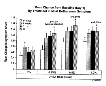
Figure 24 shows the effect of daily intravaginal application of 0.0%, 0.25%,
0.5% and
1.0% DHEA (Prasterone) for 2, 4, 8 and 12 weeks on vaginal pH in
postmenopausal
women. Data are expressed as means SEM.
Figure 25 shows the effect of daily intravaginal application of 0.0%, 0.25%,
0.5% and
1.0% DHEA (Prasterone) for 2, 4, 8 and 12 weeks on the change in severity of
the
symptom of vaginal atrophy judged by women themselves as being the most
bothersome. Values are compared to day 1 and are expressed as means + SEM.
Figure 26 shows the effect of daily intravaginal application of 0.0%, 0.25%,
0.5% and
1.0% DHEA (Prasterone) for 2, 4, 8 and 12 weeks on the change in vaginal
secretions
evaluated at vaginal examination. Data are expressed as means SEM.
Figure 27 shows the effect of daily intravaginal application of 0.0%, 0.25%,
0.5% and
1.0% DHEA (Prasterone) for 2, 4, 8 and 12 weeks on the change in vaginal color
evaluated at vaginal examination. Data are expressed as means SEM.
Figure 28 shows the effect of daily intravaginal application of 0.0%, 0.25%,
0.5% and
" 1.0% DHEA (Prasterone) for 2, 4, 8 and 12 weeks on the change in vaginal
epithelial
integrity evaluated at vaginal examination. Data are expressed as means SEM.
CA 02820566 2013-07-10
WO 2009/021323
PCT/CA2008/001444
Figure 29 shows effect of daily intravaginal application of 0.0%, 0.25%, 0.5%
and 1.0%
DHEA (Prasterone) for 2, 4, 8 and 12 weeks on the change m vaginal epithelial
thickness
evaluated at vaginal examination. Data are expressed as means SEM.
Figure 30 shows the average 24-hour serum concentrations (AUC0-24h/ 24) of
DHEA,
5-Diol, DHEA-S, El, E2 and El-S measured on days 1 and 7 following once daily
administration of vaginal ovule containing 0.5% DHEA. Data are expressed as
means
SEM (n=10). Serum steroid concentrations measured in 30-35 year-old
premenopausal
(n=47) as well as in 55-65 year-old postmenopausal (n=369) women are added as
reference data which are expressed as means and 5th and 95th centiles (dashed
lines).
*, p <0.05, **, p <0.01, experimental versus baseline. (Data are from Labrie,
Cusan et al.
2008).
Figure 31 shows the average 24-hour serum concentrations (AUCo-24h/24) of 4-
Dione,
testosterone, DHT ADT-G, 3a-Dio1-3G and 3a-Dio1-17G measured on days 1 and 7
following once daily administration of vaginal ovule containing 0.5% DHEA.
Data are
expressed as means SEM (n=10). Serum steroid concentrations measured in 30-
35
year-old premenopausal (n=47) and 55-65 year-old postmenopausal (n=369) women
are
added as reference data which are expressed as means and 5th and 95th centiles
(dashed
lines). *, p < 0.05, experimental versus baseline. (Data are from Labrie,
Cusan et al.
2008).
DETAILED DESCRIPTION OF THE INVENTION
[00141.Set Forth below are a list of articles discussed herein in short form
citations:
Allen, Loyd V Jr, Worthen Dennis B, and Mink Bill, in Suppositories , Chaper 3
pages
27-49, Published by the Pharmaceutical Press, London, UK, 2008
CA 02820566 2013-07-10
WO 2009/021323
PCT/CA2008/001444
11
Archer, D. F. (2007). "Drospirenone-containing hormone therapy for
postmenopausal
women. Perspective on current data." I Reprod Med 52(2 Suppl): 159-64.
Ayton, R. A., G. M. Darling, et al. (1996). "A comparative study of safety and
efficacy of
continuous low dose oestradiol released from a vaginal ring compared with
conjugated
equine oestrogen vaginal cream in the treatment of postmenopausal urogenital
atrophy." Br I Obstet Gynaeco1103(4): 351-8.
Bachmann, G., R. A. Lobo, et al. (2008). "Efficacy of low-dose estradiol
vaginal tablets in
the treatment of atrophic vaginitis: a randomized controlled trial." Obstet
Gynecol
111(1): 67-76.
Bachmann, G. A., M. Notelovitz, et al. (1992). "Long-term non-hormonal
treatment of
vagina dryness." I Clin Pract Sex 8.
Baker, V. L. and R. B. Jaffe (1996). "Clinical uses of antiestrogens." Obstet
Gynecol Surv
51: 45-59.
E.E. Baulieu, G. Thomas, S. Legrain, N. Lahlou, M. Roger, B. Debuire, V.
Faucounau, L.
Girard, M.P. Nervy, F. Latour, M.C. Leaud, A. Mokrane, H. Pitti-Ferrandi, C.
Trivalle,
0. de Lacharriere, S. Nouveau, B_ Rakoto-Arison, J.C. Souberbielle, J. Raison,
Y. Le
Bouc, A. Raynaud, X. Girerd and F. Forette, Dehydroepiandrosterone (DHEA),
DHEA
sulfate, and aging: contribution of the DHEAge Study to a sociobiomedical
issue, Proc.
Natl. Acad. Sc-i. U.S.A. 97 (2000), pp. 4279-4284.
Baxendale, P. M., M. J. Reed, et al. (1981). "Inability of human endometrium
or
myometrium to aromatize androstenedione." J Steroid Biochem 14(3): 305-6.
Belanger, B. Candas, A. Dupont, L. Cusan, P. Diamond, J.L. Gomez and F.
Labrie,
Changes in serum concentrations of conjugated and unconjugated steroids in 40-
to 80-
year-old men, J. Clin. Endocrinol. Metal,. 79 (1994), pp. 1086-1090.
CA 02820566 2013-07-10
WO 2009/021323
PCT/CA2008/001444
12
Belanger, G. Pelletier, F. Labrie, 0. Barbier and S. Chouinard, Inactivation
of
androgens by UDP-glucuronosyltransferase enzymes in humans, Trends Endocrinol.
Me tab. 14 (2003), pp. 473-479.
Beral, V. (2003). "Breast cancer and hormone-replacement therapy in the
Million
Women Study." Lancet 362(9382): 419-27.
Beral, V., D. Bull, et al. (2005). "Endometrial cancer and hormone-replacement
therapy
in the Million Women Study." Lancet 365(9470): 1543-51.
Berger, L., M. El-Ally, et al. (2005). "Effects of dehydroepiandrosterone,
Premarin and
Acolbifene on histomorphology and sex steroid receptors in the rat vagina." I
Steroid
Biochem Mol Biol 96(2): 201-15.
Bulun, S. E., Z. Lin, et al. (2005). "Regulation of aromatase expression in
estrogen-
responsive breast and uterine disease: from bench to treatment." Pharmacol Rev
57(3):
359-83.
J.E. Buster, P.R. Casson, A.B. Straughn, D. Dale, E.S. Umstot, N. Chiamori and
G.E.
Abraham, Postmenopausal steroid replacement with micronized
dehydroepiandrosterone: preliminary oral bioavailability and dose
proportionality
studies, Am. J. Obstet. Gynecol. 166 (1992), pp. 1163-1168 discussion 1168-
1170.
Chlebowski, R. T., S. L. Hendrix, et al. (2003). "Influence of estrogen plus
progestin on
breast cancer and mammography in healthy postmenopausal women: the Women's
Health Initiative Randomized Trial." Tama 289(24): 3243-53.
D.L. Coleman, E.H. Leiter and R.W. Schwizer, Therapeutic effects of
dehydroepiandrosterone (DHEA) in diabetic mice, Diabetes 31 (1982), pp. 830-
833.
Colditz, G. A., K. M. Egn, et al. (1993). "Hormone replacement thrapy and riks
of breast
cancer: results from epidemiologic studies." Am. T. Obstet. Gyneco1.168: 1473-
1480.
CA 02820566 2013-07-10
WO 2009/021323
PCT/CA2008/001444
13
Colditz, G. A., S. E. Hankinson, et al. (1995). "The use of estrogens and
progestins and
the risk of breast cancer in postmenopausal women." N. Engl. I. Med. 332: 1589-
1593.
Collaborative Group on Hormonal Factors in Breast Cancer (1997). "Breast
cancer and
hormone replacement therapy: collaborative reanalysis of data from 51
epidemiological
studies of 52,705 women with breast cancer and 108,411 women without breast
cancer."
Lancet 350(9084): 1047-59.
Corrao, G., A. Zambon, et al. (2008). "Menopause hormone replacement therapy
and
cancer risk: an Italian record linkage investigation." Ann Onco119(1): 150-5.
Coughlin, S. S., A. Giustozzi, et al. (2000). "A meta-analysis of estrogen
replacement
therapy and risk of epithelial ovarian cancer." T Clin Epidemiol 53(4): 367-
75.
Deutsch, S., R. Ossowski, et al. (1981). "Comparison between degree of
systemic
absorption of vaginally and orally administered estrogens at different dose
levels in
postmenopausal women." Am I Obstet Gyneco1139(8): 967-8.
Dew, J. E., B. G. Wren, et al. (2003). "A cohort study of topical vaginal
estrogen therapy
in women previously treated for breast cancer." Climacteric 6(1): 45-52.
P. Diamond, L. Cusart, J.L. Gomez, A. Belanger and F. Labrie, Metabolic
effects of 12-
month percutaneous DHEA replacement therapy in postmenopausal women, J.
En docrinol. 150 (1996), pp. S43--S50.
Dugal, R., K. Hesla, et al. (2000). "Comparison of usefulness of estradiol
vaginal tablets
and estriol vagitories for treatment of vaginal atrophy." Acta Obstet Gynecol
Scand
79(4): 293-7.
Englund, D. E. and E. D. Johansson (1978). "Plasma levels of oestrone,
oestradiol and
gonadotrophins in postmenopausal women after oral and vaginal administration
of
conjugated equine oestrogens (Premarin)." Br T Obstet Gynaecol 85(12): 957-64.
CA 02820566 2013-07-10
WO 2009/021323
PCT/CA2008/001444
14
Fallowfield, L., D. Cella, et al. (2004). "Quality of life of postmenopausal
women in the
Arimidex, Tamoxifen, Alone or in Combination (ATAC) Adjuvant Breast Cancer
Trial."
Clin Oncol 22(21): 4261-71.
Feeley, K. M. and M. Wells (2001). "Hormone replacement therapy and the
endometrium." I Clin Pathol 54(6): 435-40.
Furuhjelm, M., E. Karlgrert, et al. (1980). "Intravaginal administration of
conjugated
estrogens in premenopausal and postmenopausal women." Intl Gynaecol Obstet
17(4):
335-9.
Galhardo, C. L., J. M. Soares, Jr., et al. (2006). "Estrogen effects on the
vaginal pH, flora
and cytology in late postmenopause after a long period without hormone
therapy." Clin
Exp Obstet Gynecol 33(2): 85-9.
Gambrell, R. D., Jr., F. M. Massey, et al. (1980). "Use of the progestogen
challenge test to
reduce the risk of endometrial cancer." Obstet Gynecol 55(6): 732-8.
Garg, P. P., K. Kerlikowske, et al. (1998). "Hormone replacement therapy and
the risk of
epithelial ovarian carcinoma: a meta-analysis." Obstet Gynecol 92(3): 472-9.
Grady, D., T. Gebretsadik, et al. (1995). "Hormone replacement therapy and
endometrial
cancer risk: a meta-analysis." Obstet Gynecol 85(2): 304-13.
Gupta, P., B. Ozel, et al. (2008). "The effect of transdermal and vaginal
estrogen therapy
on markers of postmenopausal estrogen status." Menopause 15(1): 94-7.
Holmberg, L. and H. Anderson (2004). "HABITS (hormonal replacement therapy
after
breast cancer--is it safe?), a randomised comparison: trial stopped." Lancet
363(9407):
453-5.
CA 02820566 2013-07-10
WO 2009/021323
PCT/CA2008/001444
Holmberg, L., 0. E. Iversen, et al. (2008). "Increased risk of recurrence
after hormone
replacement therapy in breast cancer survivors." J Natl Cancer Inst 100(7):
475-82.
Holmgren, P. A., M. Lindskog, et al. (1989). "Vaginal rings for continuous low-
dose
release of oestradiol in the treatment of urogenital atrophy." Maturitas
11(1): 55-63.
Hulley, S. B. (2002). ''Noncardiovascular disease outcomes during 6.8 years of
hormone
therapy: Heart and estrogen/progestin replacement study follow-up (HERS II)."
TAMA
288: 58-66.
Jick, S. S., A. M. Walker, et al. (1993). "Estrogens, progesterone, and
endometrial cancer."
Epidemiology 4(1): 20-4.
C.C. Johnston Jr., S.L. Hui, R.M. Witt, R. Appledorn, R.S. Baker and C.
Longcope, Early
menopausal changes in bone mass and sex steroids, J. Clin. Endocrinol. Metab.
61 (1985),
pp. 905-911.
D.W. Hum, A. Belanger, E. Levesque, 0. Barbier, M. Beaulieu, C. Albert, M.
Vallee, C.
Guillemette, A. Tchernof, D. Turgeon and S. Dubois, Characterization of UDP-
glucuronosyltransferases active on steroid hormones, J. Steroid Biocheni. Mol.
Biol. 69
(1999), pp. 413-423.
H. Kawano, H. Yasue, A. Kitagawa, N. Hirai, T. Yoshida, H. Soejima, S.
Miyamoto, M.
Nakano and H. Ogawa, Dehydroepiandrosterone supplementation improves
endothelial function and insulin sensitivity in men, J. Clin. Endocrinol.
Metab. 88 (2003),
pp. 3190-3195.
Kendall, A., M. Dowsett, et al. (2006). "Caution: Vaginal estradiol appears to
be
contraindicated in postmenopausal women on adjuvant aromatase inhibitors." Ann
Oncol 17(4): 584-7.
Kvorning, J. D. N. and H. K. Jensen (1986). Pharmaceutical development of lose-
dose
estradiol vagitories. International Workshop, Copenhagen.
CA 02820566 2013-07-10
WO 2009/021323
PCT/CA2008/001444
16
Labrie, C., A. Belanger, et al. (1988). "Androgenic activity of
dehydroepiandrosterone
and androstenedione in the rat ventral prostate." Endocrinology 123: 1412-
1417.
Labrie, F. (1991). "Intracrinology." Mol. Cell. Endocrinoi. 78: C113-C118.
F. Labrie, Future perspectives of SERMs used alone and in combination with
DHEA,
Endocr. Relat. Cancer 13 (2006), pp. 335-355.
Labrie, F. (2007). "Drug Insight: breast cancer prevention and tissue-targeted
hormone
replacement therapy." Nature Clinical Practice, Endocrinology & Metabolism
3(8): 584-
593.
F. Labrie, A. Belanger, J. Simard, V. Luu-The and C. Labrie, DHEA and
peripheral
androgen and estrogen formation: intracrinology, Ann. N.Y. Acad. Sci. 774
(1995), pp.
16-28.
Labrie, F., A. Belanger, et al. (2007). "Metabolism of DHEA in postmenopausal
women
following percutaneous administration." I Steroid Biochem Mol Bio1103(2): 178-
88.
Labrie, F.õ,51. Belanger, et al. (2007) "Bioa-v-ailability and metabolism of
oral and
percutaneous dehydroepiandrosterone in postmenopausal women" J Steroid Biochem
Mol Biol. Oct;107(1-2):57-69..
Labrie, F., A. Belanger, et al. (2006). "Androgen glucuronides, instead of
testosterone, as
the new markers of androgenic activity in women." Journal Ster Biochem & Mol
Biol 99:
182-188.
Labrie, F., A. Belanger, et al. (2005). "GnRH agonists in the treatment of
prostate cancer."
Endocrine Reviews 26(3): 361-379.
Labrie, F., A. Belanger, et al. (1997). "Marked decline in serum
concentrations of adrenal
C19 sex steroid precursors and conjugated androgen metabolites during aging."
I Clin
Endocrinol Metab 82: 2396-2402.
CA 02820566 2013-07-10
WO 2009/021323
PCT/CA2008/001444
17
Labrie, F., A. Belanger, et al. (2007a). "Bioavailability and metabolism of
oral and
percutaneous dehydroepiandrosterone in postmenopausal women." I Steroid
Biochem
Mol Biol 107(1-2): 57-69.
F. Labrie, A. Belanger, P. Belanger, R. Berube, C. Martel, L. Cusan, J. Gomez,
B. Candas,
V. Chaussade, I. Castiel, C. Deloche and J. Leclaire, Metabolism of DHEA in
postmenopausal women following percutaneous administration, J. Steroid
Biochem. Mol.
Biol. 103 (2) (2007b), pp. 178-188.
Labrie, F., L. Cusan, et al. (2008). "Effect of Intravaginal DHEA on Serum
DHEA and
Eleven of its Metabolites in Postmenopausal Women." Journal Ster Biochem & Mol
Biol:
In press.
Labrie, F., L. Cusan, et al. (2008). "Effect of One-Week Treatment with
Vaginal Estrogen
Preparations on Serum Estrogen Levels in Postmenopausal Women." Menopause In
press.
Labrie, F., L. Cusan, et al. (2008). "Changes in serum DHEA and eleven of its
metabolites during 12-month percutaneous administration of DHEA." I Steroid
Biochem Mol Biol 110(1-2): 1-9.
Labrie, F., P. Diamond, et al. (1997). "Effect of 12-month
dehydroepiandrosterone
replacement therapy on bone, vagina, and endometrium in postmenopausal women."
Clin Endocrinol Metab 82(10): 3498-505.
Labrie, F., A. Dupont, et al. (1985). Complete androgen blockade for the
treatment of
prostate cancer. Important Advances in Oncology. V. T. de Vita, S. Hellman and
S. A.
Rosenberg. Philadelphia, J.B. Lippincott: 193-217.
F. Labrie, V. Lou-The, S.X. Lin, C. Labrie, J. Simard, R. Breton and A.
Belanger, The key
role of 1713-HSDs in sex steroid biology, Steroids 62 (1997), pp. 148-158.
CA 02820566 2013-07-10
WO 2009/021323
PCT/CA2008/001444
18
Labrie, V. Luu-The, S.-X. Lin, J. Simard, C. Labrie, M. El-Alfy, G. Pelletier
and A.
Belanger, Intracrinology: role of the family of 17p-hydroxysteroid
dehydrogenases in
human physiology and disease, J. Mol. Endocrinol. 25 (2000), pp. 1-16.
Labrie, F., V. Luu-The, et al. (2005). "Is DHEA a hormone? Starling Review." I
Endocrino1187: 169-196.
Labrie, F., V. Luu-The, et al. (2003). ''Endocrine and intracrine sources of
androgens in
women: inhibition of breast cancer and other roles of androgens and their
precursor
dehydroepiandrosterone." Endocrine Reviews 24(2): 152-182.
Labrie, F., V. Luu-The, et al. (2006). "Dehydroepiandrosterone (DHEA) is an
anabolic
steroid like dihydrotestosterone (DHT), the most potent natural androgen, and
tetrahydrogestrinone (THG)." I Steroid Biochem Mol Bio1100(1-3): 52-8.
F. Labrie, J. Simard, V. Luu-The, A. Belanger, G. Pelletier, Y. Morel, F.
Mebarki, R.
Sanchez, F. Durocher, C. Turgeon, Y. Labrie, E. Rheaume, C. Labrie and Y.
Lachance,
The 3I3-hydroxysteroid dehydrogenase/isomerase gene family: lessons from type
II 3P-
HSD congenital deficiency. In: V. Hansson, F.O. Levy and K. Tasken, Editors,
Signal
Transduction in Testicular Cells. Ernst Schering Research Foundation Workshop,
vol Suppl. 2,
Springer-Verlag, Berlin (1996), pp. 185-218.
Labrie, J. Simard, V. Luu-The, A. Belanger and G. Pelletier, Structure,
function and
tissue-specific gene expression of 3I3-hydroxysteroid dehydrogenase/5-ene-4-
ene
isomerase enzymes in classical and peripheral intracrine steroidogenic
tissues, J. Steroid
Biochem. Mol. Biol. 43 (1992), pp. 805-826.
F. Labrie, R. Poulin, J. Simard, V. Luu-The, C. Labrie and A. Belanger,
Androgens,
DHEA and breast cancer. In: T. Gelfand, Editor, Androgens and Reproductive
Aging,
Taylor and Francis, Oxsfordshire, UK (2006), pp. 113-135.
CA 02820566 2013-07-10
WO 2009/021323
PCT/CA2008/001444
19
Labrie, Y. Sugimoto, V. Luu-The, J. Simard, Y. Lachance, D. Bachvarov, G.
Leblanc, F.
Durocher and N. Paquet, Structure of human type II 5*-reductase, Endocrinology
131
(1992), pp. 1571-1573.
Y. Labrie, F. Durocher, Y. Lachance, C. Turgeon, J. Simard, C. Labrie and F.
Labrie, The
human type H 173-hydroxysteroid dehydrogenase gene encodes two alternatively-
spliced messenger RNA species, DNA Cell Biol. 14 (1995), pp. 849-861.
Lacey, J. V., P. J. Mink, et al. (2002). "Menopausal hormone replacement
therapy and
risk of ovarian cancer." TAMA 288: 334-341.
Li, L., S. J. Plummer, et al. (2008). "A common 8q24 variant and the risk of
colon cancer:
a population-based case-control study." Cancer Epidemiol Biomarkers Prey
17(2): 339-
42.
C.H. Liu, G.A. Laughlin, U.G. Fischer and S.S. Yen, Marked attenuation of
ultradian
and circadian rhythms of dehydroepiandrosterone in postmenopausal women:
evidence for a reduced 17,20-desmolase enzymatic activity, J Clin. Endocrinol.
Metab. 71
(1990), pp. 900-906.
Long, C. Y., C. M. Liu, et al. (2006). "A randomized comparative study of the
effects of
oral and topical estrogen therapy on the vaginal vascularization and sexual
function in
hysterectomized postmenopausal women." Menopause 13(5): 737-43.
V. Luu-The, I. Dufort, N. Paquet, G. Reimnitz and F. Labrie, Structural
characterization
and expression of the human dehydroepiandrosterone sulfotransferase gene, DNA
Cell
Biol. 14 (1995), pp. 511-518.
V. Luu-The, Y. Zhang, D. Poirier and F. Labrie, Characteristics of human types
1, 2 and
3 17P-hydroxysteroid dehydrogenase activities: oxidation-reduction and
inhibition, J.
Steroid Biochem. Mol. Biol. 55 (1995), pp. 581-58
Lyytinen, FL, E. Pukkala, et al. (2006). "Breast cancer risk in postmenopausal
women
using estrogen-only therapy." Obstet Gynecol 108(6): 1354-60.
CA 02820566 2013-07-10
WO 2009/021323
PCT/CA2008/001444
E.G. MacEwen and I.D. Kurzman, Obesity in the dog: role of the adrenal steroid
dehydroepiandrosterone (DHEA), J. Nutr. 121 (1991), pp. S51-S55.
Mandel, F. P., F. L. Geola, et al. (1983). "Biological effects of various
doses of vaginally
administered conjugated equine estrogens in postmenopausal women."I Clin
Endocrinol Metab 57(1): 133-9.
Manonai, J., U. Theppisai, et al. (2001). "The effect of estradiol vaginal
tablet and
conjugated estrogen cream on urogenital symptoms in postmenopausal women: a
comparative study." 1 Obstet Gynaecol Res 27(5): 255-60.
Martin, P. L., S. S. Yen, et al. (1979). "Systemic absorption and sustained
effects of
vaginal estrogen creams." lama 242(24): 2699-700.
Marx, P., G. Schade, et al. (2004). "Low-dose (0.3 mg) synthetic conjugated
estrogens A
is effective for managing atrophic vaginitis." Maturitas 47(1): 47-54.
Mattson, L. A., G. Culberg, et al. (1989). "Vaginal administration of low dose
estradiol-
effects on endometrium and vaginal cytology." Maturitas 11: 217-2.22.
R.B. Mazess, On aging bone loss, Clin. Orthop. 165 (1982), pp. 239-252.
Meisels, A. (1967). "The maturation value." Acta Cyto111: 249.
Mertens, H. J., M. J. Heineman, et al. (1996). "Androgen receptor content in
human
endometrium." Eur I Obstet Gynecol Reprod Biol 70(1): 11-3.
Mettler, L. and P. G. Olsen (1991). "Long-term treatment of atrophic vaginitis
with low-
dose oestradiol vaginal tablets." Maturitas 14(1): 23-31.
C.J. Migeon, A.R. Keller, B. Lawrence and T.H. Shepart II.,
Dehydroepiandrosterone
and and rosterone levels in human plasma. Effect of age and sex: day-to-day
and diurnal
variations, J. Clin. Endocrine!. Metab. 17 (1957), pp. 1051-1062.
CA 02820566 2013-07-10
WO 2009/021323
PCT/CA2008/001444
21
A.J. Morales, J.J. Nolan, J.C. Nelson and S.S. Yen, Effects of replacement
dose of
dehydroepiandrosterone in men and women of advancing age, J. Clin. Endocrinol.
Metal'. 78 (1994), pp. 1360-1367.
Morales, L., P. Neven, et al. (2004). "Acute effects of tamoxifen and third-
generation
aroma tase inhibitors on menopausal symptoms of breast cancer patients."
Anticancer
Drugs 15(8): 753-60.
N.A.M.S. (2007). "Position Statement of the North American Menopause Society."
Menopause 14: 357-69.
Nachtigall, L. E. (1995). "Clinical trial of the estradiol vaginal ring in the
U.S." Maturitas
22 Suppl: S43-7.
Naessen, T., K. Rodriguez-Macias, et al. (2001). "Serum lipid profile improved
by ultra-
low doses of 17 beta-estradiol in elderly women." I Clin Endocrinol Metab
86(6): 2757-
62.
Nelson, II. 1-L-)., IT( K. Vesco, et al. (2006). "Nonhornional therapies for
menopausal hot
flashes: systematic review and meta-analysis." lama 295(17): 2057-71.
J.E. Nestler, C.O. Barlascini, J.N. Clore and W.G. Blackard,
Dehydroepiandrosterone
reduces serum low density lipoprotein levels and body fat but does not alter
insulin
sensitivity in normal men, J. Clin. Endocrinol. Metal'. 66 (1988), pp. 57-61.
Nilsson, K. and G. Heimer (1992). "Low-dose oestradiol in the treatment of
urogenital
oestrogen deficiency--a pharmacokinetic and pharmacodynamic study." Maturitas
15(2): 121-7.
Notelovitz, M., S. Funk, et al. (2002). "Estradiol absorption from vaginal
tablets in
postmenopausal women.'' Obstet Gynecol 99(4): 556-62.
CA 02820566 2013-07-10
WO 2009/021323
PCT/CA2008/001444
22
Orentreich, N., J. L. Brind, et al. (1984). "Age changes and sex differences
in serum
dehydroepiandrosterone sulfate concentrations throughout adulthood." .1. Clin,
Endocrinol. Metab. 59: 551-555.
Pandit, L. and J. G. Ouslander (1997). "Postmenopausal vaginal atrophy and
atrophic
vaginitis." Am I Med Sci 314(4): 228-31.
Persson, I., H. 0. Adami, et al. (1989). ''Risk of endometrial cancer after
treatment with
oestrogens alone or in conjunction with progestogens: results of a prospective
study."
Bmj 298(6667): 147-51.
Ponzone, R., N. Biglia, et al. (2005). "Vaginal oestrogen therapy after breast
cancer: is it
safe?" Eur J Cancer 41(17): 2673-81.
Rigg, L. A., H. Hermann, et al. (1978). "Absorption of estrogens from vaginal
creams." N
Engl J Med 298(4): 195-7.
B.L. Riggs, H.W. Wahner, W.L. Dunn, R.B. Mazess, K.P. Offord and L.J. Melton,
Differential changes in bone mineral density of the appendicular and axial
skeleton
with aging: relationship to spinal osteoporosis, J. Clin. Invest. 67 (1981),
pp. 328-335.
Riman, T., P. W. Dickman, et al. (2002). "Hormone replacement therapy and the
risk of
invasive epithelial ovarian cacner in Swedish women." I Nail Cancer Inst 94:
497-504.
Rinaldi, S., H. Dechaud, et al. (2001). "Reliability and validity of
commercially available,
direct radioimmunoassays for measurement of blood androgens and estrogens in
postmenopausal women." Cancer Epidemiol Biomarkers Prey 10(7): 757-65.
CA 02820566 2013-07-10
WO 2009/021323
PCT/CA2008/001444
23
Rioux, J. E., C. Devlin, et al. (2000). "17beta-estradiol vaginal tablet
versus conjugated
equine estrogen vaginal cream to relieve menopausal atrophic vaginitis."
Menopause
7(3): 156-61.
Rodriguez, C., A. V. Patel, et al. (2002). "Estrogen replacement therapy and
ovarian
cancer mortality in a large prospective study of US women." TAMA 285: 1460-
1465.
Rosenberg, L. U., C. Magnusson, et al. (2006). "Menopausal hormone therapy and
other
breast cancer risk factors in relation to the risk of different histological
subtypes of
breast cancer: a case-control study." Breast Cancer Res 8(1): R11.
Rossouw, J. E., G. L. Anderson, et al. (2002). "Risks and benefits of estrogen
plus
progestin in healthy postmenopausal women: principal results From the Women's
Health Initiative randomized controlled trial." Jama 288(3): 321-33.
Salminen, H. S., M. E. Saaf, et al. (2007). "The effect of transvaginal
estradiol on bone in
aged women: a randomised controlled trial." Maturitas 57(4): 370-81.
Schiff, I., D. Tulchinsky, et al. (1977). "Vaginal absorption of estrone and
17beta-
estradiol." Fertil Steril 28(10): 1063-6.
Schmidt, G., S. B. Andersson, et al. (1994). "Release of 17-beta-oestradiol
from a vaginal
ring in postmenopausal women: pharmacokinetic evaluation." Gynecol Obstet
Invest
38(4): 253-60.
E.D. Schriock, C.K. Buffington, G.D. Hubert, B.R. Kurtz, A.E. Kitabchi, J.E.
Buster and
J.R. Givens, Divergent correlations of circulating dehydroepiandrosterone
sulfate and
testosterone with insulin levels and insulin receptor binding, J. Clin.
Endocrinol. Metab.
66 (1988), pp. 1329-1331.
Sillero-Arenas, M., M. Delgado-Rodriguez, et al. (1992). "Menopausal hormone
replacement therapy and breast cancer: a meta-analysis." Obstet. Gynecol. 79:
286-294.
CA 02820566 2013-07-10
WO 2009/021323
PCT/CA2008/001444
24
Simon, J. A., K. Z. Reape, et al. (2007). "Randomized, multicenter, double-
blind,
placebo-controlled trial to evaluate the efficacy and safety of synthetic
conjugated
estrogens B for the treatment of vulvovaginal atrophy in healty postmenopausal
women." Fertil Steril In press.
E.R. Simpson, Role of aromatase in sex steroid action, J. Mol. Endocrinol. 25
(2000), pp.
149-156.
Simunic, V., I. Banovic, et al. (2003). "Local estrogen treatment in patients
with
urogenital symptoms." Intl Gynaecol Obstet 82(2): 187-97.
Smith, D. C., R. Prentice, et al. (1975). "Association of exogenous estrogen
and
endometrial carcinoma." N. Engl. I. Med. 293: 1164-1167.
Smith, P., G. Heimer, et al. (1993). "Oestradiol-releasing vaginal ring for
treatment of
postmenopausal urogenital atrophy." Maturitas 16(2): 145-54.
Sourla, A., M. Flamand, et al. (1998). "Effect of dehydroepiandrosterone on
vaginal and
uterine histomorphology in the rat." I. Steroid Biochem. Mol. Biol. 66(3): 137-
149.
Steinberg, K. K., S. B. Thacker, et al. (1991). "A meta-analysis of the effect
of estrogen
replacement therapy on the risk of breast cancer." TAMA 265: 1985-1990.
K.K. Steinberg, L.W. Freni-Titulaer, E.G. DePuey, D.T. Miller, D.S. Sgoutas,
C.H. Coralli,
D.L. Phillips, T.N. Rogers and R.V. Clark, Sex steroids and bone density in
premenopausal and perimenopausal women, J. Clin. Endocrinol. Metab. 69 (1989),
pp.
533-539.
Suckling, J., A. Lethaby, et al. (2006). "Local oestrogen for vaginal atrophy
in
postmenopausal women." Cochrane Database System Rev 18(4): CD001500.
CA 02820566 2013-07-10
WO 2009/021323
PCT/CA2008/001444
Swanson, M. Lorentzon, L. Vandenput, D. Mellstrom, F. Labrie, A. Rane, J.
Jakobsson,
C. Ohlsson, UGT2B7 H268Y polymorphism is associated with serum sex steroid
levels
and cortical bone size in young adult men, JCEM (2007), in press.
Tchernof, J.P. Despres, A. Belanger, A. Dupont, D. Prud'homme, S. Moorjani,
P.J.
Lupien and F. Labrie, Reduced testosterone and adrenal C19 steroid levels in
obese
men, Metabolism 44 (1995), pp. 513-519.
Turgeon, J.S. Carrier, E. Levesque, D.W. Hum and A. Belanger, Relative
enzymatic
activity, protein stability, and tissue distribution of human steroid-
metabolizing UGT2B
subfamily members, Endocrinology 142 (2001), pp. 778-787.
Utian, W. H., D. Shoupe, et al. (2001). "Relief of vasomotor symptoms and
vaginal
atrophy with lower doses of conjugated equine estrogens and
niedroxyprogesterone
acetate." Fertil Steril 75(6): 1065-79.
Vermeulen and L. Verdonck, Radioinununoassays of 1713-hydroxy-5*-androstan-3-
one,
4-androstene-3,17-dione, dehydroepiandrosterone, 1713-hydroxyprogesterone and
progesterone and its application to human male plasma, I. Steroid Biochem. 7
(1976), pp.
1-10.
D.T. Villareal and J.O. Holloszy, Effect of DHEA on abdominal fat and insulin
action in
elderly women and men: a randomized controlled trial, JAMA 292 (2004), pp.
2243-
2248.
Voigt, L. F., N. S. Weiss, et al. (1991). "Progestagen supplementation of
exogenous
oestrogens and risk of endometrial cancer." Lancet 338(8762): 274-7.
Weisberg, E., R. Ayton, et al. (2005). "Endometrial and vaginal effects of low-
dose
estradiol delivered by vaginal ring or vaginal tablet." Climacteric 8(1): 83-
92.
Wied, G. L. (1993). "Industrial developments in automated cytology as
submitted by
their developers." Anal Quant Cytol Histo115(5): 358-70.
CA 02820566 2013-07-10
WO 2009/021323
PCT/CA2008/001444
26
Wines, N. and E. Willsteed (2001). "Menopause and the skin." Australas I
Dermatol
42(3): 149-8; quiz 159.
Zang, H., L. Sahlin, et al. (2007). "Effects of testosterone treatment on
endometrial
proliferation in postmenopausal women." I Clin Endocrinol Metab 92(6): 2169-
75.
B. Zumoff, G.W. Strain, L.K. Miller and W. Rosner, Twenty-four-hour mean
plasma
testosterone concentration declines with age in normal premenopausal women, J.
Clin.
Endocnnol. Metab. 80 (1995), pp. 1429-1430.
[0015].Vaginal dryness is found in 75% of postmenopausal women (Wines and
Willsteed 2001; N.A.M.S. 2007). For various reasons, especially the fear of
complications
by estrogens, only 20 to 25% of symptomatic women with vaginal atrophy seek
medical
treatment (Pandit and Ouslander 1997; N.A.M.S. 2007). There is thus a clear
medical
need and a major opportunity to improve the quality of life of a large
population of
women left suffering from vaginal atrophy for a large proportion of their
lifetime. In
can be mentioned that while hot flashes abate spontaneously with time, vaginal
atrophy
symptoms, namely vaginal dryness, vulvovaginal irritation/iching and
dyspareunia
usually increase in severity with time in the absence of treatment.
[0016].Based upon the well known fact that estrogen secretion by the ovaries
ceases at
menopause, systemic and local estrogens have so-far been the exclusive
approach for
the treatment of vaginal atrophy. However, systemic estrogens progestin
(HRT) and
estrogens alone (ERT) have been shown to increase the risk of breast cancer
(Steinberg,
Thacker et al. 1991; Sillero-Arenas, Delgado-Rodriguez et al. 1992; Colditz,
Egn et al.
1993; Colditz, Hankinson et al. 1995; Collaborative Group on Hormonal Factors
in
Breast Cancer 1997; Hulley 2002; Beral 2003; Chlebowski, Hendrix et al. 2003;
Holmberg
and Anderson 2004; Lyytinen, Pukkala et al. 2006; Corrao, Zambon et al. 2008;
CA 02820566 2013-07-10
WO 2009/021323
PCT/CA2008/001444
27
Holmberg, Iversen et al. 2008; Li, Plummer et al. 2008), ovarian cancer (Garg,
Kerlikowske et al. 1998; Coughlin, Giustozzi et al. 2000; Lacey, Mink et al.
2002; Riman,
Dickman et al. 2002; Rodriguez, Patel et al. 2002; Rossouw, Anderson et al.
2002;
Lyytinen, Pukkala et al. 2006) as well as endometrial cancer (estrogens alone)
(Gambrel!, Massey et al. 1980; Persson, Adami et al. 1989; Voigt, Weiss et al.
1991; lick,
Walker et al. 1993; Grady, Gebretsadik et al. 1995; Beral, Bull et al. 2005).
The publicity
which followed the Women's Health Initiative Study (Rossouw, Anderson et al.
2002)
had the greatest impact, thus putting in doubt the safety of the available
treatments of
menopausal symptoms (Archer 2007).
E00171. Although intravaginal formulations were developed to avoid systemic
exposure
to estrogens, a long series of studies have unanimously demonstrated that all
intravaginal estrogen formulations lead to relatively high serum estrogen
levels
measured directly or through their systemic effects (Englund and Johansson
1978; Rigg,
Hermann et al. 1978; Martin, Yen et al. 1979; Furuhjelm, Karlgren et al. 1980;
Deutsch,
Ossowski et al. 1981; Mandel, Geola et al. 1983; Nilsson and Heimer 1992;
NachtivIl
1995; Ayton, Darling et al. 1996; Dugal, Hesla et al. 2000; Rioux, Devlin et
al. 2000;
Manonai, Theppisai et al. 2001; Notelovitz, Funk et al. 2002; Ponzone, Biglia
et al. 2005;
Weisberg, Ayton et al. 2005; Galhardo, Soares et al. 2006; Kendall, Dowsett et
al. 2006;
Long, Liu et al. 2006; Bachmann, Lobo et al. 2008). These data showing a
significant
increase in serum estrogen levels clearly indicate that the use of
intravaginal estrogen
formulations is also potentially associated with an increased risk of breast
and uterine
cancer (Kvorning and Jensen 1986; Mattson, Culberg et al. 1989; Rosenberg,
Magnusson
et al. 2006; N.A.M.S. 2007). Concerns have in fact been officially raised
about the
stimulatory effects of vaginal estrogen formulations on the endometrium
((N.A.M.S.
2007).
CA 02820566 2013-07-10
WO 2009/021323
PCT/CA2008/001444
28
[0018}.Most previous measurements of the serum estradiol (E2) levels after
intravaginal administration of estrogens used radioimmunoassays, a technology
lacking
specificity, accuracy, reliability and sensitivity (Rinaldi, Dechaud et at.
2001). We have
measured serum estrogens using GLP (Good Laboratory Practice)-validated mass
spectrometry assays following intravaginal administration of the two most
commonly
used estrogen formulations (Labrie, Cusan et al. 2008). This study could
definitively
show that both the E2 pill (25 ttg E2/ day) and conjugated estrogens cream (1
g of
0.625 mg conjugated estrogens/day), after one-week of daily treatment, cause
an
approximately 5-fold increase in serum E2 in postmenopausal women. Such data
indicate that the effects of estrogens applied locally in the vagina are
unlikely to be
limited to the vagina and that systemic action is expected as previously
suggested
(Englund and Johansson 1978; Rigg, Hermann et al. 1978; Martin, Yen et al.
1979;
Furuhjelm, Karlgren et at. 1980; Deutsch, Ossowski et al. 1981; Mandel, Geola
et al. 1983;
Nilsson and Heimer 1992; Nachtigall 1995; Ayton, Darling et al. 1996; Dugal,
HesIa et al.
2000; Rioux, Devlin et al. 2000; Manonai, Theppisai et al. 2001; Notelovitz,
Funk et al.
2002; Ponzone, Biglia et al. 2005; Weisberg, Ayton et al. 2005; Galhardo,
Soares et al.
2006; Kendall, Dowsett et al. 2006; Long, Liu et al. 2006; Bachmann, Lobo et
al. 2008).
[0019J.In addition to the above-indicated safety concerns of estrogens
administered
both systemically and locally, recent data have clearly demonstrated that
women are
not only deficient in estrogens at time of menopause but that they have also
been
progressively deprived, starting in the thirties, from the androgens made in
specific
peripheral target tissues by the intracrine transformation of
dehydroepiandrosterone
(DHEA) into androgens and/or estrogens (Labrie, Belanger et al. 1988; Labrie
1991;
Labrie, Luu-The et al. 2003; Labrie, Luu-The et al. 2005). In fact, serum DHEA
and
DHEA-sulfate progressively decrease from the peak seen at the age of 30 years
(Orentreich, Brind et al. 1984; Labrie, Belanger et al. 1997; Labrie, Luu-The
et al. 2003) to
a value 60% lower at time of menopause (Labrie, Belanger et al. 2006).
CA 02820566 2013-07-10
WO 2009/021323
PCT/CA2008/001444
29
[0020].Concerning the role of androgens in women, it is important to mention
that
women secrete 50% as much androgens as observed in men (Labrie, Belanger et
al. 1997;
Labrie, Luu-The et al. 2005). Since serum DHEA is the predominant source of
androgens which play a series of physiological roles in women (Labrie, Luu-The
et al.
2003; Labrie 2007), the 60% decrease in circulating DHEA already found at time
of
menopause leads to a similar 60% decrease in the total androgen pool (Labrie,
Belanger
et al. 2006) with the resulting potential signs and symptoms of
hypoandrogenicity in the
bone, muscle, skin, mammary gland, vagina, brain as well as on glucose,
insulin and
lipid metabolism (Labrie, Luu-The et al. 2003; Labrie 2007). Among the
androgen target
tissues, recent data have shown that the vagina is sensitive to androgens
following
DHEA administration in the rat with beneficial effects, not only on the
superficial
epithelial layer of the vagina but also on collagen fibers in the lamina
propria and on the
muscularis (Berger, El-Ally et al. 2005).
[00211.Based upon the data of our prPelinical (goiirlA, FlAmancl Pt al. 1998;
Berger, El-
Ally et al. 2005) and clinical (Labrie, Diamond et al. 1997; Labrie, Cusan et
al. 2008)
studies showing beneficial effects on the vagina of DHEA administered
percutaneously
or locally, the present clinical trial is a prospective, randomized and
placebo-controlled
study of the effect of three doses of intravaginal DHEA administered daily for
12 weeks
on the changes in superficial and parabasal cells, vaginal pH and the most
bothersome
symptom of vaginal atrophy as primary objectives. The data clearly show that
locally
administered DHEA is very efficient and rapid in correcting all the signs and
symptoms
of vaginal atrophy, a near maximal effect being already achieved at 2 weeks at
a DHEA
dose causing no significant change in serum estrogens or androgens while all
other
steroids remain unchanged or well within the range found in normal
postmenopausal
women.
CA 02820566 2013-07-10
WO 2009/021323 PCT/CA2008/001444
[0022]. When DHEA is administered locally in the vagina, the beneficial
effects
of estrogens and androgens made locally in the vagina are achieved without any
significant release of estradiol or testosterone into the blood (Labrie, Cusan
et al. J. Ster.
Biochem. Mol. Biol. In press). In the formation of androgens and/or estrogens
from
DHEA by the process of intracrinology, any tissue is unpredictable because the
= response depends upon the activity of the enzymatic machinery
specifically present in
each cell of each tissue. Thus, it is not possible to predict, from the
androgens and
estrogens that are produced from DHEA in one tissue, the extent to which
similar
androgens and estrogens may be produced in another tissue.
[0023].The results of the clinical trial ERC-210 (Example 3) clearly
demonstrate for the
first time that the local administration of DHEA as hormone precursor
replacement
therapy (HPRT) is highly efficient and rapid in correcting all the symptoms
and signs of
vaginal atrophy in postmenopausal women. Most importantly, this is achieved at
a
dose (0.5%) of DHEA which does not increase the serum levels of active
estrogens or
androgens and with no or minimal changes in serum DHEA and any of its
metabolites
which all remain well within the range of values found in normal
postmenopausal
women (Labrie, Cusan et al. 2008).
[0024].While 75% of postmenopausal women suffer from vaginal atrophy (Wines
and
Willsteed 2001; N.A.M.S. 2007), thus affecting their quality of life during a
major part of
their lifetime, only 20% seek treatment (Pandit and OusLander 1997). The fear
of breast
cancer related to increased blood levels of estrogens is the main reason
involved. Since
estrogen secretion into the systemic circulation is exclusively of ovarian
origin and
ceases at menopause, administration of estrogens to postmenopausal women does
not
appear to be physiological. In the aftermath of WHI, the scientific challenge
is to explore
alternative hormonal therapy types and formulations that would provide all the
menopausal advantages of estrogens while improving women's quality of life,
CA 02820566 2013-07-10
WO 2009/021323
PCT/CA2008/001444
31
minimizing risks and maximizing benefits (Archer 2007). Since the non-estrogen
based treatments have not shown convincing efficacy (Nelson, Vesco et al.
2006;
Suckling, Lethaby et al. 2006), women and their physicians are left with no
safe
treatment for vaginal atrophy.
[0025]. Various forms of estrogens are an efficient treatment for vulvovaginal
atrophy
(Pandit and Ouslander 1997; Utian, Shoupe et al. 2001). In fact, the vaginal
E2 tablet has
shown an efficacy similar to the E2 ring (Weisberg, Ayton et al. 2005) as well
as to the
conjugated estrogen cream (Rioux, Devlin et al. 2000; Manonai, Theppisai et
al. 2001).
[0026].This novel HPRT is in marked contrast with the 5-fold increase in serum
E2
measured by mass spectrometry after treatment with intravaginal E2 or
conjugated
estrogens (Labrie, Cusan et al. 2008). These recent data on the changes in
serum
estrogens confirm a long series of studies showing that all intravaginal
estrogen
formulations lead to elevated serum estrogen concentrations measured directly
by
railinirnmnnoAssgr or through their systemic effects (Englund and Inhnsson
1978;
Rigg, Hermann et al. 1978; Martin, Yen et al. 1979; Furuhjelm, Karlgren et al.
1980;
Deutsch, Ossowski et al. 1981; Mandel, Geola et al. 1983; Nilsson and Heimer
1992;
Nachtigall 1995; Ayton, Darling et al. 1996; Dugal, Hesla et al. 2000; Rioux,
Devlin et al.
2000; Manonai, Theppisai et al. 2001; Notelovitz, Funk et al. 2002; Ponzone,
Biglia et al.
2005; Weisberg, Ayton et al. 2005; Galhardo, Soares et al. 2006; Kendall,
Dowsett et al.
2006; Long, Liu et al. 2006; Bachmann, Lobo et al. 2008).
[0027].The most common adverse events reported with vaginal estrogens are
vaginal
bleeding and breast pain, both secondary to increased serum estrogens
(Suckling,
Lethaby et al. 2006). These side effects have been reported for the E2 ring,
conjugated
estrogens cream as well as E2 tablet (Ayton, Darling et al. 1996; Weisberg,
Ayton et al.
2005). As mentioned above, concerns also exist about the stimulatory effects
of vaginal
CA 02820566 2013-07-10
32
estrogens on the endometrium (N.A.M.S. 2007). Uterine bleeding, breast pain
and
perineal pain were reported in 9% of women W.00 took the vaginal tablet for 24
weeks
while 34% complained of the same symptoms in the vaginal conjugated estrogen
cream
group (Rioux, Devlin et al. 2000). (Suckling, Lethaby et al. 2006) reported no
difference
between the different vaginal estrogen preparations.
[0028].1t is well known that atrophic vaginitis in postmenopausal women can be
worsened or induced by the use of aroma tase inhibitors for the treatment of
breast
cancer. In fact, these drugs exert their benefits on breast cancer by
decreasing E2
biosynthesis in all tissues, thus increasing the frequency and severity of
menopausal
symptoms (Fallowfield, Cella et al. 2004; Morales, Neven et al. 2004). In a
recent study
TM
where seven breast cancer patients treated with aromatase inhibitors received
Vagifem
at a daily dose of 25 pg for 2 weeks and then, thereafter, twice weekly, serum
E2 rose
from a median of 3 pmo1/1 to 72 pmo1/1, at 2 weeks (range 3 to 232) (Kendall,
Dowsett
et al. 2006). Serum E2 levels generally decreased thereafter to values of 40
pmo1/1 or less
although values of 137 and 219 pmo1/1 were found at weeks 7-10. A patient who
TM
received Premarin cream had serum E2 levels of 83 pmo1/1 at 2 weeks. It should
be
mentioned that blood sampling for E2 measurement was performed at time of
patient's
visit, a timing not likely to correspond to the highest serum E2 levels after
Vagifem
administration. It is thus more than likely that the values reported in
(Kendall, Dowsett
et al. 2006) underestimate, up to an unknown extent, the true elevation of
serum E2 after
intravaginal Vagifem pill or Premarin cream administration. The authors
concluded
that the use of Vagifem with aromatase inhibitors is contraindicated. These
findings
obtained in breast cancer women treated with aromatase inhibitors raise a
serious issue
about the use of any vaginal as well as any oral or transdermal estrogen
preparation in
postmenopausal women.
CA 02820566 2013-07-10
WO 2009/021323
PCT/CA2008/001444
33
[0029].The relatively high elevation of serum E2 following treatment with
various
vaginal estrogen preparations leading to increased risk of breast cancer is a
well
recognized issue (Rosenberg, Magnusson et al. 2006). Although a study having a
small*
number of events and a short follow-up (a 4.7% subgroup among 1472 women) did
not
find a statistically significant difference in disease-free survival in the
subgroup of
women who used vaginal estrogen (Dew, Wren et al. 2003), it does not appear
reasonable or acceptable to increase the serum E2 levels during breast cancer
therapy
when the objective of treatment with aromatase inhibitors is precisely to
achieve the
maximal inhibition of E2 biosysthesis.
[00301.In an early study with Vagifem, a E2 tablet, when administered at the
25 ug dose,
led to serum E2 levels of 80 pmo1/1 with values below 50 pmo1/1 at 14h and
later
(Kvorning and Jensen 1986). In a more recent study with Vagifem, maximal and
mean
24 h serum E2 concentrations were measured at 180 99 pmo1/1 and 84 pmo1/1
for the
25 jig dose while values of 81 62 pmo1/1 and 40 pmo1/1, respectively, were
found for
the 10 vtg Licµe (Notelovit,, Funk et al. 2002). Other vaginal estrogen
tablets and creams
have led to even higher serum estrogen levels (Schiff, Tulchinsky et al. 1977;
Rioux,
Devlin et al. 2000).
[0031].With the 10 lig and 25 lig E2 vaginal tablets, serum E2 was found to
increase to
maximal values of approximately 90 and 160 pmo1/1, respectively, from basal
values of
approximately 35 pmo1/1 (Nilsson and Helmer 1992). Serum E2 with Vagifem has
been
reported at a Cmax of 51 34 pg/ml on day 1, this value being practically
unchanged
on days 14 (47 21 pg/ml) and 84 (49 27 pg/ml) (Vagifem, Physician Package
Insert
1999).
[0032].In another study, after 52 weeks of treatment with 25 ttg Vagifem, the
serum
levels of E2 were reported to have remained unchanged from 10.3 21.5 pg/ml
to
CA 02820566 2014-08-27
34
9.9 pg/ml (Bachmann, Lobo et al. 2008). Such data can be explained by the fact
that
blood sampling was most likely performed 3 or 4 days after Vagifem
application. It is
also important to mention that the elevated pretreatment serum E2 levels in
that study
most likely relate to the lack of specificity of the immuno-based assays used
since
normal E2 serum levels measured by mass spectrometry in postmenopausal women
are
two to three times lower (Labrie, Belanger et al. 2006).
[0033J.In an early study, the oral and vaginal administration of 1.25 mg
Premarin led to
serum levels of E2 and estrone up to at least 100 pg/ml and 1000 pg/ml
respectively,
during the 24h following administration, the levels being somewhat higher
after vaginal
application. Serum gonadotropin levels were decreased in most subjects
(Englund and
Johansson 1978). Similar data were reported by (Rigg, Hermann et al. 1978). In
a recent
study, following 3 months of daily oral or intravaginal administration of
0.625 mg
Premarin, the serum E2 levels increased to 83.1 and 58.6 pg/ ml respectively
(Long, Liu
et al. 2006), thus illustrating the very important systemic exposure after
both
intravaginal and oral estrogen administration since serum E2 was only 36%
lower after
intravaginal compared to oral administration of conjugated estrogens. In a 12-
week
study with Premarin vaginal cream at the daily 2 g dose, three times a week,
21% of
women experienced bleeding after a progestogen test (Nachtigall 1995).
Moreover, of
these women, 12% showed an increase in endometrial thickness at echography.
[0034]. No increase in serum El, E2 or EiS levels have been reported with the
use of the
vaginal ring (Nachtigall 1995; Gupta, Ozel et al. 2008) although significant
increases in
EiS and E2 have been observed in women older than 60 years (Naessen, Rodriguez-
Macias et al. 2001). In the ESTringTm group of a recent study, serum E2
increased from
16 22 pmo1/1 to 49 64 pmo1/1 at week 24 (Weisberg, Ayton et al. 2005). In
the
Vagifem group, on the other hand, serum E2 increased from 15 33 pmo1/1 to 36
51
pmo1/1. These authors, nevertheless, reported that serum E2 remained within or
near
CA 02820566 2013-07-10
WO 2009/021323
PCT/CA2008/001444
the values found in normal postmenopausal women. At 48 weeks of treatment with
ESTring or Vagifem, 30-32% of women had complaints of urinary frequency, 36-
39% of
urinary urgency and 18-33% complained of dyspareunia (Weisberg, Ayton et al.
2005).
[0035]:Three studies have documented that the E2 vaginal ring permits low
serum E2
during the 90-day period except for the burst in serum estrogen that reaches
the lower
region of those seen in normal cycling women or 100 to 200 pmoles/L during the
first
0.5 - 8h after insertion of the ring (Holingren, Lindskog et al. 1989;
Schmidt, Andersson
et al. 1994) (Baker and Jaffe 1996). That the daily delivery of 7.5 lig of E2
by the
intravaginal route has systemic effects is shown by the observation of a
significant
increase in bone mineral density of total hip and lumbar spine after 2 years
of treatment
with such an intravaginal dose of E2 (Salminen, Saaf et al. 2007).
[0036J.As mentioned above, concerns exist about the stimulatory effects of
vaginal
estrogens on the endometrium (N.A.M.S. 2007). After 12 weeks of treatment of
32
women with 251..tg of intravaginal E2 (Vagifen-i), one patient had simple
hyperplasia
without atypia (Bachmann, Lobo et al. 2008). In a 24-week study involving 80
women,
one case of proliferative endometrium was found (Rioux, Devlin et al. 2000)
and in
another 52-week study of 31 women, two had a proliferative endometrium
(Mettler and
Olsen 1991).
[0037]. In a 12-week study with Premarin vaginal cream at the dose of 2g.
three times a
week, 21% of women experienced bleeding after a progestogen test (Nachtigall
1995).
Of these, 12% showed an increase in endometrial thickness by echography. The
use of a
0.3 mg dose of conjugated estrogens administered intravaginally, three times a
week,
may induce endometrial proliferation, albeit rarely, since endometrial
proliferation was
seen in only one of twenty cases (Nachtigall 1995).
CA 02820566 2013-07-10
WO 2009/021323
PCT/CA2008/001444
36
[0038]. The sustained-releaset estradiol ring (ESTring) induced endometrial
proliferation similar to the 0.62,1, mg Premarin cream (Ayton, Darling et al.
1996) but less
then the 1.25 mg Premarin cream (Nachtigall 1995). In fact, both the vaginal
ring
(ESTring) and conjugated estrogen cream (Premarin cream) have been shown to
induce
endometrial proliferation (Nachtigall 1995; Ayton, Darling et al. 1996). Two
cases of
moderate endometrial proliferation or hyperplasia in an endometrial polyp were
found
with the E2 ring (Nachtigall 1995), while two cases of hyperplasia (one simple
and one
complex, without atypia) were found with the conjugated estrogen cream in a
trial of
conjugated estrogen cream versus E2 tablet (Rioux, Devlin et al. 2000). The E2
vaginal
tablet has been associated with endometrial hyperplasia similar to the estriol
vaginal
tablet (Dugal, Hesla et al. 2000; Manonai, Theppisai et al. 2001) but less
than the
conjugated estrogens cream (Manonai, Theppisai et al. 2001).
[0039]. Although serum estrogen levels are increased to a lower degree
following local
intravaginal application compared to oral or percutaneous HRT or ERT, the risk
of
breast cancer remains an issue and the safety of the intravaginal estrogens is
in doubt
(Suckling, Lethaby et al. 2006; N.A.M.S. 2007). In fact, although the increase
in serum
estrogens is lower after the intravaginal compared to the oral or percutaneous
route of
administration, it is significantly elevated above normal postmenopausal
levels for all
intravaginal estrogen formulations (Ponzone, Biglia et al. 2005).
[00401. In addition to the increased breast cancer risk associated with the
administration
of estrogens, it is important to remember that the true hormonal difference
between the
postmenopausal women who do not suffer from vaginal atrophy (estimated at 25%
of
the post-menopausal population) and the remaining 75% of post-menopausal women
who suffer from vaginal atrophy (Wines and Willsteed 2001; N.A.M.S. 2007), is
not
related to the secretion of estrogens in the systemic circulation since
ovarian estrogen
secretion has ceased in all women at time of menopause. Consequently, a
deficit in
CA 02820566 2013-07-10
WO 2009/021323
PCT/CA2008/001444
37
estrogen secretion is not a valid explanation for the occurrence of symptoms
of
vaginal atrophy in the majority of postmenopausal women.
[0041].Sex steroid formation, however, does not stop with the cessation of
ovarian
function at menopause. The recent progress in our understanding of the
endocrine
physiology in women show that after menopause, DHEA secreted by the adrenals
is the
only source of sex steroids made exclusively in target tissues (Labrie 1991).
Contrary to
the estrogens of ovarian origin which are secreted in the general circulation
where they
can be measured, DHEA is an inactive precursor which is transformed in the
peripheral
tissues at various rates according the level of expression of the
steroidogenic enzymes in
each tissue. The process of intracrinology permits local intratissular
formation of active
sex steroids with no significant release of the active steroids in the
circulation (Labrie,
Dupont et al. 1985; Labrie, Belanger et al. 1988; Labrie 1991; Labrie, Luu-The
et al. 2005).
[0042].'The secretion of DHEA, however, decreases with age, a 60% decrease
being
eauy tJUJCI vu at Mite t/1 II lel ItipcIUSC ldUJIC, LAM- 1 Ile CL al. LW.",
IC, vetal %el Cl
al. 2005; Labrie, Luu-The et al. 2005; Labrie, Belanger et al. 2006; Labrie,
Luu-The et al.
2006; Labrie 2007). The only difference between the symptomatic and the
asymptomatic
postmenopausal women is the amount of DHEA secreted by the adrenals or the
sensitivity of the vaginal tissue to DHEA. The difference of sensitivity of
different
women is likely to be related up, to an unknown extent, to the level of
activity of the
enzymatic machinery specific to each cell type in each tissue (Labrie 1991;
Labrie,
Belanger et al. 2005). With this knowledge, DHEA and not estrogens is a
physiological
hormonal replacement therapy for postmenopausal women.
[0043]. As well demonstrated in our previous studies (Labrie 1991; Labrie, Luu-
The et al.
2003; Labrie, Luu-The et al. 2005; Labrie, Belanger et al. 2007),
supplementation with
physiological amounts of exogeneous DHEA permits the biosynthesis of androgens
CA 02820566 2013-07-10
WO 2009/021323
PCT/CA2008/001444
38
and/or estrogens only in the appropriate target tissues which contain the
required
steroidogenic enzymes of intracrinology (Labrie, Luu-The et al. 2005). The
active
androgens and estrogens synthesized locally from DHEA in peripheral target
tissues
exert their action in the same cells where their formation takes place. Most
importantly,
very little leakage of the active sex steroids into the circulation takes
place, thus
explaining the marked beneficial effects observed in the vagina with no
significant
change in circulating estrogens or androgens (Labrie, Cusan et al. 2008). This
local
biosynthesis, action and inactivation of estrogens and androgens in target
tissues
eliminates the exposure of other tissues to excess sex steroids and thus
eliminates the
increased risks of undesirable side effects from elevated estrogen exposure,
including
breast, ovarian and uterine cancer (Gambrell, Massey et al. 1980; Persson,
Adami et al.
1989; Steinberg, Thacker et al. 1991; Voigt, Weiss et al. 1991; Sillero-
Arenas, Delgado-
Rodriguez et al. 1992; Colditz, Egn et al. 1993; Jick, Walker et al. 1993;
Colditz,
Hankinson et al. 1995; Grady, Gebretsadik et al. 1995; Collaborative Group on
Hormonal Factors in Breast Cancer 1997; Garg, Kerlikowske et al. 1998;
Coughlin,
Giustozzi et al. 2000; HuIley 2002; Lacey, Mink et al. 2002; Riman, Dickman et
al 2002;
Rodriguez, Patel et al. 2002; Rossouw, Anderson et al. 2002; Beral 2003;
Chlebowski,
Hendrix et al. 2003; Holmberg and Anderson 2004; Beral, Bull et al. 2005;
Lyytinen,
Pukkala et al. 2006; Corrao, Zambon et al. 2008; Holmberg, Iversen et al.
2008; Li,
Plummer et al. 2008).
[0044].Change in pH is now recognized as a valid parameter which reflects the
beneficial effect of vaginal atrophy therapy. After 12 weeks of intravaginal
treatment
with 25 pg E2, the percentage of patients having a pH less than 5.0 was 51%
compared
to 21% in the placebo group (Bachmann, Lobo et al. 2008). At baseline,
however, 11.2%
and 13% of women had a pH below 5.0 in the two corresponding groups. In the
clinical
trial ERC-210 (Example 3), no patient had a pH below 5.0 at start of therapy
and 12%,
CA 02820566 2013-07-10
WO 2009/021323
PCT/CA2008/001444
39
36%, 46% and 48% had pH values below 5.0 at 12 weeks in the 0%, 0.25%, 0.5%
and
1.0% DHEA groups, respectively.
[0045].In clinical trial ERC-210 (Example 3), the effect of DHEA on the
maturation of the
vaginal epithelial cells is particularly rapid: with the 0.5% DHEA ovule, 79%
of the
maximal effect on parabasal cells was already observed at 2 weeks while 48% of
the
maximal stimulatory effect exerted on superficial cells was observed at the
same time
interval. On the other hand, 85% of the maximal effect of 0.5% DHEA on the
percentage
of superficial cells was achieved at 4 weeks. Similarly, 63% of the maximal
effect of 0.5%
DHEA on the most bothersome symptom was observed at 2 weeks and 87% was
reached at 4 weeks. Moreover, only 17.8% of women reported no change in their
most
bothersome symptom at 12 weeks in the 0.5% DHEA group compared to 48.8% in the
placebo group.
[0046].The effect of DHEA on parabasal cells is rapid since the % of parabasal
cells was
decreased to less than 20% at one month with the three DHEA doses used. The
effect on
the % of superficial cells is also very rapid with 100% of the effect being
seen at 2 weeks
with the high (1%) DHEA dose. In a study with vaginal estrogen cream or
tablet,
approximately 50% of the effect measured at 12 weeks was observed at 2 weeks
(Rioux,
Devlin et al. 2000). Such data indicate that the rapidity of the effect of
DHEA is not
inferior and possibly superior to the effect of the vaginal E2 and conjugated
estrogen
formulations.
[0047].1n a study of the effect of oral estrogens in 71 postmenopausal women,
daily
administration of 0.3 mg oral synthetic conjugated estrogens decreased
parabasal cells
from 23% to 2.3% while superficial cells increased from 2.1% to 15.9% (Marx,
Schade et
al. 2004). In a study comparing the 0.3 mg and 0.625 mg doses of conjugated
equine
CA 02820566 2013-07-10
estrogens (Utian, Shoupe et al. 2001), the 0.625 mg dose has shown a greater
effect on
the % of superficial cells.
[00481.In a recent study, the vaginal maturation value (VMV) increased from
27.45 at
baseline to 56.85 (p<0.0001) in the estrogen-treated group (Simon, Reape et
al. 2007).
The percentage of superficial cells increased by 17.15 from baseline while the
percentage
of parabasal cells decreased by 41.66% in the estrogen-treated group. In the
same study,
the vaginal pH decreased from 6.74 at baseline to 5.05 (decrease of 1.69 or
24%) in the
estrogen group). The severity of the most bothersome symptoms decreased from
2.58 to
1.04 (-1.54) in the estrogen group compared to a decrease from 2.59 to 1.84 (-
0.75) in the
placebo group. Such data observed with estrogens are comparable to the 1.56
decrease
in severity of the most bothersome symptoms at 12 weeks in the 0.5% DHEA group
and
the 0.67 decrease in the placebo group observed in clinical trial ERC-210
(Example 3).
TM
100491 At week 12, 11% of ESTring subjects and 24% of Vagifem subjects had
persistent
atrophic epithelium. At week 48, the respective values were 8% and 14%
(Weisberg,
Ayton et al. 2005). At 48 weeks of treatment with Vagifem or ESTring, vaginal
dryness
was still present in 33% of women (Weisberg, Ayton et al. 2005). Pruritus
vulvae, on the
other hand, remained present in 15% and 20% of women after treatment with
ESTring
and Vagifem, respectively while 33% and 28% of women still had dyspareunia
after
treatment with ESTring and Vagifem, respectively. Bleeding after the
progestogen test
was 7% in the Vagifem group and 0% in the ESTring group.
[0050].After 3 months of daily administration of 0.625 mg Premarin orally or
intravaginally (cream), respective 70.6% and 75% improvements of dyspareunia
were
observed (Long, Liu et al. 2006). It was concluded in that study that 1 g of
0.625 mg
Premarin was the minimal dose for the treatment of sexual dysfunction.
CA 02820566 2013-07-10
WO 2009/021323
PCT/CA2008/001444
41
[0051].In women who received 25 pg E2 intravaginally, dyspareunia persisted in
12.4% of cases after 12 months of treatment (Simunic, Banovic et al. 2003).
The success
rate of therapy of local E2 tablets was 84.5% as judged by patients and 86.1%
as judged
by doctors (Simunic, Banovic et al. 2003). Bachman et al, 1992 (Bachmann,
Notelovitz et
al. 1992) have reported that 40-50% of women on oral estrogen replacement
therapy had
persistent complaints of vaginal dryness.
[0052].As reported previously after 12 months of treatment with DHEA (Labrie,
Diamond et al. 1997), the clinical trial ERC-210 (Example 3) shows no effect
on
endometrial histology after 3 months of intravaginal administration of the
hormone
precursor DHEA as shown by histopathological examination of the endometrial
biopsies obtained before and after 12 weeks of treatment. These findings are
in
agreement with the absence of aromatase activity in the human endometrium
(Baxendale, Reed et al. 1981; Bulun, Lin et al. 2005). These findings are also
strongly
supported by the well recognized clinical observation that endometrial atrophy
is
characteristic of postmenopause despite the continuous secretion of DHEA
thorought
life (Labrie, Luu-The et al. 2005; Labrie, Belanger et al. 2006). The absence
in the human
endometrium of the steroidogenic enzymes necessary to transform DHEA into
estrogens is in agreement with the physiological role of the endometrium which
is
active exclusively during the reproductive years when its function is
essentially
controlled by hormones of ovarian and placental origins. There is no
physiological role
of the endometrium after menopause which would justify any continued action of
estrogens after cessation of estrogen secretion by the ovaries. Accordingly,
the enzymes
required for the synthesis of estrogens from DHEA are not expressed in the
endometrium which a tissue fully dependent upon estrogens of ovarian origin.
[0053]. Estrogens administered alone have long been known to stimulate
endometrial
= proliferation (Smith, Prentice et al. 1975) while progestins administered
in combination
CA 02820566 2013-07-10
WO 2009/021323
PCT/CA2008/001444
42
with estrogens inhibit the stimulatory effect of estrogens (Feeley and Wells
2001).
Since androgen receptors are expressed in the human endometrium and stroma
(Mertens, Heineman et al. 1996), it is of interest to mention that a clinical
study which
investigated the effect of androgens showed no effect on the endometrium of a
relatively high dose of testosterone in postmenopausal women (testosterone
undecanoate, 40 mg every second day) (Zang, Sahlin et at. 2007). In women who
received estradiol valerate (2 mg/day), Ki labeling increased by 50% at 3
months of
treatment while simultaneous administration of testosterone decreased
proliferation to
28%. Ki67 labeling was increased only in the two groups receiving estrogen but
it was
decreased by the addition of testosterone in the stroma. While having no
stimulatory
effect on endometrial proliferation in women, testosterone appears to exert
some
antiestrogenic effect in the endometrium.
[0054].While the FDA guidance encourages sponsors to develop the lowest doses
and
exposures for both estrogens and progestins, we must recognize that although
estrogens are efficient in correcting the symptoms of vaginal atrophy and
vasomotor
symptoms, systemic estrogens are not the physiological hormones that permit
25% of
postmenopausal women to avoid the moderate to severe symptoms of vaginal
atrophy.
These women remain relatively asymptomatic thorough all their postmenopausal
years.
Since the only source of sex steroids in postmenopausal women, both
symptomatic and
asymptomatic, is local estrogen and androgen biosynthesis from adrenal DHEA,
by the
mechanisms of intracrinology. Replacement with DHEA is the only physiological
approach which permits to provide women suffering from postmenopausal symptoms
the missing amount of DHEA responsible for their symptoms. With the approach
called
hormone precursor replacement therapy (HPRT), vaginal atrophy and vasomotor
symptoms should be corrected with no more risk than that of the fellow
postmenopausal women who have no symptoms of vaginal atrophy because of a
higher
CA 02820566 2013-07-10
WO 2009/021323
PCT/CA2008/001444
43
exposure to DHEA and the sex steroids made intracellularly by the process of
intracrinology.
[0055]. Sex steroid precursors administered in accordance with the
invention are
preferably administered in a dosage range (1) between 0.5 to 100 mg per day,
(preferably 3 to 50 mg per day, and most preferably between 3 and 13 mg per
day),
when intravaginally administered; (2) in a dosage range between 15 to 200 mg
per day
(preferably 30 mg to 100 mg per day), when administered on the skin; (3) in a
dosage
range between 10 to 200 mg per day (preferably 25 mg to 100 mg per day), e.g.,
75 mg
per day, when orally administered; or (4) in a dosage range between 1.0 to 25
mg per
day (preferably 3.25 to 20 mg per day), when parentally administered (i.e.
intramuscular, or subcutaneous).
[0056]. In a pharmaceutical composition for vaginal administration, DHEA or
other precursor is preferably present in a concentration between 0.1 and 10%
by weight
relative to total weight of the composition more preferably between 0.2 and
3.0 percent,
especially between 0.25 and 2.0 percent. For example, a 1.3 milliliter (mL)
vaginal
suppository having a 0.5% DHEA (by weight of total composition), administered
once
daily, desirably provides 6.5 mg/day of DHEA. Larger or smaller suppositories
may be
used, as may different concentrations, while maintaining dosage in the desired
range.
[0057]. In a pharmaceutical composition for administration on skin, DHEA or
other precursor is preferably present in a concentration between 0.1 and 10%
by weight
relative to total weight of the composition more preferably between 0.2 and
2.0 percent,
especially between 0.3 and 1.5 percent.
[0058]. In a pharmaceutical composition for oral administration, DHEA or
other
precursor is preferably present in a concentration between 5 and 98% by weight
relative
to total weight of the composition more preferably between 10 and 50 percent,
especially between 15 and 40 percent.
CA 02820566 2013-07-10
WO 2009/021323
PCT/CA2008/001444
44
[0059]. In a pharmaceutical composition for parental administration (..e.
intramuscular, or subcutaneous), DHEA or other precursor is preferably present
in a
concentration between 0.2 mg/mL and 25 mg/mL, more preferably between 0.65 and
15 mg/mL, especially between 2 mg/mL and 10 mg/mL.
CA 02820566 2013-07-10
WO 2009/021323
PCT/CA2008/001444
EXAMPLE OF EFFICAY OF THE INVENTION
Example 1
Clinical Trail ERC-213
DHEA Bioavailability following administration of Vaginal Suppositories in Post
Menopausal Women with vaginal atrophy Phase I randomized, Placebo-Controlled
Parmacokinetics and Local Action of Daily Administration of DHEA Suppositories
for One
Week
[0060].The primary objective of that study was the evaluation of the systemic
bioavailability of DHEA and its metabolites following daily intravaginal
application of
suppositories at four different DHEA concentrations. This study was a
randomized,
placebo-controlled and double-blind trial of 10-subjects per arm. Forty
postmenopausal
women were thus randomized to receive a daily dose of one suppository of the
following DHEA concentrations: 0.0%, 0.5% (6.5 mg of DHEA/suppository), 1.0%
(13
mg of DHEA/suppository) or 1.8% (23.4 mg of DHEA/suppository).
[0061J.The maturation index as well as the vaginal pH were measured at
pretreatment
as well as after 7 days of treatment in order to obtain an indication of the
local effect of
DHEA during that short time period.
[00621.As illustrated in Figure 1B, Table 1 and Table 2, daily int-ravaginal
application of
a 1.3 ml suppository containing 0.5%, 1.0% and 1.8% DHEA led to a progressive
increase of serum DHEA with AUC 0-24h values of 24.8 4.8 ng.h/mI, 56.2 8.9
ng.h/m1
(p < 0.05), 76.2 10.3 ng.h/m1 (p < 0.01) and 114.3 9.97 ng.h/m1 (p <
0.01),
respectively. There was thus 127%, 207% and 361% increases over control at the
0.5%,
1.0% and 1.8% doses of DHEA, respectively. As observed for all other steroids,
similar
values of the AUC 0-24 h were observed on days 1 and 7.
CA 02820566 2013-07-10
WO 2009/021323
PCT/CA2008/001444
46
[00631.In fact, the average serum value of 4.76 0.42 ng/ml of DHEA following
treatment with the highest dose (Table 2) is similar to the value of 4.47
1.19 ng/ml
found in fourty-seven (47) 30-35 year-old premenopausal normal women (Labrie,
Belanger et al. 2006). That serum DHEA following any of the doses of DHEA used
remains within the limits of normal premenopausal women is well illustrated in
Figure
7A.
[0064]. As observed previously following oral or percutaneous administration
of DHEA
(Labrie, Belanger et al. 2007), serum 5-diol follows a pattern almost
superimposable to
that of DHEA, although much lower concentrations are seen. In fact, the AUG 0-
24 h
value goes from 5.60 0.60 ng.h/m1 in the placebo group on day 7 to 9.83
1.14 (p <
0.05), 13.8 1.87 (p < 0.01) and 21.0 1.66 (p <0.01) at the 0.5%, 1.0% and
1.8% DHEA
doses, respectively (1D, Table 1). Such changes correspond to 75%, 147% and
276%
increases over control. Only the 1.8% DHEA dose causes increases in serum 5-
diol
exceeding the values found in normal premenopausal women (Figure 7B) during
the 24
h following daily intravaginal administration of DHEA on day 7.
[00651 The AUG 0-24 h value of serum Testo showed no significant change at the
0.5%
dose (2.79 0.30 ng.h/m1 versus 2.58 0.33 ng.h/m1 in the placebo group)
(Figure 2B).
At the 1.0% and 1.8% doses, AUC 0-24h values of 4.54 0.91 ng.h/m1 (p < 0.05)
and 5.97
0.69 ng.h/m1 (p < 0.01) were found (Table 1). These values translate into
average
serum Testo levels of 0.11 0.01 (N.S.), 0.12 0.01 (N.S.), 0.19 0.04 (p
<0.05) and 0.25
0.03 (p < 0.01) ng/ml, respectively. Even at the highest 1.8% DHEA dose used,
serum
Testo levels remained within the normal range of premenopausal women measured
at
0.18 0.07 ng/ml (0.06 - 0.31, 5th - 95th centiles) (Labrie, Belanger et al.
2006) (Figure
7E). The 1.0% dose (0.18 0.07 ng/ml), on the other hand, corresponds exactly
to the
values found in normal premenopausal women, namely 0.19 0.4 (Figure 7E).
CA 02820566 2013-07-10
WO 2009/021323
PCT/CA2008/001444
47
[00661. Figure 2C and D, serum DHT increased from an AUC 0-24 h value of 0.58
0.07
ng.h/m1 in the placebo group on day 7 to 0.93 0.11 (N.S.), 1.31 0.26 (p <
0.05) and
1.93 0.23 ( p <001) ng.h/m1 in the 0.5%, 1.0% and 1.8% DHEA groups,
respectively
(Table 2). These values correspond to average serum DI-IT levels of 0.02
0.01, 0.04
0.01, 0.05 0.01 and 0.08 0.01 ng/ml (Table 2), thus reaching, at the
highest DHEA
dose, the normal serum DHT levels of 0.07 0.03 ng/ml observed in
premenopausal
women (Labrie, Belanger et al. 2006) (Figure 7F).
[00671 The average serum E1 levels were measured at 12.6 1.41 ng/ml in the
placebo
group on day 7 (Table 2) while there was no significant change at the 0.5%
DHEA dose
(15.4 2.04 ng/ml). An increase to 24.1 3.54 ng/ml (p < 0.01) and 25.0
2.85 ng/ml (p
< 0.01) was observed at the 1.0% and 1.5% DHEA doses, respectively. The
corresponding AUC 0-24 h values are illustrated in Figure 3B and are indicated
in Table 1.
[0068]. Average serum E2 levels were measured at 2.77 0.29 pg/ml and 4.04
0.69
pg/ml (N.S.) in the placebo and 0.5% DHEA groups, respectively (Table 2).
Average
serum E2 concentrations of 6.01 1.31 pg/m1 (p < 0.05) and 5.68 0.84 pg/ml
(p <0.05)
were found on day 7 in women who received the 1.0% and 1.8% DHEA doses for
absolute increases of 3.18 and 2.85 pg/ml over placebo, respectively.
Comparable
findings were observed for serum EFS with average serum levels of 0.12 0.02
ng/ml
and 0.13 ng/ml (N.S.) in the placebo and 0.5% DHEA groups, respectively (Table
2).
Values of 0.18 0.03 ng/ml and 0.25 0.25 ng/ml were measured in the 1.0%
and 1.8%
DHEA groups, respectively. Only the 1.8% DHEA group shows a statistical
difference
(p < 0.01) with the placebo group.
[00691.As can be seen in 4B and D, a comparable pattern is seen for both El-S
and
DHEA-S. The AUC 0-24 value of serum DHEA-S was measured at 8.35 2.22 ng.h/m1
in the placebo group and 13.3 3.16 ng.h/ ml in the 0.5% DHEA group (N.S.).
With the
two higher D1JEA doses, the AUC o-24 h values were measured at 16.5 2.71
ng.h/m1
CA 02820566 2013-07-10
WO 2009/021323
PCT/CA2008/001444
48
(N.S.) and 19.3 3.59 ng.h/m1 (p < 0.05), respectively (Figure 4D, Table 1).
These
values of DHEA-S at all doses of DHEA remain below the serum DHEA-S levels
observed in premenopausal women which show an average of 1.27 0.62 ng/ml
(7C).
[0070].As illustrated in Figure 5B, the AUC 0-24 h values of serum 4-dione
following
DHEA administration on day 7 were measured at 6.34 0.80 and 8.71 0.84
ng.h/ ml
(N.S.) in the placebo and 0.5% DHEA groups, respectively. At the two higher
DHEA
doses, the AUC 0-24 h values of 4-dione increased slightly to 11.1 1.51 (p <
0.01) and
11.9 0.81 (p <0.01) ng.h/ml, respectively. As can be seen in 7D and Table 2,
all these
values of serum 4-dione remained well below the average serum 4-dione
concentrations
observed in normal premenopausal women. In fact, the highest DHEA dose led to
average serum 4-dione concentrations of 0.50 0.03 ng/ml while the average
value in
30-35 year old cycling women is 0.96 0.35 ng/ml (Labrie, Belanger et al.
2006)
(Appendix 2), thus reaching only 50% of the serum 4-dione levels observed in
premenopausal women.
[00711.Considering the crucial role of measurements of the serum levels of ADT-
G,
3a-dio1-3G and 3a-dio1-17G (Labrie, Belanger et al. 2006) it is of interest to
see in 5D and
Table 2 that serum levels of ADT-G increased from an average value of 6.97
1.20
ng/ml in the placebo group to 19.2 3.99 ng.h/m1 in the 0.5% DHEA group (p <
0.01).
Values of 19.7 2.48 and 25.7 2.88 ng.h/m1 were measured in the 1.0% and
1.8%
DHEA groups, respectively (p < 0.01 vs placebo for both DHEA-treated groups).
Similar changes can be seen for the minor androgen metabolites 3a-dio1-3G and
3a-dio1-17G (6B, 6D, 8B and 8C, Table 1 and Table 2). It is important to
indicate, as
illustrated in Figure 8, that even at the highest dose of DIIEA used, the
average serum
levels of ADT-G 3a-dioI-3G and 3a-dio1-17G remained 36%, 11% and 6% below the
average serum levels found in premenopausal women.
CA 02820566 2013-07-10
WO 2009/021323
PCT/CA2008/001444
49
[0072].As shown in Table 2, the sum of the androgen metabolite glucuronides
measured over a 24 h period on day 7 of the administration of a 1.3 ml
suppository
containing 1.8% DHEA (23.4 mg DHEA) is only 28.2 ng/ml while the mean serum
concentration of the same metabolite in 30-35 year-old premenopausal women is
42.8
ng/ml (Labrie, Belanger et al. 2006) (Appendix 2). Accordingly, the highest
DHEA dose
used leads to only 65.7% of the value corresponding to the total androgen
metabolites
found in normal cycling young women. The 0.5% and 1.0% DHEA doses, on the
other
hand, lead to sums of androgen metabolites of 21.02 ng and 21.53 ng/ml,
respectively,
thus corresponding to only 49.0% and 50.2% of the values observed in
premenopausal
women (Figure 9). We have previously found that daily -oral administration of
100 mg
of DHEA leads to 74% of the levels found in premenopausal women (Labrie,
Belanger
et al. 2007).
[00731.We have previously observed that following oral or percutaneous
administration
of DHEA, the changes in serum DHEA are an approximately 100% overestimate of
the
changes in steroid formation reflected by changes in serum ADT-G, 3a-dioI-3G
and
3a-dio1-17G (Labrie, Belanger et al. 2007). As can be seen in Figure 9,
average serum
DHEA levels went from 23% of the value observed in premenopausal women of the
placebo group to 52%, 71% and 106% in women who received the 0.5%, 1.0% and
1.8%
DHEA doses, respectively. The data of Figure 9 indicate that changes in serum
DHEA
following intravaginal DHEA administration are also an overestimate of the
changes in
androgen formation and probably even more in estrogen formation as illustrated
by the
even smaller changes in serum E1-S (Table 1). In fact, at the 1.0% dose, serum
androgen
metabolites increased by 31.6% of the value found in premenopausal women while
serum DHEA increased by 49.1% (55% overestimate). At the highest DHEA dose,
serum
androgen metabolites increased by 47.1% while serum DHEA increased by 83.5%
(77%
overestimate).
CA 02820566 2013-07-10
WO 2009/021323 PCT/CA2008/001444
Table 1: Areas Under the Curve (AUCo-24h) Values of DHEA and Eleven of its
Metabolites on Days 1 and 7 of Daily Administration of Intravaginal DHEA
Suppositories to 40-75 Year-Old Postmenopausal Women with Vaginal Atrophy.
DHEA5-DIOL TESTO DHT El E2
__ ._.. ,
DAY 1 DAY 7 DAY 1 DAY 7 DAY 1 DAY? DAY 1 DAY?
DAY 1 DAY? DAY 1 DAY/
GROUP VALUE
rig.hirni ng.h/plt ng Wait, nglifml. ng WI ng hlmt. ng.h/n31 ng.tuYnt pg hfm1
pg hind. pg himL pg.APPL
PLACEBO MEAN 24,47 24,82 5,55 5,60 2,71' 2,58* 0,61 0,58 305,58 301,92
69,51 66,49
OEM 4.80 4,77 0.59 0.60 0.34 0.33 0.08 0.07
34,56 33.77 7,63 8.90
DHEA 0.5% MEAN 65,49 56,17 10,91 9,83 2,79 2,79 0,91 0,93 336,52
369,69 87,79 96,93
SEM 7.80 8.94 1.03 1,14 0.29 0.30 0.10 0,11
37,96 48.86 11,34 16,46
DHEA 1.0% MEAN 74,82 76,22 12,09 13,84 3,79 4,54 1,11 1,31 418,08 578,59
101,57 144,34
SEM 6,71 10,28 1.66 1.87 0.70 0,91 0,23 0,26
70,91 84,90 22,97 31,47
DHEA 1.8% MEAN 123,52 114,30 18,98 21,04 5,13 5,97 1,62 1,93 433,74
600,93 89,76 136,28
SEM 9,43 9.96 1.05 166 0.72 0,69 0,19 0,23
37.68 68,35 11,65 20,27
-
El-S 1 DHEA-S 4-DIONE ADT-G 3u-DIOL-3G
3a-DIOL-17G
DAY 1 DAY 7DAY 1 DAY? DAY 1 DAY? DAY 1 DAY 7 DAY 1
DAY 7 DAY 1 DAY 7
1,
GROUP 17hMil. ng .b/mt.j pg.hint pg,11hrt
;shirr& ng .hlm1, ng b/ml. ng.hhnt ng
ng.hlml .hhnl. ng.h/mL nghtmL
I
PLACEBO 3,15 2,93 8,71 8,35 6,23 6,34
176,53 167,39 12,00 12,00 12,53 12,20
0.62 0,47 2.41 2.22 0,71 0.80 30,86 28,87 0,00
0,00 0,53 0.20
''-- .--
DHEA 0.5% 3,19 3,24 i 13,59 13,29
9,03 8,71 474,10 461,15 16,01 16,73 24,68 26,74
0.60 0,63 1 3,42 3.16 098 0.84 126,99 95.77 2,02
197 4.70 5,02
I
DHEA 1.0% 3,14 4,37 14,42 16,49
10,28 11,06 417,73 471,54 17,12 20,14 20,88 24,94
Dm 0,60 3,07 2,71 1.35 1,51 66,09 59.54 2.28
3.26 4,42 4,75
DHEA 1.8% 4,23 5,93 14,99 19,33
10,61 11,94 510,77 617,73 20,36 26,02 22,00 32,23
0.76 1,11 2.62 3.59 0.63 0.81 52.78 69.01 2.31
3.38 2.68 4.35
a: Data from one patient were excluded
CA 02820566 2013-07-10
WO 2009/021323 PCT/CA2008/001444
51
Table 2: Average Serum Steroid Levels of DHEA and Eleven of its Metabolites
on Day 1 and 7 of Daily Administration of Intravaginal DHEA Suppositories to
40-75
Year-Old Postmenopausal Women with Vaginal Atrophy.
The values were obtained by dividing the AUC 0-24 h values measured on days
1 and 7 by 24 thus yielding the average serum concentration of each steroid
over a 24-h
period. Serum steroid concentrations measured in 30-35 year-old premenopausal
women are added as reference.
; OHEA 5-010L TESTO DUI El __ E2-1
: DAY 1 DAY? DAY? DAY? i DAY 1 I DAY? 1 DAY 1 1 DAY? DAY? DAY? DAY
1 DAY?
GROUP VALUE "9""I 1 '''''''' n'''''' "" "
. "" : "" V'T i "9q "' w '4
-Vn't 051 PY 41t't '"
_ . _.......
= i
PLACEBO MEAN 1,02 ! 1,03 0,23 0,23 0,11* I 0,11 1 0,026 0,024 12,73 12,58
2,90 2,77
SEM 0.20 i am 0.02 0.02 0.01 . 0.01 0.003 0.003 104
1.41 0.32 0.29
'
OHEA 0.5% MEAN 2,73 i 2,34 0,45 0,41 0,12 0,12 1 0,038 0,039 14,02 15,40
3,68 4,04'
SEM 0.33 ! 0.37 I 0.04 0.05 0.01 0Ø1 1 0.004 0.004
1.51 UM 0,47 0,69
1 _
DHEA 1.0% MEAN 3,12 ' 3,18 0,50 0,58 0,16 0,19 0,046 1 0,055 17,42 24,11
4,23 6,01
SEM 029 843 0 07 008 0.03 0.04 0010 0011 2 95
3,54 0.96 1.31
. .
DHEA 1.8% I MEAN 5,15 4,76 0,79 0,8810,21 0,25 0,068 I 0,081 18,07 . 2$,04
3,74 5,68
1 SEM 039 0.42 084 0 07 0 03 , 0.03 0.009 ;
0.010 187 : 2.0 049 0.54
1 1
. . . . .
3045 YEAR-OLO 1 MEAN 4,47 0.49 0,18 0,07 53,96 82,05
PREMENOPAUSAL SD 2 9 . 070 037 0.03 232* 4219
WOMEN Med,an 4.14 0.44 0.17 0.07 4847 71.38
)7417) ... 0.).¨.4... 103.934 025.064 ;108 -0 31 003.014
23 74 0146 j 27.00 39901
(1.911,MAX) ,,,K ,,,,, , (003.0, 1
0/1),r:3091 (1771-111,0 1
._ ., .........................................
' E 1 -S DHEA-S 4-DIONE ADT-G I 3u-DIOL-3G
,3a-010L-17G
DAY 1 1 DAY? DAY 1 ! DAY? DAY! ' DAY? DAY! ' DAY 7 I DAY', DAY? DAY 1 DAY?
GROUP VALUE "4" "'"4- I 141"4- "g'.nt " 144 "9"L ! "9h*
'Int 'gm* '4'1
PLACEBO MEAN 0,13 0,12 0,36 0,35 0,26 0,26 7,36 6,97 1 0,50 0,50 0,52
0,51
SEM 0.03 0.02 0.10 0.09 0.09 0,03 1,29
120 i 0.00 0,00 0.02 001
____________________________________________ -4 ___________
DHEA 0.5% MEAN 0,13 0,13 0,57 0,55 0,38 0,36 19,75 19,21 1 0,67 0,70
1,03 1,11
SEM 002 803 814 0.13 0.04 0.03 5.29
3,99 I 0,08 0,08 020 0.21
1
I 1
OHEA 1.0% MEAN 0,13 0,18 0,60 0,69 0,43 0,46 111,41 I 19,65 I 0,71 0,84
0,87 1,04
SEM 002 i 803 0,13 0.11 016 006 2.75 I 248 I
0.10 0,14 038 020
.4.,___ _______________________________
__________________ H¨I __
I
DHEA 1.8% MEAN = 0,18 I 0,25 0,62 0,81 0,44 0,50 21,28 ! 25,74 1 0,85 1,08
0.92 1,34
sod ' 003 1 900 0,11 0.1.5 0.03 003
2.20 I 2- 80 f 1 0- 10 0.14 011 0.18
1 I
20-35 YOAN.OLO MEAN : 1,10 1,27 0.96 40,21 i 1,21
1,43
PREMENOPAUSAL SO : 0.93 082 . 0.35 29.91 , 0.033
0 93
WOMEN Medvn . 0,87 1,04 0.92 31.62 1 1.06 1,35
(n47) i..95,.* ; 031 -3,50 0.56 - 2 65 0.45 - 1 64 12.7-
N82 I [125 'o, 026-0.66
1811N MAX) - 1021,4 401 3045 - 7 71) 1031- 1 70 . 3046. 1325)
i 1075-4 33) 1025 51?)
_____________ ..1. ..
'One parent 5*00480 from AN 9o).1)
(Labrie, Belanger et al. 2006)
CA 02820566 2013-07-10
WO 2009/021323
PCT/CA2008/001444
52
[0074].1t should be mentioned, however, as shown in Table 3, that there was a
strong
tendency for lower pre-treatment values of many steroids in the placebo group.
This is
related to particularly low values in the placebo group for DHEA, DHEA-S, 4-
dione,
Testo, DHT, E2, ADT-G and 3a-diol-17G. Since all the average serum steroid
values
observed after administration of the 0.5% and 1.0% doses of DHEA remain within
or
well below the values found in normal premenopausal women, no attempt was made
to
correct this apparent bias. It is of interest to mention that the average 24 h
serum levels
of all steroids measured on day 7 of daily administration of a 0.5% DHEA
suppository.
correspond almost exactly to the values measured in normal 55- to 65-year-old
women
while the 1.0% DHEA suppository leads to values within the range observed in
55- to
65-year old normal women (La brie, Belanger et al. 2006).
[0075].Since the androgen metabolites are the most reliable measure of
transformation
of exogeneous DHEA into active androgens, the present data indicate that even
the
highest dose of DHEA used in the present study meet the FDA requirements of
serum
steroid levels which remain within the normal range found in normal
premenopausal
women.
CA 02820566 2013-07-10
WO 2009/021323 PCT/CA2008/001444
53
Table 3: Basal Serum Steroid Levels on Days 1 and 7 of Daily Administration
of
Intravaginal Increasing Doses of DHEA Data are Expressed in ng/ml Except for
E1 and
E2 (pg/ml) and DHEA-S (pg/ml).
Steroid Placebo DHEA 0.5% DHEA 1.0% DHEA 1.8%
DHEA Day 1 0.72 0.14 1.09 0.24 0.94 1 0.19 0.99
0.15
Day 7 0.69 1 0.14 1.29 0.26 1.43 0.19 1.83 1 0.13
5-diol Day 1 0.22 1 0.02 0.26 0.03 0.26 0.05 0.251
0.02
Day 7 0.22 0.02 0.31 0.05 036 0.05 0.46 0.04
DHEA-S Day 1 0.372 0.102 0.543 0.157 0.572 0.144
0.447 0.094
Day 7 0.368 0.100 0.592 0.160 0.717 0.125 0.805
0.143
4-dione Day I 0.18 1 0.02 0.21 0.03 0.23 1 0.04 0.22
0.03
Day 7 0.16 0.02 0.25 0.03 0.34 0.06 0.38 0.03
=
Testo Day 1 0.10 0.01 0.09 0.01 0.12 0.03 0.15
0.03
Day 7 0.09 0.01 0.10 0.01 0.18 0.03 0.23 0.03
DHT Day 1 0.024 0.003 0.026 0.003 0.037 0.010
0.029 0.002
Day 7 0.023 0.002 0.035 0.004 0.047 0.010 0.062
0.006
Ei Day 1 11.98 1.65 11.83 1.28 14.72 2.79
13.59 1.88
Day 7 11.71 1.19 13.53 1.66 22.15 3.21 23.77 3.35
E2 Day 1 3.00 0.44 3.13 0.37 4.30 1.38 3.42 0.62
Day 7 2.75 0.28 3.94 0.65 5.98 1 1.26 6.00 i 1.10
E1-S Day 1 0.137 0.024 0.133 0.029 0.117 0.016
0.164 0.033
Day 7 0.143 0.025 0.151 0.034 0.203 0.016 0.259
0.049
ADT-G Day 1 7.42 1.48 13.65 3.71 11.49 1 2.10
9.44 1.23
Day 7 6.72 1 1.17 17.02 4.39 16.34 1 2.30 19.26 1.96
3a-dio1-3G Day 1 0.50 ' 0.61 0.07 0.71 t 0.14 0.58 0.05
Day 7 0.50 ' 0.75 0.11 0.96 0.25 0.96 0.15
3a-dio1-17G Day 1 0.50 ' 0.84 0.16 0.85 0.22 0.65 0.06
Day 7 .5 a 0.95 0.16 1.18 0.30 1.27 0.19
a: Steroid levels are below the limit of quantification for all subjects
(limit of
quantification=0.50 ng/ mL).
[0076]. After only one week of daily administration of the DHEA suppositories,
the
maturation index increased by 107% (p <0.01), 75% (p <0.05) and 150% (p <
0.01) in the
CA 02820566 2013-07-10
WO 2009/021323
PCT/CA2008/001444
54
0.5%, 1.0% and 1.8% DHEA groups, respectively (Figure 10A). No change was
observed in the placebo group between day 1 and day 7. Vaginal pH, on the
other hand,
decreased from 6.29 0.21 to 5.75 0.27 (p < 0.05), 6.47 0.23 to 5.76
0.22 (p < 0.01)
and 6.53 0.25 to 5.86 0.28 (p < 0.05), respectively in the 0.5%, 1.0% and
1.8% DHEA
groups (Figure 10B). No change of vaginal pH was observed in the placebo
group.
CA 02820566 2013-07-10
WO 2009/021323
PCT/CA2008/001444
Example 2
Bioavailability and metabolism of oral and percutaneous dehydroepiandrosterone
in
postmenopausal women
1. Introduction
[0071.Humans, along with the other primates, are unique among animal species
in
having adrenals that secrete large amounts of the inactive precursor steroids
DHEA and
especially DHEA-S, which are converted into active androgens and/or estrogens
in
peripheral tissues (Labrie, 1991; Labrie, Belanger et al., 1995; Labrie, Luu-
The et al.,
1997; Labrie, Simard et al., 1996; Labrie, Luu-The et al., 2005; Labrie,
Poulin et al., 2006
and Simpson 2000). In fact, plasma DHEA-S levels in adult men and women are
100-
500 times higher than those of testosterone and 1000-10,000 times higher than
those of
estradiol, thus providing a large reservoir of substrate for conversion into
androgens
and/or estrogens in the peripheral intracrine tissues which possess the
enzymatic
machinery necessary to transform DHEA into active sex steroids (Labrie 1991,
and
Labrie, Luu-The et al., 2005). In fact, the term intracrinology was first
coined in 1988
(Labrie, Belanger et al., 1988) to describe the synthesis of the active
steroids made in the
same cells where they exert their action with no or minimal release into the
extracellular
space and general circulation before being inactivated (Labrie, 1991).
[0078]. The marked reduction in the formation of DHEA-S by the adrenals during
aging
(Belanger et al., 1994; Vermeulen and Verdonck, 1976; and Migeon et al., 1957)
results
in a dramatic fall in the formation of androgens and estrogens in peripheral
target
tissues, a situation potentially associated with age-related diseases such as
insulin
resistance (Schriock et al. 1988 and Coleman et al. 1982) and obesity (Nestler
et al. 1988
; MacEwen and Kurman, 1991, and Tchernof et al. 1995).Moreover, much attention
has
CA 02820566 2013-07-10
WO 2009/021323
PCT/CA2008/001444
56
been given to the benefits of DHEA administered to postmenopausal women,
especially on the bone, skin, vabinum, glucose and insulin metabolism, fat
mass, as well
as well-being after oral (Villareal and Holloszy, 2004; Baulieu et al., 2000;
Morales, et al.
1994; and Kawano et al. 2003) and percutaneous (Diamond et al., 1996 and
Labrie
Diamond et al. 1997) administration. It thus becomes of particular importance
to obtain
more precise knowledge about the bioavailability, pharmacokinetics and
metabolism of
DHEA following these two routes of administration.
[0079]. Since we have already shown, using a pharmacological dose of DHEA
administered percutaneously for 2 weeks, that measurements of serum
testosterone
(testo) and estradiol (E2) levels do not provide a reliable assessment of the
true
intracellular pool of androgens and estrogens (Labrie, Belanger et al., 1997;
Labrie,
Belanger et al., 2006 and Labrie, Belanger, et al, 2007b) we have compared the
serum
levels of DHEA and nine steroids known to be most closely associated with
active
androgens and estrogens and their metabolites. A detailed analysis of the 24 h
changes
of serum steroid levels was performed on the first day and after 2 weeks of
daily
administration of DHEA by the oral route as well as percutaneously using a
DHEA
cream or gel_
2. Subjects and methods
[0080]. Thirty-six healthy 60-70-year-old postmenopausal women participated in
the
study after IRB approval and having given their written informed consent. Body
weight
was within 20% of normal body weight according to Metropolitan Life Tables.
[0081].No subject suffered from a significant metabolic or endocrine disorder,
coronarian disease or hypertension. No women had treatment with androgens or
anabolic steroids within 6 months prior to the screening visit. All
participants had a
medical history, complete physical examination and serum biochemistry profile
CA 02820566 2013-07-10
WO 2009/021323
PCT/CA2008/001444
57
including lipids, complete blood count, urine analysis and detailed serum
hormone
determinations during the screening phase of the protocol.
3. Study design, treatment and measurements
[0082].This study was a randomized open-label trial of 12 subjects per arm.
After
written informed consent was obtained and women were found eligible, each
subject
was randomized to receive DHEA by cream, gel or orally. Daily, before
breakfast, for 14
days, subjects received, at the research clinic, either 4 g of 10% DHEA gel or
4 g of 10%
DHEA cream applied on a total 30 cm x 30 cm area of the thighs or two 50 mg
capsules
of DHEA orally before breakfast.
[0083].Blood sampling was performed at 08:00-09:00 h at screening and before
application of DHEA, on the first day of dosing, as well as on days 2, 4, 7,
10 and 14. On
the 1st and 14th days, blood samples were obtained at 0.5 h, 1 h, 1.5 h, 2 h,
3 h, 4 h, 5 h,
6 h, 7 h, 8 h, 12 h and 24 h following DHEA administration.
4. Serum steroid analysis
[0084]. DI IEA, DHEA-S, androst-5-ene-313,1713-diol
(5-diol), testosterone,
androstenedione (4-dione), 17P-estradiol (E2), estrone (E1), estrone sulfate
(E1-S),
androsterone glucuronide (ADT-G), and androstane-3*,1713-diol glucuronide (3 a-
diol-
G) were measured by gas chromatography/mass spectrometry (DHEA, 5-diol, 4-
dione,
testosterone, El and E2) using electron impact or chemical ionization and by
liquid
chromatography/ tandem mass spectrometry using turboionspray (DHEA-S, ADT-
G and 3 a-diol-G) as described (Labrie, Belanger et al., 2006; Labrie,
Belanger, et al,
2007b and Swanson et al. 2007).
5. Calculations and statistical analysis
[0085].0n days 1 and 14, the area under the curve of the serum concentration
of each
steroid was measured between 0 h and 24 h (AUC 0-24 h). The areas under the
curves
CA 02820566 2013-07-10
WO 2009/021323
PCT/CA2008/001444
58
were calculated by a linear trapezoidal method (model-independent). The
relative
bioavailability of the DHEA gel, DHEA cream and DHEA capsules was based on the
mean difference in the log-transformed AUC values. All calculations were
performed
with the SAS software (SAS Institute, Cary, NC, USA).
6. Results
[0086]. The oral administration of two capsules of 50 mg of DHEA led to an
increase of
serum DHEA from 2.3 0.3 rig/m1 to a maximal value of 15.6 2.5 ng/ml at 1 h
with a
progressive decrease thereafter to 5.7 0.5 ng/ml at 6 h followed by a
plateau up to
24 h (Fig. 11A). When 4 g of a 10% DHEA gel or cream were applied on a 30 cm x
30 cm
area of the skin of the thighs, serum DHEA levels only started to increase at
12 h to
reach values of 8.2 2.0 and 8.0 1.2 nmo1/1, respectively, at 24 h (Fig.
11A). There was
no significant difference between the cream or gel in the serum levels of DHEA
at any
of the time intervals studied up to 24 h after first application of the
precursor steroid on
the skin.
E00871.When serum 5-diol was measured after oral first administration of DHEA,
the
concentration of 5-diol increased from a pretreatment concentration of
0.31 0.03 ng/ml to a maximal value of 1.19 0.13 ng/ml at 1 h with a slow
and
progressive decrease thereafter to reach 0.79 0.05 ng/ml at 24 h (Fig. 11B).
It can be
seen in the same figure that the serum levels of 5-diol increased much more
slowly after
administration of DHEA percutaneously by cream or gel to reach the first
statistically
significant different values of 1.00 0.14 ng/ml for the cream and 0.72
0.14 ng/ml for
the gel at 24 h.
[00881.Following oral DHEA, serum 4-dione increased from 0.6 0.1 ng/ml to a
maximal value of 9.5 2.2 ng/ml at 1 h followed by a rapid decrease
thereafter to
values which remained on a plateau of about 1.2 ng/ml between 8 h and 24 h
(Fig.
12A). Following administration of DHEA by cream or gel, on the other hand, the
first
CA 02820566 2013-07-10
WO 2009/021323
PCT/ÃA2008/001444
59
significant increase of serum 4-dione was only observed at 24 h at values of
0.9 0.1
and 0.8 0.1 ng/nil for the cream and gel, respectively.
[00891.A comparable pattern was observed for serum testosterone. In fact,
after oral
administration of two 50 mg capsules of DHEA, serum testosterone increased
from
0.38 0.03 ng/ml to a maximal value of 0.79 0.14 ng/ml at 1 h. This rise
was followed
by a rapid decrease to 0.30 0.08 ng/ml at 6 h followed by a plateau
thereafter until
24 h (Fig. 12B). When DHEA was applied as cream or gel, the first increase was
observed at 24 h at a value of approximately 0.45 ng/ml. As can be seen in
Fig. 13A and
B, the first administration of DHEA by the oral or percutaneous route had no
statistically significant effect on the serum levels of El or E2 during the
first 24 h.
[00901.Serum DHEA-S on the other hand followed a pattern similar, although
slightly
delayed, compared to DHEA and 5-diol following oral administration of two
capsules
of 50 mg DHEA (Fig_ 14A). Thus, serum DHEA-S increased from 0.4 0_1 pg/ml to
7.7 1.0 pg/ml at 1 h to a maximal value of 8.4 0.6 pg/ml at 2 h with a
progressive
decrease to 2.7 0.3 pg/ml. at 24 h. No significant change of serum DHEA-S
was
observed during the first 24 h after administration of DHEA in a cream or gel.
Serum
El-S, on the other hand, did not change significantly during the first 24 h
following the
first
100911 Serum ADT-G, the main metabolite of androgens, increased from 14 3
ng/ml to
760 150 ng/ml at 1 h and 790 140 ng/ml at 2 h to then decrease
progressively to
92 5 ng/ml at 12 h and 70 5 ng/ml at 24 h (Fig. 15A). Serum 3 a-diol-G, on
the other
hand, increased from 2.2 0.5 ng/ml to 14.5 2.0 ng/ml at 2 h (Fig. 15B).
The decrease
observed thereafter for 3 a-diol-G was however much slower than that of ADT-G,
a
decrease of only about 40% being observed between 2 h and 24 h after oral
administration of DHEA. Following application of 4 g of 10% DHEA on the skin,
there
was no significant change of serum ADT-G or 3 a-diol-G up to 24 h (Fig. 15B).
CA 02820566 2013-07-10
WO 2009/021323
PCT/CA2008/001444
[0092].When the measurements of the same kinetic parameters were repeated on
the
14th day of daily dosing, it could be seen that the administration of two
capsules of
50 mg of DHEA led, from a predosing value of 4.2 0.4 ng/ml, to a maximal
concentration of 14.8 4.4 ng DHEA/ ml at 1 h followed by a progressive
decrease
thereafter to 4.5 0.4 ng/ml at 24 h (Fig. 16A). On the other hand, when DHEA
was
administered by cream or gel, no significant change was observed during the 24-
h
period and serum DHEA remained between 10 ng/ml and 15 ng/ml following
application of the cream and between 7 ng/ml and 11 ng/ml following
application of
the gel.
[00931.Similarly, when serum 5-diol was measured on the 14th day of treatment,
the
serum concentration of this steroid increased from 0.46 0.04 ng/ml to
1.37 0.21 ng/ml at 1 h with a slow decrease thereafter to reach 0.64 0.06
ng/ml at
24 h (Fig. 16B). As observed for DHEA, serum 5-diol remained approximately
constant
during the 24-h period at about 1.5-1.9 nginal following application of the
cream and
1.0-1.3 ng/ml following application of the gel.
100941 When serum 4-dione was measured on the 14th day of dosing, the serum
concentration of this steroid increased from 1.3 0.2 ng/ml to a maximal
value of
9.8 1.7 ng/ml at 1 h followed by a rapid decrease to 1.5 0.1 ng/ml at 6 h
with a value
of 1.2 0.1 ng/ml measured at 24 h (Fig. 17A). Following application of DHEA
on the
skin as a cream or gel, there was a non-significant increase of serum 4-dione
to
approximately 2.5 ng/ml at 2 h with values, thereafter, remaining on a plateau
at 1.0-
1.6 ng/ml up to 24 h (Fig. 17A).
100951.Serum testosterone increased on the 14th day of dosing following oral
administration of 100 mg of DHEA from 0.31 0.04 ng/ml to a maximal value of
0.83 0.11 ng/ml at 1 h followed by a progressive decrease to a value of
0.37 0.04 ng/ml at 24 h (Fig. 178). Following DHEA application as a cream or
a gel,
CA 02820566 2013-07-10
WO 2009/021323
PCT/CA2008/001444
61
serum levels of testosterone remained unchanged during the 24-h period at
approximately 0.3 ng/ml, this value being not significantly different from
pretreatment.
As observed on the first day, there was no significant change in the serum
levels of El
(Fig. 18A) or E2 (Fig. 18B) during the 24 h which followed the 14th daily
administration
of DHEA by the oral or percutaneous route.
100961.From a predosing level of 1.95 0.15 pg/ml, serum DHEA-S increased to
8.3 0.4 pg/ml at 1 h to decrease progressively to 2.6 0.3 pg/ml at 24 h
(Fig. 19A). No
significant change in serum DHEA-S was observed after application of DHEA on
the
skin. Serum El-S, on the other hand, did not change during the 24 h following
the 14th
daily administration of DHEA by the oral or percutaneous route (Fig. 19B).
100971 While starting at a higher level on day 14 than on day 1, serum ADT-G
increased
rapidly from 66 1 ng/ml to 996 105 ng/ml at 1 h to decrease progressively
thereafter
to 116 ng/ ml at 12h and 91 15 ng/ ml at 24 h (Fig. 20A). No significant
change in
serum ADT-G levels occurred following the application of DHEA on the skin.
Serum
3 a-diol-G, on the other hand, increased from 12 2.5 narn1 to 29.4 5.5
rig/m1 at 2 h
to decrease slowly thereafter to reach 13 3.0 ng/ml at 24 h following 14th
daily oral
administration of 100 mg DHEA. No significant change was observed on serum 3 a-
diol-G after percutaneous administration of DHEA (Fig. 20B).
[00981 In order to obtain a more precise measure of the accumulation of DHEA
and its
metabolites, we next compared the areas under the curves of the serum steroid
concentrations (AUCO-24h values) measured on the 1st and 14th days of dosing.
As can
be predicted from Fig. 11õ Fig. 12, Fig. 13, Fig. 14, Fig. 15, Fig. 16, Fig.
17, Fig.1 8, Fig.1 9
and Fig. 20, the AUC0-24 h values of all steroids, except the metabolites of
estrogens (Ei-
S) and androgens (ADT-G and 3 a-diol-G), due to some accumulation of these
steroids,
are similar on the 1st and 14th days of administration of DHEA by the oral
route (Table
4). Following percutaneous administration of 131 LEA, on the other hand, due
to the
CA 02820566 2013-07-10
WO 2009/021323 PCT/CA2008/001444
62
slower absorption of DHEA following administration in a cream or gel, 155% and
86%
higher values of the DHEA AUCo-24 h values are observed on the 14th day
compared to
the first day of dosing, respectively. Higher values are also observed for all
the other
steroids, except for F.1, E2 and testosterone which showed no
Table 4.
AUCO-24 h values measured on the 1st and 14th days of dosing as well as their
ratio
........ _ ........ .
_.. ...., .......
i
,
steroid DHEA i 5-Dioi : 4-Dione 1 Testosterone El t E2
1 DHEA-5 i E1-5 i ADT-G : 3 a-Diol-
i G
= (ng him]) 1 (ng h/ ml) 1 (ng hind) I (ng h/ nil) (pg h/ ml) i (pg
h/ml) I (pg hind) 1 (pg h/rril) (ng h/ ad) 1 (og wird)
2x 50 mg capsules
I ___
i 7
1st i I 1 1 1 = 1 '
1 153(19) 1 19.0 (20) 1402(39) , 9.47 (31) 745(30) 1 136(20) 1
108(20) 1 4.81 (39) 4112 (24) 1 259(44) :
closing
1
1 1 ;
- .;_
, I--- -1 -1--
14th I
144 (26) . 20.4 (25) 43.6 (27) 9.72 (23) ' 910(23) :
165(25) , 95.0 (16) ; 7.44 (36) i 5607 (28) : 453 (41)
dosing
. : ' = =
i .
14th/lst ! 0.94 ' 1.07 - 1.08 I 1.03 1.22 ' 1.21 '
0.88 :1.55 1.36 - 1_75
. . . - . .
.
4 g 10% cream
.7
. = . . i . ....
;
1st .
107 (33) : 13.7 (31) , 12.3 (43) 1 8.35 (16) ! 680 (48)
147(50) 10.7 (45) ! 4.83 (58) 404 (62) 50.6 (93)
dosing !
1 t
i
14th
273 (36) ; 39.7(31)22.8. (33) I 8.77 (16) 1 847 (22) :
175(2?) 19.9 (34) 7.96 (39) 1 977(66) 114(11)4)
dosing i
I f
I
- - .1.
T-
i - - -
14thilst I 255 ; 2.90 1185 1100 i 1.24 1 119 1.86
1.65 1 2.42 2.25
r - - -
4 g 10% gel
f .
1st .
! 101 (49) 10.3 (55) 13.3 (45) 8.76 (11) ! 620 (31) 214
(137) 11.2 (35) 5.53 (84) ' 254 (30) 38.3 (86)
dosing 1
'' - - - = =--- . - - - .-
14th
' 188(30) 27.2 (32) 21.3 (51) 804(22) 785 (40)
152(24) 18.6 (34) i 9.11 (106) 455(23) 60.3 (85)
dos i
_ _ . ,õ .. . õ = . _ . . .
CA 02820566 2013-07-10
WO 2009/021323 PCT/CA2008/001444
63
i I 1 .
3
ADT_G ' ct-Diol
Steroid - E
DHEA , 5-Diol 1 4-Dione I ! Testosterone I El !
E2 ,DHEA-S i El-S
i (ng Wird) I (ng h/m1) 1 (ng hind) i (ng h/m1) I (pg
h/m1) / (pg h/ml) I (pg him!) 1 (pg him]) 1 (ng h/m1) (ng iv, ml)
i
. .. . .. .. .. . .. .. ....... .._ _. . õ .
. .. .. ! . . _
14th/lst 1.86 : 2.64 t 1.60 0.% 1.27 0.71 I 1.66 ;
1.65 :179 ! 1.57
1
Values within parenthesis represent % coefficient of variation. DHEA was
administered
by the oral route (2x 50 mg capsules) or following application on the skin of
4 g of 10%
DHEA cream or 4 g of 10% gel.
[00991.As can be clearly seen in Table 5 and Fig. 21, there was no significant
change in
the serum Ei, E2 or testosterone AU CO24h values measured on the 14th day of
dosing
compared to the predosing levels. Significant increases, however, were
observed for all
two other steroids. Thus, following daily oral dosing with 100 mg DHEA for 2
weeks,
the area under the concentration curve of DHEA measured during the 24 h
following
administration of the steroid increased 167% over the pretreatment value while
for 5-
diol, 4-dione, D1IEA-S, El-S, ADT-G and 3 a-diol-G, respective increases of
138%, 238%,
873%, 60%, 1820% and 874% were observed.
Table 5.
Pretreatment and 14th day AUC0-24 h values of DHEA and its metabolites
,--
, DHEA 1 5-Diol 4-Dione ! Testosterone i El ! E2
DI1EA-S ! El -S ADT-C i
Steroid : Diol-G
, (ng h/ml) 1 (ng him!) (ng him') i (ng h/ml) i (pg h/ ml) i (pg h/ml) (pg
him]) (pg h/m1) (sigh/ml) (ng wino
i, t I :--
Basal .
1 8.56 1--
! 53.9 12.9 i 8.72 I 717 I 135 9.76 I 4.64 292 I
46.6
(pretreatment)
i
' _ . = . - . - _
(A) 2" 50 mg capsules
. . . -7-= . :
,
14th day 144 : 20.4 i 43.6 1 972 910 : 165 t 95.0
744 : 5607 453
,
1 : =
14th/basal 2.67 2.38 ! 3.38 1.11 . 1.27 : 1.22 9.73
1.60 19.2 - 9.74
I
.. . . . . . . .. _. , . ..._ ... ..
. .
CA 02820566 2013-07-10
WO 2009/021323 PCT/CA2008/001444
64
DHEA , 5-Diol ' 4-Dione 1 Testosterone i El i
E2 DHEA-S . El-S i ADT-G t ,
i
1
Steroid I ? Diol-G
!
; (ng him]) i (ng hind) 1 (ng him') (ng h/m1) : (pg h/ml) ! (pg him))
(pghim]) (pg him!) (ng him])
= (ng hind)
- - -
(13) 4 g 10% cream
14th day i 273 : 397 j 228 i 8.77 847 1 175 ; 199
796 i 977 i 114
i
1
. ; t
14th/basal , 5.06 4.63 11.77 : 1.01 1.18 1 1.30
.2.04 1.72 t3.34 t 2.45
.
..t
. ._ ¨ ---
-
(C.) 4 g 10% gel t
!
________________________________________________________________ ______1__ __
,
14th day 1 188 ; 27.2 1 21.3 1 s.04 1 785 1 152 i 18.6
1 91.1 1 455 I 60.3 I
I '
!
I = i i i _____ ¨ 1,-
_---,--_-1.______
7
14th/basal i 3.49 i 3 ri 092 .18 I 1A5 i 1.09 ¨ I 113
I 191 1 196 I 156 1 130 1
DHEA was administered by the oral route or percutaneously by cream or gel.
Basal
AUC0..24h values were calculated by multiplying the pretreatment basal serum
steroid
levels including screening by 24 h.
[00100]. Except for DHEA and 5-diol, lower increases were observed
following
administration of DHEA cream or gel. In fact, following application of the
DHEA
cream, the DHEA-S AUC0-24h value increased by only 104% while the 4-dione, El-
S,
ADT-G and 3 ct-diol-G AUCo-24h values increased by 77%, 72%, 234% and 145%
over
control, respectively. The AUC values for DHEA and 5-diol, on the other hand,
increased by 406% and 363%, respectively (Table 5, Fig. 21). Comparable but
somewhat
lower increases were observed with the DHEA gel where the serum 4-dione, DHEA-
S,
El-S, ADT-G and 3 a-diol-G AUCO-24 h values increased by 65%, 91%, 96%, 56%
and 30%
over control while the AUC0-24h values for DHEA and 5-diol increased by 249%
and
238%, respectively. .
[00101]. Our recent findings (Labrie, Belanger et al., 2007b) have shown
that the
serum DHEA changes observed following exogenous DHEA administration are at
least
a 100% overestimate of the true changes in sex steroid formation. In support
of these
CA 02820566 2013-07-10
WO 2009/021323
PCT/CA2008/001444
data, Fig. 21 shows that following DI lEA administration by cream or gel, the
changes
in serum DHEA are a marked overestimate of the changes in serum levels of Al
the
steroids measured except for 5-diol, the immediate metabolite of DHEA. For the
DHEA
cream, the changes in the AUCo-24h values of serum 4-dione, DHEA-S, El-S, ADT-
G and
3 a-diol-G are only 77%, 104%, 72%, 234% and 145% compared to the 406%
increase
over pretreatment levels observed for serum DHEA.
[00102]. For the androgens, it is now well established that uridine
glucuronosyl
transferase 257 (UGT 2B7), UGT 2B15 and UGT 2817 are the three enzymes
responsible
for the glucuronidation of all androgens and their metabolites in the human
(Belanger
et al. 2003). This recent completion of the identification and
characterization of all the
human UDT-glucuronosyl transferases makes possible the use of the glucuronide
derivatives of androgens as markers of total androgenic activity in both women
and
men (Labrie, Belanger et al., 2006; Labrie, Belanger, et al, 200Th and Swanson
et al.
2007) .Accordingly, since all androgens are metabolized into ADT-G and 3 a-
diol-G, the
estimate of the percentage of efficacy of percutaneous DHEA for transformation
into
active androgens is thus estimated at 52% when adding the changes in ADT-G and
3 a--StoX¨F (a weighted 211% value compared to the DHEA changes of 406%).
Similarly, following DHEA gel administration, the 249% increase in serum DHEA
translates into only 65%, 91%, 96%, 56% and 30% increases in the AUCo-24h
values of
serum 4-dione, DHEA-S, El-S1, ADT-G and 3 a -diol-G, respectively.
[001031. Since the high level of glucuronidation in the intestine and liver
explains
the high serum level of ADT-G and 3 a-diol-G (Belanger et al. 2003) following
oral
administration of DHEA, the relatively small increase in serum El-S (60%)
compared to
the 167% increase in serum DHEA after oral DHEA indicates a 36% relative
efficacy of
transformation into estrogens. As shown earlier (Labrie, Belanger et al.,
1997; Labrie,
Belanger et al., 2006; Labrie, Belanger, et al, 2007b), the present data
indicate that DHEA
CA 02820566 2013-07-10
WO 2009/021323
PCT/CA2008/001444
66
administrated to postmenopausal women is predominantly transformed into
androgens rather than into estrogens.
Discussion
[00104]. The present data clearly show that during chronic treatment with
DHEA
by cream or gel, the concentration of all the steroids rapidly reaches a
plateau with no
detectable change in the serum concentration of any of the steroids measured
during
daily application of DHEA on the skin. Accordingly, from 24 h after first
administration
of DHEA percutaneously, the concentration of all steroids remains at the same
level,
thus showing that daily application of DHEA on the skin maintains constant
serum
levels of DHEA and all its metabolites. In postmenopausal women, it is already
known
that the circadian variation of serum DHEA is relatively small compared to the
situation
in normally cycling premenopausal women (Lui and Laughlin, 1990).
[00105]. The present data also show that following daily oral
administration of
DHEA, there is no significant accumulation of DHEA or of its metabolites.
Moreover,
the metabolism of DHEA following its administration by the oral or
percutaneous route
is quantitatively similar, the quantitative differences being explained by the
entero-
hepatic metabolism following oral administration.
[00106]. The higher AUCci-24h values of serum DHEA-S, ADT-G and 3 a-diol-G
combined with the lower AUC0_24h values of DHEA and 5-diol following oral
versus
percutaneous administration indicate that metabolism through the
gastrointestinal tract
and/or first passage through the liver leads not only to a higher level of
transformation
of DHEA into DHEA-S through the activity of DHEA-sulfotransferase (Luu-The et
al.,
1995) but also to an increased metabolism of DHEA into androgens and their
inactivation through the activity of liver glucuronosyltransferases (Belanger
et al. 2003;
Turgeon et al., 2001 and Hum et al., 1999). In fact, as shown in Table 1 the
exposure to
DHEA of 144 ng h/ml (AUC0-24h) on the 14th day of oral administration of 100
mg of
CA 02820566 2013-07-10
WO 2009/021323
PCT/CA2008/001444
67
DHEA leads to AUC0-24h values of 5607 ng h/ml and 453 ng h/m1 for ADT-G and 3a-
diol-G, respectively. On the other hand, after percutaneous administration
with the 10%
DHEA cream, the AUC values for DHEA, ADT-G and 3 a -diol-G are 273 ng h/ml,
977 ng h/ml and 114 ng h/ml, respectively. Thus, after oral administration, I
ng h/ml
of DHEA corresponds to an AUC value of 421 ng for the
combination of the two
metabolites of androgens (ADT-G + 3a-diol-G) while following application of
the
DI IEA cream, 1 ng himl of DHEA exposure corresponds to 4.0 ng h/ml for the
sum of
the two androgen metabolites. Such data indicate that administration of DHEA
by the
oral route leads to an approximately 10-fold higher level of transformation of
DHEA
into ADT-G and 3 a-diol-G than after percutaneous administration, at least at
the doses
used. When the same calculations are made for the data obtained after
administration of
DHEA by gel, an exposure to DHEA of 1 ng h/ ml is accompanied by an AUCo-24h
value
of 2.7 ng h/ml for ADT-G + 3 a-diol-G, thus indicating an even higher ratio
between
oral and percutaneous DHEA administration.
[001071 As shown
in Table 4 while a DHEA AUC0_24h value of 1 ng h/ml leads to
an AUCo-ah value of 660 ng h/ml for DHEA-S following oral administration of
DHEA,
corresponding values of 73 ng h/ 1 and 99 ng h/1 are observed after
application of the
precursor steroid by cream or gel. There is thus a 6.7-9.0-fold higher amount
of DHEA-
in the circulation following the same exposure to circulating DHEA (serum AUCO-
24h
value) after oral compared to percutaneous administration of DHEA under the
conditions tested. The present data show a comparable influence of the passage
of
DHEA through the gastro-intestinal tract and the liver on serum DHEA-S, ADT-G
and 3
a-diol-G levels.
1001081 Although a
lower difference is seen, relatively higher levels of serum 4-
dione are observed after oral administration of DHEA compared to percutaneous
administration of the precursor steroid. Thus, after oral administration of
DHEA, a
1 ng h/ml value of the DHEA AUC0_24h leads to a 0.3 ng him] 4-dione AUCo-24h
value
CA 02820566 2013-07-10
WO 2009/021323
PCT/CA2008/001444
68
while values of 0.08 ng h/ml and 0.11 ng h/ml are observed after
administration of
DHEA by cream and gel, respectively. As measured in the circulation, the
transformation of DHEA into 4-dione is thus 2.70-3.76 times higher following
oral
compared to percutaneous administration of DHEA.
[00109]. The data of Table 5 show that the DHEA AUC0-24h value is increased
by
167% over control following the daily oral administration of 100 mg DHEA
compared
to pretreatment basal levels while the daily percutaneous administration of 4
g of 10%
DH EA cream and gel increases the serum DHEA levels by 406% and 249%,
respectively.
Since 400 mg of DHEA were applied on the skin compared to 100 mg by the oral
route,
and assuming linearity, the present data indicate that the oral route is 2.9-
and 4.8-fold
more efficient compared to the formulation used for the DHEA cream and gel,
respectively.
[00110]. In a study also performed in postmenopausal women, the oral
administration of 150 mg and 300 mg of micronized DHEA resulted in maximal
serum
DHEA-S, DHEA and testosterone of approximately 1.5 mg/ml, 15 ng/ ml and
2.75 ng/ml after the 300 mg dose and 10 pg/ml, 12 pg/ml and 1.6 ng/ ml after
the
150 mg DHEA dose, respectively (Buster et al. 1992). Examination of these
early results
shows that a 20-fold increase in serum DHEA-S led to only a 6.9-fold increase
in serum
testosterone while serum DHEA was increased to 11.6-fold. Moreover, when the
measured serum testosterone values are adjusted to one-third to take into
account the
two-thirds non-specific binding in the radioimmunoassay, the serum
testosterone levels
remained within the physiological levels during the 12 h which follow the
administration of the 150 mg DHEA dose (Buster et al. 1992).
[00111]. Similar differences observed between the oral and percutaneous
routes for
serum DHEA are seen for 5-diol which is transformed directly from DHEA by 1713-
hydroxysteroid dehydrogenase (Labrie, Luu-The, et al. 2000) . In fact, while
the 5-diol
CA 02820566 2013-07-10
WO 2009/021323
PCT/CA2008/001444
69
AUCo-24h value is increased by approximately 138% over control after oral
administration of 100 mg of DHEA, increases of 363% and 218% are measured
after
application of 400 mg of DHEA cream and gel, respectively.
[001121 As
mentioned above, man is unique, with some other primates, in having
adrenals that secrete large amounts of the precursor steroids DHEA and DHEA-S,
which are converted into 4-dione and then into potent androgens and/or
estrogens in
peripheral intracrine tissues (Labrie, 1991; Labrie, Belanger, et al, 1995;
Labrie et al.
1996; Labrie, Luu-The, et at, 1997; Simpson, 2000; Labrie, Luu-the, et at.
2005; Labrie,
Poulin, et al., 2006 and Labrie, Belanger, et al., 1998) . It is thus
remarkable that man, in
addition to possessing very sophisticated endocrine and paracrine systems, has
largely
vested in sex steroid formation in peripheral tissues Labrie, 1991) and
(Belanger, et al,
1998). In fact, while the ovaries and testes are the exclusive sources of
androgens and
estrogens in lower mammals, the situation is very different in man and higher
primates,
where active sex steroids are in large part or wholly synthesized locally in
peripheral
tissues, thus providing target tissues with controls which adjust the
formation and
metabolism of sex steroids to local requirements.
[00113]. Adrenal
secretion of DHEA and DHEA-S increases during adrenarche in
children at the age of 6-8 years, and maximal values of circulating DHEA-S are
reached
between the ages of 20 and 30 years. Thereafter, serum DHEA and DHEA-S levels
decrease markedly (Belanger et al. 1994). In fact, at 70 years of age, serum
DHEA-S
levels are decreased to approximately 20% of their peak values, while they can
decrease
by 95% by the age of 85-90 years (Belanger et al. 1994) and (Migeon et at.,
1957) . The
70-95% reduction in the formation of DHEA and DHEA-S by the adrenals during
aging
results in a dramatic reduction in the formation of androgens and estrogens in
peripheral target tissues (Labrie, Belanger et al., 2006). Such a marked
decrease in the
formation of sex steroids in peripheral tissues could well be involved in the
pathogenesis of a series diseases associated with aging.
CA 02820566 2013-07-10
WO 2009/021323
PCT/CA2008/001444
[001141. As mentioned earlier, transformation of DHEA and DHEA-S into
active
androgens and/or estrogens in peripheral target tissues depends upon the level
of
expression of the various steroidogenic and metabolizing enzymes in each cell
type
(Labrie, 1991). Elucidation of the structure of most of the tissue-specific
genes that
encode the steroidogenic enzymes responsible for the transformation of DHEA
and
DHEA-S into androgens and/or estrogens has permitted rapid progress in this
area
(Labrie, Belanger et al., 1995; Labrie, Luu-The et al., 1997; Labrie, Simard
et al., 1996;
Labrie, Luu-The et al., 2005; Labrie, Poulin et al., Labrie, Luu-The, et al.
2000, Labrie,
Sugimoto, et al. 1992, Labrie, Simard et al, 1992; Luu-The, et al. 1995; and
Labrie,
Durocher et al. 1995) .
[00115]. The data showing the presence of relatively high levels of
androgen
metabolites in normal women (Labrie, Belanger, et al. 1997; Labrie, Belanger
et al., 2006;
Labrie, and Belanger, et al, 2007b) strongly suggest that the androgens play a
major
physiological but still underestimated role in women. The 44.5% fall which
occurs in
serum DHEA from 20 to 30 years of age to the age of 40-50 years in women
(Belanger
et al., 2006) could well explain the bone loss which precedes menopause. In
fact, age-
related bone loss has been reported to begin in the fourth decade and changes
in bone
turnover have been found well before menopause (Mazess 1982; Riggs, et al.
1981, and
Johnston et al. 1985). In agreement with these findings, bone density was
lower at all
sites examined in women classified as perimenopausal compared to premenopausal
(
Steinberg, et al., 1989). In agreement with these findings, the changes in
precursor
androgen secretion by the adrenals precede by 10-20 years the decrease in
ovarian
estrogen secretion which abruptly stops at menopause (Labrie, Belanger, et al.
2006)
[001161. It is important to realize that not only serum DHEA and DHEA-S
decrease
by 50% between the ages of 21 years and 50 years but that a similar decrease
is observed
for serum testosterone (Zumoff et al. 1995). Such data could well suggest that
hormone
replacement therapy with androgens or their precursor(s) should start early at
CA 02820566 2013-07-10
WO 2009/021323
PCT/CA2008/001444
71
menopause in order to compensate for this early fall in the secretion of
androgen
precursors by the adrenals and the parallel decrease in serum testosterone (
Labrie,
2006).
[00117]. The active androgens and estrogens synthesized in peripheral
target
tissues exert their activity in the cells of origin and very little diffusion
of the active sex
steroids occurs, thus resulting in very low levels in the circulation. In
fact, as observed
previously (Labrie, Belanger et al., 1997) and confirmed in the present study,
the most
striking effects of DHEA administration are seen on the circulating levels of
the
glucuronide derivatives of the metabolites of DHT, namely ADT-G and 3 a-diol-G
while no significant or only minor changes are seen in the serum levels of
testosterone,
El or E2. These active steroids are produced locally in the peripheral
intracrine tissues
which possess the appropriate steroidogenic enzymes to synthesize DHT from the
adrenal precursors DHEA and DHEA-S as well as the enzymes that transform DHT
into
the inactive metabolites ADT and 3a-diol which are further modified by
glucuronidation (Belanger et al. 2003).
[00118]. In a recent study, daily oral administration of 50 mg of DHEA had
no
significant effect on serum testosterone or DHT while DHEA and ADT-G were
increased to a similar extent (80-90%) (Ant et al. 2001),In another study,
predosing
serum levels of DHEA-S in postmenopausal women were increased from 0.55 pg/ml
to
about 1.4 pg/ml (Casson et al. 1998), after daily oral administration of 25 mg
of DHEA
for 6 months. Serum DHEA and testosterone levels, however, measured 23 h after
last
administration of DHEA, were not changed significantly. Another study has
indicated
that the 50 mg daily oral dose of DHEA leads to serum androgen levels in the
premenopausal range (Buster et al. 1992).
[00119]. The present data clearly demonstrate that DHEA and DHEA-S are
converted in specific peripheral intracrine tissues into active androgens
and/or
CA 02820566 2013-07-10
WO 2009/021323
PCT/CA2008/001444
72
estrogens which can exert their biological effects at their site of synthesis
with no or
only small release of active steroids in the circulation. Accordingly, changes
in serum
levels of testosterone, El or E2 cannot be used as parameters of
transformation of
DHEA into androgens or estrogens (Labrie, Belanger et al., 2006). In fact, the
active
steroids are metabolized in the same cells where they have been synthesized
and
exerted their action into inactive glucuronidated and sulfated metabolites
which finally
diffuse in the extracellular compartment and can be measured in the
circulation (Labrie,
Belanger et al., 2006; Labrie, Belanger et al., 1997 and Labrie, Belanger et
al. 2007).
Measurement of the conjugated metabolites of androgens is the only approach
that
permits an accurate estimate of the total androgen pool in women. It is most
likely that
a similar situation exists for estrogens, although a precise evaluation of the
pharmacokinetics of estrogen metabolism and identification of their
metabolites
remains to be established.
CA 02820566 2013-07-10
WO 2009/021323
PCT/CA2008/001444
73
Example 3
Clinical Trail ERC-210
Intravaginal DHEA, the physiological treatment of vaginal atrophy
SUBJECTS AND METHODS
1001201 This study is a phase III, prospective, multicenter, randomized,
placebo-
controlled, and double-blind trial of 50 subjects per arm (for a total of 200
subjects). Two
hundred postmenopausal women were thus randomized to receive a daily ovule of
the
following DHEA concentrations: 0.0%, 0.25% (3.25 mg DHEA), 0.5% (6.5 mg DHEA)
or
1.0% (13 mg DHEA) applied intravaginally with an applicator. The study was
divided
into two phases, namely screening followed by a treatment period of 12-week
duration.
1001211. The inclusion criteria were the following:
Postmenopausal women who satisfy either a or b or c:
a. No menses for at least one year, or;
b. FSH levels > 40 m1U/mL (within 60 days prior to Day 1) in women with no
menses >6 months but < 12 months, or hysterectomized women who were
premenopausal at the time of hysterectomy or;
c. Six weeks or more (of screening visit) following bilateral oophorectomy.
- Women who have self-identified at least one moderate to severe of the
following
symptoms:
= Vaginal dryness (none, mild, moderate or severe).
= Vaginal and/or vulvar irritation/itching (none, mild, moderate or
severe).
= Vaginal pain associated with sexual activity (none, mild, moderate or
severe).
Women should identify which symptom is the most bothersome to her at
start of treatment. The change of this symptom will be followed and will serve
to
evaluate the effect of treatment.
CA 02820566 2013-07-10
WO 2009/021323
PCT/CA2008/001444
74
- Women between 40 and 75 years of age.
Willing to participate in the study and sign an informed consent.
- Women having a low maturation index (no greater part of guidance than 5%
of
superficial cells on vaginal smear).
- Women having a vaginal pH above 5.
Normal mammography within 9 months of study start.
- Normal breast examination.
A normal PAP smear (which includes inflammatory changes) within the last 12
months (of Day 1). For hysterectomized women, the PAP smear will consist of at
least
one slide.
- No former or present narcotic addiction or alcoholism.
Body weight within the range of 18.5 to 35 of ideal body weight according to
body mass index (BM1) (WHO).
- No hepatic or renal impairment or condition known to affect drug or
steroid
metabolism.
- Normal baseline hematology, clinical chemistry, and urinalysis.
- Negative serology for HIV1/HIV2 and hepatitis B and C.
The exclusion criteria were:
- Undiagnosed abnormal genital bleeding.
- Previous diagnosis of cancer, except skin cancer (non melanoma).
- Endometrial hyperplasia at biopsy performed at screening or endometrial
cancer.
- Active or history of thromboembolic disease.
Significant metabolic or endocrine disease.
Clinically significant gastrointestinal, liver or gallbladder disease.
Recurrent migraine headache not controlled by conventional therapy.
Diabetes mellitus not controlled by conventional therapy.
- Significant complication on previous hormonal therapy.
CA 02820566 2013-07-10
WO 2009/021323
PCT/CA2008/001444
Use of estrogen alone injectable drug therapy or progestin implant within 3
months prior to study entry (screening visit).
Use of estrogen pellet or progestin injectable drug within six months prior to
study entry.
Oral estrogen, progestin or DHEA exposure or intrauterine progestin therapy in
the eight weeks prior to baseline assessments.
Vaginal hormonal products (rings, creams or gels) or transdermal estrogen
alone
or estrogen! progestin products in the 4 weeks prior to baseline assessments.
Patients can washout as follows, but the questionnaire on vaginal atrophy must
be
answered after the required washout period:
= At least an eight-week washout period for prior oral estrogen, DHEA
and/or
progestin therapy.
= At least a four-week washout period for prior transdermal hormone therapy
= At least a four-week washout period for locally delivered hormone
replacement
therapy for vaginal dryness.
= At least 6 months for prior estrogen pellet therapy or progestin
injectable drug
therapy.
= Eight weeks or longer for prior intrauterine progestin therapy.
= Six months or longer for prior progestin implants and estrogen alone
injectable
drug therapy.
Previous treatment with androgens or anabolic steroids within 3 months prior
to
screening visit.
Oral corticosteroid treatment within six weeks of study start.
No chronic use of corticosteroid allowed (intermittent nasal spray or topical
on
skin, eyes or ears is permitted).
Cardiac failure or manifest coronary heart disease.
Hypertension equal to or above 160/95 mm Hg or not controlled by standard
therapy.
CA 02820566 2013-07-10
WO 2009/021323
PCT/CA2008/001444
76
Confirmed clinically significant depression or confirmed history of severe
psychiatric disturbance.
The administration of any investigational drug within 30 days of screening
visit.
Clinically relevant abnormal serum biochemistry or haematology.
Baseline cervical cytology showing low-grade squamous intraepithelial lesion
(LGISIL) or worse.
Smoking more than 10 cigarettes a day.
Drugs that interfere with the metabolism of estrogens (eg, ketoconazole,
steroid
formation or action inhibitors).
SERMs or drug interacting with steroid receptors.
Known presence of uterine fibroma or palpable at gynecological exam.
Coagulation disorders or on anticoagulant drug therapy.
Laboratory tests
[001221. The usual
laboratory tests, namely hematology (including complete blood
count and coagulation), blood chemistry and urinalysis were performed at all
visits.
Serum FSH had to be measured only in women who had no menses for > 6 months
but
<12 months or who were premenopausal at the time of hysterectomy. Serum
steroid
levels of DHEA, DHEA-S, androst-5-ene-313, 1713-diol (5-diol),
dihydrotestosterone
(DFIT), testosterone (testo), androstenedione (4-dione), estrone (E1),
estradiol (E2), E1-S,
androsterone glucuronide (ADT-G), androstane-3a, 17f3-dio1-3G (3a-dio1-3G) and
3a-
dio1-17G were measured at the Laboratory of Molecular Endocrinology, CHUL
Research Center by mass spectrometry as described (Labrie, Belanger et al.
2006; Labrie,
Belanger et al. 2007; Labrie, Cusan et al. 2008).
Vaginal pH and cytology
[00123]. For the
maturation index and Papanicolaou (PAP) smear, all samples were
examined by the same cytopathologist (Dr. Robert Dube, Department of cytology-
CA 02820566 2013-07-10
WO 2009/021323
PCT/CA2008/001444
77
pathology, Enfant-Jesus Hospital, Quebec City, Canada) blinded to the
treatment
regimens. A 100-cell count was performed to classify cells as superficial (S),
intermediate (I) and parabasal (P) squamous cell types (Meisels 1967; Wied
1993).
[00124]. Vaginal smears were obtained by scraping the middle third of the
side
wall of the vagina with the rounded end of an Ayre spatula. The material was
then
applied to a glass slide and immediately fixed with Spray-Cyte. These samples
were
sent to the central laboratory for determination of the maturation index.
[00125]. Vaginal pH was measured by applying a pH indicator strip directly
to the
lateral wall of the vagina with a forceps. For the Papanicolaou smear - if not
done in the
last 12 months, specimens were obtained from the endocervix, exocervix and
vaginal
vault and immediately fixed with cytospray. The specimens were collected with
an
Ayre spatula.
Endometrial Biospy
[00126]. Endometrial biopsy was performed at screening and at month 3 at
end of
study. All biopsies were examined by the same pathologist at the central
laboratory
(Dr. Robert Dube, Department of cytology-pathology, Enfant-Jesus Hospital,
Quebec
City, Canada).
Vaginal examination
[00127]. At the same time intervals of 2, 4, 8 and 12 weeks, the
gynaecologist or
physician in charge of the study at each site performed a vaginal exam to
evaluate the
degree of severity (none, mild, moderate or severe, analyzed using values of
0, 1, 2 and
3, respectively) for the main signs of vaginal atrophy, namely vaginal
secretions,
vaginal color, vaginal epithelial integrity and vaginal epithelial surface
thickness. As
can be seen in Figs 26 to 29, a time-dependent dose-related and statistically
significant
improvement of all four signs of vaginal atrophy was seen. In fact, the
beneficial effects
CA 02820566 2013-07-10
WO 2009/021323
PCT/CA2008/001444
78
observed by the gynaecologist and or physician are almost superimposable to
those
self-reported by women on their most bothersome symptoms.
[001281 Vaginal
examination was performed at screening and then at day 1 and at
2, 4, 8 and 12 weeks. Vaginal secretions, vaginal color, vaginal epithelial
integrity and
vaginal epithelial surface thickness were evaluated according to the following
degrees
of severity: none, mild, moderate or severe. The definitions of severity were
as follows:
a) Vaginal secretions
No atrophy: Normal clear secretions noted on vaginal walls.
Mild: Superficial coating of secretions, difficulty with speculum insertion.
Moderate: Scant not covering the entire vaginal vault, may need lubrication
with
speculum insertion to prevent pain.
Severe: None, inflamed, ulceration noted, need lubrication with speculum
insertion to prevent pain.
b) Vaginal epithelial integrity
No atrophy: Normal.
Mild: Vaginal surface bleeds with scraping.
Moderate: Vaginal surface bleeds with light contact.
Severe: Vaginal surface has petechiae before contact and bleeds with light
contact.
c) Vaginal epithelial surface thickness
No atrophy: Rugation and elasticity of vault.
Mild: Poor rogation with some elasticity noted of vaginal vault.
Moderate: Smooth, some elasticity of vaginal vault.
Severe: Smooth, no elasticity, constricts of the upper 1/3 of vagina or loss
of
vaginal tone (cystocele and rectocele).
d) Vaginal color
No atrophy: Pink_
CA 02820566 2013-07-10
WO 2009/021323
PCT/CA2008/001444
79
Mild: Lighter in color.
Moderate: Pale in color.
Severe: Transparent, either no color or inflamed.
STATISTICS
[00129]. Summary tabulations will be prepared that will display the number
of
observations, mean or geometric mean as appropriate, standard deviation,
standard
error of the mean, 95% two-sided confidence interval (CI), median, minimum,
and
maximum for continuous variables, and the number and percent per category for
categorical data. Statistical analyses will be performed at the two-sided
significance
level of 0.05 unless otherwise stated. The categories for summarization will
in general
consist of the dose levels of the DHEA treatments, 0% (placebo), 0.25%, 0.5%
and 1.0%
DI WA.
[00130]. The primary endpoints for analysis will consist of the following:
[00131]. Statistically significant improvement in the moderate to severe
symptom
identified by the subject as most bothersome to her. The symptom severity is
based on
symptoms of increasing severity: none, mild, moderate or severe. These ratings
will be
analyzed using the values 0, 1, 2 and 3, respectively; all subjects must have
at least one
baseline symptom that is graded as 2 or 3. The symptoms of interest are
vaginal
dryness, vaginal and/or vulvar irritation/itching, and vaginal pain associated
with
sexual activity.
[00132]. Statistically significant decrease in parabasal cells and a
statistically
significant increase in superficial cells. The data is measured in percentage.
The
maturation value will also be calculated.
[00133]. Statistically significant lowering of vaginal pH.
CA 02820566 2013-07-10
WO 2009/021323
PCT/CA2008/001444
Analysis Populations
[00134]. The Intent-to-Treat (ITT) Population will consist of the treated
subjects
with a baseline and at least one post-baseline efficacy assessment. Subjects
who may
have received the wrong treatment will be analyzed as randomized. This
analysis
population is to be considered the primary analysis population. Subjects in
this
population who are missing observations post-baseline will have the last value
carried
forward for efficacy analyses.
[00135]. The Per Protocol (PP) Population consists of the subset of the
treated
population that completes the study through the time point of 12 weeks with no
major
protocol violations considered to compromise efficacy data. Major protocol
violations
will be determined before the study blind is broken, based on review of data
listings
and monitoring reports of protocol deviations. Subjects in the PP population
must have
received at least 90% of the required number of applications of study
treatment in the
protocol-specified duration for that subject, based on the subject diary data.
Subjects in
the PP population must be compliant -with the visit windmv schedule.: 3 days
for
Day 14, and 7 days for Weeks 4, 8 and 12. Subjects who received the wrong
treatment,
but for whom the treatment received can be unequivocally confirmed, will be
analyzed
in the PP Population as treated, provided they have no other violations that
compromise their data. The PP population will be a supportive population for
efficacy
data analysis.
[00136]. The Safety Population will be defined as all subjects who receive
an
administration of either test article (DHEA at any dose or Placebo), and who
have any
safety information available. All safety data analyses will be based on this
population.
Analysis will be based on the treatment actually received.
Efficacy Evaluation
CA 02820566 2013-07-10
WO 2009/021323
PCT/CA2008/001444
81
[00137]. Efficacy analyses will be performed primarily on the ITT
Population,
and the Per-Protocol population will provide supportive efficacy analyses. The
primary
study objective is to evaluate the dose-response of vaginal mucosal parameters
to the
local action of DHEA in postmenopausal women suffering from vaginal atrophy,
specifically by determination of the minimal dose of DHEA that produces
maximal
effect on the vaginal mucosa. The co-primary efficacy endpoints to address
this
objective are decrease in parabasal cells, decrease in vaginal pH, increase in
superficial
cells (collectively, these endpoints will be denoted as physiological
parameters) and
subject self-reported most bothersome symptom including vaginal dryness,
vaginal
and/or vulvar itching/irritation, and vaginal pain associated with sexual
activity
(collectively, these endpoints will be denoted as symptom score parameters).
In
addition to these primary endpoints, the maturation value will also be
calculated. The
self-reported symptom scores take the following values: none, mild, moderate
or severe
to be analyzed using values of 0, 1, 2 or 3, respectively. All endpoints must
demonstrate
statistically significant effects relative to placebo, therefore no
statistical adjustment is
required for multiple endpoints_
[001381. The primary timepoint for analysis will be the 12-week assessment,
with
additional presentations of the data for 2, 4 and 8 weeks. The change from
baseline to
post-baseline assessment will be used for analysis as well as the difference
with placebo.
RESULTS
[001391. Since parabasal cells are usually the predominant category in the
vaginal smear
of postmenopausal women suffering from at least one moderate to severe symptom
of
vaginal atrophy, it can be seen in Fig. 22 and Table 6 that already at 2 weeks
of
treatment, the lowest dose of DHEA (0.25%) decreased the % of parabasal cells
by
29.5 0.51% from 56.0 to 26.5% while decreases of 37.8 0.46% and 36.6% were
observed, respectively, with the 0.5% and 1.0% DHEA doses at the same time
interval.
CA 02820566 2013-07-10
WO 2009/021323
PCT/CA2008/001444
82
At the standard duration of 12 weeks of treatment, decreases of 39.5 0.57%
(p<0.000001), 45.6 0.55% (p<0.000001) and 45.2 0.53% (p<0.000001) were
observed
with the 0.25%, 0.5% and 1.0% DHEA doses, respectively, while no significant
effect
was observed in the placebo group at any time interval.
[00140]. While no significant effect was seen at 12 weeks in the placebo group
on the %
change in superficial cells (Table 6), increases of 3.96 0.10% (p=0.0002),
6.71 0.14%
(p=0.00001) and 5.92 0.12% (p=0.00001) were measured in the 0.25%, 0.5% and
1.0%
DHEA groups, respectively. It can also be seen that at the 0.5% DHEA dose,
48.0% of
the Maximal effect was reached at 2 weeks while at 4 and 8 weeks, 84.8% and
99.0% of
the maximal effect were achieved. At the 1.0% DHEA dose, the maximal effect
was
already reached at 2 weeks. Fig. 23 illustrates the absolute values of the %
of superficial
cells at the different DHEA doses and time intervals.
[00141].Vaginal pH was decreased at 12 weeks by 0.47 0.11 from 6.52 0.13
units in
the placebo group (Table 6, Fig. 24) while decreases of 1.12 0.11 (p=0.0005)
from
6.49 0.12 units, 1.35 0.3 from 6.56 0.13 pH units, 1.35 0.13 from 6.56
0.13 pH
units and of 1.39 0.14 from 6.34 0.3 pH units were observed in the 0.25%,
0.50% and
1.0% DHEA-treated groups, respectively (Table 7). It can be seen in the same
table that
at the 0.5% DHEA dose, 70.6% and 94.1% of the maximal effect on pH (reduction
of 1.36
pH units) was achieved at 2 and 4 weeks of treatment, respectively. The low
0.25%
DHEA dose, on the other hand, reached only 83.0% of the maximal effect of 0.5%
DHEA
at 12 weeks. No significant difference in the change of pH was observed
between the
0.50% and 1.0% DHEA doses at 4, 8 and 12 weeks (Table 7). Fig. 24 illustrates
the
absolute pH values at the different DHEA doses and time intervals.
[00142]. All women needed to have at entry one or more of the following
symptoms of
vaginal atrophy evaluated by herself as moderate to severe: dryness, vaginal
or vulval
CA 02820566 2013-07-10
WO 2009/021323
PCT/CA2008/001444
83
irritation/itching or vaginal pain at sexual activity. The self-identified
symptoms
reported as none, mild, moderate or severe were analysed using values of 0, 1,
2 and 3,
respectively. As illustrated in Table 8, at the 12-week interval, the severity
of the most
bothersome symptom was reduced by 0.67 0.15 in the placebo group, 1.27
0.16 in the
0.25% DHEA group (p=0.004 vs placebo), 1.56 0.15 in the group receiving 0.5%
DHEA
(p<0.0001 vs placebo) and 1.37 0.14 in the group receiving the higher 1.0
DHEA dose
(p=0.0008 vs placebo). Fig. 25 illustrates the degree of improvement of the
most
bothersome symptom at the different DHEA doses and time intervals. Vaginal
dryness,
pain associated with sexual activity and vaginal and /or vulvar
irritation/itching were
identified at baseline as the most bothersome symptom. In the placebo group,
vaginal
dryness accounted for % of the improvements noted by the participants.
[00143]. As illustrated in Fig. 25, the improvement of the most bothersome
symptom
was already highly significantly different (p=0.004) at the 0.25% DHEA dose.
The
percentage of women with no change or a worsening of a score of 1 at 12 weeks
went
from 53.5% in the placebo group to 27.5%, 17.8% and 19.6% in the 0.25%, 0.5%
and 1.0%
groups, respectively (Table 9). The improvements by 2 or 3 categories of
severity were
observed in 21.8% of women treated with placebo while 50.0%, 53.3% and 47.9%
of
women who received the 0.25%, 0.5% and 1.0% DHEA formulations reported such an
important improvement. Only 4.6% of women indicated a decrease from severe to
none
in the placebo group compared with 7.5%, 20% and 10.9% in the same DHEA-
treated
groups.
[001441. At the same time intervals of 2, 4, 8 and 12 weeks, the gynecologist
or physician
in charge of the clinical trial at each study site performed a vaginal exam to
evaluate the
degree of severity (none, mild, moderate or severe analyzed using values of 0,
1, 2, and
3, respectively) for the main signs of vaginal atrophy, namely vaginal
secretions,
vaginal color, vaginal epithelial integrity and vaginal epithelial surface
thickness. As
CA 02820566 2013-07-10
WO 2009/021323
PCT/CA2008/001444
84
can be seen in Figs 26 to 29, a time-dependent and dose-related as well as
highly
statistically significant improvement of all four signs of vaginal atrophy was
seen. In
fact, the beneficial effects observed by the gynecologist or physician are
almost
superimposable to those self-reported by women on their most bothersome
symptoms
as well as to the effects on vaginal parabasal and superficial cells and pH
which are
objective parameters of DHEA action.
[001451 Figs 30 and 31 illustrate the average 24h (calculated from AUCo-24h
values
measured on days 1 and 7 of treatment) serum steroid levels of DHEA and eleven
of its
metabolites taken from a recent study (Labrie, Cusan et al. 2008). It can be
seen that only
serum DHEA and 5-diol (and 4-dione at day 1) are increased significantly but
well
within the limits of values found in postmenopausal women (Labrie, Belanger et
al.
2006). Serum estrogens (E1, E2 and EiS) as well as serum androgens (testo and
DFIT) are
not significantly affected.
CA 02 82 05 66 2013-07-10
WO 2009/021323
PCT/CA2008/001444
Table 6
Change from day .1 in % parabasal and superficial cells
during local treatment with increasing doses of DHEA*
2 weeks 4 weeks 8 weeks 12 weeks
Parabasal cells
0.0% DHEA + 3.6 0.32 + 0.02 0.32 -1.17 0.37
+ 1.04 0.35
0.25% - - 29.5 0.51 - 38.4 0.51 - 40.3 0.55
- 39.5 0.57
0.50% - -37.8 0.46 -43.4 0.50 - 47.8 0.49 - 45.6
0.55
1.0% - - 36.6 0.50 -42.5 0.51 - 43.7
0.50 - 45.2 0.53
Superficial cells
0.0% DHEA 0.10 0.03 0.37 0.03 0.40 0.03 0.53
0.05
0.25% - 2.32 0.07 3.38 0.08 3.42 0.09 3.96
0.10
0.50% - 3.22 0.05 5.69 0.09 6.64 0.11 6.71
0.14
1.0% - 6.26 0.16 6.64 0.14 6.88 0.16 5.92
0.12
mean SEM FL270608-1
CA 02820566 2013-07-10
WO 2009/021323
PCT/CA2008/001444
86
Table 7
Change from day 1 in vaginal pH during
local treatment with increasing doses of DHEA*
DHEA dose 2 weeks 4 weeks 8 weeks 12 weeks
0.0% DHEA -0.23 0.08 -0.37 0.09 -0.51 0.10 -0.47
0.11
0.25% - -0.76 0.12 -0.93 0.12 -1.09 0.10 -1.12
0.11
0.50% - -0.96 0.14 -1.28 0.12 -1.36 0.12 -1.35
0.13
1.0% -1.13 0.12 -1.30 0.12 -1.41 0.12 -1.39
0.14
mean SEM
FL270608-2
CA 02 82 05 66 2013-07-10
WO 2009/021323
PCT/CA2008/001444
87
Table 8
Change from day 1 in the most bothersome symptoms of vaginal atrophy
during local treatment with increasing doses of DHEA*
DHEA dose 2 weeks 4 weeks 8 weeks 12 weeks
0.0% DHEA -0.49 0.16 -0.79 0.16 -0.61 0.16 -0.67
0.15
0.25% -0.70 0.17 -1.11 0.16 -1.19 0.16 -1.27
0.16
0.50% - -0.98 015 -1.36 0.15 -1.37 0.17 -1.56 0.15
1.0% - -1 00 0.15 -1.29 0.14 -1.38 I 0.17 -1.37
0.14
* mean SEM F12706083
CA 02820566 2013-07-10
WO 2009/021323 PCT/CA2008/001444
88
Table 9
Change from day 1 in the most bothersome symptoms at 12 weeks of treatment
with 0% (placebo),
0.25%, 0.50% and 1.0% DHEA. Change from one category (Severe ---k moderate ---
. mild ¨ none)
was taken as -1 while a change of 2 categories was -2, etc...
Category 1
+1
1
change 1
i
Doses % women
0.0% DHEA 4.65 16.3 25.6 I 48.8 I 4.65
7.50 42.5 22.5 1 25.0 230
0.50% " 20.0 333 28.9 17.8 0.0 ,
¨ ______________________
1.0% - 10.9 37.0 32.6 17.4 2.17 1
FL270608-4
CA 02820566 2013-07-10
89
PHARMACEUTICAL COMPOSITION EXAMPLES
[00146]. Set forth below, by way of example and not of limitation, are several
pharmaceutical compositions utilizing preferred active sex steroid precursor
DHEA. The
concentration of active ingredient may be varied over a wide range as
discussed herein.
The amounts and types of other ingredients that may be included are well known
in the
art.
Example A
Vaginal or oral Tablet
Ingredient Weight 1)/0
(by weight of total composition)
DHEA 5.0
Gelatin 6.5
Lactose 70.5
Starch 18.0
Example B
1.3 ml Vaginal suppository
Ingredient Weight %
(by weight of total composition)
DHEA 0.50
Witepsol H-15 base 99.50
CA 02820566 2013-07-10
DHEA suppositories were prepared using Witepsol H-15 base (available from
Medisca, Montreal, Canada). Any other lipophilic base such as but non limited
to
butter, cocoa butter, Cotomar, Dehydag Base, Fattibase, Hexaride Base 95,
Hydrokote,
Suppocire, Wecobee, theobroma oil, Japocire, Ovucire, Massa Estarinum or other
combinations of the foregoing could used.
Example C
Vaginal or topical cream
Ingredient Weight %
(by weight of total composition)
DHEA 1.0
Emulsifying Wax, NF 18.0
Light mineral oil, NF 12.0
Benzyl alcohol 1.0
Ethanol 95% USP 34.0
Purifed water, USP
1 14.0
Vaginal or oral Gelatin capsule
[00147]. Other sex steroid precursors may be substituted for DHEA in the above
formulations. More than one precursor may be included in which case the
combined
weight percentage is preferably that of the weight percentage for the single
precursor
given in the examples above.
[00148]. The invention has been described in terms of preferred embodiments
and
examples, but is not limited thereby. Those of skill in the art will readily
recognize the
broader applicability and scope of the invention which is limited only by the
patent claims
herein.