Note: Descriptions are shown in the official language in which they were submitted.
1
Device for Photometrically or Spectrometrically Examining a Liquid Sample
The invention relates to a device for photometrically or spectrometrically
examining a
liquid sample, comprising a cuvette, which can be arranged in the beam path
between a
radiation source and a radiaton detector and which accommodates the liquid
sample to be
examined, a radiolucent inlet section for coupling in radiation produced by
means of the
radiation source, which radiation interacts with a sample volume, and a
radiolucent outlet
section for coupling out radiation intended to be detected in the radiation
detector.
Such devices are employed for conducting analytical methods in order to
qualitatively
and quantitatively detect chemical parameters of liquid samples. The cuvette
constitutes a
liquid cell which accommodates the liquid sample to be examined. The sample is
reacted
with an adequate reagent in order to induce changes in the optical properties
of the solution
which may be measured photometrically. For this purpose, a radiation source is
provided
which produces visible light, infrared light or ultraviolet light, depending
on the application.
The cuvette displays an inlet window which is transparent to the employed
excitation
radiation for coupling in the excitation radiation which, after having passed
through the
sample volume, is coupled out via the outlet window. Hitherto, cuvette tests
or equivalent
tests have usually been performed using cuvettes with plane-parallel walls
displaying
incorporated inlet and outlet windows. Additionally, a lens system is provided
in many cases
in order to achieve an appropriate beam deflection or beam transformation on
its way from
the radiation source to the detector.
In the context of a transmitted light refractometer, the practice of arranging
a hollow
cuvette in the telecentric beam path of a monochromatic light source
generating a divergent
beam bundle, which is formed into a parallel beam bundle by means of a
condenser and is
focused, after having passed through the cuvette, onto a line-shaped sensor by
means of a
lens, has been known from DE 42 23 840 Al, for example. Such devices allow for
precise
deflection and imaging of the radiation to be examined, which is specifically
adapted to the
respective application. Disadvantageously, such imaging systems are very cost-
intensive;
furthermore, installing and adjusting the optical system is difficult and can
often only be
performed by a person with the necessary technical skill. Moreover, a great
number of
transition areas and interfaces are involved, causing imaging errors and
performance loss.
In another context, DE 38 35 347 Al describes a liquid cell having
semicircular ends
which is employed for laser intensification or phase conjugation by utilizing
stimulated
scattering processes.
CA 2820995 2019-02-20
CA 02820995 2013-06-10
2
Further, different types of turbidity sensors are known from DE 10 2006 052
887 Al,
EP 0 404 258 A2 and DE 43 36 520 Al.
By way of contrast, the object of the present invention is to create a
constructionally
simple, cost effectively producible device of the initially described kind,
which enables
precise imaging of the excitation radiation used for examining the liquid
sample and is easy
to install and adjust.
This object is established in the device of the initially described kind by an
inlet section
having an inlet surface convexly curved in such a way and/or an outlet section
having an
outlet surface spherically convexly curved in such a way that the incident
radiation is focused
in the manner of a converging lens.
Accordingly, at least one of the cuvette surfaces intended for coupling in or
coupling
out radiation is convexly curved so that the incident radiation can be
focused, i.e. the beam
expansion can be reduced. In this way, the cuvette directly assumes tasks of
the optical
system which has hitherto been functionally and constructively separated from
the cuvette.
By having essential elements of beam formation integrated into the cuvette, a
compact, cost
effective photometric device may be provided which may be easily set up and
positioned in
the beam path between the radiation source and the radiation detector. Thus,
installation
expenditure is reduced considerably; furthermore, adjustment is substantially
easier
compared to conventional devices with separate optical systems. The number of
transition
areas is substantially lower than with external optical systems so that
imaging errors and
performance losses may be minimized. The device is thus particularly suited
for photometric
or spectrometric examinations which need to be performed quickly and cost
effectively, do
not require a sophisticated high-quality optical system but need to be as
simple as possible
to operate. Preferably, both the inlet surface and the outlet surface are
convexly curved so
that in combination the effect of a biconvex converging lens is achieved.
Depending on the
application, it is, however, imaginable for either the inlet surface or the
outlet surface to be
convexly curved; this configuration is then comparable to a plane-convex
converging lens. Of
course, it is not intended that the invention be limited to cuvettes having
only one inlet or
outlet surface; in particular, it is often desirable to couple out the beam
bundle at more than
one outlet sections in order to gain additional information on the radiation
interacting with the
sample volume. The convexly curved inlet and/or outlet surface may extend
along the entire
inlet or outlet section of the cuvette; it is, however, conceivable to have an
inlet and/or outlet
section which is convexly curved only in some parts. Preferably, the inlet
and/or outlet
sections have coatings, each of which is expediently formed by a A/4-layer. In
order to
expediently form a beam, the inlet surface and/or outlet surface are
essentially spherically
CA 02820995 2013-06-10
3
curved in the area of the cuvette interfaces intended for coupling in or
coupling out radiation.
Constructing the optically active surfaces, i.e. the inlet and/or outlet
surfaces, in the shape of
spherically curved surfaces is preferred from a manufacturing point of view;
it is also
conceivable to construct the inlet and/or outlet surface with a slightly
aspherical curve, i.e. in
a rotationally symmetrical form, which, in contrast to exactly spherical
surfaces, does not
equate to a section of a spherical surface. The additional degrees of freedom
of spherical
lenses may be used to reduce imaging errors which are inevitable with exactly
spherical
surfaces.
In a first preferred embodiment a cuvette comprises a liquid cell through
which
radiation passes essentially along the cell's longitudinal axis and which, in
particular, is of a
substantially cylindrical shape, wherein an end surface of the liquid cell is
formed as a
convexly curved inlet surface or outlet surface. The end surfaces of the
Guyette are, in
particular, arranged essentially transversely to the longitudinal axis of the
cuvette. If both end
surfaces are convexly curved, radiation can be made to pass conveniently
through the liquid
sample. This embodiment is advantageous in that the radiation in the Guyette
passes through
a relatively long distance, making for a large interacting volume and enabling
highly accurate
examination of the chemical parameters, for example the concentration of a
certain solution
component. Expediently, the end surfaces of the, in particular, substantially
cylindrical liquid
cell are curved such that the excitation radiation is focused into an
essentially parallel beam
bundle along the longitudinal axis of the cuvette, said beam passing
essentially completely
through the solution contained in the liquid cell.
In another preferred embodiment it is of advantage if the cuvette comprises a
liquid cell
through which radiation passes essentially transversely to the cell's
longitudinal axis and
which, in particular, is of a substantially cylindrical shape, wherein the
convexly curved inlet
surface and/or outlet surface are formed on the cell's lateral surfaces. In
this embdiment,
accordingly, inlet and outlet surfaces are provided which curve convexly, i.e.
outwardly from
the lateral surfaces of the liquid cell.
If the cuvette is designed as a flow through cuvette which has a supply line
and a
discharge line for the liquid sample under examination, then the chemical or
physical
processes may be examined continuously. This, in particular, enables
continuous detection
of changes in the chemical parameters, such as concentrations.
To avoid air inclusions in the liquid sample it is favorable if, with respect
to the cuvette's
operating position, the supply line is connected to the cuvette vertically
below the discharge
line, wherein the discharge line is preferably connected to an upper-side
section of the
Guyette. Accordingly, the liquid sample is supplied from below and is
discharged from further
CA 02820995 2013-06-10
=
=
4
above, reliably preventing, or at least considerably reducing, air bubble
formation which may
interfere with the examination. For this purpose, it is particularly favorable
to connect the
discharge line to the upper side of the cuvette such that the liquid sample is
discharged
upwards from the uppermost point.
With respect to improved blending of the liquid sample and favorable flow
conditions, it
is of advantage that a longitudinal axis of the supply line and/or a
longitudinal axis of the
discharge line be inclined relative to a longitudinal axis and/or a transverse
axis of the flow
through cuvette.
In an alternative embodiment of the flow through cuvette, improved flow
conditions may
be achieved by using a supply line and/or discharge line exhibiting sections
with different
cross sectional areas.
For many applications, in particular flow through cytometry and related
measuring
procedures, it is of advantage if the cuvette exhibits at least one convexly
curved outlet
surface for a forwardly scattered beam bundle and another convexly curved
outlet surface for
a transversely scattered beam bundle. Flow through cytometry relies on the
emission of
optical radiation of a cell which is subjected to radiation of a high
intensity produced, for
example, by a laser beam source. The scattered light is indicative of the size
and form of the
cell. The forward scatter light (FSC), i.e. the light diffracted at small
angles, depends
particularly on the cell volume. The beam bundle scattered in transverse
direction, usually
referred to as sideward scatter light (SSC), mainly provides information as to
the granularity,
size and structure of the cell or of cell components. Comparing forward
scatter light and
sideward scatter light to each other enables a differentiation of various
blood cells, for
example. In order to conduct flow through cytometry, it is favorable if the
cuvette has a
narrow channel through which the cell suspension is passed in a very thin
spurt.
The invention further relates to a device comprising a radiation source
configured in
particular to produce a divergent beam bundle, which preferably is a light
emitting diode
(LED), and a radiation detector, preferably a CCD sensor ("charge coupled
device").
Depending on the application, other types of radiation source, in particular a
continuous
radiation source, may of course be provided as well; if a high intensity is
required, then in
particular a laser source may be employed. However, the use of light emitting
diodes is
preferred in many cases as these constitute a very cost effective variant
which is generally
available for most of the wavelength ranges. A CCD camera is preferably
equipped to detect
transmitted radiation containing information on the liquid sample essentially
along the entire
length of the cuvette.
Expediently, a reference sensor is provided in order to calibrate the
radiation detector.
CA 02820995 2013-06-10
According to a preferred embodiment, a stirring device for stirring the liquid
sample is
provided with which the liquid sample may be blended during the measurement.
The stirring
device is preferably configured as a magnetic stirrer.
For conducting photometric examinations at high measurement resolution, it is
favorable if the convexly curved inlet surface focuses a beam bundle, in
particular a
divergent beam bundle, into a substantially parallel beam bundle, which, after
having passed
through the sample volume, is focused into a convergent beam bundle by means
of the
convexly curved outlet surface, which latter bundle is detectable by the
radiation detector. In
this way, radiation passes through a relatively large sample, thereby
amplifying the
measurement resolution, which depends on the sample volume. The lens system
integrated
into the cuvette makes it thus possible to specifically adapt the sample
volume through which
the radiation passes to the demands imposed on an analytical method, in
particular with
respect to the achievable resolution. In addition, the burden imposed on the
sample by
radiation may be reduced considerably if the radiation passes through a
comparably large
sample volume. This is highly important, in particular for the examination of
organic samples
by ultraviolet (UV) light, for example.
In a further preferred embodiment of the invention, an inlet surface of the
cuvette is
curved in such a way that the radiation impinging onto the inlet surface is
focused within a
relatively small focal area of the liquid sample; this is achieved by a
relatively small radius of
curvature of the inlet surface. In a constructionally simple manner, this
design permits
generation of a high energy density in the focal area of the liquid sample
under examination.
Provision of a high energy density is essential for many applications, for
example in flow
through cytometry. Thus, to energize a sample volume, radiation of relatively
low intensity
may be used, which is then focused by means of the inlet surface curved in the
manner of a
converging lens in order to achieve the required energy density in the sample
volume. This
permits the use of light emitting diodes as a radiation source, which have the
advantage of
low cost and availability for a wide range of wavelengths. The radius of
curvature of the inlet
surface is advantageously selected in line with the form and expansion of the
incident
radiation, which may be a divergent or parallel beam bundle.
The invention is described in more detail below, with reference to preferred
exemplary
embodiments shown in the drawings. The invention is of course not limited to
these
embodiments. In detail, the drawing in:
Fig. 1 shows a view of a device for photometrically or spectrometrically
examining a
liquid sample by means of a cuvette, wherein the inlet section and/or outlet
section for
= CA 02820995 2013-06-10
6
excitation radiation are formed on plane-parallel lateral walls of the cuvette
in accordance
with the state of the art;
Fig. 2 shows a view of a device for photometric or spectrometric examinations,
which is
constructed according to a first embodiment of the invention as a flow through
cuvette
through which radiation passes in the longitudinal direction, comprising
convexly curved end
surfaces;
Fig. 3 shows a view of a device for photometric or spectrometric examinations
with a
cuvette, through which, according to another embodiment of the invention,
radiation passes
in the transverse direction, wherein the convexly curved inlet and/or outlet
window are
formed on the lateral surfaces of the cuvette;
Fig. 4 shows a view of a device for photometric or spectrometric examinations
with a
cuvette, which, according to a further embodiment of the invention, focuses
excitation
radiation into a small focal area by means of a convexly curved inlet window;
Fig. 5 shows a view of a device for photometric or spectrometric examinations,
of the
kind used in flow through cytometry, wherein the cuvette, which is designed
according to
another embodiment of the invention, exhibits two convexly curved outlet
windows for
coupling out the forward scatter light and the sideward scatter light. .
Fig. 6 and Fig. 7 show a view of a flow through cuvette according to a further
embodiment of the invention, which features an improved supply line and
discharge line
system with respect to the flow conditions;
Fig. 8 shows a view of a flow through cuvette, featuring an alternative supply
line and
discharge line system;
Fig. 9 shows a view of a configuration for photometric or spectrometric
examinations,
comprising a dichroic mirrow and a reference sensor;
Fig. 10 shows a view of an alternative configuration for photometric or
spectrometric
examinations, comprising a translucent mirror.
Fig. 1 shows a device 1, known from the state of the art, for photometrically
determining a chemical parameter of a liquid sample 2, said device containing
a solution to
be examined, which is reacted with a suitable reagent in order to induce a
modification of the
optical properties of the solution which may be measured photometrically. The
chemical
parameter may be, for example, the concentration. Photometry relies on the
measurement of
the optical properties of radiation passing through the liquid sample 2. In a
simple case, the
absorption of the radiation may be used as a measure of the searched-for
concentration of a
solution component. In other cases, the scatter or diffracion ratio is
detected. Alternatively, or
in addition to photometrically examining the liquid sample 2, spectrometric
measurements
CA 02820995 2013-06-10
7
may be performed. Device 1 exhibits a cuvette 3, which is arranged between a
radiation
source 4 for generating radiation appropriate for photometric examination and
a radiation
detector 5 for detecting the transmitted radiation. Cuvette 3 displays an
inlet section 6 on a
wall directed towards the radiation source 4 for coupling in excitation
radiation generated by
means of the radiation source 4; in addition, an outlet section 7 is provided
on an opposite
wall of the cuvette 3, through which radiation interacting with a sample
volume 8 of the liquid
sample 2 is coupled out. The transmitted radiation impinges upon the radiation
detector 5,
which determines the searched-for chemical parameter of the liquid sample 2
from the
measured physical quantity, in particular from the radiation intensity of the
transmitted
radiation. The cuvette 3 shown in Fig. 1 is configured with plane-parallel
walls according to
the state of the art. As is apparent from Fig. 1, only a very small sample
volume 8 is
measured by this cuvette 3; the main portion of the excitation radiation does
not reach the
radiation detector 5. Fig. 1, schematically delineates an illuminated area 9,
which is many
times greater than an excitation cross section 10 of the excitation radiation,
which fans out
continuously between radiation source 4 and radiation detector 5. Thus, only a
fraction of the
excitation energy is utilized for examining the liquid sample 2. The signal
strength at the
radiation detector 5 is determined essentially by the intensity of the
excitation radiation and
the ratio of the illuminated area 9 to the sensor surface. Thus, the shown
configuration only
produces a relatively low signal strength and a low resolution, which may in
certain cases be
unsufficient for determining low concentrations.
For this reason, complex lens systems (not shown in Fig. 1) are often used in
the state
of the art to provide for appropriate imaging of the excitation radiation,
with the aim of
magnifying the effective sample volume 8 to be examined or the signal
impinging upon the
radiation detector 5. However, additional optical components such as condenser
or objective
lenses, for example, are expensive to produce. Furthermore, adjusting these
lenses is
difficult as the lenses have to be positioned in precise alignment with the
cuvette 3 in order to
deflect or focusthe radiation as desired.
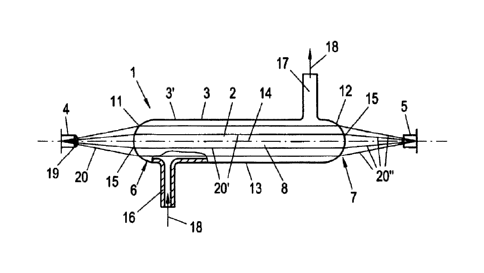
In contrast, cuvette 3 of a first embodiment of the invention, as depicted in
Fig. 2, has
an inlet section 6 with a convexly curved inlet surface 11, which focuses the
incident
radiation impinging upon the convex inlet surface 11 in the manner of a
converging lens.
Correspondingly, the outlet section 7 of cuvette 3 has a convexly curved
outlet surface 12 in
order to focus the transmitted radiation as it is coupled out from the cuvette
3. The curvature
of the inlet surface 11 and/or the outlet surface 12 is convex with respect to
the inner cavity
of cuvette 3, relative to which the inlet surface 11 and/or outlet surface 12
are curved
outwardly. The convexly curved inlet surface 11 and/or outlet surface 12 thus
focus the
CA 02820995 2013-06-10
8
radiation impinging upon the respective surface, reducing the fanning out of
the beam
bundle. Thus, cuvette 3 directly takes over the tasks of an optical system,
which was formed
by separate optical components in earlier devices 1. The beam formation is
thus
accomplished by the convexly curved inlet surface 11 and/or outlet surface 12,
which are
integrated into cuvette 3 so that a compact photometric device 1 is made
available without
the need for elaborate installations and adjustments plus costly additional
optical
components. This is particularly advantageous for applications involving
excitation radiation
in the ultraviolet (UV) or infrared (IR) ranges, as these require special
glasses which are
elaborate and expensive to produce.
Cuvette 3, depicted in Fig. 2, is shaped as a longitudinal, substantially
cylindrical liquid
cell 13, through which radiation passes along its longitudinal axis 14. This
cuvette 3 displays
two end surfaces 15 arranged transversely to its longitudinal axis 14; these
are convexly
curved inlet surface 11 and convexly curved outlet surface 12. Cuvette 3 is
configured as a
flow through cuvette 3', equipped with a supply line 16 to introduce liquid
sample 2 into liquid
cell 13. The cuvette 3 is also equipped with a discharge line 17 to discharge
examined liquid
sample 2 from liquid cell 13. The flow through cuvette 3' enables continuous
examination of
the chemical parameters of liquid sample 2. As is further apparent from Fig.
2, liquid sample
2 is introduced into the liquid cell 13 in arrow direction from below with
respect to the
operating position of cuvette 3and is discharged, after having passed through
liquid cell 13,
upwardly through discharge line 17. This configuration considerably reduces
the formation of
air inclusions, which would hamper examination of liquid sample 2. For this
purpose, the
embodiment provides, in particular, for the discharge line 17 to be connected
to the flow
through cuvette 3' at the uppermost position with respect to the operating
position of the
cuvette.
Radiation source 4, which is expediently configured as a cost effective light
emitting
diode (LED) 19 for a whole range of wavelengths, produces a divergent beam
bundle 20,
which is focused into a substantially parallel beam bundle 20' by means of the
convexly
curved inlet surface 11. In this way, excitation radiation passes through a
substantially larger
sample volume 8 than in conventional configurations. After having passed
through sample
volume 8, the substantially parallel beam bundle 20' is concentrated by means
of the
convexly curved outlet surface 12 , into a convergent beam bundle 20", which
is focused on
the sensor surface of radiation detector 5. Accordingly, the excitation
radiation is used very
efficiently and essentially the entire content of the liquid cell 13 is
measured as a sample
volume 8.
CA 02820995 2013-06-10
9
Fig. 3 shows an alternative embodiment of device 1, wherein cuvette 3 displays
a liquid
cell 13' through which radiation passes essentially transversely to the cell's
longitudinal axis
21. Liquid cell 13' may be of cylindrical or generally rectangular shape.
Depending on the
application, cuvette 3 may be configured as a flow through cuvette 3' or as a
cuvette into
which the reagent is introduced prior to examination. Each of the convexly
curved surfaces,
inlet surface 11 and outlet surface 12, is formed on a lateral surface 22 of
the cuvette 3. In
this embodiment, too, the convexly curved inlet surface 11 and outlet surface
12 produce the
effect of a converging lens, particularly of a biconvex converging lens, so
that, by means of
cuvette 3, useful imaging of the excitation radiation is obtained.
In Fig. 4, another embodiment of device 1 according to the invention is shown,
wherein
cuvette 3 displays a stronger convex curvature compared to the previously
mentioned
exemplary embodiments. Here, the divergent excitation radiation is directly
focused into a
convergent beam bundle directly on being coupled into cuvette 3; this bundle
has a
comparably small focal area 23 in the sample volume 8. This embodiment enables
very high
energy density to be transferred to the sample volume 8. This is of advantage
in that a
comparably cost effective light emitting diode 19 may be used as a radiation
source 4 instead
of a typically employed laser.
Finally, in Fig. 5, an embodiment of device 1 corresponding to the embodiment
of Fig. 4
is shown, wherein a second convexly curved outlet window 12' arranged
substantially
perpendicular to the inlet surface 11 and/or outlet surface 12 is provided.
This embodiment of
cuvette 3 enables analytical procedures to be conducted in the manner of flow
through
cytometry. A forwardly scattered beam bundle , i.e. forward scatter light 24,
is coupled out
via outlet surface 12 and is detected by a forward scatter light detector 26.
Additionally, a
beam bundle scattered sidewards , i.e. sideward scatter light 25, is coupled
out via outlet
surface 12' and is detected by a sideward scatter light detector 27. Flow
through cytometry is
used to examine cell suspensions, for example, which are directed in a thin
spurt through
channel 28 of cuvette 3 (comp. also Fig. 4). In an alternative embodiment (not
shown),
cuvette 3 may have a substantially circular cross section. In addition,
cuvette 3 may display
at least a third outlet window (not shown), which preferably is arranged
opposite to outlet
window 12'. A further sideward scatter light detector may be configured with
the third outlet
window, which detects transversely scattered sideward scatter light, just as
sideward scatter
light detector 27 does.
The radius of curvature of the convexly curved inlet surface 11 and/or outlet
surface 12
has to be adapted to the desired focusing of the excitation radiation or the
transmitted
radiation, depending on the application. With respect to cost effective
production, spherically
CA 02820995 2013-06-10
curved inlet surface(s) 11 and/or outlet surface(s) 12, 12' are appropriate.
In applications with
high demands on imaging accuracy it may be favorable to configure the inlet
surface 11
and/or outlet surface(s) 12, 12' in the form of spherical surfaces in order to
avoid lens errors.
Fig. 6 and 7 show a longitudinal and a cross-sectional view of a flow through
cuvette 3'
respectively, which features a favorable configuration of supply line 16 and
discharge line 17
with respect to the flow conditions within liquid cell 13. As is evident from
Fig. 6, both, a
longitudinal axis 16' of supply line 16 and a longitudinal axis 17' of
discharge line 17 are
inclined relative to the longitudinal axis 14 of liquid cell 13. As is evident
from Fig. 7, both the
longitudinal axis 16' of supply line 16 and the longitudinal axis 17' of
discharge line 17 are
furthermore arranged at an inclination angle with respect to a transverse axis
29 of liquid cell
13. In this embodiment, liquid sample 2 is supplied to and discharged from
liquid cell 13
essentially tangentially, thereby achieving better blending of liquid sample 2
and reduced
turbulence in the liquid flow.
In Fig. 8, an alternative embodiment of flow through cuvette 3' is shown,
wherein
supply line 16 and discharge line 17 each comprise two portions 16a, 16b and
17a, 17b
respectively, having different cross sectional areas. Accordingly, supply line
16 has a portion
16a running in the direction of the longitudinal axis 14 of liquid cell 13 and
ending in a portion
16b aligned at right angles to it. This latter portion, though which liquid
sample 2 is supplied
to liquid cell 13, has a larger cross-section than portion 16a. Portion 17b of
discharge line 17,
which follows on liquid cell 13, is cross sectionally larger than the
downstream portion 17a of
discharge line 17, onto which portion 17b of discharge line 17 adjoins in
longitudinal
direction.
In Fig. 9, a configuration for conducting photometric or spectrometric
examinations is
shown schematically, comprising a cuvette 3 which contains liquid sample 2, a
radiation
source 4 and two separate radiation detectors 5, which detect different or
complementary
interactions of coupled in radiation with liquid sample 2. Additionally, a
reference sensor 30 is
provided for calibrating the measuring signal. In the configuration shown in
Fig. 9, a dichroic
mirror 31 is provided for dividing the radiation emitted by radiation source
4, which mirror
reflects a part of the light spectrum into the direction of inlet section 6
and lets the other
wavelength ranges through.
In Fig. 10, an alternative configuration for conducting photometric or
spectrometric
examinations is shown, which provides for a translucent mirror 32 instead of
the dichroic
mirror 31 shown in Fig. 9, which directs a part of the radiation emitted by
radiation source 4
to reference sensor 30 while the transmitted part of the radiation impinges
upon the convexly
= CA 02820995 2013-06-10
11
curved end surface 15 of inlet section 6. To detect the radiation interacting
with liquid sample
2, radiation detectors 5 are arranged in the areas of inlet section 6 and
outlet section 7.