Note: Descriptions are shown in the official language in which they were submitted.
CA 02821981 2016-12-12
CA 2821981
DEVICES, SYSTEMS AND METHODS FOR THE TREATMENT OF
MEDICAL DISORDERS
CROSS REFERENCE TO RELATED APPLICATIONS
[0001] This application claims priority to the following applications: U.S.
Application
No. 61/423,011, entitled "Devices, Systems and Methods for Treatment of
Neuropsychiatric
Disorders." filed on December 14, 2010; U.S. Application No. 61/440,784,
entitled "Devices,
Systems and Methods for Treatment of Cardiac Related Disorders," filed on
February 8, 2011;
U.S. Application No. 61/445,505, entitled -Devices, Systems and Methods for
Treatment of
Fatigue and Other Medical Disorders," filed on February 22, 2011; and U.S.
Application No.
61/479,787, entitled "Devices, Systems and Methods for Treatment of Medical
Disorders,"
filed on April 27, 2011.
[0002] This application is related to U.S. Application No. 12/898,686,
entitled
-Devices, Systems and Methods for Treatment of Neuropsychiatric Disorders,"
filed on Oct. 5,
2010, which claims priority to the following applications: U.S. Application
No. 61/248,827,
entitled "Devices and Methods for Treatment of Psychiatric Disorders," filed
October 5, 2009;
U.S. Application No. 61/289,829, entitled "Extracranial Implantable Devices,
Systems and
Methods for Treatment of Neuropsychiatric Disorders," filed December 23, 2009;
U.S.
Application No. 61/305,514, entitled "Systems, Devices and Methods for
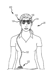
Treatment of
Neurological Disorders and Conditions," filed February 17, 2010; and U.S.
Application No.
61/354,641, entitled "Extracranial Implantable Devices, Systems and Methods
for Treatment of
Neurological Disorders." filed June 14, 2010.
[0003] This application is related to U.S. Application No. 12/898,675,
entitled
-Devices, Systems and Methods for Treatment of Neurological Disorders," filed
on Oct. 5,
2010, which claims priority to the following applications: U.S. Application
No. 61/248,827,
entitled "Devices and Methods for Treatment of Psychiatric Disorders," filed
October 5, 2009;
U.S. Application No. 61/289,829, entitled "Extracranial Implantable Devices,
Systems and
Methods for Treatment of Neuropsychiatric Disorders," filed December 23, 2009;
U.S.
Application No. 61/305,514, entitled "Systems, Devices and Methods for
Treatment of
Neurological Disorders and Conditions," filed February 17, 2010; and U.S.
Application No.
- 1 -
CA 02821981 2016-12-12
=
CA 2821981
61/354,641, entitled "Extracranial Implantable Devices, Systems and Methods
for Treatment of
Neurological Disorders," filed June 14, 2010
[0004] This application is related to WO 2012/082961, entitled
"Extracranial
Implantable Devices, Systems and Methods for Treatment of Medical Disorders,"
filed on even
date herewith.
FIELD
[0005] The present disclosure generally relates to cutaneous
neuromodulation devices
and systems and methods of using the same. More specifically, methods,
devices, and systems
configured for the treatment of medical disorders, such as neuropsychiatric
disorders including
mood, cognitive and behavioral disorders, heart disease and other cardiac
related disorders, and
fatigue, via trigeminal nerve stimulation ("TNS") are provided. Devices and
systems
configured for stimulation of superficial sensory branches of cranial nerves
and their methods
of application are described.
BACKGROUND
[0006] Many medical disorders, including neuropsychiatric disorders,
cardiac related
disorders and fatigue are traditionally treated with pharmacotherapy and/or
psychotherapy.
However, a substantial percentage of patients with these and other conditions
do not recover
despite multiple trials of treatment and there may be significant and long
term side effects to
the traditional treatment methods.
[0007] For example, interventions for fatigue commonly employ
medications,
particularly psychostimulant medications. Such medications include
methylphenidate,
amantadine, pemoline, and modafinil (reviewed by Peuckmann et al., Cochrane
Database Syst
Rev 2010, 11:CD006788). These medications carry potential for side effects,
such as blurred
vision, depression or anxiety, liver failure, psychosis, suicidal thinking,
swelling of the
hands/leg/feet, shortness of breath, palpitations, elevated blood pressure,
anorexia and
- 2 -
CA 02821981 2013-06-14
WO 2012/082960 PCT/US2011/065002
addiction.
[0008] For some medical disorders, brain stimulation has been a primary
treatment
alternative, and electroconvulsive therapy (ECT, or "electroshock" therapy)
has been the
dominant brain stimulation approach since the first part of the 20th century.
ECT carries
risks of memory and other cognitive side effects, considerable cost, and risks
of anesthesia.
Two implantable approaches have also been described: deep brain stimulation
(DBS), in
which electrodes are implanted directly within the brain, and vagus nerve
stimulation (VNS)
in which stimulating electrodes are implanted on the vagus nerve in the neck.
While the U.S.
Food and Drug Administration (FDA) have approved systems for deep brain
stimulation for
the treatment of essential tremor, Parkinson's disease, dystonia and obsessive
compulsive
disorder, DBS is presently an experimental intervention for other
neuropsychiatric conditions.
The risks of DBS include infection, hemorrhage, and injury to deep brain
structures. In
reports of clinical studies with VNS, many of the patients who undergo VNS
treatments do
not achieve remission, and there is no reliable predictor of good outcomes
from the implanted
VNS device.
[0009] Against this backdrop, the present disclosure is provided.
[0010] The information included in this Background section of the
specification,
including any references cited herein and any description or discussion
thereof, is included
for technical reference purposes only and is not to be regarded as subject
matter by which the
scope of the invention is to be bound.
SUMMARY
[0011] One aspect of the subject matter of the present disclosure addresses
the
aforementioned needs by providing a method of treating medical disorders, and
systems and
devices configured to stimulate the ophthalmic (supraorbital), infraorbital
and mentalis
branch(es) of the trigeminal nerve to treat medical disorders.
[0012] In another aspect of the present disclosure, there is provided an
electrode
assembly configured for the cutaneous stimulation of the trigeminal nerve.
[0013] In yet another aspect of the present disclosure, a method of
treating medical
disorders using the disclosed electrode assembly is provided.
[0014] In one aspect, a system for trigeminal nerve stimulation for
treatment of a
medical disorder is provided. The system includes a pulse generator and a
cutaneous
- 3 -
CA 2821981
electrode assembly in electrical communication with the pulse generator. In
one aspect, the
assembly includes a first electrode comprising at least one contact configured
for cutaneous
placement at a first region of a patient's face, wherein the first electrode
is configured to
contact a portion of the patient's face overlying the cutaneous distribution
of at least one branch
of the trigeminal nerve to stimulate the trigeminal nerve to modulate at least
one body system
for treatment of a medical disorder, wherein the at least one branch of the
trigeminal nerve is
selected from the group consisting of: ophthalmic nerve, infraorbital nerve,
mentalis nerve,
supratrochlear nerve, supraorbital nerve, infratrochlear nerve,
zygomaticotemporal nerve,
zygomaticofacial nerve, zygomaticoorbital nerve, nasal nerve, and
auriculotemporal nerve, and
wherein the medical disorder is selected from the group consisting of: cardiac
related disorders,
fatigue, tinnitus, obesity, diabetes, dyslipidemia, metabolic syndrome,
obstructive sleep apnea,
arthritis, cachexia/anorexia, inflammation, asthma, inflammatory bowel
disease, atopic
dermatitis, sepsis, hepatitis, disorders of regulation of breathing, disorders
of gastrointestinal
function, gastroesophageal reflux, diarrhea and constipation, dysphagia and
other disturbances
of swallowing, gastroparesis, functional bowel syndromes, post-operative
ileus, dyspepsia,
motion sickness, chemotherapy-related nausea and emesis, autonomic regulation
in menopausal
hot flashes, regulation of hemostasis, sleep/insomnia and a neuropsychiatric
disorder selected
from the group consisting of attention deficit disorder (ADD), attention
deficit hyperactivity
disorder (ADHD), autism and autism spectrum disorders (ASD), substance use
disorders and
related behavioral addictions, eating disorders and obsessive compulsive
disorder (OCD),
psychotic disorders, dementing disorders, or a combination thereof
[0015] In one aspect, the medical disorder is a cardiac related disorder
selected from the
group consisting of heart disease, cardiac arrhythmias, myocardial infarction,
sudden cardiac
death after myocardial infarction, heart failure, cerebral ischemia, sudden
infant death
syndrome (SIDS), impaired blood flow conditions, atrial fibrillation or sudden
death in
epilepsy. The at least one branch of the trigeminal nerve is an ophthalmic
nerve or an
infraorbital nerve, wherein the body system is a trigeminal nerve cardiac
reflex and wherein
stimulation of the ophthalmic nerve or the infraorbital nerve modulates or
activates the
trigeminal nerve cardiac reflex to treat or prevent a cardiac related
disorder. In one aspect, the
body system is a vagus nerve circuit, and wherein stimulation of the at least
one branch of the
trigeminal nerve modulates the vagus nerve circuit to treat a
- 4 -
CA 2821981 2018-03-16
CA 02821981 2013-06-14
WO 2012/082960 PCT/US2011/065002
cardiac related disorder. In one aspect, the medical disorder is fatigue,
wherein the body
system is a locus coeruleus or a reticular activating system, and wherein
stimulation of the at
least one branch of the trigeminal nerve modulates the locus coeruleus or
modulates the
reticular activating system to treat fatigue. In one aspect, the medical
disorder is selected
from the group consisting of obesity and other disorders related to weight and
feeding,
inflammation, disorders of regulation of breathing, disorders of
gastrointestinal function,
autonomic regulation in menopausal hot flashes, regulation of hemostasis and
sleep/insomnia,
wherein the body system is a vagus nerve circuit, and wherein stimulation of
the at least one
branch of the trigeminal nerve modulates the vagus nerve circuit to treat said
medical
disorder. In one aspect, the medical disorder is a dementing disorder wherein
the body system
is a vagus nerve circuit or a trigeminal nerve cardiac reflex, and wherein
stimulation of the at
least one branch of the trigeminal nerve modulates the vagus nerve circuit or
the trigeminal
nerve cardiac reflex to treat said medical disorder.
[0016] In one aspect, the assembly further comprises a second electrode
comprising at
least one contact configured for cutaneous placement at a second region of the
patient's face,
wherein the second electrode is configured to contact a portion of the
patient's face overlying
the cutaneous distribution of at least one branch of the trigeminal nerve,
wherein the at least
one branch of the trigeminal nerve is selected from the group consisting of:
ophthalmic nerve,
infraorbital nerve, supraorbital nerve, mentalis nerve, supratrochlear nerve,
infratrochlear
nerve, zygomaticotemporal nerve, zygomaticofacial nerve, zygomaticoorbital
nerve, nasal
nerve, and auriculotemporal nerve. In one embodiment, first electrode and the
second
electrode are configured to contact a portion of the patient's face overlying
the cutaneous
distribution of a same branch of the trigeminal nerve. In another embodiment,
the first
electrode and the second electrode are configured to contact a portion of the
patient's face
overlying the cutaneous distribution of a different branch of the trigeminal
nerve. The
stimulation may be provided uni- or bilaterally.
[0017] In one aspect, the system is configured for minimal current
penetration into a
brain of a patient. The system may further include a closed loop device
configured to provide
self-tuning adaptive feedback control to the system. In one embodiment,
stimulation of the at
least one branch of the trigeminal nerve is determined based on measurement of
activity in a
brain region to detect an acute biological change. In one embodiment, the at
least one branch
of the trigeminal nerve is stimulated at a first set of stimulation parameters
for a first time
- 5 -
CA 02821981 2013-06-14
WO 2012/082960 PCT/US2011/065002
period, at a second set of stimulation parameters for a second time period,
and at a third set of
stimulation parameters for a third time period. In one embodiment, the at
least one branch of
the trigeminal nerve is stimulated at the first, second and third set of
parameters in a cycle at
least twice. In one embodiment, the pulse generator is configured to apply
electrical signals at
a frequency between approximately 1 and 300 Hertz, at a pulse duration between
approximately 50 and 500 microseconds, at an output current density of not
greater than
approximately 10 mA/cm2and an output charge density of not greater than
approximately 10
microCoulomb/cm2 at the cerebral cortex.
[0018] In one aspect, a cutaneous electrode assembly for trigeminal nerve
stimulation
for treatment of a medical disorder is provided. The assembly includes a first
electrode
comprising at least one contact configured for cutaneous placement at a first
region of the
patient's face, wherein the first electrode is configured to contact a portion
of the patient's
face overlying the cutaneous distribution of at least one branch of the
trigeminal nerve to
stimulate the trigeminal nerve to modulate at least one body system for
treatment of a
medical disorder, wherein the at least one branch of the trigeminal nerve is
selected from the
group consisting of: ophthalmic nerve, infraorbital nerve, mentalis nerve,
supratrochlear
nerve, supraorbital nerve, infratrochlear nerve, zygomaticotemporal nerve,
zygomaticofacial
nerve, zygomaticoorbital nerve, nasal nerve, and auriculotemporal nerve, and
wherein the
medical disorder is selected from the group consisting of: cardiac related
disorders, fatigue,
tinnitus, obesity, diabetes, dyslipidemia, metabolic syndrome, obstructive
sleep apnea,
arthritis, cachexia/anorexia, inflammation, asthma, inflammatory bowel
disease, atopic
dermatitis, sepsis, hepatitis, disorders of regulation of breathing, disorders
of gastrointestinal
function, gastrocsophageal reflux, diarrhea and constipation, dysphagia and
other
disturbances of swallowing, gastroparcsis, functional bowel syndromes, post-
operative ileus,
dyspepsia, motion sickness, chemotherapy-related nausea and emesis, autonomic
regulation
in menopausal hot flashes, regulation of hemostasis, sleep/insomnia and a
neuropsychiatric
disorder selected from the group consisting of attention deficit disorder
(ADD), attention
deficit hyperactivity disorder (ADHD), autism and autism spectrum disorders
(ASD),
substance use disorders and related behavioral addictions, eating disorders
and obsessive
compulsive disorder (OCD), psychotic disorders, dementing disorders, or a
combination
thereof. In one embodiment, the assembly may further include a second
electrode comprising
at least one contact configured for cutaneous placement at a second region of
the patient's
- 6 -
CA 02821981 2013-06-14
WO 2012/082960 PCT/US2011/065002
face, wherein the second electrode is configured to contact a portion of the
patient's face
overlying the cutaneous distribution of at least one branch of the trigeminal
nerve, wherein
the at least one branch of the trigeminal nerve is selected from the group
consisting of:
ophthalmic nerve, supraorbital nerve, infraorbital nerve, mentalis nerve,
supratrochlear nerve,
infratrochlear nerve, zygomaticotemporal nerve, zygomaticofacial nerve,
zygomaticoorbital
nerve, nasal nerve, and auriculotemporal nerve. In some embodiments, the first
electrode and
the second electrode are configured to contact a portion of the patient's face
overlying the
cutaneous distribution of a same branch of the trigeminal nerve. In some
embodiments, the
first electrode and the second electrode are configured to contact a portion
of the patient's
face overlying the cutaneous distribution of a different branch of the
trigeminal nerve. In one
embodiment, the medical disorder is a cardiac related disorder selected from
the group
consisting of heart disease, cardiac arrhythmi as, myocardial infarction,
sudden cardiac death
after myocardial infarction, heart failure, cerebral ischemia, sudden infant
death syndrome
(SIDS), impaired blood flow conditions, atrial fibrillation or sudden death in
epilepsy. In one
embodiment, the body system is a vagus nerve circuit, and wherein stimulation
of the at least
one branch of the trigeminal nerve modulates the vagus nerve circuit to treat
a cardiac related
disorder. In one embodiment, the medical disorder is fatigue, wherein the body
system is a
locus coeruleus or a reticular activating system, and wherein stimulation of
the at least one
branch of the trigeminal nerve modulates the locus coeruleus or modulates the
reticular
activating system to treat fatigue. In one embodiment, the medical disorder is
selected from
the group consisting of obesity and other disorders related to weight and
feeding,
inflammation, disorders of regulation of breathing, disorders of
gastrointestinal function,
autonomic regulation in menopausal hot flashes, regulation of hemostasis and
sleep/insomnia,
wherein the body system is a vagus nerve circuit, and wherein stimulation of
the at least one
branch of the trigeminal nerve modulates the vagus nerve circuit to treat said
medical
disorder. In one embodiment, the medical disorder is a dementing disorder
wherein the body
system is a vagus nerve circuit or a trigeminal nerve cardiac reflex, and
wherein stimulation
of the at least one branch of the trigeminal nerve modulates the vagus nerve
circuit or the
trigeminal nerve cardiac reflex to treat said medical disorder. In one
embodiment, the
assembly produces minimal current penetration into a brain of a patient.
[0019] In one aspect, a method for treating a medical disorder by
trigeminal nerve
stimulation is provided. The method includes contacting a first region of a
patient's face with
- 7 -
CA 02821981 2013-06-14
WO 2012/082960 PCT/US2011/065002
a cutaneous electrode assembly with at least one branch of the trigeminal
nerve to stimulate
the trigeminal nerve for treatment of a medical disorder and applying
electrical signals to the
electrode assembly to stimulate the at least one branch of the trigeminal
nerve to modulate a
system of the patient's body for treatment of a medical disorder. In one
embodiment, the
cutaneous electrode assembly includes a first electrode comprising at least
one contact
configured for cutaneous placement at a first region of the patient's face,
wherein the first
electrode contacts a portion of the patient's face overlying the cutaneous
distribution of at
least one branch of the trigeminal nerve. The at least one branch of the
trigeminal nerve is
selected from the group consisting of: ophthalmic nerve, supraorbital nerve,
infraorbital
nerve, mentalis nerve, supratrochlear nerve, infratrochlear nerve,
zygomaticotemporal nerve,
zygomaticofaci al nerve, zygomaticoorbital nerve, nasal nerve, and
auriculotemporal nerve.
The medical disorder is selected from the group consisting of: cardiac related
disorders,
fatigue, tinnitus, obesity, diabetes, dyslipidemia, metabolic syndrome,
obstructive sleep
apnea, arthritis, cachexialanorexia, inflammation, asthma, inflammatory bowel
disease, atopic
dermatitis, sepsis, hepatitis, disorders of regulation of breathing, disorders
of gastrointestinal
function, gastroesophageal reflux, diarrhea and constipation, dysphagia and
other
disturbances of swallowing, gastroparesis, functional bowel syndromes, post-
operative ileus,
dyspepsia, motion sickness, chemotherapy-related nausea and emesis, autonomic
regulation
in menopausal hot flashes, regulation of hemostasis, sleep/insomnia and a
neuropsychiatric
disorder selected from the group consisting of attention deficit disorder
(ADD), attention
deficit hyperactivity disorder (ADHD), autism and autism spectrum disorders
(ASD),
substance use disorders and related behavioral addictions, eating disorders
and obsessive
compulsive disorder (OCD), psychotic disorders, dementing disorders, or a
combination
thereof. In one embodiment, the method may further include a second electrode
comprising
at least one contact configured for cutaneous placement at a second region of
the patient's
face, wherein the second electrode is configured to contact a portion of the
patient's face
overlying the cutaneous distribution of at least one branch of the trigeminal
nerve, wherein
the at least one branch of the trigeminal nerve is selected from the group
consisting of:
ophthalmic nerve, supraorbital nerve, infraorbital nerve, mentalis nerve,
supratrochlear nerve,
infratrochlear nerve, zygomaticotemporal nerve, zygomaticofacial nerve,
zygomaticoorbital
nerve, nasal nerve, and auriculotemporal nerve. In one embodiment, step of
applying
electrical signals comprises applying electrical signals at a frequency
between approximately
- 8 -
CA 02821981 2013-06-14
WO 2012/082960 PCT/US2011/065002
20 and 300 Hertz, at a current of 0.05 to 5 milliamperes (mA) and at a pulse
duration of less
than or equal to 500 microseconds. In one embodiment, the step of applying
electrical signals
comprises applying electrical signals at a frequency between approximately 20
and 300
Hertz, at a pulse duration between approximately 50 and 500 microseconds, at
an output
current density of not greater than approximately 10 mA/cm2 and a charge
density of not
greater than approximately 10 microCoulomb/cm2 at the cerebral cortex. In one
embodiment,
the step of applying electrical signals comprises applying electrical signals
at an output
current density of not greater than approximately 10 mA/cm2. In one
embodiment, the step
of applying electrical signals comprises applying electrical signals at an
output current
density of between approximately 2.5 and 5 mA/cm2. In one embodiment, the step
of
applying electrical signals comprises applying electrical signals at an output
current density
of not greater than approximately 7 mA/cm2 In one embodiment, the step of
applying
electrical signals comprises applying electrical signals at an output current
density of not
greater than approximately 5 mA/cm2. In one embodiment, the step of applying
electrical
signals comprises applying electrical signals to minimize current penetration
into a brain of a
patient. In one embodiment, the medical disorder is a cardiac related disorder
selected from
the group consisting of heart disease, cardiac arrhythmias, myocardial
infarction, sudden
cardiac death after myocardial infarction, heart failure, cerebral ischemia,
sudden infant death
syndrome (SIDS), impaired blood flow conditions, atrial fibrillation or sudden
death in
epilepsy. In one embodiment, the body system is a vagus nerve circuit, and
wherein
stimulation of the at least one branch of the trigeminal nerve modulates the
vagus nerve
circuit to treat a cardiac related disorder. In one embodiment, the medical
disorder is fatigue,
wherein the body system is a locus coeruleus or a reticular activating system,
and wherein
stimulation of the at least one branch of the trigeminal nerve modulates the
locus cocruleus or
modulates the reticular activating system to treat fatigue. In one embodiment,
the medical
disorder is selected from the group consisting of obesity and other disorders
related to weight
and feeding, inflammation, disorders of regulation of breathing, disorders of
gastrointestinal
function, autonomic regulation in menopausal hot flashes, regulation of
hemostasis and
sleep/insomnia, wherein the body system is a vagus nerve circuit, and wherein
stimulation of
the at least one branch of the trigeminal nerve modulates the vagus nerve
circuit to treat said
medical disorder. In one embodiment, the medical disorder is a dementing
disorder wherein
the body system is a vagus nerve circuit or a trigeminal nerve cardiac reflex,
and wherein
- 9 -
CA 02821981 2013-06-14
WO 2012/082960 PCT/US2011/065002
stimulation of the at least one branch of the trigeminal nerve modulates the
vagus nerve
circuit or the trigeminal nerve cardiac reflex to treat said medical disorder.
[0020] In one aspect, a kit for trigeminal nerve stimulation for treatment
of a medical
disorder. In one embodiment, the kit includes an electrode assembly as
disclosed elsewhere
herein and instructions for applying the electrode assembly to a patient for
treatment of a
medical disorder, wherein the medical disorder is selected from the group
consisting of:
cardiac related disorders, fatigue, tinnitus, obesity, diabetes, dyslipidemia,
metabolic
syndrome, obstructive sleep apnea, arthritis, cachexia/anorexia, inflammation,
asthma,
inflammatory bowel disease, atopic dermatitis, sepsis, hepatitis, disorders of
regulation of
breathing, disorders of gastrointestinal function, gastroesophageal reflux,
diarrhea and
constipation, dysphagia and other disturbances of swallowing, gastroparesis,
functional bowel
syndromes, post-operative ileus, dyspepsia, motion sickness, chemotherapy-
related nausea
and emesis, autonomic regulation in menopausal hot flashes, regulation of
hemostasis,
sleep/insomnia and a neuropsychiatric disorder selected from the group
consisting of
attention deficit disorder (ADD), attention deficit hyperactivity disorder
(ADHD), autism and
autism spectrum disorders (ASD), substance use disorders and related
behavioral addictions,
eating disorders and obsessive compulsive disorder (OCD), psychotic disorders,
dementing
disorders or a combination thereof. The kit may also include a pulse generator
and
instructions for applying electrical signals to the electrode assembly for
treatment of a
medical disorder.
[0021] In one aspect, a method for initiation, activation or stimulation of
a vagus
nerve circuit by trigeminal nerve stimulation for treatment of a medical
disorder is provided.
The method may include contacting a first region of a patient's face with a
cutaneous
electrode assembly with at least one branch of the trigeminal nerve to
stimulate the trigeminal
nerve for treatment of a medical disorder and applying electrical signals to
the electrode
assembly to stimulate the at least one branch of the trigeminal nerve to
modulate the vagus
nerve circuit for treatment of a medical disorder which may benefit from vagus
nerve
stimulation via the trigeminal nerve. The cutaneous electrode assembly
includes a first
electrode comprising at least one contact configured for cutaneous placement
at a first region
of the patient's face. The at least one branch of the trigeminal nerve is an
ophthalmic nerve
or an infraorbital nerve. In one embodiment, the medical disorder is a cardiac
related
disorder selected from the group consisting of heart disease, cardiac
arrhythmias, myocardial
- 10 -
CA 02821981 2013-06-14
WO 2012/082960
PCT/US2011/065002
infarction, sudden cardiac death after myocardial infarction, heart failure,
cerebral ischemia,
sudden infant death syndrome (SIDS), impaired blood flow conditions, atrial
fibrillation or
sudden death in epilepsy. In one embodiment, the medical disorder is selected
from the
group consisting of obesity and other disorders related to weight and feeding,
inflammation,
disorders of regulation of breathing, disorders of gastrointestinal function,
autonomic
regulation in menopausal hot flashes, regulation of hemostasis and
sleep/insomnia, and
wherein stimulation of the at least one branch of the trigeminal nerve
modulates the vagus
nerve circuit to treat said medical disorder. In one embodiment, the medical
disorder is a
dementing disorder and wherein stimulation of the at least one branch of the
trigeminal nerve
modulates the vagus nerve circuit to treat said medical disorder.
[0022] In one aspect, a behind the ear device for polycranial nerve
stimulation for
treatment of a medical disorder is provided. The device includes an external
ear body
including a pulse generator and a battery and an ear canal body including an
electrode
assembly in electrical communication with the pulse generator. The electrode
assembly
includes at least one electrode comprising at least one contact configured to
contact the
cutaneous distribution of at least one branch of the trigeminal nerve at, in
or about a patient's
ear, and stimulation of the at least one branch of the trigeminal nerve
modulates a system in
the body to treat a medical disorder. In one embodiment, the at least one
branch of the
trigeminal nerve is selected from the group consisting of: ophthalmic nerve,
infraorbital
nerve, supraorbital nerve, mentalis nerve, supratrochlear nerve,
infratrochlear nerve,
zygomaticotemporal nerve, zygomaticofacial nerve, zygomaticoorbital nerve,
nasal nerve,
and auriculotemporal nerve. In one embodiment, the device further includes a
second
electrode comprising at least one contact configured for subcutaneous or
percutaneous
placement at a second region of the patient's face, wherein the second
electrode is
configured to be implanted in proximity to, adjacent to or in contact with at
least one branch
of the trigeminal nerve, wherein the at least one branch of the trigeminal
nerve is selected
from the group consisting of: ophthalmic nerve, infraorbital nerve,
supraorbital nerve,
mentalis nerve, supratrochlear nerve, infratrochlear nerve, zygomaticotemporal
nerve,
zygomaticofacial nerve, zygomaticoorbital nerve, nasal nerve, and
auriculotemporal nerve. In
one embodiment, the first electrode and the second electrode are configured
for implantation
in proximity to, adjacent to or in contact with a same branch of the
trigeminal nerve. In one
embodiment, the first electrode and the second electrode are configured for
implantation in
-11-
CA 02821981 2013-06-14
WO 2012/082960 PCT/US2011/065002
proximity to, adjacent to or in contact with a different branch of the
trigeminal nerve. The
device produces minimal current penetration into a brain of a patient. The
device may further
include a closed loop device configured to provide self-tuning adaptive
feedback control to
the system. Stimulation of the at least one branch of the trigeminal nerve is
determined based
on measurement of activity in a brain region to detect an acute biological
change. In one
embodiment, the at least one branch of the trigeminal nerve is stimulated at a
first set of
stimulation parameters for a first time period, at a second set of stimulation
parameters for a
second time period, and at a third set of stimulation parameters for a third
time period. In one
embodiment, the at least one branch of the trigeminal nerve is stimulated at
the first, second
and third set of parameters in a cycle at least twice. In one embodiment, the
pulse generator
is configured to apply electrical signals at a frequency between approximately
1 and 300
hertz, at a pulse duration between approximately 50 and 500 microseconds, at
an output
current density of not greater than approximately 10 mA/cm2 and an output
charge density of
not greater than approximately 10 microCoulomb/cm2 at the cerebral cortex. In
one
embodiment, the pulse generator is configured to apply electrical signals at
an output current
density of not greater than approximately 10 mA/cm2. In one embodiment, the
pulse
generator is configured to apply electrical signals at an output current
density of between
approximately 2.5 and 5 mA/cm2. In one embodiment, the pulse generator is
configured to
apply electrical signals at an output current density of not greater than
approximately 7
mA/cm2. In one embodiment, the pulse generator is configured to apply
electrical signals at
an output current density of not greater than approximately 5 mAlcm2. In one
embodiment,
the medical disorder is selected from the group consisting of: neurological
disorders, cardiac
related disorders, fatigue, tinnitus, obesity, diabetes, dyslipidemia,
metabolic syndrome,
obstructive sleep apnea, arthritis, cachexia/anorexia, inflammation, asthma,
inflammatory
bowel disease, atopic dermatitis, sepsis, hepatitis, disorders of regulation
of breathing,
disorders of gastrointestinal function, gastroesophageal reflux, diarrhea and
constipation,
dysphagia and other disturbances of swallowing, gastroparesis, functional
bowel syndromes,
post-operative ileus, dyspepsia, motion sickness, chemotherapy-related nausea
and emesis,
autonomic regulation in menopausal hot flashes, regulation of hemostasis,
sleep/insomnia and
a neuropsychiatric disorder selected from the group consisting of depression,
attention deficit
disorder (ADD), attention deficit hyperactivity disorder (ADHD), autism and
autism
spectrum disorders (ASD), substance use disorders and related behavioral
addictions, eating
- 12 -
CA 02821981 2013-06-14
WO 2012/082960
PCT/US2011/065002
disorders and obsessive compulsive disorder (OCD), psychotic disorders,
dementing
disorders, or a combination thereof. In one embodiment, the medical disorder
is a cardiac
related disorder selected from the group consisting of heart disease, cardiac
arrhythmias,
myocardial infarction, sudden cardiac death after myocardial infarction, heart
failure, cerebral
ischemia, sudden infant death syndrome (SIDS), impaired blood flow conditions,
atrial
fibrillation or sudden death in epilepsy. In one embodiment, the at least one
branch of the
trigeminal nerve is an ophthalmic nerve or an infraorbital nerve, wherein the
body system is a
trigeminal nerve cardiac reflex and wherein stimulation of the ophthalmic
nerve or the
infraorbital nerve modulates or activates the trigeminal nerve cardiac reflex
to treat or prevent
a cardiac related disorder. In one embodiment, the body system is a vagus
nerve circuit, and
wherein stimulation of the at least one branch of the trigeminal nerve
modulates the vagus
nerve circuit to treat a cardiac related disorder. In one embodiment, the
medical disorder is
fatigue, wherein the body system is a locus coeruleus or a reticular
activating system, and
wherein stimulation of the at least one branch of the trigeminal nerve
modulates the locus
coeruleus or modulates the reticular activating system to treat fatigue. In
one embodiment,
the medical disorder is selected from the group consisting of obesity and
other disorders
related to weight and feeding, inflammation, disorders of regulation of
breathing, disorders of
gastrointestinal function, autonomic regulation in menopausal hot flashes,
regulation of
hemostasis and sleep/insomnia, wherein the body system is a vagus nerve
circuit, and
wherein stimulation of the at least one branch of the trigeminal nerve
modulates the vagus
nerve circuit to treat said medical disorder. In one embodiment, the medical
disorder is a
dementing disorder wherein the body system is a vagus nerve circuit or a
trigeminal nerve
cardiac reflex, and wherein stimulation of the at least one branch of the
trigeminal nerve
modulates the vagus nerve circuit or the trigeminal nerve cardiac reflex to
treat said medical
disorder.
[0023] In one
aspect, a completely in canal device for polycranial nerve stimulation
for treatment of a medical disorder. The device includes an elongated body
defining a lumen
therethrough and further including a pulse generator and a battery housed
within the body and
an electrode assembly in electrical communication with the pulse generator and
located about
an outer circumferential surface of the elongated body. The assembly includes
at least one
electrode comprising at least one contact configured to contact the cutaneous
distribution of
at least one branch of the trigeminal nerve at, in or about a patient's ear.
Stimulation of the at
- 13 -
CA 02821981 2013-06-14
WO 2012/082960 PCT/US2011/065002
least one branch of the trigeminal nerve modulates a system in the body to
treat a medical
disorder. In one embodiment, the at least one branch of the trigeminal nerve
is selected from
the group consisting of: ophthalmic nerve, supraorbital nerve, infraorbital
nerve, mentalis
nerve, supratrochlear nerve, infratrochlear nerve, zygomaticotemporal nerve,
zygomaticofacial nerve, zygomaticoorbital nerve, nasal nerve, and
auriculotemporal nerve.
In one embodiment, the device further includes a second electrode comprising
at least one
contact configured for subcutaneous or percutaneous placement at a second
region of the
patient's face, wherein the second electrode is configured to be implanted in
proximity to,
adjacent to or in contact with at least one branch of the trigeminal nerve,
wherein the at least
one branch of the trigeminal nerve is selected from the group consisting of:
ophthalmic nerve,
supraorbital nerve, infraorbital nerve, mentalis nerve, supratrochl ear nerve,
infratrochl ear
nerve, zygomaticotemporal nerve, zygomaticofacial nerve, zygomaticoorbital
nerve, nasal
nerve, and auriculotemporal nerve. In one embodiment, the first electrode and
the second
electrode are configured for implantation in proximity to, adjacent to or in
contact with a
same branch of the trigeminal nerve. In one embodiment, the first electrode
and the second
electrode are configured for implantation in proximity to, adjacent to or in
contact with a
different branch of the trigeminal nerve. The device produces minimal current
penetration
into a brain of a patient. In one embodiment, the device may further include a
closed loop
device configured to provide self-tuning adaptive feedback control to the
system. In one
embodiment, stimulation of the at least one branch of the trigeminal nerve is
determined
based on measurement of activity in a brain region to detect an acute
biological change. In
one embodiment, the at least one branch of the trigeminal nerve is stimulated
at a first set of
stimulation parameters for a first time period, at a second set of stimulation
parameters for a
second time period, and at a third set of stimulation parameters for a third
time period. In one
embodiment, the at least one branch of the trigeminal nerve is stimulated at
the first, second
and third set of parameters in a cycle at least twice. In one embodiment, the
pulse generator
is configured to apply electrical signals at a frequency between approximately
1 and 300
Hertz, at a pulse duration between approximately 50 and 500 microseconds, at
an output
current density of not greater than approximately 10 mA/cm2 and an output
charge density of
not greater than approximately 10 microCoulomb/cm2 at the cerebral cortex. In
one
embodiment, the pulse generator is configured to apply electrical signals at
an output current
density of not greater than approximately 10 mA/cm2. In one embodiment, the
pulse
- 14 -
CA 02821981 2013-06-14
WO 2012/082960 PCT/US2011/065002
generator is configured to apply electrical signals at an output current
density of between
approximately 2.5 and 5 mA/cm2. In one embodiment, the pulse generator is
configured to
apply electrical signals at an output current density of not greater than
approximately 7
mA/cm2. In one embodiment, the pulse generator is configured to apply
electrical signals at
an output current density of not greater than approximately 5 mAlcm2. In one
embodiment,
the medical disorder is selected from the group consisting of: neurological
disorders, cardiac
related disorders, fatigue, tinnitus, obesity, diabetes, dyslipidemia,
metabolic syndrome,
obstructive sleep apnea, arthritis, cachexia/anorexia, inflammation, asthma,
inflammatory
bowel disease, atopic dermatitis, sepsis, hepatitis, disorders of regulation
of breathing,
disorders of gastrointestinal function, gastroesophageal reflux, diarrhea and
constipation,
dysphagia and other disturbances of swallowing, gastroparesis, functional
bowel syndromes,
post-operative ileus, dyspepsia, motion sickness, chemotherapy-related nausea
and emesis,
autonomic regulation in menopausal hot flashes, regulation of hemostasis,
sleep/insomnia and
a neuropsychiatric disorder selected from the group consisting of depression,
attention deficit
disorder (ADD), attention deficit hyperactivity disorder (ADHD), autism and
autism
spectrum disorders (ASD), substance use disorders and related behavioral
addictions, eating
disorders and obsessive compulsive disorder (OCD), psychotic disorders,
dementing
disorders, or a combination thereof. In one embodiment, the medical disorder
is a cardiac
related disorder selected from the group consisting of heart disease, cardiac
arrhythmias,
myocardial infarction, sudden cardiac death after myocardial infarction, heart
failure, cerebral
ischemia, sudden infant death syndrome (SIDS), impaired blood flow conditions,
atrial
fibrillation or sudden death in epilepsy. In one embodiment, the at least one
branch of the
trigeminal nerve is an ophthalmic nerve or an infraorbital nerve, wherein the
body system is a
trigeminal nerve cardiac reflex and wherein stimulation of the ophthalmic
nerve or the
infraorbital nerve modulates or activates the trigeminal nerve cardiac reflex
to treat or prevent
a cardiac related disorder. In one embodiment, the body system is a vagus
nerve circuit, and
wherein stimulation of the at least one branch of the trigeminal nerve
modulates the vagus
nerve circuit to treat a cardiac related disorder. In one embodiment, the
medical disorder is
fatigue, wherein the body system is a locus coeruleus or a reticular
activating system, and
wherein stimulation of the at least one branch of the trigeminal nerve
modulates the locus
coeruleus or modulates the reticular activating system to treat fatigue. In
one embodiment,
the medical disorder is selected from the group consisting of obesity and
other disorders
- 15 -
CA 02821981 2013-06-14
WO 2012/082960 PCT/US2011/065002
related to weight and feeding, inflammation, disorders of regulation of
breathing, disorders of
gastrointestinal function, autonomic regulation in menopausal hot flashes,
regulation of
hemostasis and sleep/insomnia, wherein the body system is a vagus nerve
circuit, and
wherein stimulation of the at least one branch of the trigeminal nerve
modulates the vagus
nerve circuit to treat said medical disorder. In one embodiment, the medical
disorder is a
dementing disorder wherein the body system is a vagus nerve circuit or a
trigeminal nerve
cardiac reflex, and wherein stimulation of the at least one branch of the
trigeminal nerve
modulates the vagus nerve circuit or the trigeminal nerve cardiac reflex to
treat said medical
disorder.
[0024] In one aspect, use of the device for polycranial stimulation as
disclosed herein
for treatment of a medical disorder is provided. The medical disorder is
selected from the
group consisting of: cardiac related disorders, fatigue, tinnitus, obesity,
diabetes,
dyslipidemia, metabolic syndrome, obstructive sleep apnea, arthritis,
cachexia/anorexia,
inflammation, asthma, inflammatory bowel disease, atopic dermatitis, sepsis,
hepatitis,
disorders of regulation of breathing, disorders of gastrointestinal function,
gastroesophageal
reflux, diarrhea and constipation, dysphagia and other disturbances of
swallowing,
gastroparesis, functional bowel syndromes, post-operative ileus, dyspepsia,
motion sickness,
chemotherapy-related nausea and emesis, autonomic regulation in menopausal hot
flashes,
regulation of hemostasis, sleep/insomnia and a neuropsychiatric disorder
selected from the
group consisting of attention deficit disorder (ADD), attention deficit
hyperactivity disorder
(ADHD), autism and autism spectrum disorders (ASD), substance use disorders
and related
behavioral addictions, eating disorders and obsessive compulsive disorder
(OCD), psychotic
disorders, dementing disorders, or a combination thereof. In one embodiment,
the medical
disorder is a cardiac related disorder selected from the group consisting of
heart disease,
cardiac arrhythmias, myocardial infarction, sudden cardiac death after
myocardial infarction,
heart failure, cerebral ischemia, sudden infant death syndrome (SIDS),
impaired blood flow
conditions, atrial fibrillation or sudden death in epilepsy. In one
embodiment, the at least one
branch of the trigeminal nerve is an ophthalmic nerve or an infraorbital
nerve, wherein the
body system is a trigeminal nerve cardiac reflex and wherein stimulation of
the ophthalmic
nerve or the infraorbital nerve modulates or activates the trigeminal nerve
cardiac reflex to
treat or prevent a cardiac related disorder. In one embodiment, the body
system is a vagus
nerve circuit, and wherein stimulation of the at least one branch of the
trigeminal nerve
- 16 -
CA 02821981 2013-06-14
WO 2012/082960 PCT/US2011/065002
modulates the vagus nerve circuit to treat a cardiac related disorder. In one
embodiment, the
medical disorder is fatigue, wherein the body system is a locus coeruleus or a
reticular
activating system, and wherein stimulation of the at least one branch of the
trigeminal nerve
modulates the locus coeruleus or modulates the reticular activating system to
treat fatigue. In
one embodiment, the medical disorder is selected from the group consisting of
obesity and
other disorders related to weight and feeding, inflammation, disorders of
regulation of
breathing, disorders of gastrointestinal function, autonomic regulation in
menopausal hot
flashes, regulation of hemostasis and sleep/insomnia, wherein the body system
is a vagus
nerve circuit, and wherein stimulation of the at least one branch of the
trigeminal nerve
modulates the vagus nerve circuit to treat said medical disorder. In one
embodiment, the
medical disorder is a dementing disorder wherein the body system is a vagus
nerve circuit or
a trigeminal nerve cardiac reflex, and wherein stimulation of the at least one
branch of the
trigeminal nerve modulates the vagus nerve circuit or the trigeminal nerve
cardiac reflex to
treat said medical disorder.
[0025] In one aspect use of the system as disclosed elsewhere herein for
treatment of
a medical disorder is provided. The medical disorder is selected from the
group consisting of:
cardiac related disorders, fatigue, tinnitus, obesity, diabetes, dyslipidemia,
metabolic
syndrome, obstructive sleep apnea, arthritis, cachexia/anorexia, inflammation,
asthma,
inflammatory bowel disease, atonic dermatitis, sepsis, hepatitis, disorders of
regulation of
breathing, disorders of gastrointestinal function, gastroesophageal reflux,
diarrhea and
constipation, dysphagia and other disturbances of swallowing, gastroparesis,
functional bowel
syndromes, post-operative ileus, dyspepsia, motion sickness, chemotherapy-
related nausea
and cmcsis, autonomic regulation in menopausal hot flashes, regulation of
hemostasis,
sleep/insomnia and a neuropsychiatric disorder selected from the group
consisting of
attention deficit disorder (ADD), attention deficit hyperactivity disorder
(ADHD), autism and
autism spectrum disorders (ASD), substance use disorders and related
behavioral addictions,
eating disorders and obsessive compulsive disorder (OCD), psychotic disorders,
dementing
disorders, or a combination thereof. In one embodiment, the medical disorder
is a cardiac
related disorder selected from the group consisting of heart disease, cardiac
arrhythmias,
myocardial infarction, sudden cardiac death after myocardial infarction, heart
failure, cerebral
ischemia, sudden infant death syndrome (SIDS), impaired blood flow conditions,
atrial
fibrillation or sudden death in epilepsy. In one embodiment, the at least one
branch of the
- 17 -
CA 02821981 2013-06-14
WO 2012/082960 PCT/US2011/065002
trigeminal nerve is an ophthalmic nerve or an infraorbital nerve, wherein the
body system is a
trigeminal nerve cardiac reflex and wherein stimulation of the ophthalmic
nerve or the
infraorbital nerve modulates or activates the trigeminal nerve cardiac reflex
to treat or prevent
a cardiac related disorder. In one embodiment, the body system is a vagus
nerve circuit, and
wherein stimulation of the at least one branch of the trigeminal nerve
modulates the vagus
nerve circuit to treat a cardiac related disorder. In one embodiment, the
medical disorder is
fatigue, wherein the body system is a locus coeruleus or a reticular
activating system, and
wherein stimulation of the at least one branch of the trigeminal nerve
modulates the locus
coeruleus or modulates the reticular activating system to treat fatigue. In
one embodiment,
the medical disorder is selected from the group consisting of obesity and
other disorders
related to weight and feeding, inflammation, disorders of regulation of
breathing, disorders of
gastrointestinal function, autonomic regulation in menopausal hot flashes,
regulation of
hemostasis and sleep/insomnia, wherein the body system is a vagus nerve
circuit, and
wherein stimulation of the at least one branch of the trigeminal nerve
modulates the vagus
nerve circuit to treat said medical disorder.
[0026] In one aspect, use of the assembly as disclosed elsewhere herein for
treatment
of a medical disorder is provided. The medical disorder is selected from the
group consisting
of: cardiac related disorders, fatigue, tinnitus, obesity, diabetes,
dyslipidemia, metabolic
syndrome, obstructive sleep apnea, arthritis, cachexia/anorexia, inflammation,
asthma,
inflammatory bowel disease, atopic dermatitis, sepsis, hepatitis, disorders of
regulation of
breathing, disorders of gastrointestinal function, gastroesophageal reflux,
diarrhea and
constipation, dysphagia and other disturbances of swallowing, gastroparesis,
functional bowel
syndromes, post-operative ilcus, dyspepsia, motion sickness, chemotherapy-
related nausea
and emesis, autonomic regulation in menopausal hot flashes, regulation of
hemostasis,
sleep/insomnia and a neuropsychiatric disorder selected from the group
consisting of
attention deficit disorder (ADD), attention deficit hyperactivity disorder
(ADHD), autism and
autism spectrum disorders (ASD), substance use disorders and related
behavioral addictions,
eating disorders and obsessive compulsive disorder (OCD), psychotic disorders,
dementing
disorders, or a combination thereof In one embodiment, the medical disorder is
a cardiac
related disorder selected from the group consisting of heart disease, cardiac
arrhythmias,
myocardial infarction, sudden cardiac death after myocardial infarction, heart
failure, cerebral
ischemia, sudden infant death syndrome (SIDS), impaired blood flow conditions,
atrial
- 18 -
CA 2821981
fibrillation or sudden death in epilepsy. In one embodiment, the at least one
branch of the
trigeminal nerve is an ophthalmic nerve or an infraorbital nerve, wherein the
body system is a
trigeminal nerve cardiac reflex and wherein stimulation of the ophthalmic
nerve or the
infraorbital nerve modulates or activates the trigeminal nerve cardiac reflex
to treat or prevent a
cardiac related disorder. In one embodiment, the body system is a vagus nerve
circuit, and
wherein stimulation of the at least one branch of the trigeminal nerve
modulates the vagus
nerve circuit to treat a cardiac related disorder. In one embodiment, the
medical disorder is
fatigue, wherein the body system is a locus coeruleus or a reticular
activating system, and
wherein stimulation of the at least one branch of the trigeminal nerve
modulates the locus
coeruleus or modulates the reticular activating system to treat fatigue. In
one embodiment, the
medical disorder is selected from the group consisting of obesity and other
disorders related to
weight and feeding, inflammation, disorders of regulation of breathing,
disorders of
gastrointestinal function, autonomic regulation in menopausal hot flashes,
regulation of
hemostasis and sleep/insomnia, wherein the body system is a vagus nerve
circuit, and wherein
stimulation of the at least one branch of the trigeminal nerve modulates the
vagus nerve circuit
to treat said medical disorder. In one embodiment, the medical disorder is a
dementing disorder
wherein the body system is a vagus nerve circuit or a trigeminal nerve cardiac
reflex, and
wherein stimulation of the at least one branch of the trigeminal nerve
modulates the vagus
nerve circuit or the trigeminal nerve cardiac reflex to treat said medical
disorder.
[0027] This Summary is provided to introduce a selection of concepts in a
simplified
form that are further described below in the Detailed Description. This
Summary is not
intended to identify key features or essential features of the claimed subject
matter, nor is it
intended to be used to limit the scope of the claimed subject matter. A more
extensive
presentation of features, details, utilities, and advantages of the present
invention is provided in
the following written description of various embodiments of the invention,
illustrated in the
accompanying drawings, and defined in the appended claims.
[0027A] Various embodiments on the claimed invention relate to a system
for nerve
stimulation for treatment of a medical disorder, the system comprising: a
pulse generator; and
a cutaneous electrode assembly in electrical communication with the pulse
generator, the
assembly comprising at least one first electrode contact configured for
cutaneous placement
-19-
CA 2821981 2019-12-17
, .
CA 2821981
over or adjacent to a supraorbital nerve on one side of a patient's forehead
and at least one
second electrode contact configured for cutaneous placement over or adjacent
to a remaining
supraorbital nerve on an opposing side of the patient's forehead for
stimulation of the
supraorbital nerves at the patient's forehead, while minimizing current
penetration into a brain
of the patient, to modulate at least one body system of the patient by
increasing activation of a
medial prefrontal cortex, a superior frontal gyms, a lateral frontal cortex,
and a middle temporal
gyms and/or inhibiting a superior parietal cortex and a temporal-occipital
cortex for treatment
of said medical disorder selected from the group consisting of attention
deficit disorder (ADD)
and attention deficit hyperactivity disorder (ADHD).
[002713] Various embodiments on the claimed invention also relate
to a cutaneous
electrode assembly for nerve stimulation for treatment of a medical disorder,
the assembly
comprising: at least one first electrode contact for cutaneous placement over
or adjacent to a
supraorbital nerve on one side of a patient's forehead; and at least one
second electrode contact
for cutaneous placement over or adjacent to a remaining supraorbital nerve on
an opposing side
of the patient's forehead, wherein the at least one first electrode contact
and the at least one
second electrode contact are configured for stimulation of the supraorbital
nerves at the
patient's forehead, while minimizing current penetration into a brain of the
patient, to modulate
at least one body system of the patient by increasing activation of a medial
prefrontal cortex, a
superior frontal gyrus, a lateral frontal cortex, and a middle temporal gyms
and/or inhibiting a
superior parietal cortex and a temporal-occipital cortex for treatment of said
medical disorder
selected from the group consisting of: attention deficit disorder (ADD) and
attention deficit
hyperactivity disorder (ADHD).
BRIEF DESCRIPTION OF THE DRAWINGS
[0028] The present disclosure, both as to its organization and
manner of operation,
together with further objects and advantages, may be understood by reference
to the following
description, taken in connection with the accompanying drawings, in which:
-19a-
CA 2821981 2019-12-17
CA 02821981 2013-06-14
WO 2012/082960
PCT/US2011/065002
[0029] Figs. 1A and 1B illustrate the location of several branches (nerves)
of the
trigeminal nerve and the location of the major foramina for the superficial
branches of the
trigeminal nerve;
[0030] Fig. 1C is a diagram of the principal afferent and efferent
projections of the
nucleus of the solitary tract;
[0031] Fig. 1D illustrates the connection between the trigeminal nerve and
the vagus
nerve;
[0032] Fig. 2 shows average Positron Emission Tomography (PET) scanning
data
from a pair of adults being treated using aspects of the present disclosure
and demonstrating
brain regions with increased regional blood flow;
[0033] Fig. 3 shows average PET scanning data from a pair of adults being
treated
using aspects of the present disclosure and demonstrating brain regions with
decreased
regional blood flow;
[0034] Fig. 4 shows an embodiment of a system including an electrode
assembly
provided according to aspects of the present disclosure;
[0035] Fig. 5A depicts an enlarged view of the electrode assembly of Fig.
4;
[0036] Fig. 5B depicts representative dimensions of the electrode assembly
of Fig.
5A;
[0037] Figs. 6A-6C depict various embodiments of the cutaneous electrode
assembly
of Fig. 4;
[0038] Fig. 7 shows another embodiment of an electrode assembly that may be
used
with the system of Fig. 4;
[0039] Figs. 8A to 8C-2 illustrate an ear and another embodiment of a
system
according to aspects of the present disclosure;
[0040] Fig. 9 depicts one embodiment of the sequential employment of N sets
of
stimulation parameters in accordance with aspects of the present disclosure;
[0041] Fig. 10 depicts one embodiment of a system for determining patient
specific
stimulation parameters according to aspects of the present disclosure.
[0042] Fig. 11A is a table showing an average of the results of four
assessment tests
pre-treatment and post treatment of a treatment study for psychiatric
disorders using aspects
of the present disclosure;
[0043] Fig. 11B is a bar graph of the data shown in Fig. 11A;
- 20 -
CA 02821981 2013-06-14
WO 2012/082960
PCT/US2011/065002
[0044] Fig. 11C is a graph illustrating the change over time of the data
shown in Fig.
11A;
[0045] Fig. 12 summarizes one embodiment of current, charge, current
density and
charge density parameters for a subject exposed to cutaneous stimulation of
the supraorbital
nerve;
[0046] Fig. 13 illustrates patient response to cutaneous stimulation of the
supraorbital
and infraorbital nerve according to one aspect of the present disclosure.
[0047] Fig. 14 illustrates patient response to cutaneous stimulation of the
trigeminal
nerve according to one aspect of the present disclosure; and
[0048] Figs. 15A-15B illustrates one embodiment of a protocol for
mitigating
potential accommodation.
DETAILED DESCRIPTION
[0049] The present disclosure relates to methods, devices and systems used
for the
treatment or prevention of various medical disorders via stimulation of the
superficial
elements of the trigeminal nerve. The medical disorders may include, but are
not limited to,
neuropsychiatric disorders. neurological disorders, cardiac related disorders,
fatigue, tinnitus,
obesity, diabetes, dyslipidemia, metabolic syndrome, obstructive sleep apnea,
arthritis,
cachexia,/anorexia, inflammation, asthma, inflammatory bowel disease, atopic
dermatitis,
sepsis, hepatitis, disorders of regulation of breathing, disorders of
gastrointestinal function,
gastroesophageal reflux, diarrhea and constipation, dysphagia and other
disturbances of
swallowing, gastroparesis, functional bowel syndromes, post-operative ilcus,
dyspepsia,
motion sickness, chemotherapy-related nausea and emesis, autonomic regulation
in
menopausal hot flashes, regulation of hemostasis and sleep/insomnia. The
present disclosure
also relates to methods, devices and systems used for the treatment of various
medical
disorders via stimulation of the superficial elements of the trigeminal nerve
to modulate the
activity of the vagus nerve. More specifically, cutaneous methods of
stimulation of the
superficial branches of the trigeminal nerve located extracranially in the
face, namely the
supraorbital, supratrochlear, infraorbital, auriculotemporal,
zygomaticotemporal,
zygomaticoorbital, zygomaticofacial, infratrochlear, nasal and mentalis nerves
(also referred
to collectively as the superficial trigeminal nerve) are disclosed herein.
Methods for the
treatment of various medical disorders, including neuropsychiatric disorders,
heart disease
-21 -
CA 02821981 2013-06-14
WO 2012/082960 PCT/US2011/065002
and other cardiac related disorders and fatigue, by eTNS (external trigeminal
nerve
stimulation) are also provided. Systems and devices configured for therapeutic
stimulation of
the trigeminal nerve or branches thereof, such as the superficial trigeminal
nerve, and their
methods of application are also described.
[0050] As described in more detail herein, when the peripheral branches of
the
trigeminal nerve are carefully stimulated at frequencies of 1-300 Hz, at pulse
durations of 50-
500usec, at output currents generally between 1 and 40 mA, or other parameters
as disclosed
elsewhere herein, our studies have shown selective activation or inhibition of
brain structures
involved in the control of various medical disorders as disclosed herein.
Thus, measured
stimulation of branches of the trigeminal nerve at safe frequencies, pulse
durations, and
currents can be used to treat these medical disorders.
[0051] In addition, the unique anatomy of the trigeminal nerve, and its
direct and
indirect connections with key areas of the brainstem (including pons and
medulla) and other
structures of the nervous system involved with the vagus nerve may allow the
use of
cutaneous stimulation of the TNS as a method to modulate the vagus nerve to
treat various
medical disorders, including, but not limited to, obesity and other disorders
related to weight
and feeding, inflammation, disorders of regulation of breathing, disorders of
gastrointestinal
function, autonomic regulation in menopausal hot flashes, regulation of
hemostasis and
sleep/insomnia. Because the trigeminal nerve projects to the dorsal motor
nucleus of the
vagus nerve, trigeminal nerve stimulation can be used as a safe and non-
invasive method to
deliver stimulation of vagus nerve circuits in the brain, without implanting a
vagus nerve
stimulator, and without direct stimulation of the cervical vagus nerve or its
branches. The
methods, systems and devices described herein are noninvasive.
[0052] Some brain stimulation methods aim to generate currents in large
volumes of
the cortex and treat the brain as a bulk conductor, for example, ECT
(electroconvulsive
therapy) at the whole-lobe level and rTMS (repetitive transcrani al magnetic
stimulation) at
the large regional level (i.e. dorsolateral prefrontal cortex). Additionally,
deep brain
stimulation is generally predicated on stimulation of small but regional
volumes that lead to
discharges in a very large number of cells. The systems, devices and methods
of the present
disclosure send minimal, if any, current into the brain; instead, signals are
sent into the brain
in order to modify the activity of relevant neuroanatomical structures.
Without wishing to be
bound by any particular theory, the electrical pulses generate signals in the
cutaneous
- 22 -
CA 02821981 2013-06-14
WO 2012/082960 PCT/US2011/065002
branches of the trigeminal nerve and the electric fields are generally
confined to the skin
tissue and there is minimal, if any, leakage into the brain. These electrical
pulses traveling
through the trigeminal pathways in the brain trigger a cascade of change in
neuronal signaling
events that involve very limited and precise recruitment of specific networks
of neurons
identified on figures attached that effect long lasting effects capable to
modulate the diseases
herein claimed. The neuroanatomic pathways allow targeted modulation of
activity of the
trigeminal nerve and the vagus nerve and further networks. Thus, the systems,
devices and
methods as disclosed herein utilize the brain's existing infrastructure to
transmit signals to the
targets of interest. In the context of this disclosure, minimal current
penetration means (1) a
charge density of approximately 0 uC/cm2 at the cerebral cortex, or (2)
calculated, measured,
or modeled charge densities below the following thresholds at the cerebral
cortex: (a) at
currents, charge densities, or charge per phase not likely to cause direct
activation of
pyramidal neurons and axons; and (b) to prevent brain injury, a charge density
of less than
10uC/cm2 in one embodiment, and, in other embodiments, a charge density of
less than 1.0
uC/cm2 and in some embodiments, a charge density of less than 0.001 to 0.1
uC/cm2, and at
combinations of charge density and charge per phase not known to cause brain
injury. In
some embodiments, a lower charge density may be used when the central nervous
system of
an individual patient is sufficiently sensitive to lower levels of stimulation
that the lower level
will still permit clinical benefit to accrue.
[0053] The following description is provided to enable any person skilled
in the art to
make and use the subject matter of this disclosure. Various modifications,
however, will
remain readily apparent to those skilled in the art, since the general
principles of the disclosed
subject matter have been defined herein specifically to describe: (1) methods
of treating
medical disorders by trigeminal nerve stimulation, (2) a system and an
electrode assembly
configured for cutaneous trigeminal nerve stimulation; and (3) methods of
treating medical
disorders using such system and electrode assembly.
[0054] To provide context for the disclosure, a brief description of the
trigeminal
nerve and its connection to the vagus nerve is now provided. With reference to
Figs. lA and
1B, the trigeminal nerve is the largest cranial nerve, and has extensive
connections with
brainstem and other brain structures. It is the fifth (of twelve) cranial
nerves, and is often
designated as Cranial Nerve V (CN V). The trigeminal nerve has three major
sensory
branches over the face, all of which are bilateral, and highly accessible. The
supraorbital
- 23 -
CA 02821981 2013-06-14
WO 2012/082960 PCT/US2011/065002
nerve, or ophthalmic nerve, is frequently referred to as the V1 division. The
infraorbital
branch, or maxillary nerve, is commonly referred to as the V2 division. The
mandibular
nerve (also known as the mentalis branch) is referred to as the V3 division.
The supraorbital
nerve supplies sensory information about pain, temperature, and light touch to
the skin of the
forehead, the upper eyelid, the anterior part of the nose, and the eye. The
infraorbital branch
supplies sensory information about pain, temperature, and light touch
sensation to the lower
eyelid, cheek, and upper lip. The mentalis branch supplies similar sensory
modalities to the
skin of the lower face (e.g. jaw and tongue) and lips.
[0055] These branches exit the skull through three foramina, as shown in
Figs. IA
and 1B. The supraorbital nerve or ophthalmic nerve exits at foramen 1 (the
supraorbital
foramen or notch), approximately 2.1-2.6 cm from the nasal midline (in
adults), and is
located immediately above the orbital ridge that is located below the eyebrow.
The
infraorbital branch or maxillary nerve exits at foramen 2 (the infraorbital
foramen),
approximately 2.4-3.0 cm from the nasal midline (in adults), and the mentalis
nerve exits at
foramen 3 (the mentalis foramen), approximately 2.0-2.3 cm from the nasal
midline (in
adults). The nasal nerve is a division of the ophthalmic nerve. Other sensory
branches,
including the zygomaticofacial, zygomaticoorbital, zygomaticotemporal, and
auriculotemporal, arise from other foramina.
[0056] Fibers from the three major branches join together to form the
trigeminal
ganglion (also called the Gasserian ganglion). From there, fibers ascend into
the brainstem at
the level of the pons to synapse with the main sensory nucleus of the pons,
the mesencephalic
nucleus of V, and the spinal nucleus and tract of V. Pain fibers descend in
the spinal nucleus
and tract of V, and then ascend to the ventral posterior medial nucleus (VPM)
of the
thalamus. Light touch sensory fibers are large myelinated fibers, which ascend
to the ventral
posterior lateral (VPL) nucleus of the thalamus. Afferent sensory fibers
project from the
trigeminal nuclei to the thalamus and the cerebral cortex.
[0057] The trigeminal nucleus has reciprocal projections to the nucleus
tractus
solitarius or nucleus of the solitary tract (NTS), the locus coeruleus, the
cerebral cortex and
the vagus nerve. The NTS receives afferents from the vagus nerve and
trigeminal nerve. As
can be understood from Fig. 1C, the NTS integrates input from multiple
sources, and projects
to structures in the brainstem and forebrain, including the locus coeruleus.
Fig. 1C, which is a
modified reproduction from Ruffoli, R. et al, is a diagram of the principal
afferent and
- 24 -
CA 02821981 2013-06-14
WO 2012/082960 PCT/US2011/065002
efferent projections of the nucleus of the solitary tract (see Ruffoli, R. et
al., The chemical
neuroanatomy of vagus nerve stimulation, J. Chem. Neuroanat. (2011),
doi:10.1016/j.jchemneu.2010.12.002). The NTS connects to the medulla oblongata
to control
blood pressure and the respiratory center. The NTS projects to the dorsal
motor nucleus of
the vagus and the nucleus ambiguus parasympathetic pregangliar neurons and
influences
cardiac activity. The NTS connection to the nucleus ambiguus results in
innervation the
striate muscles involved in swallowing and heart rate. The NTS also projects
to the
periaqueductal grey and visceral nuclei of the spinal cord, mediating visceral
sensation.
Efferent pathways reach the BNTS, from which they are relayed to the amygdala.
Inputs
from NTS reach the cerebral cortex via the parabrachial complex and the VPM.
(see
generally, Ruffoli et al. 2011). Additionally it also has connections to other
nuclei in the
brain, for example the dorsal cochlear nucleus which affects
tinnitus.(Soleymani et al
Surgical approaches to tinnitus treatment: A review and novel approaches, Surg
Neurol
Int 2011, 2:154.)
[0058] The locus coeruleus is a paired nuclear structure in the dorsal
pons, and is
located just beneath the floor of the fourth ventricle. The locus coeruleus
has extensive
axonal projections to a broad number of brainstem, sub-cortical and cortical
structures, and is
an important part of the reticular activating system. The locus coeruleus is a
core part of the
brainstem noradrenergic pathway, and produces the neurotransmitter
norepinephrine.
Norepinephrine plays a key role in attention, alertness, blood pressure and
heart rate
regulation, and mood.
[0059] Turning now to Fig. 1D, and with continued reference to Fig. 1C, the
trigeminal nerve is also connected to the vagus nerve. Afferent sensory fibers
from the three
trigeminal divisions (V1, V2, V3) project to the Gasserian ganglion, synapse
there, and then
project to the main sensory nucleus of the trigeminal nerve. Axons from the
sensory nucleus
then project via the Internucial fibers of the Reticular Formation to the
Dorsal Motor Nucleus
of the vagus nerve (the tenth cranial nerve, also designated as Cranial Nerve
X or CN X) in
the dorsal medulla. Efferent fibers from each right and left vagus nerve
nuclei then form the
main trunk of the vagus nerve. Thus, because of the underlying anatomy, and
projections
from the trigeminal nerve nuclei to the vagus nerve nuclei, stimulation of the
peripheral
branches of the trigeminal nerve can be utilized to activate the vagus nerve.
This results in
vagus nerve stimulation from peripheral trigeminal nerve stimulation. Since
trigeminal nerve
- 25 -
CA 02821981 2013-06-14
WO 2012/082960 PCT/US2011/065002
activation of the vagus nerve can be performed in a non-invasive fashion,
activating the
vagus nerve via activation of the peripheral branches of the trigeminal nerve
has advantages
over direct vagus nerve stimulation, which is currently performed using a
surgically
implantable electrode and pulse generator attached to the vagus nerve. This
engagement of
the vagus nerve via trigeminal nerve stimulation has direct clinical
application to treating
medical disorders as disclosed herein, which may benefit from increased vagus
nerve or
parasympathetic activity. Without wishing to be bound by any particular
theory, the systems
and methods disclosed herein for stimulation of the trigeminal nerve to
activate the vagus
nerve may also be relevant for neurological, psychiatric, cardiac or other
medical disorders
where vagus nerve stimulation is activated or provided via stimulation of the
trigeminal nerve
and its branches. Thus, trigeminal nerve stimulation is a potential method to
initiate, activate
and provide stimulation of vagus nerve circuits.
[0060] In one aspect, the disclosure describes the application of
trigeminal nerve
stimulation to treat medical disorders including: neuropsychiatric and
neurological disorders,
cardiac related disorders, fatigue, tinnitus and other medical disorders.
Stimulation of
peripheral and cutaneous branches of the trigeminal nerve in the face, ear or
scalp can be
applied and stimulated at safe frequencies, pulse durations and amplitudes.
Such treatment is
advantageous over the currently used pharmacological approaches which often
have
undesirable side effects or lack specificity in their actions.
[0061] In another aspect, the disclosure describes the application of
trigeminal nerve
stimulation as a method to stimulate the vagus nerve to treat medical
disorders including:
neuropsychiatric and neurological disorders, cardiac related disorders,
fatigue, tinnitus,
obesity and other disorders related to weight and feeding, inflammation,
disorders of
regulation of breathing, disorders of gastrointestinal function, autonomic
regulation in
menopausal hot flashes, regulation of hemostasis and insomnia and disturbances
of sleep.
Since the trigeminal nerve projects to the dorsal motor nucleus of the vagus
nerve, trigeminal
nerve stimulation can be used as a safe and non-invasive method to deliver
stimulation of
vagus nerve circuits, without implanting a vagus nerve stimulator, and without
direct
stimulation of the cervical vagus nerve or its branches.
Psychiatric and Neuropsychiatric Disorders
[0062] The unique anatomy of the trigeminal nerve, and its direct and
indirect
- 26 -
CA 02821981 2013-06-14
WO 2012/082960 PCT/US2011/065002
connections with key areas of the brainstem, thalamus, amygdala, insula,
anterior cingulate
and other cortical and subeortical areas involved with sensory processing,
attention, emotion,
cognition, and autonomic function, may allow the use of external stimulation
for a variety of
neuropsychiatric conditions in which stimulation may be desirable.
[0063] The present disclosure relates to methods, devices and systems used
for the
treatment of mood, anxiety, post traumatic stress disorder, neuropsychiatric
disorders,
including mood (such as depression), anxiety (such as post-traumatic stress
disorder) and
psychotic disorders (e.g. schizophrenia), and cognitive and behavioral
disorders as well as
attention deficit disorder (ADD), attention deficit hyperactivity disorder
(ADHD), autism and
autism spectrum disorders (ASD), substance use disorders and related
behavioral addictions,
eating disorders and obsessive compulsive disorder (OCD) (collectively,
neuropsychiatric
disorders) via stimulation of the superficial elements of the trigeminal nerve
("TNS"). More
specifically, cutaneous methods of stimulation of the superficial branches of
the trigeminal
nerve located extracranially in the face, namely the supraorbital,
supratrochlear, infraorbital,
auriculotemporal, zygomaticotemporal, zygomaticoorbital, zygomaticofacial,
infratrochlear,
nasal and mentalis nerves (also referred to collectively as the superficial
trigeminal nerve) are
disclosed herein. Methods for the treatment of mood and other neuropsychiatric
disorders
including but not limited to attention deficit disorder (ADD), attention
deficit hyperactivity
disorder (ADHD), autism and autism spectrum disorders (ASD), substance use
disorders and
related behavioral addictions, eating disorders, psychotic disorders, and
obsessive compulsive
disorder (OCD) by eTNS (external trigeminal nerve stimulation) are also
provided. Systems
and devices configured for therapeutic stimulation of the trigeminal nerve or
branches
thereof, such as the superficial trigeminal nerve, and their methods of
application are also
described.
[0064] While not wishing to be bound by any particular theory, in certain
embodiments, the connections between the trigeminal nerve and the locus
coeruleus,
thalamus, amygdala, anterior cingulate, and other central nervous system
structures as
described above may be relevant to a potential role of the trigeminal nerve in
neuropsychiatric disorders, including mood (such as depression), anxiety (such
as post-
traumatic stress disorder), psychosis (such as schizophrenia), and other
cognitive and
behavioral disorders. Thus, cutaneous stimulation of the trigeminal nerve can
be effective in
the treatment of these neuropsychiatric disorders.
- 27 -
CA 02821981 2013-06-14
WO 2012/082960 PCT/US2011/065002
[0065] The PET scan data of Figs. 2 and 3 support the use of TNS in humans
for
treatment of neuropsychiatric disorders, namely depression and anxiety
disorders, such as
PTSD. As discussed in more detail below, the PET scans show sections of the
brain with
increased activity (Fig. 3) and decreased activity (Fig. 3). For example,
increased activity is
seen in the medial prefrontal cortex, including the ACC, (see Fig 2, which is
indicated by the
color (darker) pixels in panels (a) and (b)). Increased activity of the
dorsolateral prefrontal
cortex is also shown in Fig 2, panel c as the large colored (darker) area in
the lower right of
the image. Increased activity is also seen in the orbitofrontal cortex, as
shown in Fig 2 in
panel b as the small region at the lower left of the image. Modulation of the
activity in these
and other brain structures, which are shown to be affected by trigeminal nerve
stimulation,
assist in improving the symptoms of depression and anxiety disorders, such as
PTSD, and
other medical disorders disclosed elsewhere herein.
[0066] Other medical disorders may also be treated according to aspects of
the
present disclosure, as indicated by PET scan data (see Figs. 2 and 3) obtained
from two adults
that were treated according to aspects of the present disclosure. Fig. 2 shows
an increased
activity in the medial prefrontal cortex, including the ACC, which is
indicated by the color
(darker) pixels in panels (a) and (b). Increased activity in the superior
frontal gyms is seen in
panels (c) and (d), on the upper (superior) surface of the brain, while the
increased activity in
the lateral frontal cortex is seen most clearly in panel (c), in the lower-
right part of that
image. Fig. 3 shows a decreased activity in the superior parietal cortex which
is seen in panel
(a) as the colored (darker) region in the upper left of that image, panel (b)
as the colored
(darker) pixels in the upper right, panel (c) as the upper two regions of
color (darker) pixels,
and in panel (d) as the colored (darker) region near the top of the brain. The
decreased
activity in the cortex is consistent with the antiepileptic effects of eTN S.
The temporal-
occipital cortex is seen in panel (c) as the largest colored (darker) region,
and in panel (d) as
the middle of the three colored areas. Modulation of the activity in these and
other brain
structures, which are shown to be affected by trigeminal nerve stimulation,
assist in
improving the symptoms of attention deficit disorder (ADD), attention deficit
hyperactivity
disorder (ADHD), autism and autism spectrum disorders (ASD), substance use
disorders and
related behavioral addictions, eating disorders, psychosis, and obsessive
compulsive disorder
(OCD).
- 28 -
CA 02821981 2013-06-14
WO 2012/082960 PCT/US2011/065002
Attention Deficit Disorder (ADD), Attention Deficit Hyperactivity Disorder
(ADHD),
Autism, and Autism Spectrum Disorders (ASD)
[0067] Without wishing to be bound by any particular theory, neuroimaging
studies
have implicated dysfunction in several brain regions in the pathophysiology
and treatment
response of these disorders, which commonly arise early in life. As defined by
the American
Psychiatric Association's Diagnostic and Statistical Manual of Mental
Disorders (American
Psychiatric Association, 4th edition, 2000), attention deficit/hyperactivity
disorder (ADHD)
is marked by symptoms of inattention, hyperactivity, and impulsivity, while
the diagnosis of
ADD (now formally ADHD/inattentive type) lacks the hyperactivity and
impulsivity features.
In ADD and ADHD, prior research has found abnormalities in multiple regions,
including the
anterior cingulate cortex (ACC) and parietal cortex (e.g., Makris et al.,
2010, Atten Disord
13(4):407-13; Dickstein SG, et al. 2006 J Child Psycho! Psychiatty.
47(10):1051-62). As
defined by the American Psychiatric Association's Diagnostic and Statistical
Manual of
Mental Disorders (American Psychiatric Association, 4th edition, 2000), autism
(also termed
autistic disorder) is characterized by pervasive deficits in development in
areas such as
reciprocal social interaction skills, communication skills, or the presence of
stereotyped
behavior, interests, and activities. ASD includes related diagnoses such as
Asperger's
Syndrome, in which most features are present but not a delay in language
development.
Regions implicated in Autism and ASD include ACC, frontal cortex, temporal
cortex, and
parietal cortex (e.g., Hall GB, Szechtman H, Nahmias C. 2003. Am J Psychiatry.
160(8):1439-41; McAlonan GM, et al. 2005. Brain. 128(Pt 2):268-76; Cherkasova
MV,
Hechtman L. 2009. Can J Psychiatry. 54(10):651-64; Konrad K, et al. 2006. Biol
Psychiany.
59(7):643-51.)
[0068] Regional activation with trigeminal nerve stimulation was examined
using
Positron Emission Tomographic (PET) scanning in two adults. Figure 2 shows
areas of
increased blood flow emerging after acute exposure to TNS; regions of
statistically
significant differences between epochs of exposure and non-exposure are
indicated. Areas
that exhibited significant increases in regional activation with TNS included
the medial
prefrontal cortex (including ACC), the superior frontal gyrus, the lateral
frontal cortex, and
the middle temporal gyrus. Figure 3 shows areas of decreased blood flow under
the same
conditions; significant regional inhibition was found in the superior parietal
cortex temporal-
occipital cortex. Modulation of the activity in these and other brain
structures, which are
- 29 -
CA 02821981 2013-06-14
WO 2012/082960 PCT/US2011/065002
shown to be affected by trigeminal nerve stimulation, could assist in
improving the cognitive
and behavioral symptoms of ADD, ADHD, Autism, and ASD.
Substance Use Disorders and Related Behavioral Addictions.
[0069] Disorders of substance abuse and dependence (e.g., of alcohol,
cocaine,
marijuana, tobacco, etc.) are defined as disorders of maladaptive patterns of
behavior, as
defined by the American Psychiatric Association's Diagnostic and Statistical
Manual of
Mental Disorders (American Psychiatric Association, 4th edition, 2000), and
include criteria
such as tolerance to a substance, withdrawal upon discontinuing use, an
inability to cut down
or control use of the substance, and giving up important social, occupational,
or recreational
activities because of using the substance. Behavioral addictions (e.g.,
internet addiction,
sexual addiction, pathological gambling) share clinical features similar to
those maladaptive
patterns of behavior which are centered on chemical substances, but with
engagement in the
problem behavior rather than consuming a substance. Without wishing to be
bound by any
particular theory, neuroimaging studies have implicated dysfunction in several
brain regions
in the pathophysiology and treatment response in these disorders, particularly
the anterior
cingulate cortex (ACC), frontal cortex, and parietal cortex (Goldstein RZ and
Volkow ND.
2011. Neuropsychopharmacology. 36(1):366-7; Vollstadt-Klein S, et al., 2010.
Alcohol Clin
Exp Res. 34(5):771-6; Fineberg NA, et al., 2010. Neuropsychopharmacology.
35(3):591-604;
Dannon PN, et al. 2011. Brain Imaging Behay. 5(1):45-51, published online Nov
16, 2010.).
As noted above, PET scan data showed acute alterations in regional brain
activity with
exposure to TNS; these areas include those regions implicated in substance use
disorders and
in behavioral addictions. Modulation of activity in these and other brain
structures, which are
shown to be affected by trigeminal nerve stimulation, could assist in
improving the cognitive
and behavioral symptoms of substance use and behavioral addiction disorders.
Eating Disorders.
[0070] Eating disorders include illnesses such as anorexia nervosa, bulimia
nervosa,
and other disorders related to eating (e.g., binge eating), as defined by the
American
Psychiatric Association's Diagnostic and Statistical Manual of Mental
Disorders (American
Psychiatric Association, 4th edition, 2000); in all, problems center disorders
of eating
behaviors, predominantly related to perceived body image, consumption of food,
and/or
- 30 -
CA 02821981 2013-06-14
WO 2012/082960
PCT/US2011/065002
expenditure of energy (e.g. excessive exercise); these behaviors can lead to
abnormal weight
and potentially life-threatening states of malnutrition or metabolic
abnormalities. Without
wishing to be bound by any particular theory, neuroimaging studies have
implicated several
brain regions in these disorders, including ACC and prefrontal cortex, and
abnormal afferent
inputs to the brain via the vagus nerve (Joos A, et al., 2010 Psychiatry Res.
182(2):146-51;
Miyake et al., 2010. Psychical); Res. 181(3):183-92; Faris PL, et al., 2006 J
Affect Disord.
92(1):79-90.) As noted above, PET scan data showed acute alterations in
regional brain
activity with exposure to TNS; these areas include those regions implicated in
eating
disorders. Modulation of activity in these and other brain structures, which
are shown to be
affected by trigeminal nerve stimulation, could assist in improving the
symptoms of eating
disorders.
Obsessive Compulsive Disorder.
[0071] Obsessive Compulsive Disorder (OCD), as defined by the American
Psychiatric Association's Diagnostic and Statistical Manual of Mental
Disorders (American
Psychiatric Association, 4th edition, 2000), is marked by the presence of
obsessive,
ruminative thoughts (e.g. fears of contamination with dirt or germs), and
compulsive
behaviors (e.g., ritualized handwashing). Without wishing to be bound by any
particular
theory, neuroimaging studies have implicated several brain regions in these
disorders,
including ACC, caudate nucleus, striatum, prefrontal cortex, and parietal
cortex (e.g., Huyser
C, et al., 2010. J Ain Acad Child Adolesc Psychiatry. 49(12):1238-48;
Matsumoto R, et al.,
2010. Psychiatry Clin Neurosci. 64(5):541-7). As noted above, PET scan data
showed acute
alterations in regional brain activity with exposure to TNS; these areas
include some of those
regions implicated in OCD. Modulation of activity in these and other brain
structures, which
are shown to be affected by trigeminal nerve stimulation, could assist in
improving the
symptoms of OCD
[0072] Surprisingly, our data show that TNS affects heart rate and cardiac
function,
physiologic measures under vagal control. (Pop et al, Epilepsy & Behavior 2011
and figure
13). Trigeminal nerve stimulation thus provides non-invasive modulation of,
and access to,
the autonomic nervous system, including the parasympathetic pathways of the
vagus system.
While not wishing to be bound by any particular theory, some clinical effects
of TNS may be
mediated by trigeminal modulation of the vagus nerve system, while other
clinical effects of
-31 -
CA 02821981 2013-06-14
WO 2012/082960 PCT/US2011/065002
TNS are independent of vagal circuit modulation, and yet others may reflect a
combination of
direct trigeminal effects and indirect effects mediated by the vagus nerve
system. For
example, with regard to the antiepileptic effects of TNS, our human PET data
show decreased
activity in cortical areas related to seizure initiation, propagation and
inhibition, which are
independent of known vagal inputs. (Figures 2 and 3) There is also data from
pre-clinical
animal models showing that TNS inhibits neocortical neuronal firing via a
direct mechanism
independent of vagal synapses as evidenced by the rapid onset of the effect.
(Fanselow et at,
Abstract 2.220, Annual Meeting of the American Epilepsy Society, San Antonio,
TX 2010)
Surprisingly, the clinical response to TNS can arise directly from trigeminal
effects
independent of the vagus nerve or mediated through, and in combination with,
the vagus
nerve and its circuits. In addition, since the trigeminal nerve projects to
the dorsal motor
nucleus of the vagus nerve, trigeminal nerve stimulation can be used as a safe
and non-
invasive method to deliver stimulation to vagus nerve circuits, without
implanting a vagus
nerve stimulator, and without direct stimulation of the cervical vagus nerve
or its branches.
Stimulation of the vagus nerve circuits via trigeminal nerve reduces seizure
activity. As
described elsewhere herein, our data demonstrates a 4% reduction in heart rate
via acute
stimulation of the trigeminal nerve (e.g. modulation of the vagus nerve via
trigeminal nerve
stimulation activates the trigeminal-cardiac reflex.)
Psychotic Disorders including Schizophrenia
[0073] Without wishing to be bound by any particular theory, the cause(s)
of
psychotic illnesses, such as schizophrenia, remain to be fully understood, but
findings from
neuroimaging studies implicate specific brain regions in the development of
symptoms, such
as hallucinations, delusions, impaired reality testing, and disorganized
thought processes.
Areas such as the temporo-parietal cortex, bilateral prefrontal cortical
regions, and the
anterior cingulate cortex have been linked to psychosis (e.g., Fusar-Poli P,
et al.
Neuroanatomy of vulnerability to psychosis: a voxel-based meta-analysis.
Neurosci Biobehav
Rev. 2011. 35(5):1175-85). Data from our PET scan study (above) showed acute
alterations
in regional brain activity with exposure to TNS; these areas include those
regions implicated
in schizophrenia and other psychotic disorders. Modulation of activity in
these and other
brain structures, which are shown to be affected by trigeminal nerve
stimulation, could assist
in improving the symptoms of schizophrenia and other psychotic disorders and
can be treated
- 32 -
CA 02821981 2013-06-14
WO 2012/082960 PCT/US2011/065002
according to the systems, devices and methods disclosed herein.
Dementing Disorders including Alzheimer's Disease
[0074] Dementing disorders are marked by cognitive impairments,
particularly
problems with memory and behavior, and include specific illnesses such as
Alzheimer's
Disease, Vacular Dementia, and Fronto-temporal Dementia. Multiple cortical and
subcortical
structures may be disrupted in these disorders. Activity in many of these
structures may be
modulated by inputs from the locus coeruleus (e.g., Samuels ER, Szabadi E.
Functional
neuroanatomy of the noradrenergic locus coeruleus: its roles in the regulation
of arousal and
autonomic function part II: physiological and pharmacological manipulations
and
pathological alterations of locus coeruleus activity in humans. Carr
Neuropharmacol. 2008
Sep;6(3):254-85). Without wishing to be bound by any particular theory, the
circuitry of the
trigeminal nerve system is able to send signals to the locus coeruleus, so
that TNS-driven
modulation of the locus coeruleus impacts these disorders. In addition,
stimulation of the
vagus nerve has been used to treat symptoms of Alzheimer's disease (e.g.,
Merrill CA, et al.
Vagus nerve stimulation in patients with Alzheimer's disease: Additional
follow-up results of
a pilot study through 1 year. J Clin Psychiatry. 2006. 67(8):1171-8).
Modulation of activity
in these and other brain structures can be used to treat the medical disorders
as disclosed
herein according to the systems, devices and methods disclosed herein.
Heart Disease and Other Cardiac Related Disorders
[0075] The trigeminal-cardiac reflex or trigemino-cardiac reflex (TCR) is a
central
nervous system reflex which functions to increase cerebral blood flow and
provide
neuroprotection when the brain is exposed to hypoxia or diminished blood flow.
An
exaggerated form of this reflex can occur during neurosurgical, eye, or sinus
procedures as
the result of fraction or manipulation of branches of the trigeminal nerve.
Under these
conditions, significant reductions in heart rate, heart block, or complete
asystole have been
reported. (See generally, Schaller et al., J Neurosurgical Anesthesiology,
2009; 21:187-95)
[0076] In the past, the TCR had been used to clinical benefit to reduce the
heart rate
in the setting of life threatening or severe arrhythmias. For example,
physicians have utilized
the TCR to slow the heart rate through application of ocular pressure during
supraventricular
tachycardia. This primitive, poorly-controlled technique could be associated
with adverse
- 33 -
CA 02821981 2013-06-14
WO 2012/082960 PCT/US2011/065002
events such as excessive reductions in heart rate, and with the advent of
improved drug
therapy for arrhythmias, this technique is no longer in common use.
[0077] Reflex bradycardia, hypotension and occasionally asystole as a
result of the
TCR have been reported for many years as a complication encountered during
ophthalmologic and neurosurgical procedures. These adverse events arise from
stimulation
of the TCR in an uncontrolled and nonspecific fashion. Through stimulation of
the peripheral
branches of the trigeminal nerve, employing particular frequencies, pulse
durations and
current outputs, the TCR can be activated (or utilized) in a controlled
fashion to provide
therapeutic ends including protection of the brain and the heart, as well as
modulation of the
activity of these organs.
[0078] The unique anatomy of the trigeminal nerve, and its direct and
indirect
connections with key areas of the brainstem (including pons and medulla) and
other
structures of the nervous system involved with the vagus nerve and/or the TCR
may allow the
use of cutaneous stimulation of the TNS as a method to activate the TCR to
prevent and/or
treat cardiac related disorders, including, but not limited to, preventing
and/or treating cardiac
arrhythmias, arrhythmias and sudden cardiac death after myocardial infarction,
heart failure,
SIDS, cerebral ischemia, impaired blood flow conditions, atrial fibrillation
and reducing the
risk of sudden death in epilepsy. In addition, since the trigeminal nerve
projects to the dorsal
motor nucleus of the vagus nerve, trigeminal nerve stimulation can be used as
a safe and non-
invasive method to deliver stimulation of vagus nerve circuits, without
implanting a vagus
nerve stimulator, and without direct stimulation of the cervical vagus nerve
or its branches.
[0079] Turning back to Fig. ID, and with reference to Fig. IC, the TCR is
the result
of connections between divisions of the trigeminal nerve, the intcmuncial
fibers of the
reticular formation and the vagus nerve nuclei, including the motor nucleus of
the vagus
nerve. Projections from the vagus nerve innervate the heart. Stimulation of
this pathway and
reflex arc can cause selective reduction in heart rate. Afferent sensory
fibers from the three
trigeminal divisions (V1, V2, V3) project to the Gasserian ganglion, synapse
there, and then
project to the main sensory nucleus of the trigeminal nerve. Axons from the
sensory nucleus
then project via the Intemucial fibers of the Reticular Formation to the
Dorsal Motor Nucleus
of the vagus nerve (Cranial Nerve X) in the dorsal medulla. Efferent fibers
from each right
and left vagus nerve nuclei then form the main trunk of the vagus nerve.
Branches from the
cervical portion of the vagus nerve then form the left and right cardiac
nerves (both superior
- 34 -
CA 02821981 2013-06-14
WO 2012/082960 PCT/US2011/065002
and inferior branches). These branches innervate the heart: the left vagus
nerve projects
primarily to the Atrioventricular Node (AV node), and the right vagus nerve
projects to the
Sinoatrial Node (SA node). Via these branches, the vagus nerve acts to reduce
the heart rate,
modify conduction, and stabilize the myocardium in response to stress and
ischemia. The
TCR reflex is protective. It lowers heart rate in the presence of ischemia by
protecting the
heart from fast cardiac arrhythmias (tachyarrhythmias), and by increasing
cerebral blood flow
in the setting of hypoxia. Without wishing to be bound by any particular
theory, stimulation
of the trigeminal nerve, particularly via the ophthalmic, supraorbital,
supratrochlear or
infraorbital branches, can be performed safely to modulate the TCR and prevent
and/or treat
heart disease and related cardiac disorders. Proper, controlled activation of
this reflex arc,
using a range of parameters, through cutaneous trigeminal nerve stimulation,
can be used to
protect the heart by reducing heart rate, reducing heart rate variability, and
preventing or
treating tachyarrhythmias and preventing sudden cardiac death. When properly
applied,
utilization of this reflex arc through trigeminal nerve stimulation can also
protect the brain by
conserving oxygen and reducing the adverse effects of ischemia and seizures.
Conditions
benefiting by measured activation of the TCR include heart failure, SIDS,
supraventricular
and ventricular tachycardia, acute myocardial infarction, impaired blood flow
conditions,
atrial fibrillation prevention of sudden cardiac death and sudden death in
epilepsy, and
neuroprotection.
[0080] As a result, because of the anatomy underlying the TCR, and
projections from
the trigeminal nerve nuclei to the vagus nerve nuclei, stimulation of the
peripheral branches
of the trigeminal nerve can be utilized to activate the vagus nerve. This
results in vagus nerve
stimulation from peripheral trigeminal nerve stimulation. Since trigeminal
nerve activation
of the vagus nerve can be performed in non-invasive fashion, activating the
vagus nerve via
activation of the peripheral branches of the trigeminal nerve has surprising
advantages over
direct vagus nerve stimulation, which is currently performed using a
surgically implantable
electrode and pulse generator attached to the vagus nerve. This engagement of
the vagus
nerve via trigeminal nerve stimulation has direct clinical application to
preventing and/or
treating and/or preventing cardiac related disorders, (and other disorders as
described
elsewhere herein) which may benefit from increased vagus nerve or
parasympathetic activity.
Without wishing to be bound by any particular theory, the system disclosed
herein for
stimulation of the trigeminal nerve to activate the TCR may also be relevant
for other
- 35 -
CA 02821981 2013-06-14
WO 2012/082960 PCT/US2011/065002
neurological, psychiatric, cardiac or other disorders where vagus nerve
stimulation is
activated or provided via stimulation of the trigeminal nerve and its
branches. Since the TCR
reflects vagus nerve activation via stimulation of the trigeminal nerve,
trigeminal nerve
stimulation is a potential method to initiate, activate and provide vagus
nerve stimulation.
[0081] Stimulation of peripheral and cutaneous branches of the trigeminal
nerve in
the face, ear or scalp and the vagus nerves can be applied and stimulated at
safe frequencies,
pulse durations and amplitudes. An external device can be applied in, for
example, the
ambulance, emergency room, intensive care unit or other setting, to activate
the TCR (or the
allied oculo-cardiac reflex in the setting of ophthalmic nerve stimulation).
Controlled
stimulation may activate the TCR to safely reduce heart rate, and heart rate
variability in
acute myocardial infarction and heart failure, prevent and/or treat cardiac
arrhythmias, protect
the heart and brain from injury and ischemia, and reduce the risk of sudden
death from heart
disease, SIDS and epilepsy, help stabilize cardiac rhythm and prevent sudden
cardiac death
and treatment of impaired blood flow conditions and atrial fibrillation. Such
treatment may
be used to reduce mortality in heart disease. Such treatment and prevention is
advantageous
over the currently used pharmacological approaches which often have
undesirable side
effects or lack specificity in their actions. The ability to peripherally and
bilaterally stimulate
the vagal nerve circuits through the trigeminal pathways connection in the
brainstem provides
possibility of strong effects, not obtained with unilateral stimulation of the
vagal nerve.
[0082] In one aspect, the disclosure describes the application of
trigeminal nerve
stimulation as a method to activate the trigeminal cardiac reflex (TCR) to
prevent and treat
cardiac arrhythmias; prevent arrhythmias and sudden cardiac death after
myocardial
infarction; treat heart failure; treat cerebral ischemia; treat impaired blood
flow conditions
and atrial fibrillation; and reduce the risk of sudden death in epilepsy and
SIDS. In addition,
since the trigeminal nerve projects to the dorsal motor nucleus of the vagus
nerve, trigeminal
nerve stimulation can be used as a safe and non-invasive method to deliver
stimulation of
vagus nerve circuits, without implanting a vagus nerve stimulator, and without
direct
stimulation of the cervical vagus nerve or its branches.
Heart Failure
[0083] Heart Failure is characterized by an increase in heart rate in
response to
diminished ventricular function. The increased heart rate results in increased
energy
- 36 -
CA 02821981 2013-06-14
WO 2012/082960 PCT/US2011/065002
demands upon an injured and dysfunctional myocardium. Further, there is
abnormal
parasympathetic control of the heart, as measured by a depressed baro-receptor
reflex, which
can lead to arrhythmias, and is associated with increased mortality. (Schwartz
et al., Heart
Rhythm 2009; 6:S76-S81). Vagus nerve stimulation, using implantable electrodes
attached to
the cervical portion of the vagus nerve, reduces heart rate and improves left
ventricular
function in animals and humans. (De Ferrari et al. 2010; Schwartz et al. 2009;
Annegers et
al., Epilepsia 2000; 41:549-53). In a rat model of chronic heart failure,
vagus nerve
stimulation was evaluated to determine its effects on heart rate and outcome.
(Annegers et al.,
2000) Using this model, a 10-15% reduction in heart rate was associated with
significant
improvement in survival from heart failure. (Annegers et al., 2000) Rats that
underwent
vagus nerve stimulation had a mortality of only 14%, versus 50% mortality
among untreated
rats: a 73% relative reduction in death rate. (Annegers et al., 2000) Pilot
human studies of
vagus nerve stimulation for heart failure are promising; preliminary data from
a multicenter
study of vagus nerve stimulation shows improved cardiac function (as measured
by left
ventricular systolic volumes and ejection fractions) when the heart rate was
reduced by 5-10
beats per minute by vagus nerve stimulation. (De Ferrari et al. 2010; Schwartz
et al., 2009).
Myocardial Infarction and Sudden Cardiac Death
[0084] Vagus nerve activity, as measured by baroreflex sensitivity, is
significantly
reduced and impaired after myocardial infarction. (Schwartz et al., 2009) As a
result, there is
reduced protection against severe life threatening arrhythmias and an
increased risk of sudden
death. Immediately after myocardial infarction, there is a surge in
sympathetic activity,
resulting in an increased heart rate, and increased stress on the myocardium.
(Schwartz et al.,
2009) Unopposed sympathetic activity can result in worsening of the
infarction, and the
propensity for lethal arrhythmias. In a dog model of cardiac ischemia and
sudden death,
implanted vagus nerve stimulation significantly reduced the risk of lethal
arrhythmias (e.g.
ventricular fibrillation). (Schwartz et al., 2009) Dogs treated with vagus
nerve stimulation
after the induction of myocardial ischemia experienced ventricular
fibrillation in only 12%,
versus 92% in dogs who did not undergo vagus nerve stimulation.
[0085] In the setting of acute myocardial infarction, trigeminal nerve
stimulation
represents a novel method of increasing vagus nerve activity, reducing heart
rate, and
counteracting the undesired effects of sympathetic activity on the heart.
Paramedics,
- 37 -
CA 02821981 2013-06-14
WO 2012/082960
PCT/US2011/065002
emergency room, and intensive care staff can apply trigeminal nerve
stimulation using
external electrodes, reducing the heart rate via controlled engagement of the
trigeminal¨
cardiac reflex, and protect the heart from excessive sympathetic activity.
This may improve
outcome after myocardial infarction and reduce the risk of sudden cardiac
death and lethal
arrhythmias. (Schwartz et al., 2009)
Sudden Death in Epilepsy
[0086] Sudden unexpected death in epilepsy (SUDEP) is a major cause of
death in
people with epilepsy, accounting for 20-30% of the mortality associated with
epilepsy.
Sudden Unexpected Death in Epilepsy is generally defined as: "sudden,
unexpected,
witnessed, or unwitnessed, non-traumatic, and non-drowning death in an
individual with
epilepsy, with or without evidence of a seizure...in which the postmortem
examination does
not reveal a cause for death." (Li M et al., Circulation 2004; 109:120-124)
The mechanisms
of SUDEP are not completely understood, but two causes have been proposed:
asphyxia/hypoxia and lethal arrhythmias related to deranged vagus-mediated
autonomic
control of the heart. There is evidence that vagus nerve stimulation may lower
the risk of
SUDEP after two years of stimulation, a finding requiring further
investigation. (Li M et al.,
2004) However, since current commercial forms of vagus nerve stimulation
require surgical
implantation to stimulate the cervical trunk of the vagus nerve in the neck,
trigeminal nerve
stimulation represents a novel and less invasive method to improve
parasympathetic
autonomic function, reduce heart rate variability, and protect the brain and
heart. Therefore,
trigeminal nerve stimulation can be utilized to improve the degree of vagus
nerve-mediated
autonomic control of the heart, and help to prevent sudden death in epilepsy.
Further, since
the TCR is a cerebral protective reflex, which protects the brain during
hypoxia, utilizing it in
patients at risk for sudden death in epilepsy may protect brain and heart
function during and
after seizures, when hypoxia may commonly occur.
Atrial Fibrillation
[0087] Some related cardiac related conditions are characterized by an
onset event
which, if left unrecognized and untreated, could lead to serious injury, such
as the onset of
atrial fibrillation, a cardiac rhythm disturbance that is a recognized risk
factor for ischemic
stroke. In one embodiment of the system, individuals who are at risk for
developing atrial
- 38 -
CA 02821981 2013-06-14
WO 2012/082960 PCT/US2011/065002
fibrillation could be instructed to self-apply and activate the TNS system to
engage the TCR.
In another embodiment, and as described in more detail below with respect to a
closed loop
device, a sensing element may detect a change in the condition of a patient
(e.g., an
electrocardiographic monitor would detect the onset of a potentially-dangerous
heart rhythm)
and automatically initiate trigeminal nerve stimulation.
Impaired Blood Flow Conditions
[0088] Because of the neuroprotective effects of the TCR, the use of
trigeminal nerve
stimulation may also include conditions in which impairment of blood flow to
the brain may
cause and/or worsen the progression of these conditions (collectively,
"impaired blood flow
conditions"). For example, many forms of dementia (e.g., Alzheimer's Disease,
Vascular
Dementia, Frontotemporal Dementia) arc associated with impairments in blood
flow to the
brain, and interventions which may enhance delivery of blood to the brain may
be clinically
useful. Similarly, other conditions of the brain, such as multiple sclerosis,
Pick's disease, the
transient hypoxia produced by sleep apnea, or infectious disease of the brain
(e.g. Lyme
Disease, HIV/AIDS) may also have a course which may be worsened by impairments
in
blood flow and may be improved through the neuroprotective actions of the TCR,
and
therefore could benefit from TNS.
Other Medical Conditions and Disorders
[0089] Stimulation of a specific cranial nerve, the trigeminal nerve, has
been found to
reduce symptoms of fatigue in patients with major depressive disorder or with
epilepsy.
Stimulation of the trigeminal nerve to modulate activity of the vagus nerve
has, surprisingly,
also been found to treat other medical disorders. This non-pharmacological
treatment for
fatigue and other medical disorders may reduce the disability experienced by
individuals with
fatigue or other medical disorders, by addressing impairments from the medical
condition
while reducing or minimizing the side effects (including interaction with
other medications
and risk of addiction) posed by psychostimulants or other medications
conventionally used to
treat these conditions.
[0090] The unique anatomy of the trigeminal nerve, and its direct and
indirect
connections with key areas of the brainstem (including pons and medulla) and
other
structures of the nervous system involved with the vagus nerve may allow the
use of
- 39 -
CA 02821981 2013-06-14
WO 2012/082960 PCT/US2011/065002
cutaneous stimulation of the TNS as a method to modulate the vagus nerve or
vagus nerve
circuits to, surprisingly, treat various medical disorders, including, but not
limited to,
neurological disorders such as epilepsy, seizure related disorders, acute
brain injury, chronic
brain injury, chronic daily headache, migraine, disorders related to migraine
and headache
and movement disorders, and neuropsychiatric disorders, such as depression,
mood disorders,
cognitive disorders, behavioral disorders and anxiety disorders and others as
disclosed
elsewhere herein, obesity and other disorders related to weight and feeding,
inflammation,
disorders of regulation of breathing, disorders of gastrointestinal function,
autonomic
regulation in menopausal hot flashes, regulation of hemostasis and
sleep/insomnia. Because
the trigeminal nerve projects to the dorsal motor nucleus of the vagus nerve,
trigeminal nerve
stimulation can be used as a safe and non-invasive method to deliver
stimulation of vagus
nerve circuits, without implanting a vagus nerve stimulator, and without
direct stimulation of
the cervical vagus nerve or its branches.
Fatigue
[0091] In another aspect, the present disclosure relates to methods,
devices and
systems used for the treatment of fatigue via stimulation of the superficial
elements of the
trigeminal nerve ("TNS") to modulate the locus coeruleus or modulate the
reticular activating
system.
[0092] Without wishing to be bound by any particular theory, mechanisms of
action
by which TNS may counter fatigue include, but are not limited to: (a)
influence on the
activity of the locus coeruleus, a brain center involved in the production and
regulation of the
neurotransmitter norepinephrine, and (b) influence on the activity of the
reticular activating
system (RAS), a brain system involved in regulating levels of consciousness,
arousal,
wakefulness and attention, and (c) influence on activity of the vagus nerve,
which allows for
signaling between the brain and multiple internal organs and body systems
(e.g. immune), as
detailed below.
Tinnitus
[0093] Tinnitus, sometimes called "ringing in the ears," is a condition in
which a
person has the experience of hearing a sound in the absence of corresponding
external sound.
Tinnitus is common, affecting 20% of the population above the age of 55. It is
commonly
- 40 -
CA 02821981 2013-06-14
WO 2012/082960 PCT/US2011/065002
associated with injury to the auditory system and it can arise in many
contexts, including
exposure to abnormally loud sounds, ear infections, foreign objects in the
ear, nose allergies
that prevent (or induce) fluid drain, as a side effect of some medications, as
a part of aging, or
as a part of a congenital hearing loss. Without wishing to be bound by any
particular theory,
stimulation of the trigeminal nerve may be able to treat the symptoms of
tinnitus.
[0094] The cochlear nuclei are the principal brainstem structures
responsible for
hearing. The paired cochlear nuclei are located in the dorsal and lateral
portions of the right
and left medulla. The cochlear nuclei are divided into two predominant
regions, the dorsal
cochlear nucleus (DCN) and the ventral cochlear nucleus (VCN). The cochlear
nuclei receive
auditory (hearing) input from the cochlear nerves, which receives its input
from the ear,
specifically the cochlea. Fibers from the cochlear nuclei project to the
central auditory
pathways, including the lateral lemniscus, inferior colliculus, medical
geniculate body, and
finally to the primary auditory cortex.
[0095] The cochlear nuclei receive input from both the cochlear (auditory)
nerve, and
other pathways, including the trigeminal nerve, which provides somatosensory
information
from the face. There are fibers of the trigeminal nerve located over the
anterior portion of the
external ear canal, and the input from these and other trigeminal branches may
help serve to
help localizing the source of sound to the listener. Trigeminal nerve input
serves to modulate
the response of the two cochlear nuclei, and can inhibit or increase the
response of the
cochlear nuclei to auditory input (sound). (Shore et al., "Dorsal cochlear
nucleus responses to
somatosensory stimulation are enhanced after noise-induced hearing loss." Eur
J Neurosci
2008; 27:155-168)
[0096] When the cochlear nerve is injured, the cochlear nuclei (especially
the DCN)
exhibit enhanced sensitivity to trigeminal input, and increased inhibition of
the cochlear
nuclei. (Shore et al. 2008) This enhanced sensitivity may play a role in the
pathogenesis of
tinnitus. (Shore et al. 2008)
[0097] Without wishing to be bound by any particular theory, stimulation of
the
trigeminal nerve may result in reducing tinnitus by modulating trigeminal
input to the
cochlear nuclei. Since the cochlea exhibit heightened sensitivity to
trigeminal input,
stimulation of the trigeminal nerve can be performed to reduce or modulate
trigeminal
enhanced inhibition of the cochlear nuclei after injury, or increase or
modulate trigeminal
activation of the cochlear nuclei after cochlear nerve injury.
- 41 -
CA 02821981 2013-06-14
WO 2012/082960 PCT/US2011/065002
[0098] In some embodiments, trigeminal nerve stimulation can be delivered
via
stimulation of auricular branches located over the anterior auditory canal, or
by stimulating
cutaneous branches including the auriculotemporal, zygomaticotemporal,
mentalis,
infraorbital, or supraorbital branches via cutaneous(or transcutaneous)
stimulation of these
branches. In some embodiments, frequencies may range from 1-5000 Hz, at
amplitudes of 0.1
¨ 40 mA. In some embodiments, frequencies may range from 1-10000 Hz, at
amplitudes of
0.1 ¨ 40 mA. TNS may be used to calm the dorsal cochlear nucleus (or other
relevant
structure) with a feedback control loop that may allow the patient in real-
time to provide an
audiologist with information on which stimulation parameters (such as
frequency, pulse
width, duty cycle) best mitigate the ringing in the patient's ears. In
addition, self-tuning
control algorithms can adjust the stimulation parameters to mitigate
accommodation effects
and changes in the ringing frequency spectrum.
Obesity and other disorders related to weight and feeding and related
conditions
[0099] Without wishing to be bound by any particular theory, TNS can be
used to
modulate vagus nerve activity to treat obesity. Conditions related to obesity
that may also be
treated by modulating vagus nerve activity include: diabetes (worsened in
obesity), metabolic
syndrome (worsened in obesity), dyslipidemia (worsened in obesity),
obstructive sleep apnea
(precipitated by excessive soft tissue which may obstruct the airway, in
obesity), arthritis
(both osteoarthritis, tied to weight load on the joint, and rheumatoid
arthritis, where excess
weight accelerates joint destruction), and cachexia/anorexia (arising either
from cancer or
from a psychiatric disorder). See e.g.Val-Laillet D, et al., Slower eating
rate is independent to
gastric emptying in obese minipigs, Physiol Behay., 2010 Nov 2;101(4): 462-8,
Epub 2010
Aug 5.; Tome D, et al., Protein, amino acids, vagus nerve signaling, and the
brain, Am J Clin
Nutr., 2009 Sep;90(3):838S-843S, Epub 2009 Jul 29.; Kral JG, et al., Vagal
nerve function in
obesity: therapeutic implications, World J Surg,2009 Oct;33(10):1995-2006.;
Green MA, et
al., An association between eating disorder behaviors and autonomic
dysfunction in a
nonclinical population. A pilot study, Appetite, 2009 Aug;53(1):139-42, Epub
2009 May 13.;
Song CK, et al., Anterograde transneuronal viral tract tracing reveals central
sensory circuits
from white adipose tissue, Am J Physiol Regul Integr Comp Physiol, 2009
Mar;296(3):R501-
11, Epub 2008 Dec 24.; Acampa M, et al., Sympathetic overactivity and plasma
leptin levels
in Rett syndrome, Neurosci Lett, 2008 Feb 13;432(1):69-72, Epub 2007 Dec 23.;
Kapica M,
- 42 -
CA 02821981 2013-06-14
WO 2012/082960 PCT/US2011/065002
et al., Obestatin stimulates the secretion of pancreatic juice enzymes through
a vagal pathway
in anaesthetized rats - preliminary results, J Physiol Pharmacol, 2007 Aug;58
Suppl 3:123-30;
The following journal articles which may include studies that show an effect
on
cachexia/anorexia by modulating vagus nerve activity: Suneja M, et al.,
Hormonal regulation
of energy-protein homeostasis in hemodialysis patients: an anorexigenic
profile that may
predispose to adverse cardiovascular outcomes, Am J Physiol Endocrinol Metab,
2011
Jan;300(1):E55-64, Epub 2010 Oct 19; Laviano A, et al., Neural control of the
anorexia-
cachexia syndrome, Am J Physiol Endocrinol Metab, 2008 Nov;295(5):E1000-8,
Epub 2008
Aug 19; Plata-Salaman CR, Central nervous system mechanisms contributing to
the
cachexia-anorexia syndrome, Nutrition, 2000 Oct;16(10):1009-12.
Inflammatory processes
[00100] Without wishing to be bound by any particular theory, TNS can be
used to
modulate vagus nerve activity to treat inflammatory processes in the body.
Conditions
related to these inflammatory processes that may also be treated by modulating
vagus nerve
activity include: asthma, inflammatory bowel disease, atopic dermatitis,
sepsis and hepatitis.
The following journal articles may include studies that show an effect on
inflammatory
processes and other conditions in which inflammation plays a role, by
modulating vagus
nerve activity: inflammatory processes: Minutoli L, et al., Melanocortin 4
receptor
stimulation decreases pancreatitis severity in rats by activation of the
cholinergic anti-
inflammatory pathway, Crit Care Med, 2011 May;39(5):1089-96.; Lehrer P, et
al.,
Voluntarily produced increases in heart rate variability modulate autonomic
effects of
endotoxin induced systemic inflammation: an exploratory study, Appl
F'sychophysiol
Biofeedback, 2010 Dec;35(4):303-15; Ottani A, et al., Melanocortins counteract
inflammatory and apoptotic responses to prolonged myocardial
ischemia/reperfusion through
a vagus nerve-mediated mechanism, Eur J Pharmacol, 2010 Jul 10;637(1-3):124-
30, Epub
2010 Apr 10; Thayer JF, Vagal tone and the inflammatory reflex, Cleve Clin J
Med, 2009
Apr;76 Suppl 2:S23-6; Haensel A, et al., The relationship between heart rate
variability and
inflammatory markers in cardiovascular diseases, Psychoneuroendocrinology,
2008
Nov;33(10):1305-12, Epub 2008 Sep 25; Thayer JF and Sternberg EM, Neural
aspects of
immunomodulation: focus on the vagus nerve, Behav Immun, 2010 Nov;24(8):1223-
8, Epub
- 43 -
CA 02821981 2013-06-14
WO 2012/082960 PCT/US2011/065002
2010 Jul 30; Balbo SL, et al., Fat storage is partially dependent on vagal
activity and insulin
secretion of hypothalamic obese rat, Endocrine, 2007 Apr;31(2):142-8; Pavlov
VA, et al.,
Brain acetylcholinesterase activity controls systemic cytokine levels through
the cholinergic
anti-inflammatory pathway, Brain Behav Immun, 2009 Jan;23(1):41-5, Epub 2008
Jun 27;
Kox M, et al., Increased vagal tone accounts for the observed immune paralysis
in patients
with traumatic brain injury, Neurology, 2008 Feb 5;70(6):480-5; Marsland AL,
et al.,
Stimulated production of proinflammatory cytokines covaries inversely with
heart rate
variability, Psychosom Med, 2007 Nov;69(8):709-16, Epub 2007 Oct 17;
asthma: Li HF and Yu J., Airway chemosensitive receptors in vagus nerve
perform neuro-
immune interaction for lung-brain communication, Adv Exp Med Biol,
2009;648:421-6.;
inflammatory bowel disease: Meregnani J, et al., Anti-inflammatory effect of
vagus nerve
stimulation in a rat model of inflammatory bowel disease, Auton Neurosci, 2011
Feb
24;160(1-2):82-9, Epub 2010 Nov 11;Van Der Zanden EP, et al., The vagus nerve
as a
modulator of intestinal inflammation, Neurogastroenterol Motil, 2009
Jan;21(1):6-17.; atopic
dermatitis Boettger MK, et al., Increased vagal modulation in atopic
dermatitis., J Dermatol
Sci, 2009 Jan;53(1):55-9, Epub 2008 Sep 13.; sepsis: Huston JM, et al.,
Transcutaneous
vagus nerve stimulation reduces serum high mobility group box 1 levels and
improves
survival in murine sepsis, Crit Care Med, 2007 Dec;35(12):2762-8.;hepatitis:
Hiramoto T, et
al., The hepatic vagus nerve attenuates Fas-induced apoptosis in the mouse
liver via alpha7
nicotinic acetylcholine receptor, Gastroenterology, 2008 Jun;134(7):2122-31,
Epub 2008 Mar
8.)
Disorders of the regulation of breathing
[00101] Without wishing to be bound by any particular theory, TNS can be
used to
modulate vagus nerve activity to treat disorders of the regulation of
breathing. The following
link provides a journal article which may include studies that show an effect
on disorders of
the regulation of breathing by modulating vagus nerve activity: Tadjalli A, et
al.,
Identification of a novel form of noradrenergic-dependent respiratory motor
plasticity
triggered by vagal feedback, J Neurosci, 2010 Dec 15;30(50):16886-95).
Disorders of gastrointestinal function
[00102] Without wishing to be bound by any particular theory, TNS can be
used to
- 44 -
CA 02821981 2013-06-14
WO 2012/082960 PCT/US2011/065002
modulate vagus nerve activity to treat disorders of gastrointestinal function.
These disorders
may include: gastroesophageal reflux, diarrhea and constipation,
gastrointestinal pain
syndromes ("functional bowel syndromes"), post-operative ileus, dyspepsia,
motion sickness,
and chemotherapy-related nausea and emesis. The following journal articles may
include
studies that show an effect on disorders of gastrointestinal function by
modulating vagus
nerve activity: gastroesophageal reflux: Niedringhaus M, et al., "Dorsal motor
nucleus of the
vagus: a site for evoking simultaneous changes in crural diaphragm activity,
lower
esophageal sphincter pressure, and fundus tone," Am J Physiol Regul Integr
Comp Physiol.
(2008) 294(1):R121-31; diarrhea and constipation; dysphagia and other
disturbances of
swallowing (e.g. following a stroke or traumatic brain injury (TBI)):Bansal V,
et al.,
"Stimulating the central nervous system to prevent intestinal dysfunction
after traumatic brain
injury," J. Trauma (2010) 68(5):1059-64; gastroparesis: Hasler WL. "Methods of
gastric
electrical stimulation and pacing: a review of their benefits and mechanisms
of action in
gastroparesis and obesity," Neurogastroenterol Motil. (2009) 21(3):229-43;
gastrointestinal
pain syndromes ("functional bowel syndromes"); post-operative ileus: Lubbers
T, et al.,
"Controlling postoperative ileus by vagal activation," World J Gastroenterol
(2010)
16(14):1683-87; The FO, et al., "Activation of the eholinergic anti-
inflammatory pathway
ameliorates postoperative ileus in mice," Gastroenterology (2007) 133(4):1219-
28;
dyspepsia: Hjelland 1E, et al., "Breathing exercises with vagal biofeedback
may benefit
patients with functional dyspepsia," Scand J Gastroenterol. (2007) 42(9):1054-
62; motion
sickness: Percie du Sert N, et al., "Telemetry in a motion-sickness model
implicates the
abdominal vagus in motion-induced gastric dysrhythmia," Exp Physiol. (2010)
95(7):768-73;
chemotherapy-related nausea and emesis: Urayama Y, et al., "Electrical and
chemical
stimulation of the nucleus raphe magnus inhibits induction of retching by
afferent vagal
fibers," Auton Neurosci. (2010) 152(1-2):35-40; Ray AP, et al., "Receptor-
selective agonists
induce emesis and Fos expression in the brain and enteric nervous system of
the least shrew
(Cryptotis parva)," Pharmacol Biochem Behay. (2009) 94(1):211-18; Wang JJ, et
al.,
"Electro-acupuncture of Tsusanli and Shangchuhsu regulates gastric activity
possibly through
mediation of the vagus-solotary complex," Hepatogastroenterology
(2007)54(78):1862-67.
Autonomic instability of menopausal hot flashes
[00103] Without wishing to be bound by any particular theory, TNS can be
used to
- 45 -
CA 02821981 2013-06-14
WO 2012/082960 PCT/US2011/065002
modulate vagus nerve activity to treat autonomic instability of menopausal hot
flashes. The
following journal article may include studies that show an effect on autonomic
instability of
menopausal hot flashes by modulating vagus nerve activity: Thurston RC, et
al., "Hot flashes
and cardiac vagal control: a link to cardiovascular risk?," Menopause (2010)
17(3):456-61.
Regulation of hemostasis (blood clotting)
[00104] Without wishing to be bound by any particular theory, TNS can be
used to
modulate vagus nerve activity to regulation hemostasis (blood clotting). The
following links
provide journal articles which may include studies that show an effect on
hemostasis by
modulating vagus nerve activity: Czura CJ, et al., "Vagus nerve stimulation
regulates
hemostasis in swine," Shock (2010) 33(6):608-13; Kraemer M, et al., "The
influence of
vasovagal response on the coagulation system," Clin Auton Res. (2010)
20(2):105-11.
Insomnia and disturbances of sleep
[00105] Sleep disturbances can arise in a range of conditions, including
sleep apnea,
hyperthyroidism, depression, and primary insomnia. Stimulation of the
trigeminal nerve may
be able to treat sleep disturbances by means of its influences on brain
systems related to
wake/sleep cycles and arousal. Without wishing to be bound by any particular
theory,
projections from the trigeminal nerve to the nucleus of the tractus solitarius
(NTS) convey
signals to the NTS and then to other brain regions involved in the regulation
of sleep and
wakefulness, for example, via the parabrachial nucleus, to the hypothalamus,
amygdala,
insula, lateral prefrontal cortex, and other regions of relevance (A. Jean.
Arch Int Physiol
Biochim Biophys. 1991 99:A3-52; T.R. Henry Neurology 2002 59(6 Suppl 4):S3-14;
R.
Ruffoli et al., .1 Chem Neuroanat, in press). Other projections to the locus
coeruleus (LC),
the brain's major source of the neurotransmitter norepinephrine, and to the
reticular
activating system (RAS) may also play a role in sleep/wake regulation.
[00106] As supporting experimental data of the beneficial effects of TNS on
insomnia,
the scores on the insomnia items of the Quick Inventory of Depressive
Symptomatology
(www.ids-qids.org) for ten adults with major depression who participated in a
clinical trial of
TNS were examined. On this well-established rating scale, the first three
questions assess (a)
sleep onset insomnia (i.e., delay in falling asleep), (b) nocturnal insomnia
(awakening during
the night), and (c) early morning insomnia (awakening earlier than intended
and being unable
- 46 -
CA 02821981 2013-06-14
WO 2012/082960 PCT/US2011/065002
to return to sleep). Summarizing the responses to these three items gives an
index of severity
of insomnia in these subjects, ranging from zero (no symptoms) to six (maximal
disturbance
across all three types of insomnia symptom). Over the course of this 8 week
trial, this
measure of insomnia severity fell from an average of 2.5 (1.8 s.d.) to 1.2
(1.0 s.d.), a decrease
of over 50% which achieved statistical significance (2-tail paired t-test
p<0.05).
Neurological Disorders
[00107] The neuroanatomic pathways allow targeted modulation of activity in
areas
involved in epilepsy and other neurological conditions and disorders (e.g.
locus coeruleus,
anterior cingulate, insular cortex). Thus, the systems, devices and methods as
disclosed
herein utilize the brain's existing infrastructure to transmit signals to the
targets of interest.
Example conditions and disorders include: coma and vegetative State, headache
and
migraine, movement disorders, include, but are not limited to, tremors,
twitches, and spasms,
involuntary increases in tone of muscles, such as dystonias, and complex
movements, such as
dyskinesias and ehoreas, tardive and other dyskinesias.
[00108] For a discussion of certain embodiments of methods, systems and
devices
using cutaneous electrodes according to aspects of the present disclosure,
reference is now
made to Figs. 4-7, which show various embodiments of the systems and devices
that may be
used for the cutaneous stimulation of the superficial branches of the
trigeminal nerve and
methods of using the same.
[00109] According to one aspect of the present disclosure, a method of
treating
medical disorders using trigeminal nerve stimulation ("TNS") is provided.
Broadly speaking,
the method of treating medical disorders by TNS comprises positioning external
electrodes
over or near at least one of the foramina or branches of the trigeminal nerve
(Figs. IA and
1B), and stimulating the electrodes using a stimulator for a fixed time at
specified operational
parameters. The electrodes need not be applied at the main branch of the
nerve; they can be
applied in the area of the skin supplied by that nerve, which may be inches
away from the
main branch of the nerve. In one embodiment, the external electrodes are
positioned over the
foramina of the supraorbital or ophthalmic nerves (Fig. 1A, Foramen 1) since
unilateral
stimulation or bilateral stimulation of the trigeminal nerve is achievable by
placing single or
separate electrodes on the right and/or left sides (e.g. by placing an
electrode assembly, such
as two separate electrodes, a single paired electrode or two pairs of
electrodes, each electrode
- 47 -
CA 02821981 2013-06-14
WO 2012/082960 PCT/US2011/065002
having at least one contact, over the forehead or other region of the
patient's face). In one
embodiment, the electrode assembly is configured for unilateral stimulation.
In one
embodiment, the electrode assembly is configured for bilateral stimulation. In
some
embodiments, bilateral stimulation may offer similar or better efficacy than
unilateral
stimulation because the function of different brain structures may not be the
same on right
and left. There may also be synergistic effects that arise with bilateral
stimulation. In some
embodiments, two separate electrodes or a single paired electrode may be
placed over the
forehead. In alternative embodiments, the electrode can be positioned over the
foramina of
the infraorbital foramen (infraorbital or maxillary nerves) (Fig. 1A, Foramen
2) or the
mentalis foramen (mentalis or mandibular nerves) (Fig. 1B, Foramen 3). In yet
other
embodiments, the stimulation can be unilaterally applied to one foramen of the
trigeminal
nerves. In other embodiments, the method of treating fatigue and other medical
disorders
includes positioning external electrodes over a plurality of foramina and
simultaneously
stimulating different trigeminal nerves. In other embodiments, electrodes may
be positioned
at a region of the patient's face (on the right and/or left side)
corresponding with the
supratrochlear nerve, infratrochlear nerve, zygomaticotemporal,
zygomaticofacial,
zygomaticoorbital, nasal, and/or auriculotemporal nerves and/or their
respective foramina. It
should be appreciated that the operations/steps of the methods described
herein may be
performed in the order illustrated, in another suitable order and/or one or
more operations
may be performed simultaneously. Moreover, in some embodiments, the methods
may
include more or fewer operations/steps than those illustrated/described
elsewhere herein.
[00110] According to one aspect of the present disclosure, the method of
treating
fatigue and other medical disorders by TNS comprises selecting patient
specific values for
the operational parameters for the stimulation of each individual patient
within a defined
range. In one embodiment, the values of the operational parameters are
selected such that a
patient will experience a stimulation sensation, such as a mild tingling over
the forehead and
scalp without being in discomfort or in pain. In one embodiment, the values of
the
operational parameters are selected such that skin irritation, burns or other
skin injury, pain,
headache, and undesired effects on the brain (e.g. inducing seizures), and/or
the cranial
nerves are minimized or reduced. In one embodiment, the method of selecting
operational
parameters comprises evaluating variables such as the configuration and size
of the electrode,
the pulse duration, the electrode current, the duty cycle and the stimulation
frequency; which
- 48 -
CA 02821981 2013-06-14
WO 2012/082960 PCT/US2011/065002
are important factors in ensuring that the total charge, the charge density,
and charge per
phase are well within accepted limits for the skin, nerve and brain. For
example, to minimize
skin irritation, it is not sufficient to merely state the total current, but
the current density
needs to be defined. In one embodiment, selection of the electrical
stimulation parameters,
electrode design, and inter-electrode distance are chosen such that the
electrical stimulation
zone includes the ophthalmic or other cutaneous nerve branches (approximately
3-4 mm
below the skin surface), while preventing or minimizing current penetration
beneath the skull
bone as described above.
[00111] As described in more detail below with respect to Figs. 4-7, the
electrodes
connect to leads for conveying the electrical stimuli from a neurostimulator.
In some
embodiments, the neurostimulation may be provided using an electrical
neurostimulator at
the following exemplary settings: frequency 1-300 IIz, current 1-40 mA, pulse
duration
(pulse width) of 50-500 microseconds, a duty cycle of up to 50%, for at least
one hour per
day. In some embodiments for treatment of fatigue, the neurostimulation may be
provided
using an electrical neurostimulator at the following exemplary settings:
frequency 120 Hz,
current up to 25mA, pulse duration (pulse width) of 250 microseconds, a duty
cycle of
30seconds on/30 seconds off, for at least eight hours per day. In some
embodiments, the
current amplitudes are less than 7 mA, or less than 6 mA, depending on the
size, impedance,
resistance, or configuration of the electrode(s). In some embodiments, the
current amplitude
is between about 2.5mA and about 5 mA. In still another embodiment, the output
current may
be limited to an exact current, e.g. 5 mA, up to a maximum of a fixed current
of 7 mA,
depending on the size, resistance, or impedance of the electrode. In another
embodiment, the
output current is limited to a range not to exceed 10 mA, or 7 mA, or 5 mA.
For patient
comfort and low power consumption, stimulation parameters at the lower end of
these ranges
may be used, but this may be balanced with differences in clinical effect
which may vary
over the range of stimulation parameters. In other embodiments, different
values of the
operational parameters may be used. In alternative embodiments, a single
external electrode
can be used. In some embodiments, as described in more detail below, a
portable external
stimulator, which can be attached to a patient's clothing, is used.
[00112] In one embodiment, as can be understood from Figs. 4-7, a system
200 for
treatment of various medical disorders via TNS includes an electrode assembly
100, electrical
cable or wire 120 and an external neurostimulator or pulse generator 122. The
electrode
- 49 -
CA 02821981 2013-06-14
WO 2012/082960 PCT/US2011/065002
assembly may be configured for the bilateral simultaneous and asynchronous
stimulation of
the ophthalmic nerves. The neurostimulator or pulse generator may be any type
of
appropriate stimulating, signal-generating device. In the illustrated
embodiment, the
generator 122 is portable and attached to the belt of a patient 20. However,
either a portable
or non-portable pulse generator may be used. As shown in Fig. 4, the electrode
assembly 100
is connectable to an external stimulator 122 either by lead wires 124
connected to an
electrical cable 120 or wirelessly. That is, in one embodiment, the electrical
cable or wire 120
is configured to provide a physical and electrical link between the generator
122 and the
electrode assembly 100 via lead wires 124. In other embodiments, the generator
122 and the
electrode assembly 100 communicate wirelessly (i.e. the wire 120 and leads 124
are not
used). The system 200 or elements thereof, such as the electrode assembly 100,
may be part
of a kit. In some embodiments, the kit may also include instructions for
placement of the
electrode assembly and/or system to stimulate the trigeminal nerve to activate
the vagus
nerve to treat or prevent various medical disorders as disclosed herein. In
some
embodiments, the kit may also include instructions for placement of the
electrode assembly
and/or system to stimulate the trigeminal nerve to activate the TCR to treat
or prevent a
cardiac related disorder. In some embodiments, the kit may also include
instructions for
monitoring the clinical effects of the stimulation to ensure proper adjustment
of stimulation
parameters and system configuration. In some embodiments, the kit may also
include
instructions for treatment of various medical disorders as disclosed herein
according to a
method as disclosed herein. The instructions may be provided in any readable
format or as a
link to a website.
[00113] In some embodiments, the system 200 may also include a regulation
device to
ensure safe use of the system. The regulation device is configured to be
attached to the pulse
generator 122 and, in some embodiments, is configured to govern the maximum
charge
balanced output current below approximately 1-25 mA to minimize current
penetration to the
brain and increase patient tolerance. In some embodiments, the regulation
device is
configured to govern the maximum charge balanced output current below
approximately 40
mA. The regulation device may be internally programmed to range from 0.25- 5.0
mA, 0 ¨
10mA, 0-15mA, depending on the surface area, placement, and orientation of the
electrode,
and whether the electrode is stimulating near or adjacent to the skull, or
away from the skull,
(mentalis), where current ranges may be higher or lower. Current TENS units
stimulate with
- 50 -
CA 02821981 2013-06-14
WO 2012/082960
PCT/US2011/065002
maximum output currents of up to 100 mA's, which result in currents which may
penetrate
the skull and which may not be well tolerated.
[00114] In some embodiments, the electrode assembly 100 further includes a
retainer
element 130 configured to secure the electrode assembly to a patient's
forehead. In one
embodiment, the retainer element 130 can be an elastic band or strap. In
alternative
embodiments, the electrode assembly 100 can be secured in place by a hat or a
cap which
also serves to conceal the electrode assembly from view. In still other
embodiments, the
electrode assembly may be secured by adhesive, such as an adhesive strip, an
adhesive
backing surrounding the conducting area, or an adhesive conductive gel.
[00115] In some
embodiments, the system may utilize a closed loop design and may
include a closed loop or sensing device. In such a system, the closed loop
device may
include the stimulating electrode or additional set of electrodes, indwelling
catheters, or
cutaneous or implantable physiologic monitors. The device may be configured to
detect heart
rate, pulse oximetry, cerebral blood flow, systolic, diastolic blood pressure,
or mean arterial
pressure, transcranial Doppler, cardiac parameters (ejection fraction,
pulmonary, atrial, or
ventricular pressures), heart rate variability (using time, frequency, or non-
linear or other
measures of heart rate variability), the presence of molecules that could
signify a potentially-
dangerous condition (e.g., tropinin in the bloodstream, a biomaker that may
indicate injury to
the heart muscle tissue, as might be treated in an ambulance, an emergency
room, and/or an
intensive care unit) or the achievement of a desired clinical effect (e.g.,
levels of pro-
inflammatory cytokines), or other physiologic parameters to provide self-
tuning adaptive
feedback control for the neurostimulator including, but not limited to, fuzzy
controllers, LQG
controllers and artificial neural networks (ANN). Adaptive learning
controllers can learn
from the previous response of a particular patient or similar patients to
stimulation settings
which helped alleviate conditions being treated, such as tachycardia or atrial
fibrillation. For
example, in one embodiment, a closed loop device may detect heart rate and
adjust the output
current or voltage or other parameter to limit heart rate reductions to a
prescribed level. In
some embodiments, this qualitative and/or quantitative feedback may be used by
the system
to automatically or otherwise adjust the stimulation parameters in a closed-
loop fashion to
optimize the clinical effects of the stimulation.
[00116] In some
embodiments, the electrode assembly comprises an electrode with at
least one contact. In some embodiments, a single electrode may have a
plurality of contacts.
-51 -
CA 02821981 2013-06-14
WO 2012/082960 PCT/US2011/065002
In some embodiments, the electrode assembly comprises pair of electrodes with
a pair of
contacts. In some embodiments, the electrode assembly may be a strip electrode
with at least
one contact. In some embodiments, the strip electrode may include a plurality
of contacts.
[00117] The electrode assembly 100 shown in Figs. 4-5B is also referred to
as a
bilateral supraorbital electrode. As illustrated in Figs. 4-5B, the electrode
assembly 100
includes a first pair of contacts 112a, 112b for placement on a first region
of the patient's
face, and a second pair of contacts 114a, 114b for placement on a second
region of the
patient's face. In some embodiments, the first region is the right side of the
patient's face and
the second region is the left side of the patient's face. The first pair of
contacts comprises a
first upper contact 112a and a first lower contact 112b, while the second pair
of contacts
comprises a second upper contact 114a and a second lower contact 114b. The
first and
second contact pairs are connected to each other by an insulative connection
region 116. The
electrode assembly 100 comprises an inner contact surface 118 that comes into
contact with a
patient's skin at four contact areas, each corresponding to one of the four
contacts 112a,
112b, 114a, 114b. The inner contact surface 118 comprising the four contact
areas includes a
buffered gel-like adhesive that provides good electrical conductivity with
minimum skin
irritation, an example of such gel includes the commercially available
hydrogels from AmGel
Technologies (AmGel Technologies, Fallbrook, CA, USA).
[00118] In one embodiment, the electrode assembly 100 is configured to
stimulate both
the right and left ophthalmic nerves either simultaneously or asynchronously.
The insulative
connection region 116 serves to assist a patient in lining up the electrode
assembly 100 with
the midline of the nose to ensure proper placement of the electrode assembly
100 over both
ophthalmic nerves, which lie on the average about 2.1 to 2.6 cm from the nasal
midline of an
adult patient. Thus, the electrode assembly can be placed accurately (e.g. by
the patient)
without knowledge of the location of the ophthalmic nerve or key landmarks
relative to the
nerve, thereby reducing the possibility of inadequate stimulation due to
errors in positioning
of the electrodes.
[00119] The placement of the first contact pair 112a, 112b and the second
contact pair
114a, 114b on opposite sides of the nasal midline assures that stimulation
current moves
orthodromically or in the direction of the afferent ophthalmic or supraorbital
nerve.
Furthermore, this configuration of the electrode assembly 100 allows the
contact pairs
112a/112b and 114a/114b to be stimulated independently and/or unilaterally, as
the response
- 52 -
CA 02821981 2013-06-14
WO 2012/082960 PCT/US2011/065002
to stimulus may be localized and thus varied from one side of the midline to
the other side.
That is, the presently disclosed electrode assembly permits individual
adjustment of current
for the first and second regions or right and left sides, as applicable,
thereby reducing
asymmetric stimulation and/or perceived asymmetric stimulation. FIGS. 6A-6C
illustrate
other embodiments of the electrode assembly 100, which configurations may be
used to
stimulate the right and/or left ophthalmic nerve and/or other branches of the
trigeminal nerve
as disclosed herein, such as the infraorbital nerve branch. It can be
appreciated that a single
electrode with one or more contacts or multiple electrodes with one or more
contacts may be
used. The bilateral supraorbital electrode is specially configured for
bilateral supraorbital
stimulation. In some embodiments, it is scalable based on the location of use,
stimulation
parameters and input from computer modeling so as to negate or minimize or
render safe,
current penetration into the brain. As skin irritation may occur, a similar
configuration could
be applied unilaterally, so as to provide relief to one side of the forehead,
to promote skin
tolerability and to reduce the risk of irritation. Other configurations of
size and inter-
electrode distance can be conceived for different branches of the trigeminal
nerve, as shown
in Figs. 6A-6C. In one embodiment, a strip electrode with at least two
contacts may be used
to stimulate the infraorbital nerve. In other embodiments, two separate
electrodes may be
used to stimulate the infraorbital nerve. In another embodiment, a strip
electrode with at least
two contacts may be used to stimulate the auriculotemporal and/or
zygomaticofacial nerve.
In still other embodiments, two separate electrodes may be used to stimulate
the
auriculotemporal and/or zygomaticofacial nerve.
[00120] For stimulations wherein electrical pulses of a single polarity
(monophase ¨
either all positive pulses or all negative pulses) are generated, the upper
contacts 112a, 114a
and lower contacts 112b, 114 have fixed polarities. For stimulations wherein
electrical pulses
of alternating polarities (biphasic ¨ alternating positive and negative pulses
or pulse trains)
are generated, the upper contacts 112a, 114a and lower contacts 112b, 114b
have alternating
polarities. Also, the inferior electrode typically serves as the cathode for
the leading phase of
the stimulating pulse. In the case of a monophasic stimulation, the inferior
electrode
generally becomes the cathode.
[00121] As can be understood from Fig. 5B, each of the contacts 112a, 112b,
114a,
114b is sized to deliver an electrical pulse over a large enough surface area
to minimize any
skin injury due to excess current density and/or charge density, and to
minimize or eliminate
- 53 -
CA 02821981 2013-06-14
WO 2012/082960 PCT/US2011/065002
current penetration beyond the inner surface of the skull bone. The distance
between the first
contact pair 112a, 112b and the second contact pair 114a, 114b is configured
to stimulate the
ophthalmic nerves while minimizing or eliminating current delivery to the
surface of the
brain. In one embodiment, the mid-point of each of the contacts is
approximately 2.5 cm
(range 1.5 cm to 3.5 cm) from the nasal midline. The electrode size and the
inter-electrode
distance may vary for children and adults, males and females based on
anatomical
differences. In one embodiment, the electrode is approximately 32.5mm in
length by
12.5mm in height and the inter-electrode distance between, for example, the
upper pair of
electrodes 112a, 114a is 17.5 mm and the inter-electrode distance between, for
example, the
upper electrode 112a and the lower electrode 112b is 20mm. In other
embodiments, the
length of the electrode may be greater than or less than 32.5mm and greater
than or less than
12.5 mm in height. In still other embodiments, the inter electrode distance
can be in a range
greater than 20mm and/or less than 17.5mm. In various embodiments, the surface
area of
each of the contacts 112a, 112b, 114a, and 114b can be within a range of about
0.5 cm2 to
about 20 cm2. In various embodiments, the distance between the contacts 112a
and 112b and
the distance between contacts 114a, and 114b can be in a range of about 0.5 cm
to about 10
cm. Those of skill in the art will recognize that one or more of the above
distances can be
used as a border of a range of distances.
[00122] Fig. 7 illustrates another embodiment of the electrode assembly
100. As
shown in Fig. 7, a patient 10 is wearing two separate electrodes 12 on the
forehead, one over
each eyebrow, corresponding to the foramina of the ophthalmic nerves.
[00123] In another aspect of the present disclosure, embodiments in which
stimulation
is applied to fibers of multiple cranial nerves ("polycranial nerve
stimulation") are disclosed.
With respect to Figs. 8A to 8C-2, in such embodiments, stimulation can be
applied to aspects
of the trigeminal nerve which innervate portions of the ear, particularly the
auricle (external
ear) and the ear canal (see e.g. Figs. 8A and 8B). In addition, in this area
of the body, more
than one nerve may supply adjacent and/or overlapping areas of a single
anatomical structure.
Sensory signals from these skin areas may be conveyed to centers in the brain
by nerves
including the auriculotemporal nerve, a branch of the trigeminal nerve, and
also by other
nerves (e.g., posterior auricular nerve, from the facial nerve, or the
auricular branch of the
vagus nerve). To achieve stimulation of the trigeminal nerve and other nerves
in this manner,
electrodes may be placed on the skin of the auricle and/or of the ear canal.
Such
- 54 -
CA 02821981 2013-06-14
WO 2012/082960 PCT/US2011/065002
embodiments are less noticeable when worn by a patient and may increase
patient use and/or
compliance.
[00124] In one such embodiment, as illustrated in Fig. 8B, a "completely in
the canal"
or "CIC" system 300 may include a CIC device 302. The CIC device 302 includes
electrodes
or an electrode assembly 100 which may be configured as conductive rings or
spots 310
placed on an outer circumferential surface of an elongated body or cylindrical
device 305.
The CIC device 302 may also include a pulse generator and a battery located
within the
elongated body 305 of the device 302. In some embodiments, the battery may be
a non-
rechargeable zinc air battery or other rechargeable battery known in the art.
The electrode(s)
or electrode assembly 100 and the pulse generator may be connected via a wire
or similar
connection, or may communicate wirelessly. Such communication may employ radio
frequency, ultrasound, or other methods as may be apparent to one skilled in
the art.
[00125] The CIC device 302 is configured to be received in the ear canal
315. In some
embodiments, the elongated body 305 may have a hollow channel or a lumen 320
defined
therethrough such that the ear canal 315 is not occluded and hearing is not
reduced. The
device 302 allows sound waves to propagate through the ear canal so the
patient can still hear
while wearing the device. In some embodiments, the CIC device 302 may include
tabs 304
extending from a proximal end 306 of the elongated body 305. The tabs 304 are
configured
to be received in the entrance 314 of the canal 315 and aid the user in
removing the device
302 from the ear canal 315.
[00126] The system 300 may further include a charging device, such as a
charging
stand or base 322. The base 322 may include an elongated plunger body 327 to
remove
cerumen (earwax). In some embodiments, the base 322 may further include an
inductive
coupling coil 325 for charging.
[00127] In use, the device 302 can be removed from the stand 322. The
plunger body
327 of the stand 322 may be inserted into the ear to remove any ear wax. The
stand and
plunger body are removed. The CIC device 302 can then be inserted into the ear
canal 315
and secured in place by resting the tabs 302 at the entrance 314 of the ear
canal 315.
Stimulation is provided to the target nerve(s) at operational parameters as
disclosed herein
upon communication between the pulse generator and the electrode assembly.
When not in
use, the CIC device 302 can be removed by grasping the tabs 304 and removing
the device
305 from the ear canal 315 and placing it in a charging stand 322. The CIC
device may be
- 55 -
CA 02821981 2013-06-14
WO 2012/082960 PCT/US2011/065002
worn in one or both ear canals and for any prescribed length of time (or time
of day)
depending upon the indication to be treated.
[00128] In another embodiment, as illustrated in Fig. 8C-1, a "behind the
ear" or
"BTE" system 400 may include a BTE device 402 including an ear canal body 405
and an
external ear body 410. The ear canal body 405 includes electrode(s) or an
electrode assembly
100 configured to contact the surface of the skin within the ear canal 315, at
the opening of
the ear canal 316 or at/about another surface of the external ear 317. In some
embodiments,
the electrode(s) or electrode assembly 100 may be located on an external
surface of the ear
canal body 405. The ear canal body 405 is configured to be received at the
opening of the car
canal and/or in the canal.
[00129] The external ear body 410 may also include a pulse generator and a
battery
located within the external ear body 410 of the device 402. In some
embodiments, the
electrode(s) or electrode assembly 100 may be located on an external surface
of the external
ear body 410. In some embodiments, the battery may be a non-rechargeable zinc
air battery
or other rechargeable battery known in the art. The electrode(s) or electrode
assembly 100
and the pulse generator may be connected via a wire or similar connection, or
may
communicate wirelessly. Such communication may employ radio frequency,
ultrasound, or
other methods as may be apparent to one skilled in the art. The external ear
body 410 is
configured to be received and/or secured behind the ear, similar to an
external hearing aid
device. In some embodiments, the system 400 may also include a charging stand
(not
shown).
[00130] In use, and as illustrated in Fig. 8C-2, the BTE device 402 is
inserted into the
ear canal 315 or about the car 330 and secured by placing the external ear
body behind the ear
330. Stimulation is provided to the target nerve(s) at operational parameters
as disclosed
herein upon communication between the pulse generator and the electrode
assembly. When
not in use, the BTE device 402 can be removed from the ear 330 and placed in
the charging
stand. The BTE device may be worn on one or both ears and for any prescribed
length of
time (or time of day) depending upon the indication to be treated.
[00131] One skilled in the art can appreciate there may be various
adaptations of the
embodiments shown in Figs. 8B-8C-2. For example, other devices, such as the
ear piece of
eyeglasses, in-ear headphones or headphones adapted to be placed outside the
ear may
include an electrode assembly 100, pulse generator and battery configured for
use as
- 56 -
CA 02821981 2013-06-14
WO 2012/082960 PCT/US2011/065002
described above with respect to Figs. 8B-8C-2. Such devices may be configured
to stimulate
the trigeminal nerve by methods and for treatment of disorders as disclosed
elsewhere herein.
Such devices may increase patient use and/or compliance by camouflaging the
TNS device.
[00132] Those skilled in the art will appreciate that various adaptations
and
modifications of the above-described embodiments of the electrode assembly 100
are within
the scope and spirit of the present disclosure. For example, one embodiment of
the present
device comprises a unilateral electrode assembly configured for the unilateral
stimulation of
ophthalmic nerves. Also, the electrode assembly can also be configured for the
stimulation of
the maxillary nerves or the mandibular nerves. Alternatively, an electrode
assembly
configured for the simultaneous stimulation of a plurality of trigeminal nerve
branches is also
within the scope of the present disclosure. In one embodiment, the system or
electrode
assembly as disclosed herein may be configured to stimulate the infraorbital
nerve branch.
[00133] In use, in one embodiment, the electrode assembly 100 is positioned
over the
forehead of the patient 20 such that the centerline of the insulative
connection region 116
lines up with the midline of the patient's nose. In some embodiments, the
electrode assembly
100 is placed over the supraorbital foramina, located over the orbital ridge
approximately 2.1-
2.6 cm lateral to nasal midline. The electrode assembly 100 may then be
connected to the
external neurostimulator 122 via lead wires 124 and the electrical cable 120.
In other
embodiments, the electrode assembly 100 is connected to the neurostimulator
122 via a
wireless connection. Stimulation according to patient specific operational
parameters as
determined according to the methods described herein is then applied.
[00134] According to one aspect of the present disclosure, there is
provided a method
of treatment of medical disorders using the electrode assembly 100, as
described above. In
one embodiment, the method of treating medical disorders comprises positioning
the
electrode assembly 100 to the forehead of a patient, connecting the electrode
assembly 100 to
an external stimulator 122, and stimulating the electrode assembly 100 at
defined values of
the operational parameters as disclosed herein.
[00135] According to one aspect of the present disclosure, there is
provided a method
of treating medical disorders using an embodiment of the electrode assembly as
described
herein. In one embodiment, the method of treating medical disorders comprises
positioning
the electrode assembly at a first region of a face of a patient, connecting
the electrode
assembly to an external stimulator, and stimulating the electrode assembly at
defined values
- 57 -
CA 02821981 2013-06-14
WO 2012/082960 PCT/US2011/065002
of the operational parameters as disclosed herein. In one embodiment, the
first region is a
region corresponding to the auriculotemporal nerve. In one embodiment, the
first region is a
region corresponding to the zygomaticofacial nerve. In one embodiment, the
first region is a
region corresponding to the supraorbital nerve.
[00136] In one embodiment, the bilateral supraorbital electrode 100
illustrated in Figs.
4-5A is stimulated at a stimulus frequency between about 1Hz and about 300 Hz,
at a pulse
duration between 50 microseconds (iusec) and 500 iusec, at an output current
density of less
than 40 mA/cm2 and an output charge density of less than l0uCoulomb/cm2 at the
cerebral
cortex for at least one-half to one hour per day. In other embodiments, the
bilateral
supraorbital electrode 100 illustrated in Figs. 4-5A is stimulated at a
stimulus frequency
120Hz, at a pulse duration of 250 iusec, at an output current density of up to
approximately 25
mA/cm2 at a duty cycle of 30 seconds on/30 seconds off, and an output charge
density of less
than 101uCoulomb/cm2 at the cerebral cortex for at least eight hours per day.
In some
embodiments, the output current density is less than 7 mA, or less than 6 mA,
depending on
the size, impedance, resistance, or configuration of the electrode(s). In some
embodiments,
the output current density is between about 2.5mA and about 5 mA. In still
another
embodiment, the output current may be limited to an exact current, e.g. 5 mA,
up to a
maximum of a fixed current of 7 mA, depending on the size, resistance, or
impedance of the
electrode. In another embodiment, the output current is limited to a range not
to exceed 10
mA, or 7 mA, or 5 mA. In general, the stimulation would yield no or negligible
charge
densities at the cerebral cortex. In some cases, stimulation can be provided
for less than one-
half hour per day.
[00137] In some embodiments, the electrodes are arrayed in pairs, arranged
as two
pairs (4-contact), three pairs (six contact), or four pairs (eight contact),
with current moving
orthodromically (toward the proximal trigeminal ganglion). The electrodes are
< than 50
mm2 and <450 mm2 . In some embodiments, the electrodes are between
approximately
50mm2 and 450mm2. The current amplitude provided by the system/electrode
assembly is <
2.5 mA, <5.0 mA, < 7.5 mA, or not greater than 10 mA's). Such low current may
reduce or
minimize pain felt by the patient. In some embodiments, the specific limits to
output current
may be a function of physician programming, or automatically adjusted or
programmed to the
type, number of contacts, surface area, or impedance/resistance of the device.
[00138] Those of skill in the art will recognize that one or more of the
above
- 58 -
CA 02821981 2013-06-14
WO 2012/082960 PCT/US2011/065002
parameters can be used as a border of a range of parameters.
[00139] In various embodiments, the stimulation is delivered at a specific
pulse width
or range of pulse widths (or pulse duration). The stimulation can be set to
deliver pulse
widths in any range within a lower limit of about 10 microseconds and an upper
limit of
about 3 seconds. In various embodiments, the stimulation can be set to deliver
pulse widths
in the range greater than and/or less than one or more of 10 s, 20ius, 30ius,
40ius, 50 las, 60
us, 70 us, 80 las, 90 las, 100 us, 1201as , 125 us, 150 ius, 175 las, 200 us,
225 las, 250 us, up to
500 us. Those of skill in the art will recognized that one or more of the
above times can be
used as a border of a range of pulse widths.
[00140] In some embodiments, the stimulation amplitude is delivered as a
voltage or
current controlled stimulation. In other embodiments it can be delivered as a
capacitive
discharge. In various embodiments, the current amplitude can be in any range
within a lower
limit of about 300 A and an upper limit of about 30mA-40mA, depending on the
surface
area of the electrodes, inter-electrode distance, the branch(es) stimulated,
and the modeling
data as described above. In various embodiments, the amplitude can be in a
range greater
than and/or less than one or more of 50pA, 75 !IA, 100 uA, 125 pA, 150 uA, 175
uA, 200
A, 225uA, 250 uA, 275 A, 300 A, 325 A, 350 A, 375 tiA, 400 A, 425 A, 450
A,
475 A, 500 A, 525 A, 550 A, 575 A, 600 A, 625 A, 650 A, 675 A, 700
A, 725
A, 850 A, 875 A, 900 A, 925 A, 950 A, 975 A, 1 mA, 2 mA, 3 mA, 4 mA, 5
mA, 6
mA, 7 mA, 8 mA, 9 mA, 10 mA, 11mA, 12mA, 13mA, 14mA, 15mA, 16mA, 17mA, 18mA,
19mA,20 mA, 25mA, 30mA, 35mA and 40mA. In some embodiments, the output current
is
less than 7 mA, or less than 6 mA, depending on the size, impedance,
resistance, or
configuration of the electrode(s). In some embodiments, the current amplitude
is between
about 2.5mA and about 5 mA. In still another embodiment, the output current
may be limited
to an exact current, e.g. 5 mA, up to a maximum of a fixed current of 7 mA,
depending on the
size, resistance, or impedance of the electrode. In another embodiment, the
output current is
limited to a range not to exceed 10 mA, or 7 mA, or 5 mA. Those of skill in
the art will
recognize that one or more of the above amplitudes can be used as a border of
a range of
amplitudes, and that devices which use a voltage-based output can deliver a
voltage output
which at a range of electrode impedances would yield similar currents.
[00141] In various embodiments, the stimulation can be delivered at one or
more
frequencies, or within a range of frequencies. The stimulation can be set to
be delivered at
- 59 -
CA 02821981 2013-06-14
WO 2012/082960 PCT/US2011/065002
frequencies in any range within an upper limit of about 150 Hz and a lower
limit of about 1
Hz. In various embodiments, the stimulation can be set to be delivered at
frequencies less
than, and/or greater than one or more of 50 Hz, 45 Hz, 40 Hz, 35 Hz, 30 Hz, 25
Hz, 20 Hz, 15
Hz, or 10 Hz. In various embodiments, the stimulation can be set to be
delivered at
frequencies greater than, and/or less than, one or more of 20Hz, 30Hz, 40Hz,
50 Hz, 60 Hz,
70 Hz, 80 Hz, 90 Hz, 100 Hz, 120Hz, 125 Hz, 150 Hz, up to 300 Hz. In some
embodiments,
where a higher frequency may be desired or required for treatment (such as
tinnitus), the
upper bound of the frequency may be 10,000Hz (10kHz). Those of skill in the
art will
recognize that one or more of the above frequencies can be used as a border of
a range of
frequencies.
[00142] In various embodiments, the stimulation is delivered at a specific
duty cycle or
range of duty cycles within a range from 100% down to about 5%. In various
embodiments,
the stimulation can be set to be delivered at a duty cycle in the range
greater than and/or less
than one or more of 5%, 10%, 15%, 20%, 25%, 30%, 35%, 40%, 45%, 50%, 55%, 60%,
65%, 70%, 75%, 80%, 85%, 90%, 95%, or 100%. In some embodiments, to ensure
preservation of the nerve, a duty cycle of 10% to 50% may be preferable. In
some
embodiments, duty cycles up to 100% may be useful in particular circumstances.
Those of
skill in the art will recognize that one or more of the above percentages can
be used as a
border of a range of duty cycles.
[00143] In other embodiments, different values of the operational
parameters may be
used. In one embodiment, the values of the operational parameters are selected
such that a
patient will experience a stimulation sensation, such as a mild tingling over
the forehead and
scalp without being in discomfort or in pain. The neurostimulation parameters
are important
factors in the treatment method. In one embodiment, the values of the
operational parameters
are selected to minimize skin irritation, burns, undesired effects on the
brain and/or the
ophthalmic nerves. In one embodiment, the method of selecting operational
parameters
comprises evaluating variables such as the configuration and size of the
electrode, the pulse
duration, the electrode current, the duty cycle and the stimulation frequency,
each of which
are important factors in ensuring that the total charge, the charge density,
and charge per
phase are well within accepted safety limits for the skin, nerve and brain.
For example, to
minimize skin irritation, it is not sufficient to merely consider the total
current, but the current
density needs to be defined. Additionally, it is important to select the
electrical stimulation
- 60 -
CA 02821981 2013-06-14
WO 2012/082960 PCT/US2011/065002
parameters, electrode design, and inter-electrode distance, such that the
electrical stimulation
zone includes the ophthalmic nerve (approximately 3-4mm deep), or other target
nerve, while
preventing or minimizing current penetration beneath the skull bone.
[00144] The stimulation is carried out at the above-described values of the
operational
parameters. The values of the operational parameters are advantageously
selected such that a
patient will experience a stimulation sensation, such as mild tingling over
the forehead and
scalp, without causing the patient marked discomfort or pain. These values may
vary
according to the treatment of interest; however, the systems and devices
disclosed herein
stimulate at parameters where current penetration below the surface of the
skull and/or into
the brain is prevented or minimized.
[00145] In addition to the direct application of an electrical signal of
the desired
characteristics (e.g., pulse width and shape, repetition frequency,
amplitude), it will be
apparent to one skilled in the art that the presence of such a signal in the
target tissue (i.e., in
the trigeminal nerve) can be effected through the use of interferential
stimulation. In
interferential stimulation, two (or more) signals are applied to the tissue of
the body, and
these signals are designed to differ from each other in such a way that when
they combine
("heterodyne" or "interfere") within the tissue, they produce the desired
signal (interference
signal). This approach to creating a desired signal within nerve tissue may be
advantageous
in some clinical circumstances because the impedance of skin and adjacent
tissue depends
upon frequency, and this approach may allow for application of lower amounts
of energy of
the tissue to accomplish a clinically-effective level of nerve stimulation.
[00146] As discussed in more detail below with respect to Figs. 15A-15B,
when nerves
are stimulated with a constant signal, at times they may accommodate to the
presence of that
stimulation and their response to the stimulation may decline over time. To
avoid this issue
of accommodation, it may be desirable in some circumstances to vary the
specific details of
the stimulus within ranges, though such means as sweeping the frequency of
stimulation
within a range of frequencies (e.g., rather than stimulate only at 120 Hz, the
frequency of the
signal may be varied by a specific range or frequencies over a programmable,
pre-determined
or random amount, for example a protocol as follows: 20 Hz for 10 -60 minutes,
30 Hz for
10-60 minutes, 60 Hz for 10-60 minutes, 120 Hz for 10-60 minutes, 240 Hz for
10-60
minutes) or hopping from one discrete frequency of stimulation to another from
time to time,
or varying the width a stimulus pulse either continuously (swept within a
range) or discretely
- 61 -
CA 02821981 2013-06-14
WO 2012/082960 PCT/US2011/065002
(selected from a set of discrete pulse widths). As will be apparent to one
skilled in the art, the
varying may take on a variety of patterns, such as a triangular or trapezoidal
ramp or a
sinusoidal or similar modulation pattern. Also, varying the duty cycle or on-
off times, for
example ranging the duty cycle from 10% to 50% over 1-24 hours, 50% to 10%
over 1-24
hours, than 50% to 100%, or other intervals and time periods so as to prevent
or respond to
accommodation of the nerve or its related target brainstem, brain structures,
and associated
brain regions.
[00147] In some embodiments, an external device may be used to identify the
location
of the branch or branches of the trigeminal nerve that will be targeted in an
individual patient
for stimulation by an implanted electrode assembly. The external device may be
used for
mapping and targeting the desired branch or branches of the trigeminal nerve
and for
identifying the individual stimulation parameters that are optimal for
efficacy and safety. In
one embodiment, the device may include a plurality of external
(transcutaneous) TNS
electrodes. The practitioner approximates the location of the target branch
and affixes the
electrodes to the patient's skin above the target location. Stimulation may be
applied and the
actual location or preferred (optimal) stimulation location of the target
branch or branches
may be determined. Stimulation parameters may also be established. Once the
location
and/or stimulation parameters have been established via the external device,
that data may be
used to help guide the placement of the implanted electrodes for an individual
patient and to
establish the customized stimulation parameters for that patient.
[00148] In addition, the use of external electrodes for stimulation of the
trigeminal
nerve may identify individuals who are likely to derive therapeutic benefit
from a minimally
invasive system in addition to the optimal specific locations and parameters
of stimulation
based on person-to-person variability. Various neurodiagnostic, imaging, or
cutaneous nerve
mapping methods may be able to delineate differences in individual anatomy to
optimize
stimulation for efficacy and/or safety. Furthermore, the use of a minimally
invasive system
may allow screening and identification of those individuals who are likely to
derive benefit
from other implantable systems. This can be conceptualized as linking the two
approaches as
stage I (external TNS of the trigeminal nerve), and stage II (implanted TNS of
the superficial
trigeminal nerve), such that stage I can screen for stage II. By monitoring a
patient for
evidence of useful therapeutic effect, such as by reduction in the severity of
symptoms or
reductions in heart rate, the results of treatment at one stage may be used to
judge the likely
- 62 -
CA 02821981 2013-06-14
WO 2012/082960 PCT/US2011/065002
effect of treatment with a more invasive treatment from a higher stage.
Personalized and Varying Stimulation Parameters
[00149] In some embodiments, externally applied electrodes are placed on
the skin
over the trigeminal nerve dermatomes (e.g., forehead), and gentle electrical
signals are used
to stimulate the nerve, typically for 8 hours (while sleeping), using
stimulation parameters
such as a pulse width of 250 microsec, repetition rate of 120 Hz, duty cycle
of 30s on then
30s off, and current of up to 25 mA. The electrical signals have been shown to
lead to
selective activation or inhibition of a set of brain structures, such as the
locus coeruleus and
the anterior cingulate.
[00150] Data indicates that stimulation at other parameters may have
clinical effects as
well, such as a frequency in the range of I to 10 Hz, a cycle of 2 seconds on
and 90 seconds
off, and pulse widths between 100 to 500 microseconds. In one embodiment, the
system may
deliver stimulation at one set of parameters (e.g., 120 Hz, 250 microsec) for
a period of time
(e.g., several minutes) followed by a different set of parameters (such as 60
Hz, 200
microsec) for a period of time, then other additional parameter sets (e.g. 2
Hz, 250 microsec)
before cycling back to the first set.
[00151] Fig. 9 illustrates the sequential employment of N sets of
parameters, with
Parameter Set 1 500, Parameter Set 2 501, on through the final, Nth set 502
Parameter Set N.
In one embodiment, the first parameter set 500 (Parameter Set 1) is employed
by the
stimulation generator for the duration specified in the parameter set. Once
those stimuli have
been produced, a second parameter set 501 (Parameter Set 2) is employed, and
this sequential
utilization of different parameter sets continues until the final (Nth)
parameter set 502
(Parameter Set N) is employed, after which the sequence may begin again. This
cycling
through the N different parameter sets may occur repeatedly during the
treatment
administration.
[00152] In some embodiments, a plurality of stimulation parameters may be
used to
improve the clinical treatment effects. In such a system, several sets of
parameters are
utilized and the system may automatically vary the stimulation among the sets
of parameters.
This plurality of sets is intended to avoid any adaptation of the patient's
nervous system to
repeated exposure to the same unvarying stimulation pattern. In some
embodiments, the
stimulation pattern is selected to prevent or minimize current penetration
into the brain.
- 63 -
CA 02821981 2013-06-14
WO 2012/082960 PCT/US2011/065002
[00153] In some embodiments, a system and method in which measurement of a
biological feature (e.g., activity in a brain region) is used to detect an
acute biological change
which may be used to select a personalized set of parameters (such as
repetition frequency,
pulse width, or duty cycle) which are predicted to produce an intended
clinical effect (or the
absence of that effect for use as a sham (placebo) control condition).
[00154] Use of imaging or other biological measures to personalize the
stimulation
parameters to be used in Trigeminal Nerve Stimulation may improve the clinical
treatment
effects. In some embodiments, instead of delivering the stimulation at a set
or sets of
parameters selected based on prior studies of groups of individuals with the
same clinical
condition (e.g., epilepsy or depression), the treating physician may monitor
the individual
patient's biological response to stimulation, such as with a PET neuroimaging
scan (see
description related to Figs. 2 and 3 above), an EEG-derived value of current
density in the
brain, a fMRI scan, measures of heart activity or blood pressure, or other
such measure, to
select personalized parameters that produce an acute change in a biological
measure which is
linked to and may be predictive of later clinical outcomes. Additionally,
parameters may be
selected for use in a clinical research study in order to have a set of
parameters which is
unlikely to produce the desired clinical effect (i.e., for use as a sham
(placebo) control
condition). Additionally, this approach may be used to determine if there is
penetration of
current into the brain tissue directly from the stimulating electrodes.
[00155] Fig. 10 depicts a system 610 for determining patient specific
stimulation
parameters. In one embodiment, the system 610 includes a biological sensing
device 601, a
measurement or measuring device 602 and a stimulation generator 604. The
biological
sensing device may be a neuroimaging device, such as a magnetic resonance
imaging (Mu)
scanner, a positron emission tomography (PET) scanner, or similar device; or a
physiologic
device, such as an electroencephalograph (EEG), an electrocardiograph (ECG or
EKG), a
blood pressure sensory, pulse oximeter, or other similar device. The data from
the sensing
device is provided to a measurement or measuring device 602 such as an imaging
workstation, a computer to perform quantitative analysis of EEG signals, a
graphical display
of electrocardiographic data, or similar system. The information from the
measurements is
interpreted by the prescribing physician 603 or other clinician, and is used
to make
adjustments to the stimulation parameters of the generator 604 to achieve a
personalized
setting which may lead to a desired clinical effect. As will be apparent to
one skilled in the
- 64 -
CA 02821981 2013-06-14
WO 2012/082960 PCT/US2011/065002
art, aspects of the adjustments may be made through an automated device in
lieu of the
person 603.
[00156] In use, a patient 600 is placed in proximity to a biological
sensing device 601,
which is coupled, either directly or indirectly to a measurement or measuring
device 602.
Output from the measuring device 602 is observed by the prescribing physician
or other
clinician 603 and adjustments may be made to the stimulation parameters as
disclosed
elsewhere herein that are supplied by the stimulation generator 604 to the
trigeminal nerve of
patient 600.
Examples
[00157] The following examples are presented to set forth more clearly the
subject
matter of this disclosure without imposing any limits on the scope thereof and
to illustrate the
clinical benefits of trigeminal nerve stimulation for the treatment of medical
disorders,
including but not limited to, neuropsychiatric disorders, cardiac related
disorders and fatigue
or other medical disorders. In the first example, patients with major
depressive disorder were
treated by TNS with external cutaneous electrodes. In the second example, a
patient was
treated using cutaneous electrodes for bilateral supraorbital stimulation. In
the third example,
patients were treated using cutaneous electrodes for bilateral supraorbital
and/or infraorbital
stimulation and the group average data is presented. In the fourth example,
patients were
treated using cutaneous electrodes for bilateral supraorbital stimulation. In
the fifth example,
a sample protocol for mitigating potential accommodation is presented.
Example 1
[00158] FIGS. 11A-11C illustrate the results from a pilot study of external
trigeminal
nerve stimulation for the treatment of depression. Subjects with major
depression who met
inclusion and exclusion criteria were followed for 8-weeks in an open label
(unblinded) study
conducted at UCLA.
[00159] Inclusion Criteria were: Age 18-65 years old who met DSM-IV
criteria for an
acute, recurrent episode of Major Depressive Disorder (MDD) and were in a
major
depressive episode (MDE) of moderate severity. Other inclusion criteria were:
the current
MDE must be > 4 months in duration, no response to at least one antidepressant
over at least
six weeks during the current MDE, and concomitant use of at least one
antidepressant. All
- 65 -
CA 02821981 2013-06-14
WO 2012/082960 PCT/US2011/065002
had prominent residual symptoms, with mean Hamilton Depression Rating Scale
(HDRS-28)
scores at study entry of 25.4 (3.9 s.d.), range 19 to 29. Subjects placed
stimulating electrodes
over the supraorbital branches of the trigeminal nerve for at least 8 hours
per day (primarily
while asleep), with current adjusted to maintain comfortable levels. Five
subjects completed
the trial. Primary outcome was change in HDRS at 8 weeks.
[00160] Exclusion criteria were: current pregnancy; meeting DSM-IV criteria
for
atypical or psychotic or bipolar depression; a history of schizophrenia,
schizoaffective
disorder, or other non-mood disorder psychosis; a current secondary DSM-IV
diagnosis (or
signs) of delirium, dementia, amnestic disorder or other cognitive disorder;
clinically
significant current suicidal intent; significant cardiac, medical or
progressive neurological or
medical illness; facial pain or trigeminal neuralgia; a VNS or other
implantable electrical
device such as a pacemaker; current use of a TENS or VNS unit, or history of
non-
compliance.
[00161] All subjects received unblinded TNS augmentation (adjunctive)
treatment for
at least 8-hours each day. Assessments were made at study intake, and at weeks
2, 4, 6, and 8
in the acute treatment phase. Subjects who wished to continue the treatment
were allowed to
participate in an optional 6-month long-term extension phase with monthly
monitoring visits.
[00162] Subjects underwent stimulation using an electrical stimulator, such
as for
example the EMS Model 7500 commercially available from TENS Products, Inc.
(www.tensproducts.com) operated at a frequency of 120 Hertz, a current less
than 20 mA, a
pulse duration of 250 iusec, and a duty cycle at 30 seconds on and 30 seconds
off, for a
minimum of 8 hours per day.
[00163] Prior to initiating treatment and at subsequent follow-up
assessment visits, the
symptom severity of each subject was quantified using the Hamilton Depression
Rating Scale
(HDRS, scored using both 17- and 28-item versions), the Beck Depression
Inventory (BDI),
and the Quick Inventory of Depressive Symptomatology (QIDS), with the group
average
values on each of these scales being tabulated in the table shown in Fig. 6A.
All three are
assessment instruments designed to measure the severity of depression. The
HDRS is a well-
established rating scale instrument which is filled out by a clinician after
interviewing and
observing the individual subject in order to measure the severity of
depression; in this study,
ratings on all 28 items (questions) were made, and the scale was scored
according to standard
methods using all items (HDRS28) and the standard subset of 17 items (HDRS17).
The BDI is
- 66 -
CA 02821981 2013-06-14
WO 2012/082960 PCT/US2011/065002
a 21-question multiple choice self-report survey that is used to measure the
severity of
depression. The QIDS-C16 is a 16-question clinician-rated survey that is used
to measure the
severity of depression. Each of these scales affords different strengths and
limitations in
assessing a patient's symptom severity (e.g. BDI emphasizes cognitive symptoms
of
depression, while the HDRS weights neurovegetative symptoms prominently), and
all are
commonly used in clinical trials in major depression; the use of multiple
scales allowed a
more comprehensive assessment of the effects of trigeminal nerve stimulation
than any single
scale in this initial study of this treatment for major depression.
[00164] As shown in Fig. 11A, and graphically illustrated in Figs. 11B and
11C,
decreases in HDRS28 were significant, from 25.4(3.9 s.d.) at entry to 13.6
(6.3 s.d.) at week 8
(2-tail t-test p<0.01, Cohen's d2.4). Responses on the BDI similarly declined,
from 26.8
(8.1) to 10.6 (4.9) (p<0.01, d 2.3). Decreases on the 16-item clinician-rated
QIDS were also
significant, decreasing from 10.8 (3.4) to 5.5 (4.4) (p<0.05, d 1.3). Thus,
significant
decreases in symptom severity were achieved in the 8 weeks of acute TNS
treatment.
Furthermore, changes in symptoms occurred across all symptom areas, such as
depressed
mood, anxiety, sleep, and energy. These findings support the use of TNS
treatment which
may also have use as an adjunct to pharmacotherapy when medications have
failed to
produce remission of symptoms.
Example 2
[00165] FIG. 12 summarizes current, charge, current density and charge
density
recorded in a subject during exposure to cutaneous stimulation of the
supraorbital nerve. FIG.
7 illustrates representative parameters for bilateral supraorbital stimulation
recorded in a
subject using an EMS 7500 stimulator, 120 HZ, 150-250 usec, Tyco superior
silver electrodes
1.25", placed one inch from the midline above the eyebrows. Data recorded with
Fluke
Oscilloscope, 50 mV/div, resistor = 10.1 II. In general, these findings show
that, as the pulse
width increased, the maximum tolerable current decreased.
[00166] Cutaneous electrical stimulation of the supraorbital branch of the
trigeminal
nerve with round 1.25-inch TENS patch electrodes results in current densities
and charge
density/phase that are well within the limits of safety. In general, the
maximum current
comfortably tolerated by TNS patients studied previously is approximately 25
mA, and
patients typically are stimulated at an amplitude setting well below 25 mA (6-
10 mA).
- 67 -
CA 02821981 2013-06-14
WO 2012/082960 PCT/US2011/065002
[00167] The 1.25-inch TENS electrodes are circular electrodes with a radius
of 1.59
cm. The surface area can be calculated as A = IT r 2 = [II] X [1.59 cm]2 =
7.92 cm2. Using
these electrodes, typical stimulation current ranges from 6 ¨ 10 mA at pulse
durations of 150-
250usec.
[00168] Current Density: In a typical subject, stimulation currents of 6-10
mA result in
current densities ranging from .76 to 1.3 mA/cm2. McCreery et al have
established a
maximum safe current density of 25mA/cm at the stimulating electrode for
transcranial
electrical stimulation. Assuming even higher currents of up to 25 mA with
electrodes of
surface area 7.92 cm2, current densities may range to a maximum of 3.16mA/cm2.
From .76
mA/cm2 to 3.16mA/cm2, INS delivers a current density 8 ¨ 33 times less than
the maximum
safe allowable current density. Charge Density (Charge density/phase): Yuen et
al have
identified a safe limit for charge density/phase delivered at the cerebral
cortex of 40 uC/cm2.
[Yuen et al 1981] and more recently McCreery et al. (McCreery et al 1990)have
identified 10
uC/cm2 as the safe limit. Assuming 10 mA at 250usec, the charge density/phase
is [.010A] x
[250usec]/7.92 = 0.32 uC/cm2 at the stimulating electrode. Assuming even
higher levels of
stimulation, 25mA at 250usec, the maximum charge density per phase is
0.79uC/cm2. At
these levels, the charge density is generally 12 to 120 fold less at the
stimulating electrode
than the maximum allowed at the cerebral cortex. Since the cortex is a minimum
of 10-13
mm from the stimulating electrodes, and given the interposed layers of skin,
fat, bone, dura,
and CSF, the actual charge densities will be significantly lower. This is of
importance in
avoiding the undesired passage of current directly through brain tissue as a
bulk conductor.
[00169] As shown in FIG. 12, stimulation intensity responses in a subject
with
electrodes of surface area 7.92 cm2, at pulse durations between 150-250 usec,
results in
current densities at the scalp well below currently recommended current
densities for
transcranial stimulation, which are 25 mAlcm2, and charge densities at the
scalp significantly
lower than safe charge densities at the cerebral cortex (0.15-0.18 uC/cm2).
Example 3
[00170] FIG. 13 illustrates the response to TNS at 120 Hz, 10-30 seconds
on/30
seconds off, infraorbital or supraorbital stimulation in patients with
epilepsy. Note the
measured and mild reductions in heart rate, consistent with activation of the
Trigeminal
Cardiac Reflex. This reflects the effects of vagus nerve stimulation from
Trigeminal Nerve
- 68 -
CA 02821981 2013-06-14
WO 2012/082960 PCT/US2011/065002
Stimulation. Mild reductions in heart rate occur without significant changes
in systolic or
diastolic blood pressure. The reduction in heart rate is protective in the
setting of myocardial
infarction, heart failure, tachyarrhythmia's, and conditions associated with
the risk of sudden
death.
Example 4
[00171] FIG. 14 illustrates the changes in fatigue scores with trigeminal
nerve
stimulation. Data was collected from 10 adults who received TNS nightly. The
level of
fatigue was assessed using the sum of items 14 (-energy level") and 15 (-
feeling slowed
down") on the Quick Inventory of Depressive Symptomology (ids-qids.org). Mean
scores
declined from a level of 2.2 (s.d1.3) at pretreatment baseline (bsl), to 0.8
(s.d. 0.9) at six
weeks of treatment (w6), a statistically significant improvement (2 tailed
paired t test
p=0.001).
Example 5
[00172] FIGS. 15A-15B illustrate a sample protocol for mitigating the
potential effects
of accommodation.
[00173] All directional references (e.g., proximal, distal, upper, lower,
upward,
downward, left, right, lateral, front, back, top, bottom, above, below,
vertical, horizontal,
clockwise, and counterclockwise) are only used for identification purposes to
aid the reader's
understanding of the present invention, and do not create limitations,
particularly as to the
position, orientation, or use of the invention. Connection references (e.g.,
attached, coupled,
connected, and joined) are to be construed broadly and may include
intermediate members
between a collection of elements and relative movement between elements unless
otherwise
indicated. As such, connection references do not necessarily infer that two
elements are
directly connected and in fixed relation to each other. The exemplary drawings
are for
purposes of illustration only and the dimensions, positions, order and
relative sizes reflected
in the drawings attached hereto may vary.
[00174] The above specification and examples provide a complete description
of the
structure and use of exemplary embodiments of the invention. Although various
embodiments of the invention have been described above with a certain degree
of
- 69 -
CA 02821981 2013-06-14
WO 2012/082960 PCT/US2011/065002
particularity, or with reference to one or more individual embodiments, those
skilled in the art
could make numerous alterations to the disclosed embodiments without departing
from the
spirit or scope of this invention. Other embodiments are therefore
contemplated. For
example, stimulation of the target nerve may be accomplished by cutaneous
application of
energy in many forms, such as magnetic or ultrasonic. It is intended that all
matter contained
in the above description and shown in the accompanying drawings shall be
interpreted as
illustrative only of particular embodiments and not limiting. Changes in
detail or structure
may be made without departing from the basic elements of the invention as
defined in the
following claims.
- 70 -