Note: Descriptions are shown in the official language in which they were submitted.
51749-68
=
SYSTEM FOR MITRAL VALVE REPAIR AND REPLACEMENT
CROSS-REFERENCE TO RELATED APPLICATIONS
100011 This application claims the benefit of priority to U.S.
Prov. Pat. App. Nos.
61/460,041 filed December 23, 2010 and 61/499,630 filed June 21, 2011.
FIELD OF THE INVENTION
100021 The present invention relates generally to medical devices
used for the
repair of dysfunctional heart valves. More particularly, the present invention
relates to
devices and methods used for the repair and/or replacement of the mitral
valve.
BACKGROUND OF THE INVENTION
10001 Conditions affecting the proper functioning of the mitral
valve include, for
example, mitral valve regurgitation, mitral valve prolapse and mitral valve
stenosis.
Mitral valve regurgitation is a disorder of the heart in which the leaflets of
the mitral
valve fail to coapt into apposition at peak contraction pressures, resulting
in abnormal
leaking of blood from the left ventricle into the left atrium. There are a
number of
structural factors that may affect the proper closure of the mitral valve
leaflets. For
example, many patients suffering from heart disease experience dilation of the
heart
muscle, resulting in an enlarged mitral annulus. Enlargement of the mitral
annulus makes
it difficult for the leaflets to coapt during systole. A stretch or tear in
the chordae
tendineae, the tendons connecting the papillary muscles to the inferior side
of the mitral
valve leaflets, may also affect proper closure of the mitral annulus. A
ruptured chordae
tendineae, for example, may cause a valve leaflet to prolapse into the left
atrium due to
inadequate tension on the leaflet. Abnormal backflow can also occur when the
functioning of the papillary muscles is compromised, for example, due to
ischemia. As
the left ventricle contracts during systole, the affected papillary muscles do
not contract
sufficiently to effect proper closure.
1
CA 2822381 2018-04-10
CA 02822381 2013-06-19
WO 2012/087842
PCT/US2011/065627
100041 Mitral valve prolapse, or when the mitral leaflets bulge
abnormally up in
to the left atrium, causes irregular behavior of the mitral valve and may also
lead to mitral
valve regurgitation. Normal functioning of the mitral valve may also be
affected by
mitral valve stenosis, or a narrowing of the mitral valve orifice, which
causes impedance
of filling of the left ventricle in diastole.
100051 Typically, treatment for mitral valve regurgitation has
involved the
application of diuretics and/or vasodilators to reduce the amount of blood
flowing back
into the left atrium. Other procedures have involved surgical approaches (open
and
intravascular) for either the repair or replacement of the valve. For example,
typical
.. repair approaches have involved where the leaflets of the valve are either
made to cinch
or portions of the dilated annulus are resected.
100061 Cinching of the annulus has been accomplished by the
implantation of
annular or pen-annular rings which are generally secured to the annulus or
surrounding
tissue. Other repair procedures have also involved cinching or clipping of the
valve
leaflets into partial apposition with one another as well. Alternatively, more
invasive
procedures have involved the replacement of the entire valve itself where
mechanical
valves or biological tissue are implanted into the heart in place of the
mitral valve. These
are conventionally done through large open thoracotomies and are thus very
painful and
require long recovery periods.
100071 However, with many repair and replacement procedures the durability
of
the devices or improper sizing of annuloplasty rings or replacement valves may
result in
additional problems for the patient. Moreover, many of the repair procedures
are highly
dependent upon the skill of the cardiac surgeon where poorly or inaccurately
placed
sutures may affect the success of procedures.
100081 Mitral valve replacement, compared with aortic valve replacement,
poses
unique anatomical obstacles, rendering percutaneous mitral valve replacement
significantly more involved and challenging than aortic. First, unlike the
relatively
symmetric and uniform aortic valve, the mitral valve annulus has a non-
circular oval or
kidney-like shape, and may be of unpredictable geometry, often times lacking
symmetry.
Such unpredictability makes it difficult to design a mitral valve prosthesis
having the
ability to conform to the mitral annulus. Lack of a snug fit between the
leaflets and/or
2
CA 02822381 2013-06-19
WO 2012/087842
PCT/US2011/065627
annulus and the prosthesis leaves gaps therein, creating bacicflow of blood
through these
gaps. Placement of a cylindrical valve prostheses, for example, may leave gaps
in
commissural regions of the native valve, potentially resulting in perivalvular
leaks in
those regions,
100091 In addition to its irregular, unpredictable shape, the mitral valve
annulus
lacks a significant amount of radial support from surrounding tissue. The
aortic valve, for
example, is completely surrounded by muscular tissue, helping to anchor a
prosthetic
valve by providing native structural support. The mitral valve, on the other
hand, is
bounded by muscular tissue on the outer wall only. The inner wall of the
mitral valve is
bounded by only a thin wall of tissue separating the mitral valve annulus from
the inferior
portion of the aortic tract. As a result, significant radial forces on the
mitral annulus, such
as that imparted by expanding stent prostheses, could lead to collapse of the
inferior
portion of the aortic tract with potentially fatal consequences.
100101 The chordae tendineae of the left ventricle may also present an
obstacle in
deploying a mitral valve prosthesis. This is unique to the mitral valve since
aortic valve
anatomy does not include chordae. The maze of chordae in the left ventricle
makes
navigating and positioning a deployment catheter that much more difficult in
mitral valve
replacement and repair. Deployment and positioning of a prosthetic valve or
anchoring
device on the ventricular side of the native valve is also complicated by the
presence of
the chordae.
100111 Given the difficulties associated with current procedures,
there remains the
need for simple, effective, and less invasive devices and methods for treating
dysfunctional heart valves.
SUMMARY OF TI-IE INVENTION
100121 An interventional device may be advanced intravascularly into the
heart of
a patient and deployed upon or along the mitral valve to stabilize the valve
leaflets. The
interventional device may also facilitate the placement or anchoring of a
prosthetic mitral
valve implant in an efficient manner. The interventional device may generally
comprise
a subannular set of arms pivotably and/or rotatably coupled to a supra-annular
set of
arms. The distal set of anus may be advanced past the catheter opening to a
subannular
3
CA 02822381 2013-06-19
WO 2012/087842
PCMJS2011/065627
position (e.g., below the annulus of the mitral valve and behind the native
leaflets) and
reconfigured from a low-profile delivery configuration to a deployed
securement
configuration. The proximal arm members may then also be deployed such that
the distal
and proximal arm members, once fully deployed, may grip the leaflets and/or
the annulus
between the two sets of arms to stabilize the leaflets. In either case, the
arm members
may be deployed either sequentially or simultaneously depending upon the
desired order
of deployment.
100131 When the proximal and distal stabilizing assemblies are
actuated to
reconfigure from their axially-elongated low-profile configuration, the
assemblies may
reconfigure into a deployed expanded configuration where the pivoting
arrangements of
each arm and joining member allows the assemblies to extend radially in a jack-
like
configuration to a deployed configuration. In the deployed configuration, each
of the arm
members may pivot to collapse the arm members in a radial direction relative
to a
longitudinal axis of the assembly against the side surfaces of an adjacent arm
member
assembly such that the resulting deployed shape of the arm members may form a
curved
or partially curved configuration which may follow along a periphery of the
mitral valve.
100141 In one example for delivering and deploying one or more
interventional
devices, the devices may be deployed from a supra-annular approach from within
left
atrium of the heart H or from a subannular approach from within the left
ventricle.
Moreover, one or more interventional devices may be deployed in or near one or
both
valve comrnissures with the deployed arm members compressing the leaflets
therebetween, stabilizing a portion of the valve leaflets while allowing the
remainder of
the leaflet(s) to move in an uninhibited fashion. While the one or more
interventional
devices may be utilized alone, a stent, scaffold, or replacement valve
assembly may
optionally used as well in combination with the one or more assemblies. The
valve
assembly may be expanded and optionally anchored to the stabilizing assemblies
such
that the valve assembly extends above, below, or entirely through the mitral
valve.
100151 Once the interventional device has been delivered and/or
expanded into its
deployed configuration, the device may be locked into its deployed shape and
left
implanted upon or along the mitral valve. To ensure that the device remains
secured
upon the valve leaflets, various locking mechanisms may be incorporated into
the device.
4
CA 02822381 2013-06-19
WO 2012/087842
PCMJS2011/065627
For example various locking mechanisms such as, e.g., screw threads, gripping
element
with a release wire, or other suitable attachment mechanisms may be used.
100161 In yet another variation, one or more of the arm members
themselves may
be formed of multiple links or segments which increase the flexibility of the
device. The
arm members formed of the links or segments may provide for increased
flexibility of the
assemblies when placed against the leaflets. Having the increased flexibility
may allow
for the interventional device to more closely conform to a particular anatomy
of a valve
and may further provide for enhanced support of the valve.
100171 Additionally and/or alternatively, one or all of the arm
members may have
rounded or curved edges to facilitate delivery of the device through the
catheter as well as
to reduce any potential wear against the internal catheter surface. For
example, if a
delivery catheter having a 6 mm internal diameter, each respective arm member
may
have a cross sectional width, e.g., of about 5 mm and a height, e.g., of about
2 mm.
Having the curved edges may allow for the translation of the device through
the catheter
lumen without wearing along the lumen surfaces. Moreover, the curved surfaces
and
edges of each arm member may also reduce any potential wear on the contacted
mitral
leaflets as well.
100181 In any of the variations of the interventional devices
described herein,
various features or projections such as pins, castellations, raised tabs, or
any other
projections, protrusions, bumps, or features which may facilitate engagement
with a
replacement mitral valve implant may be formed along one or more arm members.
These
features may be located along the surface of the arm members which face the
central
region of the mitral valve when deployed.
100191 Additionally and/or alternatively, these various features or
projections
may also be defined along the surfaces of the arm members which come into
direct
contact against the mitral valve leaflets. For example, the arm members of
both proximal
and distal stabilizing assemblies which extend into contact against the
surfaces of the
mitral leaflets may also incorporate various features. Examples shown may
include
projections, tabs, or pins which may simply compress upon the opposed surfaces
of the
mitral leaflets or they may be correspondingly designed to interdigitate or
lock in an
alternating pattern with respect to opposed features or projections when
brought down
5
CA 02822381 2013-06-19
WO 2012/087842
PCMJS2011/065627
upon the mitral leaflets into a locking configuration. Moreover, such features
or
projections may be covered by a fabric or covering, such as a kitted sleeve,
to present a
relatively atraumatic surface.
100201 In yet another variation, the arm members may be further varied
by
incorporating narrowed or tapered arms that may reduce any risk of
perivalvular leakage
in the space between the arms, if any. Alternatively, the stabilizing
assemblies may
incorporate narrowed or tapered arms which die directly into the posterior
wall of the
mitral valve such that any replacement valve may directly contact against the
posterior
wall without any gaps.
100211 Another variation of the arm members may incorporate extensions
which
may extend linearly out or may fold out from the posterior set of arms to fill
in any gaps
along the posterior leaflet. The extensions may optionally extend partially or
may lock
with respect to an apposed extension. Yet another variation may incorporate a
coupling
mechanism such as a sliding suture lock which may be advanced over wires or
sutures
extending from the arms of multiple assemblies to create a rigid or secure
connection
between each of the implanted assemblies in their deployed configurations upon
the valve
leaflets.
100221 Yet another variation may include arm members which may be
configured
in an alternative arrangement where the distal stabilizing structure may be
configured to
have deployed arm members which are relatively shorter than the deployed arm
members
of the proximal stabilizing structure to facilitate deployment of the distal
stabilizing
structure without interfering with the chordae tendineae or papillary muscles
found
within the left ventricle. The lengths of the shortened distal stabilizing arm
members
may vary along any range and may also be configured to be relatively longer
than the
arms of the proximal stabilizing structure in yet other variations.
100231 With respect to locking mechanisms, various types of mechanisms
may be
utilized to lock the interventional device into its deployed configuration.
The
interventional device may incorporate one or more respective locking
mechanisms (e.g.,
pins, ratchets, crimps, collars, threaded fasteners, rivets, knotted
tensioning loops, etc.)
positioned along a side surface of the arm members such that the locking
mechanisms are.
received into respective receiving channels defined along apposed arm members
when
6
CA 02822381 2013-06-19
WO 2012/087842
PCT/US2011/065627
reconfigured into the deployed configuration. As previously described, a
tensioning wire,
suture, or catheter may be coupled to a distal end of the interventional
device such that
when tensioned, the device may reconfigure into a laterally-elongated,
deployed
configuration. Also, as the arm members fold into their deployed shape, the
locking
mechanisms may be received into their respective receiving channels and locked
automatically to secure the arm members into their deployed configurations.
100241 In yet additional variations, rather than the proximal
interventional device
being modified, the distal interventional device may be modified as well. One
variation
may include a telescoping assembly which may be deployable in the sub-annular
space
below the plane upon the ventricular side of the mitral valve. The telescoping
assembly
may be comprised of telescoping arms which are attached to a pivoting assembly
which
may be used to position the arms from a low-profile extended configuration to
an angled
deployed configuration. Once positioned for extension, one or more telescoping
members may extend linearly at an angle relative to one another (acute, right,
or obtuse
depending upon the desired configuration) from each arm.. Alternatively, the
telescoping
members may extend in a curved or arcuate manner to form curved arm member
when
deployed. In yet another configuration, one telescoping arm may extend
linearly while
the opposite arm extends to Form a curved deployed arm. Having the arms
telescope
outward may avoid entanglement with various ventricular obstructions such as
the
chordae tendineae and/or papillary muscles. With the arms fully extended, the
proximal
stabilizing structure may then be deployed for securement upon the upper
leaflet surfaces.
100251 Another variation may also utilize two or more arms which may
project
linearly from a catheter and extend perpendicularly or at an angle relative to
the catheter
to form a curved arm along a supravalvular position upon the upper leaflet
surface or
surfaces as well as along a subvalvular position along a lower leaflet surface
or surfaces.
Alternatively and/or additionally, the arms may be advanced for positioning
upon or
adjacent to the anterior and posterior annulus.
100261 The two or more arms may project through corresponding openings
which
are adjacently positioned along the catheter and in one variation, two
proximal arms may
extend from the catheter along a supravalvular position while two additional
distal arms
7
CA 02822381 2013-06-19
WO 2012/087842
PCMJS2011/065627
may extend from the catheter along a subvalvular position to at least
partially compress
or stabilize the valve leaflets between the proximal and distal pair of arms.
100271 After locating or situating the assembly at the level of one or
both mitral
commissures or in other gaps between the segments of the mitral leaflets, the
assembly
.. provides the passage of supravalvular arms and subvalvular arms which may
be placed at
least partially or completely circumferentially above and below the anterior
and posterior
annulus or upon the valve leaflets. The apparatus may then be used to provide
a platform
for the placement and fixation of existing transcatheter and sutureless valve
prostheses.
100281 The arms may be constructed from various biocompatible
materials
sufficient to provide flexibility yet are rigid or semi-rigid enough to
provide support to
the valve leaflets, e.g., shape memory alloys, stainless steels, etc.
Alternatively, the arm
members may be constructed so as to form inflatable tubular structures that
may have
rigidity induced by an inflation gas, fluid, or other medium (e.g., saline,
water, etc.)
introduced into the arm structures at a sufficiently high pressure.
Alternatively, the
.. rigidity along the arm members may be induced by inflating the arms with a
hardening
fluid which is liquid when introduced but which hardens or solidifies after
filling the arm
members. Additionally and/or alternatively, the arm members may have any
number of
frictional components or projections (barbs, spikes, etc., or any of the
projections or
elements described herein) formed upon the contact surfaces of the arm members
to
increase the fixation between the arms and the underlying tissue.
100291 Moreover, the length of the arm members may be varied to extend
about
the periphery of the valve annulus partially or entirely around the periphery
to overlap
upon themselves. Alternatively, a second assembly may be used in combination
with a
first assembly such that each assembly is positioned and deployed at opposed
ends of the
valve. Each of the assemblies may have their arm members extended towards one
another to increase annular rigidity.
100301 In yet another variation of the interventional device, a
supporting ring may
be utilized in combination with one or more retaining members rather than with
a
interventional device. A prosthetic supra-annular ring may be shaped or sized
similarly
to a periphery of the mitral valve and may also support an implanted
prosthetic valve.
One or more openings may also be defined at either end of the ring along the
8
CA 02822381 2013-06-19
WO 2012/087842
PCMJS2011/065627
circumference to provide guidance for wire or sutures which may pass through
each
respective opening. The couplings may be attached to respective wire or suture
such that
the couplings may be received within the respective openings defined through
the ring in
a locking manner when each wire or suture is tensioned to secure a position of
each
respective retainer member relative to the ring. The couplings may define one
or more
tapered members which allow for their insertion into and/or through the
openings which
inhibit their retraction or withdrawal to allow for adjustable securement of
the ring and
retainer members upon the mitral valve annulus. Alternatively, various other
mechanisms such as ratcheting teeth, pawls, spherical locking elements,
hitch/ring
assembly, etc. may be used.
[00311 Another variation of the interventional devices(s) include at
least two
independently deployable structures positionable in a sub-annular space and
configured
to engage a subannular surface of the mitral valve when deployed. The
independently
deployable structures may be positioned at any point along the annulus, e.g.
on opposing
sides of the valve, in the valve commissures, etc. Likewise, the at least two
independently
. deployable structures may be interconnected, as described herein.
Furthermore, the
device may include a prosthetic valve coupleable to the at least two
independently
deployable structures.
100321 The interventional device(s) may also comprise a stabilizing
stnicture
movable between a first configuration and a second configuration. In the first
configuration the stabilizing structure(s) are positionable between the
leaflets. The first
configuration may assume a variety of forms, including, for example, a
flexible, linear
configuration and/or an axially-elongated configuration. In the first
configuration the
stabilizing structure(s) may be positionable between the leaflets of the
mitral valve into a
subannular space. In the second configuration, the stabilizing structure is
configured to
engage a ventricular surface of the valve and/or leaflets. Like the first
configuration, the
second configuration may assume a variety of forms, including a curved
configuration
which may approximate the shape of the native valve annulus. Furthermore, the
device
may include a prosthetic valve coupleable to the at least two independently
deployable
.. structures.
9
.=
81797542
[0033] The device may also include a first and second stabilizing
structure
positionable in a subannular space of the heart valve. A prosthetic valve may
be coupleable to
the stabilizing structures.
[0034] In yet another variation, the interventional devices(s) may
include a first
portion of the device which is positionable in a subannular space as well as a
second portion
of the device positionable in a supra-annular space. The first portion may
also include two
laterally extending wings positionable in the subannular space, where the
laterally extending
wings are capable of collapsing to a linear, flexible configuration and also a
laterally-
elongated, rigid configuration. Furthermore, the first portion and second
portion may
compress a mitral leaflet(s) and/or annulus therebetween. The second portion
may be
detachable from the first portion. In addition, a flexible tether may be
coupled to the first or
subannular portion of the device. Likewise, the device may include a coupling
mechanism for
coupling the first portion to the second portion at the native valve site when
the second
portion is positioned in the subannular space.
[0034a] According to one aspect of the present invention, there is provided
a system for
the treatment of conditions affecting the mitral valve, comprising: a proximal
stabilizing
structure having a first pair of arm members pivotably coupled to a second
pair of arm
members; and a distal stabilizing structure having a third pair of arm members
pivotably
coupled to a fourth pair of arm members; wherein the distal and proximal
stabilizing
structures are pivotably coupled via at least one link such that the distal
and proximal
stabilizing structures are configurable between an axially-elongated
configuration and a
laterally-elongated configuration.
[0034b] According to another aspect of the present invention, there is
provided a
system for the treatment of conditions affecting the mitral valve, comprising:
a proximal
stabilizing structure having a first pair of arm members pivotably coupled to
a second pair of
arm members; and a distal stabilizing structure having a third pair of arm
members pivotably
coupled to a fourth pair of arm members; wherein the distal and proximal
stabilizing
structures are pivotably coupled via at least one link such that the distal
and proximal
CA 2822381 2018-04-10
81797542
stabilizing structures are configurable between an axially-elongated
configuration and a
laterally-elongated configuration. wherein at least one of the arm members in
the distal
stabilizing structure or the proximal stabilizing structure is comprised of a
plurality of links or
segments.
[0034c] According to another aspect of the present invention, there is
provided a
system for the treatment of conditions affecting the mitral valve, comprising:
a proximal
stabilizing structure having a first pair of arm members pivotably coupled to
a second pair of
arm members; and a distal stabilizing structure having a third pair of arm
members pivotably
coupled to a fourth pair of arm members; wherein the distal and proximal
stabilizing
structures are pivotably coupled via at least one link such that the distal
and proximal
stabilizing structures are configurable between an axially-elongated
configuration and a
laterally-elongated configuration. wherein at least one of the arm members
further comprise
one or more projections or protrusions along a surface of the arm members.
[0034d] According to another aspect of the present invention, there is
provided a
system for the treatment of conditions affecting the mitral valve, comprising:
a proximal
stabilizing structure having a first pair of arm members pivotably coupled to
a second pair of
arm members; and a distal stabilizing structure having a third pair of arm
members pivotably
coupled to a fourth pair of min members wherein a length of the third and/or
fourth pair of
arm members are shorter than a length of the first and/or second arm members;
wherein the
distal and proximal stabilizing structures are pivotably coupled via at least
one link such that
the distal and proximal stabilizing structures are configurable between an
axially-elongated
configuration and a laterally-elongated configuration.
[0034e] According to another aspect of the present invention, there is
provided a
system the treatment of conditions affecting the mitral valve, comprising: a
proximal
stabilizing structure having a first pair of arm members pivotably coupled to
a second pair of
arm members; and a distal stabilizing structure having a third pair of arm
members pivotably
coupled to a fourth pair of arm members; wherein the distal and proximal
stabilizing
structures are pivotably coupled via at least one link such that the distal
and proximal
10a
CA 2822381 2018-04-10
81797542
stabilizing structures are configurable between an axially-elongated
configuration and a
laterally-elongated configuration in which the first and second assemblies are
radially offset
from one another.
[0034f] According to another aspect of the present invention, there is
provided a
system for the treatment of conditions affecting the mitral valve, comprising:
a proximal
stabilizing structure having a first pair of arm members pivotably coupled to
a second pair of
arm members; a distal stabilizing structure having a third pair of arm members
pivotably
coupled to a fourth pair of arm members; and, an extension arm member attached
to the first
or second pair of arm members, the extension arm member having an length which
extends
beyond the first or second pair of arm members; wherein the distal and
proximal stabilizing
structures are pivotably coupled via at least one link such that the distal
and proximal
stabilizing structures are configurable between an axially-elongated
configuration and a
laterally-elongated configuration.
[0034g] According to another aspect of the present invention, there is
provided a
system the treatment of conditions affecting the mitral valve, comprising: a
proximal
stabilizing structure having a first pair of arm members pivotably coupled to
a second pair of
arm members; a distal stabilizing structure having at least one pair of
telescoping arm
members which are reconfigurable between a axially-elongated configuration and
an extended
configuration; and, wherein the proximal stabilizing structure is pivotably
coupled via at least
one link to the distal stabilizing structure such that the proximal
stabilizing structure is
configurable between an axial ly-elongated configuration and a laterally-
elongated
configuration.
BRIEF DESCRIPTION OF DRAWINGS
[0035] Fig. 1 shows a perspective view of one variation of a catheter
assembly for
.. intravascularly delivering and deploying an interventional device.
[0036] Figs. 2A and 2B show front and side views, respectively, of one
variation of an
interventional device in its low-profile axially-elongated delivery
configuration.
10b
CA 2822381 2018-04-10
81797542
[0037] Fig. 2C shows a perspective view of the interventional device in
a partially
expanded configuration where a proximal stabilizing structure and a distal
stabilizing structure
are partially reconfigured.
[0038] Fig. 2D shows a side view of the interventional device in its
deployed
configuration for placement along and upon the valve.
[0039] Figs. 3A to 3D illustrate front and perspective views of another
variation of the
interventional device incorporating extension members optionally having an
engagement
feature defined along the extension member for adjustable securement with a
corresponding
extension member.
10c
CA 2822381 2018-04-10
CA 02822381 2013-06-19
WO 2012/087842
PCT/US2011/065627
100401 Figs. 4A and 4B illustrate perspective and side views of
another variation
of a interventional device having arm members which are formed of segments or
links
which provide increased flexibility for conforming against the anatomy of the
valve.
100411 Figs. 5A to 5C illustrate variations of the segmented or linked
arm
members which may be tensioned into a predefined curvature or shape.
100421 Figs. 6 shows a perspective views of yet another variation of
segmented or
linked arm members which may be coupled via pivots.
100431 Figs. 7A and 7B show perspective views of yet another variation
of
segmented or linked arm members which may be formed into a single undulating
pattern.
100441 Fig. 8 illustrates an end view of arm members which may be formed to
have curved or rounded edges to facilitate deployment from the catheter as
well as reduce
any potential wear against tissue.
100451 Figs. 9A to 9C illustrate front, side, and perspective views of
another
variation of the interventional device having the one or more features formed
upon the
respective arm members for contacting against the leaflets.
100461 Figs. 10A to 10C illustrate partial cross-sectional side views
or the
reconfigured interventional device having one or more various features upon
the arm
members for adhering against the leaflets.
100471 Figs. I IA and 11 B illustrate top views of variations where
the arm
.. members may be configured to be tapered or narrowed for minimizing
interference with
the leaflets.
100481 Fig. I 1C illustrates a top view of another variation where the
arm
members may include extensions for providing additional stabilization to the
leaflets or
where the interventional devices may be secured to one another for further
stabilizing the
leaflets.
100491 Figs. 12A and 12B illustrate perspective and side views of
another
variation where the distal stabilizing structure may be formed to have arm
members
which are relatively shorter than the arm members of the proximal stabilizing
structure
when in their deployed configurations.
100501 Figs. 13A to 13C illustrate perspective and side views of another
variation
where the distal and proximal stabilizing assemblies may be staggered with
respect to one
11
CA 02822381 2013-06-19
WO 2012/087842
PCT/US2011/065627
another in their deployed configurations to increase the stabilizing surface
area against
the leaflets or to provide for further securement of the leaflets.
100511 Figs. 14A to 14C illustrate perspective views of another
variation of an
interventional device which may incorporate telescoping arm members for
deployment
along the subannular surface.
100521 Figs. 15A to 15C illustrate top and end views of another
variation of
telescoping arm members which may configure into curved arm members.
100531 Figs. 15D and 15E illustrate a perspective view of another
variation of a
device having two or more arms which may project perpendicularly or at an
angle
relative to a catheter for capturing a valve annulus or leaflets between the
arm members.
100541 Fig. 15F illustrates another variation where the subvalvularly
positioned
arms may be configured to extend from an inner catheter which is translatable
relative to
an outer catheter to facilitate compression of the tissue between the extended
arm
members.
100551 Figs. 16A to I 6B illustrate a perspective view of another variation
where
the hinge member may be positioned along a side of the arm members away from
the
valve annulus when deployed.
100561 Figs. 17A to 17E illustrate side views of another variation
where the
proximal and distal stabilizing structures may be deployed and reconfigured in
sequence.
100571 Figs. 18A to 18F illustrate perspective views of one example where a
first
interventional device may be deployed and secured at a first end of the mitral
valve and
where a second interventional device may be deployed and secured at.a second
end of the
mitral valve such that each interventional device may curve around a periphery
of the
valve.
100581 Figs. 19A and 19B illustrate top views of a defective mitral valve
where
the posterior and anterior mitral leaflets fail to coapt and how the
interventional devices
may be positioned along the leaflets at opposed ends of the valve to
facilitate coaptation
of the leaflets.
100591 Fig. 20A illustrates an anatomical view of the thin vessel wall
surrounding
the anterior mitral leaflet.
12
CA 02822381 2013-06-19
WO 2012/087842
PCT/US2011/065627
100601 Fig. 20B illustrates an anatomical view of placement of a valve
assembly
utilizing one interventional device along the anterior leaflet.
100611 Figs. 21A to 21C illustrate side and perspective views of an
interventional
device having a respective extension member.
100621 Figs. 22A to 22F illustrate perspective views of one or more
interventional
devices having a respective extension member deployed upon a valve into
locking
engagement with one another.
[00631 Figs. 23A and 23B illustrate front and perspective views of
another
variation where the interventional device may incorporate curved stabilizing
arms which
.. may extend over the leaflet into securement with one another.
100641 Fig. 23C illustrates a top view of an interventional device
with curved
stabilizing arms.
100651 Fig. 24A illustrates the catheter assembly of Figs. 151) andl5E
positioned
within a valve, such as a mitral valve, with the arm members extended and
compressed
upon the annular and/or leaflet tissue.
100661 Figs. 248 and 24C illustrate partial cross-sectional side views
of the
catheter assembly deploying the arm members and detaching from the assembly
and
securing a prosthesis to the arm members and through the valve.
100671 Fig. 24D illustrates a perspective view of an additional
catheter assembly
deployed in apposition to a first assembly.
100681 Figs. 25 to 27 illustrate side, detail, and partial cross-
sectional side views
of another variation of an interventional device which may be reconfigured and
locked
into its deployed configuration using various locking mechanisms such as a
threaded
collar.
100691 Fig. 28 illustrates a front view of another variation where the arm
members may incorporate locking features extending from a first set of arms
for
engagement with a second set of arms for securing the device in its deployed
configuration.
100701 Figs. 29A to 30 illustrate partial cross-sectional side views
of ratcheting
locking mechanisms which may be utilized to lock the interventional device.
13
CA 02822381 2013-06-19
WO 2012/087842
PCT/US2011/065627
100711 Figs. 30A to 31B illustrate partial cross-sectional side views
of other
examples of crimped locking mechanisms which may be utilized to lock the
interventional device.
100721 Figs. 32A to 32C illustrate cross-sectional side views of
another locking
mechanism where the locking member may be tensioned to hold the interventional
device
into its laterally-elongated configuration.
100731 Figs. 33A to 33C illustrate partial cross-sectional side views
of another
variation where the locking mechanism may incorporate a pin for locking
partially or
entirely through a slotted receiving channel.
100741 Fig. 34 illustrates a partial cross-sectional side view of another
variation of
a locking mechanism which utilizes a threaded member for securing the
interventional
device.
100751 Figs. 35A and 35B illustrate partial cross-sectional side views
of another
variation where a deformable rivet may be used as a locking mechanism.
100761 Figs. 36A and 368 illustrate front and detail front views of another
variation of a locking mechanism where a wire or suture may be passed through
the
interventional device and adjustably secured between the hinges or engagement
links
100771 Fig. 37 illustrates a top view of another variation of a
locking mechanism
where a loop slides over adjacent links to lock the structures in place.
100781 Fig. 38A to 38B illustrates a perspective view or another variation
where a
scaffold or implant valve assembly may be integrated with the one or more
interventional
devices.
100791 Fig. 39 illustrates a side view of a locking mechanism for
attaching the
valve assembly.
100801 Figs. 40A to 40B illustrate top and perspective views of variations
of rings
which may be secured upon the one or more interventional devices.
100811 Figs. 41A to 41D illustrate perspective and tops views of other
variations
where the one or more interventional devices may incorporate a reinforcement
ring.
100821 Figs. 42A and 42B illustrate perspective views of variations of
rings
further incorporating projections or engagement mechanisms for securement to
the
leaflets or surrounding annulus.
14
CA 02822381 2013-06-19
WO 2012/087842
PCT/US2011/065627
100831 Figs. 43A to 43C illustrate side and perspective views of
another variation
of an interventional device utilizing one or more subannular stabilizing
members with a
supra-annular ring.
100841 Figs. 44A to 44F illustrate an example for deploying the
subannular
.. stabilizing members and supra-annular ring upon a mitral valve.
[00851 Figs. 45A to 45E illustrate another variation of an
interventional device
utilizing a distal stabilizing structure with a supra-annular ring
[0086] Figs. 46A and 46B illustrate perspective views of various
examples of
features, such as pins, castellations, projections, tabs, etc. which may be
formed upon the
arm members of the interventional device for contact against the leaflet or
tissue surfaces
or for securing an implanted interventional device.
100871 Figs. 47A to 47F illustrate partial cross-sectional side views
of a heart
where a catheter assembly may be advanced intravascularly through an inferior
vena cava
and transseptally into a left atrium of a patient and into proximity to the
mitral valve.
100881 Figs. 47G to 47J illustrate partial cross-sectional side views where
one or
more interventional devices may be deployed from a supra-annular approach and
reconfigured upon the mitral valve leaflets.
100891 Fig. 47K illustrates how a replacement valve assembly may be
optionally
delivered and secured to the interventional devices.
100901 Figs. 48A to 48D illustrate partial cross-sectional side views of
another
example where a catheter assembly may be advanced intravascularly through an
aortic
valve and into a left ventricle of a patient.
100911 Figs. 48E to 481 illustrate how one or more interventional
devices may be
deployed from an subannular approach and reconfigured upon the mitral valve
leaflets
with an optional replacement valve assembly.
DETAILED DESCRIPTION OF THE INVENTION
100921 In repairing and/or replacing a defective heart valve, such as
a mitral
valve, an interventional device may be advanced intravascularly into the heart
of a patient
and deployed upon or along the mitral valve to affect the abnormal functioning
of the
valve leaflets. The interventional device may also facilitate the placement or
anchoring
CA 02822381 2013-06-19
WO 2012/087842
PCMJS2011/065627
of a prosthetic mitral valve implant in an efficient manner. In one variation,
the
interventional device may generally comprise a distal stabilizing structure 14
pivotably
and/or rotatably coupled to a proximal stabilizing structure 12. The distal
stabilizing
structure 14 may be advanced past the catheter opening, through the mitral
annulus, and
reconfigured from a low-profile, axially-elongated delivery configuration, as
shown in
Fig. 2A, to a laterally-elongated deployed configuration, as shown in Fig. 2C.
Deployment of the distal stabilizing structure may result from the urging of a
biasing
element, such as a torsion spring, and/or the tensioning of a control member
such as a
suture or wire. The proximal stabilizing structure 12 may also be deployed,
either
.. sequentially (as shown in Figs. 18A-18F) or simultaneously with the distal
stabilizing
structure 14 (as shown in Fig. IC), such that the distal and proximal
stabilizing structures
12, 14 may grip the leaflets and/or annulus between the two valve assemblies
12, 14 in
order to stabilize the leaflets and/or to provide a stable platform to which a
prosthetic
valve may be anchored.
100931 As used herein, the terms "distal" and "proximal" are relative to
the
catheter assembly 2 along the axis of the catheter assembly 2. For example,
the distal end
of the guidewire 9 is farther from the handle 4 of the catheter assembly 2 and
the
proximal end of the guidewire 9 is the portion of the guidewire 9 closer to
the handle 4 of
the catheter assembly 2.
100941 As used herein, "stabilizing structure" may refer to a structure
placed
above, below, along, or within the annulus, and may take a conformation
encompassing
the entire circumference of the annulus or a partial circumference of the
annulus.
100951 As used herein, depending on the intravascular approach
utilized (e.g.,
retrograde, antegrade, etc.) the distal and proximal stabilizing structures
may have
varying orientations with respect to the mitral valve annulus. For example,
the distal
stabilizing structure may be positioned supra-annularly if the retrograde
approach is
utilized or may be positioned subannularly if the antegrade approach is
utilized.
Likewise, the proximal stabilizing structure may be positioned subannularly if
the
retrograde approach is utilized or may be positioned supra-annularly if the
antegrade
approach is utilized.
100961 I. Device Embodiments
16
CA 02822381 2013-06-19
WO 2012/087842
PCT/US2011/065627
100971 Fig. 1 illustrates a perspective view of one variation of a
deployment
catheter assembly 2 which may be used to intravascularly deliver and deploy
the
interventional device. Generally, the catheter assembly 2 may comprise a
handle 4 which
is coupled to a proximal end of a catheter shaft 6, e.g., 18F-20F diameter.
Catheter shaft
may include at least one catheter port(s) 5. A distal end 7 of the catheter
may define an
opening through which a guidewire 9 may be passed as well as a delivery shaft
6 which
may be coupled to the interventional device for delivery and/or deployment
from the
catheter.
100981 Figs. 2A and 2B show the top and side views of one variation of
the
interventional device 10. The interventional device 10 may generally comprise
a distal
stabilizing structure 14 pivotably and/or rotatably coupled to a proximal
stabilizing
structure 12. In this variation, the proximal and distal stabilizing
structures 12, 14 are
illustrated as having similar or equal lengths although the respective lengths
may be
varied to be non-uniform depending upon the desired deployed configuration, as
further
described below.
100991 Figs. 2A and 28 show the interventional device 10 in a low-
profile
delivery configuration for storage and delivery from a catheter lumen. When
the
interventional device 10 is in its delivery configuration, both first and
second stabilizing
assemblies 12, 14 are in their axially-elongated configurations.
101001 In the deployed configuration, each of the arm members may pivot to
collapse the arm members in a lateral direction relative to a longitudinal
axis of the
assembly 10. Arm members may collapse against the side surfaces of adjacent
arm
members such that the resulting laterally-elongated shape of the arm members
may form
a curved or partially curved configuration which may follow along a periphery
of the
mitral valve annulus. For example, the deployed arm members may be formed to
extend
over a 60 span. In this variation, deployment of the interventional device 10
transforms
the arm members from a flexible linear arrangement into a rigid arc of fixed
radius.
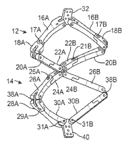
101011 Fig. 2C shows one variation of the device in one variation of
an
intermediate configuration, or between the axially-elongated and laterally-
elongated
.. configurations. When the first and second stabilizing assemblies 12, 14
reconfigure from
their axially-elongated configurations to their deployed laterally-elongated
17
CA 02822381 2013-06-19
WO 2012/087842
PCT/US2011/065627
configurations, the pivoting arrangements of each arm member and joining
member
allows the arms and joining members to extend laterally in a jack-like
fashion, as shown
in the perspective view of Fig. 2C. The distal 14 and proximal 12 stabilizing
structures
may transform from the laterally-elongated configuration to the axially-
elongated
configuration independently, dependently, sequentially, simultaneously or any
combination thereof. Fig. 2D shows the interventional device 10 in its
deployed
configuration, wherein both the proximal and distal stabilizing structures 12,
14 are in a
laterally-elongated configuration.
101021 The proximal stabilizing structure 12 may be comprised of a
first pair of
arm members 16A, 16B which are pivotably joined to a proximal engagement link
32 at a
first end through joints 15A, I5B, and also pivotably joined to respective
joining
members I8A, I8B at a second end through joints I7A, 17B. While the first pair
of arm
members 16A, 16B may pivot around joints 15A. 15B within a first plane
parallel to the
broad face of link 32, the coupling at the second end may pivot around joints
17A, 17B
within a second plane parallel to the broad face of the superior portion of
arms 16A, 16B,
which can be transverse (Fig. 2D) or angled (e.g., Fig. 2C) relative to the
first plane. The
joining members 18A, 18B may be further pivotably coupled to a first end of a
second
pair of arms 20A, 20B via respective links 34A, 34B which allow for pivotable
movement in a third plane parallel to the broad face of links 34A, 34B. The
second pair
of arms 20A, 20B may be further coupled pivotably to joining members 22A, 22B
such
that the pivotable movement of the second ends of the second pair of arms 20A,
208 may
occur around respective joints 21A, 21B within a fourth plane parallel to the
superior
portion of arms 20A, 20B. Joining members 22A, 228 may then be. pivotably
coupled to
a middle engagement link 36 such that the pivotable movement of the second
ends of the
joining members 22A, 22B may occur around link 36 within a fifth plane
parallel to the
broad face of link 36.
101031 The distal stabilizing structure 14 may be coupled similarly to
the
proximal stabilizing structure 12 where joining members 24A, 24B may be
pivotably
coupled to the middle engagement link 36 such that the pivotable movement of
the
joining members 24A, 24B may occur around link 36 within the fifth plane.
Joining
members 24A, 24B may be further pivotably coupled to a first end of a third
pair of arms
18
CA 02822381 2013-06-19
WO 2012/087842
PCT/US2011/065627
26A, 26B such that the pivotable movement of the arms 26A, 26B may occur
around
joints 25A, 25B within a sixth plane parallel to the broad face of the
superior portions of
arms 26A, 26B. The second ends of arms 26A, 26B may be pivotably coupled to
joining
members 28A, 28B via links 38A, 38B where pivoting movement may occur within a
seventh plane parallel to the broad face of links 38A, 38B. A first end of a
fourth pair of
arms 30A, 30B may be pivotably coupled to the joining members 28A, 28B around
respective joints 29A, 2911, such that the pivotable movement of the first end
of arms
30A, 30B is within an eighth plane parallel to the inferior faces of joining
members 28A,
28B. The second end of each arm 30A, 30B may be pivotably coupled to distal
engagement link 40 in a pivoting engagement which allows for pivoting motion
around
respective joints 31A, 31B within a ninth plane parallel to the broad face of
link 40.
101041 There are several advantages to utilization of multi-arm, multi-
link
assemblies. First, multi-arm, multi-link assembly provides for multiple planes
of pivotal
movement around multiple axes of rotation, allowing greater manipulation of
the profile
and shape of the interventional device 10, both in its delivery and deployed
configuration.
The flexibility of the interventional device 10 presents an advantage in that
it may assume
a linear, low-profile delivery configuration, shown, for example, in Fig. 2A,
while
remaining flexible enough to bend along the catheter lumen during delivery
and/or
deployment. Despite the flexibility of the interventional device 10, however,
the presence
of multiple links and arms also provides substantial rigidity once the
interventional
device 10 is in the fully deployed configuration, where each assembly is in
its laterally-
elongated configuration. Such rigidity may be provided by the offsetting of
the arms and
joining members within each layer of each annular structure. For example, in
Fig. 2D
distribution of arms and joining members is such that, once the distal
stabilizing structure
14 is in the laterally-elongated configuration, first pair of arms 16A, 1613
are no longer
able to rotate around, for example, respective joints 17A, 17B since first
pair of arms
16A, 16B straddles respective joints 21A, 2113. This is but one example of the
interlocking mechanisms employed by the multi-arm, multi-link structure of
each annular
structure.
101051 Each of the arm members and joining members may be made from any
number of suitable biocompatible materials, e.g., stainless steel, various
polymers,
19
CA 02822381 2013-06-19
WO 2012/087842
PCMJS2011/065627
ELGILOY (Elgin, IL), pyrolytic carbon, silicone, polytetrafluoroethylene
(PTFE), or
any number of other materials or combination of materials depending upon the
desired
results. The arm members may also be coated or covered with a material that
promotes
tissue in-growth, e.g., Dacron, PTFE, etc.
101061 Figs. 3A-3D illustrate another variation of the interventional
device, where
the arm and joining arm members have a more consistently arcuate shape along
its
periphery than the arms and joining members of interventional device 10.
Interventional
device 80 where each of the proximal and distal stabilizing structures 82, 84
may be
formed of a first pair of arms 54A, 54B and joining members 56A, 56B and a
second pair
of arms 58A, 588 and joining members 60A, 608 each pivotably joined, as
previously
described, but where the arm members form a more uniform and curvilinear
shape.
Similarly, the distal stabilizing structure 84 may be coupled via a middle
link and is
formed of joining members 62A, 62B and a third pair of arms 64A, 64B and
further by
joining members 66A, 668 and a fourth pair of arms 68A, 68B each pivotably
joined to
one another. Figs. 3C and 3D illustrate how the first and second assemblies
82, 84 may
pivot along their respective links and pivoted connections to expand into a
laterally-
elongated configuration, shown in Fig. 3D.
101071 In yet another variation, one or more the arm members
themselves may be
formed of multiple links or segments coupled together in such a way so as to
increase a
flexibility of the assembly. An example is illustrated in the perspective and
side views of
Figs. 4A and 48. As shown, an interventional device 140 may have a proximal
stabilizing structure 142 and a distal stabilizing structure 144 where at
least some or the
respective arm members are comprised of multiple small links or segments 146
linked
together by flexible elongate couplings. The arm members formed of the links
or
segments 146 may provide for increased flexibility of the assemblies when
placed against
the leaflets. Having the increased flexibility may allow for the
interventional device to
more closely conform to a particular anatomy of a valve and may further
provide for
enhanced support of the valve and may require less clearance within the heart
chambers
for deployment.
101081 In other variations where the arm members are comprised of segmented
arms, one or more of the arm members may have links or segments 150 which may
CA 02822381 2013-06-19
WO 2012/087842
PCT/US2011/065627
become rigid by the tensioning of a pullwire 152 to maintain a particular
shape of the arm
member. As illustrated in the example of Fig. 5A, a pullwire 152 may extend
through
each of the links such that when tensioned the arm member may become rigid as
the links
150 compress against one another and when released, allows the arm member to
become
.. flexible, as shown in Fig. 5B. Alternatively and/or additionally, the
interfacing ends of
the links or segments 154 may be preformed to have various angles or shapes
such that
when tensioned by pullwire 152, the arm member assumes a predetermined shape,
as
shown in Fig. 5C.
[01091 In yet other variations with segmented arm members, one or more
of the
arm members may be formed as links or segments 160 coupled via slotted
connections
162 which are rotatably hinged 164 to allow for bending in a single plane but
provides for
stiffness in a transverse plane, as shown in Fig. 6. Alternatively, the links
or segments
160 may be hinged in an alternating manner to allow for differential bending
of the
structure. Yet another variation is shown in the perspective view of Fig. 7A
which
illustrates an arm member which is formed of a patterned member 166 such as an
undulating pattern formed by molded or machined portions 168 removed from the
arm
member. Such a configuration may also allow for differential bending of the
structure
such that flexibility against a leaflet surface may be provided while
maintaining a degree
of structural stiffness along another plane. A pullwire 152 may be passed
through a
lumen extending through the length of the arm member such that by tensioning
the wire
152 the arm member will bend into a desired shape, as shown in Fig. 7B.
101101 Additionally and/or alternatively, one or all of the arm
members may have
rounded or curved edges 170, as shown in the end view of Fig. 8, to facilitate
delivery of
the assembly through catheter 54 as well as to reduce any potential wear
against the
internal 'catheter surface or injury to valve tissue. For example, if a
delivery catheter
having a 6 mm internal diameter, each respective arm member may have a cross
sectional
width, e.g., of about 5 ram and a height, e.g., of about 2 mm. Having the
curved edges
170 may allow for the translation of the assembly through the catheter lumen
without
wearing along the lumen surfaces. Moreover, the curved surfaces and edges of
each arm
member may also reduce any potential wear on the contacted mitral leaflets as
well.
21
CA 02822381 2013-06-19
WO 2012/087842
PCMJS2011/065627
101111 In any of the variations of the interventional devices
described herein,
various features or projections such as pins 190, castellations 192, raised
tabs 194, or any
other projections, protrusions, bumps 196, or features which may facilitate
engagement
with a replacement mitral valve implant may be formed along one or more arm
members,
for example along the surface of the arm members which face the central region
of the
mitral valve when deployed as shown in Figs. 9A to 9C. Additionallyand/or
alternatively, these various features may additionally or alternatively be
defined along the
surfaces of the arm members which come into direct contact against the mitral
valve
leaflets. For example, as shown in the cross-sectional side view of Figs. 10A
to 10C, the
arm members of both proximal and distal stabilizing structures 12, 14 which
extend into
contact against the surfaces of the mitral leaflets may also incorporate
various features.
Examples shown may include projections 190, tabs 192, or pins 194 which may
simply
compress upon the opposed surfaces of the mitral leaflets or they may be
correspondingly
designed to interdigitate or lock in an alternating pattern with respect to
opposed features
or projections when brought down upon the mitral leaflets into a locking
configuration.
Moreover, such features or projections may be covered by a fabric or covering,
such as a
kitted sleeve, to present a relatively atraumatic surface.
101121 In yet another variation, the arm members may be further varied
by
incorporating narrowed or tapered arms 200 that may reduce any risk of
perivalvular
.. leakage in the space between the arms, if any, as shown in the top view of
Fig. 11A.
Alternatively, the stabilizing assemblies may incorporate narrowed or tapered
arms 202
which taper or narrow to a point as they approach the posterior wall 203 of
the mitral
valve MV such that any replacement valve may directly contact against the
posterior wall
203 without any gaps, as shown in Fig. 11B.
101131 Fig. I IC shows a top view of another variation where the arm
members
may incorporate extensions 204 which may extend linearly or may fold out from
the
posterior set of arms to fill in any gaps along the posterior leaflet PML. The
extensions
204 may optionally extend partially or may lock with respect to an apposed
extension
204, as described in further detail below.
101141 Figs. 12A and 12B show perspective and front views of yet another
variation where the arm members may be configured in an alternative
arrangement. In
22
CA 02822381 2013-06-19
WO 2012/087842
PCMJS2011/065627
this variation, the supra-annular structure 212 may be configured to have
deployed arm
members which are relatively shorter than the deployed arm members of the
proximal
stabilizing structure 210 to facilitate deployment of the subannular assembly
212 without
interfering with the chordae tendineae CT, or papillary muscles PM or wall of
the left
ventricle. The lengths of the shortened subannular arm members may vary along
any
range and may also be configured to be relatively longer than the arms of the
supra-
annular assembly 210 in yet other variations the supra-annular arms may be
long enough
to completely encircle the valve.
101151 Figs. 13A to 13C illustrate perspective and cross-sectional
side views of
additional variations in the arm member configuration. As shown in Fig. 13A,
the
individual arm members may configured such that the subannular and supra-
annular
assemblies 12, 114 are radially offset in such a way that the subannular arm
members are
positioned towards the center of the valve orifice, further than the supra-
annular arm
members, such that the effective width of the combined arm members covers a
larger
area of the valve leatlets which moves the leaflet hinge point further toward
the center of
the valve orifice and limits the upwards billowing of the leaflets, e.g.,
during systole, to
improve the ability of the leaflets to close effectively.
101161 Figs. 13B and 13C illustrate cross-sectional side views where
the arm
members of the supra-annular structure 12 are positioned further away from the
center of
the valve orifice (in an opposite direction from that shown in Fig. 13A). In
this variation,
the arm members of the supra-annular structure 12 may be substantially
adjacent to (as
shown in Fig. 13B) or may just extend beyond (as shown in Fig. 13C) or may
overlap
slightly with the arm members of the subannular structure14. Such an
arrangement
increases the area of contact with the leaflets and may help to ensure the
securement of
the assembly to the leaflets. In addition, as shown in Fig. 13C, where the
subannular
structure is further offset from the supra-annular as to have a gap disposed
radially
between them, the leaflet may be folded or crimped through the gap so as to
further
enhance the grip on the leaflets.
101171 In yet additional variations, rather than the proximal
interventional device
being modified, the distal interventional device may be modified. One
variation is shown
in the perspective views of Figs. 14A to 14C which illustrate a telescoping
assembly 230
23
CA 02822381 2013-06-19
WO 2012/087842
PCT/US2011/065627
which may be deployable in the sub-annular space below the plane upon the
ventricular
side of the mitral valve MV. The telescoping assembly 230 may be comprised of
telescoping arms 232A, 232B which are attached to a pivoting assembly 434
which may
be used to position the arms 232A, 232B from a low-profile axial configuration
to a
radially-oriented deployed configuration, as shown in the figures. Once
positioned for
extension, one or more telescoping members 236A, 236B may extend linearly at
an angle
relative to one another (acute, right, or obtuse depending upon the desired
configuration)
from each arm 232A, 232B. Alternatively, the telescoping members 236A, 236B
may
extend in a curved or arcuate manner to form curved arm member when deployed.
In yet
another configuration, one telescoping arm may extend linearly while the
opposite arm
extends to form a curved deployed arm. Having the arms telescope outwardly
just below
the leaflets may avoid entanglement with various ventricular obstructions such
as the
chordae tendineae CT and/or papillary muscles PM. With the arms fully
extended, the
proximal stabilizing structure 12 may then be deployed tbr securement upon the
upper
leaflet surfaces, as shown in Fig. 14C.
101181 Another variation of telescoping arm members may be seen in the
end and
side views of Figs. 15A to 15C. These telescoping arm members may be used for
either
the first or second assembly, or both. The telescoping assembly 240 may
generally
comprise telescoping arms 242A, 242B which may be partially curved or straight
and
coupled to one another via a pivoting assembly (not shown) to allow for an
axially-
elongated delivery profile. One or more telescoping members 244A, 244B may be
slidably nested within each segment, as shown in Fig. I 5C, so as to minimize
profile and
maintain rigidity in their fully deployed position. The members 244A, 244B may
each
have matching curvatures and have their longitudinal axis coincident with one
another
such that when the arms are deployed, they may extend to follow a perimeter of
the
mitral valve, as shown in Fig. 15B.
101191 Figs. 15D and 15E show yet another variation where two or more
arm
members may be projected from within a catheter 54 to a deployed configuration
where
the arm members extend to form a curved or arcuate element. The arm members
may
extend perpendicularly or at an angle relative to the catheter 54 for
extending over a
valve, such as the mitral valve, both supravalvularly and subvalvularly to
compress upon
24
CA 02822381 2013-06-19
WO 2012/087842
PCMJS2011/065627
the annulus or upon the a periphery of the valve leaflets. Thus, the catheter
54 may be
inserted or directly positioned at the level of the medial or lateral mitral
valve
commissure with supravalvular and subvalvular exit sites for the arm members
to be
placed in the, e.g., subannular space, between the valve leaflets and the
ventricle and
separate arm members to be placed in the, e.g., supraannular space, to achieve
annular
stabilization.
101201 As illustrated in the perspective view of Fig. 15D, catheter 54
is shown
having arm member deployment assembly 261 attached to a distal end of the
catheter 54
via detachable coupling 253. The deployment assembly 261 may define two or
more
openings 251 through which the arm members, which are positioned within the
catheter
54 during delivery, may be extended through for deployment. The openings 251,
in this
variation, may be positioned about the deployment assembly 261 to allow for
the arm
members to extend in a curved or arcuate manner from the catheter 54. Thus,
the
openings 251 may be positioned in opposition to one another or at an angle
relative to
one another. An example is illustrated here showing at least two openings 251A
and
251B positioned adjacent to one another about a circumference of assembly 261
for
deploying at least two arm members supravalvularly. Two additional openings
251C and
25ID are also shown adjacent to one another and distal to the openings 251A
and 251B,
respectively, at a distance for deploying at least two arm members
subvalvularly.
101211 As shown in the perspective view of Fig. 15E, arm members 255A and
255B are illustrated advanced from catheter 54 and extending through
respective
openings 251A and 251B. Also shown are arm members 257A and 257B extending
from
respective openings 251C and 251D and projecting adjacent to respective arm
members
255A and 255B. Each of the arm members may extend from a straightened
configuration
within the catheter 54 during delivery to a curved or arcuate configuration
when urged
distally from within the catheter 54, e.g., using a pushing mechanism or other
actuator,
and when released from the constraints of the catheter 54 lumen. The arm
members may
curve into a shape which approximates a periphery of the valve, such as the
mitral valve,
such that when urged from the respective openings the opposing arm members
extend
perpendicularly or at an angle relative to the catheter 54 and curve towards
one another,
as shown. For instance, as arm members 255A and 255B project from their
respective
CA 02822381 2013-06-19
WO 2012/087842
PCMJS2011/065627
openings 25IA and 251B, they may extend at an angle relative to catheter 54
and also
initially extend away from one another to then curve and extend towards one
another
such that the deployed shape approximates the valve periphery. Arm members
257A and
257B may similarly extend adjacent to arm members 255A and 255B.
101221 Each of the arm members may also form an atraumatic blunt end 259 so
as
to prevent or inhibit tissue damage as the arm members are projected. The arm
members
may be constructed from various biocompatible materials sufficient to provide
flexibility
yet are rigid or semi-rigid enough to provide support to the valve leaflets,
e.g., shape
memory alloys such as nitinol, stainless steels, etc. Alternatively, the arm
members may
be constructed so as to be form inflatable tubular structures that may have
rigidity
induced by an inflation gas, fluid, or other medium (e.g., saline, water,
etc.) introduced
into the arm structures at a sufficiently high pressure. Alternatively, the
rigidity along the
arm members may be induced by inflating the arms with a hardening fluid.
Additionally
and/or alternatively, the arm members may have any number of frictional
components or
projections (barbs, spikes, etc., or any of the projections or elements
described herein)
formed upon the contact surfaces of the arm members to increase the fixation
between the
arms and the underlying tissue.
10123] Moreover, the length of each arm member may be uniform with
respect to
one another or they may be varied depending upon the designed configuration
and
anatomy of the valve. While the arm members may be projected to extend
partially about
the periphery of the valve, they may alternatively be projected to extend
distally such that
the respective distal ends overlap upon one another at least partially to
increase annular
rigidity.
101241 Once deployed, the supravalvularly positioned arm members 255A,
255B
may compress against their respective subvalvularly positioned arm members
257A,
257B such that the annular or leaflet tissue therebetween may be compressed
and
supported structurally. To further compress and support the tissue, the
supravalvularly
positioned arm members 255A, 255B and subvalvularly positioned arm members
257A,
257B may be located along separate deployment devices. An example is
illustrated in
Fig. 15F, which shows catheter 54 having supravalvularly positioned arm
members
255A, 255B projecting from its distal end but with subvalvularly positioned
arm
26
CA 02822381 2013-06-19
WO 2012/087842
PCMJS2011/065627
members 257A, 257B extending from a deployment assembly 265 attached to a
separate
deployment catheter 263 which may be positioned within catheter 54. The
separation of
the pair of arm members may allow for catheter 263 to be translated 267
relative to
catheter 54 to further compress or adjust the positioning of the assembly
relative to the
valve.
101251 Figs. 16A and 16B illustrate top views of the interventional
device where
placement of the middle hinge or pivot 254 may be varied. For example, as
shown in Fig.
16A, hinge or pivot 25 may be located on the outer edge allowing the arm
members to
extend towards the periphery of the valve as much as possible. Alternatively,
as shown in
Fig. 16B, the hinge or pivot 254 may also be placed as close as possible to
the center of
the mitral orifice so that the arm members may be positioned as close as
possible to the
inner perimeter of the mitral valve MV to provide support while distorting the
leaflets as
little as possible.
101261 II. Deployment
10127j Figs. 17A to I 7E illustrate one variation of the mechanism of
deployment
for an interventional device. A distal stabilizing structure 14 is advanced
beyond the
distal opening 262 of the catheter sheath 54. An actuation member 264 (e.g.,
wire, suture,
catheter, etc.) may be attached to a distal stabilizing structure 14 may be
tensioned or
actuated while a proximal portion of the distal stabilizing structure 14 is
maintained
against the catheter opening 104. With the proximal stabilizing structure 12
still
constrained within the catheter 54, the arms of the distal stabilizing
structure 14 may be
reconfigured to deploy, as shown in Figs. 17B-17C. For example, when an
actuation
force is applied to the actuation member in a proximal direction, the distal
end 261 of the
distal stabilizing structure 14 is urged in a proximal direction while the
proximal end 263
of the distal stabilizing structure 14 is prevented from proximal movement by
the catheter
sheath 54, thus pulling the distal end 261 towards the proximal end 263. With
the distal
stabilizing structure 14 fully in a laterally-elongated configuration, as
shown in Fig. I 7C,
the catheter opening 262 may be withdrawn further until the proximal
stabilizing
structure 12 is exposed, as shown in Fig. 17D. The actuation member 264 may
then be
tensioned further with the catheter opening 104 used as a backstop against the
proximal
27
CA 02822381 2013-06-19
WO 2012/087842
PCT/US2011/065627
portion of the proximal stabilizing structure 12 to reconfigure the proximal
stabilizing
structure 12 into its laterally-elongated configuration, as shown in Figs. 17D
and 17E.
101281 Figs. 18A to 18F show perspective views illustrating how the
one or more
interventional devices 10, 10' may be deployed relative to the mitral valve
leaflets for
providing leaflet stabilization and/or anchoring devices for a prosthetic
valve. As shown,
in a typical antegrade approach (as discussed herein) a first interventional
device 10 may
be advanced between the posterior and anterior mitral leaflets PML, AML until
the distal
stabilizing structure 14 through the valve to a subannular position. The
distal stabilizing
structure 14 may be deployed first or both the proximal and distal stabilizing
structures
12, 14 may be reconfigured simultaneously such that proximal and distal
stabilizing
structures 12, 14 reconfigure into their laterally-elongated configurations on
opposite
sides of the annulus, compressing the leaflets, as shown in Figs. 18A to 18C.
The
catheter 54 has been omitted for clarity purposes only.
101291 A second interventional device 10' positioned within the
catheter 54
proximally of the first interventional device 10 may then be deployed at a
second location
along or upon the mitral valve by repositioning the catheter accordingly and
then
advancing the distal stabilizing structure 14' to a subannular position and
the proximal
stabilizing structure 12' to a supra-annular position. Once suitably
positioned, the
stabilizing structures 12', 14' may be deployed sequentially or simultaneously
to lock
upon their respective leaflet surfaces, as shown in Figs. 18D to 18F.
101301 Deployment of the interventional device(s) may be biased along
the
anterior side of the mitral valve, as shown in Fig. 18F. The mitral valve is
bound by
muscular tissue MW on the posterior side of the valve only. The inner wall of
the mitral
valve, surrounding the anterior leaflet, is bound by a thin vessel wall TVW
separating the
mitral valve annulus from the inferior portion of the aortic tract (as shown
in Fig. 20A)..
As a result, little native structural support, if any at all, is provided on
the anterior side of
the mitral annulus. Therefore, by deploying each interventional device 10, 10'
such that
the majority of the stabilizing assemblies lie on or along the anterior
leaflet, the
interventional device(s) 10, 10' provide additional support for stabilizing
the annulus
and/or anchoring a replacement valve, as shown in Fig. 20B. In order to
provide
adequate circumferential support for a catheter-delivered prosthetic valve,
the
28
CA 02822381 2013-06-19
WO 2012/087842
PCT/US2011/065627
interventional devices 10, 10' together preferably cover, e.g., at least about
60% of the
circumference of the mitral valve.
101311 The first and second interventional devices 10, 10' may be
accordingly
positioned in the anterior and posterior commissures such that the curved arm
members
follow along a periphery of the valve annulus. Moreover, although two
interventional
devices 10, 10' are shown, a single interventional device may be used alone at
either
commissure. The arms may also be configured at various angles depending upon
the
desired configuration. Likewise, more than two interventional devices may also
be
deployed.
101321 Fig. I 9A illustrates a top view of a dysfunctional mitral valve MV
where
the posterior and anterior mitral leaflets PM 1, AML fail to coapt while Fig.
198
illustrates a top view of the mitral valve MV having a first and second
interventional
device 10, 10' deployed and positioned at either commissure. As shown, the
interventional devices 10, 10' may follow the periphery of the annulus while
maintaining
a central region of the mitral valve MV uninhibited such that the leaflets may
be
supported by the assemblies to facilitate coaptation of the leaflets.
101331 111. Locking Mechanisms
101341 Once the interventional device 10 has been deployed, the device
10 may
be locked into its deployed shape and left implanted upon or along the mitral
valve. To
ensure that the device remains secured upon the valve leaflets, various
locking
mechanisms may be implemented into the device.
101351 In the variation shown in the front and perspective views of
Figs. 2IA to
21C, the extension arm members may generally comprise an attachment member 270
which is attached or connected, for instance, along the arm member 272. The
attachment
member 270 may further extend linearly or curvilinearly along an extending
member 274
which has a first atrauinatic surface which may contact against a surface of
the leaflet.
When the interventional device has been reconfigured into its laterally-
elongated
configuration, as shown in Fig. 21C, the attachment member 270 may extend
along a
circumferential arm from the arm member at a distance from the proximal and
distal
stabilizing structures 82, 84. The opposite surface of extending member 274
may define
one or more projections or teeth 276 for coming into a ratcheted locking
engagement with
29
CA 02822381 2013-06-19
WO 2012/087842
PCT/US2011/065627
an opposed corresponding set of projections on a second extending member of an
additional repair assembly. In the example shown, the projections or teeth 276
may be
positioned along the extending member 274 such that the atraumatic side of the
member
274 may rest upon the surface of the valve leaflet while the projections or
teeth 276 may
extend away from the leaflet surface.
101361 Figs. 22A to 22F illustrate an example of how one or more
interventional
devices 80, 80' may be deployed relative to one another such that the
extending members
274, 274' may be brought into an engaging contact. As previously described,
the first
interventional device 80 may be deployed and expanded at a first commissure
such that
the extending member 274 is deployed along a periphery of the valve annulus
with the
projections or teeth 354 positioned away from the leaflet surface, as shown in
Figs. 22A
to 22C. The second repair assembly 80' may then be deployed and expanded at
the
second commissure in apposition to the first assembly 80 such that the second
extending
member 274' is also deployed along the periphery of the valve annulus. The
second
assembly 80' may have the projections or teeth 276' positioned towards the
leaflet
surfaces such that they may come into an engaging contact with the first
extending
member 274, as shown in Figs. 22D to 22F. The engagement between the extending
members 274, 274' may be ratcheted or loosened to adjust the positioning of
the
assemblies 80, 80' and the amount of support imparted to the underlying
leaflets.
101371 Figs. 23A to 23B illustrate front and perspective views of yet
another
locking mechanism variation for a interventional device where a pair of curved
stabilizing arms 280A, 280B may be combined with a distal stabilizing
structure 84. The
curved arms 280A, 28013 may be folded when delivered through the catheter 54,
as
shown in Fig. 23A, but may extend radially outward to curve towards one
another at their
respective distal ends such that the curved arms 280A, 280B extend over or
upon the
leaflets and coincide between the leaflet commissure. The distal ends of the
each curved
arm 280A, 280B may define one or more openings 284 through which a locking
suture or
wire 282 may be passed to secure the arms to one another. In this manner, the
positioning of the arms over the span of the valve may further provide
stabilization over
the entire valve.
CA 02822381 2013-06-19
WO 2012/087842
PCT/US2011/065627
101381 Fig. 23C illustrates a top view of another locking mechanism
variation for
securing two or more interventional devices. A pair of curved stabilizing arms
280A,
280B may be combined with a distal stabilizing structure 84 to secure one
interventional
device to a second interventional device. The curved arms 280A, 280B may be
folded
when delivered through the catheter 54, as shown in Fig. 23A, but may extend
radially
outward so as to conform to the shape of the arms of the stabilizing
structures and/or
native valve. The length of the stabilizing arms 280A, 280B may be adjusted so
as to
exceed that of the distal arms, thereby extending past the ends of the distal
arms of one
interventional device to overlap with and/or connect to the distal arms of a
second
interventional device. Stabilizing arms 280A, 280B may be held in place
through, e.g.,
pins, hooks, tabs, wires, sutures, etc. anywhere along the arms of the second
interventional device.
101391 Figs. 24A to 24C illustrate perspective and partial cross-
sectional side
views of yet another variation which utilizes the devices illustrated above in
Figs. 15D
and 15E. A portion of the surrounding mitral wall MW may be seen in the figure
for
reference. After advancing the stcerable catheter intravascularly to, e.g.,
the mitral valve
located within the left atrial chamber, at the medial or lateral commissure
from the atrial
side or ventricular side, the catheter 54 may be positioned such that the
detachable
coupling 253 is positioned at least partially through the valve. With the
proximal
openings 251A and 25111 positioned above the valve within the left atrium and
the distal
openings 251C and 251D positioned below the valve within the left ventricle,
the
supravalvular arm members 255A and 2558 may be advanced from within the
catheter
54 into their deployed configuration situated, e.g., in the supra-annular
space upon the
annulus AN or upon the superior surfaces of the posterior PML and anterior
mitral
leaflets AML, and subvalvular arm members 257A and 257B may be similarly
advanced
from within the catheter 54 into their deployed configuration, e.g., in the
sub-annular
space upon the annulus AN or upon the inferior surfaces of the posterior PML
and
anterior mitral leaflets AML, in apposition to their respective supravalvular
arm
members. As described above, the arm members may be uniform in length relative
to
one another or non-uniform in length and either partially or completely
circumferentially
deployed over or upon the valve.
31
CA 02822381 2013-06-19
WO 2012/087842
PCT/US2011/065627
101401 Fig. 24B illustrates the partial cross-sectional side view of
the catheter 54
positioned trans-septally in a superior position relative to the mitral valve.
The deployed
arm members may be seen after deployment and upon the annulus AN or valve
leaflets.
Because of the low-profile of the arm members, particularly the subvalvularly
positioned
arm members 257A and 257B, they may be introduced into the subannular space
within
the left ventricle LV and through the surrounding chordae tendinae CT attached
to the
leaflets without being inhibited.
101411 After assuring adequate arm member placement, the coupling 253
of the
catheter 54 may then be disconnected from the shaft of the catheter 54 leaving
the
deployed arm members in position. Because the arm members may have a spring
like
quality while imparting compressive and/or radial forces to the underlying
valve, they
may function to stabilize the assembly at the annular level.
101421 The assembly may further provide a platform for placement of an
implantable valve prosthesis which may be secured to the valve without the
need for
sutures, as illustrated in the partial cross-sectional side view of Fig. 24C.
In patients with
mitral regurgitation who are candidates for valve replacement, the assembly
may be
placed, as described herein, while under fluoroscopic, echocardiographic, and
other
imaging guidance. The rigidity of the arm member assembly may provide a
platform for
placement of a transcatheter valve and/or sutureless prosthesis such that a
replacement
valve prosthesis 398 may be advanced intravascularly and deployed through the
valve
while anchoring against or along the reinforced valve annulus and/or leaflets
or directly
against the deployed arm members.
101431 Another approach is placement of assembly under direct vision
or
surgically. The valve commissure is identified and the tip of the catheter 54
placed at the
junction of the subannular and supraannular regions. The assembly may also be
percutaneously, trans-atrially, trans-septally, trans-apically or directly
introduced and
implanted as well. Passage of the arm members is continued into the subannular
space
followed by passage of the arm members into the supraannular space. The
described
approaches and the present device also may be used to provide a stable, rigid
or semi-
rigid annulus for the deployment of transcatheter valve and sutureless
prostheses in other
locations, such as the tricuspid valve.
32
CA 02822381 2013-06-19
WO 2012/087842
PCMJS2011/065627
101441 In yet another variation, the catheter 54 may be utilized with
an additional
catheter 54', which may also be advanced into the heart chamber adjacent to
the first
catheter 54 or through an alternative path, to increase annular rigidity.
Regardless of the
entry path, with the first catheter 54 positioned at a first location about
the valve, such as
at a first location of the valve commissure, the second catheter 54' may be
positioned
simultaneously or sequentially at a second location about the valve, such as
at a second
location of the opposite valve commissure, as shown in the perspective view of
Fig. 24D.
101451 With the arm members 255A and 255B deployed supravalvularly and
arm
members 257A and 257B deployed subvalvularly, the additional supravalvular arm
members 255A' and 255B' may be deployed supravalvularly and additional arm
members 257A' and 2578' may be deployed subvalvularly. The additional arm
members
of the second catheter 54' may be deployed sequentially or simultaneously with
the
deployment of the arm members of the first catheter 54. Once each of the arm
members
have been deployed, each respective connector may be detached to leave the arm
member
assembly implanted upon the valve and the respective catheters 54, 54' may be
withdrawn. As previously described, the arm members may then be left implanted
to
provide structural support to the valve or a valve prosthesis may be
introduced and
implanted through the valve utilizing the arm members for structural support.
101461 Another variation on a locking mechanism for the interventional
device is
illustrated in the side views of Figs. 25 to 27 which show an outer catheter
290 which
may be temporarily coupled to the proximal link 298 of the interventional
device 10 via
an outer catheter attachment 294, e.g., screw thread, gripping element with a
release wire,
or other suitable attachment mechanism. A separate inner catheter or wire 292
may pass
through the outer catheter 290 and within a lumen 296 defined through the
device 10 to a
distally positioned inner catheter attachment 298, e.g., screw thread,
gripping element
with a release wire, or other suitable attachment mechanism. The inner
catheter or wire
292 may be optionally pre-shaped or configured to hold or maintain a
predetermined
shape for holding the assembly 10 in the desired shape or configuration for
facilitating
deployment. Of course, the inner catheter or wire 292 may also be maintained
in a
straightened and flexible configuration which may allow the assembly 10 to
naturally
form itself into an appropriate curve.
33
CA 02822381 2013-06-19
WO 2012/087842
PCT/US2011/065627
101471 During delivery and prior to assembly expansion, the inner
catheter or
wire 292 may be maintained in a stable position relative to the outer catheter
290. The
inner catheter or wire 292 may be actuated or tensioned relative to the outer
catheter 290
to expand or extend the device into its deployed configuration. To secure the
laterally-
elongated configuration, one variation for locking the device may comprise an
outer
catheter attachment screw 308 positioned at a distal end of the outer catheter
290. The
attachment screw 308 may define a threaded portion 310 which may be rotated to
engage
the threading 306 defined along the lumen 306 of the device 10 such that the
screw 308 is
advanced distally through the lumen 306 until a locking collar 312 secures the
proximal
end of the device 10 relative to the distal end of the device 10, as shown in
Fig. 27.
101481 To release the device 10 from the catheters, one or more pairs
of
engagement arms having one or more protrusions 300 may comprise the inner
catheter
attachment 298 at the distal end of the inner catheter 290. The protrusions
300 may be
maintained against one or more corresponding locking members 302 defined along
the
distal end of the lumen 296. A release wire positioned through a lumen 304
defined
through the inner catheter 292 may be tensioned to allow the engagement arms
to release
from the locking members 302 thus allowing the interventional device 10 to
detach from
the catheter, as shown in Fig. 26.
101491 In yet another variation of the interventional device(s), Fig.
28 illustrates a
side view of another variation where the device may incorporate one or more
respective
locking mechanisms 320 (e.g., pins, ratchets, etc.) positioned along a upper
or lower
surface of the arm members such that the locking mechanisms 320 are received
into
respective receiving channels 322 or another cooperating structure defined
along apposed
arm members when reconfigured into the deployed configuration. As previously
described, a tensioning wire, suture, or catheter 324 may be coupled to a
distal anuular
structure 14 such that when tensioned, the device may collapse into its
laterally-elongated
configuration. Also, as the arm members fold into their laterally-elongated
configuration,
the locking mechanisms 320 may be configured to penetrate through the leaflets
and to be
received into their respective receiving channels 322 and locked automatically
to secure
the arm members into their deployed configurations.
34
CA 02822381 2013-06-19
WO 2012/087842
PCMJS2011/065627
101501 Examples of the different types of locking mechanisms 320 which
may be
utilized with the stabilizing assemblies may be seen the cross-sectional side
views of
Figs. 29A and 29B. In this example, ratchet 330 may have a tapered lock 332
which may
be incorporated into a distal arm member of the interventional device. Tapered
lock 332
has a proximal shoulder 331 of larger diameter than openings 334. As the
attached
tensioning wire 336 is pulled, the tapered locking portion 262 may be pulled
through the
one or more openings 334 defined along the interventional device, e.g.,
through the
pivoting mechanism, until the tapered locking portion 332 is fully drawn
through the
assembly to lock the device in its deployed configuration, as in Fig. 29B.
Fig. 30
illustrates a cross-sectional side view of another variation of a locking
ratchet 338 but in
place of shoulder 331 the tapered portion may define one or more serrations or
projections 340 to engage complementary features within openings 334 to
enhance the
locking securement.
101511 Figs. 30A and 30B illustrate cross-sectional side views of
another locking
mechanism which may be drawn through the interventional device to lock the
device in
its deployed configuration. In this variation, the distal end of the
interventional device
may have a locking member 342 such as a wire or suture which may be tensioned
through the interventional device and crimped or flattened to form a broadened
retainer
344 directly upon the member 342 to prevent its withdrawal through the
assembly.
101521 Figs. 31A and 3 IB illustrate cross-sectional side views of yet
another
locking mechanism. In this example, the locking member 342 may be tensioned
through
the interventional device and a crimping collar 346 may be positionable over
the member
342 and crimped upon the member 342 when compressed member 342 is drawn
tightly.
101531 Figs. 32A to 32C illustrate cross-sectional side views of
another locking
mechanism where the locking member 342 may be tensioned to hold the
interventional
device into its laterally-elongated configuration. One of the proximally
positioned arm
members or the proximal engagement link 32 may incorporate a locking pin 348
which is
urged against the locking member 342 via a biasing element 350 such as a
spring. As the
locking member 342 is drawn proximally through the interventional device, the
biased
.. pin 348 may be inserted at least partially, as shown in Fig. 32B, or
entirely, as shown in
Fig. 32C, through an opening or slot 352 defined through a distal portion of
the member
CA 02822381 2013-06-19
WO 2012/087842
PCT/US2011/065627
342. With the locking pin 348 inserted through the opening or slot 352,
further
movement of the member 342 may be inhibited relative to the interventional
device
thereby locking the configuration of the assembly.
101541 Fig. 33A illustrates a cross-sectional side view of yet another
variation of
a locking mechanism where a wire or rod 354 may be tensioned to move the
proximal
and distal stabilizing structures 12, 14 into their expanded configuration. A
separate
collet 356 may slide along the wire or rod 354 when the collet 356 is in an
open
configuration such that one or more movable locking members 358 extending
radially
within the collet 356 provide enough space for the wire or rod 354 to travel
freely
through, as shown in Fig. 33B. Once the interventional device has been
desirably
expanded, the collet 356 may be drawn down distally along the wire or rod 354
and
locking members 358 moved radially inward to clamp down upon the wire or rod
354, as
shown in Fig. 33C, thereby preventing movement of the collet 356 relative to
the wire
354 and thus preventing or inhibiting the interventional device from
reconfiguring back
into its low-profile shape.
101551 Fig. 34 illustrates a cross-sectional side view of yet another
locking
mechanism where a fastener 362 having threading 364 along its length may be
simply
rotated or screwed into the expanded interventional device to lock the
configuration of
the proximal and distal stabilizing structures 12, 14.
[0156] Figs. 35A and 35B illustrate cross-sectional side views of another
locking
mechanism where a rivet 370 having a deformable collar 368 may be secured upon
the
interventional device once the assembly has reconfigured into its deployed
configuration.
A separate press 366 may be brought to bear upon the deformable collar 368
such that the
collar 369 deforms radially to lock a position of the rivet 370 relative to
the arm
members. In this manner, the expanded configuration of the proximal and distal
stabilizing structures 12, 14 may be secured.
101571 Yet another locking mechanism is illustrated in the front and
detail front
views of Figs. 36A and 36B which show the relative positioning of the proximal
and
distal engagement links 32,40 along the stabilizing structures 12, 14. In this
variation, a
tensioning suture or wire 372 may pass through the interventional device and
couple the
distal and proximal engagement links 32, 40 to one another. With the suture or
wire 372
36
CA 02822381 2013-06-19
WO 2012/087842
PCT/US2011/065627
secured to at least one of the links, the remaining end of the suture or wire
372 may be
adjustably secured to the opposing link using a one-way sliding knot to allow
for
adjustable locking of the interventional device. The remaining end of the
suture or wire
372 may, for example, pass through a central opening 332 of the proximal
engagement
link 52 and then pass proximally through a first laterally offset proximal
opening 376 and
then crossover the wire 372 to second laterally offset proximal opening 374,
from which
it extends to the distal engagement link 32.. Pulling the suture or wire 372
in a first
direction 380, e.g., towards the opposing link, may allow for tensioning
adjustment of the
proximal and distal stabilizing structures 12, 14 while sliding of the suture
or wire 372 in
the opposing direction 382 is prevented due to friction between the portions
377 of the
suture or wire that engage each other, thus locking the upper and lower arm
members.
This configuration may also allow adjustments to be made to the structures to
allow for
the release of the device from a particular configuration and the re-locking
of the device,
if so desired. For example, by releasing tension in the crossover portion 377
of the suture
or wire 372, it will be allowed to slide in direction 382 to release the
distal annular
structure.
101581 Yet another variation is shown in the top view of Fig. 37 which
illustrates
a variation where a coupling mechanism such as a sliding suture lock 384 may
be
advanced over wires or sutures 386 extending from the arms of multiple
assemblies to
create a rigid or secure connection between each of the implanted assemblies
12, 12' in
their laterally-elongated configurations upon the valve leaflets. This is
particularly useful
when the distal and proximal stabilizing structures are deployed so that their
combined
periphery is biased towards one end of the valve, as shown, for example, in
Fig.18F.
101591 IV. Valve
101601 In any of the interventional device variations described, one or
more
assemblies may be utilized alone or in combination with an implanted stent,
scaffold, or
valve implant. In the variation shown in the perspective view of Figs. 38A and
38B, an
example is illustrated showing how a self-expanding valve implant or scaffold
398 may
be deployed relative to the interventional devices. Once the one or more
interventional
devices 80, 80' have been deployed and expanded along the mitral valve,
catheter 54 may
be repositioned above the mitral valve with one or more wires or sutures 392
extending
37
CA 02822381 2013-06-19
WO 2012/087842
PCT/US2011/065627
from the catheter 54 and to the one or more links 52, 52'. A pusher catheter
or shaft 390
having valve implant or scaffold 392 attached may be urged from the catheter
54 to push
the implant or scaffold 398 in its fully expanded shape via openings or loops
394 located
along the implant or scaffold 392 through which may be looped around the wires
or
sutures 392 extend to help guide the implant 398 into position in engagement
with the
assemblies 80, 80'. The implant 398 may have its lumen 396 positioned to
coincide with
the valve opening such that the implant 398 is secured above, below, or
through the
valve. Each of the links may comprise one or more retractable locks 394 which
may
allow the openings or loops 394 to slide over and force the retraction of the
locks 402, as
shown in Fig. 39, until the openings or loops 394 have cleared the locks 402
after which
they may extend outwardly to lock a position of the valve 398 relative to the
interventional devices 80, 80'.
101611 To help secure the implant 398 relative to the valve, the one
or more
assemblies 80, 80' may incorporate one or more protrusions 400, as previously
described,
along the arm members facing the valve central region, as shown in the
perspective view
of Figs. 40A and 40B. The protrusions 400 may extend inwardly from the arm
members
and engage the sides of the implant 400 or interstices therein to resist or
inhibit any
movement between the implant 398 relative to the valve, as shown in Fig. 40A.
In this
example, implant 398 will be held within catheter 54 until positioned within
the
interventional devices 80 then released so as to expand into engagement with
the inner
walls thereof.
101621 Yet another variation for securing the interventional devices
80, 80'
relative to one another as well as to provide an engagement surface for any
potential
implant is shown in the perspective view of Fig. 41A. With the one or more
interventional devices 80, 80' deployed along the valve, a supporting ring 404
having a
circumferential structure with one or more openings 406 defined along the
circumference
may be deployed from the catheter 54 and guided via wires or sutures 392
extending
through the openings 406. The terminal ends of the wires or sutures 378 may be
attached
to the respective links 52, 52' such that the openings 406 may slide directly
upon and
over the links 52, 52'. Each of the links may comprise one or more retractable
locks 402
which may allow the openings 406 to slide over and force the retraction of the
locks 402,
38
CA 02822381 2013-06-19
WO 2012/087842
PCT/US2011/065627
as shown in Fig. 39, until the openings 406 have cleared the locks 402 after
which they
may extend outwardly to lock a position of the ring 404 relative to the
stabilizing
structures 80, 80'.
101631 While the support ring 404 may be comprised as a simple ring
defining an
opening 408 therethrough, as shown in the top view of Fig. 41C, the ring may
be
configured into alternative variations. One example is shown in the top view
of Fig. 4IB
where supporting ring 403 may comprise a partial ring, such as a C-clip, in
which the
terminal ends of the clip are coupled to one another via a connecting wire or
elastic band
405 such that an opening 407 is defined by the structure. The partial ring may
also be
comprised of individual segments 401 which are hinged or linked to one another
such
that the supporting ring 403 may conform to variations in the alignment of the
interventional devices 80, 80' or to variations in the anatomy as well while
still providing
structural support. Another variation is shown in the perspective view of Fig.
41D which
illustrates a support ring 404 having a collar 410 which extends axially away
from the
ring 404 and defines while defining an opening 412 therethrough. Collar 410
may have a
more circular shape, greater height and smaller diameter than ring 404 so as
to provide a
cylindrized platform in which a stented valve may be deployed.
[01641 Figs. 42A to 42B illustrate perspective views of additional
ringed
structures which may be attached to the one or more interventional devices 80,
80'. In
one variation, supra-annular ring 420, shown in Fig. 42A, may comprise a
number of
projections or protrusions 422 or loop or clip element 424, as shown in Fig.
42B, which
extend outwardly from the ring circumference.
101651 In yet another variation of the interventional device, a
supporting ring may
be utilized in combination with one or more retaining members rather than with
an
interventional device. Fig. 43A shows a cross-sectional side view of an
example of how
a ring 452 may be axially-elongated and positioned within a catheter 54 along
with
proximally and distally positioned retainer members 450, 450B for
intravascular delivery.
Fig. 43B shows a perspective view of the deployed ring assembly where
subannular
retainer members 450A, 450B may be configurable from a low profile shape
during
delivery to a deployed shape in which the distal arms of the retainer members
extend into
curved, arcuate, or semi-circular configurations for contact the sub-valvular
surface of the
39
CA 02822381 2013-06-19
WO 2012/087842
PCT/US2011/065627
leaflets. Accordingly, the retainer members 450A, 450B may be made from
resilient
materials such as shape memory materials (e.g., nickel-titanium alloys, shape
memory
polymers, etc.) Locking couplings 458A, 458B may be positioned to extend
proximally
of each respective retainer member 450A, 450B.
101661 The prosthetic supra-annular ring 452 may be shaped or sized
similarly to
a periphery of the mitral valve and/or be configured to support an implanted
prosthetic
valve. One or more openings 454A, 454B may also be defined at either end of
the ring
along the circumference to provide guidance for wire or sutures 456A, 456B
which may
pass through each respective opening. The couplings 458A, 458B may be attached
to
respective wire or suture 456A, 456B such that the couplings may be received
within the
respective openings 454A, 454B defined through the ring 452 in a locking
manner when
each wire or suture is tensioned to secure a position of each respective
retainer member
450A, 450B relative to the ring 452. The couplings 458A, 458B may define one
or more
tapered members which allow for their insertion into and/or through the
openings 454A,
454B and engagement with a flange 457 therein to inhibit their retraction or
withdrawal
to allow for adjustable securement of the ring 452 to retainer members 450A,
450B upon
the mitral valve annulus, as shown in Fig. 43C. Alternatively, various other
mechanisms
such as ratcheting teeth, pawls, spherical locking elements, hitch/ring
assembly, etc. may
be used to couple retainer members 450A, 450B to ring 452.
101671 An example of how the ring assembly may be deployed is shown in the
partial cross-sectional side views of Figs. 44A to 44F. The mitral valve
leaflets are not
shown only for clarity. As illustrated in Figs. 44A and 44B, the distal end of
catheter 54
may be placed through a first commissure of the mitral valve MV from the left
atrium
LA into the left ventricle LV and a first retainer member 450A and coupling
458A may
be deployed from the catheter in the subannular space below the leaflets to
reconfigure
into a deployed configuration. The catheter 54 may be withdrawn proximally
into the left
atrium LA and the ring 452 may then be ejected from the catheter 54 from
within the left
atrium LA superior to the mitral valve MV as well as with the tether 456A
remaining
attached to the first retainer member 450A through the opening in the ring
452. With the
catheter 54 used as a backstop against the ring 452, the tether 456A may be
tensioned and
pulled to draw the coupling 458A into the ring opening to lock the retainer
member 450A
CA 02822381 2013-06-19
WO 2012/087842
PCT/US2011/065627
against the valve annulus AN and/or leaflets as shown in Figs. 44C and 44D.
The
catheter 54 distal end may then be placed through the ring 452 in the opposite
commissure to deploy the second retainer member 450B inferior to the mitral
valve
annulus AN and within the left ventricle LV, as shown in Fig. 44E. The tether
456B may
then be tensioned to draw the second retainer member 450B against the valve
annulus
AN and to lock the coupling 458B to the ring to secure the ring 452 position
relative to
the valve, as shown Fig. 44F.
101681 In yet another variation, Figs. 45A to 45C show another
variation
illustrating how a ring 452 may be deployed in combination with a distal
stabilizing
structure 84. As shown in the cross-sectional side view of Fig. 45A, the ring
452 may be
axially-elongated into a low-profile configuration for delivery positioned
between a distal
stabilizing structure 84 and an optional proximal stabilizing structure 84'.
The distal
stabilizing structure 84 may be deployed from the catheter 54 and secured, as
shown in
Fig. 458, in a subannular position. The ring 452 may then be deployed in a
supra-
annular position and allowed to reconfigure into its deployment shape. With
tethers
456A, 456B passing from catheter 54 and through respective openings along the
ring
452, a pusher catheter 460 may be deployed to push or urge a respective
locking retainer
462A, 462B along a respective tether 456A, 456B to secure the position of the
first and
second stabilizing assemblies 84, 84' relative to the ring 452, as shown in
Figs. 45C and
45D, such that the valve leaflets are secured therebetween. Fig. 45E shows a
cross-
sectional side view of an example of a locking retainer 462A having a lumen
464 for
sliding along tether 456A (uni-directionally in one example) a pair of angled
pawls which
engage tether 456A and a tapered portion for locking into the opening defined
along the
ring 452.
101691 In any of the variations of the interventional devices described
herein,
various features or projections such as pins 180, castellations 182, raised
tabs 184, or any
other projections, protrusions, bumps, or features which may facilitate
engagement with a
replacement mitral valve implant may be formed along one or more arm members,
as
shown in the perspective views of Figs. 46A and 46B. These features may be
located
along the surface of the arm members which face the central region of the
mitral valve
41
CA 02822381 2013-06-19
WO 2012/087842
PCT/US2011/065627
when deployed or on any other surface of the arm members as may be useful for
enhancing engagement with the prosthetic valve.
101701 It should be noted that any of the ring members described above
in
connection with, e.g. Figs. 38-46, may be configured to receive a separate
catheter-
delivered valve for deployment therein, or may have either a temporary or
permanent
valve pre-mounted therein. Since a relatively long period of time may elapse
between
placement of the anchor and implantation of the prosthetic valve, a temporary
valve sewn
into or otherwise secured within the anchoring structures of the invention
assures proper
regulation of blood flow in the interim. As the name denotes, temporary valves
are not
intended for long term use, typically being required for a period from about
15 minutes to
several hours or at most a few days. Prosthetic valves may be implanted within
a
temporary valve or may be implanted after the temporary- valve has been
removed.
101711 V. Intravascular Approaches to the Mitral Valve
101721 In one example for delivering and deploying one or more
interventional
devices 10, Figs. 47A to 47K illustrate partial cross-sectional side views of
a heart H
interior to show a typical antegrade approach. As shown in Fig. 47A, a
guidewire 9 may
be advanced intravascularly using any number of techniques, e.g., through the
inferior
vena cava IVC or superior vena cava SVC not shown), through the atrial septum
AS and
into the right atrium RA. Catheter 54 may be advanced along the guidewire 9
and into
the right atrium RA until reaching the anterior side of the atrial septum AS,
as shown in
Fig. 478. Once the catheter 54 reaches the anterior side of the atrial septum
IAS, a
piercing needle and/or dilator 500 may be advanced through the catheter to
cross the
atrial septum AS from the right atrium RA into the left atrium LA, as shown in
Fig. 47C.
At this point, the guidewire 9 may be exchanged for the needle 70 and the
catheter sheath
withdrawn. A catheter 54 may then be advanced over the guidewire 9 and into
the left
atrium LA and into a position above the dysfunctional mitral valve MV, as
shown in Fig.
3D.
101731 In a typical antegrade approach, the distal opening 262 of the
catheter 54
may be advanced into proximity to the mitral valve MV and optionally passed
between
the posterior mitral leaflet PML and anterior mitral leaflet AML and at least
partially
past the plane of the mitral valve annulus, as shown in Fig. 47E. A first
interventional
42
CA 02822381 2013-06-19
WO 2012/087842
PCMJS2011/065627
device 10 is advanced through the catheter 54 to the distal end of the
catheter 54. The
distal stabilizing structure 14, in its axially-elongated configuration, may
then be
advanced distally from the catheter 54 and below the valve leaflets and then
deployed
such that the assembly is reconfigured to its laterally-elongated
configuration without
interference from the chordae tendineae CT or papillary muscles PM within the
left
ventricle LV, as shown in Figs. 47F and 47G.
101741 With the distal stabilizing structure 14 deployed in a
subannular position,
the distal end of catheter 54 may be partially withdrawn further into the left
atrium LA
and the proximal stabilizing structure 12 may then be deployed from the
catheter 54 and
reconfigured into its laterally-elongated shape in a supra-annular position,
as shown in
Figs. 47H and 471, such that portions of the posterior and anterior mitral
leaflets PML,
AML are secured between the arms of the stabilizing structures 12, 14. An
actuation
member 504 (e.g., wire, suture, catheter, etc.) may be coupled to the
interventional device
10 and used to reconfigure and/or lock the proximal and distal stabilizing
structures 12,
14 into their laterally-elongated configurations, as previously described
herein.
101751 The process may be repeated to position and deploy a second
interventional device 10' at a second end of the mitral valve MV such that the
leaflets are
secured between the arm members of each of the stabilizing assemblies 12, 14
and 12',
14'. With the deployed arm members compressing the leaflets therebetween, the
curved
or arcuate shape of the deployed assemblies may follow along a periphery or
annulus of
the mitral valve MV such that a central region of the valve remains
uninhibited and the
posterior and anterior mitral leaflets PML, AML may coapt sufficiently. The
interventional device may further eliminate or reduce prolapse of the leaflets
into the left
atrium by effectively shortening their length and moving their hinge points
inwardly from
the heart wall.
101761 While the one or more interventional devices 10, 10' may be
utilized
alone, a stent, scaffold, or replacement valve assembly 506 may optionally
used as well
in combination with the one or more assemblies. Figs. 47J and 47K show one
example
where replacement valve assembly 506 may be further delivered through the
catheter 54
via delivery wire or catheter 508 and positioned within the central region
defined
between the stabilizing structures 12, 14 and 12', 14'. The valve assembly 506
may then
43
81797542
be expanded into engagement with the stabilizing structures such that the
valve assembly
506 extends above, below, or entirely through the mitral valve MV. Examples of
preassembled, percutaneous prosthetic valves include, e.g., the CoreValve
RevalvingTM
System from Medtronic/CoreValve Inc. (Irvine, CA, USA), Edwards-Sapien'm from
Edwards Lifesciences (Irvine, CA, USA).
[0177] Figs. 48A to 481 illustrate anotlier variation for delivering
and deploying
one or more interventional devices using a typical retrograde approach. In
this example, a
guidewire 9 may be advanced intravascularly via a femoral approach through the
aorta
AO and aortic valve AV and into the left ventricle LV of the heart H, as shown
in Fig.
48A. The catheter 54 may be advanced along the guidewire 9 until the catheter
distal end
is positioned within the left ventricle LV in proximity to the mitral valve
MV, as shown
in Figs. 48B and 48C. The distal end of the catheter 54 may be optionally
advanced at
least partially through the mitral valve MV and into the left atrium LA where
the distal
stabilizing structure 14 may be deployed from the catheter 54, as shown in
Fig. 48D, and
.. reconfigured into its expanded configuration for contact against the supra-
annular
surfaces of the posterior and anterior mitral leaflets PML, AML, as shown in
Fig. 48E.
With the distal stabilizing structure 14 deployed, the catheter 54 may be
retracted at least
partially back into the left ventricle LV where the proximal stabilizing
structure 12 may
be deployed from the catheter and then reconfigured into its deployed
configuration, as
shown in Figs. 48F and 48G.
101781 A second interventional device 10' may be deployed. A second
pair of
proximal and distal structures 12', 14' may be deployed from the catheter 54
and
positioned along the mitral valve MV in an apposed position relative to the
first
assemblies 12, 14 using the same subannular approach, as shown in Fig. 4H. As
.. previously described, a stent, scaffold, or replacement valve' assembly 506
may be
optionally delivered through the catheter 54 and positioned through the
central region
defined between the stabilizing assemblies 12, 14 and 12', 14' and deployed
therein such
that the valve assembly 506 extends above, below, or through the mitral valve
MV, as
shown in Fig. 481. Examples of preassembled, percutaneous prosthetic valves
include,
e.g., the CoreValve RevalvingTM System from Medtronic/CoreValve Inc. (Irvine,
CA,
USA), Edwards-Sapien.
44
CA 2822381 2018-04-10
CA 02822381 2013-06-19
WO 2012/087842
PCT/US2011/065627
101791 In any of the variations and examples described herein,
different features
may be combined between the embodiments described in various combinations
depending upon the desired device and results.
101801 The applications of the disclosed invention discussed above are
not limited
to certain treatments or regions of the body, but may include any number of
other
treatments and areas of the body. Modification of the above-described methods
and
devices for carrying out the invention, and variations of aspects of the
invention that are
obvious to those of skill in the arts are intended to be within the scope of
this disclosure.
Moreover, various combinations of aspects between examples are also
contemplated and
are considered to be within the scope of this disclosure as well.