Note: Descriptions are shown in the official language in which they were submitted.
CA 02822481 2013-06-20
WO 2012/093068
PCT/EP2011/074273
A pharmaceutical composition of a complex of an anti-DIG antibody and
digoxigenin that is conjugated to a peptide
The present invention relates to a pharmaceutical composition of complex of a
monospecific antibody that binds to digoxigenin, and a digoxigenin-conjugated
peptide, to the recovered complex as well as to a method of producing such
complex or composition. Furthermore the use of such a pharmaceutical
composition as a medicament is described.
Background of the Invention
US 5,804,371 relates to hapten-labelled peptides and their use in an
immunological
method of detection.
WO 2006/094269 and WO 2009/136352 relate to antiangiogenic compounds, to
VEGF binding peptides and macromolecules incorporating these peptides.
WO 2006/095166 relates to modified PYY (3-36) peptides and their effects on
feeding behavior.
WO 2007/065808 relates to Neuropeptide-2 Receptor agonists and PYY
derivatives and their use for the treatment of diseases such as obesity and
diabetes.
Decarie A., et al, Peptides, 15 (1994) 511-518, relates to a digoxogenin-
labeled
peptide (Bradykinin) and its application to chemiluminoenzyme immunoassay of
Bradykinin in inflamed tissues. No isolated or recovered complex of a
digoxogenin-labeled peptide and an anti-DIG antibody is described. Also nor
pharmaceutical composition or use of such complex is described.
Summary of the Invention
One aspect of the invention is a pharmaceutical composition comprising a
complex
of:
a) a monospecific antibody that binds to digoxigenin, and
b) digoxigenin wherein the digoxigenin is conjugated to a peptide consisting
of 5 to
60 amino acids.
Another aspect of the invention is a a complex of:
a) a monospecific antibody that binds to digoxigenin, and
b) digoxigenin wherein the digoxigenin is conjugated to a peptide consisting
CA 02822481 2013-06-20
WO 2012/093068
PCT/EP2011/074273
- 2 -
of 5 to 60 amino acids,
wherein the complex has been recovered after production. In one
embodiment the antibody of a) is a monoclonal antibody.
In one embodiment the antibody of a) comprises a heavy chain variable domain
of
SEQ ID NO:37 and a light chain variable domain of SEQ ID NO:36.
In one embodiment the antibody of a) is a humanized or human antibody.
In one embodiment the antibody of a) comprises a heavy chain variable domain
of
SEQ ID NO:39 and a light chain variable domain of SEQ ID NO:38.
In one embodiment the peptide is selected from the group consisting of:
Ac-IK-Pqa-RHYLNWVTRQ(N-methyl)RY (SEQ ID NO:26);
GIGAVLKVLTTGLPALISWIKRKRQQ (SEQ ID NO:32);
FALLGDFFRKSKEKIGKEFKRIVQRIKDFLRNLVPRTES (SEQ ID
NO: 33);
NKRFALLGDFFRKSKEKIGKEFKRIVQRIKDFLRNLVPR (SEQ ID
NO:34); and
QHRYQQLGAGLKVLFKKTHRILRRLFNLAK (SEQ ID NO:35).
One embodiment is the pharmaceutical composition or the complex according to
the invention for the treatment of metabolic diseases.
One embodiment is the pharmaceutical composition or the complex according to
the invention for the treatment of cancer.
One embodiment is the pharmaceutical composition or the complex according to
the invention for the treatment of inflammatory diseases.
One embodiment is a method of producing a complex according to the invention
comprising the steps of
- complexation of the monospecific antibody that binds to digoxigenin, and
digoxigenin wherein the digoxigenin is conjugated to a peptide consisting
of 5 to 60 amino acids
CA 02822481 2013-06-20
WO 2012/093068
PCT/EP2011/074273
- 3 -
- recovering of the resulting complex.
The pharmaceutical compositions and complexes according to the invention show
valuable properties like good in vivo serum half-life (as compared to the
parent
peptides) and they have high biological activity. They are therefore
especially
useful as peptide based medicaments with a defined structure.
Description of the Figures
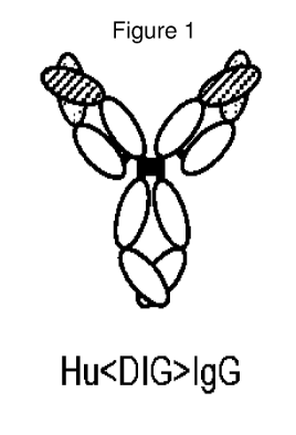
Figure 1: Schematic model of humanized <Dig> IgG
Figure 2: Procedure for digoxigenation (conjugation of digoxigenin
to) of
peptides (see e.g. Figure 2A) and examples of the digoxigenated
fluorophore Dig-Cy5 (Figure 2a, the fluorophore was used as
analytical surrogate for the peptide) and of the digoxigenated
PYY-derivative DIG-moPYY (DIG-moPYY) (Figure 2C):
Figure 3: Exemplary scheme of a complex of a monospecific
digoxigenin
binding anti¨DIG antibody and bispecific anti¨DIG antibody
with digoxigenin which conjugated to a peptide or to fluorophore
Figure 4: Proof of concept: complexes of anti¨DIG antibodies
(bispecifics
are used for proof of concept) with digoxigenated fluorophore (as
analytical surrogate for peptides) Cy5: Size exclusion
chromatography of digoxigenated Cy5 <Her2>-<Dig> bispecific
antibody complex indicates charging with digoxigenated Cy5 and
homogeneity of charged molecules. A chromatogram: 1: Her2
Dig Cy5 (1:0) 2: Her2 Dig Cy5 (1:0.5), 3: Her2 Dig Cy5 (1:1), 4:
Her2 Dig Cy5 (1:2), 5: Her2 Dig Cy5 (1:3), 6: Her2 Dig Cy5
(1:5). 7: Her2 Dig Cy5 (0:1), B analysis: Charging of bivalent
digoxigenin-binding antibodies becomes saturated at a 2:1
payload: antibody ratio.
Figure 5: Complex of anti¨DIG antibody with digoxigenin which
conjugated to a peptide: Antibody complexation of digoxigenin
which conjugated to a peptide results in a complex of defined size
as demonstrated by size exclusion chromatography.
Figure 6: Charging of anti¨DIG antibody with digoxigenin which
conjugated to a peptide: SEC-MALLS analyses demonstrate that
antibody complexation of digoxigenated peptides result in a
complex of defined size which is larger than uncomplexed
CA 02822481 2013-06-20
WO 2012/093068
PCT/EP2011/074273
- 4 -
antibody or uncomplexed peptide and contains 2 peptides per
antibody derivative.
Figure 7: Improved biological activity of digoxigenated and antibody-
complexed peptide compared to PEGylated peptide in vitro
Figure 8: Improved in
vivo serum half-life/stability of a digoxigenated
fluorescent dye (as surrogate for peptide) upon antibody
compl ex ati on.
Figure 9: Improved in vivo serum half-life/stability of a digoxigenated
peptide upon antibody complexation.
Figure 10: Improved in
vivo activity of antibody-complexed digoxigenated
peptides compared to uncomplexed peptides. In vivo potency of
the IgG-complexed DIG-moPYY-peptide can be detected by
reduction in food intake in treated animals.
Figure 11:
Improved in vivo activity of antibody-complexed digoxigenated
peptides compared to uncomplexed peptides. In vivo potency of
the IgG-complexed DIG-moPYY-peptide can be detected the
differences of food intake in animals that received uncomplexed
peptides compared to animals that received a 17-fold lower dose
of complexed peptide.
Detailed Description of the Invention
One aspect of the invention is a pharmaceutical composition comprising a
complex of:
a) a monospecific antibody that binds to digoxigenin, and
b) digoxigenin wherein the digoxigenin is conjugated to a peptide
consisting of 5 to 60 amino acids.
Another aspect of the invention a complex of:
a) a monospecific antibody that binds to digoxigenin, and
b) digoxigenin wherein the digoxigenin is conjugated to a peptide
consisting of 5 to 60 amino acids.
In one embodiment the peptide comprises 10 to 50 amino acids. Peptides with 12
or more amino acids typically have a secondary structure. Therefore in one
embodiment the peptide comprises 12 to 40 amino acids. In one embodiment the
peptide comprises 12 to 30 amino acids.
CA 02822481 2013-06-20
WO 2012/093068
PCT/EP2011/074273
- 5 -
The terms "digoxigenin " or "digoxygenin" or "DIG" are used interchangeable
herein and refer to 3-[(3S,5R,8R,9S,10S,12R,13S,14S,17R)-3,12,14-trihydroxy-
10,13 -dimethy1-1,2,3,4,5,6,7,8,9,11,12,15,16,17-tetradecahydro-cyclopenta[a]-
phenanthren-17-y1]-2H-furan-5-one (CAS number 1672-46-4). Digoxigenin (DIG)
is a steroid found exclusively in the flowers and leaves of the plants
Digitalis
purpurea, Digitalis orientalis and Digitalis lanata (foxgloves) (Polya, G.,
Biochemical targets of plant bioactive compounds, CRC Press, New York (2003)
p. 847).
The terms "anti-digoxigenin antibody" and "an antibody that binds to
digoxigenin"
refer to an antibody that is capable of binding digoxigenin with sufficient
affinity
such that a complex of a) a monospecific antibody that binds to digoxigenin,
and
b) digoxigenin wherein the digoxigenin is conjugated to a peptide consisting
of 5 to
60 amino acids, is formed which is useful as a therapeutic agent prolonging
the
half-time of the peptide.
The term "a digoxigenin that is conjugated to therapeutic peptide" refers to a
digoxigenin which is covalently linked to a peptide. Typically the digoxigenin
is
conjugate via its 3-hydroxy group to the peptide. Activated Digoxigenin-3-
carboxy-methyl derivatives are often used as starting materials for such
conjugated
digoxigenin peptides. In
one embodiment the digoxigenin is conjugated
(preferably via its 3-hydroxy group) to the peptide via a linker. Said linker
can
comprise a) a methylene-carboxy-methyl group (-CH2-C(0)-), b) from 1 to 10
(preferably from 1 to 5) amino acids (e.g. selected from glycine, serine,
glutamate,
13-alanine, y-aminobutyric acid, c-aminocaproic acid or lysine) and/or c) one
or
more (preferably one or two) compounds having the structural formula NH2-
[(CH2)nO]CH2-CH2-COOH in which n is 2 or 3 and x is 1 to 10, preferably 1 to
7 (which results (at least partly) in a linker (part) of the formula -NH-
[(CH2)nO]CH2-CH2-C(0)-; one example of such a compound is e.g. 12-amino-
4,7,10-trioxadodecanoic acid (results in a TEG (Triethylenglycol) linker or
TEG
spacer, see Example 5)). In one embodiment the linker further comprises a
maleimido group. Examples of digoxigenin conjugated to a peptide via such
linkers
are described in the Example 5 below. The linker has a stabilizing and
solubilizing
effect since it preferably contains charges or/and can form hydrogen bridges.
In
addition it can sterically facilitate the binding of the anti-digoxigenin
antibody to
the digoxigenin-conjugated peptide. In one embodiment the linker is located at
a
side chain of an amino acid of the peptide (e.g. conjugated to a lysine or
cystein
side chain via the amino or thio group). In one embodiment the linker is
located at
CA 02822481 2013-06-20
WO 2012/093068
PCT/EP2011/074273
- 6 -
the amino terminus or at the carboxy terminus of the peptide. The position of
the
linker on the peptide is typically chosen at a region where the biological
activity of
the peptide is not affected. Therefore the attachment position of the linkers
depends
on the nature of the peptide and the relevant structure elements which are
responsible for the biological activity. The biological activity of the
peptide to
which the digoxigenin attached can be tested in an in vitro assay.
The term "peptide," as used herein refers to a polymer of amino acids. As used
herein, these terms apply to amino acid polymers in which one or more amino
acid
residues is an artificial chemical analog of a corresponding naturally
occurring
amino acid. These terms also apply to naturally occurring amino acid polymers.
Amino acids can be in the L or D form. Peptides may be cyclic, having an
intramolecular bond between two non-adjacent amino acids within the peptide,
e.g.,
backbone to backbone, side-chain to backbone and side-chain to side-chain
cyclization. Cyclic peptides can be prepared by methods well know in the art.
See
e.g., U.S. Patent No. 6,013,625. Typical biologically active peptides are
described
e.g. in Bellmann-Sickert, K., et al., Trends Pharm. Sci. 31 (2010) 434-441.
All peptide sequences are written according to the generally accepted
convention
whereby the alpha-N-terminal amino acid residue is on the left and the alpha-C-
terminal amino acid residue is on the right. As used herein, the term "N-
terminus"
refers to the free alpha-amino group of an amino acid in a peptide, and the
term "C-
terminus" refers to the free a-carboxylic acid terminus of an amino acid in a
peptide. A peptide which is N-terminated with a group refers to a peptide
bearing a
group on the alpha-amino nitrogen of the N-terminal amino acid residue. An
amino
acid which is N-terminated with a group refers to an amino acid bearing a
group on
the alpha-amino nitrogen.
Unless indicated otherwise by a "D" prefix, e.g., D-Ala or N-Me-D-Ile, or
written
in lower case format, e.g., a, i, 1, (D versions of Ala, Ile, Leu), the
stereochemistry
of the alpha-carbon of the amino acids and aminoacyl residues in peptides
described in this specification and the appended claims is the natural or "L"
configuration. The Cahn-Ingold-Prelog "R" and "S" designations are used to
specify the stereochemistry of chiral centers in certain acyl substituents at
the N-
terminus of the peptides. The designation "R,S" is meant to indicate a racemic
mixture of the two enantiomeric forms. This nomenclature follows that
described
in Cahn, R. S., et al., Angew. Chem. lnt. Ed. Engl. 5 (1966) 385-415.
CA 02822481 2013-06-20
WO 2012/093068
PCT/EP2011/074273
- 7 -
In general, the term "amino acids" as used herein refers to natural an non-
natural
amino acids and their derivatives. Examples of such amino acids include, but
are
not limited to, Aad (alpha-Aminoadipic acid), Abu (Aminobutyric acid), Ach
(alpha-aminocyclohexane-carboxylic acid), Acp (alpha-aminocyclopentane-
carboxylic acid), Acpc (1-Aminocyclopropane-1-carboxylic acid), Aib (alpha-
aminoisobutyric acid), Aic (2-Aminoindane-2-carboxylic acid; also called 2-2-
Aic), 1-1-Aic (1 -aminoindane-l-carboxylic acid), (2-aminoindane-2-carboxylic
acid), Ala, Allylglycine (Ally1Gly), Alloisoleucine (allo-Ile), Arg, Asn, Asu
(alpha-
Aminosuberic acid, 2-Aminooctanedioc acid), Asp, Bip (4-phenyl-phenylalanine-
caroxylic acid), BnHP ((2S,4R)-4-Hydroxyproline), Cha (beta-
cyclohexylalanine),
Cit (Citrulline), Cyclohexylglycine (Chg), Cyclopentylalanine, beta-
Cyclopropyl
alanine, Cys, Dab (1,4-Diaminobutyric acid), Dap (1,3-Diaminopropionic acid),
p
(3,3-diphenylalanine-carboxylic acid), 3,3-Diphenylalanine, Di-n-propylglycine
(Dpg), 2-Furylalanine, Gln, Glu, Gly, His, Homocyclohexylalanine (HoCha),
Homocitrulline (HoCit), Homocycloleucine, Homoleucin (HoLeu), Homoarginine
(HoArg), Homoserine (HoSer), Hydroxyproline, Ile, Leu, Lys, Lys(Ac), (1) Nal
(1-
Naphtyl Alanine), (2) Nal (2- Naphtyl Alanine), Met, 4-Me0-Apc (1-amino-4-(4-
methoxypheny1)-cyclohexane-1-carboxylic acid), Nor-leucine (Nle), Nva
(Norvaline), Omathine, 3-Pal (alpha-amino-3-pyridylalanine-carboxylic acid), 4-
Pal (alpha-amino-4-pyridylalanine-carboxylic acid), Phe, 3,4,5,F3-Phe (3,4,5-
Trifluoro-phenylalanine), 2,3,4,5,6,F5-Phe (2,3,4,5,6-Pentafluoro-
phenylalanine),
Pqa (4-oxo-6-(1-piperaziny1)-3(4H)-quinazoline-acetic acid (CAS 889958-08-1)),
Pro, Pyridylalanine, Quinolylalanine,
Ser, Sarcosine (Sar), Thiazolylalanine,
Thienylalanine, Thr, Tic (alpha-amino-1,2,3,4,tetrahydroisoquinoline-3-
carboxylic
acid), Tic(OH), Tle (tertbutylGlycine), Trp, Tyr, Tyr(Me), Val.
In one embodiment of the invention the amino acid is selected from the group
cinsisiting of the list above.
For convenience in describing this invention, the abbreviations for the
natural
amino acids are listed below:
Asp=D=Aspartic Acid; Ala=A=Alanine; Arg=R=Arginine; Asn=N=Asparagine;
Gly=G=Gly eine; Glu=E=Glutamic Acid; Gln=Q=Glutamine; Hi s=H=Hi sti dine;
Ile=I=Isoleucine; Leu=L=Leucine; Lys=K=Lysine; Met=M=Methionine;
Phe=F=Phenylalanine; Pro=P=Proline; Ser=S=Serine; Thr=T=Threonine;
Trp=W=Tryptophan; Tyr=Y=Tyrosine; Cys = C= Cysteine; and Val=V=Valine.
CA 02822481 2013-06-20
WO 2012/093068
PCT/EP2011/074273
- 8 -
A non-limiting list of abbreviations for some of the typical amino acids
derivatizations is shown below:
Ac= Acetyl; Boc= 9-Fluorenylmethoxycarbonyl; Dde= ; Fmoc=9-
Fluorenylmethoxycarbonyl; Mtr= 4-Methoxy-2,3,6-trimethylbenzenesulfonyl; Pbf=
2,2,4,6,7-Pentamethyldihydrobenzofuran-5-sulfonyl; Trt= Trityl, tBu = tert-
Butyl;
TEG = 4,7,10-trioxadodecanoic acid ( = Triethylenglycol (TEG)-linker).
In one embodiment the peptide is a neuropeptide-2 receptor agonist as
described
e.g. WO 2007/065808. In one embodiment the peptide is selected from the group
consisting of
IK-Pqa-RHYLNLVTRQRY (SEQ ID NO:2);
IK-Pqa-RHYLNLVTRQ(N-methyl)RY (SEQ ID NO:3);
IK-Pqa-RHYLNLVTRQ(N-methyl)R(m-)Y (SEQ ID NO:4);
IK-Pqa-RHYLNLVTRQ(N-methyl)R(3-I)Y (SEQ ID NO:5);
IK-Pqa-RHYLNLVTRQ(N-methyl)R(3-5 di F)Y (SEQ ID NO:6);
IK-Pqa-RHYLNLVTRQ(N-methyl)R(2-6 di F)Y (SEQ ID NO:7);
IK-Pqa-RHYLNLVTRQ(N-methyl)R(2-6 di Me)Y (SEQ ID NO:8);
IK-Pqa-RHYLNLVTRQ(N-methyl)RF (0-CH3) (SEQ ID NO:9);
IK-Pqa-RHYLNLVTRQ(N-methyl)RF (SEQ ID NO:10);
IK-Pqa-RHYLNLVTRQ(N-methyl)R(4-NH2)Phe (SEQ ID NO:11);
IK-Pqa-RHYLNLVTRQ(N-methyl)R(4-F)Phe (SEQ ID NO:12);
IK-Pqa-RHYLNLVTRQ(N-methyl)R(4-CH2OH)Phe (SEQ ID NO:13);
IK-Pqa-RHYLNLVTRQ(N-methyl)R(4-CF3)Phe (SEQ ID NO:14);
IK-Pqa-RHYLNLVTRQ(N-methyl)R(3-F)Phe (SEQ ID NO:15);
IK-Pqa-RHYLNLVTRQ(N-methyl)R(2,3.4,5,6-Penta-F)Phe (SEQ ID NO:16);
IK-Pqa-RHYLNLVTRQ(N-methyl)R(3 .4- di Cl)Phe (SEQ ID NO:17);
IK-Pqa-RHYLNLVTRQ(N-methyl)RCha (SEQ ID NO:18);
IK-Pqa-RHYLNLVTRQ(N-methyl)RW (SEQ ID NO:19);
IK-Pqa-RHYLNLVTRQ(N-methyl)R(1)Nal (SEQ ID NO :20);
IK-Pqa-RHYLNLVTRQ(N-methyl)R(2)Nal (SEQ ID NO :21);
IK-Pqa-RHYLNLVTRQR-C-a-Me-Tyr (SEQ ID NO:22);
IK-Pqa-RHYLNWVTRQ(N-methyl)RY (SEQ ID NO:23);
CA 02822481 2013-06-20
WO 2012/093068
PCT/EP2011/074273
- 9 -INle-Pqa-RHYLNWVTRQ(N-methyl)RY (SEQ ID NO:24);
Ac-IK-Pqa-RHYLNWVTRQ(N-methyl)R(2-6 di F)Y (SEQ ID N0:25);
Ac-IK-Pqa-RHYLNWVTRQ(N-methyl)RY (SEQ ID N0:26);
Pentyl-IK-Pqa-RHYLNWVTRQ(N-methyl)RY (SEQ ID NO :27);
Trimetylacetyl-IK-Pqa-RHYLNWVTRQ(N-methyl)RY (SEQ ID NO :28);
Cyclohexyl-IK-Pqa-RHYLNWVTRQ(N-methyl)RY (SEQ ID NO :29);
Benzoyl-IK-Pqa-RHYLNWVTRQ(N-methyl)RY (SEQ ID NO :30);
and
Adamtyl-IK-Pqa-RHYLNWVTRQ(N-methyl)RY (SEQ ID NO:31).
In one embodiment the peptide is Ac-IK-Pqa-RHYLNWVTRQ(N-methyl)RY.
In one embodiment the peptide is Ac-IK-Pqa -RHYLNWVTRQ(N-methyl)R (2-6
di F)Y.
In one embodiment the peptide is selected from the group consisting of:
Ac-IK-Pqa-RHYLNWVTRQ(N-methyl)RY (SEQ ID NO:26);
GIGAVLKVLTTGLPALISWIKRKRQQ (SEQ ID NO:32);
FALLGDFFRKSKEKIGKEFKRIVQRIKDFLRNLVPRTES (SEQ ID
NO: 33);
NKRFALLGDFFRKSKEKIGKEFKRIVQRIKDFLRNLVPR (SEQ ID
NO:34); and
QHRYQQLGAGLKVLFKKTHRILRRLFNLAK (SEQ ID NO:35).
In one embodiment the peptide is substantially homologous to a peptide
selected
from the group consisting of:
In one embodiment the peptide is selected from the group consisting of:
Ac-IK-Pqa-RHYLNWVTRQ(N-methyl)RY (SEQ ID NO:26);
GIGAVLKVLTTGLPALISWIKRKRQQ (SEQ ID NO:32);
CA 02822481 2013-06-20
WO 2012/093068
PCT/EP2011/074273
- 10 -
FALLGDFFRKSKEKIGKEFKRIVQRIKDFLRNLVPRTES (SEQ ID
NO: 33);
NKRFALLGDFFRKSKEKIGKEFKRIVQRIKDFLRNLVPR (SEQ ID
NO:34); and
QHRYQQLGAGLKVLFKKTHRILRRLFNLAK (SEQ ID NO:35).
"Substantially homologous" means at least about 85% (preferably at least about
90%, and more preferably at least about 95% or most preferably at least about
98%,
of the amino-acid residues match over the defined length of the peptide
sequences.
Sequences that are substantially homologous can be identified by comparing the
sequences using standard software available in sequence data banks, such as
BLAST programs available from the National Cancer Center for Biotechnology
Information at ncbi.nlm.nih.gov.
In one embodiment the peptide is characterized in that it which shows
biological
activity in an in vitro assay. In one embodiment the biological activity is
anti-
proliferative, anti-inflammatory, anti-cancer, anti-viral, or the biological
activity is
metabolic disease related (see e.g. Example 7).
In one embodiment the complex is characterized in that the contains non-
natural
amino acids. In one embodiment the complex is characterized in that the
peptide
that cannot be produced in living organisms.
The term "antibody" herein is used for a monospecific antibody in the broadest
sense and encompasses various antibody structures, which, including but not
limited to monoclonal antibodies, polyclonal antibodies, and antibody
fragments so
long as they exhibit the desired antigen-binding activity.
An "antibody fragment" refers to a molecule other than an intact antibody that
comprises a portion of an intact antibody that binds the antigen to which the
intact
antibody binds. Examples of antibody fragments include but are not limited to
Fv,
Fab, Fab', Fab'-SH, F(ab')2; diabodies; linear antibodies; single-chain
antibody
molecules (e.g. scFv).
The term "monospecific antibody that binds to digoxigenin" as used herein
refers
to an antibody that specifically binds only to (the cardiac glycoside)
digoxigenin or
derivatives thereof like e.g. digoxin, digitoxin, but that does not
specifically bind to
a further (distinct) antigen like e.g. a protein antigen like e.g. HER2 or IGF-
1R.
CA 02822481 2013-06-20
WO 2012/093068
PCT/EP2011/074273
- 11 -
The term "bispecific antibody that binds to digoxigenin" as used herein refers
to an
antibody that specifically binds to (the cardiac glycoside) digoxigenin or
derivatives thereof like e.g. digoxin, digitoxin, and that also specifically
bind to a
further (distinct) antigen like e.g. a protein antigen like e.g. HER2 or IGF-
1R.
An "acceptor human framework" for the purposes herein is a framework
comprising the amino acid sequence of a light chain variable domain (VL)
framework or a heavy chain variable domain (VH) framework derived from a
human immunoglobulin framework or a human consensus framework, as defined
below. An acceptor human framework "derived from" a human immunoglobulin
framework or a human consensus framework may comprise the same amino acid
sequence thereof, or it may contain amino acid sequence changes. In some
embodiments, the number of amino acid changes are 10 or less, 9 or less, 8 or
less,
7 or less, 6 or less, 5 or less, 4 or less, 3 or less, or 2 or less. In some
embodiments,
the VL acceptor human framework is identical in sequence to the VL human
immunoglobulin framework sequence or human consensus framework sequence.
The term "chimeric" antibody refers to an antibody in which a portion of the
heavy
and/or light chain is derived from a particular source or species, while the
remainder of the heavy and/or light chain is derived from a different source
or
species.
The "class" of an antibody refers to the type of constant domain or constant
region
possessed by its heavy chain. There are five major classes of antibodies: IgA,
IgD,
IgE, IgG, and IgM, and several of these may be further divided into subclasses
(isotypes), e.g., IgGi, IgG2, IgG3, IgG4, IgAi, and IgA2. The heavy chain
constant
domains that correspond to the different classes of immunoglobulins are called
a,
8, c, 7, and , respectively.
The term "complex" of a) a monospecific antibody that binds to digoxigenin,
and
b) digoxigenin wherein the digoxigenin is conjugated to a peptide consisting
of 5 to
60 amino acids, as used herein refers to the non-covalent binding complex
formed
by the antibody and the digoxigenin (that is conjugated to the peptide of the
invention) based on the antibody-antigen interaction.
An "effective amount" of an agent, e.g., a pharmaceutical formulation, refers
to an
amount effective, at dosages and for periods of time necessary, to achieve the
desired therapeutic or prophylactic result.
CA 02822481 2013-06-20
WO 2012/093068
PCT/EP2011/074273
- 12 -
The term "Fe region" herein is used to define a C-terminal region of an
immunoglobulin heavy chain that contains at least a portion of the constant
region.
The term includes native sequence Fe regions and variant Fe regions. In one
embodiment, a human IgG heavy chain Fe region extends from Cys226, or from
Pro230, to the carboxyl-terminus of the heavy chain. However, the C-terminal
lysine (Lys447) of the Fe region may or may not be present. Unless otherwise
specified herein, numbering of amino acid residues in the Fe region or
constant
region is according to the EU numbering system, also called the EU index, as
described in Kabat et al., Sequences of Proteins of Immunological Interest,
5th ed.,
Public Health Service, National Institutes of Health, Bethesda, MD (1991).
"Framework" or "FR" refers to variable domain residues other than
hypervariable
region (HVR) residues. The FR of a variable domain generally consists of four
FR
domains: FR1, FR2, FR3, and FR4. Accordingly, the HVR and FR sequences
generally appear in the following sequence in VH (or VL): FR1-H1(L1)-FR2-
H2(L2)-FR3 -H3 (L3)-FR4 .
The terms "full length antibody", "intact antibody", and "whole antibody" are
used
herein interchangeably to refer to an antibody having a structure
substantially
similar to a native antibody structure or having heavy chains that contain an
Fe
region as defined herein.
The terms "host cell", "host cell line", and "host cell culture" are used
interchangeably and refer to cells into which exogenous nucleic acid has been
introduced, including the progeny of such cells. Host cells include
"transformants"
and "transformed cells," which include the primary transformed cell and
progeny
derived therefrom without regard to the number of passages. Progeny may not be
completely identical in nucleic acid content to a parent cell, but may contain
mutations. Mutant progeny that have the same function or biological activity
as
screened or selected for in the originally transformed cell are included
herein.
A "human antibody" is one which possesses an amino acid sequence which
corresponds to that of an antibody produced by a human or a human cell or
derived
from a non-human source that utilizes human antibody repertoires or other
human
antibody-encoding sequences. This definition of a human antibody specifically
excludes a humanized antibody comprising non-human antigen-binding residues.
A "human consensus framework" is a framework which represents the most
commonly occurring amino acid residues in a selection of human immunoglobulin
CA 02822481 2013-06-20
WO 2012/093068
PCT/EP2011/074273
- 13 -
VL or VH framework sequences. Generally, the selection of human
immunoglobulin VL or VH sequences is from a subgroup of variable domain
sequences. Generally, the subgroup of sequences is a subgroup as in Kabat et
al.,
Sequences of Proteins of Immunological Interest, fifth ed., NIH Publication 91-
3242, Bethesda MD (1991), Vols. 1-3. In one embodiment, for the VL, the
subgroup is subgroup kappa I as in Kabat et al., supra. In one embodiment, for
the
VH, the subgroup is subgroup III as in Kabat et al., supra.
A "humanized" antibody refers to a chimeric antibody comprising amino acid
residues from non-human HVRs and amino acid residues from human FRs. In
certain embodiments, a humanized antibody will comprise substantially all of
at
least one, and typically two, variable domains, in which all or substantially
all of
the HVRs (e.g., CDRs) correspond to those of a non-human antibody, and all or
substantially all of the FRs correspond to those of a human antibody. A
humanized
antibody optionally may comprise at least a portion of an antibody constant
region
derived from a human antibody. A "humanized form" of an antibody, e.g., a non-
human antibody, refers to an antibody that has undergone humanization.
The term "hypervariable region" or "HVR," as used herein, refers to each of
the
regions of an antibody variable domain which are hypervariable in sequence
and/or
form structurally defined loops ("hypervariable loops"). Generally, native
four-
chain antibodies comprise six HVRs; three in the VH (H1, H2, H3), and three in
the VL (L1, L2, L3). HVRs generally comprise amino acid residues from the
hypervariable loops and/or from the "complementarity determining regions"
(CDRs), the latter being of highest sequence variability and/or involved in
antigen
recognition. Exemplary hypervariable loops occur at amino acid residues 26-32
(L1), 50-52 (L2), 91-96 (L3), 26-32 (H1), 53-55 (H2), and 96-101 (H3)
(Chothia,
C., and Lesk, A.M., J. Mol. Biol. 196 (1987) 901-917). Exemplary CDRs (CDR-
Li, CDR-L2, CDR-L3, CDR-H1, CDR-H2, and CDR-H3) occur at amino acid
residues 24-34 of Li, 50-56 of L2, 89-97 of L3, 31-35B of H1, 50-65 of H2, and
95-102 of H3. (Kabat et al., Sequences of Proteins of Immunological Interest,
5th
ed., Public Health Service, National Institutes of Health, Bethesda, MD
(1991)).
With the exception of CDR1 in VH, CDRs generally comprise the amino acid
residues that form the hypervariable loops. CDRs also comprise "specificity
determining residues", or "SDRs", which are residues that contact antigen.
SDRs
are contained within regions of the CDRs called abbreviated-CDRs, or a-CDRs.
Exemplary a-CDRs (a-CDR-L1, a-CDR-L2, a-CDR-L3, a-CDR-H1, a-CDR-H2,
and a-CDR-H3) occur at amino acid residues 31-34 of Li, 50-55 of L2, 89-96 of
CA 02822481 2013-06-20
WO 2012/093068
PCT/EP2011/074273
- 14 -
L3, 31-35B of H1, 50-58 of H2, and 95-102 of H3 (see Almagro, J.C., and
Fransson, J., Front. Biosci. 13 (2008) 1619-1633). Unless otherwise indicated,
HVR residues and other residues in the variable domain (e.g., FR residues) are
numbered herein according to Kabat et al., supra.
The term "monoclonal antibody" as used herein refers to an antibody obtained
from
a population of substantially homogeneous antibodies, i.e., the individual
antibodies comprising the population are identical and/or bind the same
epitope,
except for possible variant antibodies, e.g., containing naturally occurring
mutations or arising during production of a monoclonal antibody preparation,
such
variants generally being present in minor amounts. In contrast to polyclonal
antibody preparations, which typically include different antibodies directed
against
different determinants (epitopes), each monoclonal antibody of a monoclonal
antibody preparation is directed against a single determinant on an antigen.
Thus,
the modifier "monoclonal" indicates the character of the antibody as being
obtained
from a substantially homogeneous population of antibodies, and is not to be
construed as requiring production of the antibody by any particular method.
For
example, the monoclonal antibodies to be used in accordance with the present
invention may be made by a variety of techniques, including but not limited to
the
hybridoma method, recombinant DNA methods, phage-display methods, and
methods utilizing transgenic animals containing all or part of the human
immunoglobulin loci, such methods and other exemplary methods for making
monoclonal antibodies being described herein.
"Percent (%) amino acid sequence identity" with respect to a reference
polypeptide
sequence is defined as the percentage of amino acid residues in a candidate
sequence that are identical with the amino acid residues in the reference
polypeptide sequence, after aligning the sequences and introducing gaps, if
necessary, to achieve the maximum percent sequence identity, and not
considering
any conservative substitutions as part of the sequence identity. Alignment for
purposes of determining percent amino acid sequence identity can be achieved
in
various ways that are within the skill in the art, for instance, using
publicly
available computer software such as BLAST, BLAST-2, ALIGN or Megalign
(DNASTAR) software. Those skilled in the art can determine appropriate
parameters for aligning sequences, including any algorithms needed to achieve
maximal alignment over the full length of the sequences being compared. For
purposes herein, however, % amino acid sequence identity values are generated
using the sequence comparison computer program ALIGN-2. The ALIGN-2
CA 02822481 2013-06-20
WO 2012/093068
PCT/EP2011/074273
- 15 -
sequence comparison computer program was authored by Genentech, Inc., and the
source code has been filed with user documentation in the U.S. Copyright
Office,
Washington D.C., 20559, where it is registered under U.S. Copyright
Registration
No. TXU510087. The ALIGN-2 program is publicly available from Genentech,
Inc., South San Francisco, California, or may be compiled from the source
code.
The ALIGN-2 program should be compiled for use on a UNIX operating system,
including digital UNIX V4.0D. All sequence comparison parameters are set by
the
ALIGN-2 program and do not vary.
In situations where ALIGN-2 is employed for amino acid sequence comparisons,
the % amino acid sequence identity of a given amino acid sequence A to, with,
or
against a given amino acid sequence B (which can alternatively be phrased as a
given amino acid sequence A that has or comprises a certain % amino acid
sequence identity to, with, or against a given amino acid sequence B) is
calculated
as follows:
100 times the fraction X/Y
where X is the number of amino acid residues scored as identical matches by
the
sequence alignment program ALIGN-2 in that program's alignment of A and B,
and where Y is the total number of amino acid residues in B. It will be
appreciated
that where the length of amino acid sequence A is not equal to the length of
amino
acid sequence B, the % amino acid sequence identity of A to B will not equal
the %
amino acid sequence identity of B to A. Unless specifically stated otherwise,
all %
amino acid sequence identity values used herein are obtained as described in
the
immediately preceding paragraph using the ALIGN-2 computer program.
The term "pharmaceutical formulation" refers to a preparation which is in such
form as to permit the biological activity of an active ingredient contained
therein to
be effective, and which contains no additional components which are
unacceptably
toxic to a subject to which the formulation would be administered.
A "pharmaceutically acceptable carrier" refers to an ingredient in a
pharmaceutical
formulation, other than an active ingredient, which is nontoxic to a subject.
A
pharmaceutically acceptable carrier includes, but is not limited to, a buffer,
excipient, stabilizer, or preservative.
As used herein, "treatment" (and grammatical variations thereof such as
"treat" or
"treating") refers to clinical intervention in an attempt to alter the natural
course of
CA 02822481 2013-06-20
WO 2012/093068
PCT/EP2011/074273
- 16 -
the individual being treated, and can be performed either for prophylaxis or
during
the course of clinical pathology. Desirable effects of treatment include, but
are not
limited to, preventing occurrence or recurrence of disease, alleviation of
symptoms,
diminishment of any direct or indirect pathological consequences of the
disease,
preventing metastasis, decreasing the rate of disease progression,
amelioration or
palliation of the disease state, and remission or improved prognosis. In some
embodiments, antibodies of the invention are used to delay development of a
disease or to slow the progression of a disease.
The term "variable region" or "variable domain" refers to the domain of an
antibody heavy or light chain that is involved in binding the antibody to
antigen.
The variable domains of the heavy chain and light chain (VH and VL,
respectively)
of a native antibody generally have similar structures, with each domain
comprising four conserved framework regions (FRs) and three hypervariable
regions (HVRs) (see, e.g., Kindt et al., Kuby Immunology, 6th ed., W.H.
Freeman
and Co., page 91 (2007)). A single VH or VL domain may be sufficient to confer
antigen-binding specificity. Furthermore, antibodies that bind a particular
antigen
may be isolated using a VH or VL domain from an antibody that binds the
antigen
to screen a library of complementary VL or VH domains, respectively (see,
e.g.,
Portolano, S., et al., J. Immunol. 150 (1993) 880-887; Clackson, T., et al.,
Nature
352 (1991) 624-628).
The term "vector", as used herein, refers to a nucleic acid molecule capable
of
propagating another nucleic acid to which it is linked. The term includes the
vector
as a self-replicating nucleic acid structure as well as the vector
incorporated into
the genome of a host cell into which it has been introduced. Certain vectors
are
capable of directing the expression of nucleic acids to which they are
operatively
linked. Such vectors are referred to herein as "expression vectors".
Compositions and Methods
In one aspect, the invention is based, in part, on a complex a) a monospecific
antibody that binds to digoxigenin, and b) digoxigenin wherein the digoxigenin
is
conjugated to a peptide consisting of 5 to 60 amino acids; and a
pharmaceutical
composition of it. In certain embodiments, antibodies that bind to digoxigenin
are
provided. Antibodies of the invention are useful, e.g., for the diagnosis or
treatment of cancer, metabolic or inflammatory or viral diseases.
CA 02822481 2013-06-20
WO 2012/093068
PCT/EP2011/074273
- 17 -
Exemplary Complexes of monospecific anti-digoxigenin antibodies and
digoxigenin conjugated to a peptide consisting of 5 to 60 amino acids
In one aspect, the invention is based, in part, on a complex a) a monospecific
antibody that binds to digoxigenin, and b) digoxigenin wherein the digoxigenin
is
conjugated to a peptide consisting of 5 to 60 amino acids.
Antibody Affinity
As used herein, the terms "binding" or an antibody "that binds to" or "that
specifically binds to" are use interchangeable and refer to the binding of the
antibody to an epitope of the tumor antigen in an in vitro assay, preferably
in an
plasmon resonance assay (BIAcore, GE-Healthcare Uppsala, Sweden) with purified
wild-type antigen. The affinity of the binding is defined by the terms ka
(rate
constant for the association of the antibody from the antibody/antigen
complex), kD
(dissociation constant), and KD (1cD/ka). Binding or specifically binding
means a
binding affinity (KD) of 10-8 M or less, preferably 10-8 M to 10-13 M (in one
embodiment 10-9 M to 10-13 M). Thus, an antibody that binds to digoxigenin
according to the invention is specifically binding to digoxigenin with a
binding
affinity (KD) of 10-8 mo1/1 or less, preferably 10-8 M to 10-13 M (in one
embodiment
10-9M to 10-13 M).
Anti-Digoxigenin Antibodies
Antibodies that bind specifically to the cardiac glycosides digoxin,
digitoxin, and
digoxigenin can be generated e.g. as described in Hunter, M.M., et al., J.
Immunol.
129 (1982) 1165-1172. One example of such antibody is the monoclonal antibody
26-10 that binds to the cardiac glycosides digoxin, digitoxin, and digoxigenin
with
high-affinity (KD = 9 nM) (Schildbach, J.F., et al., J. Biol. Chem. 268 (1993)
21739-21747; Burks, E.A., et al., PNAS 94 (1997) 412-417).
To prepare an immunogen for immunization e.g. digoxin or digoxigenin can be
conjugated to human serum albumin (Digoxin-HAS; Digoxigenin-HSA). Also
Digoxigenin or digoxin-3-CM0 (CMO =(0-carboxymethyl)oxime) conjugated to
KLH (keyhole limpet hemocyanin) is often used. Also Digoxigenin itself can be
used. Other methods to prepare digoxigenin immunogens are described e.g. in US
4469797. The resulting antibodies often bind to the cardiac glycosides
digoxin,
digitoxin, and digoxigenin ( i.e. they show cross-reactivity).
CA 02822481 2013-06-20
WO 2012/093068
PCT/EP2011/074273
- 18 -
Typical antibodies that bind to digoxigenin include the monoclonal antibody 26-
10,
monoclonal antibody 21H8 (AbcamCat# ab420); monoclonal antibody 1.A2.1
(Santa Cruz Cat# sc-70963), monoclonal antibody (1.71.256 Roche Applied
Science Cat# 11333062910).
Chimeric and Humanized Antibodies
In certain embodiments, an antibody provided herein is a chimeric antibody.
Certain chimeric antibodies are described, e.g., in U.S. Patent No. 4,816,567;
and
Morrison, S.L., et al., Proc. Natl. Acad. Sci. USA, 81(1984) 6851-6855). In
one
example, a chimeric antibody comprises a non-human variable region (e.g., a
variable region derived from a mouse, rat, hamster, rabbit, or non-human
primate,
such as a monkey) and a human constant region. In a further example, a
chimeric
antibody is a "class switched" antibody in which the class or subclass has
been
changed from that of the parent antibody. Chimeric antibodies include antigen-
binding fragments thereof
In certain embodiments, a chimeric antibody is a humanized antibody.
Typically, a
non-human antibody is humanized to reduce immunogenicity to humans, while
retaining the specificity and affinity of the parental non-human antibody.
Generally, a humanized antibody comprises one or more variable domains in
which
HVRs, e.g., CDRs, (or portions thereof) are derived from a non-human antibody,
and FRs (or portions thereof) are derived from human antibody sequences. A
humanized antibody optionally will also comprise at least a portion of a human
constant region. In some embodiments, some FR residues in a humanized antibody
are substituted with corresponding residues from a non-human antibody (e.g.,
the
antibody from which the HVR residues are derived), e.g., to restore or improve
antibody specificity or affinity.
Humanized antibodies and methods of making them are reviewed, e.g., in
Almagro, J.C., and Fransson, J., Front. Biosci. 13 (2008) 1619-1633, and are
further described, e.g., in Riechmann, L., et al., Nature 332 (1988) 332-327;
Queen,
C., et al., Proc. Natl. Acad. Sci. USA 86 (1989) 10029-10033; US Patent Nos.
5,821,337, 7,527,791, 6,982,321, and 7,087,409; Kashmiri, S.V., et al.,
Methods 36
(2005) 25-34 (describing SDR (a-CDR) grafting); Padlan, E.A., Mol. Immunol. 28
(1991) 489-498 (describing "resurfacing"); Dall'Acqua, W.F., et al., Methods
36
(2005) 43-60 (describing "FR shuffling"); and Osbourn et al., Methods 36:61-68
CA 02822481 2013-06-20
WO 2012/093068
PCT/EP2011/074273
- 19 -
(2005) and Klimka, A., et al., Br. J. Cancer 83 (2000) 252-260 (describing the
"guided selection" approach to FR shuffling).
Human framework regions that may be used for humanization include but are not
limited to: framework regions selected using the "best-fit" method (see, e.g.,
Sims,
M.J., et al. J. Immunol. 151 (1993) 2296-2308); framework regions derived from
the consensus sequence of human antibodies of a particular subgroup of light
or
heavy chain variable regions (see, e.g., Carter, P., et al., Proc. Natl. Acad.
Sci.
USA, 89 (1992) 4285-4289; and Presta, L.G., et al., J. Immunol. 151 (1993)
2623-
2632); human mature (somatically mutated) framework regions or human germline
framework regions (see, e.g., Almagro, J.C., and Fransson, J., Front. Biosci.
13
(2008) 1619-1633); and framework regions derived from screening FR libraries
(see, e.g., Baca, M., et al., J. Biol. Chem. 272 (1997) 10678-10684 and Rosok,
M.J., et al., J. Biol. Chem. 271 (1996) 22611-22618).
Human Antibodies
In certain embodiments, an antibody provided herein is a human antibody. Human
antibodies can be produced using various techniques known in the art. Human
antibodies are described generally in van Dijk, M.A., and van de Winkel, J.G.,
Curr. Opin. Chem. Biol. 5 (2001) 368-374 and Lonberg, N., Curr. Opin. Immunol.
(2008) 450-459.
20 Human
antibodies may be prepared by administering an immunogen to a transgenic
animal that has been modified to produce intact human antibodies or intact
antibodies with human variable regions in response to antigenic challenge.
Such
animals typically contain all or a portion of the human immunoglobulin loci,
which
replace the endogenous immunoglobulin loci, or which are present
extrachromosomally or integrated randomly into the animal's chromosomes. In
such transgenic mice, the endogenous immunoglobulin loci have generally been
inactivated. For review of methods for obtaining human antibodies from
transgenic
animals, see Lonberg, N., Nat. Biotech. 23 (2005) 1117-1125. See also, e.g.,
U.S.
Patent Nos. 6,075,181 and 6,150,584 describing XENOMOUSETm technology;
U.S. Patent No. 5,770,429 describing HuMAB technology; U.S. Patent No.
7,041,870 describing K-M MOUSE technology, and U.S. Patent Application
Publication No. US 2007/0061900, describing VELOCIMOUSE technology).
Human variable regions from intact antibodies generated by such animals may be
further modified, e.g., by combining with a different human constant region.
CA 02822481 2013-06-20
WO 2012/093068
PCT/EP2011/074273
- 20 -
Human antibodies can also be made by hybridoma-based methods. Human
myeloma and mouse-human heteromyeloma cell lines for the production of human
monoclonal antibodies have been described (see, e.g., Kozbor, D., J. Immunol.,
133
(1984) 3001-3005; Brodeur et al., Monoclonal Antibody Production Techniques
and Applications, Marcel Dekker, Inc., New York (1987), pp. 51-63; and
Boerner,
P., et al., J. Immunol. 147 (1991) 86-95). Human antibodies generated via
human
B-cell hybridoma technology are also described in Li, J., et al., Proc. Natl.
Acad.
Sci. USA 103 (2006) 3557-3562. Additional methods include those described, for
example, in U.S. Patent No. 7,189,826 (describing production of monoclonal
human IgM antibodies from hybridoma cell lines) and Ni, J., Xiandai Mianyixue
26 (2006) 265-268 (describing human-human hybridomas). Human hybridoma
technology (Trioma technology) is also described in Vollmers, H.P., and
Brandlein,
S., Histology and Histopathology 20 (2005) 927-937, and Vollmers, H.P., and
Brandlein, S., Methods and Findings in Experimental and Clinical Pharmacology
27 (2005) 185-191.
Human antibodies may also be generated by isolating Fv clone variable domain
sequences selected from human-derived phage display libraries. Such variable
domain sequences may then be combined with a desired human constant domain.
Techniques for selecting human antibodies from antibody libraries are
described
below.
Library-Derived Antibodies
Antibodies of the invention may be isolated by screening combinatorial
libraries
for antibodies with the desired activity or activities. For example, a variety
of
methods are known in the art for generating phage display libraries and
screening
such libraries for antibodies possessing the desired binding characteristics.
Such
methods are reviewed, e.g., in Hoogenboom, H.R., et al., Methods in Molecular
Biology 178 (2001) 1-37 and further described, e.g., in McCafferty, J., et
al.,
Nature 348, 552-554; Clackson, T., et al., Nature 352 (1991) 624-628; Marks,
J.D.,
et al., J. Mol. Biol. 222 (1991) 581-597; Marks, J.D., and Bradbury, A.,
Methods in
Molecular Biology 248 (2003) 161-176; Sidhu, S.S., et al., J. Mol. Biol. 338
(2004)
299-310; Lee, C.V., et al., J. Mol. Biol. 340 (2004) 1073-1093; Fellouse,
F.A.,
Proc. Natl. Acad. Sci. USA 101 (2004) 12467-12472; and Lee, C.V., et al., J.
Immunol. Methods 284 (2004) 119-132.
CA 02822481 2013-06-20
WO 2012/093068
PCT/EP2011/074273
-21 -
In certain phage display methods, repertoires of VH and VL genes are
separately
cloned by polymerase chain reaction (PCR) and recombined randomly in phage
libraries, which can then be screened for antigen-binding phage as described
in
Winter, G., et al., Ann. Rev. Immunol. 12 (1994) 433-455. Phage typically
display
antibody fragments, either as single-chain Fv (scFv) fragments or as Fab
fragments.
Libraries from immunized sources provide high-affinity antibodies to the
immunogen without the requirement of constructing hybridomas. Alternatively,
the
naive repertoire can be cloned (e.g., from human) to provide a single source
of
antibodies to a wide range of non-self and also self antigens without any
immunization as described by Griffiths, A.D., et al., EMBO J. 12 (1993) 725-
734.
Finally, naive libraries can also be made synthetically by cloning
unrearranged V-
gene segments from stem cells, and using PCR primers containing random
sequence to encode the highly variable CDR3 regions and to accomplish
rearrangement in vitro, as described by Hoogenboom, H.R., and Winter, G., J.
Mol.
Biol., 227 (1992) 381-388. Patent publications describing human antibody phage
libraries include, for example: US Patent No. 5,750,373, and US Patent
Publication
Nos. 2005/0079574, 2005/0119455, 2005/0266000, 2007/0117126, 2007/0160598,
2007/0237764, 2007/0292936, and 2009/0002360.
Antibodies or antibody fragments isolated from human antibody libraries are
considered human antibodies or human antibody fragments herein.
Antibody Variants
In certain embodiments, amino acid sequence variants of the antibodies
provided
herein are contemplated. For example, it may be desirable to improve the
binding
affinity and/or other biological properties of the antibody. Amino acid
sequence
variants of an antibody may be prepared by introducing appropriate
modifications
into the nucleotide sequence encoding the antibody, or by peptide synthesis.
Such
modifications include, for example, deletions from, and/or insertions into
and/or
substitutions of residues within the amino acid sequences of the antibody. Any
combination of deletion, insertion, and substitution can be made to arrive at
the
final construct, provided that the final construct possesses the desired
characteristics, e.g., antigen-binding.
Substitution, Insertion, and Deletion Variants
In certain embodiments, antibody variants having one or more amino acid
substitutions are provided. Sites of interest for substitutional mutagenesis
include
CA 02822481 2013-06-20
WO 2012/093068
PCT/EP2011/074273
- 22 -
the HVRs and FRs. Conservative substitutions are shown in Table below under
the
heading of "conservative substitutions." More substantial changes are provided
in
Table 1 under the heading of "exemplary substitutions," and as further
described
below in reference to amino acid side chain classes. Amino acid substitutions
may
be introduced into an antibody of interest and the products screened for a
desired
activity, e.g., retained/improved antigen binding, decreased immunogenicity,
or
improved ADCC or CDC.
Table:
Original Exemplary Preferred
Residue Substitutions Substitutions
Ala (A) Val; Leu; Ile Val
Arg (R) Lys; Gln; Asn Lys
Asn (N) Gln; His; Asp, Lys; Arg Gln
Asp (D) Glu; Asn Glu
Cys (C) Ser; Ala Ser
Gln (Q) Asn; Glu Asn
Glu (E) Asp; Gln Asp
Gly (G) Ala Ala
His (H) Asn; Gln; Lys; Arg Arg
Ile (I) Leu; Val; Met; Ala; Phe; Leu
Norleucine
Leu (L) Norleucine; Ile; Val; Met; Ala; Ile
Phe
Lys (K) Arg; Gln; Asn Arg
Met (M) Leu; Phe; Ile Leu
Pro (P) Ala Ala
Ser (S) Thr Thr
Thr (T) Val; Ser Ser
Trp (W) Tyr; Phe Tyr
Tyr (Y) Trp; Phe; Thr; Ser Phe
Val (V) Ile; Leu; Met; Phe; Ala; Leu
Norleucine
CA 02822481 2013-06-20
WO 2012/093068
PCT/EP2011/074273
- 23 -
Amino acids may be grouped according to common side-chain properties:
(1) hydrophobic: Norleucine, Met, Ala, Val, Leu, Ile;
(2) neutral hydrophilic: Cys, Ser, Thr, Asn, Gln;
(3) acidic: Asp, Glu;
(4) basic: His, Lys, Arg;
(5) residues that influence chain orientation: Gly, Pro;
(6) aromatic: Trp, Tyr, Phe.
Non-conservative substitutions will entail exchanging a member of one of these
classes for another class.
One type of substitutional variant involves substituting one or more
hypervariable
region residues of a parent antibody (e.g. a humanized or human antibody).
Generally, the resulting variant(s) selected for further study will have
modifications
(e.g., improvements) in certain biological properties (e.g., increased
affinity,
reduced immunogenicity) relative to the parent antibody and/or will have
substantially retained certain biological properties of the parent antibody.
An
exemplary substitutional variant is an affinity matured antibody, which may be
conveniently generated, e.g., using phage display-based affinity maturation
techniques such as those described herein. Briefly, one or more HVR residues
are
mutated and the variant antibodies displayed on phage and screened for a
particular
biological activity (e.g. binding affinity).
Alterations (e.g., substitutions) may be made in HVRs, e.g., to improve
antibody
affinity. Such alterations may be made in HVR "hotspots," i.e., residues
encoded
by codons that undergo mutation at high frequency during the somatic
maturation
process (see, e.g., Chowdhury, P.S., Methods Mol. Biol. 207 (2008) 179-196),
and/or SDRs (a-CDRs), with the resulting variant VH or VL being tested for
binding affinity. Affinity maturation by constructing and reselecting from
secondary libraries has been described, e.g., in Hoogenboom, H.R., et al.,
Methods
in Molecular Biology 178 (2001) 1-37). In some embodiments of affinity
maturation, diversity is introduced into the variable genes chosen for
maturation by
any of a variety of methods (e.g., error-prone PCR, chain shuffling, or
oligonucleotide-directed mutagenesis). A secondary library is then created.
The
library is then screened to identify any antibody variants with the desired
affinity.
Another method to introduce diversity involves HVR-directed approaches, in
which several HVR residues (e.g., 4-6 residues at a time) are randomized. HVR
CA 02822481 2013-06-20
WO 2012/093068
PCT/EP2011/074273
- 24 -
residues involved in antigen binding may be specifically identified, e.g.,
using
alanine scanning mutagenesis or modeling. CDR-H3 and CDR-L3 in particular are
often targeted.
In certain embodiments, substitutions, insertions, or deletions may occur
within
one or more HVRs so long as such alterations do not substantially reduce the
ability of the antibody to bind antigen. For example, conservative alterations
(e.g.,
conservative substitutions as provided herein) that do not substantially
reduce
binding affinity may be made in HVRs. Such alterations may be outside of HVR
"hotspots" or SDRs. In certain embodiments of the variant VH and VL sequences
provided above, each HVR either is unaltered, or contains no more than one,
two or
three amino acid substitutions.
A useful method for identification of residues or regions of an antibody that
may be
targeted for mutagenesis is called "alanine scanning mutagenesis" as described
by
Cunningham, B.C., and Wells, J.A., Science 244 (1989) 1081-1085. In this
method,
a residue or group of target residues (e.g., charged residues such as arg,
asp, his,
lys, and glu) are identified and replaced by a neutral or negatively charged
amino
acid (e.g., alanine or polyalanine) to determine whether the interaction of
the
antibody with antigen is affected. Further substitutions may be introduced at
the
amino acid locations demonstrating functional sensitivity to the initial
substitutions. Alternatively, or additionally, a crystal structure of an
antigen-
antibody complex to identify contact points between the antibody and antigen.
Such contact residues and neighboring residues may be targeted or eliminated
as
candidates for substitution. Variants may be screened to determine whether
they
contain the desired properties.
Amino acid sequence insertions include amino- and/or carboxyl-terminal fusions
ranging in length from one residue to polypeptides containing a hundred or
more
residues, as well as intrasequence insertions of single or multiple amino acid
residues. Examples of terminal insertions include an antibody with an N-
terminal
methionyl residue. Other insertional variants of the antibody molecule include
the
fusion to the N- or C-terminus of the antibody to an enzyme (e.g. for ADEPT)
or a
polypeptide which increases the serum half-life of the antibody.
Recombinant Methods and Compositions
Antibodies may be produced using recombinant methods and compositions, e.g.,
as
described in U.S. Patent No. 4,816,567. In one embodiment, isolated nucleic
acid
CA 02822481 2013-06-20
WO 2012/093068
PCT/EP2011/074273
- 25 -
encoding an anti-digoxigenin antibody described herein is provided. Such
nucleic
acid may encode an amino acid sequence comprising the VL and/or an amino acid
sequence comprising the VH of the antibody (e.g., the light and/or heavy
chains of
the antibody). In a further embodiment, one or more vectors (e.g., expression
vectors) comprising such nucleic acid are provided. In a further embodiment, a
host
cell comprising such nucleic acid is provided. In one such embodiment, a host
cell
comprises (e.g., has been transformed with): (1) a vector comprising a nucleic
acid
that encodes an amino acid sequence comprising the VL of the antibody and an
amino acid sequence comprising the VH of the antibody, or (2) a first vector
comprising a nucleic acid that encodes an amino acid sequence comprising the
VL
of the antibody and a second vector comprising a nucleic acid that encodes an
amino acid sequence comprising the VH of the antibody. In one embodiment, the
host cell is eukaryotic, e.g. a Chinese Hamster Ovary (CHO) cell or lymphoid
cell
(e.g., YO, NSO, Sp20 cell). In one embodiment, a method of making an anti-
digoxigenin antibody is provided, wherein the method comprises culturing a
host
cell comprising a nucleic acid encoding the antibody, as provided above, under
conditions suitable for expression of the antibody, and optionally recovering
the
antibody from the host cell (or host cell culture medium).
For recombinant production of an anti-digoxigenin, nucleic acid encoding an
antibody, e.g., as described above, is isolated and inserted into one or more
vectors
for further cloning and/or expression in a host cell. Such nucleic acid may be
readily isolated and sequenced using conventional procedures (e.g., by using
oligonucleotide probes that are capable of binding specifically to genes
encoding
the heavy and light chains of the antibody).
Suitable host cells for cloning or expression of antibody-encoding vectors
include
prokaryotic or eukaryotic cells described herein. For example, antibodies may
be
produced in bacteria, in particular when glycosylation and Fc effector
function are
not needed. For expression of antibody fragments and polypeptides in bacteria,
see, e.g., U.S. Patent Nos. 5,648,237, 5,789,199, and 5,840,523 (see also
Charlton,
K.A., Methods in Molecular Biology 248 (2004) 245-254, describing expression
of
antibody fragments in E. coli). After expression, the antibody may be isolated
from
the bacterial cell paste in a soluble fraction and can be further purified.
In addition to prokaryotes, eukaryotic microbes such as filamentous fungi or
yeast
are suitable cloning or expression hosts for antibody-encoding vectors,
including
fungi and yeast strains whose glycosylation pathways have been "humanized",
CA 02822481 2013-06-20
WO 2012/093068
PCT/EP2011/074273
- 26 -
resulting in the production of an antibody with a partially or fully human
glycosylation pattern (see Gerngross, T.U., Nat. Biotech. 22 (2004) 1409-1414;
and
Li, H., et al., Nat. Biotech. 24 (2006) 210-215).
Suitable host cells for the expression of glycosylated antibody are also
derived
from multicellular organisms (invertebrates and vertebrates). Examples of
invertebrate cells include plant and insect cells. Numerous baculoviral
strains have
been identified which may be used in conjunction with insect cells,
particularly for
transfection of Spodoptera frugiperda cells.
Plant cell cultures can also be utilized as hosts. See, e.g., US Patent Nos.
5,959,177, 6,040,498, 6,420,548, 7,125,978, and 6,417,429 (describing
PLANTIBODIES TM technology for producing antibodies in transgenic plants).
Vertebrate cells may also be used as hosts. For example, mammalian cell lines
that
are adapted to grow in suspension may be useful. Other examples of useful
mammalian host cell lines are monkey kidney CV1 line transformed by 5V40
(COS-7); human embryonic kidney line (293 or 293 cells as described, e.g., in
Graham, F.L., et al., J. Gen Virol. 36 (1977) 59-74); baby hamster kidney
cells
(BHK); mouse sertoli cells (TM4 cells as described, e.g., in Mather, J.P.,
Biol.
Reprod. 23 (1980) 243-252); monkey kidney cells (CV1); African green monkey
kidney cells (VERO-76); human cervical carcinoma cells (HELA); canine kidney
cells (MDCK; buffalo rat liver cells (BRL 3A); human lung cells (W138); human
liver cells (Hep G2); mouse mammary tumor (MMT 060562); TM cells, as
described, e.g., in Mather, J.P., et al., Annals N.Y. Acad. Sci. 383 (1982) 44-
68;
MRC 5 cells; and F54 cells. Other useful mammalian host cell lines include
Chinese hamster ovary (CHO) cells, including DHFR- CHO cells (Urlaub, G., et
al., Proc. Natl. Acad. Sci. USA 77 (1980) 4216-4220); and myeloma cell lines
such
as YO, NSO and Sp2/0. For a review of certain mammalian host cell lines
suitable
for antibody production, see, e.g., Yazaki, P.J., and Wu, A.M., Methods in
Molecular Biology 248 (2004) 255-268.
Assays
Anti-digoxigenin antibodies provided herein may be identified, screened for,
or
characterized for their physical/chemical properties and/or biological
activities by
various assays known in the art.
CA 02822481 2013-06-20
WO 2012/093068
PCT/EP2011/074273
- 27 -
Pharmaceutical Formulations
Pharmaceutical formulations of a complex a) a monospecific antibody that binds
to
digoxigenin, and b) digoxigenin wherein the digoxigenin is conjugated to a
peptide
consisting of 5 to 60 amino acids, as described herein are prepared by mixing
such
antibody having the desired degree of purity with one or more optional
pharmaceutically acceptable carriers (Remington's Pharmaceutical Sciences,
16th
edition, Osol, A. (ed.), (1980)), in the form of lyophilized formulations or
aqueous
solutions. Pharmaceutically acceptable carriers are generally nontoxic to
recipients
at the dosages and concentrations employed, and include, but are not limited
to:
buffers such as phosphate, citrate, and other organic acids; antioxidants
including
ascorbic acid and methionine; preservatives (such as octadecyldimethylbenzyl
ammonium chloride; hexamethonium chloride; benzalkonium chloride;
benzethonium chloride; phenol, butyl or benzyl alcohol; alkyl parabens such as
methyl or propyl paraben; catechol; resorcinol; cyclohexanol; 3-pentanol; and
m-
cresol); low molecular weight (less than about 10 residues) polypeptides;
proteins,
such as serum albumin, gelatin, or immunoglobulins; hydrophilic polymers such
as
polyvinylpyrrolidone; amino acids such as glycine, glutamine, asparagine,
histidine, arginine, or lysine; monosaccharides, disaccharides, and other
carbohydrates including glucose, mannose, or dextrins; chelating agents such
as
EDTA; sugars such as sucrose, mannitol, trehalose or sorbitol; salt-forming
counter-ions such as sodium; metal complexes (e.g. Zn-protein complexes);
and/or
non-ionic surfactants such as polyethylene glycol (PEG).
Exemplary
pharmaceutically acceptable carriers herein further include insterstitial drug
dispersion agents such as soluble neutral-active hyaluronidase glycoproteins
(sHASEGP), for example, human soluble PH-20 hyaluronidase glycoproteins, such
as rHuPH20 (HYLENEX , Baxter International, Inc.). Certain exemplary
sHASEGPs and methods of use, including rHuPH20, are described in US Patent
Publication Nos. 2005/0260186 and 2006/0104968. In one aspect, a sHASEGP is
combined with one or more additional glycosaminoglycanases such as
chondroitinases.
Exemplary lyophilized antibody formulations are described in US Patent No.
6,267,958. Aqueous antibody formulations include those described in US Patent
No. 6,171,586 and WO 2006/044908, the latter formulations including a
histidine-
acetate buffer.
CA 02822481 2013-06-20
WO 2012/093068
PCT/EP2011/074273
- 28 -
The formulation herein may also contain more than one active ingredients as
necessary for the particular indication being treated, preferably those with
complementary activities that do not adversely affect each other. Such active
ingredients are suitably present in combination in amounts that are effective
for the
purpose intended.
Active ingredients may be entrapped in microcapsules prepared, for example, by
coacervation techniques or by interfacial polymerization, for example,
hydroxymethylcellulose or gelatin-microcapsules and poly-(methylmethacylate)
microcapsules, respectively, in colloidal drug delivery systems (for example,
liposomes, albumin microspheres, microemulsions, nano-particles and
nanocapsules) or in macroemulsions. Such techniques are disclosed in
Remington's
Pharmaceutical Sciences 16th edition, Osol, A. (ed.) (1980).
Sustained-release preparations may be prepared. Suitable examples of sustained-
release preparations include semipermeable matrices of solid hydrophobic
polymers containing the antibody, which matrices are in the form of shaped
articles, e.g. films, or microcapsules.
The formulations to be used for in vivo administration are generally sterile.
Sterility may be readily accomplished, e.g., by filtration through sterile
filtration
membranes.
One aspect of the invention is a pharmaceutical composition according to the
invention for the treatment of metabolic diseases.
Another aspect is a pharmaceutical composition according to the invention for
the
treatment of cancer.
Another aspect is a pharmaceutical composition according to the invention for
the
treatment of inflammatory diseases.
One further aspect of the invention is a complex according to the invention
for the
treatment of metabolic diseases.
Another aspect is a complex according to the invention for the treatment of
cancer.
Another aspect is a complex according to the invention for the treatment of
inflammatory diseases.
CA 02822481 2013-06-20
WO 2012/093068
PCT/EP2011/074273
- 29 -
One further aspect of the invention is a complex according to the invention
for the
manufacture of a medicament for the treatment of metabolic diseases.
Another aspect is a complex according to the invention for the manufacture of
a
medicament for the treatment of cancer.
Another aspect is a complex according to the invention for the manufacture of
a
medicament for the treatment of inflammatory diseases.
Another aspect of the invention is a method of treatment of a patient
suffering from
a metabolic disease, cancer or a inflammatory disease, by administering an
effective amount of a complex according to the invention to said patient in
the need
of such treatment.
Articles of Manufacture
In another aspect of the invention, an article of manufacture containing
materials
useful for the treatment, prevention and/or diagnosis of the disorders
described
above is provided. The article of manufacture comprises a container and a
label or
package insert on or associated with the container. Suitable containers
include, for
example, bottles, vials, syringes, IV solution bags, etc. The containers may
be
formed from a variety of materials such as glass or plastic. The container
holds
a composition which is by itself or combined with another composition
effective
for treating, preventing and/or diagnosing the condition and may have a
sterile
access port (for example the container may be an intravenous solution bag or a
vial
having a stopper pierceable by a hypodermic injection needle). At least one
active
agent in the composition is an antibody of the invention. The label or package
insert indicates that the composition is used for treating the condition of
choice.
Moreover, the article of manufacture may comprise (a) a first container with a
composition contained therein, wherein the composition comprises an antibody
of
the invention; and (b) a second container with a composition contained
therein,
wherein the composition comprises a further cytotoxic or otherwise therapeutic
agent. The article of manufacture in this embodiment of the invention may
further
comprise a package insert indicating that the compositions can be used to
treat a
particular condition. Alternatively, or additionally, the article of
manufacture may
further comprise a second (or third) container comprising a pharmaceutically-
acceptable buffer, such as bacteriostatic water for injection (BWFI),
phosphate-
buffered saline, Ringer's solution and dextrose solution. It may further
include
CA 02822481 2013-06-20
WO 2012/093068
PCT/EP2011/074273
- 30 -
other materials desirable from a commercial and user standpoint, including
other
buffers, diluents, filters, needles, and syringes.
Sequence Listing
SEQ ID NO:1 PYY 3-36:
IKPEAPGEDASPEELNRYYASLRHYLNLVTRQRY
(3-36)
PYY derivatives
SEQ ID NO:2 IK-Pqa-RHYLNLVTRQRY
SEQ ID NO:3 IK-Pqa-RHYLNLVTRQ(N-methyl)RY
SEQ ID NO:4 IK-Pqa-RHYLNLVTRQ(N-methyl)R(m-)Y
SEQ ID NO:5 IK-Pqa-RHYLNLVTRQ(N-methyl)R(3-I)Y
SEQ ID NO:6 IK-Pqa-RHYLNLVTRQ(N-methyl)R(3-5 di F)Y
SEQ ID NO:7 IK-Pqa-RHYLNLVTRQ(N-methyl)R(2-6 di F)Y
SEQ ID NO:8 IK-Pqa-RHYLNLVTRQ(N-methyl)R(2-6 di Me)Y
SEQ ID NO:9 IK-Pqa-RHYLNLVTRQ(N-methyl)RF(0-CH3)
SEQ ID NO:10 IK-Pqa-RHYLNLVTRQ(N-methyl)RF
SEQ ID NO:11 IK-Pqa-RHYLNLVTRQ(N-methyl)R(4-NH2)Phe
SEQ ID NO:12 IK-Pqa-RHYLNLVTRQ(N-methyl)R(4-F)Phe
SEQ ID NO:13 IK-Pqa-RHYLNLVTRQ(N-methyl)R(4-CH2OH)Phe
SEQ ID NO:14 IK-Pqa-RHYLNLVTRQ(N-methyl)R(4-CF3)Phe
SEQ ID NO:15 IK-Pqa-RHYLNLVTRQ(N-methyl)R(3-F)Phe
SEQ ID NO:16 IK-Pqa-RHYLNLVTRQ(N-methyl)R(2,3.4,5,6-Penta-
F)Phe
SEQ ID NO:17 IK-Pqa-RHYLNLVTRQ(N-methyl)R(3.4-diC1)Phe
SEQ ID NO:18 IK-Pqa-RHYLNLVTRQ(N-methyl)RCha
SEQ ID NO:19 IK-Pqa-RHYLNLVTRQ(N-methyl)RW
SEQ ID NO:20 IK-Pqa-RHYLNLVTRQ(N-methyl)R(1)Nal
SEQ ID NO:21 IK-Pqa-RHYLNLVTRQ(N-methyl)R(2)Nal
SEQ ID NO:22 IK-Pqa-RHYLNLVTRQR-C-a-Me-Tyr
SEQ ID NO:23 IK-Pqa-RHYLNWVTRQ(N-methyl)RY
SEQ ID NO:24 INle-Pqa-RHYLNWVTRQ(N-methyl)RY
SEQ ID NO:25 Ac-IK-Pqa-RHYLNWVTRQ(N-methyl)R(2-6 di F)Y
SEQ ID NO:26 Ac-IK-Pqa-RHYLNWVTRQ(N-methyl)RY (= moPYY)
SEQ ID NO :27 Pentyl-IK-Pqa-RHYLNWVTRQ(N-methyl)RY
SEQ ID NO :28 Trimetylacetyl-IK-Pqa-RHYLNWVTRQ(N-methyl)RY
SEQ ID NO :29 Cyclohexyl-IK-Pqa-RHYLNWVTRQ(N-methyl)RY
CA 02822481 2013-06-20
WO 2012/093068
PCT/EP2011/074273
- 31 -
SEQ ID NO:30 B enzoyl-IK-Pqa-RHYLNWVTRQ(N-methyl)RY
SEQ ID NO:31 Adamtyl-IK-Pqa-RHYLNWVTRQ(N-methyl)RY
Peptides with antitumor effect
SEQ ID NO:32 GIGAVLKVLTTGLPALISWIKRKRQQ
SEQ ID NO:33 FALLGDFFRK SKEKIGKEFKRIVQRIKDFLRNLVPRT
ES
SEQ ID NO:34 NKRF ALL GDFFRK SKEKIGKEFKRIVQRIKDFLRNLV
PR
SEQ ID NO:35 QHRYQQLGAGLKVLFKKTHRILRRLFNLAK
Anti-DIG antibodies
SEQ ID NO:36 variable light chain domain VL of murine <Dig> 19-11
SEQ ID NO:37 variable heavy chain domain VH of murine <Dig> 19-11
SEQ ID NO:38 variable light chain domain VL of humanized <Dig> 19-
11
SEQ ID NO:39 variable heavy chain domain VH of humanized <Dig>
19-11
Although the foregoing invention has been described in some detail by way of
illustration and example for purposes of clarity of understanding, the
descriptions
and examples should not be construed as limiting the scope of the invention.
The
disclosures of all patent and scientific literature cited herein are expressly
incorporated in their entirety by reference.
EXAMPLES
The following are examples of methods and compositions of the invention. It is
understood that various other embodiments may be practiced, given the general
description provided above.
Experimental Procedures
Example 1: Isolation and characterization of cDNAs encoding the VH
and
VL domains of a murine <Dig> IgG1 kappa from mouse
hybridoma clone 19-11
Example 2: Humanization of the VH and VL domains of mu<Dig> 19-11
Example 3: Composition, expression and purification of recombinant
humanized <Dig> antibodies and bispecific derivatives
Example 4: Binding of recombinant humanized <Dig> antibodies, -
fragments
and ¨fusion proteins to digoxigenated compounds
CA 02822481 2013-06-20
WO 2012/093068
PCT/EP2011/074273
- 32 -
Example 5: Generation of digoxigenated compounds
Example 6: Generation of defined complexes of digoxigenated
compounds
with <Dig>IgG
Example 7: Digoxigenated peptides and complexes with <Dig>
antibodies
retain functionality
Example 8: Digoxigenated antibody-complexed PYY(3-36) derived
peptides
have better potency than PEGYlated PYY(3-36) derived peptides
in cell culture experiments
Example 9: Serum stability and serum levels of complexes of
digoxigenated
Cy5 or digoxigenated PYY-derived peptides with <Dig> IgG
Example 10: In vivo activity of complexes of digoxigenated PYY-
derived
peptides with <Dig> IgG
TABLES
Table 1: Binding affinities of the murine `wildtype' DIG-IgG and
recombinant <Dig> derivatives to different digoxigenated
antigens
Table 2: cytotoxic potency of unmodified and digoxigenated human-
derived peptides
Table 3: fluorescence of the unmodified and digoxigenated and complexed
fluorophore Cy5
Table 4: Biologic activity in vitro of PYY derivatives in the cAMP
assay
Table 5: PK parameters of uncomplexed and antibody-complexed Dig-
fluorophore and Dig-peptide
Example la:
Anti-Dig antibodies
Antibodies that bind specifically to the cardiac glycosides digoxin,
digitoxin, and
digoxigenin can be generated as described e.g. in Hunter, MM., et al, J.
Immunol.
129 (1982) 1165-1172. One example of such antibody is the monoclonal antibody
26-10. The 26-10 antibody binds to the cardiac glycosides digoxin, digitoxin,
and
digoxigenin with high-affinity (KD = 9 nM) (Schildbach, J. F., et al., J.
Biol.
Chem. 268 (1993) 21739-21747; Burks, E.A., et al., PNAS 94 (1997) 412-417).
CA 02822481 2013-06-20
WO 2012/093068
PCT/EP2011/074273
- 33 -
By applying these methods and using a digoxin conjugated to human serum
albumin for the immunization we generated the monoclonal, murine <Dig>
antibody hybridoma clone 19-11.
Example lb:
Isolation and characterization of cDNAs encoding the VII and VL domains of
a murine <Dig> IgG1 kappa from mouse hybridoma clone 19-11
A prerequisite for the design, generation, optimization and characterization
of
recombinant <Dig> antibodies, antibody fragments and ¨fusion proteins is the
availability of protein and (DNA) sequence information. Therefore, from the
hybridoma clone 19-11 this information for the VH and VL domains of murine
<Dig> antibody was obtained. The experimental steps that needed to be
performed
subsequently were (i) the isolation of RNA from <Dig> producing 19-11
hybridoma cells, (ii) conversion of this RNA into cDNA, then into VH and VL
harboring PCR fragments, and (iii) integration of these PCR fragments into
plasmids vectors for propagation in E.coli and determination of their DNA (and
deduced protein) sequences. More details of the herewith described
experimental
steps have been described in PCT/EP2010/004051.
RNA preparation from 19-11 hybridoma cells:
RNA was prepared from 5x10e6 antibody expressing hybridoma cells (clone 19-
11) applying the Rneasy-Kit (Qiagen). Briefly, the sedimented cells were
washed
once in PBS and sedimented and subsequently resuspended for lysis in 500 11.1
RLT-Puffer (+13-ME). The cells were completely lysed by passing through a
Qiashredder (Qiagen) and then subjected to the matrix-mediated purification
procedure (ETOH, RNeasy columns) as described in the manufacturers manual.
After the last washing step, RNA was recovered from the columns in 50 ul RNase-
free water. The concentration of the recovered RNA was determined by quantify
A260 and A280 of 1:20 diluted samples. The integrity (quality, degree of
degradation) of the isolated RNA samples was analyzed by denaturing RNA gel
electrophoresis on Formamide-Agarose gels (see Maniatis Manual). Examples of
these RNA gel electrophoreses, which showed discrete bands that represent the
intact 18s and 28 s ribosomal RNAs. Intactness (and approx 2:1 intensity
ratios) of
these bands indicated a good quality of the RNA preparations. The isolated
RNAs
from the 19-11 hybridoma were frozen and stored at -80 C in aliquots.
CA 02822481 2013-06-20
WO 2012/093068
PCT/EP2011/074273
- 34 -
Generation of DNA fragments encoding 19-11 VII and VII by RACE PCR:
The cDNA for subsequent (RACE-) PCR reactions were prepared from 19-11 RNA
preparations by applying the FirstChoice Kit (Ambion) reagent kit using the
described reactions for a standard 5"-RLM RACE protocol. Pwo DNA polymerase
was used for the PCR reaction. For that, 10 [tg of 19-11 RNA or control RNA
(from mouse thymus) was applied, and processed as described to integrate the
5"RACE adapter. We did not need to apply the 'outer PCR' reaction and directly
proceeded to the 'inner PCR': This involved combining primer pairs consisting
of
the 5"RACE Inner Primer (from the kit) and either C-kappa or CH1 specific
primers. The primer sequence for cKappa to amplify the VL region was 5'-
TTTTTTGCGGCCGCCctaacactcattcctgttgaagctc -3'. The primer sequence for
CH1 to amplify the VH region was 5'- TTTTTTGCGGCCGCGTAC
ATATGCAAGGCTTACAACCACAATCC -3'. For these primer combinations,
annealing temperatures of 60 C are suitable and temperatures between 55 and 65
C
/(Gradient PCR) have been applied to perform the PCR (94 C 0.5 min, 55-65 C 1
min ¨ 72 C lmin, 35 cycles, completion by 10 min extension at 72 C).
Successful
specific amplification of antibody VH or VL region containing DNA fragments
was reflected by occurrence of discrete 600bp to 800 bp DNA fragments which
were obtained from 19-11 RNA. These DNA fragments contain the VH and VL
encoding sequences of the <Dig> hybridoma 19-11.
Cloning of the DNA fragments encoding 19-11 VII and VII into plasmids and
determination of their DNA- and Protein sequences:
The VH and VL-encoding PCR fragments were isolated by agarose gel extraction
and subsequent purification by standard molecular biology techniques (Maniatis
Manual). The Pwo-generated purified PCR fragments were inserted into the
vector
pCR bluntII topo by applying the pCR bluntII topo Kit (Invitrogen) exactly
following the manufacturers instructions. The Topo- ligation reactions were
transformed into E.coli Topol0 ¨one-shot competent cells. Thereafter, E.coli
clones that contained vectors with either VL- or VH containing inserts were
identified as colonies on LB-Kanamycin agar plates. Plasmids were subsequently
prepared from these colonies and the presence of the desired insert in the
vector
was confirmed by restriction digestion with EcoRI. Because the vector backbone
contains EcoRI restriction recognition sites flanking each side of the insert,
plasmids harboring inserts were defined by having EcoRi-releasable inserts of
approx 800bp (for VL) or 600 bp (for VH).The DNA sequence and the deduced
CA 02822481 2013-06-20
WO 2012/093068
PCT/EP2011/074273
- 35 -
protein sequence of the 19-11 VL and VH was determined by automated DNA
sequencing on multiple clones for VH and VL. The amino acid sequence of the VL
of <Dig> clone 19-11 is shown in SEQ ID NO:36 and of the VH sequence of
<Dig> clone 19-11 in SEQ ID NO:37.
Example 2:
Humanization of the VH and VL domains of mu<Dig> 19-11
The objective of humanization of antibody sequences is to generate molecules
hat
retain full functionality of the original antibodies of murine origin, but
that harbor
no (or only very few or non-relevant) sequences or structures that are
recognized as
'foreign' by the human immune system. Different procedures are available and
have been published that can address this challenge (Almagro, J.C., and
Fransson,
J., Frontiers in Bioscience 13 (2008) 1619-1633; Hwang, W.Y.K., and Foote, J.,
Methods 36 (2005) 3-10). The functionality of variable regions of antibodies
is
determined by secondary and tertiary (and quaternary) structures, whose
formation
however base on the primary sequence of VH and VL (and of adjacent and
interacting entities). Because of that, the major challenge of humanization is
to
(fully) retain structure-defined functionality despite the need to change the
primary
protein sequence at some positions. Thus, knowledge about the structure of
functionally important regions of antibodies (CDR regions) is very important
to
support humanization. To generate humanized mu<Dig> 19-11 derived variants we
combined the following experimental wet-lab as well as in-silico procedures.
Starting with (i) in silico ¨predictions of the antigen binding site of
mu<Dig> 19-11
we were able to (ii) predict in-silico hu<Dig> variants with a high degree of
human-likeness as well as high probability to retain full functionality.
Finally (iii)
we experimentally determined the (X-ray) structure of <Dig> antibody
(fragments)
with and without antigen to validate and improve upon our in silico model.
More
details of the herewith described design parameters and experimental steps
have
been described in PCT/EP2010/004051.
In silico modeling of the antigen binding site of mu<Dig> 19-11:
The basis for our in-silico structure model for the mu<Dig>19-11 Fv region are
the
protein sequences that were deduced from the experimentally determined VH and
VL mRNA sequences. A structure model of the protein encoded by these
sequences was generated in silico by homology modeling of the Fv domain of the
murine antibody combined with energy minimization. For that, CDRs and
CA 02822481 2013-06-20
WO 2012/093068
PCT/EP2011/074273
- 36 -
framework sequences to apply for the homology modeling were separately
searched for homology over the PDB (Protein DataBank). For each CDR and for
the frameworks, the more homolog structures were superimposed. A model was
subsequently built from the different part for both the light and the heavy
chains
followed by a (energy) minimization of the complex. The structure model of the
mu <Dig> 19-11 Fv region that resulted from our homology-modeling procedure
showed that one rather particular feature of the predicted structure is a
prominent
cavity that appears to extend deep into the VH-VL interface. The main
determinant
for formation of this narrow cavity is the long CDR3 loop of VH. The interior
of
the cavity is lined with a methionine (deeper residue), 2 serines, 2 prolines,
an a
few tyrosines (flanking walls). The antigen digoxigenin that is recognized by
this
antibody is bound in a hapten-like manner into the deep cavity.
Crystallization and X-ray structure determination of the binding region of the
murine anti-Dig FAT region in the presence of antigen:
To enable further optimization of the humanized VH and VL sequences of the
anti-
digoxigenin antibody, we experimentally determined the structure of the parent
(murine) antibody. For that, Fab fragments were generated by protease
digestion of
the purified IgGs, applying well known state of the art methods (papain
digestion).
Fab fragments were separated from remaining Fc-fragments by protein A
chromatography (which removes Fc), thereafter subjected to size exclusion
chromatography (Superdex200 HiLoad 120 ml 16/60 gel filtration column, GE
Healthcare, Sewden) to remove protein fragments. For crystallization, purified
Fabs
in 20 mM His-HC1, 140 mM NaC1, pH 6.0 and Cy5 labeled Digoxigenin (DIG-3-
cme-dea-Cy5 = DIG-Cy5 / powder) were complexed with digoxigenated
fluorescent dye Cy5 (Dig-Cy5). Prior to crystal setups the protein solution
was
concentrated. For complex formation DIG-Cy5 was dissolved in 20 mM His-HC1,
140 mM NaC1, pH 6.0 and added to a final molar ratio of 5:1 to the
concentrated
protein solution. Crystals of murine Fab in complex with DIG-Cy5 were obtained
using the hanging drop vapor diffusion method at 25 C after mixing 1 11.1
protein
solution (24 mg/ml) with 1 11.1 reservoir solution containing 60% (v/v) 2-
methyl-
1,3-propandiol (MPD) / 0.1 M sodium acetate pH 4.6 / 5 mM CaC12. Crystals were
flash frozen in liquid nitrogen crystals without the need of any further
cryoprotection._Diffraction data of murine Fab in complex with DIG-Cy5 were
collected at X06SA (SLS, Villingen, Switzerland) on September 11th 2009. Data
were integrated and scaled with XDS (Kabsch, J. Appl. Cryst. 21(1993) 916-
924).
Crystals of the complex belong to space group P42212 with a=b=138.01 A,
CA 02822481 2013-06-20
WO 2012/093068
PCT/EP2011/074273
- 37 -
c=123.696, a=f3=y=90 and diffracted to a resolution of 2.8 A. _The structure
was
solved by molecular replacement using the program BALBES (see Long, F., et
al.,
Acta Crystallogr. D Biol. Crystallogr. 64 (Pt. 1) (2008) 125-132) by
generating a
search model based on structures with PDB ID 3cfd, 2a6d, 2a6j (Debler, E.W.,
et
al., Science 319 (2008) 1232-1235; Sethi, D.K., et al., Immunity 24 (2006) 429-
438). In total 2 Fab molecules could be located in the asymmetric unit. The
initial
models were completed and refined by manual model building with the program
COOT (Emsley, P., and Cowtan, K., Acta Crystallogr. D Biol. Crystallogr. 60
(Pt.
12 Pt. 1) (2004) 2126-2132) and refinement using the program PHENIX (Zwart,
P.H., et al., Methods Mol. Biol. 426 (2008) 419-435). After first rounds of
refinement a difference electron density for the DIG moiety of DIG-Cy5
appeared.
A model for DIG was obtained from PDB ID llke (Korndorfer, I.P., et al., J.
Mol.
Biol. 330 (2003) 385-396) and refinement parameters for DIG were generated by
the online tool PRODRG (Schuttelkopf, A.W., and van Aalten, D.M., Acta
Crystallogr. D Biol. Crystalogr. 60 (Pt. 8) (2004) 1355-1363). The model of
DIG
was placed in the electron density for final refinement steps. Figures were
prepared
with the program PYMOL (DeLano, W.L., The PyMOL Molecular Graphics
System (2008)).
The results of the experimental structure determination have been described in
detail in PCT/EP2010/004051. The structure revealed that the obtained crystal
form
contained two independent DIG-Cy5:anti-DIG Fab complexes in the asymmetric
unit and atomic models for both complexes could be build. The DIG moiety of
DIG-Cy5 is well ordered in both Fab molecules in the asymmetric unit although
it
appears to be bound in one molecule of the asymmetric unit more tightly than
in
the other one. DIG is bound in a pocket located at the interface of chain L
and
chain H in the middle of the CDR. Atom 032 of DIG is pointing towards the
bottom of the pocket and the linker with Cy5 is located outside and points
into the
solvent. In addition to DIG, a clear 2F0-Fc electron density is visible for
the first C
atom of the linker to Cy5 (panel B in Figure 45b). Due to the flexibility of
the
linker neither the remainder of the linker nor Cy5 are visible in the electron
density
map. This disorder indicates that the linker is not attached to the protein
and long
enough to allow attachment of molecules of different nature and size such as
dyes,
siRNA and others to DIG without influencing the recognition of DIG by the
antibody._Interestingly the binding pocket is not completely hydrophobic as
expected for a hydrophobic molecule as DIG but contains some positive charge
potential. The binding pocket is lined by four Tyrosin residues (57, 59, 109,
110) as
CA 02822481 2013-06-20
WO 2012/093068
PCT/EP2011/074273
- 38 -
well as A33, W47, P61, P99 and M112 of the heavy chain. From the light chain
residues Q89, S91, L94, P96 and F98 are involved in pocket formation. The
possible hydrogen bonding partners N35 and Y36 of the light chain form the
bottom of the pocket but are not reached by the DIG. Only one direct hydrogen
bond is involved in DIG binding and is formed between 032 of DIG and Q89 of
the light chain. Two more hydrogen bonds are not direct but mediated through
water molecules. 012 is interacting with the carbonyl oxygen of Y109 and the
side
chain of S35 of the heavy chain. A fourth hydrogen bond is formed between 014
and backbone carbonyl oxygen of S91 (chain L) but again mediated by a water
molecule. Comparisons of the number and the lengths of the hydrogen bonds in
both molecules of the asymmetric unit indicate that in the second complex DIG
is
not able to fully enter the pocket. In one molecule the DIG moiety immerses
relatively deep into the pocket and forms four hydrogen bonds. The second DIG
is
bound more loosely bound, it does not enter the pocket as deep as in the other
molecule and forms only three hydrogen bonds that are weaker than in the other
molecule.
Definition of mu<Dig>19-11 humanized variants which retain full
functionality:
The results of the experimental determination of the binding region at a
resolution
of 2.8 A enables the characterization of the binding mode of the ligand to its
antibody. It further confirms that structure is generally similar to the
structure
model that we predicted by in-silico analyses of the primary sequence. The
availability of the in silico modeled structure as well as of experimentally
determined 'real' structure of the variable region of the parent antibody (see
PCT/EP2010/004051 for more details) is a prerequisite for detailed modeling
and
further improvement via protein engineering of recombinant digoxigenin binding
modules. Amino acid sequences that represent desired humanized VH and VL
domains were defined by applying a procedure which is based on CDR-grafting
and introduction of additional mutations which modulate binding specificity
and
affinity. The basic principle underlying this procedure is the attribution of
a 'score
value' for each amino acid that differs from the mouse sequence among the
human
germlines. This score is defined by its putative influence of the amino acid
change
on the antigen recognition capability or on the stability of the complex.
Human
germline are selected based on their lower score and their relative high
usage.
TEPITOPE analyses (predicting T-cell epitopes) are included in this
humanization
procedure with the objective to have few to no t-cell epitopes in the
resulting
CA 02822481 2013-06-20
WO 2012/093068
PCT/EP2011/074273
- 39 -
humanized molecule. The 'human' sequences initially defined by this procedure
may need to be replaced by the (original) murine ones when the score is too
high
(indicating high probability of negative interference). This is most
frequently
required for amino acid changes in the CDR or in the surrounding region of the
CDR sequences. In some instances, 'back-mutations' to murine residues are
required not only in the CDRs but also within the framework to retain
stability and
functionality._The resulting hu<Dig> variant that we chose is based on the
human
Framework VH3 11 and VL1 39 combination, and has a high degree of human-
likeliness. For VL, it was not necessary to integrate any backmutation in the
framework of the human VK1 39 and the human j element of IGKJ4-01/02
germlines. This lead to a high human character and a relatively low number of
TEPITOPE alerts. The VH variant is originated from the human VH3 23 germline
and the human J IGHJ6-01-2. The variant J is built on the human VH3 11
germline. Moreover, using our scoring methodology, we were able to introduce
one
human amino acid within CDRS in order to increase the human character and
decrease the number of TEPITOPE alerts. The amino acid sequence of the
humanized VH is shown in SEQ ID NO:38 and of the humanized VL in SEQ ID
NO:39.
Generation of Digoxygenin binding modules with increased affinity:
Further optimization of the humanized VH and VL sequences of the anti-
digoxigenin antibody was applied to generate modules with even higher affinity
towards digoxigenin. Based upon the experimentally determined as well as in-
silico
calculated predicted structures (see above, based upon structure modeling
without
experimental structure determination), we identified three positions in which
alterations might affect affinity. These were located at (Kabat positions)
5er49,
11e57 and A1a60 of the VH domain. Replacement of the amino acid VHSer49 with
Ala, VHI1e57 with Ala and of VHA1a60 with Pro generated the respective
antibody
derivatives. Binding entities that are composed of this sequence could be
expressed
and purified with standard Protein-A and size exclusion technologies (see
Example
3 'Composition, expression and purification of recombinant humanized <Dig>
antibodies, -fragments and bispecific ¨fusion proteins). The resulting
molecules
were fully functional and displayed improved affinity towards digoxigenin
compared to the humanized parent molecule. This was demonstrated by Surface-
Plasmon-Resonance (BiaCore) experiments (see Example 4 'Binding of
recombinant <Dig> antibodies, -fragments and bispecific ¨fusion proteins to
digoxigenated antigens' for details). The results of these experiments proved
that
CA 02822481 2013-06-20
WO 2012/093068
PCT/EP2011/074273
- 40 -
the affinity towards digoxigenin is improved approximately 10-fold by
introducing
VH49, VH57 and VH60 mutations. The relevance of these positions was thereafter
confirmed by inspecting the experimentally determined structure of the Dig-
binding variable region.
Example 3:
Composition, expression and purification of recombinant humanized <Dig>
antibodies
Murine and humanized <Dig> modules were combined with constant regions of
human antibodies, either to form chimeric or humanized IgG's or to generate
bispecific fusion proteins with other antibody sequences. The generation of
humanized <Dig> IgGs that bind Dig required (i) design and definition of amino-
and nucleotide sequences for such molecules, (ii) expression of these
molecules in
transfected cultured mammalian cells, and (iii) purification of these
molecules from
the supernatants of transfected cells. Also bispecific derivatives that bind
Dig as
well as other targets (e.g. receptor tyrosine kinases Her2 or IGF1R) were also
generated as used as model systems for proof of concept studies, where e.g.
the
defined complexation of the peptides or fluorophores could be demonstrated in
the
Examples below. Additional details of the herewith described experimental
steps
have been described in PCT/EP2010/004051.
Design and definition of amino- and nucleotide sequences of <Dig> IgG and
bispecific antibody derivatives that bind Digoxygenin as well as Her2 or
IGF 1R:
To generate a humanized IgG that harbors the binding specificity of the
(original)
murine mu<Dig>19-11 Fv region, we fused the above defined humanized VH
sequence in frame to the N-terminus of CH1-CH2-CH3 of IgGl. Similarly, we
fused the above defined humanized VL sequence in frame to the N-terminus of
Ckappa. The amino acid -sequences of the resulting hu<Her2><Dig> IgG H- and
L-chains have been described in PCT/EP2010/004051. A schematic representation
of a humanized digoxigenin-binding IgG is provided in Figure 1.
To generate bispecific antibody derivatives that contain the binding
specificity of
hu<Dig> as well as specificities to the receptor tyrosine kinase Her2 or
IGF1R, we
fused the <Dig> single-chain Fv module defined by humanized VH and VL
sequences in frame to the C-terminus of the H-chain of a previously described
<Her2> antibody (e.g. US patent 5,772,997), or of a IGF1R antibody,
respectively.
CA 02822481 2013-06-20
WO 2012/093068
PCT/EP2011/074273
-41 -
The applied <Dig> scFv module was further stabilized by introduction of a VH44-
VL100 disulfide bond which has been previously described (e.g. Reiter, Y., et
al.,
Nature Biotechnology 14 (1996) 1239-1245). The amino acid and sequences of the
resulting bispecific antibody derivatives that bind Her2 or IGF1R as well as
Digoxigenin have been described in PCT/EP2010/004051.
Expression of <Dig> IgG and of bispecific antibody derivatives that bind
Digoxigenin as well as Her2 or IGF1R:
The <Dig> IgG and the bispecific antibody derivatives were expressed by
transient
transfection of human embryonic kidney 293-F cells using the FreeStyleTM 293
Expression System according to the manufacturer's instruction (Invitrogen,
USA).
For that, light and heavy chains of the corresponding bispecific antibodies
were
constructed in expression vectors carrying pro- and eukaryotic selection
markers.
These plasmids were amplified in E.coli, purified, and subsequently applied
for
transient transfections. Standard cell culture techniques were used for
handling of
the cells as described in Current Protocols in Cell Biology (2000),
Bonifacino, J.S.,
Dasso, M., Harford, J.B., Lippincott-Schwartz, J. and Yamada, K.M. (eds.),
John
Wiley & Sons, Inc. The suspension FreeStyleTM 293-F cells were cultivated in
FreeStyleTM 293 Expression medium at 37 C/8 % CO2 and the cells were seeded in
fresh medium at a density of 1-2x10⁶ viable cells/ml on the day of
transfection.
The DNA-293fectinTM complexes were prepared in Opti-MEM I medium
(Invitrogen, USA) using 333 11.1 of 293fectinTM (Invitrogen, Germany) and 250
tg
of heavy and light chain plasmid DNA in a 1:1 molar ratio for a 250 ml final
transfection volume. The IgG or bispecific antibody containing cell culture
supernatants were clarified 7 days after transfection by centrifugation at
14000 g
for 30 minutes and filtration through a sterile filter (0.22 p.m).
Supernatants were
stored at -20 C until purification. To determine the concentration of
antibodies
and derivatives in the cell culture supernatants, affinity HPLC chromatography
was
applied. For that, cell culture supernatants containing antibodies and
derivatives
that bind to Protein A were applied to an Applied Biosystems Poros A/20 column
in 200 mM KH2PO4, 100 mM sodium citrate, pH 7.4 and eluted from the matrix
with 200 mM NaC1, 100 mM citric acid, pH 2,5 on an UltiMate 3000 HPLC
system (Dionex). The eluted protein was quantified by UV absorbance and
integration of peak areas. A purified standard IgG1 antibody served as a
standard.
CA 02822481 2013-06-20
WO 2012/093068
PCT/EP2011/074273
- 42 -
Purification of <Dig> IgG and of bispecific antibody derivatives that bind
digoxygenin as well as Her2 or IGF1R:
7 days after transfection of the expression plasmids, the HEK293 cell
supernatants
were harvested. The recombinant antibody (-derivatives) contained therein were
purified from the supernatant in two steps by affinity chromatography using
Protein
A-SepharoseTM (GE Healthcare, Sweden) and Superdex200 size exclusion
chromatography. Briefly, the monospecific and bispecific antibody containing
clarified culture supernatants were applied on a HiTrap ProteinA HP (5 ml)
column
equilibrated with PBS buffer (10 mM Na2HPO4, 1 mM KH2PO4, 137 mM NaC1
and 2.7 mM KC1, pH 7.4). Unbound proteins were washed out with equilibration
buffer. The bispecific antibodies were eluted with 0.1 M citrate buffer, pH
2.8, and
the protein containing fractions were neutralized with 0.1 ml 1 M Tris, pH
8.5.
Then, the eluted protein fractions were pooled, concentrated with an Amicon
Ultra
centrifugal filter device (MWCO: 30 K, Millipore) to a volume of 3 ml and
loaded
on a Superdex200 HiLoad 120 ml 16/60 gel filtration column (GE Healthcare,
Sweden) equilibrated with 20mM Histidin, 140 mM NaC1, pH 6Ø The protein
concentration of purified antibodies and derivatives was determined by
determining
the optical density (OD) at 280 nm with the OD at 320nm as the background
correction, using the molar extinction coefficient calculated on the basis of
the
amino acid sequence according to Pace et. al., Protein Science, 1995, 4, 2411-
1423.
Monomeric antibody fractions were pooled, snap-frozen and stored at -80 C.
Part
of the samples were provided for subsequent protein analytics and
characterization.
The homogeneity of the DIGHu2 antibody construct and the bispecific DIG
constructs were confirmed by SDS-PAGE in the presence and absence of a
reducing agent (5 mM 1,4-dithiotreitol) and staining with Coomassie brilliant
blue.
The NuPAGE Pre-Cast gel system (Invitrogen, USA) was used according to the
manufacturer's instruction (4-20% Tris-Glycine gels). Under reducing
conditions,
polypeptide chains related to the IgG (and also bispecific FIT fusions) showed
upon
SDS-PAGE at apparent molecular sizes analogous to the calculated molecular
weights. Expression levels of all constructs were analysed by Protein A.
Average
protein yields were between 6 and 35 mg of purified protein per liter of cell-
culture
supernatant in such non-optimized transient expression experiments. More
details
of the herewith described expression and purification steps are described in
PCT/EP2010/004051.
CA 02822481 2013-06-20
WO 2012/093068
PCT/EP2011/074273
- 43 -
Example 4:
Binding of recombinant humanized <Dig> antibodies, -fragments and ¨fusion
proteins to digoxigenated compounds
The analyses that are described below were performed to evaluate if the
humanization procedure resulted in <Dig> derivatives that had retained full
binding
activity. For that, binding properties of the recombinant <Dig> derivatives
were
analyzed by surface plasmon resonance (SPR) technology using a Biacore T100 or
Biacore 3000 instrument (GE Healthcare Bio-Sciences AB, Uppsala). This system
is well established for the study of molecule interactions. It allows a
continuous
real-time monitoring of ligand/analyte bindings and thus the determination of
association rate constants (ka), dissociation rate constants (kd), and
equilibrium
constants (KD) in various assay settings. SPR-technology is based on the
measurement of the refractive index close to the surface of a gold coated
biosensor
chip. Changes in the refractive index indicate mass changes on the surface
caused
by the interaction of immobilized ligand with analyte injected in solution. If
molecules bind to immobilized ligand on the surface the mass increases, in
case of
dissociation the mass decreases. To perform the binding studies capturing anti-
human IgG antibody was immobilized on the surface of a CM5 biosensor chip
using amine-coupling chemistry. Flow cells were activated with a 1:1 mixture
of
0.1 M N-hydroxysuccinimide and 0.1 M 3-(N,N-dimethylamino)propyl-N-
ethylcarbodiimide at a flow rate of 5 11.1/min. If not described else wise,
anti-human
IgG antibody was injected in sodium acetate, pH 5.0 at 10 pg/ml, which
resulted in
a surface density of approximately 12000 RU. A reference control flow cell was
treated in the same way but with vehicle buffers only instead of the capturing
antibody. Surfaces were blocked with an injection of 1 M ethanolamine/HC1 pH
8.5. To compare the binding of the humanized protein variants with that of the
murine <Dig> IgG from the original hybridoma 19-11, capturing anti-mouse IgG
antibody was immobilized on the surface of a CM5 biosensor chip in the same
fashion as described above for the anti-human IgG antibody. To evaluate the
functionality of the recombinant <Dig> derivatives, binding of the recombinant
hu<Dig> modules, incl. (i) humanized IgG, (ii) fusion proteins harboring
hu<Dig>
disulfide-stabilized scFvs was assayed with digoxigenated antigens The
resulting
binding affinities were compared to the binding of the murine `wildtype' DIG-
IgG
from which the recombinant humanized modules were derived.
CA 02822481 2013-06-20
WO 2012/093068
PCT/EP2011/074273
- 44 -
Comparison of hybridoma-derived murine <Dig> 19-11 with humanized
<Dig> IgG:
Anti-mouse IgG antibody was immobilized on the surface of a CM5 biosensor chip
in the same fashion as described above. Anti-human IgG antibody was injected
at 2
pg/ml, which resulted in a surface density of approximately 600 RU. The
regeneration was carried out by injecting 0,85 % H3PO4 for 60 s at 5 11.1/min
and
then injecting 5 mM NaOH for 60 s at 5 11.1/min to remove any non-covalently
bound protein after each binding cycle. The samples to be analyzed were
diluted in
HBS-P (10 mM HEPES, pH 7.4, 150 mM NaC1, 0.005% Surfactant P20) and
injected at a flow rate of 5 11.1/min. The contact time (association phase)
was 3 min
for the antibodies at a concentration between 1 and 5 nM. In order to measure
binding affinities different digoxigenated antigens were injected at
increasing
concentrations, that were 0.3125, 0.625, 1.25, 2.5, 5 and 10 nM for DIG-BP4.
The
contact time (association phase) was 3 min, the dissociation time (washing
with
running buffer) 5 min for each molecule at a flow rate of 30 11.1/min. All
interactions were performed at 25 C (standard temperature). In case of the
murine
<DIG> 19 11 the regeneration solution of 10mM Glycine/HC1 pH 1.5 was injected
for 60 s at 30 11.1/min flow to remove any non-covalently bound protein after
each
binding cycle. In case of the humanized <DIG> IgG the regeneration was carried
out by injecting 0,85 % H3PO4 for 60 s at 5 11.1/min and then injecting 5 mM
NaOH
for 60 s at 5 11.1/min. Signals were detected at a rate of one signal per
second. _The
results of these analyses are shown in Table 1 and indicate that the
recombinant
humanized <Dig> binds digoxigenated compounds with the same functionality and
high affinity as the murine parent antibody. The Kd of murine antibody towards
digoxigenated protein (Dig-BP4, European Patent EP 1545623 B1) was found to
be 33 pM , and that of the humanized antibody was<76 pM. Similarly, the Kd of
murine antibody towards digoxigenated nucleic acids (siRNA-Dig) was found to
be 269 pM, and that of the humanized antibody was 12 nM. Thus, we conclude
that the functionality of the <Dig> antibody was retained in its humanized
variant
(The amino acid sequence of the humanized VH is shown in SEQ ID NO:38 and of
the humanized VL in SEQ ID NO:39).
Comparison of hybridoma-derived murine <Dig> 19-11 with recombinant
humanized <Dig>- single-chain Fv¨fusion proteins:
Anti-mouse and anti ¨human IgG antibodies were immobilized on the surface of a
CM5 biosensor chip in the same fashion as described above. The samples to be
CA 02822481 2013-06-20
WO 2012/093068
PCT/EP2011/074273
- 45 -
analyzed were diluted in FIBS-P and injected at a flow rate of 5 1/min. The
contact
time (association phase) was 3 min for the antibodies at a concentration
between 1
and 5 nM. In order to measure binding affinities different digoxigenated
antigens
were injected at increasing concentrations, that were 0.3125, 0.625, 1.25,
2.5, 5 and
10 nM for DIG-BP4, and between 0.018 and 120 nM for DIG-siRNA. The contact
time (association phase) was 3 min, the dissociation time (washing with
running
buffer) 5 min for each molecule at a flow rate of 30 1/min. All interactions
were
performed at 25 C (standard temperature). The regeneration solution of 10mM
Glycine/HC1 pH 1.5 was injected for 60 s at 30 1/min flow to remove any non-
covalently bound protein after each binding cycle. When RNAses were used as
ligands the regeneration was carried out by injecting 0,85 % H3PO4 for 60 s at
5
1/min and then injecting 5 mM NaOH for 60 s at 5 1/min. Signals were detected
at a rate of one signal per second. The results of these analyses are shown in
Table
1 and indicate that the recombinant humanized <Dig> scFv module that is
present
in the applied bispecific fusion protein (Her2-Dig,) binds digoxigenated
proteins
and nucleic acids with the same functionality and high affinity as the murine
parent
antibody. The Kd of murine antibody towards digoxigenated protein (Dig-BP4)
was found to be 33 pM, and that of the humanized single-chain Fv was 68 pM.
Similarly, the Kd of murine antibody towards digoxigenated nucleic acids
(siRNA-
Dig, see Example 11) was found to be 269 pM, and that of the humanized single-
chain Fv was 35 nM. Thus, we conclude that the functionality of the wild-type
antibody is also retained in the recombinant humanized <Dig> scFv module that
is
present in bispecific fusion proteins.
Table 1: Binding affinities of the murine `wildtype' DIG-IgG and recombinant
<Dig> derivatives to different digoxigenated antigens
Antibody derivative Affinity to DIG-BP4
murine DIG-IgG 19-11 33 pM
humanized DIG-IgG <76 pM
humanized <Dig>- single-chain Fv¨fusion 68 pM
proteins
Further SPR studies were performed in which the binding affinity of the
humanized
<DIG>-IgG, IGF1R-DIG and the murine <DIG>M-19-11 was compared in binding
to a mono-digoxigenated protein DIG-myoglobin. The binding affinities of the
humanized <DIG>-IgG and of the disulfide-stabilized <DIG> scFv derivatives to
DIG-Myo were comparable (¨ 15-25 nM) but the affinity of the murine <DIG>M-
CA 02822481 2013-06-20
WO 2012/093068
PCT/EP2011/074273
- 46 -
19-11 was clearly better. The higher affinities of humanized <DIG>-IgG (<76
pM,
see table 1) and disulfide-stabilized <DIG> scFv to DIG-BP4 (68 pM, see table
1)
are most likely due to an avidity effect of binding to DIG-BP4, because the
protein
DIG-BP4 carries more than one DIG molecule on its surface.
Example 5:
Generation of digoxigenated compounds
For the generation of compounds for complexation to digoxygenin-binding
antibodies, it is necessary to (i) couple digoxygenin via suitable linkers to
the
compound and (ii) assure that the coupling occurs in a manner that allows the
compound to retain its functionality. The compounds that we prepared as
examples
to evaluate these functionalities include a digoxigenated fluorophore (Dig-
Cy5) and
a set of digoxigenated peptide derivatives. The coupling procedure and
reagents are
schematically shown in Figure 2A. Compositions of Dig-Cy5 and a digoxigenated
PYY peptide derivative are shown in Figure 2B and Figure 2C, respectively.
Peptides that we have used as examples to evaluate this technology are
Mellittin,
FALLLvl, FALLv2 and Fam5b. The latter three peptides have been identified in a
screen for bioactive peptides of human origin . These peptides can be coupled
to
digoxygenin via addition of an amino-terminal Cystein.
The amino acid sequences of these peptides are as follows:
Melittin: GIGAVLKVLTTGLPALISWIKRKRQQ (SEQ ID NO: 32)
FALLvl: FALLGDFFRKSKEKIGKEFKRIVQRIKDFLRNLVPRTES (SEQ
ID NO: 33)
FALLv2: NKRFALLGDFFRKSKEKIGKEFKRIVQRIKDFLRNLVPR (SEQ
ID NO: 34)
Fam5b: QHRYQQLGAGLKVLFKKTHRILRRLFNLAK(SEQ ID NO: 35)
Another peptide derivative that we have used as examples to evaluate this
technology is a PYY derivative containing unnatural amino acids. Within this
text,
this peptide derivative of PYY is termed moPYY (for modified PYY derivative).
The sequence of this peptide moPYY is as follows:
Ac-IK-Pqa-RHYLNWVTRQ(N-methyl)RY (SEQ ID NO: 26)
CA 02822481 2013-06-20
WO 2012/093068
PCT/EP2011/074273
- 47 -
This peptide can be coupled to digoxygenin via the c-amino group of a lysine
at
position 2.
Other PYY derivative peptide derivatives that can be used as examples to
evaluate
this technology are listed below:
IK-Pqa-RHYLNLVTRQRY (SEQ ID NO:2);
IK-Pqa-RHYLNLVTRQ(N-methyl)RY (SEQ ID NO:3);
IK-Pqa-RHYLNLVTRQ(N-methyl)R(m-)Y (SEQ ID NO:4);
IK-Pqa-RHYLNLVTRQ(N-methyl)R(3-I)Y (SEQ ID NO:5);
IK-Pqa-RHYLNLVTRQ(N-methyl)R(3-5 di F)Y (SEQ ID NO:6);
IK-Pqa-RHYLNLVTRQ(N-methyl)R(2-6 di F)Y (SEQ ID NO:7);
IK-Pqa-RHYLNLVTRQ(N-methyl)R(2-6 di Me)Y (SEQ ID NO:8);
IK-Pqa-RHYLNLVTRQ(N-methyl)RF(0-CH3) (SEQ ID NO:9);
IK-Pqa-RHYLNLVTRQ(N-methyl)RF (SEQ ID NO:10);
IK-Pqa-RHYLNLVTRQ(N-methyl)R(4-NH2)Phe (SEQ ID NO:11);
IK-Pqa-RHYLNLVTRQ(N-methyl)R(4-F)Phe (SEQ ID NO:12);
IK-Pqa-RHYLNLVTRQ(N-methyl)R(4-CH2OH)Phe (SEQ ID NO:13);
IK-Pqa-RHYLNLVTRQ(N-methyl)R(4-CF3)Phe (SEQ ID NO:14);
IK-Pqa-RHYLNLVTRQ(N-methyl)R(3-F)Phe (SEQ ID NO:15);
IK-P qa-RHYLNLVTRQ (N-methyl)R(2,3 .4,5, 6-P enta-F)Phe (SEQ ID NO:16);
IK-Pqa-RHYLNLVTRQ(N-methyl)R(3 .4-di Cl)Phe (SEQ ID NO:17);
IK-Pqa-RHYLNLVTRQ(N-methyl)RCha (SEQ ID NO:18);
IK-Pqa-RHYLNLVTRQ(N-methyl)RW (SEQ ID NO:19);
IK-Pqa-RHYLNLVTRQ(N-methyl)R(1)Nal (SEQ ID NO :20);
IK-Pqa-RHYLNLVTRQ(N-methyl)R(2)Nal (SEQ ID NO :21);
IK-Pqa-RHYLNLVTRQR-C-a-Me-Tyr (SEQ ID NO:22);
IK-Pqa-RHYLNWVTRQ(N-methyl)RY (SEQ ID NO:23);
INle-Pqa-RHYLNWVTRQ(N-methyl)RY (SEQ ID NO:24);
Ac-IK-Pqa-RHYLNWVTRQ(N-methyl)R(2-6 di F)Y (SEQ ID NO:25);
Pentyl-IK-Pqa-RHYLNWVTRQ(N-methyl)RY (SEQ ID NO :27);
Trimetylacetyl-IK-Pqa-RHYLNWVTRQ(N-methyl)RY (SEQ ID NO :28);
Cyclohexyl-IK-Pqa-RHYLNWVTRQ(N-methyl)RY (SEQ ID NO :29);
CA 02822481 2013-06-20
WO 2012/093068
PCT/EP2011/074273
- 48 -
Benzoyl-IK-Pqa-RHYLNWVTRQ(N-methyl)RY
(SEQ ID NO :30);
and
Adamtyl-IK-Pqa-RHYLNWVTRQ(N-methyl)RY (SEQ
ID NO:31).
Another compound that we have used as example to evaluate this technology is
the
fluorescent compound Cy5. The composition of this compound is shown in Figure
2B. This compound can be coupled to digoxygenin via NETS-ester chemistry.
Generation of peptides with amino-terminal Cystein for digoxigenin
conjugation:
Peptide syntheses were performed according to established protocols (FastMoc
0.25 mmol) in an automated Applied Biosystems ABI 433A peptide synthesizer
using Fmoc chemistry. In iterative cycles the peptide sequences were assembled
by
sequential coupling of the corresponding Fmoc-amino acids. In every coupling
step, the N-terminal Fmoc-group was removed by treatment of the resin with 20%
piperidine in N-methyl pyrrolidone. Couplings were carried out employing Fmoc-
protected amino acids (1 mmol) activated by HBTU/HOBt (1 mmol each) and
DIPEA (2 mmol) in DMF (45-60 min vortex). After every coupling step, unreacted
amino groups were capped by treatment with a mixture of Ac20 (0.5 M), DIPEA
(0.125 M) and HOBt (0.015 M) in NMP (10 min vortex). Between each step, the
resin was extensively washed with N-methyl pyrrolidone and DMF. Incorporation
of sterically hindered amino acids was accomplished in automated double
couplings. For this purpose, the resin was treated twice with 1 mmol of the
activated building block without a capping step in between coupling cycles.
Upon
completion of the target sequences, Fmoc-12-amino-4,7,10-trioxadodecanoic acid
(TEG-spacer) was coupled to the FAM5B and INF7 peptides using standard amino
acid coupling conditions. Subsequently, Fmoc-Cys(Trt)-OH was attached to the
amino terminus of all peptide sequences (FAM5B and INF7 with spacer, Melittin,
FALLv 1 and FALLv2 without spacer). After final Fmoc deprotection, the peptide
resin was placed into a filter frit and treated with a mixture of
trifluoroacetic acid,
water and triisopropylsilane (19 mL : 0.5 mL : 0.5 mL) for 2.5 h. The cleavage
solution was filtered and the peptides were precipitated by addition of cold
(0 C)
diisopropyl ether (300 mL) to furnish a colorless solid, which was repeatedly
washed with diisopropyl ether. The crude product was re-dissolved in a mixture
of
acetic acid/water, lyophilized and subsequently purified by preparative
reversed
phase HPLC employing an acetonitrile/water gradient containing 0.1 % TFA
(Merck Cromolith prep RP-18e column, 100x25 mm).
CA 02822481 2013-06-20
WO 2012/093068
PCT/EP2011/074273
- 49 -
Coupling of peptides with amino terminal cystein to digoxigenin:
To a solution of the corresponding cysteine-modified peptide (6-20 mg) in a
0.1 M
KPO4 buffer (1 mL) was added an equimolar quantity of Digoxigenin-3-carboxy-
methyl-ethylamido maleimide dissolved in 100 !IL DNIF. The reaction mixture
was
gently tumbled for 2-20 h at ambient temperature, filtered, and the target
compound
was isolated by preparative reversed phase HPLC employing an
acetonitrile/water
gradient containing 0.1 % TFA (Merck Cromolith prep RP-18e column, 100x25
mm). After lyophilization the Digoxigenin-peptide conjugate was obtained as a
colorless solid. The molecular weight of the peptide Melittin is 2949.64, the
molecular weight of the resulting peptide-Dig conjugate is 3520.33. The
molecular
weight of the peptide FALLvl is 4710.59, the molecular weight of the resulting
peptide-Dig conjugate is 5384.43. The molecular weight of the peptide FALLv2
is
4791.76, the molecular weight of the resulting peptide-Dig conjugate is
5465.59.
The molecular weight of the peptide Fam5b is 3634.37, the molecular weight of
the
resulting peptide-Dig conjugate is 5410.47. The molecular weight of the
peptide
INF7 is 2896.25, the molecular weight of the resulting peptide-Dig conjugate
is
3466.94. Until the point of complexation to the antibody, we stored the
conjugate
in aliquots dissolved in H20 at -20 C. Figure 7A represents schematically the
composition of the peptide - digoxygenin conjugate.
Generation of the digoxigenated form of a PYY(3-36)-peptide derivative:
The PYY(3-36)-peptide derivative (termed moPYY) was obtained by automated
solid-phase synthesis of the resin-bound peptide sequence Ac-IK(Dde)-Pqa-
R(Pbf)H(Trt)Y(tBu)LN(Trt)W(B oc)VT(tBu)R(Pbf)Q(Trt)-MeArg(Mtr)-Y(tBu)-
TentaGel-RAM resin. Peptide synthesis was performed according to established
protocols (FastMoc 0.25 mmol) in an automated Applied Biosystems ABI 433A
peptide synthesizer using Fmoc chemistry. Employing a TentaGel RAM resin
(loading: 0.18 mmol/g; Rapp Polymers, Germany), the peptide sequence was
assembled in iterative cycles by sequential coupling of the corresponding Fmoc-
amino acids (scale: 0.25 mmol). In every coupling step, the N-terminal Fmoc-
group
was removed by treatment of the resin (3 x 2.5 min) with 20% piperidine in N-
methyl pyrrolidone (NMP). Couplings were carried out employing Fmoc-protected
amino acids (1 mmol) activated by HBTU/HOBt (1 mmol each) and DIPEA (2
mmol) in DMF (45-60 min vortex). At positions 2, 3, and 14, respectively, the
amino acid derivatives Fmoc-Lys(ivDde)-0H, Fmoc-Pqa-OH, and Fmoc-N-Me-
Arg(Mtr)-OH were incorporated into the synthesis sequence. After every
coupling
CA 02822481 2013-06-20
WO 2012/093068
PCT/EP2011/074273
- 50 -
step, unreacted amino groups were capped by treatment with a mixture of Ac20
(0.5 M), DIPEA (0.125 M) and HOBt (0.015 M) in NMP (10 min vortex). Between
each step, the resin was extensively washed with N-methyl pyrrolidone and DMF.
Incorporation of sterically hindered amino acids was accomplished in automated
double couplings. For this purpose, the resin was treated twice with 1 mmol of
the
activated building block without a capping step in between coupling cycles.
After
completion of the target sequence, the resin was transferred into a fritted
solid-
phase reactor for further manipulations.
For the removal of the ivDde group, the peptide resin (Ac-IK(Dde)-Pqa-
R(Pbf)H(Trt)Y(tBu)LN(Trt)W(B oc)VT(tBu)R(Pbf)Q(Trt)-MeArg(Mtr)-Y(tBu)-
TentaGel-RAM resin) was swelled with DMF for 30 min, and was subsequently
treated with a 2% solution of hydrazine hydrate in DMF (60 mL) for 2 h. After
washing the resin extensively with isopropanol and DMF, a solution of Fmoc-12-
amino-4,7,10-trioxadodecanoic acid (for introducing the TEG-linker) (887 mg,
2m
mmol), HATU (760.4 mg, 2 mmol), HOAt (272.2 mg, 2 mmol) and a 2 M
diisopropylethyl amine (2 mL, 4 mmol) in DMF (3 mL) was added, and the
mixture was shaken for 16 h. The resin was washed with DNIF and the Fmoc-group
was cleaved with a mixture 40% pyridine in DNIF. Subsequently, the resin was
placed into a filter frit and treated with a mixture of trifluoroacetic acid,
water and
triisopropylsilane (19 mL : 0.5 mL : 0.5 mL) for 2.5 h. The cleavage solution
was
filtered and the peptide was precipitated by addition of cold (0 C)
diisopropyl
ether (300 mL) to furnish a colorless solid, which was repeatedly washed with
diisopropyl ether. The crude product was re-dissolved in a mixture of acetic
acid/water and lyophilized to give the title compound as a colorless solid
(337 mg,
0.137 mmol, 55 %), which was used for the subsequent manipulation without
further purification. For analytical characterization of the peptide
derivative we
applied the following conditions an d received the following data: Analytical
HPLC: tR=9.8 min (Merck Chromolith Performance RP-18e, 100 x 4.6 mm, water
+ 0.1% TFA
acetonitrile/water + 0.1% TFA 80:20, 25 min); ESI-MS (positive
ion mode): m/z: calcd for C115E1173N35026: 2461.9; found: 1231.7 [M+2El]2+,
calc'd:
1231.9; 821.5 [M+3E1]3+, calc'd: 821.6; 616.4 [M+41]1+, calc'd: 2461.9.
Preparation of a digoxigenated peptide derivative DIG-moPYY:
To a solution of peptide Ac-IK(H2N-TEG)-Pqa-RHYLNWVTRQ(N-methyl)RY
(100 mg, 40.6 [tmol) in water (5 mL) was added Digoxigenin-3-carboxy-methyl-N-
hydroxysuccinimide (26.6 mg, 48.8 [tmol) dissolved in NMP (1 mL).
CA 02822481 2013-06-20
WO 2012/093068
PCT/EP2011/074273
-51 -
Trietyhlamine (13.6 L, 97.6 [tmol) was added and the mixture was tumbled for 2
h
at room temperature. Subsequently, additional Digoxigenin-3-carboxy-methyl-N-
hydroxysuccinimide (13.3 mg, 24.4 [tmol) dissolved in NMP (0.5 mL), and
triethylamine (6.8 L, 48.8 [tmol) were added and the solution was tumbled for
15
h. The crude product was purified by preparative reversed phase HPLC employing
an acetonitrile/water gradient containing 0.1 % TFA (Merck Cromolith prep RP-
18e column, 100x25 mm) to furnish the Dig-PYY peptide (29 mg, 10.0 [tmol, 25
%) as a colorless solid. For analytical characterization of the peptide
derivative we
applied the following conditions an d received the following data: Analytical
HPLC: tR=11.3 min (Merck Chromolith Performance RP-18e, 100 x 4.6 mm, water
+ 0.1% TFA
acetonitrile/water + 0.1% TFA 80:20, 25 min); ESI-MS (positive
ion mode): m/z: calcd for C140E1207N35032: 2892,4; found: 964.9 [M+2H]2+,
calc'd:
965.1. Until the point of complexation to the antibody, we stored the
digoxigenated
peptide as lyophilisate at 4 C. Figure 2C shows the structure of DIG-moPYY.
Generation of digoxigenated Cy5:
For the generation of digoxigenated Cy5 DIG-Carboxymethyl-NHS ester
(DE 3836656) was transformed with monobac ethylendiamine. Afterwards Boc
was removed and the released amine was allowed to react with Cy5-NHS ester (GE
Healthcare, PA15106). In order to purify DIG-Cy5 a HPLC using a RP 18 column
was carried out. Eluent A was H20 containing 0.1% TFA, eluent B was
acetonitrile
containing 0.1% TFA. During the elution that was run over 60 min the
concentration of eluent B was increased from 0% to 100%._The molecular weight
of Cy5 is 791.99 Da. The molecular weight of the resulting Cy5 -Dig conjugate
is
1167.55 Da. Until the point of complexation to the antibody, we stored the
conjugate in aliquots in PBS at -20 C. Figure 2B shows the structure of Cy5-
digoxygenin conjugate.
Example 6:
Generation of defined complexes of digoxigenated peptides or fluorophores
with <Dig>IgG
Complexes of digoxigenated peptides with digoxygenin-binding antibodies and
antibody derivatives may confer benign biophysical behaviour and improved PK
parameters to peptides. Furthermore, in case bispecific antibodies are applied
as
exemplary proof of concept complexes, such complexes are capable to target the
peptides to cells which display the antigen that is recognized by the
bispecific
CA 02822481 2013-06-20
WO 2012/093068
PCT/EP2011/074273
- 52 -
antibody variant. These complexes are composed of one humanized <Target>-
<Dig> IgG which binds at its two high affinity Dig-binding sites two (one each
site) digoxigenated peptides. The composition of such complexes is shown in
Figure 3. It is desired that the peptides retain good biological activity
despite being
digoxigenated, as well as while being complexed to the antibody. It is also
desired
(in case of bispecific targeting modules) that the cell surface target binding
site of
the bispecific antibody derivative retains its binding specificity and
affinity in the
presence of complexed digoxigenated peptides. One set of peptides that we have
used as examples to evaluate this technology are Mellittin, FALLLvl, FALLv2
and
Fam5b. The latter three peptides have been identified in a screen for
bioactive
peptides of human origin. The biological activity of Mellitin and the three
human-
derived peptides can be assessed in vitro by determining their cytotoxic
effects
towards human tumor cell lines. Furthermore, another peptide that we have used
as
an example to evaluate this technology is Peptide Tyrosine Tyrosine or
Pancreatic
Peptide YY short PYY(3-36) analog (WO 2007/065808). If digoxigenated via
Lysine in position 2, it is called DIG-moPYY in the following text. This
compound
is depicted in Figure 2C. The peptide moPYY and derivatives thereof bind to
and
thereby modulate the Y2 receptor (Y2R) of the NPY receptor family. PYY is
secreted by the neuroendocrine cells in the ileum and colon in response to a
meal.
It inhibits gastric motility, increases efficiency of digestion and nutrient
absorption
and has been shown to reduce appetite presumably mediated by the Y2 receptor.
Because PYY plays a crucial role in energy homeostasis by balancing the food
intake, this peptide may be useful to treat type II diabetes or obesity
(WO 2007/065808) While moPYY is highly and specifically active in vitro it has
¨
like many other therapeutic peptides- the disadvantage of limited stability
and short
serum half life in living organisms. One approach to address these issues has
been
site-directed PEGylation (WO 2007/065808), however PEG is known to interfere
with peptide accessibility (towards receptors) and activity in many cases. The
generation of antibody:Dig-peptide complexes may therefore serve as an
alternative to PEGylation. For the generation of such complexes, it is
necessary to
(i) couple digoxygenin via suitable linkers to the peptide that allows the
peptide to
be exposed above the antibody surface and hence retain its activity; and (ii)
generate and complexes of digoxigenated peptides with the <Dig> IgG in which
the biological activity of the therapeutic peptide is retained. Another
compound
that we applied as examples to evaluate this technology is Dig-Cy5 (Fig. 2B).
This
molecule has fluorescent properties and its activity can therefore be
determined by
fluorescence imaging in vitro as well as in vivo.
CA 02822481 2013-06-20
WO 2012/093068
PCT/EP2011/074273
- 53 -
Complexation of digoxigenated peptides Melittin, FALLvl and FALLv2 with
recombinant <Target>-<Dig> bispecific antibodies:
For the generation of antibody complexes with digoxigenated compounds, it is
necessary to (i) generate and characterize complexes of digoxigenated peptides
with the digoxigenin binding antibody derivative. These complexes shall be
formed
in a defined manner (2 Dig-peptides bind to 1 <Dig>IgG). (ii) assure that
these
complexes retain activity of the compound or peptide._Recombinant <IGF1R>-
<Dig> bispecific antibodies and <Her2>-<Dig> bispecific antibodies were used
as
protein components of the coupling reaction. The composition and purification
of
these molecules has been described above. For the generation of complexes of
digoxigenated peptides with <IGF1R>-<Dig> and <Her2>-<Dig> bispecific
antibodies, we dissolved the (Melittin, FALLvl, FALLv2) peptide-Dig conjugate
in H20 to a final concentration of lmg/ml. The bispecific antibody was brought
to
a concentration of 1 mg/ml (4,85 [tM) in 20mM Histidine, 140 mM NaC1, pH=6.0
buffer. Peptide and bispecific antibody were mixed to a 2:1 molar ratio
(peptide to
antibody) by pipetting up and down and incubated for 15 minutes at RT. Then,
the
complex was used in vitro assays without further modification. Dilutions of
the
complex for these assays were carried out in Opti-MEM 1 (Invitrogen Madison,
WI). The resulting complex was defined as monomeric IgG-like molecule,
carrying
2 Dig-peptides per one antibody derivative. The defined composition (and 2:1
peptide to protein ratio) of these bispecific peptide complexes was confirmed
by
size exclusion chromatography and charging/competition experiments. Figure 3
provides a schematic representation of such defined antibody complexes. More
details of the coupling of mellittin, FALL or Fam5B to digoxygenin-binding
entities have been described in PCT/EP2010/004051.
Complexation of digoxigenated Cy5 with <Dig> IgG and <Dig> antibody
derivatives:
Humanized and murine <Dig> IgG or bispecific antibody derivatives were used as
protein components of the coupling reaction. The composition and purification
of
these molecules has been described above. For the generation of complexes of
digoxigenated Cy5 with digoxygenin-binding antibodies, we dissolved the Cy5-
Dig
conjugate in PBS to a final concentration of 0.5 mg/ml . The antibody or
antibody
derivative was used in a concentration of 1 mg/ml (5 M) in a buffer composed
of
20mM Histidin and 140 mM NaC1, pH 6 (optimized results can be obtained with a
concentration of the antibody or antibody derivative of at least 10 mg/ml).
CA 02822481 2013-06-20
WO 2012/093068
PCT/EP2011/074273
- 54 -
Digoxigenated Cy5 and antibody (-derivative) were mixed to a 2:1 molar ratio
(digoxigenated Cy5 to antibody). This procedure resulted in a homogenous
preparation of complexes of defined composition. Figure 4 shows exemplarily
the
results of a charging experiment in which a bispecific antibody derivative
containing two digoxygenin-binding sites were incubated with Dig-Cy5 in
varying
stoichiometric ratios. Charging of the antibody can be determined by measuring
the
fluorescence (650/667nm) of the antibody-associated fluorophore on a size
exclusion column. The results of these experiments demonstrate that charging
occurs only if the antibody contains digoxygenin binding sites; antibodies
without
Dig-binding specificities (such as Her2 or IGF1R binding IgGs) do not bind Dig-
Cy5. Furthermore, increased charging signals are observed for bivalent Dig-
binding antibody derivatives until a Dig-Cy5 to IgG ratio of 2:1 is reached.
Thereafter, charging related fluorescence signals reach a plateau. This proves
that
one bivalent anti-Dig IgG binds 2 molecules of Dig-Cy5. The binding complex is
rather stable because it does not dissociate within the time period and under
the
experimental conditions that are associated with analytical size exclusion
procedures
Complexation of digoxigenated PYY(3-36)-derived peptides (moPYY) with
hybridoma-derived murine <Dig> IgG and humanized recombinant <Dig>
IgG:
For the generation of complexes of digoxigenated peptides with the murine
hybridoma-derived <Dig>IgG, the mu<IgG> (lyophilisate from 10 mM KPO4, 70
mM NaCl; pH 7.5) was dissolved in 12 ml water and dialysed against 20 mM His,
140 mM NaCl; pH 6.0 to yield 300 mg (2 x 10⁻⁶ mol) in 11 ml buffer (c = 27.3
mg/ml). DIG-moPYY (11.57 mg, 4 x 10⁻⁶ mol, 2 eq.) was added in 4 portions of
2.85 mg within 1 h and incubated for another hour at room temperature. After
completion of the complexation reaction, the peptide-IgG complexes were
purified
by size exclusion chromatography via a Superdex 200 26/60 GL column (320m1) in
20 mM Histidin, 140 mM NaC1 at pH 6.0 at a flow of 2.5 ml/min. The eluted
complex was collected in 4 ml fractions, pooled and sterilized over a 0.2 p.m
filter
to give 234 mg of the IgG/peptide complex at a concentration of 14.3 mg/m1._In
a
similar manner, for generation of peptide complexes of humanized <Dig> IgG,
the
hu<Dig> IgG was brought to a concentration of 10.6 mg/ml (9.81 mg, 6,5x10⁻⁸
mol
in 0.93 ml) in 20mM His, 140 mM NaC1, pH 6,0. 0,57 mg = 1,97x10⁻⁷ mol = 3,03
eq. of the digoxigenated peptide DIG-moPYY were added to the IgG solution as
lyophilisate. Peptide and antibody were incubated for 1.5 hrs at room
temperature.
CA 02822481 2013-06-20
WO 2012/093068
PCT/EP2011/074273
- 55 -
The excess of peptide was removed by size exclusion chromatography via a
Superose 6 10/300 GL column in 20 mM Histidin, 140 mM NaC1 at pH 6.0 at a
flow of 0.5 ml/min. The eluted complex was collected in 0.5 ml fractions,
pooled
and sterilized over a 0.2 p.m filter to give 4.7 mg of the IgG/peptide complex
at a
concentration of 1.86 mg/m1._The resulting peptide-IgG complex was defined as
monomeric IgG-like molecule. Figure 5 shows the size exclusion profile of the
complex of DIG-moPYY peptide with the humanized and murine <Dig> IgG._The
resulting complex was defined as monomeric IgG-like molecule, carrying 2 Dig-
PYY derivatives per one antibody derivative. The defined composition of these
peptide complexes was confirmed by size exclusion chromatography, which also
indicated the absence of protein aggregates (Figure 5). The defined
composition
(and 2:1 peptide to protein ratio) of these bispecific peptide complexes was
further
confirmed by SEC-MALS (Size exclusion chromatography- Multi Angle Light
Scattering) analyses Figure 6. For SEC-MALS analysis, 100-500 tg of the
respective sample was applied to a Superdex 200 10/300 GL size exclusion
column
with a flowrate of 0.25-0.5 ml/min with 1 x PBS pH 7.4 as mobile phase. Light
scattering was detected with a Wyatt miniDawn TREOS/QELS detector, the
refractive index was measured with a Wyatt Optilab rEX-detector. Resulting
data
was analyzed using the software ASTRA (version 5.3.4.14). The results of SEC
MALLS analyses provide information about the mass, radius and size of the
complex. These data were then compared with those of the corresponding
uncharged antibody. The results of these experiments demonstrate that exposure
of
DIG-moPYY to the Dig-binding antibody results in complexes that contain two
DIG-moPYY derivatives per one bivalent IgG. Thus, DIG-moPYY can be
complexed with the Dig-binding antibody at defined sites (binding region) and
with
a defined stoichiometry. The binding complex is rather stable because it does
not
dissociate within the time period and under the experimental conditions that
are
associated with the analytical SEC-MALLS procedures. Characterization of the
complex by applying surface Plasmon resonance studies provided additional
evidence that the complexation reaction generated defined and completely
charged
molecules. Digoxygenin binding antibodies can be bound to the SPR chip which
results in signal increases. Subsequent addition of DIG-moPYY results in
further
signal increases until all binding sites are completely occupied. At these
conditions,
addition of more DIG-moPYY does not increase the signal further. This
indicates
that the charging reaction is specific and that the signals are not caused by
nonspecific stickyness of Dig-Peptides. Furthermore, the charging of the
antibodies
is quite stable since there is no evidence that Dig-peptides become separated
from
CA 02822481 2013-06-20
WO 2012/093068
PCT/EP2011/074273
- 56 -
the antibody. The results of these experiments demonstrate that exposure of
DIG-
moPYY to the Dig-binding antibody results in a well defined composition of
molecules of defined size. Thus, DIG-moPYY can be complexed with the Dig-
binding antibody at defined sites (binding region) and with a defined
stoichiometry.
The resulting compositions appear well defined and homogenous on SEC. The
binding complex is rather stable because it does not dissociate within the
time
period and under the experimental conditions that are associated with SEC or
SPR
procedures.
Example 7:
Digoxigenated peptides and complexes with <Dig> antibodies retain
functionality
One very important topic that needs to be addressed for any technology aimed
at
antibody-complexation of bioactive compounds is that the functionality of the
compound should be retained. The antibody technology that we describe carries
two modulation steps for bioactive peptides. In a first step we covalently
couple
digoxygenin to the bioactive peptide. In a second step, this digoxigenated
peptide is
complexed with the antibody derivative, which is a large protein. To retain
activity
of the peptide it is important to assure activity of modified peptide for both
steps:
activity assays need to show that (i) functionality of the peptide is retained
after
digoxigenation, and (ii) functionality is retained after complexation of
digoxigenated peptide to the murine or humanized <Dig>.
Comparison of the biological activities of unmodified and digoxigenated
cytotoxic peptides and of antibody-complexed cytotoxic peptides:
To evaluate whether additions or alterations of the peptide Melittin, FALLvl
and
FALLv2 by digoxygenin alters its biological activity, we performed in vitro
assays.
As these peptides are cytotoxic, their biological activity can easily be
analyzed by
monitoring the number of dead cells. To measure this number, the CytoTox-Glo
assay (Promega Madison,WI) was used. Table 2 lists results of these CytoTox-
Glo-
assays that were performed to assess the biological activity of the Melittin,
Fallvl
and Fallv2 peptides and their DIG- modified variants. For these assays, H322M
cells were seeded at a density of 15.000 cells per well in 96 well plates. The
cells
were incubated for 24 hours at 37 C, 5% CO2 and 85% humidity in RPMI with
10% FCS, Na+ Pyrovate, L-Glutamine and NEAA mix. The peptide and it's DIG-
modified variant were then added to the cells in the concentrations indicated.
The
CA 02822481 2013-06-20
WO 2012/093068
PCT/EP2011/074273
- 57 -
cells were incubated for further 48 hours. After this period, the cells were
treated
with the CytoTox-Glo-assay reagent according to the manufacturers
instructions. In
brief, this assay detects dead cells via the presence of a protease in the
medium that
cleaves a flurogenic peptide in the reagent. The luminescence of this assay
therefore represents dead cells. The 96 well plates were then analyzed in a
InfiniteF200 luminescence reader (Tecan Austria, Groding). The results of
these
assays (Table 2) show that the digoxigenated peptides retain their biological
activities when compared to non-modified peptides. The IC50 value of the
CytoTox-Glo assay was 3,28 M for unmodified peptide and 3,98 M for the
digoxigenated peptide Melittin. The activities of Fallvl and Fallv2 was
similarly
retained upon conjugation to digoxygenin (Table 2). Thus, digoxigenation did
not
interfere with the biological activity. We conclude that digoxigenation of the
Melittin, FALLvl and FALLv2 peptides does not interfere with their biological
activity. Not only covalent coupling to haptens, but also complexation of
peptides
to large antibody molecules may influence their biological activity. Because
IgG-
derived molecules are large proteins (10- 40 fold the size of peptides), it
cannot a
priori be excluded that such molecules may sterically hinder accessibility of
peptide and therefore interfere with biological activity. To address this
topic, we
analyzed the in vitro activity of peptide-antibody complexes for the cytotoxic
peptides FALLvl and Fam5b. Again, we made use of the fact that these peptide
are
cytotoxic towards tumor cell lines and therefore tested their functionality in
cytotoxicity and viability assays as described above. The results of these
assays
showed that the digoxigenated peptides retain their biological activities when
complexed with digoxigenin binding antibody derivatives. Moreover, utilizing
bispecific antibodies for targeting such peptides to tumor cells, we could
show
increased peptide-mediated cytotoxicity towards targeted cells compared to
nontargeted cells (as shown in PCT/EP2010/004051).
Table 2: cytotoxic potency of unmodified and digoxigenated human-derived
peptides
Peptide 1050 unmodified 1050
digoxigenated
peptide peptide
Melittin 3.3 tM 4.0 tM
FALLv2 9.3 iM 7.6 iM
FALLvl 7.4 iM 6.4 iM
CA 02822481 2013-06-20
WO 2012/093068
PCT/EP2011/074273
- 58 -
Fluorescence activities of unmodified and digoxigenated Cy5:
To evaluate whether digoxigenation of Cy5 alters its fluorescence features, we
compared the excitation and emission spectra of Cy5 and compared it with the
spectra of the newly generated Dig-Cy5 and with Dig-Cy5 within a complex with
bispecific antibodies. Table 3 summarizes the results of these analyses:
Conjugation of Cy5 to digoxygenin, as well as complexation of Dig-Cy5 to
antibodies does not interfere with the fluorescence features of Cy5.
Table 3: fluorescence of the unmodified and digoxigenated and complexed
fluorophore Cy5
molecule Excitation maximum Emission
maximum
(nm) (nm)
Cy5 652 680
DIG-Cy5 647 674
<DIG> DIG-Cy5 657 678
complex
Comparison of the biological activity of unmodified and digoxigenated PYY(3-
36)-derived peptides (DIG-moPYY) and complexes with <Dig> IgG:
The desired function of PYY-derived peptides is binding to and interfering
with the
signaling of its cognate receptor NPY2. Signalling via the NPY2 receptor is
involved in and/or regulates metabolic processes. To evaluate whether
modifications of the peptide moPYY with digoxygenin to generate DIG-moPYY
affect this activity, we evaluated its ability to inhibit the Forskolin
stimulated
cAMP accumulation in HEK293 cells expressing the NPY2 receptor (cAMP
assay). In parallel, we evaluated the activity of the moPYY peptide
derivatised
with PEG at the same position that was used for the digoxigenation (PEG-
moPYY). Figure 7 shows the results of cAMP-assays that were performed to
assess
the biological activity of PYY(3-36), its Y2 receptor specific modified analog
moPYY, its Dig- modified variant DIG-moPYY, of the PEGylated variant PEG-
moPYY and of the antibody-complexed Dig-variant. For the cAMP agonist assay,
the following materials were used: 384-well plate; Tropix cAMP-Screen Kit;
cAMP ELISA System (Applied Biosystems, cat. #T1505; CS 20000); Forskolin
(Calbiochem cat. # 344270); cells: HEK293/hNPY2R; growth medium:
Dulbecco's modified eagle medium (D-MEM, Gibco); 10% Fetal bovine serum
(FBS, Gibco), heat-inactivated; 1% Penicillin/Streptomycin (Pen 10000 unit/mL:
CA 02822481 2013-06-20
WO 2012/093068
PCT/EP2011/074273
- 59 -
Strep 10000 mg/mL, Gibco); 500 [tg/mL G418 (Geneticin, Gibco cat. # 11811-
031); and plating medium: DMEM/F12 w/o phenol red (Gibco); 10% FBS (Gibco,
cat. # 10082-147), heat-inactivated; 1% Penicillin/Streptomycin (Gibco, cat.
# 15140-122); 500 [tg/mL G418 (Geneticin, Gibco, cat. # 11811-031).
To perform the assay, on the first day, medium was discarded, and the
monolayer
cells were washed with 10 mL PBS per flask (T225). After decanting with PBS, 5
mL VERSENE (Gibco, cat#1504006) was used to dislodge the cells (5 min @ 37
C). The flask was gently tapped and the cell suspension was pooled. Each flask
was rinsed with 10 mL plating medium and centrifuged at 1000 rpm for 5 min.
The
suspension was pooled and counted. The suspension was resuspended in plating
medium at a density of 2.0/105 cells/mL for HEK293/hNPY2R. 50 microliters of
cells (HEK293/hNPY2R ¨ 10,000cells/well) were transferred into the 384-well
plate using Multi-drop dispenser. The plates were incubated at 37 C
overnight. On
the second day, the cells were checked for 75-85% confluence. The media and
reagents were allowed to come to room temperature. Before the dilutions were
prepared, the stock solution of stimulating compound in dimethyl sulphoxide
(DMSO, Sigma, cat#D2650) was allowed to warm up to 32 C for 5-10 min. The
dilutions were prepared in DMEM/F12 with 0.5 mM 3-Isobuty1-1-methylxanthine
("BMX, Calbiochem, cat#410957) and 0.5 mg/mL BSA. The final DMSO
concentration in the stimulation medium was 1.1% with Forskolin concentration
of
5 [EM. The cell medium was tapped off with a gentle inversion of the cell
plate on
a paper towel. 50[iL of stimulation medium was placed per well (each
concentration done in four replicates). The plates were incubated at room
temperature for 30 min, and the cells were checked under a microscope for
toxicity.
After 30 min of treatment, the stimulation media was discarded and 50 [EL/well
of
Assay Lysis Buffer (provided in the Tropix kit) was added. The plates were
incubated for 45 min at 37 C. 20 [EL of the lysate was transferred from
stimulation
plates into the pre-coated antibody plates (384-well) from the Tropix kit. 10
[EL of
AP conjugate and 20 [EL of anti-cAMP antibody was added. The plates were
incubated at room temperature while shaking for 1 hour. The plates were then
washed 5 times with Wash Buffer, 70 [EL per well for each wash. The plates
were
tapped to dry. 30 [EL /well of CSPD/Saphire-II RTU substrate/enhancer solution
was added and incubated for 45 min @ RT (shake). Signal for 1 sec/well in a
Luminometer. (VICTOR-V) was measured. The results of these assays (Figure 7)
show that the digoxigenated peptide derivative DIG-moPYY retains most of the
activity of moPYY. The IC50 value of the cAMP assay was 0.012 nM for
CA 02822481 2013-06-20
WO 2012/093068
PCT/EP2011/074273
- 60 -
unmodified peptide, 0.12 nM for the modified analog moPYY and 0.42 nM for the
digoxigenated peptide DIG-moPYY. Thus the digoxigenation had only minor
effects on the biological activity. Complexation with a large <Dig> IgG had
some
influence on activity of the Dig-peptide, but it still retained significant
activity: the
IC50 value of the cAMP assay was 2.4 nM for the peptide-antibody complex.
Also other PYY derivatives (Neuropeptide-2 receptor agonists of
WO 2007/065808) showed biologic activity in vitro, as demonstrated in the cAMP
assay (see WO 2007/065808) and are useful peptides for anti-<DIG4Dig-peptide
complexes. Summary of the in vitro results, EC50 for are illustrated in the
Table
below:
Table 4: Biologic activity in vitro of PYY derivatives in the cAMP assay
Example of Sequence Y2R
W02007/065808
EC50 (nM)
cAMP
3 IKPEAPGEDASPEELNRYYASLRHYLNL 0.033
VTRQRY (3-36)
4 IK-Pqa-RHYLNLVTRQRY 0.047
5 IK-Pqa-RHYLNLVTRQ(N-methyl)RY 0.42
6 IK-Pqa-RHYLNLVTRQ(N-methyl)R(m-)Y 1.5
7 IK-Pqa-RHYLNLVTRQ(N-methyl)R(3-I)Y 0.31
8 IK-Pqa-RHYLNLVTRQ(N-methyl)R(3-5 di 0.36
F)Y
9 IK-Pqa-RHYLNLVTRQ(N-methyl)R(2-6 di 0.19
F)Y
10 IK-Pqa-RHYLNLVTRQ(N-methyl)R(2-6 di 0.67
Me)Y
11 IK-Pqa-RHYLNLVTRQ(N-methyl)RF(0- 0.55
CH3)
12 IK-Pqa-RHYLNLVTRQ(N-methyl)RF 0.69
13 IK-Pqa-RHYLNLVTRQ(N-methyl)R(4- 0.31
NH2)Phe
14 IK-Pqa-RHYLNLVTRQ(N-methyl)R(4- 0.96
F)Phe
IK-Pqa-RHYLNLVTRQ(N-methyl)R(4- 0.45
CH2OH)Phe
CA 02822481 2013-06-20
WO 2012/093068
PCT/EP2011/074273
- 61 -
Example of Sequence Y2R
W02007/065808
EC50 (nM)
cAMP
16 IK-Pqa-RHYLNLVTRQ(N-methyl)R(4- 3.55
CF3)Phe
17 IK-Pqa-RHYLNLVTRQ(N-methyl)R(3- 0.75
F)Phe
18 IK-Pqa-RHYLNLVTRQ(N- 2.5
methyl)R(2,3.4,5,6-Penta-F)Phe
19 IK-P qa-RHYLNLVTRQ (N-methyl)R(3 .4- 1.47
diC1)Phe
20 IK-Pqa-RHYLNLVTRQ(N-methyl)RCha 0.5
21 IK-Pqa-RHYLNLVTRQ(N-methyl)RW 1.06
22 IK-Pqa-RHYLNLVTRQ(N-methyl)R(1)Nal 1.14
23 IK-Pqa-RHYLNLVTRQ(N-methyl)R(2)Nal 2.4
24 IK-Pqa-RHYLNLVTRQR-C-a-Me-Tyr 1.35
25 IK-Pqa-RHYLNWVTRQ(N-methyl)RY 0.25
26 INle-Pqa-RHYLNWVTRQ(N-methyl)RY 0.108
27 Ac-IK-Pqa-RHYLNWVTRQ(N-methyl)R(2- 0.07
6 di F)Y
28 Ac-IK-Pqa-RHYLNWVTRQ(N-methyl)RY 0.18
29 Pentyl-IK-Pqa-RHYLNWVTRQ(N- 0.51
methyl)RY
30 Trimetylacetyl-IK-Pqa-RHYLNWVTRQ(N- 0.26
methyl)RY
31 Cyclohexyl-IK-Pqa-RHYLNWVTRQ(N- 1.37
methyl)RY
32 Benzoyl-IK-Pqa-RHYLNWVTRQ(N- 0.66
methyl)RY
33 Adamtyl-IK-Pqa-RHYLNWVTRQ(N- 2.9
methyl)RY
Example 8:
Digoxigenated antibody-complexed moPYY peptides have better potency than
PEGYlated moPYY peptides in cell culture experiments.
Covalent coupling of PEG to peptides frequently interferes with the
functionality of
peptides and hence reduce their activity. For example, PEG chains that are
CA 02822481 2013-06-20
WO 2012/093068
PCT/EP2011/074273
- 62 -
frequently longer than peptides to which they are attached may 'wrap around'
the
peptides and thereby cover accessibility of essential regions. It is possible
that not
only covalent coupling to haptens, but also complexation of peptides to large
antibody molecules may influence biological activity. It appears unlikely that
IgG's can 'wrap around' Peptides like PEG chains and thereby cover
accessibility
of essential regions. However, since IgGs are large proteins (10- 40 fold the
size of
peptides), it cannot a priori be excluded that such molecules may sterically
hinder
accessibility of peptide and therefore interfere with biological activity. To
address
this topic, we compared the in vitro activity of peptide-IgG complexes vs PEG-
Peptide for the PYY-derived peptide in cAMP assays that address peptide
interactions with its cognate receptor (see above for details for the cAMP
assay).
The results of these assays (Figure 7) show that the digoxigenated peptide
retains
activity better than its PEGylated counterpart. The IC50 value of the cAMP
assay
was 0.42 nM for the digoxigenated peptide DIG-moPYY. In contrast, PEGylation
at the same position as in PEG-moPYY resulted in a molecule with greatly (>
20fold) decreased potency (IC50 = 10 nM). This shows that digoxigenation of
the
PYY(3-36) analog peptide has less impact on its biological activity compared
to
PEGylation at the same position. Furthermore, the improved potency of Dig-
peptides vs PEG-peptides is still seen upon complexation with <Dig> antibody:
The IC50 value of the cAMP assay was 2.4 nM for the peptide-antibody complex
compared to 10 nM for the PEGylated peptide. Thus, the biological activity in
vitro
was four fold better for the Dig-peptide ¨ antibody complex compared to PEG-
peptide in vitro.
Example 9:
Serum stability and serum levels of complexes of digoxigenated Cy5 or
digoxigenated moPYY peptides with <Dig> IgG
The objective of our peptide modification technology is to improve the
therapeutic
applicability of peptides. Major bottlenecks for therapeutic application of
peptides
are currently limited stability in vivo and/or short serum half life and fast
clearance.
To evaluate if complexation of hapten-labeled peptides with antibodies may
overcome these issues, we determined the PK parameters of antibody complexes
of
fluorophores or peptides in vivo and compared them with the PK of unmodified
compounds. To do that we needed to (i) charge the digoxygenin-binding IgG with
digoxigenated fluorophore Dig-Cy5 or digoxigenated Dig-PYY peptide derivative;
CA 02822481 2013-06-20
WO 2012/093068
PCT/EP2011/074273
- 63 -
(ii) apply uncomplexed and complexed compounds to animals and (iii) analyze
the
serum concentrations of the compounds over time in these animals.
Preparation of <Dig-IgG> complexes with Dig-Cy5 and DIG-moPYY:
To generate Dig-Cy5 complexes, 886,1 nmol of lyophilized DIG-Cy5 were added
to 446,4 nmol anti-DIG-Antibody in 20 mM Histidin / 140 mM NaC1 pH 6,0 in 4
portions within 1 h at RT, slowly shaking. After the addition of the last
portion the
sample was incubated for a total of 2 h. In case of the DIG-PYY complexes,
691,7
nmol of lyophilized DIG-PYY were added to 364 nmol anti-DIG-Antibody in 20
mM Histidin / 140 mM NaC1 pH 6,0 and treated equally as the DIG-Cy5
complexes. The samples were applied to a Superdex 200 HiLoad 16/60 prep grade
size exclusion column with 20 mM Histidin / 140 mM NaC1 pH6,0 as mobile
phase. Fractions containing the complex were pooled and concentrated to 19,4
mg/ml (DIG-Cy5 complex) and 19,9 mg/ml (DIG-PYY complex) with a
centrifugal filtration device (Vivaspin 20, 30 kDa MWCO, GE Healthcare). The
protein concentration of the DIG-Cy5 containing sample was determined by the
formula ((Am (A649X CF)) x dilution factor) / E. CF is the correction factor
A280mn A649mn which was determined as 0,008. Loading of the antibody with DIG-
Cy5 was calculated as 1,2 moles of DIG-Cy5 per mole antibody with the formula:
(A649/m1( Cy5 x protein concentration M)) x dilution factor. The loading of
the DIG-
PYY complexes was determined by SEC-MALLS, which resulted in a DIG-
PYY:antibody ratio of 1:1. All samples were filtered with a PVDF syringe
filter
(0,22 p.m pore size) under sterile conditions.
Determination of the serum concentrations of complexed and uncomplexed
Dig-Cy5 in vivo at different time points after i.v. application:
To analyze the influence on PK parameters of antibody-complexation of a small
fluorescent substrate, 32,1 nmol of DIG-Cy5, or of the corresponding antibody
complexated compound in 20 mM Histidin / 140 mM NaC1 pH 6,0 were applied to
2 female mice (strain NRMI) for each substance. The mice had a weight of 24g,
25g for the antibody complex and 24g and 25g for uncomplexed DIG-Cy5. About
0,1 ml blood samples were collected after the following time points: 0,08 h, 2
h and
24 h for Mouse 1 and 0,08 h, 4 h 24 h for Mouse 2. Serum samples of at least
40 1
were obtained after 1 h at RT by centrifugation (9300 x g, 3 min, 4 C). Serum
samples were stored at -80 C. To determine the amount of compound in the serum
at the given time points, we made use of the fluorescent properties of Dig-
Cy5:
CA 02822481 2013-06-20
WO 2012/093068 PCT/EP2011/074273
- 64 -
Cy5 related fluorescence in serum samples was measured in 120 1 quartz
cuvettes
at room temperature using a Cary Eclipse Fluorescence Spectrophotometer
(Varian). Exitation wavelength was 649 nm, Emission was measured at 670 nm.
Serum samples were diluted in 1 x PBS to reach an appropriate range of
Emission
intensity. Blood serum of an untreated mouse in the same dilution in 1 x PBS
as the
respective sample was used as a blank probe. Figure 8 and Table 4 shows the
results of these analyses, represented as relative (%) levels of Cy5-mediated
fluorescence normalized to the (peak) serum levels 5 min after injection. As
expected for a compound of rather small molecular weight, uncomplexed Dig-Cy5
disappears rapidly from the serum. 2 hrs after injection, less than 5% of the
fluorescence that was applied and detectable after 5 minutes in the serum was
still
detectable. At later time points, 4 hrs and 24 hrs after injection, Cy5-
mediated
signals were not detectable. This indicates rapid clearance of the compound
from
the circulation. In contrast to uncomplexed compound, antibody-complexed
compound was detectable at higher levels and at later time points. 2 hrs after
injection, still approx 70% of the fluorescence that was applied (5 min levels
set to
100%) was detectable in the serum. Significant Cy5-mediated fluorescence
levels
were also detectable at later time points with approx 60 % of the 5 min values
detectable at 4 hours (hrs) and still approx 40 % detectable 24 hrs after
injection.
This indicates that antibody complexation significantly increases the serum
half life
of a small compound.
Table 5: PK parameters of uncomplexed and antibody-complexed Dig-fluorophore
and Dig-peptide
Dig-Cy5 <Dig> Dig-
DIGmoPYY <Dig> DIGmoPYY
Cy5
Description Digoxigenated IgG- Digoxigenated IgG-complexated
Fluorophore complexated Pep-derivative Dig-Pep-derivative
Dig-
Fluorophore
Dose 0.1 [tMol/kg 0.1 [tMol/kg 0.1 [tMol/kg
0.1 [tMol/kg
PK Assay Cy5- Cy5- Dig-Pep Dig-
Pep Western
fluorescence fluorescence Western Blot Blot
T=5 min 100% 100% (+/-) very +++ (strong
(100%) weak signal)
T=2hr < 5 % 70% ++
T=4 hr <1% 60% ++
T=24 hr not detectable 40 %
CA 02822481 2013-06-20
WO 2012/093068
PCT/EP2011/074273
- 65 -
Determination of the serum concentrations of complexed and uncomplexed
Dig-peptides in vivo at different time points after i.v. application:
To analyze the influence on PK parameters of antibody-complexation of the
digoxigenated peptide, 32,1 nmol of the peptide DIG-moPYY, or of the
corresponding antibody complexated peptide in 20 mM Histidin / 140 mM NaC1
pH 6,0 were applied to 2 female mice (strain NRMI) for each substance. The
mice
had a weight of 23g and 25g for <DIG>- DIG-moPYY and 28g and 26g for DIG-
moPYY. About 0,1 ml blood samples were collected after the following time
points: 0,08 h, 2 h and 24 h for Mouse 1 and 0,08 h, 4 h 24 h for Mouse 2.
Serum
samples of at least 40 1 were obtained after 1 h at RT by centrifugation (9300
x g,
3 min, 4 C). Serum samples were stored at -80 C. The determination of the
amount
of digoxigenated peptide in the serum at the given time points proved to be
more
challenging than that of Dig-Cy5. The reason for that was that we had no
direct
means to detect the peptide in serum samples. Therefore, we devised a Western-
Blot related assay to detect digoxigenated peptide in serum. In a first step,
the
serum samples were separated on reducing SDS-PAGE. Because sample
preparation for that included exposure of the serum to high concentrations of
SDS
and reducing agents, Dig-peptides can become released from the (completely
denatured/unfolded) <Dig> IgG. To mediate the release of peptide from the
antibody complex and separate them by SDS-PAGE, 2 11.1 of each serum sample
was diluted in 18 11.1 20 mM Histidin / 140 mM NaC1 pH 6,0, mixed with 6,7
11.1 of
4x LDS sample buffer and 3 11.1 of 10x sample reducing agent (NuPAGE,
Invitrogen) for 5 min at 95 C. As a control, 2 11.1 of serum of an untreated
mouse of
the same strain was used. Samples were applied to a 4-12% Bis-Tris Gel
(NuPAGE, Invitrogen) which was run at 200 V/ 120 mA for 20 minutes using
1xMES (Invitrogen) as a running buffer. Subsequently, separated proteins and
peptide were blotted onto a PVDF membrane (0,22 p.m pore size, Invitrogen)
using
the XCell Sure Lock Mini-Cell system (Invitrogen) for 40 min at 25 V/130 mA.
Membranes were blocked in 1 % skim milk in 1 x PBS + 1% Tween20 (PBST) for
1 h at RT. Digoxigenated peptides were subsequently detected on the membrane
with anti-digoxygenin antibodies. For that, anti-Di goxigenin Antibody
MAK<DIG>M-19-11-IgG(SP/Q) was applied to the membranes in a concentration
of 13 pg/m1 in 10 ml of 1% skim milk/PBST for 2 h at RT. Membranes were
washed for 3 x 5 min in 1 x PB ST. Anti-Mouse IgG Fab-fragments coupled to
POD from the LumiLightPLus Western Blotting Kit (Roche) was applied in a 1:25
CA 02822481 2013-06-20
WO 2012/093068
PCT/EP2011/074273
- 66 -
dilution in 10 ml of 1% skim milk/PBST for 1 h at RT. Membranes were washed 3
x 5 min with 1 x PBST. Detection was carried out by incubating the membranes
in
4 ml LumiLight Western Blotting substrate for 5 min at RT. Chemiluminescence
was detected with the LumiImager Fl (Roche) with an exposure time of 20 min.
The results of our analyses are shown in Figure 9 and Table 4. Due to the high
protein concentrations in serum and due to the rather small size of the Dig-
peptide
an exact quantification of the Western-Blot derived signals is not warranted.
Furthermore, Western Blot derived techniques deliver qualitative (presence vs
absence of bands) rather than quantitative data. Nevertheless, despite of
these
technical limitations, we were able to demonstrate the presence of Dig-
peptides in
murine serum at different time points. Mice that had received antibody
complexed
peptides (Fig. 9A) showed strong signals at the earliest time point (5 min
after
administration). These signals were clearly assignable to peptides as shown by
the
size and location on the blot of the control peptides. In these serum samples,
additional signals of higher mass were also visible. These may represent
peptides
which are present in the serum and show abnormal electrophoretic behaviour in
our
assay. In sera of mice that were treated with antibody-complexed peptide,
peptide-
associated signals were strongest at the early time points and decreased over
time.
Nevertheless, peptides were still detectable with good signals at all time
points and
even 24 hrs after administration. In contrast, in mice that received
uncomplexed
peptides, barely any signal associateble to the small released peptide was
detectable
even at the earliest time point. Figure 9B shows that under normal exposure
conditions, free peptide is barely visible on the blot. Contrast enhancement
and
longer exposure times of the blot is capable to demonstrate the presence of
some
peptide 5 min after administration, however only in trace amounts. At later
time
points, no defined peptide band are detectable even with contrast enhancement.
Furthermore, the additional signals of higher mass were also much weaker in
mice
that received uncomplexed peptides. We conclude from these experiments that
uncomplexed peptides have a very short half life in the serum of mice. In
contrast,
mice that received the same peptides but in antibody complexed form, show
presence of these peptides in the serum for a greatly increased period of
time. Even
24 hrs after injection, peptides can be clearly identified in the serum of
these mice.
Thus, antibody complexation improves not only the pharmacokinetic of small
fluorescent compounds (see Figure 8) but also that of digoxigenated peptides.
CA 02822481 2013-06-20
WO 2012/093068
PCT/EP2011/074273
- 67 -
Example 10:
In vivo activity of complexes of digoxigenated PYY-derived peptides with
<Dig> IgG
The objective of the described peptide modification technology is to improve
the
therapeutic applicability of peptides. Major bottlenecks for therapeutic
application
of peptides are currently limited stability in vivo and/or short serum half
life and
fast clearance. Short serum half life and fast clearance in turn frequently
limits the
therapeutic efficacy of therapeutic peptides. Since complexation of hapten-
labeled
peptides with antibodies increases the serum half life of small compounds
including peptides (see above), we reasoned that this might lead to improved
therapeutic efficacy of antibody complexed peptides in comparison to
uncomplexed peptides. To address this topic, we determined the in vivo
biological
activity of peptide-antibody complexes and compared them with that of
uncomplexed peptides.
To determine the effect of NPY2-receptor agonists on food intake in these
experiments we applied uncomplexed PYY-derived peptides and antibody
complexed peptides to animals (adult male C57B1/6 mice) in a DIO (diet-induced-
obesity) model. The experiments were conducted on adult male C57B1/6 mice
obtained from Jackson laboratories. The mice were placed on a high fat diet
(RFD;
60% of dietary kcal as fat, BioServe F3282) for over 20 weeks to induce
obesity.
The diet-induced obese (DIO) mice were sorted by body weight and 24 h food
intake, and housed individually in standard caging at 22 C in a reversed 12-h
light/12-h dark cycle, and were acclimated to these conditions for at least 6
days
before start of the experiment. Food (HFD) and water were provided ad libitum
throughout the study. Ab-PYY3.37 fusion proteins and the vehicle controls were
injected at the beginning of the dark cycle and food intake measured at
various
time intervals up to 96 h post-dosing (N = 6-8 mice/group). Uncomplexed
peptide
moPYY was applied at concentration of 11.05 ilMol/kg. Antibody-complexed
DIG-moPYY was applied at a concentration of 0.62 ilMol/kg. Thus, the injected
molar concentration of the antibody complexed peptide was more than 17-fold
lower than that of the uncomplexed peptide. The Peptide Tyrosine Tyrosine or
Pancreatic Peptide YY short PYY(3-36) analog moPYY binds to and thereby
modulates the Y2 receptor (Y2R) of the NPY receptor family. PYY is secreted by
the neuroendocrine cells in the ileum and colon in response to a meal. It
inhibits
gastric motility, increases efficiency of digestion and nutrient absorption
and has
been shown to reduce appetite presumably mediated by the Y2 receptor. Because
CA 02822481 2013-06-20
WO 2012/093068
PCT/EP2011/074273
- 68 -
PYY plays a crucial role in energy homeostasis by balancing the food intake,
this
peptide and derivatives thereof such as moPYY or PEG-moPYY or DIG-moPYY
may be useful to treat type II diabetes or obesity. Because the peptide has
been
shown to reduce appetite presumably mediated by the Y2 receptor, its in vivo
activity can be assessed by determining the food uptake in the DIO model.
Peptide-
mediated activity is thereby reflected by reduced food intake. Decreases in
food
intake are indicative for therapeutic efficacy, no changes in food intake or
gains in
intake would correspond to low efficacy or inactivity. The results of these
studies
are shown in Figure 10 and Figure 11, where differences in food intake of
untreated
animals are compared with those that are treated with uncomplexed peptide and
those that received antibody complexed peptide. The presented data demonstrate
that the application of the uncomplexed peptide has barely any effect on food
intake in this animal model. In contrast, application of antibody complexed
peptide
(even at an almost 20-fold reduced dose) strikingly reduced the food-intake of
the
treated animals for a duration of hours to days. This demonstrates that stable
complexation of therapeutic peptides with antibodies can significantly improve
their therapeutic efficacy.