Note: Descriptions are shown in the official language in which they were submitted.
CA 02823328 2013-06-27
WO 2012/092408 PCT/US2011/067695
1
IMPROVED SURGICAL IMPLANT DEVICES AND
METHODS FOR THEIR MANUFACTURE AND USE
Technical Field
The present invention relates to the field of surgical implant devices and
methods for
their manufacture and use. Among the exemplary embodiments of the present
invention are
improvements in sealing and retention medical devices particularly applicable
to vascular
surgery and the treatment of aneurysms or other lumina' defects in other
anatomic conduits, such
as sealing and retention of replacement heart valves.
Disclosure of Invention
Medical and surgical implants are placed often in anatomic spaces where it is
desirable
for the implant to conform to the unique anatomy of the targeted anatomic
space and secure a
seal therein, preferably without disturbing or distorting the unique anatomy
of that targeted
anatomic space.
While the lumens of most hollow anatomic spaces are ideally circular, in fact,
the
cross-sectional configurations of most anatomic spaces are, at best, ovoid,
and may be highly
irregular. Such lumenal irregularity may be due to anatomic variations and/or
to pathologic
conditions that may change the shape and topography of the lumen and its
associated anatomic
wall. Examples of anatomic spaces where such implants may be deployed include,
but are not
limited to, blood vessels, the heart, other vascular structures, vascular
defects (such as thoracic
and abdominal aortic aneurysms), the trachea, the oropharynx, the esophagus,
the stomach, the
duodenum, the ileum, the jejunum, the colon, the rectum, ureters, urethras,
fallopian tubes,
biliary ducts, pancreatic ducts, or other anatomic structures containing a
lumen used for the
.. transport of gases, blood, or other liquids or liquid suspensions within a
mammalian body.
For a patient to be a candidate for existing endograft methods and
technologies, to
permit an adequate seal, a proximal neck of, ideally, at least 12 mm of normal
aorta must exist
downstream of the left subclavian artery for thoracic aortic aneurysms or
between the origin of
the most inferior renal artery and the origin of the aneurysm in the case of
abdominal aneurysms.
Similarly, ideally, at least 12 mm of norinal vessel must exist distal to the
distal extent of the
aneurysm for an adequate seal to be achieved.
CA 02823328 2013-06-27
WO 2012/092408 PCT/US2011/067695
2
Migration of existing endografts has also been a significant clinical problem,
potentially causing leakage and profusion of aneurysms and/or compromising
necessary vascular
supplies to arteries such as the carotid, subclavian, renal, or internal iliac
vessels. This problem
only has been addressed partially by some existing endograft designs, in which
barbs or hooks
have been incorporated to help retain the endograft at its intended site.
However, most existing
endograft designs are solely dependent on radial force applied by varying
length of stent material
to secure a seal against the recipient vessel walls.
Because of the limitations imposed by existing vascular endograft devices and
endovascular techniques, a significant number of abdominal and thoracic
aneurysms repaired in
the U.S. are still managed though open vascular surgery, instead of the lower
morbidity of the
endovascular approach.
Pre-sizing is required currently in all prior art endografts. Such pre-sizing
based on
CAT-scan measurements is a significant problem. This leads, many times, to mis-
sized grafts.
In such situations, more grafts segments are required to be placed, can
require emergency open
surgery, and can lead to an unstable seal and/or migration. Currently there
exists no endograft
that can be fully repositioned after deployment.
Thus, a need exists to overcome the problems with the prior art systems,
designs, and
processes as discussed above.
The invention provides surgical implant devices and methods for their
manufacture and
use that overcome the hereinafore-mentioned disadvantages of the heretofore-
known devices and
methods of this general type and that provide such features with improvements
that increase the
ability of such an implant to be precisely positioned and sealed, with better
in situ
accommodation to the local anatomy of the targeted anatomic site. The
invention provide an
adjustment tool that can remotely actuate an adjustment member(s) that causes
a configuration
change of a portion(s) of an implant, which configuration change includes but
is not limited to
diameter, perimeter, shape, and/or geometry or a combination of these, to
create a seal and
provide retention of an implant to a specific area of a target vessel or
structure.
One exemplary aspect of the present invention is directed towards novel
designs for
endovascular implant grafts, and methods for their use for the treatment of
aortic aneurysms and
other structural vascular defects. An endograft system for placement in an
anatomic structure or
blood vessel is disclosed in which an endograft implant comprises, for
example, a non-elastic
3
tubular implant body with at least an accommodating proximal end.
Accommodating, as used
herein, is the ability to vary a configuration in one or more ways, which can
include elasticity,
expansion, contraction, and changes in geometry. Both or either of the
proximal and distal ends
in an implant according to the present invention further comprise one or more
circumferential
expandable sealable collars and one or more expandable sealing devices,
capable of being
expanded upon deployment to achieve the desired seal between the collar and
the vessel's inner
wall. Exemplary embodiments of such devices can be found in co-pending U.S.
Patent
Application Serial Nos. 11/888,009, filed July 31, 2007, and 12/822,291, filed
June 24, 2010.
Further embodiments of
endovascular implants according to the present invention may be provided with
retractable
retention tines or other retention devices allowing an implant to be
repositioned before final
deployment. In other embodiments, the implant can be repositioned after final
deployment. An
endograft system according to the present invention further comprises a
delivery catheter with an
operable tubular sheath capable of housing a folded or compressed endograft
implant prior to
deployment and capable of retracting or otherwise opening in at least its
proximal end to allow
implant deployment. The sheath is sized and configured to allow its placement
via a peripheral
arteriotomy site, and is of appropriate length to allow its advancement into
the aortic valve
annulus, ascending aorta, aortic arch, and thoracic or abdominal aorta, as
required for a specific
application.
While some post-implantation remodeling of the aortic neck proximal to an
endovascular graft (endograft) has been reported, existing endograft
technology does not allow
for the management of this condition without placement of an additional
endograft sleeve to
cover the remodeled segment.
Exemplary endografts of the present invention as described herein allow for
better
accommodation by the implant of the local anatomy, using a self-expandable or
compressible
gasket for the sealing interface between the endograft collar and the
recipient vessel's inner wall.
Furthermore, exemplary endografts of the present invention as disclosed herein
are provided
with a controllably releasable disconnect mechanism that allows remote removal
of an
adjustment tool and locking of the retained sealable mechanism after
satisfactory positioning and
sealing of the endograft. In some exemplary embodiments according to the
present invention,
the controllably releasable disconnect mechanism may be provided in a manner
that allows post-
CA 2823328 2018-04-30
CA 02823328 2013-06-27
WO 2012/092408 PCT/US2011/067695
4
implantation re-docking of an adjustment member to permit post-implantation
repositioning
and/or resealing of an endograft subsequent to its initial deployment.
In other exemplary applications encompassed by the present invention, improved
devices for sealing other medical devices such as vascular cannulae may be
provided. The
present invention further includes novel designs for vascular cannulae to be
used when bi-caval
cannulation of the heart is indicated, eliminating the need to perform
circumferential caval
dissection and further reducing the tissue trauma caused by prior art balloon
or other bypass
cannulae. While the vascular cannulae of the present invention are inserted
and positioned by a
surgeon in the standard fashion, the need for circumferential dissection of
the cavae and
tourniquet placement is obviated. After the vascular cannulae of the present
invention are
positioned and secured with purse string sutures, the surgeon deploys the
adjustable sealing
devices of the cannulae by turning an adjustment tool or torque wire. Once the
sealing devices
are deployed, all of the venous return is diverted. The sealing devices deploy
around the distal
ends of the cannulae and allow blood to flow through the lumen of the
cannulae, but not around
the sealing devices. Use of these cannulae minimizes the chance of caval
injury by eliminating
the need for circumferential dissection. Additionally, the configuration of
the adjustable sealing
device in relation to the cannula is such that the adjustable sealing device
is "flush" with the
cannula so that no acute change in diameter exists along the external surface
of the cannula,
which serves to avoid tissue trauma during insertion and withdrawal into and
out of bodily
structures.
The present invention addresses several major problems presented by existing
designs
for balloon cannulae. In various exemplary embodiments according to the
present invention, the
lumens are configured such that a cannula with an adjustable sealing device
can be deployed
without compromising either the flow within the principle lumen of the cannula
or the seal
between the cannula and the structure within which the cannula lies. Moreover,
a disclosed
example of a cannula according to the present invention is provided with a
trough within the
cannula body at its distal end in which the adjustable sealing device member
lies such that, when
undeployed during insertion and withdrawal, there is a smooth interface
between the external
cannula wall and the undeployed sealing device, allowing for smoother, easier,
and safer
insertion and withdrawal.
CA 02823328 2013-06-27
WO 2012/092408 PCT/US2011/067695
Moreover, existing designs for balloon cannulae are unable to provide a truly
symmetrical placement of an inflated balloon around a central lumen of
standard diameter. The
asymmetry that results with conventional balloon inflation is sufficient to
displace the lumen
from the true center of the endovascular lumen in which the balloon cannula is
placed, resulting
5 .. in unpredictable and suboptimal flow characteristics therethrough. The
altered hemodynamics of
such flow with an existing balloon cannula increases the likelihood of intimal
vascular injury and
clot or plaque embolization. Vascular cannulae of the present invention
achieve the surprising
result of having the flow characteristics of a non-balloon cannula by
maintaining the preferred
laminar flow characteristics of a circular main lumen of consistent diameter,
positioned and
.. maintained in or near the center of vascular flow by an adjustable sealing
device originally
provided within a recessed trough in the exterior wall of the cannula, with
accessory lumens
contained within an externally circular cannular wall. This allows for better
seal, less vascular
trauma, and easier vascular ingress and egress.
In addition, vascular cannulae according to the present invention may be
provided with
.. retractable stabilizing elements to anchor the inflated balloon within a
vessel lumen during use.
Such stabilizing elements further make use of the trough within the cannula
body, with the
stabilizing elements retracting into this trough during insertion and removal,
allowing for smooth
and trauma-free entry and egress of the cannula.
Certain aspects of the present invention are directed towards novel designs
for sealable
endovascular implant grafts, and methods for their use for the treatment of
aortic aneurysms and
other structural vascular defects or for heart valve replacements. Various
embodiments as
contemplated within the present invention may include any combination of
exemplary elements
as disclosed herein or in the co-pending patent applications referenced above.
In an exemplary embodiment according to the present invention, a sealable
vascular
endograft system for placement in a vascular defect is provided, comprising an
elongated main
implant delivery catheter with an external end and an internal end for
placement in a blood vessel
with internal walls. In such an exemplary embodiment, the main implant
delivery catheter
further comprises a main implant delivery catheter sheath that may be openable
or removable at
the internal end and a main implant delivery catheter lumen containing within
a compressed or
.. folded endovascular implant. Further, in such an exemplary embodiment, an
endovascular
implant comprises a non-elastic tubular implant body with an accommodating
proximal end
CA 02823328 2013-06-27
WO 2012/092408 PCT/US2011/067695
6
terminating in a proximal sealable circumferential collar that may be expanded
by the operator to
achieve a fluid-tight seal between the proximal sealable circumferential
collar and the internal
walls of the blood vessel proximal to the vascular defect. Moreover, in such
an exemplary
embodiment, an endovascular implant may further comprises a non-elastic
tubular implant body
with an accommodating distal end terminating in a distal sealable
circumferential collar
controlled by a distal variable sealing device, which may be expanded by the
operator to achieve
a fluid-tight seal between the distal sealable circumferential collar and the
internal walls of the
blood vessel distal to the vascular defect.
In a further exemplary embodiment according to the present invention, an
implant
interface is provided for a sealable attachment of an implant to a wall within
the lumen of a
blood vessel or other anatomic conduit.
In a yet further exemplary embodiment according to the present invention, an
implant
gasket interface is provided for a sealable attachment of an implant to a wall
within the lumen of
a blood vessel or other anatomic conduit, wherein the sealable attachment
provides for auto-
adjustment of the seal while maintaining wall attachment to accommodate post-
implantation wall
remodeling.
Still other exemplary embodiments of endografts and endograft delivery systems
according to the present invention serve as universal endograft cuffs, being
first placed to offer
their advantageous anatomic accommodation capabilities, and then serving as a
recipient vessel
for other endografts, including conventional endografts.
Furthermore, exemplary embodiments of endografts and endograft delivery
systems
according to the present invention may be provided with a mechanism to permit
transfer of
torque or other energy from a remote operator to an adjustment member
comprising a sealable,
adjustable circumferential assembly controlled by an adjustment tool, which
may be detachable
therefrom and may further cause the assembly to lock upon detachment of the
tool. In some
exemplary embodiments of the present invention, the variable sealing device
may be provided
with a re-docking element that may be recaptured by subsequent operator
interaction, allowing
redocking and repositioning and/or resealing of the endograft at a time after
its initial
deployment.
Moreover, the various exemplary embodiments of the present invention as
disclosed
herein may constitute complete endograft systems, or they may be used as
components of a
7
universal endograft system as disclosed in co-pending patent applications that
may allow the
benefits of the present invention to be combined with the ability to receive
other endografts.
Finally, the present invention encompasses sealable devices that may be used
in other
medical devices such as adjustable vascular cannulas or other medical or
surgical devices or
implants, such as aortic valves.
With the foregoing and other objects in view, there is provided, in accordance
with the
invention, a surgical implant including an implant body and a selectively
adjustable assembly
attached to the implant body, having adjustable elements, and operable to
cause a configuration
change in a portion of the implant body and, thereby, permit implantation of
the implant body
within an anatomic orifice to effect a seal therein under normal physiological
conditions.
The preceding description is presented only as an exemplary application of the
devices
and methods according to the present invention.
Although the invention is illustrated and described herein as embodied in
surgical
implant devices and methods for their manufacture and use, it is,
nevertheless, not intended to be
limited to the details shown because various modifications and structural
changes may be made
therein without departing from the spirit of the invention and within the
scope and range of
equivalents of the claims. Additionally, well-known elements of exemplary
embodiments of the
invention will not be described in detail or will be omitted so as not to
obscure the relevant
details of the invention.
Additional advantages and other features characteristic of the present
invention will be
set forth in the detailed description that follows and may be apparent from
the detailed
description or may be learned by practice of exemplary embodiments of the
invention.
As required, detailed embodiments of the present invention are disclosed
herein; however, it is to be understood that the disclosed embodiments are
merely exemplary of
the invention, which can be embodied in various forms. Therefore, specific
structural and
functional details disclosed herein are not to be interpreted as limiting, but
merely as a basis for
the claims and as a representative basis for teaching one of ordinary skill in
the art to variously
employ the present invention in virtually any appropriately detailed
structure. Further, the terms
CA 2823328 2019-02-26
CA 02823328 2013-06-27
WO 2012/092408 PCT/US2011/067695
8
and phrases used herein are not intended to be limiting; but rather, to
provide an understandable
description of the invention. While the specification concludes with claims
defining the features
of the invention that are regarded as novel, it is believed that the invention
will be better
understood from a consideration of the following description in conjunction
with the drawing
figures, in which like reference numerals are carried forward.
Brief Description of Drawings
The accompanying figures, where like reference numerals refer to identical or
functionally similar elements throughout the separate views, which are not
true to scale, and
which, together with the detailed description below, are incorporated in and
form part of the
specification, serve to illustrate further various embodiments and to explain
various principles
and advantages all in accordance with the present invention. Advantages of
embodiments of the
present invention will be apparent from the following detailed description of
the exemplary
embodiments thereof, which description should be considered in conjunction
with the
accompanying drawings in which:
FIG. 1 is a fragmentary, perspective view of an exemplary embodiment of a
proximal
aspect of a selectively expandable and contractable endograft according to the
present invention
with the endograft in a relatively expanded form;
FIG. 2 is a fragmentary, perspective view of the selectively expandable and
contractable endograft of FIG. 1 with the endograft in a relatively contracted
form;
FIG. 3 is a fragmentary, perspective view of another exemplary embodiment of a
proximal aspect of an endograft according to the present invention further
incorporating a lattice
structure;
FIG. 4A is a fragmentary, perspective view of the endograft of FIG. 3 with the
endograft in a relatively contracted form;
FIG. 4B is a fragmentary, perspective view of the endograft of FIG. 3 with the
endograft in a partially expanded form;
FIG. 4C is a fragmentary, perspective view of the endograft of FIG. 3 with the
endograft in a fully expanded form;
CA 02823328 2013-06-27
WO 2012/092408 PCT/US2011/067695
9
FIG. 5A is a fragmentary, partially hidden, perspective view of an exemplary
embodiment of a microcylinder locking mechanism with an associated adjustment
tool prior to
engagement of the microcylinder locking mechanism by the adjustment tool;
FIG. 5B is a fragmentary, partially hidden, perspective view of the
microcylinder
locking mechanism and adjustment tool of FIG. 5B with engagement of the
microcylinder
locking mechanism by the adjustment tool;
FIG. 5C is a fragmentary, partially hidden, perspective view of an exemplary
embodiment of the microcylinder locking mechanism and adjustment tool of FIG.
5B after
adjustment and disengagement of the adjustment tool from the microcylinder
locking
mechanism;
FIG. 6A is an axial cross-sectional view of the microcylinder and guide bullet
along
section line A-A of FIG. 5A with tines captures in striations of the
microcylinder;
FIG. 6B is an axial cross-sectional view of the adjustment tool along section
line B-B
of FIG. 5A;
FIG. 6C is an axial cross-sectional view of the microcylinder along section
line C-C of
FIG. 5B;
FIG. 6D is an axial cross-sectional view of the microcylinder, the guide
bullet, and the
tool sheath along section line D-D of FIG. 5B without the adjustment member
with the tines
removed from the microcylinder by the adjustment tool;
FIG. 6E is an axial cross-sectional view of another exemplary embodiment of a
microcylinder locking mechanism and adjustment tool sheath according to the
invention where
the adjustment tool also has striations having a rectangular cross-sectional
shape and has a
smooth exterior;
FIG. 6F is an axial cross-sectional view of yet another exemplary embodiment
of a
microcylinder locking mechanism according to the invention in which the
microcylinder has
striations with a triangular cross-sectional shape and with the tines caught
in the striations of the
microcylinder;
FIG. 66 is an axial cross-sectional view of the microcylinder locking
mechanism of
FIG. 6F and an adjustment tool according to the invention in which the tines
are removed from
the microcylinder by the adjustment tool;
CA 02823328 2013-06-27
WO 2012/092408 PCT/US2011/067695
FIG. 7A is a longitudinal, partial cross-sectional view of an exemplary
embodiment of
an adjustment control locking mechanism according to the present invention
with a controllable
catch mechanism disengaged;
FIG. 7B is a longitudinal, partial cross-sectional view of the adjustment
control locking
5 mechanism of FIG. 7A with the controllable catch mechanism engaged.
FIG. 8A is a fragmentary, partially hidden, perspective view of an exemplary
embodiment of a microcylinder locking mechanism according to the invention
with internal
locking tines of unequal length and with an associated adjustment tool sheath
prior to
engagement of the microcylinder locking mechanism by the adjustment tool
sheath:
10 FIG. 8B is a fragmentary, partially hidden, perspective view of the
microcylinder
locking mechanism and adjustment tool sheath of FIG. 7A with engagement of the
microcylinder
locking mechanism by the adjustment tool sheath;
FIG. 8C is a fragmentary, partially hidden, perspective view of the
microcylinder
locking mechanism and adjustment tool sheath of FIG. 7B after adjustment and
disengagement
of the microcylinder locking mechanism with the adjustment tool sheath.
FIG. 9A is an axial cross-sectional view of retention tines sheathed by an
expanded
compressible foam gasket in an exemplary endograft according to the present
invention with the
tines in a non-extended state;
FIG. 9B is a fragmentary, perspective view of the retention tines of FIG. 9A
exposed
and deployed through a compressible foam gasket by an expanded sealable collar
in an
exemplary endograft according to the present invention;
FIG. 10A is a fragmentary, axial cross-sectional view of an exemplary
endovascular
interface cuff according to the present invention, in which the interface cuff
has been positioned
over an endovascular guidewire to a desired recipient site in the aorta
proximal to an aortic
aneurysm sac but has not been expanded therein;
FIG. 10B is a fragmentary, transverse cross-sectional view of the interface
cuff of FIG,
10A;
FIG. 11A is a fragmentary, axial cross-sectional view of the interface cuff of
FIG. 10A,
with expansion of the endovascular interface cuff in the aorta to achieve a
seal and with retention
tine engagement of the aortic wall in the desired recipient site proximal to
the aortic aneurysm
sac at the level of A-A';
CA 02823328 2013-06-27
WO 2012/092408 PCT/US2011/067695
11
FIG. 11B is a fragmentary, transverse cross-sectional view of the interface
cuff of FIG.
11A;
FIG. 12 is a fragmentary, axial cross-sectional view of the interface cuff of
FIG. 10A
with delivery of an endograft secured within the rigid cuff of the interface
cuff;
FIG. 13 is a fragmentary, axial cross-sectional view of the interface cuff of
FIG. 12
with the guidewire removed and with the adjustment tool detached and removed;
FIG. 14A is a fragmentary, perspective view of an exemplary embodiment of an
actively controllable endograft according to the present invention in which a
latticework external
to the lumen of an endograft can be radially displaced by controlled rotation
of an adjustment
member, the lattice structure being in a contracted state;
FIG. 14B is a fragmentary, perspective view of the actively controllable
endograft of
FIG. 14A in which the lattice structure is in an expanded state;
FIG. 15A is a side perspective view of an exemplary embodiment of an
adjustable
vascular cannula according to the present invention;
FIG. 15B is a side perspective and partially hidden view of the adjustable
vascular
cannula of FIG. 15A within a recipient blood vessel with an adjustable seal
device in a non-
deployed, contracted position; and
FIG. I SC is a side perspective and partially hidden view of the adjustable
vascular
cannula of FIG. 15B with the adjustable seal device in a deployed, expanded
position.
Best Mode for Carrying Out the Invention
As required, detailed embodiments of the present invention are disclosed
herein;
however, it is to be understood that the disclosed embodiments are merely
exemplary of the
invention, which can be embodied in various forms. Therefore, specific
structural and functional
details disclosed herein are not to be interpreted as limiting, but merely as
a basis for the claims
and as a representative basis for teaching one skilled in the art to variously
employ the present
invention in virtually any appropriately detailed structure. Further, the
terms and phrases used
herein are not intended to be limiting; but rather, to provide an
understandable description of the
invention. While the specification concludes with claims defining the features
of the invention
that are regarded as novel, it is believed that the invention will be better
understood from a
CA 02823328 2013-06-27
WO 2012/092408 PCT/US2011/067695
12
consideration of the following description in conjunction with the drawing
figures, in which like
reference numerals are carried forward.
Alternate embodiments may be devised without departing from the spirit or the
scope
of the invention. Additionally, well-known elements of exemplary embodiments
of the invention
will not be described in detail or will be omitted so as not to obscure the
relevant details of the
invention.
Before the present invention is disclosed and described, it is to be
understood that the
terminology used herein is for the purpose of describing particular
embodiments only and is not
intended to be limiting. The terms "a" or "an", as used herein, are defined as
one or more than
one. The term "plurality," as used herein, is defined as two or more than two.
The term
"another," as used herein, is defined as at least a second or more. The terms
"including" and/or
"having," as used herein, are defined as comprising (i.e., open language). The
term "coupled,"
as used herein, is defined as connected, although not necessarily directly,
and not necessarily
mechanically.
Relational terms such as first and second, top and bottom, and the like may be
used
solely to distinguish one entity or action from another entity or action
without necessarily
requiring or implying any actual such relationship or order between such
entities or actions. The
terms "comprises." -comprising," or any other variation thereof are intended
to cover a non-
exclusive inclusion, such that a process, method, article, or apparatus that
comprises a list of
elements does not include only those elements but may include other elements
not expressly
listed or inherent to such process, method, article, or apparatus. An element
proceeded by
"comprises ... a" does not, without more constraints, preclude the existence
of additional
identical elements in the process, method, article, or apparatus that
comprises the element.
As used herein, the term "about" or "approximately" applies to all numeric
values,
whether or not explicitly indicated. These terms generally refer to a range of
numbers that one of
skill in the art would consider equivalent to the recited values (i.e., having
the same function or
result). In many instances these terms may include numbers that are rounded to
the nearest
significant figure.
Herein various embodiments of the present invention are described. In many of
the
different embodiments, features are similar. Therefore, to avoid redundancy,
repetitive
description of these similar features may not be made in some circumstances.
It shall be
13
understood, however, that description of a first-appearing feature applies to
the later described
similar feature and each respective description, therefore, is to be
incorporated therein without
such repetition.
Described now are exemplary embodiments of the present invention. Referring
now to
the figures of the drawings in detail and, first, particularly to FIG. 1
thereof, there is shown a
perspective view of an exemplary embodiment of the proximal aspect of a
sealable endograft
system 1000 according to the present invention, in which the endograft is in a
relatively
expanded form. FIG. 2 is a perspective view of the embodiment of the proximal
aspect of a
sealable endograft system 1000 according to the present invention of FIG. 1,
showing the
endograft in a relatively contracted form. This exemplary endograft system
1000 has the ability
to be selectively expanded and contracted to a diameter selected by the
implanting physician. In
general, the endograft system 1000 has, along its intermediate extent and,
possibly, also at its
distal portion (at the downstream end of the prosthesis), a relatively
constant diameter portion.
At its proximal portion (at the upstream end of the prosthesis), the endograft
system 1000 is able
to impart a configuration change to selectively adjustable portion of the
implant. Features of the
inventive controllable endograft system 1000 are described in further detail
in U.S. Patent
Application Serial Nos. 11/888,009, filed July 31, 2007, and 12/822,291, filed
June 24, 2010.
=
The exemplary sealable endograft system 1000 shown in FIGS. 1 and 2 comprises
a
hollow tubular endograft body 1005 having an accommodating proximal cuff 1010
and an
intemediate, substantially rigid, tubular member 1015. The distal end of such
an endograft (not
shown in FIGS. 1 and 2) may be any or all of accommodating, elastic, rigid,
stent-laden, or even
replicate the proximal end, depending upon the various exemplary embodiments
according to the
present invention. A selectively adjustable circumferential assembly 1020 is
disposed at the
proximal cuff 1010. Contained in one exemplary embodiment of the
circumferential assembly
1020 is a circumferential channel enclosing an adjustment member 1025
(indicated only
diagrammatically with a solid line). The
adjustment member 1025 causes the
expansion/contraction of the accommodating proximal cuff 1010 by looping
around the
perimeter and by being lengthened or shortened, respectively. The adjustment
member 1025, for
example, interacts with a control device 1030 that is operable to cause an
increase or decrease in
CA 2823328 2018-04-30
CA 02823328 2013-06-27
WO 2012/092408 PCT/US2011/067695
14
the circumference of the circumferential loop 1025 by the application of
rotational torque to the
distal aspect of an adjustment tool 1035 emerging from the control device
1030. The adjustment
member 1025 can be integral with the adjustment tool 1035 in an exemplary
embodiment of the
circumferential assembly 1020, or can be removable as shown, for example, in
FIG. 10A.
Such an adjustment member 1025 may take many forms in the present invention.
In
one exemplary embodiment according to the present invention, the adjustment
member 1025 is a
micro-threaded cable that is fixed at one end to the control device 1030,
which is in the form of a
microcylinder, and the adjustment tool 1035 threads through a threaded aspect
of the
microcylinder 1030 in order to effect a change in the circumference of the
proximal cuff 1010.
A forwardly imposed torque on the adjustment tool 1035 cause expansion of the
adjustment tool
1035. Expansion of the adjustment member 1025 in its circumferential extent
has the effect of
expanding the proximal aspect of the sealable endograft system 1000 to allow
for precise sealing
of the sealable endograft system 1000 within a recipient blood vessel such as
the aorta (not
shown in FIGS. 1 or 2). Conversely, reverse torque on the adjustment tool 1035
has the effect of
decreasing the circumference of the circumferential loop of the adjustment
member 1025 and,
thus, contracting the proximal aspect of the sealable endograft system 1000,
allowing for re-
positioning as needed. In FIGS. 1 and 2, the adjustment tool 1035 may extend
distally through
the lumen of the sealable endograft system 1000. Alternatively, the adjustment
tool 1035 may
extend distally through a separate lumen provided in the sealable endograft
system 1000 (not
shown in FIGS. 1 or 2).
FIGS. 3 and 4A to 4C are perspective views of yet another exemplary embodiment
of a
proximal aspect of a sealable endograft system 1000 according to the present
invention that
further incorporates a stent or lattice structure 1041 (which, in another
embodiment, can be a
compressible foam gasket). The lattice structure 1041 is provided with a
lattice interruption
.. 1045 to allow for variations in the circumference of the proximal aspect of
the endograft. This
lattice interruption 1045 may take the form of a V-shape as shown in FIGS. 4B
and 4C or may be
otherwise configured. As in FIGS. 1 and 2, the sealable endograft system 1000
of FIG. 3 also
has an accommodating proximal cuff 1010 which encloses the terminal lattice
structure 1040 as
shown and also encloses an adjustment member 1025 that loops through a control
device 1030
that is provided to allow increase or decrease in the circumference of the,
e.g., circumferential
loop of the adjustment member 1025 by the application of rotational torque to
the distal aspect of
CA 02823328 2013-06-27
WO 2012/092408 PCT/US2011/067695
the adjustment tool 1035 emerging from the control device 1030. The
progression of FIGS. 4A
to 4C shows the endograft in a relatively contracted form in FIG. 4A, in a
partially expanded
form in FIG. 4B, and in a fully expanded form in FIG. 4C. As the lattice
interruption 1045 is
closed in FIGS. 3 and 44, it can be seen only in FIGS. 4B and 4C. One
exemplary configuration
5 for the lattice interruption 1045 can be a woven material that is
stretched in the expanded state
and attached to the lattice 1041 and. when allowed to reduce, the woven
material resist buckling.
This configuration allows the diameter to increase beyond the maximum diameter
that the graft
will allow with the stent alone.
FIG. 5A shows an exemplary embodiment of the control device 1030 in the form
of a
10 microcylinder locking mechanism 1050. This locking mechanism 1050 is
changed from a
locked state to an unlocked state by an adjustment tool 1060, which comprises
a tool sheath 1062
having a keyed collar portion 1065. The adjustment tool 1060 is fixed, in both
the longitudinal
and radial extents, to the remote adjustment tool 1035. The progression of
FIGS. 5A to 5C show
how the locking mechanism 1050 is changed from the locked state (in which
adjustment of the
15 adjustment member 1025 is prohibited) to the unlocked state (in which
adjustment of the
adjustment member 1025 is permitted), and, then, back to the locked state.
Before explaining the change between states, the configuration of an exemplary
embodiment of the locking mechanism 1050 is described further. The exterior of
the locking
mechanism 1050 is comprised of a microcylinder 1052 having a set of
circumferentially spaced-
apart, interior striations 1055. The locking mechanism 1050 is longitudinally
and rotationally
fixed to the proximal cuff 1010. A guide bullet 1070 is received within the
hollow, internally
striated microcylinder 1052. The guide bullet 1070 has a longitudinal threaded
bore that
received therein (in a threaded manner) the adjustment member 1025. The
adjustment member
1025 completely traverses the bore of the guide bullet 1070 and terminates
distally of the guide
bullet 1070 in a keyed block 1075 that is rotationally fixed to the adjustment
member 1025. The
guide bullet 1070 has at least two opposing, flexible tines 1072 that extend
radially outward, in a
natural state that, together, has a diameter greater than the internal
diameter of the locking
microcylinder 1052 (the tines can, as well, be spring loaded outwardly). The
tines 1072 have a
terminal portion that is shaped to fit within a corresponding shaped of each
striation 1055 within
the microcylinder 1052. As such, when the tines 1072 are compressed and the
guide bullet 1070
is placed within the microcylinder with the adjustment member 1025 threaded
therewithin, the
CA 02823328 2013-06-27
WO 2012/092408 PCT/US2011/067695
16
tines 1072 press outwardly against the internal surface of the microcylinder
1052 and, when
appropriately rotated therein, the tines 1072 each lock within a respective
opposing one of the
striations 1055. In such a state, the tines 1072 both form-fittingly and force-
fittingly lock within
inner striations 1055 when unconstrained. If, for example, there were three
tines 1072 separated
by 120 degrees each, then the tines 1072 would each lock within a respective
one of the striations
1055 that are, also, 120 degrees apart along the interior surface of the
microcylinder 1052. The
frictional force of the tines 1072 against the inside surface of the
microcylinder 1052 is
sufficiently strong to prevent longitudinal movement of the guide bullet 1070,
even if the keyed
block 1075 is rotated unless the tines 1072 are removed from their locked
position against the
interior surface of the microcylinder. In such a configuration, the
microcylinder 1052 and the
guide bullet 1070 prevent rotation of the adjustment member 1025 without, not
only a particular
external force applied thereto, but also a removal of the tines 1072 from the
interior surface of
the microcylinder 1052.
Rotation of the adjustment member 1025, therefore, is carried out with the
adjustment
tool 1060. The adjustment tool 1060 provides both the ability to rotate the
keyed block 1075 but
also the ability to separate the tines 1072 from the interior surface of the
microcylinder 1052. To
carry out these functions, the tool sheath 1062 has a sufficient cylindrical
length to slide between
the tines 1072 and the interior surface of the microcylinder 1052 anywhere the
tines 1072 are
contacting the interior surface. As such, the longitudinal length of the tool
sheath 1062 can be,
but does not necessarily have to be, as long as the microcylinder 1052. FIG.
5A shows the
microcylinder 1052 with the guide bullet 1070 in a locked position, prior to
interface by the
remote adjustment tool 1060. When the adjustment tool 1060 is slid into the
microcylinder
1052, as shown in the progression of FIGS. 5A to 5B, the smooth interior
surface of the tool
sheath 1062 first slides along the outer surface of the tines and, then, along
and past the distal
ends of the tines 1072, at which time the tines 1072 no longer contact the
interior surface of the
microcylinder 1052. The orientation of the microcylinder locking mechanism
1050 and the
adjustment tool 1060 in FIG. 5B now allows for repositioning of the adjustment
member 1025
and relocation of the guide bullet 1070 within the microcylinder 1052.
The keyed collar portion 1065 has a distal taper 1067 that reduces the outer
diameter of
the tool sheath 1062 inwards to such an extent that it acts as a funnel to
direct the keyed block
1075 directly into the radial center of the keyed collar portion 1065. At the
proximal-most end
CA 02823328 2013-06-27
WO 2012/092408 PCT/US2011/067695
17
of the collar portion 1065 is an internal key 1069 having an internal
circumferential shape
corresponding to an external circumferential shape of the keyed block 1075. As
such, when the
adjustment tool 1060 is inserted into the microcylinder 1052 and releases the
tines 1072 from the
interior surface thereof, the tool sheath 1062 can pass the tines 1072
(wherever they may be
inside the microcylinder 1052) sufficiently far to permit the keyed block 1075
to slide along the
interior distal taper 1067 and press against the internal bore of the key
1069. With slight rotation
either way of the adjustment tool 1060 (by rotation of the adjustment tool
1035), the keyed block
1075 will fall into the internal bore of the key 1069 in a form-fit, thereby
enabling rotation of the
adjustment member 1025 (via keyed block 1075) in a corresponding manner to any
rotation of
the adjustment tool 1035 by a user.
The locking mechanism 1050 is longitudinally and rotationally fixed to the
circumferential assembly 1020 such that rotation of the locking mechanism 1050
in a first
direction causes a contraction of the circumferential assembly 1020 and
rotation of the locking
mechanism 1050 in the opposition direction causes an expansion of the
circumferential assembly
1020. As can be seen in FIGS. 5B and 5C, the keyed block 1075 is rotated to
cause the guide
bullet 1070 to advance towards the keyed block 1075. FIG. 5C shows the
microcylinder locking
mechanism 1050 with the adjustment tool 1060 after adjustment and
disengagement of the
microcylinder locking mechanism 1050 by the adjustment tool 1060 with a fixed
repositioning of
the guide bullet 1070 and a distal lengthening of the adjustment member 1025
with respect to the
microcylinder 1052. As the final position of the keyed block 1075 is further
away from the
microcylinder 1052, and because the microcylinder 1052 is fixed to the control
device 1030 of
the circumferential assembly 1020, this exemplary movement of the adjustment
member 1025
indicates that the circumferential assembly 1020 has reduced in diameter.
Various alternative embodiments of this locking mechanism are envisioned where
a
number of the individual parts are fixed or moving with respect to other ones
of the parts of the
circumferential assembly 1020, the control device 1030, the locking mechanism
1050, and/or the
adjustment tool 1060. In one alternative embodiment of the microcylinder
locking mechanism
1050, the collar portion 1065 of the remote adjustment tool 1060 can contains
inner striations
(similar to or different from the striations 1055 of the microcylinder 1052)
that allow it to capture
and turn the guide bullet 1070 through removable fixation of the tines 1072
therein (see FIG,
CA 02823328 2013-06-27
WO 2012/092408 PCT/US2011/067695
18
6E). In such a configuration, the guide bullet 1070 can be fixed rotationally
to the adjustment
member 1025.
The inner striations 1055 of the microcylinder 1052 may be grooves, threads.
detents,
slots, or other surface features sufficient to allow capture of the tines 1072
upon their release as
shown in further detail, for example, in the cross-sections of FIGS. 6A to 6G.
FIG. 6A is a
cross-section along section line A-A of the microcylinder 1052 and guide
bullet 1070 of FIG,
5A, in which the tines 1072 having an exemplary triagonal cross-sectional
shape are caught
within two striations 1055 having an exemplary rectangular cross-sectional
shape. FIG. 6B is a
cross-section along section line B-B of the tool sheath 1062 of FIG. 5A and
illustrates the
relatively smooth outer surface of the tool sheath 1062. FIG. 6C is a cross-
section along section
line C-C of the microcylinder 1052 of FIG. 5B without the adjustment member
1025 depicted,
FIG. 6D is a cross-section along section line D-D of the microcylinder 1052.
the guide bullet
1070, and the tool sheath 1062 of FIG. 5B, in which the tool sheath 1062
captures the guide
bullet 1070 and collapses the tines 1072, thereby removing the tines 1072 from
the striations
1055 of the microcylinder 1052.
FIG. 6E shows a cross-sectional view of a variation of another exemplary
embodiment
of the locking mechanism 1050' with the adjustment tool sheath 1062' also
having striations
1055' with an exemplary rectangular cross-sectional shape. The tines 1072 are
illustrated as
expanded within two opposing striations 1055' of the tool sheath 1062'. As the
tool sheath
1062' has a smooth exterior, the tool sheath 1062' can rotate without friction
within the
microcylinder 1052'.
FIGS. 6F and 6G show cross-sectional views of yet another variation of an
exemplary
embodiment of the microcylinder locking mechanism 1050" and adjustment tool
1060". The
locking mechanism 1050" has a microcylinder 1052" with striations 1055" having
an
.. exemplary triangular cross-sectional shape. The adjustment tool sheath
1062" has a smooth
exterior and interior to slide within the microcylinder 1052" and to slidably
capture the tines
1072¨, respectively. The tines 1072" are illustrated as expanded within two
opposing
triangular striations 1055" of the microcylinder 1052" in FIG. 6F and are
captured within the
tool sheath 1062" in FIG. 6G.
FIGS. 7A and 7B show longitudinal cross-sectional details of one exemplary
embodiment of a locking mechanism 1110 for the adjustment tool 1035 according
to the present
CA 02823328 2013-06-27
WO 2012/092408 PCT/US2011/067695
19
invention. FIG. 7A shows a locking mechanism 1110 comprising a controllable
catch 1115 in a
disengaged stated. FIG. 6B shows the locking mechanism 1110 with the
controllable catch
mechanism 1115 engaged. Once the adjustment member catch 1120 is within the
target range
1117 of the locking mechanism, the user can engage a non-illustrated catch
deployment device to
capture the adjustment member catch 1120.
FIGS. 8A to 8C show details of still another embodiment of a microcylinder
locking
mechanism 1150 according to the present invention, in which internal locking
tines 1152, 1154
of unequal length are employed to prevent back rotation from torque buildup
upon detachment of
the remote adjustment tool 1060. FIG. 8A shows the locking mechanism 1150
comprised of a
microcylinder 1151 and a guide bullet 1153 with internal locking tines 1152,
1154 of unequal
length and an associated adjustment tool 1160 having a tool sheath 1164 prior
to engagement of
the microcylinder locking mechanism 1150 by the tool sheath 1164. FIG. 8B
shows the tool
sheath 1164 of FIG. 8A engaged with the microcylinder locking mechanism 1150
to deflect the
tines 1152, 1154 away from the interior surface of the microcylinder 1151.
FIG. 8C shows the
microcylinder locking mechanism 1150 in a locking position different from FIG.
8A after
adjustment has occurred and the tool sheath 1164 has been disengaged from the
microcylinder
1151.
FIGS. 9A and 9B show two aspects of details of sheathable retention tines 1130
and a
compressible foam sealing gasket 1140 for the proximal terminal aspect of some
exemplary
embodiments of endografts according to the present invention. FIG. 9A is an
axial cross section
showing sheathable retention tines 1130 sheathed by an expanded compressible
foam gasket
1040 in an exemplary proximal aspect of a sealable endograft system 1000
according to the
present invention. FIG. 9B is a perspective view showing sheathable retention
tines 1130
exposed and deployed through the compressible foam sealing gasket 1140
disposed at an
expanded proximal cuff 1010 in an exemplary endograft according to the present
invention. In
some exemplary embodiments of the present invention, the direct pressure of
the adjustment
member 1025 on the footplate 1145 of the tines may be used to extend the
sheathable tines 1130
through the compressible foam gasket 1040 and into the wall of a recipient
blood vessel. In yet
other exemplary embodiments of the present invention, direct pressure of the
adjustment member
1025 may exert force on non-illustrated footplate bands that may be attached
to or adjacent the
footplates 1145 of the tines 1130 and may be used to extend the sheathable
tines 1130 through
20
the compressible foam gasket 1040 and into the wall of a recipient blood
vessel, Such footplate
bands may, themselves, be the base of the sheathable tines 1130 in certain
exemplary
embodiments of the present invention. Not shown in FIGS. 9A and 9B, the
adjustment member
1025 may course though eyelets, other brackets or may otherwise be moveably
connected to the
footplates 1145 to maintain equal pressure and desired orientation upon
expansion of the
adjustment member loop.
In the various embodiments of sealable endograft systems according to the
present
invention, the distal attachment of the endograft to the aortic wall distal to
the aneurysm sac may
be accomplished in a conventional manner using an expandable lattice component
at the distal
cuffs, or variations on the adjustable, sealable mechanism disclosed herein
may be employed to
secure distal seals. The distal seals are subject to lower pressure demands,
and the anatomic
constraints of sufficient aortic neck distally are generally less problematic
than for the proximal
seal.
FIGS. 10 to 13 provide anatomic views of another exemplary embodiment of an
endograft implant according to the present invention in which the implant is a
universal proximal
cuff endovascular implant for treatment of an abdominal aortic aneurysm.
Endografts with the
features shown in the various embodiments of the present invention have unique
abilities to
accommodate to anatomic variations that would preclude or compromise use of
conventional
endograft systems. The universal proximal cuff implants of the present
invention allow an
operator to make use of their ability to securely seal and attach in anatomic
sites where
conventional endografts cannot be securely placed, and then allow a
conventional endograft to
securely dock with the universal proximal cuff endovascular implants distally.
Universal proximal cuff endovascular implants of the present invention may be
provided with any of the elements disclosed in the present disclosure
Such elements include, but are not limited to, attachment of
radio-opaque monitoring clip assemblies on the outer surfaces of endografts to
allow post-
implantation monitoring of slippage or endoleak formation by plain
radiographs. steerable
delivery systems to permit delivery and seal of an endograft in an
anatomically angulated or
irregular site, and/or auto-accommodation for post-implantation aortic
remodeling,
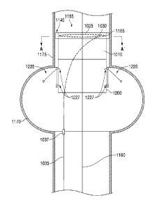
FIG. 10A is an axial cross-sectional view of an exemplary endovascular
universal
interface cuff 1155 of the present invention to be implanted into an aorta
having an aneurysm sac
CA 2823328 2018-12-21
CA 02823328 2013-06-27
WO 2012/092408 PCT/US2011/067695
21
1170 and an aortic wall 1175. The universal endovascular interface cuff 1155
has been
positioned over an endovascular guidewire 1160 to a desired recipient site AA'
proximal to the
aortic aneurysm sac 1170. The endovascular universal interface cuff 1155
further comprises an
accommodating proximal cuff 1010 and a rigid distal cuff 1200. FIG. 10B
provides a transverse
cross-sectional view of the exemplary endovascular interface cuff 1155 of FIG.
10A at the level
of A-A' in FIG. 10A. In FIGS. 10A and 10B, the compressible foam gasket 1140
is
uncompressed and, therefore, covers the retention tines 1165.
In the exemplary embodiment shown in FIG. 10B, the adjustment member 1025
courses in a circumferential loop through eyelets 1180 attached to a series of
compression
.. footplates 1185. The compression footplates 1185, among other functions,
serve to maintain an
orientation of the expanding circumferential loop 1035 in a plane transverse
to the aortic lumen
1190, and present a broader pressure contact with the underlying aortic wall
1175 when the
circumferential assembly is expanded. The compression footplates 1185 may
abut, be attached
to, or be contiguous with the retention tines 1165, which are displaced
through the compressed
compressible foam gasket 1140 and allowed to enter the aortic wall 1175 for
overall device
stabilization and retention. While four retention tines 1165 and footplates
1185 are shown, this
embodiment is merely exemplary and can be any number.
FIG. 11A shows the same axial cross-sectional view of the endovascular
universal
interface cuff 1155 of FIG. 10A but after the universal endovascular interface
cuff 1155 has
expanded to achieve a seal in the aortic wall 1175. Due to the expansion of
the cuff, the foam
gasket 1140 becomes compressed, allowing the retention tines 1165 to protrude
radially outward
to engage the aortic wall 1175 in the desired recipient site A-A' proximal to
the aortic aneurysm
sac 1170. In the exemplary embodiment shown in FIG. 11B, the adjustment member
1025 has
expanded to move the eyelets 1180 attached to the footplates 1185 outwards. As
is evident, the
interior lumen of the circumferential assembly 1020 shown in FIG. 11B has
increased
substantially as compared to the state shown in FIG. 10B. In FIG. 11B, the
compression of the
foam gasket 1140 and the engagement of the aortic wall 1175 by the retention
tines 1165 creates
a firm seal between the universal endovascular interface cuff 1155 and the
aortic wall 1175.
FIG. 12 shows the same axial cross-sectional axial of the universal
endovascular
interface cuff 1155 of the present invention as in FIGS. 10A and 11A but with
delivery of a
conventional endograft 1300 into the aortic wall 1175, which endograft 1300
has been secured
CA 02823328 2013-06-27
WO 2012/092408 PCT/US2011/067695
22
within the rigid distal cuff 1200 of the universal endovascular interface cuff
1155. The endograft
1300 can include an expandable lattice 1310. FIG. 13 shows the same cross-
sectional axial view
of an exemplary universal endovascular interface cuff 1155 of the present
invention as FIG. 12
but after removal of the endovascular guidewire 1160 and detachment and
removal of the
adjustment member 1025. Such removal and detachment can be carried out by a
release
mechanism 1037. The distal attachment of the conventional endograft is not
shown in FIGS. 12
and 13, but can be accomplished in the usual manner for conventional endograft
implantation
sufficient to prevent backfill of the aneurysm sac 1170 from the distal aorta
or the iliac vessels.
As shown in FIGS. 10A, 11A, 12, and 13, the rigid distal cuff 1200 includes,
at its
exterior, exemplary radio-opaque monitoring clip assemblies 1225 to allow post-
implantation
monitoring of slippage or endoleak formation and/or auto-accommodation for
post-implantation
aortic remodeling. Likewise, the rigid distal cuff 1200 can be provided with
interior graft
retention tines 1227 that add to securing, without leaks, the endograft 1300
to the interior of the
rigid distal cuff 1200.
The tubular endograft body 1005, the proximal cuff 1010, the tied distal cuffs
1200,
and the endograft body 1300 as described herein may be constructed of solid,
woven, non-
woven, or mesh materials such as, but not limited to, natural or synthetic
rubbers, nylon, GORE-
TEX . elastomers, polyisoprenes, polyphosphazenes, polyurethanes, vinyl
plastisols, acrylic
polyesters, polyvinylpyrrolidone-polyurethane interpolymers, butadiene
rubbers, styrene-
butadiene rubbers, rubber lattices, DACRON , PTFE, malleable metals, other
biologically
compatible materials or a combination of such biologically compatible
materials in a molded,
woven, or non-woven configuration, coated, non-coated, and other polymers or
materials with
suitable resilience and pliability qualities. In certain exemplary embodiments
according to the
present invention, it is desirable for the non-elastic tubular member 1015 and
corresponding
structures to be pliable to allow for folding or compressibility without
allowing elasticity. In
certain exemplary embodiments according to the present invention, it is
desirable for the
accommodating proximal cuff 1010 and corresponding structures to have
plasticity and be
compressible or foldable. In any given exemplary embodiment, the non-elastic
tubular implant
body 1015, the endograft body 1300, the accommodating proximal cuff 1010, and
corresponding
structures may be constructed of the same material of varying elasticity, or
these structures may
be constructed of different, but compatible materials.
CA 02823328 2013-06-27
WO 2012/092408 PCT/US2011/067695
23
The adjustment members 1025, the retention tines 1130, 1165, and the
microcylinders
1030 and other mechanical components as disclosed herein and in all other
embodiments of the
present invention may be fabricated of any suitably strong biocompatible
material, including, but
not limited to titanium, stainless steel, cobalt chromium alloys, other
metals, other metal alloys,
nitinol, plastics, or ceramics. Similarly, the adjustment members 1025, the
retention tines 1130,
1165, and the microcylinders 1030 and other mechanical components may be
milled, laser cut,
lathed, molded, or extruded.
The compressible foam gaskets 1140 as disclosed herein may be any
biocompatible
foam material of either an open or closed cell structure with sufficient
compressibility and
resilience to allow rapid recovery in a non-compressed state. In various
exemplary embodiments
according to the present invention, such foam materials may be viscoelastic
foam with a
compressible cellular material that has both elastic (spring-like) and viscous
(time-dependent)
properties. Viscoelastic foam differs from regular foam by having time-
dependent behaviors
such as creep, stress relaxation, and hysteresis.
FIGS. 14A and 14B show an alternate exemplary embodiment of a sealable
endograft
system 2000 according to the present invention in two different states. In the
view of FIG. 14A,
a hinged lattice structure 2100 is attached to an internal or external surface
of at least the
proximal portion 2210 of an endograft body 2200 (the "lattice" in these
figures is only
diagrammatic and is not intended to imply that the only possible number of
rings of lattice is
greater than one). Either the lattice structure 2100 or the endograft body
2200 can be provided
with radially displaced retention tines 2105 that, in a non-distended state of
the proximal portion
2210, can be covered within a compressible foam gasket 2300. In the embodiment
shown in
FIG. 14A. the distal portion 2220 of the endograft body 2200 comprises a non-
distensible
material and the proximal portion 2210 of the endograft body 2200 is an
accommodating cuff
comprising a distensible material forming the proximally terminal aspect of
the sealable
endograft system 2000 and enclosing the terminal hinged lattice structure 2100
therewithin.
A control system 2400 or jack screw shown in FIGS. 14A and 14B is provided to
expand and contract the lattice structure 2100. In particular, a torque wire
2410 can be fixed at
two points 2420, 2430 longitudinally separate from one another on the lattice
structure 2100.
This torque wire 2410 has exterior threads that correspond to threaded bores
of one of the two
points 2420, 2430. Accordingly, when the torque wire 2410 is rotated, the two
points 2420,
CA 02823328 2013-06-27
WO 2012/092408 PCT/US2011/067695
24
2430 of the lattice either approach one another (to expand the proximal
portion 2210) or retreat
from one another (to contract the proximal portion 2210) this imparts motion
to all contiguously
interconnected lattice elements. It is preferred to have the proximal end
point 2430 be bored for
rotation but fixed longitudinally. In this case, a smooth-bored collar 2440 is
fixed to the wall of
the graft 2200, for example, on an interior surface distal of the lattice
structure 2100. When the
adjustment tool 1035 is rotated, the torque wire 2410 correspondingly rotates
to expand or
contract the proximal portion 2210 of the endograft 2200. In this manner, in
comparison to self-
expanding prior art stent structures (e.g., made of nitinol) passively open to
their greatest extent
when relieved from radially inward compression, the lattice structure of the
present invention is
able to actively open according to the desire of the user surgeon implanting
the prosthesis. As
such, the opening performed by prior art self-expanding stent structures in
endograft prosthesis
are referred to herein as "passive opening" or "passive expansion". In
contrast thereto, the
expansion performed by the inventive controllable, hinged, lattice structure
of the present
invention for the disclosed endograft prostheses is referred to herein as
"active control" or
"active expansion" because it can be actively controlled in both the expansion
and contraction
directions according to the desire of the user. This is further in contrast to
expansion of stent
structures using balloon, which case is referred to as "balloon opening" or
"balloon expansion"
because it occurs only in one direction (expansion) without any ability to
contract actively. The
single embodiment of the jack screw shown in FIGS. 14A and 14B can be
replicated any number
of times about the circumference of the lattice structure 2100
In a non-illustrated alternative to the configuration of the system shown in
FIG. 14B,
the configuration shown in FIGS. 10A to 11B can be incorporated into the
system of FIGS. 14A
and 14B to create a hybrid system. The circumferential assembly 1020 can be
positioned at the
proximal end of the endograft and action of the circumferential loop 1035
within the proximal
cuff 1010, can be used to expand and contract the latticework 2100.
FIG. 15A is a lateral view of an exemplary embodiment of an adjustable
vascular
cannula 1230 according to the present invention. As shown in FIG. 15A, such an
adjustable
vascular cannula 1230 is a generally tubular structure with external cannula
walls 1235 defining
a cannula lumen 1240, and comprises a port end 1245, a cannula body 1250, and
a cannula tip
1255. As further shown in FIG. 15A, the cannula body 1250 is further provided
with a delivery
recess 1260 in its external wall structure at or near the junction of the
cannula tip 1255. Further
CA 02823328 2013-06-27
WO 2012/092408 PCT/US2011/067695
still, the adjustable vascular cannula 1230 of FIG. 15A comprises an
adjustable seal device 1265
attached to an adjustment member 1025 such as a torque wire that extends
beyond the port end
1245 of the adjustable vascular cannula 1230 as shown in FIG. 15B. The
adjustment member
1025 may course through the cannula lumen 1240, or it may course through an
accessory lumen
5 (not shown in FIGS. 15A or 15B) within the cannula wall 1235
substantially parallel to the
cannula lumen 1240, or it may course externally to the adjustable vascular
cannula 1230 as
shown partially within and partially outside the lumen 1240 in FIG. 15B. When
in a non-
deployed state, as shown in FIG. 15B, the adjustable seal device 1265 is
substantially flush with
the outer diameter of the cannula walls 1235 within the delivery recess 1260
of the cannula body
10 1250.
FIG. 15C shows the adjustable seal device 1265 in a deployed state, which is
the result
of torque applied externally to the adjustment member 1025 by a user. As shown
in FIG. 15C,
the adjustable seal device 1265 further comprises a hinged adjustable
latticework 1270 covered
by a sealing cuff 1275 which is constructed of a distensible material. The
adjustment member
15 .. 1025 terminates, for example, in a circumferential loop 1035 within the
sealing cuff 1275, where
it may be further covered by a compressible foam gasket 1140. The adjustment
member 1025
may further pass through a locking mechanism 1050 as disclosed elsewhere
herein which serves
to regulate the torque applied to the circumferential loop 1035. The hinged
adjustable
latticework 1270 may further be provided with one or more retention tines
1130, 1165, which are
20 radially displaced from the terminal aspect of the hinged adjustable
latticework 1270, and which
are enclosed within and covered by the compressible foam gasket 1140 when the
adjustable seal
device 1265 is not distended. When torque is applied to the adjustment member
1025 by a user,
the diameter of the circumferential loop 1035 is increased, displacing the
hinged adjustable
latticework 1270 as shown in FIG. 15C until the compressible foam gasket 1140
and the sealing
25 .. cuff 1275 is able to firmly engage the inner wall 1190 of a recipient
blood vessel 1175. A slight
additional amount of torque applied to the adjustment member 1025 is, then,
sufficient to
compress the compressible foam gasket 1140 and allow the retention tines 1130,
1165 to engage
the wall 1190 of the recipient blood vessel 1175, thus preventing slippage of
the cannula during
use. In various exemplary embodiments of the present invention, the retention
tines 1130, 1165
may be provided to engage the vessel wall 1190 in a substantially straight
manner or at angles
varying from about 1 degree to about 179 degrees. The retention tines 1130,
1165 may be
CA 02823328 2013-06-27
WO 2012/092408 PCT/US2011/067695
26
angled axially or longitudinally in various embodiments according to the
present invention.
After the use of the cannula is completed, the torque of the adjustment member
1025 may be
reversed, collapsing the adjustable seal device 1165, and allowing the
compressible foam gasket
1140 to re-expand, thus withdrawing the retention tines 1165 from the vessel
wall 1175 and
covering the retention tines 1165 to allow atraumatic cannula withdrawal.
Although the foregoing embodiments of the present invention have been
described in
some detail by way of illustration and example for purposes of clarity and
understanding, it will
be apparent to those skilled in the art that certain changes and modifications
may be practiced
within the spirit and scope of the present invention. Therefore, the
description and examples
presented herein should not be construed to limit the scope of the present
invention, the features
of which are set forth in the appended claims.
The foregoing description and accompanying drawings illustrate the principles,
exemplary embodiments, and modes of operation of the invention. However, the
invention
should not be construed as being limited to the particular embodiments
discussed above.
Additional variations of the embodiments discussed above will be appreciated
by those skilled in
the art and the above-described embodiments should be regarded as illustrative
rather than
restrictive. Accordingly, it should be appreciated that variations to those
embodiments can be
made by those skilled in the art without departing from the scope of the
invention as defined by
the following claims.