Note: Descriptions are shown in the official language in which they were submitted.
CA 02823429 2016-03-29
SIALIDASE CATALYTIC DOMAIN PROTEINS
BACKGROUND OF THE INVENTION
The invention relates to therapeutic compositions that can be used to prevent
and treat infection of human and animal subjects by a pathogen, and
specifically to
protein-based therapeutic compositions that can be used for the prevention and
treatment of viral or bacterial infections. The invention also relates to
therapeutic
protein-based compositions that can be used to prevent or ameliorate allergic
and
inflammatory responses. The invention also relates to protein-based
compositions for
increasing transduction efficiency of a recombinant virus, such as a
recombinant virus
used for gene therapy.
1
CA 02823429 2016-03-29
Influenza is a highly infectious acute respiratory disease that has plagued
the
human race since ancient times. It is characterized by recurrent annual
epidemics and
periodic major worldwide pandemics. Because of the high disease-related
morbidity and
mortality, direct and indirect social economic impacts of influenza are
enormous. Yearly
.. epidemics cause approximately 300,000 hospitalizations and 25,000 deaths in
the United
States alone. Four pandemics occurred in the last century; together they
caused tens of
millions of deaths. Mathematical models based on earlier pandemic experiences
have
estimated that 89,000-207,000 deaths, 18-42 million outpatient visits and 20-
47 million
additional illnesses will occur during the next pandemic (Meltzer, MI, Cox, NJ
and
Fukuda, K. (1999) Emerg Infect Dis 5:659-671).
Influenza is typically caused by infection of two types of viruses, Influenza
virus
A and Influenza virus B (the third type Influenza virus C only causes minor
common cold
like symptoms). They belong to the orthomyxoviridae family of RNA viruses.
Both type
A and type B viruses have 8 segmented negative-strand RNA genomes enclosed in
a lipid
.. envelope derived from the host cell. The viral envelope is covered with
spikes that are
composed of three types of proteins: hemagglutinin (HA) which attaches virus
to host
cell receptors and mediates fusion of viral and cellular membranes;
neuraminidase (NA)
which facilitates the release of the new viruses from host cells; and a small
number of M2
proteins which serve as ion channels.
Infections by influenza type A and B viruses are typically initiated at the
mucosal
surface of the upper respiratory tract. Viral replication is primarily limited
to the upper
respiratory tract but can extend to the lower respiratory tract and cause
bronchopneumonia that can be fatal.
Influenza viral protein hemagglutinin (HA) is the major viral envelope
protein. It
plays an essential role in viral infection. The importance of HA is evidenced
by the fact
that it is the major target for protective neutralizing antibodies produced by
the host
immune response (Hayden, FG. (1996) In Antiviral drug resistance (ed. D. D.
Richman), pp. 59-77. Chichester, UK: John Wiley & Sons Ltd.). It is now clear
that HA
has two different functions in viral infection. First, HA is responsible for
the attachment
.. of the virus to sialic acid cell receptors. Second, HA mediates viral entry
into target cells
by triggering fusion of the viral envelope with cellular membranes.
2
CA 02823429 2016-03-29
HA is synthesized as a precursor protein, HAO, which is transferred through
the
Golgi apparatus to the cell surface as a trimeric molecular complex. HAO is
further
cleaved to generate the C terminus HAI (residue 328 of HAO) and the N terminus
of
HA2. It is generally believed that the cleavage occurs at the cell surface or
on released
viruses. The cleavage of HAO into HA l/HA2 is not required for HA binding to
sialic
acid receptor; however, it is believed to be necessary for viral infectivity
(Klenk, HD and
Rott, R. (1988) Adv Vir Res. 34:247-281; Kido, H, Niwa, Y, Beppu, Y and
Towatari, T.
(1996) Advan Enzyme Regul 36:325-347; Skehel, JJ and Wiley, DC. (2000) Annu
Rev
Biochem 69:531-569; Zambon, M. (2001) Rev Med Virol 11:227-241.)
Currently, influenza is controlled by vaccination and anti-viral compounds.
Inactivated influenza vaccines are now in worldwide use, especially in high-
risk groups.
The vaccine viruses are grown in fertile hen's eggs, inactivated by chemical
means and
purified. The vaccines are usually trivalent, containing representative
influenza A viruses
(HIN1 and H3N2) and influenza B strains. The vaccine strains need to be
regularly
updated in order to maintain efficacy; this effort is coordinated by the World
Health
Organization (WHO). During inter-pandemic periods, it usually takes 8 months
before
the updated influenza vaccines are ready for the market (Wood, J. (2001) Phil
Trans R
Soc Lond B 356:1953-1960). However, historically, pandemics spread to most
continents
within 6 months, and future pandemics are expected to spread even faster with
increased
international travel (Gust, ID, Hampson, AW., and Lavanchy, D. (2001) Rev Med
Virol
11:59-70). Therefore it is inevitable that an effective vaccine will be
unavailable or in
very short supply during the first waves of future pandemics.
Anti-viral compounds have become the mainstay for treating inter-pandemic
diseases. Currently, they are also the only potential alternative for
controlling pandemics
during the initial period when vaccines are not available. Two classes of
antiviral
compounds are currently on the market: the M2 inhibitors, such as amantadine
and
rimantadine; and the NA inhibitors, which include oseltamivir (Tamiflu) and
zanamivir
(Relenza). Both classes of molecules have proven efficacy in prevention and
treatment of
influenza. However, side effects and the risk of generating drug-resistant
viruses remain
the top two concerns for using them widely as chemoprophylaxis (Hayden, FG.
(1996) In
Antiviral drug resistance (ed. D. D. Richman), pp. 59-77. Chichester, UK: John
Wiley
3
CA 02823429 2016-03-29
& Sons Ltd.). Most importantly, future pandemic strains, either evolved
naturally or
artificially created by genetic engineering in bio-warfare, may be resistant
to all the
available anti-viral compounds, and this will have devastating consequences
globally.
In summary, currently available vaccination and anti-viral compounds are
limited
.. by some fundamental shortcomings. Novel therapeutic and prophylactic
modalities are
needed to address future influenza pandemics.
Respiratory tract infections (RTIs) are the most common, and potentially
most severe, types of infectious diseases. Clinically, RTIs include sinusitis,
otitis,
laryngitis, bronchitis and pneumonia. Based on numerous etiology and
epidemiology
studies, it is clear that although many microorganisms have the potential to
cause RTIs,
only a handful of pathogens are responsible for vast majority of the cases.
Such
pathogens include S. pneumoniae, M. pneumoniae, H. influenzae, M. catarrhalis,
influenza A & B, and parainfluenza virus. Besides causing CAP and AECB,
several of
the bacterial pathogens, such as S. pneumoniae and H. influenzae, are also the
common
cause of acute sinusitis, otitis media, as well as invasive infections leading
to sepsis,
meningitis, etc. Therefore these microorganisms are of the highest clinical
importance.
One common feature of all respiratory pathogenic bacteria is that they
establish
commensal colonization on the mucosal surface of the upper airway; such
colonization
precedes an infection and is prerequisite for infections. The bacterial
colonization in a
neonate occurs shortly after birth. During lifetime, the upper airway,
specifically the
nasopharynx and oropharynx, remains a dynamic ecological reservoir of
microbial
species with bacteria being acquired, eliminated and re-acquired continually.
In most
cases the bacterial flora in the pharynx is harmless. However, when the
condition of the
host is altered, some microorganisms may invade adjacent tissues or
bloodstream to
cause diseases, In addition to serving as the port of entry for mucosa] and
invasive
infections by both bacteria and viruses, the nasopharynx is also the major
source of
spreading the pathogenic microorganisms between individuals, as well as the
reservoir
where antibiotic-resistant bacteria are selected (Garcia-Rodriguez and
Martinez, J
Antimicrob Chemother, (2002) 50(Suppl S2), 59-73; Soriano and Rodriguez-
Cerrato, J
Antimicrob Chemother, (2002) 50 Suppl S2, 51-58). It is well established
clinically
4
CA 02823429 2016-03-29
that individuals who are prone to RTIs tend to be persistent and recurrent
carriers of the
pathogenic bacteria (Garcia-Rodriguez and Martinez, J Antimicrob Chemother,
(2002)
50(Suppl S2), 59-73; Mbaki et al., Tohoku J Exp. Med., (1987) 153(2), 111-
121).
Helicobacter pylori is a human pathogen implicated in gastritis and peptic
ulcer. The
bacterium resides in the human stomach and binds to epithelial cells of the
gastric antrum. It has
been demonstrated that the bacterial adhesion is mediated by binding of
Helicobacter pylori
adhesin I and II to sialic acids on the epithelial surface.
Siglecs (sialic acid binding Ig-like lectins) are members of the
immunoglobulin
(Ig) superfamily that bind to sialic acid and are mainly expressed by cells of
the
hematopoietic system. At least 11 siglecs have been discovered and they seem
to
exclusively recognize cell surface sialic acid as the ligand. It is believed
that the binding
of siglecs to sialic acid mediates cell-cell adhesion and interactions
(Crocker and Varki,
Trends Immunol., (2001) 22(6), 337-342; Angata and Brinkman-Van der Linden,
Biochim. Biophys. Acta, (2002) 1572(2-3), 294-316). Siglec-8 (SAF-2) is an
adhesion
molecule that is highly restricted to the surface of eosinophils, basophils,
and mast cells,
which are the central effector cells in allergic conditions including allergic
rhinitis,
asthma and eczema. Siglec-8 is considered to be responsible for mediating the
recruitment of the three allergic cell types to the airway, the lungs and
other sites of
allergy. Siglec-1 (sialoadhesion) and siglec-2 (CD22) are the adhesion
molecules on
macrophages and B cells, both types of cells play central roles in immune
reactions that
lead to inflammation.
Recombinant viruses, in particular adeno-associated virus (AAV), can be used
to
transfer the wild type cystic fibrosis transmembrane conductance regulator
(cFTR) gene
into the epithelial cells to correct the genetic defect that causes cystic
fibrosis (Flotte and
Carter, Methods Enzymol., (1998) 292, 717-732). Clinical trials with AAV
vectors
have shown efficient and safe delivery of the Cl-r1R gene into epithelial
cells with low
levels of gene transfer (Wagner et al., Lancet, (1998) 351(9117), 1702-1703).
Compared
.. to adenoviral vectors, AAV offers more stable gene expression and
diminished cellular
immunity. However, the transduction efficiency of AAV in vivo is rather low in
the lung
5
CA 02823429 2016-03-29
(Wagner et al., Lancet, (1998) 351(9117), 1702-1703). A method that can
improve
transduction efficiency of AAV in vivo is needed to achieve full therapeutic
potential of
gene therapy for cystic fibrosis. It has been
shown that negatively charged
carbohydrates, such as sialic acid, inhibit the transduction efficiency of AAV
vector to
.. the well-differentiated airway epithelium, and treatment of the airway
epithelium by
glycosidases, including a neuraminidase, and encloglycosidase enhances
transduction
efficiency of the AAV vector (Bals etal., J Virol., (1999) 73(7), 6085-6088).
BRIEF SUMMARY OF THE INVENTION
The present invention recognizes that current therapeutics for preventing and
treating infection by pathogens are often difficult to provide in a timely
manner, can have
undesirable side effects, and can lead to drug-resistant pathogen strains. The
present
.. invention also recognizes that the current approach to treat allergy and
inflammation has
limited efficacy and is associated with side effects. In addition, the present
invention also
recognizes that the current approach to administer recombinant viruses yield
low
transduction efficiency and unsatisfactory efficacy of the gene therapy.
The present invention provides new compositions and methods for preventing and
treating pathogen infection. In particular, the present invention provides
compounds that
can act extracellularly to prevent infection of a cell by a pathogen. Some
preferred
embodiments of the present invention are therapeutic compounds having an
anchoring
domain that anchors the compound to the surface of a target cell, and a
therapeutic
domain that can act extracellularly to prevent infection of the target cell by
a pathogen,
such as a virus or bacterium.
In one aspect, the invention provides a protein-based composition for
preventing
or treating infection by a pathogen. The composition comprises a compound that
comprises at least one therapeutic domain comprising a peptide or protein,
where the
therapeutic domain has at least one extracellular activity that can prevent
the infection of
a target cell by a pathogen, and at least one anchoring domain that can bind
at or near the
membrane of a target cell.
6
CA 02823429 2016-03-29
In some embodiments of this aspect of the present invention, the at least one
therapeutic domain comprises an inhibitory activity that prevents or impedes
the infection
of a target cell by a pathogen. In a preferred embodiment, the inhibitory
activity inhibits
the activity of a protease that can process a viral protein necessary for
infection of a target
cell. In a particularly preferred embodiment, the compound comprises a
therapeutic
domain that can inhibit the processing of the HA protein of influenza virus,
and the
anchoring domain can bind the compound at the surface of a respiratory
epithelial cell.
In some embodiments of the present invention, at least one therapeutic domain
comprises a catalytic activity. In a preferred embodiment, the catalytic
activity removes a
moiety from the surface of a target cell that is necessary for infection of
the target cell. In
a particularly preferred embodiment, the therapeutic domain is a sialidase
that can digest
sialic acid moieties on the surface of epithelial target cells, and the
anchoring domain is a
GAG-binding domain of a human protein that can bind heparin or heparan sulfate
moieties at the surface of an epithelial cell.
In another aspect, the present invention includes pharmaceutical compositions
for
treating or preventing pathogen infection in a subject. Pharmaceutical
compositions
comprise a compound of the present invention comprising at least one
therapeutic domain
and at least one anchoring domain. The pharmaceutical composition can also
comprise
solutions, stabilizers, fillers and the like. In some preferred embodiments,
the
.. pharmaceutical composition is formulated as an inhalant. In some preferred
embodiments, the pharmaceutical composition is formulated as a nasal spray.
Another aspect of the present invention is a pharmaceutical composition
comprising at least one sialidase. The sialidase can be isolated from any
source, such as,
for example, a bacterial or mammalian source, or can be a recombinant protein
that is
.. substantially homologous to a naturally occurring sialidase. A
pharmaceutical
composition comprising a sialidase can be formulated for nasal, tracheal,
bronchial, oral,
or topical administration, or can be formulated as an injectable solution or
as eyedrops. A
pharmaceutical composition comprising a sialidase can be used to treat or
prevent
pathogen infection, to treat or prevent allergy or inflammatory response, or
to enhance
the transduction efficiency of a recombinant virus for gene therapy.
7
CA 02823429 2016-03-29
Yet another aspect of the present invention is a sialidase catalytic domain
protein.
In this aspect, proteins that comprise the catalytic domain of a sialidase but
comprise less
than the entire sialidase the catalytic domain sequence is derived from are
considered
sialidase catalytic domain proteins. Sialidase catalytic domain proteins can
comprise
other protein sequences, such as but not limited to functional domains derived
from other
proteins. A pharmaceutical composition comprising a sialidase can be
formulated foy
nasal, tracheal, bronchial, oral, or topical administration, or can be
formulated as an =
injectable solution or as eyeclrops. A pharmaceutical composition comprising a
sialidase
can be used to treat or prevent pathogen infection, to treat or prevent
allergy or
inflammatory response, or to enhance the transduction efficiency of a
recombinant virus
for gene therapy.
In yet another aspect, the present invention includes a method for treating or
preventing infection by a pathogen. In preferred embodiments, the method
comprises
administering sialidase activity, such as a sialidase or a sialidase catalytic
domain
protein, including a sialidase catalytic domain fusion protein, to a subject
to prevent or
treat an infection. A pathogen can be, for example, a viral or bacterial
pathogen. The
method includes applying a pharmaceutically effective amount of a compound of
the
present invention to at least one target cell of a subject. Preferably, the
pharmaceutical
composition can applied by the use of a spray, inhalant, or topical
formulation.
The present invention also provides new compositions and methods for treating
allergy and inflammation. In particular, the present invention provides
compounds that
can act extracellularly to prevent or inhibit adhesion and function of
inflammatory cells.
Some preferred embodiments of compounds for treating allergy or inflammation
comprise at least one therapeutic domain that has the said extracellular
activity and an at
least one anchoring domain that anchors the compound to the surface of a
target cell. In
some preferred embodiments, the method comprises administering a siaidase
activity,
such as a sialidase or a sialidase catalytic domain protein, including a
sialidase catalytic
domain fusion protein to a subject to prevent or treat an allergic or
inflammatory
response. The allergic or inflammatory response can be asthma, allergic
rhinitis, skin
conditions such as eczema, or response to plant or animal toxins. The method
includes
applying a pharmaceutically effective amount of a compound of the present
invention to
8
CA 02823429 2016-03-29
at least one target cell of a subject. Preferably, the pharmaceutical
composition can be
applied by the use of a spray, inhalant, or topical formulation.
The present invention also provides new compositions and methods for improving
efficiency of gene transfer by recombinant viral vectors during gene therapy.
In
particular, the present invention provides compounds that can act
extracellularly to
reduce the physical or chemical barrier that hinders transduction by gene
therapy vectors,
such as AAV vector. Some preferred compounds of the present invention for
improving
efficiency of gene transfer by recombinant viral vectors comprise at least one
therapeutic
domain that has an extracellular activity and an at least one anchoring domain
that
anchors the compound to the surface of a target cell. In some preferred
embodiments
the method comprises administering a siaidase activity, such as a sialidase or
a sialidase
catalytic domain protein, including a sialidase catalytic domain fusion
protein to a subject
to facilitate transduction of a target cell by a recombinant viral vector. The
method
includes applying an effective amount of a compound of the present invention
along with
a recombinant viral vector to at least one target cell. A pharmaceutical
composition of the
present invention can be applied by the use of a spray, inhalant, or topical
formulation.
BRIEF DESCRIPTION OF SEVERAL VIEWS OF THE DRAWINGS
Figure 1 is a schematic depiction of the primary amino acid structure of
aprotinin.
Figure 2 shows GAG-binding sequences of four human genes: PF4, human platelet
factor 4; IL8, human interleukin 8; AT HI, human antithrombin III; ApoE, human
apolipoprotein E; AAMP, human angio-associated migratory cell protein; human
amphiregulin.
Figure 3 is a sequence comparison between human sialidases NEU2 and NEU4.
Figure 4 is a table comparing substrate specificity of bacterial and fungal
sialidases.
.. Figure 5 depicts the nucleotide and amino acid sequences of Construct #1
encoding
His6-AvCD. Ncol and HindIII sites used for cloning into pTrc99a are shown in
bold.
9
CA 02823429 2016-03-29
WO 2006/031291
PCT/US2005/025831
Figure 6 depicts the nucleotide and amino acid sequences of Construct #2
encoding AR-
AvCD. Ncof and HindlII sites used for cloning into pTrc99a are shown in bold.
.. Figure 7 depicts The nucleotide and amino acid sequences of Construct #3
encoding AR-
G4S-AvCD. Ncol and HindIII sites used for cloning into pTrc99a are shown in
bold.
Figure 8 is a graph of data from an experiment showing that the AR-tag
enhances the
removal of a(2,6)-linked sialic acid from MDCK cells. The Y axis shows the
percentage
of a(2,6)-linked sialic acid remaining on the surface of MDCK cells after
treatment with
various dilutions of recombinant AvCD (Construct #1) (diamonds) or recombinant
AR-
AvCD (Construct #2) (squares).
Figure 9 is a graph depicting the protection against influenza viruses
conferred by
treating MDCK cells with recombinant AR-AvCD protein made from Construct #2 or
the isolated sialidase of A. ureafaciens. The challenge viral strains are:
A/WS/33
(111N1); AJPR/8 (H1N1); A/Japan/305/57 (H2N2); A/Victoria/504/2000 (H3N2);
A/HongKong/8/68 (H3N2); B/Lee/40; 7. B/Maryland/1/59; and Turkey/Wis/66
(H9N2).
.. Figure 10 is a graph showing the level of inhibition of influenza virus
amplification by
the recombinant AR-AvCD sialidase and the recombinant AR-G4S-AvCD sialidase.
The
challenge viral strains are: A/PR/8 (H1N1); A/WS/33 (111N1); A/Japan/305/57
(H2N2);
A/HongKong/8/68 (H3N2); B/Lee/40; 7. B/Maryland/1/59; and Turkey/Wis/66
(H9N2).
.. Figure 11 provides graphs showing that topical administration of
recombinant AR-AvCD
sialidase fusion protein reduces the inflammatory responses of ferrets
infected with an
influenza A (HIN1) virus. (A) The total number of inflammatory cells from
nasal wash
samples obtained from infected animals at the indicated times after infection.
(B) The
protein concentration was determined in cell-free nasal wash samples of
infected ferrets.
Infected ferrets were vehicle-treated (squares) or were treated with
recombinant AR-
AvCD sialidase fusion protein made from Construct #2 (triangles). Uninfected
animals
CA 02823429 2016-03-29
were also treated with recombinant AR-AvCD sialidase fusion protein
(diamonds).
Statistically significant values are labeled with * (p<0.05) and **
Figure 12 is a table depicting inhibition of viral replication, cell
protection EC50's, and
selective indexes for two sialidase catalytic domain fusion proteins of the
present
invention. All EC50's are in mU/ml.
Figure 13 is a table depicting viral replication in the respiratory tract of
ferrets treated
with a sialidase catalytic domain fusion proteins of the present invention and
ferrets
treated with a control vehicle.
DETAILED DESCRIPTION OF THE INVENTION
Definitions
Unless defined otherwise, all technical and scientific terms used herein have
the
same meaning as commonly understood by one of ordinary skill in the art to
which this
invention belongs. Generally, the nomenclature used herein and the manufacture
or
laboratory procedures described below are well known and commonly employed in
the
art. Conventional methods are used for these procedures, such as those
provided in the
art and various general references. Where a term is provided in the singular,
the
inventors also contemplate the plural of that term. Where there are
discrepancies in terms
and definitions used in documents referenced herein, the terms used in this
application
shall have the definitions given herein. As employed throughout the
disclosure, the
following terms, unless otherwise indicated, shall be understood to have the
following
meanings:
A "pathogen" can be any virus or microorganism that can infect a cell, a
tissue or
an organism. A pathogen can be a virus, bacterium, or protozoan.
11
CA 02823429 2016-03-29
A "target cell" is any cell that can be infected by a pathogen or any cell
that can
interact with inflammatory cells, or a host cell that is the intended
destination for an
exogenous gene transferred by a recombinant virus.
A "recombinant virus" or a "recombinant viral vector", a "gene therapy viral
vector" or a "gene therapy vector" is defined as a genetically engineered
virus that
comprises one or more exogenous genes. When a target cell is transduced by a
recombinant virus, the exogenous gene(s) is transferred to the target cell.
Genes
transferred to a target cell can be expressed in the cell to provide the
intended therapeutic
effects. Currently, most commonly used gene therapy viral vectors are based on
four
types of viruses: retrovirus (including lentivirus), adenovirus, adeno-
associated virus
(AAV) and herpes simplex virus type 1.
"Inflammatory cells" are the cells that carry out or participate in
inflammatory
responses of the immune system. Inflammatory cells include B lymphocytes, T
lymphocytes, macrophages, basophils, eosinophils, mast cells, NK cells and
monocytes.
An "extracellular activity that can prevent the infection of a target cell by
a
pathogen" is any activity that can block or impede infection of a target cell
by a pathogen
by acting at or near the exterior surface of a target cell. An extracellular
activity that can
prevent the infection of a target cell by a pathogen, can be an activity such
as, but not
limited to, a catalytic activity or an inhibitory activity. For example, a
catalytic activity
can be an enzymatic activity that degrades one or more entities (such as but
not limited to
ligands, receptors, or enzymes) on a pathogen, on a target cell, or in the
vicinity of a
target cell, in which the one or more entities contribute to the infection
process. A
catalytic activity can also modify one or more entities on a pathogen, on a
target cell, or
in the vicinity of a target cell, such that the infection-promoting property
of the entity is
reduced. An inhibitory activity can be an activity that, for example, binds to
a receptor or
ligand and prevents the receptor or ligand from binding a moiety, where the
binding is
necessary for or promotes the infection process. An inhibitory activity can
also be an
inhibitor of an enzyme or receptor that prevents the enzyme or receptor from
performing
a function that is necessary for or promotes the infection process. The
exterior of a target
cell includes the target cell membrane itself, as well as the extracellular
milieu
surrounding the target cell, including extracellular matrix, intracellular
spaces, and
12
CA 02823429 2016-03-29
lumina] spaces. For epithelial cells, the exterior of a target cell also
includes the apical or
lumina] surface of the cell membrane that form luminal linings, and the
extracellular
milieu near the luminal surface. An "extracellular activity that can prevent
the infection
of a target cell by a pathogen" can be any type of chemical entity, including
a protein,
polypeptide, peptide, nucleic acid, peptide nucleic acid, nucleic acid
analogue,
nucleotide, nucleotide analogue, small organic molecule, polymer, lipids,
steroid, fatty
acid, carbohydrate, and the like, including combinations of any of these.
Preferably,
however, the activity comprises a peptide or protein or coupled to a peptide
or protein.
An "extracellular activity that can improve transduction efficiency, or gene
transfer efficiency, by a recombinant virus" is any activity that reduces or
eliminates
physical or chemical barriers that impedes host cell entry by a recombinant
virus by
acting at or near the exterior surface of a target cell. An extracellular
activity that can
improve transduction efficiency, or gene transfer efficiency, by a recombinant
virus can
be an activity such as, but not limited to, a catalytic activity or an
inhibitory activity. For
example, a catalytic activity can be an enzymatic activity that degrades one
or more
entities (such as but not limited to ligands, receptors, or enzymes) on a
pathogen, on a
target cell, or in the vicinity of a target cell, in which the one or more
entities contribute
to the infection process. A catalytic activity can also modify one or more
entities on a
pathogen, on a target cell, or in the vicinity of a target cell, such that the
infection-
promoting property of the entity is reduced. An inhibitory activity can be an
activity that,
for example, binds to a receptor-or ligand and prevents the receptor or ligand
from
binding a moiety, where the binding is necessary for or promotes the infection
process.
An inhibitory activity can also be an inhibitor of an enzyme or receptor that
prevents the
enzyme or receptor from performing a function that is necessary for or
promotes the
infection process. The exterior of a target cell includes the target cell
membrane itself, as
well as the extracellular milieu surrounding the target cell, including
extracellular matrix,
intracellular spaces, and luminal spaces. For epithelial cells, the exterior
of a target cell
also includes the apical or luminal surface of the cell membrane that form
luminal
linings, and the extracellular milieu near the luminal surface. An
"extracellular activity
that can prevent the infection of a target cell by a pathogen" can be any type
of chemical
entity, including a protein, polypeptide, peptide, nucleic acid, peptide
nucleic acid,
13
CA 02823429 2016-03-29
nucleic acid analogue, nucleotide, nucleotide analogue, small organic
molecule, polymer,
lipids, steroid, fatty acid, carbohydrate, and the like, including
combinations of any of
these. Preferably, however, the activity comprises a peptide or protein or
coupled to a
peptide or protein.
An "extracellular activity that can inhibit adhesion or function of
inflammatory
cells" is any activity that can prevent inflammatory cells from contacting the
target cell
and affecting the normal physiological status of the target cell.
A "domain that can anchor said at least one therapeutic domain to the membrane
of a target cell", also called an "extracellular anchoring domain" or simply,
"anchoring
domain" refers to a chemical entity can that can stably bind a moiety that is
at or on the
exterior of a cell surface or is in close proximity to the surface of a cell.
An extracellular
anchoring domain can be reversibly or irreversibly linked to one or more
moieties, such
as, preferably, one or more therapeutic domains, and thereby cause the one or
more
attached therapeutic moieties to be retained at or in close proximity to the
exterior surface
of a eukaryotic cell. Preferably, an extracellular anchoring domain binds at
least one
molecule on the surface of a target cell or at least one molecule found in
close association
with the surface of a target cell. For example, an extracellular anchoring
domain can bind
a molecule covalently or noncovalently associated with the cell membrane of a
target
cell, or can bind a molecule present in the extracellular matrix surrounding a
target cell.
An extracellular anchoring domain preferably is a peptide, polypeptide, or
protein, and
can also comprise any additional type of chemical entity, including one or
more
additional proteins, polypeptides, or peptides, a nucleic acid, peptide
nucleic acid, nucleic
acid analogue, nucleotide, nucleotide analogue, small organic molecule,
polymer, lipids,
steroid, fatty acid, carbohydrate, or a combination of any of these.
As used herein, a protein or peptide sequences is "substantially homologous"
to a
reference sequence when it is either identical to a reference sequence, or
comprises one
or more amino acid deletions, one or more additional amino acids, or more one
or more
conservative amino acid substitutions, and retains the same or essentially the
same
activity as the reference sequence. Conservative substitutions may be defined
as
exchanges within one of the following five groups:
14
CA 02823429 2016-03-29
1. Small, aliphatic, nonpolar or slightly polar residues: Ala, Ser,
Thr, Pro,
Gly
11. Polar, negatively charged residues and their amides: Asp, Asn,
Glu, Gin
III. Polar, positively charged residues: His, Arg, Lys
IV. Large, aliphatic nonpolar residues: Met, Leu, Ile, Val, Cys
V. Large aromatic residues: Phe, Try, Trp
Within the foregoing groups, the following substitution are considered to be
"highly
conservative": Asp/Glu, His/Arg/Lys, Phe/Tyr/Trp, and Met/Leu/Ile/Val. Semi-
conservative substitutions are defined to be exchanges between two of groups
(I)-(IV)
above which are limited to supergroup (A), comprising (I), (II), and (III)
above, or to
supergroup (B), comprising (IV) and (V) above. In addition, where hydrophobic
amino
acids are specified in the application, they refer to the amino acids Ala,
Gly, Pro, Met,
Leu, Ile, Val, Cys, Phe, and Trp, whereas hydrophilic amino acids refer to
Ser, Thr, Asp,
Asn, Glu, Gln, His, Arg, Lys, and Tyr.
A "sialidase" is an enzyme that can remove a sialic acid residue from a
substrate
molecule. The sialidases (N-acylneuraminosylglycohydrolases, EC 3.2.1.18) are
a group
of enzymes that hydrolytically remove sialic acid residues from sialo-
glycoconjugates.
Sialic acids are alpha-keto acids with 9-carbon backbones that are usually
found at the
outermost positions of the oligosaccharide chains that are attached to
glycoproteins and
glycolipids. One of the major types of sialic acids is N-acetylneuraminic acid
(Neu5Ac),
which is the biosynthetic precursor for most of the other types. The substrate
molecule
can be, as nonlimiting examples, an oligosaccharide, a polysaccharide, a
glycoprotein, a
ganglioside, or a synthetic molecule. For example, a sialidase can cleave
bonds having
alpha(2,3)-Gal, alpha(2,6)-Gal, or alpha(2,8)-Gal linkages between a sialic
acid residue
and the remainder of a substrate molecule. A sialidase can also cleave any or
all of the
linkages between the sialic acid residue and the remainder of the substrate
molecule. Two
major linkages between Neu5Ac and the penultimate galactose residues of
carbohydrate
side chains are found in nature, Neu5Ac alpha (2,3)-Gal and Neu5Ac alpha (2,6)-
Gal.
Both Neu5Ac alpha (2,3)-Gal and Neu5Ac alpha (2,6)-Gal molecules can be
recognized
by influenza viruses as the receptor, although human viruses seem to prefer
Neu5Ac
alpha (2,6)-Gal, avian and equine viruses predominantly recognize Neu5Ac alpha
(2,3)-
CA 02823429 2016-03-29
Gal. A sialidase can be a naturally-occurring sialidase, an engineered
sialidase (such as,
but not limited to a sialidase whose amino acid sequence is based on the
sequence of a
naturally-occurring sialidase, including a sequence that is substantially
homologous to the
sequence of a naturally-occurring sialidase). As used herein, "sialidase" can
also mean
the active portion of a naturally-occurring sialidase, or a peptide or protein
that comprises
sequences based on the active portion of a naturally-occurring sialidase.
A "fusion protein" is a protein comprising amino acid sequences from at least
two
different sources. A fusion protein can comprise amino acid sequence that is
derived from
a naturally occurring protein or is substantially homologous to all or a
portion of a
naturally occurring protein, and in addition can comprise from one to a very
large number
of amino acids that are derived from or substantially homologous to all or a
portion of a
different naturally occurring protein. In the alternative, a fusion protein
can comprise
amino acid sequence that is derived from a naturally occurring protein or is
substantially
homologous to all or a portion of a naturally occurring protein, and in
addition can
comprise from one to a very large number of amino acids that are synthetic
sequences.
A "sialidase catalytic domain protein" is a protein that comprises the
catalytic
domain of a sialidase, or an amino acid sequence that is substantially
homologous to the
catalytic domain of a sialidase, but does not comprises the entire amino acid
sequence of
the sialidase the catalytic domain is derived from, wherein the sialidase
catalytic domain
protein retains substantially the same activity as the intact sialidase the
catalytic domain
is derived from. A sialidase catalytic domain protein can comprise amino acid
sequences
that are not derived from a sialidase, but this is not required. A sialidase
catalytic domain
protein can comprise amino acid sequences that are derived from or
substantially
homologous to amino acid sequences of one or more other known proteins, or can
comprise one or more amino acids that are not derived from or substantially
homologous
to amino acid sequences of other known proteins.
16
CA 02823429 2016-03-29
I. Composition for preventing or treating infection by a pathogen
The present invention includes peptide or protein-based compounds that
comprise
at least one domain that can anchor at least one therapeutic domain to the
membrane of a
eukaryotic cell and at least one therapeutic domain having an extracellular
activity that
can prevent the infection of a cell by a pathogen. By "peptide or protein-
based"
compounds, it is meant that the two major domains of the compound have an
amino acid
framework, in which the amino acids are joined by peptide bonds. A peptide or
protein-
based compound can also have other chemical compounds or groups attached to
the
amino acid framework or backbone, including moieties that contribute to the
anchoring
activity of the anchoring domain, or moieties that contribute to the infection-
preventing
activity or the therapeutic domain. For example, the protein-based
therapeutics of the
present invention can comprise compounds and molecules such as but not limited
to:
carbohydrates, fatty acids, lipids, steroids, nucleotides, nucleotide
analogues, nucleic acid
molecules, nucleic acid analogues, peptide nucleic acid molecules, small
organic
molecules, or even polymers. The protein-based therapeutics of the present
invention can
also comprise modified or non-naturally occurring amino acids. Non-amino acid
portions
of the compounds can serve any purpose, including but not limited to:
facilitating the
purification of the compound, improving the solubility or distribution or the
compound
(such as in a therapeutic formulation), linking domains of the compound or
linking
chemical moieties to the compound, contributing to the two-dimensional or
three-
dimensional structure of the compound, increasing the overall size of the
compound,
increasing the stability of the compound, and contributing to the anchoring
activity or
therapeutic activity of the compound.
The peptide or protein-based compounds of the present invention can also
include
protein or peptide sequences in addition to those that comprise anchoring
domains or
therapeutic domains. The additional protein sequences can serve any purpose,
including
but not limited to any of the purposes outlined above (facilitating the
purification of the
compound, improving the solubility or distribution or the compound, linking
domains of
the compound or linking chemical moieties to the compound, contributing to the
two-
dimensional or three-dimensional structure of the compound, increasing the
overall size
17
CA 02823429 2016-03-29
of the compound, increasing the stability of the compound, or contributing to
the
anchoring activity or therapeutic activity of the compound). Preferably any
additional
protein or amino acid sequences are part of a single polypeptide or protein
chain that
includes the anchoring domain or domains and therapeutic domain or domains,
but any
feasible arrangement of protein sequences is within the scope of the present
invention.
The anchoring domain and therapeutic domain can be arranged in any appropriate
way that allows the compound to bind at or near a target cell membrane such
that the
therapeutic domain can exhibit an extracellular activity that prevents or
impedes infection
of the target cell by a pathogen. The compound will preferably have at least
one protein
or peptide-based anchoring domain and at least one peptide or protein-based
therapeutic
domain. In this case, the domains can be arranged linearly along the peptide
backbone in
any order. The anchoring domain can be N-terminal to the therapeutic domain,
or can be.
C-terminal to the therapeutic domain. It is also possible to have one or more
therapeutic
domains flanked by at least one anchoring domain on each end. Alternatively,
one or
more anchoring domains can be flanked by at least one therapeutic domain on
each end.
Chemical, or preferably, peptide, linkers can optionally be used to join some
or all of the
domains of a compound.
It is also possible to have the domains in a nonlinear, branched arrangement.
For
example, the therapeutic domain can be attached to a derivatized side chain of
an amino
acid that is part of a polypeptide chain that also includes, or is linked to,
the anchoring
domain.
A compound of the present invention can have more than one anchoring domain.
In cases in which a compound has more than one anchoring domain, the anchoring
domains can be the same or different. A compound of the present invention can
have
more than one therapeutic domain. In cases in which a compound has more than
one
therapeutic domain, the therapeutic domains can be the same or different.
Where a
compound comprises multiple anchoring domains, the anchoring domains can be
arranged in tandem (with or without linkers) or on alternate sides of other
domains, such
as therapeutic domains. Where a compound comprises multiple therapeutic
domains, the
therapeutic domains can be arranged in tandem (with or without linkers) or on
alternate
sides of other domains, such as, but not limited to, anchoring domains.
18
CA 02823429 2016-03-29
A peptide or protein-based compound of the present invention can be made by
any appropriate way, including purifying naturally occurring proteins,
optionally
proteolytically cleaving the proteins to obtain the desired functional
domains, and
conjugating the functional domains to other functional domains. Peptides can
also be
chemically synthesized, and optionally chemically conjugated to other peptides
or
chemical moieties. Preferably, however, a peptide or protein-based compound of
the
present invention is made by engineering a nucleic acid construct to encode at
least one
anchoring domain and at least one therapeutic domain together (with or without
nucleic
acid linkers) in a continuous polypeptide. The nucleic acid constructs,
preferably having
.. appropriate expression sequences, can be transfected into prokaryotic or
eukaryotic cells,
and the therapeutic protein-based compound can be expressed by the cells and
purified.
Any desired chemical moieties can optionally be conjugated to the peptide or
protein-
based compound after purification. In some cases, cell lines can be chosen for
expressing
the protein-based therapeutic for their ability to perform desirable post-
translational
.. modifications (such as, but not limited to glycosylation).
A great variety of constructs can be designed and their protein products
tested for
desirable activities (such as, for example, binding activity of an anchoring
domain, or a
binding, catalytic, or inhibitory activity of a therapeutic domain). The
protein products of
nucleic acid constructs can also be tested for their efficacy in preventing or
impeding
infection of a target cell by a pathogen. In vitro and in vivo tests for the
infectivity of
pathogens are known in the art, such as those described in the Examples for
the
infectivity of influenza virus.
Anchoring Domain
As used herein, an "extracellular anchoring domain" or "anchoring domain" is
any moiety that can stably bind an entity that is at or on the exterior
surface of' a target
cell or is in close proximity to the exterior surface of a target cell. An
anchoring domain
serves to retain a compound of the present invention at or near the external
surface of a
target cell.
An extracellular anchoring domain preferably binds 1) a molecule expressed on
the surface of a target cell, or a moiety, domain, or epitope of a molecule
expressed on
19
CA 02823429 2016-03-29
the surface of a target cell, 2) a chemical entity attached to a molecule
expressed on the
surface of a target cell, or 3) a molecule of the extracellular matrix
surrounding a target
cell.
An anchoring domain is preferably a peptide or protein domain (including a
modified or derivatized peptide or protein domain), or comprises a moiety
coupled to a
peptide or protein. A moiety coupled to a peptide or protein can be any type
of molecule
that can contribute to the binding of the anchoring domain to an entity at or
near the
target cell surface, and is preferably an organic molecule, such as, for
example, nucleic
acid, peptide nucleic acid, nucleic acid analogue, nucleotide, nucleotide
analogue, small
organic molecule, polymer, lipids, steroid, fatty acid, carbohydrate, or any
combination
of any of these.
A molecule, complex, domain, or epitope that is bound by an anchoring domain
may or may not be specific for the target cell. For example, an anchoring
domain may
bind an epitope present on molecules on or in close proximity to the target
cell and that
occur at sites other than the vicinity of the target cell as well. In many
cases, however,
localized delivery of a therapeutic compound of the present invention will
restrict its
occurrence primarily to the surface of target cells. In other cases, a
molecule, complex,
moiety, domain, or epitope bound by an anchoring domain may be specific to a
target
tissue or target cell type.
Target tissue or target cell type includes the sites in an animal or human
body
where a pathogen invades or amplifies. For example, a target cell can be an
endothelial
cell that can be infected by a pathogen. A composition of the present
invention can
comprise an anchoring domain that can bind a cell surface epitope, for
example, that is
specific for the endothelial cell type. In another example, a target cell can
be an epithelial
cell and a composition of the present invention can bind an epitope present on
the cell
surface of many epithelial cell types, or present in the extracellular matrix
of different
types of epithelial cells. In this case localized delivery of the composition
can restrict its
localization to the site of the epithelial cells that are targets of the
pathogen.
A compound for preventing or treating infection by a pathogen can comprise an
anchoring domain that can bind at or near the surface of epithelial cells. For
example,
heparan sulfate, closely related to heparin, is a type of glycosaminoglycan
(GAG) that is
CA 02823429 2016-03-29
ubiquitously present on cell membranes, including the surface of respiratory
epithelium. .
Many proteins specifically bind to heparin/heparan sulfate, and the GAG-
binding
sequences in these proteins have been identified (Meyer, FA, King, M and
Gelman, RA.
(1975) Biochimica et Biophysica Acta 392: 223-232; Schauer, S. ed., pp233.
Sialic Acids
Chemistry, Metabolism and Function. Springer-Verlag, 1982). For example, the
GAG-
binding sequences of human platelet factor 4 (PF4) (SEQ ID NO:2), human
interleukin 8
(1L8) (SEQ ID NO:3), human antithrombin III (AT III) (SEQ ID NO:4), human
apoprotein E (ApoE) (SEQ ID NO:5), human angio-associated migratory cell
protein
(AAMP) (SEQ ID NO:6), or human amphiregulin (SEQ ID NO:7) (Figure 2) have been
shown to have very high affinity (in the nanomolar range) towards heparin
(Lee, MK and
Lander, AD. (1991) Pro Natl Acad Sci USA 88:2768-2772; Goger, B, Halden, Y,
Rek, A,
Mosl, R, Pye, D. Gallagher, J and Kungl, AJ. (2002) Biochem. 41:1640-1646;
Witt, DP
and Lander AD (1994) Curr Bio 4:394-400; Weisgraber, KH, Rail, SC, Mahley, RW,
Milne, RW and Marcel, Y. (1986) J Bio Chem 261:2068-2076). The GAG-binding
sequences of these proteins are distinct from their receptor-binding
sequences, so they
will not induce the biological activities associated with the full-length
proteins or the
receptor-binding domains. These sequences, or other sequences that have been
identified
or are identified in the future as heparin/heparan sulfate binding sequences,
or sequences
substantially homologous to identified heparin/heparan sulfate binding
sequences that
have heparin/heparan sulfate binding activity, can be used as epithelium-
anchoring-
domains in compounds of the present invention that can be used to prevent or
treat, for
example, respiratory epithelium-infecting viruses such as, but not limited to,
influenza
virus.
An anchoring domain can bind a moiety that is specific to the target cell type
of a
particular species or can bind a moiety that is found in the target cell type
of more than
one species. In cases where the anchoring domain can bind moieties that are
present at
the surface of target cells of more than one species, and a virus or pathogen
can infect
more than one species, a therapeutic compound can have utility for more than
one species
(providing that the therapeutic domain is also effective across the relevant
species.) For
example, in the case of therapeutic compounds that can be used against
influenza virus, a
therapeutic compound of the present invention that has an anchoring domain
that binds
21
CA 02823429 2016-03-29
heparin/heparan sulfate, the compound can be used in mammals (including
humans) as
well as avians.
Therapeutic Domain
A compound of the present invention includes at least one therapeutic domain
that
has an extracellular activity that can prevent or impede the infection of a
cell by a
pathogen, can modulate the immune response of a subject, or can improve
transduction
efficiency of a recombinant virus. The therapeutic activity can be, as
nonlimiting
examples, a binding activity, a catalytic activity, or an inhibitory activity.
In some
embodiments of the present invention, the therapeutic activity acts to modify
or inhibit a
function of the pathogen that contributes to infectivity of the cell by the
pathogen. In
other embodiments, a therapeutic domain can modify or inhibit a function of
the target
cell or target organism.
For example, the therapeutic domain can bind a receptor on a target cell that
is
necessary for binding of the pathogen to a target cell. In this way the
therapeutic moiety
. can block binding of the pathogen to a target cell and prevent infection.
In an alternative,
a therapeutic domain can bind a molecule or epitope on a pathogen to prevent
an
= interaction of the molecule or epitope with a target cell that is
necessary for infection. A
therapeutic domain can also have a catalytic activity that can degrade a
molecule or
epitope of the pathogen or host that allows for or promotes infection of a
target cell by a
host. In yet other embodiments, a therapeutic domain can be an inhibitor of an
activity
that is necessary for target cell infection by a pathogen. The inhibited
activity can be an
activity of the host organism or of the pathogen.
The therapeutic domain preferably acts extracellularly, meaning that its
infection-
preventing, inflammatory response-modulating, or transduction-enhancing
activity takes
place at the target cell surface or in the immediate area surrounding the
target cell,
including sites within the extracellular matrix, intracellular spaces, or
luminal spaces of
tissues.
A therapeutic domain is preferably a peptide or protein domain (including a
modified or derivatized peptide or protein domain), or comprises a moiety
coupled to a
peptide or protein. A moiety coupled to a peptide or protein can be any type
of molecule
22
CA 02823429 2016-03-29
that can prevent or impede the infection of a target cell by a pathogen, and
is preferably
an organic molecule, such as, for example, nucleic acid, peptide nucleic acid,
nucleic acid
analogue, nucleotide, nucleotide analogue, small organic molecule, polymer,
lipids,
steroid, fatty acid, carbohydrate, or any combination of any of these.
A therapeutic domain can be a synthetic peptide or polypeptide, or can
comprise a
synthetic molecule that can be conjugated to a peptide or polypeptide, can be
a naturally-
occurring peptide or protein, or a domain of naturally-occurring protein. A
therapeutic
domain can also be a peptide or protein that is substantially homologous to a
naturally-
occurring peptide or protein.
A therapeutic domain can have utility in a particular species, or can prevent
or
impede pathogen infection in more than one species. For example, therapeutic
domains
that inhibit pathogen functions can in general be used in a range of species
that can be
infected by the host, while therapeutic domains that interrupt host-pathogen
interactions
by interfering with a property of the host may or may not be species-specific.
In many
=
cases, anchoring domains and therapeutic domains can be effective in more than
one
species, so that compounds of the present invention can be used to advance
human and
animal health, while reducing propagation and spread of the virus through
animal hosts.
For example, when the therapeutic domain is a sialidase, a sialidase that can
cleave more
than one type of linkage between a sialic acid residue and the remainder of a
substrate
molecule, in particular, a sialidase that can cleave both alpha(2, 6)-Gal and
alpha (2, 3)-
Gal linkages, can protect humans from infections by a broad-spectrum of
influenza
viruses, including viruses that are naturally hosted in different species such
as birds, pigs
or horses.
Linkers
A compound of the present invention can optionally include one or more linkers
that can join domains of the compound. Linkers can be used to provide optimal
spacing
or folding of the domains of a compound. The domains of a compound joined by
linkers
can be therapeutic domains, anchoring domains, or any other domains or
moieties of the
compound that provide additional functions such as enhancing compound
stability,
facilitating purification, etc. A linker used to join domains of compounds of
the present
23
CA 02823429 2016-03-29
invention can be a chemical linker or an amino acid or peptide linker. Where a
compound
comprises more than one linker, the linkers can be the same or different.
Where a
compound comprises more than one linker, the linkers can be of the same or
different
lengths.
Many chemical linkers of various compositions, polarity, reactivity, length,
flexibility, and cleavability are known in the art of organic chemistry.
Preferred linkers of
the present invention include amino acid or peptide linkers. Peptide linkers
are well
known in the art. Preferably linkers are between one and one hundred amino
acids in
length, and more preferably between one and thirty amino acids in length,
although
length is not a limitation in the linkers of the compounds of the present
invention.
Preferably linkers comprise amino acid sequences that do not interfere with
the
conformation and activity of peptides or proteins encoded by monomers of the
present
invention. Some preferred linkers of the present invention are those that
include the
amino acid glycine. For example, linkers having the sequence:
(GGGGS (SEQ ID NO:10))n, where n is a whole number between 1 and 20, or more
preferably between 1 and 12, can be used to link domains of therapeutic
compounds of
the present invention.
The present invention also comprises nucleic acid molecules that encode
protein-
based compounds of the present invention that comprise at least one
therapeutic domain
and at least one anchoring domain. The nucleic acid molecules can have codons
optimized for expression in particular cell types, such as, for example E.
coli or human
cells. The nucleic acid molecules or the present invention that encode protein-
based
compounds of the present invention that comprise at least one therapeutic
domain and at
least one anchoring domain can also comprise other nucleic acid sequences,
including but
not limited to sequences that enhance gene expression. The nucleic acid
molecules can be
in vectors, such as but not limited to expression vectors.
Composition comprising at least one anchoring domain and at least one protease
inhibitor
In some aspects of the present invention, a. therapeutic domain that has an
extracellular activity that can prevent the infection of a cell by a pathogen
is a protease
24
CA 02823429 2016-03-29
inhibitor. The protease inhibitor can be any type of chemical entity, such as,
for example,
a carbohydrate or polymer, but is preferably a protein or peptide that
inhibits the activity
of an enzyme. Preferably, the protease inhibitor inhibits the activity of an
enzyme that at
least partially processes at least one pathogen or host cell protein, where
the processing of
the pathogen or host cell protein is necessary for pathogen infectivity. The
enzyme that
can process a viral protein necessary for pathogen infectivity can be a
pathogen enzyme,
or an enzyme that originates from the host organism. Preferably, the
processing enzyme
acts at or near the target cell surface, so that a compound of the present
invention that is
anchored at or near the surface of a target cell can effectively inhibit the
activity of the
enzyme.
Compounds of the present invention that comprise protease inhibitory domains
can be used to inhibit infection by any pathogen that requires a protease in
its life cycle,
in which the protease is active at or near the surface of the host cell. These
protein-based
compositions can have, for example, one of the following structures:
(Anchoring Domain)n-linker-(Protease Inhibitor)n (n-1,2, 3 or more)
or:
(Protease Inhibitor)n-linker-(Anchoring Domain)n (n=1,2,3 or more)
The protease inhibitor can be a monomeric form of a peptide or polypeptide or
can be multiple copies of the same polypeptide that are either linked directly
or with
spacing sequence in between. Alternatively, different polypeptide-based
protease
inhibitors can be linked with each other, such as, for example, aprotinin
linked with
soybean protease inhibitor as protease inhibiting functional domains. The
polypeptides or,
peptides can be linked directly or via a spacer composed of peptide linker
sequence. The
anchoring domain can be any peptide or polypeptide that can bind at or near
the surface
of target cells.
The protease inhibitor can be a naturally occurring protease inhibitor (or an
active
portion thereof) or can be an engineered protease inhibitor. A peptide
protease inhibitor
used in a compound of the present invention can have a sequence substantially
homologous to a naturally occurring protease inhibitor, having one or more
deletions,
CA 02823429 2016-03-29
additions, or substitutions while retaining the activity, or substantially
retaining the same
activity, of the naturally occurring protease inhibitor.
In one preferred embodiment of the present invention, a therapeutic compound
of
the present invention is for the prevention and treatment of influenza in
humans, and the
.. therapeutic domain is a protein or peptide protease inhibitor that can
inhibit a serine
protease that can cleave the influenza virus hemagglutinin precursor protein
HAO into
HAI and HA2.
A number of serine protease inhibitors have been shown to reduce HA cleavage
and influenza virus activation in cultured cells, in chicken embryos and in
lungs of
infected mice. They include many of the commonly used trypsin inhibitors, such
as:
aprotinin (Zhimov OP, lkizler MR and Wright PF, (2002) J Virol 76:8682-8689),
leupeptin (Zhimov OP, Ilcizler MR and Wright PF. (2002)J Virol 76:8682-8689;
Tashiro
M, Klenk HD and Rott R.(1987) J Gen Virol 68:2039-2043), soybean protease
inhibitor
(Barbey-Morel CL, Oeltmann TN, Edwards KM and Wright PF. (1987) J Infect Dis
155:667-672), e-aminocaproic acid (Zhimov OP, Ovchartenko AV and Bulcrinskaya
AG.
1982. Arch Virol 73:263-272) and n-p-tosyl-L-lysine chloromethylketone (TLCK)
(Barbey-Morel CL, Oeltmann TN, Edwards KM and Wright PF. (1987) J Infect Dis
155:667-672). Among these, aerosol inhalation of aprotinin has shown
definitive
therapeutic effects against influenza and parainfluenza bronchopneumonia in
mice
.. (Zhimov OP, Ovcharenko AV and Bukrinskaya AG. (1984) J Gen Virol 65:191-
196;
Zhimov OP, Ovcharenko AV and Bulcrinskaya AG. (1985)J Gen Virol 66:1633-1638;
Zhimov OP. (1987)J Med Virol 21:161-167; Ovcharenko AV and Zhirnov OP. (1994)
Antiviral Res 23:107-118) as well as in human (Zhimov OP. (1983) Problems
Virol. 4:9-
12 (in Russian)).
Aprotinin (SEQ ID NO: 1; Figure 1) is a 58 amino acid polypeptide inhibitor
TM
(also called Trasylol or bovine pancreatic trypsin inhibitor (BPTI)). A
compound of the
present invention can have one or more aprotinin domains; for example, a
therapeutic
composition of the present invention can have from one to six aprotinin
polypeptides,
more preferably from one to three aprotinin polypeptides. A compound of the
present
invention can also have a therapeutic domain comprising a polypeptide or
peptide having
substantial homology to the amino acid sequence of aprotinin,
26
CA 02823429 2016-03-29
A compound for preventing or treating influenza that comprises a protease
inhibitor preferably comprises an anchoring domain that can bind at or near
the surface of
epithelial cells. In some preferred embodiments, the epithelium anchoring
domain is a
GAG-binding sequence from a human protein, such as, for example, the GAG-
binding
sequence of human platelet factor 4 (PF4) (SEQ ID NO:2), human interleukin 8
(IL8)
(SEQ ID NO:3), human antithrombin HI (AT III) (SEQ ID NO:4), human apoprotein
E
(ApoE) (SEQ ID NO:5), human angio-associated migratory cell protein (AAMP)
(SEQ
ID NO:6), or human amphiregulin (SEQ ID NO:7) (Figure 2). A compound of the
present invention can also have an anchoring domain comprising a polypeptide
or peptide
having substantial homology to the amino acid sequences of the GAG-binding
domains
listed in SEQ ID NO:2, SEQ ID NO:3, SEQ ID NO:4, SEQ ID NO:5, SEQ ID
. NO:6, and SEQ ID NO:7.
Clinically, a drug comprising aprotinin and an epithelial anchoring domain can
be
administered by aerosol inhalation to cover, the entire respiratory tract to
prevent and treat
bronchopneumonia caused by influenza viruses, or any other virus, such as
parainfluenza
virus, that requires serine proteasers in its life cycle. Alternatively, an
aprotinin/epithelial
anchoring domain fusion protein can be administered as nasal spray to treat
uncomplicated early stage influenza cases or other infections by respiratory
viruses. In
addition, an aprotinin/epithelial anchoring domain fusion protein can be used
as a
prophylaxis for influenza or other viral infections before an infection
occurs.
Composition comprising at least one anchoring domain and at least one
catalytic activity
In some aspects of the present invention, a therapeutic domain that has an
extracellular activity that can prevent the infection of a cell by a pathogen
is a catalytic
activity. The enzymatic activity can be a catalytic activity that removes,
degrades or
modifies a host molecule or complex or a pathogen molecule or complex that
contributes
to the infectivity of the pathogen. Preferably the host molecule or complex or
pathogen
molecule or complex that is removed, degraded, or modified by the enzymatic
activity of
a compound of the present invention is on, at, or near the surface of a target
cell, so that a
.. compound of the present invention that is anchored to the surface of a
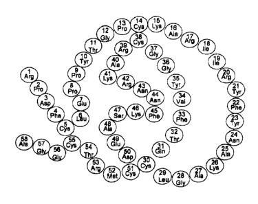
target cell can
effectively inhibit the host or pathogen molecule or complex.
27
CA 02823429 2016-03-29
For example, a therapeutic domain can have a catalytic activity that can
digest a
molecule or epitope of the pathogen or target cell that is required for host-
pathogen
binding, and subsequent entry of the pathogen into the target cell. Receptors
on target
cells that allow for the entry of viruses into cells can be the target of an
enzymatic
activity of a compound of the present invention.
Compounds of the present invention that comprise catalytic domains can be used
to inhibit infection by any pathogen that uses a receptor to gain entry to a
target cell, as
long as removal of the receptor does not impair the organism. These protein-
based
compositions can have, for example, one of the following structures:
(Anchoring Domain)n-[linker]-(Enzymatic Activity)n (n=1,2, 3 or more)
or:
(Enzymatic Activity)n (n=1,2, 3 or more)linker]-(Anchoring Domain)n,
where the linkers are optional.
The enzymatic activity can be a monomeric form of a peptide or polypeptide or
can be multiple copies of the same polypeptide that are either linked directly
or with
spacing sequence in between. The polypeptides or peptides can be linked
directly or via a
spacer composed of peptide linker sequence. The anchoring domain can be any
peptide or
polypeptide that can bind to or near the surface of target cells.
In one preferred embodiment of the present invention, a therapeutic domain
comprises a sialidase that can eliminate or greatly reduce the level of sialic
acid on the
surface of epithelial cells. Sialic acid is a receptor for influenza viruses.
Thus, treating the
surface of respiratory epithelial cells with a sialidase can prevent influenza
infections or
interrupt early infections. The therapeutic domain can comprise a complete
sialidase
protein, or an active portion thereof. . Sialic acid is a receptor for
influenza viruses, and at
least one of the receptors for parainfluenza virus, some coronavinis and
rotavirus,
Streptococcus pneumoniae, Mycoplasma pneumoniae, Haemophilus influenzae,
Moraxella
catarrhalts, Pseudomonas aeruginosa, and Helicobacter pylori. Thus, treating
the surface
of respiratory epithelial cells with a sialidase can prevent influenza or
other viral
28
CA 02823429 2016-03-29
infections or interrupt early infections, as well as prevent or reduce
colonization of
bacteria such as Streptococcus pneumoniae, Mycoplasma pneumoniae, Haemophilus
influenzae, Moraxella catarrhalis, and Pseudomonas aeruginosa. Treating the
gastrointestinal epithelial cells with a sialidase can prevent or reduce
colonization of
Helicobacter pylori in the stomach.
Sialic acid also mediates cell adhesion and interactions between inflammatory
cells
and target cells. Therefore, treating the surface of respiratory epithelial
cells with a
sialidase can prevent the recruitment of inflammatory cells to the airway
surface, and
therefore can treat allergic reactions including asthma and allergic rhinitis.
Since sialic acid serves as a barrier that hinder cell entry by a gene therapy
vector,
treating the target cells with a sialidase can increase transduction
efficiency, and therefore
improve efficacy of the gene therapy.
Preferred sialidases are the large bacterial sialidases that can degrade the
receptor
sialic acids Neu5Ac alpha(2,6)-Ga1 and Neu5Ac alpha(2,3)-Gal. For example, the
bacterial sialidase enzymes from Clostridium petfringens (Genbank Accession
Number
X87369), Actinomyces viscosus, Arthrobacter
ureafaciens, or Micromonospora viridifaciens (Genbank Accession Number D01045)
can
be used. Therapeutic domains of compounds of the present invention can
comprise all or
a portion of the amino acid sequence of a large bacterial sialidase or can
comprise amino
.. acid sequences that are substantially homologous to all or a portion of the
amino acid
sequence of a large bacterial sialidase. In one preferred embodiment, a
therapeutic
domain comprises a sialidase encoded by Actinomyces viscosus, such as that of
SEQ ID
NO:12, or such as sialidase sequence substantially homologous to SEQ ID NO:12.
In yet
another preferred embodiment, a therapeutic domain comprises the catalytic
domain of
the Actinomyces viscosus sialidase extending from amino acids 274-666 of SEQ
ID
NO:12, or a substantially homologous sequence.
Other preferred sialidases are the human sialidases such as those encoded by
the
genes NEU2 (SEQ ID NO:8; Genbank Accession Number Y16535; Monti, E, Preti,
Rossi, E., Ballabio, A and Borsani G. (1999) Genomics 57:137-143) and NEU4
(SEQ ID
NO:9; Genbank Accession Number NM080741; Monti, E, Preti, A, Venerando, B and
Borsani, G. (2002) Neurochem Res 27:646-663) (Figure 3). Therapeutic domains
of
29
CA 02823429 2016-03-29
compounds of the present invention can comprise all or a portion of the amino
acid
sequences of a human sialidase or can comprise amino acid sequences that are
substantially homologous to all or a portion of the amino acid sequences of a
human
sialidase. Preferably, where a therapeutic domain comprises a portion of the
amino acid
sequences of a naturally occurring sialidase, or sequences substantially
homologous to a
portion of the amino acid sequences of a naturally occurring sialidase, the
portion
comprises essentially the same activity as the human sialidase.
A compound for preventing or treating influenza that comprises an enzymatic
domain preferably comprises an anchoring domain that can bind at or near the
surface of
epithelial cells. In some preferred embodiments, the epithelium-anchoring
domain is a
GAG-binding sequence from a human protein, such as, for example, the GAG-
binding
amino acid sequences of human platelet factor 4 (PF4) (SEQ ID NO:2), human
interleukin 8 (IL8) (SEQ ID NO:3), human antithrombin III (AT III) (SEQ ID
NO:4),
human apoprotein E (ApoE) (SEQ ID NO:5), human angio-associated migratory cell
protein (AAMP) (SEQ ID NO:6), and human amphiregulin (SEQ ID NO:7) (Figure 2).
An epithelial anchoring domain can also be substantially homologous to a
naturally
occurring GAG-binding sequence, such as those listed in Figure 2.
It is also within the scope of the present invention to use compounds
comprising a
human sialidase, or comprising a sialidase with substantial homology to a
sialidase, in the
absence of an anchoring domain, in the treatment or prevention of pathogen
infections,
such as but not limited to influenza, paramyxovirus, coronavirus, rotavirus,
and
Pseudornonas aeruginosa infections or bacterial infections; in the treatment
or
prevention of allergic and inflammatory responses, and to improve the
transduction
efficiency of a recombinant virus.
The present invention recognizes that such infections may be prevented or
abated
by the use of sialidases, such as, but not limited to, the A. viscosus
sialidase or human
sialidases such as NEU2 and NEU4. The sialidases can optionally be adapted, by
genetic
or chemical engineering, or by pharmaceutical formulation, to improve their
half life or
retention at the respiratory epithelium.
CA 02823429 2016-03-29
Because influenza viruses primarily infect the upper respiratory tract,
removing
the receptor sialic acid locally in the nasal cavity and nasopharynx area can
prevent
infections or interrupt early infections. The sialidase can be delivered to
the upper
respiratory tract as a nasal spray, and it can be used either in therapeutic
mode during
early stage of influenza (or other infection) or in prophylactic mode before
the infection
occurs. Alternatively, it can be delivered to the lower respiratory tract as
an inhalant to
treat influenza and to prevent influenza complications, such as
bronchopneumonia.
H. Therapeutic Composition Comprising at least one Sialidase Activity
The present invention includes a therapeutic composition that comprises at
least
one sialidase activity. The sialidase activity can be a sialidase isolated
from any source,
such as, for example, a bacterial or mammalian source, or can be a recombinant
protein
that is substantially homologous to at least a portion of a naturally
occurring sialidase.
Preferred sialidases are the large bacterial sialidases that can degrade the
receptor sialic
acids Neu5Ac alpha(2,6)-Gal and Neu5Ac alpha(2,3)-Gal. For example, the
bacterial
sialidase enzymes from Clostridium perfringens (Genbank Accession Number
X87369),
Actinornyces viscosus (Genbank Accession Number L06898), Arthrobacter ureafaci
ens,
or Micromonospora viridifaciens (Genbank Accession Number D01045) or
substantially
homologous proteins can be used.
For example, therapeutic compounds of the present invention can comprise a
large bacterial sialidase or can comprise a protein with the amino acid
sequence of a large
bacterial sialidase or can comprise amino acid sequences that are
substantially
homologous to the amino acid sequence of a large bacterial sialidase. A
preferred
pharmaceutical composition of the present invention comprises the A. viscosus
sialidase
(SEQ ID NO:12), or comprises a protein substantially homologous to the A.
viscosus
sialidase.
Other preferred sialidases are the human sialidases such as those encoded by
the
genes NEU2 (SEQ ID NO:8; Genbank Accession Number Y16535; Monti, E, Preti,
Rossi, E., Ballabio, A and Borsani G. (1999) Genomics 57:137-143) and NEU4
(SEQ ID
NO:9; Genbank Accession Number NM080741; Monti, E, Preti, A, Venerando, B and
31
CA 02823429 2016-03-29
Borsani, G. (2002) Neurochem Res 27:646-663) (Figure 3). Therapeutic domains
of
compounds of the present invention can comprise a human sialidase protein that
is
substantially homologous to the amino acid sequences of a human sialidase or
can
comprise amino acid sequences that are substantially homologous to all or a
portion of
the amino acid sequences of a human sialidase. Preferably, where a therapeutic
domain
comprises a portion of the amino acid sequences of a naturally occurring
sialidase, or
sequences substantially homologous to a portion of the amino acid sequences of
a
naturally occurring sialidase, the portion comprises essentially the same
activity as the
human sialidase.
A pharmaceutical composition comprising a sialidase can include other
compounds, including but not limited to other proteins, that can also have
therapeutic
activity. A pharmaceutical composition comprising a sialidase can include
other
compounds that can enhance the stability, solubility, packaging, delivery,
consistency,
taste, or fragrance of the composition.
A pharmaceutical composition comprising a sialidase can be formulated for
nasal,
tracheal, bronchial, oral, or topical administration, or can be formulated as
an injectable
solution or as eyedrops. A pharmaceutical composition comprising a sialidase
can be
used to treat or prevent pathogen infection, to treat or prevent allergy or
inflammatory
response, or to enhance the transduction efficiency of a recombinant virus for
gene
therapy.
111. Sialidase Catalytic Domain Proteins
The present invention also includes sialidase catalytic domain proteins. As
used
herein a "sialidase catalytic domain protein" comprises a catalytic domain of
a sialidase
but does not comprise the entire amino acid sequence of the sialidase from
which the
catalytic domain is derived. A sialidase catalytic domain protein has
sialidase activity.
Preferably, a sialidase catalytic domain protein comprises at least 10%, at
least 20%, at
least 50%, at least 70% of the activity of the sialidase from which the
catalytic domain
sequence is derived. More preferably, a sialidase catalytic domain protein
comprises at
32
CA 02823429 2016-03-29
least 90% of the activity of the sialidase from which the catalytic domain
sequence is
derived.
A sialidase catalytic domain protein can include other amino acid sequences,
such
as but not limited to additional sialidase sequences, sequences derived from
other
proteins, or sequences that are not derived from sequences of naturally-
occurring
proteins. Additional amino acid sequences can perform any of a number of
functions,
including contributing other activities to the catalytic domain protein,
enhancing the
expression, processing, folding, or stability of the sialidase catalytic
domain protein, or
even providing a desirable size or spacing of the protein.
A preferred sialidase catalytic domain protein is a protein that comprises the
catalytic domain of the A. viscosus sialidase. Preferably, an A. viscosus
sialidase catalytic
domain protein comprises amino acids 270-666 of the A. viscosus sialidase
sequence
(SEQ ID NO:12). Preferably, an A. viscosus sialidase catalytic domain protein
comprises
an amino acid sequence that begins at any of the amino acids from amino acid
270 to
amino acid 290 of the A. viscosus sialidase sequence (SEQ ID NO:12) and ends
at any of
the amino acids from amino acid 665 fo amino acid 901 of said A. viscosus
sialidase
sequence (SEQ ID NO:12), and lacks any A. viscosus sialidase protein sequence
extending from amino acid 1 to amino acid 269. (As used herein "lacks any A.
viscosus
= sialidase protein sequence extending from amino acid 1 to amino acid 269"
means lacks
any stretch of four or more consecutive amino acids as they appear in the
designated
protein or amino acid sequence.)
In some preferred embodiments, an A. viscosus sialidase catalytic domain
protein
comprises amino acids 274-681 of the A. viscosus sialidase sequence (SEQ ID
NO:12)
and lacks other A. viscosus sialidase sequence. In some preferred embodiments,
an A.
viscosus sialidase catalytic domain protein comprises amino acids 274-666 of
the A.
viscosus sialidase sequence (SEQ ID NO:12) and lacks any other A. viscosus
sialidase
sequence. In some preferred embodiments, an A. viscosus sialidase catalytic
domain
protein comprises amino acids 290-666 of the A. viscosus sialidase sequence
(SEQ ID
NO:12) and lacks any other A. viscosus sialidase sequence. In yet other
preferred
embodiments, an A. viscosus sialidase catalytic domain protein comprises amino
acids
33
CA 02823429 2016-03-29
290-681 of the A. viscosus sialidase sequence (SEQ ID NO:12) and lacks any
other A.
viscosus sialidase sequence.
The present invention also comprises nucleic acid molecules that encode
protein-
based compounds of the present invention that comprise a catalytic domain of a
sialidase.
.. The nucleic acid molecules can have codons optimized for expression in
particular cell
types, such as, for example E. coli or human cells. The nucleic acid molecules
or the
present invention that encode protein-based compounds of the present invention
that
comprise at least one catalytic domain of a sialidase can also comprise other
nucleic acid
sequences, including but not limited to sequences that enhance gene
expression. The
nucleic acid molecules can be in vectors, such as but not limited to
expression vectors.
Fusion Proteins
Sialidase catalytic domain proteins can be fusion proteins, in which the
fusion
protein comprises at least one sialidase catalytic domain and at least one
other protein
.. domain, including but not limited to: a purification domain, a protein tag,
a protein
stability domain, a solubility domain, a protein size-increasing domain, a
protein folding
domain, a protein localization domain, an anchoring domain, an N-terminal
domain, a C-
terminal domain, a catalytic activity domain, a binding domain, or a catalytic
activity-
enhancing domain, Preferably, the at least one other protein domain is derived
from
another source, such as, but not limited to, sequences from another protein.
The at least
one other protein domain need not be based on any known protein sequence, but
can be
engineered and empirically tested to perform any function in the fusion
protein.
Purification domains can include, as nonlimiting examples, one or more of a
his
tag, a calmodulin binding domain, a maltose binding protein domain, a
streptavidin
domain, a streptavidin binding domain, an intein domain, or a chitin binding
domain.
Protein tags can comprise sequences that can be used for antibody detection of
proteins,
such as, for example, the myc tag, the hemagglutinin tag, or the FLAG tag.
Protein
domains that enhance protein expression, modification, folding, stability,
size, or
localization can be based on sequences of know proteins or engineered. Other
protein
domains can have binding or catalytic activity or enhance the catalytic
activity of the
sialidase catalytic domain.
34
CA 02823429 2016-03-29
Preferred fusion proteins of the present invention comprise at least one
sialidase
catalytic domain and at least one anchoring domain. Preferred anchoring
domains include
GAG-binding domains, such as the GAG-binding domain or human amphiregulin (SEQ
ID NO:7).
Sialidase catalytic domains and other domains of a fusion protein of the
present
invention can optionally be joined by linkers, such as but not limited to
peptide linkers. A
variety of peptide linkers are known in the art. A preferred linker is a
peptide linker
comprising glycine, such as G-G-G-G-S (SEQ ID NO:10).
The present invention also comprises nucleic acid molecules that fusion
proteins
of the present invention that comprise a catalytic domain of a sialidase. The
nucleic acid
molecules can have codons optimized for expression in particular cell types,
such as, for
example E. coli or human cells. The nucleic acid molecules or the present
invention that
encode fusion proteins of the present invention can also comprise other
nucleic acid
sequences, including but not limited to sequences that enhance gene
expression. The
nucleic acid molecules can be in vectors, such as but not limited to
expression vectors.
IV Pharmaceutical Compositions
The present invention includes compounds of the present invention
formulated as pharmaceutical compositions. The pharmaceutical compositions
comprise a pharmaceutically acceptable carrier prepared for storage and
preferably subsequent administration, which have a pharmaceutically effective
amount of the compound in a pharmaceutically acceptable carrier or diluent.
Acceptable carriers or diluents for therapeutic use are well known in the
pharmaceutical art, and are described, for example, in Remington's
Pharmaceutical Sciences, 18th Ed., Mack Publishing Co., Easton, PA (1990)).
Preservatives, stabilizers, dyes and even flavoring agents can be provided in
the
pharmaceutical composition. For example, sodium benzoate, sorbic acid and
esters of p-hydroxybenzoic acid can be added as preservatives. In addition,
antioxidants and suspending agents can be used.
CA 02823429 2016-03-29
Depending on the target cell, the compounds of the present invention can
be formulated and used as tablets, capsules or elixirs for oral
administration;
salves or ointments for topical application; suppositories for rectal
administration;
sterile solutions, suspensions, and the like for use as inhalants or nasal
sprays.
.. Injectables can also be prepared in conventional forms either as liquid
solutions or
suspensions, solid forms suitable for solution or suspension in liquid prior
to
injection, or as emulsions. Suitable excipients are, for example, water,
saline,
dextrose, mannitol, lactose, lecithin, albumin, sodium glutamate, cysteine
hydrochloride and the like. In addition, if desired, the injectable
pharmaceutical
.. compositions can contain minor amounts of nontoxic auxiliary substances,
such
as wetting agents, p1-1 buffering agents and the like.
The pharmaceutically effective amount of a test compound required as a
dose will depend on the route of administration, the type of animal or patient
being treated, and the physical characteristics of the specific animal under
consideration. The dose can be tailored to achieve a desired effect, but will
depend on such factors as weight, diet, concurrent medication and other
factors
which those skilled in the medical arts will recognize. In practicing the
methods
of the present invention, the pharmaceutical compositions can be used alone or
in
combination with one another, or in combination with other therapeutic or
diagnostic agents. These products can be utilized in vivo, preferably in a
mammalian patient, preferably in a human, or in vitro. In employing them in
vivo, the pharmaceutical compositions can be administered to the patient in a
variety of ways, including topically, parenterally, intravenously,
subcutaneously,
intramuscularly, colonically, rectally, nasally or intraperitoneally,
employing a
variety of dosage forms. Such methods can also be used in testing the activity
of
test compounds in vivo.
In preferred embodiments, these pharmaceutical compositions may be in
the form of orally-administrable suspensions, solutions, tablets or lozenges;
nasal
sprays; inhalants; injectables, topical sprays, ointments, powders, or gels.
When administered orally as a suspension, compositions of the present
invention are prepared according to techniques well-known in the art of
36
CA 02823429 2016-03-29
pharmaceutical formulation and may contain microcrystalline cellulose for
imparting bulk, alginic acid or sodium alginate as a suspending agent,
methylcellulose as a viscosity enhancer, and sweeteners/flavoring agents known
in the art. As immediate release tablets, these compositions may contain
microcrystalline cellulose, dicalcium phosphate, starch, magnesium stearate
and
lactose and/or other excipients, binders, extenders, disintegrants, diluents
and
lubricants known in the art. Components in the formulation of a mouthwash or
rinse include antimicrobials, surfactants, cosurfactants, oils, water and
other
additives such as sweeteners/flavoring agents known in the art.
When administered by a drinking solution, the composition comprises one
or more of the compounds of the present invention, dissolved in water, with
appropriate pH adjustment, and with carrier. The compound may be dissolved in
distilled water, tap water, spring water, and the like. The pH can preferably
be
adjusted to between about 3.5 and about 8.5. Sweeteners may be added, e.g., 1%
(w/v) sucrose.
Lozenges can be prepared according to U.S. Patent No. 3,439,089.
When administered by nasal aerosol or inhalation, the pharmaceutical
compositions are prepared according to techniques well-known in the art of
pharmaceutical formulation and may be prepared as solutions in saline,
employing benzyl alcohol or other suitable preservatives, absorption promoters
to
enhance bioavailability, fluorocarbons, and/or other solubilizing or
dispersing
agents known in the art. See, for example, Ansel, H. C. et al., Pharmaceutical
Dosage Forms and Drug Delivery Systems, Sixth Ed. (1995). Preferably these
compositions and formulations are prepared with suitable nontoxic
pharmaceutically acceptable ingredients. These ingredients are known to those
skilled in the preparation of nasal dosage forms and some of these can be
found in
Remington's Pharmaceutical Sciences, 18th Ed., Mack Publishing Co., Easton,
PA (1990, a standard reference in the field. The choice of suitable carriers
is
highly dependent upon the exact nature of the nasal dosage form desired, e.g.,
solutions, suspensions, ointments, or gels. Nasal dosage forms generally
contain
37
CA 02823429 2016-03-29
large amounts of water in addition to the active ingredient. Minor amounts of
other ingredients such as pH adjusters, emulsifiers or dispersing agents,
preservatives, surfactants, jelling agents, or buffering and other stabilizing
and
solubilizing agents may also be present. Preferably, the nasal dosage form
should
be isotonic with nasal secretions.
Nasal formulations can be administers as drops, sprays, aerosols or by any
other intranasal dosage form. Optionally, the delivery system can be a unit
dose
delivery system. The volume of solution or suspension delivered per dose can
preferably be anywhere from about 5 to about 2000 microliters, more preferably
from about 10 to about 1000 microliters, and yet more preferably from about 50
to about 500 microliters. Delivery systems for these various dosage forms can
be
dropper bottles, plastic squeeze units, atomizers, nebulizers or
pharmaceutical
aerosols in either unit dose or multiple dose packages.
The formulations of this invention may be varied to include; (1) other
acids and bases to adjust the pH; (2) other tonicity imparting agents such as
sorbitol, glycerin and dextrose; (3) other antimicrobial preservatives such as
other
parahydroxy benzoic acid esters, sorbate, benzoate, propionate, chlorbutanol,
phenylethyl alcohol, benzalkonium chloride, and mercurials; (4) other
viscosity
imparting agents such as sodium carboxymethylcellulose, microcrystalline
cellulose, polyvinylpyrrolidone, polyvinyl alcohol and other gums; (5)
suitable
absorption enhancers; (6) stabilizing agents such as antioxidants, like
bisulfite and
ascorbate, metal chelating agents such as sodium edetate and drug solubility
enhancers such as polyethylene glycols.
V. Method of preventing or treating infection by a pathogen
The present invention also includes methods of preventing or treating
infection by
a pathogen. In one aspect, the method includes: treating a subject that is
infected with a
pathogen or at risk of being infected with a pathogen with a pharmaceutical
composition
of the present invention that comprises a compound that comprises at least one
anchoring
38
CA 02823429 2016-03-29
domain that can anchor the compound at or near the surface of a target cell
and at least
one therapeutic domain comprising a peptide or protein that has at least one
extracellular
activity that can prevent the infection of a target cell by a pathogen. In
some preferred
embodiments, the method includes applying a therapeutically effective amount
of a
pharmaceutical composition of the present invention to epithelial cells of a
subject. The
subject to be treated can be an animal or human subject.
In another aspect, the method includes: treating a subject that is infected
with a
pathogen or at risk of being infected with a pathogen with a pharmaceutical
composition
of the present invention that comprises a protein-based compound that
comprises a
sialidase activity. In some preferred embodiments, the method includes
applying a
therapeutically effective amount of a pharmaceutical composition of the
present invention
to epithelial cells of a subject. The sialidase activity can be an isolated
naturally occurring
sialidase protein, or a recombinant protein substantially homologous to at
least a portion
of a naturally occurring sialidase. A preferred pharmaceutical composition
comprises a
sialidase with substantial homology to the A, viscosus sialidase (SEQ ID
NO:12). The
subject to be treated can be an animal or human subject.
In yet another aspect, the method includes: treating a subject that is
infected with
a pathogen or at risk of being infected with a pathogen with a pharmaceutical
composition of the present invention that comprises a protein-based compound
that
comprises a sialidase catalytic domain. In some preferred embodiments, the
method
includes applying a therapeutically effective amount of a pharmaceutical
composition of
the present invention to epithelial cells of a subject. The sialidase
catalytic domain is
preferably can substantially homologous to the catalytic domain of a naturally
occurring
sialidase. A preferred pharmaceutical composition comprises a sialidase
catalytic domain
with substantial homology to amino acids 274-666 the A. viscosus sialidase
(SEQ ID
NO:12). The subject to be treated can be an animal or human subject.
A pathogen can be a viral, bacterial, or protozoan pathogen. In some
embodiments,
the pathogen is one of the following: influenza viruses, parainfluenza virus,
respiratory
syncytial virus (RSV), coronavirus, rotavirus, Streptococcus pneumoniae,
Mycoplasma
pneumoniae, Haemophilus influenzae, Moraxella catarrhalis, Pseudomonas
aeruginosa,
and Helicobacter pylori. In one preferred embodiment, the pathogen is
influenza virus.
39
CA 02823429 2016-03-29
Compounds of the present invention can be designed for human use or animal
use. In some aspects of the present invention, a compound of the present
invention can be
used to prevent pathogen infection in a class of animals, such as mammals. In
some
aspects of the present invention, a composition can be used for human and
animal use
(although the formulation may differ). In these aspects, the active domains of
a
compound can be effective against more than one pathogen species, type,
subtype, or
strain and can be active in more than one host species. For example, some
preferred
compounds of the present invention that comprise, for example, active domains
such as
protease inhibitors that prevent processing of the HA protein of influenza
virus, or
sialidases that remove sialic acid receptors from target cells, or anchoring
domains such
as domains that bind heparin or heparan sulfate, can be used in birds,
mammals, or
humans. Such compounds that can be effective against a range of pathogens with
the
capacity to infect different host species can also be used in humans to combat
infection
by pathogens that are naturally hosted in other species.
In some preferred embodiments of the present invention, the pharmaceutical
composition prevents infection by influenza, and a therapeutically effective
amount of the
pharmaceutical composition is applied to the respiratory epithelial cells of a
subject. This
can be done by the use of an inhaler, or by the use of a nasal spray.
Preferably, the inhaler
or nasal spray is used from one to four times a day.
Because influenza viruses primarily infect the upper respiratory tract,
removing the
receptor sialic acid locally in the nasal cavity, pharynx, trachea and bronchi
can prevent
infections or interrupt early infections. The sialidase can be delivered to
the upper
respiratory tract as a nasal spray or as an inhalant, and it can be used
either in therapeutic
mode during early stage of influenza (or other infection) or in prophylactic
mode before
the infection occurs. Alternatively, it can be delivered to the lower
respiratory tract as an
inhalant to treat influenza and to prevent influenza complications, such as
bronchopneumonia. Similarly, the sialidase can be delivered as nasal spray or
inhalant to
prevent or reduce infection by parainfluenza virus and coronavirus. It can
also be
delivered as an inhalant or nasal spray to prevent or reduce airway
colonization by
pathogenic bacteria, including Streptococcus pneumoniae, Mycoplasma
pneumoniae,
Haemophilus influenzae, Moraxella catarrhalis and Pseudomonas aeruginosa. The
CA 02823429 2016-03-29
therapeutic compounds can optionally be adapted, by genetic or chemical
engineering, or
by pharmaceutical formulation, to improve their half-life or retention at the
respiratory
epithelium. Additionally, it can be delivered topically to the eyes or to
surgical wounds in
the form of drops, sprays or ointments to prevent and treat bacterial
infection including
infection by Pseudomonas aeruginosa. It can also be administered orally to
treat infection
by Helicobacter pylori.
Dosage
As will be readily apparent to one skilled in the art, the useful in vivo
dosage to be administered and the particular mode of administration will vary
depending upon the age, weight and type of patient being treated, the
particular
pharmaceutical composition employed, and the specific use for which the
pharmaceutical composition is employed. The determination of effective dosage
levels, that is the dose levels necessary to achieve the desired result, can
be
accomplished by one skilled in the art using routine methods as discussed
above.
In non-human animal studies, applications of the pharmaceutical compositions
are
commenced at higher dose levels, with the dosage being decreased until the
desired effect is no longer achieved or adverse side effects are reduced or
disappear. The dosage for a compound of the present invention can range
broadly
depending upon the desired affects, the therapeutic indication, route of
administration and purity and activity of the compound. Typically, human
clinical
applications of products are commenced at lower dosage levels, with dosage
level
being increased until the desired effect is achieved. Alternatively,
acceptable in
vitro studies can be used to establish useful doses and routes of
administration of
the test compound. Typically, dosages can be between about 1 ng/kg and about
10
mg/kg, preferably between about 10 ng/kg and about 1 mg/kg, and more =
preferably between about 100 ng/kg and about 100 micrograms/kg.
The exact formulation, route of administration and dosage can be chosen
by the individual physician in view of the patient's condition (see, Fingle et
al., in
The Pharmacological Basis of Therapeutics (1975)). It should be noted that the
attending physician would know how to and when to terminate, interrupt or
adjust
41
CA 02823429 2016-03-29
administration due to toxicity, organ dysfunction or other adverse effects.
Conversely, the attending physician would also know to adjust treatment to
higher
levels if the clinical response were not adequate. The magnitude of an
administrated does in the management of the disorder of interest will vary
with
the severity of the condition to be treated and to the route of
administration. The
severity of the condition may, for example, be evaluated, in part, by standard
prognostic evaluation methods. Further, the dose and perhaps dose frequency,
will also vary according to the age, body weight and response of the
individual
patient, including those for veterinary applications.
Thus, in accordance with the present invention, there is further provided a
method
of treating and a pharmaceutical composition for treating influenza virus
infection and
prevention of influenza virus infection. The treatment involves administering
to a patient
in need of such treatment a pharmaceutical carrier and a therapeutically
effective amount
of any composition of the present invention, or a pharmaceutically acceptable
salt
thereof.
In one preferred regimen, appropriate dosages are administered to each patient
by
either inhaler, nasal spray, or by oral lozenge. It will be understood,
however, that the
specific dose level and frequency of dosage for any particular patient may be
varied and
will depend upon a variety of factors including the activity of the specific
salt or other
form employed, the metabolic stability and length of action of that compound,
the age,
body weight, general health, sex, diet, mode and time of administration, rate
of excretion,
drug combination, the severity of the particular condition, and the host
undergoing
therapy.
VI. Method of reducing, preventing, or treating allergic and inflammatory
responses
The present invention also includes methods of reducing, preventing, or
treating
an allergic or inflammatory response of a subject.
In one aspect, the method includes: preventing or treating an allergic or
inflammatory response of a subject with a pharmaceutical composition of the
present
42
CA 02823429 2016-03-29
invention that comprises a protein-based compound that comprises a sialidase
activity. In
some preferred embodiments, the method includes applying a therapeutically
effective
amount of a pharmaceutical composition of the present invention to epithelial
cells of a
subject. The sialidase activity can be an isolated naturally occurring
sialidase protein, or a
recombinant protein substantially homologous to at least a portion of a
naturally
occurring sialidase. A preferred pharmaceutical composition comprises a
sialidase with
substantial homology to the A. viscosus sialidase (SEQ ID NO:12). The subject
to be
treated can be an animal or human subject.
In yet another aspect, the method includes: preventing or treating an allergic
or
inflammatory response of a subject with a pharmaceutical composition of the
present
invention that comprises a protein-based compound that comprises a sialidase
catalytic
domain. In some preferred embodiments, the method includes applying a
therapeutically
effective amount of a pharmaceutical composition of the present invention to
epithelial
cells of a subject. The sialidase catalytic domain is preferably can
substantially
homologous to the catalytic domain of a naturally occurring sialidase. A
preferred
pharmaceutical composition comprises a sialidase catalytic domain with
substantial
homology to amino acids 274-666 the A. viscosus sialidase (SEQ ID NO:12). The
subject to be treated can be an animal or human subject.
The allergic or inflammatory response can be and acute or chronic condition,
and
can include, as nonlimiting examples, asthma, other allergic responses causing
respiratory distress, allergic rhinitis, eczema, psoriasis, reactions to plant
or animal
toxins, or autoimmune conditions.
In some preferred embodiments, compounds of the present invention can be
delivered as an inhalant or nasal spray to prevent or treat inflammation in
the airway
including, but not limited to, asthma and allergic rhinitis. Compounds of the
present
invention comprising sialidase activity (including sialidase catalytic domain
proteins and
sialidase fusion proteins) can also be administered as eye drops, ear drops,
or sprays,
ointments, lotions, or gels to be applied to the skin. In another aspect, the
method includes
treating a patient who has inflammatory diseases with the present invention
that comprises
a sialidase activity that is administered intravenously or as a local
injection.
43
CA 02823429 2016-03-29
Dosage
As will be readily apparent to one skilled in the art, the useful in vivo
dosage to be administered and the particular mode of administration will vary
depending upon the age, weight and type of patient being treated, the
particular
pharmaceutical composition employed, and the specific use for which the
pharmaceutical composition is employed. The determination of effective dosage
levels, that is the dose levels necessary to achieve the desired result, can
be
accomplished by one skilled in the art using routine methods as discussed
above.
In non-human animal studies, applications of the pharmaceutical compositions
are
commenced at higher dose levels, with the dosage being decreased until the
desired effect is no longer achieved or adverse side effects are reduced or
disappear. The dosage for a compound of the present invention can range
broadly
depending upon the desired affects, the therapeutic indication, route of
administration and purity and activity of the compound. Typically, human
clinical
applications of products are commenced at lower dosage levels, with dosage
level
being increased until the desired effect is achieved. Alternatively,
acceptable in
vitro studies can be used to establish useful doses and routes of
administration of
the test compound. Typically, dosages can be between about 1 ng/kg and about
10
mg,/kg, preferably between about 10 ng/kg and about 1 mg/kg, and more
preferably between about 100 ng/kg and about 100 micrograms/kg.
The exact formulation, route of administration and dosage can be chosen
by the individual physician in view of the patient's condition (see, Fingle et
al., in
The Pharmacological Basis of Therapeutics (1975)). It should be noted that the
attending physician would know how to and when to terminate, interrupt or
adjust
administration due to toxicity, organ dysfunction or other adverse effects.
Conversely, the attending physician would also know to adjust treatment to
higher
levels if the clinical response were not adequate. The magnitude of an
administrated does in the management of the disorder of interest will vary
with
the severity of the condition to be treated and to the route of
administration. The
severity of the condition may, for example, be evaluated, in part, by standard
prognostic evaluation methods. Further, the dose and perhaps dose frequency,
44
CA 02823429 2016-03-29
will also vary according to the age, body weight and response of the
individual
patient, including those for veterinary applications.
In some preferred regimens, appropriate dosages are administered to each
patient
by either inhaler, nasal spray, or by topical application. It will be
understood, however,
that the specific dose level and frequency of dosage for any particular
patient may be
varied and will depend upon a variety of factors including the activity of the
specific salt
or other form employed, the metabolic stability and length of action of that
compound,
the age, body weight, general health, sex, diet, mode and time of
administration, rate of
excretion, drug combination, the severity of the particular condition, and the
host
undergoing therapy.
VII. Method of enhancing gene delivery by a recombinant viral vector
The present invention also includes methods of gene delivery by a recombinant
viral vector. In one aspect, the method includes: administering an effective
amount of a
compound of the present invention that comprises a protein having sialidase
activity to at
least one cell prior to or concomitant with the administration of at least one
recombinant
viral vector. A composition of the present invention can be provided in the
same
formulation as at least one recombinant viral vector, or in a separate
formulation.
In some preferred embodiments, the method includes applying a therapeutically
effective amount of a composition of the present invention and a recombinant
viral vector
to cells of a subject. The subject to be treated can be an animal or human
subject. In a
particularly preferred embodiment, a recombinant viral vector is used to
transduce
epithelial target cells of a subject for gene therapy. For example, a
recombinant viral
vector can be used to transduce airway epithelial cells of a subject with
cystic fibrosis. In
this case, a compound of the present invention can be administered by use of
an inhaler.
A recombinant virus comprising a therapeutic gene can be administered
concurrently or
separately.
In other embodiments, cells can be treated with a compound of the present
invention and a recombinant viral vector in vitro or "ex vivo" (that is, cells
removed from
a subject to be transplanted into a subject after transduction).
CA 02823429 2016-03-29
The sialidase activity can be an isolated naturally occurring sialidase
protein, or a
recombinant protein substantially homologous to at least a portion of a
naturally
occurring sialidase, including a sialidase catalytic domain. A preferred
pharmaceutical
composition comprises a sialidase with substantial homology to the A. viscosus
sialidase
(SEQ ID NO:12).
A compound of the present invention can be administered to target cells from
one
day before to two hours subsequent to the administration of the recombinant
virus.
Preferably a compound of the present invention is administered to target cells
from four
hours to ten minutes before administration of the recombinant virus.
Administration can
be
A recombinant virus is preferably a recombinant virus that can be used to
transfer
genes to mammalian cells, such as, preferably human cells. For example, a
recombinant
virus can be a retrovirus (including lentivirus), adenovirus, adeno-associated
virus
(AAV) or herpes simplex virus type I. The recombinant virus comprises at least
one
exogenous gene that is to be transferred to a target cell. The gene is
preferably a
therapeutic gene, but this need not be the case. For example, the gene can be
a gene used
to mark cells or confer drug resistance.
In a preferred embodiment, the present invention includes methods of improving
efficacy of a gene therapy vector: The method includes treating a patient with
a
compound of the present invention that comprises a sialidase activity and, in
the same or a
separate formation, with a recombinant virus. The compound of the present
invention
having sialidase activity can be administered to the patient prior to,
concomitant to, or
even subsequent to the administration of a recombinant virus. In one
embodiment, the
sialidase is substantially homologous to the Actinomyces viscosus sialidase
(SEQ ID
NO:12) or a portion thereof. In one preferred embodiment, the sialidase
comprises the
catalytic domain of the Actinomyces viscosus sialidase. In another embodiment,
the
recombinant virus is AAV. In yet another embodiment, the disease is cystic
fibrosis. In
yet another embodiment, the recombinant virus comprises the cystic fibrosis
transmembrane conductance regulator (CFTR) gene.
46
CA 02823429 2016-03-29
Dosage
As will be readily apparent to one skilled in the art, the useful in vivo
dosage to be administered and the particular mode of administration will vary
depending upon the age, weight and type of patient being treated, the
particular
pharmaceutical composition employed, and the specific use for which the
pharmaceutical composition is employed. The determination of effective dosage
levels, that is the dose levels necessary to achieve the desired result, can
be
accomplished by one skilled in the art using routine methods as discussed
above.
In non-human animal studies, applications of the pharmaceutical compositions
are
commenced at higher dose levels, with the dosage being decreased until the
desired effect is no longer achieved or adverse side effects are reduced or
disappear. The dosage for a compound of the present invention can range
broadly
depending upon the desired affects, the therapeutic indication, route of
administration and purity and activity of the compound. Typically, human
clinical
applications of products are commenced at lower dosage levels, with dosage
level
being increased until the desired effect is achieved. Alternatively,
acceptable in
vitro studies can be used to establish useful doses and routes of
administration of
the test compound. Typically, dosages can be between about 1 ng/kg and about
10
mg/kg, preferably between about 10 ng/kg and about 1 mg/kg, and more
preferably between about 100 ng/kg and about 100 micrograms/kg.
The exact formulation, route of administration and dosage can be chosen
by the individual physician in view of the patient's condition (see, Fingle et
al., in
The Pharmacological Basis of Therapeutics (1975)). It should be noted that the
attending physician would know how to and when to terminate, interrupt or
adjust
administration due to toxicity, organ dysfunction or other adverse effects.
Conversely, the attending physician would also know to adjust treatment to
higher
levels if the clinical response were not adequate. The magnitude of an
administrated does in the management of the disorder of interest will vary
with
the severity of the condition to be treated and to the route of
administration. The
severity of the condition may, for example, be evaluated, in part, by standard
prognostic evaluation methods. Further, the dose and perhaps dose frequency,
47
CA 02823429 2016-03-29
will also vary according to the age, body weight and response of the
individual
patient, including those for veterinary applications.
In some preferred regimens, appropriate dosages are administered to each
patient
by either inhaler, nasal spray, or by topical application. It will be
understood, however,
that the specific dose level and frequency of dosage for any particular
patient may be
varied and will depend upon a variety of factors including the activity of the
specific salt
or other form employed, the metabolic stability and length of action of that
compound,
the age, body weight, general health, sex, diet, mode and time of
administration, rate of
excretion, drug combination, the severity of the particular condition, and the
host
undergoing therapy.
Examples
Example 1: Synthesizing aprotinin genes, par/lying and testing aprotinin
fusion proteins.
Introduction
Influenza viral protein hemagglutinin (HA) is the major influenza envelope
protein. It plays an essential role in viral infection. The importance of HA
is evidenced
by the fact that it is the major target for protective neutralizing antibodies
produced by
the host immune response (Hayden, FG. (1996) In Antiviral drug resistance (ed.
D. D.
Richman), pp. 59-77. Chichester, UK: John Wiley & Sons Ltd.). It is now clear
that HA
has two different functions in viral infection. First, HA is responsible for
the attachment
of the virus to sialie acid cell receptors. Second, HA mediates viral entry
into target cells
by triggering fusion of the viral envelope with cellular membranes.
HA is synthesized as a precursor protein, HAO, which is transferred through
the
Golgi apparatus to the cell surface as a trimeric molecular complex. HAO is
further
cleaved to generate the C terminus HAI (residue 328 of HAO) and the N terminus
of
HA2. It is generally believed that the cleavage occurs at the cell surface or
on released
viruses. The cleavage of HAO into HAl/HA2 is not required for HA binding to a
sialic
acid receptor; however, it is essential for viral infectivity (Klenk, HD and
Rott, R. (1988)
48
CA 02823429 2016-03-29
Adv Vir Res. 34:247-281; Kido, H, Niwa, Y, Beppu, Y and Towatari, T. (1996)
Advan
Enzyme Regul 36:325-347; Skehel, J.J and Wiley, DC. (2000) Annu Rev Biochem
69:531-
569).
Sensitivity of HAO to host proteases is determined by the proteolytic site in
the
external loop of HAO molecule. The proteolytic site may contain either a
single Arg or
Lys residue (monobasic cleavage site) or several Lys and/or Arg residues in R-
X-IC/R-R
motif (multibasic cleavage site). Only the influenza A virus subtypes H5 and
H7 have
HA proteins carrying the multibasic cleavage site. All other influenza A, B
and C viruses
contain HA proteins having the monobasic cleavage site. Influenza A viruses
having
multibasic cleavage sites are more virulent and induce systemic infection in
hosts
whereas viruses with a monobasic HA site initiate infection only in the
respiratory tract in
mammals or in the respiratory and enteric tracts in avian species (Klenk, HD
and Garten
W. 1994. Trend Micro 2:39-43 for review). Fortunately, human infection by the
highly
virulent avian influenza A H5 and 117 subtypes, which carry the multibasic
cleavage site,
has so far only occurred in a handful of cases discovered mostly in Hong Kong.
The vast
majority of influenza infections are caused by viruses with HA proteins are
cleaved at the
monobasic cleavage site.
Influenza virus HA subtypes 5 and 7 that contain multibasic cleavage sites are
activated by furin, a member of the subtilisin-like endoproteases, or the pre-
protein
.. convertase family. Furin cleaves the virus intracellularly and is
ubiquitously present in
many cell types, allowing the virulent, systemic infection seen with such
viruses (Klenlc,
HD and Garten W. 1994. Trend Micro 2:39-43; Nakayama, K. 1997. Biochem 327:625-
635). All other influenza viruses, which have HAs with monobasic cleavage
sites, are
activated by secreted, trypsin-like serine proteases. Enzymes that have been
implicated
in influenza virus activation include: plasmin (Lazarowitz SG, Goldberg AR and
Choppin
PW. 1973. Virology 56:172-180), mini-plasmin (Murakami M, Towatari T, Ohuchi
M,
Shiota M, Akao M, Okumura Y, Parry MA and Kido H. (2001) Eur J Biochem 268:
2847-2855), tryptase Clara (Kido H, Chen Y and Murakami M. (1999) In B.Dunn
(ed.),
Proteases of infectious agents. p.205-217, Academic Press, New York, N.Y),
kallilcrein,
.. urokinase, thrombin (Scheiblauer H, Reinacher M, Tashiro M and Rott R.
(1992) J Infec
Dis 166:783-791), blood clotting factor Xa (Gotoh 8, Ogasawara T, Toyoda T,
Inocencio
49
CA 02823429 2016-03-29
N, Hamaguchi M and Nagai Y. (1990) EMBO J9:4189-4195), acrosin (Garten W,
Bosch
FX, Linder D, Rott R and Klenk HD. (1981) Virology 115:361-374.), proteases
from
human respiratory lavage (Barbey-Morel CL, Oeltmann TN, Edwards KM and Wright
PF. (1987) J Infect Dis 155:667-672) and bacterial proteases from
Staphylococcus
aureus (Tashiro M, Ciborowski P, Reinacher M, Pulverer G, Klenk HD and Rott R.
(1987) Virology 157:421-430) and Pseudomonas aeruginosa (Callan RJ, Hartmann
FA,
West SE and Hinshaw VS. (1997)J Virol 71:7579-7585). Activation of influenza
viruses by host serine proteases is generally considered to occur
extracellularly either at
the plasma membrane or after virus release from the cell.
Aprotinin, also called Trasylol, or bovine pancreatic trypsin inhibitor (BPTI)
is a
polypeptide having 58 amino acids. It belongs to the family of Kunitz-type
inhibitors and
competitively inhibits a wide spectrum of serine proteases, including trypsin,
chymotrypsin, plasmin and plasma kallikrein. Aprotinin has long been used as a
human
therapeutics, such as treatment of pancreatitis, various states of shock
syndrome,
hyperfibrinolytic haemorrhage and myocardial infarction. It is also used in
open-heart
surgery, including cardiopulmonary bypass operations, to reduce blood loss
(Fritz H and
Wunderer G. (1983) Arzneim-Forsch 33:479-494).
The safety of aprotinin in human has been well documented through years of
clinical applications. In addition, aprotinin is apparently a very weak
immunogen as
aprotinin-specific antibodies have not been observed in human sera so far
(Fritz 11 and
Wunderer G. (1983) Arzneim-Forsch 33:479-494). Another desired feature of
aprotinin
as a drug candidate is its superb stability. It can be kept at room
temperature for at least
18 months without any loss of activity (Fritz H and Wunderer G. (1983) Arzneim-
Forsch
33:479-494).
To achieve significant viral inhibition in animal studies that have been
performed,
aprotinin was administered at high doses. For example, 280 micrograms to 840
micrograms per day of aprotinin was injected intraperitoneally into each mouse
for 6
days (Zhirnov OP, Ovcharenko AV and Bukrinskaya AG. (1984) J Gen Virol 65:191-
196); a lower dosage was required for aerosol inhalation, still, each mouse
was given 63 -
126 micrograms per day for 6 days (Ovcharenko AV and Zhirnov OP. (1994)
Antiviral
Res 23:107-118). A very high dose of aprotinin would be required in human
based on
CA 02823429 2016-03-29
extrapolation from the mouse data. Therefore to achieve better efficacy in
human, the
potency of aprotinin molecule needs to be significantly improved.
Aprotinin functions by competitively inhibiting serine proteases that are
mostly
on the surface of host respiratory epithelial cells. Local concentration of
aprotinin in the
vicinity of host proteases is therefore the key factor determining competitive
advantage of
aprotinin. We use two approaches that work synergistically to boost
competitive
advantage of aprotinin on the surface of respiratory epithelium.
First, the avidity (functional affinity) of aprotinin is increased by making
multivalent aprotinin fusion proteins consisting of two, three, or more
aprotinin proteins
connected via linkers. Such a molecule is able to bind to membrane proteases
in a
multivalent fashion, which has significant kinetic advantage over the
aprotinin monomer. =
Monomeric aprotinin binds to bovine trypsin very tightly with dissociation
constant (Ki)
being 6.0 x mo1/1. However, its affinity compared to other proteases, such
as
chymotrypsin, plasmin and Kallilcrein, which have been implicated in
activation of
influenza viruses, is much lower with Ki being at the level of 10-8 to i
mo1/1 (Fritz H
and Wanderer G. (1983) Arzneim-Forsch 33:479-494). Multimerization can
increase
aprotinin's affinity to these proteases exponentially.
Second, we fuse aprotinin with a respiratory epithelium-anchoring domain. The
anchoring domain localizes aprotinin to the proximity of host membrane-
associated
proteases and maintains a high local concentration of aprotinin on epithelial
surface. The
anchoring domain also increases retention time of the drug on the respiratory
epithelium.
Cloning
Aprotinin is a single chain polypeptide having 58 amino acid residues and 3
intra-
chain disulfide bonds (SEQ ID NO:1). The amino acid sequence of aprotinin is
shown
in Figure 1. Genes encoding aprotinin and aprotinin fusion proteins are
synthesized by
PCR using overlapping oligonucleotides with codons optimized for E. Coil
expression as
templates. The PCR products are cloned into pCR2.1-TOPO vector (Invitrogen).
After
sequencing, the genes are subcloned into an expression vector pQE (Qiagen).
The vector
carries a purification tag, Hisx6, to allow easy purification of the
recombinant proteins.
The constructs are used to transform E. Coil. The transformed cells grown in
LB-
51
CA 02823429 2016-03-29
ampicillin medium to mid-log phase are induced by IPTG according to standard
protocols. Cells are pelleted and lysed in phosphate-buffered-saline (PBS) by
sonication.
The enzymes, which have His6 purification tag, are purified using a nickel
column
(Qiagen).
The following aprotinin fusion proteins are made:
1. Dimeric and trimeric aprotinin. Two or three aprotinin genes are linked via
a flexible
linker as the following constructs:
Aprotinin¨(GGGGS (SEQ ID NO:10))n (n=3, 4 or 5)¨Aprotinin;
and
Aprotinin¨(GGGGS(SEQ ID NO:10))n (n=3, 4 or 5)¨Aprotinin¨
(GGGGS(SEQ ID NO:10))n (n=3, 4 or 5)¨Aprotinin
The length of the linker sequence may determine three-dimensional flexibility
of the
multimeric aprotinin and thereby influence functional affinity of the
molecule. Therefore
.. constructs having linkers with various lengths are made.
Fully functional recombinant monomeric aprotinin has been produced in E. Coli
(Auerswald EA, Horlein D, Reinhardt G, Schroder Wand Schnabel E. (1988).Biol
Chem
Hoppe-Seyler Vol 369, Suppl., pp27-35). We therefore expect proper folding of
multivalent aprotinin proteins in E. colt cells. Besides expressing protein in
various
common E. Colt cell strains, such as BL21, JM83, etc, the multivalent
aprotinin proteins
are also expressed in OrigamiTm cells (Novagen, Bad Soden, Germany). The
OrigamiTM
cell strain does not have thioredoxin and glutathione reductase and thus has
an oxidizing
cytoplasm. This cell strain has been used to successfully express a number of
proteins
that contain disulfide bonds (Bessette PH, Aslund F, Beckwith J and Georgiou
G. (1999)
Pro Natl Acad Sci USA 96:13703-13708; Venturi M, Seifert C and Hunte C. (2001)
J
Mol Biol 315:1-8.).
2. The epithelium cell-anchoring aprotinin. An epithelium cell-anchoring
sequence is
fused with aprotinin. The epithelium-anchoring sequence can be any peptide or
polypeptide sequence that has affinity towards the surface of epithelial
cells. We have
52
CA 02823429 2016-03-29
selected three human GAG-binding sequences: PF4 (aa 47-70; SEQ ID NO: 2), IL-8
(aa 46-72; SEQ ID NO: 3), and AT III (aa 118-151; SEQ ID NO: 4) (Figure 2).
These sequences bind to heparin/heparan sulfate with nanomolar-level
affinities
(Table 1). Heparin/Heparan Sulfate are ubiquitously present on the respiratory
epithelium. In separate constructs, the GAG-binding sequences are fused with
the
aprotinin gene on the N terminus and on the C terminus via a generic linker
sequence
GGGGS as the following constructs:
(GAG domain¨GGGGS(SEQ ID NO:10)¨Aprotinin); and
(Aprotinin--GGGGS(SEQ ID NO :10)¨GAG domain)
15 Table 1. Affinities to Heparin
Protein Kd nM (ref)
PF4 27 (44)
IL-8 <5 (43)
ATIII 11 (42)
ApoE 620 (45)
Photometric trypsin inhibition assay
The trypsin inhibition activity of aprotinin and aprotinin fusion proteins is
measured by a photometric assay described previously in detail (Fritz H and
Wunderer G.
(1983) Arzneim-Forsch 33:479-494). Briefly, in this assay aprotinin inhibits
the trypsin-
catalyzed hydrolysis of Na-benzoyl-L-arginine-p-nitroanilide (BzArgpNA or L-
BAPA)
53
CA 02823429 2016-03-29
(Sigma), which is followed photometrically at 405 TIM. One trypsin unit
(UBAPA)
corresponds to the hydrolysis of 1 micromole substrate per min. One inhibitor
unit
(IUBApA) decreases the activity of two trypsin units by 50%, which corresponds
arithmetically to the inhibition of 1 UBAPA of trypsin. The specific activity
of aprotinin is
5 given in 1UBApA/mg polypeptide.
Surface plasmon resonance assay
The affinities of dimeric and trimeric aprotinin with various linkers are
compared
against the monomeric aprotinin using surface plasmon resonance assay, or
BlAcore
10 analysis (BlAcore, Piscataway, NJ) with human plasmin as the target.
Similarly,
BlAcore assay with heparin as the target is used to analyze affinity between
GAG
binding aprotinin fusion proteins and heparin.
When plasmin is used as the target, purified human plasmin (Sigma) is
immobilized on the CMS chip according manufacturer's instructions (BlAcore,
15 Piscataway, NJ). When heparin is the target, biotinylated albumin and
albumin-heparin
(Sigma) are captured on a streptavidin-coated BlAcore SA chip as described
previously
(Xiang Y and Moss B. (2003)J Virol 77:2623-2630).
Example 2; Establishing improved tissue culture models for studies on
influenza
20 virus infection.
Stocks of Influenza Viruses
Influenza viral strains are obtained from ATCC and the repository at St. Jude
Children's Research Hospital. All experiments involving influenza viruses are
conducted
at Bio-safety level 11.
25 Viruses are propagated by injection into the allantoic cavity of nine-
day-old
= chicken embryos as described (Zhirnov OP, Ovcharenko AV and Bulcrinskaya
AG.
(1985).1 Gen Virol 66:1633-1638). Alternatively, viral stocks are grown on
Madin-
Darby canine kidney (MDCK) cells in minimal essential medium (MEM)
supplemented
with 0.3% bovine serum albumin and 0.5 micrograms of trypsin per ml. After
54
CA 02823429 2016-03-29
incubating for 48 to 72 hours, the culture medium is clarified by low speed
centrifugation. Viral particles are pelleted by ultracentrifugation through a
25% sucrose
cushion. Purified viruses are suspended in 50% glycerol-0.1M Tris buffer (pH
7.3) and
stored at ¨20 C.
Plaque Assays
Infectivity and titer of the viral stocks are determined by two kinds of
plaque
assays, a conventional one and a modified one (Tobita, K, Sugiura, A, Enomoto,
C and
Furuyama, M. (1975) Med Microbiol Immnuol 162:9-14; Zhimov OP, Oveharenko AV
and Bukrinskaya AG. (1982) Arch Virol 71:177-183). The conventional plaque
assay is
routinely used as a virus titration method. It requires exogenous trypsin in
agar overlay
added immediately after virus infection to MDCK monolayers (Tobita, K,
Sugiura, A,
Enomoto, C and Furuyama, M. (1975) Med Microbiol Immnuol 162:9-14). This
method
artificially increases infectivity of the viral stocks being tested by
activating all the viral
particles that have uncleaved HA.
Zhimov et. al. designed a modified plaque assay consisting of a double agar
overlay, with trypsin being included in the second layer which is added 24
hours after
infection (Zhimov OP, Ovcharenko AV and Bukrinskaya AG. (1982) Arch Virol
71:177-
183). Three days after infection, cells are fixed with a 10% formaldehyde
solution,
agarose layers are removed, fixed cells are stained with hematoxylin-eosin
solution and
plaques are counted. The modified plaque assay allows accurate determination
of the real
infectivity of viral stocks that contain both cleaved and uncleaved HA.
Combining
results from both conventional and modified plaque assays, one can distinguish
viruses
containing cleaved or uncleaved HA and correlate infectivity of viral stocks
with the
status of HA cleavage.
Human Cell Culture Models
1. Short-term culture of primary human epithelial cells. Conventional in vitro
influenza
virus infection is mostly carried out in MDCK cells with exogenous trypsin
added to the
culture medium. This is far from being physiological and is inappropriate for
the work
CA 02823429 2016-03-29
proposed here because trypsin is not the protease that activate influenza
viruses in vivo.
Very limited numbers of in vitro tissue culture models that are able to
support the growth
of influenza virus without an exogenous protease have been reported so far,
those being
primary cultures with primate cells of renal origin, cells lining the
allantoic and amniotic
cavities of embryonated eggs, fetal tracheal ring organ cultures and primary
human
adenoid epithelial cells (Endo Y, Carroll KN, lkizler MR and Wright PF.
(1996)J Virol
70:2055-2058). Among these, the latest work with primary human adenoid
epithelial
cells is the closest mimic of human conditions. In this case, Endo et. al.
(Endo Y, Carroll
KN, Ikizler MR and Wright PF. (1996)J Virol 70:2055-2058) isolated epithelial
cells
from surgical samples of human adenoids, and cultured the epithelial cells on
a collagen
matrix (Vitrogen 100, Celtrix Laboratories, Palo Alto, California) in
Transwell inserts
(Costar, Cambridge, Mass). Cells were maintained in 50% Ham's F12 and 50%
Eagles
minimal essential media with supplements of growth factors and trace elements.
The cells
reached continency in 10 to 14 days, remaining largely as a monolayer but with
discrete
patches of ciliated cells, which maintained regular ciliary activity for 1 to
3 weeks after
reaching confluency. In this system, influenza A virus grew to a titer of 106
PFU/ml with
a multiplicity of infection of 0.001 (Endo Y, Carroll KN, Ikizler MR and
Wright PF.
(1996)J Virol 70:2055-2058). Progressive cytopathogenic effects were also
present
during infection. The biggest drawback of this system is that it requires
fresh human
adenoid tissue.
To solve this problem, primary human adenoid epithelial cells are replaced
with
primary human airway epithelial cells that are commercially available
(Cambrex), and the
cells are grown under the same conditions. Such short-term culture of primary
human
airway epithelial cells is relatively quick to establish and is useful as the
first-line
experimental model for most of the in vitro infection and antiviral
experiments.
2. Well-differentiated human airway epithelium (WD-HAE). In order to best
mimic the
in vivo condition of human airway, the model of well-differentiated human
airway
epithelium (WD-HAE) is used. WD-HAE is stratified epithelium that has all the
differentiated cells of the normal human airway epithelium, including
functional ciliated
cells and mucus secreting cells. Therefore, in this model system influenza
viruses are
56
CA 02823429 2016-03-29
most likely to be activated by host proteases that are physiologically
relevant. Although
WD-HAE has been widely used to study respiratory viral infections, such as
respiratory
syncytial virus (RSV) Zhang L, Peeples ME, Boucher RC, Collins PL and Pickles
(2002) J Virol 76:5654-5666) measles virus (Sinn PL, Williams G, Vongpunsawad
S,
Cattaneo R and McCray PB. (2002)J Virol 76:2403-2409, or human rhinovirus, it
has
not previously been used to study influenza viruses.
A detailed protocol of WD-HAE has been described previously (Kninkoslcy TM,
Fischer BM, Martin LD, Jones N, Akley NJ and Adler KB. (2000)Am J Respir Cell
Mol
Biol 22:685-692). Briefly, commercial primary human bronchial epithelial cells
(Cambrex) are cultured on Transwell-clear culture inserts (Costar) that are
thin-coated
with rat-tail collagen L Cells are cultured submerged for the first 5 to 7
days in medium
containing a 1:1 mixture of bronchial epithelial cell growth medium (BEGM)
(Cambrex)
and DMEM with high glucose with supplement of growth factors (Krunkosky TM,
Fischer BM, Martin LD, Jones N, Akley NJ and Adler KB. (2000)Am J Respir Cell
Mol
Biol 22:685-692). When cultures are 70% confluent (days 5 to 7), the air-
liquid interface
is created by removing the apical medium and exposing cells only to medium on
their
basal surface. Cells are cultured for additional 14 days in air-liquid
interphase, for a total
of 21 days in culture, and are then ready for experiments. The differentiated
epithelium
can be maintained in vitro for weeks.
Epithelial morphology and degree of differentiation is documented by routine
histology (Endo Y, Carroll KN, lkizler MR and Wright PF. (1996) J Virol
70:2055-
2058). Briefly, following fixation with 10% buffered formalin, the epithelial
cells are
embedded in paraffin, sectioned and stained with hematoxylin and eosin, and
with
periodic acid-Schiff stain for mucus secreting cells.
Influenza infection is carried out in the above two model systems by adding
0.001
to 1 MOI of viruses to the differentiated cells. The titer and infectivity of
viruses in the
supernatant are followed over a period of 3 to 7 days. The level of influenza
viral
amplification and the infectivity of influenza viruses are evaluated using
conventional
and modified plaque assays.
57
CA 02823429 2016-03-29
Example 3: Comparing functions of the aprotinin fusion proteins in vitro
Anti-Viral Effects of Aprotinin Fusion Proteins
1. Pre-infection treatment. Aprotinin fusion proteins are added to primary
human
cell cultures at various concentrations and allowed to incubate with the cells
for 1 hour.
The cells are washed with fresh medium and immediately inoculated with
influenza
viruses at MOI 0.01 to 1. Cells are washed again after 1 hour and cultured for
3.to 5
days. Titer and infectivity of viruses in the supernatant are measured at
various time
points by two plaque assays. The cytopathic effect caused by viral infection
is evaluated
by staining viable cells with crystal violet and quantifying by measuring
absorption at
570 nm at the end of the experiment. The percentage of cell protection by
aprotinin
fusion proteins is calculated by 100x {(aprotinin treated sample-untreated
infected
sample)/(uninfected control-untreated infected sample)} . The drug efficacy
for cell
protection is described by its Effective Concentration that achieves 50% of
the cell
protection (EC50). Since HA activation only occurs to newly released viral
particles, the
first round of viral infection occurs normally and viral titer rises in the
first 24 hours after
infection. However, starting from the second round, infectivity of viruses
drops and viral
titer gradually decreases as result of aprotinin treatment. Results from this
experiment
differentiate various types of different aprotinin fusion proteins by their
efficacies in a
single prophylactic treatment.
Alternatively, timing of initial viral inoculation is altered from immediately
after
aprotinin treatment to 2-24 hours post treatment. Viral titer, infectivity and
cytopathic
effect are measured for 3 to 5 day after infection as described above. Results
from these
experiments distinguish various aprotinin fusion proteins by the lengths of
the effective
window after a single prophylactic treatment.
2. Post-infection Treatment. For multi-dose treatment, cells are first
infected by viral
inoculations at 0.001 to 0.1 MOI for 1 hour. Various concentrations of
aprotinin fusion
proteins are added immediately afterwards, additional treatments are applied
at 8-hour
intervals during the first 48 hours post infection. Cells are cultured until
day 7 post
58
CA 02823429 2016-03-29
infection. Viral titer and infectivity in the media are followed during the
whole process.
Cytopathic effect is evaluated at the end of the experiment.
For single dose treatment, cells are first infected by viral inoculations at
0.001 to
0.1 MOI for 1 hour. Treatments of aprotinin fusion proteins at various
concentrations are
applied at different time points during the first 48 hours after infection,
but each cell
sample only receives one treatment during the whole experiment. Cells are
cultured until
day 7 post infection. Viral titer and infectivity in the media are followed
during the
whole process. Cytopathic effect is evaluated at the end of the experiment.
Results from
these experiments distinguish different types of aprotinin fusion proteins for
their
therapeutic potency.
Inhibition of HA Cleavage by Aprotinin Fusion Proteins
To demonstrate that aprotinin fusion proteins inhibit influenza viral
infection by
inhibiting cleavage of influenza HA protein, a human primary epithelial cell
culture is
infected with influenza virus at MOI of 1. Aprotinin fusion proteins are added
to the
culture either right before viral inoculation or immediately after the viral
infection. At
6.5 hour post infection, the culture is incubated for 1 hour in MEM lacking
cold
methionine and containing 35S-labeled methionine (Amersham) at a concentration
of 100
microCi/m1 (pulse). Thereafter, the cells are washed twice with MEM containing
a 10-
fold concentration of cold methionine and incubated in MEM for additional 3
hours
(chase). After labeling, cells are dissolved in radioimmunoprecipitation assay
(RIPA)
buffer, HA is precipitated by anti-serum against the particular strain of
virus used for
infection (anti-influenza sera can be obtained from ATCC and Center of Disease
Control
TM
and Prevention), and the immunocomplex is then purified by protein G-Sepharose
(Amersham). Samples are fractionated by SDS-PAGE followed by autoradiography.
In
samples untreated by aprotinin fusion proteins, HAI and HA2 are expected to be
the
predominant HA species; while in aprotinin treated samples, HAO is expected to
be the
major type of HA present.
39
CA 02823429 2016-03-29
Example 4: Synthesizing genes of five sialidases, expressing and purifying the
sialidase proteins.
Introduction
Influenza viruses belong to the orthomyxoviridae family of RNA viruses. Both
type A and type B viruses have 8 segmented negative-strand RNA genomes
enclosed in a
lipid envelope derived from the host cell. The viral envelope is covered with
spikes that
are composed of three proteins: hemagglutinin (HA), that attaches virus to
host cell
receptors and mediates fusion of viral and cellular membranes; neuraminidase
(NA),
which facilitates the release of the new viruses from the host cell; and a
small number of
M2 proteins that serve as ion channels. For Influenza A virus, HA and NA both
undergo
antigenic drift and antigenic shift, the viral subtypes are distinguished by
serologic
differences between their HA and NA proteins. There are total 15 types of HA
(H1-H15)
and 9 types of NA (N1-N9), but only three HA (H1-H3) and two NA (NI and N2)
have
been found in human Influenza A virus so far (Granoff, A. & Webster, R. G.,
ed.
Encyclopedia of Virology, 2nd Edition, Vol 2). In contrast to Influenza A
virus, no distinct
antigenic subtypes are recognized for Influenza virus B.
While Influenza B virus circulates only in humans, Influenza A virus can be
isolated from a whole host of animals, such as pigs, horses, chickens, ducks
and other
kinds of birds, which accounts for genetic reassortment of Influenza A virus
that results in
antigenic shift. Wild aquatic birds are considered to be the primordial
reservoir of all
influenza viruses for avian and mammalian species. There is extensive evidence
for
transmission of the virus between aquatic birds and other species including
pigs and
horses and indirect transmission to humans through pigs. Direct transmission
from pigs
or chickens to humans has also been documented (Ito, T. (2000) Microbiol
Immunol
44(6):423-430).
The host cell receptor for influenza viruses is the cell surface sialic acid.
Sialic
acids are a-keto acids with 9-carbon backbones that are usually found at the
outermost
positions of the oligosaccharide chains that are attached to glycoproteins and
glycolipids.
One of the major types of sialic acids is N-acetylneuraminic acid (Neu5Ac),
which is the
biosynthetic precursor for most of the other types. Two major linkages between
Neu5Ac
CA 02823429 2016-03-29
and the penultimate galactose residues of carbohydrate side chains are found
in nature,
Neu5Ac a(2,3)-Gal and Neu5Ac a(2,6)-Gal. Both Neu5Ac a(2,3)-Gal and Neu5Ac
a(2,6)-Gal molecules can be recognized by Influenza A virus as the receptor
(Schauer, R.
(1982) Adv. Carbohydrate Chem & Biochem 40:131-235), while human viruses seem
to
prefer Neu5Ac a(2,6)-Gal, avian and equine viruses predominantly recognize
Neu5Ac
a(2,3)-Ga1 (Ito, T. (2000) Microbiol Immunol 44(6):423-430).
Infections by influenza type A and B viruses are typically initiated at the
mucosal
surface of the upper respiratory tract. Viral replication is primarily limited
to the upper
respiratory tract but can extend to the lower respiratory tract and causes
bronchopneumonia that can be fatal. The risk of death is one per 10,000
infections, but is
significantly greater for high-risk groups with pre-existing cardiopulmonary
conditions
and for immunologically naïve individuals during a pandemic.
A therapeutic compound comprising a sialidase that can effectively degrade
both
receptor sialic acids, Neu5Ac a(2,6)-Gal and Neu5Ac a(2,3)-Gal, can confer
protection
against the broadest range of influenza viruses, including animal viruses. It
can also
remain effective as the viral strains change yearly. Because sialidase targets
the host cell
rather than virus and acts at the "choking point" in a viral life cycle,
generation of
resistant virus is improbable. Protein-bound sialic acid turns over
homogeneously on cell
surface with half-life of 33 hours (Kreisel, W, Volk, BA, Buchsel, R. and
Reutter, W.
(1980) Proc Nat! Acad Set USA 77:1828-1831). Therefore we estimate that once-a-
day or
twice-a-day administration of a sialidase would confer sufficient protection
against
influenza.
Sialidases are found in higher eukaryotes, as well as in some mostly
pathogenic
microbes, including viruses, bacteria and protozoans. Viral and bacterial
sialidases have
been well characterized, and the three-dimensional structures of some of them
have been
determined (Crennell, SJ, Garman, E, Laver, G, Vimr, E and Taylor, G. (1994)
Structure
2:535-544; Janakiraman, MN, White, CL, Laver, WG, Air, GM and Luo, M. (1994)
Biochemistry 33:8172-8179; Pshezhetslcy, A, Richard, C, Michaud, L, Igdoura,
S, Wang,
S, Elsliger, M, Qu, J, Leclerc, D, Gravel, R, Dallaire, L and Potier, M.
(1997) Nature
Genet 15: 316-320). Several human sialidases have also been cloned in the
recent years
(Milner, CM, Smith, SV, Carrillo MB, Taylor, GL, Hollinshead, M and Campbell,
RD.
61
CA 02823429 2016-03-29
(1997)J Bio Chem 272:4549-4558; Monti, E, Preti, A, Nesti, C, Ballabio, A and
Borsani
G. 1999. Glycobiol 9:1313-1321; Wada, T, Yoshikawa, Y, Tokuyama, S, Kuwabara,
M,
Akita, H and Miyagi, T. (1999) Biochem Biophy Res Communi 261:21-27; Monti, E,
Bassi, MT, Papini, N, Riboni, M, Manzoni, M, Veneranodo, B, Croci, G, Preti,
A,
Ballabio, A, Tettamanti, G and Borsani, G. (2000) Bichem J349:343-351). All
the
sialidases characterized share a four amino acid motif in the amino terminal
portion
followed by the Asp box motif which is repeated three to five times depending
on the
protein. (Monti, E, Bassi, MT, Papini, N, Riboni, M, Manzoni, M, Veneranodo,
B, Croci,
G, Preti, A, Ballabio, A, Tettamanti, G and Borsani, G. (2000) Bichem J349:343-
351;
Copley, RR, Russell, RB and Ponting, CP. (2001) Protein Sci 10:285-292). While
the
overall amino acid identity of the sialidase superfamily is relatively low at
about 20-30%,
the overall fold of the molecules, especially the catalytic amino acids, are
remarkably
similar (Wada, T, Yoshikawa, Y, Tokuyama, S, Kuwabara, M, Akita, 11 and
Miyagi, T.
(1999) Biochem Biophy Res Communi 261:21-27; Monti, E, Bassi, MT, Papini, N,
Riboni, M, Manzoni, M, Veneranodo, B, Croci, G, Preti, A, Ballabio, A,
Tettamanti, G
and Borsani, G. (2000) Bichem J349:343-351; Copley, RR, Russell, RB and
Pouting,
CP. (2001) Protein Sci 10:285-292).
The sialidases are generally divided into two families: "small" sialidases
have
molecular weight of about 42 kDa and do not require divalent metal ion for
maximal
activity; "large" sialidases have molecular weight above 65 IcDa and may
require divalent
metal ion for activity (Wada, T, Yoshikawa, Y, Tokuyama, S, Kuwabara, M,
Akita, H
and Miyagi, T. (1999) Biochem Biophy Res Communi 261:21-27; Monti, E, Bassi,
MT,
Papini, N, Riboni, M, Manzoni, M, Veneranodo, B, Croci, G, Preti, A, Ballabio,
A,
Tettamanti, G and Borsani, G. (2000) Bichem J349:343-351; Copley, RR, Russell,
RB
and Ponting, CP. (2001) Protein Sci 10:285-292),
Over fifteen sialidase proteins have been purified and they vary greatly from
one
another in substrate specificities and enzymatic kinetics. To confer a broad-
spectrum
protection against influenza viruses, a sialidase needs to effectively degrade
sialic acid in
both a(2,6)-0a1 and ct(2,3)-Gal linkages and in the context of glycoproteins
and some
glycolipids. Viral sialidases, such as those from influenza A virus, fowl
plague virus and
Newcastle disease virus, are generally specific for Neu5Ac ct(2,3)-Gal and
only degrade
62
CA 02823429 2016-03-29
Neu5Ac a(2,6)-Gal very inefficiently. Small bacterial sialidases generally
react poorly to
sialic acid in the context of glycoproteins and glycolipids. By contrast,
large bacterial
sialidases can effectively cleave sialic acid in both (a,2-6) linkage and (a,2-
3) linkage in
the context of most natural substrates (Figure 4; Vimr, DR. (1994) Trends
Microbiol 2:
271-277; Drzeniek, R. (1973) Histochem J5:271-290; Roggentin, P, Kleineidam,
RG and
Schauer, R. (1995) Biol Chem Hoppe-Seyler 376:569-575; Roggentin, P, Schauer,
R,
1-loyer, LL and Vimr, ER. (1993) Mol Microb 9:915-921). Because of their broad
substrate specificities, large bacterial sialidases are better candidates.
Among the large bacterial sialidases with known substrate specificity shown in
Figure 4, Vibrio cholerae sialidase requires Ca2+ for activity making it less
preferred.
More preferred sialidases include the 71 kDa enzyme from Clostridium
perfringens, the
113 kDa enzyme from Actinomyces viscosus and sialidase of Arthrobacter
ureafaciens.
A third sialidasc, the 68 kDa enzyme from Micromonospora viridifaciens, has
been
known to destroy influenza viral receptor (Air, GM and Laver, WG. (1995)
Virology
211:278-284), and is also a candidate.
These enzymes have high specific activity (600 U/mg protein for C. perfringens
(Corfield, AP, Veh, RW, Wember, M, Michalski, JC and Schauer, R. (1981) Bichem
J
197:293-299) and 680 U/mg protein for A. viscosus (Teufel, M, Roggentin, P.
and
Schauer, R. (1989) Biol Chem Hoppe Seyler 370:435-443)), are fully active
without
divalent metal iron, and have been cloned and purified as recombinant proteins
from E.
coli (Roggentin, P, Kleineidam, RG and Schauer, R. (1995) Biol Chem Hoppe-
Seyler
376:569-575, Teufel, M, Roggentin, P. and Schauer, R. (1989) Biol Chem Hoppe
Seyler
370:435-443, Sakurada, K, Ohta, T and Hasegawa, M. (1992) J Bacteriol 174:
6896-
6903). In addition, C. perfringens is stable in solution at 2-8 C for several
weeks, and at
4 C in the presence of albumin for more than two years (Wang, FZ, Akula, SM,
Pramod,
NP, Zeng, Land Chandran, B. (2001) J Virol 75:7517-27). A. viscosus is labile
towards
freezing and thawing, but is stable at 4 C in 0.1 M acetate buffer, pH 5
(Teufel, M,
Roggentin, P. and Schauer, R. (1989) Biol Chem Hoppe Seyler 370:435-443).
Although the chances of inducing immune reactions using bacterial sialidases
is
very low because the proteins will be used topically in the upper respiratory
tract and will
63
CA 02823429 2016-03-29
not to be absorbed systemically, a human enzyme would be more desirable for
long-term use
in human subjects.
Four sialidase genes have been cloned from human so far: NEU1/G9/lysosomal
sialidase (Pshezhetsky, A, Richard, C, Michaud, L, Igdoura, S, Wang, S.
Elstiger, M, Qu,
J, Leclerc, D, Gravel, R, Dallaire, Land Potier, M. (1997) Nature Genet IS:
316-320.
, Milner, CM, Smith, SV, Carrillo MB, Taylor, GL, Hollinshead, M and Campbell,
RD.
(1997). J Bio Chem 272:4549-4558); NEU3, a membrane-associated sialidase
isolated
from human brain (Wada, T, Yoshikawa, Y, Tokuyama, S, Kuwabara, M, Akita, H
and
Miyagi, T. (1999) Biochem Biophy Res Communi 261:21-27, Monti, E, Bassi, MT,
.. Papini, N, Riboni, M, Manzoni, M, Veneranodo, B, Croci, G, Preti, A,
Ballabio, A,
Tettamanti, G and Borsani, G. (2000) Bichem J349:343-351), NEU2 a 42 kDa
sialidase
expressed in human skeletal muscle at a very low level (Monti, E, Preti, A,
Nesti, C,
Ballabio, A and Borsani G. (1999) Glycobiol 9:1313-1321), and NEU4 a 497 amino
acid
protein (Genbank NM080741) expressed in all human tissues examined (Monti, E,
Preti,
.. A, Venerando, B and Borsani, G. (2002) Neurochem Res 27:646-663).
Amino acid sequence comparison reveals NEU2 (SEQ ID NO:8) and NEU4
(SEQ ID NO:9) are both cytosolic sialidases. 9 out of 12 of the amino acid
residues
which form the catalytic site of S. typhimurium sialidase are conserved in
both NEU2 and
NEU4 (Monti, E, Preti, A, Nesti, C, Ballabio, A and Borsani G. (1999)
Glycobiol 9:1313-
1321, Figure 3). In addition, NEU4 also shows a stretch of about 80 amino acid
residues
(aa 294-373) that appears unique among known mammalian sialidases (Monti, E,
Preti,
A, Venerando, B and Borsani, G. (2002) Neurochem Res 27:646-663). Unlike the
selected large bacterial sialidases, the substrate specificity of NEU2 and
NEU4 is
unknown. It will need to be tested if NEU2 and NEU4 can effectively degrade
the
.. influenza virus receptors.
Sialidase assay
NEU2, NEU4 and M. viridifaciens enzymes will be stored in PBS and 50%
glycerol at ¨20 C. C. perfringens and A. viscosus enzymes are stored in 10mM
acetate
buffer (pH5) at 4 C. Protein preps are characterized by HPLC and SDS-PAGE
64
CA 02823429 2016-03-29
electrophoresis. Specific activities and stability of the enzymes will be
monitored by
sialidase assay.
The enzymatic activity of sialidases are determined by fluorimetric 2'-(4-
methylumbellifery1)-alpha-D-N-acetylneuraminic acid) (4Mu-NANA) (Sigma) as the
substrate. Specifically, reactions are set up in duplicate in 0.1M Na
citrate/phosphate
buffer pH5.6, in the presence of 400 micrograms bovine serum albumin, with 0.2
mM
4MU-NANA in a final volume of 100 microliters, and incubated at 37 C for 5-
10min.
Reactions are stopped by addition of 1 ml of 0.2 M glycines/NaOH pH10.2.
Fluorescence
emission is measured on a fluorometer with excitation at 365 nm and emission
at 445 nm,
using 4-methylumbelliferone (4-MU) to obtain a calibration curve.
Example 5: Comparing functions of the sialidases In vitro and selecting one
sialidase for further studies.
1. Stocks of Influenza Viruses
Influenza viral strains are obtained from the ATCC and the repository at St.
Jude
Children's Research Hospital.Viral stocks are grown on Madin-Darby canine
kidney
(MDCK) cells in minimal essential medium (MEM) supplemented with 0.3% bovine
serum albumin and 0.5 micrograms of trypsin per ml. After incubating for 48 to
72
hours, the culture medium is clearified by low speed centrifugation. Viral
particles are
pelleted by ultracentrifugation through a 25% sucrose cushion. Purified
viruses are
suspended in 50% glycerol-0.1M Tris buffer (pH 7,3) and stored at ¨20 C. Viral
titer is
determined by plaque assay (Tobita, K, Sugiura, A, Enomoto, C and Furuyama, M.
(1975) Med Microbiol Immnuol 162: 9-14), or TCID50, which is the dose of virus
required to infect 50% of the MDCK cells.
Selected human and animal influenza A strains with specificity towards Neu5Ac
alpha(2,6)-Gal or Neu5Ae alpha(2,3)-Gal and have high affinity to the
receptors
(measured by high hemagglutination activity) are chosen for in vitro tests:
1. Strains that recognize receptor Neu5Ac alpha(2,6)-Gal include human
isolates
A/aichi/2/68, A/Udorn/307/72, A/Prot Chaimers/1/73 and A/Victoria/3/75, etc.
CA 02823429 2016-03-29
(Connor, RJ, Kawaolca, Y, Webster, RG and Paulson JC. (1994) Virology 205:17-
23).
2. Strains that have Neu5Ac alpha(2,3)-Gal specificity include animal isolates
A/duckUlcraine/1/63, A/duckMemphis/928/74, AJducichokk/5/77,
A/Eq/Miami/1/63, AJEq/Ur/1/63, A/Eq/Tokyo/71, A/Eq/Prague/71, etc (Connor,
RJ, Kawaoka, Y, Webster, RG and Paulson JC, (1994) Virology 205:17-23).
2. Hemagglutination Assay
This assay is used to rapidly determine the efficiency of each enzyme to
destroy
receptors Neu5Ac alpha(2,6)-Gal and Neu5Ac alpha(2,3)-Gal.
Specifically, 6 ml of Chicken red blood cells (SPAFAS Inc., Norwich, CT) are
diluted in two times the volume of PBS, centrifuge for 5 min at 500 x g and re-
suspended
in PBS of original volume. Sialidases are added to the chicken erythrocytes at
various
concentrations and allowed to incubate at room temperature for 30 min. The
cells are
then washed three times to remove sialidase proteins, and then are resuspended
in PBS to
6 ml. Control cells are incubated with BSA and washed. Various strains of
influenza
virus, which recognize either Neu5Ac alpha(2,6)-Gal or Neu5Ac alpha(2,3)-Gal
as the
receptor as listed above, are prepared in microtiter plates as serial
dilutions in PBS (100
microliters) of the original viral stocks. Sialidase-treated or control
chicken red blood cell
suspensions (100 microliters of the 0,5% solution prepared above) are added to
each well
at 4 C. The plates are read after 2 h. The lowest concentration of virus that
causes the
blood cell to agglutinate is defined as one hemagglutination unit. We will be
looking for
enzymes that effectively abolish hemagglutination by all viral strains.
3. Viral Inhibition Assay
Confluent monolayers of MOCK cells are treated with various concentrations of
sialidases for 1 h, washed twice with buffer, then infected with various
strains of
influenza virus. After incubation for 1 hr, the cells are washed again to
remove unbound
virus. To estimate the decrease in viral binding sites on cell surface, the
cells are overlaid
66
CA 02823429 2016-03-29
with agar and incubated at 37 C. The number of plaques in the sialidase
treated cells will
be compared against those in control cells. Alternatively, the cells will be
cultured in
regular medium at 37 C, and viral titers in the culture media are measured at
various time
during culture as TCID50.
To demonstrate that sialidase treatment can inhibit a pre-existing infection,
MDCK monolayers are first infected with a low titer of virus. After washing
off the
unbound virus, the cells are then cultured in the presence of a sialidase.
Fresh sialidase is
added to cell culture very 24 h. Viral titer in the cultured medium is
measured over a 72-
hour period.
4. Cytotoxicity assay
Primary human bronchial epithelial cells are purchased (Clonetics) and
cultured in
supplemented minimal medium following manufacture's instruction. Sialidases
are added
to the culture medium at various concentrations. Cell growth over a period of
7-10 days
will be measured. Cells will also be observed regularly for microscopic
cytopathic
effects.
Example 6: Constructing and testing sialidase fusion proteins.
I. Choosing a GAG-binding sequence as the anchoring domain.
One sialidase is selected for its best overall properties, including anti-
viral
.. activity, toxicity, stability, ease of production, etc. We will then
genetically link it to a
GAG-binding sequence, sub-clone the fusion genes into pQE vector, express and
purify
the fusion proteins from E. coll.
We have selected six possible human GAG-binding sequences: PF4 (aa 47-70)
(SEQ ID NO:2), IL-8 (aa 46-72) (SEQ ID NO:3), AT III (aa 118-151) (SEQ ID
NO:4),
ApoE (aa 132-165) (SEQ ID NO:5), human angio-associated migratory cell protein
(AAMP) (aa 14-25) (SEQ ID NO:6), and amphiregulin (aa 125-145) (SEQ ID NO:7)
(Figure 2). These sequences generally bind to heparin with nanomolar-level
affinities;
however, their affinities may vary from one another by an order of magnitude
(Table 1).
Since it is not clear which anchoring domain will enable the most effective
functioning of
67
CA 02823429 2016-03-29
the sialidase, all four GAG-binding sequences are fused with the sialidase
gene either on
the N terminus or the C terminus via a generic linker sequence GGGGS as the
following
constructs:
(GAG binding domain¨ GGGGS(SEQ ID NO:10) ¨Sialidase); or
(Sialidase--GGGGS(SEQ ID NO:10)¨GAG binding domain)
Different fusion proteins are compared by a modified viral inhibition assay.
Specifically, confluent monolayers of MDCK cells are treated with same amount
of each
fusion protein for a limited duration, such as 30 min. The cells are then
washed twice
with buffer to remove unbound sialidase fusion proteins, and incubated in
culture
medium for an additional 1 hour. Afterwards, strains of influenza virus are
added to the
cells for 1 hr and then cells are washed again to remove unbound virus. Viral
titers in the
culture media are measured during 72-h cultures as TCID50. The un-fused
sialidase
protein will be used to compare against the fusion proteins in this assay. If
the results are
too close to rank all fusion proteins, we will make the assay more stringent
by shortening
treatment window for the fusion proteins, lowering protein concentrations and
increasing
the level of viral challenge.
2. Optimizing the fusion protein construct
After selecting the best fusion protein from the earlier experiments, the
construct
is further optimized by testing different linker length. In this regard, the
following
constructs are made:
(Sialidase¨ (GGGGS(SEQ ID NO:10))n (n=0, 1, 2, 3, or 4) ¨GAG binding domain)
The proteins are expressed and purified and compared in the modified viral
protection
assay as described above.
In addition, if earlier data indicate that higher affinity of the fusion
protein
towards heparan sulfate brings better potency, we also plan to test if the
potency can be
further improved by increasing the GAG-binding affinity. This can be achieved
by
creating a multivalent GAG binding mechanism in the fusion protein in
constructs like
these:
68
CA 02823429 2016-03-29
(Sialidase¨(GGGGS(SEQ ID NO:10))n¨HS binding domain¨GAG binding domain);
or:
(GAG binding domain¨(GGGGS(SEQ ID NO:10))n¨Sialidase--
(GGGGS(SEQ ID NO:10))n¨GAG binding domain)
The purified fusion proteins are ranked based on their activities in the
modified
viral protection assay as described above.
3. Cytotoxicity assay
The effects of the fusion proteins on normal cell growth and morphology are
monitored by culturing primary human bronchial epithelial cells with various
concentrations of the fusion proteins and following growth curve of the cells
and
observing any microscopic cytopathic effects.
Example 7: Fusion Proteins against Other Infectious Microbes
Fusion proteins composed of a functional domain and an anchorage domain are
designed for many more different applications. For example, a sialidase fusion
protein as
proposed here can also be used as a therapeutic/prophylactic agent against
infections by
other viruses and bacteria besides influenza viruses, because many other
infectious
microbes, such as paramyxoviruses (Wassilewa, L. (1977) Arch Viral 54:299-
305),
coronaviruses (Vlasak, R., Luytjes, W., Spaan, W. and Palese, P. (1988) Proc
Nail Acad
Sci USA 85:4526-4529), rotaviruses (Fukudome, K., Yoshie, 0. and Konno, T.
(1989)
Virology 172:196-205) and Pseudomonas aeruginosa (Ramphal, R. and Pyle, M.
(1983)
Infect lmmun 41:339-44) etc, are also known to use sialic acid as cellular
receptors. For
example, aprotinin fused with a heparin-binding domain can make a fusion
protein that
be used to prevent/treat infection of other viruses besides influenza that
require host
serine proteases for activation, such as parainfluenza virus.
69
CA 02823429 2016-03-29
Example 8: Cloning Sialidase Catalytic Domain Fusion Proteins
According to the published literature on the large bacterial sialidases, the
51 kDa
Arthrobacter ureafaciens sialidase, the 71 kDa sialidase from Clostridium
perfringens and
the 113 kDa sialidase from Actinomyces viscosus seem to have similar specific
activities
and broad substrate specificity toward various sialic acid conjugates (Biology
of the Sialic
Acids (1995) , 270-273; Coaleld et al., Biochem. J., (1981) 197(2), 293-299;
Roggentin
et al., Biol. Chem. Hoppe Seyler, (1995) 376(9), 569-575; Teufel et al., Biol.
Chem.
Hoppe Seyler, (1989) 370(5), 435-443). A third sialidase, the 68 kDa enzyme
from
Micromonospora viridifaciens, was also known to destroy the influenza viral
receptor (Air
and Laver, Virology, (1995) 211(1), 278-284; (1995) , 270-273).
A. viscosus is part of the normal flora of human oral cavity and
gastrointestinal
tract (Sutter, Rev. Infect. Dis., (1984) 6 Suppl 1, S62-S66). Since the
sialidase from A.
viscosus is normally secreted by the bacterium hosted on human mucosal
surface, it
should be tolerated by the human mucosal immune system. Therefore, it is
unlikely that
A. viscosus sialidase will be immunogenic when delivered topically to the
human airway
surface. We think that this feature makes A. viscosus sialidase a good
candidate for a
therapeutic agent.
We determined that a fragment of the A. viscosus sialidase, extending from
amino
acid 274 to amino acid 667, should contain the catalytic domain (referred to
as AvCD) of
the sialidase and should be fully active on it own. We later cloned the AvCD
fragment
and demonstrated that this AvCD fragment and other A. viscosus sialidase
fragments
comprising at least amino acids 290-666 of the A. viscosus sialidase protein
sequence
(SEQ ID NO:12), such as the fragment extending from amino acid 274 to amino
acid
681, the fragment extending from amino acid 274 to amino acid 666, the
fragment
extending from amino acid 290 to amino acid 666, and the fragment extending
from
amino acid 290 to amino acid 681, have sialidase activity.
The complete sequence of A. viscosus sialidase protein and gene are set forth
in SEQ
ID NOS: 12 and 11, respectively. Based on homology with sialidases with known
3D structures (Al. virid(aciens and S. typhimurium), we assigned the catalytic
domain
(CD) sequence to be located between amino acids 274-667 (SEQ ID NO:16). To
clone
the catalytic domain of A. viscosus sialidase (AvCD), this region of the A.
viscosus
CA 02823429 2016-03-29
sialidase gene was engineered with codons optimized for expression in E.coli
(SEQ ID
NO:15). The codon-optimized AvCD nucleotide sequence encoding amino acids 274-
667
of the A. viscosus sialidase (SEQ ID NO:15) was produced by chemical synthesis
of
overlapping oligonucleotides which were annealed, amplified by PCR and cloned
into the
expression vector pTrc99a (Amersham, New Jersey, USA).
Sialidase fusion constructs were made using standard molecular cloning
methods.
The His6-AvCD construct was made by fusing six histidines (His6) to the N-
terminal residue
of the AvCD sequence. The His6-AvCD construct has the nucleotide sequence of
SEQ ID
NO:28 and translated amino acid sequence of SEQ ID NO:29. These sequences are
depicted in Figure 5.
To make the AR-AvCD construct, an anchoring domain was directly fused with
the N-terminal residue of the AvCD sequence. The anchoring domain, referred to
as AR,
was derived from the GAG binding sequence of human amphiregulin precursor
(GenBank
# AAH09799). Nucleotide sequences encoding amino acids 125 to 145 (Figure 2,
SEQ
ID NO:7) of the human amphiregulin precursor were synthesized chemically as
two
overlapping oligonucleotides. The AR-AvCD construct has the nucleotide
sequence of
SEQ ID NO:30 and translated amino acid sequence of SEQ ID NO:31.
Another construct, AR-G4S-AvCD, was made by fusing the same AR-encoding
sequence used in the AR-AvCD construct with a sequence encoding a five-amino-
acid
linker (GGGGS; SEQ ID NO:10) which then was fused with the AvCD sequence such
that in a translation product, the linker was fused to N-terminus of the
catalytic domain of
the A. viscosus sialidase. The nucleotide sequence (SEQ ID NO:34) and
translated amino
acid sequence (SEQ ID NO:35) of this construct are depicted in Figure 9. All
constructs
were cloned into the pTrc99a expression vector.
In addition, four constructs were made in which the catalytic domain of the A.
viscosus sialidase was fused to the N-terminus of the AR (GAG-binding domain
of human
amphiregulin; SEQ ID NO:7). In Construct #4 (SEQ ID NO:21), the catalytic
domain of
the A. viscosus sialidase consisted of amino acids 274-666 of SEQ ID NO:12
fused to the
GAG-binding domain of amphiregulin (SEQ ID NO:7). In Construct #5 (SEQ ID
NO:23), the catalytic domain of the A. viscosus sialidase consisted of amino
acids 274-
71
CA 02823429 2016-03-29
681 of SEQ ID NO:12 fused to the GAG-binding domain of amphiregulin (SEQ ID
NO:7). In Construct #6 (SEQ ID NO:25), the catalytic domain of the A. viscosus
sialidase consisted of amino acids 290-666 of SEQ ID NO:12 fused to the GAG-
binding
domain of amphiregulin (SEQ ID NO:7). In Construct #7 (SEQ ID NO:27), the
catalytic
domain of the A. viscosus sialidase consisted of amino acids 290-681 of SEQ ID
NO:12
fused to the GAG-binding domain of amphiregulin (SEQ ID NO:7). All of these
constructs displayed comparable sialidase activity in assays.
Example 9: Production of Sialidase Catalytic Domain Fusion Proteins
To produce the sialidase fusion proteins, the expression constructs were
transformed into E.coli BL21. A single colony was inoculated into 2.5 ml of LB
broth
and grown overnight at 37 C with shaking. In the morning 2 ml of overnight
culture was
inoculated into 500 ml of TB medium in a 2 liter shake flask and the culture
was allowed
to grow to 01)600=4.0 (2-4 hours) at 37 C with shaking. Protein expression was
induced
by addition of IPTG to a final concentration of 1 mM and continued for 3 hr
with
shaking. Cells were harvested by centrifugation at 5,000xg for 10 min. Cell
were washed
once (resuspended in PBS and recentrifuged) and resuspended in 15 ml of Lysis
buffer.
Compositions of media and buffers used in protein expression and purification.
TB medium for protein expression
Solution I
TM
Bacto-tryptone - 12 g
Yeast extract - 24 g
H20 to 800 ml
Solution 2
ICH2PO4 (anhydrous) - 2.3 g
K2HPO4 (anhydrous) ¨ 12.5 g
1-120 to 100 ml
Autoclave solutions 1 and 2 separately, cool, mix and add the following:
72
CA 02823429 2016-03-29
60 ml of 20% glycerol (filter sterilized)
20 ml of 20% glucose (filter sterilized)
Lysis buffer
50 mM phosphate, pH 8.0
10% glycerol
300 mM NaC1
Bacterial cells suspended in lysis buffer were lysed by sonication and cell
debris
was removed by centrifugation. Clarified lysate was passed through an SP-
Sepharose
column (bed volume 15 ml, flow rate 120 cm/hour). The column was reconditioned
to
lower pH and salt with one volume of PBS to ensure good retention of Fludase
during
endotoxin removal. Endotoxin was removed by washing the column with 5 volumes
of
PBS containing I% TritoirrX-100, 0.5% Sodium Deoxycholate and 0.1% SDS. The
detergents were washed away with 3 volumes of PBS and 3 volumes of lysis
buffer.
Proteins were eluted from the column with lysis buffer that contained 0.8 M
NaCl. The
fraction eluted from SP-Sepharose was adjusted to 1.9 M (N1-14)2SO4 (most
contaminating
proteins are salted out at this step) and clarified by centrifugation. The
supernatant was
loaded onto Butyl-Sepharose column (flow rate 120 cm/hour). The column was
washed
with 2 volumes of 1.3 M (NH4)2SO4 and the fusion was dated with 0.65 M
(NH4)2SO4.
TM
For the final step, size exclusion chromatography was performed on Sephacryl S-
200
equilibrated with PBS buffer at a flow rate of 25 cm/hour. Sialidase activity
was
determined against 4-MU-NANA as described in the following paragraph. Protein
concentration was determined using Bio-Rad's Bradford kit. Protein purity was
assessed
by SOS-PAGE and estimated to be >98%. Specific activity of the enzyme was
about 937
U/mg. Endotoxin in final preparations was measured using LAL test (Cambrex)
and
estimated to be <0.5 EU/ml.
For purification of His6 containing fusion protein, cation exchange on SP-
Sepharose was replaced with Metal Chelate Affinity Chromatography on Ni-NTA.
All
buffers remained the same with the exception that elution from Ni-NTA was
performed
by 0.25 M imidazole in lysis buffer.
73
CA 02823429 2016-03-29
Example 10: Sialidase Assay to Measure Activity of Sialidase Catalytic Domain
Fusion Proteins
The sialidase activity of the AR-AvCD protein encoded by Construct #2 was
assayed and compared with that of native sialidases purified from C.
perfringens (Sigma,
St. Louis, MO) and A. ureafaciens (Prozyrne, San Leandro, CA). In addition, a
fusion
protein produced from a construct in which the amphiregulin GAG sequence (SEQ
ID
NO: 7) was fused to the Neu 2 human sialidase (SEQ ID NO:8) was also assayed
for
sialidase activity.
The sialidase activity expressed as units per mg sialidase was measured by the
sialidase assay using the artificial fluorogenic substrate 4-MU-NANA (Sigma).
One unit
of sialidase is defined as the amount of enzyme that releases 10 nmol of MU
from 4-MU-
NANA in 10 min at 37 C (50 mM CH3COOH - NaOH buffer, pH 5.5) in reaction that
contains 20 nmol of 4-MU-NANA in a 0.2 ml volume. Reactions are stopped by
addition
of 1 ml of 0.2 M glycine/NaOH pH 10.2. Fluorescence emission is measured on a
fluorometer with excitation at 365 nm and emission at 445 nm, using 4-
methylumbelliferone (4-MU) to obtain a calibration curve (Potier et al., Anal.
Biochem.,
(1979) 94(2), 287-296).
Table 2. Specific activity of sialidases (units per mg).
Sialidase Specific activity
AR-NEU2 8
AR-AvCD 937
C. perfringens 333
A, ureafaciens 82
Our results show that the AvCD fusion protein (AR-AvCD) has the highest
specific activity among all the tested sialidases (Table 2). The specific
activity of AR-
AvCD is over 100 times higher than that of a human sialidase fusion (AR-NEU2),
and
over two times higher than that of C. perfringens sialidase. Experimental
results
comparing the stability of the sialidases indicate very high stability of AR-
AvCD: No
74
CA 02823429 2016-03-29
loss of activity for AR-AvCD was detected after 20 weeks at 25 C or 4 C in
solution. By
comparison, AR-NEU2 solution exhibited a half-life of 5 and 2 weeks when
stored at
25 C and 37 C, respectively.
Example 11: Optimization of the N-terminus of Sialidase Catalytic Domain
Fusion
Proteins
The N-terminus of the AR-AvCD fusion protein was partially cleaved under
certain conditions that resulted in small degrees of protein heterogeneity in
the purified
AR-AvCD prep. To solve this problem, we designed an approach to optimize the N-
terminus of the sialidase fusion construct. A library containing AR-AvCD with
random
amino acids at the N-terminus was constructed as follows. AR-AvCD was
amplified by
PCR using a primer pair in which the primer annealing on 5'-end of the gene
contained a
randomized sequence in positions corresponding to amino acids 2 and 3. The
nucleotide
sequence of the primer and the encoded amino acid sequence are shown below.
t t ttcgt.c tecc atgvnnvnnaagcgcaaaaaaaaaggcggca (SEQ ID NO:32)
metXxxXxxlysArgLysLysLysGlyGly (SEQ ID NO:33)
In SEQ ID NO:32, "n" stands for any nucleotide (a, c, g, or t) and "v" stands
for
nucleotides a, g or c. By designing the sequence in such a way (disallowing
the
nucleotide t in the first position of codons) we avoided introduction of stop
codons as
well as aromatic amino acids (Phe, Tyr, Trp) and Cys. The Esp3I restriction
endonuclease
site (shown in bold) was introduced to allow generation of Ncol compatible
overhang.
The primer annealing to 3'-end of the gene carried HindlIl site following the
stop codon.
The PCR product was digested with Esp3I - HindIll was ligated into pTrc99a
expression
vector digested with Ncol - Hindla The ligation mix was transformed into
E.coli and the
cells were grown overnight in liquid culture containing Ampicillin.
The next day the culture was diluted with fresh medium, grown to 0D600=0.8 and
induced with IPTG for 2 hours. Cells were harvested, homogenized and the
fusions were
subjected to two-step purification by liquid chromatography. Clarified lysate
was loaded
onto SP-Sepharose equilibrated with lysis buffer (50 mM HEPES, pH 8.0, 0.3 M
NaCI,
CA 02823429 2016-03-29
10% glycerol). The column was washed with 0.45 M NaCI and the fusions were
eluted
with 0.9 M NaCI. The eluate was diluted with 10% glycerol to bring the
concentration of
NaCI to 0.2 M and loaded onto Heparin-Sepharose column. The column was
developed
with a linear gradient of NaCl. The fractions that contained sialidase
activity were
resolved on SDS-PAGE, electroblotted onto PVDF membrane and the 43 lcDa band
was
subjected to amino-terminal sequencing.
The predominant N-terminal residues of the isolated sialidase fusion protein
were
either Val or Gly followed by the N-terminal residues of the AR tag. We then
synthesized new sialidase fusion constructs, Constructs #2 and #3, by
introducing a Val
in front of the AR sequence such that the first six amino acids encoded by
Constructs #2
and #3 were (Met-Val-Lys-Arg-Lys-Lys (SEQ ID NO:17)). N-terminal sequencing of
proteins made from these new fusion constructs showed 100% homogeneity with
the
initiation Met being completely removed (which is desirable for therapeutic
proteins) and
Val being the first N-terminal residue followed by the AR tag sequence. These
data are
.. consistent with earlier publications that reported the common rules of N-
terminal
processing and protein stability as function of protein's N-terminal amino
acid residue
(Hirel et al., Proc. Natl. Acad. Sci. U. S. A, (1989) 86(21), 8247-8251;
Varshayslcy,
Proc. Natl. Acad. Sci. U. S. A, (1996) 93(22), 12142-12149).
The nucleotide sequences of new fusion Construct #2 (AR-AvCD with optimized
.. N-terminus) (SEQ ID NO:18) and its amino acid sequence translation (SEQ ID
NO:19)
is depicted in Figure 6. The nucleotide sequences of new fusion Construct #3
(AR-
G4S-AvCD with optimized N-terminus) (SEQ ID NO:36) and its amino acid sequence
translation (SEQ ID NO:37) is depicted in Figure 7. The amino acid sequence of
processed proteins isolated from E. coil infected with Construct #2 is
provided herein as
SEQ ID NO:38 and the amino acid sequence of processed proteins isolated from
E. coli
infected with Construct #3 is provided herein as SEQ ID NO:39.
Example 12: Comparing Activities of Sialldase Constructs with or without an
Anchoring Domain
To evaluate if the AR sequence indeed improves the cell-surface activity of a
sialidase fusion protein, we incubated proteins purified from E. coil that
were
76
CA 02823429 2016-03-29
transformed with Construct #2; SEQ ID NO:18, depicted in Figure 6) or
Construct #1
(His6-AvCD; SEQ ID NO:28, depicted in Figure 5) with primary human bronchial
epithelial cells and measured cell-bound sialidase activity after extensive
washing. For
cells incubated with Construct #2 protein (SEQ ID NO:19), up to 10% of the
sialidase
was found to be cell-bound, and the cell-bound sialidase activity increased in
a dose-
dependent manner with the input concentration of Construct #2 protein.
However,
Construct #1 protein (SEQ ID NO:29) incubated cells only exhibited background
level
of sialidase activity. Furthermore, we treated MDCK cells with either
Construct #2
protein or Construct #1 protein and measured the level of residual 42,6)-
linked sialic
acid on the surface of the cells (Figure 8). At equal levels of enzymatic
activity below
100 mU per well, Construct #2 protein demonstrated significantly higher
potency than
Construct #1 protein. These results indicate that the AR domain indeed
enhances the
function of sialidase.
Example 13: In vitro Activities of Sialidase Fusion Proteins
Stocks of Influenza Viruses
Influenza viral strains are obtained from ATCC and the repository at St. Jude
Children's Research Hospital. All experiments involving influenza viruses are
conducted
at Bio-safety level II.
Viruses are propagated on Madin-Darby canine kidney (MDCK) cells in minimal
essential medium (MEM) supplemented with 0.3% bovine serum albumin and 0.5
micrograms of trypsin per ml. After incubating for 48 to 72 hours, the culture
medium is
clarified by low speed centrifugation. Viral particles are pelleted by
ultracentrifugation
through a 25% sucrose cushion. Purified viruses are suspended in 50% glycerol-
0.1M
Tris buffer (pH 7.3) and stored at ¨20 C.
Cell protection assay
To evaluate the ability of the Construct #2 AR-AvCD protein to protect cells
against influenza viruses, we first treated MDCK cells with AR-AvCD made from
Construct #2 or a broad-spectrum bacterial sialidase isolated from A.
ureafaciens, and
challenged the cells with a broad selection of human influenza viruses (IFV),
including
77
CA 02823429 2016-03-29
=
human IFV A of H1, H2 and H3 subtypes, human IFV B as well as an avian IFV
strain.
As shown in Figure 9, the fusion protein made from Construct #2 demonstrated
80 to
100% of cell protection that was comparable to the effect of A. ureafaciens
sialidase.
To perform the assay, MDCK cells were treated with 10 mU of AR-AvCD protein
(made using Construct #2) or the isolated sialidase of A. ureafaciens at 37 C
for 2 hrs.
The cells were subsequently challenged with influenza viruses at MOI 0.1 for 1
hr. The
cells were washed and incubated in fresh DMDM:F12 supplemented with 0.2% ITS
(GIBCO) and 0.6 p.g/m1 acetylated trypsin (Sigma). The cells were stained with
0.5%
crystal violet and 20% methanol for 5 min and rinsed with tap water. The level
of viable
cells in each well was quantitated by extracting crystal violet by 70% ethanol
and reading
at 570 nM. Cell protection was calculated by 100 x {(sialidase treated sample -
virus
only)/(uninfected sample-virus only)}.
IFV inhibition assay
We evaluated inhibition of IFV amplification by AR-AvCD protein (made using
Construct #2) and AR-G4S-AvCD protein (made using Construct #3) using a cell-
based
ELISA method (Belshe et al., J Virol., (1988) 62(5), 1508-1512).
To perform the assay, MDCK monolayers in 96 well plates were treated with 16
mU of the sialidases AR-AvCD made from Construct #2 or AR-G4S-AvCD made from
Construct #3 in EDB/BSA buffer (10 mM Sodium Acetate, 150 mM NaC1, 10 mM
CaCl2, 0.5 mM MgCl2, and 0.5% BSA) for 2 hrs at 37 C. Both the sialidase
treated and
the untreated control cells (treated with only EDB/BSA buffer) were infected
with 0.1
MOI of virus. After 1 hour, the cells were washed two times with PBS and
incubated in
DMEM:F12 supplemented with 0.2% ITS (Gibco) and 0.6 ug/ml acetylated trypsin
(Sigma). Forty to 48 hours post-infection, the levels of cell-bound virus were
determined
by using a cell-based ELISA assay. Specifically, cells were fixed in 0.05%
glutaraldehyde in PBS and were incubated with 50 pi of 103 dilution of either
anti-
influenza A NP antiserum or anti-influenza B (Fitzgerald Inc.) in 0.5% BSA and
PBS at
37 C for 1 hr. After washing, each well was incubated with HRP-protein G in
0.5% BSA
and PBS for 1 hr. After final washes, 50 1 of 25 mM sodium citrate (pH 4.5)
containing
0.02% 3,3',5,5'-tetramethylbenzidine dihydrochloride (Sigma) and 0.01%
hydrogen
78
CA 02823429 2016-03-29
peroxide was allowed to react with the cells at room temperature for 5 min.
The
reactions were stopped by adding 50 Ill of 1M H2SO4, and quantitated by
measuring
optical densities at 450 nM. Percentage viral replication inhibition is
calculated by 100%
x ((virus only samples ¨ sialidase treated samples)/(virus only samples ¨
uninfected
samples)).
Data on inhibition of viral replication and cell protection EC50's and
selective
indexes for recombinant sialidase fusion proteins AR-AvCD made from Construct
#2 and
AR-G4S-AvCD made from Construct #3 for a variety of human influenza A and
influenza B viruses, as well as equine viruses are shown in Figure 12.
As shown in Figure 10, sialidase fusion proteins strongly inhibited
amplification
of a broad selection of influenza viruses. Notably, 80-100% viral inhibition
(Figure 10)
as well as cell protection (Figure 9) was achieved although a maximum of 70-
80% of
cell surface sialic acid was removed by the sialidase treatment (Figure 8).
This finding
demonstrates that it is unnecessary to completely eliminate cell surface
sialic acid in
order to achieve the desired therapeutic effect of treating with the sialidase
fusion
proteins of the present invention. The residual 20-30% of the surface sialic
acid, while
being inaccessible for the sialidase fusion proteins, is probably inaccessible
for influenza
viruses as well.
Cytotoxicity of sialidase fusion proteins
To evaluate the cytotoxicity of AR-AvCD or AR-G4S-AvCD proteins (made from
Constructs #2 and #3), MDCK cells were seeded at low density in 96-well plates
and
cultured for 5 days in DMEM containing 10% FBS and up to 20 U of AR-AvCD
protein
OT AR-G4S-AvCD protein per well (both sialidases remained fully active during
the entire
experiment). Cell density in AR-AvCD or AR-G4S-AvCD treated or control wells
were
determined every day by staining the cells with crystal violet and measuring
absorption at
570 nM. No inhibition of cell growth was observed even at the highest
concentration of
AR-AvCD or AR-G4S-AvCD (100 U/ ml) in the culture. Therefore 1050, which is
the
drug concentration that inhibits cell growth by 50%, for AR-AvCD or AR-G4S-
AvCD is
above 100 U/ml.
79
CA 02823429 2016-03-29
Example 14: In vivo Activities of Sialidase Catalytic Domain Fusion Protein
Ferrets can be infected with human unadapted influenza viruses and produce
signs
of disease comparable to those of humans, which can be treated by antiviral
compounds,
such as zanamivir (Relenza), (Mendel et al., Antimicrob Agents Chemother,
(1998)
42(3), 640-646; Smith and Sweet, Rev. Infect. Dis., (1988) 10(1), 56-75;
Reuman et al.,
J. Virol. Methods, (1989) 24(1-2), 27-34). To evaluate in vivo efficacy of our
compounds, we tested AR-AvCD protein (made from Construct #2) in the ferret
model.
Specifically, 24 young female ferrets (0.5-0.8 kg) (Marshall Farms, North
Rose, NY) that
tested negative for the presence of anti-hemagglutinin antibodies in sera were
included in
the study. Two animals were placed in each cage and allowed to acclimate for 3
days
before the experiment. The animals were randomly divided into three groups: 8
animals
were treated with drug dilution buffer and viral challenge, 12 animals were
treated with
AR-AvCD and viral challenge, and 4 animals were treated with AR-AvCD only. A
preparation of AR-AvCD dissolved in phosphate buffered saline (PBS) that
contains 500
U/ml in sialidase activity and 0.7 mg/ml in protein concentration was used in
the study.
Animals in the drug treatment groups received 1 ml of AR-AvCD at each dose,
which
amounts to about 1 mg/kg in dosage level.
Ferrets were anesthetized and inoculated intranasally (0.5 ml into each
nostril)
with AR-AvCD or PBS twice (8 am and 6pm) and daily for a total of 7 days (2
days prior
to the viral challenge and 5 days post virus inoculation). The ferrets were
observed
following the drug application for signs of intolerance. Viral inoculation was
carried out
on day 3 between 10-11 am. The viral challenge was done with human
A/Bayern/7/95
(1-11N1)-like virus at dose 105 TC1Dso (>104 ferret IDso). The nasal washes
were collected
from all animals starting day 2 post AR-AvCD treatment and continued until day
7. To
.. collect nasal washes, 1 ml of sterile PBS was administered intranasally,
the sneezed
liquid was harvested and its volume was recorded. The nasal washes were
centrifuged.
The pelleted cells were re-suspended and counted in a hemacytometer under a
microscope. The supernatants were collected, aliquoted and stored at ¨80 C.
The protein
concentration in cell-free nasal washes was determined by using the Bio-Rad
protein
reagent according to manufacturer's protocol (Bio-Rad, Hercules, CA). For
virus
titration of the nasal washes, inoculated MDCK cells were incubated for 3 days
at 36 C
CA 02823429 2016-03-29
in a CO2 incubator. The monolayers were inspected visually for cytopathic
effect (CPE)
and aliquots of the cell culture supernatants from each well were tested for
the virus
presence by a standard hemagglutination assay with guinea pig red blood cells.
Viral
titer was determined by the Spearman Karber method ( (1996) ).
In uninfected animals given intranasal AR-AvCD (n=4), no apparent effect on
the
inflammatory cell counts and protein concentrations in the nasal washes was
observed
(Figure 15 A and B). Nasal washes from these animals were followed for 7 days
and
were all negative for .Viral shedding. No signs of drug-related toxicity were
detected in
these animals at the drug dose used in this study. In the vehicle-treated
group, virus
replicated in the nasal epithelium of all 8 ferrets. Viral shedding reached
peak values of
4.4 to 5.9 logioTCID50 (mean peak titer of 4.9) on day 1 or 2 post challenge,
diminished
over time and became negative by day 5 (Figure 13). By contrast, only 3 of 12
AR-
AvCD-treated ferrets were positive for viral shedding on day 1 post challenge
(Figure
13), and their nasal viral titers were about 100 times lower than those in the
vehicle-
treated animals (mean 2.4 0.3 vs. 4.41-0.4 logioTCID50) (Figure 13). After day
1, the
response to the AR-AvCD treatment varied substantially. Three animals were
completely
protected against infection, signs of illness, and inflammatory response
(Figure 13),
ferret tag # 803, 805, 806). The protection was also confirmed by a lack of
seroconversion on day-14 post challenge. One ferret (tag #780) did not shed
virus during
the first three days post challenge, but it died on day 4 post infection from
an unrelated
injury. The shedding in the remaining 8 ferrets varied during the course of
infection,
ranging from ferret #812 that shed virus for a day only, to the ferret #791
that shed virus
for 5 days.
Infection in the ferrets that shed virus for at least one day was confirmed by
more
than a 16-fold rise in the post-challenge anti-HA antibody titer
(seroconversion). There
was no apparent effect of AR-AvCD treatment on the anti-HA titers in post-
challenge
sera (320-1280, vs. 160-1280, vehicle- and drug-treated group, respectively).
In ferrets that shed the virus despite the AR-AvCD treatment (n=8), the
inflammatory response was reduced and animals appeared to be more alert and
active
compared to the untreated ferrets that were invariably lethargic and feverish.
For this
group of 8 infected, AR-AvCD-treated animals, the mean AUC (area under the
curve)
81
CA 02823429 2016-03-29
value calculated for the nasal protein concentrations was reduced by
approximately 40%
(2.68 vs. 4.48, arbitrary units) compared to the vehicle-treated infected
animals (Figure
11B). In vehicle-treated infected animals, the number of inflammatory cells in
nasal
washes was increased to approximately 100-fold above those in uninfected
animals on
day 2 post challenge. These levels were sustained for 4 additional days. The
AR-AvCD-
treated animals exhibited a significant reduction in the number of
inflammatory cells in
the nasal washes. Specifically, the AUC value for cell counts was reduced by
approximately 3-fold in the AR-AvCD-treated animals compared to the vehicle-
treated
infected animals (1965 vs. 674, arbitrary units, Figure 11A). The observed
reduction in
.. the inflammatory response indicates the importance of inhibiting viral
replication at the
early stage of infection.
Example 15. Inhibition of Bacterial Cell Adhesion by Sialidase Fusion Proteins
Bacteria
S. pneumoniae: 10 encapsulated strains of different serotypes are selected
from the
clinical isolates deposited at ATCC. Bacteria are maintained as frozen stocks
and
passaged on tryptic soy agar plates containing 3% sheep blood (Difco &
Micropure
Medical Inc.) for 18 hr at 37 C in 5% CO2. To label pneumococci with
radioisotope, an
.. inoculum is taken from a 1- to 2-day plate culture, added to lysine-
deficient tryptic soy
broth containing 70 pCi of [3FI] lysine per ml and incubated at 37 C in 5%
CO2. The
growth of each culture is monitored by light absorbance at 595 nm. At late
logarithmic
phase, the bacteria are harvested, washed twice by centrifugation (13,000rpm x
3min),
and resuspended in L-15 medium (without phenol red) plus 0.1% BSA (L-15-BSA)
(Cundell and Tuomanen, Microb. Pathog., (1994) 17(6), 361-374) (Barthelson et
al.,
Infect. Immun., (1998) 66(4), 1439-1444).
H. influenzae: 5 strains of type b (Hib) and 10 nontypable strains (NTIli) are
obtained
from the clinical isolates deposited at ATCC. All strains are stocked in brain
heart
infusion (13HI, Difco) containing hemin (ICN) and NAD (Sigma) and kept frozen
until
use; then they are cultured on BHI agar supplemented with hemin and NAD and
grown
82
CA 02823429 2016-03-29
for 14 hr at 37 C with 5% CO2. (Kawakami et al., Microbiol. Immunol., (1998)
42(10),
697-702). To label the bacteria with [3H), H. influenzae cells are inoculated
in BHI broth
containing hemin, NAD and [31111eucine at 250 t.I.Ci/m1 and allowed to grow
until late
logarithmic phase and then harvested, washed and resuspended in L-15-BSA
(Barthelson
et al., Infect. lmmun., (1998) 66(4), 1439-1444).
Cell Adhesion Assay
All [3111-labeled bacteria are suspended in L-15-BSA after washing, the
bacterial
concentration is determined by visual counting with a Petroff-Hausser chamber,
radioactivity is determined by scintillation counting, and the specific
activity of the [311]-
labeled cells is calculated. Preparations of bacteria with 7cpm/1000 cells or
greater are
used. The bacteria are diluted to 5x 10⁸ cells/ml. BEAS-2B cell monolayers are
incubated with [31{1-labeled bacterial suspension containing 5 x 10⁷ bacteria
at 37 C in
5%CO2. After 30 min, unbound bacteria are removed by washing with L-15-BSA for
5
times. Bacteria attached to the WD-HAE tissue samples are quantitated by
scintillation
counting.
1
Desialylation of BEAS-213 cells by sialidase fusion proteins and effects on
cell adhesion
by H. influenzae and S. pneumoniae.
BEAS-213 cells are incubated with 1-50 mU of AR-AvCD for 2 hours. Cell
adhesion assay will be carried out using H. influenzae and S. pneumoniae
strains as
described above. Mock treated cells are used as positive control. Efficacy of
AR-AvCD
is quantitated as the ECso, which is the amount of enzyme to achieve 50%
inhibition on
bacterial adhesion.
Example 16. Improving Transduction Efficiency of AA V Vector using Sialidase
Fusion Proteins
In vitro Experiments
An experiment demonstrating effect of AR-AvCD is performed in a way similar
to the procedure published (Bals et al., J Virol., (1999) 73(7), 6085-6088). A
monolayer
83
CA 02823429 2016-03-29
of Well-Differentiated Airway Epithelium (WDAE) cells is maintained in
transwells
(Karp et al., Methods Mol. Biol., (2002) 188, 115-137; Wang et at., J Virol.,
(1998)
72(12), 9818-9826). In order to eliminate sialic acid from the cell surface
the culture
medium is replaced with serum free medium in which 0.5-10 units of AR-AvCD are
dissolved. The cells are treated for 30 min to 6 hours. The cell monolayers
are washed,
transduced with AAV, and transduction efficiency is estimated using standard
procedures. Several transwells are treated with medium only (without AR-AvCD)
to
serve the purpose of control (basal transduction efficiency). Additional
controls may
include the transwells treated with AR-AvCD only to assess cytotoxic effect of
desialylation. A reporter virus is used for facile detection of transduced
cells. Examples
of reporter AAV and their use have been described in literature and include
AAV-CMV-
eGFP, AAV2LacZ (Bals et al., J Virol., (1999) 73(7), 6085-6088; Wang et al.,
Hum.
Gene Ther., (2004) 15(4), 405-413) and alkaline phosphatase (Halbert et al.,
Nat.
Bioteohnol., (2002) 20(7), 697-701). The efficiency is estimated by light
microscopy of
the cells that were fixed and treated with appropriate substrate (if lacZ or
AP containing
virus is used) or fluorescent microscopy of live cells (if GFP is used).
According to the
experiments conducted at Nex.Bio with NHBE primary epithelial cells (Cambrex,
Walkersville, MD) the maximum amount of removal of sialic acid is achieved in
less
than one hour when 10 units of AR-AvCD per transwell are used. Other cell
lines used
(e.g. MDCK) become desialylated with much less AR-AvCD administered (0.1 U for
1
hour). It is therefore our estimate that a treatment of WDAE with 10 U of AR-
AvCD for
2 hours will be sufficient to remove accessible sialic acid and provide
significant
enhancement of transduction of WDAE cells with AAV.
Testing Effect of AR-AvCD Treatment on AAV Transduction in an Animal Model.
To demonstrate effect of AR-AvCD treatment in animal model an experiment
similar to previously described is conducted (Flotte et al., Proc. Natl. Acad.
Sci. U. S.
A, (1993) 90(22), 10613-10617; Halbert et al., Nat. Biotechnol., (2002) 20(7),
697-701).
Several hours (1-6) prior to administration of AAV AR-AvCD is delivered to
mice lungs
by nasal aspiration of aerosol or lyophilized AR-AvCD powder according to
previously
published protocol (Flotte et al., Proc. Natl. Acad. Sci, U. S. A, (1993)
90(22), 10613-
84
CA 02823429 2016-03-29
10617). AAV carrying reporter gene (alkaline phosphatase) is delivered by
nasal
aspiration, mice are euthanized 4 weeks later and transduced cells are
detected in fixed
lungs as previously described (Halbert et al., J Virol., (1998) 72(12), 9795-
9805).
Example 17. Sialidase Treatment Inhibits Mast Cell Functions and Smooth Muscle
Contraction in the Trachea.
Using experimental methods described previously (Cocchiara et at, J
Neuroimmunol., (1997) 75(1-2), 9-18), it will be demonstrated that treatment
by
compounds of the present invention prevents substance P (SP) induced histamine
release
by mast cells. Using another set of experiments (Stanton et al., J Pharmacol.
Exp.
Ther., (2002) 302(2), 466-474), treatment by compounds of the present
invention will
inhibit 13-hexosaminidase release by mast cells stimulated by two PAR-
activating .
peptides (PAR stands for proteinase-activated receptors).
Compounds of the present invention will be administered intratracheally in
guinea
pigs and the airway reactivity will be assessed in the animals as described
previously
(Jarreau et al., Am. Rev. Respir. Dis., (1992) 145(4 Pt 1), 906-910; Stenton
et al., J
Pharmacol. Exp.. Ther., (2002) 302(2), 466-474). Sialidase treatment should
not induce
nonspecific airway hyperreactivity judged by the reaction to multiple
inducers. In
addition, sialidase treatment should reduce substance P-induced
bronchoconstriction.
Similarly, compounds of the present invention will be used to treated the
isolated guinea
pig and rat trachea and lung (Kai et al., Eur. J. Pharmacol., (1992) 220(2-3),
181-185;
Stenton et al., J Pharmacol. Exp. Ther., (2002) 302(2), 466-474). Again
recombinant
sialidase treatment will have no effect on smooth muscle contractions induced
by
acetylcholine, histamine and 5-hydroxytryptamine. In addition, it will inhibit
tracheal
contraction induced by antigen (ovalbumin) or compound 48/80.
85
CA 02823429 2016-03-29
=
Bibliography
Achyuthan, KE and Achyuthan AM. 2001. Comparative enzymology, biochemistry and
pathophysiology of human exo-a-sialidases (neuraminidases). Comparative
Biochem &
5 Physiol part B 129:29-64.
Air, GM and Laver, WG. 1995. Red cells bound to influenza virus N9
neuraminidase are
not released by the N9 neuraminidase activity. Virology 211:278-284.
10 Auerswald EA, Horlein D, Reinhardt G, Schroder Wand Schnabel E. 1988.
Expression,
isolation and characterization of recombinant [Argi5, 01u52] Aprotinin. Biol
Chem
Hoppe-Seyler Vol 369, Suppl., pp27-35.
Barbey-Morel CL, Oeltmann TN, Edwards KM and Wright PF. 1987. Role of
15 respiratory tract proteases in infectivity of influenza A virus. J
Infect Dis 155:667-672.
Bessette PH, Aslund F, Beckwith J and Georgiou G. 1999. Efficient folding of
proteins
with multiple disulfide bonds in the Escherichia coil cytoplasm. Pro Natl Acad
Sci USA
96:13703-13708.
Callan RJ, Hartmann FA, West SE and Hinshaw VS. 1997. Cleavage of influenza A
virus HI hemagglutinin by swine respiratory bacterial proteases. J Virol
71:7579-7585.
Connor, RJ, Kawaoka, Y, Webster, RG and Paulson JC. 1994. Receptor specificity
in
25 human, avian, and equine H2 and H3 influenza virus isolates. Virology
205:17-23.
Copley, RR, Russell, RB and Ponting, CP. 2001. Sialidase-like Asp-boxes:
sequence-
similar structures within different protein folds, Prot Sci 10:285-292,
86
CA 02823429 2016-03-29
Corfield, AP, Veh, RW, Wember, M, Michalski, JC and Schauer, R. 1981. The
release of
N-acetyl- and N-glycolloyl-neuraminic acid from soluble complex carbohydrates
and
erythrocytes by bacterial, viral and mammalian sialidases. Bichem J197:293-
299.
Crennell, SJ, Garman, E, Laver, G, Vimr, E and Taylor, G. 1994. Crystal
structure of
Vibrio Cholerae neuraminidase reveals dual lectin-like domains in addition to
the
catalytic domain. Structure 2:535-544.
Drzeniek, R. Substrate specificity of neuraminidases. 1973. Histochem J5:271-
290.
Endo Y, Carroll KN, lkizler MR and Wright PF, 1996. Growth of influenza virus
in
primary, differentiated epithelial cells derived from adenoids. J Virol
70:2055-2058.
Fritz 1-1 and Wonderer G. 1983. Biochemistry and applications of aprotinin,
the
kallilcrein inhibitor from bovine organs. Arzneim-Forsch 33:479-494.
Fukudome, K., Yoshie, 0. and Konno, T. 1989. Comparison of human, simian, and
bovine rotaviruses for requirement of sialic acid in hemagglutination and cell
adsorption.
Virology 172:196-205.
Garten W, Bosch FX, Linder D, Rott R and Klenk HD. 1981. Proteolytic
activation of
the influenza virus hemagglutinin: the structure of the cleavage site and the
enzymes
involved in cleavage. Virology 115:361-374.
Goger, B, Halden, Y, Rek, A, Mosl, R, Pye, D, Gallagher, J and Kong!, AJ.
2002.
Different affinities of glycosaminoglycan oligosaccharides for monomeric and
dimeric
interleukin-8: a model for chemokine regulation at inflammatory sites. Bichem
41:1640-
1646.
87
CA 02823429 2016-03-29
Gotoh B, Ogasawara T, Toyoda T, Inocencio N, Hamaguchi M and Nagai Y. 1990. An
endoprotease homologous to the blood clotting factor X as a determinant of
viral tropism
in chick embryo. EMBO 9:4189-4195.
Granoff, A. & Webster, R. G., ed. Encyclopedia of Virology, 2" Edition, Vol 2.
Gust, ID, Hampson, AW, and Lavanchy, D. 2001. Planning for the next pandemic.
Rev
Med Viral 11:59-70.
Hayden, FG. 1996. Amantadine and rimantadine-mechanisms. In Antiviral drug
resistance (ed. D. D. Richman), pp. 59-77, Chichester, UK: John Wiley & Sons
Ltd.
=
Hosoya M, Matsuyama S, Baba M, Susuki H and Shigeta S. 1992. Effects of
protease
inhibitors on replication of various myxovinises. Antimicrobial Agents and
Chemotherapy 36:1432-1436.
Ito, T. 2000. Interspecies transmission and receptor recognition of influenza
a virus.
Micro biol Immunol 44(6):423-430.
Janakiraman, MN, White, CL, Laver, WG, Air, GM and Luo, M. 1994, Structure of
influenza virus neuraminidase B/lee/40 complexed with sialic acid and a
dehydro analog
at 1.8-A resolution: implications for the catalytic mechanism. Biochemistry
33:8172-
8179.
Kido, H, Niwa, Y, Beppu, Y and Towatari, T. 1996. Cellular proteases involved
in the
pathogenicity of enveloped animal viruses, human immunodeficiency virus,
influenza
virus A and sendai virus. Advan Enzyme Regul 36:325-347.
Kido H, Chen Y and Murakami M. 1999. Cellular proteinases and viral infection:
influenza virus, sendai virus and HIV-1, p.205-217. In B. Dunn (ed.),
Proteases of
infectious agents. Academic Press, New York, N.Y.
88
CA 02823429 2016-03-29
Klenk, HD and Rott, R. 1988. The molecular biology of influenza virus
pathogenicity.
Adv Vir Res 34:247-281.
Klenk, HD and Garten W. 1994. Host cell proteases controlling virus
pathogenicity.
Trend Micro 2:39-43.
1Creisel, W, Volk, BA, Buchsel, R. and Reutter, W. 1980. Different half-lives
of the
carbohydrate and protein moieties of a 110,000-dalton glycoproteins isolated
from
plasma membranes of rat liver. Proc Nati Acad Sci USA 77:1828-1831.
KrunkosIcy TM, Fischer BM, Martin LD, Jones N, Akley NJ and Adler KB. 2000.
Effects of TNF-I3 on expression of ICAM-1 in human airway epithelial cells in
vitro. Am
J Respir Cell Mol Biol 22:685-692.
Lazarowitz SG, Goldberg AR and Choppin PW. 1973. Proteolytic cleavage by
plasmin
of the HA polypeptide of influenza virus: host cell activation of serum
plasminogen.
Virology 56:172-180.
Lee, MK and Lander, AD. 1991. Analysis of affinity and structural selectivity
in the
binding of proteins to glycosaminoglycans: development of a sensitive
electrophoretic
approach. Pro Nati Acad Sci USA 88:2768-2772.
Meltzer, MI, Cox, NJ and Fukuda, K. 1999. The economic impact of pandemic
influenza
in the United States: priorities for intervention. Emerg Infect Dis 5:659-671.
Meyer, FA, King, M and Gelman, RA., 1975. On the role of sialic acid in the
Theological
properties of mucus. Biochimica et Biophysica Acta 392: 223-232,
Milner, CM, Smith, SV, Carrillo MB, Taylor, GL, Hollinshead, M and Campbell,
RD.
1997. Identification of a sialidase encoded in the human major
histocompatibility
complex. JBio Chem 272:4549-4558.
89
CA 02823429 2016-03-29
Monti, E, Preti, A, Venerando, B and Borsani, G. 2002. Recent development in
mammalian sialidase molecular biology. Neurochem Res 27:646-663.
Monti, E, Preti, A, Nesti, C, Ballabio, A and Borsani G. 1999, Expression of a
novel
human sialidase encoded by the NEU2 gene. Glycobial 9:1313-1321.
Monti, E, Bassi, MT, Papini, N, Riboni, M, Manzoni, M, Veneranodo, B, Croci,
G, Preti,
A, Ballabio, A, Tettamanti, G and Borsani, G. 2000. Identification and
expression of
NEU3, a novel human sialidase associated to the plasma membrane. Bichem
1349:343-
351.
Murakami M, Towatari T, Ohuchi M, Shiota M, Akao M, Okumura Y, Parry MA and
Kido H. 2001. Mini-plasmin found in the epithelial cells of bronchioles
triggers
infection by broad-spectrum influenza A viruses and Sendai virus. Eur J
Biochem 268:
2847-2855.
Nakayama, K. 1997. Furin: a mammalian subtilisin/kex2p-like endoprotease
involved in
process of a wide variety of precursor proteins. Biochem 327:625-635.
Ovcharenko AV and Zhirnov OP. 1994. Aprotinin aerosol treatment of influenza
and
paramyxovirus bronchopneumonia of mice. Antiviral Res 23:107-118.
Pshezhetslcy, A, Richard, C, Michaud, L, Igdoura, S, Wang, S, Elsliger, M, Qu,
J,
Leclerc, D, Gravel, R, Dallaire, L and Potier, M. 1997. Cloning, expression
and
chromosomal mapping of human lysosomal sialidase and characterization of
mutations in
sialidosis. Nature Genet 15: 316-320.
Ramphal, R. and Pyle, M. 1983. Evidence for mucins and sialic acid as
receptors for
Pseudomonas aeruginosa in the lower respiratory tract. Infect Immun 41:339-44.
90
CA 02823429 2016-03-29
Roggentin, P, Kleineidam, RG and Schauer, R. 1995. Diversity in the properties
of two
sialidase isoenzymes produced by Clostridium perfringens spp.. Biol Chem Hoppe-
Seyler
376:569-575.
Roggentin, P, Schauer, R, Hoyer, LL and Vimr, ER. 1993. The sialidase
superfamily and
its spread by horizontal gene transfer. Mol Microb 9:915-921.
Rosenberg A. ed. Biology of the Sialic Acids. 1995. pp270-273.
Sakurada, K, Ohta, T and Hasegawa, M. 1992. Cloning, expression and
characterization
of the Micromonospora viridifaciens neuraminidase gene in Streptomyces
lividans. J
Bacteriol 174: 6896-6903.
Schauer, S. ed., pp233. Sialic Acids Chemistry, Metabolism and Function.
Springer-
Verlag, 1982.
Schauer, R. 1982. Chemistry, metabolism, and biological functions of sialic
acids. Adv.
Carbohydrate Chem & Biochem 40:131-235.
Scheiblauer H, Reinacher M, Tashiro M and Rott R. 1992. Interactions between
bacteria
and influenza A virus in the development of influenza pneumonia. J Infec Dis
166:783-
791.
Sinn PL, Williams G, Vongpunsawad S, Cattaneo R and McCray PB, 2002. Measles
virus preferentially transduces the basolateral surface of well-differentiated
human
airway epithelia. J Virol 76:2403-2409.
Skehel, JJ and Wiley, DC. 2000. Receptor binding and membrane fusion in virus
entry:
the influenza hemagglutinin. Antiu Rev Biochem 69:531-569.
91
CA 02823429 2016-03-29
Tashiro M, Klenk HD and Ron R. 1987, Inhibitory effect of a protease
inhibitor,
leupeptin, on the development of influenza pneumonia, mediated by concomitant
bacteria. J Gen Virol 68:2039-2043.
Tashiro M, Ciborowski P, Reinacher M, Pulverer G, Klenk HD and Rott R. 1987.
Synergistic role of staphylococcal proteases in the induction of influenza
virus
pathogenecity. Virology 157:421-430.
Teufel, M, Roggentin, P. and Schauer, R. 1989, Properties of sialidase
isolated from
Actinomyces viscosus DSM43798. Biol Chem Hoppe Seyler 370:435-443.
Tobita, K, Sugiura, A, Enomoto, C and Furuyama, M. 1975. Plaque assay and
primary
isolation of influenza A viruses in an established line of canine kidney cells
(MDCK) in
the presence of trypsin. Med Microbiol Immnuol 162:9-14.
Venturi M, Seifert C and Hunte C. 2001. High level production of functional
antibody
Fab fragments in an oxidizing bacterial cytoplasm. J Mol Biol 315:1-8.
Vimr, DR. 1994. Microbial sialidases: does bigger always mean better? Trends
Microbiol
2: 271-277.
Vlasak, R., Luytjes, W., Spaan, W. and Palese, P. 1988. Human and bovine
coronaviruses
recognize sialic acid-containing receptors similar to those of influenza C
viruses, Proc
Natl Acad Sci USA 85:4526-4529.
Wada, T, Yoshikawa, Y, Tokuyama, S, Kuwabara, M, Akita, H and Miyagi, T. 1999.
Cloning, expression, and chromosomal mapping of a human ganglioside sialidase.
Biochem Biophy .Res Comrnuni 261:21-27.
92
CA 02823429 2016-03-29
Wang, FZ, Akula, SM, Pramod, NP, Zeng, L and Chandran, B. 2001. Human
herpesvirus
8 envelope glycoproteins K8.1A interaction with the target cells involves
heparan sulfate.
J Viral 75:7517-27
Wassilewa, L. 1977. Cell receptor for paramyxoviruses. Arch Viral 54:299-305.
Weisgraber, KH, Rail, SC, Mahley, RW, Milne, RW and Marcel, Y. 1986. Human
apoliproprotein E, determination
Witt, DP and Lander AD. 1994. Differential binding of chemokines to
glycosaminoglycan subpopulations. Curr Bio 4:394-400.
Wood, J. 2001. Developing vaccines against pandemic influenza. Phil Trans R
Soc
Lond B 356:1953-1960.
Xiang Y and Moss B. 2003. Molluscum contagiosum virus interleukin-18 (IL-18)
binding protein is secreted as a full-length form that bind cell surface
glycosaminoglycans through the C-terminal tail and a furin-cleaved form with
only the
IL-18 binding domain. J Viral 77:2623-2630.
Zambon, M. 2001. The pathogenesis of influenza in humans. Rev Med Viral 11:227-
241.
Zhang L, Peeples ME, Boucher RC, Collins PL and Pickles R.J. 2002. Respiratory
syncytial
virus infection of human airway epithelial cells is polarized, specific to
ciliated cells, and without
obvious cytopathology. J Virol 76:5654-5666.
Zhirnov OP, Ovchartenko AV and Bukrinskaya AG. 1982. Protective effect of
protease
inhibitors in influenza virus infected animals. Arch Viral 73:263-272
93
CA 02823429 2016-03-29
Zhirnov OP, Ovcharenko AV and Bulcrinskaya AG. 1982. A modified plaque assay
method for accurate analysis of infectivity of influenza viruses with
uncleaved
hemagglutinin. Arch Virol 71:177-183.
Zhirnov OP. 1983. Proteolytic activation of myxoviruses and a new strategy in
the
treatment of viral diseases. Problems Virol. 4:9-12. (In Russian).
Zhirnov OP, Ovcharenko AV and Bulcrinskaya AG. 1984. Suppression of influenza
virus replication in infected mice by protease inhibitors. J Gen Virol 65:191-
196.
Zhirnov OP, Ovcharenko AV and Bulcrinskaya AG. 1985. Myxovirus replication in
chicken embryos can be suppressed by aprotinin due to the blockage of viral
glycoprotein
cleavage. J Gen Virol 66:1633-1638.
Zhirnov OP. 1987. High protection of animals lethally infected with influenza
virus by
aprotinin-rimantadine combination. J Med Virol 21:161-167.
Zhirnov OP, lkizler MR and Wright PF. 2002. Cleavage of influenza A virus
hemagglutinin in human respiratory epithelium is cell associated and sensitive
to
exogenous antiproteases. J Virol 76:8682-8689.
Bartlett J.G., Breiman R.F., Mandell L.A., & File T.M., Jr. (1998) Community-
acquired
pneumonia in adults: guidelines for management. The Infectious Diseases
Society of
America. Clin.Infect.Dis. 26, 811-838.
Andrews J., Nadjm B., Gant V., & Shetty N. (2003) Community-acquired
pneumonia.
Curr.Opin.Pulm.Med. 9, 175-180.
File T.M. (2000) The epidemiology of respiratory tract infections.
Semin.Respirinfect.
15, 184-194.
94
CA 02823429 2016-03-29
Macfarlane J. (1994) An overview of community acquired pneumonia with lessons
learned from the British Thoracic Society Study. Semin.Respirinfect. 9, 153-
165.
Matsushima T., Miyashita N., & File TM., Jr. (2002) Etiology and management of
community-acquired pneumonia in Asia. Curr. Opin.Infect.Dis, 15, 157-162.
Ball P. (1995) Epidemiology and treatment of chronic bronchitis and its
exacerbations.
Chest 108, 43S-52S.
Faden It (2001) The microbiologic and immunologic basis for recurrent otitis
media in
children. Eur.J Pediatr. 160, 407-413.
Garcia-Rodriguez, JA and Martinez, MJF. Dynamics of nasopharyngeal
colonization by
potential respiratory pathogens. J Antimicrob Chemother 50[Suppl S21, 59-73.
2002.
Soriano F. & Rodriguez-Cerrato V. (2002) Pharmacodynamic and kinetic basis for
the
selection of pneumococcal resistance in the upper respiratory tract. J
Antimicrob
Chemother 50 Suppl S2, 51-58.
Mbaki N., Rikitomi N., Nagatake T., & Matsumoto K. (1987) Correlation between
Branhamella catarrhalis adherence to oropharyngeal cells and seasonal
incidence of lower
respiratory tract infections. Tohoku J Exp.Med. 153, 111-121.
Zopf D. & Roth S. (1996) Oligosaccharide anti-infective agents. Lancet 347,
1017-1021.
Cundell D.R., Weiser J.N., Shen J., Young A., & Tuomanen E.I. (1995)
Relationship
between colonial morphology and adherence of Streptococcus pneumoniae.
Infectimmun. 63, 757-761.
Karlsson K.A. (1998) Meaning and therapeutic potential of microbial
recognition of host
glycoconjugates. Mol.Microbiol. 29, 1-11.
CA 02823429 2016-03-29
Andersson B., Porras O., Hanson L.A., Lagergard T., & Svanborg-Eden C. (1986)
Inhibition of attachment of Streptococcus pneumoniae and Haemophilus
influenzae by
human milk and receptor oligosaccharides. J Infect.Dis. 153, 232-237.
Dais R., Xiao W., Sang N., Weiner D.J., Meegalla R.L., & Wilson J.M. (1999)
Transduction of well-differentiated airway epithelium by recombinant adeno-
associated
virus is limited by vector entry. J ViroL 73, 6085-6088.
Barthelson R., Mobasseri A., Zopf D., & Simon P. (1998) Adherence of
Streptococcus
pneumoniae to respiratory epithelial cells is inhibited by sialylated
oligosaccharides.
Infect,Immun. 66, 1439-1444.
Cundell D.R. & Tuomanen E.I. (1994) Receptor specificity of adherence of
Streptococcus pneumoniae to human type-II pneumocytes and vascular endothelial
cells
in vitro. Microb.Puthog. 17, 361-374,
Fakih M.G., Murphy IF., Pattoli M.A., & Berenson C.S. (1997) Specific binding
of
Haemophilus influenzae to minor gangliosides of human respiratory epithelial
cells.
Infecainmun. 65, 1695-1700.
Kawakami K., Ahmed K., Utsunomiya Y., Rikitomi N., Hon i A., Oishi K., &
Nagatake T.
(1998) Attachment of nontypable Haemophilus influenzae to human pharyngeal
epithelial cells mediated by a ganglioside receptor. Microbiol.Immunol. 42,
697-702.
Solzbacher D., Hanisch F.G., van Alphen L., GilsdorfJ.R., & Schroten H. (2003)
Mucin
in middle ear effusions inhibits attachment of Haemophilus influenzae to
mucosal
epithelial cells. Eur.Arch.OtorhinolaryngoL 260, 141-147.
van Alphen L., Geelen-van den Broek L., Blaas L., van Ham M., & Dankert J.
(1991)
Blocking of fimbria-mediated adherence of Haemophilus influenzae by sialyl
gangliosides. Infectimmun. 59, 4473-4477.
Ahmed K., Matsumoto K., Rikitomi N., & Nagatake T. (1996) Attachment of
Moraxella
catarrhalis to pharyngeal epithelial cells is mediated by a glycosphingolipid
receptor.
FEMS MicrobioLLett. 135, 305-309.
96
CA 02823429 2016-03-29
Hazlett L.D., Moon M., & Berk R.S. (1986) In vivo identification of sialic
acid as the
ocular receptor for Pseudomonas aeruginosa. Infectimmun, 51, 687-689.
Baker N., Hansson G.C., Leffler H., Riise G., & Svanborg-Eden C. (1990)
Glycosphingolipid receptors for Pseudomonas aeruginosa. Infecammun. 58, 2361-
2366.
Schultze B., Gross H.J., Brossmer R., & Herrler G. (1991) The S protein of
bovine
coronavirus is a hemagglutinin recognizing 9-0-acetylated sialic acid as a
receptor
determinant. J Virol. 65, 6232-6237.
Wuppermann F.N., Hegemann J.H., & Jantos C.A. (2001) Heparan sulfate-like
glycosaminoglycan is a cellular receptor for Chlamydia pneumoniae. J
Infect.Dis, 184,
181-187.
Beswick E.J., Travelstead A., & Cooper M.D. (2003) Comparative studies of
glycosaminoglycan involvement in Chlamydia pneumoniae and C. trachomatis
invasion
of host cells. J Infect.Dis. 187, 1291-1300.
Martinez I. & Melero J.A. (2000) Binding of human respiratory syncytial virus
to cells:
implication of sulfated cell surface proteoglycans. J Gen. Viral. 81, 2715-
2722.
Thomas R.J. & Brooks T.J. (2004) Oligosaccharide receptor mimics inhibit
Legionella
pneumophila attachment to human respiratory epithelial cells. Microb.Pathog.
36, 83-92.
Hirmo S., Kelm S., Schauer R., Nilsson B., & Wadstrom T. (1996) Adhesion of
Helicobacter pylori strains to alpha-2,3-linked sialic acids. Glycoconj.J 13,
1005-1011.
Simon P.M., Goode P.L., Mobasseri A., & Zopf D. (1997) Inhibition of
Helicobacter
pylori binding to gastrointestinal epithelial cells by sialic acid-containing
oligosaccharides. Infecammun. 65, 750-757.
Miller-Podraza H., Bergstrom J., Milh M.A., & Karlsson K.A. (1997) Recognition
of
glycoconjugates by Helicobacter pylori. Comparison of two sialic acid-
dependent
specificities based on haemagglutination and binding to human erythrocyte
glycoconjugates. Glycoconj.J 14, 467-471.
97
CA 02823429 2016-03-29
Crocker P.R. & Varki A. (2001) Siglees, sialic acids and innate immunity.
Trends
Immunol. 22, 337-342.
Angata T. & Brinkman-Van der Linden E. (2002) I-type lectins.
Biochim.Biophys.Acta
1572, 294-316.
Lyczak J.B., Cannon CL., & Pier G.B. (2002) Lung infections associated with
cystic
fibrosis. Clin.Microbiol.Rev. 15, 194-222.
Flotte T.R. & Carter B.J. (1998) Adeno-associated virus vectors for gene
therapy of
cystic fibrosis. Methods Enzymol. 292, 717-732.
Wagner J.A., Reynolds T., Moran M.L., Moss R.B., Wine J.J., Flotte T.R., &
Gardner P.
(1998) Efficient and persistent gene transfer of AAV-CFTR in maxillary sinus.
Lancet
351, 1702-1703.
Martinez I. 8c Meier J.A. (2000) Binding of human respiratory syncytial virus
to cells:
implication of sulfated cell surface proteoglycans. J Gen.Virol. 81, 2715-
2722.
Park P,W., Pier G.B., Hinkes M.T., & Bemfield M. (2001) Exploitation of
syndecan-1
shedding by Pseudomonas aeruginosa enhances virulence. Nature 411, 98-102.
Monti E., Preti A., Venerando B., 8c Borsani G. (2002) Recent development in
mammalian sialidase molecular biology. Neurochem.Res. 27, 649-663.
(1995) Biology of the Sialic Acids, 270-273.
Roggentin P., Kleineidam R.G., & Schauer R. (1995) Diversity in the properties
of two
sialidase isoenzymes produced by Clostridium perfringens spp. Biol.Chem.Hoppe
Seyler
376, 569-575.
Sutter V.L. (1984) Anaerobes as normal oral flora. Rev.Infect.Dis. 6 Suppl 1,
S62-S66,
Gaskell A., Crennell S., & Taylor G. (1995) The three domains of a bacterial
sialidase: a
beta-propeller, an immunoglobulin module and a galactose-binding jelly-roll.
Structure.
25s 3, 1197-1205.
98
CA 02823429 2016-03-29
Alvarez P., Buscaglia C.A., & Campetella 0. (2004) Improving protein
pharmacokinetics
by genetic fusion to simple amino acid sequences. J.Biol.Chem. 279, 3375-3381.
Potier M., Mameli L., Belisle M., Dallaire L., & Melancon S.B. (1979)
Fluorometric
assay of neuraminidase with a sodium (4-methylumbelliferyl-alpha-D-N-
.. acetylneuraminate) substrate. Anal.Biochem. 94, 287-296.
Hirel P.H., Schmitter M.J., Dessen P., Fayat G., & Blanquet S. (1989) Extent
of N-
terminal methionine excision from Escherichia coli proteins is governed by the
side-chain
length of the penultimate amino acid. Proc.NatI.Acad.Sci.U.S.A 86, 8247-8251.
Varshaysky A. (1996) The N-end rule: functions, mysteries, uses.
Proc.Nall.Acad.Sci.U.S.A 93, 12142-12149.
13elshe R.B., Smith M.H., Hall C.B., Betts R., & Hay A.J. (1988) Genetic basis
of
resistance to rimantadine emerging during treatment of influenza virus
infection. J
62, 1508-1512.
Mendel D.B., Tai C.Y., Escarpe P.A., Li W., Sidwell R.W., Huffman J.H., Sweet
C.,
Jakeman K.J., Merson J., Lacy S.A., Lew W., Williams M.A., Zhang L., Chen
M.S.,
Bischofberger N., & Kim C.U. (1998) Oral administration of a prodrug of the
influenza
virus neuraminidase inhibitor GS 4071 protects mice and ferrets against
influenza
infection. Antimicrob Agents Chemother 42, 640-646.
Smith H. & Sweet C. (1988) Lessons for human influenza from pathogenicity
studies
with ferrets. Rev.Infect.Dis. 10, 56-75.
Reuman P.D., Keely S., & Schiff G.M. (1989) Assessment of signs of influenza
illness in
the ferret model. J.Virol,Methods 24, 27-34.
Virology Methods Manual. 1996. London, San Diego, New York, boston, Sydney,
Todyo, Toronto: Academic Press, Harcourt Brace & Company.
99
CA 02823429 2016-03-29
Karp PH., Moninger T.O., Weber &P., Nesselhauf T.S., Launspach J.L., Zabner
J., &
Welsh M.J. (2002) An in vitro model of differentiated human airway epithelia.
Methods
for establishing primary cultures. Methods Mol.Biol. 188, 115-137.
Wang G., Davidson B.L., Melchert P., Slepushkin V.A., van Es H.H., Bodner M.,
Jolly
D.J., & McCray P.B., Jr. (1998) Influence of cell polarity on retrovirus-
mediated gene
transfer to differentiated human airway epithelia. ./ Viral. 72, 9818-9826.
Wang A.Y., Peng P.D., Ehrhardt A., Storm T.A., & Kay M.A. (2004) Comparison of
adenoviral and adeno-associated viral vectors for pancreatic gene delivery in
vivo.
Hum.Gene Ther. 15, 405-413.
Halbert C.L., Allen J.M., & Miller A.D. (2002) Efficient mouse airway
transduction
following recombination between AAV vectors carrying parts of a larger gene.
Nat.Biotechnol. 20, 697-701.
=
Flotte T.R., Afione S.A., Conrad C., McGrath S.A., Solow R., Oka H., Zeitlin
P.L.,
Guggino W.B., & Carter B.J. (1993) Stable in vivo expression of the cystic
fibrosis
transmembrane conductance regulator with an adeno-associated virus vector.
Proc.Natl.Acad.Sci.U.S.A 90, 10613-10617.
Halbert C.L., Standaert T.A., Wilson C.B., & Miller A.D. (1998) Successful
readministration of adeno-associated virus vectors to the mouse lung requires
transient
immunosuppression during the initial exposure. J Viral. 72, 9795-9805.
Cocchiara R., Bongiovanni A., Albeggiani G., Azzolina A., Lampiasi N., Di
Blasi F., &
Geraci D. (1997) Inhibitory effect of neuraminidase on SP-induced histamine
release and
TNF-alpha mRNA in rat mast cells: evidence of a receptor-independent
mechanism. J
Neuroimmunol. 75, 9-18.
Stenton G.R., Nohara 0., Dery RE., Vliagoftis H., Gilchrist M., John i A.,
Wallace J.L.,
Hollenberg M.D., Moqbel R., & Befus A.D. (2002) Proteinase-activated receptor
(PAR)-
1 and -2 agonists induce mediator release from mast cells by pathways distinct
from
PAR-I and PAR-2. J PharmacoLExp.Ther. 302, 466-474.
100
CA 02823429 2016-03-29
Jarreau P.H., Harf A., Levame M., Lambre CR., Lorino H., & Macquin-Mavier I.
(1992)
Effects of neuraminidase on airway reactivity in the guinea pig.
Am.Rev.RespirDis. 145,
906-910.
Kai H., Makise K., Matsumoto S., Ishii T., Takahama K., Isohama Y., & Miyata
T.
(1992) The influence of neuraminidase treatment on tracheal smooth muscle
contraction.
EuriPharmacol. 220, 181-185.
101