Note: Descriptions are shown in the official language in which they were submitted.
CA 02823873 2013-07-04
W02011/091052 PCT/US2011/021735
133-005PCT
-1-
APPARATUS AND METHODS FOR BONE ACCESS AND CAVITY PREPARATION
CROSS-REFERENCE TO RELATED APPLICATIONS
[01] This application is a nonprovisional of U.S. Provisional
Applications Nos. 61/296,722, filed on January 20, 2010, and
61/389,507, filed on October 4, 2010, both of which are
hereby incorporated by reference in their entireties.
FIELD OF TECHNOLOGY
[02] Aspects of the disclosure relate to providing apparatus
and methods for repairing bone fractures. In particular, the
disclosure relates to apparatus and methods for repairing
bone fractures utilizing a device that is inserted into a
bone.
BACKGROUND
[03] Bone fracture fixation may involve using a structure to
counteract or partially counteract forces on a fractured bone
or associated bone fragments. In general,
fracture fixation
may provide longitudinal (along the long axis of the bone),
transverse (across the long axis of the bone), and rotational
(about the long axis of the bone) stability. Fracture
CA 02823873 2013-07-04
WO 2011/091052 PCT/US2011/021735
- 2 -
fixation may also preserve normal biologic and healing
function.
[04] Bone fracture fixation often involves addressing loading
conditions, fracture patterns, alignment, compression force,
and other factors, which may differ for different types of
fractures. For
example, midshaft fractures may have ample
bone material on either side of the fracture in which anchors
may be driven. End-bone
fractures, especially on the
articular surface may have thin cortical bone, soft
cancellous bone, and relatively fewer possible anchoring
locations. Typical
bone fracture fixation approaches may
involve one or both of: (1) a device that is within the skin
(internal fixation); and (2) a device that extends out of the
skin (external fixation).
[05] Internal fixation approaches typically involve one or
both of: (a) a plate that is screwed to the outside of the
bone; and (b) an implant that is inserted inside the bone.
[06] Plates are often characterized by relatively invasive
surgery, support of fractured bone segments from one side
outside of bone, and screws that anchor into the plate and
the bone.
[07] Implants may Include intramedullary rods or nails, such
as those used in mid shaft treatments. The typical
intramedullary rod or nail is fixed in diameter and is
introduced into the medullary canal through an incision.
Flexible intramedullary rod-like solutions utilize structures
that can be inserted into the medullary cavity through an
access site and then be made rigid. The flexible structures
may be reinforced with polymers or cements. Multi-
segment
CA 02823873 2013-07-04
WO 2011/091052 PCT/US2011/021735
- 3 -
fractures, of either the midshaft or end-bone, may require
alignment and stability in a manner that generates adequate
fixation in multiple directions. Implants
may be used to
treat midshaft fractures and end-bone fractures.
[08] Implant-based therapies may involve removing bone tissue
from the interior of the bone to prepare the interior for the
implant. Preparation for the implant may involve providing a
space in the bone interior for reception of the implant.
[09] Proper location, size, shape, orientation and proximity
to bone fragments and anatomical features, among other
factors, may increase the therapeutic effectiveness of the
implant.
[010] It would be desirable, therefore, to provide
apparatus and methods for preparation of a bone interior.
BRIEF DESCRIPTION OF THE DRAWINGS
[0].1] The objects
and advantages of the invention will be
apparent upon consideration of the following detailed
description, taken in conjunction with the accompanying
drawings, in which like reference characters refer to like
parts throughout, and in which:
[012] FIG. 1 shows illustrative apparatus in accordance
with principles of the invention.
[013] FIG. 2 shows illustrative anatomy in connection
with which the invention may be practiced.
[014] FIG. 3 shows a view, taken along lines 3-3 (shown
in FIG. 1) of a portion of the apparatus shown in FIG. 1.
CA 02823873 2013-07-04
WO 2011/091052 PCT/US2011/021735
- 4 -
[015] FIG. 4 shows a view, taken along lines 4-4 (shown
in FIG. 1) of a portion of the apparatus shown in FIG. 1.
[016] FIG. 5 shows a view, taken along lines 5-5 (shown
in FIG. 1) of a portion of the apparatus shown in FIG. 1.
[017] FIG. 6 shows a portion of the apparatus shown in
FIG. 1 along with other apparatus in accordance with
principles of the invention.
[018] FIG. 7 shows a portion of the apparatus shown in
FIG. 1 in a state that is different from the state shown in
FIG. 1.
[019] FIG. 8 shows a portion of the apparatus shown in
FIG. 1.
[020] FIG. 9 shows a portion of the apparatus shown in
FIG. 1 along with other apparatus in accordance with
principles of the invention.
[021] FIG. 10 shows a portion of the apparatus shown in
FIG. 1.
[022] FIG. 11 shows other illustrative apparatus in
accordance with principles of the invention.
[023] FIG. 12 shows a partial cross-sectional view, taken
along lines 12-12 (shown in FIG. 11), of the apparatus shown
in FIG. 11.
[024] FIG. 13 shows a partial cross-sectional view, taken
along lines 13-13 (shown in FIG. 11) of the apparatus shown
in FIG. 11.
CA 02823873 2013-07-04
WO 2011/091052 PCT/US2011/021735
- 5 -
[025] FIG. 14 shows other illustrative apparatus in
accordance with principles of the invention.
[026] FIG. 15 shows a portion of the apparatus shown in
FIG. 14.
[027] FIG. 16 shows a portion (labeled "16") of the
apparatus shown in FIG. 11.
[028] FIG. 17 shows a view, taken along lines 17-17
(shown in FIG. 16) of a portion of the apparatus shown in
FIG. 16.
[029] FIG. 18 shows a view, taken along lines 18-18
(shown in FIG. 17) of the apparatus shown in FIG. 17.
[030] FIG. 19 shows other illustrative apparatus in
accordance with principles of the invention.
[031] FIG. 20 shows a partial cross-sectional view, taken
along lines 20-20 (shown in FIG. 7) of the apparatus shown in
FIG. 7.
[032] FIG. 21 shows a partial cross-sectional view, taken
along lines 21-21 (shown in FIG. 8) of the apparatus shown in
FIG. 8.
[033] FIG. 22 shows a partial cross-sectional view, taken
along lines 22-22 (shown in FIG. 21) of the apparatus shown
in FIG. 21.
[034] FIG. 22A shows the apparatus shown in FIG. 22 along
with illustrative anatomy in connection with which the
invention may be practiced.
CA 02823873 2013-07-04
WO 2011/091052 PCT/US2011/021735
- 6 -
[ 035] FIG. 23 shows a view, taken along lines 23-23
(shown in FIG. 20), of the apparatus shown in FIG. 20.
[036] FIG. 24 shows a partial cross-sectional view, taken
along lines 24-24 (shown in FIG. 8) of the apparatus shown in
FIG. 8.
[037] FIG. 25 shows a portion of the apparatus shown in
FIG. 9, along with other apparatus.
[038] FIG. 26 shows a partial cross-sectional view, taken
along lines 26-26 (shown in FIG. 25), of apparatus shown in
FIG. 25.
[039] FIG. 27 shows information that may be used to
manufacture apparatus in accordance with the principles of
the invention.
[040] FIG. 28 shows a partial cross-sectional view, taken
along lines 28-28 (shown in FIG. 25), of apparatus shown in
FIG. 25.
[041] FIG. 29 shows a partial cross-sectional view, taken
along lines 29-29 (shown in FIG. 25), of apparatus shown in
FIG. 25.
[042] FIG. 30 shows apparatus shown in FIG. 25 in a state
that is different from the state shown in FIG. 25.
[043] FIG. 31 shows still other apparatus in accordance
with the principles of the invention.
[044] FIG. 32 shows yet other apparatus in accordance
with the principles of the invention.
CA 02823873 2013-07-04
WO 2011/091052 PCT/US2011/021735
- 7 -
[045] FIG. 33 shows yet other apparatus in accordance
with the principles of the invention.
[046] FIG. 34 shows yet other apparatus in accordance
with the principles of the invention.
[047] FIG. 35 shows yet other apparatus in accordance
with the principles of the invention.
[048] FIG. 36 shows yet other apparatus in accordance
with the principles of the invention.
[049] FIG. 37 shows a portion of the apparatus shown in
FIG. 36.
[050] FIG. 38 shows a partial cross-sectional view, taken
along lines 38-38 (shown in FIG. 37), of the apparatus shown
in FIG. 37.
[051] FIG. 39 shows a partial cross-sectional view, taken
along lines 39-39 (shown in FIG. 37), of the apparatus shown
in FIG. 37.
[052] FIG. 40 shows a partial cross-sectional view, taken
along lines 40-40 (shown in FIG. 37), of the apparatus shown
in FIG. 37.
[053] FIG. 41 shows yet other apparatus in accordance
with the principles of the invention.
CA 02823873 2013-07-04
WO 2011/091052 PCT/US2011/021735
_ 8 -
[054] FIG. 42 shows yet other apparatus in accordance
with the principles of the invention.
DETAILED DESCRIPTION OF THE INVENTION
[055] Apparatus and methods for preparing the interior of
a bone for therapy are provided. The therapy
may include
therapy for a bone fracture. The apparatus and methods may
involve orienting a surgical instrument for proper deployment
in the interior of the bone. The
surgical instrument may
provide access from outside the bone to the interior of the
bone. The
surgical instrument may prepare the interior to
receive a therapeutic device. The
surgical instrument may
include a therapeutic device.
[056] Apparatus and methods for positioning a surgical
instrument relative to exterior features of a bone are
provided. The apparatus may be a surgical instrument guide.
[057] The surgical instrument may be a device for
repairing the bone. The
surgical instrument may be a
prosthetic device. For example, the surgical instrument may
include one or more of the features of devices that are shown
and described in U.S. Patent Application Publication
No. 2009/0182336A1, which is hereby incorporated by reference
herein in its entirety. The
surgical instrument may be for
accessing an interior region of the bone. For
example, the
surgical instrument may be a bone saw. The
surgical
instrument may be a drill. The
surgical instrument may be
for preparing the interior region of the bone to receive a
therapeutic device. For example, the surgical instrument may
be a broach.
CA 02823873 2013-07-04
WO 2011/091052 PCT/US2011/021735
- 9 -
[058] The surgical instrument may have a portion that is
configured to be positioned in a targeted region inside the
bone.
[059] The bone may have a surface. The surface may have
a normal axis. The normal
axis may be substantially
perpendicular to the surface. The surface
may have an
anterior-posterior axis. The
anterior-posterior axis may
extend in a direction that is substantially normal to the
anterior and posterior sides of the bone. The surface
may
have a proximal-distal axis. The
proximal-distal axis may
extend in a direction that is substantially along the bone.
The bone surface may have curvature. The
curvature may
define a curvature axis. The
curvature may be
circumferential around the bone. The
curvature axis may be
parallel or near parallel with the proximal-distal axis.
[060] The surgical instrument guide may include a bottom
index. The bottom index may provide for aligning the device
at a position along the surface normal axis. The
position
may be flush with the surface. The bottom
index may be a
bottom surface of the device. The bottom index may be one or
more features that project from the bottom surface of the
device.
[061] The surgical instrument guide may include first and
second lateral extensions. The first
lateral extension may
be configured to respond to an anterior contour of the bone.
The anterior contour may be a contour on the anterior side of
the bone. The second lateral extension may be configured to
respond to a posterior contour of the bone. The
posterior
contour may be a contour on the posterior side of the bone.
CA 02823873 2013-07-04
WO 2011/091052 PCT/US2011/021735
- 10 -
The first and second lateral extensions may provide for
aligning the device along the anterior-posterior axis.
[062] The surgical instrument guide may include a distal
index. The distal index may be configured to provide visual
alignment along the proximal-distal axis.
[063] In some embodiments, the surgical instrument guide
may include a first bone contactor. The first bone contactor
may be configured to engage the surface. The apparatus may
include a second bone contactor. The second
bone contactor
may be configured to engage the surface. When the first and
second bone contactors engage the surface, the first and
second contactors resist rotation about the surface normal
axis.
[064] In some embodiments, the first and second bone
contactors may be configured to penetrate the surface.
[065] In some embodiments, the surgical instrument guide
may include first and second lateral cleats. The first
lateral cleat may be configured to engage an anterior portion
of the bone. The second lateral cleat may be configured to
engage a posterior portion of the bone. When the first and
second lateral cleats are engaged in the bone, the first and
second lateral cleats may resist rotation about the proximal-
distal axis of the bone.
[066] The surgical instrument guide may include an
instrument guide member. The
surgical instrument guide may
include an aligning member. The
aligning member may be
configured to align the guide member with the bone. The
surgical instrument guide may include a base member. The
base member may support the aligning member.
CA 02823873 2013-07-04
WO 2011/091052 PCT/US2011/021735
- 11 -
[067] In some embodiments, the surgical instrument guide
may include a lateral cleat. The lateral
cleat may be
configured to resist movement of the base member in a
direction along the circumference of the elongated bone. The
lateral cleat may include a stem that is directly fixed to
the base.
[068] In some embodiments, the surgical instrument guide
may include a bone contactor. The bone
contactor may be
configured to resist rotation of the base about an axis that
is substantially normal to the surface.
[069] In some embodiments, the bone contactor may be a
first bone contactor and the surgical instrument guide may
include a second bone contactor. The first and second bone
contactors may extend from a surface of the base. The first
and second bone contactors may be configured to contact the
bone surface along the curvature axis of the bone surface.
[070] In some embodiments, the surgical instrument guide
may include a handle support and a grip. The grip
may be
rotatable relative to the handle support when a torque
greater than a threshold torque is applied to the grip.
[071] In some embodiments, the surgical instrument guide
may include an alignment template. The
alignment template
may be configured to register the instrument guide member to
a target region inside the bone.
[072] In some embodiments, the instrument template may
include a dimension that corresponds to a dimension of a
surgical instrument that is configured for deployment in the
bone interior through the instrument guide member.
CA 02823873 2013-07-04
WO 2011/091052 PCT/US2011/021735
- 12 -
[073] In some embodiments, the template may include a
fluoroscopically detectable material.
[074] In some embodiments, the template may be fixed to
the base. The
template may map to a lateral view plane in
the cavity.
[075] In some embodiments, the template may map to an
anterior-posterior view plane in the cavity.
[076] In some embodiments, the surgical instrument guide
may include a first template that maps to the lateral view
plane and a second template that maps to the anterior-
posterior view plane.
[077] In some embodiments, the surgical instrument guide
may include a channel. The channel
may be configured to
direct an elongated fixation member into the bone. The
elongated fixation member may be a wire. The wire may be a
k-wire. The elongated fixation member may be a rod. The rod
may be a threaded rod.
[078] In some embodiments, the surgical instrument guide
may include a first channel and a second channel. The first
and second channels may be configured to direct first and
second elongated fixation members into the bone.
[079] In some embodiments, the first and second channels
may be oblique to each other.
[080] The methods may include a method for performing a
procedure in a bone interior. The method
may include
positioning an instrument template outside the bone interior
at a position that corresponds to a target region inside the
CA 02823873 2013-07-04
WO 2011/091052 PCT/US2011/021735
- 13 -
bone. The method may include generating an electronic image
showing the instrument template and the target region. The
method may include delivering an instrument to the target
region.
[081] In some embodiments, the delivering may include
arranging a guide member to direct the instrument to the
target region. The guide member may have a fixed orientation
relative to the instrument template.
[082] In some embodiments, the positioning may include
positioning a coring saw outline.
[083] In some embodiments, the positioning may include
positioning a broach outline.
[084] In some embodiments, the positioning may include
positioning a prosthesis outline.
[085] In some embodiments, the positioning may include
positioning a bone implant outline.
[086] In some embodiments, the generating may include
receiving an image using fluoroscopy.
[087] In some embodiments, the instrument template may be
a first instrument template and the method may include
positioning a second instrument template outside the bone
interior at a position that corresponds to the target region;
and generating an electronic image showing the second
instrument template and the target region.
[088] In some embodiments, the positioning of a second
instrument template may include arranging the second
CA 02823873 2013-07-04
WO 2011/091052 PCT/US2011/021735
- 14 -
instrument template in a plane that is oblique to a plane
that includes the first instrument template.
[089] In some embodiments, the positioning of the second
instrument template comprises arranging the second instrument
template in a plane that is substantially orthogonal to a
plane that includes the first instrument template.
[090] In some embodiments, the delivering may include
delivering a coring saw.
[091] In some embodiments, the delivering may include
delivering a bone interior broach.
[092] In some embodiments, the delivering may include
delivering a prosthesis.
[093] The methods may include a method for guiding an
instrument into a bone interior. The method
may include
positioning an instrument guide adjacent a bone. The
instrument guide may include a first fixation element and a
second fixation element.
[094] The method may include passing a first fixation
member through the bone such that the first fixation member
is in contact with the first fixation element. The method
may include passing a second fixation member through the bone
such that the second fixation member is in contact with the
second fixation element.
[095] In some embodiments, the passing of a second
fixation member may include orienting the second fixation
member substantially obliquely with respect to the first
fixation member.
CA 02823873 2013-07-04
WO 2011/091052 PCT/US2011/021735
- 15 -
[096] In some embodiments, the passing of the second
fixation member may include encompassing human tissue in a
region defined by the first fixation member, the second
fixation member and the instrument guide such that the
instrument guide is retained adjacent the bone by the human
tissue.
[097] Apparatus and methods for guiding an instrument
relative to an elongated bone are provided. The
apparatus
may be a surgical instrument guide.
[098] The bone may have a longitudinal axis.
[099] The surgical instrument guide may include an
instrument guide member and a base member. The base member
may support the guide member. The
instrument guide member
may be configured to pivot with respect to the base member
from a first position to a second position. The first
position may define a first angle relative to the bone
longitudinal axis. The second
position may define a second
relative to the bone longitudinal axis.
[0].00] In some
embodiments, the surgical instrument guide
may include an alignment template. The
alignment template
may register the instrument guide member to a first target
region inside the bone when the guide member is in the first
position. The alignment template may register the instrument
guide member to a second target region inside the bone when
the guide member is in the second position.
[0].01] In some
embodiments, the template may have a
dimension that corresponds to a dimension of a surgical
instrument that is configured for deployment in the bone
interior through the instrument guide member.
CA 02823873 2013-07-04
WO 2011/091052 PCT/US2011/021735
- 16 -
[0102] In some
embodiments, the template may include a
fluoroscopically detectable material.
[0103] In some
embodiments, the template may be fixed to
the guide member. The template may map to a lateral plane in
the bone interior. The template may map to an anterior plane
in the cavity. The template may map to a posterior plane in
the cavity.
[0104] In some
embodiments, the template may be a first
template and the surgical instrument guide may include a
second template. The second
template may be fixed to the
guide member. The second template may map to a lateral plane
in the cavity.
[0105] In some
embodiments, the surgical instrument guide
may include a guide member stop. The guide member stop may
be configured to fix the position of the guide member with
respect to the base member.
[0106] In some embodiments, the stop may induce a
frictional force between a first surface on the guide member
and a second surface on the base member.
[0107] In some embodiments, the stop may include a
projection that interferes with relative movement between the
guide member and the base.
[0108] The methods
may include a method for introducing an
instrument into an interior of a bone. The method
may
include introducing the instrument into a guide member that
is pivotably mounted on a base. The base may be positioned
adjacent a bone. The method may include pivoting the guide
member relative to the base to change an angle between the
CA 02823873 2013-07-04
WO 2011/091052 PCT/US2011/021735
- 17 -
guide member and the base. The method may include advancing
the instrument through the guide member.
[0109] In some
embodiments, the pivoting may include
adjusting the angle to align an instrument template with a
target region inside the interior of the bone.
[0110] In some
embodiments, the adjusting may include
viewing an electronic image that shows the instrument
template and the target region.
[0111] In some
embodiments, the method may include fixing
the angle between the guide member and the base.
[0].12] Apparatus
and methods for broaching an interior
region of a bone are provided. The bone
may include first
bone material. The first
bone material may include
cancellous bone. The bone may include second bone material.
The second bone material may include cortical bone. The
second bone material may have a density that is higher than a
density of the first bone material.
[0113] The apparatus may include rotator. The
apparatus
may include a broaching member.
[0114] The
broaching member may be moved in the bone
interior to displace, disaggregate, disintegrate, dislocate,
excavate, abrade, cut or otherwise broach bone material. The
broaching member may be rotated in the bone interior. The
rotation may be continuous. The rotation may be pulsed. The
rotation may be unidirectional. The
rotation may alternate
between a first rotational direction and a second rotational
direction.
CA 02823873 2013-07-04
NW 2011/091052 PCT/US2011/021735
- 18 -
[0115] The broaching
member may be fixed to the rotator.
The broaching member may be configured to be moved relative
to the rotator to displace bone material that is radially
away from the rotator.
[0116] In some
embodiments, the broaching member may be
configured to substantially deflect around second bone
material.
[0117] In some
embodiments, the broaching member may be
configured to form in the bone a space having a first contour
that corresponds to a shape of the broaching member. The
broaching member may be configured to form in the bone a
space having a second contour that corresponds to anatomy
that includes the second bone material. The broaching member
may be a first broaching member and the apparatus may include
a second broaching member. The second broaching member may
be disposed opposite the first broaching member.
[0118] In some
embodiments, the broaching member may
include a cutting edge.
[01].9] In some
embodiments, the broaching member may
include a flexible wire segment. The wire
segment may
include braided wire.
[0120] In some
embodiments, the apparatus may include a
reinforcement that supports the broaching member. The
reinforcement may support a cutting edge.
[0121] In some
embodiments, the broaching member may have
a proximal end that is fixed to the rotator and a distal end
that is fixed to the rotator.
CA 02823873 2013-07-04
WO 2011/091052 PCT/US2011/021735
- 19 -
[0122] In some
embodiments, the broaching member may have
a proximal end that is fixed to the rotator and a distal end
that is free.
[0123] In some
embodiments, the broaching member may
include an edge of an open cell in a mesh.
[0124] The broaching
member may include a segment that has
any suitable form. For example, the segment may be straight,
circular, rhombic, square, triangular, oval, ellipsoid,
spiral, loop-shaped, hoop-shaped, teardrop-shaped, egg-
beater-shaped, football-shaped, or any other suitable shape.
The segment may be a closed loop. The loop
may be
asymmetric.
[0125] The segment may
have one or more of a variety of
transverse cross sections, such as square, rectangular,
octagonal, contours with sharp edges, stranded cable, or
other suitable configurations to facilitate bone
displacement.
[0126] The segment may
have a leading edge. The leading
edge may be beveled at a suitable angle, including an angle
from about 5 to about 75 . The angle may cause leading edge
2202 to be generally sharp or knife-like.
[0127] The segment may
be rigid. The segment may be
resilient.
[0128] The broaching
member may have one or more ends that
are attached to apparatus such as a drive shaft or a suitable
support, such as a hub. The broaching member may have a free
end. Broaching
members with free distal ends may have any
CA 02823873 2013-07-04
WO 2011/091052 PCT/US2011/021735
- 20 -
suitable shape at the tine distal ends, such as pointed,
forked, rounded, blunt or truncated.
[0129] The
broaching member may have an end that is
attached to apparatus by crimping, welding, set-screw, snap
fit or any other suitable fastening. The
broaching member
may have one or more ends that are of unitary construction
with the apparatus.
[0130] The broaching member may include a tine. The
tine may be resilient or stiff. The tine
may have an end
that is attached to a drive shaft. The tine may have a free
end.
[0131] The broaching member may include a blade.
[0132] The broaching member may include numerous
interconnected cells. The cells
may be arranged in a
network. The cells
may be linked such that when the
structure is stressed (e.g., compressed) at a point the
stress is distributed to nearby cells. The cells
may be
constructed from laser-cut tube stock that is expanded into a
suitable shape.
[0133] The
broaching member may be one of a number of
broaching members in a broaching head. For
example, the
broaching head may have one broaching member, 2-6 broaching
members, 7-20 broaching members, more than 20 broaching
members, 100 broaching members or any suitable number of
broaching members.
[0134] When a large number (i.e., when the circumferential
density of broaching members is relatively high) of broaching
members are present during the rotation of a broaching head,
CA 02823873 2013-07-04
WO 2011/091052 PCT/US2011/021735
- 21 -
a relatively lower torque may be required to drive the
broaching head.
[0135] Broaching member
may rotate in a bone cavity that
has an irregular shape, for example, nonround, oblong, or
angular. The cavity
may be smaller than a diameter of
broaching member.
[0136] Broaching member may include any suitable
structural form such as wire, ribbon, cable, stranded wire,
braided wire, braided ribbon, or any other suitable
structural form.
[0137] Broaching member
may include any suitable material,
such as polymer, metal, composite, stainless steel, Nitinol
(shapeset, superelastic or other Nitinol), other alloy or any
other suitable material.
[0138] The broaching
member may be supported by one or
more reinforcements.
[0139] The reinforcement
may be sized and positioned to
support a segment of the broaching member in a desired
contour. The
reinforcement may provide bone-broaching
abrasiveness, momentum or both.
[0141] The reinforcement
may be a brace. The brace may be
fixed to the broaching member, for example, by crimping,
welding or press-fit. The brace may include broaching edges
for displacing bone material. The
broaching edges may have
any suitable form, such as serrated, saw-tooth, knife-edge,
rectilinear edge or any other suitable form.
CA 02823873 2013-07-04
WO 2011/091052 PCT/US2011/021735
- 22 -
[0142] The
reinforcement may be formed from polymer,
metal, alloy or any other suitable material.
[0143] The
reinforcement may be formed from a pattern that
is out into a metal tube.
[0144] In some
embodiments, the apparatus may include a
distal hub. The broaching member may have a distal end that
is fixed to the distal hub. The distal hub may be configured
to move between a first position and a second position. The
first and second positions may be located along a
longitudinal axis of the rotator.
[0145] The distal hub may be constructed of metal,
stainless steel, laser-cut tube, polymer, ceramic or any
other suitable material.
[0146] The distal
hub may include flutes. The distal hub
may include broaching edges.
[0147] The methods
may include a method for broaching an
interior region of a bone. The interior region may include a
bottom surface. The bottom
surface may be an surface of a
portion of the bone that is opposite an access hole in the
bone.
[0148] The method
may include expanding a bone broaching
member in the interior region. The method
may include
disaggregating relatively low-density material inside the
bone using the member. The method may include deflecting the
broaching member away from relatively high-density material
inside the bone.
CA 02823873 2013-07-04
WO 2011/091052 PCT/US2011/021735
- 23 -
[0149] In some embodiments, the method may include
rotating the bone broaching member using a flexible drive
shaft.
[0150] In some embodiments, the method may include
changing the elevation of the bone broaching member relative
to the bottom surface.
[0151] In some
embodiments, the disaggregating may include
cutting the relatively low-density material.
[0152] In some
embodiments, the disaggregating may include
displacing the relatively low-density material.
[0153] In some embodiments, the method may include
registering an exterior instrument guide to the bone
broaching member; visually mapping the exterior instrument
guide to the interior region; and deploying the bone
broaching member to the interior region based on the exterior
instrument guide. The
exterior instrument guide may be
exterior to the bone.
[0154] Apparatus
and methods for treating a bone interior
are provided.
[0].55] The apparatus may include a flexible sheath. The
flexible sheath may include stress-relief features that allow
bending under tension and compression. The stress-
relief
features may include slots or slot patterns. The stress-
relief features may be provided using laser-cutting.
[0156], The stress-relief features may include sintered
particles. The
particles may include metal, polymer,
composite or any other suitable material.
CA 02823873 2013-07-04
WO 2011/091052 PCT/US2011/021735
- 24 -
[0157] The
flexible sheath may have a first configuration
and a second configuration. The second
configuration may
have a smaller radius of curvature than the first
configuration. The apparatus may include a rotatable shaft.
The rotatable shaft may extend through the sheath. The
apparatus may include an elongated steering member. The
elongated steering member may be configured to deflect the
flexible sheath from the first configuration to the second
configuration.
[0158] In some
embodiments, the elongated steering member
may be configured to be elastically deformed when the
elongated steering member deflects the flexible sheath from
the first configuration to the second configuration.
[0].59] In some
embodiments, the elongated steering member
may include a first portion. The first portion may translate
along a longitudinal direction of the sheath. The elongated
steering member may include a second portion. The second
portion may be configured to extend radially outward through
a passage in the sheath when the elongated steering member
deflects the flexible sheath from the first configuration to
the second configuration.
[0160] In some
embodiments, the rotatable shaft may have a
distal end and the apparatus may include an expandable head
that extends from the distal end. The
expandable head may
include a compressed configuration for translating within the
sheath. The
expandable head may include an expanded
configuration when the expandable head is deployed outside
the sheath.
CA 02823873 2013-07-04
WO 2011/091052 PCT/US2011/021735
- 25 -
[0161] In some
embodiments, the expandable head may be
configured to displace cancellous bone and not cortical bone.
[0162] Apparatus and methods for preparation of the
interior of a bone are provided.
[0163] The
apparatus may include an elongated member. The
elongated member may have a longitudinal axis. The elongated
member may be curved about the longitudinal axis. The
elongated member may be configured to rotate about the
longitudinal axis inside the bone.
[0164] In some
embodiments, the elongated member may
include a substantially spiral segment. The spiral
segment
may include a proximal end and a distal end. The
proximal
end may be disposed at a first radius from the longitudinal
axis. The distal end may be disposed at a second radius from
the longitudinal axis. The second radius may be at least as
great as the first radius. The second radius may be greater
than the first radius.
[0165] In some
embodiments, the elongated member may be a
first elongated member and the apparatus may include a second
elongated member. The second elongated member may be curved
about the longitudinal axis. The second elongated member may
be configured to rotate about the longitudinal axis.
[0166] In some
embodiments, the second elongated member
may include a substantially spiral second segment.
[0167] In some
embodiments, the proximal end may be a
first proximal end and the distal end may be a first distal
end. The spiral second segment may include a second proximal
end and a second distal end. The second proximal end may be
CA 02823873 2013-07-04
WO 2011/091052 PCT/US2011/021735
- 26 -
disposed at a third radius from the longitudinal axis. The
second distal end may be disposed at a fourth radius from the
longitudinal axis. The fourth
radius may be at least as
great as the third radius. The fourth radius may be greater
than the third radius.
[0168] In some
embodiments, the third radius may be
substantially the same as the first radius; and the fourth
radius may be substantially the same as the second radius.
[0169] In some
embodiments, the apparatus may include a
circumferential offset. The circumferential offset may be in
a circumferential direction about the longitudinal axis. The
circumferential offset may be between the second proximal end
and the first proximal end. The
circumferential offset may
be between the second distal end and the first distal end.
[0170] In some
embodiments, the apparatus may include a
support. The support
may include a proximal support end.
The proximal support end may be fixed to a shaft. The
apparatus may include a support segment. The support segment
may be fixed to at least one of the first and second spiral
segments. The support
segment may conform to a contour of
the spiral segment.
[017].] The methods
may include a method for preparing a
bone interior. The method may include providing access to a
bone intramedullary space. The method
may include
introducing into the intramedullary space an elongated
member. The elongated member may have a substantially spiral
segment. The spiral
segment may have a longitudinal axis.
The method may include rotating the substantially spiral
CA 02823873 2013-07-04
W02011/091052 PCT/US2011/021735
- 27 -
segment about the longitudinal axis to displace cancellous
bone matter.
[0].72] In some
embodiments, the elongated member may be a
first elongated member, the substantially spiral segment may
be a first substantially spiral segment, and the method may
include introducing into the intramedullary space a second
elongated member. The second
elongated member may have a
substantially spiral second segment. The
substantially
spiral second segment may share the longitudinal axis with
the first substantially spiral segment. The method
may
include rotating the substantially spiral second segment
about the longitudinal axis.
[0173] In some
embodiments, the first spiral segment may
have a first periodic rotation cycle. The second
spiral
segment may have a second periodic rotation cycle. The
second periodic rotation cycle may lag behind the first
periodic rotation cycle by a phase lag. The phase lag may be
about Pi radians.
[0].74] Apparatus
and methods for sawing a hole in a bone
are provided. The bone may have a longitudinal bone axis.
[0175] The apparatus may include a bone coring saw. The
bone coring saw may include a tooth. The tooth may include a
first cutting member and a second cutting member. The first
cutting member may be configured to cut bone when the coring
saw rotates in a first direction. The second cutting member
may be configured to cut bone when the coring saw rotates in
a second direction. The second direction may be rotationally
opposite from the first direction.
CA 02823873 2013-07-04
WO 2011/091052 PCT/US2011/021735
- 28 -
[0176] The bone
coring saw may include a cylindrical tube.
The cylindrical tube may define a tube longitudinal direction
and a tube radial direction. The bone coring saw may include
a saw tooth. The saw tooth may extend longitudinally from an
end of the cylindrical tube. The saw
tooth may include a
cutting surface that is oblique to the tube radial direction.
[0177] The methods
may include a method for sawing a hole
in the bone. The method may include forming a substantially
cylindrical passage into the intramedullary space of a bone.
The substantially cylindrical passage may extend along a
direction that is at an acute angle to the longitudinal bone
axis. The method
may include removing from the bone a
substantially cylindrical plug that is substantially coaxial
with the passage.
[0178] In some embodiments, the forming may include
tunneling through the bone using a K-wire.
[0179] In some
embodiments, the removing may include
sawing a hole using a rotary coring saw.
[0180] In some embodiments, the method may include
rotating the rotary coring saw about a portion of the K-wire.
[0181] In some embodiments, the method may include
sustaining a coaxial relationship between the K-wire and the
rotary coring saw. The
sustaining may include rotating the
rotary coring saw about a bushing. The K-wire,
the bushing
and the rotary coring saw may be substantially coaxial.
[0182] In some embodiments, the method may include
translating the K-wire relative to the rotary coring saw to
remove from the coring saw the cylindrical plug.
CA 02823873 2013-07-04
WO 2011/091052 PCT/US2011/021735
- 29 -
[0183] The method
may include a method for providing
access to an intramedullary space of a bone. The method may
include supporting a cylindrical body of a rotary saw at an
acute angle to a surface of the bone; and engaging teeth of
the rotary saw with the surface.
[0184] Apparatus
and methods for accessing the inside of a
bone are provided.
[0185] The
apparatus may include a rotatable saw that
includes a cannula. The apparatus may include a bushing that
is disposed in the cannula. The apparatus may include a wire
that is disposed substantially coaxially with the rotatable
saw in the bushing.
[0186] In some
embodiments, the wire may include a distal
end that is configured to penetrate the bone. The wire may
include a proximal end that is configured to receive torque.
[0187] In some
embodiments, the wire may be configured to
drill a pilot hole in the bone. The pilot hole may have an
axis that forms an acute angle with a surface of the bone at
the opening of the pilot hole. The saw may
include teeth.
The teeth may be arranged adjacent a distal end of the
cannula. The bushing
may be configured to align the
rotatable saw coaxially with the axis when the teeth contact
the bone.
[0188] In some
embodiments, the apparatus may include a
biased member proximal the bushing. The biased member may be
configured to urge a distal end of the bushing toward the
bone when the teeth have penetrated into the bone.
CA 02823873 2013-07-04
WO 2011/091052 PCT/US2011/021735
- 30 -
[0189] In some
embodiments, the bushing may be fitted into
the cannula with a tolerance that provides friction between
the bushing and the rotatable saw. The
friction may resist
proximally-directed force from a bone core in the cannula
while the teeth are cutting into the bone.
[0190] In some
embodiments, the rotatable saw may include
a cylindrical body having a wall thickness that is traversed
by a vent. The vent
may be configured to exhaust bone
matter.
[0191] In some
embodiments, the wire may include a distal
diameter and a proximal diameter. The proximal diameter may
be greater than the distal diameter. The wire may include a
shoulder where the distal diameter adjoins the proximal
diameter. The
shoulder may be configured to be translated
proximally relative to the rotatable saw to eject a bone core
from the cannula.
[0192] The
apparatus may include an assembly for accessing
the inside of a bone.
[0193] The
assembly may include an arrangement of teeth.
The teeth may be supported at the end of a rotatable frame.
The frame may define one or more passageways. The
passageways may extend from a cannula inside the frame to a
region that is outside the frame.
[0194] In some
embodiments, the assembly may include a
bushing. The bushing may be disposed in the cannula. The
assembly may include a wire. The wire
may be disposed
substantially coaxially with the rotatable saw in the
bushing.
CA 02823873 2013-07-04
WO 2011/091052 PCT/US2011/021735
- 31 -
[0195] In some
embodiments, the wire may be configured to
drill a pilot hole in the bone. The pilot hole may have an
axis that forms an acute angle with a surface of the bone at
the opening of the pilot hole. The busing may be configured
to align the rotatable saw coaxially with the axis when the
teeth contact the bone.
[0196] Apparatus
and methods for preparing a bone interior
are provided. The
apparatus may have a longitudinal
apparatus axis.
[0197] The
apparatus may include one or more broaching
members. The broaching members may be blades. A first blade
may be linked to a second blade by a linkage. The linkage
may be configured to be rotated about the longitudinal axis.
The linkage maybe configured to be radially displaced from
the longitudinal apparatus axis.
[0198] In some
embodiments, at least one of the first and
second blades may be rigid.
[0199] In some
embodiments, at least one of the first and
second blades may include stainless steel.
[0200] In some
embodiments, at least one of the first and
second blades may include Nitlnol.
[0201] In some embodiments, the linkage may include a pin.
[0202] In some
embodiments, the linkage may be a first
linkage. The apparatus may include an actuator. The
actuator may be linked to the first blade by a second
linkage. The actuator may be linked to the second blade by a
third linkage. The actuator may include a main body. The
CA 02823873 2013-07-04
WO 2011/091052 PCT/US2011/021735
- 32 -
main body may include members that are configured to be
displaced relative to each other. One of the members may be
fixed relative to the main body.
[0203] In some
embodiments, at least one of the second and
third linkages may include a pin.
[0204] In some
embodiments, the third linkage is distal
the second linkage.
[0205] In some
embodiments, the actuator may be configured
to radially displace the first linkage by changing a distance
between the second linkage and the third linkage.
[0206] In some
embodiments, the actuator may include a
first elongated actuator member. The first
elongated
actuator member may be linked to the second linkage. The
actuator may include a second elongated actuator member. The
second elongated actuator member may be linked to the third
linkage. The second
elongated actuator member may be
configured to radially displace the first linkage by changing
a longitudinal offset between the first and second elongated
members.
[0207] In some embodiments, the apparatus may be
configured to traverse a path in the bone interior. The
apparatus may include a fourth linkage that constrains the
longitudinal offset based on position of the apparatus along
the path.
[0208] In some
embodiments, the fourth linkage may be a
manual linkage.
CA 02823873 2013-07-04
WO 2011/091052 PCT/US2011/021735
- 33 -
[0209] In some
embodiments, the longitudinal offset may
include a range of values. The range of values may include a
first value. The first
value may correspond to a first
linkage first radial displacement. The range
of values may
include a second value. The second value may correspond to a
first linkage second radial displacement. The second radial
displacement may be greater than the first radial
displacement.
[0210] In some
embodiments, the range may include a third
value. The third
value may correspond to a first linkage
third radial displacement. The first
linkage third radial
displacement may be less than the second radial displacement.
[0211] In some
embodiments, the apparatus may include a
cutting surface. The cutting surface may be disposed on one
of the first and second blades. At the
first and third
radial displacements, the cutting surface may be disengaged
from the bone.
[0212] In some embodiments, at the second radial
displacement, the cutting surface may be engaged with the
bone.
[0213] In some
embodiments, the first blade may have a
first bound portion. The first bound portion may be between
the first and second linkages. The first
blade may have a
first free portion. The first free portion may extend beyond
the first linkage in a direction away from the second
linkage.
[0214] In some
embodiments, the second blade may have a
second bound portion. The second
bound portion may be
between the first and third linkages. The second
blade may
CA 02823873 2013-07-04
WO 2011/091052 PCT/US2011/021735
- 34 -
have a second free portion. The second
free portion may
extend beyond the first linkage in a direction away from the
third linkage.
[0215] In some
embodiments, the first bound portion may be
longer than the second bound portion.
[0216] In some
embodiments, the second bound portion may
be longer than the first bound portion.
[0217] In some
embodiments, the first free portion may be
longer than the second free portion.
[0218] In some
embodiments, the second free portion may be
longer than the first free portion.
[0219] In some
embodiments, the apparatus may include a
cutting surface. The cutting
surface may be disposed on at
least one of the first and second blades. The fourth linkage
may be programmed to position the cutting surfaces at
different radial displacements along the path. Each of the
radial displacements may correspond to a longitudinal
position on the path.
[0220] In some
embodiments, the fourth linkage may control
the longitudinal offset based on an electronic signal. The
electronic signal may be based on a set of digital
instructions. The digital
instructions may be based on a
digitized image of the bone interior.
[0221] In some
embodiments, the apparatus may include a
third blade. The apparatus may Include a fourth blade. The
third blade may be linked to the fourth blade by a fourth
linkage. The fourth linkage may be configured to be rotated
CA 02823873 2013-07-04
W02011/091052 PCT/US2011/021735
- 35 -
about the longitudinal axis. The fourth
linkage may be
configured to be radially displaced from the longitudinal
axis. The
actuator may be configured to radially displace
the fourth linkage by changing the longitudinal offset
between the first and second elongate members.
[0222] The methods
may include a method for preparing the
bone interior. The method
may include rotating a cutting
surface inside a bone about a rotational axis. The method
may include moving a control member from a first control
position to a second control position.
[0223] The cutting
surface may be configured to occupy a
first radial position that corresponds to the first control
position. The cutting surface may be configured to occupy a
second radial position that corresponds to the second control
position. The cutting surface may be configured to occupy a
third radial position that corresponds to an intermediate
control position. The
intermediate control position may be
between the first and second control positions. The third
radial position may be at a greater radial distance from the
rotational axis than are both the first and second radial
positions.
[0224] In some
embodiments, the first and second radial
positions may be at substantially the same distance from the
rotational axis.
[0225] In some
embodiments, when the cutting surface is at
one or both of the first and second radial positions, the
cutting surface may be disengaged from the bone. When the
cutting surface is at the third radial position, the cutting
surface may be engaged with the bone.
CA 02823873 2013-07-04
WO 2011/091052 PCT/US2011/021735
- 36 -
[0226] Apparatus and methods for positioning a bone
fragment are provided.
[0227] The apparatus may include a probe support. The
probe support may have a proximal end and a distal end. The
apparatus may include a handle. The handle may be attached
to the proximal end. The apparatus may include a probe. The
probe may be attached to the distal end. The probe support
may be configured to traverse an angled access hole in a
metaphyseal bone surface. The probe
support may be
configured to provide mechanical communication between the
handle and the probe when the handle is outside a bone
interior and the probe is inside the bone interior.
[0228] In some
embodiments, the probe may have a conical
tip.
[0229] In some
embodiments, the probe may have a rounded
tip.
[0230] In some
embodiments, the probe support may include
a proximal segment and a distal segment. The
proximal
segment may extend from the handle. The distal segment may
support the probe.
[0231] In some embodiments, the proximal and distal
segments may define an obtuse angle.
[0232] In some
embodiments, the proximal segment may have
a first flexibility. The distal
segment may have a second
flexibility. The second flexibility may be greater than the
first flexibility.
CA 02823873 2013-07-04
WO 2011/091052 PCT/US2011/021735
- 37 -
[0233] In some
embodiments, the apparatus may include an
intermediate segment. The
intermediate segment may be
between the proximal and distal segments. The
intermediate
segment may include a curve.
[0234] In some
embodiments, the proximal segment may have
a first flexibility. The
intermediate segment may have a
second flexibility. The distal
segment may have a third
flexibility. The second flexibility may be greater than the
third flexibility.
[0235] The methods
may include a method for treating a
bone. The bone may have a longitudinal bone axis.
[0236] The method
may include providing a hole in the
bone. The hole may be at an angle to the longitudinal bone
axis. The hole may provide access to a bone interior region.
The method may include advancing a probe through the hole and
into the interior region. The method may include displacing
cancellous bone using the probe.
[0237] In some
embodiments, the displacing may include
identifying a spatial distribution of low-density matter in
the interior region.
[0238] In some embodiments, the method may include
displaying an image of the interior region and the probe when
the probe is inside the interior region.
[0239] The methods
may include another method for treating
the bone. The method
may include providing a hole in the
bone. The hole may be at an angle to the longitudinal bone
axis. The hole may provide access to a bone interior region.
The method may include advancing a probe through the hole and
CA 02823873 2013-07-04
WO 2011/091052 PCT/US2011/021735
- 38 -
into the interior region. The method may include displacing
bone matter using the probe.
[0240] In some
embodiments, the displacing may include
identifying a spatial distribution of cancellous bone in the
interior region.
[0241] In some embodiments, the method may include
displaying an image of the interior region and the probe when
the probe is inside the interior region.
[0242] In some
embodiments, the displacing may include
positioning a first cortical bone fragment relative to a
second cortical bone fragment.
[0243] In some embodiments, the method may include
displaying an image of the interior region and the probe when
the probe is inside the interior region.
[0244] Apparatus and methods in accordance with the
invention will be described in connection with the FIGS. The
FIGS. show illustrative features of apparatus and methods in
accordance with the principles of the invention. The
features are illustrated in the context of selected
embodiments. It will be
understood that features shown in
connection with one of the embodiments may be practiced in
accordance with the principles of the invention along with
features shown in connection with another of the embodiments.
[0245] Apparatus and methods described herein are
illustrative. Apparatus
and methods of the invention may
involve some or all of the features of the illustrative
apparatus and/or some or all of the steps of the illustrative
methods. The steps
of the methods may be performed in an
CA 02823873 2013-07-04
WO 2011/091052 PCT/US2011/021735
- 39 -
order other than the order shown or described herein. Some
embodiments may omit steps shown or described in connection
with the illustrative methods. Some embodiments may include
steps that are not shown or described in connection with the
illustrative methods.
[0246]
Illustrative embodiments will now be described with
reference to the accompanying drawings, which form a part
hereof.
[0247] The
apparatus and methods of the invention will be
described in connection with embodiments and features of an
illustrative bone repair device and associated hardware and
instrumentation. The device
and associated hardware and
instruments will be described now with reference to the FIGS.
It is to be understood that other embodiments may be utilized
and structural, functional and procedural modifications may
be made without departing from the scope and spirit of the
present invention.
[0248] FIG. 1
shows illustrative instrument guide 100
positioned at site H' on bone B. Broach head
124 may be
delivered through guide 100 to target region Rt of
intramedullary space IS. Target region Rt is illustrated as
being within cancellous bone BcA, but could be in either, or
both, of cancellous bone BcA and cortical bone Bco. Side
template 130 and top template 132 are registered to guide
tube 120. Arm 131 may support template 130. A practitioner
may position templates 130 and 132 such that templates 130
and 132 "project" onto target region Rt so that guide 100
will guide broach head 124 to target region Rt.
CA 02823873 2013-07-04
WO 2011/091052 PCT/US2011/021735
- 40 -
[0249] Template
130 may include lobe outline 134 and shaft
outline 136 for projecting, respectively, a "swept-out" area
of broach head 124 and a location of shaft-like structure
125. Template
132 may include lobe outline 138 and shaft
outline 140 for projecting, respectively, a target "swept-
out" area of broach head 124 and a target location of shaft-
like structure 125. Templates 130 and 132 may be configured
to project a shape of any suitable instrument that may be
deployed, such as a drill, a coring saw, a prosthetic device
or any other suitable instrument.
[0250] Fluoroscopic imaging may be used to position
templates 130 and 132 relative to target region Rt.
[0251] Broach head
124 may rotate in intramedullary space
IS to clear intramedullary bone matter so that a prosthetic
device may be implanted. Broach head 124 may be driven and
supported by broach control 126 and broach sheath 127.
[0252] Guide 100
may include base 102. Alignment members
104 and 106 (shown in FIG. 10) may extend from base 102 to
align guide centerline CLG of guide 100 with bone centerline
CLBs of the top surface of bone B. One or both of alignment
members 104 and 106 may be resilient. One or both
of
alignment members 104 and 106 may be stiff.
[0253] Alignment
members 104 and 106 may be relatively
free to slide along surfaces of bone B. Guide 100
may
include contacts 108 and 110 (shown in FIG. 10) that may
engage bone B along centerline CLgs. Contacts 108 and 110
may extend from a bottom surface (shown in FIG. 10) of guide
CA 02823873 2013-07-04
WO 2011/091052 PCT/US2011/021735
- 41 -
100. Contacts
108 and 110 may prevent guide centerline CLG
from rotating out of alignment with bone centerline CLBs.
[0254] Contacts
108 and 110 may assure alignment of guide
100 with the surface of bone B, because two points of contact
may be stable on an uneven surface even in circumstances in
which 3, 4 or more contacts are not stable.
[0255] Guide 100
may include lateral cleats 112 and 114
(shown in FIG. 10). Lateral
cleats 112 and 114 may engage
the surface of bone B to prevent guide 100 from rotating in
direction 0 about guide centerline CLG. Lateral
cleats 112
and 114 may be resilient to allow some sliding over bone B.
[0256] When a practitioner positions guide 100 on bone B,
alignment members 104 and 106 may be the first components of
guide 100 to engage bone B. Alignment
members 104 and 106
may bring guide centerline CLG into alignment with bone
centerline CLBs before contacts 108 and 110 and cleats 112
and 114 engage bone B. Then, in some embodiments, cleats 112
and 114 may engage bone B to inhibit rotation in direction O.
Then, in some embodiments, contacts 108 and 110 may engage
bone B along bone centerline CLBs. Contacts 108 and 110 may
have sharp points to provide further resistance to de-
alignment of guide centerline CLG from bone centerline CLBs.
In some embodiments, there may be no more than two contacts
(e.g., 108 and 110) to ensure that the contacts are in line
with bone centerline CLBs.
CA 02823873 2013-07-04
WO 2011/091052 PCT/US2011/021735
- 42 -
[0257] Guide 100 may include stem 116 and grip 118. A
practitioner may manually grip grip 118. In some
embodiments, a torque-limiter (not shown) may be provided to
limit the torque that the practitioner can apply via grip 118
to contacts 108 and 110.
[0258] Guide tube
120 may receive and guide any suitable
instrument. Guide tube 120 may be oriented at angle a with
respect to handle 116. In some
embodiments, angle a may be
fixed. In some embodiments, angle a may be adjustable. In
some embodiments, templates 130 and 132 may be fixed relative
to guide tube 120. In some
embodiments, including some
embodiments in which a is adjustable and some in which a is
not adjustable, guide tube 120 may be oriented so that the
axis LGT of guide tube 120 intersects bone B at substantially
the same point as does axis LH of stem 116. Grip 118
will
thus be positioned directly over the center of hole site H'.
[0259] Guide 100
may include channels 142 and 144 (shown
in FIG. 5). Rods 146
and 148 may be inserted through
channels 142 and 144, respectively, through cortical bone
BCO- Rods 146
and 148 may stabilize guide 100 on bone B.
Rods 146 and 148 may be K-wires. Rods 146
and 148 may be
inserted using a wire drill.
[0260] FIG. 2
illustrates anatomical features of fractured
bone B. Reference frame 200 shows that the view of bone B is
substantially in anterior/posterior plane 200. Lateral plane
204 includes volar half-plane VOL and dorsal half-plane DOR.
[026].] Bone B is
illustrated as a radius that is fractured
at fractures Fh and Fa Bone B includes bone portions Pb, Ph
CA 02823873 2013-07-04
WO 2011/091052 PCT/US2011/021735
- 43 -
and Pa in distal end D. Bone
segment Pb is the largest
portion of bone B. Bone segment Ph is a head portion of bone
B. Bone
segments Ph and Pa include articular surface AS.
Bone portions Pb, Ph and Pa are separated or partially
separated along fractures Fa and Fh. Fracture Fa
transects
articular surface AS. Fracture Fh transects head of bone B.
[0262] Bone B,
shown in a cross section that includes
approximate longitudinal axis LB, Includes cortical bone Bco
and cancellous bone BcA. Deployment
of an Implant into
distal end D of bone B may require an access hole at site H'.
Deployment of the implant may require displacement of
cancellous bone BcA.
Illustrative contours Cl, C2 and C3 in
cancellous bone BCA are different contours within which
cancellous bone BcA may be displaced. Contour C4, which is a
projection of contour C3 onto articular surface AS, shows
that contour C4, for example, may be asymmetric. For
example, contour C4 may have major axis Al and minor axis A2
(shown in half). The other contours may also be asymmetric.
[0263] Apparatus
and methods provided herein may provide
an access hole H at site Hr. An apparatus inserted at site
H' through access hole H, may travel a distance xH through
intermedullary space IS to reach a head portion of bone B.
An apparatus inserted at site I' through access hole I may
travel a distance xi through intermedullary space IS to reach
a head portion of bone B. An apparatus inserted at H' may
CA 02823873 2013-07-04
WO 2011/091052 PCT/US2011/021735
- 44 -
require a "bend" to travel through intermedullary space IS to
reach a head portion of bone B. An apparatus inserted at I'
may not require a "bend" to reach a head portion of bone B.
Apparatus and methods provided herein may displace cancellous
bone BcA within a contour such as cl, C2 or C3.
[0264] FIG. 3 shows guide 100, from the side, positioned
at site H' at which an access hole is to be provided.
Template 130 is positioned to register with target area Rt a
broach (with outline 134) and a drill (with outline 136).
Template 132 extends normal to the plane of FIG. 3.
Fluoroscopy may be used to select the target area based on
contours of cancellous bone BcA and cortical bone Bco (shown
in FIG. 2) in bone B. A rod such as a K-wire may be inserted
through hole 302 and bone B to fix a position of guide 100
relative to bone B.
[0265] FIG. 4 shows guide 100, from the top, positioned at site
H' (not shown). Template 132 is positioned to register with
target area Rt the broach (with outline 133) and the drill
(with outline 140).
[02661 Template 132 extends from the base of grip 118.
[0267] Arm 404 supports template 130, which extends normal to
the plane of FIG. 3. Fluoroscopy may be used to select the
target area based on contours of cancellous bone BcA (shown
in FIG. 2) and cortical bone Bco (shown in FIG. 2) in bone B.
A rod such as a K-wire may be inserted through hole 402 and
bone B to fix a position of guide 100 relative to bone B.
CA 02823873 2013-07-04
WO 2011/091052 PCT/US2011/021735
- 45 -
[0268] Cannula 406 is present in guide tube 120 for delivering
instruments to intramedullary space IS (shown in FIG. 2) of
bone B.
[02691 FIG. 5 shows guide 100, from above and posterior,
positioned at site H'. H' is
approximately centered along
axis LGT of guide tube 120. Distal ends of rods 146 and 148
penetrate bone B to maintain a position of guide 100. Rods
146 and 148 may be at oblique to each other. Rods 146
and
148 may be skewed relative to each other.
[0270] FIG. 6 shows illustrative drill 600 inserted in guide
tube 120 and penetrating bone B. Drill 600
may penetrate
cortical bone Bco (shown in FIG. 2) and cancellous bone BcA
(shown in FIG. 2). Drill 600
may include teeth 602, flutes
604, shaft 606, torque adapter 608 and any other suitable
features. Torque
adapter 608 may be an A-0 type torque
adapter or any other suitable torque adapter. Stop 610
may
be present to limit penetration depth dp of drill 600. Stop
610 may be any suitable feature that limits forward axial
motion of members 600. Stop 610
may include annular distal
surface 612, which may abut rim 614 of guide tube 120 when dp
is reached. Fastener
616, which may be a set screw, may be
used to fix the position of stop 610 along shaft 606 to fix
the magnitude of dp.
[0271] FIG. 7
shows illustrative intramedullary broach
700. Broach 700 may include broach head 702. Broach head
702 may include illustrative broaching member 704.
CA 02823873 2013-07-04
WO 2()11/091052 PCT/US2011/021735
- 46 -
[0272] Broaching
member 704 may be sufficiently rigid to
displace cancellous bone BcA. Broaching
member 704 may be
sufficiently flexible to be deformed by cortical bone 1300.
In some embodiments, broaching member 704 may be expandable.
Broach head 702 may be supported by and rotated by shaft
assembly 714. Broach
control 706 may include drive handle
708 for rotating and translating broach head 702. Broach
control 706 may include expansion control hub 710. Expansion
control hub 710 may be displaceable along control shaft to
expand or contract broaching member 704. Broach head 702 may
include distal end 780. Expansion
control hub 710 is shown
in the "contract" position.
[0273] FIG. 8
shows broach 700 deployed in bone B through
hole H. Broach 700
may be deployed while broaching member
704 is contracted.
[0274] Broach head 702 may be advanced, through
intramedullary space IS, into metaphyseal region M of bone B.
Broach head 702 may be disposed in any portion of
intramedullary space IS, such as in the end-bone.
[0275] Access hole
H may be sufficiently small that it
reduces the occurrence of cause stress risers at site H'.
Expansion control hub 710 is shown in the "expand" position
and broaching member 704 is shown expanded in bone B.
Broaching member 704 may be expanded during or after
deployment.
[0276] A standard
orthopaedic drill instrument (not shown)
may be used to open access hole H in cortical bone BCO (shown
in FIG. 2) at site H' on bone B. The drill instrument may be
guided by apparatus such as guide 100 (shown in FIG. 1).
CA 02823873 2013-07-04
WO 2011/091052 PCT/US2011/021735
- 47 -
Axis hole H may be drilled along broach axis LC. Broach axis
LC may form an angle p with bone axis LB. Broach 700 may be
positioned such that broach axis Lc substantially coincides
with guide tube axis LGT (shown in FIG. 1). Angle p may be
an acute angle. Angle p may
be complementary with angle a
(shown in FIG. 1).
[0277] FIG. 9
shows illustrative instrument guide 900 at
site H' on bone B. Instrument guide 900 may have one or more
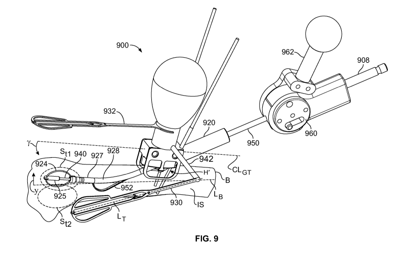
features in common with instrument guide 100 (shown in
FIG. 1). Instrument
guide 900 may include instrument
templates 930 and 932 for positioning instrument guide 900
such that an instrument can be positioned at target region
Stl.
[0278]
Illustrative steerable broach 950 may be deployed
at target region Stl in intramedullary space IS by insertion
through guide 900 at site H'. Broach 950 may include broach
head 925. Broach head 925 may have one or more features or
properties in common with broach head 125 (shown in FIG. 1).
Broach head 925 may be supported by broach sheath 927.
Broach head 925 may be rotated by drive shaft 940 which may
extend inside broach sheath 927 and receive torque from
torque adapter 908. Torque adapter 908 may provide rotation
from any suitable rotation source drive shaft 940.
[0279] Broach sheath 927 may be flexible. Broach
sheath
927 may be flexible in region 928 such that application of
off-axis tension by elevator ribbon 952 may position broach
head 925 at a distance y or -y relative to bone axis LB.
Illustrative elevator control body 960 may apply axial
CA 02823873 2013-07-04
WO 2011/091052 PCT/US2011/021735
- 48 -
compression to elevator ribbon 952 to cause broach sheath 927
to bend.
[0280] Broach sheath 927 may be configured to flex in more
than one plane. Broach sheath 927 may be configured to flex
substantially in one plane only.
[0281] Target region Stl could be in either, or both, of
cancellous bone BcA and cortical bone Bco (shown in FIG. 2).
Side template 930 and top template 932 are registered to
guide tube 920. A practitioner may position templates 930
and 932 such that templates 930 and 932 "project" onto target
region Stl so that guide 900 will guide broach head 925 to
target region Sti.
[0282] Side template 930 may be rotatable at arm 942 to
change angle y between side template 930 axis LT and guide 900
centerline CLGT. y may be selected to correspond to a degree
of elevation in direction y or -y of broach head 925. y may
be selected to correspond to a degree of actuation of control
962 of control body 960. For example, y may be selected such
that side template 930 "projects" onto target region St2.
[0283] Fluoroscopic imaging may be used to position
templates 930 and 932 relative to target region Stl.
[0284] A practitioner can select the position of H'
(distance x- shown in FIG. 2), the angle of hole H (shown in
FIG. 2) relative to bone axis LB, the degree and distribution
of flexing in region 928, the penetration of broach sheath
927, the size of broach head 925, the swept-out profile of
CA 02823873 2013-07-04
WO 2011/091052 PCT/US2011/021735
- 49 -
broaching member 924, and any other suitable parameters, to
determine the size, shape, orientation and location of a
cavity to be swept out by broaching member 924. For example,
one or more of the aforementioned parameters may be selected
to position broach head 925 in target region St2.
[0285] FIG. 10
shows guide base 102 from below on the
distal side. Stem 116
extends from the top of base 102.
Guide tube 120 extends from the distal portion of base 102.
Arm 131 extends from the side of base 102. Site H' of hole H
(shown in FIG. 2) is shown projected onto opening 1002 of
guide tube 120 and centered about axes LH and LGT.
[0286]
Illustrative contacts 108 and 110 extend down from
base 102 to engage bone B (shown in FIG. 2) and resist
rotation about vertical axes LH and LTR and translation along
guide centerline CLG. Contacts
108 and 110 may be
sufficiently sharp to penetrate or partially penetrate bone
B. Cleats 112 and 114 may engage the surface of bone B and
resist rotation about guide centerline CLG. Base 102 may
support any suitable number of contacts In any suitable
pattern or location. Base 102 may support an arrangement of
contacts that extends in a direction that is substantially
oblique or transverse to guide centerline CLG.
[0287] In some
embodiments, base 102 may include a flange
(not shown) that saddles bone B. The flange may include any
suitable number of contacts in any suitable pattern,
including an arrangement of contacts that extends in a
direction that is substantially oblique or transverse to
guide centerline CLG.
CA 02823873 2013-07-04
WO 2011/091052 PCT/US2011/021735
- 50 -
[0288] Alignment
members 104 and 106 may extend from base
102 to align guide centerline CLG of guide 100 with bone
centerline CLBS of the top surface of bone B (shown in FIG.
2). Each of alignment members 104 and 106 include continuous
alignment edges 1004 and 1006. Edge 1004
is supported by
substantially vertical struts 1007 and 1008. Edge 1006
is
supported by substantially vertical struts 1010 and 1012.
Edges 1004 and 1006 are substantially parallel to centerline
CLG.
[0289] In some
embodiments, alignment members may be or
may include tines that correspond to struts 1007, 1008, 1010
and 1012. One or more of the tines may extend straight down
from base 102. One or more of the tines may extend down and
in the proximal direction relative to base 102. One or more
of the tines may extend down and in the distal direction
relative to base 102.
[0290] In
embodiments that include one or more tines (not
shown), edges 1004 and 1006 may be absent. In those
embodiments, the tines may flex independently of each other.
One or more of the tines may be biased away from guide
centerline CLG. One or more
of the tines may be biased
toward guide centerline CLG. One or more of the tines may be
curved or arcuate.
[0291] Some embodiments may include a bushing (not shown)
in guide tube 120. The bushing may provide stability for a
K-wire in procedures in which the K-wire is used as a drill
to provide preliminary access to the inside of a bone.
[0292] FIG. 11 shows illustrative saw 1100. Saw 1100
may
be used to cut an access hole at site H' or site I' (shown in
CA 02823873 2013-07-04
WO 2011/091052 PCT/US2011/021735
- 51 -
FIG. 2) or any other suitable hole. Saw 1100
may be guided
by guide 100 (shown in FIG. 1), guide 900 (shown in FIG. 9),
guide 1900 (shown in FIG. 19) or any other suitable guide.
[0293] Saw 1100
may include wire 1102. Wire 1102 may be a
K-wire or any other suitable wire. Saw 1100
may include
centering sleeve 1104. Centering sleeve 1104 may be made of
polymer, alloy or any other suitable material. Saw 1100 may
include cutting member 1106. Cutting member 1106 may include
teeth 1108, vents 1110 and cylindrical member 1112. Vents
1110 may provide chip clearance, side-cutting, reduced
heating or other properties, among others. Saw 1100
may
include torque adapter 1114. Torque
adapter 1114 may
transmit rotation from a rotation source to one or both of K-
wire 1102 and cutting member 1106.
[0294] Wire 1102 may form an angled pilot hole in bone B.
The hole may be formed at angle 6 between saw axis Ls and
bone axis LB. After wire
1102 penetrates bone B, saw 1100
may be advanced distally until teeth 1108 engage bone B and
being to cut. Teeth 1108
will engage bone B first at point
p, in the crotch between wire 1102 and bone B. Teeth 1108
may therefore be subjected to a contact force from bone B
that is oblique to a plane defined by teeth 1108. Centering
sleeve 1104 may support teeth 1108 against the oblique force
and maintain teeth 1108 at a substantially constant radius
from axis Ls during the formation of an access hole.
[0295] A spring
1116 (shown in FIG. 13) may urge centering
sleeve 1104 distally to keep centering sleeve 1104 at or near
bone B as teeth 1108 penetrate into bone B.
CA 02823873 2013-07-04
WO 2011/091052 PCT/US2011/021735
- 52 -
[0296] FIG. 12
shows that centering sleeve 1104 may be
coaxially arranged within cutting member 1106. Wire 1102 may
be coaxially arranged within centering sleeve 1104. Collar
1202 of centering sleeve 1104 may be provided at a distal end
of centering sleeve 1104 to provide a close tolerance between
wire 1102 and centering sleeve 1104.
[0297] FIG. 13 shows spring 1116 compressed between
proximal face 1302 of centering sleeve 1104 and distal face
1304 of torque adapter 1114.
[0298] In some
embodiments, wire 1102 may be used to drill
a pilot hole in bone B without apparatus such as centering
sleeve 1104 and cutting member 1106. In such
embodiments, a
bushing (not shown) may be provided in a guide tube such as
guide tube 120 (shown in FIG. 1). Wire 1102
may be placed
through the bushing and driven by a torque adapter such as
1114. The bushing may have a bore that is sized to stabilize
a K-wire driven in rotation by a surgical drill.
[0299] It may be
desirable thereafter to cut in the bone a
hole that is substantially coaxial with the K-wire. After
the K-wire is drilled into the bone, in such embodiments, the
bushing (not shown) may be removed from the guide tube to
allow a coring saw to advance through the guide tube.
[0300] FIG. 14 shows illustrative apparatus 1400 for
cutting in bone B a hole that is substantially coaxial with
wire 1402. FIG. 14
shows a relevant portion of coring saw
guide 1450. Coring saw guide 1450 may include contacts 1452
for engaging a surface of bone B (shown in FIG. 2). Coring
saw guide 1450 may include handle-mounting recesses such as
1454. A centering
sleeve (not shown) may be disposed
CA 02823873 2013-07-04
WO 2011/091052 PCT/US2011/021735
- 53 -
coaxially between wire 1402 and cutting member 1406. In some
embodiments, a cutting member such as 1406 may be engaged by
a collar (not shown) that is configured for delivery of
torque.
[0301] A proximal
end of wire 1402 may be engaged in a
hand drill fitting and rotatingly driven into the bone as it
is advanced distally through saw guide 1450.
[0302] FIG. 15 shows wire 1402. Distal end
1502 of wire
1402 may have a first diameter. Proximal
end 1504 of wire
1402 may have a second diameter that is greater than the
first diameter. Step 1506 between the first diameter and the
second diameter may be used as a stop to limit the extent to
which wire 1402 may be driven into bone B.
[0303] Proximal
end 1504 of a wire such as 1402 may extend
along and through a cannula in an A-0 type adapter while the
adapter drives a cutting member such as 1408 distally into a
bone.
[0304] In some
embodiments, step 1506 may be used to
distally eject a bone plug from the interior of distal end
1405 of cutting member 1406 after a hole is cut and cutting
member 1406 is withdrawn from the bone.
[0305] In some
embodiments, a soft-tissue protector (not
shown) may be provided to keep soft tissue proximate the
access hole from becoming engaged by rotating apparatus. The
protector may include a cannula for guiding the rotating
apparatus into the hole. The protector may include a flange
that "funnels" the apparatus into the cannula and blocks the
soft tissue from approaching the apparatus.
CA 02823873 2013-07-04
WO 2011/091052 PCT/US2011/021735
- 54 -
[0306] FIG. 16
shows a portion of illustrative cutting
member 1106 from region 16 of FIG. 11. A
circumferential
tooth 1602 may extend into one or more of vents 1110 to
engage bone on the inside of the cutter.
[0307] Tooth 1602
may provide friction between cutting
member 1106 and the bone plug and may facilitate removal of
the bone plug upon with withdrawal of cutting member 1106
from the access hole. The distal
end of the bone plug may
not be severed from bone B native tissue by cutting member
1106. Tooth 1602
may provide =one or both of torsional and
axial force to sever the plug from bone B. Vents 1110
may
include vent edges 1604. Vent edges 1604 may cut a wall of
the access hole.
[0308] Tooth 1602
may provide friction between cutting
member 1106 and centering sleeve 1104. The
friction may
resist proximal motion of centering sleeve 1104.
[0309] FIG 17
shows illustrative teeth 1108 of cutter
member 1106 (shown in FIG. 11).
Illustrative tooth 1702 may
include cutting edge 1704, face 1706 and back 1708. Face
1706 and back 1708 may partially define adjacent gullets 1710
and 1712, which intervene between tooth 1702 and neighboring
teeth 1714 and 1716, respectively. Tooth 1702
may have
thickness t. Tooth 1702
may be circumferentially set apart
from neighboring tooth 1716 by pitch Pt. Cutting
edge 1704
may be angled relative to saw radial direction Rs by bevel
angle 4) (shown on a different tooth). Cutting
edge 1704 is
shown with f = 0 , but any suitable 4) may be used. Face 1706
may have longitudinal rake angle p.
CA 02823873 2013-07-04
W02011/091052 PCT/US2011/021735
- 55 -
[0310] Larger rake
angles (e.g., positive) may produce
lower forces, but smaller included tooth angles, and
therefore lower heat capacity. Smaller
rake angles (e.g.,
negative) may increase heat capacity and increase heat
generated in shearing but increase cutting forces.
[0311] Face 1706
is shown with p = 00, but any suitable p
may be used. Gullet 1710 may have gullet depth Dg.
[0312] In some
embodiments, tooth 1702 may include facet
1718 (shown in broken line). When facet
1718 is present,
tooth face 1706 may be shortened by distance h. Facet 1718
may have a normal (not shown) that is oriented at any
suitable angle relative to axis Ls and radius R.
[03].3] FIG. 18
shows teeth 1108 (shown in FIG. 11) as
viewed along lines 18-18 (shown in FIG. 17). Cutting
edge
1704 forms angle 0 with saw outer wall 1802. Cutting
edge
1704 is shown with 0 90 , but
any suitable 0 may be used.
For example, a tooth formed by cutting along chord Chl may
create a cutting edge having 0 > 90 . A tooth
formed by
cutting along chord Ch2 may create a cutting edge having
0 < 90 .
[0314] In some
embodiments, a cutting member may have bi-
directionally cutting teeth. Each tooth such tooth may have
a right and a left cutting edge. When the coring saw rotates
clockwise, a right edge cuts. When the
coring saw rotates
counterclockwise, a left edge cuts.
[0315] FIG. 19 shows illustrative instrument guide 1900.
Illustrative instrument guide 1900 may have one or more
CA 02823873 2013-07-04
WO 2011/091052 PCT/US2011/021735
- 56 -
features in common with one or more of guide 100 (shown in
FIG. 1) and guide 900 (shown in FIG. 9). Guide 1900
may be
used to guide an instrument into bone B at a site such as H'
or I' (shown in FIG. 2).
[0316] Guide 1900
may include base 1902. Base 1902 may be
placed against bone B (shown in FIG. 2) at site H'. Base
1902 may include contacts (not shown), alignment members (not
shown), cleats (not shown) or any other suitable features.
Grip 1918 may extend from base 1902. Base 1902
may include
pivot 1904. Pivot 1904
may pivotably support guide tube
1920. Guide tube
1920 centerline CLGT, may be positioned at
any suitable angle a' relative to axis LH' SO that saw 1950
may be advanced through bone B (not shown) at angle a'. The
intersection of axis LH, and CLGT, may substantially coincide
with site H' or site I' for different values of a'. A
practitioner may change angle a' before or during penetration
of saw 1950 into bone B. For
example, a practitioner may
initiate a pilot hole at a' 0 and then
change a' to obtain
the desired angle for the access hole.
[0317] Saw 1950
may include teeth 1952, flutes 1954,
cannula 1956 or any other suitable features, including the
features described and shown herein in connection with other
saws.
[0318] FIG. 20
shows a view of a distal portion of broach
700 taken along lines 20-20 (shown in FIG. 7). Pin 703 may
be located near the distal end of bracket 720. Pin 703 may
fix the position of the distal end of broaching member 704.
Pin 703 may support cylindrical form 705. Cylindrical
form
705 may be coaxially mounted on pin 703. Cylindrical
form
CA 02823873 2013-07-04
WO 2011/091052 PCT/US2011/021735
- 57 -
705 may support a spiral segment of broaching member 704.
One or more distal portions of broaching member 704 may be
welded or otherwise suitably fixed to cylindrical form 705.
[0319] Cylindrical
form 705 may constrain or partially
constrain the orientation of distal portions of broaching
member 704. Cylindrical
form 705 may be fixed relative to
bracket 720. Cylindrical form 705 may be rotatable relative
to bracket 720.
[0320] Broach head
702 may include end cap 701. Broaching
member 704 may remove tissue that is generally proximal end
cap 701. In some embodiments, member 704 may expand in such
a manner as to extend distally of end cap 701. In such
embodiments, the broaching member may remove tissue that is
distal of end cap 701.
[0321] Reducing or
minimizing the distance between the
distal end of broaching member 704 and end cap 701 may allow
broaching member 704 to remove tissue that is more
immediately proximal end cap 701. End cap 701
may be
positioned at the distal end of bracket 720. End cap 701 may
be configured to have a smooth, atraumatic surface. Bracket
720 may be attached to drive shaft 730.
[0322] Shaft
assembly 714 may include drive shaft 730.
Drive shaft 730 may support bracket 720 at union 732. Drive
shaft 730 may be secured to bracket 720 by pin 734. Drive
shaft 730 may provide rotation to broach head 702.
[0323] Proximal
ends 736 and 738 of broaching member 704
may be fixed to slide 740, which may be a tube. Proximal end
738 may be threaded through or keyed into windows 742 and 744
in slide 740. Proximal
end 736 may be threaded through or
CA 02823873 2013-07-04
WO 2011/091052 PCT/US2011/021735
- 58 -
keyed into slots 746 and 748 in slide 740. Slide 740
may
slide relative to drive shaft 730 to expand and contract
broaching member 704. Slide 740
is shown in the "contract"
state, in which broaching member 704 is drawn close to
bracket 720. Slide cover
750 may slide with slide 740. One
or both of slide 740 and slide cover 750 may be translated
along axis Lc by control hub 710 (shown in FIG. 7) or any
other suitable position controller.
[0324] Slide cover
750 may remain stationary relative to
drive shaft 730 when slide 740 slides relative to drive shaft
730. In
embodiments in which slide cover 750 remains
stationary when slide 740 moves, distal end 752 of slide
cover 750 may limit the radial position of broaching member
704 at a fixed distance along drive shaft 730 and thus affect
the deformation of broaching member 704 in the expanded
state.
[0325] Broaching
member 704 may undergo one or both of
elastic and plastic deformation.
[0326] FIG. 21
shows a view of a distal portion of broach
700 taken along lines 20-20 (shown in FIG. 7) when broaching
member 704 is in an expanded state. Broaching member 704 is
shown as mainly circular. However, any desired shape may be
able to be imparted in the expanded state such as but not
limited to: square, triangular, oval, ellipsoid, teardrop,
football, or any other suitable shape.
[0327] Different
shapes may be obtained using several
methods, such as utilizing a pre-set shape in a shape memory
alloy, modifying the geometry of the member cross-section
(along the member length) such that it preferentially bends
CA 02823873 2013-07-04
WO 2011/091052 PCT/US2011/021735
- 59 -
in a desired manner, constraining broaching member 704 (e.g.,
in force, shear or moment) in a way that forces the expansion
to take desired shape, having the final shape be that of the
expanded geometry and the reduced or collapsed geometry be
that of a higher strain configuration, and/or any other
suitable method of forming a desired shape.
[0328] For
example, largely or substantially preventing
radial movement of broaching member proximal ends 736 and
738, and allowing movement of the distal end of broaching
member 704 generally about pin 703 while elastically
deforming broaching member proximal ends 736 and 738, due to
reducing the distance between the distal end and proximal
ends 736 and 738 of broaching member 704, may modify the
geometry of broaching member 704 from a generally straight
configuration to a generally eggbeater shape.
[0329] The deformation may relatively increase the
distance between (a) sections 760 and 762 and (b) bracket
720. As this distance is increased, the swept-out volume of
broaching member 704, as broaching member 704 rotates
generally about an axis such as Lc (shown in FIG. 8), is
increased.
[0330] In some embodiments, a broach may include a
broaching member that includes one or more stiff tines (not
shown) that is joined to a drive shaft. The drive shaft may
have a longitudinal axis. The tine
may be joined to the
drive shaft radially close to the axis at a proximal end of
the tine. The tine
may have a distal end that is spaced
radially apart from the axis. The distal end of the tine may
be distal of the distal end of the drive shaft. There may be
numerous tines on the drive shaft. Such
embodiments may be
CA 02823873 2013-07-04
W02011/091052 PCT/15S2011/021735
- 60 -
appropriate for rotation in intramedullary space IS of bone B
(shown in FIG. 2) using high torque at low rotational speeds.
[0331] FIG. 22
shows broaching member 704 in partial cross
section from view lines 22-22 (shown in FIG. 21). Broaching
member 704 may have leading edges 2202 and 2204 that may be
rotated in direction coo by drive shaft 730 (shown in FIG.
21). Broaching
member 704 may sweep out a space in bone B
(shown in FIG. 2) based on radius Rc, which corresponds to
sections 760 and 762 (shown in FIG. 21).
[0332] Leading
edge 2202 may be beveled at angle aci.
Angle acl may be any suitable angle, including an angle from
about 5 to about 75 . Angle acl may cause leading edge 2202
to be generally sharp or knife-like. This may
aid in the
broaching member's ability to remove tissue.
[0333] Leading
edge 2204 may be beveled at angle c2.
Angle c2 may be any suitable angle, including an angle from
about 5 to about 75 . Angle c2 may cause leading edge 2204
to be generally sharp or knife-like. This may
aid in the
broaching member's ability to remove tissue.
[0334] As broaching member 704 is rotated clockwise
generally about axis Lc leading edges 2202 and 2204 may
generally be the first portion of sections 760 and 762 to
come in contact with tissues such as relatively less dense
cancellous bone BcA (shown in FIG. 2). Sections
760 and 762
may be configured to be sufficiently flexible such that if
either of sections 760 and 762 contacts relatively more dense
CA 02823873 2013-07-04
WO 2011/091052 PCT/US2011/021735
- 61 -
materials, such as diaphysis, metaphysis and epiphysis bone,
sections 760 and 762 may deflect generally radially in
direction -wc about axis Lc and/or in the linear direction
towards axis Lc at any location along the length of sections
760 and 762 or any other portion of broaching member 704.
Deflection or deformation of sections 760 and 762 may have
the affect of not disturbing the more dense tissues.
[0335] Leading
edges 2202 and 2204 may be offset from axis
Lc by offsets Ai and A2 respectively. Appropriate magnitudes
of offsets Al and A, may be selected. In some
embodiments,
offsets Ai and A2 may be constrained by the collapsed
diameter (overall diameter of broach head 702 in a plane
transverse to axis Lc when broaching member 704 is collapsed,
e.g., for deployment) of the configuration and the desired
expanded engagement (radius Rc) of broaching member 704 with
the tissue. Offsets Al
and A2 may aid in the broaching
member's efficiency at displacing tissue.
[0336] FIG. 22A
shows broach head 704 in intramedullary
space IS of bone B and illustrates how flexible broaching
members can broach bone of a relatively lower density and be
deflected by bone of a relatively higher density. Sections
760 and 762 have displaced or removed some of cancellous bone
B A from bone B by rotating in direction co; about axis L.
Sections 760 and 762 may be sufficiently stiff to remove
cancellous bone to radius R- from axis Lc in the "top" portion
of bone B. Because of
the placement of axis L relative to
the bottom portion of bone B, sections 760 and 762 contact
cortical bone BCO at the bottom of bone B. Sections 760 and
CA 02823873 2013-07-04
WO 2011/091052 PCT/US2011/021735
- 62 -
762 may be sufficiently flexible to be deflected by cortical
bone BCO. Section 760 is shown deflected in direction -co, by
bone BCO. Sections
760 and 762 thus remove bone only to
radius R.,' in the "bottom" portion of bone B.
[0337] The cavity
created by broach 700 may thus be
bounded in part by cancellous bone BCA and in part by
cortical bone BCO. The shape of the cavity portion that is
bounded by cancellous bone BCA may be governed substantially
by the geometry and mechanical properties of broach 700. The
shape of the cavity portion that is bounded by cortical bone
BCO may be governed substantially by the native anatomy of
bone B.
[0338] FIG. 23
shows a view of broach 700 along lines 23-
23 (shown in FIG 20). Broach 700 is in the contracted state.
Slide cover 750 has been removed. Slots 746, 748 and 2302 in
slide 740 may be configured to coincide with features on
proximal end 736 (shown in FIG. 21) of broaching member 704.
When proximal end 736 is engaged with slots 746, 748 and
2302, slots 746, 748 and 2302 may restrict movement of
proximal end 736 in either direction generally along axis Lc.
Slots 746, 748 and 2302 may have any suitable geometry that
allows for the engagement and axial translation of proximal
end 736.
[0339] Slots 746,
748 and 2302 may be of sufficient depth
that, when proximal end 736 is engaged in slots 746, 748 and
2302, slide cover 750 (shown in FIG. 20) has adequate radial
clearance with respect to proximal end 736 and slide 740 to
slide over slide 740 and slots 746, 748 and 2302. An inner
surface of slide cover 750 may prevent movement of proximal
CA 02823873 2013-07-04
WO 2011/091052 PCT/US2011/021735
- 63 -
end 736 from moving in a direction generally away from axis
Lc.
[0340] Slide 740
may include slots (not shown) that
correspond to proximal end 738 (shown in FIG. 20) and have
one or more features in common with, slots 746, 748 and 2302.
[0341] Broach head
720 may include broaching member wrap
section 2304. Pin 703 may
be integrated into wrap section
2304. Wrap section 2304 may be separate from pin 703. Wrap
section 2304 may be configured to allow wrapping of broaching
member 704 generally around wrap section 2304. Broaching
member 704 may be looped in wrap section 2304. Broaching
member 704 may be wrapped (as shown in FIG. 23) at least one
full turn in wrap section 2304. Wrapping about wrap section
2304 may bias segments 760 and 762 (shown in FIG. 21) away
from axis Lc.
[0342] FIG. 24
shows a cross section, viewed along lines
24-24 (shown in FIG. 8) of a portion of broach control 706
(shown in FIG. 7). Expansion control hub 710 is shown with
base 2402 at position pe. This may
correspond to the
expanded state of broaching member 704, as shown in FIG. 8.
Base 2402 may be moved distally to position pc. This may
correspond to the contracted state of broaching member 704,
as shown in FIG. 7. Expansion control hub 710 may operate in
connection with body 2408. Body 2408
may include control
shaft 712 and distal stop 2410. Control
shaft 712 may
include threads 2418.
[0343] Expansion
control hub 710 may include outer member
2412 and inner member 2414. Outer
member 2412 and inner
CA 02823873 2013-07-04
W02011/091052 PCT/US2011/021735
- 64 -
member 2414 may be fixed to each other. Slide pin
2404 may
be captured between outer member 2412 and inner member 2414.
Inner member 2414 may include threads 2416 for engagement
with threads 2418 on control shaft 712. Slide pin
2404 may
travel in slots 2405 and 2407 in body 2408.
[0344] Expansion
control hub 710 may be moved along axis
Lc by applying force to expansion control hub 710. In some
embodiments, expansion control hub 710 may be advanced axial
generally along axis Lc by applying rotational force
generally about axis Lc to expansion control hub 710 such
that threads 2416 move advance or retreat through threads
2418.
[0345] Axial movement of expansion control hub 710
relative to body 2408 may be transferred to slide 740 and
slide cover 750 while drive shaft 730 remains axially fixed
to body 2408 by pin 2406. Slide 740
may include cut-outs
2430 and 2432. Slide cover 750 may include cut-outs 2434 and
2436. Cut-outs
2430, 2432, 2434 and 2436 may provide
clearance of pin 2406 when slide 740 and slide cover 750
travel axially.
[0346] When
expansion control hub 710 is moved axially,
proximal ends 736 and 738 (shown in FIG. 20) of broaching
member 704 thus move axially. Distal end 780 (shown in FIG.
7) of broaching member 704 may be axially fixed to drive
shaft 730, which may be fixed to body 2408. Thus, when
expansion control hub 710 moves distally, the distance
between (a) proximal ends 736 and 738 and; (b) distal end 780
decreases and broaching member 704 expands. When
expansion
control hub 710 moves proximally, the distance between (a)
CA 02823873 2013-07-04
WO 2011/091052 PCT/US2011/021735
- 65 -
proximal ends 736 and 738; and (b) distal end 780 increases
and broaching member 704 contracts.
[0347] Distal stop
2410 and proximal stop 2420 may limit
axial movement of expansion control hub 710. Although
proximal stop 2420 is shown as being part of handle 708,
proximal stop 2420 may be separate from handle 708.
[0348] Handle 708 may transfer rotational motion generally
about axis Lc to control shaft 712. Control
shaft 712 may
transfer the rotation to slide pin 2404 and drive shaft pin
2406. Slide pin
2404 may transfer the rotation to slide 740
and slide cover 750. Drive shaft
pin 2406 may transfer the
rotation to drive shaft 730, which may drive broaching member
704 (shown in FIG. 21).
[0349] Distal stop
2410 is shown as being integral with
body 2408, but distal stop may be a separate element that is
attached to control shaft 712 or a different part of body
2408.
[0350] Pin 2406
may extend into recess feature 2422.
Recess feature 2422 may be a through-hole. Pin 2406
may
extend through the through hole to a location external to
body 2408.
[0351] Pin 2404
may extend into recess feature 2424.
Recess feature 2424 may be a through-hole. Pin 2404
may
extend through the through-hole to a location external to
body outer member 2412. Recess
feature may extend
circumferentially about axis Lc. If recess
feature 2424
extends circumferentially about axis Lc, expansion control
CA 02823873 2013-07-04
WO 2011/091052 PCT/US2011/021735
- 66 -
hub 710 may rotate about axis Lc substantially without
restricting, or being restricted by, pin 2404.
[0352] Body 2408
may include circumferential recess 2426.
Recess 2426 may be sized to engage 0-ring 2428. Recess 2426
may prevent axial movement between body 2408 and 0-ring 2428
generally along axis Lc. 0-ring 2428 may be sized to provide
an interference fit with outer member 2412. The interference
fit may produce friction between 0-ring 2428 and expansion
control hub 710. The
friction may allow expansion control
hub 710 to be lightly locked at any rotational position
relative to body 2408, generally about axis Lc.
[0353] FIG. 25 shows illustrative cavity preparation
apparatus 2500. Apparatus
2500 may include broach 2550.
Broach 2550 may have one or more features in common with
broach 950 (shown in FIG. 9). Broach 2550 may include one or
more of broach head 2525, elevator ribbon 2552 and control
body 2560. Apparatus 2500 may Include guide 2502. Guide
2502 may guide broach 2550 or any other suitable apparatus
through an access hole such as H or I (shown in FIG. 2).
Guide 2502 may retain soft tissue at a distance from the
access hole to prevent engagement of the soft tissue by an
instrument that is present in guide 2502.
[0354] FIGS. 26-29
show features of different portions of
apparatus 2500.
[0355] FIG. 26
shows in partial cross section illustrative
broach head 2525 and illustrative elevator ribbon 2552.
CA 02823873 2013-07-04
WO 2011/091052 PCT/US2011/021735
- 67 -
[0356] Broach head
2525 may be driven about axis LE by
rotating drive shaft 2540. Broach head
2525 may include
broaching member 2524, which may have one or more features in
common with broaching member 704 (shown in FIG. 7). Broach
head 2525 may include distal hub 2526 and proximal hub 2528.
One or both of distal hub 2526 and proximal hub 2528 may
transfer rotation to broaching member 2524. One or both
of
distal hub 2526 and proximal hub 2528 may support broaching
member 2524.
[0357] Drive shaft
2540 may extend within broach sheath
2527. Drive shaft
2540 may be supported in rotation by
bushing 2530 at the end of broach sheath 2527.
[0358]
Illustrative elevator ribbon 2552 may be anchored
to broach sheath 2527 at fixation 2532. When axial
compressive force, generally along axis LE, is applied to
elevator ribbon 2552, elevator ribbon 2552 may buckle along
its length. For example, elevator ribbon 2552 may buckle at
or near section 2534. Section
2536 may be used to support
broach sheath 2527 at an elevation relative to cancellous
bone BCA or cortical bone Bco in bone B (shown in FIG. 2).
[0359] Portions of
elevator ribbon 2552 may extend inside
broach sheath 2527 and pass through slots 2542 and 2544 to
section 2534. In some
embodiments, there may be contact
between drive shaft 2540 and elevator ribbon 2552. In some
embodiments, there may be no contact between drive shaft 2540
and elevator ribbon 2552.
[0360] Elevator
ribbon 2552, when compressed, may apply
tension to adjacent portion 2538 of broach sheath 2527 and
CA 02823873 2013-07-04
WO 2011/091052 PCT/US2011/021735
- 68 -
compression to opposite portion 2540 of broach sheath 2527.
One or both of the tension of adjacent portion 2538 and the
compression of opposite portion 2540 may cause broach sheath
2527 to curve generally about an axis such as LF.
[0361] One or both
of adjacent portion 2538 and opposite
portion 2540 may include stress-relief features that allow
bending under tension and compression. The stress-
relief
features may include slots or slot patterns. The stress-
relief features may be provided using laser-cutting. The
stress-relief may provide an equilibrium curvature such that
broach sheath 2527 is curved at rest.
[0362] The stress-
relief features may include sintered
particles. The
particles may include metal, polymer,
composite or any other suitable material.
[0363] FIG. 27
shows illustrative laser¨cut pattern 2700
for a broach sheath such as 927 (shown in FIG. 9) or 2527
(shown in FIG. 26). Pattern
2700, which is shown flat for
illustration, may be cut in a cylindrical tube to relieve
compression on one side of the tube and relieve tension on
the other side of the tube. For example, compression relief
pattern 2740 may be provided along opposite portion 2540 of
broach sheath 2527. Tension
relief pattern 2738 may be
provided along adjacent portion 2538 of broach sheath 2527.
Tension and compression relief may be increased by
lengthening lengths Li and Lp2, respectively. Bending
stiffness may be reduced by increasing pattern widths wl and
Increasing kerf and decreasing inter-cut spacing may
also decrease bending stiffness. In some
embodiments, the
tube may have an outer diameter of 0.108 in. In some
CA 02823873 2013-07-04
WO 2011/091052 PCT/US2011/021735
- 69 -
embodiments, the tube may have an outer diameter of 0.125 in.
Any suitable outer diameter may be used.
[0364] FIG. 28
shows illustrative elevator control body
2860. Elevator
control body 2860 may support the proximal
end of broach sheath 2527. Drive shaft
2540 may extend
through control body 2860 to torque adapter 2808. Torque
adapter 2808 may be cannulated. Torque adapter 2808 may be a
cannulated A-0 type adapter. Torque adapter 2808 may have a
"D"-shaped extension for engagement by a D-shaped chuck.
[0365] Torque
adapter 2808 may be torqued by any suitable
source of rotational energy.
[0366] Control
body 2860 may include housing 2862 and
actuator 2866. Handle 2864
may be used to rotate actuator
2866 through angle 6E about axis LTE relative to housing 2862.
When actuator moves through angle 6E, shaft 2868 may drive
shuttle 2870 in slot 2872. The distal end of elevator ribbon
2552 may be fixed to the shuttle, for example, by screw 2874.
When the shuttle is in a distal position, elevator ribbon
2552 is expanded (as shown in FIG. 26). When the shuttle is
in a proximal position, elevator ribbon 2552 is contracted
toward axis LE.
[0367] Actuator 2866 may include face member 2890. Face
member 2890 may be fixed relative to housing 2862. Face
member 2890 may include recess 2892. Recess 2892 may "catch"
a projection such as 2894 to act as a detent. Projection
2894 may be one of several projections that provide detent
positions. For
example, three detent positions may be
provided: forward, neutral and back. In the
forward
CA 02823873 2013-07-04
WO 2011/()91052 PCT/US2011/021735
- 70 -
position, elevator ribbon 2552 is extended. In the back
position, elevator ribbon 2552 is compressed. In the neutral
position, elevator ribbon 2552 is in a partially compressed
state.
[0368] Housing
2862 may be configured to house a torque
limiter (not shown). The torque
limiter may couple torque
adapter 2808 to drive shaft 2540 and may be used to limit the
torque that is applied to broach head 2525 (shown in FIG.
25). If broach
head 2525 were to jam in bone B (shown in
FIG. 2), the torque limiter may cap or reduce the torque on
broaching head 2525 to prevent damage to broaching head 2525,
other elements of apparatus 2500, other involved apparatus or
bone B.
[0369] FIG. 29 shows illustrative guide 2502. Guide 2502
may include cannula 2904 and funnel 2906. Funnel 2906
may
facilitate insertion of a broach head such as 2525 (shown in
FIG. 25) into a hole such as H (shown in FIG. 2).
[0370] Guide 2502
may be "preloaded" on broach sheath
2527. A
practitioner may insert a broach head into hole H
(shown in FIG. 2) and then position guide 2520 in hole H.
Funnel 2906 may protect soft tissue outside bone B. Cannula
2904 may guide the broach head through hole H when broach
head is withdrawn from hole H (for example, at the conclusion
of a cavity preparation procedure).
[037].] Outer wall
2908 of cannula 2904 may be of an
appropriate diameter to substantially fill hole H. Funnel
2906 may include ledge 2910. Ledge 2910 may limit the extent
to which cannula 2904 may extend into intramedullary space
IS.
CA 02823873 2013-07-04
WO 2011/091052 PCT/US2011/021735
- 71 -
[0372] Cannula 2904 may support detent 2912. Detent 2912
may be present to catch on the inside of cortical bone Bco
wall W to retain cannula 2904 in position in hole H. Detent
2912 may be have a tapered profile so that it can engage
walls W of different thickness. In some embodiments, detent
2912 may be passive. In passive embodiments, detent 2912 may
be resilient, biased or rigid. In some
embodiments, detent
2912 may be active. In active
embodiments, detent 2912 may
be actuated. For example, detent 2912 may be actuated by a
manual control that causes detent 2912 to extend away from
tube cannula 2904 a desired distance or a preset distance.
Cannula 2904 may include more than one detent.
[0373] Mouth 2914
of funnel 2906 may have any suitable
shape transverse to axis LE. The shape
may be rectangular,
triangular, elliptical, tear-drop, splayed, circular and any
other suitable shape.
[0374] Funnel 2906
may include a skiving-curved section
(not shown). The skiving-curved section may be at the distal
end of funnel 2906.
[0375] Guides for
rotatable broaches may include a body
that has a cannula. The body may support a broach sheath in
alignment with the cannula. A drive shaft may pass through
the cannula and extend distally through the broach sheath. A
rotation source may be connected to the drive shaft proximal
the body. The body may be hand-held. The body may have no
adaptations to mate with a hole such as H (shown in FIG. 2).
[0376] FIG. 30
shows apparatus 2500 (shown in FIG. 25)
with control 2864 at a larger angle OE and elevator ribbon
CA 02823873 2013-07-04
WO 2011/091052 PCT/US2011/021735
- 72 -
2552 in the contracted state close to broach sheath 2527.
Stress-relief features such as those shown in flat model 2700
(shown in FIG. 27) are shown in portions 2538 and 2540 of
broach sheath 2527.
[0377] FIG. 31
shows illustrative broaching member 3102.
Broaching member 3102 may be mounted by fixture 3104 to hub
3106 at the distal end of a broach shaft 3108. Broach shaft
3108 may have one or more features in common with broach
shaft 2527 (shown in FIG. 26) or any other broach shaft
discussed or shown herein. For
example, broach shaft 3108
may include stress-relief features 3110 and 3112.
[0378] Hub 3106
may have one or more features in common
with hub 2528 (shown in FIG. 26).
[0379] Broaching
member 3102 may be a self expanding
structure. Broaching member 3102 may be constructed from
laser-cut tube stock that is expanded into a suitable shape,
such as that shown. Broaching
member 3102 may include
broaching members such as 3114. Broaching
member 3102 may
include numerous interconnected cells such as cell 3116. The
cells may be defined by one or more broaching members. Some
cells may be defined by structures other than broaching
members. The cells may be arranged in a network. The cells
may be linked such that when the structure is stressed (e.g.,
compressed) at a point the stress is distributed to nearby
cells. Broaching member 3102 may thus rotate in a bone
cavity that has an irregular shape, for example, nonround,
oblong, or angular. The cavity
may be smaller than a
diameter of broaching member 3102, such as expanded diameter
DE.
CA 02823873 2013-07-04
WO 2011/091052 PCT/US2011/021735
- 73 -
[0380] Broaching
member 3102 may include broaching members
that included braided wire (not shown). Broaching
member
3102 may include broaching members that included braided
ribbon (not shown).
[03811 In some embodiments, each cell arm may be a broaching
member. When a large number (i.e., when the circumferential
density of broaching members is high) of broaching members
are present during the rotation of a broaching head, a
relatively lower torque is required to drive the broaching
head.
[0382] FIG. 32
shows illustrative broach 3200 inserted in
bone B. Broach 3200
may include broaching head 3202.
Flexible rotating drive shaft 3204 may drive broaching head
3202 in rotation in directions p' or -p'. Drive shaft
3204
may be driven by a rotation source such as handle 3206. In
some embodiments, the rotation source may include a surgical
hand drill, a dremel motor or any other suitable rotational
power source.
[0383] Drive shaft
3204 may be sheathed in a flexible
cannula (apart from broach sheath 3210, which is described
below).
[0384] Control
body 3208 may be used to insert broaching
head 3202 through a hole at site H'. During
insertion,
broaching head 3202 may be withdrawn into flexible broach
sheath 3210. Proximal
end 3212 of flexible broach sheath
3210 may be fixed to distal end 3214 of control body 3208.
Actuator 3216 may engage drive shaft 3204 and may slide
relative to control body 3208. Actuator
3216 may thus
CA 02823873 2013-07-04
WO 2011/091052 PCT/US2011/021735
- 74 -
translate drive shaft 3204 along axis Lm within guide sheath
3210.
[0385] In some
embodiments, broaching head 3202 may be
compressible and expandable. Broaching
head 3202 may be
compressed within guide sheath 3210. Broaching head 3202 may
be expanded outside of guide sheath 3210. In some
embodiments, broaching head 3202 may self-expand in bone B
after being pushed out of guide sheath 3210 by drive shaft
3204. In some
embodiments, broaching head 3202 may be
outside guide sheath 3210 when broaching head 3202 is
delivered into bone B.
[0386] Broaching head 3202 may include one or more
broaching members 3218 that have sufficient rigidity to
displace cancellous bone, but sufficient resilience to deform
when brought into contact with cortical bone and thus leave
the cortical bone substantially in place.
[0387] Broaching
members 3218 may be formed from loops.
The loops may be fixed to distal hub 3220. The loops may be
fixed to proximal hub 3222. One or both of distal hub 3220
and proximal hub 3222 maybe axially fixed to drive shaft
3204. One or both of distal hub 3220 and proximal hub 3222
maybe rotationally fixed to drive shaft 3204. Broaching head
3202 may include any suitable number of loops. Broaching
members 3218 may have one or more features in common with
broaching member 704 (shown in FIG. 7) or any other broaching
member described or shown herein.
[0388] FIG. 33
shows illustrative broaching head 3300.
Broaching head 3300 may include broaching members 3302. Each
of broaching members 3302 may have one or more features in
CA 02823873 2013-07-04
WO 2011/091052 PCT/US2011/021735
- 75 -
common with broaching member 704 (shown in FIG. 7) or any
other broaching member shown or described herein. Broaching
head 3300 may have any suitable number of broaching members
3302. For
example, broaching head 3300 may have one
broaching member, 2-6 broaching members, 7-20 broaching
members, more than 20 broaching members or any suitable
number of broaching members.
[0389] Broaching
head 3300 may be contracted toward drive
shaft 3310 and withdrawn into an outer sheath (not shown).
The outer sheath may be inserted in a hole such as H (shown
in FIG. 2). Broaching
head 3300 may then be deployed by
retracting the sheath. Broaching
members 3302 may be
sufficiently resilient to be contracted and may expand away
from drive shaft 3310 when the sheath is retracted.
[0390] Broaching
members 3302 may be supported by distal
hub 3304. Distal hub
3304 may be absent and broaching
members 3302 may have free distal ends. Broaching
members
with free distal ends may be supported at their proximal ends
near the central axis of broaching head 3300. The broaching
members may be angled radially away from the central axis of
broaching head 3300.
[0391] Broaching
members with free distal ends may have
suitable shape at the distal ends, such as pointed, forked,
rounded, blunt or truncated.
[0392] Broaching members 3302 may be supported by proximal hub
3306. Proximal
hub 3306 may be supported by broach sheath
3308. Broach
sheath 3308 may have one or more features in
common with broach sheath 127 (shown in FIG. 1).
CA 02823873 2013-07-04
WO 2011/091052 PCT/US2011/021735
- 76 -
[0393] Drive shaft 3310 may drive broaching head 3300 in
rotation. Drive shaft 3310 may extend distally to distal hub
3304. Drive shaft 3310 may extend through broach sheath 3308
to a proximal rotation source (not shown).
[0394J One or both of distal hub 3304 and proximal hub 3306
maybe axially fixed to drive shaft 3310. One or both
of
distal hub 3304 and proximal hub 3306 maybe rotationally
fixed to drive shaft 3310.
[0395] One or more
of broaching members 3302 may include a
hoop segment such as 3312. Segment
3312 may support one or
more reinforcements such as 3314.
[0396] Segment 3312 may be rigid. Segment
3312 may be
resilient. Segment
3312 may have any suitable pre-set
curvature or be substantially linear. Segment 3312 may be a
closed loop. The loop may be asymmetric.
[0397] Segment
3312 may include a length of wire, ribbon,
cable, stranded wire, or any other suitable form or
structure. Segment 3312 may include polymer, metal, alloy or
any other suitable material. Segment 3312 may be constructed
of a mesh cut from metal tube.
[0398] Reinforcement 3314 may be a tube.
Reinforcement
3314 may be formed from polymer, metal, alloy or any other
suitable material. One or more
reinforcements such as 3314
may be sized and positioned to support segment 3312 in a
desired contour. One or more reinforcements such as 3314 may
provide bone-broaching abrasiveness, momentum or both.
[0399] FIG. 34
shows illustrative broaching head 3400.
Broaching head 3400 may include broaching members 3402. Each
CA 02823873 2013-07-04
WO 2011/091052 PCT/1JS2011/021735
- 77 -
of broaching members 3402 may have one or more features in
common with broaching member 704 (shown in FIG. 7) or any
other broaching member shown or described herein. Broaching
head 3400 may have any suitable number of broaching members
3402. For
example, broaching head 3400 may have one
broaching member, 2-6 broaching members, 7-20 broaching
members, more than 20 broaching members or any suitable
number of broaching members.
04001 Broaching members 3402 may be supported by distal hub
3404. Broaching
members 3402 may be supported by proximal
hub 3406. Proximal hub 3406 may be supported by drive shaft
3410. Drive shaft
3410 may have one or more features in
common with drive shaft 730 (shown in FIG. 20) or any other
drive shaft that is shown or described herein.
04011 Drive shaft 3410 may drive broaching head 3400 in
rotation. Drive shaft 3410 may extend distally to distal hub
3404. Drive shaft
3410 may extend to a proximal rotation
source (not shown).
[0402J One or both of distal hub 3404 and proximal hub 3406
maybe axially fixed to drive shaft 3410. One or both
of
distal hub 3404 and proximal hub 3406 maybe rotationally
fixed to drive shaft 3410.
[0403] One or more
of broaching members 3402 may include a
hoop segment such as 3412.
Reinforcement 3414 may support
one or more segments such as 3412.
[0404] Segment 3412 may be rigid. Segment
3412 may be
resilient. Segment
3412 may include a length of wire,
ribbon, cable, stranded wire or any other suitable form or
CA 02823873 2013-07-04
WO 2011/091052 PCT/US2011/021735
- 78 -
structure. Segment 3412 may include polymer, metal, alloy or
any other suitable material.
[04051 Reinforcement 3414 may be a brace.
Reinforcement 3414
may be formed from polymer, metal, alloy or any other
suitable material. One or more
reinforcements such as 3414
may be sized and positioned to support segment 3412 in a
desired contour. One or more reinforcements such as 3414 may
provide bone-broaching abrasiveness, momentum or both.
[0406] The brace
may reduce material fatigue in segment
3412. The brace may help segment 3412 retain its shape under
forces of rotation and broaching resistance. The brace
may
include loops such as 3418 and 3416. The loops
may pass
around the circumference of segment 3412. In some
embodiments, loops 3418 and 3416 may encompass only a portion
of the circumference. In some embodiments, the brace may be
fixed to segment 3412, for example, by crimping, welding or
press-fit.
[0407] The brace may support broaching edges for
displacing bone material in bone B (shown in FIG. 2). The
broaching edges may have any suitable form, such as serrated,
saw-tooth, knife-edge, rectilinear edge or any other suitable
form.
[0408] The brace
may be formed from a pattern that is cut
into a metal tube.
[0409] FIG. 35
shows illustrative broaching head 3500.
Broaching head 3500 may include broaching member 3502.
Broaching member 3502 may have one or more features in common
with broaching member 704 (shown in FIG. 7) or any other
broaching member shown or described herein.
CA 02823873 2013-07-04
WO 2011/091052 PCT/US2011/021735
- 79 -
[0410] Broaching
head 3500 may have any suitable number of
broaching members such as broaching member 3502. For
example, broaching head 3400 may have one broaching member,
2-6 broaching members, 7-20 broaching members, more than 20
broaching members or any suitable number of broaching
members. When more
than one broaching member is included,
the broaching members may have different sizes or other
features.
[0411] Broaching
member 3502 is illustrated as a single
solid hoop. Broaching
member 3502 may include one or more
members that are stranded or braided. Broaching member 3502
may include wire, strip stock, sheet stock, strand, ribbon,
polymer, composite, ceramic, sintered material or any other
suitable material. Broaching
member 3502 may have one or
more of a variety of cross sections, such as square,
rectangular, octagonal, contours with sharp edges, stranded
cable, or other suitable configurations to facilitate bone
displacement.
[0412] Broaching
member 3502 may include stainless steel,
Nitinol (shapeset, superelastic or other Nitinol) or any
other suitable substance.
[0413] Broaching member 3502 may be a substantially
continuous structure. Broaching member 3502 may pass through
channel 3512 in distal hub 3504. Broaching
member 3502 may
be fastened to distal hub 3504 in channel 3512.
[0414] Broaching
member 3502 may be supported by distal
hub 3504. Broaching member 3502 may be supported by proximal
hub 3506. Proximal
hub 3506 may be supported by broach
sheath 3508. Broach
sheath 3508 may have one or more
CA 02823873 2013-07-04
WO 2011/091052 PCT/US2011/021735
- 80 -
features in common with broach sheath 127 (shown in FIG. 1)
or any other broach sheath that is shown or described herein.
[0415] Drive shaft
3510 may drive broaching head 3500 in
rotation. Drive shaft 3510 may extend distally to distal hub
3504. Drive shaft
3510 may extend to a proximal rotation
source (not shown).
[0416] One or both
of distal hub 3504 and proximal hub
3506 maybe axially fixed to drive shaft 3510. One or both of
distal hub 3504 and proximal hub 3506 maybe rotationally
fixed to drive shaft 3510.
[0417] Distal hub
3504 may be constructed of metal,
stainless steel, laser-cut tube, polymer, ceramic or any
other suitable material.
[0418] The distal
end of drive shaft 3510 may extend into
a channel (not shown) in distal hub 3504. Distal hub
3504
may be free to move axially with respect to drive shaft 3510.
The channel in distal hub 3504 may be keyed for receiving a
complementarily keyed distal end of drive shaft 3510. Drive
shaft 3510 may thus drive broaching member 3502 distal
portions 3518 and 3520.
[0419] During
rotation, broaching member 3502 may elongate
axially, along axis 1,0 and push distal hub 3504 distally
relative to drive shaft 3510. Such motion
may contract
broaching member 3502. During
rotation, broaching member
3502 may expand axially along axis LG and draw distal hub
3504 proximally relative to drive shaft 3510. Contraction
may occur, for example, when distal hub 3504 encounters
resistant material.
CA 02823873 2013-07-04
WO 2011/091052 PCT/US2011/021735
- 81 -
[0420] Distal hub
3504 may be fixed to drive shaft 3510.
Broaching member 3502 may be driven rotationally by
application of torque to proximal ends 3514 and 3516 of
broaching member 3502. Broaching
member 3502 may be driven
rotationally by application of torque to distal portions 3518
and 3520 of broaching member 3502.
[0421] Proximal
ends 3514 and 3516 of broaching member
3502 may be affixed to drive shaft 3510 by proximal hub 3506.
Proximal hub 3506 may engage proximal ends 3514 and 3516 by
crimping, welding, set-screw, snap fit or any other suitable
fastening.
[0422] Proximal
hub 3506 may include or rotate with
respect to a bearing (not shown). The bearing may be seated
in the distal end of broach sheath 3508. Thus, when
drive
shaft 3510 rotates broaching member 3502, broach sheath 3508
and the bearing do not rotate. The
orientation at which
proximal ends 3514 and 3516 of broaching member 3502 are
fixed to proximal hub 3506 may provide or retain a shape of
broaching member 3502.
[0423] Distal hub
3504 may extend a distance E in the
distal direction away from distal portions 3518 and 3520 of
broaching member 3502. Distal hub 3504 may thus contact bone
material inside bone B (shown in FIG. 2) before distal
portions 3518 and 3520 contact the material. If the material
is dense, such as cortical bone, the material may resist
distal advancement of distal hub 3504. Broaching member 3502
may thus be prevented from broaching or interacting with the
material.
CA 02823873 2013-07-04
WO 2011/091052 PCT/US2011/021735
- 82 -
[0424] Distal hub
3504 may include flutes 3522 and 3524.
Broaching edges 3526, 3528, 3530, 3532, 3534 and 3536 may
displace material inside bone B. Flutes 3522
and 3524 may
intersect with each other at the distal end of distal hub
3504.
[0425] Distal hub
3504 may have a blunt distal end without
flutes. This may
prevent broaching member 3502 from
interacting with material that resists distal advancement of
distal hub 3504. The distal
end of distal hub 3504 may be
any suitable shape.
[0426] Distal hub
3504 may be absent from broaching head
3500.
[0427] FIG. 36 shows illustrative broach 3600. Broach
3600 may include broaching head 3602, control shaft assembly
3604 and actuator 3606.
[0428] Broaching
head 3602 may include linked blades 3608,
3610, 3612 and 3613. Linked
blades 3608 and 3610 may have
broaching edges 3630 and 3632, respectively. The
broaching
edges may broach bone inside bone B (shown in FIG. 2) when
broach head 3602 is rotated about axis LI.
[0429] The blades
may positioned radially by a locking
mechanism. The blades
may be positioned radially by a
resilient mechanism such that the blades may interact with
bone tissue with sufficient pressure to displace bone tissue
of certain densities, but insufficient pressure to
substantially displace bones of a higher density.
[0430] Linked
blades 3608, 3610, 3612 and 3613 may be
linked by one or more linkages such as linkages 3614, 3616,
CA 02823873 2013-07-04
WO 2011/091052 PCT/US2011/021735
- 83 -
3618 and 3620. Linkage 3618 (and corresponding linkage 3619,
not shown) may be supported by elongated members such as
fixed struts 3622 and 3624. Fixed
struts 3622 and 3624 may
be fixed with respect to axis LT. Fixed struts 3622 and 3624
may be joined by distal tip 3634.
[0431] Linkage
3614 may be supported by one or more
elongated members, such as pull struts (not shown) that
extend axially within control shaft assembly 3604. The pull
struts may cause radial extension and contraction of the
blades by changing the axial distance between (a) linkage
3614 and (b) linkages 3618 and 3619 (not shown).
[0432] Control shaft assembly 3604 may include fixed
struts 3622 and 3624, the one or more pull struts (not
shown), housing members 3626 and 3628, one or more filler
members (not shown) and other suitable members (not shown).
[0433] Actuator
3606 may include elements for creating an
offset between elongated members such as the fixed struts and
the puller struts. Actuator
3606 may include elements for
rotating broaching head 3602 about axis LI.
[0434] FIG. 37
shows broaching head 3602 and a portion of
control shaft assembly 3604 with housing members 3626 and
3628 removed. Pullers
3702 and 3704 may be present in
control shaft assembly 3604 to move linkage 3614 axially
relative to linkages 3618 and 3619.
[0435] FIG. 38
shows illustrative portion 3800 of linkage
3614. Portion
3800 may be a pin channel that spans pull
struts 3702 and 3704 and blades 3608 and 3610. A pin (not
shown) may traverse the pin channel to axially align holes
CA 02823873 2013-07-04
WO 2011/091052 PCT/US2011/021735
- 84 -
3802, 3804, 3808 and 3810, of strut pull 3702, strut 3704,
blade 3608 and blade 3610, respectively.
[0436] FIG. 39
shows pin channel 3902 of linkage 3618 and
pin channel 3904 of linkage 3619. Pin channel 3902 traverses
blade 3612, housing member 3622 and pin fastener 3906. Pin
channel 3904 traverses blade 3613, housing member 3624 and
pin fastener 3908.
[0437] A pin (not
shown) may be present in channel 3902 to
axially fix linkage 3618 to housing member 3622. A pin (not
shown) may be present in channel 3904 to axially fix linkage
3619 to housing member 3624. Linkages
3619 and 3618 may be
offset from axis LI by offsets A3 and A4.
[0438] When broach
head 3602 is rotated in bone B (shown
in FIG. 2) in direction wi or -wi, with blades 3608 and 3610
positioned as shown, broaching edges 3630 and 3632 (shown in
FIG. 36) will sweep out a space of radius RimAx, which is the
maximum radius for broach head 3602. If linkage 3614 (shown
in FIG. 36) were moved from the axial position shown,
broaching edges 3630 and 3632 would sweep out a space of R1.
[0439] FIG. 40
shows the radial extent of tip 4002 of
blade 3610 for different axial positions of linkage 3614.
When linkage 3614 is in a most-proximal position, tip 4002
may be at RI = Rio. At R10,
broaching edge 3622 may be
disengaged from bone B (shown in FIG. 2). When linkage 3614
is in an intermediate axial position, tip 4002 may be at RI =
R11- At R11, broaching edge 3622 may be engaged with bone B.
CA 02823873 2013-07-04
WO 2011/091052 PCT/US2011/021735
- 85 -
At RT = RIMAX, broaching edge 3622 may be engaged with bone B
at a maximum radius from axis LI.
[0440] Filler
members such as filler 4004 may be placed in
spaces between pull struts. The filler members may be placed
proximate blades that are actuated by the pull struts. The
filler members may provide lateral stability to the pull
struts.
[0441] FIG. 41
shows illustrative broaching head 4100.
Broaching head 4100 may include broaching members 4102. Each
of broaching members 4102 may have one or more features in
common with broaching member 704 (shown in FIG. 7) or any
other broaching member shown or described herein. Broaching
head 4100 may have any suitable number of broaching members
4102. For
example, broaching head 4100 may have one
broaching member, 2-6 broaching members, 7-20 broaching
members, more than 20 broaching members or any suitable
number of broaching members.
[0442] Broaching
head 4100 may be contracted toward drive
shaft 4110 and withdrawn into a broach sheath (not shown).
The broach sheath may be inserted in a hole such as H (shown
in FIG. 2). Broaching
head 4100 may then be deployed by
retracting the broach sheath. Broaching members 4102 may be
sufficiently resilient to be contracted and may expand away
from drive shaft 4110 when the broach sheath is retracted.
[0443] Broaching
members 4102 may include free distal ends
such as distal end 4104. Broaching members with free distal
ends may be supported at their proximal ends near the central
axis of broaching head 4100.
CA 02823873 2013-07-04
WO 2011/091052 PCT/US2011/021735
- 86 -
[0444] Distal end
4104 may have any suitable shape, such
as pointed, forked, rounded, blunt or truncated.
104451 Broaching members 4102 may be supported proximally by
one or more of drive shaft 4110, a proximal hub (not shown),
and a broach sheath. The broach sheath may have one or more
features in common with broach sheath 127 (shown in FIG. 1).
[0446] Drive shaft
4110 may drive broaching head 4100 in
rotation. The rotation may be in direction ws. The rotation
may be in direction -ws. Drive shaft 4110 may extend through
the broach sheath (not shown) to a proximal rotation source
(not shown).
[0447] Broaching
members 4102 may be rotated at high
angular speed to break up cancellous bone, such as bone BcA
(shown in FIG. 2). One or both
of stiffness of broaching
members 4102 and angular speed may be chosen to select a bone
density threshold above which broaching members 4102 will
have reduced or substantially no effect and below which
broaching members 4102 will break up the cancellous bone.
[0448] One or more
of broaching members 4102 may include a
spiral segment such as 4106. Segment
4106 may be supported
by one or more reinforcements such as 4108.
[0449] Segment 4106 may be rigid. Segment
4106 may be
resilient. Segment
4106 may have any suitable pre-set
curvature. Segment
4106 may include a substantially linear
portion (not shown).
[0450] Segment 4106 may include a length of wire, ribbon,
cable, stranded wire, or any other suitable form or
CA 02823873 2013-07-04
WO 2011/091052 PCT/US2011/021735
- 87 -
structure. Segment 4106 may include polymer, metal, alloy or
any other suitable material. Segment 4106 may be constructed
of a mesh cut from metal tube.
[0451]
Reinforcement 4108 may be a tube. A reinforcement
4108 may be formed from polymer, metal, alloy or any other
suitable material. One or more
reinforcements such as 4108
may be sized and positioned to support segment 4106 in a
desired contour. One or more reinforcements such as 4108 may
provide bone-broaching abrasiveness, momentum or both.
[0452] Reinforcement 4108 may be a brace.
[0453] Spiral
segment 4112 may "spiral" in the same
direction as spiral segment 4106. Spiral
segment 4112 may
"spiral" in the opposite direction from spiral segment 4106
such that distal tips 4104 and 4114 "face" in opposite
circumferential directions.
[0454] Broaching
members 4102 may be absent from broaching
head 4100.
Reinforcements such as 4108 may be present in
broaching head 4100 to perform as broaching members.
[0455] FIG. 42
shows illustrative intramedullary tool
4200. Tool 4200
may include handle 4202, elongated support
4204 and probe 4206.
[0456] A
practitioner may use handle 4202 to insert probe
4206 into intramedullary space IS of bone B (shown in FIG.
2). Probe 4206
may be used to determine the spatial
distribution of cancellous bone BcA (shown in FIG. 2) in
intramedullary space IS. Probe 4206
may be used to apply
force to a bone fragment such as fragments Ph and Pa (shown
CA 02823873 2013-07-04
WO 2011/091052 PCT/US2011/021735
- 88 -
in FIG. 2) to position the bone fragment for provisional
reduction of a fracture such as Fh and Fa (shown in FIG. 2).
Probe 4206 may be viewed in situ via fluoroscopic imagery or
any other suitable type of imagery during operation of tool
4200.
[0457] Probe 4206 may include distal face 4208. Distal
face 4208 may be rounded, conical, faceted or any other
suitable shape. Probe 4206 may include a wire loop.
[0458] Probe 4206
may include polymer, alloy or any other
suitable material.
[0459] Elongated
support 4204 may include one or more
straight portions such as portion 4208. Elongated
support
4204 may include one or more curved portions such as portion
4210. Elongated support 4204 may be shaped such that probe
4206 may be inserted into an angled access hole such as H or
I (shown in FIG. 2) and advanced substantially along bone
axis LB toward distal end D of bone B (shown in FIG. 2).
[0460] Elongated
support 4204 may include one or more
rigid sections. Elongated
support 4204 may include one or
more flexible sections. A flexible
section may help probe
4206 negotiate a turn from the angled access hole into the
intramedullary space. A flexible section may help probe 4206
deflect away from high density bone, such as high density
cancellous bone or cortical bone, during advancement
substantially along bone axis LB (shown in FIG. 2).
[0461] Elongated
support 4204 may have one or more solid
sections. Elongated
support 4204 may have one or more
cannulated sections.
CA 02823873 2013-07-04
WO 2011/091052 PCT/US2011/021735
- 89 -
[0462] Elongated
support 4204 may include polymer, alloy
or any other suitable material.
[0463] Thus,
apparatus and methods for fracture repair
have been provided. Persons
skilled in the art will
appreciate that the present invention can be practiced by
other than the described embodiments, which are presented for
purposes of illustration rather than of limitation.
The present invention is limited only by the claims that
follow.