Note: Descriptions are shown in the official language in which they were submitted.
CA 02823937 2013-08-14
HIV VACCINE FORMULATIONS
This is a divisional of Canadian Patent Application No. 2,501,476, filed
October 7, 2003.
Technical Field
The present invention relates generally to immunogenic HIV compositions, in
particular to HIV vaccines and methods of formulating and administering these
vaccines.
Background
Acquired immune deficiency syndrome (AIDS) is recognized as one of the
greatest
health threats facing modern medicine. There is, as yet, no cure for this
disease.
In 1983-1984, three groups independently identified the suspected etiological
agent
of AIDS. See, e.g., Barre-Sinoussi et al. (1983) Science 220:868-871;
Montagnier et al.,
in Human T-Cell Leukemia Viruses (Gallo, Essex & Gross, eds., 1984); Vilmer et
al.
(1984) The Lancet 1:753; Popovic et al. (1984) Science 224:497-500; Levy etal.
(1984)
Science 225:840-842. These isolates were variously called lymphadenopathy-
associated
virus (LAY), human T-cell lymphotropic virus type III (HTLV-III), or AIDS-
associated
retrovirus (ARV). All of these isolates are strains of the same virus, and
were later
collectively named Human Immunodeficiency Virus (HIV). With the isolation of a
related AIDS-causing virus, the strains originally called HIV are now termed
HIV-1 and
the related virus is called HIV-2. See, e.g., Guyader et al. (1987) Nature
326:662-669;
Brun-Vezinet et al. (1986) Science 233:343-346; Clavel et al. (1986) Nature
324:691-695.
Since the implementation of highly active antiretroviral therapy (HAART) in
the
United States in 1996, the number of persons diagnosed with acquired
immunodeficiency
syndrome (AIDS) and the number of deaths among persons with AIDS have declined
substantially (Karon et al. (2001) Am J Public Health 91(7):1060-1068) as a
result, the
number of persons living with AIDS has increased. The Centers for Disease
Control
(CDC) estimates that as of December 31, 2000, approximately 340,000 persons in
the
United States were living with AIDS. (MMWR, Centers for Disease Control and
Prevention. HIV/AIDS Surveillence Report, 13(No.1) 2001).
Clinical trials in the US have been conducted with a limited number of
subjects and
further HIV vaccine development will require the identification of a suitable
population
where the HIV seroincidence is sufficiently high to enable a distinction
between protection
in the immunized population with a placebo control. Seage III et al. (2001)
Am. J.
1
CA 02823937 2013-08-14
WO 2004/032860 PCT/US2003/031935
Epidemiol. 153(7):619-627; Halpern et al. (2001)J Acquir Immune Defic Syndr
27(3):281-
8.
The primary mode of transmission of HIV is through sex and by contact with
infected body fluids including blood, semen, vaginal fluid, breast milk, and
other body
fluids containing blood. In industrialized countries, the majority of cases
reported in
which the person's risk is known are among men who have sex with men. Before
blood
screening for antibodies to HIV was instituted, transfusion-associated HIV was
a concern
in the US. (CDC. Update: HIV-2 infection among blood and plasma donors--United
States, June 1992 June 1995. MMWR, 1995. 44: p. 603-606). Other modes of
transmission include needle sharing by injection drug users, inadvertent
contact with
infected blood among hospital workers, and rare iatrogenic transmission
through the re-use
of contaminated medical equipment. Higher rates of sexually transmitted
infections signal
a rise in unsafe sex practices. Chen et al. (2001) Am J Public Health
92(9):1387-1388.
Heterosexual transmission of HIV-1 continues to rise, particularly among
women, the
young, and the economically disadvantaged and, in fact, heterosexual
transmission is the
dominant mode of transmission in the developing world. These trends highlight
the need
for the development of a preventive and/or therapeutic vaccine. Catania et at.
(2001)Am J
Public Health 91(6):907-914.
Several targets for vaccine development have been examined, including the env
and Gag gene products encoded by HIV. Gag gene products include, but are not
limited
to, Gag-polymerase (pol) and Gag-protease (prot). Env gene products include,
but are not
limited to, monomeric gp120 polypeptides, oligomeric gp140 polypeptides (o-
gp140) and
gp160 polypeptides.
Recently, use of HIV Env polypeptides in immunogenic compositions has been
described. (see, U.S. Patent No. 5,846,546 to Hurwitz et at., issued December
8, 1998,
describing immunogenic compositions comprising a mixture of at least four
different
recombinant virus that each express a different HIV env variant; and U.S.
Patent No.
5,840,313 to Vahlne et al., issued November 24, 1998, describing peptides
which
correspond to epitopes of the HIV-1 gp120 protein). In addition, U.S. Patent
No.
5,876,731 to Sia et at, issued March 2, 1999 describes candidate vaccines
against HIV
comprising an amino acid sequence of a T-cell epitope of Gag linked directly
to an amino
2
CA 02823937 2013-08-14
acid sequence of a B-cell epitope of the V3 loop protein of an HIV-I isolate
containing the
sequence GPGR. However, these groups did not identify an effective HIV
vaccine.
U.S. Patent No. 6,602,705 and International Patent Publications WO 00/39302;
WO 02/04493; WO 00/39303; and WO 00/39304 describe polynucleotides encoding
immunogenic HIV polypeptides from various subtypes.
Thus, there remains a need for immunogenic HIV compositions, specifically for
HIV vaccine formulations.
Summary
In one aspect, the invention includes an HIV DNA vaccine composition
comprising
a nucleic acid expression vector (e.g., plasmid, viral vector, etc.)
comprising at least one
HIV Gag- or Env- encoding sequence and PLO. Preferably, the nucleic acid
expression
vector is adsorbed to the PLG. In certain embodiments, the concentration of
PLG is
between about 5 and 100 fold greater than the concentration of the nucleic
acid expression
vector. For example, the concentration of nucleic acid can be between about 10
g/mL
and 5 mg/mL and the concentration of the PLO can be between about 100 pg/mL
and 100
mg/mL and/or the nucleic acid expression vector concentration per dose can be
between
approximately 1 tig/dose and 5 mg/dose and the PLG concentration per dose can
be
between approximately 10 ig/dose and 100 mg/dose. Specific formulations are
described
herein, for example, in Table 1, Table 2, or column 2 of Table 9.
There is provided herein a first HIV vaccine composition and a second
HIV vaccine composition, for use in generating an immune response in a
subject,
wherein a) the first HIV vaccine composition is for providing to the subject,
and b) the
second HIV vaccine composition is for subsequently providing to the subject;
wherein:
the first vaccine composition comprises: a nucleic acid expression vector
comprising an
HIV Gag-encoding sequence and a nucleic acid expression vector comprising an
HIV Env-encoding sequence; and the second vaccine composition comprises:
oligomers
gp140 (o-gp140) and a pharmaceutically acceptable carrier.
3
CA 02823937 2013-08-14
In another aspect, the invention includes an HIV vaccine composition
comprising
an HIV envelope protein, for example oligomeric gp140 (o-gp140); and a
pharmaceutically acceptable excipient. In certain embodiments, the
concentration of o-
gp140 is between about .1 mg/mL and 10 mg/mL. Further, in certain embodiments,
the
concentration pf o-gp140 per dose is approximately 100 Ltgidose. Specific
formulations of
HIV protein vaccines are also described herein, for example in Table 3 and
Table 11.
In another aspect, the invention comprises an HIV vaccine including one or
more
of the HIV DNA vaccines described herein (e.g., an HIV Gag DNA vaccine as
described
herein and an HIV Env DNA vaccine as described herein) and one or more of the
HIV
vaccines described herein (e.g., an HIV o-gp140 preparation).
3a
CA 02823937 2013-08-14
WO 2004/032860 PCT/US2003/031935
Any of the HIV vaccine compositions described herein may further include one
or
more adjuvants, for example MF59 or CpG. A particular formulation for MF59 is
set forth
in Table 4.
In yet another aspect, the invention includes a method of generating an immune
response in a subject, comprising (a) administering at least one HIV vaccine
composition
described herein to the subject, and (b) administering, at a time subsequent
to the
administering of step (a), at least one HIV vaccine composition described
herein. In
certain embodiments, the at least one HIV vaccine composition administered in
step (a)
comprises an 11W DNA vaccine (e.g., at least one HIV Gag vaccine and/or at
least one
HIV Env vaccine) as described herein and the HIV vaccine composition
administered in
step (b) comprises an HIV protein vaccine as described herein. Furthermore,
step (a) may
comprise multiple administrations of one or more HIV DNA vaccines as described
herein
(e.g., two or three administrations at one month intervals) and step (b) may
comprise at
least one administration of one or more HIV protein vaccines as described
herein (e.g., two
or three administrations at 1, 2, or 3 month intervals). Alternatively, step
(b) may
comprise concurrently administering at least one HIV DNA vaccine described
herein (e.g.,
an HIV Gag vaccine and/or an HIV Env vaccine) and at least one and at least
one HIV
protein vaccine as described herein. The time between the administrations of
step (a) and
step (b) can vary, for example between 1 to 6 months or even longer. In any of
the =
methods described herein, one or more administrations may be intramuscular
and/or
intradermal.
In a further aspect, the invention includes a method of making oligomeric HIV
Env
gp140 proteins, comprising the steps of introducing a nucleic acid encoding
gp140 into a
host cell; culturing the host cell under conditions such that gpl 40 is
expressed in the cell;
and isolating oligomeric gp140 (o-gp140) protein from the host cell. In
certain
embodiments, the o-gp140 is secreted from the cell and isolated from the cell
supernatant.
In a still further aspect, a method of maldng any of the HIV DNA vaccines
described herein is provided. The method comprises the step of combining a
nucleic acid
expression vector comprising a sequence encoding one or more HIV polypeptides
with
aseptic PLG microparticles such that the nucleic acid expression vector binds
to the PLG
microparticles to form a DNAJPLG 11W vaccine. In certain embodiments, the
method
further comprises the step of lyophilizing the DNA/PLG HIV vaccines.
4
CA 02823937 2013-08-14
WO 2004/032860
PCT/US2003/031935
In another aspect, the invention includes a method of making an HIV protein
vaccine as described herein, the method comprising the steps of combining o-
gp140 with
an adjuvant.
These and other embodiments of the present invention will readily occur to
those
of ordinary skill in the art in view of the disclosure herein.
Brief Description of the Drawings
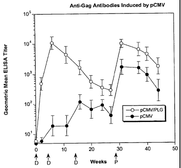
Figure IA and Figure 1B are graphs depicting the effect of PLG microparticles
on
anti-Gag antibody responses induced by DNA vaccines. Figure IA shows geometric
mean
ELISA titers of animals immunized with plasmid DNA at weeks 0, 4 and 14, then
boosted
at weeks 38 and 75 with recombinant Env protein formulated with MF59. Figure
1B
shows geometric mean titer of animals immunized with pSINCP DNA at weeks 0,4
and
14, then boosted at weeks 38 and 75 with recombinant Env protein formulated
with MF59.
Anti-Gag antibodies are plotted as geometric mean ELISA titer for naked pCMV
(solid
symbols) and PLG/pCMV (open symbols) and error bars represent SEM.
Figure 2A and Figure 2B are graphs depicting the effect of PLG microparticles
on
anti-Env antibody responses induced by DNA vaccines. Figure 2A shows geometric
mean
ELISA titers of animals immunized with plasmid DNA at weeks 0,4 and 14, then
boosted
at weeks 38 and 75 with recombinant Env protein formulated with MF59. Figure
2B
shows geometric mean titer of animals immunized with pSINCP DNA at weeks 0,4
and
14, then boosted at weeks 38 and 75 with recombinant Env protein formulated
with MF59.
Anti-Env antibodies are plotted as geometric mean ELISA titer for naked pCMV
(solid
symbols) and PLG/pCMV (open symbols) and error bars represent SEM.
Figure 3 is a graph depicting geometric mean neutralization titer after DNA
administration.
Figure 4 is a graph depicting the effect of Env protein boosting on T cell
responses
primed by DNA vaccines.
Detailed Description of the Invention
The practice of the present invention will employ, unless otherwise indicated,
conventional methods of chemistry, biochemistry, molecular biology, immunology
and
pharmacology, within the skill of the art. Such techniques are explained fully
in the
5
CA 02823937 2013-08-14
literature. See, e.g., Remington's Pharmaceutical Sciences, 18th Edition
(Easton,
Pennsylvania: Mack Publishing Company, 1990); Methods In Enzymology (S.
Colowick
and N. Kaplan, eds., Academic Press, Inc.); and Handbook of Experimental
Immunology,
Vols. I-IV (D.M. Weir and C.C. Blackwell, eds., 1986, Blackwell Scientific
Publications);
Sambrook, et at., Molecular Cloning: A Laboratory Manual (2nd Edition, 1989);
Short
Protocols in Molecular Biology, 4th ed. (Austibel et at. eds., 1999, John
Wiley & Sons);
Molecular Biology Techniques: An Intensive Laboratory Course, (Ream et al.,
eds., 1998,
Academic Press); PCR (Introduction to Biotechniques Series), 2nd ed. (Newton &
Graham
eds., 1997, Springer Verlag); Peters and Dalrymple, Fields Virology (2d ed),
Fields et al.
(eds.), B.N. Raven Press, New York, NY.
As used in this specification and the appended claims, the singular forms "a,"
"an"
and "the" include plural references unless the content clearly dictates
otherwise. Thus, for
example, reference to "an antigen" includes a mixture of two or more such
antigens.
Prior to setting forth the invention, it may be helpful to an understanding
thereof to
first set forth definitions of certain terms that will be used hereinafter.
As used herein the term "HIV polypeptide" refers to any HIV peptide from any
HIV strain or subtype, including, but not limited to Gag, poi, env, vif, vpr,
tat, rev, nef,
and/or vpu; functional (e.g., immunogenic) fragments thereof, modified
polypeptides
thereof and combinations of these fragments and/or modified peptides.
Furthermore, an
"HIV polypeptide" as defined herein is not limited to a polypeptide having the
exact
sequence of known HIV polypeptides. Indeed, the HIV genome is in a state of
constant
flux and contains several domains that exhibit relatively high degrees of
variability
between isolates. As will become evident herein, all that is important is that
the
polypeptide has immunogenic characteristics. It is readily apparent that the
term
encompasses polypeptides from any of the various HIV strains and subtypes.
Furthermore,
the term encompasses any such HIV protein regardless of the method of
production,
including those proteins recombinantly and synthetically produced.
Additionally, the term "HTV polypeptide" encompasses proteins that include
additional modifications to the native sequence, such as additional internal
deletions,
additions and substitutions (generally conservative in nature). These
modifications may be
6
CA 02823937 2013-08-14
WO 2004/032860 PCT/US2003/031935
deliberate, as through site-directed mutagenesis, or may be accidental, such
as through
naturally occurring mutational events. All of these modifications are
encompassed in the
present invention so long as the modified HIV polypeptide functions for its
intended
purpose. Thus, for example, in a vaccine composition, the modifications must
be such that
immunological activity is not lost. Similarly, if the polypeptides are to be
used for
diagnostic purposes, such capability must be retained. Thus, the term also
includes HIV
polypeptides that differ from naturally occurring peptides, for example
peptides that
include one or more deletions (e.g., variable regions deleted from Env),
substitutions
and/or insertions. Nonconservative changes are generally substitutions of one
of the above
amino acids with an amino acid from a different group (e.g., substituting Asn
for Glu), or
substituting Cys, Met, His, or Pro for any of the above amino acids.
Substitutions
involving common amino acids are conveniently performed by site specific
mutagenesis of
an expression vector encoding the desired protein, and subsequent expression
of the
altered form. One may also alter amino acids by synthetic or semi-synthetic
methods. For
example, one may convert cysteine or serine residues to selenocysteine by
appropriate
chemical treatment of the isolated protein. Alternatively, one may incorporate
uncommon
amino acids in standard in vitro protein synthetic methods. Typically, the
total number of
residues changed, deleted or added to the native sequence in the mutants will
be no more
than about 20, preferably no more than about 10, and most preferably no more
than about
5.
"Synthetic" polynucleotide sequences, as used herein, refers to HIV-encoding
polynucleotides (e.g.. Gag- and/or Env-encoding sequences) whose expression
has been
optimized, for example, by codon substitution and inactivation of inhibitory
sequences.
See, e.g., U.S. Patent No. 6,602,705 and International Publications WO
00/39302; WO
02/04493; WO 00/39303; and WO 00/39304 for examples of synthetic HIV-encoding
polynucleotides.
"Wild-type" or "native" sequences, as used herein, refers to polypeptide
encoding
sequences that are essentially as they are found in nature, e.g., Gag and/or
Env encoding
sequences as found in other isolates such as Type C isolates (e.g., Botswana
isolates
AF110965, AF110967, AF110968 or AF110975 or South African isolates).
= As used herein, the term "virus-like particle" or "VLP" refers to a
nonreplicating,
viral shell, derived from any of several viruses discussed further below. VLPs
are
7
CA 02823937 2013-08-14
WO 2004/032860 PCT/US2003/031935
generally composed of one or more viral proteins, such as, but not limited to
those proteins
referred to as capsid, coat, shell, surface and/or envelope proteins, or
particle-forming
polypeptides derived from these proteins. VLPs can form spontaneously upon
recombinant expression of the protein in an appropriate expression system.
Methods for
producing particular VLPs are known in the art and discussed more fully below.
The
presence of VLPs following recombinant expression of viral proteins can be
detected using
conventional techniques known in the art, such as by electron microscopy, X-
ray
crystallography, and the like. See, e.g., Baker et al., Biophys. J. (1991)
60:1445-1456;
Hagensee et al., I Virol. (1994) 68:4503-4505. For example, VLPs can be
isolated by
density gradient centrifugation and/or identified by characteristic density
banding.
Alternatively, cryoelectron microscopy can be performed on vitrified aqueous
samples of
the VLP preparation in question, and images recorded under appropriate
exposure
conditions.
By "particle-forming polypeptide" derived from a particular viral protein is
meant a
full-length or near full-length viral protein, as well as a fragment thereof,
or a viral protein
with internal deletions, which has the ability to form VLPs under conditions
that favor
VLP formation. Accordingly, the polypeptide may comprise the full-length
sequence,
fragments, truncated and partial sequences, as well as analogs and precursor
forms of the
reference molecule. The term therefore intends deletions, additions and
substitutions to
the sequence, so long as the polypeptide retains the ability to form a VLP.
Thus, the term
includes natural variations of the specified polypeptide since variations in
coat proteins
often occur between viral isolates. The term also includes deletions,
additions and
substitutions that do not naturally occur in the reference protein, so long as
the protein
retains the ability to form a VLP. Preferred substitutions are those that are
conservative in
nature, i.e., those substitutions that take place within a family of amino
acids that are
related in their side chains. Specifically, amino acids are generally divided
into four
families: (1) acidic -- aspartate and glutamate; (2) basic -- lysine,
arginine, histidine; (3)
non-polar -- alanine, valine, leucine, isoleucine, proline, phenylalanine,
methionine,
tryptophan; and (4) uncharged polar -- glycine, asparagine, glutamine,
cystine, serine
threonine, tyrosine. Phenylalanine, tryptophan, and tyrosine are sometimes
classified as
aromatic amino acids.
8
CA 02823937 2013-08-14
WO 2004/032860 PCT/1JS2003/031935
An "antigen" refers to a molecule containing one or more epitopes (either
linear,
conformational or both) that will stimulate a host's immune system to make a
humoral
and/or cellular antigen-specific response. The term is used interchangeably
with the term
"immunogen." Normally, a B-cell epitope will include at least about 5 amino
acids but can
be as small as 3-4 amino acids. A T-cell epitope, such as a CTL epitope, will
include at
least about 7-9 amino acids, and a helper 1-cell epitope at least about 12-20
amino acids.
Normally, an epitope will include between about 7 and 15 amino acids, such as,
9, 10, 12
or 15 amino acids. The term "antigen" denotes both subunit antigens, (i.e.,
antigens which
are separate and discrete from a whole organism with which the antigen is
associated in
nature); as well as, killed, attenuated or inactivated bacteria, viruses,
fungi, parasites or
other microbes. Antibodies such as anti-idiotype antibodies, or fragments
thereof, and
synthetic peptide mimotopes, which can mimic an antigen or antigenic
determinant, are
also captured under the definition of antigen as used herein. Similarly, an
oligonucleotide
or polynucleotide that expresses an antigen or antigenic determinant in vivo,
such as in
gene therapy and DNA immunization applications, is also included in the
definition of
antigen herein.
For purposes of the present invention, antigens are preferably derived from
any
subtype of HIV. Antigens can also be derived from any of several known
viruses, bacteria,
parasites and fungi, or tumor antigens. Furthermore, for purposes of the
present invention,
an "antigen" refers to a protein that includes modifications, such as
deletions, additions
and substitutions (generally conservative in nature), to the native sequence,
so long as the
protein maintains the ability to elicit an immunological response, as defined
herein. These
modifications may be deliberate, as through site-directed mutagenesis, or may
be
accidental, such as through mutations of hosts that produce the antigens.
An "immunological response" to an antigen or composition is the development in
a
subject of a humoral and/or a cellular immune response to an antigen present
in the
composition of interest. For purposes of the present invention, a "humoral
immune
response" refers to an immune response mediated by antibody molecules, while a
"cellular
immune response" is one mediated by T-lymphocytes and/or other white blood
cells. One
important aspect of cellular immunity involves an antigen-specific response by
cytolytic T-
cells ("CTL"s). CTLs have specificity for peptide antigens that are presented
in
association with proteins encoded by the major histocompatibility complex
(MHC) and
9
CA 02823937 2013-08-14
WO 2004/032860 PCT/US2003/031935
expressed on the surfaces of cells. CTLs help induce and promote the
destruction of
intracellular microbes, or the lysis of cells infected with such microbes.
Another aspect of
cellular immunity involves an antigen-specific response by helper T-cells.
Helper T-cells
act to help stimulate the function, and focus the activity of, nonspecific
effector cells
against cells displaying peptide antigens in association with MHC molecules on
their
surface. A "cellular immune response" also refers to the production of
cytolcines,
ch.emokines and other such molecules produced by activated T-cells and/or
other white
blood cells, including those derived from CD4+ and CD8+ T-cells.
A composition or vaccine that elicits a cellular immune response may serve to
sensitize a vertebrate subject by the presentation of antigen in association
with MHC
molecules at the cell surface. The cell-mediated immune response is directed
at, or near,
cells presenting antigen at their surface. In addition, antigen-specific T-
Iymphocytes can
be generated to allow for the future protection of an immunized host.
The ability of a particular antigen to stimulate a cell-mediated immunological
response may be determined by a number of assays, such as by
lymphoproliferation
(lymphocyte activation) assays, CTL cytotoxic cell assays, or by assaying for
T-
lymphocytes specific for the antigen in a sensitized subject. Such assays are
well known in
the art. See, e.g., Erickson et at., J. Immunol. (1993) 151:4189-4199; Doe et
al., Eur. J.
Immunol. (1994) 24:2369-2376. Recent methods of measuring cell-mediated immune
response include measurement of intracellular cytokines or cytokine secretion
by T-cell
populations, or by measurement of epitope specific T-cells (e.g., by the
tetramer
technique)(reviewed by McMichael, Ai., and O'Callaghan, C.A., J. Exp. Med.
187(9)1367-1371, 1998; Mcheyzer-Williams, M.G., et at, Immunol. Rev. 150:5-21,
1996;
Lalvani, A., et al, J. Exp. Med. 186:859-865, 1997).
Thus, an immunological response as used herein may be one that stimulates the
production of CTLs, and/or the production or activation of helper T- cells.
The HIV
antigen(s) may also elicit an antibody-mediated immune response. Hence, an
immunological response may include one or more of the following effects: the
production
of antibodies by B-cells; and/or the activation of suppressor T-cells and/or
yo T-cells
directed specifically to an antigen or antigens present in the composition or
vaccine of
interest. These responses may serve to neutralize infectivity, and/or mediate
antibody-
complement, or antibody dependent cell cytotoxicity (ADCC) to provide
protection to an
CA 02823937 2013-08-14
WO 2004/032860 PC
T/US2003/031935
immunized host. Such responses can be determined using standard immunoassays
and
neutralization assays, well known in the art.
An "immunogenic composition" is a composition that comprises an antigenic
molecule where administration of the composition to a subject results in the
development
in the subject of a humoral and/or a cellular immune response to the antigenic
molecule of
interest. The immunogenic composition can be introduced directly into a
recipient subject,
such as by injection, inhalation, oral, intranasal and mucosa] (e.g., intra-
rectally or intra-
vaginally) administration.
By "subunit vaccine" is meant a vaccine composition that includes one or more
selected antigens but not all antigens, derived from or homologous to, an
antigen from a
pathogen of interest such as from a virus, bacterium, parasite or fungus. Such
a
composition is substantially free of intact pathogen cells or pathogenic
particles, or the
lysate of such cells or particles. Thus, a "subunit vaccine" can be prepared
from at least
partially purified (preferably substantially purified) immunogenic
polypeptides from the
pathogen, or analogs thereof. The method of obtaining an antigen included in
the subunit
vaccine can thus include standard purification techniques, recombinant
production, or
synthetic production.
"Substantially purified" general refers to isolation of a substance (compound,
polynucleotide, protein, polypeptide, polypeptide composition) such that the
substance
comprises the majority percent of the sample in which it resides. Typically in
a sample a
substantially purified component comprises 50%, preferably 80%-85%, more
preferably
90-95% of the sample. Techniques for purifying polynucleotides and
polypeptides of
interest are well-known in the art and include, for example, ion-exchange
chromatography,
affinity chromatography and sedimentation according to density.
A "coding sequence" or a sequence that "encodes" a selected polypeptide, is a
nucleic acid molecule that is transcribed (in the case of DNA) and translated
(in the case of
miRNA) into a polypeptide in vivo when placed under the control of appropriate
regulatory
sequences (or "control elements"). The boundaries of the coding sequence are
determined
by a start codon at the 5' (amino) terminus and a translation stop codon at
the 3' (carboxy)
terminus. A coding sequence can include, but is not limited to, cDNA from
viral,
procaryotic or eucaryotic mRNA, genomic DNA sequences from viral or
procaryotic
11
CA 02823937 2013-08-14
WO 2004/032860 PCT/US2003/031935
DNA, and even synthetic DNA sequences. A transcription termination sequence
may be
located 3' to the coding sequence.
Typical "control elements", include, but are not limited to, transcription
promoters,
transcription enhancer elements, transcription termination signals,
polyadenylation
sequences (located 3' to the translation stop codon), sequences for
optimization of
initiation of translation (located 5' to the coding sequence), and translation
termination
sequences.
A "nucleic acid" molecule can include, but is not limited to, procaryotic
sequences,
eucaryotic mRNA, cDNA from eucaryotic mRNA, genomic DNA sequences from
eucaryotic (e.g., mammalian) DNA, and even synthetic DNA sequences. The term
also
captures sequences that include any of the known base analogs of DNA and RNA.
"Operably linked" refers to an arrangement of elements wherein the components
so
described are configured so as to perform their usual function. Thus, a given
promoter
operably linked to a coding sequence is capable of effecting the expression of
the coding
sequence when the proper enzymes are present. The promoter need not be
contiguous with
the coding sequence, so long as it functions to direct the expression thereof.
Thus, for
example, intervening untranslated yet transcribed sequences can be present
between the
promoter sequence and the coding sequence and the promoter sequence can still
be
considered "operably linked" to the coding sequence.
"Recombinant" as used herein to describe a nucleic acid molecule means a
polynucleotide of genomic, cDNA, semisynthetic, or synthetic origin which, by
virtue of
its origin or manipulation: (1) is not associated with all or a portion of the
polynucleotide
with which it is associated in nature; and/or (2) is linked to a
polynucleotide other than that
to which it is linked in nature. The term "recombinant" as used with respect
to a protein or
polypeptide means a polypeptide produced by expression of a recombinant
polynucleotide.
"Recombinant host cells," "host cells," "cells," "cell lines," "cell
cultures," and other such
terms denoting procaryotic microorganisms or eucaryotic cell lines cultured as
unicellular
entities, are used interchangeably, and refer to cells which can be, or have
been, used as
recipients for recombinant vectors or other transfer DNA, and include the
progeny of the
original cell which has been transfected. It is understood that the progeny of
a single
parental cell may not necessarily be completely identical in morphology or in
genomic or
total DNA complement to the original parent, due to accidental or deliberate
mutation.
12
CA 02823937 2013-08-14
WO 2004/032860 PCT/US2003/031935
Progeny of the parental cell which are sufficiently similar to the parent to
be characterized
by the relevant property, such as the presence of a nucleotide sequence
encoding a desired
peptide, are included in the progeny intended by this definition, and are
covered by the
above terms.
Techniques for determining amino acid sequence "similarity" are well known in
the art. In general, "similarity" means the exact amino acid to amino acid
comparison of
two or more polypeptides at the appropriate place, where amino acids are
identical or
possess similar chemical and/or physical properties such as charge or
hydrophobicity. A
so-termed "percent similarity" then can be determined between the compared
polypeptide
sequences. Techniques for determining nucleic kid and amino acid sequence
identity also
are well known in the art and include determining the nucleotide sequence of
the mRNA
for that gene (usually via a cDNA intermediate) and determining the amino acid
sequence
encoded thereby, and comparing this to a second amino acid sequence. In
general,
"identity" refers to an exact nucleotide to nucleotide or amino acid to amino
acid
correspondence of two polynucleotides or polypeptide sequences, respectively.
Two or more polynucleotide sequences can be compared by determining their
"percent identity." Two or more amino acid sequences likewise can be compared
by
determining their "percent identity." The percent identity of two sequences,
whether
nucleic acid or peptide sequences, is generally described as the number of
exact matches
between two aligned sequences divided by the length of the shorter sequence
and
multiplied by 100. An approximate alignment for nucleic acid sequences is
provided by
the local homology algorithm of Smith and Waterman, Advances in Applied
Mathematics
. 2:482-489 (1981). This algorithm can be extended to use with peptide
sequences using the
scoring matrix developed by Dayhoff, Atlas of Protein Sequences and Structure,
M.O.
Dayhoff ed., 5 suppl. 3:353-358, National Biomedical Research Foundation,
Washington,
D.C., USA, and normalized by Gribskov, Nucl. Acids Res. 14(6):6745-6763
(1986). An
implementation of this algorithm for nucleic acid and peptide sequences is
provided by the
Genetics Computer Group (Madison, WI) in their BestFit utility application.
The default
parameters for this method are described in the Wisconsin Sequence Analysis
Package
Program Manual, Version 8 (1995) (available from Genetics Computer Group,
Madison,
WI). Other equally suitable programs for calculating the percent identity or
similarity
between sequences are generally known in the art.
13
=
CA 02823937 2013-08-14
For example, percent identity of a particular nucleotide sequence to a
reference
sequence can be determined using the homology algorithm of Smith and Waterman
with a
default scoring table and a gap penalty of six nucleotide positions. Another
method of
establishing percent identity in the context of the present invention is to
use the MPSRCH
package of programs copyrighted by the University of Edinburgh, developed by
John F.
Collins and Shane S. Sturrok, and distributed by IntelliGenetics, Inc.
(Mountain View,
CA). From this suite of packages, the Smith-Waterman algorithm can be employed
where
default parameters are used for the scoring table (for example, gap open
penalty of 12, gap
extension penalty of one, and a gap of six). From the data generated, the
"Match" value
reflects "sequence identity." Other suitable programs for calculating the
percent identity or
= similarity between sequences are generally known in the art, such as the
alignment
program BLAST, which can also be used with default parameters. For example,
BLASTN
and BLASTP can be used with the following default parameters: genetic code =
standard;
filter = none; strand = both; cutoff= 60; expect =10; Matrix = BLOSUM62;
Descriptions
= 50 sequences; sort by= HIGH SCORE; Databases = non-redundant, GenBank + EMBL
+ DDBJ + PDB + GenBank CDS translations + Swiss protein + Spupdate + PIR.
=
One of skill in the art can readily determine the proper search parameters to
use for
a given sequence in the above programs. For example, the search parameters may
vary
based on the size of the sequence in question. Thus, for example, a
representative
embodiment of the present invention would include an isolated p.olynucleotide
having X
contiguous nucleotides, wherein (i) the X contiguous nucleotides have at least
about 50%
identity to Y contiguous nucleotides derived from any of the sequences
described herein,
(ii) X equals Y, and (iii) X is greater than or equal to 6 nucleotides and up
to 5000
nucleotides, preferably greater than or equal to 8 nucleotides and up to 5000
nucleotides,
more preferably 10-12 nucleotides and up to 5000 nucleotides, and even more
preferably
15-20 nucleotides, up to the number of nucleotides present in the full-length
sequences
described herein (e.g., see the Sequence Listing and claims), including all
integer values
falling within the above-described ranges.
The polynucleotides described herein include related polynucleotide sequences
having about 80% to 100%, greater than 80-85%, preferably greater than 90-92%,
more
14
CA 02823937 2013-08-14
WO 2004/032860 PCT/US2003/031935
preferably greater than 95%, and most preferably greater than 98% sequence
(including all
integer values falling within these described ranges) identity to the
sequences disclosed
herein (for example, to the claimed sequences or other sequences of the
present invention)
when the sequences of the present invention are used as the query sequence.
Two nucleic acid fragments are considered to "selectively hybridize" as
described
herein. The degree of sequence identity between two nucleic acid molecules
affects the
efficiency and strength of hybridization events between such molecules. A
partially
identical nucleic acid sequence will at least partially inhibit a completely
identical
sequence from hybridizing to a target molecule. Inhibition of hybridization of
the
completely identical sequence can be assessed using hybridization assays that
are well
known in the art (e.g., Southern blot, Northern blot, solution hybridization,
or the like, see
Sambrook, et al:, supra or Ausubel et al., supra). Such assays can be
conducted using
varying degrees of selectivity, for example, using conditions varying from low
to high
stringency. If conditions of low stringency are employed, the absence of non-
specific
binding can be assessed using a secondary probe that lacks even a partial
degree of
sequence identity (for example, a probe having less than about 30% sequence
identity with
the target molecule), such that, in the absence of non-specific binding
events, the
secondary probe will not hybridize to the target.
When utilizing a hybridization-based detection system, a nucleic acid probe is
chosen that is complementary to a target nucleic acid sequence, and then by
selection of
appropriate conditions the probe and the target sequence "selectively
hybridize," or bind,
to each other to form a hybrid molecule. A nucleic acid molecule that is
capable of
hybridizing selectively to a target sequence under "moderately stringent"
typically
hybridizes under conditions that allow detection of a target nucleic acid
sequence of at
least about 10-14 nucleotides in length having at least approximately 70%
sequence
identity with the sequence of the selected nucleic acid probe. Stringent
hybridization
conditions typically allow detection of target nucleic acid sequences of at
least about 10-14
nucleotides in length having a sequence identity of greater than about 90-95%
with the
sequence of the selected nucleic acid probe. Hybridization conditions useful
for
probe/target hybridization where the probe and target have a specific degree
of sequence
identity, can be determined as is known in the art (see, for example, Nucleic
Acid
CA 02823937 2013-08-14
WO 2004/032860 PCT/US2003/031935
Hybridization: A Practical Approach, editors B.D. Hames and Si. Higgins,
(1985)
Oxford; Washington, DC; IRL Press).
With respect to stringency conditions for hybridization, it is well known in
the art
that numerous equivalent conditions can be employed to establish a particular
stringency
by varying, for example, the following factors: the length and nature of probe
and target
sequences, base composition of the various sequences, concentrations of salts
and other
hybridization solution components, the presence or absence of blocking agents
in the
hybridization solutions (e.g., formamide, dextran sulfate, and polyethylene
glycol),
hybridization reaction temperature and time parameters, as well as, varying
wash
conditions. The selection of a particular set of hybridization conditions is
selected
following standard methods in the art (see, for example, Sambrook, et al.,
supra or
Ausubel et al., supra).
A first polynucleotide is "derived from" second polynucleotide if it has the
same or
substantially the same basepair sequence as a region of the second
polynucleotide, its
cDNA, complements thereof, or if it displays sequence identity as described
above.
A first polypeptide is "derived from" a second polypeptide if it is (i)
encoded by a
first polynucleotide derived from a second polynucleotide, or (ii) displays
sequence
identity to the second polypeptides as described above.
Generally, a viral polypeptide is "derived from" a particular polypeptide of a
virus
(viral polypeptide) if it is (i) encoded by an open reading frame of a
polynucleotide of that
virus (viral polynucleotide), or (ii) displays sequence identity to
polypeptides of that virus
as described above.
"Encoded by" refers to a nucleic acid sequence which codes for a polypeptide
sequence, wherein the polypeptide sequence or a portion thereof contains an
amino acid
sequence of at least 3 to 5 amino acids, more preferably at least 8 to 10
amino acids, and
even more preferably at least 15 to 20 amino acids from a polypeptide encoded
by the
nucleic acid sequence. Also encompassed are polypeptide sequences which are
immunologically identifiable with a polypeptide encoded by the sequence.
"Purified polynucleotide" refers to a polynucleotide of interest or fragment
thereof
that is essentially free, e.g., contains less than about 50%, preferably less
than about 70%,
and more preferably less than about 90%, of the protein with which the
polynucleotide is
naturally associated. Techniques for purifying polynucleotides of interest are
well-known
16
CA 02823937 2013-08-14
WO 2004/032860 PCT/1JS2003/031935
in the art and include, for example, disruption of the cell containing the
polynucleotide
with a chaotropic agent and separation of the polynucleotide(s) and proteins
by ion-
exchange chromatography, affinity chromatography and sedimentation according
to
density.
By "nucleic acid immunization" is meant the introduction of a nucleic acid
molecule encoding one or more selected antigens into a host cell, for the in
vivo expression
of an antigen, antigens, an epitope, or epitopes. The nucleic acid molecule
can be
introduced directly into a recipient subject, such as by injection,
inhalation, oral, intranasal
and mucosal administration, or the like, or can be introduced ex vivo, into
cells which have
been removed from the host. In the latter case, the transformed cells are
reintroduced into
the subject where an immune response can be mounted against the antigen
encoded by the
nucleic acid molecule.
"Gene transfer" or "gene delivery" refers to methods or systems for reliably
inserting DNA of interest into a host cell. Such methods can result in
transient expression
of non-integrated transferred DNA, extrachromosomal replication and expression
of
transferred replicons (e.g., episomes), or integration of transferred genetic
material into the
genomic DNA of host cells. Gene delivery expression vectors include, but are
not limited
to, vectors derived from alphaviruses, pox viruses and vaccinia viruses. When
used for
immunization, such gene delivery expression vectors may be referred to as
vaccines or
vaccine vectors.
"1' lymphocytes" or "T cells" are non-antibody producing lymphocytes that
constitute a part of the cell-mediated arm of the immune system. T cells arise
from
immature lymphocytes that migrate from the bone marrow to the thymus, where
they
undergo a maturation process under the direction of thymic hormones. Here, the
mature
lymphocytes rapidly divide increasing to very large numbers. The maturing T
cells
become irrununocompetent based on their ability to recognize and bind a
specific antigen.
Activation of irrununocompetent T cells is triggered when an antigen binds to
the
lymphocyte's surface receptors.
The term "transfection" is used to refer to the uptake of foreign DNA by a
cell. A
cell has been "transfected" when exogenous DNA has been introduced inside the
cell
membrane. A number of transfection techniques are generally known in the art.
See, e.g.,
Graham et al. (1973) Virology, 52:456, Sambrook et al. (1989) Molecular
Cloning, a
17
CA 02823937 2013-08-14
WO 2004/032860 PCT/US2003/031935
laboratory manual, Cold Spring Harbor Laboratories, New York, Davis et al.
(1986) Basic
Methods in Molecular Biology, Elsevier, and Chu et al. (1981) Gene 13:197.
Such
techniques can be used to introduce one or more exogenous DNA moieties into
suitable
host cells. The term refers to both stable and transient uptake of the genetic
material, and
includes uptake of peptide- or antibody-linked DNAs.
Transfer of a "suicide gene" (e.g., a drug-susceptibility gene) to a target
cell renders
the cell sensitive to compounds or compositions that are relatively nontoxic
to normal
cells. Moolten, F.L. (1994) Cancer Gene Ther. 1:279-287. Examples of suicide
genes are
thymidine kinase of herpes simplex virus (HSV-tk), cytochrome P450 (Manome et
at.
(1996) Gene Therapy 3:513-520), human deoxycytidine kinase (Manome et at.
(1996)
Nature Medicine 2(5):567-573) and the bacterial enzyme cytosine deaminase
(Dong et al.
(1996) Human Gene Therapy 7:713-720). Cells that express these genes are
rendered
sensitive to the effects of the relatively nontoxic prodrugs ganciclovir (HSV-
tk),
cyclophosphamide (cytochrome P450 2B1), cytosine arabinoside (human
deoxycytidine
kinase) or 5-fluorocytosine (bacterial cytosine deaminase). Culver et al.
(1992) Science
256:1550-1552, Huber et at. (1994) Proc. Natl. Acad. Sci. USA 91:8302-8306.
A "selectable marker" or "reporter marker" refers to a nucleotide sequence
included in a gene transfer vector that has no therapeutic activity, but
rather is included to
allow for simpler preparation, manufacturing, characterization or testing of
the gene
transfer vector.
A "specific binding agent" refers to a member of a specific binding pair of
molecules wherein one of the molecules specifically binds to the second
molecule through
chemical and/or physical means. One example of a specific binding agent is an
antibody
directed against a selected antigen. '
By "subject" is meant any member of the subphylum chordata, including, without
limitation, humans and other primates, including non-human primates such as
chimpanzees and other apes and monkey species; farm animals such as cattle,
sheep, pigs,
goats and horses; domestic mammals such as dogs and cats; laboratory animals
including
rodents such as mice, rats and guinea pigs; birds, including domestic, wild
and game birds
such as chickens, turkeys and other gallinaceous birds, ducks, geese, and the
like. The
term does not denote a particular age. Thus, both adult and newborn
individuals are
intended to be covered. The system described above is intended for use in any
of the
18
CA 02823937 2013-08-14
WO 2004/032860 PCT/US2003/031935
above vertebrate species, since the immune systems of all of these vertebrates
operate
similarly.
By "pharmaceutically acceptable" or "pharmacologically acceptable" is meant a
material which is not biologically or otherwise undesirable, i.e., the
material may be
administered to an individual in a formulation or composition without causing
any
undesirable biological effects or interacting in a deleterious manner with any
of the
components of the composition in which it is contained.
By "physiological pH" or a "pH in the physiological range" is meant a pH in
the
range of approximately 7.2 to 8.0 inclusive, more typically in the range of
approximately
7.2 to 7.6 inclusive.
As used herein, "treatment" refers to any of (i) the prevention of infection
or
reinfection, as in a traditional vaccine, (ii) the reduction or elimination of
symptoms,
and/or (iii) the substantial or complete elimination of the pathogen in
question. Treatment
may be effected prophylactically (prior to infection) or therapeutically
(following
infection).
"Nucleic acid expression vector" refers to an assembly that is capable of
directing
the expression of a sequence or gene of interest. The nucleic acid expression
vector may
include a promoter that is operably linked to the sequences or gene(s) of
interest. Other
control elements may be present as well. Nucleic acid expression vectors
include, but are
not limited to, plasmids, viral vectors, alphavirus vectors (e.g., Sindbis),
eukaryotic layered
vector initiation systems (see, e.g., U.S. Patent No. 6,342,372), retroviral
vectors,
adenoviral vectors, adeno-associated virus vectors and the like. See, also,
U.S. Patent No.
6,602,705 for a description of various nucleic acid expression vectors.
Expression
cassettes may be contained within a nucleic acid expression vector. The vector
may also
include a bacterial origin of replication, one or more selectable markers, a
signal that
allows the construct to exist as single-stranded DNA (e.g., a M13 origin of
replication), a
multiple cloning site, and a "mammalian" origin of replication (e.g., a SV40
or adenovirus
origin of replication). .
"Packaging cell" refers to a cell that contains those elements necessary for
production of infectious recombinant retrovirus that are lacking in a
recombinant retroviral
vector. Typically, such packaging cells contain one or more expression
cassettes which
are capable of expressing proteins which encode Gag, poi and env proteins.
19
CA 02823937 2013-08-14
WO 2004/032860 PCT/US2003/031935
"Producer cell" or "vector producing cell" refers to a cell that contains all
elements
necessary for production of recombinant retroviral vector particles.
In addition, the following is a partial list of abbreviations used herein:
118 microgram
AIDS acquired immune deficiency syndrome
APC antigen presenting cell
CCR5 chemokine receptor 5
CD4+ cluster of differeniation 4 receptor
CD8+ cluster of differeniation 8 receptor
CDC centers for disease control
CHO cells Chinese hamster ovary cells
CMV cytomegalovirus
ConA Concanvalim A
CRF case report form
CRF's circulating recombinant forms
CTAB cetyltrimetylamnonium bromide
CTL cytotoxic T lymphocyte
Cv cromium
DEAE Diethylaminoeihyl
DNA deoxyribonucleic acid
DTH delayed type hypersensitivity
ELISA enzyme-linked immunosorbent assay
ELISPOT enzyme-linked immunospot assay
ENV envelope
FIGE field inversion gel electrophoresis
GAG group-specific antigen
GLP good laboratory practices
gp glycoprotein
HAART highly active antiretroviral therapy
HAP hydroziapatic
HBsAg hepatitis B surface antigen
HCV hepatitis C virus
HIV/HIV-1 human immunodeficiency virus/Type I
hr hour
HSV herpes simplex virus
IEN interferon
IFNy interferon gamma
TM intramuscular
IND investigational new drug
IV intravenous
Kb kilobase
kD kilodalton
Kg kilogram
. mg milligram
mL milliliter
MF59 oil-in-water emulsion adjuvant
CA 02823937 2013-08-14
WO 2004/032860 PCT/US2003/031935
NaC1 sodium chloride
NIAID National Institute of Allergy and Infectious Disease
NIH National Institutes of Health
o- or 0- oligomeric
PCR polymerase chain reaction
PEG polyethylene glycol
PLG cationic poly-lactide-coglycolide
pSIN sindbis virus vector
PVA poly(vinyl alcohol)
REV viral protein - involved in regulation of viral
expression
SAE serious adverse event
SHIV simian human immunodefiency virus
SP resin modified polyester-carbonate resin
General Overview
Before describing the present invention in detail, it is to be understood that
this
invention is not limited to particular formulations or process parameters as
such may, of
course, vary. It is also to be understood that the terminology used herein is
for the purpose
of describing particular embodiments of the invention only, and is not
intended to be
limiting.
Although a number of methods and materials similar or equivalent to those
described herein can be used in the practice of the present invention, the
preferred
materials and methods are described herein.
The present invention relates to methods and compositions for the development
of
immunogenic compositions (e.g., vaccines) for HIV. For example, an HIV vaccine
as
described herein may include three or more components. Vaccines as described
herein
may be intended for intramuscular injection. In certain embodiments, two
nucleic acid
components are formulated onto (adsorbed onto) cationic poly-lactide-
coglycolide (PLG)
microparticles and administered as priming immunizations. In addition to the
DNA
components, a protein composition is also administered in one or more boosting
immunizations. The protein component typically comprises at least one HIV
polypeptide,
for example, a CHO cell-produced, recombinant oligomeric envelope protein with
a
deletion in the V2 region mixed with the NIF-59 adjuvant.
Pharmaceutic Compositions
In a preferred embodiment, the HIV vaccines described herein includes multiple
(e.g., three or more) components intended for administration (e.g.,
intramuscularly) in a 6-
21
CA 02823937 2013-08-14
WO 2004/032860 PCT/US2003/031935
9 month, or even longer, time period. The components may be given concurrently
or at
different time points. For example, two nucleic acid "priming" immunizations
may be
given, where each priming immunization includes include two separate
preparations of
DNA encoding Gag protein(s) (e.g., p55 Gag from H1V-1 SF2), and/or Env
protein(s)
(e.g., an oligomeric, V2-deleted, gp140 envelope protein from HIV-1 SF162),
both
formulated on PLG microparticles. The nucleic acids will typically be provided
separately
in unit dose vials containing between 1 1.1g to 10 mg of DNA and between 10 ug
and 100
mg of PLG (e.g., 1 mg of DNA and 25 mg of PLG microparticles). The DNA-
containing
doses are typically stored in lyophilized form and vials are generally
reconstituted in the
field. It should be noted that each unit dose vial will typically contain more
DNA (or
protein) than is actually administered to the patient. The final dosage
typically consists of
1 mg in 0.5mL each of Gag and Env DNA. The DNA components of the vaccine are
intended to prime antibody, CD4 and CD8 T cell responses to HIV antigens
(e.g.. Gag and
Env).
As noted above, the immunogenic systems (vaccines) described herein also
comprise at least one protein component, typically an HIV polypeptide from any
isolate or
strain of HIV. For example, in certain embodiments, the protein component
comprises a
recombinant oligomeric envelope protein from the SF162 strain of HIV-1.
Protein
monomers of HIV Env may be truncated to an approximate molecular size of 14010
(e.g.,
to improve solubility) and the V2 loop may be at least partially removed. The
resulting
oligomeric molecule resembles the envelope structure of HP! closely. Removal
of the V2
variable loop exposes conserved epitopes involved in receptor and/or co-
receptor binding.
Macaques primed with naked DNA vaccines encoding oligomeric V2-deleted gp140
from
the subtype B (CCR5) primary isolate SF162, and boosted with the corresponding
recombinant protein, produced antibodies capable of neutralizing a range of
distinct
subtype B primary isolates. Barnett et al. (2001)J Virol. 75(12):5526-40;
Srivastava et al.
(2002)J Virol. (6):2835-47; Srivastava et al. (2003) J. Virol. 77(20):11244-
11259.
Based on the quantities of passively administered antibodies required to
protect
macaques and the magnitude and breadth of the neutralization titers seen in
macaque
studies, suggest that the antibodies induced by vaccines described herein are
likely to
provide protection from infection in a proportion of animals. Mascola et al.
(1999) J Virol.
73(5): 4009-18. The amount of protein per does can vary from microgram to
milligram
22
CA 02823937 2013-08-14
WO 2004/032860 PCT/US2003/031935
amounts. In certain embodiments, the protein is provided such that the dose
administered
is approximately 100 micrograms in unit dose vials containing envelope protein
in sodium
citrate buffer, pH 6.0 without preservative.
The protein and/or nucleic acid compositions described herein may also
comprise a
pharmaceutically acceptable carrier. The carrier should not itself induce the
production of
antibodies harmful to the host. Pharmaceutically acceptable carriers are well
known to
those in the art. Suitable carriers are typically large, slowly metabolized
macromolecules
such as proteins, polysaccharides, polylactic acids, polyglycolic acids,
polymeric amino
acids, amino acid copolymers, lipid aggregates (such as oil droplets or
liposomes), and
inactive virus particles. Examples of particulate carriers include those
derived from
polymethyl methacrylate polymers, as well as microparticles derived from
poly(lactides)
and poly(lactide-co-glycolides), known as PLG. See, e.g., Jeffery et al.,
Pharrn. Res.
(1993) 10:362-368; McGee et at. (1997) J Microencapsul. 14(2):197-210; O'Hagan
et at.
(1993) Vaccine 11(2):149-54. Such carriers are well known to those of ordinary
skill in
the art. Additionally, these carriers may function as immunostimulating agents
("adjuvants"). Furthermore, the antigen may be conjugated to a bacterial
toxoid, such as
toxoid from diphtheria, tetanus, cholera, etc., as well as toxins derived from
E. coli.
Pharmaceutically acceptable salts can also be used in compositions of the
invention, for example, mineral salts such as hydrochlorides, hydrobromides,
phosphates,
or sulfates, as well as salts of organic acids such as acetates, proprionates,
malonates, or
benzoates. Especially useful protein substrates are serum albumins, keyhole
limpet
hemocyanin, immunoglobulin molecules, thyroglobulin, ovalbumin, tetanus
toxoid, and
other proteins well known to those of skill in the art. Compositions of the
invention can
also contain liquids or excipients, such as water, saline, glycerol, dextrose,
ethanol, or the
like, singly or in combination, as well as substances such as wetting agents,
emulsifying
agents, or pH buffering agents. Liposomes can also be used as a carrier for a
composition
of the invention, such liposomes are described above.
Briefly, with regard to viral particles, replication-defective vectors (also
referred to
above as particles) may be preserved either in crude or purified forms.
Preservation
methods and conditions are described in U.S. Patent No. 6,015,694.
Further, the compositions described herein can include various excipients,
adjuvants, carriers, auxiliary substances, modulating agents, and the like.
Preferably, the
23
CA 02823937 2013-08-14
=
compositions will include an amount of the antigen sufficient to mount an
immunological
response. An appropriate effective amount can be determined by one of skill in
the art.
Such an amount will fall in a relatively broad range that can be determined
through routine
trials and will generally be an amount on the order of about 0.1 g to about
1000 lig (e.g.,
antigen and/or particle), more preferably about 1 lig to about 300 g, of
particle/antigen.
As noted above, one or more of the components may further comprise one or more
adjuvants. Preferred adjvuants to enhance effectiveness include of the
composition
includes, but are not limited to: (1) aluminum salts (alum), such as aluminum
hydroxide,
aluminum phosphate, aluminum sulfate, etc.; (2) oil-in-water emulsion
formulations (with
or without other specific irtununostimulating agents such as murarnyl peptides
(see below)
- or bacterial cell wall components), such as for example (a) MF59111
(International
Publication No. WO 90/14837; Chapter 10 in Vaccine design: the subunit and
adjuvant
approach, eds. Powell & Newman, Plenus Press, 1995), containing 5% Squalene,
0.5%
=
Tween 80, and 0.5% Span 85 (optionally containing various amounts of MTP-PE)
formulated into submicron particles using a microfluidizer, (b) SAF,
containing 10%
Squalane, 0.4% Tweet' 80, 5% pluronic-blocked polymer L121, and thr-MDP (see
below)
either microfluidized into a submicron emulsion or vortexed to generate a
larger particle
size emulsion, and (c) RibiTm adjuvant system (RAS), (Ribi Inununochem,
Hamilton, MT)
=
containing 2% Squalene, 0.2% Tweett 80, and one or more bacterial cell wall
components
from the group consisting of monophosphorylipid A (MPL), nehalose dimycolate
(TDM),
and cell wall skeleton (CWS), preferably MPL + CWS (DetoxTm); (3) saponin
adjuvants,
such as QS21 or Stimulonni (Cambridge Bioscience, Worcester, MA) may be used
or
particle generated therefrom such as ISCOMs (immunostimulating complexes),
which
ISCOMS may be devoid of additional detergent (see, e.g., WO 00/07621); (4)
Complete
Freunds Adjuvant (CFA) and Incomplete Freunds Adjuvant (IFA); (5) cytokines,
such as
interleuldns (IL-1, IL-2, IL-4, IL-5, 1L-6, IL-7, IL-12 (WO 99/44636), IL16,
etc.),
interferons (e.g., gamma interferon), macrophage colony stimulating factor (M-
CSF),
tumor necrosis factor (1'NF), beta chemokines (MIP, 1-alpha, 1-beta Rantes,
etc.), etc.; (6)
monophosphoryl lipids A (MPL) or 3-0-deacylated MPL (3dMPL) e.g., GB-222021,
EP-
A-0689454, optionally in the substantial absence of alum when used with
pneumococcal
saccharides e.g., WO 00/56358; (7) combinations of 3dMPL with, for example,
QS21
and/or oil-in-water emulsions e.g., EP-A-0835318, EP-A-0735898, EP-A-0761231;
(8)
*Trade-mark
24
CA 02823937 2013-08-14
WO 2004/032860 PCT/US2003/031935
oligonucleotides comprising CpG motifs (Roman et al., Nat. Med., 1997, 3:849-
854;
Weiner et al., PNAS USA, 1997, 94:10833-10837; Davis et al. J Immunol., 1998,
160:870-876; Chu etal., J. Exp. Med., 1997, 186:1623-1631; Lipford etal., Eur.
J
Immunol. 1997, 27:2340-2344; Moldoveanu. etal., Vaccine, 1988, 16:1216-1224,
Krieg et
al., Nature, 1995, 3742:546-549; Klinman etal., PNAS USA, 1996, 93:2879-2883:
Ballas
etal., J Immunol., 1996, 157:1840-1845; Cowdery etal., J Immunol., 1996,
156:4570-
4575; Halpem et al., Cell. Immunol., 1996, 167:72-78; Yamamoto et aL, Jpn. J
Cancer
Res., 1988, 79:866-873; Stacey et al., J Immunol, 1996, 157:2116-2122; Messina
et al., J
Immunol., 1991, 147:17591764; Yi etal., J Immunol., 1996, 157:4918-4925; Yi
etal., J
Immunol., 1996, 157:5394-5402; Yi etal.. J Immunol., 1998, 160:4755-4761; and
Yi et
J Immunol., 1998, 1605:5898-5906; International patent applications
W096/02555,
W098/16247, W098/18810, W098/401005 W098/55495, W098/37919 and
W098/52581) i.e. containing at least one CG dinucleotide, with 5
methylcytosine
optionally being used in place of cytosine; (8) a polyoxyethylene ether or a
polyoxyethylone ester e.g. WO 99/52549; (9) a polyoxyethylene sorbitan ester
surfactant in
combination with an octoxynol (WO 01/21207) or a polyoxyethylene alkyl ether
or ester
surfactant in combination with at least one additional non-ionic surfactant
such as an
octoxynol (WO 01/21152); (10) a saponin and an immunostimulatory
oligonucleotide
(e.g., a CpG oligonucleotide) (WO 00/62800); (11) an imrnunostimulant and a
particle of
metal salt e.g. WO 00/23105; (12) a saponin and oil-in-water emulsion e.g., WO
99/11241; (13) a saponin (e.g., QS21) + 3dMPL = 1L-12 (optionally + a sterol)
e.g., WO
98/57659; (14) other substances that act as immunostimulating agents to
enhance the
effectiveness of the composition. Alum (especially aluminum phosphate and/or
hydroxide) and MF59Tm are preferred.
Muramyl peptides include, but are not limited to, N-acetyl-muramyl-L-threonyl-
D-
isoglutamine (thr-MDP), N-acteyl-normuramyl-L-alanyl-D-isogluatme (nor-MDP), N-
acetylmuramyl-L-alanyl-D-isogluatminyl-L-alanine-2-(1'-2'-dipalmitoyl-sn-
glycero-3-
huydroxyphosphoryloxy)-ethylamine (MTP-PE), etc.
Administration of the pharmaceutical compositions described herein may be by
any
suitable route (see, e.g., Section C). Particularly preferred is intramuscular
or mucosal
(e.g., rectal and/or vaginal) administration. Dosage treatment may be a single
dose
schedule or a multiple dose schedule. A multiple dose schedule is one in which
a primary
CA 02823937 2013-08-14
WO 2004/032860 PCT/US2003/031935
course of vaccination may be with 1-10 separate doses, followed by other doses
given at
subsequent time intervals, chosen to maintain and/or reinforce the immune
response, for
example at 1 to 6 months for a second dose, and if needed, a subsequent
dose(s) after
several months. The dosage regimen will also, at least in part, be determined
by the
potency of the modality, the vaccine delivery employed, the need of the
subject and be
dependent on the judgment of the practitioner.
In certain embodiments, the protein component is mixed before administration
with a proprietary oil-in-water emulsion adjuvant, MF59C.1 (hereafter referred
to as
MF59) (See, e.g,. International Publication No. WO 90/14837). Various subunit
antigens
(e.g., HCV E2, HIV gp120, HBsAg, CMV gB, and HSV 2 gD) have been combined with
MF59 adjuvant and administered to over 18,000 human subjects to date with an
excellent
safety and tolerability profile. The protein booster is intended to amplify
the primary
antibody and CD4+ T cell responses in breadth and duration and to provide a
balanced
response in both the humoral and cellular compartments of the immune system,
capable to
achieve the prevention of HIV-1 infection.
As noted above, MF59 adjuvant has been extensively evaluated in clinical
trials
with a number of different subunit antigens, including those derived from
influenza,
herpes simplex virus 2 (HSV), human immunodeficiency virus (HIV),
cytomegalovirus
(CMV), and hepatitis B virus (HBV) and is generally well tolerated with
minimal local
and systemic adverse reactions that are transient and of mild-to-moderate
severity. Over
12,000 subjects have received influenza virus vaccines combined with MF59
adjuvant
emulsion in more than 30 clinical studies. Only two patients had serious
adverse effects.
Moreover, the incidence of adverse effects depend upon the antigen used.
Prime-Boost Regimes
In certain embodiments, multiple administrations (e.g., prime-boost type
administration) will be advantageously employed. For example, nucleic acid
constructs
expressing one or more HIV antigen(s) of interest are administered.
Subsequently, the
same and/or different HIV antigen(s) are administered, for example in
compositions
comprising the polypeptide antigen(s) and a suitable adjuvant. Alternatively,
antigens are
administered prior to the DNA. Multiple polypeptide and multiple nucleic acid
administrations (in any order) may also be employed.
26
CA 02823937 2013-08-14
WO 2004/032860 PCT/US2003/031935
As described herein, one exemplary prime-boost regime described herein
includes
two or more administrations of DNAs encoding one or more HIV antigens followed
by
one or more administrations of HIV polypeptide antigens themselves. For
example, two or
more administrations of HIV Gag and HIV Env DNA/PLG compositions (e.g.,
separate
Gag and Env) may be followed by one or more administration of HIV Env protein.
HIV-1
DNA constructs are able to stimulate the cellular and humoral arms of the
immune system
and elicit immune responses capable of preventing 11IV-1 infection in
chimpanzees. Boyer
et al. (1997) Nat Med 3:526-532. Adsorption of DNA onto the surface of PLG
microparticles improves DNA uptake by the antigen presenting cells (APCs), and
enhance
cellular and humoral immune responses. O'Hagan et al. (2001) J Virol.
75(19):9037-43.
PLO is particularly preferred to deliver DNA because the polymer is
biodegradable,
biocompatible and has been used to develop several drug delivery systems.
Okada et al.
(1997) Adv Drug Deliv Rev 28(1):43-70. In certain embodiments, the ratio of
DNA:PLG
is between about 1 and 16 w/w % (or any value therebetween).
The "booster" component comprises an HIV protein from any HIV strain or
subtype, for example a recombinant oligomeric envelope protein from the
subtype B strain
(e.g., SF2, SF162, etc.) and/or subtype C strain (Botswana strains and/or
South African
strains such as TV1). See, e.g., Scriba etal. (2001) AIDS Res Hum Retroviruses
17(8):775-
81; Scriba et al. (2002) AIDS Res Hum Retroviruses 18(2):149-59; Treurnicht et
al. (2002)
J Med Virol. 68(2):141-6. The protein monomers of the Env protein may be
truncated and
the V2 loop partially removed to increase the exposure of conserved epitopes
that are more
efficient to elicit cross-reactive neutralizing antibody. Without being bound
by one theory,
it appears that the protein booster is intended to amplify the primary
antibody and CD4+ T
cell responses in breadth and duration. Barnett et al. (2001) J Virol
75(12):5526-40;
Cherpelis et al. (2001) J. Virol. 75(3):1547-50. The concentration of protein
in each dose
may vary from approximately 1 pg to over 1000 ttg (or any value therebetween),
preferably
between about 10 lig and 500 g, and even more preferably between about 30 pg
and 300
To date, IIW vaccines as described herein have demonstrated a strong record of
safety in preclinical studies and clinical trials. See, also, Example 4 below.
No evidence
of vaccine-related immunodeficiency has been reported. Toxicology studies
conducted in
mice and rabbits with the HIV vaccine demonstrated that the vaccine was very
well
27
CA 02823937 2013-08-14
WO 2004/032860 PCT/US2003/031935
tolerated. Findings were consistent with studies conducted with other viral
subunit
vaccines or with MF59 adjuvant. Reversible local (intramuscular) inflammation
is the
only notable change seen with such vaccines (see Example 4).
The goal of the HIV vaccine development program is to demonstrate the safety
and
efficacy of a novel DNA-prime plus recombinant protein-boost HIV vaccine, that
is
capable of eliciting a combination of broad humoral and cellular responses,
and preventing
HIV infection or the development of advanced HIV disease/AIDS.
Sources of HIV Antigens
Polynucleotide sequences (e.g., for use in nucleic acid expression constructs)
can
be obtained using recombinant methods, such as by screening cDNA and genomic
libraries
from cells expressing the gene, or by deriving the gene from a vector known to
include the
same. Furthermore, the desired gene can be isolated directly from cells and
tissues
containing the same, using standard techniques, such as phenol extraction and
PCR of
cDNA or genomic DNA. See, e.g., Sambrook et al., supra, for a description of
techniques
used to obtain and isolate DNA. The gene of interest can also be produced
synthetically,
rather than cloned. The nucleotide sequence can be designed with the
appropriate codons
for the particular amino acid sequence desired. In general, one will select
preferred codons
for the intended host in which the sequence will be expressed. The complete
sequence is
assembled from overlapping oligonucleotides prepared by standard methods and
as-
sembled into a complete coding sequence. See, e.g., Edge, Nature (1981)
292:756;
Nambair etal., Science (1984) 223:1299; Jay et al., J. Biol. Chem. (1984)
259:6311;
Stemmer, W.P.C., (1995) Gene 164:49-53.
Next, the gene sequence encoding the desired antigen can be inserted into a
vector
as described for example, in U.S. Patent No. 6,602,705 and International
Patent
Publications WO 00/39302; WO 02/04493; WO 00/39303; and WO 00/39304, which
describe suitable exemplary nucleic acid expression vectors and methods of
obtaining
additional vectors useful in the compositions and methods, described herein.
Expression constructs (e.g., plasmids) typically include control elements
operably
linked to the coding sequence, which allow for the expression of the gene in
vivo in the
subject species. For example, typical promoters for mammalian cell expression
include
the SV40 early promoter, a CMV promoter such as the CMV immediate early
promoter,
28
CA 02823937 2013-08-14
WO 2004/032860 PCT/US2003/031935
the mouse mammary tumor virus LTR promoter, the adenovirus major late promoter
(Ad
MLP), and the herpes simplex virus promoter, among others. Other nonviral
promoters,
such as a promoter derived from the murine metallothionein gene, will also
find use for
mammalian expression. Typically, transcription termination and polyadenylation
sequences will also be present, located 3' to the translation stop codon.
Preferably, a
sequence for optimization of initiation of translation, located 5' to the
coding sequence, is
also present. Examples of transcription terrninator/polyadenylation signals
include those
derived from SV40, as described in Sambrook et al., supra, as well as a bovine
growth
hormone terminator sequence.
Enhancer elements may also be used herein to increase expression levels of the
mammalian constructs. Examples include the SV40 early gene enhancer, as
described in
Dijkema et al., EMBO J. (1985) 4:761, the enhancer/promoter derived from the
long
terminal repeat (LTR) of the Rous Sarcoma Virus, as described in Gorman et
al., Proc.
Natl. Acad. Sci. USA (1982b) 79:6777 and elements derived from human CMV, as
. described in Boshart et al., Cell (1985) 41:521, such as elements included
in the CMV
intron A sequence.
Furthermore, HIV polypeptide-encoding nucleic acids can be constructed which
include a chimeric antigen-coding gene sequences, encoding, e.g., multiple
antigensiepitopes of interest, for example derived from one or more viral
isolates.
Alternatively, multi-cistronic cassettes (e.g., bi-cistronic cassettes) can be
constructed
allowing expression of multiple antigens from a single mRNA using the EMCV
IRES, or
the like.
Further, the HIV antigens (and polynucleotides encoding these antigens) used
in
the claimed formulations may be obtained from one or more subtypes of HN.
There are
three distinct branches in the phylogenetic tree of HIV-1 sequences, among
these, the M
(main) viruses account for almost all of the human infections worldwide. The M-
group
viruses have been divided into 9 distinct genetic subtypes or clades (A
through K).
Worldwide, the subtypes A and C account for most of the infections, these
subtypes are
most common in southern Africa and India, The subtype B is dominant in the
American
continent, Australia and Europe. Malim et al. (2001) Cell 104(4): 469-72.
These subtypes
are followed in frequency by newer circulating recombinant forms (CRFs). HIV-1
displays
an unprecedented genetic diversity within a subtype and even within a single
individual.
29
CA 02823937 2013-08-14
WO 2004/032860 PCT/US2003/031935
Kwong et al. (2000) J Virol. 74(4): 1961-72. This diversity is simply enormous
when
compared to the diversity found in viruses for which effective vaccines have
been
developed. Moore et at. (2001)J Virol. 75(13): 5721-9. Thus, though vaccines
described
herein are typically developed based on dominant genetic subtypes, for HIV,
effective
vaccines against a specific subtype can be readily generated using the
teachings herein.
Industrial Applicability
The discovery that HIV was the etiological agent of AIDS in 1983-84 raised
hopes
for the rapid development of a vaccine. More than 40 candidate HIV vaccines
have
already been tested in phase I and II clinical trials, and the first phase IT
trials are now
under way in the United States and Thailand. Esparza, J. (2001) Bull World
Health
Organ. 79(12): 1133-7. However, a major impediment for the development of the
vaccine
has been the lack of scientific evidence on the immunological correlates of
protection
against HIV and AIDS. Clerici et al. (1996) Immunol Lett 51(1-2):69-73. Even
though
most HIV infected individuals develop broad immunological responses against
the virus,
these responses are incapable of eliminating the infection or preventing
disease
progression. This problem is further complicated by the fact that HIV strains
vary
significantly in different parts of the world. HIV exhibits extensive genetic
sequence
heterogeneity, particularly in the genes encoding for viral envelope proteins.
Different
subtype viruses can combine among themselves, generating additional
circulating
recombinant forms (CRFs). McCutchan et al. (1996)J. Virol. 70(6):3331-3338.
Using vaccination to induce a specific anti HIV-1 immune response that is more
effective than the natural response to the HIV-1 infection has proven
difficult to achieve.
In most of the infections for which vaccines are effective, viremia or
bacteremia is a
critical phase that permits the immune system to contain the pathogen before
it reaches the
target organ. Ada et al. (2001) New Engl. J Med. 345:1042-1053. Thus, it has
been
postulated that the lack of adequate immune control of HIV-1 is likely due to
several
factors, including 11IV-1's ability to infect and deplete CD4+ cells, the main
target during
the initial phase of viremia (Greene et al. (2002) Nat Med. 8(7):673-80); HIV-
1's ability to
mutate the sequence of its surface antigens rapidly; the fact that HIV-1 is a
weak
immunogen that has the ability to mask surface epitopes that would otherwise
be
CA 02823937 2013-08-14
WO 2004/032860 PCT/US2003/031935
recognized by neutralizing antibodies; and/or the fact that HIV can evade
cellular immune
responses and establish latent infection at sites that are inaccessible to the
immune system
(Gotch et al. (2000) Curr Opin Infect Dis 13(1):13-17).
Further, although most licensed vaccines elicit both cellular and antibody
responses, little is understood about how these known vaccines actually
protect against
infection. It has been postulated that functional antibody responses can
eliminate the
inoculum either by killing bacteria, inactivating viruses or neutralizing
toxins. Plotkin et
al. (2001) Pediatr Infect Dis J. 20(1):63-75. However, previously, the HIV
vaccines tested
have not been able to elicit adequate titers of HIV-1 specific broad
neutralizing antibodies
against diverse primary isolates of HIV-1.
Among the scientific community, there is general agreement that in order to be
successful, an HIV/AIDS vaccine should i) induce antibodies able to neutralize
a broad
range of primary isolates, ii) induce a durable CD8+ mediated cytotoxic
response against a
variety of strains, and iii) induce a strong CD4+ T cell response to sustain
the CTL
activity. See, e.g., Mascola et al. (1999)J Viral 73(5): 4009-18. Passively
administered
antibodies alone can protect macaques against both mucosal and IV challenges
with
pathogenic SHIV. See, e.g., Mascola et al. (2001) Curr Opin Immuno113(4):489-
95.
There is, however, skepticism that broadly cross-reactive neutralizing
antibodies can be
elicited in humans by immunization. This has led some investigators to abandon
efforts to
include envelope in their vaccines and promote vaccines that rely exclusively
on cellular
immunity for protection. See, also, Kaul et al. (2001) J Clin Invest 107(3):
341-9).
However, such vaccines are unlikely to protect from infection and may be
expected to
limit disease progression.
Thus, the compositions and methods described herein preferably elicit a
combination of humoral (neutralizing antibody) and cellular (CD4+ T cells and
CD8+ T
Cells) responses, although humoral or cellular responses individually may be
sufficient.
The priming regimen is preferably based on nucleic acid vectors (e.g., pCMV or
pSIN)
that comprise Gag and/or Env HIV genes, respectively. DNA-based vaccines are
attractive
because they are flexible and relatively simple to produce. Their distribution
may be
simplified because DNA itself is very durable when properly stored.
Immunization with
DNA encoding antigenic proteins elicits both antibody and cell-mediated immune
responses. DNA immunization has provided protective immunity in various animal
31
CA 02823937 2013-08-14
WO 2004/032860 PCT/US2003/031935
models. See, e.g., Donnelly et al. (1997) Life Sci. 60(3):163-72. A DNA
vaccine encoding
a malaria antigen was tolerated relatively well by 20 volunteers, with only
few and mild
local reactogenicity and systemic symptoms. Wang et al. (1998) Science
282(5388):476-
80. A DNA-based vaccine containing HIV-1 Env and Rev genes was administered to
15
asymptomatic HIV-infected patients who were not using antiviral drugs. The
vaccine
induced no local or systemic reactions, and no laboratory abnormalities were
detected.
Specifically, no patient developed autoimmune antibodies. MacGregor et al.
(1998)J
Infect Dis 178(1):92-100. Ongoing Phase 1 clinical trials show that
therapeutic
vaccinations indeed boost anti-HIV-1 immune responses in humans. Ugen et al.
(1998)
Vaccine 16(19):1818-21.
The boost component of the compositions and methods described herein typically
includes an HIV protein (e.g., a HIV envelope gp140 protein that has a
deletion of the V2
loop, thus exposing conserved epitopes). The HIV protein vaccines described
herein
generally comprise subunit recombinant antigens and are predicted to be both
well
tolerated and immunogenic (humoral and cellular) in view of the safety and
efficacy date
obtained with non-recombinant HIV protein vaccines.
Formulations and Administration
As noted above, the compositions are preferably administered using a "prime-
boost" approach, for example, two priming injections (e.g., each including two
separate
preparations of DNA encoding p55 Gag from HIV-1 SF2, and oligomeric, V2 loop-
deleted, gp140 envelope protein from HIV-1 SF162, both formulated on PLG
microparticles (Env or Gag PLG/DNA)) are administered. The boost composition
comprises a protein, for example an antigen is composed of a recombinant
oligomeric, V2
loop-deleted, gp140 envelope protein (HIV o-gp140) in combination with MF59
adjuvant.
The protein is typically mixed with the adjuvant shortly before injection.
The DNA vaccines may be provided in 5.0 mL Type I glass vials containing 1.4
mg
of DNA and 35 mg of PLO microparticles per vial, in lyophilized form. HIV o-
gp140
antigen is supplied as a liquid in 3-mL Type I glass vials containing 140 in
0.35
MF59 adjuvant is supplied in 3-mL Type I glass vials containing 0.7 mL/vial.
Generally, the dose of DNA and protein actually administered to the subject is
less than
contained in the vial, for example approximately 1.0 mg of DNA is typically
administered
32
CA 02823937 2013-08-14
WO 2004/032860 PCT/US2003/031935
to the subject when the vial contains 1.4 mg. Similarly, approximately 100 g
of protein is
typically administered to the subject when each unit dose vial contains 140 g
of protein.
Any suitable delivery mode can be used for the nucleic acids and polypeptides.
Liposomes can also be used for delivery of these molecules. For a review of
the use of
liposomes as carriers for delivery of nucleic acids, see, Hug and Sleight,
Biochim. Biophys.
Acta. (1991) 1097:1-17; Straubinger et al., in Methods of Enzymology (1983),
Vol. 101,
pp. 512-527. Liposomal preparations for use in the present invention include
cationic
(positively charged), anionic (negatively charged) and neutral preparations,
with cationic
liposomes particularly preferred. Cationic liposomes have been shown to
mediate
intracellular delivery of plasmid DNA (Feigner et al., Proc. Natl. Acad. Sci.
USA (1987)
84:7413-7416); mRNA (Malone et al., Proc. NatL Acad. ScL USA (1989) 86:6077-
6081);
and purified transcription factors (Debs et al., J. Biol. Chem. (1990)
265:10189-10192), in
functional form. Cationic liposomes are readily available. For example, N[1-
2,3-
dioleyloxy)propy1]-N,N,N-triethylammonium (DOTMA) liposomes are available
under the
trademark Lipofectin, from GIBCO BRL, Grand Island, NY. (See, also, Feigner et
al.,
Proc. Natl. Acad. Sci. USA (1987) 84;7413-7416). Other commercially available
lipids
include (DDAB/DOPE) and DOTAP/DOPE (Boerhinger).
Similarly, anionic and neutral liposomes are readily available, such as, from
Avanti
Polar Lipids (Birmingham, AL), or can be easily prepared using readily
available
materials. Such materials include phosphatidyl choline, cholesterol,
phosphatidyl
ethanolamine, dioleoylphosphatidyl choline (DOPC), dioleoylphosphatidyl
glycerol
(DOPG), dioleoylphoshatidyl ethanolamine (DOPE), among others. These materials
can
also be mixed with the DOTMA and DOTAP starting materials in appropriate
ratios.
Methods for making liposomes using these materials are well known in the art.
The DNA and/or protein antigen(s) can also be delivered in cochleate lipid
compositions similar to those described by Papahadjopoulos et al., Biochem.
Biophys.
Acta. (1975) 394:483-491. See, also, U.S. Patent Nos. 4,663,161 and 4,871,488.
The vaccine components may also be encapsulated, adsorbed to, or associated
with,
particulate carriers. Such carriers present multiple copies of a selected
antigen to the
immune system and promote trapping and retention of antigens in local lymph
nodes. The
particles can be phagocytosed by macrophages and can enhance antigen
presentation
through cytokine release. Examples of particulate carriers include those
derived from
33
CA 02823937 2013-08-14
WO 2004/032860 PCT/US2003/031935
polymethyl methacrylate polymers, as well as microparticles derived from
poly(lactides)
and poly(lactide-co-glycolides), known as PLO. See, e.g., Jeffery et al.,
Pharm. Res.
(1993) 10:362-368; McGee JP, et al., .1 Microencapsul. 14(2):197-210, 1997;
O'Hagan DT,
et al., Vaccine 11(2):149-54, 1993. Suitable microparticles may also be
manufactured in
the presence of charged detergents, such as anionic or cationic detergents, to
yield
microp articles with a surface having a net negative or a net positive charge.
For example,
microparticles manufactured with anionic detergents, such as
hexadecyltrimethylatnmonium bromide (CTAB), i.e. CTAB-PLG microparticles,
adsorb
negatively charged macromolecules, such as DNA. (see, e.g., Intl Application
Number
PCT/US99/17308). Methods of making and using PLG particles to deliver nucleic
acids
are described in International Patent Publications WO 98/33487; WO 00/06123;
WO
02/26212; and WO 02/26209.
Polymers such as polylysine, polyarginine, polyomithine, spermine, spennidine,
as
well as conjugates of these molecules, may also be used for transferring a
nucleic acid of
interest.
Additionally, biolistic delivery systems employing particulate carriers such
as gold
and tungsten, are especially useful for delivering nucleic acid vectors of the
present
invention. The particles are coated with the nucleic acid(s) to be delivered
and accelerated
to high velocity, generally under a reduced atmosphere, using a gun powder
discharge
from a "gene gun." For a description of such techniques, and apparatuses
useful therefore,
see, e.g., U.S. Patent Nos. 4,945,050; 5,036,006; 5,100,792; 5,179,022;
5,371,015; and
5,478,744. Also, needle-less injection systems can be used (Davis, H.L., et
al, Vaccine
12:1503-1509, 1994; Bioject, Inc., Portland, OR).
The compositions described herein may either be prophylactic (to prevent
infection) or therapeutic (to treat disease after infection). The compositions
will comprise
a "therapeutically effective amount" of the gene of interest such that an
amount of the
antigen can be produced in vivo so that an immune response is generated in the
individual
to which it is administered. The exact amount necessary will vary depending on
the
subject being treated; the age and general condition of the subject to be
treated; the
capacity of the subject's immune system to synthesize antibodies; the degree
of protection
desired; the severity of the condition being treated; the particular antigen
selected and its
mode of administration, among other factors. An appropriate effective amount
can be
34
CA 02823937 2013-08-14
WO 2004/032860
PCT/US2003/031935
readily determined by one of skill in the art. Thus, a "therapeutically
effective amount"
will fall in a relatively broad range that can be determined through routine
trials.
The compositions will generally include one or more "pharmaceutically
acceptable
excipients or vehicles" such as water, saline, glycerol, polyethyleneglycol,
hyaluronic acid,
ethanol, etc. Additionally, auxiliary substances, such as wetting or
emulsifying agents, pH
buffering substances, and the like, may be present in such vehicles. Certain
facilitators of
nucleic acid uptake and/or expression can also be included in the compositions
or
coadministered, such as, but not limited to, bupivacaine, cardiotoxin and
sucrose.
Once formulated, the compositions of the invention can be administered
directly to
the subject (e.g., as described above) or, alternatively, delivered ex vivo,
to cells derived
from the subject, using methods such as those described above. For example,
methods for
the ex vivo delivery and reimplantation of transformed cells into a subject
are known in the
art and can include, e.g., dextran-mediated transfection, calcium phosphate
precipitation,
polybrene mediated transfection, lipofeetamine and LT-1 mediated transfection,
protoplast
fusion, electroporation, encapsulation of the polynucleotide(s) (with or
without the
corresponding antigen) in liposomes, and direct microinjection of the DNA into
nuclei.
Direct delivery of polynucleoides and polypeptides in vivo will generally be
accomplished, as described herein, by injection using either a conventional
syringe or a
gene gun, such as the Accell gene delivery system (PowderJect Technologies,
Inc.,
Oxford, England). The constructs can be injected either subcutaneously,
epidennally,
intradermally, intrammosally such as nasally, rectally and vaginally,
intraperitoneally,
intravenously, orally or, preferably, intramuscularly. Dosage treatment may be
a single
dose schedule or a multiple dose schedule. Administration of nucleic acids may
also be
combined with administration of peptides or other substances.
Below are examples of specific embodiments for carrying out the present
invention. The examples are offered for illustrative purposes only, and are
not intended to
limit the scope of the present invention in any way. Efforts have been made to
ensure
accuracy with respect to numbers used (e.g., amounts, temperatures, etc.), but
some
experimental error and deviation should, of course, be allowed for.
35
CA 02823937 2013-08-14
WO 2004/032860
PCT/US2003/031935
Experimental
Example 1: Vaccine Manufacturing Process and Release
A. PLG/DNA HIV Vaccines
For PLG/DNA priming immunization with nucleic acid, plasmid DNA (Env or
Gag) was adsorbed onto biodegradable polymer microparticles (PLG) essentially
as
follows. To manufacture the DNA vaccines, E. coli (strain DH5) was transformed
with
plasmids encoding the HIV Env and Gag genes. A modified alkaline lysis method
was
used to isolate plasmid DNA from chromosomal DNA, proteins, and other cellular
debris.
Plasmid DNA was concentrated by precipitation using PEG 8000. The plasmids
were then
purified by two chromatography steps and transferred by ultrafiltration into
formulation
buffer.
PLG microparticles Were produced by an aseptic manufacturing process. See,
e.g.,
U.S. Patent Nos. 5,603,960; 6,534,064 and 6,573,238; Gupta et al. (1998) Adv
Drug Deliv
Rev. 32(3):225-246; O'Hagan (1998) J Pharm Pharmacol. 50(1):1-10. In
particular, PLG
(dissolved in methylene chloride) was homogenized with formulation buffer and
CTAB
(cation surfactant) solution under high speed and high shear of mixing to form
a stable
emulsion. The removal of methylene chloride by nitrogen purge causes PLG to
form
microparticles, due to the tendency of the cationic surfactant to stay at the
PLG interface.
These positively charged microparticles bind with negatively charged DNA to
form the
PLG/DNA immunogen.
B. HN o-gp140 Antigen
The recombinant, Oligomeric HIV gp-140 (o-gp140) was prepared essentially as
described in Srivastava et al. (2003) J Virol. 77(20):11244-11259. Following
fermentation
of the host cells, the cell culture supernatant was harvested, filtered,
concentrated, and
purified.
The purified o-gp140 protein fraction was further treated to remove
adventitious
viruses. The first of these steps was viral inactivation at pH 3.5 for 1 hour.
The sample was
then concentrated and diafiltered into a buffer at pH 4 in preparation for
cation capture
using SP resin, which captures o-gp140 and allows many viruses to flow
through. The o-
gp140 was eluted, concentrated and diafiltered into formulation buffer. This
formulated
36
CA 02823937 2013-08-14
WO 2004/032860 PCT/US2003/031935
bulk product was then filtered through a Ultipon10 VF grade DV50 virus removal
membrane followed by filtration through a 0.2 [tm membrane.
C. MF59 Adjuvant
MF59 adjuvant (MF59C.1) is an oil-in-water emulsion with a squalene internal
oil
phase and a citrate buffer external aqueous phase. See, e.g., U.S. Patent Nos.
6,299,884
and 6,086,901; Ott et al. "MF59--Design and Evaluation of a Safe and Potent
Adjuvant for
Human Vaccines," Vaccine Design: The Subunit and Adjuvant Approach (Powell,
M.F.
and Newman, M.J. eds.) Plenum Press, New York, pp. 277-296 (1995). Two
nonionic
surfactants, sorbitan trioleate and polysorbate 80, serve to stabililize the
emulsion. The
safety of the MF59 adjuvant has been demonstrated in animals and in humans in
combination with a number of antigens. See, e.g., Higgins et al., "MF59
Adjuvant
Enhances the Immunogenicity of Influenza Vaccine in Both Young and Old Mice,"
Vaccine 14(6):478-484 (1996).
Example 2: Vaccine Composition
The components of the PLO/DNA priming vaccines, o-gp140 boost antigen, and
MF59 adjuvant are provided in the following tables.
37
CA 02823937 2013-08-14
WO 2004/032860 PCT/US2003/031935
Table 1 PLG DNA (Env) Vaccine Composition
Component Quantity per mL* Quantity per dose
(maximum dose)
'Poly (D.L-Lactide-co-glycolide) 50.0 mg 25.0 ma
Plasmid DNA (Env) 2.0 Iria 1.0 mg
Hexadecyltrimethylammonium Bromide 0.5 mg 0.25 mg
Mannitol, USP, EP 44 ma 22 mg
Sucrose, USP/NF 14.7 IIla 7.35 ma
EDTA, Disodium salt Dihydrate, USP 0.37 mg 0.28 ma
Sodium Citrate Dihydrate, USP/EP 1.4 mg 1.10 mg
Citric Acid Monohydrate, USP/EP 0.04 mg 0.02 mg
Water for Injection us us
*following reconstitution
Table 2 PLG DNA (Gag) Vaccine Composition
Component Quantity per mL* Quantity per dose
(maximum dose)
Poly (D.L-Lactide-co-glycolide) 50.0 ma 25.0 trig
Plasmid DNA (Gag) 2.0 Mg 1.0 M2
Hexadecyltrimethylammonium Bromide 0.5 mg 0.25 mg
Mannitol, USP, EP 44 ma 22 me
Sucrose, USP/NF 14.7 ma 7.35 mg
EDTA, Disodium salt Dihydrate, USP 0.37 mg 0.18 mg
Sodium Citrate Dihydrate, USP/EP 1.4 mg 0.70 ma
Citric Acid Monohydrate, USP/EP 0.04 mg 0.02 ma
Water for Injection os QS
*following reconstitution
Table 3 HIV o-gp140 Antigen Composition
Component Quantity per mL Quantity per dose
(100 jig)
o-gp140 0.4 mg 0.1 mg
Sodium citrate, dihydrate 2.75 mg 0.69 mg
Citric acid, monohydrate 0.15 mg 0.04 mg
Sodium chloride 17.53 mg 4.38 mg
Water for Injection qs qs
38
CA 02823937 2013-08-14
WO 2004/032860 PCT/US2003/031935
Table 4 IVIF59C.1 Adjuvant Composition
_Component _ Quantity per mL Quantity per dose
Squalene 39 mg 9.75 mg
_Polysorbate 80 _ 4.7 mg 1.18 mg
Sorbitan trioleate 4.7 mg 1.18 mg
Sodium citrate, dihydrate, USP 2.68 mg 0.66 mg
Citric acid, monohydrate, USP 0.17 mg 0.04 mg
Water for Injection qs qs
The schedule for vaccination injections is to inject at multiple time points
(e.g., at 5
or 6 different time points), administered at 0, 1, 2, 6, 9 and possibly 12
months. Several
immunization schedules are evaluated to maximize the immune response. These
schedules
may include vaccinations at 4 or 5 timepoints, according to any schedule, for
example as
set forth below. All vaccinations will be administered by intramuscular
injection in the
outpatient setting. Table 5 shows an exemplary immunization protocol.
Table 5
Protocol Of Immunization
STUDY AGENTS
A: Clade B Gag+Env DNAIPLG mIcroparticles, dose indicated below
B: Clade B gpl40 Env proteln,100 itg
Month (day)
DNA Dose Protein 0 1 2 4 6 9 12*
Dose GaglEnv (pg) Dos (0) (28) (56) (112) _
(168) (236) (365)
1 1000/1000 100 lig A A A B B
2 1000/1000 100 tig j A A A + B
3 1000/1000 100 lig A A A + B
71 1 1000/1000 100 g A A
# schedule
*If needed to sustain an immunologic response
Example 3: Handling and Storage
To prepare the DNA/PLG vaccine for administration, one vial of each DNAJPLG
(Env or Gag) is reconstituted by drawing 0.7 mL Water for Injection into a
syringe and
adding it to each of the two vials. The vials are swirled vigorously for up to
two minutes.
The mixing is complete when the suspensions appear uniform, milky, and fully
dispersed.
The reconstituted solutions are administered without further preparation to
deliver the
39
CA 02823937 2013-08-14
WO 2004/032860 PCT/US2003/031935
highest DNA/PLG dose (1000 lig). To prepare the 500- g, and 250-14 doses, use
a new
syringe to add an additional 0.7 mL or 2.1 mL of 0.9% NaC1 solution (Normal
Saline),
respectively, to the all ready reconstituted vials, and swirl to mix. Using a
new syringe,
draw up 0.5 mL of the Env PLG/DNA mixture, and then 0.5 mL of the Gag DNA/PLG
mixture, into the same syringe. The total DNA dose, in a combined volume of 1
mL, can
then be administered intramuscularly (IM) into the deltoid muscle.
HIV o-gp140 antigen will be mixed before administration with MF59 adjuvant. To
prepare the vaccine dose for administration, mix the contents of the MF59 vial
by repeated
gentle swirling and inversion (not vigorous shaking) and then withdraw 0.35 mL
into a
1-mL sterile syringe. Inject this adjuvant into the 3 mL vial containing the
thawed HIV o-
gp 140 antigen and mix by gentle swirling. Use a new syringe to draw up 0.5 mL
of the
mixture, which can then be administered intramuscularly (IM) into the deltdid.
The final
vaccine has a milky white opacity. The injection should be given shortly after
addition of
the adjuvant.
The thawed HIV o-gp140 antigen is stable at 2 to 8 C for 8 hours. Antigen
that
has been thawed for over 8 hours (even with refrigeration), is not preferred,
as it may have
reduced potency.
Individuals receiving placebo will receive 0.5 mL of calcium- and magnesium-
free
phosphate-buffered saline. Supplied as a clear, colorless solution in vials
containing a
volume to deliver a 1 mL dose. The vials must be stored in a refrigerator at 2
to 8 C.
A. Vaccine Storage Conditions
The lyophilized DNA/PLG vaccines are stored at 2-8 C. HIV o-gp140 are stored
frozen below ¨60 C and the MF59 adjuvant is stored in a refrigerator at 2 to
8 C. MF59
should not be frozen.
Example 4: Animal Studies
A nonclinical safety assessment program was designed to support the clinical
administration of three intramuscular (1M) doses of the HIV DNA vaccine
formulation
followed by three IM doses of the HIV Protein vaccine formulation. One
clinical dose
(1.0 mL) of the DNA vaccine formulation contains 1 mg Env-DNA, 1 mg Gag-DNA,
and
CA 02823937 2013-08-14
WO 2004/032860 PCT/US2003/031935
50 mg PLO whereas one clinical dose (0.5 mL) of the HIV Protein Vaccine
contains 0.1
mg/0.25 nth Env protein plus 0.25 mL MF59.
The following GLP studies were conducted to assess whether integration into
host
genomic DNA occurs and to characterize tissue localization and persistence of
the HIV
DNA vaccine formulation when administered as a single IM injection to New
Zealand
White rabbits and BALB/c mice, respectively. These studies are further
described below
in Section A titled "An Integration Study with DNA-PLO Formulations after a
Single
Intramuscular Injection to New Zealand White Rabbits" and Section B titled
"Single Dose
Biodistribution Study of HIV DNA Vaccine Formulations in BALB/c Mice."
As described in further detail below, in these studies, toxicity was evaluated
based
on viability, clinical observations, body weights, and macroscopic postmortem
examinations. Physical examinations and dermal scoring of injection sites were
also
performed in the mouse biodistribution study. Results of these studies
demonstrated that
administration of a single dose of the Env-DNA vaccine formulation resulted in
no
integration into the rabbit genomic DNA and good tolerability in New Zealand
White
rabbits and BALB/c mice. The analysis of mouse tissues for distribution of the
HIV DNA
vaccine formulation was also performed.
In addition, the following GLP toxicology study was conducted to assess the
systemic and local tolerability of the HIV vaccine formulation when
administered to New
Zealand White rabbits via IM injection. (See, Section C below, titled
"Multiple-Dose
Intramuscular Injection Toxicity Study with HIV DNA Vaccine Formulation in New
Zealand White Rabbits"). In this study, animals received four doses, two weeks
apart, of
the HIV DNA vaccine formulation followed by four doses, two weeks apart, of
the HIV
Protein vaccine formulation. The first HIV Protein vaccine dose was
administered on the
same day as the last HIV DNA vaccine dose. A recovery period of two weeks was
included in the study design. Rabbits received the planned clinical dose (1 mL
HIV DNA
vaccine/dose; 0.5 mL HIV Protein vaccine/dose) by the clinical route of
administration
(IM). However, rabbits received four doses each of the HIV DNA vaccine and the
HIV
Protein vaccine, exceeding the intended clinical regimen (three doses each) by
one dose.
The rabbit dosing regimen was condensed relative to the clinical regimen
(monthly),
however, rabbit immunogenicity studies have demonstrated that an every two-
week
regimen is appropriate from an immunological standpoint.
41
CA 02823937 2013-08-14
WO 2004/032860 PCT/US2003/031935
In this study, toxicity was evaluated based on clinical signs, dermal scoring
of
injection sites, body weights and temperatures, food consumption,
ophthalmoscopy,
clinical pathology (hematology, serum chemistry, and coagulation including
fibrinogen),
organ weights, and macroscopic postmortem and histopathological examinations.
Analysis
of serum for antibodies (anti-nuclear and Env- and Gag-antibodies) was also
performed.
Under the conditions of this study and based on the available preliminary data
(terminal
organ weights, macroscopic evaluation and histology pending), no systemic or
local effects
related to the administration of the HIV vaccine formulation were identified.
The safety and persistence at the injection site of the HIV DNA vaccine
formulation was further assessed in the following non-GLP studies, described
in further
detail below in Section D titled "Exploratory DNA/PLG Local Irritation
Tolerance Study
in Male Ne,w Zealand White Rabbits" and Section E titled "Single Dose
Intramuscular and
Multiple-Dose (Two) Mouse Immunogenicity Study with PCR Injection Site
Assessment."
The single dose study was conducted to evaluate the potential local irritant
effects
of various concentrations of DNA/PLG in New Zealand White male rabbits when
administered by a single IM injection. Potential toxicity was evaluated based
on clinical
signs, dermal scoring of injection sites, body weight, comprehensive
macroscopic
examination, and microscopic evaluation of injection sites. Under the
conditions of this
study, various concentrations of DNA/PLG were well tolerated when administered
to male
New Zealand White rabbits as a single IM injection.
The multiple-dose immunogenicity study assessed the presence of Gag¨DNA PLG
at the 1M injection site four and eight weeks post-last dose in female BALB/c
mice that
received two administrations (Days 0 and 28) of Gag-DNA PLG formulations.
Results
demonstrated that the PLG formulations were comparable to a naked-DNA control
with
regard to persistence and that the amount remaining in the injection site 4
and 8 weeks
post-last dose was insignificant (approximately 10-7% of the infected amount).
A. An Integration Study with DNA-PLG Formulations after a Single Intramuscular
Injection to New Zealand White Rabbits
To assess the integration of the HIV DNA-PLG vaccine formulation (Env-DNA
PLG and Gag-DNA PLG) into the host genomic DNA when administered via a single
IM
injection to New Zealand White rabbits the following studies were performed.
The study
42
CA 02823937 2013-08-14
WO 2004/032860 PCT/US2003/031935
consisted of three groups of 2 animals/sex/group. On Day 0, treated rabbits
received a
single IM injection (0.5 mL/leg) of either the Env-DNA PLG or the Gag-DNA PLG
in
each hind leg. (See, Table 6). Control rabbits received no injection. All
animals were
necropsied on Day 29.
Table 6 Experimental Study Design
Number of Animals
Treatment
Total
Necropsy'
Group
Dose' Volume'
Material
DNA (mg) (mL)
1 Control 0 0 2 2 2 2
2 Env-DNA PLG 2 1 2 2 2 2
3 Gag-DNA PLG 2 1 2 2 2 2
'Group 2 and 3 animals received a dose of 1 mg DNA, 25 mg PLG/0.5 mL/leg in
each hind leg for a total
dose/animal of 2 mg DNA, 50 mg PLG. Dosing occurred on Day 0 of the study.
bGroup 2 and 3 animals received a volume of 0.5 rnLileg for a total
volume/animal of 1 niL.
'Necropsy was performed 30 days post-dosing (Day 29)
Potential toxicity was evaluated based on viability observations for mortality
and
general condition, body weights, and a comprehensive postmortem macroscopic
examination. In addition, injection sites were collected at necropsy for
Polymerase Chain
Reaction (PCR) analysis to evaluate the integration of the DNA vaccine into
the rabbit
genomic DNA. Additional tissues (see Table 7) were also collected for
potential PCR
analysis in the event of positive integration results at the injection site.
For the PCR
analysis, DNA was extracted from the rabbit tissue, quantitated, and subjected
to field
inversion gel electrophoresis (FIGE) to separate the rabbit genomic DNA from
the
extrachromosomal plasmid DNA. DNA of a size greater than 17 kb was excised and
purified from the gel. Both the extracted and the FIGE purified DNAs (1 pg)
were
analyzed using a quantitative PCR assay to assess the integration of the
target sequence
(plasmid vector Env-DNA PLG) in each preparation. DNA extracted from tissues
of
control animals was pooled according to sex; DNA from treated animals was not
pooled
but analyzed separately.
43
CA 02823937 2013-08-14
WO 2004/032860 PCT/US2003/031935
Table 7. Tissues collected for PCR analysis
Bone marrow (sternum, femur) Lungs (with mainstem bronchi)
Brain (medulla, pons, cerebrum, cerebellum) Lymph nodes (submandibular)
Kidneys Ovaries
Injection Sites Spleen
Liver Testes
There were no deaths and no treatment-related adverse effects on clinical
signs and
body weights. No treatment related changes were noted in the macroscopic
examination
either. Results of the PCR integration analysis revealed no integration of the
Env-DNA
PLG into the host genomic DNA (see Table 8). Because no integration occurred
at the
injection sites, no additional tissues were evaluated.
=
44
CA 02823937 2013-08-14
WO 2004/032860 PCT/US2003/031935
Table 8. Quantitative PCR assay results of injection sites
Env-DNA PLG (copies/n DNA)
SAMPLE
Extracted DNAa FIGE Purified DNAb
Control Male LLD LLD
Male # 2020 2637 LLD
Male # 2021 2364 LLD
Control Female LLD LLD
Female #2520 33890 LLD
Female # 2521 19814 LLD
quantification of the target sequence in genomic DNA prior to field inversion
gel
electrophoresis (extrachromosomal plasmid DNA plus genomic DNA)
quantification of the target sequence in genomic DNA purified by field
inversion gel
electrophoresis (genomic DNA only)
LLD = lower that the limit of detection of the assay (5 copies/pg DNA)
In conclusion, a single IM dose of either Env-DNA PLG or Gag-DNA PLG
containing a total of 2 mg of DNA and 50 mg of PLG were well tolerated in New
Zealand
White rabbits. No treatment-related adverse effects were noted and no
integration of
plasmid vector Env-DNA into rabbit genomic DNA obtained from the injection
sites was
detected.
B. Single Dose Biodistribution Study of HIV DNA Vaccine Formulations in
BALB/c Mice
= To assess the tissue localization and persistence of the HIV DNA PLG vaccine
formulations (Env-DNA PLG and Gag-DNA PLG) after a single administration via
IM
injection to BALB/c mice, the following studies were performed. The study
included five
groups of 15 animals/sex/group. On Day 1, treated mice received a single 1M
injection of
either a high or a low dose of Env-DNA PLG or Gag-DNA PLG in the right biceps
femoris area. Control mice received no injection. Five animals/sex/group were
necropsied
one week (Day 8), two months (Day 61), or three months (Day 91) post-dosing.
(Table 9).
Potential toxicity was evaluated based on viability observations for mortality
and
general condition, physical examinations, body weights, dermal Drazie scoring
of injection
sites, and a comprehensive postmortem macroscopic examination. In addition,
selected
CA 02823937 2013-08-14
WO 2004/032860 PCT/US2003/031935
tissues (see Table 10) were collected at each necropsy for PCR analysis to
evaluate the
biodistribution and persistence of the DNA vaccine into mouse tissues. For the
PCR
analysis, DNA was extracted from each mouse tissue, quantitated, subjected to
PCR
amplification using a fluorescence probe, and followed by fluorescence
detection. Of the
collected tissues, only tissues from the Env-DNA PLG treated rabbits were
analyzed.
Table 9. Experimental Study Design
Dose Number of Animals / Sex
Group and Dose
Treatment Lever
volume Total Day 8 - Day 61 Day 91
0AL/closer Necropsy
Necropsy Necropsy
1
(Control) 0 0 15 5 5 5
None
2 10tg
Env-DNA DNA
20 15 5 5 5
PLG 0.25 mg
PLG
3 1001/g
Env-DNA DNA
50 15 5 5 5
PLG 2.5 mg
PLG
4 10 j_tg
Gag-DNA DNA
20 15 5 5 5
PLG 9.25 mg
PLG
5 100
Gag-DNA DNA
50 15 5 5 5
PLG 2.5 mg
PLG
'Dosing occurred on Day 1 of the study.
46
CA 02823937 2013-08-14
WO 2004/032860 PCT/US2003/031935
Table 10. Tissues Collected for PCR Analysis
Bone marrow (both femurs) Lung
Brain Lymph nodes (mandibular)
Kidneys Ovaries
Injection Site (right biceps femoris) Spleen
Liver Testes
There were no deaths that could be associated with administration of the test
articles and no treatment-related adverse effects on clinical signs and body
weights. No
erythema or edema was seen at the injection sites. No treatment related
changes were
noted in the macroscopic examination.
In conclusion, a single IM dose of either Env-DNA PLG or Gag-DNA PLG
containing up to 100 lig of DNA and up to 2.5 mg of PLG was well tolerated in
BALB/c
mice. No treatment-related adverse effects were noted.
C. Multiple-Dose Intramuscular Injection Toxicity Study with HIV DNA Vaccine
Formulation in New Zealand White Rabbits
To assess the local and systemic toxicity of the HIV Vaccine formulation in
New
Zealand White rabbits after repeated administration and to determine the
reversibility of
findings, the following studies were conducted. Two groups of 8
animals/sex/group were
used. Treated rabbits received four doses of the HIV DNA vaccine formulation
(Env- and
Gag-DNA PLG) given every other week followed by four doses of the HIV Protein
Vaccine formulation, also given every other week. The last HIV DNA vaccine
dose and
the first HW Protein vaccine dose were administered on the same day (Day 43).
Doses
were administered via 1M injections into the quadricep leg muscle and legs
were alternated
except on Day 43 when both legs were injected. Control animals received four
IM
injections of saline solution followed by four IM injections of MF59. Four
animals/sex/group were necropsied three days (Day 88, main necropsy) or two
weeks post-
dosing (Day 99, recovery necropsy). Table 11 describes the experimental
design.
Potential toxicity was evaluated based on clinical signs, dermal scoring of
injection
sites, body temperature, body weight, food consumption, ophthalmic
examination, clinical
pathology (hematology, coagulation, and serum chemistry parameters), terminal
organ
47
CA 02823937 2013-08-14
WO 2004/032860
PCT/US2003/031935
weights, comprehensive macroscopic examination, and microscopic evaluation of
selected
tissues.
Table 11. Experimental Study Design
GROUP la
Control (dose volume)
DAY OF STUDY
Treatment
1 15 29 43 57 71 85 88 99
Saline
I mL I mL I mL I mL none none none
Control
MF59
none none none 0.5 mL 0.5 mL 0.5 mL 0.5 mL
Controlb
GROUP 2'
DNA Vaccine (dose volume)
Main Recovery
Env- & 2d 3"1 4th= Necropsy
Necropsy'
Gag-DNA dose dose dose dose none none none
PLG' (1 rd.) (1 nth) (1 mL) (1 mL)
2nd 4th
Env Protein dose dose dose dose
none none none
Dosed (0.5 (0.5 (0.5 (0.5
mL) mL) mL) mL)
al6 animals (8 M + 8 F)
aconsists of 0.25 mL of MF59 plus 0.25 mL saline
`consists of 0.5 mL Env-DNA PLG (2 mg DNA, 50 mg PLG/mL) plus 0.5 mL Gag-DNA
PLG (2 mg DNA, 50 mg PLG/mL)
dconsists of 0.25 mL of Env Protein (0.4 mg/mL) plus 0.25 mL MF59 =
`4 animals/sex/group
The animals were observed twice daily for mortality and morbidity and once
daily
for signs of toxicity. In addition, detailed observations were made-predose, 4
hr post-dose
on each dosing day, weekly, and at each necropsy. Injection sites were
assessed for signs
of irritation and graded based on a modified Draize score prior to dosing and
24 and 48 hr
after each injection. Body temperatures were taken pre-treatment, prior to
each dose, and
24 hr after each dose. Body weights were recorded pre-treatment, weekly
thereafter, and at
necropsy. Food consumption was assessed weekly. The ophthalmology evaluation
was
performed pre-treatment and prior to each necropsy. Blood samples for
hematology,
48
CA 02823937 2013-08-14
WO 2004/032860 PCT/1JS2003/031935
serum chemistry, and coagulation (including fibrinogen) analysis were
collected pre-
treatment, pre-dose on Days 29 and 57, and on Days 87 and 99. Additional blood
samples
were taken pre-treatment, pre-dose on Days 15, 43, 71, and on Days 87 and 99
for
antibody (anti-nuclear and Env- and Gag-antibodies) analysis. At each
necropsy, a
complete macroscopic examination and microscopic evaluation of selected
tissues (see
Table 12) were performed. Organ weight data on selected organs (Table 13) were
also
collected. In addition, selected tissues were collected for possible
assessment of
distribution of the DNA vaccine into host tissues by PCR analysis (Table 14).
Table 12. Histopathology Tissue List
Eyes Kidneys
Femur with bone marrow (including knee joint) Liver
Gall Bladder Lung and bronchi
Lesions (if any) Optic nerve
Lymph nodes (inguinal, lumbar, mesenteric, and popliteal) Spleen
Injection Sites Thymus
Table 13. Organ Weights List
Adrenals Heart Spleen
Brain Kidneys Testis
Epididymis Liver Thymus
Gall Bladder Ovaries
Table 14. Tissues Collected for Potential PCR Analysis
Brain Spleen Lung
Mandibular lymph node Liver Injection Sites
Ovaries/Testis Kidney Bone marrow
Preliminary data (up to Day 84) revealed no deaths that could be associated
with
administration of the test articles and no treatment-related adverse effects
on clinical signs,
body weights, food consumption, and body temperature. Dermal scoring of the
injection
sites revealed occasional instances of edema or erythema in a few animals from
both the
control and treated group. Although the incidence of these dermal irritation
reactions was
slightly higher in Group 2 (HIV Vaccine treatment) animals, the findings were
mild in
49
CA 02823937 2013-08-14
WO 2004/032860 PCT/US2003/031935
severity (very slight to slight) and completely resolved by the next
observation period.
Available preliminary data (up to Day 57) for clinical pathology demonstrated
that there
were no treatment-related effects on hematology, coagulation, or clinical
chemistry
parameters.
In conclusion, under the conditions of this study and based on the available
preliminary data, no systemic effects related to the administration of the HIV
vaccine
formulation were identified. Local effects consisted of occasional instances
of very slight
to slight erythema or edema at the injection sites, which appeared fully
resolved by the
next observation period. Four IM injections of the HIV DNA vaccine given every
other
week, followed by four IM injections of the HIV Protein vaccine, also given
every other
week, were well tolerated by New Zealand White rabbits.
D. Exploratory DNA/PLG Local Irritation Tolerance Study in Male New Zealand
White Rabbits - Single Dose Intramuscular
To assess the potential local irritant effects of various concentrations of
DNA/PLG
in New Zealand White male rabbits when administered by a single IM injection,
the
followings studies were performed using two groups of 9 male rabbits each. On
Day 1,
each rabbit received a 0.5 mL IM injection of the test and control articles.
Three
rabbits/group were necropsied one day (Day 2), one week (Day 8), or two weeks
post-
dosing (Day 15). Experimental design is depicted in Table 15.
Potential toxicity was evaluated based on clinical signs, dermal scoring of
injection
sites, body weight, comprehensive macroscopic examination, and microscopic
evaluation
of injection sites.
Table 15. Experimental Design
No. of Treatment'
Necropsy Day ¨ No. of animals
Group Males IM Site 1 IM Site 2 IM Site 3 IM Site 4 2
8 15
100 mg 1% DNA 1% DNA
1 9 Saline + 100 mg + 100 mg 3 3
3
PLG/PVA PLG 039 PLG (BY)
0.1% 100 mg 2% DNA 4% DNA
2 9 +50 mg + 25 mg 3 3 3
CTBA PLG/PVA PLG (DF) PLG (DF)
'Injection volume = 0.5 la
DF = Development Formulation
RF = Research Formulation
CA 02823937 2013-08-14
WO 2004/032860
PCT/US2003/031935
There was no mortality and no treatment-related effects on body weight.
Apparent
bruising of the injection sites was observed sporadically in 4/9 and 5/9
rabbits in Groups 1
and 2, respectively, during days 1 - 4. Bruising was noted at all injection
sites except
injection site # 2. This finding of slight bruising at the injection sites is
consistent with IM
injections. Results of the dermal Draize scoring of the injection sites are
presented in
Table 16. Very slight edema was noted in two Group 1 rabbits OM sites 3 and 4)
on Days
13 to 15 and in one Group 2 rabbit (1M site 4) on Days 13 to 14. Postmortem
macroscopic
findings were limited to the injection sites and consisted of red firm areas,
tan areas,
hemorrhage on fascia overlying muscle, and subcutaneous hemorrhagic areas.
These
,findings were more prevalent on Day 2. Histopathological examination of the
injection
sites revealed the characteristic response to needle trauma (muscle fiber
degeneration and
hemorrhage) in the saline treated sites. Evaluation of the test article
treated sites revealed,
on Day 2, minimal to mild treatment-related inflammation that was similar for
all
formulations. On Day 8, granulomatous changes were the predominant findings
and there
was no difference between the formulations. These granulomatous changes are
consistent
with know responses to PLG microspheres and/or the regenerative process. By
Day 15,
the histological changes were partially [1% DNA + 100 mg PLG (development and
research formulations), 2% DNA + 50 mg PLG, 4% DNA + 25 mg PLG] or fully
resolved
(100 mg PLG/PVA). See, also Table 16.
Table 16. Dermal Irritation Results
Group Test/Control Article Identification Findings
Saline None
1 100 mg PLG/PVA None
1% DNA + 100 mg PLG (DF) Very slight edema in 1 rabbit on Days
13- 15.
1% DNA + 100 mg PLG (RF) Very slight edema in 1 rabbit on Days
13 - 15.
0.1% CTAB None
100 mg PLG/PVA None
2
2% DNA +50 mg PLG (DF) None
4% DNA + 25 mg PLG (DF) Very slight edema in 1 rabbit on Days
13 - 14.
DF = Development Formulation
RF = Research Formulation
In conclusion, various concentrations of DNA/PLG were well tolerated when
administered to male New Zealand White rabbits as a single IM injection.
Injection site
51
CA 02823937 2013-08-14
WO 2004/032860 PCT/US2003/031935
findings were most frequent/strongest (mild to minimal) on day 2 and were
partially to
fully resolved by the end of the recovery period.
E. Multiple-Dose (Two) Mouse Immunogenicity Study with PCR Injection Site
Assessment
To assess immunogenicity and persistence of Gag-DNA PLG formulations at the
IM injection sites, ten female BALB/c mice per group were treated as outlined
in Table 17.
Animals were dosed on days 0 and 28 and IM injection sites were harvested 4
and 8 weeks
post-last dose. The formulations tested in this study were similar to the
formulation used
in the toxicology studies.
Table 17. Experimental Design
Necropsy ¨ No of mice
Group No of mice Treatment'
Main"
Recovery' ,
1 10 1 tg Gag-DNA, 24 p.g PLG 5 5
2 10 10 [tg Gag-DNA, 240 ug PLG 5 5
3 10 10 tg Gag-DNA 5 5
'Administered by IM injection on Days 0 and 28; "Four weeks post-last dose;
'Eight weeks post-last dose
Results of the PCR analysis of injection sites are presented in Table 18.
Results
showed that the DNA-PLG formulations were comparable to the naked-DNA control
with
regard to persistence. Although the Gag-DNA was still detectable at the
injection site 4
and 8 weeks post-last dose, the amount remaining was insignificant
(approximately 104 %
of the amount of DNA injected).
52
CA 02823937 2013-08-14
WO 2004/032860 PCT/US2003/031935
Table 18. PCR Analysis of Injection Sites
Group andMean DNA Standard
Treatment copy number' Deviation
Time % from Time 0
1 0 1.6 X 10¹¹ 0 100
1 p.g Gag-DNA, Main Necropsy' 470.6 378.9 2.9 X 10"7
24 pg PLG Recovery Necropsyb 178.4 74.5 1.1 X 10⁴
2 0 1.6 X 10'2 0 100
p.g Gag-DNA, Main Necropsy' 1061.4 432.7 6.6X 10'8
240 pg PLG Recovery Necropsy' 209 108.0 1.3 X 10'8
0 1.6 X 10¹² 0 100
3
Main Necropsy' 473 108.7 3.0 X 10⁻⁸
10 pg Gag-DNA
Recovery Necropsyb 66.3 22.9 4.1 X 10⁴
'Four weeks post-last dose
bEight weeks post-last dose
`The mean DNA copy number at time 0 was estimated based on the number of
copies/jig of DNA injected.
5
Conclusions
Under the conditions of these studies, single and/or multiple administrations
of the
HIV vaccine formulation was well tolerated in animal models (New Zealand White
rabbits
and BALB/c mice) and, in addition, the formulations elicited potent immune
responses. In
10 the multiple-dose rabbit study, the HIV vaccine formulation produced no
treatment-related
adverse effects on clinical observations, body weights and temperatures, food
consumption, and clinical pathology (hematology, coagulation, and clinical
chemistry).
Dermal scoring of injection sites revealed occasional instances of very slight
to slight
erythema or edema, which appeared to be reversible. These findings at the
injection site
are consistent with those observed in a single-dose local tolerance rabbit
study. In the
latter, histopathological evaluation revealed treatment-related minimal to
mild
inflammation at the injection site, which partially or fully resolved by the
end of the
recovery period. In further studies, PCR analysis of the injection sites
demonstrated that
the Env-DNA PLG did not integrate into the host genomic DNA and that the Gag-
DNA
PLG did not persist at the injection sites after 4 or 8 weeks.
In the multiple-dose rabbit study, animals received the planned clinical dose
(1 mL
HIV DNA vaccine, 0.5 mL HIV Protein vaccine/dose) by the clinical route of
administration (IM). However, rabbits received four doses each of the HIV DNA
vaccine
and the HIV Protein vaccine, exceeding the intended clinical regimen (three
doses each) by
53
CA 02823937 2013-08-14
WO 2004/032860 PCT/US2003/031935
one dose. Further, on a body weight basis, the dose in rabbits (approximately
2.5 Kg) was
approximately 24 times higher than the same dose in humans (approximately 60
Kg).
Therefore, administration of the clinical dose and regimen to normal human
subjects is
expected to be well tolerated.
In addition, the vaccine formulations were shown to be immunogenic as high
titers
of antibodies Gag and Env were observed.
Example 5: Enhanced Potency of Plasmid DNA/PLG Microparticle HIV Vaccines
in Rhesus Macaques Using a Prime-Boost Regimen with Recombinant Proteins
The following study was conducted to determine the effect of PLG-mediated
delivery on immunogencity.
A. Preparation of Vectors, Protein, PLG
HIV vaccines as described herein were evaluated in rhesus macaques as follows.
Plasmids pCMVKm2.GagMod.SF2 and pCMVKm2.o-gp140.SF162 were prepared
essentially as described in U.S. Patent No. 6,602,705. Sindbis constructs were
prepared by
excising the gag and env inserts from pCMVKm2 constructs and ligating them
into
pS1NCP (a modified version of pSIN1.5, as describe essentially in Hariharan et
al. (1998)
J Virol 72(2):950-8).
Recombinant Env protein o-gp140SF162AV2 was produced in CHO cells and
purified essentially as described in Srivastava et al. (2003) J Virol.
77(20):11244-11259.
Cationic PLG microparticles were prepared as follows. The microparticles were
prepared using an IKA homogenizer at high speed to emulsify 10 ml of a 5% w/v
polymer
solution in methylene chloride with 1 mL of PBS. The primary emulsion was then
added
to 50 ml of distilled water containing CTAB (0.5% w/v). This resulted in the
formation of
a water-in-oil-in-water emulsion that was stirred at 6000 rpm for 12 hours at
room
temperature, allowing the methylene chloride to evaporate. The resulting
microparticles
were washed four times in distilled water by centrifugation at 10,000 g and
freeze dried.
The DNA was adsorbed onto PLG-CTAB microparticles by incubating 1 mg of DNA in
1
ml of 1X TE buffer with 100 mg of microparticles overnight at 4 C with gentle
rocking.
The microparticles were then pelleted by centrifugation at 10,000 rpm for 10
minutes,
washed with 1X TE buffer, re-centrifuged, and suspended in 5 ml of deionized
water and
54
CA 02823937 2013-08-14
WO 2004/032860 PCT/US2003/031935
freeze dried. The size distribution of the microparticles was determined using
a particle
size analyzer (Master sizer, Malvern Instruments, UK).
DNA constructs were adsorbed onto PLG particles are described above.
Similarly,
HIV p55 gag protein was adsorbed onto anionic PLG microparticles as follows.
Microparticles were prepared by homogenizing 10 ml of 6% w/v polymer solution
in
methylene chloride with 40 ml of distilled water containing SDS (1% w/v) at
high speed
using a 1 0 mm probe. This resulted in an oil-in-water emulsion, which was
stirred at 1000
rpm for 12 hours at room temperature, and the methylene chloride was allowed
to
evaporate. The resulting microparticles were filtered through 38 gm mesh,
washed 3 times
in distilled water, and freeze-dried. The size distribution of the
microparticles was
determined using a particles size analyzer (Master sizer, Malvern Instruments,
UK).
50 mg of lyophilized SDS blank particles were incubated with 0.5 mg of p55gag
protein in 10 ml 25mM Borate buffer, pH 9, with 6M Urea. 50 mg lyophilized DSS
blank
microparticles were incubated with 0.5 mg of gp120 protein in 10 mL PBS.
Particles were
left on a lab rocker, (Aliquot mixer, Miles Laboratories) at room temperature
for 5 hours.
The microparticles were separated from the incubation medium by
centrifugation, and the
SDS pellet was washed once with Borate buffer with 6 M urea, then three times
with
distilled water, and lyophilized.
The loading level of protein adsorbed to microparticles was determined by
dissolving 10 mg of the microparticles in 2 ml of 5% SDS-0.2 M sodium
hydroxide
solution at room temperature. Protein concentration was measured by BCA
protein assay
(Pierce Rockford, Illinois). The Zeta potential for both blank and adsorbed
microparticles
was measured using a Malvern Zeta analyzer (Malvern Instruments, UK).
B. Vaccination
Rhesus immunization studies were undertaken to evaluate two DNA vaccine
vectors and a cationic PLG microparticle DNA delivery system in a prinie-boost
regimen
with recombinant proteins. Groups of 5 rhesus macaques were immunized by
intramuscular injection, injection on weeks 0, 4 and 14 with DNA vaccines
encoding HIV
SF2 Gag (0.5 mg) and HIV SF162 gp140 Env (1.0 mg) with or without adsorption
to PLG
microparticles. The animals were boosted with yeast-derived p55 Gag protein
adsorbed
onto anionic PLG microparticles (Gag/PLG) on week 29. Finally, the animals
were
CA 02823937 2013-08-14
WO 2004/032860 PCT/US2003/031935
boosted with CHO cell-derived oligomeric gp140 Env protein with a deleted V2
region
administered with the oil-in-water M1F59 adjuvant (Env/MF59) on weeks 38 and
75.
hrununogenicity of the vaccine compositions was assessed at various times
after
each immunization by quantitative and qualitative measurements of antibody
(ELISA,
neutralization) and T cell responses (lymphoproliferation, intracellular
cytokine staining,
CTL).
C. Antibody assays
The antibody responses against Env and Gag proteins were measured by an
enzyme-linked immunosorbent assay (ELISA). For both ELISA's, Nunc Maxisorp
plates
were coated overnight at 4 C with 50 1 of 5 ii.g/m1 of Env protein or Gag
protein in PBS,
pH 7Ø The coated wells were blocked for 1 hr at 37 C with 150 ill of 5 %
goat serum
(Gibco BRL, Grand Island, NY) in phosphate-buffered saline (PBS). Serum
samples were
initially diluted 1:25 or 1:100 in the Blocking buffer followed by three-fold
serial dilution.
The bound antibodies were detected with horseradish peroxidase-conjugated goat
anti-
monkey IgG (Southern Biotechnology Associates, Inc, diluted 1: 5,000 with the
blocking
buffer) and incubated for 1 hour at 37 C. For development, 3,3', 5,5'
tetramethylbenzidine (TMB) was incubated for 15 minutes according to the
manufacturer's instructions, and the reaction was stopped by adding 2 N HCL.
The assay
plates were then read on an ELISA plate reader at an absorbance wavelength of
450 nm. A
serum standard was included on each microtiter plate, and a reference value of
the standard
was used for the normalization of the sample ELISA titers. The titers
represent the inverse
of the serum dilution, giving an optical density of 0.5. Virus neutralizing
antibodies were
assessed against homologous HIV-1 SF162 virus, using standard techniques.
D. Purification of Rhesus PBMC and Derivation of B Lvmphoblastoid Cell Lines
LB-LCL)
Rhesus peripheral blood mononuclear cells (PBMC) were separated from
heparinized whole blood on Ficoll-Hypaque gradients. To derive rhesus B-
lymphoblastoid
cell lines, PBMC were exposed to Herpesvirus papio-containing culture
supernatant from
the 594S cell line in the presence of 0.5 ug/m1 Cyclosporin A (Sigma). Rhesus
PBMC
were cultured at 2-3 x 10⁶ per well in 1.5 ml in 24- well plates for 8 days in
ALM-V:R.PMI
56
CA 02823937 2013-08-14
1640 (50:50) culture medium (Gibco) supplemented with 10% heat-inactivated
fetal
bovine serum (AR10). Antigen-specific cells were stimulated by the addition of
a pool of
either gag or env peptides (10.7 g/m1 total peptide). Recombinant human IL-7
(15 ng/ml,
R&D Systems, Minneapolis, MN) was added at the initiation of culture. Human
r1L2
(Proleukin, 20 IU/ml, Chiron) was added on days 1, 3, and 6.
E. 3ICr-Release Assay for CTL Activity
Autologous B-LCL were infected with recombinant vaccinia viruses (rVV)
expressing gag (rVVgag-polsn) or env (rVVgp160envsn62), then labeled overnight
with
Na2[310104 (NEN, Boston, MA; 10 Ci per 2.5 x 10³ B-LCL) and washed.
Recombinant
VV infected, str-labeled B-LCL were added (2500 per round bottom well) to
duplicate
wells containing 3-fold serial dilutions of cultured PBMC. Unlabeled B-LCL (1
x 103 per
well) were added to inhibit non-specific cytolysis. After 4 h, 50 I of
culture supernatant
was harvested, added to Lumaplates (Packard, Meriden, CT) and counted with a
Wallac
Microbeta TriLux liquid scintillation counter (Perkin Elmer Life Sciences,
Boston, MA).
31Cr released from lysed targets was normalized by the formula: Percent
specific 5ICr
release = 100% x (mean experimental release ¨ spontaneous release) / (maximum
release ¨
spontaneous release), where spontaneous release = mean counts per minute (cpm)
released
from target cells in the absence of PBMC and maximum release = mean cpm
released
from target cells in the presence of 0.1% Triton X-100. A response was scored
as positive
if the net specific lysis (antigen-specific minus non-specific lysis) was
greater than or
equal to 10% at two consecutive PBMC dilutions.
F. Lymphoproliferation Assay
2 x 10³ PBMC were incubated in flat bottom microtiter wells in a volume of 0.2
ml
AR10 in the absence or presence of p55 Gag protein (3 g/m1) or a pool of Env
peptides
(16 g/m1). Six replicate cultures were established. After 4 days incubation
[3111-
thymidine ([31-1]TdR, Amersham, Piscataway, NJ) was added (1 uCi/well).
Following
overnight incubation, cultures were harvested onto glass microfiber filters.
Cellular uptake
of [311]TdR was measured with a Microbeta Tata liquid scintillation counter
(Perkin
Elmer).
*Trade-mark
57
CA 02823937 2013-08-14
WO 2004/032860 PCT/1JS2003/031935
G. Intracellular Cytokine Staining and Flow Cytometry
Rhesus PBMC were incubated overnight at 37 C in the absence or presence of
antigen (gag peptide pool, 30 g/ml, or env peptide pool, 30 pg/m1). Anti-CD28
(1 g/ml,
Pharmingen, San Diego, CA) was added as a source of costimulation and
Brefeldin A
(1:1000, Pharmingen) was added to prevent cytokine secretion. After overnight
incubation
PBMC were stained for cell surface CD4 (anti-CD4 allophycocyanin conjugate,
clone
SK3, Becton Dickinson, San Jose, CA) and CD8 (anti-CD8a PerCP conjugate, clone
SK1,
Becton Dickinson), permeabilized with Cytofix/Cytoperm (Pharmingen), and then
stained
for intracellular IFN-y (monoclonal antibody 4S.B3, phycoerythrin conjugate,
Pharmingen)
and INF-a (MAbl 1, FITC conjugate, Pharmingen). Stained cells were analyzed
with a
FACSCaIiburTM flow cytometer (Becton Dickinson). =
H. Comparison of DNA Vaccine Vectors
Immunogenicity of DNA vectors without PLO was evaluated. For anti-Gag
antibodies, neither vector (pCMV or pSINCP) was effective when given in saline
as a
primary immunization regimen. However, boosting of animals primed with naked
gag
DNA using Gag/PLG protein antigen rapidly induced significant antibody
responses.
Similarly, Env/MF59 protein rapidly boosted anti-Env antibodies. At no time
was there a
significant difference in the antibody titers induced by pCMV or pS1NCP.
Helper T cell responses were measured by both lymphoproliferation (LPA) and
intracellular cytokine staining (ICS). Peripheral blood mononuclear cells
(PBMC) were
stimulated with recombinant p55gag protein or with a pool of synthetic env
peptides. As
with antibody responses, the naked pCMV and pSINCP DNA vaccines were not very
effective at inducing LPA or ICS responses. However, for Gag LPA responses,
pSINCP
seemed to be generally more potent. Statistical significance between the
pSINCP and
pCMV groups was reached at weeks 20 and 27 (p.018, 0.023, respectively).
Similarly, pSINCP seemed to be more effective at inducing Env LPA responses.
Significantly higher LPA responses between groups were observed during DNA
priming at
weeks 20, 24, and 27 (p=0.028, 0.022, and 0.044, respectively), as well as
after the Env
protein boost at week 44 (p=0.016).
To quantify T cell responses further, PBMC were stimulated overnight with
antigen and then stained the PBMC with PE-conjugated anti-IFN-y mAb and FITC-
58
CA 02823937 2013-08-14
WO 2004/032860 PCT/US2003/031935
conjugated anti-INF-a mAb (intracellular). PBMC were counterstained with APC-
conjugated anti-CD4 and PerCP-conjugated anti-CD8 and analyzed by flow
cytometry for
cytokine-positive cells, particularly for IFN-y/TNF-a-double positive cells,
which were the
most prevalent antigen-specific cells. No significant differences in
frequencies of antigen-
peptides or env peptides, IL-2, and IL-7. On day 8, PBMC cultures were
harvested,
serially diluted, and added to microtiter wells containing SiCr-labeled
autologous B-LCL
that had been infected the day before with recombinant vaccinia vectors that
expressed gag
In summary, both pCMV and pSINCP naked DNA vaccines induced antibody and
T cell responses against HIV Gag and Env.
I. PLG Micro_particle Delivery of DNA Vaccines
Animals were also immunized as described above with DNA/PLG compositions to
evaluate immunogenicity of DNA vaccines adsorbed to PLG microparticles.
Adsorption
of the HIV DNA vaccines onto cationic PLG microparticles was effective at
enhancing
59
CA 02823937 2013-08-14
WO 2004/032860 PCT/US2003/031935
The PLG/DNA vaccines induced helper T cell responses against Gag and Env, as
measured by LPA and ICS. By LPA, the magnitudes of the responses in the naked
and
PLG/DNA groups generally were similar, but when grouped (pCMV + pSINCP), PLG
had
significantly higher responses at 6 weeks for Gag and 16 weeks for Env,
compared to
naked DNA (p=0.05). The frequencies of cytokine production by CD4 T cells, as
measured by ICS, showed enhanced responses in the Gag PLG group (pCMV + pSINCP
groups combined) versus naked DNA at 2 weeks post second DNA (p<0.05). No
differences were observed for the Env DNA vaccines. CD8 T cells responses were
measured by ICS and 51Cr release. By ICS, the responses were generally low and
no
differences were seen among the groups. By 51Cr release of cultured PBMC, good
CTL
responses were detected against Gag, but not against Env. The total number of
Gag CTL
responses was 24 in the PLG groups and 18 in the naked DNA groups over the
course of
the study, with an apparent earlier onset of anti-Gag CTL in the pCMV/PLG
group (3 of 5
animals at 2 weeks post first DNA).
In summary, PLG delivery of HIV DNA vaccines was effective at inducing
antibody and cellular immune responses. Moreover, PLG significantly enhanced
immunogenic responses as compared to naked DNA. Particularly strong
enhancement of
antibody responses was observed for both the pCMV and pSINCP DNA vaccines. For
Gag, this was true during both the DNA priming and protein boosting phases of
the study.
Cellular immune responses also were enhanced in some cases by PLG during DNA
priming, as seen by earlier onset, increased magnitude, and increased
frequency of
responses.
J. Protein Boosting
The animals were boosted with recombinant Gag protein adsorbed onto anionic
PLG microparticles at 29 weeks, then with recombinant Env in MF59 adjuvant at
38 and
75 weeks (15, 24, and 51 weeks, respectively, after the last DNA
immunization).
Antibody titers were boosted markedly in all groups (Figs. 1,2). After
boosting with gag
protein the anti-gag antibody titers were approximately tenfold higher in the
animals
primed with PLG/CTAB-DNA than those primed with naked DNA. The anti-gag titers
equaled (DNA/PLG) or exceeded (DNA/saline) the peak titers achieved by DNA
priming.
For Env, titers in all groups were significantly boosted above peak titers
after DNA
CA 02823937 2013-08-14
WO 2004/032860 PCT/US2003/031935
priming (p=0.0002 to 0.02) (Fig. 2). The second Env protein boost restored
antibody titers
to levels seen after the first Env protein boost. Virus-neutralizing antibody
responses were
not detected in any animals after DNA vaccine priming. However, increasing
titers were
observed after one and two protein booster immunizations, with overall
geometric mean
titers of 8 and 64, respectively (p=0.00071) (Fig. 3). At both of these time
points, the titers
were not statistically different among the various vaccine groups.
T cell responses also appeared to be boosted after protein immunization. For
Gag,
mean SI increased 4- to 7-fold over baseline after protein boosting, with the
number of
responders increasing from 7 to 14 (out of 20). However, the magnitude of the
responses
was not higher than those seen at the peak after DNA priming.
After Env protein boosting, mean SI increased 11- to 25-fold over baseline and
these responses were higher than those measured after DNA priming. By ICS,
little or no
increases were observed after Gag protein boosting, but substantial increases
in the
proportion of cells secreting IFNI and TNF-a were seen after each Env protein
boosting.
Furthermore, the overall magnitude of the ICS response was higher after the
second
compared to the first protein boost (p=0.0008) (Fig. 4), with responses
approaching 4% of
CD4 T cells in some animals. As expected, CTL responses were not boosted by
protein
immunization.
In summary, boosting DNA-primed macaques with recombinant Gag and Env
proteins resulted in rapid and significant enhancement of antibody and T cell
responses. In
some cases, the magnitude of these responses was markedly higher than achieved
after
DNA priming.
Thus, DNA/PLG vaccines as described herein induce strong immune responses in
rhesus macaques, with particular enhancement of antibody responses and an
effect on
helper and cytotoxic T cells. The effectiveness of boosting DNA/PLG-primed
macaques
with recombinant protein was also established, including strong Thl-type
cytokine
production from T cells after Env protein boosting.
Example 6: Human Studies
Based on data from previous HIV vaccine trials (with other products), the rate
of
serious adverse experiences in the placebo controls is approximately 3.5%.
Extensive
61
CA 02823937 2013-08-14
WO 2004/032860 PCT/US2003/031935
safety data on the use of other recombinant glycoprotein antigens with MF59
indicate that
such vaccine antigens, when administered with MF59, are very safe and
generally well
tolerated. Additionally, these vaccines have elicited a strong antibody
response against the
particular antigens.
An exemplary protocol for human studies is shown below in Table 19. Although
exemplified with regard to subtype B, it will readily apparent that the
protocol can also be
used as is, or with modifications, for other strains or subtypes of HIV.
Table 19: Human Protocol
STUDY AGENTS
A: Clade B Gag+Env DNA/PLG microparticles, dose indicated below (fig)
B: Clade B gp140 Env protein, 100 pg
P: Placebo: PBS
PART ONE Immunization Schedule in Months (Days)
Group #/grp DNA Protein 0 (0) 1 (28) 2 (56) 4 (112) 6 9
dose dose (168) (236)
1 10 250/250 100 pg A A A
2 Placebo P P P
2 10 500/500 100 [tg A A A
2 Placebo P P P P P
3 10 1000/1000 100 pg A A A
2 Placebo P P P
PART TWO
-4 20 1090/1000 100 pg A A A B B
4 Placebo P P P
5 30 1000/1000 100 lig A A A + B B
6 Placebo P P
6 30 1000/1000 100 lig A A A + B B
6 Placebo P P
7 30 1000/1000 100 Itg A A
6 Placebo P P
TOTAL: 168
62