Note: Descriptions are shown in the official language in which they were submitted.
77203-238
METHODS, SYSTEMS AND COMPUTER PROGRAM PRODUCTS FOR
NONINVASIVE DETERMINATION OF BLOOD FLOW DISTRIBUTION USING
SPECKLE IMAGING TECHNIQUES AND HEMODYNAMIC MODELING
CLAIM OF PRIORITY
[0001] The present application claims priority from U.S. Provisional
Application
No. 61/431,161, filed January 10, 2011 and United States Provisional
Application
No. 61/476,854, filed April 19, 2011.
RESERVATION OF COPYRIGHT
[0002] A portion of the disclosure of this patent document contains
material which is
subject to copyright protection. The copyright owner, East Carolina University
of Greenville,
N.C., has no objection to the reproduction by anyone of the patent document or
the patent
disclosure, as it appears in the Patent and Trademark Office patent file or
records, but otherwise
reserves all copyright rights whatsoever.
FIELD
[0003] The present inventive concept relates generally to determination of
blood flow
distribution and, more particularly, to the use of speckle imaging techniques
for non-invasive
determination of blood flow distribution.
BACKGROUND
[0004] Revascularization is an interventional procedure for the
provision of a new,
additional, or augmented blood supply to a body part or organ.
Revascularization typically
involves a thorough analysis and/or diagnosis and treatment of the existing
diseased vasculature of
the affected organ. In some instances, revascularization can be aided by the
use of different
imaging modalities, such as magnetic resonance imaging (MRI), positron
emission tomography
(PET) scan, computed tomography (CT) scan, and X ray fluoroscopy.
[0005] Revascularization is designed to improve blood flow to tissues
perfused by the
principal arterial vessel(s) supplying that tissue. Revascularization may be
needed, for example,
due to an obstruction in the native arterial vessel supplying that tissue.
Coronary
1
CA 2824134 2018-02-15
CA 02824134 2013-07-08
WO 2012/096878
PCT/US2012/020626
artery bypass grafting (CABG) is a revascularization procedure that may be
used to increase
blood flow to ischemic myocardium by bypassing the native coronary
obstructions.
[0006] There are two measurement components to the revascularization
evaluation, blood
flow in the principal arterial supply and quantitative perfusion in the
tissue. Conventional
methods for measurement of blood flow and perfusion are limited, despite the
benefit these
measurements would bring to the clinical evaluation of the quality of the
revascularization
procedure.
[0007] Some conventional interoperative methods of measuring blood flow are
based on
ultrasound detection of blood flow in the graft conduits, but not the native
principal arterial
vessel(s). Some conventional angiographic evaluation methods include
conventional
coronary angiography performed in a hybrid operating room setting at the time
of surgery.
Recently, Novadaq Technologies, Inc. of Toronto, Canada has introduced
fluorescence
imaging that uses both angiographic image evaluation and quantitative
perfusion evaluation
to CABG.
[0008] However, ultrasound detection typically requires physical contact
between the
graft vessel and a probe. Furthermore, ultrasound detection typically relies
on proper
placement of the probe around the vessel to obtain accurate measurement of
flow speed and
can be unreliable, measurement to measurement.
[0009] Coronary angiography typically requires radiation and administration
of toxic
image contrast agent. Furthermore, hybrid operating rooms used for coronary
angiography
can be relatively expensive, making this method unavailable to many patients
undergoing
CABG.
[0010] Fluorescence imaging typically requires injection of non-toxic dye
into the
patient. Furthermore, fluorescence imaging typically cannot provide
information to directly
determine the speed of blood flow in principal vessels. Despite the above,
there remains a
need for alternative methods of determining blood flow.
SUMMARY
[0011] Some embodiments of the present inventive concept provide a non-
invasive
method for measuring blood flow in principal vessels of a heart of a subject,
the method
including illuminating a region of interest in the heart with a coherent light
source, wherein
the coherent light source has a wavelength of from about 600 nm to about 1100
nm;
sequentially acquiring at least two speckle images of the region of interest
in the heart during
a fixed time period, wherein sequentially acquiring the at least two speckle
images comprises
2
CA 02824134 2013-07-08
WO 2012/096878 PCT/US2012/020626
acquiring the at least two speckle images in synchronization with motion of
the heart of the
subject; and electronically processing the at least two acquired speckle
images based on the
temporal variation of the pixel intensities in the at least two acquired
speckle images to
generate a laser speckle contrast imaging (LSCI) image and determine spatial
distribution of
blood flow speed in the principal vessels and perfusion distribution in tissue
in the region of
interest in the heart from the LSCI image.
[0012] In further embodiments, sequentially acquiring the at least two
speckle images
may include electronically monitoring an EKG cardiac cycle of the subject; and
electronically
synchronizing acquisition of speckle images with the EKG signals.
[0013] In still further embodiments, the sequentially acquiring and the
electronically
evaluating may be performed before a procedure performed on a subject and
after the
procedure performed on the subject. The method may further include comparing
the
determined blood flow speed in the principal vessels and perfusion
distribution in tissue in
the region of interest in the heart before the procedure with the determined
blood flow speed
in the principal vessels and perfusion distribution in tissue in the region of
interest in the heart
after the procedure to access success of the procedure.
[0014] In some embodiments, the method further includes calculating a
velocity field for
the region of interest in the heart; calculating blood flow speed in the
region of interest in the
heart based on the calculated velocity field; and comparing the calculated
blood flows speed
in the region of interest to the blood flow speed determined using the
acquired at least two
speckle images of the region of interest in the heart to verify results
obtained using the at
least two speckle images.
[0015] In further embodiments, the velocity field is calculated using
equations (9) and
(10) set out below.
[0016] In still further embodiments, the coherent light source may have a
wavelength of
from about 600 nm to about 1100 nm and may allow relatively deep penetration
of light into
tissues to thereby allow an accurate determination of blood flow speed in the
principal vessels
and the perfusion distribution.
[0017] In some embodiments, the coherent light source may include a laser
configured to
illuminate the region of interest with a substantially constant intensity. The
laser may have a
fixed or variable wavelength of from about 600nin to about 1100run. The laser
may generate
a laser beam having a substantially constant intensity within a field-of-view
(FOV) of an
imaging unit. The laser may be a low power and continuous-wave laser such that
the subject
does not require any protective apparatus to shield the subject from effects
of the laser. ,
3
CA 02824134 2013-07-08
WO 2012/096878 PCT/US2012/020626
[0018] In further embodiments, data acquisition may include sequentially
acquiring from
about 50 to about 1000 speckle images using the camera during the fixed time
period of from
about 1 ms to about 200 ms.
[0019] In still further embodiments, sequentially acquiring may include
acquiring from
about 200 to about 500 speckle images during the fixed time period.
100201 In some embodiments, the fixed time period may be selected based on
in situ
acquisition of blood flow speed of the subject in the region of interest.
[0021] Further embodiments of the present inventive concept provide a non-
invasive
method for measuring blood flow in principal vessels of a subject, the method
including
illuminating a region of interest with a coherent light source, wherein the
coherent light
source has a wavelength of from about 600 nm to about 1100 nm; sequentially
acquiring at
least two speckle images of the region of interest during a fixed time period;
electronically
processing the at least two acquired speckle images based on the temporal
variation of the
pixel intensities in the at least two acquired speckle images to generate a
laser speckle
contrast imaging (LSCI) image and determine spatial distribution of blood flow
speed in the
principal vessels and quantify perfusion distribution in tissue in the region
of interest from the
LSCI image; calculating a velocity field for the region of interest;
calculating blood flow rate
in the region of interest based on the calculated velocity field; and
comparing the calculated
blood flow speed in the region of interest to the blood flow speed determined
using the
acquired at least two speckle images of the region of interest to verify
results obtained using
the at least two speckle images.
[0022] Still further embodiments of the present inventive concept provide a
non-invasive
system for measuring blood flow in principal vessels in a heart of a subject,
the system
including a coherent light source configured to illuminate a region of
interest in the heart of
the subject, the coherent light source having a wavelength of from about 600
run to about
1100 nm. A camera in communication with the coherent light source is provided
that is
configured to sequentially acquire at least two speckle images of the region
of interest in the
heart during a fixed time period, wherein acquisition of the at least two
speckle images is
synchronized with motion of the heart of the subject. A data processing
circuit is also
provided that is configured to evaluate the temporal variation of the pixel
intensities in the at
least two acquired speckle images to generate an LSCI image and determine
spatial
distribution of blood flow speed in the principal vessels and quantify
perfusion distribution in
tissue in the region of interest in the heart from the LSCI image.
4
77203-238
[0023] Some embodiments of the present inventive concept provide a
computer
program product for measuring blood flow in principal vessels in a heart of a
subject, the
computer program product comprising a non-transitory computer-readable storage
medium
having computer-readable program code embodied in the medium. The computer-
readable
program code includes computer readable program code configured to
electronically evaluate
temporal variation of the pixel intensities in the at least two acquired
speckle images to
generate an LSCI image and determine spatial distribution of blood flow speed
in the
principal vessels and quantify perfusion distribution in tissue in the region
of interest in the
heart from the LSCI image, wherein the at least two speckle images are
sequentially acquired
using a camera during a fixed time period when the region of interest of the
subject is
illuminated by a coherent light source having a wavelength of from about 600
nm to about
1100 nm; and computer readable program code configured to sequentially acquire
the at least
two speckle images in synchronization with motion of the heart of the subject.
[0023a] According to one aspect of the present invention, there is
provided a non-
invasive method for measuring blood flow distribution in principal vessels of
a heart of a
subject, the method comprising: passively illuminating a region of interest in
the heart with a
coherent light source, wherein the coherent light source has a wavelength of
from about 600
nm to about 1100 nm; sequentially acquiring at least two speckle images of the
region of
interest in the heart during a fixed time period, wherein sequentially
acquiring the at least two
speckle images comprises acquiring the at least two speckle images in
synchronization with
motion of the heart of the subject and the acquisition being triggered by the
motion of the
heart of the subject; and electronically processing the at least two acquired
speckle images
based on a temporal variation of pixel intensities in the at least two
acquired speckle images to
generate a laser speckle contrast imaging (LSCI) image and determine spatial
distribution of
blood flow speed in the principal vessels and quantify perfusion distribution
in tissue in the
region of interest in the heart from the LSCI image.
[0023b] According to another aspect of the present invention, there is
provided a non-
invasive method for measuring blood flow distribution in principal vessels of
a subject, the
method comprising: passively illuminating a region of interest with a coherent
light source,
5
CA 2824134 2018-02-15
77203-238
wherein the coherent light source has a wavelength of from about 600 nm to
about 1100 nm;
sequentially acquiring at least two speckle images of the region of interest
during a fixed time
period; electronically processing the at least two acquired speckle images
based on temporal
variation of pixel intensities in the at least two acquired speckle images to
generate a laser
speckle contrast imaging (LSCI) image and determine spatial distribution of
blood flow speed
in the principal vessels and quantify perfusion distribution in tissue in the
region of interest
from the LSCI image; calculating a velocity field for the region of interest;
calculating blood
flow rate distribution in the region of interest based on the calculated
velocity field; and
comparing the calculated blood flow speed in the region of interest to the
blood flow speed
determined using the acquired at least two speckle images of the region of
interest to verify
results obtained using the at least two speckle images.
[0023c] According to another aspect of the present invention, there is
provided a non-
invasive system for measuring blood flow distribution in principal vessels in
a heart of a
subject, the system comprising: a coherent light source configured to
passively illuminate a
region of interest in the heart of the subject, the coherent light source
having a wavelength of
from about 600 nm to about 1100; a camera in communication with the coherent
light source
that is configured to sequentially acquire at least two speckle images of the
region of interest
in the heart during a fixed time period, wherein acquisition of the at least
two speckle images
is synchronized with and trigger by motion of the heart of the subject; and a
data processing
circuit configured to evaluate a temporal variation of pixel intensities in
the at least two
acquired speckle images to generate a laser speckle contrast imaging (LSCI)
image and
determine spatial distribution of blood flow speed in the principal vessels
and quantify
perfusion distribution in tissue in the region of interest in the heart from
the LSCI image.
[0023d] According to another aspect of the present invention, there is
provided a
computer program product for measuring blood flow distribution in principal
vessels in a
heart of a subject, the computer program product comprising: a non-transitory
computer-
readable storage medium having computer-readable program code embodied in the
medium,
the computer-readable program code comprising: computer readable program code
configured
to electronically evaluate temporal variation of pixel intensities in at least
two acquired
5a
CA 2824134 2018-02-15
77203-238
speckle images to generate a laser speckle contrast imaging (LSCI) image and
determine
spatial distribution of blood flow speed in the principal vessels and
perfusion distribution in
tissue in a region of interest in the heart from the LSCI image, wherein the
at least two
acquired speckle images are sequentially acquired using a camera during a
fixed time period
when the region of interest of the subject is passively illuminated by a
coherent light source
having a wavelength of from about 600 nm to about 1100 nm; and computer
readable program
code configured to sequentially acquire the at least two speckle images in
synchronization
with motion of the heart of the subject, the acquisition being triggered
responsive to the
motion of the heart of the subject.
[0024] It is noted that aspects of the inventive concept described with
respect to some
embodiments, may be incorporated in different embodiments although not
specifically
described relative thereto. That is, all embodiments and/or features of any
embodiment can be
combined in any way and/or combination. Applicant reserves the right to change
any
originally filed claim and/or file any new claim accordingly, including the
right to be able to
amend any originally filed claim to depend from and/or incorporate any feature
of any other
claim or claims although not originally claimed in that manner. These and
other objects and/or
aspects of the present inventive concept are explained in detail in the
specification set forth
below. Further features, advantages and details of the present inventive
concept will be
appreciated by those of ordinary skill in the art from a reading of the
figures and the detailed
description of the embodiments that follow, such description being merely
illustrative of the
present inventive concept.
BRIEF DESCRIPTION OF THE FIGURES
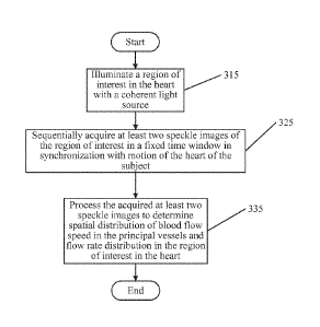
[0025] Figure 1 is a block diagram of a non-invasive system for
measuring blood flow
in principal vessels of a subject in accordance with some embodiments of the
present
inventive concept(s).
[0026] Figure 2A is a block diagram of a data processing system
according to
embodiments of the present inventive concept(s).
5b
CA 2824134 2018-02-15
CA 02824134 2013-07-08
WO 2012/096878 PCT/US2012/020626
[0027] Figure 2B is a more detailed block diagram of the data processing
system
illustrated in Figure 2 in accordance with some embodiments of the present
inventive
concept(s).
[0028] Figures 3 and 4 are flowcharts illustrating operations for measuring
blood flow in
principal vessels in accordance with various embodiments of the present
inventive concept(s).
[0029] Figure 5 is a digital photograph of a system for measuring flow of
blood phantom
used in an experiment performed in accordance with some embodiments of the
present
inventive concept(s).
[0030] Figures 6 through 9 are close up digital photographs of certain
elements in the
system illustrated in Figure 5 in accordance with some embodiments of the
present inventive
concept(s).
[0031] Figures 10 and 11 are digital photographs of an exemplary flow
generation system
used in some embodiments of the present inventive concept(s).
[0032] Figure 12 is a graph illustrating the change in height (cm) vs. flow
rate (ml/min) in
the flow generation system in accordance with some embodiments of the present
inventive
concept(s).
[0033] Figures 13A through 13D are digital images illustrating a "no flow"
case in
accordance with some embodiments of the present inventive concept(s).
[0034] Figures 14A through 14D are images illustrating a "flow 1" case in
accordance
with some embodiments of the present inventive concept(s).
[0035] Figures 15A through 15D are images illustrating a "flow 2" case in
accordance
with some embodiments of the present inventive concept(s).
[0036] Figures 16A through 16D are images illustrating a "flow 3" case in
accordance
with some embodiments of the present inventive concept(s).
[0037] Figures 17A through 17D are images (averaged over a number of frames)
illustrating a speckle image for each of four flow cases illustrated in
Figures 13 through 16 in
accordance with some embodiments of the present inventive concept(s).
[0038] Figures 18A through 18D are inverted speckle contrast images
illustrating each of
the four flow cases illustrated in Figures 13 through 16 in accordance with
some
embodiments of the present inventive concept(s).
[0039] Figures 19A through 19D are graphs illustrating a vertical profile
of inverted
speckle contrast images for each of the four flow cases in Figures 13 through
16 in
accordance with some embodiments of the present inventive concept(s).
6
CA 02824134 2013-07-08
WO 2012/096878 PCT/US2012/020626
[0040] Figure 20 is a graph illustrating predicted flow rate (ml/min) vs.
inverted speckle
image pixel intensity in accordance with some embodiments of the present
inventive
concept(s).
[0041] Figure 21 is a digital photograph of an exemplary system for
measuring flow of
blood phantom in accordance with some embodiments of the present inventive
concept(s).
[0042] Figures 22 through 27 are close up photographs of the elements in
the system
illustrated in Figure 21 in accordance with some embodiments of the present
inventive
concept(s).
[0043] Figure 28 is a graph illustrating flow rate (ml/min), flow speed
(cm/min) and
corresponding LAD flow rate vs. inverted speckle contrast image pixel
intensity in
accordance with some embodiments of the present inventive concept(s).
[0044] Figure 29 is a graph illustrating flow speed vs. inverted speckle
contrast image
pixel intensity in accordance with some embodiments of the present inventive
concept(s).
[0045] Figure 30 is a graph illustrating average flow speed determined from
speckle
contrast images vs. flow speed determined from the pump flow rate in
accordance with some
embodiments of the present inventive concept(s).
[0046] Figures 31A, 31C and 31D are images illustrating effect of specular
reflectance on
acquired speckle image data in accordance with some embodiments of the present
inventive
concept(s).
[0047] Figure 31B is a graph illustrating effect of specular reflectance on
acquired
speckle image data in accordance with some embodiments of the present
inventive
concept(s).
[0048] Figures 32A, 32C and 32D are images illustrating removal of specular
reflectance
of Figures 31A, 31C and 31D in accordance with some embodiments of the present
inventive
concept(s).
[0049] Figure 32B is a graph illustrating removal of specular reflectance
of Figures 31A,
31B and 31D in accordance with some embodiments of the present inventive
concept(s).
[0050] Figure 33 is a digital photograph of an exemplary system for
measuring blood
flow including a reservoir used in an experiment performed in accordance with
some
embodiments of the present inventive concept(s).
[0051] Figures 34A through 34D are digital images illustrating a "tube
clamped" situation
in accordance with some embodiments of the present inventive concept(s).
[0052] Figures 35A through 35D are images illustrating a "pump reading
100mL"
situation in accordance with some embodiments of the present inventive
concept(s).
7
CA 02824134 2013-07-08
WO 2012/096878 PCT/US2012/020626
[0053] Figures 36A through 36D are images illustrating a "pump reading
500mL"
situation in accordance with some embodiments of the present inventive
concept(s).
[0054] Figures 37A through 37D are images illustrating a "pump reading
1000mL"
situation in accordance with some embodiments of the present inventive
concept(s).
[0055] Figures 38A through 38D are averaged speckle images (averaged over a
number
of frames) for each of the four flow cases illustrated in Figures 33 through
37 in accordance
with some embodiments of the present inventive concept(s).
[0056] Figures 39A through 39D are colorized inverted speckle contrast
images
illustrating each of the four flow case illustrated in Figures 33 through 37
in accordance with
some embodiments of the present inventive concept(s).
[0057] Figures 40A through 40D are graphs illustrating a vertical line
profile of the
inverted speckle contrast images for each of the four flow cases in Figures
33through 37 in
accordance with some embodiments of the present inventive concept(s).
[0058] Figure 41 is a graph illustrating flow speed (cm/min) vs. inverted
speckle contrast
image pixel intensity in accordance with some embodiments of the present
inventive
concept(s).
[0059] Figure 42 is a graph illustrating flow speed (cm/min) vs. inverted
speckle contrast
image pixel intensity in accordance with some embodiments of the present
inventive concept.
[0060] Figures 43A through 43D are images illustrating a "pump reading OmL"
situation
in accordance with some embodiments of the present inventive concept(s).
[0061] Figures 44A through 44D are images illustrating a "clamped pump"
situation in
accordance with some embodiments of the present inventive concept(s).
[0062] Figure 45 is a diagram of a blood vessel having a narrowing in the
middle thereof
that can be accessed using methods and systems in accordance with some
embodiments of the
present inventive concept.
[0063] Figure 46 is a diagram illustrating velocity profiles of various
locations in the
blood vessel illustrated in Figure 45 obtained using flow hemodynamic modeling
in
accordance with some embodiments of the present inventive concept.
[0064] Figure 47 is a graph illustrating shear rate (which is related to
flow rate) and
horizontal coordinate (diameter) across the blood vessel illustrated in
Figures 45 and 46 when
the shear stress is about 1.0 second in accordance with some embodiments of
the present
inventive concept.
[0065] Figure 48 is a flowchart illustrating operations for measuring blood
flow in
principal vessels in accordance with various embodiments of the present
inventive concept(s).
8
CA 02824134 2013-07-08
WO 2012/096878 PCT/US2012/020626
DETAILED DESCRIPTION OF EMBODIMENTS
[0066] Embodiments of the present inventive concept will now be described more
fully
hereinafter with reference to the accompanying figures, in which preferred
embodiments of
the inventive concept are shown. This inventive concept may, however, be
embodied in
many different forms and should not be construed as limited to the embodiments
set forth
herein. Like numbers refer to like elements throughout. In the figures,
layers, regions,
elements or components may be exaggerated for clarity. Broken lines illustrate
optional
features or operations unless specified otherwise.
[0067] The terminology used herein is for the purpose of describing
particular
embodiments only and is not intended to be limiting of the inventive concept.
As used
herein, the singular forms "a", "an" and "the" are intended to include the
plural forms as well,
unless the context clearly indicates otherwise. It will be further understood
that the terms
"comprises" and/or "comprising," when used in this specification, specify the
presence of
stated features, integers, steps, operations, elements, and/or components, but
do not preclude
the presence or addition of one or more other features, integers, steps,
operations, elements,
components, and/or groups thereof. As used herein, the term "and/or" includes
any and all
combinations of one or more of the associated listed items. As used herein,
phrases such as
"between X and Y" and "between about X and Y" should be interpreted to include
X and Y.
As used herein, phrases such as "between about X and Y" mean "between about X
and about
Y." As used herein, phrases such as "from about X to Y" mean "from about X to
about Y."
[0068] Unless otherwise defined, all terms (including technical and
scientific terms) used
herein have the same meaning as commonly understood by one of ordinary skill
in the art to
which this inventive concept belongs. It will be further understood that
terms, such as those
defined in commonly used dictionaries, should be interpreted as having a
meaning that is
consistent with their meaning in the context of the specification and relevant
art and should
not be interpreted in an idealized or overly formal sense unless expressly so
defined herein.
Well-known functions or constructions may not be described in detail for
brevity and/or
clarity.
[0069] It will be understood that when an element is referred to as being
"on", "attached"
to, "connected" to, "coupled" with, "contacting", etc., another element, it
can be directly on,
attached to, connected to, coupled with or contacting the other element or
intervening
elements may also be present. In contrast, when an element is referred to as
being, for
example, "directly on", "directly attached" to, "directly connected" to,
"directly coupled" with
9
CA 02824134 2013-07-08
WO 2012/096878 PCT/US2012/020626
or "directly contacting" another element, there are no intervening elements
present. It will
also be appreciated by those of skill in the art that references to a
structure or feature that is
disposed "adjacent" another feature may have portions that overlap or underlie
the adjacent
feature.
[0070] It will be understood that, although the terms first, second, etc.
may be used herein
to describe various elements, components, regions, layers and/or sections,
these elements,
components, regions, layers and/or sections should not be limited by these
terms. These
terms are only used to distinguish one element, component, region, layer or
section from
another element, component, region, layer or section. Thus, a first element,
component,
region, layer or section discussed below could be termed a second element,
component,
region, layer or section without departing from the teachings of the inventive
concept. The
sequence of operations (or steps) is not limited to the order presented in the
claims or figures
unless specifically indicated otherwise.
[0071] Spatially relative terms, such as "under", "below", "lower", "over",
"upper" and
the like, may be used herein for ease of description to describe one element
or feature's
relationship to another element(s) or feature(s) as illustrated in the
figures. It will be
understood that the spatially relative terms are intended to encompass
different orientations of
the device in use or operation in addition to the orientation depicted in the
figures. For
example, if a device in the figures is inverted, elements described as "under"
or "beneath"
other elements or features would then be oriented "over" the other elements or
features.
Thus, the exemplary term "under" can encompass both an orientation of over and
under. The
device may be otherwise oriented (rotated 90 degrees or at other orientations)
and the
spatially relative descriptors used herein interpreted accordingly. Similarly,
the terms
"upwardly", "downwardly", "vertical", "horizontal" and the like are used
herein for the
purpose of explanation only unless specifically indicated otherwise.
[0072] As will be appreciated by one of skill in the art, embodiments of
the present
inventive concept may be embodied as a method, system, data processing system,
or
computer program product. Accordingly, the present inventive concept may take
the form of
an embodiment combining software and hardware aspects, all generally referred
to herein as
a "circuit" or "module." Furthermore, the present inventive concept may take
the form of a
computer program product on a non-transitory computer usable storage medium
having
computer-usable program code embodied in the medium. Any suitable computer
readable
medium may be utilized including hard disks, CD-ROMs, optical storage devices,
or other
electronic storage devices.
CA 02824134 2013-07-08
WO 2012/096878 PCT/US2012/020626
[0073] Computer program code for carrying out operations of the present
inventive
concept may be written in an object oriented programming language such as
Matlab,
Mathematica, Java, Smalltalk, C or C++. However, the computer program code for
carrying
out operations of the present inventive concept may also be written in
conventional
procedural programming languages, such as the "C" programming language or in a
visually
oriented programming environment, such as Visual Basic.
[0074] Certain of the program code may execute entirely on one or more of a
user's
computer, partly on the user's computer, as a stand-alone software package,
partly on the
user's computer and partly on a remote computer or entirely on the remote
computer. In the
latter scenario, the remote computer may be connected to the user's computer
through a local
area network (LAN) Or a wide area network (WAN), or the connection may be made
to an
external computer (for example, through the Internet using an Internet Service
Provider).
[0075] The inventive concept is described in part below with reference to
flowchart
illustrations and/or block diagrams of methods, devices, systems, computer
program products
and data and/or system architecture structures according to embodiments of the
inventive
concept. It will be understood that each block of the illustrations, and/or
combinations of
blocks, can be implemented by computer program instructions. These computer
program
instructions may be provided to a processor of a general-purpose computer,
special purpose
computer, or other programmable data processing apparatus to produce a
machine, such that
the instructions, which execute via the processor of the computer or other
programmable data
processing apparatus, create means for implementing the functions/acts
specified in the block
or blocks.
[0076] These computer program instructions may also be stored in a computer-
readable
memory or storage that can direct a computer or other programmable data
processing
apparatus to function in a particular manner, such that the instructions
stored in the computer-
readable memory or storage produce an article of manufacture including
instruction means
which implement the function/act specified in the block or blocks.
[0077] The computer program instructions may also be loaded onto a computer
or other
programmable data processing apparatus to cause a series of operational steps
to be
performed on the computer or other programmable apparatus to produce a
computer
implemented process such that the instructions which execute on the computer
or other
programmable apparatus provide steps for implementing the functions/acts
specified in the
block or blocks.
11
CA 02824134 2013-07-08
WO 2012/096878 PCT/US2012/020626
[0078] As discussed above, there is a need for effective non-invasive
methods for
determining blood flow distribution. It is believed that none of the existing
methods offer a
fully non-invasive cost effective solution to the problem of flow speed
determination.
Accordingly, some embodiments of the inventive concept provide methods,
systems and
computer program products for determining speed distribution of blood flow
without
requiring the use of dye injection, contrast agent or a contact probe. Some
embodiments of
the inventive concept use speckle imaging techniques to determine blood flow
distribution.
As used herein, a "speckle" image acquisition refers to the recording of
elastically scattered
light, i.e. the scattered light has a wavelength that is the same as the
incident light, from an
object illuminated by a coherent light such as the output from a coherent
light source. In
particular, the ''speckle" is actually a diffraction pattern, which is highly
correlated to the
morphology of the object being imaged. If certain parts of the object are in
translational
motion, i.e. blood stream flowing in a coronary artery, the corresponding part
or pixels of the
speckle image will vary with time in fashions different from those parts not
undergoing such
translational motion. This difference in the temporal variation of pixel
intensity in the
speckle image provides a mechanism to non-invasively measure flow speed (m/s
or cm/min)
in principal vessels. With knowledge of the diameters of the principal vessels
that can be
determined from the same set of acquired speckle images, the blood flow rate
(ml/min)
within FOV may be determined. The combined information of blood flow rates and
their
distribution in different vessels provide critical data for evaluating the
effectiveness of
Coronary artery bypass grafting (CABG) and other surgical procedures in
improving
patients' revascularization status and clinical prognosis. Speckle image
acquisition is
generally discussed, for example, in Velocity measurement of a diffuse object
by using a time-
varying speckle by Ohtsubo et al.
[0079] Thus, some embodiments of the present inventive concept provide a
non-invasive
technique for measuring blood flow that provides the ability to quantatively
measure blood
flow in principal vessels and perfusion distribution in areas perfuscd by one
or more of those
principal vessels as will be discussed further below with respect to Figures 1
through 44.
[0080] Furthermore, the data acquired from the obtained set of speckle
images can be
verified using flow hemodynamic modeling in accordance with some embodiments
of the
present inventive concept. In particular, using the Navier-Stokes equation,
which provides
the governing equation for fluid dynamics, a velocity field associated with
the FOV (in the
principal vessels) can be obtains. As used herein, "velocity field" refers to
a distribution of
fluid velocity in space and time. This velocity field may then be used to
calculate flow rate
12
CA 02824134 2013-07-08
WO 2012/096878 PCT/US2012/020626
as well as other quantities of interest, such as pressure. These quantities of
interest, for
example, flow rates, can then be compared with the experimental data
calculated using the
obtained set of speckle images. Thus, the hemodynamic modeling may be used to
validate
the experimental data as well as the success of the procedure as will be
discuss further below
with respect to Figures 45 through 48.
[0081] Referring first to Figure 1, a non-invasive system for measuring
blood flow in
principal vessels of a subject in accordance with some embodiments of the
present inventive
concept will be discussed. As discussed above, "non-invasive" refers to a
system or method
that does not require the subject to be injected with a dye, penetrated with
an object or
touched with an intrabody probe or probes. Thus, as used herein, the term non-
invasive
refers to a system or method that makes minimal contact with the subject. As
used herein,
"subject" refers to the person or thing being imaged. It will be understood
that although
embodiments of the present inventive concept are discussed herein with respect
to measuring
blood flow in principal vessels of the subject, embodiments of the present
inventive concept
are not limited to this configuration. The subject can be any subject,
including a veterinary,
cadaver study or human subject. As used herein, "perfusion" refers to blood
flow at the tissue
perfusion distribution level detected with speckle imaging.
[0082] As illustrated in Figure 1, the system 100 includes a communications
device 110,
a coherent light source unit 120, a camera 130, a synchronization module 170
and an EKG
device 180. Although the system of Figure 1 is depicted as only including
these elements, it
will be understood that other elements may also be present in the system
without departing
from the scope of the present inventive concept. For example, the systems
illustrated in the
photographs of Figures 5 and 21 include additional elements not present in the
system
illustrated in Figure 1.
[0083] Referring again to Figure 1, in some embodiments, the coherent light
source unit
120 may be a laser unit, which may include a laser 123 and a beam shaping lens
125. The
laser unit 120 may provide a coherent light source that illuminates a region
of interest 140.
The coherent light source provided by the laser unit 120 may have a wavelength
of from
about 600 nm to about 1100 nm. As used herein, the "region of interest" refers
to the region
of the subject that is being imaged, for example, the principal vessels and
tissue, organs, etc.
to determine blood flow therein. Although embodiments of the present inventive
concept are
discussed primarily herein with respect to blood flow distribution in the
principal vessels,
embodiments of the present inventive concept are not limited to this
configuration. For
13
CA 02824134 2013-07-08
WO 2012/096878 PCT/US2012/020626
example, blood flow in organs may be determined without departing from the
scope of the
present inventive concept.
[0084] The laser unit 120 may have light output at a fixed or variable
wavelength of from
about 600nm to about 1100nm without departing from the scope of the present
inventive
concept. The laser 120 can be configured to illuminate the region of interest
140 with a laser
beam 127 having substantially constant intensity within FOV of an imaging
unit. In some
embodiments, the constant or near constant intensity of the laser beam can
facilitate acquiring
speckle images with a high signal-to-noise (SNR) ratio. The laser 120 can be a
low power
continuous-wave laser. Thus, the subject does not need to wear any protective
apparatus, for
example, clothing or goggles, to shield the subject from potential adverse
effects of the laser.
In some embodiments, for example, the laser 120 may be of 633 nm in wavelength
and lmW
in power.
[0085] Use of a laser or other coherent light source having a wavelength of
from about
600 nm to about 1100 nm allows relatively deep penetration of light into
tissue and can
provide an accurate determination of blood flow speed in the principal vessels
and the
perfusion distribution as will be discussed further below.
[0086] In some embodiments, the laser unit 120 may be used to illuminate
the coronary
artery and be triggered by the electrocardiogram (EKG) provided by EKG device
180
through the synchronization module 170 and measurements can be taken from the
same point
outside the heart and the same point on the heart itself In other words, the
FOV is fixed by
two parameters, the point on the heart and the distance from the camera
outside the heart.
The FOV is kept the same so that the synchronization can be performed.
[0087] Referring again to Figure 1, the camera 130 communicates with the
laser unit 120
and the communications device 110. The camera 130 is configured to
sequentially acquire at
least two speckle images of the region of interest during a fixed time period.
The faster the
camera 130, the shorter the fixed time period has to be for acquiring the same
number of
speckle images. In some embodiments, the camera 130 may be a CCD camera, for
example,
a Lumenera Lm075 or similar devices.
[0088] As used herein, the fixed time period is typically short enough to
reduce or
possibly minimize motion effects, but long enough to obtain sufficient light
signals. Several
examples of this fixed timer period are discussed throughout the
specification, for example,
the fixed time period may be from about 1.0 to about 200 ms, or within a
single EKG cardiac
cycle. However, it will be understood that the fixed time period is not
limited to the specific
14
CA 02824134 2013-07-08
WO 2012/096878
PCT/US2012/020626
time periods discussed herein. For example, the fixed time period may be
greater than a
single EKG cardiac cycle without departing from embodiments discussed herein.
[0089] The camera 130 may be configured to acquire from about 50 to about
1000
speckle images during the fixed time period. In some embodiments, the camera
may only
need to acquire from about 50 to about 500 speckle images to provide a
meaningful result.
The fixed time period may be selected based on data associated with in situ
determined blood
flow speed. In some embodiments, the fixed time period is relatively short,
typically less
than 1 second, or from about 1.0 ms to about 200 ms.
[0090] The acquisition of the speckle images can be synchronized with the
motion of the
heart of the subject. For example, in some embodiments, acquisition of the
speckle images
may be synchronized with the EKG of the subject such that the motion of the
heart will have
minimal effect on determination of blood flow speed. Thus, the fixed time
period would be
located within a single EKG cardiac cycle.
[0091] Referring again to Figure 1, the communications device 110 is
configured to
process the at least two acquired speckle images based on temporal variation
of pixel
intensities among the acquired speckle images to determine spatial
distribution of blood flow
speed in the principal vessels and perfusion distribution in tissue in the
region of interest.
The at least two acquired speckle images can be electronically evaluated
and/or processed
using an image processing algorithm that combines temporal and spatial
calculations of the at
least two acquired speckle images. The at least two acquired speckle images
have a direct
relationship to the blood flow speed in the principal vessels and the
perfusion distribution.
[0092] In particular, some embodiments of the present inventive concept use
speckle
imaging techniques to yield blood flow speed in the principal vessels and
perfusion
distribution over the FOV. As used herein, FOV refers to the area of the
imaged object that
can be viewed by the imaging sensor. Due to the coherence among the scattered
light from
different parts of the illuminated region of the imaged object, the intensity
of the scattered
light arriving at a detecting element of an imaging sensor depends on the
relative spatial
relation among the different parts. The dependency leads to a "speckle"
appearance of the
acquired image since intensity of scattered light having an optical wavelength
of from about
200 am to about 2000 nm can vary quickly over a small spatial domain with a
size of about
cm. These concepts will be discussed further below.
[0093] Referring now to Figures 2A and 2B, a data processing system 200
that may be
used in the system 100 illustrated in Figure 1 in accordance with some
embodiments of the
inventive concept will be discussed. The data processing system 200 may be
included in the
CA 02824134 2013-07-08
WO 2012/096878 PCT/US2012/020626
communications device 110, the camera 130 or split between various elements of
the system
100 without departing from the scope of the present inventive concept. As
illustrated in
Figure 2, an exemplary embodiment of a data processing system 200 suitable for
use in the
system 100 of Figure 1 includes a user interface 244 such as a keyboard,
keypad, touchpad or
the like, I/O data ports 246 and a memory 236 that communicates with a
processor 238. The
I/O data ports 246 can be used to transfer information between the data
processing system
200 and another computer system or a network. These components may be
conventional
components, such as those used in many conventional data processing systems,
which may be
configured to operate as described herein.
[0094] Referring now to Figure 2B, a more detailed block diagram of the
data processing
system 200 in accordance with some embodiments of the present inventive
concept will be
discussed. The processor 238 communicates with a display 345 via and
address/data bus 347,
the memory 236 via an address/data bus 348 and the I/O data ports 246 via an
address/date
bus 349. The processor 238 can be any commercially available or custom
microprocessor or
ASICs. The memory 236 is representative of the overall hierarchy of memory
devices
containing the software and data used to implement the functionality of the
data processing
system 200. The memory 236 can include, but is not limited to, the following
types of
devices: cache, ROM, PROM, EPROM, EEPROM, flash memory, SRAM, and DRAM.
[0095] As shown in Figure 2B, the memory 236 may include several categories
of
software and data used in the data processing system 200: an operating system
352;
application programs 354; input/output (I/O) device drivers 358; and data 356.
As will be
appreciated by those of skill in the art, the operating system 352 may be any
operating system
suitable for use with a data processing system, such as OS/2, AIX or zOS from
International
Business Machines Corporation, Armonk, NY, Windows95, Windows98, Windows2000,
WindowsXP, or Vista from Microsoft Corporation, Redmond, WA, Unix, Linux, Lab
View,
or a real-time operating system such as QNX or VxWorks, or the like. The I/O
device drivers
358 typically include software routines accessed through the operating system
352 by the
application programs 354 to communicate with devices such as the I/O data
port(s) 246 and
certain memory 236 components. The application programs 354 are illustrative
of the
programs that implement the various features of the data processing system 200
included a
system in accordance with some embodiments of the present inventive concept
and
preferably include at least one application that supports operations according
to some
embodiments of the present inventive concept. Finally, the data 356 represents
the static and
16
CA 02824134 2013-07-08
WO 2012/096878 PCT/US2012/020626
dynamic data used by the application programs 354, the operating system 352,
the I/O device
drivers 358, and other software programs that may reside in the memory 236.
[0096] As illustrated in Figure 2B, the data 356 according to some
embodiments of the
present inventive concept may include acquired speckle images 360,
intermediate data 361,
calculated blood flow rates 363 and modeling data 364. Although the data 356
illustrated in
Figure 2B includes three different files 360, 361, 363 and 364, embodiments of
the present
inventive concept are not limited to this configuration. Two or more files may
be combined
to make a single file; a single file may be split into two or more files and
the like without
departing from the scope of the present inventive concept.
[0097] As further illustrated in Figure 2B, the application programs 354
may include a
light source trigger module 351, an image capture module 352, a processing
module 353 and
a modeling module 354 in accordance with some embodiments of the inventive
concept.
While the present inventive concept is illustrated, for example, with
reference to the light
source trigger module 351, the image capture module 352, the processing module
353 and the
modeling module 354 being application programs in Figure 2B, as will be
appreciated by
those of skill in the art, other configurations may also be utilized while
still benefiting from
the teachings of the present inventive concept. For example, the light source
trigger module
351, the image capture module 352 the processing module 353 and the modeling
module 354
may also be incorporated into the operating system 352 or other such logical
division of the
data processing system 300. Thus, the present inventive concept should not be
construed as
limited to the configuration of Figure 2B, but is intended to encompass any
configuration
capable of carrying out the operations described herein.
[0098] Furthermore, while the light source trigger module 351, the image
capture module
352 the processing module 353 and the modeling module 354 are illustrated in a
single data
processing system, as will be appreciated by those of skill in the art, such
functionality may
be distributed across one or more data processing systems. Thus, the present
inventive
concept should not be construed as limited to the configuration illustrated in
Figures 2A and
2B, but may be provided by other arrangements and/or divisions of function
between data
processing systems.
[0099] In particular, the light source trigger module 351 may be configured
to illuminate
a region of interest with a coherent light source. The coherent light source
may have a
wavelength of from about 600 nm to about 1100 nm as discussed above. The image
capture
module 352 may be configured to sequentially acquire at least two speckle
images of the
region of interest during a fixed time period. The processing module 353 may
be configured
17
CA 02824134 2013-07-08
WO 2012/096878 PCT/US2012/020626
to process the at least two acquired speckle images based on a diffraction
pattern of each the
at least two speckle images to determine spatial distribution of blood flow
speed in the
principal vessels and perfusion distribution in tissue in the region of
interest.
[00100] The modeling module 354 may be configured to calculate a velocity
field for the
region of interest; calculate blood flow speed in the region of interest based
on the calculated
velocity field; and compare the calculated blood flow in the region of
interest to the blood
flow speed determined using the acquired at least two speckle images of the
region of interest
to verify results obtained using the at least two speckle images. In some
embodiments, the
modeling module 354 is configured to calculate the velocity field using
Equations 9 and 10
set out below.
[00101] Thus, blood flow speed as well as other quantities may be
calculated using both
the speckle method and the velocity field method before a procedure is
performed on a
subject and after a procedure is performed on the subject to verify that the
procedure was
successful. By comparing the measurements/quantities before and after the
procedure, the
success of the procedure may be determined, which will be discussed further
below.
[00102] Referring now to the flowcharts of Figures 3 and 4, operations of a
non-invasive
method for measuring blood flow in principal vessels of a subject will be
discussed. As
illustrated in Figure 3, operations begin at block 315 by illuminating a
region of interest in
the heart with a coherent light source. The coherent light source may have a
wavelength of
from about 600 nm to about 1100 nm. Providing a coherent light source with a
wavelength
of from about 600 mu to about 1100 nm may allow for non-invasive, deep
penetration of
light into tissues and provides an accurate determination of blood flow speed
in the principal
vessels and the perfusion distribution within the layer of light penetration.
[00103] In some embodiments, the coherent light source may be provided by a
laser
configured to illuminate the region of interest. The laser may have a fixed or
variable
wavelength. The laser may produce a beam having substantially constant
intensity within a
FOV of an imaging unit. The laser may be a low energy and continuous-wave
laser such that
the subject does not require any protective apparatus to shield the subject
from effects of the
laser.
[00104] Referring again to Figure 3, operations continue at block 325 by
sequentially
acquiring at least two speckle images of the region of interest during a fixed
time period. The
fixed time period may be selected based on data associated with in situ
determined blood
flow speed. In some embodiments, the at least two speckle images may be
acquired in
synchronization with motion of a heart of the subject such that the motion of
the heart will
18
CA 02824134 2013-07-08
WO 2012/096878 PCT/US2012/020626
have minimal effect on determination of blood flow speed using the acquired at
least two
speckle images. For example, the fixed time period can correspond to a single
EKG cardiac
cycle or defined portion thereof cycle.
[00105] The camera may be configured to acquire the at least two speckle
images during
the fixed time period. In some embodiments, from about 50 to about 1000
speckle images
may be acquired using the camera during the fixed time period of from about 1
ms to about
200 ms. In some embodiments, about 200 to about 500 speckle images may be
acquired.
Higher numbers of speckle images typically allow better signal-to-noise ratios
in the
calculated LSCI image but take longer time to acquire.
[00106] Referring again to Figure 3, operations continue at block 335 by
electronically
processing the acquired speckle images based on the temporal variation of the
pixel
intensities in the acquired speckle images to generate a laser speckle
contrast imaging (LSCI)
image and determine spatial distribution of blood flow speed in the principal
vessels and
perfusion distribution in tissue in the region of interest from the LSCI
image.
[00107] In some embodiments, electronically evaluating speckle image data
may include
electronically evaluating the acquired speckle images using an image
processing algorithm
that combines temporal and spatial calculations of the acquired speckle images
to generate a
LSCI image and determine spatial distribution of blood flow speed. The at
least two speckle
images may have a direct relationship to the blood flow speed in the principal
vessels and the
perfusion distribution which are utilized in generating an LSCI image for
determination of
the spatial distribution of blood flow speed. For example, following equations
can be used to
obtain the intensity at each pixel of the LSCI image K (i, j) from the
acquired speckle image
set {In} with n=1,2,.. .,N, i.e.,
K(i, j) = cr(i, l)
Equation (1)
P(I, ./)
where
1
ii(j, I) = Jõ (I, Equation (2)
I 11=1
1 N
a(i, j) E (/ (i,i)-P(i,i))2 Equation (3)
N n
In the above calculations, In(i, j) refers to the pixel at (i, j) location in
a speckle image
acquired at nth time point and N (> 1) is the total number of acquired speckle
images.
19
CA 02824134 2013-07-08
WO 2012/096878 PCT/US2012/020626
[00108] It will be understood that the operations of blocks 315, 325 and
335 may be
performed before and after a procedure performed on the subject. The results
before and
after the procedure may be compared to verify the success of the procedure in
the subject.
[00109] Referring now to Figure 4, operations for a non-invasive method for
measuring
blood flow in principal vessels of a subject in accordance with some
embodiments will be
discussed. Operations begin at block 415 by illuminating a region of interest
in the heart with
a coherent light source, wherein the coherent light source has a wavelength of
from about 600
nm to about 1100 nm. At least two speckle images of the region of interest are
sequentially
acquired during a fixed time period (block 425) in synchronization with the
motion of the
heart. Temporal and spatial variation of pixel intensities of the at least two
acquired speckle
images are electrically evaluated to determine spatial distribution of blood
flow speed in the
principal vessels and perfusion distribution in tissue in the region of
interest of the
heart(block 435).
[00110] A velocity field for the region of interest in the heart is
calculated (block 445). In
some embodiments, the velocity field is calculated using equations (9) and
(10) set out below.
Blood flow speed in the region of interest of the heart based on the
calculated velocity field is
calculated (block 455). The calculated blood flow speed in the region of
interest in the heart
is compared to the blood flow speed determined using the acquired at least two
speckle
images of the region of interest to verify results obtained using the at least
two speckle
images (block 465). Thus, embodiments of the present inventive concept may be
used to
verify experimental results as will be discussed further below.
[00111] It will be understood that the operations of blocks 415, 425, 435,
445, 455 and 465
may be performed before and after a procedure performed on the subject. The
results before
and after the procedure may be compared to verify the success of the procedure
in the
subject.
[00112] The following non-limiting examples are provided by way of example.
EXAMPLES
[00113] Referring to Figure 5, a digital photograph of a prototype system
500 to detect
flow speed using laser speckle contrast imaging (LSCI) technology in
accordance with some
embodiments of the present inventive concept will be discussed. As illustrated
in Figure 5,
the system includes a communication device 510, such as a laptop computer, a
laser unit 520
including a laser generator and a focusing lens, a camera 530, a flow
generator 580, flow
CA 02824134 2013-07-08
WO 2012/096878 PCT/US2012/020626
liquid 590 and a flow target 585. Table 1 set out below summarizes the actual
equipment/devices used in this experiment.
Devices used in Experiment 1 Notes
CCD camera (530) Lumenera Lm075
--
Laser (520) 633nm in wavelength, lmW in power
Liquid used in flow (590) 20% Intralipid 1 to 4 ratio mixed with
water plus fruit color
Computer/Communications Device (510) Laptop PC
TABLE 1
[00114] As discussed above, the faster the camera 530, the smaller the
fixed time period
has to be to obtain an adequate number of speckle images to provide a
meaningful result.
Thus, the limitation of the frame rate of the camera 530 in the prototype
system may have
impacted the final result of this experiment. The laser 520 is a low power
continuous-wave
laser providing a single-wavelength coherent light source. Thus, the subject
of the imaging
does not typically require any protection from such a laser, such as
protective clothing or
eyewear. The laser 520 produces a beam having a wavelength of from about 600nm
to about
1100nm in some embodiments. During the experiment, the laser beam produced by
the laser
520 is used to illuminate the region of interest with substantially constant
intensity with the
FOV of the imaging unit. This is an important aspect of the experiment because
it allows the
resulting images to have a high SNR.
[00115] Colored intralipid was used as the flow liquid 590 during the
experiment due to
the fact that a light scattering characteristics of the colored intralipid is
similar to those of
mammalian blood. Thus, the colored intralipid mimics the blood flowing in the
human body.
The communications device 510 used was a laptop computer, although embodiments
of the
present inventive concept are not limited to the use of a laptop computer. The
acquired
speckle images are provided to the communications device 510 and are used to
calculate
blood flow in according with some embodiments of the present inventive
concept. As
discussed above, the data is calculated using an image processing algorithm
that combines
temporal and spatial calculations of the acquired speckle images. Thus,
spatial distribution of
blood flow speed in principal vessels and perfusion distribution can be
determined.
[00116] Referring now to Figures 6 through 9, close up photographs of the
devices
illustrated in Figure 5 used during the first experiment are provided. In
particular, Figure 6 is
a close up photograph of the laser unit 520; Figure 7 is a close up photograph
of the camera
21
CA 02824134 2013-07-08
WO 2012/096878
PCT/US2012/020626
530; Figure 8 is a close up photograph of the communications device 510; and
Figure 9 is a
close of the flow liquid 590.
[00117] Table 2
set out below summarizes parameters for the camera 530 used during the
first experiment. The parameters are for the camera 530 while the image
sequence is
acquired.
Length of image sequence ¨ 1 second
Frame rate ¨95 frames/second
Image resolution 320*240 pixels
Exposure time per frame 3 ms
Gain 1
TABLE 2
[00118] The upper
limit (V1i1it) of flow that can be detected based on the setup of the first
experiment can be summarized by the following equation:
AL
Vtinzit = Equation (4)
In Equation 1, AL is the diameter of the tube, which was 0.26cm in the first
experiment;
0.4
r = 0.4T = ¨
f is the estimated exposure time, 3.0 ms in the first experiment; and f is the
frame rate. Thus, in the first experiment Vumit can be roughly estimated at
about 9.0
cm/second.
[00119] Referring
now to Figures 10 and 11, photographs of the flow generation system
used in the first experiment will be discussed. Figure 10 illustrates the flow
generation
system 580. The higher the bottle in the flow generation system 580, the
faster the flow of
the flow liquid 590. Figure 11 is a close up photograph of the tube target 585
used as the
flow target for the first experiment. Table 3 below summarizes the
relationship between the
change in height of the bottle and the flow of liquid in the flow generation
system in
accordance with the first experiment. The change in height is measured from
the height of
the bottle to the end of the tube.
22
CA 02824134 2013-07-08
WO 2012/096878 PCT/US2012/020626
Time delta height flow rate flow rate
predicted flow
Volume (cm 3)
(secs) (cm) (cm /sec) (ml/min) rate
(ml/min)
68 100 108 1.5 88.2 90.6
73 100 98 1.4 82.2 83,0
80 100 88 1.3 75.0 75.4
90 100 78 1.1 66.7 67.6
103 100 68 1.0 58.3 59.8
120 100 58 0.8 50.0 51.8
136 100 48 0,7 44.1 43,7
173 100 38 0,6 34.7 35.4
TABLE 3
[00120] Thus, as illustrated by the delta height (cm) and flow columns of
Table 3, the
higher the bottle, the faster the flow rate of the colored intralipid liquid.
[00121] The estimated relationship between delta height (cm) and flow rate
(ml/min) is
represented by Equation (2) set out below:
flow rate = 134 X 'I/TNT Equation (5);
Where Ah is the change in height of the bottle relative to the vertical
position of the tube end.
Figure 12 summarizes the data in Table 3 and is a graph illustrating delta
height (cm) vs. flow
rate (ml/min).
[00122] Four cases of flow rate were measured by the LSCI method in the
first experiment
setup, no flow, flow 1, flow 2 and flow 3. The details of each of the flow
rates are
summarized in Table 4 set out below. The first experiment was repeated three
times for each
case to ensure accuracy and repeatability. Figures 13A through 13D illustrate
resultant
images obtained for the ''no flow" state. Figure 13A is an averaged image
obtained by
averaging in a pixel-to-pixel fashion 97 frames for the "no flow" case; Figure
13B is a
vertical line profile image in the middle of inverted speckle contrast image
for the "no flow"
case; Figure 13C is an inverted speckle contrast image for the "no flow" case;
and Figure 13D
is a colorized inverted speckle contrast image for the "no flow" case.
[00123] Figures 14A
through 14D illustrate resultant images obtained for the "flow 1"
case. Figure 14A is an averaged image obtained by averaging in a pixel-to-
pixel fashion 97
frames for the "flow 1" case; Figure 14B is a vertical line profile image in
the middle of the
inverted speckle contrast image for the "flow 1" case ; Figure14C is an
inverted speckle
contrast image for the "flow 1" case ; and Figure 14D is a colorized inverted
speckle contrast
image for the "flow 1" case .
23
CA 02824134 2013-07-08
WO 2012/096878 PCT/US2012/020626
[00124] Figures 15A through15D illustrate resultant images obtained for the
"flow 2" case.
Figure 15A is an averaged image obtained by averaging in a pixel-to-pixel
fashion 89 frames
for the "flow 2" case; Figure 15B is a vertical line profile image in the
middle of an inverted
speckle contrast image for the "flow 2" case ; Figure 15C is an inverted
speckle contrast
image for the "flow 2" case ; and Figure 15D is a colorized inverted speckle
contrast image
for the "flow 2" case .
[00125] Figures 16A through 16D illustrate resultant images obtained for
the "flow 3"
case. Figure 16A is an averaged image obtained by averaging in a pixel-to-
pixel fashion 89
frames for the "flow 3" case; Figure 16B is a vertical line profile image in
the middle of an
inverted speckle contrast image for the "flow 3" case ; Figure 16C is an
inverted speckle
contrast image for the "flow 3" case ; and Figure 16D is a colorized inverted
speckle contrast
image for the "flow 3" case.
[00126] Figures 17A through 17D are the averaged images for each of the
cases, "no
flow", "flow 1", "flow 2", and "flow 3," respectively. Figures 18A through 18D
are colorized
inverted speckle contrast images for each of the cases , "no flow", "flow 1",
"flow 2", and
"flow 3," respectively. Figures 19A through 19D are vertical line profile
images for each of
the cases, "no flow", "flow 1", "flow 2", and "flow 3," respectively.
[00127] Table 4 set out below, summarizes the data for all four flow cases.
In particular,
the relationship between delta height and predicted flow is readily apparent.
Figure 20 is a
graph illustrating predicted flow rate (ml/min) vs. inverted speckle contrast
image pixel
intensity as set out in Table 4.
delta inverted speckle
predicted flow
Status height contrast image
rate (ml/min)
(cm) pixel intensity
No flow 0 0 15
Flow 1 10 10.6 67
Flow 2 100 84.5 88
Flow 3 193 150.7 108
TABLE 4
[00128] To summarize the first experiment, the laser speckle contrast
imaging setup is
clearly able to differentiate between a no flow state and the three flow speed
cases .
However, the sensitivity and precision were not ideal and the point of "no
flow" was not
consistent with the other three flow points as illustrated in Figure 20. Some
of the
imprecision may be due to using a bottle for the flow speed generation method.
This may
24
CA 02824134 2013-07-08
WO 2012/096878 PCT/US2012/020626
have caused variation and lack of constant flow. Furthermore, frame rate of
the camera may
have limited to number of speckle images that could be obtained. The laser
beam intensity
used during the experiment was uneven in the FOV, i.e. there are dark spots in
the FOV.
[00129] However, even given this imprecision, the results shown that the
higher the frame
rate the better the LSCI image quality and it was discovered that the exposure
time should be
as long as it can in the condition that the same frame rate can be achieved.
[00130] Referring first to Figure 21, a photograph of the system 2100 for a
second
experiment to detect flow speed using the LSCI technology in accordance with
some
embodiments of the present inventive concept will be discussed. As illustrated
in Figure 21,
the system includes a communications device 2110, such as a laptop computer, a
laser unit
2120 including a laser generator and a beam shaping lens, a camera 2130, a
flow generator
2181 provided by a biomedical pump, flow liquid 2190, a flow target 2185 and
an
electromagnetic flow detector 2191. Table 5 set out below summarizes the
actual
equipment/devices used in this experiment.
Devices used in Experiment 2 Notes
CCD camera (2130) Lumenera Lm075
Laser (2120) 633nm in wavelength, lmW in power
Liquid used in flow (2190) 20% Intralipid
Saline water 0.9%
Biomedical pump (2181) With electromagnetic flow detector (2191)
Communications Device/Computer(2110) Laptop PC
TABLE 5
[00131] As discussed above, the faster the camera 2130, the smaller the
fixed time period
has to be to obtain an adequate number of speckle images to provide a
meaningful result.
Thus, the limitation of the frame rate on the camera 2130 may have impacted
the final result
of this experiment. The laser 2120 is a low power laser providing a single
coherent light
source. Thus, the subject of the imaging does not typically require any
protection from the
laser, such as protective clothing or eyewear. The laser 2120 produces a beam
having a
wavelength of from about 600nm to about 1100nm in some embodiments. During the
experiment, the laser beam produced by the laser 2120 is used to illuminate
the region of
interest with substantially constant intensity with the FOV of the imaging
unit. This may
allow the speckle contrast images to have a high signal-to-noise ratio.
[00132] Colored intralipid was used as the flow liquid 2190 during the
experiment due to
the fact that light scattering characteristics of intralipid are similar to
those of mammalian
blood. Thus, the colored intralipid will mimic the blood flowing in the human
body. The
CA 02824134 2013-07-08
WO 2012/096878 PCT/US2012/020626
communications device 2110 used was a laptop computer, although embodiments of
the
present inventive concept are not limited to the use of a laptop computer. The
acquired
speckle images are provided to the communications device 2110 and are used to
calculate
blood flow in according with some embodiments of the present inventive
concept. As
discussed above, the data are calculated using an image processing algorithm
that combines
temporal and spatial calculations of the acquired speckle images. Thus,
spatial distribution of
blood flow speed in principal vessels and perfusion distribution can be
determined.
[00133] Referring now to Figures 22 through 27, close up photographs of the
devices
illustrated in Figure 21 used during the second experiment arc provided. In
particular, Figure
22 is a close up photograph of the laser unit 2120; Figure 23 is a close up
photograph of the
camera 2130; Figure 24 is a close up photograph of the communications device
2110; Figure
25 is a close up photograph of the biomedical pump 2181; Figure 26 is a close
up photograph
of the electromagnetic flow detector 2191; and Figure 27 is a close up
photograph of the flow
target 2185.
[00134] Table 6 set out below summarizes parameters for the camera 530 used
during the
second experiment. The parameters are for the camera 2130 while the image
sequence is
acquired.
Length of image sequence ¨1 second
Frame rate ¨95 frames/second
Image resolution 320*240
Exposure time per frame 3 ms (split into 3 part to calculate std
and
average every 3 continuous frames)
Gain 1
Working distance ¨1.5 m
Aperture 2-4
Length of the video loop 3*1 second
TABLE 6
[00135] Estimation of the blood flow rate and speed in accordance with the
second
experiment will be discussed. Since the LSCI technology measures the flow
speed (cnYmin)
rather than flow rate (ml/min), the range of the flow speed of blood in the
main branches of
coronary artery can be established. The second experiment focused on this
range. It is
contemplated that the flow rate can be calculated using the flow speed and a
cross sectional
area of the vessel at the same location. Table 7 set out below illustrates the
estimated blood
flow rate and speed range in LAD.
26
CA 02824134 2013-07-08
WO 2012/096878
PCT/US2012/020626
Average diameter flow rate range flow speed range
of LAD (cm) (ml/min, cm3/min) (cm/min)
0.4 0-100 0-796
TABLE 7
[00136] Procedures used for in accordance with the second experiment will
now be
discussed. The biomedical pump was calibrated by comparing measured liquid
volume
during a certain time with the reading from the electromagnetic flow detector
2591 as
illustrated in Table 8 set out below. In particular, pump calibration using
20% Intralipid
solution mixed by saline water with 1:4 ratio (volume was measured from 100mL
to 300mL
and reading from the detector was recorded when liquid reaches 200mL).
flow rate
flow rate
reading Calculated flow
Time Time Volume reading Percentage
from flow rate speed
(s) (min) (mL) from pump Error
L/min pump (ml/min) (cm/min)
)
(
(mUmin) _
15 0.25 200 0.78 780 800 3% 1095
18 0.30 200 0.65 650 667 3% 913
22 0.37 200 0.55 550 545 1% 772
:
26 0.43 200 0.45 450 462 3% 632
30 0.50 200 0.39 390 400 3% 548
49 0.82 200 0.24 240 245 2% 337
62 1.03 200 0.18 180 194 7% 253
111 1.85 200 0.11 110 108 2% 154
TABLE 8
[00137] Three Intralipid solutions with different concentrations were
measured to simulate
light scattering characteristics of blood as will be discussed below and to
examine the
stability of the LSCI technology. The three intralipid concentrations are
summarized below
in Table 9. Table 10 summarizes the volume and linear flow rate range measured
during the
second experiment.
27
CA 02824134 2013-07-08
WO 2012/096878
PCT/US2012/020626
Ratio of 20%
Intralipid to Intralipid
saline water concentration repeat Note
Tube surface is processed by sand
paper and collimated reflectance
lights were greatly reduced. Aperture
1:39 0.5% 3 of the
camera was large. -2.5
Tube surface is processed by sand
paper and collimated reflectance
lights were greatly reduced. Aperture
1:19 1% 3 of the
camera was medium. -3
1:9 2% 3 Same as
above
TABLE 9
flow rate reading Corresponding flow Estimated maximum
in 3/8 inch Calculated rate in 4 mm flow rate in
diameter tube flow speed diameter LAD
coronary artery
0-1000 mL/min 0-1404 cm/min 0-176 mL/min 100
mL/min
TABLE 10
[00138] Results of the
second experiment will now be discussed. Table 11 set out below
summarizes the results for (1): 0.5% Intralipid solution (1:39); (2): 1%
Intralipid solution
(1:19); (3) 2% Intralipid solution (1:9) in accordance with the second
experiment.
flow rate
reading flow Corresponding
Std of L Std of L Std
of L
from speed LAD flow rate L (1) L (2) L (3)
(1) (2) (3)
pump (cm/min) (mL/min)
(mL/min)
0 0 0 56 0.6 51 0.5 44 0.1
100 140 18 88 4.7 88 1.0 75 2.0
200 281 35 97 4.6 94 2.0 92 1.0
300 421 53 105 4.3 110 2.1 104 1.1
400 562 71 111 5.5 112 0.9 109 0.3
500 702 88 113 2.9 117 2.8 117 1.4
600 842 106 121 4.0 118 2.2 121 2.8
700 983 123 120 1.0 113 3.4 127 0.3
800 1123 141 115 5.6 109 3.1 128 0.7
900 1264 159 113 3.8 113 2.1 123 5.7
1000 1404 176 112 3.8 106 6.2 124 3.8
TABLE 11 (Std is standard deviation, L is the inversed speckle contrast
see Equation 6 for details)
28
CA 02824134 2013-07-08
WO 2012/096878
PCT/US2012/020626
[00139] Referring now to Figure 28, flow speed and corresponding LAD flow
rate vs. the
inverted speckle contrast image pixel intensity 1/K ; Results obtained with
ratio of 20%
Intralipid to saline water of (1) 1:39; (2) 1:19; (3) 1:9 will be discussed.
As illustrated in
Figure 28, the more diluted Intralipid solution has a larger background noise
while flow
reading is zero; after the flow speed exceeds 800 cm/min, the 1/K begins to
saturate and has
become less sensitive to the flow speed change; and compared to the huge
concentration rage
of the Intralipid solution, there is only very small change in the curves.
[00140] The quantitative relation between laser speckle contrast image
pixel intensity and
the flow speed will now be discussed. To make a positive correlation between
the flow
speed and the calculated image pixel intensity, the following equation is used
to construct the
inverted speckle contrast image L :
L(i ) 1
P(i, i) Equation (6)
, j =
K(, .1)
[00141] The quantitative relationship between laser speckle contrast image
pixel K(i,j) and
the flow speed have been derived as the following
K(i, j) cc Vr,(1, T Equation (7)
where T is the camera integration time, Te is a correlation time of scattering
particles
undergoing motion of speed v and given by r, = _____________________ , X, is
laser light wavelength. Based on
2;z-v
the above relation, the inverted speckle image pixel intensity can be written
as
L(i, j) = Lo+ j) Equation (8)
where Lo is an added term to account for background noise and should be zero
after the
baseline has been removed; a is a constant related to the imaging parameters,
laser
parameters, time/spatial smoothing parameters for obtaining K and the
components of the
liquid;. Thus, after removing the baseline from the data of "zero flow" case
from each single
measurements in case (1)¨(3), the Equation (8) can be used to fit the measured
data.
[00142] Figure 29 is a graph illustrating flow speed vs. the L with curve
fitting based on
Equation (8). Table 12 summarizes the results of curve fitting , with R as the
value of
confidence of fitting (R=1 means perfect fitting).
29
CA 02824134 2013-07-08
WO 2012/096878
PCT/US2012/020626
Case (1) case (2) case (3)
Lo 0 0 0
a 2.3149 2.5413 2.7606
0.9906 0.9797 0.9974
TABLE 12
[00143] Based on the prediction model (curve fitting), flow speed in
measurements (1)¨(3)
were obtained to compare with the flow speed calculated from the pump
readings. The
results are depicted in Figure 30, a graph illustrating the average of
calculated flow speed
(cm/min) from L in case (1)¨(3) vs. the flow speed determined from the pump
readings.
[00144] To summarize the second experiment, the experiment showed that the
inverted
speckle image pixel intensity L acquired with the current imaging system can
correlate to the
speed of the flow within certain range, (flow speed < 800 cm/min or LAD flow
rate< 100
mL/min), which corresponds to the maximum blood flow rate in average main
branch of
coronary artery. Compared to the huge concentration range of the Intralipid
solution, there is
only very small change in the curves. The light scattering parameters of blood
might fall in
between case (1) and (3) as illustrated in Table 13 set out below, where La is
absorption
coefficient, fls is scattering coefficient, g is anisotropy factor and us' =
(I -g)us is the reduced
scattering coefficient.
oxygenated
2,=633 nm blood 10% Intralipid
ua (1/mm) 0.2 0.0003
us (1/mm) 31 34.75
0.99 0.83
usi (1/mm) 0.31 5.98
TABLE 13
[00145] When bubbles are trapped in the biomedical pump, they may affect
the accuracy
of the flow reading from electromagnetic flow meter and might affect results
of
measurement. Specular reflectance from the smooth surface of the tube will
cause saturation
of the camera and overwhelm the speckle information as shown in Figures 31A
through 31D.
In the second experiment, sand paper was used to create rough surface on the
tube to
eliminate this influence, the results of which are illustrated in Figures 32A
through 32D, i.e.
specular reflectance is removed by roughening the surface of the tube. The
speed detection
limit of L depends on the frame rate and the number of frames used to generate
the speckle
CA 02824134 2013-07-08
WO 2012/096878 PCT/US2012/020626
contrast image somehow is related to the signal-to-noise ratio of L. Smoothing
of the original
images with a moving window of 3 to 5 frames helps increase the signal-to-
noise ratio.
[00146] Further experiments performed in accordance with some embodiments of
the
present inventive concept will now be discussed with respect to Figures 33
through 43.
Figure 33 is a digital photograph of a prototype system setup used to detect
blood flow speed
using the LSCI technology in accordance with some embodiments of the present
inventive
concept. The experiment discussed with respect to Figures 33 through 43 is
similar to the
experiment discussed above with respect to Figures 21 through 32, but an extra
reservoir
3397 is present to add red blood cells into the circulation from a blood bag
3398 (expired and
for research use only), which is acquired from the Red Cross. Thus, many of
the details of
the experiment will not be repeated herein.
[00147] Table 14 set out below summarizes the actual equipment/devices used
in this
experiment.
Device Parameters
CCD camera Lumenera Lm075
Laser 633nmin wavelength, lmW in power
Liquid used in Blood (1 unit of red blood cells with hematocrit 70% +
1000mL saline
flow water)
Saline water 0.9%
Biomedical pump With electromagnetic flow rate detector
Computer Laptop PC
TABLE 14
[00148] Table 15 set out below summarizes parameters for the camera used
during the
second experiment. The parameters are for the camera while the image sequence
is acquired.
Length of image sequence 1 second
Frame rate ¨95 frames/second
Image resolution 320*240
Exposure time per frame 3 ms
Gain 1
working distance 1.5-4 m
Aperture 2
Length of the video loop 3*1 second (split into 3 part to calculate std and
average
every 3 continuous frames)
TABLE 15
[00149] Four flow cases were measured by LSCI using the setup in Figure 33
and each
situation was performed three times to test the repeatability. The flow cases
tested are "tube
is clamped", "pump is reading 100mL", "pump is reading 500mL" and "pump is
reading
31
CA 02824134 2013-07-08
WO 2012/096878 PCT/US2012/020626
1000mL." Table 16 set out below summarized the blood flows measured and the
results for
each situation.
flow rate reading Corresponding std of
flow speed
from pump LAD flow rate
(cm/min) (blood)
(mL/min) (mL/min) (blood)
Tube clamped 35 2
0 0 0 84 1
100 140 18 144 2
200 281 35 153 4
300 421 53 156 6
400 562 71 164 6
500 702 88 168 4
600 842 106 174 3
700 983 123 179 3
800 1123 141 180 10
900 1264 159 188 1
1000 1404 176 191 4
TABLE 16 (Std is standard deviation, L is the inversed speckle contrast
see Equation 6 for details)
[00150] Figures 34A through 34D illustrate resultant images obtained for
the "tube is
clamped" case. Figure 34A is an averaged image of 90 frames for the "tube is
clamped" case;
Figure 34B is a vertical line profile image in the middle of inverted speckle
contrast image
for the "tube is clamped" case; Figure 34C is an inverted speckle contrast
image for the "tube
is clamped" case; and Figure 34D is a false-color inverted speckle contrast
image for the
"tube is clamped" case.
[00151] Figures 35A through 35D illustrate resultant images obtained for
the "pump reads
100mL" case. Figure 35A is an averaged image of 97 frames for the "100mL"
case; Figure
35B is a vertical line profile in the middle of an inverted speckle contrast
image for the
"100mL" case; Figure35C is an inverted speckle contrast image for the "100mL"
case; and
Figure 35D is a false-color inverted speckle contrast image for the "100mL"
case.
[00152] Figures 36A through36D illustrate resultant images obtained for the
"pump reads
500mL" case. Figure 36A is an averaged image of 97 frames for the "500mL"
case; Figure
36B is a vertical line profile image in the middle of an inverted speckle
contrast image for the
"500mL" case; Figure 36C is an inverted speckle contrast image for the "500mL"
case; and
Figure 36D is a false-color speckle contrast image for the "500mL" case.
32
CA 02824134 2013-07-08
WO 2012/096878 PCT/US2012/020626
[00153] Figures 37A through 37D illustrate resultant images obtained for
the "pump reads
1000mL" case. Figure 37A is an averaged image of 98 frames for the "1000mL"
case; Figure
37B is a vertical line profile image in middle of an inverted speckle contrast
image for the
"1000mL" case; Figure 37C is an inverted speckle contrast image for the
"1000mL" case; and
Figure 37D is a false-color inverted speckle contrast image for the "1000mL"
case.
[00154] Figures 38A through 38D are averaged images for each of the cases,
"pump
clamped", "100mL", "500mL", and "1000mL," respectively. Figures 39A through
39D are
colorized inverted speckle contrast images for each of the cases, "pump
clamped", ''100mL",
"500mL", and "1000mL," respectively. Figures 40A through 40D are vertical line
profile
images for each of the cases, "pump clamped", "100mL", "500mL", and "1000mL,"
respectively.
[00155] Figure 41 of a graph of flow speed vs. L with to the line fitting
the data based on
Equation (8). Table 17 set out below summarized the curve fitting parameters
using
Equation (8).
L(1)
Lo 100
a 3.0
0.96
TABLE 17
[00156] The laser speckle imaging setup illustrated in Figure 33 is clearly
able to
differentiate different flow speeds of human blood. In particular, when the
reading from the
pump is zero, the LSC1 image still has contrast related to flow speed as
illustrated in Figures
43A through 43D. Until the tube is clamped, the contrast disappears as shown
in Figures 44A
through 44D. Thus, when the flow speed is very low, is the pump reading still
accurate? The
power of the speed term v in Equation (8) after curve fitting was found not
equal to 0.5,
which is different from the Intralipid solution as discussed above with
respect to Figures 21-
32. If the data related to "no flow" case is discarded, one can see that the
rest of data fit the
Equation (8) well, the result is summarized in the graph of Figure 42
illustrating flow speed
vs. L with Equation (8) after the baseline removed. Thus, the Equation (6)
with the power
factor of 0.5 will fit the data well. Table 18 set out below summarizes the
curve fitting
parameters.
33
77203-238
L(1)
Lo 120
a 1.86
0.994
TABLE 18
1001571 As discussed above, the data obtained using the speckle images
discussed above can
be verified using hemodynamic modeling as will be discussed with respect to
Figures 45 through 47.
The Navier-Stokes equation provides the governing equation for fluid dynamics.
1001581 The Navier-Stokes equation is set out below in Equations (9) and
(10):
p = (¨au + uV = u) = ¨V p + ,u -V2u+F
Ot Equation (9)
Lp +V = (pu) = 0 Equation (10)
Ot
[00159] where p is the density (kg/m3), u is the velocity vector (m/s),
p is the pressure (N/in2
or Pascals), F is the volume force vector (N/m3) and is the viscosity.
[00160] Solving the Navier-Stokes equations produces a velocity field,
i.e. a distribution of
fluid velocity in space and time. Once this velocity field is obtained, other
quantities of interest, such
as flow rate and drag force, can be calculated. These calculated quantities
can then be compared to the
experimental data obtained using the speckle images discussed above to
validate the data.
[00161] Furthermore, these quantities may be calculated before a procedure
is performed on a
subject and after a procedure is performed on the subject to verify that the
procedure was successful.
For example, measurements, images and calculations discussed above may be
performed before a
subject undergoes a carotid endarterectomy (CEA), which is a surgical
procedure used to reduce the
likelihood, or possibly prevent stroke, by correcting stenosis (narrowing) in
the common carotid artery.
Endarterectomy is the removal of material on the inside (end-) of an artery. A
blood vessel 4501
including a narrowing 4597 is illustrated, for example, in Figure 45. A
velocity field/profile may be
calculated at various point in the blood vessel as illustrated in Figure 46
before and after the carotid
endarterectomy to correct the narrowing 4597. Thus, by comparing the
measurements/quantities before
and after the procedure, the success of the procedure may be determined.
Figure 47 is a graph
illustrating
34
CA 2824134 2018-02-15
77203-238
fluid rate estimate along the diameter of the blood vessel 4501 illustrated in
Figures 45 and
46.
[00162] Although uses of the methods and systems discussed herein are
discussed
specifically with respect to carotid endarterectomies, it will be understood
that embodiments
of the present inventive concept are not limited to this configuration. For
example,
embodiments of the present inventive concept may be used for the brain, colon,
or any other
applicable part of the subject that may benefit from the techniques discussed
herein.
[00163] Operations for a non-invasive method for measuring blood flow
in principal
vessels of a subject in accordance with some embodiments will be discussed
with respect to
Figure 48. Operations begin at block 4816 by illuminating a region of interest
with a coherent
light source, wherein the coherent light source has a wavelength of from about
600 nm to
about 1 100 nm. At least two speckle images of the region of interest are
sequentially acquired
during a fixed time period (block 4826). Temporal and spatial variation of
pixel intensities of
the at least two acquired speckle images are electrically evaluated to
determine spatial
distribution of blood flow speed in the principal vessels and perfusion
distribution in tissue in
the region of interest (block 4836).
[00164] A velocity field for the region of interest is calculated
(block 4846). In some
embodiments, the velocity field is calculated using equations (9) and (10) set
out below.
Blood flow speed in the region of interest based on the calculated velocity
field is calculated
(block 4856). The calculated blood flow speed in the region of interest is
compared to the
blood flow speed determined using the acquired at least two speckle images of
the region of
interest to verify results obtained using the at least two speckle images
(block 4866). Thus,
embodiments of the present inventive concept may be used to verify
experimental results as
will be discussed further below.
[00165] It will be understood that the operations of blocks 4816, 4826,
4836, 4846,
4856 and 4866 may be performed before and after a procedure performed on the
subject. The
results before and after the procedure may be compared to verify the success
of the procedure
in the subject.
CA 2824134 2018-02-15
77203-238
1001661 The foregoing is illustrative of the present inventive concept
and is not to be
construed as limiting thereof. Although a few exemplary embodiments of the
present
inventive concept have been described, those skilled in the art will readily
appreciate that
many modifications are possible in the exemplary embodiments without
materially departing
from the novel teachings and advantages of this inventive concept.
Accordingly, all such
modifications are intended to be included within the scope of the inventive
concept
Therefore, it is to be understood that the foregoing is illustrative of the
present inventive
concept and is not to be construed as limited to the specific embodiments
disclosed, and that
modifications to the disclosed embodiments.
36
CA 2824134 2018-02-15