Note: Descriptions are shown in the official language in which they were submitted.
CA 02824223 2013-07-09
WO 2012/095639 1 PCT/GB2012/000037
METHODS, COMPOSITIONS, AND KITS FOR DETERMING THE PRESENCE/ ABSENCE
OF A VARIANT NUCLEIC ACID SEQUENCE
BACKGROUND OF THE INVENTION
This invention relates to methods, compositions and kits for determining the
presence/ absence of
a target nucleic acid or one or more variant nucleotide sequences contained in
a test sample.
Single nucleotide polymorphisms (SNPs) are the most common type of variation
in the human
genome. Point mutations are also usually SNPs but the term is normally
reserved for those with a
low frequency or where there is a known functional, disease-causing role for
the variation
(Gibson NJ, 2006, Clin Chim Acta. 363(1-2):32-47). There are many applications
for genotyping
polymorphisms and detecting rare mutations. The detection of rare variants is
important for the
early detection of pathological mutations, particularly in cancer. For
instance, detection of cancer-
associated point mutations in clinical samples can improve early diagnostics,
the identification of
minimal residual disease during chemotherapy, determination of personalized
therapies and can
detect the appearance of tumor cells in relapsing patients. For example, Kras
mutation in codons
12 and 13 occurs in 80-90% of pancreatic cancer and 35-50% of colorectal
cancer. The
measurement of mutation load is also important for the assessment of
environmental exposure to
mutagens, to monitor endogenous DNA repair, and to study the accumulation of
somatic
mutations in aging individuals. Additionally, more sensitive and quantitative
methods to detect
rare variants can revolutionise prenatal diagnosis, enabling the
characterization of foetal cells
present in maternal blood.
A vast number of methods have been introduced, but no single method has been
widely accepted.
Many methods for detecting low-frequency variants in genomic DNA use the
polymerase chain
reaction (PCR) to amplify mutant and wild-type targets. The PCR products are
then analysed in a
variety of ways, including sequencing, oligonucleotide ligation, restriction
digestion, mass
spectrometry or hybridization with allele-specific oligonucleotides to
identify the variant against
the background of wild-type DNA. Other methods use allele-specific PCR to
selectively amplify
target nucleic acid containing the low-frequency variant, with or without
additional selection. For
example, by digesting PCR products with a restriction enzyme that specifically
cleaves the wild-
type product. Current approaches have inherent limitations due to the lack of
total specificity of
allele-specific primers during PCR, which creates false positives. As a
result, all current
CA 02824223 2013-07-09
WO 2012/095639 2
PCT/GB2012/000037
approaches have limited sensitivity and accuracy (reviewed by Jeffreys AJ and
May CA, 2003
Genome Res. 13(10):2316-24).
The unifying problem behind all of these PCR approaches for detecting rare
variants is replication
infidelity during amplification or impreciseness of probe hybridization. This
is apparent in a
popular mutation detection method described by Newton et al (Nucleic Acids
Res. 17:2503-16,
1989; U.S. Pat. No. 5,595,890). This system, an amplification refractory
mutation system
(ARMS), exploits allele-specific primers that are used for a PCR reaction.
This method relies on
conditions which permit extension from primers with 3 ends complementary to
specific sequence
variants, whereas wild-type sequences are not extended. This procedure
requires specific primers
for each mutation and the PCR conditions are quite rigorous. Mis-priming
during amplification
often yields inaccurate or misleading results.
Recently, enrichment and detection methods called PNA (or LNA) clamp PCR have
been
developed (B. Taback et al., 2004; A. Senescau et al., 2005; X David Ren et
al., 2009; Todd S.
Laughlin et al., 2008; K. Udagawa et al., 2005; Hitoshi Miyazawa, et al.,
2008). High affinity
nucleic acid analogues such as peptide-nucleic acids (PNAs) are used to
inhibit nucleic acid
amplification (U.S. Pat. No. 5,891,625, and D.B. Demers et al., 1995, H. Orum
et al.,1993)).
PNA(LNA)-DNA duplex is more stable than DNA-DNA duplex. Therefore, PNA (or
LNA) can
specifically block primer annealing or chain extension on a perfectly matched
template.
US Patent No. 7,803,543 discloses a method for determining whether a target
polynucleotide
sequence contained in a nucleic acid sample has nucleotide variation(s) in a
selected region
thereof, the steps of which involve the use of a pair of primers that allow
the formation of a PCR
product which has a sequence covering that of the selected region of the
target polynucleotide
sequence via a PCR process, and a peptide nucleic acid (PNA) that acts as a
PCR clamp as well
as a sensor probe. The method uses a first primer which is spaced apart from
the 5' end of the
sequence of the selected region by 30 nucleotides or more. The PCR process of
this method
requires that the extension reaction sets to run at a temperature lower than
the melting
temperature of the perfectly matched probe. This method has a number of
drawbacks. The
labelled PNA probe is difficult and expensive to synthesize. The method
requires an anchor probe
which has a high Tm and complicates the reaction and design. If the sample
contains the normal
target nucleic acid only, the PNA may shut down the reaction, therefore
without a separate
control, there is no way of knowing if the reaction has worked. This method
has poor
reproducibility and sensitivity.
CA 02824223 2013-07-09
WO 2012/095639 3
PCT/GB2012/000037
US Patent Publication No. 2004/0014105A1 disclose methods for the selective
enrichment of
low-abundance polynucleotides in a sample. These methods use enzymatically non-
extendable
nucleobase oligomers to selectively block polymerase activity on high
abundance species, thereby
resulting in an enrichment of less abundant species in the sample.
US Patent Publication No. 2004/0091905A1 disclose methods for detecting a
mutant
polynucleotide in a mixture of mutant polynucleotides, wild-type
polynucleotides and unrelated
polynucleotides. The method uses an extension primer complementary to a first
target sequence
in both the wild-type and mutant polynucleotides. The method also uses a probe
complementary
to a second target sequence in the wild-type polynucleotides but not in the
mutant polynucleotides.
Extension of the primers annealed to the first target sequence in mutant
polynucleotides produces
long extension products. Extension of the primers annealed to the first target
sequence in wild-
type polynucleotides is blocked by the probe annealed to the second target
sequence. Short
extension products or no extension products are produced. The extension
products are isolated
and used in a polymerase chain reaction (PCR). The PCR preferentially
amplifies long extension
products.
Lay, et at (Lay, M. J. & Wittwer, C T. (1997), Clin. Chem., 43:2262-2267)
reported the use of
fluorescent probes and melting curve analysis for genotyping. Hybridization
probe coupled with
melting curve analysis is widely used for the detection of mutations or SNPs.
It usually requires a
pair of oligonucleotide probes, the anchor and the sensor (P. S. Bernard et
al. (1998), Am. J.
Pathol., 153:1055-1061). The anchor and the sensor are labelled with different
fluorescent dyes,
such that fluorescence energy transfer occurs between the two when they anneal
to adjacent sites
of a complementary PCR strand. Recent studies using a PNA clamp coupled with a
pair of
hybridization probes in PCR, demonstrated that homogeneous detection of rare
mutations in a
closed tube reaction can be achieved (C.Y.Chen et al. 2004; J. Dabritz et al.
2005; K.A. Kreuzer
et al. 2003; Y. Nagai et al. 2005). However, in these studies, the PNA
competes with the sensor
probe for binding to the target nucleic acid. Therefore detection by the
melting curve profile is
very inefficient. In addition, if more than one variant nucleotide in the same
region is expected to
be detected, multiple sensor probes have to be designed.
Nevertheless, it will be appreciated that the provision of nucleic acid
detection methods that are
both accurate and sensitive would provide a contribution to the art.
CA 02824223 2013-07-09
WO 2012/095639
GEN ?UWE wu 4
PCT/GB2012/000037
DETAILED DESCRIPTION
To facilitate an understanding of the invention, a number of terms are defined
below.
As used herein, a "sample" refers to any substance containing or presumed to
contain nucleic
acids and includes a sample of tissue or fluid isolated from an individual or
individuals.
Particularly, the nucleic acid sample may be obtained from an organism
selected from viruses,
bacteria, fungi, plants, and animals. Preferably, the nucleic acid sample is
obtained from a
mammal. In a preferred embodiment of this invention, the mammal is human. The
nucleic acid
sample can be obtained from a specimen of body fluid or tissue biopsy of a
subject, or from
cultured cells. The body fluid may be selected from whole blood, serum,
plasma, urine, sputum,
bile, stool, bone marrow, lymph, semen, breast exudate, bile, saliva, tears,
bronchial washings,
gastric washings, spinal fluids, synovial fluids, peritoneal fluids, pleural
effusions, and amniotic
fluid.
As used herein, the term "nucleotide sequence" refers to either a homopolymer
or a
heteropolymer of deoxyribonucleotides, ribonucleotides or other nucleic acids.
As used herein, the term "nucleotide" generally refers to the monomer
components of nucleotide
sequences even though the monomers may be nucleoside and/or nucleotide
analogs, and/or
modified nucleosides such as amino modified nucleosides in addition to
nucleotides. In addition,
"nucleotide" includes non-naturally occurring analog structures.
As used herein, the term "nucleic acid" refers to at least two nucleotides
covalently linked
together. A nucleic acid of the present invention will generally contain
phosphodiester bonds,
although in some cases nucleic acid analogs are included that may have
alternate backbones,
comprising, for example, phosphoramides, phosphorothioate, phosphorodithioate,
0-
methylphosphoroamidite linkages, and peptide nucleic acid backbones and
linkages. Other
nucleic acid analogs include those with positive backbones, non-ionic
backbones and non-ribose
backbones. Nucleic acids may be single-stranded or double-stranded, as
specified, or contain
portions of both double-stranded and single-stranded sequence. The nucleic
acid may be DNA,
both genomic and cDNA, RNA or DNA-RNA hybrids, where the nucleic acid contains
any
combination of deoxyribo- and ribo-nucleotides, and any combination of bases,
including uracil,
adenine, thymine, cytosine, guanine, inosine, xathanine, hypoxathanine, etc.
Reference to a
"DNA sequence" can include both single-stranded and double-stranded DNA. A
specific
CA 02824223 2013-07-09
WO 2012/095639 5
PCT/GB2012/000037
sequence, unless the context indicates otherwise, refers to the single
stranded DNA of such
sequence, the duplex of such sequence with its complement (double stranded
DNA) and/or the
complement of such sequence.
As used herein, the "polynucleotide" and "oligonucleotide" are types of
"nucleic acid", and
generally refer to primers, probes, oligomer fragments to be detected,
oligomer controls and
unlabelled blocking oligomers and shall be generic to polydeoxyribonucleotides
(containing 2-
deoxy-D-ribose), polyribonucleotides (containing D-ribose), and any other type
of polynucleotide
which is an N-glycoside of a purine or pyrimidine base, or modified purine or
pyrimidine bases.
There is no intended distinction in length between the term "nucleic acid",
"polynucleotide" and
"oligonucleotide", and these terms will be used interchangeably. "Nucleic
acid", "DNA" and
similar terms also include nucleic acid analogs. The oligonucleotide is not
necessarily physically
derived from any existing or natural sequence but may be generated in any
manner, including
chemical synthesis, DNA replication, reverse transcription or a combination
thereof.
When two different, non-overlapping or with some overlapping, oligonucleotides
anneal to
different regions of the same linear complementary nucleic acid sequence, and
the 3' end of one
oligonucleotide points toward the 5' end of the other, the former may be
called the "upstream"
oligonucleotide and the latter the "downstream" oligonucleotide.
As used herein, the terms "target sequence", "target nucleic acid", "target
nucleic acid sequence"
and "nucleic acids of interest" are used interchangeably and refer to a
desired region which is to
be either amplified, detected or both, or is the subject of hybridization with
a complementary
oligonucleotide, polynucleotide, e.g., a blocking oligomer, or the subject of
a primer extension
process. The target sequence can be composed of DNA, RNA, analogs thereof, or
combinations
thereof. The target sequence can be single-stranded or double-stranded. In
primer extension
processes, the target nucleic acid which forms a hybridization duplex with the
primer may also be
referred to as a "template." A template serves as a pattern for the synthesis
of a complementary
polynucleotide. A target sequence for use with the present invention may be
derived from any
living or once living organism, including but not limited to prokaryotes,
eukaryotes, plants,
animals, and viruses, as well as synthetic and/or recombinant target
sequences.
"Primer" as used herein refers to more than one primer and refers to an
oligonucleotide, whether
occurring naturally or produced synthetically, which is capable of acting as a
point of initiation of
synthesis when placed under conditions in which synthesis of a primer
extension product, which
CA 02824223 2013-07-09
WO 2012/095639 6
PCT/GB2012/000037
is complementary to a nucleic acid strand is induced i.e., in the presence of
nucleotides and an
agent for polymerization such as DNA polymerase and at a suitable temperature
and in a suitable
buffer. Such conditions include the presence of four different
deoxyribonucleoside triphosphates
and a polymerization-inducing agent such as DNA polymerase or reverse
transcriptase, in a
suitable buffer ("buffer" includes substituents which are cofactors, or affect
pH, ionic strength,
etc.), and at a suitable temperature. The primer is preferably single-stranded
for maximum
efficiency in amplification. The primers herein are selected to be
substantially complementary to
a strand of each specific sequence to be amplified. This means that the
primers must be
sufficiently complementary to hybridize with their respective strands. A non-
complementary
nucleotide fragment may be attached to the 5'-end of the primer, with the
remainder of the primer
sequence being complementary to the diagnostic section of the target base
sequence. Commonly,
the primers are complementary, except when non-complementary nucleotides may
be present at a
predetermined primer terminus as described. In another expression, the primers
herein are
selected to be substantially identical to a strand of each specific sequence
to be amplified. This
means that the primers must be sufficiently identical to one strand, so that
they can hybridize with
their respective other strands.
The complement of a nucleic acid sequence as used herein refers to an
oligonucleotide which,
when aligned with the nucleic acid sequence such that the 5' end of one
sequence is paired with
the 3' end of the other, and is in "antiparallel association." Complementarity
need not be perfect;
stable duplexes may contain mismatched base pairs or unmatched bases.
As used herein, the term "Tm" is used in reference to the "melting
temperature." The melting
temperature is the temperature at which a population of double-stranded
polynucleotide
molecules or nucleobase oligomers, in homoduplexes or heteroduplexes, become
half dissociated
into single strands. The equation for calculating the Tm between two molecules
takes into account
the base sequence as well as other factors including structural and sequence
characteristics and
the nature of the oligomeric linkages. The melting temperature can be obtained
in many ways. For
example, the melting temperature can be theoretically determined based on the
base length of a
duplex, and a mismatch in the duplex will result in a decrease in Tm. However,
the Tm of a duplex
is usually determined experimentally by subjecting a sample of duplexes to a
gradual increase in
temperature and continuously measuring the dissociation of duplexes into
single strands. Methods
for determining Tm are well known in the art. For example, Tm may be
determined by a shift in
UV absorbance, by Surface Plasmon Resonance (SPR), or preferably by
fluorescence.
CA 02824223 2013-07-09
WO 2012/095639 7
PCT/GB2012/000037
The term "melting profile analysis" or "melting curve analysis" as used herein
refers to a
procedure for analysing the melting temperatures of an amplified products or a
probe hybridised
to the amplified products generated from the cycling profile of a PCR process.
As used herein, the term "complementary" refers to the ability of two
nucleotide sequences to
bind sequence-specifically to each other by hydrogen bonding through their
purine and/or
pyrimidine bases according to the usual Watson-Crick rules for forming duplex
nucleic acid
complexes. It can also refer to the ability of nucleotide sequences that may
include modified
nucleotides or analogues of deoxyribonucleotides and ribonucleotides to bind
sequence-
specifically to each other by other than the usual Watson Crick rules to form
alternative nucleic
acid duplex structures.
The term "identical" means that two nucleic acid sequences have the same
sequence or a
complementary sequence. "Identical" and "complementary", sometimes, mean the
same thing.
For example, there is a diagnostic region in a target nucleic acid sequence
which contains variant
nucleotides. This diagnostic region is a double-stranded region in a DNA
fragment. A probe
targeting this diagnostic region is complementary to one strand of the
diagnostic region, but is
identical to the other strand of the diagnostic region.
For purposes of the present invention, the term "substantially complementary"
or "substantially
identical" means that the primer or probe must be sufficiently complementary
or identical to
hybridize with their respective strands. As such, the primer sequence or probe
sequence need not
reflect the exact sequence of the template. Therefore, equal or more than 70%,
preferably more
than 80%, more preferably more than 90% and most preferably more than 95% or
99% of
nucleobases on one strand of the probe or primer should be identical to the
target sequence or be
able to find its Watson-Crick binding partner on the other strand of the probe
(or in the nucleic
acid of interest) in an alignment such that the corresponding nucleotides can
hybridize to each
other.
As used herein, the terms "diagnostic region", "selected region" and "variable
region" are
interchangeable and refer to a specific region of a target polynucleotide that
is suspected to have
nucleotide variation(s).
CA 02824223 2013-07-09
WO 2012/095639 8
PCT/GB2012/000037
As used herein, the term "hybridization" and "annealing" are interchangeable,
and refers to the
process by which two nucleotide sequences complementary to each other bind
together to form a
duplex sequence or segment.
The terms "duplex" and "double-stranded" are interchangeable, meaning a
structure formed as a
result of hybridization between two complementary sequences of nucleic acids.
Such duplexes
can be formed by the complementary binding of two DNA segments to each other,
two RNA
segments to each other, or of a DNA segment to an RNA segment, the latter
structure being
termed as a hybrid duplex. Either or both members of such duplexes can contain
modified
nucleotides and/or nucleotide analogues as well as nucleoside analogues. As
disclosed herein,
such duplexes are formed as the result of binding of one or more probes to a
sample sequence.
As used herein, the terms "wild-type nucleic acid", "normal nucleic acid",
"nucleic acid with
normal nucleotides", "wild-type DNA" and "wild-type template" are used
interchangeably and
refer to a polynucleotide which has a nucleotide sequence that is considered
to be normal or
unaltered.
As used herein, the term "mutant polynucleotide", "mutant nucleic acid",
"variant nucleic acid",
and "nucleic acid with variant nucleotides", refers to a polynucleotide which
has a nucleotide
sequence that is different from the nucleotide sequence of the corresponding
wild-type
polynucleotide. The difference in the nucleotide sequence of the mutant
polynucleotide as
compared to the wild-type polynucleotide is referred to as the nucleotide
"mutation", "variant
nucleotide" or "variation." The term "variant nucleotide(s)" also refers to
one or more
nucleotide(s) substitution, deletion, insertion, methylation, and/or
modification changes.
"Amplification" as used herein denotes the use of any amplification procedures
to increase the
concentration of a particular nucleic acid sequence within a mixture of
nucleic acid sequences.
The term "label" and "moiety", which may be interchangeable, as used herein
refers to any atom
or molecule which can be used to provide or aid in the provision of, a
detectable signal or not a
detectable signal, which simply functions for other purposes, for example, for
increasing the
melting temperature of a oligonucleotide, or for resistance of nuclease
degradation, or for
blocking the 3' end to prevent extension, and can be attached to a nucleic
acid. Labels or moieties
may provide signals detectable by fluorescence, radioactivity, colorimetry,
gravimetry,
CA 02824223 2013-07-09
WO 2012/095639 9
PCT/GB2012/000037
magnetism, enzymatic activity and the like, or may provide no signal for
example dark quencher.
Labels or moieties may provide no detectable signal, such as dark quencher,
phosphate group etc.
The term "adjacent" or "substantially adjacent" as used herein refers to the
positioning of two
regions on the target nucleic acid sequence or two oligonucleotides on the
complementary strand
of the template nucleic acid. The two region or two oligonucleotides may be
separated by 0 up to
approx. 40 nucleotides, more preferably, 0, 1, 2, 3, 4, 5, 6, 7, 8, 9 or 10
nucleotides. A zero
nucleotide gap means that the two regions or two oligonucleotides directly
abut one another. In
other words, the two regions, or the two template regions hybridised by two
oligonucleotides may
be contiguous, i.e. there is no gap between the two template regions.
Alternatively, the two
regions hybridised by the oligonucleotides may be separated by 1 to about 40
nucleotides.
The term "overlapping" as used herein refers to the positioning of two target
regions, or two
oligonucleotides on the complementary strand of the template nucleic acid. The
two regions or
the two oligonucleotides may be overlapping by 1 to about 40 nucleotides. In
other words, the
two regions may have a common region which is complementary to two different
oligonucleotides.
The terms "labelled oligonucleotide" and "probe" are interchangeable, as used
herein and refer to
an oligonucleotide that is capable of forming a duplex structure by
complementary base pairing
with a sequence of a target polynucleotide.
The terms "thermally cycling," "thermal cycling", "thermal cycles" or
''thermal cycle" refer to
repeated cycles of temperature changes from a total denaturing temperature, to
an annealing (or
hybridising) temperature, to an extension temperature and back to the total
denaturing
temperature. The terms also refer to repeated cycles of a denaturing
temperature and an extension
temperature, where the annealing and extension temperatures are combined into
one. A total
denaturing temperature unwinds all double stranded fragments into single
strands. An annealing
temperature allows a primer to hybridize or anneal to the complementary
sequence of a separated
strand of a nucleic acid template. The extension temperature allows the
synthesis of a nascent
DNA strand of the amplicon. The term "single round of thermal cycling" means
one round of
denaturing temperature, annealing temperature and extension temperature. In
the single round of
thermal cycling, there may be internal repeats of annealing temperature and
extension
temperature. For example, a single round of thermal cycling may include a
denaturing
temperature, an annealing temperature, an extension temperature, another
annealing temperature
CA 02824223 2013-07-09
WO 2012/095639 10
PCT/GB2012/000037
and another extension temperature. Alternatively, in a single round of thermal
cycling there may
be multiple annealing temperatures.
The terms "reaction mixture", "amplification mixture" or "PCR mixture" as used
herein refer to a
mixture of components necessary to amplify at least one amplicon from nucleic
acid templates.
The mixture may comprise nucleotides (dNTPs), a thermostable polymerase,
primers, and a
plurality of nucleic acid templates. The mixture may further comprise a Tris
buffer, a monovalent
salt and Mg2+. The concentration of each component is well known in the art
and can be further
optimized by an ordinary skilled artisan.
The terms "amplified product" or "amplicon" refer to a fragment of DNA
amplified by a
polymerase using a pair of primers in an amplification method such as PCR.
The term "melting profile" refers to a collection of measurements of an oligo
(or poly)nucleotide
and its complement which indicate the oligo(or poly)nucleotide molecule's
transition from
double-stranded to single-stranded nucleic acid (or vice-versa). The
transition of a nucleic acid
from double-stranded to single-stranded form is often described in the art as
the "melting" of that
nucleic acid molecule. The transition may also be described as the
"denaturation" or
"dissociation" of the nucleic acid. Accordingly, a melting profile of the
present invention may
also be referred to as a "dissociation profile", a "denaturation profile", a
"melting curve", a
"dissociation curve", a "hybridisation/dissociation profile" etc.
The practice of the present invention will employ, unless otherwise indicated,
conventional
techniques of molecular biology, microbiology and recombinant DNA techniques,
which are
within the skill of a person skilled in the art. All patents, patent
applications, and publications
mentioned herein, both supra and infra, are hereby incorporated by reference.
In one aspect, the invention provides a method for determining the presence or
absence of variant
nucleotide(s) in a diagnostic region of a target nucleic acid sequence in a
sample, comprising:
(a) providing a first primer and a second primer which are capable of
amplifying product
comprising a sequence covering that of the diagnostic region of the target
nucleic acid sequence
via an amplification process, wherein the first primer comprises a sequence
based on that of a first
region of the target nucleic acid sequence (i.e. the first primer sequence is
identical or
substantially identical to the first region), wherein the first region
overlaps the 5'part of the
diagnostic region of the target nucleic acid sequence, but does not overlap
the variant
CA 02824223 2013-07-09
WO 2012/095639 11
PCT/GB2012/000037
nucleotide(s), wherein the 3' end of the first region is adjacent to the 5'
side of the variant
nucleotide(s), in other words, the first primer is not allele-specific or
mutation-specific,
alternatively, the 3' end of the first region overlaps the variant
nucleotide(s), wherein the first
primer is allele-specific (or variant-specific, or mutant-specific) primer,
which comprises 3'
terminus nucleotide complementary to the variant nucleotide, wherein the
second primer
comprises a sequence based on that of a second region located downstream of
the diagnostic
region of the target nucleic acid sequence,
providing a blocking oligonucleotide which can be a plain oligonucleotide
without a label
but with an unextendable 3' end, or preferably a labelled oligonucleotide
(also referred to as a
probe) comprising a detectable or undetectable moiety, the blocking
oligonucleotide has a
sequence based on that of the diagnostic region of a reference target nucleic
acid sequence having
no variant nucleotide(s) (also referred to as normal nucleotide(s)) therein,
wherein the
corresponding nucleotide(s) on the blocking oligonucleotide is identical to
the normal
nucleotide(s) on the target nucleic acid sequence, such that hybridization of
the blocking
oligonucleotide probe to the diagnostic region of said reference target
nucleic acid sequence
results in the formation of a first duplex having a first melting temperature
(Tml), hybridization of
the blocking oligonucleotide probe to the diagnostic region of the (mutated)
target nucleic acid
sequence containing variant nucleotide(s) results in the formation of a second
duplex having a
second melting temperature (Tm2), wherein the Tm2 is lower than the Tml,
wherein the values of
Tml and Tm2 are obtainable experimentally or are calculated theoretically;
(b) carrying out an amplification reaction on a reaction mixture using nucleic
acid
polymerase, the blocking oligonucleotide probe and the pair of first and
second primers with a
nucleic acid sample under conditions which are permissive for the PCR process;
and
(c) subjecting the amplification products to a melting analysis to determine
melting
temperatures of the labelled oligonucleotide probe hybridised to the PCR
products, wherein the
presence of a signal (or a melting peak) of the second melting temperature(s)
of the second
duplex in the melting profile analysis is indicative of the presence of the
variant nucleotide(s) in
the diagnostic region of the target nucleic acid sequence contained in the
nucleic acid sample.
The PCR process can be performed normally using the build-in or normal ramping
rate, or the
PCR process may use slow ramping rates or multiple annealing temperatures.
The blocking oligonucleotide may be a labelled oligonucleotide probe which
plays a dual role in
this invention. Firstly a labelled oligonucleotide probe may act as a blocker
or competitor, which
binds the same area where the first primer binds. Secondly, the labelled
oligonucleotide probe
CA 02824223 2013-07-09
WO 2012/095639 12
PCT/GB2012/000037
may act as detector, which is measured at the melting curve analysis. It
should be appreciated that
the labelled oligonucleotide probe may not contain any label or may contain
undetectable label
which may be a quencher or 3' phosphate group to block the oligonucleotide
extension, which is
still within the scope of the present invention. The unlabelled or
undetectable probe may still act
as a blocker or competitor which binds the same site as the first primer
binds. The unlabelled
probe or undetectable probe may not be measured at the melting curve analysis.
On the other
hand, the unlabelled or undetectable probe may be measured at the melting
curve analysis if the
reaction mixture contains double-strand binding dye, such as SYBR green.
The nucleic acid sample may be obtained from any organism, for example,
viruses, bacteria,
fungi, plants, and animals (including mammal and human). The nucleic acid
sample can be
obtained from a specimen of body fluid or tissue biopsy of a subject, or from
cultured cells. The
body fluid may be selected from whole blood, serum, plasma, urine, sputum,
bile, stool, bone
marrow, lymph, semen, breast exudate, bile, saliva, tears, bronchial washings,
gastric washings,
spinal fluids, synovial fluids, peritoneal fluids, pleural effusions, and
amniotic fluid.
The target nucleic acid sequence may comprise a nucleic acid fragment or gene
which contains
variant nucleotide(s), and may be selected from the group consisting of
disorder-associated SNP
or gene, drug-resistance gene, and virulence gene. The disorder-associated
gene may include, but
is not limited to cancer-associated genes and genes associated with a
hereditary disease. The
cancer-associated gene may include, but is not limited to: K-ras, H-ras, N-
ras, p53 (TP53),
CDKN2A (p16), PIC3K, PTEN, RB1, epidermal growth factor receptor gene, BRAF,
BRCA1,
BRCA2, STK11, VHL, Kit and Jalc2. According to this invention, the hereditary
disease includes,
but is not limited to, maternally inherited disorders due to mutations in
mitochondrial DNA
As used herein, the term "drug-resistance gene" refers to genes encoding the
factors that govern
the responsiveness to a drug for treatment. The drug-resistance genes may
include, for example,
the epithelial growth factor receptor (EGFR) gene which encodes EGFR in
respect to the drug
(gefitnib) for treatment of lung cancer, the multi-drug resistance-associated
protein (MRP) gene
encoding MRP in respect to the drug for treatment of ovarian cancer, and the
lung resistance
protein (LRP) gene in respect to the drug for treatment of ovarian cancer. As
used herein, the term
"virulence gene" refers to genes encoding virulence factors from any
pathogenic organism (e.g.,
bacteria, protists, yeast, fungi, etc.).
CA 02824223 2013-07-09
WO 2012/095639 13
PCT/GB2012/000037
The variant nucleotide(s) in the diagnostic region of the target
polynucleotide sequence may
include one or more nucleotide substitutions, deletions, insertions and/or
abnormal methylation.
DNA methylation is an important epigenetic modification of the genome.
Abnormal DNA
methylation may result in silencing of tumor suppressor genes and is common in
a variety of
human cancer cells. In order to detect the presence of any abnormal
methylation in the target
polynucleotide, a preliminary treatment should be conducted prior to the
practice of the present
method. Preferably, the nucleic acid sample should be chemically modified by a
bisulphite
treatment, which will convert cytosine to uracil but not the methylated
cytosine (i.e., 5-
methylcytosine, which is resistant to this treatment and remains as cytosine)
(R. Y. H., Wang et al.
(1980), Nucleic Acids Res., 8, 4777-4790). In addition, the oligonucleotide
probe should be
designed based on the sequence of the bisulphite-treated wild-type DNA. With
these
modifications, the method of this invention can be applied to the detection of
abnormal
methylation(s) in the target nucleic acid.
PCR is the preferred amplification for practicing the present invention. PCR
is a method for
amplifying a target polynucleotide based on repeated cycles of denaturation,
primer annealing and
extension reaction. In fact, any amplification method involving thermal
cycling is suitable. For
example, a thermal cycling amplification method called polymerase chain
displacement
amplification (PCDR) (PCT/GB07/03793) can be used to practice the present
invention.
The methods of the present invention may use normal ramping rates of
temperature cycling or
normal three-step or two-step cycling program. Alternatively, the methods of
the present
invention may use slow ramping rates or use multiple annealing temperatures.
In each cycle, a
reduced ramp rate including a slow cooling rate or slow heating rate may be
used. The reaction
may also require an increased number of cycles, for example more than 45
cycles.
During a PCR, the hybridisation (annealing) of the primer/probe to the target
nucleic acid
sequence in each thermal cycle may be achieved by lowering the temperature
(also referred to as
ramp down) slowly from a high temperature (called middle temperature, which
may not be a
denaturing temperature) to the lowest annealing temperature. Alternatively,
the PCR may use
slow heating rate to increase the temperature from an annealing temperature to
an extension
temperature. The PCR may also use both slow cooling rate and slow heating rate
during
temperature transition.
CA 02824223 2013-07-09
WO 2012/095639 14
PCT/GB2012/000037
The traditional PCR is normally performed at a maximum ramp rate within each
thermal cycle. In
this invention, using the slow ramp rate from the middle temperature to the
annealing step (or
from annealing step to extension step) or using multiple annealing
temperatures may increase the
efficiency of amplification or may alter the sensitivity in detecting low
prevalent mutations. Here
an example using slow cooling rate is described. It is not necessary that the
ramp down rate
should be slowed directly from denaturation temperature. However it is the
annealing process of
the primer and probe hybridised to the target sequence that should be at a
slow pace, i.e. at a slow
ramp rate. It is, therefore, desirable that the temperature from the
denaturation step may ramp
down at a maximum rate to a temperature (herein referred to as middle
temperature), at which the
primer and/or probe are not annealing or are just about to anneal to the
target nucleic acid
sequence. From the middle temperature to the annealing temperature the ramp
rate is then slowed
down. The middle temperature can be the same as the first melting temperature
(T,õ1) of a first
duplex, or preferably the middle temperature can be higher than the Tõ11 by 6
C, 5 C, 4 C, 3 C,
2 C, orl C. Alternatively, the middle temperature can be lower than the Tint
by 3 C, 2 C, or 1 C.
The middle temperature may be in the range of the first melting temperature
plus three to the first
melting temperature minus two (T,,,i+3 to Trni-2), For example, if Tn,1-57 C,
the middle
temperature can preferably be from 60 C to 55 C, or most preferably 57 C.
In one embodiment the PCR includes a temperature ramp-down from a denaturation
temperature
to the middle temperature at maximum ramp rate, and a subsequent temperature
ramp-down from
the middle temperature to the lowest annealing temperature at a slow ramp
rate. Alternatively,
PCR includes a temperature ramp-down from a denaturation temperature to the
lowest annealing
temperature at a maximum ramp rate, and a subsequent temperature ramp-down
from the lowest
annealing temperature to an extension temperature at a slow ramp rate.
Alternatively, PCR
includes a temperature ramp-down from a denaturation temperature to the middle
temperature at
maximum ramp rate, and a subsequent temperature ramp-down from the middle
temperature to
the lowest annealing temperature at a slow ramp rate, and a subsequent
temperature ramp-down
from the lowest annealing temperature to an extension temperature at a slow
ramp rate. It is
preferred that said slow ramp rate is lower than 2.5 C/sec, lower than 2
C/sec, lower than
1.5 C/sec, lower than 1 C/sec, or lower than 0.9 C/sec, or lower than 0.8
C/sec, or lower than
0.7 C/sec, lower than 0.6 C/sec, lower than 0.5 C/sec, lower than 0.4 C/sec,
lower than
0.3 C/sec, lower than 0.2 C/sec, or lower than 0.1 C/sec. Preferred ramp rate
may be between
0.5 C/sec to 0.2 C/sec or lower.
CA 02824223 2013-07-09
WO 2012/095639 5
PCT/GB2012/000037
1
For some PCR machines, the temperature ramp rate may not be adjustable. An
alternative to the
above slow ramp rate is that the PCR process may include a series of multiple
annealing
temperatures in each cycle of the PCR thermal program, wherein said multiple
annealing
temperatures run in a sequence from a high annealing temperature to a low
annealing temperature,
or from a low annealing temperature to a higher annealing temperature within
each thermal cycle.
The multiple annealing temperatures may comprise at least two annealing
temperatures or at least
three annealing temperatures (T1, T2, T3...) or more, wherein T1 is higher
than T2 which is
higher than T3(T1>T2>T3), wherein in each thermal cycle, the temperatures run
in a sequence
from denaturing temperature, Tl, T2, T3, to the extension temperature. The
first annealing
temperature T1 is preferably the same as the middle temperature. The second
annealing
temperature (T2), third annealing temperature (T3), fourth annealing
temperature (T4) and so on
may be proportionally spaced between the T1 and the lowest annealing
temperature. Alternatively,
T1 is lower than T2 which is lower than T3(T1cT2<T3), wherein in each thermal
cycle, the
temperatures run in a sequence from denaturing temperature, T1, T2, T3, to the
extension
temperature, wherein the first annealing temperature T1 is the lowest
annealing temperature, the
second annealing temperature (T2), third annealing temperature (T3), fourth
annealing
temperature (T4) and so on may be proportionally spaced between the lowest
annealing
temperature and the extension temperature. The lowest annealing temperature
may preferably be
in the range of the second melting temperature (T.2) minus four to the second
melting
temperature (T.2) plus four (Tm2-4 to T.2+4). The lowest annealing temperature
may be more
preferably in the range of the second melting temperature (T.2) minus three to
the second melting
temperature (Tm2) plus three (Tm2-3 to T.2+3). The lowest annealing
temperature may be more
preferable in the range of the second melting temperature (Tm2) minus two to
the second melting
temperature (Tm2) plus two (T.2-2 to T+2). The lowest annealing temperature
may be more
preferable in the range of the second melting temperature (Tm2) minus one to
the second melting
temperature (T.2) plus one (Tm2-1 to Tm2+ 1 ). The lowest annealing
temperature may be most
preferably the same as the second melting temperature (Tm2).
The thermocycling parameters are different from the traditional PCR to take
advantage of the
differential thermal stability of the oligonucleotide probe hybridised to the
two types of target
nucleic acids: the normal and mutated sequences. Assuming both the normal and
mutated target
sequences are present in a sample, during temperature ramp down from the
middle temperature to
the lowest annealing temperature, the probe strongly binds to the diagnostic
region with the
normal nucleotides, which is mostly paired with the probe when the temperature
ramp down
reaches the lowest temperature. The probe does not bind to the diagnostic
region with the variant
CA 02824223 2013-07-09
WO 2012/095639 16
PCT/GB2012/000037
nucleotides, which is mostly paired with the first primer when the temperature
ramp down
reaches the lowest temperature. The first primer will have a higher chance of
binding to the
variant nucleic acid than binding to the normal nucleic acid. The result is
that the mutated target
nucleic acid is enriched in the amplification. The oligonucleotide probe in
the present invention
will not only play a role as the competitor (or blocker) with the
amplification primer to enrich the
mutated target nucleic acid, but it also plays a role as the detector. The
probe may comprise
detectable label, which means it can be detected. Like real-time PCR probes,
the labelled
oligonucleotide probe of the present invention binds to the PCR product,
resulting in the signal
change, which can be monitored during each cycle of the amplification. Most
importantly, the
probe of the present invention is used to hybridise to the PCR product at the
end of the reaction
for assaying the melting profile, which provides the indication as to the
presence or absence of the
variant nucleotides in the target sequence. In summary, firstly the labelled
oligonucleotide probe
of the present invention is used as a competitor (or blocker) which competes
with the first primer
to bind with the target nucleic acid, resulting in the enrichment of the
target nucleic acid with
variant nucleotide(s). Secondly, the labelled oligonucleotide probe of the
present invention may
be used as a real-time PCR probe, which is capable of being monitored in real-
time, although this
feature is not essential for the practice of the present invention. Thirdly,
the labelled
oligonucleotide probe of the present invention may be used for melting curve
analysis at the end
of PCR amplification. Lastly, an anchor probe is not needed in methods of the
present invention.
Many previously reported methods were using a hybridisation probe system or
variant
hybridisation probe system, where an anchor probe is needed (U.S. Pat. No.
7,803.543; Luo et.al.
2006, Nucleic Acid Res.; Dabritz et.al. 2005, Br. J. Cancer; Chen et.al. 2004,
Clin. Chem). The
reported hybridisation probe system comprises a pair of oligonucleotides-the
anchor and the
sensor- each labelled with a different fluorescent dye, such that fluorescence
energy transfer
occurs between the two when they anneal adjacent sites of a target sequence,
wherein the melting
curve profile of the sensor probe (designed to anneal to the variable region),
allows for
homogeneous genotyping in a closed tube.
In the practice of this invention, it is found that the use of a slow ramping
rate or the multiple
annealing temperatures may increase the amplification efficiency; it may also
alter the sensitivity
of mutation detection. An appropriate ramping rate or the duration and numbers
of the multiple
annealing temperatures in each cycle need to be chosen in consideration of the
balance of the
amplification efficiency and the sensitivity of mutation detection.
CA 02824223 2013-07-09
WO 2012/095639 17 PCT/GB2012/000037
The PCR process in the present invention may comprise an extension reaction
set to run at a
temperature higher than the first melting temperature of the first duplex. It
should be appreciated
that without this extension reaction the method still works, although it may
not be optimal. It is
well known that during the annealing step, the annealed primer may be extended
because the
DNA polymerase used in the PCR process can extend the primer at various
temperatures, which
can be below the optimal temperatures.
The duration time at each step, including the denaturation, multiple annealing
and extension, can
be determined normally as with the standard PCR. The duration time may be
important for
methods of this invention, as the length at each annealing temperature may
affect the sensitivity
or efficiency of amplification and detection of rare mutations.
In one embodiment the first primer hybridises (anneals) to a region in the
target nucleic acid
sequence, so that PCR amplification can take place. This region in the target
nucleic acid
sequence is referred to as the first region. The target nucleic acid is
normally double-stranded; the
first region referred to herein means both strands of the same region in the
double-stranded target
nucleic acid. The first primer is complementary or substantially complementary
to one strand of
the first region; at the same time, it is also true that the first primer is
identical or substantially
identical to the opposite strand of the first region.
The blocking oligonucleotide hybridises to a diagnostic region in the target
nucleic acid sequence.
The diagnostic region is a region where variant nucleotide(s) may be present.
The diagnostic
region referred to herein means both strands of the same region in the double-
stranded target
nucleic acid, where the variant nucleotide(s) are located. The blocking
oligonucleotide probe is
complementary or substantially complementary to one strand of the diagnostic
region; at the same
time, it is also true that the blocking oligonucleotide probe is identical or
substantially identical to
the opposite strand of the diagnostic region.
To simplify the explanation, hereinafter the first region and the diagnostic
region of the target
sequence are referred to as one of the strands in the first region and the
diagnostic region,
respectively, having the same or similar sequence to the first primer and the
blocking
oligonucleotide probe.
Numbering provided with reference to the figures is provided to assist in
understanding and
should not be construed as limiting.
CA 02824223 2013-07-09
WO 2012/095639 18
PCT/GB2012/000037
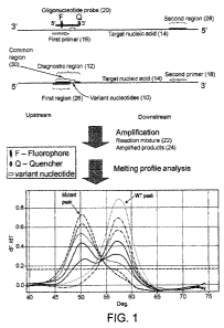
In one embodiment, the first region (26) overlaps the 5'part of the diagnostic
region (12) of the
target nucleic acid sequence (14), but does not overlap the variant
nucleotide(s) (10). The 3' end
of the first region is adjacent to the 5' side of the variant nucleotide(s)
(Figure 1). The first primer
(16) anneals to the first region, some of which is also part of the diagnostic
region, but it does not
anneal to the variant nucleotide(s). In other words, the first primer is not
allele-specific or
mutation-specific.
In another embodiment, the 3' end of the first region overlaps the variant
nucleotide(s), wherein
the first primer is allele-specific (or variant-specific, or mutant-specific)
primer, which comprises
3' terminus nucleotide complementary to the variant nucleotide.
In one embodiment, the 3' end of the first region may abut the 5' side of at
least one of the variant
nucleotide(s) in the diagnostic region of the target nucleic acid sequence,
wherein the annealed
first primer is extended, the first extended nucleotide is the variant
nucleotide. The first primer
anneals to the first region with the 3' end immediately next to the variant
nucleotide.
In another embodiment, the 3' end of the first region may be spaced apart from
the 5' of the
variant nucleotide(s) by one to nine nucleotides, wherein when the annealed
first primer is
extended, the second to tenth extended nucleotide(s) are the variant
nucleotide(s). In other words,
the first primer anneals to the first region one to nine nucleotides away from
the variant
nucleotide(s).
The diagnostic region may be divided into two parts: the 5' part and 3' part,
or may comprise an
additional unmatched part which is located between the 5' part and 3' part.
The 5' part of the
diagnostic region matches or is similar to the 5' portion of the probe (20).
The 3' part of the
diagnostic region matches or is similar to the 3' portion of the probe. The
unmatched part of the
diagnostic region does not have matching sequence to the probe. The 5' part of
the diagnostic
region overlaps with the first region. In other words, the 5' part of the
diagnostic region and the
first region comprise some common sequence. It should be appreciated that the
5' part of the
diagnostic region and the first region may comprise other sequence that is not
common. The 3'
part of the diagnostic region contains variant nucleotide(s). The diagnostic
region may contain a
single mutation, for example BRAF V600E, or may contain potential multiple
mutation sites, for
example mutations in Kras codon 12 and codon 13.
CA 02824223 2013-07-09
WO 2012/095639 PCT/GB2012/000037
19
The length of the first region and the diagnostic region are dependent on the
sizes of the first
primer (16) and the oligonucleotide probe (20).
It is preferred that the first primer, capable of hybridising to the target
nucleic acid sequence, has
a melting temperature which is in the range of the second melting temperature
(Tm2) minus five
to the second melting temperature (Tm2) plus five (Tõ,2-5 to Tm2+5). It is
more preferred that the
first primer, capable of hybridising to the target nucleic acid sequence, has
a melting
temperature which is in the range of the second melting temperature (Tin2)
minus three to the
second melting temperature (Trn2) plus three (T.2-3 to Tm2+3). It is even more
preferred that the
first primer, capable of hybridising to the target nucleic acid sequence, has
a melting temperature
which is the same or similar as the second melting temperature or lower than
the second melting
temperature by 1 C to 5 C.
The first primer (16) and second primer (18) preferably comprise naturally
occurring nucleotides,
although modified nucleotides or linkages can be included in the first and
second primer.
In one embodiment, the blocking oligonucleotide may comprise naturally
occurring nucleotides
only. In other embodiments the blocking oligonucleotide may comprise naturally
occurring
nucleotides and modified nucleotides or linkages. It may not be desirable that
the blocking
oligonucleotide probe is solely made of PNA or LNA. The modified nucleotides
or linkages may
comprise LNA, PNA, d(2-am)ATP, 5-methylcytosine, minor groove binders,
phosphorothioate
linkages, superbase or base analogues. Sometimes the blocking oligonucleotide
probe comprises
one or more modified nucleotides or bases. The nucleotide(s) corresponding to
the variant
nucleotides may be modified.
The blocking oligonucleotide probe may comprise nucleotides, nucleotide
derivatives, nucleotide
analogs, and/or non-nucleotide chemical moieties. Modifications of the probe
that may facilitate
or enhance probe binding include, but are not limited to, the incorporation of
minor groove
binders; the incorporation of positively charged or neutral phosphodiester
linkages in the probe to
decrease the repulsion of the polyanionic backbones of the probe and target
(see Letsinger et al.,
1988, J. Amer. Chem. Soc. 110:4470); the incorporation of alkylated or
halogenated bases, such
as 5-bromouridine, in the probe to increase base stacking; the incorporation
of ribonucleotides
into the probe to force the probe:target duplex into an "A" structure, which
has increased base
stacking; the substitution of 2,6-diaminopurine (amino adenosine) for some or
all of the
adenosines in the probe, and/or the substitution of 5-methylcytosine for
cytosine in the probe; the
CA 02824223 2013-07-09
WO 2012/095639 20
PCT/GB2012/000037
incorporation of nucleotide derivatives such as LNA (locked nucleic acid), PNA
(peptide nucleic
acid) or the like.
It is preferred that a moiety that enhances the binding of the blocking
oligonucleotide probe to the
target sequence is attached to the 3' part of the probe, or at the 3'end of
the probe, or at the 5' end
of the probe. For example, a minor groove binder may be attached at the 3'end
or 5' end of the
probe, fluorophore and/ or quencher may be attached at the 3'end or 5' end of
the probe. Some
nucleotide analogs that increase the binding of the probe to the target
sequence may be positioned
at any nucleotides or preferably positioned at the nucleotides surrounding or
corresponding to the
variant nucleotides. Alternatively, the probe may be just a plain
oligonucleotide with the 3' end
blocked.
Generally the 3 terminus of the blocking oligonucleotide probe will be
"blocked" or made
"unextendable" to prohibit incorporation of the probe into a primer extension
product. The probe
can be made unextendable by using non-complementary bases or by adding a
chemical moiety
such as dye, a quencher, biotin or a phosphate group to the 3' hydroxyl of the
last nucleotide,
which may, depending upon the selected moiety, serve a dual purpose by also
acting as a label for
subsequent detection or capture of the nucleic acid attached to the label. The
probe can also be
made unextendable by removing the 3'-OH or by using a nucleotide that lacks a
3'-OH such as a
dideoxynucleotide.
The blocking oligonucleotide probe comprises detectable or undetectable moiety
(label) which
may include, but not limited to, a fluorescent moiety, a photoluminescent
moiety, a luminescent
moiety, quencher moiety, minor groove binder moiety or a chemiluminescent
moiety.
In a preferred embodiment of the present invention, when the first primer is
not an allele-specific
primer, the PCR process in the method uses high fidelity (proofreading) DNA
polymerase, which
possesses 3' to 5' exonuclease activity. The blocking oligonucleotide is
modified at the 3' part
such that the blocking oligonucleotide is not digested by the 3' to 5'
exonuclease activity of the
high fidelity DNA polymerase. The blocking oligonucleotide may be modified by
phosphorothioate linkages at the 3 part.
When the blocking oligonucleotide probe comprises detectable label (F), the
signal intensity of
the probe is either increased or decreased by hybridization. The probe may be
labelled by a single
detectable moiety (Fig. 3C, D) or may be labelled by multiple moieties (F; Q)
(Fig. 3 A).
CA 02824223 2013-07-09
WO 2012/095639 21 PCT/GB2012/000037
The label on the probe may be a fluorophore (F) or a quencher (Q) (a non-
fluorescent dye). In one
embodiment, the labelled oligonucleotide contains a single label, which is
attached to the 5' or 3'
end of the oligonucleotide (Fig. 3B, C, E, F). Alternatively, the probe may
comprise an interactive
pair of labels, for example fluorophores and/or non-fluorophore dyes (Fig.
3A). One example of
such interactive labels is a fluorophore-quencher pair. The label on the probe
can be located
anywhere, as long as it interacts with other labels or other entities such as
G nucleotides on the
oligonucleotide.
The labelled oligonucleotide probe may comprise a reporter label and a
quencher label, wherein
the quencher label is capable of quenching the fluorescence of said reporter
label when said
oligonucleotide probe is in a single-stranded conformation and is not
hybridized to said target
nucleic acid, wherein said oligonucleotide probe is capable of forming a
double stranded
conformation when hybridized to said target nucleic acid, where the
fluorescence of said reporter
label is unquenched such that the fluorescence intensity of said reporter
label is greater than the
fluorescence intensity of said reporter label when said oligonucleotide probe
is in a single
stranded conformation not hybridized to said target nucleic acid.
In one embodiment, labels are attached at both ends of the probe, for example,
a fluorophore is
attached to the 5' end of the probe and a quencher is attached to the 3' end
of the probe.
In another embodiment, a quencher label is attached at 3' end of the probe and
a reporter label is
attached to an internal nucleotide of the probe. This design is especially
useful for a long probe
with more than 25 nucleotides. The internal reporter label is generally less
than 20 nucleotides
away from 3' end, or preferably less than 19 nucleotides away from 3' end, or
more preferably
less than 18 nucleotides away from 3' end, or more preferably less than 17
nucleotides away from
3' end, or more preferably less than 16 nucleotides away from 3' end, or more
preferably less
than 15 nucleotides away from 3' end, or more preferably less than 14
nucleotides away from 3'
end, or more preferably less than 13 nucleotides away from 3' end, or more
preferably less than
12 nucleotides away from 3' end, or more preferably less than 11 nucleotides
away from 3' end,
or more preferably less than 10 nucleotides away from 3' end. The internal
reporter label may be
less than 9 nucleotides away from 3' end, or less than 8 nucleotides away from
3' end, or less
than 7 nucleotides away from 3' end, or less than 6 nucleotides away from 3'
end, or less than 5
nucleotides away from 3' end (Figure 1). This type of probe is not suitable
for hydrolysis probe-
CA 02824223 2013-07-09
WO 2012/095639 22
PCT/GB2012/000037
based real-time PCR, but is designed for signal generation by hybridisation
with amplified
product and melting curve analysis of probe-amplicon duplex.
The quencher label is preferably a non-fluorescent dye label, which may be
attached to the 3'
terminus or 5' terminus of the oligonucleotide probe, or to an internal
residue of the
oligonucleotide probe. The reporter label may be a fluorescent dye label,
which may be attached
to an internal residue of the oligonucleotide probe, or to the 5' terminus or
3' terminus of the
oligonucleotide probe. The reporter label is preferably a fluorophore, which
may be selected from
the group consisting of fluorescein, fluorescein derivatives, cyanine dyes,
fluorescein-cyanine
conjugates, and similar.
"Fluorophore" is used herein to refer to a moiety that absorbs light energy at
a defined excitation
wavelength and emits light energy at a different defined wavelength. Examples
of fluorescence
labels include, but are not limited to: Alexa Fluor dyes (including Alexa
Fluor 350, Alexa Fluor
488, Alexa Fluor 532, Alexa Fluor 546, Alexa Fluor 568, Alexa Fluor 594, Alexa
Fluor 633,
Alexa Fluor 660 and Alexa Fluor 680), AMCA, AMCA-S, BODIPY dyes (BODIPY FL,
BODIPY R6G, BODIPY TMR, BODIPY TR, BODIPY 530/550, BODIPY 558/568, BODIPY
564/570, BODIPY 576/589, BODIPY 581/591, BODIPY 630/650, BODIPY 650/665),
Carboxyrhodamine 6G, carboxy-X-rhodamine (ROX), Cascade Blue, Cascade Yellow,
Cyanine
dyes (Cy3, Cy5, Cy3.5, Cy5.5), Dansyl, Dapoxyl, Dialkylaminocoumarin, 4', 5'-
Dichloro-2 ',7'-
dimethoxy-fluorescein, DM-NERF, Eosin, Erythrosin, Fluorescein, FAM,
Hydroxyeoumarin,
IRDyes (IRD40, IRD 700, IRD 800), JOE, Lissamine rhodamine B, Marina Blue,
Methoxycoumarin, Naphthofluorescein, Oregon Green 488, Oregon Green 500,
Oregon Green
514, Pacific Blue, PyMPO, Pyrene, Rhodamine 6G, Rhodamine Green, Rhodamine
Red, Rhodol
Green, 2 ', 4', 5 ', 7'-Tetra-bromosulfone-fluorescein, Tetramethyl-rhodamine
(TMR),
Carboxytetramethylrhodamine (TAMRA), Texas Red and Texas Red-X.
As used herein, the term "quencher" includes any moiety that is capable of
absorbing the energy
of an excited fluorescent label when it is located in close proximity to the
fluorescent label and
capable of dissipating that energy. A quencher can be a fluorescent quencher
or a non-fluorescent
quencher, which is also referred to as a dark quencher. The fluorophores
listed above can play a
quencher role if brought into proximity to another fluorophore, wherein either
FRET quenching
or contact quenching can occur. It is preferred that a dark quencher which
does not emit any
visible light is used. Examples of dark quenchers include, but are not limited
to, DABCYL ( 4-
(4'-dimethylaminophenylazo) benzoic acid) succinimidyl ester, diarylrhodamine
carboxylic acid,
succinimidyl ester (QSY-7), and 4 ',5'-dinitrofluorescein carboxylic acid,
succinirnidyl ester
CA 02824223 2013-07-09
WO 2012/095639 23
PCT/GB2012/000037
(QSY-33), quencherl, or "Black hole quenchers" (BHQ-1, BHQ-2 and BHQ-3),
nucleotide
analogs, nucleotide G residues, nanoparticles, and gold particles.
Since the diagnostic region may be divided into: the 5' part, 3' part and
optional unmatched part,
the blocking oligonucleotide may also be divided into two portions, a first
portion comprising
sequence substantially identical to the 5' part sequence of the diagnostic
region, and a second
portion comprising sequence substantially identical to the 3' part sequence of
the diagnostic
region, wherein the first portion and second portion of the blocking
oligonucleotide probe are
contiguous, the 5' part and 3' part of the diagnostic region may be contiguous
or may not be
contiguous. In one embodiment, the blocking oligonucleotide probe has a
sequence identical to
the diagnostic region of the target sequence. Alternatively, the diagnostic
region of the target
sequence may comprise some sequence (the unmatched part) that is not present
in the blocking
oligonucleotide probe. In other words, the blocking oligonucleotide comprises
non-match extra
nucleotides or nucleotide deletions in the middle positions of the blocking
oligonucleotide. The
non-match extra nucleotides or nucleotide deletions can be one, two, three or
more than three
nucleotides, which can be located at the middle of the blocking
oligonucleotide or scattered at
different locations of the blocking oligonucleotide. Nucleotide deletions are
preferred. The
nucleotide deletions or the extra nucleotides reduce the Tm of the blocking
oligonucleotide and
play a role to widen the difference between Tml and Tm2. The nucleotide
deletions or the extra
nucleotides also allow the blocking oligonucleotide to block the primer
binding on the wild-type
sequence more efficiently, as the overlapping part between the first region
and the diagnostic
region can be large. In some applications, it is preferred that the blocking
oligonucleotide probe
hybridises to the diagnostic region with mismatch nucleotides or bulge, which
are preferably
located between the 5' part and 3' part of the diagnostic region.
The blocking oligonucleotide probe used in the present invention is not
degradable, even when
the reaction mix comprises DNA polymerase with 5' nuclease activity or with 3'
to 5'
exonuclease activity. The blocking oligonucleotide probe, the first primer and
the amplified PCR
product are not able to form a structure recognizable by the 5' nuclease
activity of a DNA
polymerase. The blocking oligonucleotide probe may be modified at 3' part such
that it cannot be
cleaved by the 3' to 5' exonuclease activity of a DNA polymerase.
In the above mentioned methods, the blocking labelled oligonucleotide and
second primer are
separate molecules, i.e. they are not linked. The inventor has found that the
method of the present
invention can work equally well when the labelled oligonucleotide (20) and
second primer (18)
CA 02824223 2013-07-09
WO 2012/095639 24
PCT/GB2012/000037
are linked together, i.e. they are linked to become a single oligonucleotide
(18'- 20'). The labelled
oligonucleotide probe is attached at 5' end of the second primer (Figure 2).
This oligonucleotide
is named as linked-primer-probe (18'- 20'), which comprises a 5' probe portion
and a 3' primer
portion. The probe portion in the linked-primer-probe may have all or most
characteristics similar
to the labelled oligonucleotide probe described above. However, the linkage of
second primer and
probe makes this new molecule having some novel properties. This linked-primer-
probe acts as
primer and initiates extension on the template. Upon denaturation the extended
strand is separated
from the template. Under hybridisation condition, the probe portion in the
linked-primer-probe on
the extended strand folds back and hybridise with its extended strand,
creating a stem-loop
structure (32) (Figure 2). In this stem-loop structure, the probe portion in
the linked second
primer-probe plays the same role as labelled oligonucleotide probe in the
methods described
above, where probe and second primer are separate molecules.
The 5' probe portion in the linked-primer-probe is mismatched to the mutated
target sequence,
destabilizing the stem-loop structure and allowing the primer to hybridise to
the stem part of the
secondary structure and complete the extension of the full-length PCR product.
The probe portion
in the linked-primer-probe is matched to the wild-type target sequence,
blocking primer
hybridisation with the stem part of the secondary structure and limiting
formation of the full-
length PCR product (Figure 2). A 1 or 2-bp mismatch at the 5' end probe
portion of the linked-
primer-probe may be included to prevent 3'-end extension of the stem-loop of
the first primer
extended strand that may form from the full-length single strand. The 5' probe
portion and 3'
primer portion may be linked by normal nucleotide(s), so that polymerase can
copy the whole
linked primer-probe. Alternatively, the 5' probe portion and 3' primer portion
may be linked by a
chemical moiety which can't be copied, such as a hydrocarbon arm, an HEG, non-
nucleotide linkage,
a basic ribose, nucleotide derivatives or a dye, so that polymerase cannot
copy a part or the whole
probe portion of the linked primer-probe. One of the labels may be attached to
the chemical
moiety which can't be copied, such as a basic ribose.
Since the first primer and blocking oligonucleotide comprise some common
sequence, they
compete in binding to the same area of the target nucleic acid sequence. When
a variant
nucleotide(s) is present, the probe does not bind strongly, therefore, the
first primer has a higher
chance of binding and extending, resulting in preferential amplification of
the target nucleic acid
containing the variant nucleotide(s). The melting profile reveals that a
melting peak with a
temperature the same as or similar to the second melting temperature is
present.
CA 02824223 2013-07-09
WO 2012/095639 25 PCT/GB2012/000037
When the first primer is not an allele-specific primer, and when the variant
nucleotide(s) is not
present, the first primer may still bind to the target nucleic acid sequence
containing the normal
nucleotide(s) and extend, resulting in the generation of the PCR product of
the normal target
nucleic acid, which could serves as a PCR control.
In the method of the present invention, when the first primer is not an allele-
specific primer and if
both normal and variant nucleotides are present in the target nucleic acid
sequence, depending on
the ratio of variant and normal target nucleic acids, both melting peaks of
Tmi and Tm2 may be
visible in the melting profile analysis at the end of the PCR. When the
variant nucleic acid is
present at a high proportion, the melting profile may only show the melting
peak with Tm2. When
the variant nucleic acid is present at a certain proportion, the melting
profile may show both the
melting peaks with Tmi and Tm2, the relative height of both peaks will give an
indication as to the
amount of each target nucleic acid in comparison with standard controls with
known
concentrations of each target. When the sample contains the normal nucleic
acid only, the
melting profile will only show the melting peak with Tmi, which gives an
indication that the PCR
works and there is no variant nucleic acid present in the sample. Although the
present method is
largely a detection of presence or absence, it can also provide some
quantitative data. As
mentioned above, the relative melting peak heights of Tmi and Tm2 give an
indication of the
amount of each target nucleic acid present. Compared with a standard control
with known
concentrations, the quantitative data can be precise.
The variant nucleotide(s) may include one or more nucleotide substitutions,
deletions, insertions,
or abnormal methylation.
The amplification reaction used in the present invention is preferably PCR,
although other
amplification methods can be used. One important factor affecting the
sensitivity of detecting
mutations which are present at a low frequency in a sample is the
concentration ratio between the
first primer and the blocking oligonucleotide probe. Generally, the
concentration ratio of first
primer/labelled oligonucleotide probe is less than one. However, one cannot
determine precisely
which ratio would work best, unless various ratios are tried in experiments.
In fact, the measured
concentration of an oligonucleotide from the manufacturer is not always
accurate. Any subtle
inaccuracy will affect the ratio of the two oligonucleotides, so one has to do
an experiment to
check that a correct amount of primer and probe are applied. The trend is that
the lower the ratio,
the higher the sensitivity. However, if the ratio is too low, which means too
little first primer is
used, the PCR may not work effectively.
CA 02824223 2013-07-09
WO 2012/095639 26
PCT/GB2012/000037
When the first primer is not an allele-specific primer, asymmetric PCR may be
typically
performed with an excess of the second primer. Any DNA polymerase can be used.
For detecting
a low concentration of mutant sequences in a background of normal sequences, a
proof reading
(or high fidelity) DNA polymerase with the 3' to 5' exonuclease may be used,
for example, PWO
DNA polymerase, Pfu DNA polymerase, Vent DNA polymerase, or KOD DNA
polymerase. The
non-proofreading DNA polymerase may introduce PCR errors into the PCR product,
which may
interfere with the true mutation. The proof reading DNA polymerase has a lower
error rate, and
helps detecting rare mutated DNA in a sample, especially the formalin-fixed
paraffin-embedded
(FFPE) sample.
The present invention also provides a method of analysing a biological sample
for the presence
and/or the amount of mutations or polymorphisms at multiple loci of different
target nucleic acid
sequences in a single reaction vessel. For example, detecting Kras codons 12-
13 and BRAF
V600E can be done in a single reaction, detecting EGFR mutations exon 21
L858R, exon 19
deletions and exon 20 mutation T790M can also be done in a single reaction.
The primers and
probes for Kras and BRAF can be mixed together in a single PCR reaction. The
probes for Kras
and BRAF may be labelled by different dyes so that they can be detected in
different detection
channels.
The amplification reaction mixture will comprise standard amplification
reagents. A sample is
provided which is suspected to contain the target nucleic acid or the
nucleotide variant of interest.
The target nucleic acid contained in the sample may be double-stranded genomic
DNA or cDNA
if necessary, which is then denatured, using any suitable denaturing method.
The denatured
nucleic acid strands are then incubated with oligonucleotide primers and
probes under
hybridisation conditions, i.e. conditions that enable the binding of the
primers and/or probes to the
single nucleic acid strands.
In another aspect, the present invention provides a method for determining the
presence or
absence of variant nucleotide(s) in a diagnostic region of a target nucleic
acid sequence in a
sample, comprising:
(a) providing a first primer and a second primer which are capable of
amplifying a product
comprising a sequence covering that of the diagnostic region of the target
nucleic acid sequence
via an amplification process, wherein the first primer comprises a sequence
based on that of a first
region of the target nucleic acid sequence (i.e. the first primer sequence is
identical or
CA 02824223 2013-07-09
WO 2012/095639 27
PCT/GB2012/000037
substantially identical to the first region), the first region overlaps the 5'
part of the diagnostic
region of the target nucleic acid sequence, and the 3' end of the first region
overlaps the variant
nucleotide(s), and in which the first primer is an allele-specific (or variant-
specific) primer, which
comprises a 3' terminus nucleotide complementary to the variant nucleotide,
and the second
primer comprises a sequence based on that of a second region located
downstream of the
diagnostic region of the target nucleic acid sequence,
providing a blocking oligonucleotide probe which may be a plain
oligonucleotide without
detectable label but with an unextendable 3' end, or a labelled
oligonucleotide probe comprising a
moiety, and in which the blocking oligonucleotide probe has a sequence based
on that of the
diagnostic region of a reference target nucleic acid sequence having no
variant nucleotide(s) (also
referred to as normal nucleotide(s)) therein, wherein the corresponding
nucleotide(s) on the
blocking oligonucleotide probe is identical to the normal nucleotide(s) on the
target nucleic acid
sequence, such that hybridization of the blocking oligonucleotide probe to the
diagnostic region
of said reference target nucleic acid sequence results in the formation of a
first duplex having a
first melting temperature (T.1), hybridization of the blocking oligonucleotide
probe to the
diagnostic region of the (mutated) target nucleic acid sequence containing
variant nucleotide(s)
results in the formation of a second duplex having a second melting
temperature (T.2), wherein
the T.2 is lower than the T.1, wherein the values of T.1 and T.2 are
obtainable experimentally
or are calculated theoretically,
wherein the blocking oligonucleotide probe comprises non-match extra
nucleotides or
nucleotide deletions in the middle positions of the blocking oligonucleotide
probe, wherein when
the blocking oligonucleotide probe hybridises to the diagnostic region of a
nucleic acid target, the
hybridisation creates an unpaired base bulge, which is either located on the
blocking
oligonucleotide probe (in case of the extra unmatched nucleotides in the
blocking oligonucleotide
probe) or on the template (in case of the nucleotide deletions in the blocking
oligonucleotide
probe),
optionally providing a detector probe which is capable of hybridising to the
amplified
target sequence (Fig. 3B),
(b) carrying out an amplification reaction on a reaction mixture using nucleic
acid
polymerase, the blocking oligonucleotide probe, optionally a detector probe
and the pair of the
first and second primers with a nucleic acid sample under conditions which are
permissive for the
amplification process; and
(c) if the first primer comprises label(s), detecting the amplified products
by a melting
profile analysis of the amplicons, or if the reaction comprises a detector
probe, detecting the
amplified product by detecting a change in the detectable signal of the
detector probe.
CA 02824223 2013-07-09
WO 2012/095639 28
PCT/GB2012/000037
The detector probe may be an ordinary real-time PCR probe, which can be TaqMan
probe,
Molecular beacon or hybridisation probe.
In another aspect, the present invention provides a method for determining the
presence or
absence of variant nucleotide(s) in a diagnostic region of a target nucleic
acid sequence in a
sample, comprising:
(a) providing a first primer and a second primer which are capable of
amplifying a product
comprising a sequence covering that of the diagnostic region of the target
nucleic acid sequence
via an amplification process, wherein the first primer comprises a sequence
based on that of a first
region of the target nucleic acid sequence (i.e. the first primer sequence is
identical or
substantially identical to the first region), the first region overlaps the 5'
part of the diagnostic
region of the target nucleic acid sequence, and the 3' end of the first region
overlaps the variant
nucleotide(s), and in which the first primer is an allele-specific (or variant-
specific) primer, which
comprises a 3' terminus nucleotide complementary to the variant nucleotide,
and the second
primer comprises a sequence based on that of a second region located
downstream of the
diagnostic region of the target nucleic acid sequence,
providing a blocking oligonucleotide probe which may be a plain
oligonucleotide without
a detectable label but with an unextendable 3' end, or a labelled
oligonucleotide probe comprising
a moiety, and in which the blocking oligonucleotide probe has a sequence based
on that of the
diagnostic region of a reference target nucleic acid sequence having no
variant nucleotide(s) (also
referred to as normal nucleotide(s)) therein, wherein the corresponding
nucleotide(s) on the
blocking oligonucleotide probe is identical to the normal nucleotide(s) on the
target nucleic acid
sequence, such that hybridization of the blocking oligonucleotide probe to the
diagnostic region
of said reference target nucleic acid sequence results in the formation of a
first duplex having a
first melting temperature (Tin1), hybridization of the blocking
oligonucleotide probe to the
diagnostic region of the (mutated) target nucleic acid sequence containing
variant nucleotide(s)
results in the formation of a second duplex having a second melting
temperature (Tm2), wherein
the Tm2 is lower than the Tin 1, wherein the values of Tin] and Tm2 are
obtainable experimentally
or are calculated theoretically,
wherein the moiety on the labelled oligonucleotide probe may be attached to
the 3' end
and/or the 5' end, and is capable of increasing the melting temperature of the
labelled
oligonucleotide probe in comparison to the plain oligonucleotide without the
moiety, wherein the
moiety may be a fluorescent dye or a non-fluorescent dye, which may be a
fluorophore or a
quencher,
CA 02824223 2013-07-09
WO 2012/095639
PCT/GB2012/000037
29
optionally providing a detector probe which is capable of hybridising to the
amplified
target sequence (Fig. 3B),
(b) carrying out an amplification reaction on a reaction mixture using nucleic
acid
polymerase, the blocking oligonucleotide probe, optionally a detector probe
and the pair of the
first and second primers with a nucleic acid sample under conditions which are
permissive for the
amplification process; and
(c) if the first primer comprises label(s), detecting the amplified products
by a melting
profile analysis of the amplicons, or if the reaction comprises a detector
probe, detecting the
amplifed product by detecting a change in the detectable signal of the
detector probe.
The detector probe may be an ordinary real-time PCR probe, which can be TaqMan
probe,
Molecular beacon or hybridisation probe.
In another aspect, the present invention provides a method for determining the
presence or
absence of variant nucleotide(s) in a diagnostic region of a target nucleic
acid sequence in a
sample, comprising:
(a) providing a first primer and a second primer which are capable of
amplifying a product
comprising a sequence covering that of the diagnostic region of the target
nucleic acid sequence
via an amplification process, wherein the first primer comprises a sequence
based on that of a first
region of the target nucleic acid sequence (i.e. the first primer sequence is
identical or
substantially identical to the first region), the first region overlaps the 5'
part of the diagnostic
region of the target nucleic acid sequence, and the 3' end of the first region
overlaps the variant
nucleotide(s), and in which the first primer is an allele-specific (or variant-
specific) primer, which
comprises a 3' terminus nucleotide complementary to the variant nucleotide,
and the second
primer comprises a sequence based on that of a second region located
downstream of the
diagnostic region of the target nucleic acid sequence,
wherein the allele-specific primer comprises one, two, three or more than
three non-match
extra nucleotides, alternatively one, two, three, more than three nucleotide
deletions in the
positions within ten, nine, eight, seven, six, five, four or three nucleotides
from the 3' terminus of
the primer, wherein when this type of primer anneals to the primer binding
site of a nucleic acid
target, the hybridisation creates an unpaired base bulge, which is either
located on primer (in the
case of the extra unmatched nucleotides in the primer) or on the template (in
the case of the
nucleotide deletions in the primer), and/or providing
CA 02824223 2013-07-09
WO 2012/095639 30
PCT/GB2012/000037
an oligonucleotide probe which acts as a blocker having a sequence based on
that of the
diagnostic region of a reference target nucleic acid sequence having no
variant nucleotide(s)
therein, where the oligonucleotide probe comprises non-match extra nucleotides
or nucleotide
deletions in middle positions of the oligonucleotide probe, such that when the
oligonucleotide
probe hybridises to the diagnostic region of a nucleic acid target
hybridisation creates an unpaired
base bulge, which is either located on the oligonucleotide probe in the case
of the extra
unmatched nucleotides in the oligonucleotide probe or on the template in the
case of the
nucleotide deletions in the oligonucleotide probe,
optionally providing a detector probe which is capable of hybridising to the
amplified
target sequence (Fig. 3B),
(b) carrying out an amplification reaction on a reaction mixture using nucleic
acid
polymerase, the blocking oligonucleotide probe, optionally a detector probe
and the pair of the
first and second primers with a nucleic acid sample under conditions which are
permissive for the
amplification process; and
(c) if the first primer comprises label(s), detecting the amplified products
by a melting
profile analysis of the amplicons, or if the reaction comprises a detector
probe, detecting the
amplified product by detecting a change in the detectable signal of the
detector probe.
The detector probe may be an ordinary real-time PCR probe, which can be TaqMan
probe,
Molecular beacon or hybridisation probe.
The blocking oligonucleotide probe plays a role as blocker, which competes
with first primer in
binding to the same or a similar site of a target region. The blocking
oligonucleotide probe
comprises non-match extra nucleotides or nucleotide deletions in the middle
positions of the
blocking oligonucleotide, the non-match extra nucleotides or nucleotide
deletions can be one, two,
three or more than three nucleotides, which can be located at the middle of
the blocking
oligonucleotide or scattered at different locations of the blocking
oligonucleotide. Nucleotide
deletions are preferred. The nucleotide deletions or the extra nucleotides
reduce the Tm of the
blocking oligonucleotide and play a role to widen the difference between Tml
and Tm2. The
nucleotide deletions or the extra nucleotides also enable the blocking
oligonucleotide to block the
primer binding on the wild-type sequence more efficiently, as the overlapping
part between the
first region and the diagnostic region can be large. The blocking
oligonucleotide can be a labelled
oligonucleotide probe. The moiety on the labelled oligonucleotide may be
attached to the 3' end
and/or the 5' end, and is capable of increasing the melting temperature of the
labelled
oligonucleotide in comparison with the plain oligonucleotide without the
moiety, wherein the
CA 02824223 2013-07-09
WO 2012/095639
PCT/GB2012/000037
31
moiety may be a fluorescent dye or non-fluorescent dye, which can be a
fluorophore or a
quencher.
When the first primer is an allele-specific primer, the first primer may
comprises one, two, three
or more than three non-match extra nucleotides, alternatively one, two, three,
or more than three
nucleotide deletions in the positions within six, five, four or three
nucleotides from the 3'
terminus of the primer, wherein when this type of primer anneals to the primer
binding site of a
nucleic acid target in a sample, the hybridisation creates an unpaired base
bulge, which is either
located on the primer (in case of the extra unmatched nucleotides in the
primer) or on the
template (in the case of the nucleotide deletions in the primer), wherein the
3'-teminal base of the
first primer is complementary to the variant base being detected. The
amplification reaction may
comprise the use of a plurality of first primers for multiplex detection of
multiple variant
nucleotides. The first primer may not comprise a label. Alternatively,
different first primers may
comprise different labels or the same label in different sequence contexts,
where the label may
increase or decrease the detection signal when the first primers are
incorporated into the amplified
products.
The first primer may be attached with a fiuorophore at the 5' end and with a
quencher at an
internal nucleotide, alternatively the first primer may be attached with a
quencher at the 5' end
and with a fluorophore at an internal nucleotide or the first primer may be
attached with a
fluorophore at the 5' end and with a same or a different fluorophore at an
internal nucleotide (Fig.
3F), such that when the primer is incorporated into the amplified product, the
fluorescent signal is
increased, and the amplified product is capable of being analysed by melting
curve analysis. The
distance between the labels (fluorophore or quencher) at the 5' end and at the
internal nucleotide,
may be more than 4 nucleotides but less than 25 nucleotides, or may be more
than 5 nucleotides
but less than 20 nucleotides, or may be more than 6 nucleotides but less than
15 nucleotides.
The allele-specific first primer may be designed with a mismatch in the
position of the second
nucleotide from the 3' terminus nucleotide, or the third nucleotide from the
3' terminus
nucleotide or the fourth nucleotide from the 3' terminus nucleotide with
respect to both the
mutant and wild-type alleles. The second nucleotide from the 3' terminus
nucleotide of the allele-
specific primer is popularly termed as 3'- penultimate nucleotide position.
The 3'-terninal base of
the first primer is complementary to the variant base being detected. This
design results in a
single 3'-terminal mismatch between the mutant-specific primer and the wild-
type template, but a
CA 02824223 2013-07-09
WO 2012/095639 32
PCT/GB2012/000037
double, 3'-terminal mismatch between the mutant-specific primer and the wild-
type template. It
has been shown that greater allele selectivity is achieved by this design.
In another embodiment, the allele-specific first primer may be designed with
one, two, three or
more than three non-match extra nucleotides, alternatively one, two, three or
more than three
nucleotide deletions in the positions within ten, nine, eight, seven, six,
five, four or three
nucleotides from the 3' terminus. When this type of primer anneals to the
primer binding site of a
nucleic acid target in a sample, the hybridisation creates a bulge, which is
either located on the
primer (in the case where there are extra unmatched nucleotides in the primer)
or on the template
(in the case where there are nucleotide deletions in the primer). The 3'-
terninal base of the first
primer is complementary to the variant base being detected. This design
results in an unmatched
base bulge on the unmatched nucleotide positions but complementary 3' ends
between the
mutant-specific primer and the mutant template, which may cause efficient
primer extension.
However a double, or triple or more 3'-end mismatches between the mutant-
specific primer and
the wild-type template may inhibit primer extension. It has been shown that
greater allele
selectivity is achieved by this design. Examples of such design were
demonstrated in detecting
EGFR exon 21 mutations L858R and L861Q (Example 4).
The mutation-specific primer for L858R has a sequence:
SEQ ID NO. 21 5'-CAAGATCACAGATTTTGGCG-3'
which was designed to have a nucleotide "G" deletion within five nucleotides
from the 3' end.
The mutation-specific primer for L861QR has a sequence:
SEQ ID NO. 22 5'-GATTTTGGGCTGGCCAACA-3'
which was designed to have a nucleotide "A" deletion within five nucleotides
from the 3' end.
For detecting EGFR exon 20 T790M mutation, the mutation-specific primer for
T790M has a
sequence:
SEQ ID NO. 24 5'-CCGAAGGGCATGAGCTCA-3'
CA 02824223 2013-07-09
WO 2012/095639 33
PCT/GB2012/000037
which was designed to have a nucleotide "G" deletion within three nucleotides
from the 3' end.
The present invention also provides a method and primers for detecting EGFR
exon 21 mutations
L858R and L861Q and exon 20 mutation T790M.
The primer does not need to be the exact length same as these primers, as long
as they can be
used in an amplification reaction under appropriate conditions. Any primer
comprising a 3' part
sequence identical to the sequence of the 3' part of any of these three
primers is within the scope
of this invention. The 3' part of any of these three primers can be 18
nucleotides from the 3' end,
or 17 nucleotides from the 3' end, or 16 nucleotides from the 3' end, or 15
nucleotides from the 3'
end, or 14 nucleotides from the 3' end, or 13 nucleotides from the 3' end, or
12 nucleotides from
the 3' end, or 11 nucleotides from the 3' end, or 10 nucleotides from the 3'
end, or 9 nucleotides
from the 3' end, or 8 nucleotides from the 3' end.
The labelled oligonucleotide may comprise a detectable or undetectable label,
functioning as a
blocker of wild-type amplification. It has been found that the label on the
probe can increase the
melting temperature of an oligonucleotide, such that 5' or 3' end labelled
fluorophores or
quenchers can increase oligonucleotide's Tm by 2 C to 6 C. The label must be
attached at the
terminus, either at the 5' or 3' end, or both ends of an oligonucleotide (Fig.
3B, C, E, F).
It was also found that the first primer can comprise label(s), which can be
used for monitoring
PCR amplification and for melting curve analysis. Depending on which
nucleotide the label is
attached to, the signal can increase or decrease when the primer is
incorporated into a PCR
product. A very useful design is that the primer is attached with a
fluorophore or quencher at the
5' end and at an internal nucleotide (Fig. 3F). The combinations can be 5'
fluorophore - internal
fluorophore, 5' fluorophore - internal quencher or 5' quencher - internal
fluorophore.
The reaction may contain detector probe which is labelled and first primers
which are not labelled.
The reaction may comprise both a labelled first primer and a detector probe.
In the step (c),
detecting the amplified products may be achieved by a melting profile analysis
of the amplicons
and by detecting a change in the detectable signal of the detector probe.
In another aspect, the present invention provides a labeled oligonucleotide
primer (first primer)
for assaying a target nucleic acid sequence in a sample, comprising: a
reporter label and a
quencher label (or another reporter label),
CA 02824223 2013-07-09
WO 2012/095639 34 PCT/GB2012/000037
wherein said oligonucleotide primer is capable of forming a double stranded
conformation
when hybridized to the target nucleic acid or incorporated into a primer
extension product, where
the fluorescence of said reporter label is unquenched,
wherein said quencher label is a non-fluorescent label, which is attached to
an internal
nucleotide of the oligonucleotide primer or to the 5' end of the primer,
wherein said reporter label is a fluorescent dye label (or a fluorophore),
which is attached
to the 5' end of the oligonucleotide primer or to an internal nucleotide,
alternatively, the labeled
oligonucleotide primer comprises two reporter labels, one of which is attached
to the 5' end, and
another is attached to an internal nucleotide, wherein said oligonucleotide
primer is suitable for
PCR amplification and melting curve analysis.
wherein the fluorophore and quencher (or another fluorophore) on said
oligonucleotide
primer may be less than 6 nucleotides apart, or less than 7 nucleotides apart,
or less than 8
nucleotides apart, or less than 9 nucleotides apart, or less than 10
nucleotides apart, or less than
11 nucleotides apart, or less than 12 nucleotides apart, or less than 13
nucleotides apart, or less
than 14 nucleotides apart, or less than 15 nucleotides apart.
The present invention further provides a reaction mixture, which comprises: a
first primer and a
second primer (or linked-second primer-probe) which are capable of amplifying
a product
comprising a sequence covering that of the diagnostic region of the target
nucleic acid sequence
via an amplification process, wherein the first primer comprises a sequence
based on that of a first
region of the target nucleic acid sequence (i.e. the first primer sequence is
identical or
substantially identical to the first region), wherein the first region
overlaps the 5' part of the
diagnostic region of the target nucleic acid sequence, but does not overlap
the variant
nucleotide(s), wherein the 3' end of the first region is adjacent to the 5'
side of the variant
nucleotide(s), wherein the first primer is not an allele-specific primer,
wherein the second primer
(or the 3' primer portion of the linked-second primer-probe) comprises a
sequence based on that
of a second region located downstream of the diagnostic region of the target
nucleic acid
sequence, alternatively, the first primer is an allele-specific (or variant-
specific) primer, which
comprises 3' terminus nucleotide complementary to the variant nucleotide,
a blocking oligonucleotide, which is an unlabelled oligonucleotide with an
unextendable
3' end or a labelled oligonucleotide probe (or the 5' probe portion of the
linked-second primer-
probe), having a sequence based on that of the diagnostic region of a
reference target nucleic acid
sequence having wild-type nucleotide(s) (also referred to as normal
nucleotide(s)) therein,
wherein the corresponding nucleotide(s) on the blocking oligonucleotide (or
probe portion of
linked primer-probe) is identical to the normal nucleotide(s) on the target
nucleic acid sequence,
CA 02824223 2013-07-09
WO 2012/095639 35 PCT/GB2012/000037
such that hybridization of the blocking oligonucleotide probe (or probe
portion) to the diagnostic
region of said reference target nucleic acid sequence results in the formation
of a first duplex
having a first melting temperature (Tml), hybridization of the blocking
oligonucleotide probe (or
probe portion) to the diagnostic region of the (mutated) target nucleic acid
sequence containing
variant nucleotide(s) results in the formation of a second duplex having a
second melting
temperature (Tm2), wherein the Tm2 is lower than the Tml, wherein the values
of Tml and Tm2 are
obtainable experimentally or are calculated theoretically, and
a DNA polymerase which may be a proof-reading DNA polymerase with 3' to 5'
exonuclease activity.
The present invention further provides a composition, which comprises: a first
primer and a
second primer (or linked-second primer-probe) which are capable of amplifying
a product
comprising a sequence covering that of the diagnostic region of the target
nucleic acid sequence
via an amplification process, wherein the first primer comprises a sequence
based on that of a first
region of the target nucleic acid sequence (i.e. the first primer sequence is
identical or
substantially identical to the first region), wherein the first region
overlaps the 5' part of the
diagnostic region of the target nucleic acid sequence, but does not overlap
the variant
nucleotide(s), wherein the 3' end of the first region is adjacent to the 5'
side of the variant
nucleotide(s), wherein the first primer is not an allele-specific primer,
wherein the second primer
(or the 3' primer portion of the linked-second primer-probe) comprises a
sequence based on that
of a second region located downstream of the diagnostic region of the target
nucleic acid
sequence, alternatively, the first primer is an allele-specific (or variant-
specific) primer, which
comprises a 3' terminus nucleotide complementary to the variant nucleotide,
and
a blocking oligonucleotide probe, which is an unlabelled oligonucleotide with
an
unextendable 3' end or a labelled oligonucleotide probe (or the 5' probe
portion of the linked-
second primer-probe), comprising a moiety and having a sequence based on that
of the diagnostic
region of a reference target nucleic acid sequence having wild-type
nucleotide(s) (also referred to
as normal nucleotide(s)) therein, wherein the corresponding nucleotide(s) on
the labelled
oligonucleotide probe (or probe portion of linked primer-probe) is identical
to the normal
nucleotide(s) on the target nucleic acid sequence, such that hybridization of
the blocking
oligonucleotide probe (or probe portion) to the diagnostic region of said
reference target nucleic
acid sequence results in the formation of a first duplex having a first
melting temperature (Tml),
hybridization of the blocking oligonucleotide probe (or probe portion) to the
diagnostic region of
the (mutated) target nucleic acid sequence containing variant nucleotide(s)
results in the
formation of a second duplex having a second melting temperature (Tm2),
wherein the Tm2 is
CA 02824223 2013-07-09
WO 2012/095639 36
PCT/GB2012/000037
lower than the Tml, wherein the values of Tm 1 and Tm2 are obtainable
experimentally or are
calculated theoretically.
The present invention further provides a labelled oligonucleotide probe, which
comprises a
quencher label or a reporter label attached at a 3' end of the probe and a
reporter label or a
quencher label attached to the 5' end or to an internal nucleotide of the
probe. The internal
reporter label is generally less than 20 nucleotides away from the 3' end, or
preferably less than
19 nucleotides away from the 3' end, or more preferably less than 18
nucleotides away from the
3' end, or more preferably less than 17 nucleotides away from the 3' end, or
more preferably less
than 16 nucleotides away from the 3' end, or more preferably less than 15
nucleotides away from
the 3' end, or more preferably less than 14 nucleotides away from the 3' end,
or more preferably
less than 13 nucleotides away from the 3' end, or more preferably less than 12
nucleotides away
from the 3' end, or more preferably less than 11 nucleotides away from the 3'
end, or more
preferably less than 10 nucleotides away from the 3' end. The internal
reporter label may be less
than 9 nucleotides away from the 3' end, or less than 8 nucleotides away from
the 3' end, or less
than 7 nucleotides away from the 3' end, or less than 6 nucleotides away from
the 3' end, or less
than 5 nucleotides away from the 3' end.
The present invention further provides a kit for determining whether a target
nucleic acid
sequence in a sample has variant nucleotide(s) in a diagnostic region thereof,
comprising:
a first primer and a second primer (or linked-second primer-probe) which are
capable of
amplifying a product comprising a sequence covering that of the diagnostic
region of the target
nucleic acid sequence via an amplification process, wherein the first primer
comprises a sequence
based on that of a first region of the target nucleic acid sequence (i.e. the
first primer sequence is
identical or substantially identical to the first region), wherein the first
region overlaps the 5' part
of the diagnostic region of the target nucleic acid sequence, but does not
overlap the variant
nucleotide(s), wherein the 3' end of the first region is adjacent to the 5'
side of the variant
nucleotide(s), alternatively, the first primer is allele-specific (or variant-
specific) primer, which
comprises a 3' terminus nucleotide complementary to the variant nucleotide,
wherein the second
primer (or the 3' primer portion of the linked-second primer-probe) comprises
a sequence based
on that of a second region located downstream of the diagnostic region of the
target nucleic acid
sequence, and
a blocking oligonucleotide probe, which is an unlabelled oligonucleotide with
an
unextendable 3' end or a labelled oligonucleotide probe (or the 5' probe
portion of the linked-
second primer-probe), comprising moiety(s), having a sequence based on that of
the diagnostic
CA 02824223 2013-07-09
WO 2012/095639 37 PCT/GB2012/000037
region of a reference target nucleic acid sequence having wild-type
nucleotide(s) (also referred to
as normal nucleotide(s)) therein, wherein the corresponding nucleotide(s) on
the blocking
oligonucleotide probe (or probe portion of linked primer-probe) is identical
to the normal
nucleotide(s) on the target nucleic acid sequence, such that hybridization of
the blocking
oligonucleotide probe (or probe portion) to the diagnostic region of said
reference target nucleic
acid sequence results in the formation of a first duplex having a first
melting temperature (Tml),
hybridization of the blocking oligonucleotide probe (or probe portion) to the
diagnostic region of
the (mutated) target nucleic acid sequence containing variant nucleotide(s)
results in the
formation of a second duplex having a second melting temperature (T,2),
wherein the Tm2 is
lower than the Tm1, wherein the values of Tml and Tm2 are obtainable
experimentally or are
calculated theoretically.
In any above mentioned method, reaction mixture, composition or kit, the
allele-specific first
primer may be designed to comprise one or two or three non-match extra
nucleotides,
alternatively with one or two nucleotide deletions in the positions within
six, five, four or three
nucleotides from the 3' terminus. When this type of primer anneals to the
primer binding site of a
nucleic acid target in a sample, the hybridisation creates a bulge, which is
either located on the
primer (in the case of the extra unmatched nucleotides in the primer) or on
the template (in the
case of the nucleotide deletions in the primer). The 3'-terninal base of the
first primer is
complementary to the variant base being detected. This design results in an
unmatched base bulge
on the unmatched nucleotide positions but complementary 3' ends between the
mutant-specific
primer and the mutant template, which may cause efficient primer extension.
However a double,
or triple or more 3'-end mismatch between the mutant-specific primer and the
wild-type template
may inhibit primer extension. The mutation specific primer may comprise
label(s), which can be
used for monitoring amplification and for melting curve analysis. Depending on
which nucleotide
the label is attached to, the signal can increase or decrease when the primer
is incorporated into an
amplifed product. A very useful design is that the primer is attached with a
fluorophore or
quencher at the 5' end and at an internal nucleotide (Fig. 3F). The
combinations can be 5'
fluorophore - internal fluorophore, 5' fluorophore - internal quencher or 5'
quencher - internal
fluorophore.
If the first primer is not a mutation-specific primer, the 3' end of the first
region substantially
complementary (identical) to the first primer may abut the 5' side of at least
one of the variant
nucleotide(s) in the diagnostic region of the target nucleic acid sequence,
such that when the
annealed first primer is extended, the first extended nucleotide is the
variant nucleotide.
CA 02824223 2013-07-09
WO 2012/095639 38
PCT/GB2012/000037
In other embodiments, the 3' end of the first region may be spaced apart from
the 5' of the variant
nucleotide(s) by one to nine nucleotides, such that when the annealed first
primer is extended, the
second to tenth extended nucleotide(s) is the variant nucleotide.
The blocking oligonucleotide which may be a labelled oligonucleotide probe (or
the 5' probe
portion of the linked second primer-probe) or a plain unlabelled
oligonucleotide with an
unextendable 3' end may comprise naturally occurring nucleotides or modified
nucleotides or
linkages. The modified nucleotides or linkages may be selected from a group of
LNA, PNA, d(2-
am)ATP, 5-methylcytosine, minor groove binders, phosphorothioate linkage (S-
Oligo) or base
analogues.
The labelled oligonucleotide comprise moiety(s), which may include, but is not
limited to
fluorescent moiety, a non-fluoresecent dye, a quencher moiety, a
photoluminescent moiety, a
luminescent moiety or a chemiluminescent moiety.
The labelled oligonucleotide probe (or the 5' probe portion of the linked
second primer-probe)
may comprise a reporter label and a quencher label, wherein the quencher label
is capable of
quenching the fluorescence of said reporter label when said oligonucleotide
probe is in a single-
stranded conformation and is not hybridized to said target nucleic acid,
wherein said oligonucleotide probe or the 5' probe portion of the linked
second primer-
probe is capable of forming a double stranded conformation when hybridized to
said target
nucleic acid, where the fluorescence of said reporter label is unquenched such
that the
fluorescence intensity of said reporter label is greater than the fluorescence
intensity of said
reporter label when said oligonucleotide probe is in a single stranded
conformation not hybridized
to said target nucleic acid.
The blocking oligonucleotide, which is labelled oligonucleotide probe or the
5' probe portion of
the linked second primer-probe or a plain oligonucleotide with an unextendable
3' end may be
divided into two portions. The first portion comprises a sequence identical to
the 5' part sequence
of the diagnostic region, and the second portion comprises a sequence
identical to the 3' part
sequence of the diagnostic region, wherein the first portion and second
portion of the labelled
oligonucleotide probe is contiguous, the 5' part and 3' part of the diagnostic
region may not be
contiguous. In other words, the blocking oligonucleotide may comprise extra
non-match
nucleotides or nucleotide deletions in the middle positions of the blocking
oligonucleotide, such
CA 02824223 2013-07-09
WO 2012/095639
PCT/GB2012/000037
that when the blocking oligonucleotide hybridises to the diagnostic region of
a nucleic acid target,
the hybridisation creates an unpaired base bulge, which is either located on
the blocking
oligonucleotide (in the case of the extra unmatched nucleotides in the
blocking oligonucleotide)
or on the template (in the case of the nucleotide deletions in the blocking
oligonucleotide). The
extra non-match nucleotides or deletions may be one nucleotide, or two
nucleotides, or three
nucleotides, or four nucleotide, or five nucleotides, or more than 5
nucleotides.
The present invention also provides a method for multiplex detection of
multiple mutations in a
single closed tube. In a multiplex reaction, two pairs or more than two pairs
of primers are used to
amplify multiple target nucleic acid sequences. An endogenous gene may be
amplified in the
same reaction as an amplification control. The endogenous gene amplification
control may be
used as reference to deterrnine if the mutant amplification is positive or
negative, or used as
normaliser to quantitate the target nucleic acid.
The probe of the present invention may be used in a homogeneous assay system
wherein the
detection and analysis of nucleic acid sequences are performed along with the
amplification of a
target nucleic acid. Alternatively, the probes of the invention may be used in
end-point detection
assays independent of target amplification.
Analysis may occur during amplification in a homogeneous assay system, which
may involve
real-time monitoring fluorescence signal cycle by cycle. Since the probe is
not hydrolysed during
amplification, the target nucleic acid may be studied through melting curve
analysis subsequent to
amplification.
BRIEF DESCRIPTION OF THE DRAWINGS
FIG.1 depicts a schematic of an illustrative embodiment of one aspect of the
present
invention. Components include the following: first primer (16), second primer
(18) and blocking
oligonucleotide probe (20). On cooling, both blocking labelled oligonucleotide
probe and first
primer compete to bind the same site of amplicons, enriching the mutated
target sequence (14).
Melting of the probe element provides targeted genotyping, where high Tm peak
indicates wild
type sequence; low Tm peak indicates mutated sequence.
FIG.2 The linked-second primer-probe (18'; 20') is an amplification primer
that includes a
tail as a labelled probe element that is complementary to the extension
product of the primer. On
CA 02824223 2013-07-09
WO 2012/095639 40
PCT/GB2012/000037
cooling, an intra-molecular stem-loop (32) of the excess linked-primer-probe
strand is formed.
The 5' labelled probe element matches to the wild type so that the stem-loop
is strongly formed.
However, a 1-bp or more mis-match exists between the probe element and the
mutant allele.
During PCR amplification, the first primer is hybridised to the destabilized
mutant stem, but wild-
type extension is blocked, leading to enrichment of the mutant allele.
FIG.3A-G depicts various labels on blocking labelled oligonucleotides and
first primers.
(A) The labelled oligonucleotide contains a fluorophore (F) and quencher (Q)
at the 5'end and 3'
end. The first primer is not labelled and may be not an allele-specific
primer;
(B) The labelled oligonucleotide contains a quencher (Q) at the 3' end, and
the first primer may
be an allele-specific primer. The reaction also contains a detector probe;
(C) The labelled oligonucleotide contains a fluorophore (F) at the 3' end, and
the first primer may
be an allele-specific primer;
(D) The labelled oligonucleotide contains a fluorophore (F) internally, and
the first primer may be
an allele-specific primer;
(E) The labelled oligonucleotide contains a quencher (Q) at the 3' end, the
first primer contains a
fluorophore (F) at the 5' end, and may be an allele-specific primer;
(F) The labelled oligonucleotide contains a Fluorophore (F) or a quencher (Q)
at the 3' end, the
first primer contains a fluorophore (F) or a quencher (Q) at the 5' end and at
an internal
nucleotide, and the first primer may be an allele-specific primer; and
(G) The blocking oligonucleotide without a label comprises nucleotide
deletions in the middle
positions of the blocking oligonucleotide, when the blocking oligonucleotide
hybridises to the
diagnostic region of a nucleic acid target, the hybridisation creates unpaired
base bulge.
FIG.4 melting curve analysis of EGFR exon 21 mutation L858R on serial
dilutions of
mutated DNA in wild type DNA background.
CA 02824223 2013-07-09
WO 2012/095639 41
PCT/GB2012/000037
The invention will now be further described with reference to the following
non-limiting
examples. Other embodiments of the invention will occur to those skilled in
the art in light of
these.
The disclosure of all references cited herein, in as much as they may be used
by those skilled in
the art to carry out the invention, are hereby specifically incorporated
herein by cross-reference.
EXAMPLE 1
EGFR (Epidermal Growth Factor Receptor) signalling pathway causes cell growth
and
proliferation via several signalling molecules, including KRAS and BRAF.
Oncogenic mutations
in these genes may cause cancer. Recent evidence indicates that the presence
of epidermal growth
factor receptor (EGFR) or KRAS mutations in non-small cell lung cancer (NSCLC)
can predict
the response of the tumour to drugs such as gefinitib.
In this example, the point mutation L858R in EGFR exon 21 was chosen as a
model mutation site
to test this technique. Primers and probes were designed to amplify a region
containing the
mutation site. The forward primer has a 3' terminus nucleotide right before
the mutation site, not
overlapping the L858R nucleotide, i.e. the forward primer is not an allele-
specific primer. The
sequences of forward primers and reverse primers are listed in Table I. The 5'
part of the probe
overlaps some sequence of the forward primers, and its 3' part contains
sequence covering the
mutation site (matching the wild type sequence). The sequence of probe is also
listed in Table 1.
The probe contains fluorophore Fam at its 5' end and black quench dye BHQ1 at
the 3' end.
Tablel. EGFR exon21 L858R, sequences of primers and probe
number name sequence (5'-3')
length (bp)
SEQ ID NO. 1 EGFR21forwardl GATCACAGATTTTGGGC 17
SEQ ID NO. 2 EGFR21forward2 AGATCACAGATTTTGGGC 18
SEQ ID NO. 3 EGFR2lreversel CTTTGCCTCCTTCTGCATGGTA 22
SEQ ID NO. 4 EGFR2lreverse2 CTCCTTACTTTGCCTCCTTCTGCA 24
SEQ ID NO. 5 EGFR21 probe GATCACAGGTTTTGCTGGCAAACT 26
GC
Preparation of templates
CA 02824223 2013-07-09
WO 2012/095639
PCT/GB2012/000037
42
A DNA fragment containing the L858R EGFR exon21 was synthesised and cloned
into a vector.
The mutated plasmid DNA is mixed with wild type human genomic DNA such that
the
concentrations of mutated DNA are 10%, 1%, 0.5%. The wild type DNA is 3000
copies/1AI.
PCR reaction
PCR mixture was prepared as listed in Table 2. Every PCR run included water
control and wild
type DNA control. Normal Taq polymerase or hot-start Taq polymerase was used.
In some
experiments, DNA polymerase with proof-reading activity such as PWO DNA
polymerase and
probe with the 3' end modified to be resistant to 3' exonuclease activity of
the DNA polymerase
were used. It was found that the use of DNA polymerase with proof-reading
activity gave good
results, which showed increased sensitive of detection of mutations.
Table 2. PCR mixture
Composition Final
concentration
PCR Buffer lx
dNTPs 0.1 mM
EGFR21 forward primer 0.05 M
EGFR2lreverse primer 0.2 1.1M
EGFR21pro be 0.25 [tM
Polymerase 0.4 U
H20 appropriate
DNA template 1 pi
Total volume 20 IA
PCR condition
A PCR program was set up as set out in Table 3. Fam signal collection was
carried out on
annealing step 50 C:
Table 3. PCR program
step temperature duration cycles
1 95 C lmin 1
2 93 C 3sec
3 56 C 20sec
4 53 C 20sec
5 50 C 20sec 50
6 72 C 6sec
CA 02824223 2013-07-09
WO 2012/095639 43
PCT/GB2012/000037
7 95 C 30sec 1
8 Melting curve: 39-75 C, every time increase 1
C,
hold 5sec, collection of FAM fluorecence signals.
When PCR finished, the melting curve was performed. The wild type DNA gave a
melting peak
at 57 C, whereas the mutated DNA gave a melting peak at 51 C. In the case
where the PCR tubes
contained the 1% and 0.5% mutated DNA, both peaks were seen.
EXAMPLE 2
In Example 1, the labelled oligonucleotide probe and second primer are
separate molecules, i.e.
they are not linked. In this example, a linked-primer-probe was used in which
the labelled
oligonucleotide and the second primer (the reverse primer) are linked
together, i.e. they are linked
to become a single oligonucleotide (Figure 2).
For detecting EGFR exon21 point mutation L858R, the linked-primer-probe, had a
sequence:
SEQ ID NO 6.
51tGATCACAGGTI1TTGCTGGCAAACTGCCTTTGCCTCCTTCTGCATGGTA-3'
It comprises a 5' probe portion and a 3' primer portion. The sequence of 3-28
nucleotides is the
probe portion, the 26th nucleotide dT is attached with quencher BHQ1. The last
22 nucleotides
(3' part) is the primer portion, serving as primer. The first two nucleotides
tt are introduced to
mismatch the target sequence. The 5' end of the linked-primer-probe is
attached with fluorescent
dye Fam.
The probe portion in linked-primer-probe matches the wild-type target
sequence, which
hybridizes to the diagnostic region of the extended strand and blocks
hybridisation of the first
primer (the forward primer) with the stem part of the secondary structure and
limits formation of
the full-length PCR product (Figure 2). A 2-bp mismatch at the 5' end probe
portion of the linked-
primer-probe is included to prevent 3'-end extension of the stem-loop of the
first primer extended
strand that may form from the full-length single strand. In this example, the
5' probe portion and
3' primer portion may be linked by normal nucleotide(s), so that polymerase
can copy the whole
linked primer-probe.
CA 02824223 2013-07-09
WO 2012/095639 44
PCT/GB2012/000037
PCR condition and cycling program were set up as per Example 1.
EXAMPLE 3
Kras codon 12 and 13 mutation detection.
The allele-specific primers are:
SEQ ID NO. 7 5' Fam-caaTGGTAGTTGGAGCTGT-3';
SEQ ID NO. 8 5'-gtgtTGTGGTAGTTGGAGCTGT-3' (Fam-dT at second nucleotide from
the 5'
end);
SEQ ID NO. 9 5' Fam-tcaGTGGTAGTTGGAGCTT-3' (dabcyl-dT at the 9th nucleotide
from the
5'end);
SEQ ID NO. 10 5' Fam-caaTGGTAGTTGGAGCTC-3';
SEQ ID NO. 11 5' Cy5-caGaGGTAGTTGGAGCTGA-3';
SEQ ID NO. 12 5' cy5-acaGTGGTAGTTGGAGCTA-3';
SEQ ID NO. 13 5' Fam-aacGaTAGTTOGAGCTGGTGA-3' (dabc)'l-dT at 10th nucleotide
from
the 5' end);
SEQ ID NO. 14 5' Fam-caaaGGTAGTTGGAGCTGC-3';
SEQ ID NO. 15 5' Fam-TAAGTTGTGGTAGTTGGAGCTGT (Fam-dT at the 8th nucleotide
from the 5' end);
SEQ ID NO. 16 5' Dabcyl-taagGTAGTTGGAGCTGGTGA (Fam-dT at the 9th nucleotide);.
The reverse primer is:
SEQ ID NO. 17 5' TTACCTCTATTGTTGGATCATATTC-3';.
CA 02824223 2013-07-09
WO 2012/095639 45
PCT/GB2012/000037
The labelled oligonucleotide probe (blocking probe) is:
SEQ ID NO. 18 5' Fam- TGGTAGITGGCTGGTGGCG-BHQ1-3'; or
SEQ ID NO. 19 5'- GGTAGTTGGATGGTGGCG-BHQ I 3';.
The detector probe (dual labelled TaqMan probe) is:
SEQ ID NO. 20 5' Hex- taggcaagagtgecttgacga-BHQ1 3'.
Each assay reaction mixture (20 ul total) contained lx TaqMan Gene Expression
master mixture
(applied Biosystems, PN 4333458N), 0.5ng/ulgenomic DNA, 50nM allele-specific
primer,
200nM TaqMan probe, 500nM reverse primer, 500nM blocking probe. The reactions
were
incubated in 96-well plate at 95 C for 10 minutes, then for 10 cycles at 95 C
for 10 seconds,
51 C for 40 second and 72 C for 20 seconds, then for 50 cycles at 95 C for 10
seconds, 56 C for
30 second and 60 C for 30 seconds. Following PCR, a melting protocol from 50 C
to 92 C was
used. All reactions were run in duplicate in Stratagene MX3005P real time PCR
machine.
Assays were performed using general experimental design and reaction
conditions indicated
above. Singleplex, doubleplex and tripleplex for detecting one mutation, two
mutations and three
mutations were performed in a single closed tube reaction. Real-time
monitoring of the
fluorescent emission was used to determine the Ct value; while melting curve
analysis was used
to determine which mutation is present in the targeted DNA.
The allele-specific primer may be labelled with a fluorophore at the 5' end
alone, for example
primers SEQ ID NO. 7, SEQ ID NO. 8 (Fam-dT at second nucleotide).
When the allele-specific primer SEQ ID NO. 7 is incorporated into a PCR
product, the
fluorescent signal is decreased. When the allele-specific primer (SEQ ID NO.
8) (Fam-dT at the
second nucleotide) is incorporated into a PCR product, the fluorescent signal
is increased. The
allele-specific primer may be labelled with a fluorophore at the 5' end and a
quencher (dabcyl or
BHQ) at an internal nucleotide, for example primer SEQ ID NO. 9 (dabcyl-dT at
the nucleotide
9), which, when incorporated into a PCR product, results in an increased
fluorescent signal.
Similarly, the allele-specific primer may be labelled with a fluorophore at
the 5' end and a same
or different fluorophore at an internal nucleotide, for example primer SEQ ID
NO. 15 (5' end-
CA 02824223 2013-07-09
WO 2012/095639 6
PCT/GB2012/000037
4
Fam, Fam-dT at the nucleotide 8), which, when incorporated into a PCR product,
results in an
increased fluorescent signal.
The allele-specific primer may be labelled with a quencher at the 5' end and a
fluorophore at an
internal nucleotide, for example primer SEQ ID NO. 16 (5' end dabcyl, Fam-dT
at nucleotide 9),
which, when incorporated into a PCR product, results in an increased
fluorescent signal
The allele-specific primer, when incorporated into a PCR product, results in
an increased or
decreased fluorescent signal, which can be monitored in real-time during PCR,
or can be used for
melting curve analysis. The amplification curve and / or melting profile can
be used to determine
which nucleotides (variant or normal) are present in a target nucleic acid.
The reaction may contain a detector probe, which can be a TaqMan probe. In
this example the
detector probe is HEX-BHQ1 dual labelled oligonucleotide (SEQ ID NO. 20). A
reaction may
contain both detector probe and labelled allele-specific primers, which both
can be monitored in
real time during PCR but only the labelled allele-specific primer, when
incorporated into a PCR
product, can be used for melting curve analysis.
The reaction may contain another labelled oligonucleotide probe (also known as
blocking probe).
The blocking probe functions as blocker to prevent wild type sequence being
amplified. The
blocking probe competes with allele-specific primer for the binding site,
which binds to blocking
probe more strongly. The labels on the blocking probe may be fluorescent or
non-fluorescent dyes.
The blocking oligonucleotides (SEQ ID NO. 18) or (SEQ ID NO. 19 contain labels
Fam and/or
BHQ1 at the 5' end and 3' end, which increase the Tm of the labelled
oligonucleotide in
comparison with the oligonucleotide without label.
EXAMPLE 4
Detection of EGFR exon 21 mutations L858R and L861Q, exon 20 mutation T790M
The mutation-specific primer for L858R has a sequence
SEQ ID NO. 21 5'-CAAGATCACAGATTTTGGCG-3'
which was designed to have a nucleotide "G" deletion within five nucleotides
from the 3' end.
CA 02824223 2013-07-09
WO 2012/095639 47
PCT/GB2012/000037
The mutation-specific primer for L861QR has a sequence
SEQ ID NO. 22 5'-GATTTTGGGCTGGCCAACA-3'
which was designed to have a nucleotide "A" deletion within five nucleotides
from the 3' end.
The reverse primer has a sequence:
SEQ ID NO. 23 5'-CTTACTTTGCCTCCTTCTGCA-3'.
The mutation-specific primer for EGFR exon 20 mutation T790M has a sequence:
SEQ ID NO. 24 5'-CCGAAGGGCATGAGCTCA-3'
which was designed to have a nucleotide "G" deletion within three nucleotides
from the 3' end.
Blocking oligonucleotide for detecting EGFR exon 21 mutations L858R and L861Q
were
designed to have a sequence:
SEQ ID NO. 25 5'-GATCACAGgTTTTGCTGGCAAACTGc-3'-Ph
in which the 3' end is blocked by a phosphate group. PCR conditions were the
same as described
in Example 3. The results showed that these primers, with nucleotide deletions
in the 3' ends,
were more specific than the primers without nucleotide deletions.
CA 02824223 2013-07-09
WO 2012/095639 48
PCT/GB2012/000037
The following passages are provided as clauses and are not to be considered as
claims:
1. A method for determining the presence or absence of variant
nucleotide(s) in a diagnostic
region of a target nucleic acid sequence in a sample, comprising:
(a) providing a first primer and a second primer which are capable of
amplifying a product
comprising a sequence covering that of the diagnostic region of the target
nucleic acid sequence
via a PCR process, wherein the first primer comprises a sequence based on that
of a first region of
the target nucleic acid sequence (i.e. the first primer sequence is identical
or substantially
identical to the first region), wherein the first region overlaps the 5' part
of the diagnostic region
of the target nucleic acid sequence, but does not overlap the variant
nucleotide(s), wherein the 3'
end of the first region is adjacent to the 5' side of the variant
nucleotide(s), wherein the second
primer comprises a sequence based on that of a second region located
downstream of the
diagnostic region of the target nucleic acid sequence,
providing a labelled oligonucleotide probe comprising a detectable moiety and
having a
sequence based on that of the diagnostic region of a reference target nucleic
acid sequence having
no variant nucleotide(s) (also referred to as normal nucleotide(s)) therein,
wherein the
corresponding nucleotide(s) on the labelled oligonucleotide is identical to
the normal nucleotide(s)
on the target nucleic acid sequence, such that hybridization of the labelled
oligonucleotide probe
to the diagnostic region of said reference target nucleic acid sequence
results in the formation of a
first duplex having a first melting temperature (Tml), hybridization of the
labelled oligonucleotide
probe to the diagnostic region of the (mutated) target nucleic acid sequence
containing variant
nucleotide(s) results in the formation of a second duplex having a second
melting temperature
(Tm2), wherein the Tm2 is lower than the Tml, wherein the values of Tml and
Tm2 are obtainable
experimentally or are calculated theoretically;
(b) carrying out an amplification reaction on a reaction mixture using nucleic
acid
polymerase, the labelled oligonucleotide probe and the pair of the first and
second primers with a
nucleic acid sample under conditions which are permissive for the PCR process;
and
(c) subjecting the PCR products to a melting profile analysis to determine
melting
temperatures of the labelled oligonucleotide probe hybridised to the PCR
products, wherein the
presence of the second melting temperature(s) of the second duplex in the
melting profile analysis
is indicative of the presence of the variant nucleotide(s) in the diagnostic
region of the target
nucleic acid sequence contained in the nucleic acid sample.
2. The method according to claim 1, wherein said PCR uses slow ramping
rates or multiple
annealing temperatures.
CA 02824223 2013-07-09
WO 2012/095639 49
PCT/GB2012/000037
3. The method according to claim 2, wherein said slow ramp rate is
lower than 2 C/sec, or is
lower than 1 C/sec, or is lower than 0.5 C/sec, or is lower than 0.2 C/sec.
4. The method according to claim 2, wherein said PCR process includes a
series of multiple
annealing temperatures in each cycle of the PCR thermal program, wherein said
multiple
annealing temperatures run in a sequence from the middle temperature to the
lowest annealing
temperature or from the lowest annealing temperature to the extension
temperature within each
thermal cycle.
5. The method according to claim 1, wherein 3' end of the first region
abuts the 5' of at least
one of the variant nucleotide(s) in the diagnostic region of the target
nucleic acid sequence,
wherein when the annealed first primer is extended, the first extended
nucleotide is the variant
nucleotide.
6. The method according to claim 1, wherein 3' end of the first region is
spaced apart from
the 5' of the variant nucleotide(s) by one to nine nucleotides, wherein when
the annealed first
primer is extended, second to tenth extended nucleotide(s) is the variant
nucleotide.
7. The method according to claim 1, wherein the first primer, capable of
hybridising to the
target nucleic acid sequence, has a melting temperature which is the same or
similar to the second
melting temperature, or has a melting temperature which is in the range of the
second melting
temperature minus three to the second melting temperature plus three (Tm2-3 to
Tm2+3).
8. The method according to claim 1, wherein said labelled oligonucleotide
probe comprises
naturally occurring nucleotides.
9. The method according to claim 1, wherein said labelled oligonucleotide
probe comprises
modified nucleotides or linkages.
10. The method according to claim 9, wherein said modified nucleotides or
linkages comprise
LNA, PNA, d(2-amA)TP, 5-methylcytosine, minor groove binders, or base
analogues.
11. The method according to claim 1, wherein the moiety is a fluorescent
moiety, a
photoluminescent moiety, a luminescent moiety, or a chemiluminescent moiety.
CA 02824223 2013-07-09
WO 2012/095639 50
PCT/GB2012/000037
12. The method according to claim 1, wherein said labelled oligonucleotide
probe comprises a
reporter label and a quencher label, wherein the quencher label is capable of
quenching the
fluorescence of said reporter label when said oligonucleotide probe is in a
single-stranded
conformation and is not hybridized to the target nucleic acid,
wherein said oligonucleotide probe is capable of forming a double stranded
conformation
when hybridized to said target nucleic acid, where the fluorescence of said
reporter label is
unquenched such that the fluorescence intensity of said reporter label is
greater than the
fluorescence intensity of said reporter label when said oligonucleotide probe
is in a single
stranded conformation not hybridized to the target nucleic acid, wherein the
quencher label is
attached to the 3'end of the probe, the reporter label is attached to the 5'
end of the probe or
reporter label is attached to a internal nucleotide of the probe.
13. The method according to claim 1, wherein said labelled oligonucleotide
probe is divided
into two portions, first portion comprising sequence substantially identical
to the 5' part sequence
of the diagnostic region, and second portion comprising sequence substantially
identical to the 3'
part sequence of the diagnostic region, wherein the first portion and second
portion of the labelled
oligonucleotide probe are contiguous, the 5' part and 3' part of the
diagnostic region are not
contiguous.
14. The method according to claim 1, wherein the labelled oligonucleotide
and second primer
is linked together, i.e. they are linked to become a single oligonucleotide,
wherein the labelled
oligonucleotide probe is attached at 5' end of the second primer, wherein the
linked-primer-probe
acts as primer and initiates extension on the template, the probe portion in
the linked-primer-
probe on the extended strand folds back and hybridise with its extended
strand, creating a stem-
loop structure, the 5' probe portion in linked-primer-probe is mismatched to
the mutated target
sequence, destabilizing the stem-loop structure and allowing the primer to
hybridize to the stem
part of the secondary structure and complete the extension of the full-length
PCR product, the
probe portion in linked-primer-probe is matched to the wild-type target
sequence, blocking primer
hybridization with the stem part of the secondary structure and limiting
formation of the full-
length PCR product.
15. A labeled oligonucleotide probe for assaying a target nucleic acid
sequence in a sample,
comprising: a reporter label and a quencher label, which is capable of
quenching the fluorescence
CA 02824223 2013-07-09
WO 2012/095639 51 PCT/GB2012/000037
of said reporter label when said oligonucleotide probe is in single-stranded
conformation and is
not hybridized to a target nucleic acid,
wherein said oligonucleotide probe is capable of forming a double stranded
conformation
when hybridized to the target nucleic acid, where the fluorescence of said
reporter label is
unquenched such that the fluorescence intensity of said reporter label is
greater than the
fluorescence intensity of said reporter label when said oligonucleotide probe
is in single stranded
conformation not hybridized to the target nucleic acid,
wherein said quencher label is non-fluorescent label, which is attached to the
3' terminus
of the oligonucleotide probe,
wherein said reporter label is fluorescent dye label, which is attached to an
internal residue
of the oligonucleotide probe,
wherein the internal reporter label is less than 16 nucleotides away from 3'
end, or is less
than 15 nucleotides away from 3' end, or is less than 14 nucleotides away from
3' end,
wherein said oligonucleotide probe is not suitable for hydrolysis probe-based
real-time
PCR,