Note: Descriptions are shown in the official language in which they were submitted.
CA 02824264 2013-07-09
WO 2012/102837
PCT/US2012/020170
1
METHOD AND DEVICE FOR TREATING OSTEOARTHRITIS
NONINVASIVELY
BACKGROUND
Field
[0001] Aspects of the present disclosure relate generally to a method and
system for
treating osteoarthritis with electromagnetic stimulation and/or ultrasound.
Background
[0002] Osteoarthritis (OA) of the knee is the most common form of OA affecting
more
than ten million Americans and is the most common cause of disability in the
United
States. Symptoms may include pain, stiffness, limited range of motion and
localized
swelling. Currently, there is no known cure for OA and current treatments are
intended to mitigate the symptoms.
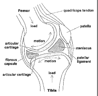
[0003] As shown in Figure 1, the human knee is a synovial joint between the
femur and
tibia. The joint is contained within a fibrous joint capsule with a synovial
membrane lining. The ends of the bones are covered with articular cartilage
and the
bone beneath the cartilage is the subchondral bone. Hyaline articular
cartilage loss
is the central signature event in OA. While the exact etiology of OA is
unknown,
the pathophysiology involves a combination of mechanical, cellular, and
biochemical processes.
[0004] With reference to Figure 2, there are three primary types of bone:
woven bone,
cortical bone, and cancellous bone. Woven bone is found during fracture
healing
(callus formation). Cortical bone, also called compact or lamellar bone, is
remodeled from woven bone and forms the internal and external tables of flat
bones
and the external surfaces of long bones. Cancellous bone (trabecular bone)
lies
between cortical bone surfaces and consists of a network of honeycombed
interstices
containing hematopoietic elements and bony trabeculae. The trabeculae are
predominantly oriented perpendicular to external forces to provide structural
support.
CA 02824264 2013-07-09
WO 2012/102837
PCT/US2012/020170
2
[0005] Bone remodeling is the process by which bone is renewed to maintain
bone
strength and mineral homeostasis. Remodeling involves continuous removal of
discrete packets of old bone, replacement of these packets with newly
synthesized
proteinaceous matrix, and subsequent mineralization of the matrix to form new
bone. The remodeling process resorbs old bone and forms new bone and
cancellous
bone is continually undergoing remodeling on the internal endosteal surfaces.
[0006] There is a vascular component which is integrally associated with the
process of
bone remodeling. This vascular contribution has both an anatomic basis and
functional relevance. The subchondral region is highly vascular with terminal
vessels in direct contact with the deepest hyaline cartilage layer. Bone
remodeling
occurring in a bone chamber is also related to the existence of and increased
flow
through microvessels that conform closely to the contour of the cancellous
bone
surface. Pericytes are intimately involved in the process of angiogenesis
which
accompanies the vascular component involved with cancellous bone remodeling.
The microvasculature has been linked to the regulation of coupling between
bone
resorption and bone formation. This structure forms the anatomic basis for the
knowledge that the vascular system is associated with osteogenesis during bone
remodeling.
[0007] Cellular changes have been identified in osteoarthritis of the knee to
include
bone marrow edema, extensive intertrabecular fibrosis and sclerosis, as well
as
vascularization and thickening of the trabeculae in the subchondral bone.
These
combine to increase the stiffness of the subchondral bone, transmitting
increased
load to the overlying cartilage and leading to secondary cartilage damage. The
increased venous vascular resistance contributes to venous congestion,
increased
intraosseous pressure, congestive bone pain, diminished nutrient delivery and
progression of the disease. There is an. interrelationship between cartilage
damage
and subchondral bone integrity.
[0008] The current understanding of the pathogenesis of OA has led the
American
Academy of Orthopaedic Surgeons (AAOS) to develop recommendations for
treatment of knee OA. These include activity modification, weight loss,
fitness,
range of motion and quadriceps strengthening exercises, patellar taping,
acupuncture, glucosamine, NSAIDS, acetaminophen, analgesics, and injections of
CA 02824264 2013-07-09
WO 2012/102837
PCT/US2012/020170
3
arthroscopy, meniscectomy, osteotomy and knee replacement surgery. In this
context, there is a need for an enhanced method for treating OA in a
noninvasive
manner.
SUMMARY
[0009] In accordance with one or more embodiments and corresponding disclosure
thereof, various aspects are described in connection with a method for OA
treatment
at an affected area or joint. For example, the method may involve identifying
a
treatment site of the joint, and providing at least one transducer module at
the
treatment site. The at least one transducer module may be in operative
communication with a signal generator module, and may include at least one
transducer for delivering electromagnetic signals, which may include
ultrasound
signals. The method may also involve stimulating (a) bone remodeling, (b) bone
cells and associated precursors, and/or (c) pericytes at the joint with the
electromagnetic signals delivered to the treatment site at a user-selected
intensity
(e.g., between about 21 milliwatts per square centimeter and about 39
milliwatts per
square centimeter) by exciting the at least one transducer module with the
signal
generator module. This may be for a user-selected period of time, as well.
[0010] In accordance with one or more aspects of the embodiments described
herein,
there is provided an apparatus for treating OA. For example, the apparatus may
include a signal generator for generating electromagnetic (e.g., ultrasound)
signals.
The apparatus may include at least one transducer module in operative
communication with the signal generator and configured to be placed at the
affected
joint. The
apparatus may also include a controller interface in operative
communication with the signal generator. The apparatus may further include at
least
one processor in operative communication with the signal generator and the
controller interface, wherein the at least one processor may be configured to
stimulate bone remodeling at the joint with the electromagnetic signals
delivered to
the joint, in response to user input received via the controller interface.
For
example, ultrasound signals delivered to the treatment site may have an
ultrasound
frequency of about 1.5 MHz and a spatial average¨temporal average (SATA) of
about 30 milliwatts per square centimeter.
CA 02824264 2013-07-09
WO 2012/102837
PCT/US2012/020170
4
[0011] To the accomplishment of the foregoing and related ends, one or more
aspects
comprise the features hereinafter fully described and particularly pointed out
in the
claims. The following description and the annexed drawings set forth in detail
certain illustrative aspects and are indicative of but a few of the various
ways in
which the principles of the aspects may be employed. Other novel features will
become apparent from the following detailed description when considered in
conjunction with the drawings and the disclosed aspects are intended to
include all
such aspects and their equivalents.
BRIEF DESCRIPTION OF THE DRAWINGS
[0012] Figure 1 illustrates a human knee.
[0013] Figure 2 illustrates cancellous bone and components thereof
[0014] Figure 3 is a block diagram showing an embodiment of a system for
treating a
human knee affected by OA.
[0015] Figure 4 shows an embodiment of a device for treating OA.
[0016] Figure 5 shows another embodiment of a device for treating OA.
[0017] Figure 6 illustrates an embodiment of a methodology for treating OA of
an
affected area/joint.
[0018] Figure 7 shows further aspects of the methodology of Figure 6.
[0019] Figure 8 illustrates an embodiment of an apparatus for OA treatment, in
accordance with the methodologies of Figure 6-7.
DESCRIPTION
[0020] The detailed description set forth below, in connection with the
appended
drawings, is intended as a description of various configurations and is not
intended
to represent the only configurations in which the concepts described herein
may be
practiced. The detailed description includes specific details for the purpose
of
providing a thorough understanding of the various concepts. However, it will
be
apparent to those skilled in the art that these concepts may be practiced
without
these specific details. In some instances, well-known structures and
components are
shown in block diagram form in order to avoid obscuring such concepts.
CA 02824264 2013-07-09
WO 2012/102837
PCT/US2012/020170
[0021] Electric and electromagnetic fields may be generated and applied to
bones via
currently known techniques, such as, for example, direct current (DC), pulsed
electromagnetic fields (PEMFs), combined magnetic fields (CMFs), or capacitive
coupling for capacitively coupled electric fields (CCEFs). Physiologic effects
of
electromagnetic stimulation of bone may include, for example,
piezoelectricity,
cellular proliferation and/or differentiation, synthesis of extracellular
matrix,
transmembrane signal transduction, synthesis of growth factors, increased DNA
production, altered gene expression, etc. There is also evidence that-
pericytes may
contribute to the ostogenic response to electrical stimulation. Exposure to
electromagnetic fields produces a temporal acceleration and quantitative
increase in
endochondral bone formation and trabecular maturation through the process of
bone
remodeling.
[0022] Another use of electromagnetic stimulation of biologic tissues relates
to the
stimulation of cartilage cells rather than bone cells. Pulsed electromagnetic
fields
have been used to stimulate cartilage cells in clinical trials, and
capacitively coupled
electrical fields have also been shown to stimulate in vivo chondrocytes.
[0023] Ultrasound is another technology known to stimulate bone growth and
remodeling. Although the mechanism for how ultrasound stimulates bone healing
is
unknown, it has been hypothesized that the pressure waves it produces provide
micro-mechanical stress and strain causing biochemical alterations at the
cellular
level leading to enhanced bone formation.
[0024] In related aspects, since fibrous tissue is nonvascular and normal bone
is
vascular the bone remodeling associated with electromagnetic stimulation with
bone
growth stimulators has inherent changes associated with the vascular system.
The
capillary contained in the growing end of the basic multicellular unit during
cancellous bone remodeling is believed by the inventor to redistribute
regional
blood flow to alleviate subchondral venous congestion and thereby decrease
pain
with subsequent improvements in the delivery of nutrients that will affect
disease
progression. It is the commonality of the presence of fibrous tissue in
fibrous non-
union and in the subchondral cancellous bone in OA, combined with the
knowledge
that subchondral venous congestion is present in OA and the fact that changes
in the
anatomic vasculature and circulatory functions are inherent in the process of
CA 02824264 2013-07-09
WO 2012/102837
PCT/US2012/020170
6
cancellous bone remodeling, that allow for the OA to be treated via
application of
the below-described techniques.
[0025] In accordance with aspects of the subject of this disclosure, there are
provided
techniques for the treatment of osteoarthritis (OA) or similar conditions
using
noninvasive electromagnetic stimulation and/or ultrasound. The use of
noninvasive
electromagnetic stimulation and/or ultrasound for the treatment of OA is
unique
from previous treatments of fracture nonunion in that the bone which is
present in
fracture nonunion is woven bone, whereas with OA the target tissue is
cancellous
bone. The indication for use is different as well as the target tissue. The
current
application is unique in that the intended tissue is cancellous bone with
alleviation
of venous congestion through the process of bone remodeling, as distinct from
the
target tissue of bone callus as with fracture nonunion.
[0026] The technique of application of these electromagnetic and/or ultrasonic
signals
to joints affected with OA may be specific for the joint affected and the type
of
signal. For example, with CCEFs and with ultrasound, electrodes may be placed
across the affected joint as is illustrated in Figure 3, which shows a human
knee 300
affected with OA. With continued reference to Figure 3, electrodes 302 and 304
may be placed on two sides of the knee 300. For example, the electrodes 302
and
304 may be placed on the medial and lateral sides. The electrodes 302 and 304
may
be connected to a signal generator 310 via electrical leads 312 and 314,
respectively.
In one embodiment, the signal generator 310 may comprise a CCEF signal
generator
and/or an ultrasound generator unit. The signal generator 310 generate
stimulative
signals (e.g., electromagnetic signals and/or ultrasound signals) delivered to
the knee
300 via electrodes 302 and 304. This mode of application has the advantage of
ease
of use and versatility for use in various joints.
[0027] When PEMFs or CMFs are applied there are restrictions of geometry such
that it
may be desirable to use various frames to support the coils that create these
electromagnetic signals. For example, these frames can be circumferential and
comprise opposing coils (see Figure 4) or an open geometry (see Figure 5). The
frame design may be altered for applications to include knees, hips or spine
and may
be manufactured in various sizes to correlate with the corresponding anatomy.
Such
frames may encompass the affected joint, may be made in a "C' type shape, or
be
CA 02824264 2013-07-09
WO 2012/102837
PCT/US2012/020170
7
electromagnetic signals. The signaling parameters and treatment protocols that
are
used for each of these frames, coils, the geometries and the electrodes may be
specific for the signaling modality.
[0028] With reference to Figure 4, there is shown an embodiment of a
transducer
module 400, which in this example is a frame of circumferential coils. The
transducer module 400 may be configured to define an opening 410 for placement
of
a joint or another affected area. The transducer module 400 may comprise a
hinge
420 and/or a latch 430. The transducer module 400 may comprise a patient or
user
interface 440 for controlling the delivering of stimulative signals to the
affected
j oint/area.
[0029] With reference to Figure 5, there is shown an embodiment of a OA
treatment
device 500 that comprises a transducer module 500, which in this example is a
frame with an open geometry coil. The transducer module 500 may be in
operatively coupled to a patient or user interface 520 via a cord or connector
512.
The user interface 520 may comprise user input buttons/controllers and display
for
controlling the characteristics and/or type of the stimulative signals, as
well as the
manner (e.g., duration, frequency, etc.) in which the stimulative signals are
delivered
to a treatment site of the affected area.
[0030] When ultrasound is used, the method of application may begin with a
medical
diagnosis of OA of a joint in a human. The ultrasonic signaling may be applied
across an affected OA joint through electrode patches placed on the skin
overlying
the joint. For example, a treatment regimen with low intensity pulsed
ultrasound
may be about twenty minutes per day for a period of several months, using dual
frequency treatment heads of about 1 MHz and 3 MHz, with a maximum intensity
of about 400 milliwatts and low beam nonuniformity ratio of about 3.1 to about
3.5.
[0031] In related aspects, a variety of other ultrasonic signaling parameters
and
geometries may be used. For example, a single transducer rather than two
electrodes could be used where an ultrasound transducer in contact with the
skin of
the patient transmits ultrasound pulses to the affected joint. In one
approach,
alternate signaling characteristics may include a nominal frequency of the
ultrasound of about 1.5 MHz, the width of each pulse varied between about 10
and
about 2,000 microseconds, and the pulse repetition rate varied between about
100
CA 02824264 2013-07-09
WO 2012/102837
PCT/US2012/020170
8
and about 1,000 Hz. For example, the power level of the ultrasound may be
maintained below about 100 milliwatts per square centimeter. In another
example,
the power level or the spatial average-temporal value of the ultrasound may be
maintained below about 161 milliwatts per square centimeter. Treatment
duration
may be, e.g., no more than about twenty minutes per day for a period of
several
months. It is noted that variety of other applicable signal characteristics
and
geometries may be implemented for ultrasound stimulation of bone cells or the
bone
remodeling process as described herein.
[0032] In further related aspects, the ultrasound signal delivered to the
treatment site
may have a ultrasound frequency of about 1.4 MHz to about 1.6 MHz. The
ultrasound signal delivered to the treatment site may have a modulating signal
burst
width of about 180 microseconds to about 220 microseconds. The ultrasound
signal
delivered to the treatment site may have a repetition rate of about 0.9 KHz to
about
1.1 KHz. The ultrasound signal delivered to the treatment site may have a
effective
radiating area of about 3.8 to about 4.0 square centimeters.
[0033] In yet further related aspects, the ultrasound signal delivered to the
treatment site
may have a temporal average power of about 80 milliwatts to about 153
milliwatts.
The ultrasound signal delivered to the treatment site may have a temporal
maximum
power of about 437 milliwatts to about 813 milliwatts. The ultrasound signal
delivered to the treatment site may have a peak power of about 0.87 watts to
about
1.63 watts.
[0034] In still further related aspects, the ultrasound signal delivered to
the treatment
site may have a spatial average¨temporal average (SATA) of about 20 milliwatts
per square centimeter and about 40 milliwatts per square centimeter. In
another
example, the ultrasound signal delivered to the treatment site may have a SATA
of
about 21 milliwatts per square centimeter and about 39 milliwatts per square
centimeter. In a specific example, the SATA may be about 30 milliwatts per
square
centimeter.
[0035] In further related aspects, the ultrasound signal delivered to the
treatment site
may have a spatial average¨temporal maximum (SATM) of about 112 milliwatts
per square centimeter to about 210 milliwatts per square centimeter. The
ultrasound
CA 02824264 2013-07-09
WO 2012/102837
PCT/US2012/020170
9
signal delivered to the treatment site may have a beam non-uniformity ratio of
about
3.1 to about 4Ø
[0036] In one approach, electromagnetic signaling from CCEFs small skin
pads/electrodes may be placed on either side of the affected joint and wom for
about
24 hours per day until healing occurs, or up to 9 months or other time period
appropriate for a given condition being treated for a specific patient. For
example,
electrodes may be applied across a human joint with a medical diagnosis of OA
with
the electrodes at about 180 degrees from each other in the transverse plane
with a
tolerance of misalignment of plus or minus about 20 degrees. Electrodes are
preferably placed in an orientation to minimize effects of flexion and
extension of
the affected joint on the positioning and adherence of the electrodes. For
example
with use in the knee, the electrodes may be placed on the lateral aspects of
the knee.
In the plane of the long axis of the bones proximal and distal to the joint,
the
tolerance of misalignment may be equal to the diameter of an electrode,
typically
about 1-3/8 inch. These considerations of electrode placement and care, as
well as
tolerances, are also applicable to the treatment with ultrasound which
similarly
utilizes electrodes.
[0037] In related aspects, it is advisable to remove the hair at the electrode
site. The
signal generator and connecting cables may be removed before showering.
Protective covers may be used to prevent the need to remove the adhesive
electrodes
during periods of personal hygiene. The essential signal characteristics may
be
between about 5 milliamps and about 10 milliamps. The voltage which is the
driving force for the delivery of the current may be less critical and
typically
between about 3.0 V and about 6.3 V. For example, the signal generator may be
worn on a belt with cables connecting the signal generator to the electrodes.
[0038] Pulsed electromagnetic fields are may be delivered via treatment coils
placed
adjacent to the affected joint, and may be used, for example, for up to about
6-8
hours per day for about 3 to 6 months. Combined magnetic fields deliver a time-
varying magnetic field by superimposing the time-varying magnetic field onto
an
additional static magnetic field. A variety of signal parameters may be used
for this
purpose and electromagnetic frequencies in a range of about 0 to about 150
hertz
may be used to stimulate bone cells, and thereby stimulate bone remodeling. It
is
_ _ .
CA 02824264 2015-02-12
PEMFs and CMF's to generate electromagnetic signals known to stimulate bone
remodeling, bone cells and precursors, and/or pericytes.
[0039] In accordance with one or more aspects of the embodiments described
herein, the
method of application may begin with a medical diagnosis of OA of a joint in a
human.
The affected joint may be then placed within the device which creates the
electromagnetic signal. When this device uses the geometry of a
circumferential frame, as
shown in the embodiment of Figure 4, the latch may be opened and the affected
joint may
be centered in the device and closed. When the geometry used is on an open
frame, as
shown in the embodiment of Figure 5, the affected joint may be centered with
the use of
adjoining straps or the like. The device may be activated with the use a
patient interface
or the like such that the electromagnetic field is applied.
[0040] In related aspects, when CMF's are used, the treatment period may be
about 30
minutes daily with a duration of up to about 9 months or other defined time
period,
depending on the particular patient and nature of the OA. In further related
aspects, the
device power source may be a battery or other energy cell. Manufacturing
components
may place restrictions on the storage temperature of the device to be between
about 5 and
about 140 degrees Fahrenheit, with an operating temperature range between
about 50
degrees and about 100 degrees Fahrenheit, and an operating humidity range
maximum of
about 85% Relative Humidity.
[0041] It is noted that the discussion of PEMFs or CMF's for the treatment of
OA of the
knee is for illustrative purposes as a preferred embodiment of the design;
however, other
devices such as, for example, capacitively coupled electrical fields, direct
electrical
stimulation, direct ultrasound, or the like, may also be implemented. The
scope of the
claims should not be limited by the example of treatment of OA of the knee.
Rather, OA
of other areas of the body (e.g., hip, vertebra, etc.) may also benefit from
the techniques
described herein, with the design of the frame or electrode geometries being
configured
for the pertinent treatment site/anatomy.
[0042] In view of exemplary systems shown and described herein, methodologies
that
may be implemented in accordance with the disclosed subject matter, will be
better
appreciated with reference to various flow charts. While, for purposes of
simplicity
CA 02824264 2013-07-09
WO 2012/102837
PCT/US2012/020170
11
of explanation, methodologies are shown and described as a series of
acts/blocks, it
is to be understood and appreciated that the claimed subject matter is not
limited by
the number or order of blocks, as some blocks may occur in different orders
and/or
at substantially the same time with other blocks from what is depicted and
described
herein. Moreover, not all illustrated blocks may be required to implement
methodologies described herein.
[0043] In accordance with one or more aspects of the subject of this
disclosure, there
are provided methods for treating OA of an affected area or joint (e.g., knee,
hip,
and vertebra). With reference to Figure 6, illustrated is a methodology 600
that may
involve, at 610, identifying a treatment site of the joint. The method 600 may
involve, at 620, providing at least one transducer module at the treatment
site, the at
least one transducer module being in operative communication with a signal
generator module. The at least one transducer module may include at least one
transducer for delivering stimulative signals, the stimulative signals
comprising
electromagnetic signals and/or ultrasound signals. The method 600 may involve,
at
630, stimulating (a) bone remodeling, (b) bone cells and associated
precursors,
and/or (c) pericytes at the joint with the stimulative signals delivered to
the
treatment site at a user-selected intensity for a user-selected time period by
exciting
the at least one transducer module with the signal generator module, thereby
treating
the OA. It is noted that the stimulative signals stimulate bone cells and the
remodeling process of the bone cells, with the subchondral bone and hyaline
cartilage as a functional unit. It is also noted that the at least one
transducer module
may include frame(s) and/or coil(s), each comprising a geometry corresponding
to
an anatomical characteristic of the joint or treatment area.
[0044] With reference to Figure 7, providing the at least one transducer
module may
involve, at 640, placing at least one electrode at the treatment site.
Stimulating (a)
the bone remodeling, (b) the bone cells and the associated precursors, and/or
(c) the
pericytes may involve, at 650, delivering the electromagnetic signals via at
least one
of DC, PEMFs, combined magnetic fields CMFs, and CC. For example, the user-
selected intensity of the electromagnetic signals include: (a) a current of
about 5
milliamps and about 10 milliamps; and/or (b) a driving force voltage of about
3.0 V
and about 6.3 V. In related aspects, the user-selected time period may be
about 6
lionrc to anont R lionrc ner (Inv for ahnnt fn hnut 9 montlic
CA 02824264 2013-07-09
WO 2012/102837
PCT/US2012/020170
12
[0045] In the alternative, or in addition, Stimulating (a) the bone
remodeling, (b) the
bone cells and the associated precursors, and/or (c) the pericytes may
involve, at
660, delivering the ultrasound signals with alternate signaling
characteristics
including a nominal frequency of about 1.5 MHz, a width of each pulse varied
between about 10 microseconds and about 2,000 microseconds, and a pulse
repetition rate varied between about 100 Hz and about 1,000 Hz. In another
embodiment, Stimulating the (a) bone remodeling, (b) the bone cells and the
associated precursors, and/or (c) the pericytes may involve may involve, at
670,
delivering the ultrasound signals for about twenty minutes per day for a
period of
several months, using dual frequency treatment heads of about 1 MHz and 3 MHz,
with a maximum intensity of about 400 milliwatts, and a low beam nonuniformity
ratio of about 3.1 to about 3.5.
[0046] In accordance with one or more aspects of the embodiments described
herein,
there are provided devices and apparatuses for treating OA of an affected
joint/area,
as described above with reference to Figures 6-7. With reference to Figure 8,
there
is provided an exemplary apparatus 800 that may be configured as a patient
treatment device/system, or as a processor or similar component for use within
the
device/system. The apparatus 800 may include functional blocks that can
represent
functions implemented by a processor, software, or combination thereof. As
illustrated, in one embodiment, the apparatus 800 may comprise an electrical
component or module 802 for generating stimulative signals comprising
electromagnetic signals and/or ultrasound signals. The apparatus 800 may
comprise
an electrical component 804 for receiving user input. The apparatus 800 may
comprise an electrical component 806 for stimulating (a) bone remodeling, (b)
bone
cells and associated precursors, and/or (c) pericytes with stimulative signals
delivered to the affected joint/area at a user-selected intensity for a user-
selected
time period, in response to the received user input.
[0047] In related aspects, the apparatus 800 may optionally include a
processor
component 810 having at least one processor, in the case of the apparatus 800
configured as a network entity, rather than as a processor. The processor 810,
in
such case, may be in operative communication with the components 802-806 via a
bus 812 or similar communication coupling. The processor 810 may effect
CA 02824264 2013-07-09
WO 2012/102837
PCT/US2012/020170
13
initiation and scheduling of the processes or functions performed by
electrical
components 802-806.
[0048] In further related aspects, the apparatus 800 may include a
communication/transceiver component 814. The apparatus 800 may optionally
include a component for storing information, such as, for example, a memory
device/component 816. The computer readable medium or the memory component
816 may be operatively coupled to the other components of the apparatus 800
via
the bus 812 or the like. The memory component 816 may be adapted to store
computer readable instructions and data for effecting the processes and
behavior of
the components 802-806, and subcomponents thereof, or the processor 810, or
the
methods disclosed herein. The memory component 816 may retain instructions for
executing functions associated with the components 802-806. While shown as
being external to the memory 816, it is to be understood that the components
802-
806 can exist within the memory 816.
[0049] In accordance with one or more aspects of the subject of this
disclosure, there
are provided methods for treating OA with ultrasound signals. With reference
to
Figure 9, illustrated is a methodology 900 that may involve, at 910,
identifying a
treatment site of the joint. The method 900 may involve, at 920, providing at
least
one transducer module at the treatment site, the at least one transducer
module being
in operative communication with a signal generator module. The at least one
transducer module may include at least one transducer for delivering
ultrasound
signals. The method 900 may involve, at 930, stimulating (a) bone remodeling,
(b)
bone cells and associated precursors, and/or (c) pericytes at the joint with
the
ultrasound signals delivered to the treatment site at a user-selected
intensity by
exciting the at least one transducer module with the signal generator module,
thereby treating the OA.
[0050] In related aspects, the user-selected intensity may be between about 20
milliwatts per square centimeter and about 40 milliwatts per square centimeter
(e.g.,
30 milliwatts per square centimeter). Providing the at least one transducer
module
may involve placing at least one ultrasound transducer at the treatment site,
the at
least one ultrasound transducer having a ultrasound frequency of about 1.4 MHz
to
about 1.6 MHz.
CA 02824264 2015-02-12
14
[0051] In further related aspects, stimulating the bone remodeling may involve
delivering
the ultrasound signals with alternate signaling characteristics including: a
nominal
frequency of about 1.5 MHz and a spatial average-temporal maximum of about 161
milliwatts per square centimeter. The signaling characteristics may include a
width of
each pulse varied between about 10 microseconds and about 2,000 microseconds.
The
signaling characteristics may include a pulse repetition rate varied between
about 100 Hz
and about 1,000 Hz.
[0052] In yet further related aspects, stimulating the bone remodeling may
involve
delivering the ultrasound signals using dual frequency treatment heads of
about 1 MHz
and 3 MHz, with a maximum intensity of about 400 milliwatts, and a low beam
nonuniformity ratio of about 3.1 to about 4Ø
[0053] The previous description of the disclosure is provided to enable any
person skilled
in the art to make or use the disclosure. Various modifications to the
disclosure will be
readily apparent to those skilled in the art, and the generic principles
defined herein may
be applied to other variations. Thus, the scope of the claims should not be
limited by the
embodiments set forth in the examples, but should be given the broadest
interpretation
consistent with the description as a whole.