Note: Descriptions are shown in the official language in which they were submitted.
CA 02824863 2013-07-15
WO 2012/099884
PCT/US2012/021561
- 1 -
METHODS FOR AMPLIFICATION AND DETECTION OF PRIONS
PRIORITY CLAIM
This claims the benefit of U.S. Patent Application No. 61/433,881, filed
January 18, 2011, which is incorporated by reference herein in its entirety.
FIELD OF THE DISCLOSURE
The present disclosure relates to methods and compositions for the detection
of infectious proteins or prions in samples, including the diagnosis of prion
related
diseases.
PARTIES TO JOINT RESEARCH AGREEMENT
The Government of the United States of America, U.S. Department of Health
and Human Services, as represented by the National Institute of Allergy and
Infectious Disease, an institute of the National Institutes of Health; and
Prionics AG
are parties to a joint research agreement related to the technology disclosed
herein.
BACKGROUND
The transmissible spongiform encephalopathies (TSEs) or prion diseases are
fatal neurodegenerative disorders that include human Creutzfeldt-Jakob disease
(CJD), bovine spongiform encephalopathy (BSE), sheep scrapie, cervid chronic
wasting disease (CWD), and transmissible mink encephalopathy (TME). The
infectious agent, or prion, of the TSEs appears to be composed primarily of an
abnormal, misfolded, oligomeric, and usually partially protease-resistant form
of
se
prion protein (e.g., PrP-res, PrP prp)
vcm, . PrP-
res is formed post-translationally
from the normal cellular prion protein (PrPc) (Borchelt et al., J Cell Biol,
110, 743-
752, 1990;Caughey and Raymond, J Biol Chem, 266, 18217-18223, 1991). PrP-res,
which in purified form can resemble amyloid fibrils, induces the
polymerization and
conformational conversion of PrPc to infectious PrP-res/PrPse (Castilla et
al., Cell,
121, 195-206, 2005; Deleault et al., Proc Natl Acad Sci USA, 104, 9741-9746,
2007) or to PrPse-like partially protease-resistant forms in a variety of in
vitro
CA 02824863 2013-07-15
WO 2012/099884
PCT/US2012/021561
- 2 -
reactions (Caughey et al., Annu Rev Biochem, 78, 177-204, 2009; Deleault et
al.,
2007, supra; Kocisko et al., Nature, 370, 471-474, 1994; Saborio et al.,
Nature, 411,
810-813, 2001). These studies demonstrate that PrP-res can self-propagate, and
although the mechanism is not fully understood, it appears to be a seeded or
templated polymerization (Gadjusek, Infectious amyloids: Subacute Spongiform
Encephalopathies as Transmissible Cerebral Amyloidoses. In Fields,B.N.,
Knipe,D.M., and Howley,P.M. (Eds.), Field's Virology, Lippincott-Raven,
Philadelphia, pp. 2851-2900, 1996; Horiuchi et al., Proc Natl Acad Sci US A,
97,
5836-5841, 2000; Jarrett and Lansbury, Jr., Cell, 73, 1055-1058, 1993).
The ability to detect prions rapidly and sensitively would be an important
asset in managing TSEs. Early prion detection in individuals is critical to
the
prevention of spread and the initiation of potential treatments. Prions can be
found
in a wide variety of tissues and accessible bodily fluids from infected
mammalian
hosts, including blood (Brown et al., Transfusion, 38, 810-816, 1998;
Manuelidis et
al., Science, 200, 1069-1071, 1978; Mathiason et al., Science, 314, 133-136,
2006;
Saa et al., 2006a; Terry et al., J Virol, 83, 12552-12558, 2009; Thorne and
Terry, J
Gen Virol, 89, 3177-3184, 2008), breast milk (Konold et al., BMC Vet Res, 4,
14,
2008; Lacroux et al., PLoS Pathog, 4, e1000238, 2008), saliva (Mathiason et
al.,
Science, 314, 133-136, 2006; Vascellari et al., J Virol, 81, 4872-4876, 2007),
urine
(Gregori et al., Emerg Infect Dis, 14, 1406-1412, 2008; Murayama et al., PLoS
ONE, 5, 2007), feces (Safar et al., J Infect Dis, 198, 81-89, 2008), and nasal
fluids
(Bessen et al., PLoS Pathogens, 6, e1000837, 2010). In most cases, the ability
to
rapidly measure prion infectivity in these fluids is limited by the low amount
of
infectious agent. Knowledge of the prion titers in these fluids or tissues and
their
products is important for prion diagnosis and in assessing the public health
exposure
risks to those materials. Furthermore, is useful to be able to detect prions
in
environmental samples, food products and animal feed. Thus, a need remains for
rapid, sensitive and specific assays for prions.
CA 02824863 2013-07-15
WO 2012/099884 PCT/US2012/021561
- 3 -
SUMMARY OF THE DISCLOSURE
Methods are disclosed for detecting prion proteins. These methods provide
sensitive and specific identification of prions in both biological and
environmental
samples. These methods include the use of both immunopreciptiation and an
amplification assay that uses shaking in the absence of sonication, such as
QuIC or
RT-QuIC.
In some embodiments, methods are provided for detecting prion protein, that
include contacting a sample with an effective amount of an antibody that
specifically
binds a PrP-res for sufficient time to form an immune complex, and mixing the
immune complex with purified recombinant prion protein (rPrPc) to make a
reaction
mixture. The immune complex can be separated from the sample. An amplification
reaction is performed, that includes incubating the reaction mixture to permit
coaggregation of the PrP-res with the rPrlpc in the reaction mixture and
maintaining
incubation conditions that promote coaggregation of the rPrlpc with the PrP-
res to
result in a conversion of the rPrlpc to rPrP-res(se) while inhibiting (e.g.,
preventing)
development of spontaneously formed rPrP-res(sP'). The reaction mixture is
agitated, wherein agitating comprises shaking the reaction mixture without
sonication. rPrP-res(se) is detected in the reaction mixture, wherein
detection of
rPrP-res(se) in the reaction mixture indicates that PrP-res is present in the
sample.
In some embodiments, amounts of rPrP-res(se) in the reaction mixture can be
quantitated. In additional embodiments, detecting rPrP-res(se) in the reaction
mixture
includes the use of Thioflavin T (ThT).
In further embodiments, the rPrlpc can be replenished by adding additional
rPrlpc substrate prior to detecting in the reaction mixture.
In additional embodiments, the immune complex can be pre-incubated, such
as with a buffer comprising a detergent, such as sodium dodecyl sulfate, prior
to
performing the amplification reaction. The antibody that specifically binds
PrP-res
can be bound to a solid substrate, including but not limited to, magnetic
beads. In
further embodiments, the antibody is 15B3.
CA 02824863 2013-07-15
WO 2012/099884 PCT/US2012/021561
- 4 -
In additional embodiments, the rPrlpc can be a chimeric rPrPc, such as a
chimeric hamster-sheep rPrPc. In specific non-limiting examples, the assay can
detect vCJD and other forms of STEs.
In one specific, non-limiting example, methods for detecting prion protein in
a biological sample are provided, wherein the methods include contacting the
biological sample, such as plasma, blood, serum, cerebral spinal fluid or a
tissue
sample with an effective amount of antibody 15B3 coupled to a solid substrate
for
sufficient time to form an immune complex on the solid substrate. The immune
complex on the substrate is separated from the other components of the
biological
sample. The immune complex on the solid substrate is incubated with a buffer
comprising about 0.01% to about 0.05 % sodium dodecyl sulfate. The immune
complex on the solid substrate is mixed with purified recombinant prion
protein
(rPrPc), such as hamster sheep chimeric recombinant prion protein (rPrPc), and
Thioflavin T, to make a reaction mixture and an amplification reaction is
performed.
The amplification reaction includes: (a) incubating the reaction mixture to
permit
coaggregation of the PrP-res with the rPrlpc that are present in the reaction
mixture;
(b) maintaining incubation conditions that promote coaggregation of the rPrlpc
with
the PrP-res to result in a conversion of the rPrlpc to rPrP-res(se) while
inhibiting
development of rPrP-res(sP'); (c) agitating aggregates formed during step (i),
wherein
the reaction mixture is shaken and then not shaken for a substantially equal
period of
time, such as shaken for about 60 seconds and then not shaken for about 60
seconds,
or shaken for about 30 seconds and then not shaken for about 30 seconds; (d)
adding
additional recombinant prion protein (rPrPc), such as hamster sheep chimeric
prion
protein, to the reaction mixture prior to the formation of detectable rPrP-
res(sc). The
steps, such as steps (c) and (d) can optionally be repeated. In some
embodiments, he
rPrP-res(se) in the reaction mixture is detected using ThT fluorescence,
wherein
fluorescence of the reaction mixture indicates that PrP-res was present in the
sample.
In additional examples, the rPrlpc can be replenished by adding additional
rPrlpc
substrate prior to detecting rPrP-res(se) in the reaction mixture. In
additional
examples,
CA 02824863 2013-07-15
WO 2012/099884 PCT/US2012/021561
- 5 -
The foregoing and other features and advantages of the invention will
become more apparent from the following detailed description of a several
embodiments which proceeds with reference to the accompanying figures.
BRIEF DESCRIPTION OF THE FIGURES
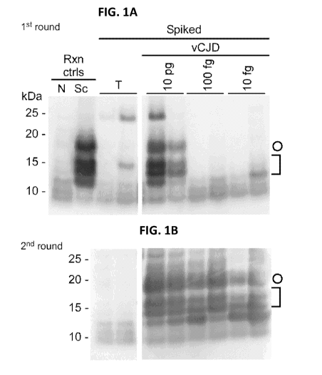
Figs. la-lb. IP-S-QuIC detection of >10 fg human vCJD PrP-res spiked
into human plasma. Dilutions of human non-prion (tumor, T) control or vCJD
brain homogenates were spiked into 500 pi of human plasma to give final
dilutions
of 4 x 10⁻⁷(T); and 4 x 10⁻⁷, 4 x 10⁻⁹, and 4 x 10⁻¹⁰ (vCJD; containing ¨ 10
pg , 100
fg and 10 fg PrP-res, respectively). PrPvcm was immunoprecipitated and
subjected
to S-QuIC as described in Materials and Methods. The first S-QuIC round was at
50
C for 8 hour (h) (Fig. la) and 1/10 of the first-round reaction volume was
used to
seed the 2' round (45 C for 10 h) (Fig. lb). Plasma-free positive and negative
control reactions were seeded directly with 2 pi of 5 x 10⁻⁷ dilutions of
hamster
uninfected (N) or scrapie (Sc) brain, the latter containing ¨100 fg PIT' seed.
Hamster rPrIpc 23-231 was used as a substrate in all reactions and comigrated
with
the 25 kDa marker. PK-digested products were analyzed by immunoblot using the
polyclonal R20 antibody as previously reported (24). Open circles mark 17-kDa
fragments and brackets indicate the lower molecular weight bands (10-13 kDa).
Figs 2a-2b. IP-S-QuIC detection of endogenous PrPsc in plasma of
scrapie-infected hamsters by IP-S-QuIC. (Fig. 2a) Plasma samples from scrapie
263K and uninfected (N) hamsters (500 pi) were subjected to IP-S-QuIC as
described in the Examples Section with the 1st round S-QuIC at 50 C for 10
hours
(h) and the 2' round (Fig. 2b) at 50 C for 8 h, except for lanes marked with
asterisks
which show the 1st round products seeded with sample #6 for comparison. Plasma-
free positive and negative control reactions, rPrIpc 23-231 substrate and
analysis of
PK-digested products were as described for Figure 1. Open circles mark 17-kDa
fragments and brackets indicate the lower molecular weight bands (10-13 kDa).
Figs. 3a-3b. IP-RT-QuIC detection of endogenous PrPsc in plasma and
serum of scrapie-infected hamsters. (Fig. 3a) IP-RT-QuIC analyses of plasma
samples from a scrapie 263K and a normal hamster, and a serum sample from a
CA 02824863 2013-07-15
WO 2012/099884 PCT/US2012/021561
- 6 -
scrapie 263K hamster. (Fig. 3b) Analyses of plasma samples from nine scrapie
263K
and one uninfected hamster. In all cases, 500 pi samples were
immunoprecipitated
using 15B3-coated beads for ¨20 h at 37 C. One fifth of the beads was pre-
incubated with 0.05% SDS in PBS at room temperature for ¨20 minutes and used
to
seed RT-QuIC containing 300 mM NaCl. RT-QuIC reactions were incubated at
42 C and hamster rPrIpc 90-231 was used as a substrate in all reactions. The
vertical
axes indicate the average fluorescence from 4 replicate reaction wells. Error
bars
show standard deviations for selected sets of replicates in (Fig. 3a). In
(Fig. 3b), all
individual reactions that registered positive fluorescence achieved nearly
identical
maximal fluorescence values (-260k units), but in many of the scrapie-seeded
cases,
only a subset of replicate reactions rose above background fluorescence within
63 h.
With such all-or-nothing responses among replicates, standard deviations
cannot be
calculated from all of the replicates; instead, on the right, the fraction of
positive
wells per total replicates is indicated at the end of the reactions. Error
bars
representing standard deviations calculated for the positive replicates (only)
at >40-h
time points barely, if at all, exceeded the size of the symbols, and therefore
are not
shown.
Figs. 4a-4b. eQuIC detection of human Prrc" spiked into human
plasma. Dilutions of human non-prion (tumor and Alzheimer's disease) control
or
vCJD brain tissues were spiked into 500 pi of human plasma to give final
dilutions
of 4 x 10⁻⁷ (tumor and Alzheimer's disease); and 4 x 10-12, 4 x 10-13 and 4 x
10-14
(vCJD; containing ¨ 100 ag, 10 ag and 1 ag PrP-res, respectively). PrPvcm was
immunoprecipitated using 15B3-coated beads (Fig. 4a) or mock anti-IgM-coated
beads (Fig. 4b) and a portion of the beads were used to seed replicate eQuIC
reactions. After 24 h the substrate was replaced. The chimeric Ha-S rPrIpc was
used
as a substrate in all reactions. The vertical axes indicate the average
fluorescence
from 4 replicate wells and the fractions on the right indicate the
positive/total
replicate reactions associated with the adjacent traces.
Figs. 5a-5b. eQuIC detection of endogenous PrPsc in plasma of scrapie-
infected hamsters. (Fig. 5a). eQuIC analysis of plasma samples (without
preclearing) from 8 uninfected hamsters and 6 scrapie-infected hamsters, with
one
CA 02824863 2013-07-15
WO 2012/099884 PCT/US2012/021561
- 7 -
collected at 30 dpi (preclinical) and 5 at 80 dpi (near-terminal). The
vertical axis
indicates the average fluorescence from 4 replicate wells and the fractions on
the
right indicate the positive/total replicate reactions associated with the
adjacent traces.
Although all replicate reactions seeded with the scrapie samples were
positive,
submaximal average fluorescence observed for 3 of the samples at 60 h. In the
latter
cases, the bead distribution in the well partially interfered with
fluorescence
readings; when such wells (n=3) were reread at 64 h after manual stirring with
a
pipette, the fluorescence achieved maximal levels (grey trace). In contrast,
stirring
uninfected control wells (n=4) did not increase their fluorescence. Hamster
rPrIpc 90-
231 was used as a substrate. (Fig. 5b). eQuIC analysis of precleared plasma
samples
from 3 uninfected and 7 scrapie-infected hamsters (3 collected at 80 dpi; 2 at
30 dpi;
1 at 10 dpi).
Fig. 6. Schematic diagram of potential mechanisms of substrate
replacement effect.
Figs. 7a-7b. Better IP-S-QuIC sensitivity and consistency of PrPsc
detection in spiked human plasma using 15B3 vs. mock beads. (Fig. 7a)
Comparison of 15B3 vs mock beads with 2-h IP from 100 pi plasma and 2-round S-
QuIC. Dilutions of hamster uninfected "normal" (N) or scrapie 263K (Sc) brain
homogenates were spiked into 100 pi of human plasma to give final brain
dilutions
of 10-8(N); and 10-8, 10-9 and 10-10 (Sc; ¨100, 10, and 1 fg PrP-res,
respectively).
PrPse was immunoprecipitated using 40 pi (1.6 x 10⁷ total beads) of 15B3-
coated
beads (15B3) or mock anti-IgM-coated beads (C) for 2 h at 37 C. Beads were
resuspended in 10 pi of PBS. One fifth of the beads was used to seed a 1st
round 5-
QuIC at 50 C for 10 h and 1/10 of the 1St round reaction volume was used to
seed the
2' round (50 C for 10 h). (Fig. 7b) 15B3 beads with 20-h IP from 500 pi plasma
and single-round S-QuIC. Dilutions of N or Sc brain homogenates were spiked
into
500 pi of human plasma to give final brain tissue dilutions of 2 x 10⁻⁸ (N);
and 2 x
10-8¨ 2 x 10-11 (Sc; containing ¨1 pg-1 fg PrP-res ,respectively). PrPse was
immunoprecipitated using 15B3 beads for ¨20 h at 37 C. The remainder of the
protocol was as in (Fig. 7a) except that only a single-round S-QuIC at 50 C
for 10 h
was performed. Plasma-free positive and negative control reactions were seeded
CA 02824863 2013-07-15
WO 2012/099884
PCT/US2012/021561
- 8 -
directly with 2 pi of 5 x i07 dilutionsof hamster N or Sc brain, the latter
containing
¨100 fg PrP-res seed. Hamster rPrIpc 23-231 was used as a substrate in all S-
QuIC
reactions. PK-digested products were analyzed by immunoblot using the
polyclonal
R20 antibody as previously reported (Orru et al., 2009. Protein Eng Des Sel
22:515-
521, 2009). Open circles mark 17-kDa fragments and brackets indicate the lower
molecular weight bands (10-13 kDa).
Figs. 8a-8b. SDS pre-treatment of 15B3-bound PrPsc accelerates RT-
QuIC detection. Dilutions of hamster N or Sc 263K brain homogenates were
spiked into 500 pi of human plasma to give final brain dilutions of 2 x 10⁻⁷
(containing ¨10 pg PrP-res in the case of Sc). IP incubations with beads were
for
¨20 h at 37 C. One fifth of the beads was used to seed RT-QuIC (Fig. 8a) and
an
equivalent number of beads was pre-incubated with 0.05% SDS in PBS at room
temperature for ¨20 minutes and used to seed RT-QuIC reactions containing 300
mM NaC1 (Fig. 8b). Reactions were incubated at 42 C and hamster rPrIpc 90-231
was used as a substrate in all reactions. The vertical axis indicates the
average
fluorescence from 4 replicate wells and the fractions on the right indicate
the
positive/total replicate reactions associated with the adjacent traces.
Figs 9a-9c. Improved RT-QuIC detection of 15B3-bound human
PrPvc" with hamster-sheep chimeric rPrl3c (Ha-S rPrPc) vs. human rPrl3c 23-
231 with NaC1 variation. Dilutions of human non-prion (tumor) control or vCJD
brain tissues were spiked into 500 pi of human plasma to give final dilutions
of 4 x
10-7 (Figs. 9a-9b), containing ¨10 pg PrP-res, in the case of vCJD) or 4 x 10-
7 and 4
x 10-9 and 4 x 1040 (Fig. 9c, containing ¨10 pg, 100 fg and 10 fg PrPr' ,
respectively in the case of vCJD). The samples were subjected to IP-RT-QuIC as
described in the Materials and Methods section except for indicated variations
in
NaC1 concentration and rPrIpc substrate. The vertical axis indicates the
average
fluorescence from 4 replicate wells and the fractions on the right indicate
the
positive/total replicate reactions associated with the adjacent traces.
Figs. 10a-10b. Comparison of 15B3 beads to Magnabeads in eQuIC.
Dilutions of human non-TSE Alzheimer's disease (AD) control or vCJD brain
tissues were spiked into 0.5 ml of human plasma to give final dilutions of 4 x
10-7
CA 02824863 2013-07-15
WO 2012/099884 PCT/US2012/021561
- 9 -
(AD); and 4 x 10⁻⁷, 4 x 10⁻¹⁰, 4 x 10-13 and 4 x 10-14 (vCJD; containing ¨ 10
pg, 10
fg, 10 ag and 1 ag PrP-res, respectively). PrPvcm was immunoprecipitated using
1.6
x 107 total 15B3-coated beads (Fig. 10a) or an equivalent number of
MAGNABINDTM beads (Fig. 10b) for ¨20 h at 37 C using immunoprecipitation
buffer (Prionics). Beads were washed twice with wash buffer (Prionics) and
resuspended in 10 pi of PBS. The remainder of the protocol was as described in
Material and Methods, starting with the preincubation in 0.05% SDS in PBS. The
vertical axis indicates the average fluorescence from four replicate wells and
the
fractions on the right indicate the positive/total replicate reactions
associated with
the adjacent traces. Similar results were obtained using the MAGNABINDTM prpse
capture conditions (Miller and Supattapone, 2011, J.Virol. 85:2813-2817).
Fig. 11. Improved speed & sensitivity of IP-RTQ using higher 15B3
content on beads. Dilutions of hamster normal (NBH) or scrapie 263K brain
homogenates were spiked into 500 pi of human plasma to give final brain tissue
dilutions of 2 x 10⁻⁹ (NBH); and 2 x 10⁻⁹ and 2 x 10⁻¹⁰ (263K; containing ¨100
or 10
fg PrP-res, respectively). PrP-res was immunoprecipitated using 40 pi of 15B3
beads for ¨20 hours at 37 C. Beads were washed twice with 0.2% Sarkosyl/TBS
and resuspended in 10 pi of PBS. Following a 0.05% SDS pre-treatment, one
fifth of
the beads was used to seed RTQ reactions. Hamster rPrIpc 90-231 was used as a
substrate in all reactions. The vertical axis indicates the average
fluorescence from 2
replicate wells.
Fig. 12. 15B3 antibody titration for eQuIC detection of sheep scrapie
brain homogenate spiked into plasma. The results demonstrate that increasing
the
amount of 15B3 results in improved sensitivity of the sheep eQuIC, which
allows
faster detection of Sheep scrapie brain tissue dilutions containing L-100fg of
PrP-res
in 0.5 ml of plasma.
Fig. 13. eQuIC detection of ARQ sheep brain homogenate in spiked
sheep plasma. The assay provided detection of >-100ag PrP-res (5 x 10-13
dilution
of brain tissue) in 500u1 of sheep plasma.
Fig. 14. eQuIC detection of endogenous PrP-res in plasma of scrapie
positive sheep. The assay detected endogenous PrP-res in plasma from three
CA 02824863 2013-07-15
WO 2012/099884
PCT/US2012/021561
- 10 -
slinically affected scrapie-infected sheep. No prions were detected in plasma
samples from four non-infected sheep.
Fig. 15. Sensitivity of detection of sCJD brain homogenate spiked into
plasma by e-QuIC. The assay detected sCJD brain homogenate spikes containing
as little as ¨10ag of PIT' in 0.5mL of human plasma.
Fig. 16. Sensitivity of detection of sCJD brain homogenate spiked into
cerebrospinal fluid (CSF) by e-QuIC. The assay detected sCJD brain tissue
dilutions containing as little as ¨ 10ag of PrPres in 0.5 ml of human
cerebrospinal
fluid.
Fig. 17. Sensitivity of detection of mouse-adapted RML scrapie brain
homogenate spiked into plasma by e-QuIC. The assay detected down to 10-13
RML scrapie brain tissue dilutions (containing ¨ 100ag_ of PrP-res) in 0.5 ml
of
plasma.
Fig. 18. eQuIC detection of endogenous PrPres in plasma of scrapie
positive wild type (WT) & GPI- mice. The assay detected endogenous PrP-res in
plasma from a wild-type mouse and a transgenic mouse expressing only PrP-sen
that
lacks the glycophosphatidylinositol anchor (GPI). No prions were detected
in a plasma sample from a non-infected wild type normal mouse.
SEQUENCE LISTING
The nucleic and amino acid sequences listed are shown using standard letter
abbreviations for nucleotide bases, and three letter code for amino acids, as
defined
in 37 C.F.R. 1.822. For nucleic acid sequences, only one strand of each
nucleic acid
sequence is shown, but the complementary strand is understood as included by
any
reference to the displayed strand.
SEQ ID NO: 1 is an amino acid sequence of a recombinant Syrian golden
hamster proteinase K-sensitive prion protein.
KKRPKPGGWNTGGSRYPGQGSPGGNRYPPQGGGTWGQPHGGGWGQPHGGGWGQPHGGGWGQ
PHGGGWGQGGGTHNQWNKPS KPKTNMKHMAGAAAAGAVVGGEGGYMEGS AMSRPMMHFGN
DWEDRYYRENMNRYPNQVYYRPVDQYNNQNNFVHDCVNITIKQHTVITTTKGENFI ETDIKIME
RVVEQMCTTQYQKESQAYYDGRRS
CA 02824863 2013-07-15
WO 2012/099884
PCT/US2012/021561
- 11 -
SEQ ID NO: 2 is an amino acid sequence of a recombinant mouse (Prnp-a)
proteinase K-sensitive prion protein.
KKRPKPGG WNTGGSRYPG QGSPGGNRYP PQGGTWGQPH GGGWGQPHGG SWGQPHGGSW
GQPHGGGWGQ GGGTHNQWNK PSKPKTNLKH VAGAAAAGAV VGGLGGYMLG
SAMSRPMIHF GNDWEDRYYR ENMYRYPNQV YYRPVDQYSN QNNFVHDCVN ITIKQHTVTT
TTKGENFILT DVKMMERVVE QMCVTQYQKE SQAYYDGRRS
SEQ ID NO: 3 is an amino acid sequence of a recombinant human (129M)
proteinase K-sensitive prion protein.
KKRPKPGG WNTGGSRYPG QGSPGGNRYP PQGGGGWGQP HGGGWGQPHG GGWGQPHGGG
WGQPHGGGWG QGGGTHSQWN KPSKPKTNMK HMAGAAAAGA VVGGLGGYML
GSAMSRPIIH FGSDYEDRYY RENMHRYPNQ VYYRPMDEYS NQNNFVHDCV NITIKQHTVT
TTTKGENFTE TDVKMMERVV EQMCITQYER ESQAYYQRGS S
SEQ ID NO: 4 is an amino acid sequence of a recombinant human (129V)
proteinase K-sensitive prion protein.
KKRPKPGG WNTGGSRYPG QGSPGGNRYP PQGGGGWGQP HGGGWGQPHG GGWGQPHGGG
WGQPHGGGWG QGGGTHSQWN KPSKPKTNMK HMAGAAAAGA VVGGLGGYVL
GSAMSRPIIH FGSDYEDRYY RENMHRYPNQ VYYRPMDEYS NQNNFVHDCV NITIKQHTVT
TTTKGENFTE TDVKMMERVV EQMCITQYER ESQAYYQRGS S
SEQ ID NO: 5 is an amino acid sequence of a recombinant bovine (6-
octarepeat) proteinase K-sensitive prion protein.
KKRPKP GGGWNTGGSR YPGQGSPGGN RYPPQGGGGW GQPHGGGWGQ PHGGGWGQPH
GGGWGQPHGG GWGQPHGGGG WGQGGTHGQW NKPSKPKTNM KHVAGAAAAG
AVVGGLGGYM LGSAMSRPLI HFGSDYEDRY YRENMHRYPN QVYYRPVDQY SNQNNFVHDC
VNITVKEHTV TITTKGENFI ETDIKMMERV VEQMCITQYQ RESQAYYQRG As
SEQ ID NO: 6 is an amino acid sequence of a recombinant ovine (136A
154R 171Q) proteinase K-sensitive prion protein.
154R 171Q) proteinase K-sensitive prion protein.
KKRPKP GGGWNTGGSR YPGQGSPGGN RYPPQGGGGW GQPHGGGWGQ PHGGGWGQPH
GGGWGQPHGG GGWGQGGSHS QWNKPSKPKT NMKHVAGAAA AGAVVGGLGG
YMLGSAMSRP LIHFGNDYED RYYRENMYRY PNQVYYRPVD QYSNQNNFVH DCVNITVKQH
TVTTTTKGEN FILTDIKIME RVVEQMCITQ YQRESQAYYQ RGAS
CA 02824863 2013-07-15
WO 2012/099884
PCT/US2012/021561
- 12 -
SEQ ID NO: 7 is an amino acid sequence of a recombinant Deer (96G
132M 138S) proteinase K-sensitive prion protein.
KKRPKP GGGWNTGGSR YPGQGSPGGN RYPPQGGGGW GQPHGGGWGQ PHGGGWGQPH
GGGWGQPHGG GGWGQGGTHS QWNKPSKPKT NMKHVAGAAA AGAVVGGLGG
YMLGSAMSRP LIHFGNDYED RYYRENMYRY PNQVYYRPVD QYNNQNTFVH DCVNITVKQH
TVTTTTKGEN FILTDIKMME RVVEQMCITQ YQRESQAYYQ RGAS
SEQ ID NO: 8 is an amino acid sequence of a full-length Syrian golden
hamster proteinase K-sensitive prion protein.
MANLSYWLLALFVAMWTDVGLCKK RPKPGGWNTG GSRYPGQGSP GGNRYPPQGG
GTWGQPHGGG WGQPHGGGWG QPHGGGWGQP HGGGWGQGGG THNQWNKPSK
PKTNMKHMAG AAAAGAVVGG LGGYMLGSAM SRPMMHFGND WEDRYYRENM
NRYPNQVYYR PVDQYNNQNN FVHDCVNITI KQHTVTTTTK GENFTETDIK IMERVVEQMC
TTQYQKESQA YYDGRRSSAV LFSSPPVILL ISFLIFLMVG
SEQ ID NO: 9 is an amino acid sequence of a full-length mouse (Prnp-a)
proteinase K-sensitive prion protein.
MANLGYWLLA LFVTMWTDVG LCKKRPKPGG WNTGGSRYPG QGSPGGNRYP PQGGTWGQPH
GGGWGQPHGG SWGQPHGGSW GQPHGGGWGQ GGGTHNQWNK PSKPKTNLKH
VAGAAAAGAV VGGLGGYMLG SAMSRPMIHF GNDWEDRYYR ENMYRYPNQV
YYRPVDQYSN QNNFVHDCVN ITIKQHTVTT TTKGENFILT DVKMMERVVE QMCVTQYQKE
SQAYYDGRRS SSTVLFSSPP V1LLISFLIF LIVG
SEQ ID NO: 10 is an amino acid sequence of a full-length human (129M)
proteinase K-sensitive prion protein.
MANLGCWMLV LFVATWSDLG LCKKRPKPGG WNTGGSRYPG QGSPGGNRYP PQGGGGWGQP
HGGGWGQPHG GGWGQPHGGG WGQPHGGGWG QGGGTHSQWN KPSKPKTNMK
HMAGAAAAGA VVGGLGGYML GSAMSRPIIH FGSDYEDRYY RENMHRYPNQ VYYRPMDEYS
NQNNFVHDCV NITIKQHTVT ITTKGENFIL TDVKMMERVV EQMCITQYER ESQAYYQRGS
SMVLFSSPPV 1LLISFLIFL IVG
SEQ ID NO: 11 is an amino acid sequence of a full-length human (129V)
proteinase K-sensitive prion protein.
MANLGCWMLV LFVATWSDLG LCKKRPKPGG WNTGGSRYPG QGSPGGNRYP PQGGGGWGQP
HGGGWGQPHG GGWGQPHGGG WGQPHGGGWG QGGGTHSQWN KPSKPKTNMK
CA 02824863 2013-07-15
WO 2012/099884 PCT/US2012/021561
- 13 -
HMAGAAAAGA VVGGLGGYVL GSAMSRPIIH FGSDYEDRYY RENMHRYPNQ VYYRPMDEYS
NQNNFVHDCV NITIKQHTVT TTTKGENFTE TDVKMMERVV EQMCITQYER ESQAYYQRGS
SMVLFSSPPV ILLISFLIFL IVG
SEQ ID NO: 12 is an amino acid sequence of a full-length chimeric
Hamster-Sheep (H-S) proteinase K-sensitive prion protein wherein residues 23-
137
are of the Syrian hamster sequence and the remaining residues 138-231 were
homologous to sheep residues 141-234 (R154,Q171 polymorph).
HMKKRPKPGGWNTGGSRYPGQGSPGGNRYPPQGGGTWGQPHGGGWGQPHGGGWGQPHG
GGWGQPHGGGWGQGGGTHNQWNKPSKPKTNMKHMAGAAAAGAVVGGLGGYMLGS AM
SRPLIHFGNDYEDRYYRENMYRYPNQVYYRPVDQYSNQNNFVHDCVNITVKQHTVTTTTK
GENFTETDIKIMERVVEQMCITQYQRESQAYYQRGAS.
DETAILED DESCRIPTION OF SEVERAL EMBODIMENTS
The methods disclosed herein allow testing for prion contamination,
diagnostics and/or surveillance in a number of biological samples, including
blood,
blood fractions, blood products, urine, nasal fluids, saliva, cerebral spinal
fluid,
feces, muscle biopsies, lymphoid tissues, skin samples, samples of tissues for
transplantation, amongst others. These methods have medical and veterinary
applications, and also can be used to test biotechnology products and
environmental
samples (such as water, soils, plants, landfills, sewage) and agriculture
samples
(such as animal-based foods, animal-based feeds & nutritional supplements,
animal
waste products, byproducts, carcasses, slaughterhouse wastes, specified risk
materials) to ensure there is no contamination by prions. The presently
disclosed
methods also can be used for prion-free herd/flock certification, such as in
cattle,
sheep, and cervids. The methods disclosed herein can also be used to detect
spontaneous Creutzfeldt-Jacob disease.
Currently, the most direct and reliable assay for the detection of TSE
infectivity is animal bioassay. Quantification of infectivity can be achieved
by end-
point (Stamp et al., 1959) or limiting dilution bioassays (Gregori et al.,
2004). For
some combinations of prion agent and host species, strong correlations between
infectivity titer and disease incubation period have been established in
laboratory
rodents, allowing the use of incubation period to measure infectivity levels
(Hunter
CA 02824863 2013-07-15
WO 2012/099884 PCT/US2012/021561
- 14 -
et al., 1963;Prusiner et al., 1982). The disadvantage of these bioassays is
that they
are animal-intensive, time-consuming and expensive. For certain murine-adapted
scrapie strains, the cell culture based standard scrapie cell assay (SSCA) can
also be
used to measure infectivity levels by end-point and limiting dilution methods
(Klohn
et al., 2003). The SSCA offers several advantages over animal bioassays, but
it still
requires weeks to perform and has been limited to a few mouse-adapted scrapie
strains. An analogous cell-based assay for cervid prions (designated CPCA) has
also
been reported (Bian et al., J Virol, 84, 8322-8326, 2010). The limitations of
the
animal bioassay, SSCA and CPCA, mean that more practical assays for prion
quantitation are needed.
A number of highly sensitive in vitro methods for prion detection have been
reported (Atarashi et al., Nat Methods, 4, 645-650, 2007; Atarashi et al., Nat
Methods, 5, 211-212, 2008; Bieschke et al., Proc Natl Acad Sci USA, 97, 5468-
5473, 2000; Chang et al., J Virol Methods, 159, 15-222009; Colby et al., Proc
Natl
Acad Sci U SA, 104, 9741-9746, 2007; Fujihara et al., FEBS J, 276, 2841-2848,
2009; Orru et al., Protein Eng Des Sel, 22, 515-521, 2009; Rubenstein et al.,
J Gen
Virol, 91, 1883-1892, 2010; Saa et al., Science, 313, 92-94, 2006a; Saa et
al., J Biol
Chem, 281, 35245-35252, 2006b; Terry et al., J Virol, 83, 12552-12558, 2009;
Trieschmann et al., BMC Biotechnol, 5, 26, 2005; Wilham et al., PLoS Pathog,
6,
e1001217, 2010). Fluorescence correlation spectroscopy can be used to detect
femtomolar concentrations of PrP-res aggregates in cerebral spinal fluid (CSF)
samples treated with fluorescently tagged antibodies (Bieschke et al., supra,
2000).
The binding of fluorescently labeled recombinant PrPc (rPrPe) to synthetic
prion
protein aggregates allowed their ultra-sensitive detection by FACS analyses
and a
similar approach allowed the discrimination of sera of several BSE-infected
and
non-infected cattle (Trieschmann et al., 2005). Using the protein misfolding
cyclic
amplification (PMCA) reactions in multi-round sonicated reactions using brain-
derived PrPc as a substrate, as little as 1 ag of PrP-res can be detected (Saa
et al.,
supra, 2006b). Coupling of limited serial PMCA with highly sensitive
fluorescence
detection technique called surround optical fiber immunoassay (SOPHIA) allows
more rapid detection of as little as 10 ag PrP-res and discrimination of prion-
infected
CA 02824863 2013-07-15
WO 2012/099884
PCT/US2012/021561
- 15 -
versus uninfected blood samples (Chang et al., supra, 2009;Rubenstein et al.,
. J
Gen Virol, 91, 1883-1892, 2010).
The speed and practicality of PMCA assays has also been improved by the
use of rPrPe (Atarashi et al., supra, 2007) and by substituting shaking for
the
sonication step as described for the quaking-induced conversion (QuIC)
reactions
(Atarashi et al., supra, 2008; Orru et al., supra, 2009). The standard QuIC
(also
called "SQ") assay can detect sub-femtogram amounts of PrP-res (less than one
lethal intracerebral dose) in hamster brain homogenates (BH) within a single
day.
The effectiveness of the SQ for prion detection was demonstrated by its
ability to
discriminate normal from prion-infected hamsters using 2-[il samples of CSF
(Atarashi et al., supra, 2008;Orru et al., supra, 2009) or nasal lavage
(Bessen et al.,
PLoS Pathogens, 6, e1000837, 2010). Adaptations of SQ reactions have led to
the
sensitive detection of variant CJD (vCJD) in human tissue and scrapie in sheep
tissue (Orru et al., supra, 2009).
The readout for SQ and PMCA assays is the detection of specific protease-
resistant prion-seeded rPrP products by immunoblotting, which is difficult to
adapt
to automated high-throughput formats. An alternative, and potentially higher-
throughput approach was used for the amyloid seeding assay (ASA) in which the
fluorescent dye thioflavin T (ThT) was used to detect prion seeding of rPrIpc
polymerization (Colby et al., Proc Natl Acad Sci USA, 104, 20914-20919, 2007,
incorporated herein by reference). The ASA can also detect protease sensitive
disease-causing prions and has a 98% correlation with neuropathological signs
of
prion disease (Colby et al., PLoS Pathog, 6, e1000736, 2010). However, a
potentially confounding aspect of ASA is the frequent spontaneous formation of
rPrP fibrils (without seeding by prions) within about twice the lag phase of
prion-
seeded reactions (Colby et al., Proc Natl Acad Sci U SA, 104, 20914-20919,
2007).
The problem of spontaneous fibril formation is greatly reduced in another
prion-
seeded rPrPe polymerization assay, real-time (RT)-QuIC (also called RTQ, see,
for
example, Wilham et al., PLoS Pathog, 6, e1001217, 2010, which describes the
assay
and is incorporated herein by reference) which combines several aspects of the
SQ
assay (intermittent shaking, rPrIpc preparation, sample preparation, and a
lack of
CA 02824863 2013-07-15
WO 2012/099884 PCT/US2012/021561
- 16 -
chaotropic salts) with a fluorescent ThT readout like that of the ASA.
Until recently, a major limitation of the PMCA, SQ, RTQ and ASA methods
was the lack of prion quantitation. Chen and colleagues reported a method
called
quantitative PMCA (qPMCA) in which PrPse content is estimated by the number of
PMCA rounds necessary for a positive response (Chen et al., Nat Methods, 7,
519-
520, 2010). More recently, a different approach, using end-point dilution
titration,
was described in conjunction with the RTQ as a method for determining relative
prion quantitation with in vitro prion seeding assays (Wilham et al., supra,
2010).
Moreover, prion seeding activity was measured in the nasal fluids and CSF of
prion-
infected hamsters. Thus, in conjunction with the end-point dilution analysis,
the
RTQ can rapidly determine relative prion concentrations with a sensitivity
that rivals
that of animal bioassays, but with greatly reduced time and cost.
The presently described methods substantially improve the sensitivity and
applicability of prion seeding/amplification assays such as the SQ and RTQ, in
part
by integrating them with novel prion/PrP-res/PrPse immunoprecipitation and
treatment protocols. The methods enable the capture and detection of extremely
low
levels of prions in various fluids or tissue extracts, including complex
biological
specimens such as blood plasma, which can contain strong inhibitors of prions.
Terms
Unless otherwise noted, technical terms are used according to conventional
usage. Definitions of common terms in molecular biology may be found in
Benjamin Lewin, Genes V, published by Oxford University Press, 1994 (ISBN 0-19-
854287-9); Kendrew et al. (eds.), The Encyclopedia of Molecular Biology,
published
by Blackwell Science Ltd., 1994 (ISBN 0-632-02182-9); and Robert A. Meyers
(ed.),
Molecular Biology and Biotechnology: a Comprehensive Desk Reference, published
by VCH Publishers, Inc., 1995 (ISBN 1-56081-569-8).
In order to facilitate review of the various embodiments of this disclosure,
the
following explanations of specific terms are provided:
Aggregate: More than one molecule in association, such as dimers,
multimers, and polymers of prion proteins, for instance aggregates, dimers,
CA 02824863 2013-07-15
WO 2012/099884 PCT/US2012/021561
- 17 -
multimers, and polymers of PrP-res or rPrP-res(se) .
Agitation: Introducing any type of turbulence or motion into a mixture or
reaction mix, for examples by sonication, stirring, or shaking. In some
embodiments, agitation includes the use of force sufficient to fragment rPrP-
res(se)
aggregates, which disperses rPrP-res(se) aggregates and/or polymers to
facilitate
further amplification. In some examples fragmentation includes complete
fragmentation, whereas in other examples, fragmentation is only partial, for
instance, a population of aggregates can be about 1%, 2%, 5%, 10%, 20%, 30%,
40%, 50%, 60%, 70%, 80%, 90%, or 100% fragmented by agitation. Exemplary
agitation methods are described in the Examples section below.
Antibody: A polypeptide ligand comprising at least a light chain or heavy
chain immunoglobulin variable region which specifically recognizes and binds
an
epitope of an antigen or a fragment thereof. An antibody can specifically bind
PrP-
res/PrPse. Antibodies can be composed of a heavy and a light chain, each of
which
has a variable region, termed the variable heavy (VH) region and the variable
light
(VL) region. Together, the VH region and the VL region are responsible for
binding
the antigen recognized by the antibody.
The term antibody includes intact immunoglobulins and the variants and
portions of them well known in the art, such as Fab' fragments, F(ab)'2
fragments,
single chain Fv proteins ("scFv"), and disulfide stabilized Fv proteins
("dsFv"). A
scFv protein is a fusion protein in which a light chain variable region of an
immunoglobulin and a heavy chain variable region of an immunoglobulin are
bound
by a linker, while in dsFvs, the chains have been mutated to introduce a
disulfide
bond to stabilize the association of the chains. The term also includes
genetically
engineered forms such as chimeric antibodies (for example, humanized
antibodies),
heteroconjugate antibodies (such as, bispecific antibodies). See also, Pierce
Catalog
and Handbook, 1994-1995 (Pierce Chemical Co., Rockford, IL); Kuby, J.,
Immunology, 3rd Ed., W.H. Freeman & Co., New York, 1997.
Typically, a naturally occurring immunoglobulin has heavy (H) chains and
light (L) chains interconnected by disulfide bonds. There are two types of
light
chain, lambda (X) and kappa (k). There are five main heavy chain classes (or
CA 02824863 2013-07-15
WO 2012/099884
PCT/US2012/021561
- 18 -
isotypes) which determine the functional activity of an antibody molecule:
IgM, IgD,
IgG, IgA and IgE.
Each heavy and light chain contains a constant region and a variable region,
(the regions are also known as "domains"). In combination, the heavy and the
light
chain variable regions specifically bind the antigen. Light and heavy chain
variable
regions contain a "framework" region interrupted by three hypervariable
regions,
also called "complementarity-determining regions" or "CDRs". The extent of the
framework region and CDRs have been defined (see, Kabat et al., Sequences of
Proteins of Immunological Interest, U.S. Department of Health and Human
Services,
1991, which is hereby incorporated by reference). The Kabat database is now
maintained online. The sequences of the framework regions of different light
or
heavy chains are relatively conserved within a species. The framework region
of an
antibody, that is the combined framework regions of the constituent light and
heavy
chains, serves to position and align the CDRs in three-dimensional space.
The CDRs are primarily responsible for binding to an epitope of an antigen.
The CDRs of each chain are typically referred to as CDR1, CDR2, and CDR3,
numbered sequentially starting from the N-terminus, and are also typically
identified
by the chain in which the particular CDR is located. Thus, a VH CDR3 is
located in
the variable domain of the heavy chain of the antibody in which it is found,
whereas
a VL CDR1 is the CDR1 from the variable domain of the light chain of the
antibody
in which it is found. An antibody that binds an antigen of interest has a
specific VH
region and the VL region sequence, and thus specific CDR sequences. Antibodies
with different specificities (due to different combining sites for different
antigens)
have different CDRs. Although it is the CDRs that vary from antibody to
antibody,
only a limited number of amino acid positions within the CDRs are directly
involved
in antigen binding. These positions within the CDRs are called specificity
determining residues (SDRs).
References to "VH" or "VH" refer to the variable region of an
immunoglobulin heavy chain, including that of an Fv, scFv, dsFy or Fab.
References to "VC or "VL" refer to the variable region of an immunoglobulin
light
chain, including that of an Fv, scFv, dsFy or Fab.
CA 02824863 2013-07-15
WO 2012/099884 PCT/US2012/021561
- 19 -
A "monoclonal antibody" is an antibody produced by a single clone of
B-lymphocytes or by a cell into which the light and heavy chain genes of a
single
antibody have been transfected, or a progeny thereof. Monoclonal antibodies
are
produced by methods known to those of skill in the art, for instance by making
hybrid antibody-forming cells from a fusion of myeloma cells with immune
spleen
cells. Monoclonal antibodies include humanized monoclonal antibodies.
Antibody binding affinity: Affinity of an antibody for an antigen, such as
PrP-res. In one embodiment, affinity is calculated by a modification of the
Scatchard method described by Frankel et al., Mol. Immunol., 16:101-106, 1979.
In
another embodiment, binding affinity is measured by an antigen/antibody
dissociation rate. In yet another embodiment, a high binding affinity is
measured by
a competition radioimmunoas say. In several examples, a high binding affinity
is at
least about 1 x 10⁻⁸ M. In other embodiments, a high binding affinity is at
least
about 1.5 x 10⁻⁸M, at least about 2.0 x 10⁻⁸M, at least about 2.5 x 10⁻⁸M, at
least
about 3.0 x 10⁻⁸M, at least about 3.5 x 10⁻⁸M, at least about 4.0 x 10⁻⁸M, at
least
about 4.5 x 10⁻⁸M, or at least about 5.0 x 10⁻⁸ M.
Antigen: A compound, composition, or substance that can stimulate the
production of antibodies or a T-cell response in an animal, including
compositions
that are injected or absorbed into an animal. An antigen reacts with the
products of
specific humoral or cellular immunity, including those induced by heterologous
immunogens. The term "antigen" includes all related antigenic epitopes.
"Epitope"
or "antigenic determinant" refers to a site on an antigen to which B and/or T-
cells
respond. In one embodiment, T-cells respond to the epitope, when the epitope
is
presented in conjunction with an MHC molecule. Epitopes can be formed both
from
contiguous amino acids or noncontiguous amino acids juxtaposed by tertiary
folding
of a protein. Epitopes formed from contiguous amino acids are typically
retained on
exposure to denaturing solvents whereas epitopes formed by tertiary folding
are
typically lost on treatment with denaturing solvents. An epitope typically
includes at
least 3, and more usually, at least 5, about 9, or about 8-10 amino acids in a
unique
spatial conformation. Methods of determining spatial conformation of epitopes
CA 02824863 2013-07-15
WO 2012/099884
PCT/US2012/021561
- 20 -
include, for example, x-ray crystallography and 2-dimensional nuclear magnetic
resonance. An antigen can be a tissue-specific antigen, or a disease-specific
antigen,
such as PrP-res. These terms are not exclusive, as a tissue-specific antigen
can also
be a disease specific antigen.
Conservative variant: In the context of a prion protein, refers to a peptide
or amino acid sequence that deviates from another amino acid sequence only in
the
substitution of one or several amino acids for amino acids having similar
biochemical properties (so-called conservative substitutions). Conservative
amino
acid substitutions are likely to have minimal impact on the activity of the
resultant
protein. Further information about conservative substitutions can be found,
for
instance, in Ben Bassat et al. (J. Bacteriol., 169:751-757, 1987), O'Regan et
al.
(Gene, 77:237-251, 1989), Sahin-Toth et al. (Protein Sci., 3:240-247, 1994),
Hochuli et al. (Bio/Technology, 6:1321-1325, 1988) and in widely used
textbooks of
genetics and molecular biology. In some examples, prion protein variants can
have
no more than 1, 2, 3, 4, 5, 10, 15, 30, 45, or more conservative amino acid
changes.
In one example, a conservative variant prion protein is one that functionally
performs substantially like a similar base component, for instance, a prion
protein
having variations in the sequence as compared to a reference prion protein.
For
example, a prion protein or a conservative variant of that prion protein, will
aggregate with PrP-res (or PrPse), for instance, and will convert rPrIpc to
rPrP-res(se)
(or will be converted to rPrP-res(se)). In this example, the prion protein and
the
conservative variant prion protein do not have the same amino acid sequences.
The
conservative variant can have, for instance, one variation, two variations,
three
variations, four variations, or five or more variations in sequence, as long
as the
conservative variant is still complementary to the corresponding prion
protein.
In some embodiments, a conservative variant prion protein includes one or
more conservative amino acid substitutions compared to the prion protein from
which it was derived, and yet retains prion protein biological activity. For
example,
a conservative variant prion protein can retain at least 10% of the biological
activity
of the parent prion protein molecule from which it was derived, or
alternatively, at
least 20%, at least 30%, or at least 40%. In some preferred embodiments, a
CA 02824863 2013-07-15
WO 2012/099884 PCT/US2012/021561
- 21 -
conservative variant prion protein retains at least 50% of the biological
activity of
the parent prion protein molecule from which it was derived. The conservative
amino acid substitutions of a conservative variant prion protein can occur in
any
domain of the prion protein.
Contacting: "Contacting" includes in solution and solid phase, for example
contacting a sample with a specific binding agent, such as an antibody that
specifically binds PrP-res.
Conditions sufficient to detect: Any environment that permits the desired
activity, for example, that permits an antibody to bind an antigen, such as
PrP-res,
and the interaction to be detected. For example, such conditions include
appropriate
temperatures, buffer solutions, and detection means such as and digital
imaging
equipment.
Detect: To determine if an agent (such as a signal or protein, for example
PrP-res) is present or absent. In some examples, this can further include
quantification, for example the quantification of the amount of PrP-res in a
sample,
such as a serum sample, or a fraction of a sample.
Diagnostic: Identifying the presence or nature of a pathologic condition,
such as, but not limited to, identifying the presence of PrP-res, such as in
Creutzfeldt-Jacob disease. Diagnostic methods differ in their sensitivity and
specificity. The "sensitivity" of a diagnostic assay is the percentage of
diseased
individuals who test positive (percent of true positives). The "specificity"
of a
diagnostic assay is 1 minus the false positive rate, where the false positive
rate is
defined as the proportion of those without the disease who test positive.
While a
particular diagnostic method may not provide a definitive diagnosis of a
condition, it
suffices if the method provides a positive indication that aids in diagnosis.
"Prognostic" is the probability of development (for example severity) of a
pathologic condition.
Disaggregate: To partially or complete disrupt an aggregate, such as an
aggregate of PrP-res or rPrP-res(se).
Encode: Any process whereby the information in a polymeric
macromolecule or sequence is used to direct the production of a second
molecule or
CA 02824863 2013-07-15
WO 2012/099884
PCT/US2012/021561
- 22 -
sequence that is different from the first molecule or sequence. As used
herein, the
term is construed broadly, and can have a variety of applications. In some
aspects,
the term "encode" describes the process of semi-conservative DNA replication,
wherein one strand of a double-stranded DNA molecule is used as a template to
encode a newly synthesized complementary sister strand by a DNA-dependent DNA
polymerase.
In another aspect, the term "encode" refers to any process whereby the
information in one molecule is used to direct the production of a second
molecule
that has a different chemical nature from the first molecule. For example, a
DNA
molecule can encode an RNA molecule (for instance, by the process of
transcription
incorporating a DNA-dependent RNA polymerase enzyme). Also, an RNA
molecule can encode a peptide, as in the process of translation. When used to
describe the process of translation, the term "encode" also extends to the
triplet
codon that encodes an amino acid. In some aspects, an RNA molecule can encode
a
DNA molecule, for instance, by the process of reverse transcription
incorporating an
RNA-dependent DNA polymerase. In another aspect, a DNA molecule can encode
a peptide, where it is understood that "encode" as used in that case
incorporates both
the processes of transcription and translation.
Fluorophore: A chemical compound, which when excited by exposure to a
particular stimulus, such as a defined wavelength of light, emits light
(fluoresces),
for example at a different wavelength (such as a longer wavelength of light).
Fluorophores are part of the larger class of luminescent compounds.
Luminescent
compounds include chemiluminescent molecules, which do not require a
particular
wavelength of light to luminesce, but rather use a chemical source of energy.
Therefore, the use of chemiluminescent molecules (such as aequorin) can
eliminate
the need for an external source of electromagnetic radiation, such as a laser.
Examples of particular fluorophores that can attached to antibodies that
specifically binds PrPse are provided in U.S. Patent No. 5,866,366 to
Nazarenko et
al., such as 4-acetamido-4'-isothiocyanatostilbene-2,2'disulfonic acid,
acridine and
derivatives such as acridine and acridine isothiocyanate, 5-(2'-
CA 02824863 2013-07-15
WO 2012/099884
PCT/US2012/021561
-23 -
aminoethyl)aminonaphthalene-l-sulfonic acid (EDANS), 4-amino-N-[3-
vinylsulfonyl)phenylinaphthalimide-3,5 disulfonate (Lucifer Yellow VS), N-(4-
anilino-l-naphthyl)maleimide, anthranilamide, Brilliant Yellow, coumarin and
derivatives such as coumarin, 7-amino-4-methylcoumarin (AMC, Coumarin 120), 7-
amino-4-trifluoromethylcouluarin (Coumaran 151); cyanosine; 4',6-diaminidino-2-
phenylindole (DAPI); 5', 5"-dibromopyrogallol-sulfonephthalein
(Bromopyrogallol
Red); 7-diethylamino-3-(4'-isothiocyanatopheny1)-4-methylcoumarin;
diethylenetriamine pentaacetate; 4,4'-diisothiocyanatodihydro-stilbene-2,2'-
disulfonic acid; 4,4'-diisothiocyanatostilbene-2,2'-disulfonic acid; 5-
[dimethylamino]naphthalene-l-sulfonyl chloride (DNS, dansyl chloride); 4-
dimethylaminophenylazopheny1-4'-isothiocyanate (DABITC); eosin and derivatives
such as eosin and eosin isothiocyanate; erythrosin and derivatives such as
erythrosin
B and erythrosin isothiocyanate; ethidium; fluorescein and derivatives such as
5-
carboxyfluorescein (FAM), 5-(4,6-dichlorotriazin-2-yl)aminofluorescein (DTAF),
2'7'-dimethoxy-4'5'-dichloro-6-carboxyfluorescein (JOE), fluorescein,
fluorescein
isothiocyanate (FITC), and QFITC (XRITC); fluorescamine; IR144; IR1446;
Malachite Green isothiocyanate; 4-methylumbelliferone; ortho cresolphthalein;
nitrotyrosine; pararosaniline; Phenol Red; B-phycoerythrin; o-
phthaldialdehyde;
pyrene and derivatives such as pyrene, pyrene butyrate and succinimidyl 1-
pyrene
butyrate; Reactive Red 4 (CibacronTM Brilliant Red 3B-A); rhodamine and
derivatives such as 6-carboxy-X-rhodamine (ROX), 6-carboxyrhodamine (R6G),
lissamine rhodamine B sulfonyl chloride, rhodamine (Rhod), rhodamine B,
rhodamine 123, rhodamine X isothiocyanate, sulforhodamine B, sulforhodamine
101 and sulfonyl chloride derivative of sulforhodamine 101 (Texas Red);
N,N,N',N'-
tetramethyl-6-carboxyrhodamine (TAMRA); tetramethyl rhodamine; tetramethyl
rhodamine isothiocyanate (TRITC); riboflavin; rosolic acid and terbium chelate
derivatives; LightCycler Red 640; Cy5.5; and Cy56-carboxyfluorescein; 5-
carboxyfluorescein (5-FAM); boron dipyrromethene difluoride (BODIPY);
N,N,N',N'-tetramethyl-6-carboxyrhodamine (TAMRA); acridine, stilbene, -6-
carboxy-fluorescein (HEX), TET (Tetramethyl fluorescein), 6-carboxy-X-
rhodamine
(ROX), Texas Red, 2',7'-dimethoxy-4',5'-dichloro-6-carboxyfluorescein (JOE),
Cy3,
CA 02824863 2013-07-15
WO 2012/099884 PCT/US2012/021561
- 24 -
Cy5, VIC (Applied Biosystems), LC Red 640, LC Red 705, Yakima yellow
amongst others.
Other suitable fluorophores include those known to those skilled in the art,
for example those available from Molecular Probes (Eugene, OR). In particular
examples, a fluorophore is used as a donor fluorophore or as an acceptor
fluorophore. In some examples, a fluorophore is detectable label, such as a
detectable label attached to an antibody.
Immunoassay: A biochemical test that measures the presence or
concentration of a substance in a sample, such as a biological sample, for
example a
serum sample obtained from a subject, using the reaction of an antibody to its
cognate antigen, for example the specific binding of an antibody to a protein,
such
PrP-res. Both the presence of antigen or the amount of antigen present can be
measured. In some examples, the amount of PrP-res is measured.
Immunoprecipitation (IP): The technique of precipitating a protein antigen
out of solution using an antibody or peptides that specifically binds to that
particular
protein. These solutions will often be in the form of a crude lysate of an
animal
tissue. Other sample types could be body fluids or other samples of biological
origin.
Generally, in IP the antibody or peptides are coupled to a solid substrate at
some
point in the procedure.
Isolated: An "isolated" biological component, such as a peptide or
assembly of polypeptides (for example PrPse), cell, nucleic acid, or serum
samples
has been substantially separated, produced apart from, or purified away from
other
biological components in the cell of the organism in which the component
naturally
occurs, for instance, other chromosomal and extrachromosomal DNA and RNA, and
proteins. Nucleic acids, peptides and proteins that have been "isolated" thus
include
nucleic acids and proteins purified by standard purification methods. The term
also
embraces nucleic acids, peptides and proteins prepared by recombinant
expression
in a cell as well as chemically synthesized peptide and nucleic acids. The
term
"isolated" or "purified" does not require absolute purity; rather, it is
intended as a
relative term. Thus, for example, an isolated peptide preparation is one in
which the
peptide or protein is more enriched than the peptide or protein is in its
natural
CA 02824863 2013-07-15
WO 2012/099884
PCT/US2012/021561
-25 -
environment within a cell. Preferably, a preparation is purified such that the
protein
or peptide represents at least 50% of the total peptide or protein content of
the
preparation, such as at least 50%, at least 60%, at least 70%, at least 80%,
at least
90%, at least 95%, or even at least 99% of the peptide or protein
concentration.
Nucleic acid molecule: A polymeric form of nucleotides, which can include
both sense and anti sense strands of RNA, cDNA, genomic DNA, and synthetic
forms and mixed polymers of the above. A nucleotide refers to a
ribonucleotide,
deoxynucleotide or a modified form of either type of nucleotide. A "nucleic
acid
molecule" as used herein is synonymous with "nucleic acid" and
"polynucleotide."
A nucleic acid molecule is usually at least 10 bases in length, unless
otherwise
specified. The term includes single and double stranded forms of DNA. A
nucleic
acid molecule can include either or both naturally occurring and modified
nucleotides linked together by naturally occurring and/or non naturally
occurring
nucleotide linkages.
Prion: A type of infectious agent composed mainly of protein. Prions cause
a number of diseases in a variety of animals, including bovine spongiform
encephalopathy (BSE, also known as mad cow disease) in cattle and Creutzfeldt-
Jakob disease in humans. All known prion diseases affect the structure of the
brain
or other neural tissue, and all are untreatable and fatal. The "transmissible
spongiform encephalopathies (TSEs)" or prion diseases are fatal
neurodegenerative
disorders that include human Creutzfeldt-Jakob disease (CJD), bovine
spongiform
encephalopathy (BSE), sheep scrapie, cervid chronic wasting disease (CWD), and
transmissible mink encephalopathy (TME).
Prions are believed to infect and propagate by refolding abnormally into a
structure that is able to convert normal molecules of the protein into the
abnormally
structured forms (for instance, PrPse in scrapie or PrPvcjD in variant CJD),
which are
usually partially resistant to proteinase K digestion, and hence will be
designated
generically herein as PrP-res for PrP-resistant. Most, if not all, known
prions can
polymerize into amyloid fibrils rich in tightly packed beta sheets. This
altered
structure renders them unusually resistant to denaturation by chemical and
physical
agents, making disposal and containment of these particles difficult.
CA 02824863 2013-07-15
WO 2012/099884
PCT/US2012/021561
- 26 -
In prion diseases, the pathological, typically protease-resistant form of
prion
protein, PrP-res, appears to propagate itself in infected hosts by inducing
the
conversion of its normal host-encoded protease-sensitive precursor, PrP-sen or
PrPc,
which is sensitive to proteinase K digestion, into PrP-res. PrP-sen (PrPc) is
a
monomeric glycophosphatidylinositol-linked glycoprotein that is low in I3-
sheet
content, and highly protease-sensitive. Conversely, PrP-res (e.g. PrPse)
aggregates
are high in I3-sheet content and partially protease-resistant. Mechanistic
details of
the conversion are not well understood, but involve direct interaction between
PrP-
res and PrPc, resulting in conformational changes in PrPc as the latter is
recruited
into the growing PrP-res multimer (reviewed in Caughey & Baron, Nature 443,
803-
810, 2006). Accordingly, the conversion mechanism has been tentatively
described
as autocatalytic seeded (or nucleated) polymerization. In the assays disclosed
herein, addition of a biological sample comprising PrP-res or prions results
in the
conversion of recombinant PrPc (rPrPc) into rPrP-res(se) in a reaction mixture
which
can then be detected. The recombination protein, rPrP-res(se) is a generic
term for
the prion-induced rPrP conversion product, regardless of the species and
strain of
origin of the prions. The recombinant protein, rPrP-res(se), is not
infectious.
PMCA or Protein Misfolding Cyclic Amplification: A method for
amplifying PrP-res in a sample by mixing PrPc with the sample, incubating the
reaction mix to permit PrP-res to initiate the conversion of PrPc to
aggregates of
PrP-res, fragmenting any aggregates formed during the incubation step
(typically by
sonication), and repeating one or more cycles of the incubation and
fragmentation
steps.
Polypeptide: A polymer in which the monomers are amino acid residues
that are joined together through amide bonds. When the amino acids are alpha-
amino acids, either the L-optical isomer or the D-optical isomer can be used,
the L-
isomers being preferred. The terms "polypeptide" or "protein" as used herein
is
intended to encompass any amino acid sequence and include modified sequences
such as glycoproteins. The term "polypeptide" is specifically intended to
cover
naturally occurring proteins, as well as those that are recombinantly or
synthetically
produced.
CA 02824863 2013-07-15
WO 2012/099884
PCT/US2012/021561
- 27 -
The term "polypeptide fragment" refers to a portion of a polypeptide which
exhibits at least one useful epitope. The term "functional fragments of a
polypeptide" refers to all fragments of a polypeptide that retain an activity
of the
polypeptide. Biologically functional fragments, for example, can vary in size
from a
polypeptide fragment as small as an epitope capable of binding an antibody
molecule to a large polypeptide capable of participating in the characteristic
induction or programming of phenotypic changes within a cell.
QuIC or Quaking Induced Conversion: A particular type of PrP
amplification assay, in which shaking of the reaction vessels is performed
instead of
sonication to disrupt aggregated rPrPc and rPrP-res(se).
Real Time (RT)-QuIC: An assay that includes intermittent shaking to
disrupt aggregated PrPc and PrP-res and includes the use of a fluorescent
readout,
such as the fluorescent dye thioflavin T (ThT). Exemplary protocols are
disclosed,
for example, in Wilham et al., PLOS Pathog. 6(12): e1001217, pages 1-15.
Generally, this assay uses PrPc as a substrate, intermittently shaken
reactions,
predominantly detergent-free (such as < 0.002% of SDS) or detergent-free, and
chaotrope-free reactions conditions, and ThT-based fluorescent detections of
prion
seeded rPrPc amyloid fibrils.
Sample: A biological sample obtained from a subject, such as a human or
veterinary subject, which contains for example nucleic acids and/or proteins.
As
used herein, biological samples include all clinical samples useful for
detection of
PrP-res/prions in subjects, including, but not limited to, cells, tissues, and
bodily
fluids, such as: blood; derivatives and fractions of blood, such as serum;
extracted
galls; biopsied or surgically removed tissue, including tissues that are, for
example,
unfixed, frozen, fixed in formalin and/or embedded in paraffin; tears; milk;
skin
scrapes; surface washings; urine; sputum; cerebrospinal fluid; prostate fluid;
pus; or
bone marrow aspirates. In particular embodiments, the biological sample is
obtained from a subject, such as in the form of a blood sample, such as serum
sample. Samples also include environmental samples, such as soil or water
samples.
CA 02824863 2013-07-15
WO 2012/099884
PCT/US2012/021561
- 28 -
Sequence identity: The similarity between two nucleic acid sequences or
between two amino acid sequences is expressed in terms of the level of
sequence
identity shared between the sequences. Sequence identity is typically
expressed in
terms of percentage identity; the higher the percentage, the more similar the
two
sequences. Methods for aligning sequences for comparison are described in
detail
below, in section IV E of the Detailed Description.
Single Round: Performing a method wherein serial amplification is not
performed. For example, rPrP-res(se) can be amplified in a sample, by mixing
the
sample with purified rPrlpc to make a reaction mix; performing an
amplification
reaction that includes (i) incubating the reaction mix to permit coaggregation
of the
rPrlpc with the PrP-res that may be present in the reaction mix, and
maintaining
incubation conditions that promote coaggregation of the rPrlpc with the PrP-
res and
results in a conversion of the rPrlpc to rPrP-res(se) while inhibiting
development of
rPrP-res(sP') (protease-resistant rPrP products that are generated
spontaneously in
the absence of prions or PrP-res) (ii) agitating aggregates formed during step
(i);
(iii) optionally repeating steps (i) and (ii) one or more times. rPrP-res(se)
is detected
in the reaction mix, wherein detection of rPrP-res(se) in the reaction mix
indicates
that PrP-res was present in the sample. Additional substrate (rPrPc) can be
added
during the reaction, such as during the lag phase (between the addition of the
sample
and the formation of detectable of rPrP-res(se)). However, a portion of the
reaction
mix is not removed and incubated with additional rPrlpc in a separate reaction
mixture.
Sonication: The process of disrupting or dispersing biological materials
using sound wave energy.
Specific binding agent: An agent that binds substantially only to a defined
target. In some embodiments, a specific binding agent is an antibody that
specifically binds PrP-res but not PrPc.
The term "specifically binds" refers to the preferential association of an
antibody or other ligand, in whole or part, with an antigen. Specific binding
may be
distinguished as mediated through specific recognition of the antigen.
Although
CA 02824863 2013-07-15
WO 2012/099884 PCT/US2012/021561
- 29 -
selectively reactive antibodies bind antigen, they may do so with low
affinity. On
the other hand, specific binding results in a much stronger association
between the
antibody (or other ligand) and antigen (or cells bearing the antigen) than
between the
bound antibody (or other ligand) and another protein (or cells lacking the
antigen).
Specific binding typically results in greater than 2-fold, such as greater
than 5-fold,
greater than 10-fold, or greater than 100-fold increase in amount of bound
antibody
or other ligand (per unit time) to a cell or tissue expressing the target
epitope as
compared to a cell or tissue lacking this epitope. A variety of immunoassay
formats
are appropriate for selecting antibodies or other ligands specifically
immunoreactive
with a particular protein. For example, solid-phase ELISA immunoassays are
routinely used to select monoclonal antibodies specifically immunoreactive
with a
protein. See Harlow & Lane, Antibodies, A Laboratory Manual, Cold Spring
Harbor Publications, New York (1988), for a description of immunoassay formats
and conditions that can be used to determine specific immunoreactivity.
Unless otherwise explained, all technical and scientific terms used herein
have the same meaning as commonly understood by one of ordinary skill in the
art to
which this disclosure belongs. The singular terms "a," "an," and "the" include
plural
referents unless context clearly indicates otherwise. Similarly, the word "or"
is
intended to include "and" unless the context clearly indicates otherwise. It
is further
to be understood that all base sizes or amino acid sizes, and all molecular
weight or
molecular mass values, given for nucleic acids or polypeptides are
approximate, and
are provided for description. Although methods and materials similar or
equivalent
to those described herein can be used in the practice or testing of this
disclosure,
suitable methods and materials are described below. The term "comprises" means
"includes." All publications, patent applications, patents, and other
references
mentioned herein are incorporated by reference in their entirety. In case of
conflict,
the present specification, including explanations of terms, will control. In
addition,
the materials, methods, and examples are illustrative only and not intended to
be
limiting.
CA 02824863 2013-07-15
WO 2012/099884 PCT/US2012/021561
- 30 -
Suitable methods and materials for the practice or testing of the disclosure
are
described below. However, the provided materials, methods, and examples are
illustrative only and are not intended to be limiting. Accordingly, except as
otherwise noted, the methods and techniques of the present disclosure can be
performed according to methods and materials similar or equivalent to those
described and/or according to conventional methods well known in the art and
as
described in various general and more specific references that are cited and
discussed
throughout the present specification (see, for instance, Sambrook et al.,
Molecular
Cloning: A Laboratory Manual, 2d ed., Cold Spring Harbor Laboratory Press,
1989;
Sambrook et al., Molecular Cloning: A Laboratory Manual, 3d ed., Cold Spring
Harbor Press, 2001; Ausubel et al., Current Protocols in Molecular Biology,
Greene
Publishing Associates, 1992 (and Supplements to 2000); Ausubel et al., Short
Protocols in Molecular Biology: A Compendium of Methods from Current Protocols
in Molecular Biology, 4th ed., Wiley & Sons, 1999).
The methods disclosed herein utilize affinity purification, such as
immunoprecitiation of prion proteins, followed by another detection method,
such
as, but not limited to, a quaking induced conversion assay (QuIC) or a Real-
Time
quaking induced conversion assay (RT-QuIC) to detect prions in a sample, such
as a
biological sample. The methods disclosed herein allow the testing for prion
contamination, diagnostics and/or surveillance in a number of biological
samples,
including blood, blood fractions, and blood products, urine, nasal fluids,
saliva,
cerebral spinal fluid, feces, muscle biopsies, lymphoid tissues, skin samples,
samples
of tissues for transplantation, amongst others. These methods have both
medical/veterinary applications, and also can be used to test biotechnology
products
and environmental samples (such as water, soils, plants, landfills, sewage)
and
agriculture samples (such as animal-based foods, animal-based feeds &
nutritional
supplements, animal waste products, byproducts, carcasses, slaughterhouse
wastes,
specified risk materials) to ensure there is no contamination by prions. The
presently
disclosed methods also can be used for prion-free herd/flock certification,
such as in
cattle, sheep, and cervids. The combination of affinity purification (such as
CA 02824863 2013-07-15
WO 2012/099884
PCT/US2012/021561
- 31 -
immunopreciptiation) and QuIC or RT-QuIC provide an unexpectedly superior
sensitivity and specificity for the detection of PrP-res.
I. Overview of Prions and Prion Disease
The transmissible spongiform encephalopathies (TSEs, or prion diseases) are
infectious neurodegenerative diseases of mammals that include (but are not
limited
to) scrapie in sheep, bovine spongiform encephalopathy (BSE; also known as mad
cow disease) in cattle, transmissible mink encephalopathy (TME) in mink,
chronic
wasting disease (CWD) in elk, moose, and deer, feline spongiform
encephalopathy
in cats, exotic ungulate encephalopathy (EUE) in nyala, oryx and greater kudu,
and
Creutzfeldt-Jakob disease (CJD) and its varieties (iatrogenic Creutzfeldt-
Jakob
disease (iCJD), variant Creutzfeldt-Jakob disease (vCJD), familial Creutzfeldt-
Jakob disease (fCJD), and sporadic Creutzfeldt-Jakob disease (sCJD)),
Gerstmann-
Straussler-Scheinker syndrome (GSS), fatal familial insomnia (WI), sporadic
fatal
insomnia (sFI), and kuru in humans. TSEs have incubation periods of months to
years, but after the appearance of clinical signs often are rapidly
progressive,
untreatable, and invariably fatal. Attempts at TSE risk reduction have led to
profound changes in the production and trade of agricultural goods, medicines,
cosmetics, and biotechnology products.
In TSEs the pathological, protease-resistant form of prion protein, termed
PrPse or PrP-res, appears to propagate itself in infected hosts by inducing
the
conversion of its normal host-encoded precursor, PrP-sen, also known as PrPc,
into
PrP-res. PrPc is a monomeric glycophosphatidylinositol-linked glycoprotein
that is
low in I3-sheet content, and highly protease-sensitive. Conversely, PrP-res
aggregates are high in I3-sheet content and partially protease-resistant.
Mechanistic
details of the conversion are not well understood, but involve direct
interaction
between PrP-res and PrPc, resulting in conformational changes in PrPc as the
latter
is recruited into the growing PrP-res multimer (reviewed in Caughey & Baron
(2006) Nature 443, 803-810). Accordingly, the conversion mechanism has been
tentatively described as autocatalytic seeded (or nucleated) polymerization.
CA 02824863 2013-07-15
WO 2012/099884
PCT/US2012/021561
- 32 -
To better understand the mechanism of prion propagation, many attempts to
recapitulate PrP-res formation in cell-free systems have been made. Initial
experiments showed that PrP-res can induce the conversion of PrPc to PrP-res-
like
products with strain- and species-specificities, albeit with substoichiometric
yields.
More recently, it was shown that PrP-res formation and TSE infectivity can be
amplified indefinitely in crude brain homogenates, a medium containing
numerous
potential cofactors for conversion (Castilla et al., (2005) Cell 121, 195-
206).
Dissection of this "protein misfolding cyclic amplification" (PMCA) reaction
showed that PrP-res and prion infectivity also could be amplified using PrPc
purified from brain tissue as long as polyanions such as RNA were added
(Deleault
et al., (2007) Proc Natl Acad Sci USA.104(23):9741-6). Recombinant PrPc
(rPRPc,
also called rPrP-sen) from E. coli lacks glycosylation and the GPI anchor can
be
induced to polymerize into amyloid fibrils spontaneously or when seeded by
preformed rPrP fibrils. Although most rPrP amyloid preparations are not
infectious,
some preparations composed of rPrPc alone, or in combination with lipids and
nucleic acids have at least modest amounts of infectivity [Legname et al.
(2004)
Science 305, 673-676; Kim et al., (2010) J Biol Chem 285(19):14083-7; Wang et
al., (2010) Science 327(5969):1132-5; Makarava et al., (2010) Acta Neuropath
119(2):177-87; Colby et al., (2010) PLoS Path 6(1):e1000736].
A key challenge in coping with TSEs is the rapid detection of low levels of
TSE infectivity (prions) by rapid methods. The most commonly used marker for
TSE infections is PrP-res, and the PMCA reaction allows extremely sensitive
detection of PrP-res at levels below single infectious units in infected
tissue.
However, as previously noted, current limitations of PMCA include the time
required to achieve optimal sensitivity (-3 weeks) and the use of brain PrPc
as the
amplification substrate.
The most common TSE in animals is scrapie, but the most famous and
dangerous TSE is BSE, which affects cattle and is known by its lay term "mad
cow
disease." In humans, the most common TSE is CJD, which occurs worldwide with
an incidence of 0.5 to 1.5 new cases per one million people each year. Three
different forms of CJD have been traditionally recognized: sporadic (sCJD; 85%
of
CA 02824863 2013-07-15
WO 2012/099884
PCT/US2012/021561
- 33 -
cases), familial (fCJD; 10%), and iatrogenic (iCJD; 5%). However, in 1996, a
new
variant form of CJD (vCJD) emerged in the UK that was associated with
consumption of meat infected with BSE. In contrast with typical sCJD, vCJD
affects young patients with an average age of 27 years, and causes a
relatively long
illness (14 months compared with 4.5 months for sCJD). Because of insufficient
information available about the incubation time and the levels of exposure to
contaminated cattle food products, it is difficult to predict the future
incidence of
vCJD. In animals, there is little evidence for inherited forms of the disease,
and
most cases appear to be acquired by horizontal or vertical transmission.
The clinical diagnosis of sCJD is based on a combination of rapidly
progressive multifocal dementia with pyramidal and extrapyramidal signs,
myoclonus, and visual or cerebellar signs, associated with a characteristic
periodic
electroencephalogram (EEG). A key diagnostic feature of sCJD that
distinguishes it
from Alzheimer's disease and other dementias is the rapid progression of
clinical
symptoms and the short duration of the disease, which is often less than 2
years.
The clinical manifestation of fCJD is very similar, except that the disease
onset is
slightly earlier than in sCJD. Family history of inherited CJD or genetic
screening
for mutations in the prion protein gene are used to establish fCJD diagnosis,
although lack of family history does not exclude an inherited origin.
Variant CJD appears initially as a progressive neuropsychiatric disorder
characterized by symptoms of anxiety, depression, apathy, withdrawal and
delusions,
combined with persistent painful sensory symptoms and followed by ataxia,
myoclonus, and dementia. Variant CJD is differentiated from sCJD by the
duration
of illness (usually longer than 6 months) and EEG analysis (vCJD does not show
the
atypical pattern observed in sCJD). A high bilateral pulvinar signal noted
during
MRI is often used to help diagnose vCJD. In addition, a tonsil biopsy can be
used to
help diagnose vCJD, based on a number of cases of vCJD have been shown to test
positive for PrPvcm staining in lymphoid tissue (such as tonsil and appendix).
However, because of the invasive nature of this test, it is performed only in
patients
who fulfill the clinical criteria of vCJD where the MRI of the brain does not
show
the characteristic pulvinar sign.
CA 02824863 2013-07-15
WO 2012/099884
PCT/US2012/021561
- 34 -
GSS is a dominantly inherited illness that is characterized by dementia,
Parkinsonian symptoms, and a relatively long duration (typically, 5-8 years).
Clinically, GSS is similar to Alzheimer's disease, except that is often
accompanied
by ataxia and seizures. Diagnosis is established by clinical examination and
genetic
screening for prion protein mutations. FFI is also dominantly inherited and
associated with prion protein mutations. However, the major clinical finding
associated with FFI is insomnia, followed at late stages by myoclonus,
hallucinations, ataxia, and dementia.
A need remains to be able to detect the TSEs, including CJD, as well as to be
able to detect prions in biological and environmental samples, including but
not
limited to detecting prion contamination in the blood products. Thus, there is
a need
for a rapid and sensitive assay for the detection of prions. The present
disclosure
provides an assay system that uses immunopreciptiation, followed by a second
detection method, such as QuIC and RT-QuIC, to provide a sensitive and
specific
method for detecting prions. In some embodiments, pre-emptive replenishment of
rPrPC substrate in the QuIC or RT-QuIC assay is utilized. Without being bound
by
theory, this combined assay provides unanticipated increases in sensitivity,
and
surprising reductions in assay time. These assays allow the capture and
detection of
prions from extremely low-titered (for example, 0.001 infectious unit per ml)
and/or
inhibitor-laden fluids such as blood plasma in short periods, such as within
two
days.
II. Immunoprecipitation
The methods disclosed herein include contacting a sample, such as a
biological sample, with an antibody that specifically binds only the disease
related
conformation of a prion protein (e.g. PrPse, PrPvcm or PrP-res). In the
methods
disclosed herein, the sample, such as a biological sample, is contacted with a
capture-monoclonal antibody (or epitope-binding fragment thereof), which can
be
immobilized on a solid substrate. Monoclonal antibodies can be selected that
specifically bind an epitope that is expressed on PrP-res, but not on PrPc.
CA 02824863 2013-07-15
WO 2012/099884 PCT/US2012/021561
- 35 -
The monoclonal antibodies that specifically bind PrP-res or PrPse can be
from any species, such as murine antibodies. The monoclonal antibodies can be
produced by known monoclonal antibody production techniques. Typically,
monoclonal antibodies are prepared by recovering spleen cells from immunized
animals with the protein of interest and immortalizing the cells in
conventional
fashion, for example, by fusion with myeloma cells or by Epstein-Barr virus
transformation, and screening for clones expressing the desired antibody. See,
for
example, Kohler and Milstein Eur. J. Immunol. 6:511 (1976). Monoclonal
antibodies, or the epitope-binding region of a monoclonal antibody, may
alternatively be produced by recombinant methods. Thus, in some embodiments,
chimeric or humanized forms of a monoclonal antibody are utilized, wherein the
antibody of use includes the complementarity determining regions (CDRs) of an
antibody that specifically binds PrP-res or PrPse.
By way of example, where the protein of interest is a prion protein that is
capable of changing conformation to form PrP-res aggregates, the monoclonal
antibody can be a murine monoclonal antibody that is generated by immunizing
"knock out" mice with recombinant normal mouse cellular protein (PrPc). Spleen
cells (antibody producing lymphocytes of limited life span) from the immunized
mice can then be fused with non-producing myeloma cells (tumor lymphocytes
that
are "immortal") to create hybridomas. The hybridomas can then be screened for
the
production of antibody specific to PrP-res or PrPse and the ability to be
propagated in
tissue culture. These hybridomas can then be cultured to provide a permanent
and
stable source for the specific monoclonal antibodies. Particular monoclonal
antibodies produced by this method are disclosed in U.S. Pat. No. 6,528,269.
These
monoclonal antibodies include 2F8, 5B2, 6H3, 8C6, 8H4 and 9H7 produced by cell
lines PrP2F8, PrP5B2, PrP6H3, PrP8C6, PrP8H4 and PrP9H7, that can specifically
bind human PrP-res, and also bind PrP-res from mouse, cow, sheep and other
species, see also U.S. Published Patent Application No. 2005/0118720, which is
incorporated herein by reference.
The methods disclosed herein can also utilize monoclonal antibody 15B3,
which is described in U.S. Published Patent Application No. 2008/0220447,
CA 02824863 2013-07-15
WO 2012/099884
PCT/US2012/021561
- 36 -
published September 1, 2008, which is incorporated herein by reference. The
antibody 15B3 is available from Prionics AG, Zurich, Switzerland and methods
to
generate this antibody are disclosed in PCT Publication No. WO 98/37210, which
is
incorporated herein by reference. This PCT Publication also describes
antibodies
that bind PrP-res but not PrPc. PCT Publication No. WO 98/37210 discloses that
hybridomas that produce antibody 15B3 were deposited in accordance with the
Budapest treaty at DSMZ--Deutsche Sammlung von Mikroorganismen und
Zellkulturen GmbH (Germany) (Zellkulturen GmbH, Inhoffenstra13e 7 B
38124 Braunschweig, Germany) under Accession Number: DSM ACC2298.
The IgM monoclonal antibody 15B3 specifically recognizes the disease-
associated form of the prion protein (i.e., PrP-res or PrPse) and is capable
of
detecting abnormal PrP in brain homogenates without the need of PK digestion
(Korth et al., Nature 1997; 390:74-77, 1997, herein incorporated by
reference). In
addition, the antibody 15B3 was shown to bind to protease-sensitive forms of
PrPse
in a transgenic mouse model of Gerstmann Straussler Scheinker syndrome which
is
of considerable importance as it was shown that infectivity in blood is
sensitive to
protease digestion (Nazor et al., EMBO J. 24(13):2472-80, 2005; Yakovleva et
al.,
Transfusion 44:1700-5, 2004).
The capture-monoclonal antibody (such as 15B3, Ig 261, Ig W226 or 262)
can be immobilized on a solid phase by insolubilizing the capture-monoclonal
antibody before the assay procedure, as by adsorption to a water-insoluble
matrix or
surface (U.S. Pat. No. 3,720,760, herein incorporated by reference in its
entirety) or
non-covalent or covalent coupling, for example, using glutaraldehyde or
carbodiimide cross-linking, with or without prior activation of the support
with, e.g.,
nitric acid and a reducing agent (as described in U.S. Pat. No. 3,645,852 or
in
Rotmans et al., J. Immunol. Methods 57:87-98, 1983), or afterward, such as by
immunoprecipitation.
The solid phase used for immobilization may be any inert support or carrier
that is essentially water insoluble and useful in immunometric assays,
including
supports in the form of, for example, surfaces, particles, porous matrices,
sepharose,
etc. Examples of commonly used supports include small sheets, Sephadex,
CA 02824863 2013-07-15
WO 2012/099884 PCT/US2012/021561
- 37 -
polyvinyl chloride, plastic beads, magnetic beads, and assay plates or test
tubes
manufactured from polyethylene, polypropylene, polystyrene, and the like
including
96-well microtiter plates and 384-well microtiter well pates, as well as
particulate
materials, such as filter paper, agarose, cross-linked dextran, and other
polysaccharides. Alternatively, reactive water-insoluble matrices, such as
cyanogen
bromide-activated carbohydrates and the reactive substrates described in U.S.
Pat.
Nos. 3,969,287; 3,691,016; 4,195,128; 4,247,642; 4,229,537; and 4,330,440 are
suitably employed for capture-monoclonal antibody immobilization. In one
example,
the immobilized capture-monoclonal antibodies are coated on a microtiter
plate, and
in particular the solid phase can be a multi-well microtiter plate. For
example, the
multi-well microtiter plate can be a microtest 96-well ELISA plate. The solid
phase
can be a magnetic bead, such as DYNABEADS (Invitrogen) or other magnetic
beads, such as those available from NEW ENGLAND BIOLABS or DYNAL
magnetic beads.
Generally, the capture-monoclonal antibody (such as, but not limited to,
15B3) is attached to the solid substrate. This attachment can be through a non-
covalent or covalent interaction or physical linkage as desired. Techniques
for
attachment include those described in U.S. Pat. No. 4,376,110 and the
references
cited therein. If covalent binding is used, the plate, bead or other solid
phase can be
incubated with a cross-linking agent together with the capture reagent under
conditions well known in the art.
The solid substrate can also have an antibody, such as a rabbit anti-mouse
antibody or a rabbit anti-human antibody covalently linked to the solid
substrate.
The antibody attached to the solid substrate can then be incubated with a
second
antibody of interest (such as a mouse or human antibody) to achieve attachment
of
the second antibody to the solid substrate. In one specific non-limiting
example, a
rabbit anti-mouse antibody is coupled to the solid substrate, which is then
incubated
with a second antibody that specifically binds a prion protein, such as, but
not
limited to, 15B3, IgG W226 or IgG 261.
Commonly used cross-linking agents for attaching a capture-monoclonal
antibody to the solid phase substrate include, for example 1,1-
bis(diazoacety1)-2-
CA 02824863 2013-07-15
WO 2012/099884 PCT/US2012/021561
- 38 -
phenylethane, glutaraldehyde, N-hydroxysuccinimide esters, esters with 4-
azidosalicylic acid, homobifunctional imidoesters, including disuccinimidyl
esters
such as 3,3'-dithiobis(succinimidylpropionate), and bifunctional maleimides
such as
bis-N-maleimido-1,8-octane. Derivatizing agents, such as methyl-3- [(p-
azidophenyl)dithio]propioimidate yield photoactivatable intermediates capable
of
forming cross-links in the presence of light.
If micro-titer well plates (e.g., 96-well plates or 384-well plates) are
utilized,
they can be coated with affinity purified capture monoclonal antibodies
(typically
diluted in a buffer) at, for example, room temperature and for about 2 to
about 3
hours. The plates can also be coated with the antibody that specifically binds
PrP-
res or PrPse directly. The plates may be stacked and coated long in advance of
the
assay itself, and then the assay can be carried out simultaneously on several
samples
in a manual, semi-automatic, or automatic fashion, such as by using robotics.
Similarly, if DYNABEADS , such as DYNABEADS M-450 (rat anti-
mouse IgM) are utilized, the beads can be coated with the antibody using any
procedures known in the art. In one non-limiting examples, the DYNABEADS are
suspended in a vial using vortexing, and then an appropriate amount of the
DYNABEADS , is moved to a polypropylene or polystyrene tube. The tube is
placed on a magnet for a short period of time, and then removed from the
magnet. A
coating buffer is added, and the beads are mixed, such as by using a vortex.
In one
non-limiting example, a coating buffer comprising about 0.01% to 1%, such as
about
0.1% bovine serum albumin in phosphate buffered saline is utilized. Examples
of
additional blocking agents for the coating buffer might include, but are not
limited to
egg albumin, casein, and non-fat milk. The antibody of interest is added (such
as,
but not limited to, 15B3, IgG W226 or IgG 261), and the DYNABEADS are
incubated with the antibody of interest with gentle mixing for a sufficient
time for
the antibody to adhere to the beads. A magnet can then be used to separate the
coated beads from the supernatant, and a coating buffer can be added. The
DYNABEADS coupled to the antibody can be washed repeatedly, and stored for
future use.
CA 02824863 2013-07-15
WO 2012/099884 PCT/US2012/021561
- 39 -
In one example, the antibody (such as, but not limited to, 15B3) can be
coupled to the substrate as about 5 lug antibody per 100 pi DYNABEADS . In
another example, the antibody (such as, but not limited to, 15B3) can be
coupled to
the substrate as per 1x10⁻⁶ DYNABEADS@ per lug of 15B3 antibody. In another
example, 2, 3, 4, 5, 6, 7, 8, 9, 10, 11, 12, 15,20 or 30-fold more antibody
can be
utilized, such as 30-50 pg, such as 36 lug of antibody, for example 15B3 per
100 pi
DYNABEADS@ (for example, 4 x 10⁸ beads/ml). In other embodiments, lng to 10
i_tg of antibody can be used for 1 x 10⁸ beads. In yet another example, 100-
300[tg of
antibody, for example 15B3 per 1 x 10⁻⁸DYNABEADS@ (for example, 4 x10⁸
beads/ml) can be utilized. In some non-limiting examples, the concentration of
the
antibody on the magnetic beads is about 10-500 lug of 15B3 per 1 x 10⁸ beads.
Coated plates or beads optionally can be treated with a blocking agent that
binds non-specifically to and saturates the binding sites to prevent unwanted
binding
of the free ligand to the excess sites on the wells of the plate. Examples of
appropriate blocking agents for this purpose include gelatin, bovine serum
albumin,
egg albumin, casein, and non-fat milk.
After coating and blocking, a sample to be analyzed is added to the
immobilized antibody. The sample can be a biological sample or an
environmental
sample. The sample can be homogenized (such as for a tissue sample, such as a
brain sample), and appropriately diluted with, for example, a lysis buffer
(e.g.,
phosphate-buffered saline (PBS) with 1% Nonidet P-40, 0.5% sodium
deoxycholate,
5 mM EDTA, and pH 8.0). Other detergents can be used, such as anioinic,
cationic
or non-ionic detergents, including but not limited to sodium dodecyl sulfated
(SDS)
to homogenize a sample. Alternatively, mechanical means can be utilized, such
as
using pipetting or devices such as blenders and homogenizers. The biological
sample can be a blood, serum, plasma, or a sample of another biological fluid,
such
as, but not limited to cerebral spinal fluid or nasal fluid. The sample can be
a tissue
sample, such as a brain sample or a lymphoid tissue sample (such as tonsils).
The
sample can be diluted, such as in buffer, for example a buffer including
bovine
serum albumin. In one embodiment, the sample is diluted in a buffer, such as
tris
buffered saline (TBS) or phosphate buffered saline (PBS), optionally including
a
CA 02824863 2013-07-15
WO 2012/099884
PCT/US2012/021561
- 40 -
detergent. The detergent can be a cationic, anionic or non-ionic detergent. In
one
embodiment the detergent is Sarkosyl. For example, the beads can be contacted
with the sample in the presence of about 0.1% to about 1%, such as about 0.4 %
Sarkosyl in TBS. In another embodiment, the beads can be contacted with the
sample in the presence of about 0.1% to about 1% Sarkosyl in TBS, such as 0.4%
to
about 1% Sarkosyl in a buffer, such as TBS or PBS. In some examples, about
0.1%,
about 0.4%, about 1%, about 2%, about 3% or about 4% Sarkosyl in a buffer,
such
as TBS or PBS, is utilized.
For sufficient sensitivity, the amount of sample added to the immobilized
capture monoclonal antibody can be such that the immobilized capture
monoclonal
antibodies are in molar excess of the maximum molar concentration of the
conformational altered protein anticipated in the biological sample after
appropriate
dilution of the sample.
The conditions for incubation of the biological sample and immobilized
monoclonal antibody are selected to maximize sensitivity of the assay and to
minimize dissociation. Preferably, the incubation is accomplished at fairly
constant
temperatures, ranging from about 0 C to about 40 C, such as at about 4 C,
room
temperature (e.g., about 25 C), about 35 C to about 39 C, or at about 37
C, or
about 35 C to 40 C. In some embodiments he temperature is about 19 to about
40
C, such as at room temperature. The time for incubation can be for example, 2
hours to 12 hours, such as overnight. In some examples, the incubation period
is 2,
4, 6, 8, 10, 12, 20 or 24 hours, for example overnight at about 0 C to about
40 C,
such as at about 4 C, room temperature (e.g., about 25 C), or 37 C. In
specific
non-limiting examples, the incubation is about 2 hours at room temperature or
overnight at 4 C, such as about 12 hours at 4 C or for about 10 to 20 hours
at room
temperature, such as 20 hours at room temperature or 37 C.
Following contact of the biological sample with the immobilized capture-
monoclonal antibody (such as 15B3), the biological sample is washed. The
washing
medium is generally a buffer ("washing buffer") with a pH determined using the
considerations and buffers typically used for the incubation step. The washing
may
be done, for example, one, two, three or more times. The washing can be
performed
CA 02824863 2013-07-15
WO 2012/099884 PCT/US2012/021561
- 41 -
at any temperature, such as from about 0 C to about 40 C, such as at room
temperature (e.g., 25 C) or at 37 C. In additional embodiments, the method
comprises using SDS in a buffer, such as 0.01% to 0.1% SDS, such as about
0.01%,
0.02%, 0.03%, 0.04%, 0.05%, 0.06% or 0.07% SDS, for example 0.04% to 0.06%
SDS, such as about 0.05% SDS. Examples of washing buffers include, but are not
limited to, phosphate buffered saline (PBS) and Tris buffered saline (TBS),
optionally including Sarkosyl, such as about 0.05-0.5% Sarkosyl, such as 0.1%,
0.2%, 0.3% OR 0.4% Sarkosyl. One exemplary washing buffer is 0.2% Sarkosyl in
TBS.
The solid substrate, such as magnetic beads that have been contacted with the
sample, can then be processed to detect bound prion protein, such as using a
Standard QuIC (SQ) reaction or a real-time QuIC (RTQ) reaction, as discussed
below. In one embodiment, prion proteins (e.g. PrPse) bound to the antibody
are not
released (eluted) prior to detecting the bound prion proteins, rather the
reaction mix
including both the solid substrate comprising the antibody and the prion
proteins are
directly used in an assay to detect PrP-res or PrPse, such as, but not limited
to, SQ or
RTQ. Thus, the immune complexes comprising the antibody that specifically
binds
PrP-res are not separated from the reaction mixture, but used directly in a SQ
or
RTQ assay.
III. QuIC (SQ) and RT-QuIC (RTQ)
The prion detection method termed protein misfolding cyclic amplification
(PMCA) is based on the ability of prions to replicate in vitro in tissue
homogenates
containing PrPc (see, for instance, PCT Publication No. W00204954). PMCA
involves amplification of PrP-res through incubation with a suitable prion
protein
substrate derived from brain tissue, serial amplification of the PrPc, for
instance by
alternating incubation and sonication steps, and detection of the resulting
PrP-res.
In some instances, incubation and sonication are alternated over a period of
approximately three weeks, and intermittently a portion of the reaction mix is
removed and incubated with additional PrPc in order to serially amplify the
PrP-res
in the sample. Following the repeated incubation/sonication/dilution steps,
the
CA 02824863 2013-07-15
WO 2012/099884
PCT/US2012/021561
- 42 -
resulting PrP-res is detected in the reaction mix. Although brain extract-
based
PMCA is a very sensitive assay for detecting PrP-res, it has a number of
limitations,
notably the time required to achieve optimal sensitivity (2-3 weeks) and the
use of
brain-derived PrPc as the amplification substrate. This method also uses
sonication.
In contrast, in the QuIC methods (SQ and RTQ), agitation is performed by
shaking and not by sonication. These assays use recombinantly-expressed rPrPc
as
a substrate (Atarashi et al., (2008) Nat Methods, 5, 211-212, incorporated
herein by
refernece), which can be obtained rapidly in high purity and in large amounts,
whereas purification of naturally occurring PrPc from brain tissue is
difficult and
gives much lower yields (Deleault et al. (2005) J. Biol. Chem. 280, 26873-
26879;
Pan et al. (1993) Proc. Natl. Acad. Sci. USA 90, 10962-10966; Hornemann et
al.,
(2004) EMBO Rep. 5, 1159-1164). Furthermore, unlike PrPc in brain homogenates
or purified from brain, rPrPc can be easily mutated or strategically labeled
with
probes to simplify and accelerate the detection of relevant products.
There are two types of PrP-res amplification methods that utilize rPrPc, one
that uses sonication (rPrP-PMCA) (Atarashi et al., (2007) Nat Methods, 4, 645-
650)
and one that utilizes shaking (QuIC) (Atarashi et al., (2008) Nat Methods, 5,
211-
212). These methods facilitate fundamental studies of the structure and
conversion
mechanism of PrP-res. Site-directed mutations can allow precise labeling of
rPrPc
with a variety of probes that can report on conformational changes, and both
inter-
molecular and intra-molecular distances within rPrP-res(se) aggregates.
Furthermore,
RTQ allows detection of the amplification product using thioflavin T (ThT). In
enhanced RTQ, rPrPc is preemptively replenished before much detectable (ThT-
positive) polymerization has occurred (such as before 24 hours of incubation),
while
retaining the existing rPrP-res(se).
The QuIC and RT-QuIC methods generally involve mixing a sample (for
example a tissue sample, CSF sample, or plasma sample that is suspected of
containing prions or PrP-res) with purified rPrPc to make a reaction mix, and
performing a primary reaction to form and amplify specific forms of rPrP-
res(se) in
the mixture. This primary reaction includes incubating the reaction mix to
permit
the PrP-res to initiate the conversion of rPrPc to specific aggregates or
polymers of
CA 02824863 2013-07-15
WO 2012/099884 PCT/US2012/021561
- 43 -
rPrP-res(se); fragmenting any aggregates or polymers formed during the
incubation
step; and repeating the incubation and fragmentation steps one or more times,
for
instance from about 1 to 2 times, 1 to 4 times, 1 to 10 times, or 10 to about
50 times.
In some embodiments of the method, serial amplification is carried out by
removing
a portion of the reaction mix and incubating it with additional rPrPc. In
other
embodiments, additional rPrIpc is added to the reaction, such as during the
lag phase
(prior to the formation of detectable rPrP-res(se), such as prior to 24 hours
of the
reaction), and the incubation and fragmentation steps are repeated.
In further embodiments, the method is performed without serial
amplification, such that substrate bound prions are retained in a reaction
vessel, and
that substrate is replenished without removing potential PrP-res seeds. For
example,
PrP-res can be amplified in a sample, by mixing the sample with purified
rPrIpc to
make a reaction mix; performing an amplification reaction that includes (i)
incubating the reaction mix to permit coaggregation of the rPrIpc with the PrP-
res/PrPse that may be present in the reaction mix, and maintaining incubation
conditions that promote coaggregation of the rPrIpc with the PrP-res and
results in a
conversion of the rPrIpc to rPrP-res(se) while inhibiting development of rPrP-
res(sP');
(ii) agitating aggregates formed during step (i); (iii) optionally repeating
steps (i) and
(ii) one or more times. rPrP-res(se) is detected in the reaction mix, wherein
detection
of rPrP-res(se) in the reaction mix indicates that PrP-res was present in the
sample.
Additional substrate (rPrPc) can be added during the reaction, such as during
the lag
phase between the addition of the sample and the detection of rPrP-res(se)
formation.
However, when a single round of amplification is used, a portion of the
reaction
mix is not removed and incubated with additional rPrPc. In some embodiments,
the
rPrIpc can be replenished by adding additional rPrIpc substrate to the
reaction mix.
Generally, with either QuIC or RT-QuIC (SQ or RTQ), the reaction includes
the use of shaking in the absence of sonication (the QuIC reaction), and the
use of
cycles of shaking/rest that are about 1:1 in duration. In one non-limiting
example,
the reaction alternates 60 seconds of shaking and 60 seconds of no shaking
(rest). In
another non-limiting example, the reaction alternates 30 seconds of shaking
and 30
seconds of no shaking (rest). However, the times can be varied, such as 45
seconds
CA 02824863 2013-07-15
WO 2012/099884
PCT/US2012/021561
- 44 -
of shaking and 45 seconds of no shaking or 70 seconds of shaking and 70
seconds of
no shaking. Thus the period of rest and the period of shaking are equal. In
other
embodiments, the period of rest and the period of shaking are about 120
seconds in
length for the total cycle. Thus, in some examples, the reaction includes or
90
seconds of shaking and 30 seconds of no shaking, or 100 seconds of shaking and
20
seconds of no shaking, or 80 seconds of shaking and 40 seconds of rest. In
additional embodiments, the total cycle time is about 60, 70, 80, 90, 100, 110
or 120
seconds in length and includes at least 30 seconds, at least 40, or at least
50 seconds
of shaking.
Reactions have also been found to be particularly efficient at 37-60 C, for
example 45-55 C, such as about 50 C, or at about 42 C to 46 C. These
conditions
are particularly effective at promoting the formation of rPrP-res(se) (notably
the 17
kDa PK-resistant species), while reducing rPrP-res(sP') formation within the
first 24
hours of unseeded reactions. Thus, the reaction can be performed for 3 to 12
hours,
such as 6 to 12 hours, such as 8 to 10 hours. However, longer amplification
reactions of 14 hours, 16 hours, 20 hours, 24 hours, such as at least 45
hours, 48
hours or even 65 or 96 hours, can also provide excellent results, depending on
the
reaction temperature. In some embodiments, the reaction is performed for 3 to
96
hours. For example, the reaction can be performed for no more than 12 hours,
no
more than 24 hours, no more than 36 hours, no more than 48 hours, no more than
72
hours, no more than 96 hours or no more than 120 hours. In some examples the
reaction is performed from about 5 hours to about 120 hours.
In some embodiments, the reaction is performed using sodium chloride
(NaC1) at a concentration of 100 mM to 500mM, such as about 100 mM, 200 mM,
300 mM, 400 mM NaCl. In other embodiments, the reaction is performed using 200
to 400 mM NaCl.
In methods wherein immunoprecipitation and real time QuIC is used (IP-
RTQ reactions or eQuIC), ThT is used to detect rPrP-res(se). If the solid
substrate is
a bead, such as magnetic beads, the beads and any associated prions or prion-
induced RTQ conversion products tend to cling to the bottom of reaction
vessel,
such as a well. Thus, the reaction fluid can easily be changed, and the
substrate
CA 02824863 2013-07-15
WO 2012/099884
PCT/US2012/021561
- 45 -
replenished in its pre-RTQ state, without removing many beads or bound
reaction
products from the well. The rPrIpc substrate can be replenished preemptively
during
the lag phase, such as before ThT positivity indicated much consumption by
conversion to prion- seeded amyloid product. With replenishment, IP-RTQ is
highly
sensitive, such that the overall sensitivity of the RTQ was increased by at
least 1000-
fold and overall reaction time is greatly reduced. The concentration of
substrate is
generally 0.1 mg/ml.
Thus, QuIC reaction can be an RT-QuIC reaction, and thus can include
thioflavin T (ThT) which allows detection of the rPrP-res(se) . The RT-QuIC
assay
incorporates rPrPe as a substrate, intermittent shaking of the reactions such
as in 96-
well plates, detergent- and chaotrope-free reaction conditions and ThT-based
fluorescence detection of prion-seeded rPrPe amyloid fibrils. One advantage of
using ThT is that it can be included in the reaction mixture. Thioflavin T is
a
benzothiazole dye that exhibits enhanced fluorescence upon binding to amyloid
fibrils (see Khurana et al., J. Structural Biol. 151: 229-238, 2005), and is
commonly
used to detect amyloid fibrils.
H3C 0/
= /CH3 N
\
N CH3
+ \
C1- CH3
Following amplification, the prion-initiated rPrP-res(se) in the reaction mix
is
detected. If ThT is included in the reaction (RT-QuIC), then rPrP-res(se) can
be
detected using fluorescence at 450 +/- 10 nM excitation and 480 +/- 10 nm
emission
(see for example, Wilham et al., PLOS Pathogens 6(12): 1-15, 2010,
incorporated
herein by reference.) ThT can be included directly in the amplification
mixture. In
some embodiments, if ThT is included, the reaction mix does not include
chaotropes
or detergents. In some embodiments, if ThT is included, the reaction mix does
not
include chaotropic agents or detergents that can alter the rPrP-res(se)-
sensitivity of
ThT. In one non-limiting example, when 15B3 immunopreciptiation is used with
RTQ reactions the final concentration of ThT in each reaction is 1 mM. In
other
examples, ThT is used at a final concentration of about 0.1 to 1 mM in the
reaction.
CA 02824863 2013-07-15
WO 2012/099884 PCT/US2012/021561
- 46 -
The sodium chloride (NaC1) concentration can be varied in the reaction. In
some embodiments, a concentration of about 200-400 mM NaC1 allows sensitive
detection of hamster, sheep, deer and human PrP-res while reducing the
incidence of
spontaneous conversion of the substrate. In some embodiments, detergent at a
concentration of greater than 0.002% is not included in an RTQ reaction.
if QuIC is utilized, PrP-res can be detected by means other than ThT
fluorescence, for example, using an antibody (see below). In some examples,
the
reaction mix is digested with proteinase K (which digests the remaining rPrPc
in the
reaction mix) prior to detection of the rPrP-res(se). Two types of mis-folded
prion
protein can be generated in QuIC reactions, one occurring spontaneously (rPrP-
res(sP011)) and the other initiated by the presence of prions (rPrP-res(se))
in the test
sample. Thus, discrimination between the former and the latter can be done on
the
basis of differing protein fragment sizes generated upon exposure to
proteinase K.
An unexpectedly superior decrease in the amount of rPrP-res(sP011) formed is
achieved
with the QuIC assays. Thus, RT-QuIC (RTQ) (which includes thioflavin T)
reactions need not be subjected to proteinase K treatment. Thus, this step is
optional.
All of the methods disclosed herein, such as SQ and RTQ, will work under a
variety of conditions. In several embodiments, optimal conditions that support
specific prion/PrP-res-seeded SQ include the use of a detergent, such as an
ionic
and/or a non-ioinic detergent. The conditions can include the use of about
0.05-
0.1% of an ionic detergent, such as SDS. The conditions also can include the
use of
about 0.05-0.1% of a nonionic detergent such as TX-100 in the reaction
mixture.
With regard to the PrPc substrate, it has also been found that the SQ and
RTQ assays can perform cross-species amplification of target PrP-res. In fact,
rHaPrP and chimeric rPrPc can be used in SQ and RTQ reactions for
amplification
of human prions. rHaPrP appears to have a structure that promotes the
formation of
these aggregates with minimal formation of rPrP-res(sP011) byproduct. Hence
rHaPrPc, and chimeras including HaPrP components, can be used to amplify
target
PrP-res in a sample taken from a species other than a hamster, such as a
sample
taken from a human, sheep, cow or cervid.
CA 02824863 2013-07-15
WO 2012/099884
PCT/US2012/021561
- 47 -
The PrPc in used in the reaction can be recombinant prion protein, for
example prion protein from cells engineered to over express the protein. Any
prion
protein sequence can be used to generate the rPrPc, for instance: Xenopus
laevis
(GENBANK Accession No: NP001082180), Bos Taurus (GENBANK
Accession No: CAA39368), Danio verio (GENBANK Accession No:
NP991149), Tragelaphus strepsiceros (GENBANK Accession No: CAA52781),
Ovis aries (GENBANK Accession No: CAA04236), Trachemys scripta
(GENBANK Accession No: CAB81568), Gallus gallus (GENBANK
Accession No: AAC28970), Rattus norvegicus NP036763), Mus muscu/us
(GENBANK Accession No: NP035300), Monodelphis domestica (GENBANK
Accession No: NP001035117), Homo sapiens (GENBANK Accession No:
BAA00011), Giraffa camelopardalis (GENBANK Accession No: AAD13290),
Oryctolagus cuniculus (GENBANK Accession No: NP001075490), Macaca
mulatto (GENBANK Accession No: NP001040617), Bubalus bubalus
(GENBANK Accession No: AAV30514), Tragelaphus imberbis (GENBANK
Accession No: AAV30511), Boselaphus tragocamelus (GENBANK Accession
No: AAV30507), Bos gams (GENBANK Accession No: AAV 30505), Bison
bison (GENBANK Accession No: AAV30503), Bos javanicus (GENBANK
Accession No: AAV30498), Syncerus coffer coffer (GENBANK Accession No:
AAV30492), Syncerus coffer nanus (GENBANK Accession No: AAV30491), and
Bos indicus (GENBANK Accession No: AAV30489). These GENBANK
sequences are incorporated herein by reference.
In some embodiments, only a partial prion protein sequence is expressed as
rPrPc. For instance, in certain examples rPrPc includes amino acids 23-231
(SEQ ID
NOS: 1, 2) of the hamster (SEQ ID NO: 8) or mouse (SEQ ID NO: 9) prion protein
sequences, or the corresponding amino acids of other prion protein sequences,
for
instance amino acids 23-231 (SEQ ID NO: 3)of human (129M) prion protein (SEQ
ID NO: 10), amino acids 23-231 (SEQ ID NO: 4) of human (129V) prion protein
(SEQ ID NO: 11), amino acids 25-241 (SEQ ID NO: 5) of bovine (6-octarepeat)
prion protein, amino acids 25-233 (SEQ ID NO: 6) of ovine (136A 154R 171Q)
prion protein, or amino acids 25-234 (SEQ ID NO: 7) of deer (96G 132M 138S)
CA 02824863 2013-07-15
WO 2012/099884
PCT/US2012/021561
- 48 -
prion protein. The partial prion protein sequence expressed as rPrPc can
correspond
to the polypeptide sequences of the natural mature full-length PrPc molecule,
meaning that the rPrPc polypeptide lacks both the amino-terminal signal
sequence
and carboxy-terminal glycophosphatidylinositol-anchor attachment sequence. In
another example, amino acids 30-231, 40-231, 50-231, 60-231, 70-231, 80-231,
or
90-231 of any one of human, human 129V, bovine, ovine, or deer are utilized in
the
assays described herein. One of skill in the art can readily produce these
polypeptides using the sequence information provided in SEQ ID NOs: 1-11, or
using information available in GENBANK (as available on July 20, 2007).
The rPrPc can be a chimeric rPrPc, wherein a portion of the protein is from
one species, and a portion of the protein is from from another species, can
also be
utilized. In one example about 10 to about 90%, such as about 10%, about 20%,
about 30%, about 40%, about 50%, about 60%, about 70% about 80% or about 90%
of the rPrPc is from one species, and, correspondingly, about 90%, about 80%,
about70%, about 60%, about 50%, about 40%, about 30%, about 20% or about
10% is from another species. Chimeric proteins can include, for example,
hamster
rPrPc and rPrPc from another species, such as human PrPc. In another
embodiment,
the chimeric protein includes hamster PrPc and sheep PrPc. A chimeric hamster-
sheep rPrPc construct can be used, such as, but not limited to, for the
detection of
human vCJD prions. In one embodiment, a chimeric rPrPc (designated Ha-S PrPc)
is used, wherein the chimeric molecule includes residues 23-137 were of the
Syrian
hamster sequence and the remaining residues 138-231 were homologous to sheep
residues 141-234 (R1 54,Q171 polymorph).
In some embodiments, to produce rPrPc, host cells are transformed with a
nucleic acid vector that expresses the rPrPc, for example human, cow, sheep or
hamster rPrPc, or a chimeric form thereof. These cells can be mammalian cells,
bacterial cells, yeast cells, insect cells, or whole organisms, such as
transgenic mice.
Other cells also can serve as sources of the PrPc. In particular examples the
cell is a
bacterial cell, such as an E. coli cell. Purified rPrPc from rPrPc expressing
cells or,
in some cases, raw cell lysates, can be used as the source of the non-
pathogenic
protein.
CA 02824863 2013-07-15
WO 2012/099884
PCT/US2012/021561
- 49 -
In some embodiments the recombinant protein is fused with an additional
amino acid sequence. For example, over expressed protein can be tagged for
purification or to facilitate detection of the protein in a sample. Some
possible
fusion proteins that can be generated include histidine tags, Glutathione 5-
transferase (GST), Maltose binding protein (MBP), green fluorescent protein
(GFP),
and Flag and myc-tagged rPrP. These additional sequences can be used to aid in
purification and/or detection of the recombinant protein, and in some cases
are
subsequently removed by protease cleavage. For example, coding sequence for a
specific protease cleavage site can be inserted between the PrPc coding
sequence
and the purification tag coding sequence. One example for such a sequence is
the
cleavage site for thrombin. Thus, fusion proteins can be cleaved with the
protease to
free the PrPc from the purification tag.
Any of the wide variety of vectors known to those of skill in the art can be
used to over-express rPrPc. For example, plasmids or viral vectors can be
used.
These vectors can be introduced into cells by a variety of methods including,
but not
limited to, transfection (for instance, by liposome, calcium phosphate,
electroporation, particle bombardment, and the like), transformation, and
viral
transduction.
Recombinant PrPc also can include proteins that have amino sequences
containing substitutions, insertions, deletions, and stop codons as compared
to wild
type sequences. In certain embodiments, a protease cleavage sequence is added
to
allow inactivation of protein after it is converted into prion form. For
example,
cleavage sequences recognized by Thrombin, Tobacco Etch Virus (Life
Technologies, Gaithersburg, Md.) or Factor Xa (New England Biolabs, Beverley,
Mass.) proteases can be inserted into the sequence. In some embodiments,
inactivation of protein after it is converted into the PrP-res seeded form is
unnecessary because the rPrP-res(se) resulting from the reaction has little or
no
infectivity.
Changes also can be made in the pPrPc protein coding sequence, for
example in the coding sequence for mouse, human, bovine, sheep, goat, deer
and/or
elk prion protein (GENBANK accession numbers NM_011170, NM_183079,
CA 02824863 2013-07-15
WO 2012/099884
PCT/US2012/021561
- 50 -
AY335912, AY723289, AY723292, AF156185 and AY748455, respectively, all of
which are incorporated herein by reference, July 20, 2007). For example,
mutations
can be made to match a variety of mutations and polymorphisms known for
various
mammalian prion protein genes (see, for instance, Table 1). Furthermore,
chimeric
PrP molecules comprising sequences from two or more different natural PrP
sequences (for instance from different host species or strains) can be
expressed from
vectors with recombinant PrP gene sequences, and such chimeras can be used for
RT-QuIC and QuIC detection of prion from various species. Cells expressing
these
altered prion protein genes can be used as a source of the rPrPc, and these
cells can
include cells that endogenously express the mutant rPrP gene, or cells that
have been
made to express a mutant rPrP protein by the introduction of an expression
vector.
Use of a mutated rPrIpc can be advantageous, because some of these proteins
are
more easily or specifically converted to protease-resistant forms, or are less
prone to
spontaneous (prion-independent) conversion, and thus can further enhance the
sensitivity of the method.
In certain embodiments, cysteine residues are placed at positions 94 and 95
of the hamster prion protein sequence in order to be able to selectively label
the rPrP
at those sites using sulfhydryl-reactive labels, such as pyrene and
fluorescein linked
to maleimide-based functional groups. In certain embodiments, these tags do
not
interfere with conversion but allow much more rapid, fluorescence-based
detection
of the reaction product. In one example, pyrenes in adjacent molecules of rPrP-
res(sc)
are held in close enough proximity to allow eximer formation, which shifts the
fluorescence emission spectrum in a distinct and detectable manner. Free
pyrenes
released from, or on, unconverted rPrIpc molecules are unlikely to form eximer
pairs. Thus, the reaction can be run in a multiwell plate, digested with
proteinase K,
and then eximer fluorescence can be measured to rapidly test for the presence
of
rPrP-res. Sites 94 and 95 were chosen for the labels because the PK-resistance
in
this region of PrP-res distinguishes rPrP-res(sc) from rPrP-res(sP'), giving
rise to the 17
kDa rPrP-res(se) band. Other positions in the PK-resistant region(s) that
distinguish
the 17-kDa rHa PrP-res(se) fragment from all rHaPrP-res(sP') fragments also
can
work for this purpose.
CA 02824863 2013-07-15
WO 2012/099884 PCT/US2012/021561
- 51 -
TABLE 1
Pathogenic human Human Ovine Bovine
mutations polymorphisms polymorphisms polymorphisms
2 octarepeat insert Codon 129 Met! Val Codon 171 Arg/Glu 5 or 6
octarepeats
4-9 octarepeat insert Codon 219 Glu/Lys Codon 136 Ala! Val
Codon 102 Pro-Leu
Codon 105 Pro-Leu
Codon 117 Ala-Val
Codon 145 Stop
Codon 178 Asp-A
Codon 180 Val-Ile
Codon 198 Phe-Ser
Codon 200 Glu-Lys
Codon 210 Val-Ile
Codon 217 Asn-Arg
Codon 232 Met-Ala
Recombinant prion proteins (rPrPc) can be produced by any methods known
to those of skill in the art. In one example, in vitro nucleic acid
amplification (such
as polymerase chain reaction (PCR)) can be utilized as a method for producing
nucleic acid sequences encoding prion proteins. PCR is a standard technique
that is
described, for instance, in PCR Protocols: A Guide to Methods and Applications
(Innis et al., San Diego, CA:Academic Press, 1990), or PCR Protocols, Second
Edition (Methods in Molecular Biology, Vol. 22, ed. by Bartlett and Stirling,
Humana Press, 2003).
A representative technique for producing a nucleic acid sequence encoding a
recombinant prion protein by PCR involves preparing a sample containing a
target
nucleic acid molecule that includes the prion protein-encoding sequence. For
example, DNA or RNA (such as mRNA or total RNA) can serve as a suitable target
nucleic acid molecule for PCR reactions. Optionally, the target nucleic acid
molecule can be extracted from cells by any one of a variety of methods well
known
CA 02824863 2013-07-15
WO 2012/099884
PCT/US2012/021561
- 52 -
to those of ordinary skill in the art (for instance, Sambrook et al.,
Molecular
Cloning: A Laboratory Manual, Cold Spring Harbor Laboratory Press, New York,
1989; Ausubel et al., Current Protocols in Molecular Biology, Greene Publ.
Assoc.
and Wiley-Intersciences, 1992). Prion proteins are expressed in a variety of
mammalian cells. In examples where RNA is the initial target, the RNA is
reverse
transcribed (using one of a myriad of reverse transcriptases commonly known in
the
art) to produce a double-stranded template molecule for subsequent
amplification.
This particular method is known as reverse transcriptase (RT)-PCR.
Representative
methods and conditions for RT-PCR are described, for example, in Kawasaki et
al.
(In PCR Protocols, A Guide to Methods and Applications, Innis et al. (eds.),
21-27,
Academic Press, Inc., San Diego, California, 1990).
The selection of amplification primers will be made according to the
portion(s) of the target nucleic acid molecule that is to be amplified. In
various
embodiments, primers (typically, at least 10 consecutive nucleotides of prion-
encoding nucleic acid sequence) can be chosen to amplify all or part of a
prion-
encoding sequence. Variations in amplification conditions may be required to
accommodate primers and amplicons of differing lengths and composition; such
considerations are well known in the art and are discussed for instance in
Innis et al.
(PCR Protocols, A Guide to Methods and Applications, San Diego, CA: Academic
Press, 1990). From a provided prion protein-encoding nucleic acid sequence,
one
skilled in the art can easily design many different primers that can
successfully
amplify all or part of a prion protein-encoding sequence.
As described herein, a number of prion protein-encoding nucleic acid
sequences are known. Though particular nucleic acid sequences are disclosed,
one
of skill in the art will appreciate that also provided are many related
sequences with
the functions described herein, for instance, nucleic acid molecules encoding
conservative variants of a prion protein. One indication that two nucleic acid
molecules are closely related (for instance, are variants of one another) is
sequence
identity, a measure of similarity between two nucleic acid sequences or
between two
amino acid sequences expressed in terms of the level of sequence identity
shared
between the sequences. Sequence identity is typically expressed in terms of
CA 02824863 2013-07-15
WO 2012/099884
PCT/US2012/021561
- 53 -
percentage identity; the higher the percentage, the more similar the two
sequences.
Methods for aligning sequences for comparison are well known in the art.
Various programs and alignment algorithms are described in: Smith and
Waterman,
Adv. Appl. Math. 2:482, 1981; Needleman and Wunsch, J. Mol. Biol. 48:443,
1970;
Pearson and Lipman, Proc. Natl. Acad. Sci. USA 85:2444, 1988; Higgins and
Sharp,
Gene 73:237-244, 1988; Higgins and Sharp, CABIOS 5:151-153, 1989; Corpet et
al., Nucleic Acids Research 16:10881-10890, 1988; Huang, et al., Computer
Applications in the Biosciences 8:155-165, 1992; Pearson et al., Methods in
Molecular Biology 24:307-331, 1994; Tatiana et al., (1999), FEMS Microbiol.
Lett.,
174:247-250, 1999. Altschul et al. present a detailed consideration of
sequence-
alignment methods and homology calculations (J. Mol. Biol. 215:403-410, 1990).
The National Center for Biotechnology Information (NCBI) Basic Local
Alignment Search Tool (BLASTTm, Altschul et al., J. Mol. Biol. 215:403-410,
1990) is available from several sources, including the National Center for
Biotechnology Information (NCBI, Bethesda, MD) and on the Internet, for use in
connection with the sequence-analysis programs blastp, blastn, blastx, tblastn
and
tblastx. A description of how to determine sequence identity using this
program is
available on the internet under the help section for BLASTTm.
For comparisons of amino acid sequences of greater than about 30 amino
acids, the "Blast 2 sequences" function of the BLASTTm (Blastp) program is
employed using the default BLOSUM62 matrix set to default parameters (cost to
open a gap [default = 5]; cost to extend a gap [default = 2]; penalty for a
mismatch
[default = -3]; reward for a match [default = 1]; expectation value (E)
[default = 10.0]; word size [default = 3]; number of one-line descriptions (V)
[default = 100]; number of alignments to show (B) [default = 100]). When
aligning
short peptides (fewer than around 30 amino acids), the alignment should be
performed using the Blast 2 sequences function, employing the PAM30 matrix set
to
default parameters (open gap 9, extension gap 1 penalties). Proteins with even
greater similarity to the reference sequences will show increasing percentage
identities when assessed by this method, such as at least 50%, at least 60%,
at least
70%, at least 80%, at least 85%, at least 90%, at least 95%, at least 98%, or
at least
CA 02824863 2013-07-15
WO 2012/099884
PCT/US2012/021561
- 54 -
99% sequence identity to the sequence of interest.
For comparisons of nucleic acid sequences, the "Blast 2 sequences" function
of the BLASTTm (Blastn) program is employed using the default BLOSUM62
matrix set to default parameters (cost to open a gap [default = 11]; cost to
extend a
gap [default = 1]; expectation value (E) [default = 10.0]; word size [default
= 11];
number of one-line descriptions (V) [default = 1001; number of alignments to
show
(B) [default = 100]). Nucleic acid sequences with even greater similarity to
the
reference sequences will show increasing percentage identities when assessed
by
this method, such as at least 60%, at least 70%, at least 75%, at least 80%,
at least
85%, at least 90%, at least 95%, r at least 98%, or at least 99% sequence
identity to
the prion sequence of interest.
Another indication of sequence identity is nucleic acid hybridization. In
certain embodiments, prion protein-encoding nucleic acid variants hybridize to
a
disclosed (or otherwise known) prion protein-encoding nucleic acid sequence,
for
example, under low stringency, high stringency, or very high stringency
conditions.
Hybridization conditions resulting in particular degrees of stringency will
vary
depending upon the nature of the hybridization method of choice and the
composition and length of the hybridizing nucleic acid sequences. Generally,
the
temperature of hybridization and the ionic strength (especially the Na
concentration) of the hybridization buffer will determine the stringency of
hybridization, although wash times also influence stringency. Calculations
regarding hybridization conditions required for attaining particular degrees
of
stringency are discussed by Sambrook et al. (ed.), Molecular Cloning: A
Laboratory
Manual, 2nd ed., vol. 1-3, Cold Spring Harbor Laboratory Press, Cold Spring
Harbor, NY, 1989, chapters 9 and 11.
The following are representative hybridization conditions and are not meant
to be limiting.
Very High Stringency (detects sequences that share at least 90% sequence
identity)
Hybridization: 5x SSC at 65 C for 16 hours
Wash twice: 2x SSC at room temperature (RT) for 15 minutes each
Wash twice: 0.5x SSC at 65 C for 20 minutes each
CA 02824863 2013-07-15
WO 2012/099884
PCT/US2012/021561
- 55 -
High Stringency (detects sequences that share at least 80% sequence identity)
Hybridization: 5x-6x SSC at 65 C-70 C for 16-20 hours
Wash twice: 2x SSC at RT for 5-20 minutes each
Wash twice: lx SSC at 55 C-70 C for 30 minutes each
Low Stringency (detects sequences that share at least 50% sequence identity)
Hybridization: 6x SSC at RT to 55 C for 16-20 hours
Wash at least twice: 2x-3x SSC at RT to 55 C for 20-30 minutes each.
Prion protein variants, that include the substitution of one or several amino
acids for amino acids having similar biochemical properties (so-called
conservative
substitutions), can also be used in the presently described methods.
Conservative
amino acid substitutions are likely to have minimal impact on the activity of
the
resultant protein, such as its ability to convert to PrP-res. Further
information about
conservative substitutions can be found, for instance, in Ben Bassat et al.
(J.
Bacteriol., 169:751-757, 1987), O'Regan et al. (Gene, 77:237-251, 1989),
Sahin-Toth et al. (Protein Sci., 3:240-247, 1994), Hochuli et al.
(Bio/Technology,
6:1321-1325, 1988) and in widely used textbooks of genetics and molecular
biology.
In some examples, prion protein variants can have no more than 3, 5, 10, 15,
20, 25,
30, 40, or 50 conservative amino acid changes. The following table shows
exemplary conservative amino acid substitutions that can be made to a prion
protein,
for instance the recombinant prion proteins shown in SEQ ID NOs: 1-7, such
that
thy can still be used in the presently claimed assays.
CA 02824863 2013-07-15
WO 2012/099884
PCT/US2012/021561
- 56 -
Table 2:
Original Residue Conservative Substitutions
Ala Ser
Arg Lys
Asn Gln; His
Asp Glu
Cys Ser
Gln Asn
Glu Asp
Gly Pro
His Asn; Gln
Be Leu; Val
Leu Ile; Val
Lys Arg; Gln; Glu
Met Leu; Ile
Phe Met; Leu; Tyr
Ser Thr
Thr Ser
Trp Tyr
Tyr Trp; Phe
Val Ile; Leu
To purify PrPc from recombinant (or natural) sources, the composition is
subjected
to fractionation to remove various other components from the composition.
Various
techniques suitable for use in protein purification are well known. These
include,
for example, precipitation with ammonium sulfate, PTA, PEG, antibodies and the
like, or by heat denaturation followed by centrifugation; chromatography steps
such
as metal chelate, ion exchange, gel filtration, reverse phase,
hydroxylapatite, lectin
affinity, and other affinity chromatography steps; isoelectric focusing; gel
electrophoresis; and combinations of such and other techniques.
IV. Sources of Samples for rPrP-res(sc) Amplification Assays
The samples analyzed using the methods described herein can include any
composition capable of being contaminated with a prion. Such compositions can
include tissue samples, biopsy samples, or bodily fluids including, but not
limited
to, plasma, blood, lymph nodes, brain, spinal cord, tonsils, spleen, skin,
muscles,
CA 02824863 2013-07-15
WO 2012/099884
PCT/US2012/021561
- 57 -
appendix, olfactory epithelium, cerebral spinal fluid, urine, feces, milk,
intestines,
tears and/or saliva. The sample can be a human sample or a veterinary sample,
such
as, but not limited to a sample from a cow, sheep or deer. The presently
disclosed
methods also can be used for prion-free herd/flock certification, such as in
cattle,
sheep, and cervids.
Other compositions from which samples can be taken for analysis, for
instance, include food stuffs, pharmaceutical agents (such as animal-derived
biological agents), drinking water, forensic evidence, surgical implements,
and/or
mechanical devices. Thus, samples that can be tested include biotechnology
products and environmental samples (such as water, soils, plants, landfills,
sewage)
and agriculture samples (such as animal-based foods, animal-based feeds and
nutritional supplements, animal waste products, byproducts, carcasses,
slaughterhouse wastes, specified risk materials) to ensure there is no
contamination
by prions.
V. Methods for Detecting rPrP-res(sc) in Amplification Mixes in the
Absence
of ThT
Once rPrP-res(se) has been generated using rPrlpc amplification, such as using
rPrP-PMCA (such as the QUIC assay), rPrP-res(se) can be detected in the
reaction
mixture. Direct and indirect methods can be used for detection of rPrP-res(se)
in a
reaction mixture. Detection using ThT is described above. For methods in which
rPrP-res(se) is directly detected, separation of newly-formed rPrP-res(se)
from
remaining rPrlpc usually is required. This typically is accomplished based on
the
different natures of rPrP-res(se) versus rPrPc. For instance, rPrP-res(se)
typically is
highly insoluble and resistant to protease treatment. Therefore, in the case
of rPrP-
res(se) and rPrPc, separation can be by, for instance, protease treatment.
Lateral flow
assays or SOPHIA can also be used.
CA 02824863 2013-07-15
WO 2012/099884 PCT/US2012/021561
- 58 -
A. Protease Treatment
When rPrP-res(se) and rPrIpc are separated by protease treatment, reaction
mixtures are incubated with, for example, Proteinase K (PK). An exemplary
protease treatment includes digestion of the protein, for instance, rPrPc, in
the
reaction mixture with 1-20 [tg/m1 of PK for about 1 hour at 37 C. Reactions
with
PK can be stopped prior to assessment of prion levels by addition of PMSF or
electrophoresis sample buffer. Depending on the nature of the sample,
incubation at
37 C with 1-50 [tg/m1 of PK generally is sufficient to remove rPrPc.
rPrP-res(se) also can be separated from the rPrIpc by the use of ligands that
specifically bind and precipitate the misfolded form of the protein, including
conformational antibodies, certain nucleic acids, plasminogen, PTA and/or
various
peptide fragments.
B. Western Blot
In some examples, reaction mixtures fractioned or treated with protease to
remove rPrIpc are then subjected to Western blot for detection of rPrP-res(se)
and the
discrimination of rPrP-res(se) from rPrP-res(sP'). Typical Western blot
procedures
begin with fractionating proteins by sodium dodecyl sulfate-polyacrylamide gel
electrophoresis (SDS-PAGE) under reducing conditions. The proteins are then
electroblotted onto a membrane, such as nitrocellulose or PVDF and probed,
under
conditions effective to allow immune complex (antigen/antibody) formation,
with an
anti-prion protein antibody. Exemplary antibodies for detection of prion
protein
include the 3F4 monoclonal antibody, monoclonal antibody D13 (directed against
residues 96-106 (Peretz et al. (2001) Nature 412, 739-743)), polyclonal
antibodies
R18 (directed against residues 142-154), and R20 (directed against C-terminal
residues 218-232) (Caughey et al. (1991) J. Virol. 65, 6597-6603).
Following complex formation, the membrane is washed to remove non-
complexed material. An exemplary washing procedure includes washing with a
solution such as PBS/Tween, or borate buffer. The immunoreactive bands are
visualized by a variety of assays known to those in the art. For example, the
enhanced chemoluminesence assay (Amersham, Piscataway, N.J.) can be used.
CA 02824863 2013-07-15
WO 2012/099884
PCT/US2012/021561
- 59 -
If desired, prion protein concentration can be estimated by Western blot
followed by densitometric analysis, and comparison to Western blots of samples
for
which the concentration of prion protein is known. For example, this can be
accomplished by scanning data into a computer followed by analysis with
quantitation software. To obtain a reliable and robust quantification, several
different dilutions of the sample generally are analyzed in the same gel.
C. ELISA, Immunochromato graphic Strip Assay, and Conformation
Dependent Immunoassay
As described above, immunoassays in their most simple and direct sense are
binding assays. Specific non-limiting immunoassays of use include various
types of
enzyme linked immunosorbent assays (ELISAs), immunochromatographic strip
assays, radioimmunoas says (RIA), and specifically conformation-dependent
immunoassays.
In one exemplary ELISA, anti-PrP antibodies are immobilized onto a
selected surface exhibiting protein affinity, such as a well in a polystyrene
microtiter
plate. Then, a reaction mixture suspected of containing prion protein antigen
is
added to the wells. After binding and washing to remove non-specifically bound
immune complexes, the bound prion protein can be detected. Detection generally
is
achieved by the addition of another anti-PrP antibody that is linked to a
detectable
label. This type of ELISA is a simple "sandwich ELISA." Detection also can be
achieved by the addition of a second anti-PrP antibody, followed by the
addition of a
third antibody that has binding affinity for the second antibody, with the
third
antibody being linked to a detectable label.
In another exemplary ELISA, the reaction mixture suspected of containing
the prion protein antigen is immobilized onto the well surface and then
contacted
with the anti-PrP antibodies. After binding and washing to remove non-
specifically
bound immune complexes, the bound anti-prion antibodies are detected. Where
the
initial anti-PrP antibodies are linked to a detectable label, the immune
complexes
can be detected directly. Again, the immune complexes can be detected using a
second antibody that has binding affinity for the first anti-PrP antibody,
with the
CA 02824863 2013-07-15
WO 2012/099884
PCT/US2012/021561
- 60 -
second antibody being linked to a detectable label.
Another ELISA in which protein of the reaction mixture is immobilized
involves the use of antibody competition in the detection. In this ELISA,
labeled
antibodies against prion protein are added to the wells, allowed to bind, and
detected
by means of their label. The amount of prion protein antigen in a given
reaction
mixture is then determined by mixing it with the labeled antibodies against
prion
before or during incubation with coated wells. The presence of prion protein
in the
sample acts to reduce the amount of antibody against prion available for
binding to
the well and thus reduces the ultimate signal. Thus, the amount of prion in
the
sample can be quantified.
Irrespective of the format employed, ELISAs have certain features in
common, such as coating, incubating or binding, washing to remove non-
specifically bound species, and detecting the bound immune complexes. These
are
described below.
In coating a plate with either antigen or antibody, one generally incubates
the
wells of the plate with a solution of the antigen or antibody, either
overnight or for a
specified period of hours. The wells of the plate are then washed to remove
incompletely adsorbed material. Any remaining available surfaces of the wells
are
then "coated" with a nonspecific protein that is antigenically neutral with
regard to
the test antibodies. These include bovine serum albumin, casein, and solutions
of
milk powder. The coating allows for blocking of nonspecific adsorption sites
on the
immobilizing surface, and thus reduces the background caused by nonspecific
binding of antibodies onto the surface.
It is customary to use a secondary or tertiary detection means rather than a
direct procedure with ELISAs, though this is not always the case. Thus, after
binding of a protein or antibody to the well, coating with a non-reactive
material to
reduce background, and washing to remove unbound material, the immobilizing
surface is contacted with the biological sample to be tested under conditions
effective to allow immune complex (antigen/antibody) formation. Detection of
the
immune complex then requires a labeled secondary binding ligand or antibody,
or a
secondary binding ligand or antibody in conjunction with a labeled tertiary
antibody
CA 02824863 2013-07-15
WO 2012/099884
PCT/US2012/021561
- 61 -
or third binding ligand.
"Under conditions effective to allow immune complex (antigen/antibody)
formation" means that the conditions preferably include diluting the antigens
and
antibodies with solutions such as BSA, bovine gamma globulin, milk proteins,
and
phosphate buffered saline (PBS)/Tween. These added agents also tend to assist
in
the reduction of nonspecific background. "Suitable" conditions also mean that
the
incubation is at a temperature and for a period of time sufficient to allow
effective
binding. Incubation steps are typically from about 1 to 2 to 4 hours, at
temperatures
preferably on the order of 25 C to 27 C, or can be overnight at about 4 C
or so.
Following all incubation steps in an ELISA, the contacted surface is washed
so as to remove non-complexed material. An exemplary washing procedure
includes washing with a solution such as PBS/Tween or borate buffer. Following
the formation of specific immune complexes between the test sample and the
originally bound material, and subsequent washing, the occurrence of even
minute
amounts of immune complexes can be determined.
To provide a detecting means, the second or third antibody generally will
have an associated label to allow detection. In some examples, this is an
enzyme
that will generate color development upon incubating with an appropriate
chromogenic substrate. Thus, for example, the first or second immune complex
is
contacted and incubated with a urease, glucose oxidase, alkaline phosphatase
or
hydrogen peroxidase-conjugated antibody for a period of time and under
conditions
that favor the development of further immune complex formation (for instance,
incubation for two hours at room temperature in a PBS-containing solution such
as
PBS-Tween).
After incubation with the labeled antibody, and subsequent to washing to
remove unbound material, the amount of label is quantified, for instance, by
incubation with a chromogenic substrate such as urea and bromocresol purple or
2,2'-azino-di-(3-ethyl-benzthiazoline-6-sulfonic acid) and H202, in the case
of
peroxidase as the enzyme label. Quantification is then achieved by measuring
the
degree of color generation, for instance, using a visible spectra
spectrophotometer.
CA 02824863 2013-07-15
WO 2012/099884
PCT/US2012/021561
- 62 -
D. rPrPc Labeling
In certain embodiments, the recombinant rPrlpc substrate protein can be
labeled to enable high sensitivity of detection of protein that is converted
into rPrP-
res(se). For example, rPrlpc can be radioactively labeled, epitope tagged, or
fluorescently labeled. The label can be detected directly or indirectly.
Radioactive
labels include, but are not limited to 1251, 32P, 33P, and 35S.
The mixture containing the labeled protein is subjected to an amplification
assay, such as QuIC, and the product detected with high sensitivity by
following
conversion of the labeled protein after removal of the unconverted protein for
example by proteolysis. Alternatively, the protein can be labeled in such a
way that a
signal can be detected upon the conformational changes induced during
conversion.
An example of this is the use of FRET technology, in which the protein is
labeled by
two appropriate fluorophores, which upon refolding become close enough to
exchange fluorescence energy (see for example U.S. Pat. No. 6,855,503).
In certain embodiments, cysteine residues are placed at positions 94 and 95
of the hamster prion protein sequence in order to be able to selectively label
the
rPrlpc at those sites using sulfhydryl-reactive labels, such as pyrene and
fluorescein
linked to maleimide-based functional groups. In certain embodiments, these
tags do
not interfere with conversion but allow much more rapid, fluorescence-based
detection of the reaction product. In one example, pyrenes in adjacent
molecules of
rPrP-res(se) are held in close enough proximity to allow eximer formation,
which
shifts the fluorescence emission spectrum in a distinct and detectable manner.
Free
pyrenes released from, or on, unconverted rPrlpc molecules are unlikely to
form
eximer pairs. Thus, the rPrP-res(se) amplification reaction can be run in a
multiwell
plate, digested with proteinase K, and then eximer fluorescence can be
measured to
rapidly test for the presence of rPrP-res(se). Sites 94 and 95 are chosen for
the labels
because the PK-resistance in this region of constituent PrP molecules
distinguishes
rPrP-res(se) from rPrP-res(spon), giving rise to the 17 kDa rPrP-res(se) band.
Other
positions in the PK-resistant region(s) that distinguish the 17-kDa rPrP-
res(se)
fragment from all rPrP-res(sP n) fragments also can work for this purpose.
CA 02824863 2013-07-15
WO 2012/099884
PCT/US2012/021561
- 63 -
In certain other embodiments, the use of a fluorescently-tagged rPrIpc
substrate for the reaction is combined with the use an immunochromatographic
strip
test with an immobilized rPrP-res(se) specific antibody (for example, from
Prionics
AG, Schlieren-Zurich, Switzerland). Binding of the rPrP-res(se) to the
antibody is
then detected with a fluorescence detector.
The disclosure is illustrated by the following non-limiting Examples.
EXAMPLES
TSEs are largely untreatable and difficult to diagnose definitively prior to
irreversible clinical decline or death. The transmissibility of TSEs within
and
between species highlights the need for practical tests for even the smallest
amounts
of infectivity. Currently, most in vitro methods have major limitations that
would
preclude their use in routine diagnostic or screening applications.
A key challenge in managing transmissible spongiform encephalopathies
(TSEs) or prion diseases in medicine, agriculture, and wildlife biology is the
development of practical tests for prions that are at, or below, infectious
levels. Of
particular interest are tests capable of detecting prions in blood components
such as
plasma, but blood typically has extremely low prion concentrations and
contains
inhibitors of the most sensitive prion tests. As disclosed herein, coupling of
immunoprecipitation and an improved real time QuIC reaction dramatically
enhanced detection of variant Creutzfeldt-Jakob disease (vCJD) brain tissue
diluted
into human plasma. Dilutions of 1014-fold, containing ¨2 ag/ml of proteinase K-
resistant prion protein, were readily detected, indicating ¨10,000-fold
greater
sensitivity for vCJD brain than has previously been reported. Plasma and serum
samples from scrapie-infected and uninfected hamsters were discriminated, even
in
early preclinical stages. This combined assay, termed enhanced QuIC (eQuIC),
provides a markedly sensitive assay that can be used routine detection of low
levels
of prions in tissues, fluids or environmental samples.
CA 02824863 2013-07-15
WO 2012/099884
PCT/US2012/021561
- 64 -
Example 1
Exemplary Methods
Provided below are exemplary methods for detecting prion proteins that
include immunoprecipitation and amplification. The protocols provided should
not
be construed to be limiting.
A) 15B3 coating of DYNABEADS
1. Vortex Rat anti-Mouse IgM DYNABEADS (Invitrogen, cat. No. 110.39D)
for 30 seconds.
2. Transfer an aliquot of beads (e.g. 250 pi) to new tube for 15B3 antibody
coating procedure.
3. Place tube on magnet for 2 minutes and remove bead storage buffer.
4. Add fresh Coating Buffer (0.1% BSA in 1XPBS, filtered and kept at -4 C):
5-fold the original bead volume (e.g. 1250 p.1).
5. Vortex.
6. Place tube on magnet for 2 minutes and remove Coating Buffer (see above).
7. Repeat steps 4 & 5.
8. Add 5-fold the original bead volume (e.g. 1250 pi) of Coating Buffer.
9. Add 15B3 antibody to resuspended beads at a final concentration of 360
lug/m1.
10. Incubate with "end-over-end" rotation at room temperature for 2 hours.
11. Wash three times with 5-fold the initial bead volume of Coating Buffer.
12. Resuspend beads in original volume with Coating Buffer (e.g. 250 pi) &
store at -4 C.
B) 15B3 immunoprecipitation of 263107CJD PrP-res in 500 ul of human
plasma
1. Vortex 15B3 coated beads for 30 seconds.
2. Aliquot 40 pi of vortexed 15B3-coated beads per tube/sample.
CA 02824863 2013-07-15
WO 2012/099884 PCT/US2012/021561
- 65 -
3. Place tubes containing 15B3-coated beads on magnet for 2 minutes and
remove Coating buffer.
4. Add 500 pi of Immunoprecipitation Buffer (0.4% Sarkosyl in lx TBS) to
beads.
1. Add 500 pi of plasma to the beads in Immunoprecipitation Buffer (total
volume in tube will be 1 ml).
2. Incubate at 37C for 24 hours with "end-over-end" rotation.
3. Place tubes on magnet 2 minutes and eliminate buffer.
4. Wash twice with 500 pi per tube of 0.2% Sarkosyl in 1XTBS (Tris buffered
saline) Buffer.
5. Resuspend washed beads in 10 pi of 1X PBS (phosphate buffered saline,
filtered and kept at room temperature) and use 2 pi to seed Standard- or Real
Time-QuIC
C) Standard QuIC (SO) reaction
Materials:
a. Reaction tubes: 0.5 ml conical microcentrifuge tubes with screw caps
(Fisher
02-681-334)
b. 15B3-coated beads in 1X PBS used to immunoprecipitate prion seeding
activity in plasma and kept at 4 C.
c. Hamster 23-231 rPrIpc in 10mM Sodium Phosphate Buffer (pH5.8)
d. 4X QUIC buffer (Final composition: 0.4 % SDS, 0.4 % TritonX-100, and 4X
PBS):
10% SDS stock (40 ul/m1)
10% TritonX-100 stock (40 ul/m1)
10X PBS stock (400 pl/m1), pH 6.9:
Na2HP047H20 26.8 g/L
NaH2PO4H20 13.8 g/L
NaC1 75.9 g/L
MilliQ H20 (520 pl/m1)
CA 02824863 2013-07-15
WO 2012/099884
PCT/US2012/021561
- 66 -
Protocol:
1) Thaw hamster (23-231) rPrIpc and filter with a 100 kD microtube filter
(PALL) by spinning 500 pi of protein at 4000 X g for 5 min.
2) Dilute rPrIpc 1:10 in 0.1% SDS/PBS and measure UV absorbance at 280nm:
[Protein mg/mL] = [280 nm absorbance / 2.6 (i.e., the PrP extinction
coefficient) x Dilution Factor = X mg/mL, wherein X= rPrIpc stock
concentration; Ideal protein concentration will be between 0.4 and 0.3
mg/ml.
Note:
Want 0.1 mg/mL rPrIpc in 1000_, reaction = 10 lug/X = Y [t.L rPrIpc
per reaction (where Y= volume of rPrIpc to be added to achieve a final
concentration of 0.1 mg/ml per reaction)
X and Y are variables: X is the concentration of the rPrIpc which can
vary depending on the specific preparation, and Y is the volume of
protein added to the reaction to have a final concentration of 0.1
mg/ml
Amount of water in reaction = 100 ¨ Y ¨ 2 ¨ 25 = Z [t.L Water per
reaction (where Z= volume of water that will be added to achieve a final
reaction volume of 100 p.L). The final reaction volume is 100 [t.L, so once
the volume of rPrPC is established that needs to be added along with all the
other components (e.g. NaC1, PBS), the remaining volume to get to 100u1 is
water.
3) Prepare reaction mixture in tubes as described above (added in the order
specified).
30
CA 02824863 2013-07-15
WO 2012/099884
PCT/US2012/021561
- 67 -1st Round Reaction Mixture:
Z pi MilliQ H20
25 pi 4X QUIC buffer
2 jai 15B3 beads in 1XPBS
Y ul rPrP-sen
100 jai total volume
Wherein Z= 73-Y
Vortex first three components for 5 s prior to adding the rPrPc. Add the
rPrIpc
gently, to avoid creation of bubbles. Cap reaction tubes, but do not vortex.
4) Place tubes in Eppendorf THERMOMIXER with 24 x 0.5 ml tube block.
5) Incubate tubes in THERMOMIXER R for 8-10 hours at 50 C, alternating
between 60 seconds of shaking at 1500 rpm and no shaking for 60 sec.
6) Spin the tubes briefly to bring any solution down out of the caps.
7) Remove aliquot for 2' QuIC round and/or prepare for PK digestion and
immunoblot analysis (see below).
rd standard QuIC Round:
1) Prepare reaction mixture in fresh reaction tubes similar to 1st round
described
above. Note: gently vortex sample tubes to evenly suspend any pellet just
prior to transferring volume to seed the 2' round reaction tube.
2) Filter and measure A280 of rPrIpc as stated in Steps 1 and 2 above.
3) Prepare reaction mixture as described in the previous paragraph (add in the
order specified):
CA 02824863 2013-07-15
WO 2012/099884
PCT/US2012/021561
- 68 -
rd Round Reaction Mixture:
Z pi MilliQ H20
25 pi 4X QUIC buffer
pi sample volume from 1st round reaction
5 Y ul rPrIpc
100 pi total volume
Wherein Z = 73-Y
10 Note: Vortex first three components for 5 s prior to adding the
rPrPc. Add
the rPrIpc gently, to avoid creating bubbles. Cap reaction tubes, but do not
vortex. Proceed as with steps 4-7 of 1st QuIC round.
4. Digest 10 pi of sample with 3 lug/m1Proteinase K for 1 hour at 37 C in 1%
Sarkosyl/PBS
5. Add 15 pi of 2X Sample Buffer containing 4 M urea to each sample
6. Vortex samples for 1 minute
7. Place in boiling water bath for 10 minutes
8. To eliminate beads, place samples on magnet for 2 minutes and transfer
supernatant to fresh tubes
9. Load samples onto SDS-PAGE gel (15 pl/well) & analyze by Western Blot
(see Orru et al., 2009)
D) Real Time (RT)-QuIC Reaction
Materials:
a. Reaction plates: 96 well Optical Bottom Plate
b. Hamster (90-231) rPrIpc or Ha-S full length rPrIpc in 10 mM Sodium
Phosphate Buffer (pH5.8)
c. Real-Time QuIC Buffer (RTQB), [10mM phosphate buffer (pH 7.4), 300-
400 mM NaC1, 0.1 mg/mL rPrPc, 10 [tM Thioflavin T (ThT), and 10 mM
ethylenediaminetetraacetic acid tetrasodium salt (EDTA)]
d. 0.05% SDS/PBS for 15B3 beads pre-treatment
CA 02824863 2013-07-15
WO 2012/099884 PCT/US2012/021561
- 69 -
e. Freshly made: 0.032 g ThT/10 ml in MilliQ H20 (10 mM)
Protocol 1st Round Reaction:
1. Following immunoprecipitation as described in section B, mix 15B3 beads
stored in 1X PBS in a 1:1 ratio with 0.05% SDS/PBS and vortex
2. Incubate 15B3 beads at room temperature for at least 15 min, vortexing
every
¨5 min
3. Prepare RTQB cocktail (considering 96 pi of cocktail per well x number of
wells to be seeded + 2 extra wells)
4. RT-QuIC mixture per well:
MilliQ water X pl
5X PBS Buffer: 20 .1
2 M NaCl: 8.5 or 13.5 pi
100 mM EDTA: 1 pi
10 mM ThT 1 pi
rPrIpc Y pi
Seed (15B3 beads + 0.05% SDS/PBS) 4 ul
Each reaction in each well has a final volume of 100 pl. "Y" is the volume of
rPrPC that is added to each reaction to have a final concentration of 0.1
mg/ml. This volume varies depending on the concentration of the protein
added. Thus the volume of MilliQ water added per reaction ("X") -varies
depending on the volume of rPrIpc added to that specific reaction.
Vortex first five components for 5 s prior to adding the rPrPc, gently invert
after
adding rPrIpc
5. Aliquot 96 pi of RTQ cocktail per well
6. Seed RT-QuIC reaction with 4 pi of 15B3 beads + 0.05% SDS/PBS directly
in well
7. Seal the plate with plate sealer (Nalge Nunc International 265301)
CA 02824863 2013-07-15
WO 2012/099884
PCT/US2012/021561
- 70 -
8. Incubate the plate in BMG Polarstar plate reader at 42-46 C, and measure
ThT fluorescence every ¨ 15 minutes with a shaking kinetic cycle.
9. Shaking program: 1 minute shaking at 700 rpm Double Orbital, then 1
minute resting, except for the final 1 minute for fluorescence measurement.
10. Fluorescence measurement settings:
- excitation: 450 nm, emission: 480 nm
- bottom read, number of flashes: 20
- manual gain: 1000, Integration time: 20 las
Substrate replenishment step:
11. Make 10 mM ThT stock in MilliQ H20 (weigh 0.032 g ThT/ 10 ml MilliQ
H20, filter, and keep on ice)
12. Prepare RT-QuIC cocktails as follows:
13. RT-QuIC mixture per well:
MilliQ water X pl
5X PBS Buffer: 20 [t.1
2M NaCl: 8.5 or 13.5 pi
100 mM EDTA: 1 pi
10 mM ThT 1 pi
rPrIpc Y pi
Seed (15B3 beads + 0.05% SDS/PBS) 0 [t.1
14. Spin plate @ 3000xg for 10 min
15. Take off & discard plate sealer
16. Pipette off 90 pi from each well taking care NOT to touch the bottom of
the
well with pipette tip
17. Gently add 100 pl/well of fresh substrate to the same wells
18. Seal plate again (fresh sealer) and run RT-QuIC as detailed in steps 8-10.
CA 02824863 2013-07-15
WO 2012/099884 PCT/US2012/021561
-71 -
Example 2
Additional Material and Methods
Recombinant prion protein purification: Syrian golden hamster (residues 23-
231; accession K02234), human (residues 23-231; accession # M13899.1) rPrIpc
and
hamster-sheep chimera rPrIpc [Syrian hamster residues 23-137 followed by sheep
residues 141-234 of the R154,Q171 polymorph (accession # AY907689)] were
amplified and ligated into the pET41 vector (EMD Biosciences) and sequences
were
verified. Protein expression and purification were performed as previously
described (see, for example, Wilham et al., PLoS.Pathog. 6:e1001217, 2010;
Atarashi et al., Nat.Methods 4:645-650, 2007). Purity of rPrIpc proteins was >
99%
as estimated by SDS-PAGE, immunoblotting and mass spectrometry (data not
shown).
Plasma sample collection and tissue homogenate preparation: Syrian golden
hamsters were inoculated intracerebrally with 50 pi 1% brain homogenate (BH)
(Figure 5) or 108-fold diluted BH (Figs. 2 and 3) from hamsters clinically
affected
with the 263K scrapie strain and held for the designated time periods prior to
brain
tissue or blood collection. The hamsters inoculated with the lower dose of
scrapie
took longer to become ill so tissues were collected from "near terminal"
hamsters at
103-116 dpi for Figure 2 & 3 compared to 80 dpi for Fig. 5. For plasma
collections,
hamsters were euthanized by deep isofluorane anesthesia and exsanguinated via
heart stick. Blood was immediately transferred to BD Vacutainer (sodium
citrate,
Becton-Dickinson) tube and mixed gently. Samples were centrifuged at 3000 rpm
in
a Beckman J6-HC centrifuge for 15 minutes (min). Plasma was transferred to a
new
tube and stored at -80 C. Hamster serum samples were collected in a similar
fashion
but with no sodium citrate. When designated (i.e., Figure 2B), plasma samples
were
centrifuged at 16k relative centrifugal force (rcf) for 30 seconds (s) after
thawing and
immediately prior to the immunoprecipitation step (using the plasma
supernatant).
Pooled human plasma (Innovative Research) was stored at -20 C. For brain BH
dilution spiking experiments, human plasma aliquots were thawed over night at
4 C
and subjected to a 10 min 2000 x g spin to eliminate precipitated fraction.
CA 02824863 2013-07-15
WO 2012/099884 PCT/US2012/021561
- 72 -
Hamster and human 10% (w/v) BH were made as previously reported (Saa et
al., J.Biol.Chem. 281:35245-35252, 2006), aliquoted and stored at -80 C. For
spiking experiments, BH was serially diluted in either 1% or 0.1% SDS in
phosphate
buffered saline (PBS) with 130 mM NaC1 and N2 media supplement (Gibco)
(20,21,24), for S-QuIC or RT-QuIC assays, respectively. Two microlilters of
the
designated BH dilutions were used to spike 0.5 ml of human plasma.
15B3 coating of magnetic beads: Rat anti-Mouse IgM Dynabeads
(Invitrogen) were vortexed for 30 seconds and 250 pi of beads (1x10⁸total
beads)
were transferred to a new tube for the coating procedure. Following incubation
on
the magnet for 2 minutes, bead storage buffer was discarded and two washes
with 5
original suspended bead volumes using Coating Buffer (0.1% BSA in PBS; made
fresh, filtered and kept at 4 C) were performed. A ratio of lx106 beads per
lug of
15B3 antibody (Prionics) was used. Tubes were incubated with "end-over-end"
rotation at room temperature for 2 h. Next, three more washes with Coating
Buffer
were carried out and beads were resuspended in Coating Buffer (initial bead
volume)
and stored at 4 C. Mock control beads were prepared as described for 15B3
beads
but with no addition of 15B3 antibody.
Preparation of MA GAIAB/NDTm beads: MAGN AB1N DT"' beads (Pierce,
Rockford, IL) were vortexed for 30 s and 1.6 x 10⁷ total beads were
transferred to a
new tube. The beads were rinsed twice with 500 pi of 0.5% Triton X-100 in PBS
and resuspended in their initial volume with Assay Buffer (TBS, 1% Triton X-
100,
1% Tween 20).
Immunoprecipitation of 263K and vCJD PrP-res in plasma: 15B3 coated
beads, mock beads or MAGNA B INDTm beads were briefly vortexed and 1.6 x 10⁷
total beads were transferred to a new tube. Following a 2 minute (min)
incubation
on the magnet, the storage (coating) buffer was discarded and 500 pi of
Immunoprecipitation Buffer (Prionics) was added. Next, 500 pi of BH-spiked
human plasma or 500 pi of hamster plasma from uninfected or scrapie-positive
CA 02824863 2013-07-15
WO 2012/099884 PCT/US2012/021561
-73 -
animals was added to the beads. Samples were incubated with "end-over-end"
rotation at room temperature or 37 C over night (ON). Subsequently, samples
were
incubated on the magnet for 2 min, plasma-buffer mix was discarded, and beads
were washed twice with 500 pi of Wash Buffer (Prionics). All beads were
resuspended into 10 pi of PBS and used fresh.
S-QuIC and RT-QuIC: The S-QuIC assay was performed as previously
described (Atarashi et al., Nat.Methods 5:211-212, 2008; Orrti et al., Protein
Eng
Des Sel 22:515-521, 2009). 15B3 coated- or mock bead S-QuIC reactions were
each
seeded with 2 pi of beads in phosphate buffered saline (PBS). The RT-QuIC was
performed as previously described (Wilham et al., PLoS.Pathog. 6:e1001217,
2010)
except for a few modifications. Briefly, 15B3 coated-, mock or MagnaBind beads
from the immunoprecipitation step (resuspended in 10 pi PBS) were combined
with
0.05% SDS/PBS (1:1 ratio), incubated at room temperature for 20 minutes and
reactions were seeded with 4 pi of 0.05% SDS/PBS-bead mix. RT-QuIC reactions
were incubated at 46 C unless indicated otherwise in figure legends. Substrate
replacement was performed by interrupting the RT-QuIC reaction after 24 h, and
spinning the plate at 3000 x g for 10 minutes at 4 C. Next, 90 pi of
supernatant
were removed from each well, taking care not to perturb the beads, and 100 pi
of
new reaction buffer containing fresh rPripc was gently added to each well. RT-
QuIC
was continued for an additional 36-60 h.
Example 3
Immunoaffinity capture of prions from blood plasma for SQ assays
Attempts were made to detect prions spiked into human and sheep plasma
samples by directly adding plasma aliquots to the SQ and RTQ assays. However,
plasma components strongly inhibited both assays, consistent with previously
reported inhibition of another related assay (Trieschmann et al., 2005). These
inhibitors might be serum lipoproteins that are known to bind prions (Safar et
al.,
2006). Accordingly methods were devised to capture and concentrate prions in a
detectable form from plasma.
CA 02824863 2013-07-15
WO 2012/099884 PCT/US2012/021561
- 74 -
Prion immunoaffinity beads were prepared by coupling monoclonal antibody
15B3 to magnetic beads. This antibody has a strong preference for binding PrP-
res
and other PrP oligomers over PrPc (Korth et al. Nature 390:74-77, 1997;
Biasini et
al., J.Neurochem. 105:2190-2204, 2008; Biasini et al., PLoS.ONE. 4:e7816,
2009).
The ability of 15B3-coupled beads to capture 263K hamster brain PrPse from 0.5
ml
human plasma was first tested using the SQ assay. As described previously
(Atarashi et al., Nat.Methods 5:211-212, 2007; Atarashi et al., Nat.Methods
4:645-
650, 2008), positive reactions were indicated by the characteristic pattern of
17-, 13-,
12-, and 11-kD protease-resistant rPrP-res(se) bands in immunoblots. Initial
experiments indicated more sensitive detection of sheep PrP-res using the Ha-S
substrate along with 0.1% SDS pre-treatment.
Two-round reactions were performed by seeding aliquots of first-round
reaction products into fresh rPrPc substrate. Control (mock) beads coated only
with
anti-IgM antibodies (without 15B3) had some affinity for prions, as indicated,
for
example, by the positive rPrP-res(se) products generated in one of the two
replicate
single-round reactions seeded with beads incubated with plasma spiked with a 4
x
10-9 dilution of scrapie brain homogenate containing ¨100 fg PrP-res (Fig. 7,
lane
marked by the asterisk). However, 15B3-coated beads were ¨100-fold more
efficient at capturing lower levels of prions from plasma, enabling detection
of
dilutions containing >1 fg PrP-res (Figs. 7a and 7b). This IP-S-QuIC protocol
gave
positive reactions from as little as 4 x 10⁻¹⁰ dilutions of vCJD brain
homogenate
containing ¨10 fg of human PrP-res (Fig. 1) and 2 x 10-11 dilutions of scrapie
hamster brain containing ¨1 fg of PrP-res (Fig. 7). In contrast, no positive
rPrP-
res(Sc) reaction products were obtained in reactions seeded with non-TSE human
or
hamster brain homogenates. Moreover, 15B3 IP-S-QuIC detected prion activity
naturally present in 0.5 ml of plasma from nine near-terminal scrapie-infected
hamsters while no positive S-QuIC reactions were seeded by plasma from a
negative
control hamster in 2-round reactions (Fig. 2).
CA 02824863 2013-07-15
WO 2012/099884 PCT/US2012/021561
-75 -
Example 4
15B3 IP of prions in plasma for detection by RT-QuIC (eQuIC)
The 15B3 IP was adapted to detection by RT-QuIC (designated IP-RT-
QuIC). The RT-QuIC assay uses intermittent shaking of reactions in 96-well
plates,
rPrPe as the substrate, virtually detergent- (<0.002% SDS) and chaotrope-free
conditions, and ThT-based detection of prion-seeded amyloid fibrils (Wilham et
al.,
supra, 2010; Atarashi et al., Nat.Med. 17:175-178, 2011). Positive reactions
are
indicated by an enhancement of ThT fluorescence in the presence of rPrP
amyloid
fibrils, which can be plotted as the average fluorescence from replicate
wells. In
screening for conditions that allow the detection of prions captured on 15B3
beads
from blood plasma with the RT-QuIC assay, it was found that pre-incubation of
the
prion-bound beads with 0.05% SDS for ¨20 min at room temperature, in addition
to
a Sarkosyl wash of the beads, accelerated prion amplification in the otherwise
detergent-free RT-QuIC (Fig. 8).
The IP-RT-QuIC protocol detected ¨10-10 dilutions of scrapie brain in human
plasma, but was less sensitive for vCJD brain (Fig. 9C). For detecting
scrapie,
hamster rPrPc 90-231 was used as a substrate. For vCJD, it was found that a
chimeric rPrPc molecule, comprised of Syrian hamster residues 23-137 followed
by
sheep residues 141-234 (R154,Q171 polymorph), provided for greater sensitivity
and
less spontaneous (prion-independent) conversion to ThT-positive products than
was
observed with the homologous human PrPc 23-231 construct (Fig. 9).
Using the hamster rPrPc 90-231 substrate,15B3 IP of PrPse endogenous to
0.5 ml plasma or serum from scrapie-affected hamsters yielded some, but
usually not
all, positive replicate reactions indicating that the PrPse levels in these
samples were
at, or near, the detection limit (Fig. 3). Collectively, these initial results
showed that
15B3 beads captured highly diluted prions from plasma or serum in a manner
compatible with both S-QuIC or RT-QuIC detection, but the sensitivity of IP-RT-
QuIC was borderline for detecting prions endogenous to scrapie hamster plasma.
CA 02824863 2013-07-15
WO 2012/099884 PCT/US2012/021561
- 76 -
Example 5
Enhanced QuIC (eQuIC) detection of 15B3-captured prions with substrate
replacement
To improve the sensitivity of IP- RT-QuIC a substrate replacement step was
introduced after ¨24 h of the RT-QuIC reaction. In IP-RT-QuIC reactions, the
beads
and associated prions or prion-induced RT-QuIC conversion products tended to
adhere to the bottom of reaction wells. Thus, reaction fluid could be removed
and
fresh rPrIpc added while retaining most of the beads or bead-bound reaction
products
in the well. This combination of IP and RT-QuIC with substrate replacement,
which
we call enhanced QuIC (eQuIC), allowed detection of 4 x 10-14 dilutions of
vCJD
brain tissue (-1 ag vCJD PrP-res) within ¨28 h in all replicate reactions
(n=4) in 3
independent experiments (see, for example, Fig. 4A) performed using four
different
lots of human plasma. With a further 4 x 10-15 dilution, 3 of 4 replicate
reactions
were positive in a single experiment (data not shown). By comparison,
Alzheimer's
and tumor brain negative control dilutions gave uniformly negative reactions
in each
of these eQuIC experiments. Mock beads lacking 15B3 gave much reduced
sensitivity and consistency (Fig. 4b and Fig. 10). Moreover, the 15B3-coupled
beads
provided for >106-fold more sensitive eQuIC detection than superparamagnetic
nanoparticles that were reported recently to have prion-binding capacity
(Miller et
al., J.Virol. 85:2813-2817, 2011) (Fig. 10). These results showed the ability
of the
15B3-based eQuIC to detect extremely low concentrations of prions spiked into
human plasma.
Example 6
eQuIC detection of endogenous prions in hamster plasma samples
It was also tested if eQuIC improved the detection of prions endogenous to
plasma from scrapie-infected hamsters. In contrast to the earlier results with
the
unenhanced RT-QuIC (Fig. 3), all of the replicate eQuIC reactions from a total
of 13
scrapie hamsters were positive, while none of those from 11 uninfected
hamsters
were positive within 65 h (Fig. 5). Of the scrapie-infected hamsters, 9 were
clinically affected (80 days post infection, dpi), and 4 were subclinical (3
at 30 dpi; 1
CA 02824863 2013-07-15
WO 2012/099884 PCT/US2012/021561
- 77 -
at 10 dpi). Thus eQuIC detected prions in plasma long before clinical signs of
scrapie which, in this model, begin at ¨60 dpi. With some scrapie samples the
replicate wells, although all individually positive, gave submaximal average
fluorescence values (Fig. 5a). This variability, as well as lag-phase
variability,
appeared to be due to aggregated plasma components because these variations
were
not seen with samples that were precleared by brief centrifugation immediately
prior
to immunoprecipitation (Fig. 5b).
Collectively, the results showed that prions can be captured from a complex
inhibitor-laden biological fluid in a manner that is compatible with
ultrasensitive
detection by in vitro prion amplification assays. The eQuIC assay in
particular
provided a practical, high-throughput and rapid means of testing for amounts
of PrP-
res (1 ag) that was several orders of magnitude below those typically required
to
cause prion disease by intracerebral inoculation into animals. The ability of
eQuIC
to detect prions in plasma samples raises indicates that this assay can be
used to
improve prion disease diagnosis in humans and animals, and to screen the blood
supply for prion contamination. Discrimination of scrapie-infected and
uninfected
hamsters based on eQuIC analysis of their blood plasmas samples was
demonstrated.
As 5-10 fold more CJD infectivity has been found in leukocyte fractions of
blood
(Brown et al., Haemophilia. 13 Suppl 5:33-40, 2007), eQuIC analysis of
leukocytes
would be very sensitive.
The two-stage substrate addition disclosed herein for the eQuIC differs from
serial (multiple-round) amplification steps that were used in protein
misfolding
cyclic amplification (PMCA) (Saa et al., Science 313:92-94, 2006; Saa et al.,
J.Biol.Chem. 281:35245-35252, 2006), rPrP-PMCA (Atarashi et al., Nat.Methods
4:645-650, 2008) and QuIC (Atarashi et al., Nat.Methods 5:211-212, 2008;
Atarashi
et al, Nat.Methods 4:645-650, 2007) reactions because most of the bead-bound
prions and prion-seeded products are retained in the reaction vessel, so that
the
substrate can be replaced without removing most of the seed particles. In
contrast, in
serial PMCA and S-QuIC reactions, only a small proportion (typically < 10%) of
the
total reaction is transferred to a new vessel containing fresh substrate so
that much of
the seeding activity from the first round is lost.
CA 02824863 2013-07-15
WO 2012/099884 PCT/US2012/021561
- 78 -
Without being bound by theory, the results suggest that at least two processes
are occurring during the initial lag phase of the eQuIC reaction, specifically
between
the addition of seed and substrate replacement (Fig. 6). First, the rPrlpc can
be
moving into a pool that is less rapidly accessible to prion-seeded fibril
assembly,
such as an off-pathway oligomer (00); otherwise, the addition of fresh rPrlpc
after
20 h, but before the initial substrate is converted to detectable ThT-positive
fibrillar
products, would not accelerate the reaction. Thus, a "less rapidly accessible"
pool
rather than an inaccessible pool is suggested, because even without substrate
replenishment the vast majority of the substrate can still be converted if
given
enough time. Secondly, the initial seed must be being altered and primed in
some
way to seed more rapid fibril assembly upon the addition of fresh substrate;
otherwise, it would have been capable of seeding rapid ThT-positive rPrP-
res(se)
assembly at the beginning of the reaction, when there was the same
concentration of
fresh substrate. Again, without being bound by theory, this priming effect
might be
explained by secondary-nucleation mechanisms (Ferrone et al., J.Mol.Biol.
183:611-
631, 1985; Padrick and Miranker, Biochemistry 41:4694-4703, 2002), such as
those
marked with red stars in Fig. 6. For example during the lag phase, prion seeds
may
elongate by incorporating rPrlpc at a relatively slow and largely undetected
rate
determined in part by the concentration of seed particles. With continued
elongation,
the seeded rPrP fibrils would become long enough to be sheared by agitation,
increasing the seed particle concentration and accelerating overall fibril
assembly.
Moreover, other types of fibril-dependent secondary nucleation might
contribute to
the acceleration of fibril assembly. For instance, fibril assembly might be
hastened
by the pre-alignment or scaffolding of rPrlpc substrate or amyloidogenic
intermediate
(Al) along the sides of an existing fibril, either with or without the need
for a
similarly aligned seed. In any case, further studies will be required to
define the
mechanistic underpinnings of the effects of 2-phase substrate addition.
The more effective rPrlpc substrate for the RT-QuIC is not always one that is
most homologous with the type of prion/PrP-res being assayed. Surprisingly,
the
substrate that worked best for the detection of human vCJD was the chimeric
hamster-sheep construct (Ha-S rPrPc), rather than a human rPrlpc molecule.
CA 02824863 2013-07-15
WO 2012/099884 PCT/US2012/021561
- 79 -
The IP-RT-QuIC assay offers considerable advantages when compared to
other ultrasensitive prion/PrPse assays. Relative to the first generation RT-
QuIC
assay, the eQuIC not only allows for prion detection in inhibitor-laden
samples such
as plasma, but also enhances the sensitivity for vCJD brain homogenate
dilutions
into human plasma by at least 10,000-fold. Compared to PMCA reactions that
have
been described, the IP-RT-QuIC is more rapid for a given sensitivity level,
more
practical by using bacteria rather than brain as the source of PrPc substrate,
more
easily replicated by using shaking rather than sonication, and more amenable
to
high-throughput analyses due to multiwell plate-based reactions and
fluorescence
detection.
Edgeworth (Lancet 377:487-493, 2011) have produced a vCJD PrP-res
detection assay which includes prion capture on stainless steel beads and an
ELISA
detection method. Whereas this capture-ELISA assay detected 1010-fold
dilutions of
vCJD brain homogenate in whole blood, the eQuIC assay disclosed herein
detected
1014-fold dilutions in plasma. The Edgeworth assay detected PrPvcm in blood
from
15 symptomatic patients with a ¨70 % sensitivity and 100 % specificity, which
is
nearly as effective as the RT-QuIC in diagnosing sporadic CJD using CSF
samples
(Atarashi et al., Nat.Med. 17:175-178, 2011). The ¨10,000-fold greater
sensitivity of
the eQuIC assays that are disclosed herein in detecting brain-derived vCJD
seeding
activity provides improved sensitivity of vCJD and sCJD diagnosis using blood,
plasma, CSF or other samples. The assays disclosed herein also have use in a
wide
variety of materials such as foods, feeds, transplanted tissues, medical
devices,
agricultural wastes and byproducts, soils, water sources, and other
environmental
samples.
Example 7
15B3 capture of prions for detection by RT-QuIC
Conditions were sought that allow for the detection of prions captured on
15B3 beads from blood plasma with the RTQ assay. There were additional factors
that improved the speed and sensitivity of RTQ detection. Specifically, 1)
Coupling
of 12-fold higher concentration of 15B3 antibody to the beads (Fig. 11): the
higher
CA 02824863 2013-07-15
WO 2012/099884 PCT/US2012/021561
- 80 -
antibody density on the beads may help compensate for potential PrP-res
binding
inhibitors in plasma and/or accelerates binding by providing a higher
concentration
of binding sites;treatment of the prion-bound beads with 0.05% SDS for 15-20
min,
at RT, in addition to the Sarkosyl wash prior to RTQ. In some examples, the
reactions utilize 0.360 lug of 15B3 per 4 x 10⁵ Dynabeads.
Initially, it was found that prion seeding activity could be eluted from the
beads with 1 M NaC1 prior to SDS treatment and addition to RTQ reactions. As
noted above, the eluted material required a pre-incubation with 0.05% SDS for
15-
20 min, at room temperature, to get enhanced prion specific seeding activity
in the
RTQ.
In further tests, it was found that a portion of the prion-bound beads (i.e.,
1/5
of the total beads resuspended in 10 pi of 1XPBS) obtained from the IP and SDS-
treatment steps could more simply be mixed directly with rPrIpc substrate in
the RTQ
reaction plate well to initiate the reaction as described in detail in the
protocol in
Example 1.
The amount of 15B3 antibody loaded onto the beads was doubled ("20X") to
determine if increased concentration of the antibody on the beads would
increase
sensitivity of the assay for sheep ARQ brain homogenate (containing 100 fg or
10 pg
PrP-res) spiked into 0.5 ml sheep plasma. In these studies "20X" indicates 200
jig
15B3 incubated with 1 x 10⁸ total beads and "10X" indicates 10011g 15B3
incubated
with 1 x 10⁸ total beads). Ha-S rPrIpc was used as the substrate. Improved
sensitivity (see the dilution containing 100 fg PrP-res) was achieved with
higher
loading of 15B3.
The "20X" eQuIC conditions were used to detect sheep ARQ scrapie brain
homogenates (containing down to 100 ag PrP-res) spiked into 0.5 ml plasma (see
Fig. 13, four replicate reactions). The same eQuIC condition were used to test
whether the assay can detect prion seeding activity endogenous to 0.5 ml
plasma
samples from scrapie infected sheep (ARQIVRQ, VRQ\VRQ), as opposed to brain
homogenate spiked in plasma. The results (see Fig. 14) showed clear
discrimination
between 3 scrapie-positive and 4 normal sheep. All replicate reactions (n=4)
were
positive with each of the prion containing samples and negative for samples
that did
CA 02824863 2013-07-15
WO 2012/099884 PCT/US2012/021561
- 81 -
not contain prions.
Example 8
Additional eQuIC Assays
eQuIC was also used to detect sporadic CJD brain homogenate spikes into
0.5 ml normal human plasma. Dilutions of sCJD brain homogenates down to those
containing 1 fg PrP-res were detected in all replicate reactions (n=4) in both
300 and
400 mM NaCl. Substitution of the Ha-S rPrIpc substrate for the human rPrIpc
resulted in similar sensitivity for sCJD, specifically down to dilutions
containing 1 fg
PrP-res. These reactions used full length human rPrP-sen as the substrate.
Dilutions
containing as little as 10 ag of PrP-res were detected in all replicate
reactions (n=4),
while those spiked with a 104 dilution of control (National Institute for
Biological
Standards and Control (NIBSC, UK) NHBZ0/0001 normal brain tissue) gave no
responses (see Fig. 15).
An analogous experiment was done in which sCJD brain homogenate
dilutions were spiked into 0.5 ml normal human cerebrospinal fluid. Dilutions
containing as little as 10 ag of PrP-res were detected in all replicate
reactions (n=4),
while those spiked with a 104 dilution of control ((National Institute for
Biological
Standards and Control (NIBSC, UK) NHBZO/0001 normal brain tissue) gave no
responses (see Fig. 16).
eQuIC detection of mouse RML scrapie brain homogenate dilutions in 0.5 ml
of mouse plasma were also performed. These reaction conditions were similar to
those described above (46 C, 300 g of 15B3 with 1 x 10⁸ total beads) except
mouse rPrIpc 90-231 was used as a substrate. All replicate reactions (n=4)
were
positive for dilutions down to 10-13 (containing 100 ag PrP-res) while those
seeded
with a 10-6 dilution of normal mouse brain gave no positive reactions within
100 h
(Fig. 17).
eQuIC detection was also used to identify prion seeding activity endogenous
to 200 1 of plasma of 22L scrapie-infected mice. Single samples were tested,
one
from a wild-type mouse and another from a transgenic mouse expressing only PrP-
CA 02824863 2013-08-26
82
sen that lacks the glycophosphatidylinositol anchor. In both cases, all
replicate
reactions (n=4) gave positive responses, while no positive responses were
observed
from a plasma sample from an uninfected wild-type mouse (Fig. 18).
In view of the many possible embodiments to which the principles of our
invention may be applied, it should be recognized that illustrated embodiments
are
only examples of the invention and should not be considered a limitation on
the
scope of the invention. Rather, the scope of the invention is defined by the
following claims. We therefore claim as our invention all that comes within
the
scope of these claims.
SEQUENCE LISTING IN ELECTRONIC FORM
In accordance with Section 111(1) of the Patent Rules, this
description contains a sequence listing in electronic form in ASCII
text format (file: 63198-1693 Seq 15-AUG-13 vl.txt).
A copy of the sequence listing in electronic form is available from
the Canadian Intellectual Property Office.
The sequences in the sequence listing in electronic form are
reproduced in the following table.
SEQUENCE TABLE
<110> The United States of America as represented by the
Secretary, Department of Health and Human Services;
Prionics AG
<120> METHODS FOR AMPLIFICATION AND DETECTION OF PRIONS
<130> 63198-1693
<140> CA National Phase of PCT/US2012/021561
<141> 2012-01-17
<150> 61/433,881
<151> 2011-01-18
CA 02824863 2013-08-26
82a
<160> 12
<170> PatentIn version 3.5
<210> 1
<211> 209
<212> PRT
<213> Mesocricetus auratus
<400> 1
Lys Lys Arg Pro Lys Pro Gly Gly Trp Asn Thr Gly Gly Ser Arg Tyr
1 5 10 15
Pro Gly Gin Gly Ser Pro Gly Gly Asn Arg Tyr Pro Pro Gin Gly Gly
20 25 30
Gly Thr Trp Gly Gin Pro His Gly Gly Gly Trp Gly Gin Pro His Gly
35 40 45
Gly Gly Trp Gly Gin Pro His Gly Gly Gly Trp Gly Gin Pro His Gly
50 55 60
Gly Gly Trp Gly Gin Gly Gly Gly Thr His Asn Gin Trp Asn Lys Pro
65 70 75 80
Per Lys Pro Lys Thr Asn Met Lys His Met Ala Gly Ala Ala Ala Ala
85 90 95
Gly Ala Val Val Gly Gly Leu Gly Gly Tyr Met Leu Gly Ser Ala Met
100 105 110
Ser Arg Pro Met Met His Phe Gly Asn Asp Trp Glu Asp Arg Tyr Tyr
115 120 125
Arg Glu Asn Met Asn Arg Tyr Pro Asn Gin Val Tyr Tyr Arg Pro Val
130 135 140
Asp Gin Tyr Asn Asn Gin Asn Asn Phe Val His Asp Cys Val Asn Ile
145 150 155 160
Thr Ile Lys Gin His Thr Val Thr Thr Thr Thr Lys Gly Glu Asn Phe
165 170 175
Thr Glu Thr Asp Ile Lys Ile Met Glu Arg Val Val Glu Gin Met Cys
180 185 190
Thr Thr Gin Tyr Gin Lys Glu Ser Gin Ala Tyr Tyr Asp Gly Arg Arg
195 200 205
Ser
<210> 2
<211> 208
<212> PRT
<213> Mus musculus
<400> 2
Lys Lys Arg Pro Lys Pro Gly Gly Trp Asn Thr Gly Gly Ser Arg Tyr
1 5 10 15
Pro Gly Gin Gly Ser Pro Gly Gly Asn Arg Tyr Pro Pro Gin Gly Gly
20 25 30
Thr Trp Gly Gin Pro His Gly Gly Gly Trp Gly Gin Pro His Gly Gly
35 40 45
Ser Trp Gly Gin Pro His Gly Gly Ser Trp Gly Gin Pro His Gly Gly
50 55 60
Gly Trp Gly Gin Gly Gly Gly Thr His Asn Gin Trp Asn Lys Pro Ser
65 70 75 80
CA 02824863 2013-08-26
82b
Lys Pro Lys Thr Asn Leu Lys His Val Ala Gly Ala Ala Ala Ala Gly
85 90 95
Ala Val Val Gly Gly Leu Gly Gly Tyr Met Leu Gly Ser Ala Met Ser
100 105 110
Arg Pro Met Ile His Phe Gly Asn Asp Trp Glu Asp Arg Tyr Tyr Arg
115 120 125
Glu Asn Met Tyr Arg Tyr Pro Asn Gin Val Tyr Tyr Arg Pro Val Asp
130 135 140
Gin Tyr Ser Asn Gin Asn Asn Phe Val His Asp Cys Val Asn Ile Thr
145 150 155 160
Ile Lys Gin His Thr Val Thr Thr Thr Thr Lys Gly Glu Asn Phe Thr
165 170 175
Glu Thr Asp Val Lys Met Met Glu Arg Val Val Glu Gin Met Cys Val
180 185 190
Thr Gin Tyr Gin Lys Glu Ser Gin Ala Tyr Tyr Asp Gly Arg Arg Ser
195 200 205
<210> 3
<211> 209
<212> PRT
<213> Homo sapiens
<400> 3
Lys Lys Arg Pro Lys Pro Gly Gly Trp Asn Thr Gly Gly Ser Arg Tyr
1 5 10 15
Pro Gly Gin Gly Ser Pro Gly Gly Asn Arg Tyr Pro Pro Gin Gly Gly
20 25 30
Gly Gly Trp Gly Gin Pro His Gly Gly Gly Trp Gly Gin Pro His Gly
35 40 45
Gly Gly Trp Gly Gin Pro His Gly Gly Gly Trp Gly Gin Pro His Gly
50 55 60
Gly Gly Trp Gly Gin Gly Gly Gly Thr His Ser Gin Trp Asn Lys Pro
65 70 75 80
Ser Lys Pro Lys Thr Asn Met Lys His Met Ala Gly Ala Ala Ala Ala
85 90 95
Gly Ala Val Val Gly Gly Leu Gly Gly Tyr Met Leu Gly Ser Ala Met
100 105 110
Ser Arg Pro Ile Ile His Phe Gly Ser Asp Tyr Glu Asp Arg Tyr Tyr
115 120 125
Arg Glu Asn Met His Arg Tyr Pro Asn Gin Val Tyr Tyr Arg Pro Met
130 135 140
Asp Glu Tyr Ser Asn Gin Asn Asn Phe Val His Asp Cys Val Asn Ile
145 150 155 160
Thr Ile Lys Gin His Thr Val Thr Thr Thr Thr Lys Gly Glu Asn Phe
165 170 175
Thr Glu Thr Asp Val Lys Met Met Glu Arg Val Val Glu Gin Met Cys
180 185 190
Ile Thr Gin Tyr Glu Arg Glu Ser Gin Ala Tyr Tyr Gin Arg Gly Ser
195 200 205
Ser
<210> 4
<211> 209
CA 02824863 2013-08-26
82c
<212> PRT
<213> Homo sapiens
<400> 4
Lys Lys Arg Pro Lys Pro Gly Gly Trp Asn Thr Gly Gly Ser Arg Tyr
1 5 10 15
Pro Gly Gin Gly Ser Pro Gly Gly Asn Arg Tyr Pro Pro Gin Gly Gly
20 25 30
Gly Gly Trp Gly Gin Pro His Gly Gly Gly Trp Gly Gin Pro His Gly
35 40 45
Gly Gly Trp Gly Gin Pro His Gly Gly Gly Trp Gly Gin Pro His Gly
50 55 60
Gly Gly Trp Gly Gin Gly Gly Gly Thr His Ser Gin Trp Asn Lys Pro
65 70 75 80
Ser Lys Pro Lys Thr Asn Met Lys His Met Ala Gly Ala Ala Ala Ala
85 90 95
Gly Ala Val Val Gly Gly Leu Gly Gly Tyr Val Leu Gly Ser Ala Met
100 105 110
Ser Arg Pro Ile Ile His Phe Gly Ser Asp Tyr Glu Asp Arg Tyr Tyr
115 120 125
Arg Glu Asn Met His Arg Tyr Pro Asn Gin Val Tyr Tyr Arg Pro Met
130 135 140
Asp Glu Tyr Ser Asn Gin Asn Asn Phe Val His Asp Cys Val Asn Ile
145 150 155 160
Thr Ile Lys Gin His Thr Val Thr Thr Thr Thr Lys Gly Glu Asn Phe
165 170 175
Thr Glu Thr Asp Val Lys Met Met Glu Arg Val Val Glu Gin Met Cys
180 185 190
Ile Thr Gin Tyr Glu Arg Glu Ser Gin Ala Tyr Tyr Gin Arg Gly Ser
195 200 205
Ser
<210> 5
<211> 218
<212> PRT
<213> Bos taurus
<400> 5
Lys Lys Arg Pro Lys Pro Gly Gly Gly Trp Asn Thr Gly Gly Ser Arg
1 5 10 15
Tyr Pro Gly Gin Gly Ser Pro Gly Gly Asn Arg Tyr Pro Pro Gin Gly
20 25 30
Gly Gly Gly Trp Gly Gin Pro His Gly Gly Gly Trp Gly Gin Pro His
35 40 45
Gly Gly Gly Trp Gly Gin Pro His Gly Gly Gly Trp Gly Gin Pro His
50 55 60
Gly Gly Gly Trp Gly Gin Pro His Gly Gly Gly Gly Trp Gly Gin Gly
65 70 75 80
Gly Thr His Gly Gin Trp Asn Lys Pro Ser Lys Pro Lys Thr Asn Met
85 90 95
Lys His Val Ala Gly Ala Ala Ala Ala Gly Ala Val Val Gly Gly Leu
100 105 110
Giy Gly Tyr Met Leu Gly Ser Ala Met Ser Arg Pro Leu Ile His Phe
115 120 125
CA 02824863 2013-08-26
82d
Gly Ser Asp Tyr Glu Asp Arg Tyr Tyr Arg Glu Asn Met His Arg Tyr
130 135 140
Pro Asn Gin Val Tyr Tyr Arg Pro Val Asp Gin Tyr Ser Asn Gin Asn
145 150 155 160
Asn Phe Val His Asp Cys Val Asn Ile Thr Val Lys Glu His Thr Val
165 170 175
Thr Thr Thr Thr Lys Gly Glu Asn Phe Thr Glu Thr Asp Ile Lys Met
180 185 190
Met Glu Arg Val Val Glu Gin Met Cys Ile Thr Gin Tyr Gin Arg Glu
195 200 205
Ser Gin Ala Tyr Tyr Gin Arg Gly Ala Ser
210 215
<210> 6
<211> 210
<212> PRT
<213> Ovis aries
<400> 6
Lys Lys Arg Pro Lys Pro Gly Gly Gly Trp Asn Thr Gly Gly Ser Arg
1 5 10 15
Tyr Pro Gly Gin Gly Ser Pro Gly Gly Asn Arg Tyr Pro Pro Gin Gly
20 25 30
Gly Gly Gly Trp Gly Gin Pro His Gly Gly Gly Trp Gly Gin Pro His
35 40 45
Gly Gly Gly Trp Gly Gin Pro His Gly Gly Gly Trp Gly Gin Pro His
50 55 60
Gly Gly Gly Gly Trp Gly Gin Gly Gly Ser His Ser Gin Trp Asn Lys
65 70 75 80
Pro Ser Lys Pro Lys Thr Asn Met Lys His Val Ala Gly Ala Ala Ala
85 90 95
Ala Gly Ala Val Val Gly Gly Leu Gly Gly Tyr Met Leu Gly Ser Ala
100 105 110
Met Ser Arg Pro Leu Ile His Phe Gly Asn Asp Tyr Glu Asp Arg Tyr
115 120 125
Tyr Arg Glu Asn Met Tyr Arg Tyr Pro Asn Gin Val Tyr Tyr Arg Pro
130 135 140
Val Asp Gin Tyr Ser Asn Gin Asn Asn Phe Val His Asp Cys Val Asn
145 150 155 160
Ile Thr Val Lys Gin His Thr Vol Thr Thr Thr Thr Lys Gly Glu Asn
165 170 175
Phe Thr Glu Thr Asp Ile Lys Ile Met Glu Arg Val Val Glu Gin Met
180 185 190
Cys Ile Thr Gin Tyr Gin Arg Glu Ser Gin Ala Tyr Tyr Gin Arg Gly
195 200 205
Ala Ser
210
<210> 7
<211> 210
<212> PRT
<213> Cervus unicolor
CA 02824863 2013-08-26
82e
<400> 7
Lys Lys Arg Pro Lys Pro Gly Gly Gly Trp Asn Thr Gly Gly Ser Arg
1 5 10 15
Tyr Pro Gly Gin Gly Ser Pro Gly Gly Asn Arg Tyr Pro Pro Gin Gly
20 25 30
Gly Gly Gly Trp Gly Gin Pro His Gly Gly Gly Trp Gly Gin Pro His
35 40 45
Gly Gly Gly Trp Gly Gin Pro His Gly Gly Gly Trp Gly Gin Pro His
50 55 60
Gly Gly Gly Gly Trp Gly Gin Gly Gly Thr His Ser Gin Trp Asn Lys
65 70 75 80
Pro Ser Lys Pro Lys Thr Asn Met Lys His Val Ala Gly Ala Ala Ala
85 90 95
Ala Gly Ala Val Val Gly Gly Leu Gly Gly Tyr Met Leu Gly Ser Ala
100 105 110
Met Ser Arg Pro Leu Ile His Phe Gly Asn Asp Tyr Glu Asp Arg Tyr
115 120 125
Tyr Arg Glu Asn Met Tyr Arg Tyr Pro Asn Gin Val Tyr Tyr Arg Pro
130 135 140
Val Asp Gin Tyr Asn Asn Gin Asn Thr Phe Val His Asp Cys Val Asn
145 150 155 160
Ile Thr Val Lys Gin His Thr Val Thr Thr Thr Thr Lys Gly Glu Asn
165 170 175
Phe Thr Glu Thr Asp Ile Lys Met Met Glu Arg Val Val Glu Gin Met
180 185 190
Cys Ile Thr Gin Tyr Gin Arg Glu Ser Gin Ala Tyr Tyr Gin Arg Gly
195 200 205
Ala Ser
210
<210> 8
<211> 254
<212> PRT
<213> Mesocricetus auratus
<400> 8
Met Ala Asn Leu Ser Tyr Trp Leu Leu Ala Leu Phe Val Ala Met Trp
1 5 10 15
Thr Asp Val Gly Leu Cys Lys Lys Arg Pro Lys Pro Gly Gly Trp Asn
20 25 30
Thr Gly Gly Ser Arg Tyr Pro Gly Gin Gly Ser Pro Gly Gly Asn Arg
35 40 45
Tyr Pro Pro Gin Gly Gly Gly Thr Trp Gly Gin Pro His Gly Gly Gly
50 55 60
Trp Gly Gin Pro His Gly Gly Gly Trp Gly Gin Pro His Gly Gly Gly
65 70 75 80
Trp Gly Gin Pro His Gly Gly Gly Trp Gly Gin Gly Gly Gly Thr His
85 90 95
Asn Gin Trp Asn Lys Pro Ser Lys Pro Lys Thr Asn Met Lys His Met
100 105 110
Ala Gly Ala Ala Ala Ala Gly Ala Val Val Gly Gly Leu Gly Gly Tyr
115 120 125
Met Leu Gly Ser Ala Met Ser Arg Pro Met Met His Phe Gly Asn Asp
130 135 140
CA 02824863 2013-08-26
82f
Trp Giu Asp Arg Tyr Tyr Arg Glu Asn Met Asn Arg Tyr Pro Asn Gin
145 150 155 160
Val Tyr Tyr Arg Pro Val Asp Gin Tyr Asn Asn Gin Asn Asn Phe Val
165 170 175
His Asp Cys Val Asn Ile Thr Ile Lys Gin His Thr Val Thr Thr Thr
180 185 190
Thr Lys Gly Glu Asn Phe Thr Glu Thr Asp Ile Lys Ile Met Glu Arg
195 200 205
Val Val Glu Gin Met Cys Thr Thr Gin Tyr Gin Lys Glu Ser Gin Ala
210 215 220
Tyr Tyr Asp Gly Arg Arg Ser Ser Ala Val Leu Phe Ser Ser Pro Pro
225 230 235 240
Val Ile Leu Leu Ile Ser Phe Leu Ile Phe Leu Met Val Gly
245 250
<210> 9
<211> 254
<212> PRT
<213> Mus musculus
<400> 9
Met Ala Asn Leu Gly Tyr Trp Leu Leu Ala Leu Phe Val Thr Met Trp
1 5 10 15
Thr Asp Val Gly Leu Cys Lys Lys Arg Pro Lys Pro Gly Gly Trp Asn
20 25 30
Thr Gly Gly Ser Arg Tyr Pro Gly Gin Gly Ser Pro Gly Gly Asn Arg
35 40 45
Tyr Pro Pro Gin Gly Gly Thr Trp Gly Gin Pro His Gly Gly Gly Trp
50 55 60
Gly Gin Pro His Gly Gly Ser Trp Gly Gin Pro His Gly Gly Ser Trp
65 70 75 80
Gly Gin Pro His Giy Giy Giy Trp Gly Gin Giy Giy Gly Thr His Asn
85 90 95
Gin Trp Asn Lys Pro Ser Lys Pro Lys Thr Asn Leu Lys His Val Ala
100 105 110
Gly Ala Ala Ala Ala Gly Ala Val Val Gly Gly Leu Gly Gly Tyr Met
115 120 125
Leu Gly Ser Ala Met Ser Arg Pro Met Ile His Phe Gly Asn Asp Trp
130 135 140
Glu Asp Arg Tyr Tyr Arg Glu Asn Met Tyr Arg Tyr Pro Asn Gin Val
145 150 155 160
Tyr Tyr Arg Pro Val Asp Gin Tyr Ser Asn Gin Asn Asn Phe Val His
165 170 175
Asp Cys Val Asn Ile Thr Ile Lys Gin His Thr Val Thr Thr Thr Thr
180 185 190
Lys Gly Glu Asn Phe Thr Glu Thr Asp Val Lys Met Met Glu Arg Val
195 200 205
Val Glu Gin Met Cys Val Thr Gin Tyr Gin Lys Glu Ser Gin Ala Tyr
210 215 220
Tyr Asp Gly Arg Arg Ser Ser Ser Thr Val Leu Phe Ser Ser Pro Pro
225 230 235 240
Val Ile Leu Leu Ile Ser Phe Leu Ile Phe Leu Ile Val Gly
245 250
CA 02824863 2013-08-26
82g
<210> 10
<211> 253
<212> PRT
<213> Homo sapiens
<400> 10
Met Ala Asn Leu Gly Cys Trp Met Leu Val Leu Phe Val Ala Thr Trp
1 5 10 15
Ser Asp Leu Gly Leu Cys Lys Lys Arg Pro Lys Pro Gly Gly Trp Asn
20 25 30
Thr Gly Gly Ser Arg Tyr Pro Gly Gin Gly Ser Pro Gly Gly Asn Arg
35 40 45
Tyr Pro Pro Gin Gly Gly Gly Gly Trp Gly Gin Pro His Gly Gly Gly
50 55 60
Trp Gly Gin Pro His Gly Gly Gly Trp Gly Gin Pro His Gly Gly Gly
65 70 75 80
Trp Gly Gin Pro His Gly Gly Gly Trp Gly Gin Gly Gly Gly Thr His
85 90 95
Ser Gin Trp Asn Lys Pro Ser Lys Pro Lys Thr Asn Met Lys His Met
100 105 110
Ala Gly Ala Ala Ala Ala Gly Ala Val Val Gly Gly Leu Gly Gly Tyr
115 120 125
Met Leu Gly Ser Ala Met Ser Arg Pro Ile Ile His Phe Gly Ser Asp
130 135 140
Tyr Glu Asp Arg Tyr Tyr Arg Glu Asn Met His Arg Tyr Pro Asn Gin
145 150 155 160
Val Tyr Tyr Arg Pro Met Asp Glu Tyr Ser Asn Gin Asn Asn Phe Val
165 170 175
His Asp Cys Val Asn Ile Thr Ile Lys Gin His Thr Val Thr Thr Thr
180 185 190
Thr Lys Gly Glu Asn Phe Thr Glu Thr Asp Val Lys Met Met Glu Arg
195 200 205
Val Val Glu Gin Met Cys Ile Thr Gin Tyr Glu Arg Glu Ser Gin Ala
210 215 220
Tyr Tyr Gin Arg Gly Ser Ser Met Val Leu Phe Ser Ser Pro Pro Val
225 230 235 240
Ile Leu Leu Ile Ser Phe Leu Ile Phe Leu Ile Val Gly
245 250
<210> 11
<211> 253
<212> PRT
<213> Homo sapiens
<400> 11
Met Ala Asn Leu Gly Cys Trp Met Leu Val Leu Phe Val Ala Thr Trp
1 5 10 15
Ser Asp Leu Gly Leu Cys Lys Lys Arg Pro Lys Pro Gly Gly Trp Asn
20 25 30
Thr Gly Gly Ser Arg Tyr Pro Gly Gin Gly Ser Pro Gly Gly Asn Arg
35 40 45
Tyr Pro Pro Gin Gly Gly Gly Gly Trp Gly Gin Pro His Gly Gly Gly
50 55 60
Trp Gly Gin Pro His Gly Gly Gly Trp Gly Gin Pro His Gly Gly Gly
65 70 75 80
CA 02824863 2013-08-26
82h
Trp Gly Gin Pro His Gly Gly Gly Trp Gly Gin Gly Gly Gly Thr His
85 90 95
Ser Gin Trp Asn Lys Pro Ser Lys Pro Lys Thr Asn Met Lys His Met
100 105 110
Ala Gly Ala Ala Ala Ala Gly Ala Val Val Gly Gly Leu Gly Gly Tyr
115 120 125
Val Leu Gly Ser Ala Met Ser Arg Pro Ile Ile His Phe Gly Ser Asp
130 135 140
Tyr Glu Asp Arg Tyr Tyr Arg Glu Asn Met His Arg Tyr Pro Asn Gin
145 150 155 160
Val Tyr Tyr Arg Pro Met Asp Glu Tyr Ser Asn Gin Asn Asn Phe Val
165 170 175
His Asp Cys Val Asn Ile Thr Ile Lys Gin His Thr Val Thr Thr Thr
180 185 190
Thr Lys Gly Glu Asn Phe Thr Glu Thr Asp Val Lys Met Met Glu Arg
195 200 205
Val Val Glu Gin Met Cys Ile Thr Gin Tyr Glu Arg Glu Ser Gin Ala
210 215 220
Tyr Tyr Gin Arg Gly Ser Ser Met Val Leu Phe Ser Ser Pro Pro Val
225 230 235 240
Ile Leu Leu Ile Ser Phe Leu Ile Phe Leu Ile Val Gly
245 250
<210> 12
<211> 211
<212> PRT
<213> Artificial Sequence
<220>
<223> Chimeric hamster-sheep proteinase K-sensitive prion protein
<400> 12
His Met Lys Lys Arg Pro Lys Pro Gly Gly Trp Asn Thr Gly Gly Ser
1 5 10 15
Arg Tyr Pro Gly Gin Gly Ser Pro Gly Gly Asn Arg Tyr Pro Pro Gin
20 25 30
Gly Gly Gly Thr Trp Gly Gin Pro His Gly Gly Gly Trp Gly Gin Pro
35 40 45
His Gly Gly Gly Trp Gly Gin Pro His Gly Gly Gly Trp Gly Gin Pro
50 55 60
His Gly Gly Gly Trp Gly Gin Gly Gly Gly Thr His Asn Gin Trp Asn
65 70 75 80
Lys Pro Ser Lys Pro Lys Thr Asn Met Lys His Met Ala Gly Ala Ala
85 90 95
Ala Ala Gly Ala Val Val Gly Gly Leu Gly Gly Tyr Met Leu Gly Ser
100 105 110
Ala Met Ser Arg Pro Leu Ile His Phe Gly Asn Asp Tyr Glu Asp Arg
115 120 125
Tyr Tyr Arg Glu Asn Met Tyr Arg Tyr Pro Asn Gin Val Tyr Tyr Arg
130 135 140
Pro Val Asp Gin Tyr Ser Asn Gin Asn Asn Phe Val His Asp Cys Val
145 150 155 160
Asn Ile Thr Val Lys Gin His Thr Val Thr Thr Thr Thr Lys Gly Glu
165 170 175
CA 02824863 2013-08-26
82i
Asn Phe Thr Glu Thr Asp Ile Lys Ile Met Glu Arg Val Val Glu Gin
180 185 190
Met Cys Ile Thr Gin Tyr Gin Arg Glu Ser Gin Ala Tyr Tyr Gin Arg
195 200 205
Gly Ala Ser
210