Note: Descriptions are shown in the official language in which they were submitted.
CA 02825043 2013-07-17
WO 2012/161749 PCT/US2012/021366
ACCOMMODATING INTRAOCULAR LENS
CROSS-REFERENCES TO RELATED APPLICATIONS
[0001] This application claims the benefit of U.S. Application No. 13/350,612,
filed
January 13, 2012, which claims the benefit of U.S. Provisional Application No.
61/526,147,
filed August 22, 2011, and U.S. Provisional Application No. 61/488,964, filed
May 23, 2011,
which are hereby incorporated by reference in their entireties for all
purposes.
STATEMENT AS TO RIGHTS TO INVENTIONS MADE UNDER
FEDERALLY SPONSORED RESEARCH OR DEVELOPMENT
[0002] This invention was made with government support under EEC0310723
awarded by
the National Science Foundation. The government has certain rights in the
invention.
BACKGROUND
[0003] 1. Field of the Art
[0004] Embodiments of the present invention generally relate to surgically
implanted eye
prostheses, in particular, to microfabricated, fluid-filled intraocular lens
devices.
[0005] 2. Description of the Related Art
Surgical Procedure
[0006] An intraocular lens 000 can be used to replace a natural crystalline
lens in human
patients. Surgically replacing the crystalline lens includes making a main
incision of
approximately 2 to 4 millimeters (mm) in the periphery of the patient's
cornea, cutting a 5.5
to 6 mm diameter circular hole in the eye's anterior capsule surrounding the
lens, and
removing the lens with phacoemulsification.
[0007] Because replacing the crystalline lens with an intraocular lens is an
invasive
procedure, this option is reserved for when vision is significantly impaired.
Most commonly,
it is used when the lens has become cataracted.
1
CA 02825043 2013-07-17
WO 2012/161749 PCT/US2012/021366
[0008] However, several factors are making this a less invasive procedure with
faster
recovery times. These include the trend of using smaller surgical
instrumentation with a
correspondingly smaller main incision to reduce postoperative recovery time
and
astigmatism. Furthermore, femtosecond pulse lasers are beginning to be used
for
lens/cataract removal, which makes the procedure safer, faster, and more
accurate.
Surgical Complications
[0009] The most common surgical complication of lens replacement is posterior
capsular
opacification (PCOS), which occurs when residual lens epithelial cells move to
the posterior
portion of the capsule and proliferate. This makes the capsule hazy and
creates visual
disturbances. PCOS is treated by externally using a neodymium-doped yttrium
aluminium
garnet (Nd:YAG) laser to remove a circular section of the posterior capsule.
[0010] Intraocular lenses are often designed with a square edge to prevent
lens epithelial
cells from migrating to the posterior capsule, and therefore prevents PCOS.
[0011] Similar to posterior capsular opacification, anterior capsular
opacification can also
cause contraction of the lens capsule and visual opacification.
Accommodation and Presbyopia
[0012] "Accommodation" is where an eye changes optical power to focus on an
object.
This occurs from contraction of a ciliary muscle, which releases tension on
the lens capsule.
Upon release of this tension, the human lens naturally bulges out, increasing
optical power.
[0013] Presbyopia is a clinical condition in which the eye can no longer focus
on near
objects. It is believed that this is a multifactorial process caused primarily
by a loss of
elasticity of the human lens. Therefore, replacing the human lens with an
accommodating
intraocular lens provides the capability to restore focusing ability and cure
presbyopia.
Existing Devices
[0014] Current intraocular lenses can be categorized into three categories:
monofocal,
multifocal, and accommodating.
[0015] Monofocal lenses provide a single focal distance. Therefore, patients
with a
monofocal intraocular lens can no longer focus their eyes. This makes it
difficult to focus on
near objects.
2
CA 02825043 2013-07-17
WO 2012/161749 PCT/US2012/021366
[0016] To alleviate this condition, multifocal intraocular lenses were
developed.
Multifocal intraocular lenses provide simultaneous focus at both near and far
distances.
However, because of the unique optical design, patients may have a loss of
sharpness of
vision even when glasses are used. Patients can also experience visual
disturbances such as
halos or glare.
[0017] Accommodating intraocular lenses use the natural focusing ability of
the eye to
change the power of the intraocular lens. There are many designs of
accommodating
intraocular lenses, including single optics that translate along the visual
axis of the eye to
focus, dual optics that move two lenses closer and further apart, and
curvature-changing
lenses that change focal power by changing the curvature of the lens.
Future Market
[0018] Less invasive and faster surgical procedures in conjunction with
accommodating
intraocular lenses may allow intraocular lenses to be used for wider
applications than are
currently used today. This includes treatments for cataracts as well as
presbyopia. This is a
much larger market because almost all individuals undergo presbyopia around
the fourth
decade of life.
BRIEF SUMMARY
[0019] Systems, devices, and methods of the present application are related to
an
intraocular lens having one or more valve areas consisting of an elastomeric
patch. The
elastomeric patch is sized such that it self-seals after a needle puncture,
such that the optically
transparent fluid within the intraocular lens can be injected or withdrawn in
order to adjust a
lens after implantation. A slit can be manufactured into the patch that is
sized for self-closing
and allows standard gauge surgical needles to pass through. The patch can
include a stepped
area for additional closing power. The patch can be brightly colored so that
it is more easily
found by a surgeon. In another design, a wagon-wheel shaped valve with a
plurality of
wedge-shaped openings can be encapsulated in the walls of the lens. The center
of the wagon
wheel or each of the wedge-shaped openings can be pierced by a needle.
[0020] An intraocular lens can have a shape-memory alloy whose curvature can
be
wirelessly adjusted without later surgery. Air bubble-capture traps can be
manufactured into
the internal side of the lens in order to trap bubbles and hold them until a
surgeon can remove
them. A plurality of ports, such as the patches described above, can be placed
so that
3
CA 02825043 2013-07-17
WO 2012/161749 PCT/US2012/021366
multiple instruments can access the lens simultaneously. Markings on the side
of the lens can
indicate pressure or other stress in the lens.
[0021] Adhesive can be used to not only form a bond between an intraocular
lens and the
lens capsule but also placed to prevent cells from migrating to the optical
center region of the
lens.
[0022] Some embodiments of the present application are related to an
intraocular lens
apparatus. The lens apparatus includes a biocompatible polymer balloon
fillable with an
optically clear medium, the balloon configured for insertion into a capsular
bag of an eye, and
an elastomeric patch intimately attached to the balloon, the elastomeric
membrane having a
thickness sufficient self-sealing of needle punctures at nominal lens
pressures.
[0023] The patch can have a thickness equal to or greater than 100 [tm and or
a thickness
equal to or less than 700 [tm, thereby being thin enough to avoid contact with
a posterior iris
when implanted in an eye. In some applications, the patch has a thickness
between 160 i_tni
and 350 [tm, and in other application, the patch has a thickness between 150
[tm and 250 lam.
[0024] The patch can be colored, and it can have a pre-formed slit (straight
or with a
stepped portion) adapted for a needle to pass through.
[0025] Some embodiments are related to an intraocular lens apparatus including
a
biocompatible polymer balloon fillable with an optically clear medium, the
balloon
configured for insertion into a capsular bag of an eye, and a shape memory
alloy configured
to be wirelessly modifiable by a remote source.
[0026] Some embodiments are related to an intraocular lens apparatus including
a
biocompatible polymer balloon fillable with an optically clear medium, the
balloon
configured for insertion into a capsular bag of an eye, and means for
capturing air bubbles
from inside the balloon, such as an out-pocket with a one-way valve and a port
for admittance
of a surgical instrument for removing air bubbles.
[0027] Some embodiments are related to an intraocular lens apparatus including
a
biocompatible polymer balloon, the balloon having a plurality of individually
fillable
compartments, each compartment fillable with an optically clear medium, the
balloon
configured for insertion into a capsular bag of an eye.
[0028] Some embodiments are related to an intraocular lens apparatus including
a
biocompatible polymer balloon fillable with an optically clear medium, the
balloon
configured for insertion into a capsular bag of an eye, and a plurality of
ports attached to the
4
CA 02825043 2013-07-17
WO 2012/161749 PCT/US2012/021366
balloon, the ports facilitating simultaneous entry into the balloon by a
plurality of surgical
injection devices.
[0029] Some embodiments are related to an intraocular lens apparatus including
a
biocompatible polymer balloon fillable with an optically clear medium, the
balloon
configured for insertion into a capsular bag of an eye, and a needle-
pierceable port formed
from a frame of material having a rigidity greater than that of the balloon,
the frame
encapsulated in place on a wall of the balloon by an envelope of polymer
material affixed to
the wall.
[0030] The frame can have a wagon-wheel configuration defining a plurality of
wedge-
shaped openings, each of which provides a needle-pierceable port. Alternately,
the center of
the wagon-wheel configuration can be pierced.
[0031] Some embodiments are related to an intraocular lens apparatus including
a
biocompatible polymer balloon fillable with an optically clear medium, the
balloon
configured for insertion into a capsular bag of an eye, the balloon having a
plurality of
circular or other pre-spaced markings thereon indicating an amount of flex
and/or pressure
within the balloon.
[0032] Some embodiments are related to a method of coupling an intraocular
lens
apparatus and a lens capsule. The method includes applying a circular annulus
of adhesive,
and implanting a lens apparatus such that the circular annulus of adhesive
adheres the lens
apparatus to a lens capsule, the circular annulus of adhesive forming a
barrier to prevent
migration of cells.
[0033] Reference to the remaining portions of the specification, including the
drawings and
claims, will realize other features and advantages of the present invention.
Further features
and advantages of the present invention, as well as the structure and
operation of various
embodiments of the present invention, are described in detail below with
respect to the
accompanying drawings. In the drawings, like reference numbers indicate
identical or
functionally similar elements.
BRIEF DESCRIPTION OF THE DRAWINGS
[0034] FIG. 1 is a cross section of a human eye in a non-accommodated (left
side) and an
accommodated state (right side).
5
CA 02825043 2013-07-17
WO 2012/161749 PCT/US2012/021366
[0035] FIG. 2 is a cross section of a human eye with a traditional capsulotomy
of the prior
art.
[0036] FIG. 3 is a cross section of a human eye with a minimally invasive
peripheral
capsulotomy in accordance with an embodiment.
[0037] FIG. 4 is a cross section of a human eye with an injectable
accommodating
intraocular lens being injected into the capsule in accordance with an
embodiment.
[0038] FIG. 5 is a cross section of a human eye with an injectable
accommodating
intraocular lens being inflated with an optically clear medium inside the
capsule in
accordance with an embodiment.
[0039] FIG. 6 is a cross section of a human eye with a peripheral incision and
an injectable
accommodating intraocular lens inserted into the lens capsule in a non-
accommodated (left
side) and an accommodated state (right side) state in accordance with an
embodiment.
[0040] FIG. 7 is an injectable accommodating intraocular lens in accordance
with an
embodiment.
[0041] FIG. 8 is the injectable accommodating intraocular lens with a flexible
central
portion in accordance with an embodiment.
[0042] FIG. 9 illustrates a wagon wheel-shaped frame port having needle-
pierceable
portions in accordance with an embodiment.
[0043] FIG. 10 is a chart illustrating experimentally determined thicknesses
of a valves that
self-seal the lens at different pressures.
[0044] FIG. 11 is a chart illustrating needle diameters found to fill
injectable
accommodating intraocular lenses in a specific amount of time.
[0045] FIG. 12 is a picture of a lens with an injection tube before
dissolvable mold material
has been removed in accordance with an embodiment.
[0046] FIG. 13 is a close-up picture of a 1.5 [tm thick parylene lens with its
injection
system cauterized in accordance with an embodiment.
[0047] FIG. 14 is a picture of a lens with mold material dissolved and an
injection system
attached in accordance with an embodiment.
[0048] FIG. 15 is a picture of a parylene lens filled with 20 centistoke
silicone fluid in
accordance with an embodiment.
6
CA 02825043 2013-07-17
WO 2012/161749 PCT/US2012/021366
[0049] FIG. 16 is a picture of an exemplary composite parylene-on-silicone
lens in
accordance with an embodiment.
[0050] FIG. 17 illustrates an exemplary air bubble capture mechanism in
accordance with
an embodiment.
[0051] FIG. 18 illustrates a silicone intraocular lens manufacturing process
using molds in
accordance with an embodiment.
[0052] FIG. 19A is a picture of a 30 [tm silicon elastomer shell fused on two
halves around
the equator and entry valve in accordance with an embodiment.
[0053] FIG. 19B is an elevated picture of the shell of FIG. 19A.
[0054] FIG. 20A is a picture of an intraocular lens implanted in a cadaver
human eye in
accordance with an embodiment.
[0055] FIG. 20B is a picture of the implanted intraocular lens of FIG. 20A
with a section of
the iris removed to show a lens patch (valve).
[0056] FIG. 21A is a side elevation view of an intraocular lens patch with a
slit that is
closed in accordance with an embodiment.
[0057] FIG. 21B is a side elevation view of the intraocular lens patch of FIG.
21A that is
about to be pierced by a needle.
[0058] FIG. 21C is a side elevation view of the intraocular lens patch of FIG.
21B that is
pierced by a needle.
[0059] FIG. 22A is a side elevation view of an intraocular lens patch with a
stepped slit that
is closed in accordance with an embodiment.
[0060] FIG. 22B is a side elevation view of the intraocular lens patch of FIG.
22A that is
about to be pierced by a needle.
[0061] FIG. 22C is a side elevation view of the intraocular lens patch of FIG.
22B that is
pierced by a needle.
DETAILED DESCRIPTION
[0062] An injectable accommodating intraocular lens system is disclosed as
well as devices
and systems relating thereto. In various embodiments, the lens is constructed
to form a
flexible, thin, biocompatible bag. During surgery, the bag is filled with an
optically clear
7
CA 02825043 2013-07-17
WO 2012/161749 PCT/US2012/021366
medium, such as silicone fluid. During insertion into the lens capsule of the
eye, the
intraocular lens has little or no medium in it in order to reduce its overall
dimensions,
allowing insertion through a small surgical incision. After insertion, the
intraocular lens is
inflated with the clear medium to a target dioptric power. Once inserted, the
accommodating
intraocular lens deforms in response to the natural focusing mechanism of the
existing ciliary
muscle to change focus in a manner similar to a human lens.
[0063] Because of its ability to fit through small incisions, the injectable
accommodating
intraocular lens can be used with minimally invasive surgical techniques,
making recovery
time for a patient more rapid and reducing surgical complications. A minimally
invasive
surgical procedure, resulting in an ability of the intraocular lens to
accommodate, makes this
device well suited not only to fix cataracts, but also for other less serious
conditions such as
presbyopia.
The Bag
[0064] The bag of the injectable accommodating intraocular lens is typically
made of an
optically clear flexible material. This allows it to be deformed by
contraction and relaxing of
ciliary muscles during accommodation. However, other biocompatible materials
may also or
alternatively be used. In some embodiments, the bag consists of a
biocompatible polymer,
for example, a parylene, acrylic, and/or silicone elastomer.
[0065] In some embodiments, the bag comprises a composite of more than one
material
layered on top of another, for example, parylene coating a silicone elastomer.
A composite
structure can be used to alter the flexing properties of the lens, improve
stability of the
materials, and prevent lens epithelial cells from traveling across the
intraocular lens.
[0066] Parylene and silicone bags in accordance herewith may be under 100
micrometers
(pm) in thickness, and in some embodiments under 10 pm. Parylene bags under 10
pm in
thickness have been found to be effective, and silicone bags under 40 [tm have
been found to
be effective.
[0067] For compatibility with subsequent ocular procedures, the bag and
optically clear
medium are constructed of materials that are not damaged by a Nd:YAG laser.
Furthermore,
the materials used along the visual axis of the device, such as parylene,
desirably are stable-
despite light exposure for decades¨and do not change color over time.
[0068] When inserted and inflated, the bag is mechanically coupled to the lens
capsule in
order to accommodate when the ciliary muscles contract. The coupling occurs at
the
8
CA 02825043 2013-07-17
WO 2012/161749 PCT/US2012/021366
periphery of the lens. This allows the device to function after both anterior
and posterior
capsulotomies have been performed.
[0069] In operation within the eye, ciliary muscles contract and relax,
causing the capsule
diameter to decrease and increase. In a manner similar to the intact human
crystalline lens,
the lens capsule then transmits this force to the prosthetic accommodative
intraocular lens.
As the diameter of the capsule decreases, the anterior and posterior surfaces
of the lens round,
decreasing their radius of curvature, and in turn increasing the power of the
lens.
[0070] To prevent anterior or posterior capsular opacification, a
circumferential square-
edge protrusion is made around the periphery of the lens at the posterior
and/or anterior side
in order to prevent migration of lens epithelial cells along the surface of
the capsule. In some
implementations, a protrusion is made around the periphery of the lens at the
anterior side.
The anterior ridge is particularly important for surgical cases when only a
small capsulotomy
is performed because lens epithelial cells may migrate to the anterior surface
of the capsule
causing visual disturbances. These square edges contact the lens capsule,
inducing strain and
a continuous circumferential angular discontinuity, which forms a barrier
preventing lens
epithelial cells from migrating from the periphery to the optical axis.
[0071] In one implementation, the bag is made from a material with a higher
index of
refraction than the optically clear medium. The two materials form a single
lens with a
variable index of refraction, similar to a gradient index (GRIN) lens. Two
exemplary
materials for this implementation are parylene with a refractive index of 1.6
and silicone fluid
with an index of 1.4. Different indexes of refraction for the bag and
optically clear medium
form a single lens with a variable index of refraction.
[0072] In one implementation, a shape memory alloy, such as nickel titanium
(Nitinol), is
used to non-invasively adjust the power of the lens. The shape memory alloy is
integrated
into the lens. When the shape memory alloy changes shape, it causes the lens
deform,
therefore changing dioptric power. The shape memory alloy is actuated with a
remote
source, such as a radio frequency (RF) transmitter. Therefore, no surgically
invasive
procedure is required to modify the power of the lens after implantation.
Air Bubble Capture
[0073] One implementation of an intraocular lens device has a feature that
facilitates
capture of air bubbles. This feature is typically located along the periphery
of the lens. One
example of this is a narrow inlet that expands into a larger out-pocket. Once
an air bubble
9
CA 02825043 2013-07-17
WO 2012/161749 PCT/US2012/021366
travels through the inlet, it is caught in the larger out-pocket. Exemplary
profiles of the out-
pocket include a simple chamber or a maze. Furthermore, certain
implementations of the lens
have a one-way valve, for example a flap valve, which allows the air bubble
into an out-
pocket but prevents it from escaping. Any residual air bubbles that have not
been removed
are then positioned and captured.
[0074] One implementation of an intraocular lens device contains a section of
the lens that
naturally allows an air bubble to diffuse through. This section may be located
along the
superior aspect of the lens or along the periphery of an air-bubble capture
feature.
[0075] One implementation of an intraocular lens device contains a section of
the lens that
interacts with an instrument to allow surgical removal of the air bubble. The
instrument
either pierces the periphery of the lens to remove the air bubble or causes
the air bubble to
diffuse through the lens wall. The air bubble may diffuse across the wall of
the lens if
vacuum is locally applied externally. It is generally preferable to remove air
bubbles during
the surgical implant procedure.
Optically Clear Medium
[0076] The intraocular lens bag can be filled with an optically clear medium
with an index
of refraction higher than the surrounding aqueous humor and vitreous. A low
viscosity
silicone fluid or hydrogel may be used, for example. A low viscosity silicone
fluid not only
allows the lens to respond quickly to changes in the ciliary muscle, but also
allows rapid
injection through small diameter hypodermic needles. The use of a hydrogel or
equivalent
material allows tuning of the bulk modulus of the lens for optimal
accommodative amplitude.
Although hydrogel is used as an exemplary material, equivalent materials can
be used.
[0077] In one intraocular lens implementation, the optically clear medium is
used to change
the refractive power of the lens. This is accomplished by changing the ratio
of fluids in the
lens. It can also be accomplished by using a medium having a tunable
refractive index. In
the former case, as the lens is filled it changes shape, and therefore optical
power. In the
latter case, the lens power is modified by adding or exchanging fluid with a
different
refractive index or changing the refractive index of the medium itself As an
example,
changing the concentration of a dissolved solute or percentage of
nanocomposite in the
medium can change the refractive index of the fluid and hence the dioptric
power of the lens.
This approach can be used to adjust optical power during the initial procedure
as well as after
surgery, for example to adjust for visual changes.
CA 02825043 2013-07-17
WO 2012/161749 PCT/US2012/021366
[0078] If desired, a blue blocking capability may be added to the lens. For
example, a
colored biocompatible polymer that absorbs harmful blue or small wavelengths
of light can
be added. The balloon can attenuate ultraviolet A or B rays. In addition, blue
blocking and
ultraviolet A and/or B blocking capability can be added to the fluid filling
the lens.
[0079] In addition, pharmaceuticals can be added to the optically clear medium
for
intraocular delivery over an extended period of time. Refilling can occur
through the
injection site.
Injection Site
[0080] The optically clear medium can be injected into the intraocular lens
through an
injection site. After optically clear medium has been injected into the lens,
the injection site
seals to prevent fluid leakage. For a single sealing design, sealing can be
accomplished by
injecting through a thin hollow tube attached to the lens. After injection,
the tube is welded
closed with local heat using a hot microtweezers or an equivalent micro device
for safe
intraocular use. Any peripheral residue of the tube is then removed from the
surgical site.
For multiple uses or fine adjustment of the lens, a reusable fill/discharge
port can be made on
the side of the lens bag. A hypodermic needle can pass through the port and
inflate or deflate
the lens accordingly.
[0081] One implementation of the injection site on the intraocular lens has a
reusable fill-
discharge port that is surgically accessible during insertion and adjustment,
but it is moved
peripherally off the optical axis once filling is complete to prevent visual
disturbances. The
injection site can be moved peripherally off the central 4.25 mm diameter of
the lens.
Preferably, the injection site is moved peripherally outside the center 6 mm
diameter of the
lens.
[0082] To avoid any potential damage to surrounding tissue from heat,
alternate
implementations of the injection site can use a self-sealing elastomer. During
injection of the
optically clear medium, a hollow tube, such as a small hypodermic needle, is
used to pierce a
slot in the elastomer membrane. During this process, the elastomer deforms
away from the
hypodermic needle. Next, the hollow tube slides through the incision. After
injection of the
fluid, the tube is removed and the elastomer retracts to its original
position, sealing the
incision. The thickness of the elastomer is determined by the amount of
pressure in the lens
and the injection diameter. The membrane can be equal to or greater than 100
[tm and less
than or equal to 700 pm. In some embodiments, a range of between 160 [tm and
350 [tm is
optimal. In other embodiments, a range of between 150 [tm and 250 [tm is
optimal.
11
CA 02825043 2013-07-17
WO 2012/161749 PCT/US2012/021366
[0083] Optimally, the thickness should be thin enough to avoid contact with
the
surrounding tissue such as the iris, zonules, or ciliary muscle. In
particular, it should be thin
enough to avoid contact with the posterior iris. Clinically contact with this
can cause a series
of medical conditions including glaucoma or uveitis-glaucoma-hyphema (UGH)
syndrome.
[0084] To prevent lateral movement of the injection tube during insertion, the
elastomer
injection site may be coated on one or both sides with a stiffer material,
such as parylene.
The stiffer material serves as a rigid guide for the injection tube, while the
elastomer is used
to seal the incision once the injection tube is removed. In one
implementation, a guide for the
injection needle is used to allow the needle to penetrate the same injection
site multiple times.
Multiple injections might be used for adjusting the base power of the lens
after it has been
placed in the same or subsequent surgical procedures.
[0085] One implementation of the injectable intraocular lens utilizes two
injection sites.
One injection site is used to infuse the optically clear medium, and the other
site is used to
aspirate the medium. Recirculation of the optically clear medium can be
employed to remove
unwanted debris or small air bubbles. It can also be used when exchanging a
fluid of one
index of refraction with another fluid of different index of refraction.
Surgical Procedure
[0086] A compact cross section of the inflatable intraocular lens allows less
invasive
procedures than traditional surgical methods. One method of performing the a
lens extraction
can involve using a femtosecond laser to create a main incision, lens
sectioning, and a small
capsulotomy of 1 to 2 mm in diameter. The crystalline lens is aspirated or
emulsified out of
the opening and the intraocular lens is then injected. The capsule is
maintained intact to
provide a good mechanical coupling between the capsule and the lens.
[0087] After insertion of the intraocular lens, it is filled with an optically
clear medium.
The dioptric power of the lens may be varied by adjusting the index of
refraction of the
medium, the amount of medium injected into the lens, a combination of these
two
parameters, or otherwise. Individually fillable compartments in the lens can
separately store
fluids with different indexes of refraction. The volume of fluid in each of
the departments
can determine the combined dioptric power. The dioptric power of the lens can
be
determined before surgery, or monitored and adjusted during the surgical
procedure.
Furthermore, dioptric power can be adjusted post-surgery after the surgical
incisions have
healed or monitored on a temporal basis and adjusted. In one implementation,
post-surgical
adjustment of power involves entering the eye with a small-diameter hypodermic
needle,
12
CA 02825043 2013-07-17
WO 2012/161749 PCT/US2012/021366
cannula, or similar device, and then inserting an injection system into the
injection site. In
one implementation, a 30-gauge cannula or smaller is used to enter the eye,
the injection
system is inserted through the cannula, and then inserted into the injection
site. In other
implementations, a remote source, such as a radio-frequency source, is used to
adjust the
profile of a shape memory alloy embedded in the lens to change the dioptric
power of the
lens.
Markings on Lens
[0088] In certain configurations, an intraocular lens has a series of markings
on its anterior
or posterior surface. The markings can be circular in shape. Deformation of
the markings
can indicate a shape change of a particular portion of the lens. Clinically
this can be used to
measure the amount of dioptric power in the lens. After implantation of the
device, a
clinician can visually observes the change in the marking to monitor the level
of
accommodation of the lens. In addition, the markings can be used to measure
base power of
the lens.
[0089] In certain renditions of the lens, the markings are used to monitor
intraocular
pressure in a non-contact manner. Clinically this can be used for monitoring
glaucoma
patients.
Fixing the Lens to the Lens Capsule
[0090] In certain embodiments of the invention, a portion of the lens can be
glued or
otherwise adhered to lens capsule. In an exemplary embodiment, the anterior
portion of the
lens is glued to the periphery of the anterior capsulorhexis. When glued to
the lens capsule,
the lens forms a rigid connection with the capsule, allowing it to deform in a
physiologically
similar manner to the original lens. In addition, the adhesive prevents cells,
such as lens
epithelial cells, from migrating across the capsulorhexis. With an anterior
capsulorhexis, the
lens cells are prevented from creating opacification or visual disturbances to
the anterior
surface of the lens.
[0091] Adhesives can include temperature-responsive polymers, such as poly (N-
isopropylacrylamide). The adhesive can be applied manually after the lens is
placed or be
previously mounted on the lens. In one embodiment of the invention, the
adhesive is
mounted on the lens in a circular annulus on the posterior and anterior
surface of the lens.
Upon injection and inflation of the lens, the adhesive sets, forming a seal
along the optical
axis of the eye. The seal can be 4.5 mm in diameter. Any residual cells in the
equatorial
13
CA 02825043 2013-07-17
WO 2012/161749 PCT/US2012/021366
region of the lens capsule can be prevented from migrating across the glued
areas, thereby
preventing opacification of the intraocular lens or the lens capsule.
Figures
[0092] FIG. 1 is a cross section of a human eye in a non-accommodated (left
side) and an
accommodated state (right side). The normal physiology of the eye allows
accommodation
of crystalline non-accommodated lens 3a by contraction of ciliary muscle 1,
which releases
tension on zonules 2 and causes a rounding of the lens to accommodated lens
3b. The lens is
surrounded by capsule 4, which transmits the force from the zonules to the
lens itself
[0093] FIG. 2 is a cross section of a human eye with a traditional
capsulotomy. The
surgical procedure of removing crystalline lens 3a and inserting an
intraocular lens typically
begins with cutting a main incision on the periphery of cornea 5. Next, a
circular hole,
known as a "capsulotomy" is cut with a diameter of approximately 5.5 mm in the
anterior,
central portion of lens capsule 6. This hole provides surgical access to lens
3a, which is then
removed.
[0094] Unfortunately, the capsulotomy typically damages the integrity of lens
capsule 4
and hinders its ability to fully transmit forces to the implanted lens.
Integrity of the lens
capsule is especially important for an accommodating intraocular lens, which
often requires a
strong mechanical coupling between the intraocular lens and the lens capsule.
[0095] FIG. 3 is a cross section of a human eye with a minimally invasive
peripheral
capsulotomy in accordance with an embodiment. A small peripheral capsulotomy
of less
than 3 mm in diameter is made in the lens capsule, and the crystalline lens is
extracted from
the small incision. In one embodiment, peripheral incision 7 is less than 2 mm
in diameter.
[0096] FIG. 4 shows an injectable, accommodating intraocular lens 8 being
inserted into
the lens capsule through a small peripheral incision, after the crystalline
lens 3a has been
surgically removed. The distal end of the insertion device 9 is first inserted
through the main
surgical incision 10 and then inside the lens capsule 4 through a small
peripheral incision.
Insertion device 9 has a narrow tube on its distal end. The narrow tube has an
outer diameter
smaller than the diameter of the peripheral incision, for example, less than 2
mm. The inner
diameter of the insertion device is large enough to allow uninflated lens 8 to
pass through
without damaging the lens. During injection, the interior portion 12 of the
injectable
accommodating intraocular lens has little or no fluid in it so it can pass
through insertion
device 9.
14
CA 02825043 2013-07-17
WO 2012/161749 PCT/US2012/021366
[0097] Although FIG. 4 shows the lens inserted through a peripheral incision
7, it can be
used with other incisions such as the traditional capsulotomy 6 shown in FIG.
2.
[0098] FIG. 5 shows injectable accommodating intraocular lens 8 being inflated
with an
optically clear medium. The medium passes from an infusion source on the
proximal end of
the fluid injector 13 through the fluid injector, into interior portion 12 of
intraocular lens 8.
The fluid injector passes into lens 8 through injection site 14, which is
sealed after fluid
injector 13 is removed. The method of sealing can be from the relaxation of an
elastomer
membrane such as silicone, from external sealing such as gluing or cautery, or
otherwise.
[0099] In one embodiment the optically clear medium is a low viscosity
silicone fluid, for
example, 100 centistokes, and fluid injector 13 is attached to lens 8 before
insertion of the
lens. In this implementation, the lens 8 is inserted, and then immediately
filled with the same
tool.
[0100] FIG. 6 is a cross section of a human eye with a peripheral incision and
an injectable
accommodating intraocular lens inserted into the lens capsule in a non-
accommodated (left
side) and an accommodated state (right side) state. Lens 8 is filled to a base
dioptric power
with the optically clear medium in central portion 12. On the left side of the
figure, the
injectable accommodating intraocular lens 8 is in the unaccommodated, or non-
accommodated state. On the right hand side of the figure the lens is in the
accommodated
state. Similar to the physiology of a healthy human lens, ciliary muscle 1
contracts, releasing
tension on zonules 2 causing deformation of lens capsule 4 and lens 8 to round
and change
dioptric power. Lens 8 is in direct contact with the capsule 4, and this
mechanical connection
is typically required for lens 8 to change shape with the capsule.
[0101] The edge of the lens 8 fits tightly against lens capsule 4, providing a
seal that
prevents lens epithelial cells from migrating and causing posterior or
anterior capsular
opacification.
[0102] An implementation uses circular anterior lens protrusions 15a along the
anterior
portion of the lens and circular posterior lens protrusions 15b along the
posterior portion of
the lens to form circular ridges. The ridges cause an angular discontinuity in
the lens capsule
4. This provides a barrier on the anterior and posterior surface of the
capsule and lens,
preventing equatorial lens epithelial cells from migrating to the center of
lens capsule 4 or
intraocular lens 8. In the exemplary embodiment, the ridges are set at a
diameter larger than
4.25 mm stay out of the optical path of the lens/eye. This can prevent light
scattering in the
eye and subsequent visual disturbances.
CA 02825043 2013-07-17
WO 2012/161749 PCT/US2012/021366
[0103] FIG. 7 is an injectable accommodating intraocular lens in accordance
with an
embodiment. Lens 8 is shown with central portion 12 filled with an optically
clear medium.
Injection valve 14 is shown in the periphery of the lens to prevent light
scattering from the
central portion of the lens. However, its placement is far enough from the
periphery to allow
surgical access through the dilated pupil. In one implementation, the
injection valve is filled
while it is surgically accessible and then moved peripherally away from the
optical axis of the
eye. Upon subsequent procedures for injection or removal of fluid, the valve
is surgically
moved towards the optical axis, fluid is injected or removed, and the valve is
moved
peripherally again. Anterior and posterior protrusions 15a and 15b are shown
as well.
[0104] Similar to the human lens, this lens has multiple indices of
refraction, similar to a
gradient index (GRIN) lens. More specifically, the polymer shell of lens 8 may
have a higher
or lower index of refraction than the optically clear fluid inside.
[0105] FIG. 8 shows one embodiment of lens 8 with a central portion of the
optic that is
more flexible than the peripheral portions of the lens. In this figure, the
central portion of the
lens is thinned on the anterior side of the lens 16 and the posterior side of
the lens 17 to
increase flexibility. When the lens flexes during accommodation, the posterior
central
portion 16 and anterior central portion 17 of the lens flex more than other
portions of the lens,
amplifying the total curvature change and dioptric power change in the center
of the lens.
The central flexible portions 16 and 17 of the lens are less than 5 mm in
diameter, and
preferably about 3 mm in diameter.
[0106] Although the left side of FIG. 8 shows the central flexible portions of
the lens as a
thinned portion of the lens, one skilled in the art will recognize there are
many methods to
make the central portion more flexible. These include but are not limited to
using two
materials for the lens with the more flexible material used for the central
portion of the lens.
Alternatively, as shown on the right side of FIG. 8, hinged portion 18 of the
lens can be used
to cause central portion 19 between the hinges of the lens to preferentially
flex. The hinged
portion 18 can be located outside the visual axis of the lens to prevent
visual disturbances,
and preferably has a diameter of 4.25 mm or larger.
[0107] Although the illustrative embodiments of the invention shown in FIG. 8
are flexible
on one side, one skilled in the art will recognize that any of the designs can
be modified so
the flexible portion of the lens is solely on the anterior, solely on the
posterior, or on both
sides of the lens.
16
CA 02825043 2013-07-17
WO 2012/161749 PCT/US2012/021366
[0108] One implementation of the injectable accommodating intraocular lens has
multiple
compartments that are individually filled. By differentially filling the
compartments, the
curvature of the lens can correct for aberrations in the optical system of the
eye such as
astigmatism.
[0109] FIG. 9 shows an embodiment of injection valve 14 that utilizes a wagon
wheel-
shaped frame of stretchable elastomer 20 (e.g., silicone) surrounded by
supporting polymer
21 (e.g., parylene). This can be useful where two materials such as silicone
and parylene do
not adhere well to one another. Valve 14 has central portion 22 and peripheral
portion 23.
Supporting polymer 21 surrounds and envelopes the frame on all sides,
encapsulating the
frame and providing strength to prevent lateral tearing of the stretchable
polymer 20. Central
section 22 in the wagon wheel-shaped frame can be pierced by a needle and/or
the wedge-
shaped sections can be pierced to provide ports to the inside of the
intraocular lens. Different
shapes without spokes are contemplated. Alternatively, it is possible to use a
stretchable
elastomer coated with support polymer only on one side, with or without a
central clearing in
the support polymer.
[0110] A self-sealing valve can consist of a stretchable elastomer. Once a
fluid injector is
retracted from the stretchable elastomer, the latter self-seals, preventing
leakage from the
lens.
[0111] The thickness of a stretchable elastomer required to self-seal itself
depends on the
diameter of the fluid injector, the geometry of the stretchable elastomer,
etc.
[0112] FIG. 10 is a chart illustrating experimentally determined thicknesses
of a valves that
self-seal the lens at different pressures. In the figure, data is charted from
thin membrane seal
testing with air on one side and water on the other side. A thin silicone
elastomer membrane
was sealed across a 1/16 inch diameter hole. Different diameter size
hypodermic needles
were used to pierce the center of the membrane. Next, a pressure differential
was applied
across the membrane and leakage of air was visually observed. The sealing
pressure was
defined as the pressure required for air to leak through the incision in the
silicone membrane.
[0113] If a hypodermic needle is used, data similar to that of FIG. 10 can be
used to pick
the correct seal thickness for a given incision diameter. For example, if the
membrane is
circular and has a diameter of 1/16 inch, then for a 110 [tm diameter needle
to seal more than
2 psi air, the membrane thickness of 105 [tm or more should be used.
17
CA 02825043 2013-07-17
WO 2012/161749 PCT/US2012/021366
[0114] The surgical time for lens removal and replacement is short and is
often less than
fifteen minutes. This is beneficial because faster procedures reduce
postoperative
complications, reduce overall procedure cost, and lower surgeon fatigue.
Because the
intraocular lens requires filling during the operation, it is important to
reduce the overall
filling time. In one embodiment, the lens system is intended to be filled in
less than 60
seconds, for example, less than 20 seconds.
[0115] The speed at which the injectable accommodative intraocular lens is
filled with fluid
depends on the volume of the lens, the pressure differential being used to
push the fluid
through the fluid injector, the viscosity of the fluid, the geometry of the
fluid injector, etc.
[0116] FIG. 11 is a chart illustrating commercially available hypodermic
needle diameters
found to fill injectable accommodating intraocular lenses in a specific amount
of time. For
the tests, 20 centistokes silicone fluid was used. The data is reported as the
time (in seconds)
to fill a human lens, which was estimated to have a volume of 160 mm3 with a
driving
pressure of 70 psi. Based on the sample data in FIG. 11, the geometries of the
25 Ga, 30 Ga,
and 33 Ga hypodermic needles would all be acceptable for injection of the 20
centistokes
fluid at 70 psi, while the 34-Ga needle geometry would not be acceptable
because it requires
over 20 seconds to fill.
[0117] A few methods of manufacturing the injectable accommodating intraocular
lens are
described for illustrative purposes. In one method, the lens shape is molded
with a
dissolvable material, such as a wax. Chemical vapor deposition of parylene is
performed on
the wax mold, making the shape of the lens. During the deposition process, the
surface finish
of the deposited material can be made smoother by using a light coating of a
liquid to wet the
surface of the wax mold. For example, dipping the wax mold in a
polydimethylsiloxane
(PDMS) fluid before deposition fills in slight surface roughness from the wax
mold, creating
a better optical surface for the lens.
[0118] FIG. 12 is a picture of a lens with an injection tube before
dissolvable mold material
has been removed in accordance with an embodiment. The wax mold is either
supported by
injection tube 24 or by a small needle. A silicone elastomer valve is placed
on the side, either
by placing a small drop of silicone elastomer and curing or by placing a cured
silicone
elastomer valve on the deposited parylene. A second chemical deposition of
parylene is
performed to encapsulate the valve. If an injection tube is used, it is then
cut open distally
from the lens, and the wax mold is dissolved out of the lens. The tube can be
sealed by
cautery or glue after dissolving the wax.
18
CA 02825043 2013-07-17
WO 2012/161749 PCT/US2012/021366
[0119] FIG. 13 is a close-up picture of a 1.5 [tm thick parylene lens with its
injection
system cauterized at 25 in accordance with an embodiment.
[0120] Alternatively, a single chemical vapor deposition can be performed on
the wax mold
with the injection tube. A fluid injector is used to inject into the injection
tube during
insertion of the lens. When the lens is filled, the fluid injector is removed
and the injection
tube is closed off with cautery, glue, or other similar method and potentially
cut off
[0121] FIG. 14 is a picture of a lens with mold material dissolved and an
injection system
attached in accordance with an embodiment.
[0122] Likewise, parylene deposition can be done on the lens while it is
either rolled, or
levitated in the chemical deposition chamber. Next, the stretchable elastomer
patch is placed
on the deposited parylene, and a second parylene deposition is performed in a
similar manner.
Finally, the patch valve is opened by inserting the fluid injector or other
instrument into the
interior of the lens and the molding material is dissolved out.
[0123] FIG. 15 is a picture of a parylene lens filled with 20 centistoke
silicone fluid in
accordance with an embodiment.
[0124] FIG. 16 shows an exemplary composite parylene on silicone lens. A 40-
[tm thick
silicone lens was spin coated, and an injection site was molded to the lens.
Next, the silicone
surface was modified with reactive oxygen ions and then silanization to
increase adhesion
with parylene. Parylene was then deposited on the lens. The peripheral
parylene was etched
away with oxygen plasma, leaving a silicone lens covered with parylene along
the central
optical axis. A circular ring at the top of the image indicates the border of
the
parylene/silicone composite and the peripheral silicone.
[0125] FIG. 17 shows an exemplary air bubble-capture mechanism. Once air
bubbles
travel through inlet and one-way valve 27, they are captured in out pocket 26
area. Although
the profile of the inlet 27 allows air bubbles to be captured easily, the
profile of out-pocket 26
makes it difficult for the air bubble to return into the main body of the
lens.
[0126] FIG. 18 illustrates a silicone intraocular lens manufacturing process
using molds in
accordance with an embodiment. A silicone elastomer such as NuSil MED4-4210
can be
used to mimic the Young's modulus of a human lens capsule. In this case, the
Young's
modules of silicone is 1 MPa as compared with 1.5-6 MPa in a natural human
lens. A
capsular thickness of 30 [tm is formed in silicone as compared with 3-21 [tm
in a natural
human lens.
19
CA 02825043 2013-07-17
WO 2012/161749 PCT/US2012/021366
[0127] In manufacturing process 1800, the lens body is fabricated by spin
coating silicone
elastomer 1801 and 1802 on molds 1811 and 1812, respectively. One mold
corresponds to
the anterior half of the lens; the other mold corresponds to the posterior
half of the lens.
[0128] After spin coating, the two halfs 1801 and 1802 are clamped and fused
together in
device 1814 and placed in a convection oven to cure.
[0129] Microelectromechanical systems (MEMS) refill valve 1803 is fabricated
by molding
a colored silicone patch in a 250 [tm thick SU8-100 mold 1813. Patch 1803 is
peeled from
the mold and attached to lens 1804 using adhesive to anterior segment 1801 of
the lens. After
attaching the MEMS refill valve to the lens, an incision is made in the refill
valve to allow
silicone oil to be injected into the body of the lens after surgical
implantation.
[0130] FIGS. 19A-19B are pictures of a 30 [tm silicon elastomer shell fused on
two halves
around the equator and entry valve in accordance with an embodiment. A
(square)
rectangular entry valve patch is colored yellow so that a surgeon can easily
locate it. A
circular shape can also be used, among other shapes. Patch 1903 has an
innermost edge
(toward the center of the lens) that is concave, specifically shaped as an arc
with a center
corresponding to the central axis of the lens. This provides an unobstructed
circular clear
aperture of the lens.
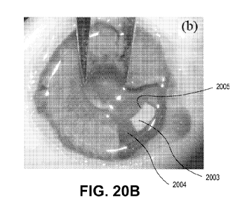
[0131] FIGS. 20A-20B are a picture of an intraocular lens implanted in a
cadaver human
eye in accordance with an embodiment. A rectangular patch valve is visible in
the lower
right quadrant of the eye in FIG. 20A. In FIG. 20B a section of the eye's iris
is removed to
show lens patch valve 2003 on intraocular lens 2004. Innermost edge 2005 is
arcuate,
following a constant radius around the center of the optical axis but set just
beyond the
optical path of the eye for a fully dilated pupil.
[0132] FIGS. 21A-21C are side elevation views of an intraocular lens patch
with a pre-
formed slit in accordance with an embodiment. Left side 2121 and right side
2122 of pre-
formed slit 2123 are shown in a closed configuration in FIG. 21A. Fluid from
below is
sealed in by the patch because elastomeric stresses seal the slit tight. In
FIG. 21B, needle
2130 begins to move down and, imperfectly to the left, against the slit to
gain entry. Slit
2123 begins to open. In FIG. 21C, needle 2130 juts through the slit, bending
left side 2121
and slightly crumpling elastomeric right side 2122. Sides 2121 and 2122 seal
against the
outside diameter of needle 2130, keeping fluid from inside the lens from
leaking out.
CA 02825043 2013-07-17
WO 2012/161749 PCT/US2012/021366
[0133] FIGS. 22A-22C are side elevation views of an intraocular lens patch
with a stepped
slit in accordance with an embodiment. Left side 2221 and right side 2222 of
preformed slit
2224 are closed due to elastomeric stresses in FIG. 22A. Slit 2224 has shelf
or stepped
portion 2225, which joins slit 2224 with lower portion of slit 2226. The shelf
is similar to
using a needle to make an incision at an angle. In FIG. 22B, needle 2230
begins to move
down and, imperfectly to the left, against the slit to gain entry to the lens.
In FIG. 22C,
needle 2230 just through the slit, bending left side 2221 and slightly
crumpling elastomeric
right side 2222. Sides 2221 and 2222 seal against the outside diameter of
needle 2230,
keeping fluid from inside the lens from leaking out.
[0134] It has been found that elastomeric patches of 100 [tm or greater are
thick enough to
self-close for many standard needles. A patch of 160 [tm and thicker work with
362 [tm
diameter standard 28-gauge needles. A patch of 250 [tm gives a factor of
safety for the 28-
gauge needle. This works for nominal pressures within the lens of under 1 psi,
which change
by 0.06 psi during accommodation.
[0135] A needle for injecting or removing fluid from the intraocular lens can
be 908 [tm
diameter (20-gauge), 362 [tm diameter (28-gauge), 311 [tm diameter (30-gauge),
110 [tm
diameter (36-gauge), or other sizes. The smaller the needle to be used, the
thinner the patch
(as shown in FIG. 10).
[0136] A plurality of patches can be used to allow for multiple ports in the
lens. One port
can be used for filling or removing optically clear fluid from the lens, while
another port can
simultaneously remove air bubbles from an out-pocket.
[0137] The invention has been described with reference to various specific and
illustrative
embodiments. However, it should be understood that many variations and
modifications may
be made while remaining within the spirit and scope of the following claims.
21