Note: Descriptions are shown in the official language in which they were submitted.
CA 02825081 2013-07-18
WO 2012/116926
PCT/EP2012/053118
Antigen binding proteins
The present invention relates to antigen binding proteins comprising two Fe
parts,
methods for their production, pharmaceutical compositions containing said
antigen
binding proteins, and uses thereof
Background of the Invention
In the last two decades various engineered antibody derivatives, either mono
or-
multispecific, either mono- or multivalent have been developed and evaluated
(see
e.g. Holliger, P., et al., Nature Biotech. 23 (2005) 1126-1136; Fischer, N.,
and
Leger 0., Pathobiology 74 (2007) 3-14).
US 2004/0033561 refers to the DNA and the production of monovalent
monobodies by co-expression of a heavy chain and a modified heavy chain.
However during expression a considerably amount of undesired homodimer is
formed as by-product, which is difficult to separate from the desired
heterodimeric
monobodies, as the homodimer and the heterodimer have the same or similar
molecular weights. WO 2007/048037 refers to monovalent IgGs which
corresponds to the heterodimeric monobodies of US 2004/0033561, but which can
have a tagging moiety attached to the heavy chain for easier purification of
the
heterodimer from the difficult-to-separate homodimeric by-product.
Summary of the Invention
The invention comprises an antigen binding protein comprising
a) two modified heavy chains of an antibody which specifically binds to
an antigen, wherein VH of each heavy chain is replaced by the VL
of said antibody, said modified heavy chains being associated with
each other via their CH domains of the Fe part;
b) two modified heavy chains of said antibody wherein CH1 of each
heavy chain is replaced by CL of said antibody, said modified
heavy chains being associated with each other via their CH
domains of the Fe part;
and wherein the VL domains of the heavy chains of a) are associated
with the VH domains of the heavy chains of b), and the CH1
CA 02825081 2013-07-18
WO 2012/116926
PCT/EP2012/053118
- 2 -
domains of the heavy chains of a) are associated with the CL
domains of the heavy chains of b).
In one embodiment the antigen binding protein according to the invention is
characterized in that
the CH3 domains of the Fc part of the modified heavy chains of a) and
the CH3 domains of the Fc part of the modified heavy chains of b) are of
the same isotype.
In one embodiment the antigen binding protein according to the invention is
characterized in that
the CH2 and CH3 domains of the Fc part of the modified heavy chains of
a) and the CH2 and CH3 domains of the Fc part of the modified heavy
chains of b) are of the same isotype.
In one embodiment the antigen binding protein according to the invention is
characterized in that
the CH2 and CH3 domains of the Fc part of the modified heavy chains of
a) and the CH2 and CH3 domains of the Fc part of the modified heavy
chains of b) are of the IgG isotype.
In one embodiment the antigen binding protein according to the invention is
characterized in that
the CH2 and CH3 domains of the Fc part of the modified heavy chains of
a) and the CH2 and CH3 domains of the Fc part of the modified heavy
chains of b) are of the IgG1 isotype.
In one embodiment the antigen binding protein according to the invention is
characterized in comprising
a) two modified heavy chains comprising the amino acid sequence of
SEQ ID NO:1; and
b) two modified heavy chains comprising the amino acid sequence of
SEQ ID NO:2;
CA 02825081 2013-07-18
WO 2012/116926
PCT/EP2012/053118
- 3 -
a) two modified heavy chains comprising the amino acid sequence of
SEQ ID NO:3; and
b) two modified heavy chains comprising the amino acid sequence of
SEQ ID NO:4; or
a) two modified heavy chains comprising the amino acid sequence of
SEQ ID NO:5; and
b) two modified heavy chains comprising the amino acid sequence of
SEQ ID NO:6.
In one embodiment the antigen binding protein according to the invention is
characterized in that
either the two modified heavy chains of a),
or the two modified heavy chains of b),
are further modified by the amino acid substitutions 5364G, L368F,
D399K and K409D (wherein the amino acid positions are numbered
according to the EU Index of Kabat).
In one embodiment the antigen binding protein according to the invention is
characterized in that
a) two modified heavy chains comprising the amino acid sequence of
SEQ ID NO:1; and
b) two modified heavy chains comprising the amino acid sequence of
SEQ ID NO:2;
wherein either the two modified heavy chains of a),
or the two modified heavy chains of b),
are further modified by the amino acid substitutions 5364G, L368F,
D399K and K409D (wherein the amino acid positions are numbered
according to the EU Index of Kabat);
a) two modified heavy chains comprising the amino acid sequence of
SEQ ID NO:3; and
b) two modified heavy chains comprising the amino acid sequence of
SEQ ID NO:4;
wherein either the two modified heavy chains of a),
or the two modified heavy chains of b),
CA 02825081 2013-07-18
WO 2012/116926
PCT/EP2012/053118
- 4 -
are further modified by the amino acid substitutions S364G, L368F,
D399K and K409D (wherein the amino acid positions are numbered
according to the EU Index of Kabat);
or
a) two modified heavy chains comprising the amino acid sequence of
SEQ ID NO:5; and
b) two modified heavy chains comprising the amino acid sequence of
SEQ ID NO:6
wherein either the two modified heavy chains of a),
or the two modified heavy chains of b),
are further modified by the amino acid substitutions 5364G, L368F,
D399K and K409D (wherein the amino acid positions are numbered
according to the EU Index of Kabat).
In one embodiment the antigen binding protein according to the invention is
characterized in that
the CH3 domains of the Fc part of the modified heavy chains of a) and
the CH3 domains of the Fc part of the modified heavy chains of b) are of
a different isotype.
In one embodiment the antigen binding protein according to the invention is
characterized in that
the CH3 domains of the Fc part of the modified heavy chains of a) are of
the IgG1 isotype;
and the CH3 domains of the Fc part of the modified heavy chains of b)
are of the IgA isotype.
In one embodiment the antigen binding protein according to the invention is
characterized in comprising
a) two modified heavy chains comprising the amino acid sequence of
SEQ ID NO:7; and
b) two modified heavy chains comprising the amino acid sequence of
SEQ ID NO:4.
CA 02825081 2013-07-18
WO 2012/116926
PCT/EP2012/053118
- 5 -
In one embodiment the antigen binding protein according to the invention is
characterized in comprising
a) two modified heavy chains comprising the amino acid sequence of
SEQ ID NO:3; and
b) two modified heavy chains comprising the amino acid sequence of
SEQ ID NO:8.
In one embodiment the antigen binding protein according to the invention is
characterized in that
The CH2 and CH3 domains of the Fc part of the modified heavy chains
of a) are of the IgG1 isotype;
and the CH2 and CH3 domains of the Fc part of the modified heavy
chains of b) are of the IgA isotype.
In one embodiment the antigen binding protein according to the invention is
characterized in that the CH2 domain of the Fc parts of a) and b) are of IgG1
isotype, and the antigen binding protein is afucosylated with an amount of
fucose
of 80% or less (preferably of 65% to 5%) of the total amount of
oligosaccharides
(sugars) at Asn297 is of human IgG1 isotype.
The invention further comprises a method for the preparation of an antigen
binding
protein according to the invention
comprising the steps of
a) transforming a host cell with vectors comprising nucleic acid molecules
encoding
an antigen binding protein according to the invention
b) culturing the host cell under conditions that allow synthesis of said
antigen binding protein molecule; and
c) recovering said antigen binding protein molecule from said culture.
The invention further comprises nucleic acid encoding the antigen binding
protein
according to the invention.
The invention further comprises vectors comprising nucleic acid encoding the
antigen binding protein according to the invention.
CA 02825081 2013-07-18
WO 2012/116926
PCT/EP2012/053118
- 6 -
The invention further comprises host cell comprising said vectors.
The invention further comprises composition, preferably a pharmaceutical or a
diagnostic composition of an antigen binding protein according to the
invention.
The invention further comprises pharmaceutical composition comprising an
antigen binding protein according to the invention.
The invention further comprises method for the treatment of a patient in need
of
therapy, characterized by administering to the patient a therapeutically
effective
amount of an antigen binding protein according to the invention.
It has now been found that the antigen binding proteins according to the
invention
have valuable characteristics such as biological or pharmacological activities
(as
e.g. enhanced ADCC compared to parent antibodies). They can be used e.g. for
the
treatment of diseases such as cancer. The antigen binding proteins according
to the
invention have furthermore highly valuable pharmacokinetic properties (like
e.g.
AUCO-inf, Cmax or CO).
Description of the Figures
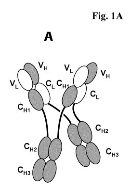
Figure 1A and B: A) Schematic structure of the antigen binding protein
according
to the invention (abbreviated MoAb-Dimer) with CH1-CL
crossover. B) Scheme of the major byproduct - monovalent
antibody monomer (MoAb) with CH1-CL crossover
(abbreviated MoAb).
Figure 1C: C) Association of two modified heavy chains a and b:
Heterodimerisation of two different chains (a with b) directly
leads to the monovalent antibody B (route 2).
Homodimerisation of two identical chains (a with a and b with
b) leads to the putative intermediates aa and bb (via route 1)
that can associate to form "MoAb-Dimer" A . Modification of
the CH3-CH3 contacts may affect distribution of products A
(MoAb-Dimer) and B (MoAb). Modifications that favor
heterodimerisation (e.g. knobs into holes) will increase the
relative amount of compound B via route 2, whereas
modifications that maintain attractive interactions between
identical chains but lead to repulsion of different chains (e.g.
CH3 domains of a and b taken from different isotypes) will
CA 02825081 2013-07-18
WO 2012/116926
PCT/EP2012/053118
- 7 -
favor route 1 and thus increase the amount of A. White: light
chain domains. Dashed: heavy chain domains.
Figure 2: Biochemical characterization of MoAb-Dimer c-Met (5D5
MoAb-Dimer ("CH3-wt")) (CH3-wt refers to the unchanged,
wild type CH3 domain). (A) Protein A purified antibody was
separated on an Superdex 200 26/60 column. (B) Peak fractions
(1,2,3) were pooled and subjected to SDS-PAGE under non-
reducing and reducing conditions. Polyacrylamide gels were
stained with Coomassie Blue dye. Individual peaks correspond
to MoAb (3), MoAb-Dimer (2) and a higher molecular weight
aggregate (1).
Figure 3: Biochemical characterization of MoAb-Dimer IGF-1R (IGF-1R
AK18 MoAb-Dimer ("CH3-wt")) (CH3-wt refers to the
unchanged, wild type CH3 domain). (A) Protein A purified
antibody was separated on an Superdex 200 26/60 column. (B)
Peak fractions (1,2) were pooled and subjected to SDS-PAGE
under non-reducing and reducing conditions. Polyacrylamide
gels were stained with Coomassie Blue dye. Individual peaks
correspond to MoAb (2) and MoAb-Dimer (1). C) The
molecular mass of the peaks fractions 1 and 2 was investigated
by SEC-MALLS.
Figure 4: Biochemical characterization of Her3 205 MoAb-Dimer
("CH3-wt") (CH3-wt refers to the unchanged, wild type CH3
domain). (A) Protein A purified antibody was separated on an
Superdex 200 26/60 column. (B) Peak fractions (1,2) were
pooled and subjected to SDS-PAGE under non-reducing and
reducing conditions. Polyacrylamide gels were stained with
Coomassie Blue dye. Individual peaks correspond to MoAb (3),
MoAb-Dimer (2) and a higher molecular weight aggregate (1).
Figure 5: Schematic
picture of the surface plasmon resonance assay
applied to analyze the IGF-1R binding affinity. An anti human
IgG antibody (JIR 109-005-098) was immobilized on the
surface of a CM5 biosensorchip and subsequently captured
MoAb or MoAb-Dimer antibodies. Further injection of
recombinant IGF-1R ectodomain confirmed functionality of
antigen binding sites in MoAb and MoAb-Dimer molecules.
CA 02825081 2013-07-18
WO 2012/116926
PCT/EP2012/053118
- 8 -
Figure 6: Cellular binding of MoAb-Dimer (IGF-1R AK18 MoAb-Dimer
("CH3-wt") (B) and parent antibody Mab IGF-1R (A) to A549
cells with flow cytometric analysis. A549 cells were incubated
with a dilution series of the indicated antibodies. Bound
antibodies were visualized with an Fc-binding secondary
fluorophor coupled antibody.
Figure 7: ADCC Assay with IGF1R Mab non-glycoengineered (non-ge)
and glycoengineered (ge) and non-glycoengineered IGF-1R
MoAb-Dimer (IGF1R AK18 MoAb-Dimer ("CH3-wt")). Donor
derived peripheral blood mononuclear cells (PBMC) were
incubated with prostate cancer cells (DU145) in the presence of
non-ge IGF1R Mab (1), ge IGF1R Mab (2) and non-ge IGF1R
AK18 MoAb-Dimer ("CH3-wt") (3).
Figure 8: Internalization of IGF-1R was assessed in HT29 cells
following
incubation with IGF-1R IgG1 (Mab IGF-1R) antibody and
IGF-1R MoAb-Dimer (IGF1R AK18 MoAb-Dimer
("CH3-wt")). The graph depicts total IGF-1R levels upon
antibody exposure which were determined in an ELISA-based
assay setup.
Figure 9: Autophosphorylation of IGF-1R was assessed following
incubation of 3T3-IGF-1R cells with IGF-1R IgG1 antibody
and IGF-1R MoAb-Dimer (IGF1R AK18 MoAb-Dimer
("CH3-wt")) in the presence of 10 nM IGF-1. The graph depicts
phospho IGF-1R levels upon antibody exposure which were
determined in an ELISA-based assay setup.
Figure 10: Analysis of obtained MoAb-Dimer (=antigen binding
protein
according to the invention) versus MoAb-monomer
(= monovalent byproduct) ratios as determined by HPLC.
Different antibody with wild type CH3 (CH3-wt) domains and
modified CH3 domains were transiently expressed and the
ratios of dimer versus monomer determined.
Figure 11: ESI-MS spectrum of the IGF-1R MoAb Dimer (SEC fraction
1)
under non reducing condition and after deglycosylation
Figure 12: ESI-MS spectrum of the IGF-1R MoAb Dimer (SEC fraction
1)
after degylcosylation and reduction.
CA 02825081 2013-07-18
WO 2012/116926
PCT/EP2012/053118
- 9 -
Detailed Description of the Invention
The invention comprises an antigen binding protein comprising
a) two modified heavy chains of an antibody which specifically binds
to an antigen, wherein VH of each heavy chain is replaced by the
VL of said antibody, said modified heavy chains being associated
with each other via their CH domains of the Fc part;
b) two modified heavy chains of said antibody wherein CH1 of each
heavy chain is replaced by CL of said antibody, said modified
heavy chains being associated with each other via their CH
domains of the Fc part;
and wherein the VL domains of the heavy chains of a) are associated
with the VH domains of the heavy chains of b), and the CH1
domains of the heavy chains of a) are associated with the CL
domains of the heavy chains of b).
In one embodiment the antigen binding protein according to the invention is
characterized in that
the CH3 domains of the Fc part of the modified heavy chains of a) and
the CH3 domains of the Fc part of the modified heavy chains of b) are of
the same isotype.
In one embodiment the antigen binding protein according to the invention is
characterized in that
the CH2 and CH3 domains of the Fc part of the modified heavy chains of
a) and the CH2 and CH3 domains of the Fc part of the modified heavy
chains of b) are of the same isotype.
In one embodiment the antigen binding protein according to the invention is
characterized in that
the CH2 and CH3 domains of the Fc part of the modified heavy chains of
a) and the CH2 and CH3 domains of the Fc part of the modified heavy
chains of b) are of the IgG isotype.
CA 02825081 2013-07-18
WO 2012/116926
PCT/EP2012/053118
- 10 -
In one embodiment the antigen binding protein according to the invention is
characterized in that
the CH2 and CH3 domains of the Fc part of the modified heavy chains of
a) and the CH2 and CH3 domains of the Fc part of the modified heavy
chains of b) are of the IgG1 isotype.
In one embodiment the antigen binding protein according to the invention is
characterized in comprising
a) two modified heavy chains comprising the amino acid sequence of
SEQ ID NO:1; and
b) two modified heavy chains comprising the amino acid sequence of
SEQ ID NO:2;
a) two modified heavy chains comprising the amino acid sequence of
SEQ ID NO:3; and
b) two modified heavy chains comprising the amino acid sequence of
SEQ ID NO:4; or
a) two modified heavy chains comprising the amino acid sequence of
SEQ ID NO:5; and
b) two modified heavy chains comprising the amino acid sequence of
SEQ ID NO:6.
To improve the yields of the antigen binding protein according to the
invention (i.e.
to improve MoAb dimer over MoAb monomer ratio (see Example 9)), the IgG1
CH3 domains of a) can be modified further by mutations so that the IgG1 CH3
domains of a) and the natural (wt) IgG1 CH3 domains of b) differ. The
modification/mutation has to be carried out in a way to maintain attractive
interactions between identical chains but lead to repulsion of different
chains (see
also Fig 1C).
In one embodiment the antigen binding protein according to the invention is
characterized in that
either the two modified heavy chains of a),
or the two modified heavy chains of b),
CA 02825081 2013-07-18
WO 2012/116926
PCT/EP2012/053118
- 11 -
are further modified by the amino acid substitutions S364G, L368F,
D399K and K409D (wherein the amino acid positions are numbered
according to the EU Index of Kabat).
The EU Index numbering system of Kabat is described in Kabat, et al.,
Sequences
of Proteins of Immunological Interest, 5th ed., Public Health Service,
National
Institutes of Health, Bethesda, MD (1991).
In one embodiment the antigen binding protein according to the invention is
characterized in that
a) two modified heavy chains comprising the amino acid sequence of
SEQ ID NO:1; and
b) two modified heavy chains comprising the amino acid sequence of
SEQ ID NO:2;
wherein either the two modified heavy chains of a),
or the two modified heavy chains of b),
are further modified by the amino acid substitutions 5364G, L368F,
D399K and K409D (wherein the amino acid positions are numbered
according to the EU Index of Kabat);
a) two modified heavy chains comprising the amino acid sequence of
SEQ ID NO:3; and
b) two modified heavy chains comprising the amino acid sequence of
SEQ ID NO:4;
wherein either the two modified heavy chains of a),
or the two modified heavy chains of b),
are further modified by the amino acid substitutions 5364G, L368F,
D399K and K409D (wherein the amino acid positions are numbered
according to the EU Index of Kabat);
or
a) two modified heavy chains comprising the amino acid sequence of
SEQ ID NO:5; and
b) two modified heavy chains comprising the amino acid sequence of
SEQ ID NO:6
wherein either the two modified heavy chains of a),
CA 02825081 2013-07-18
WO 2012/116926
PCT/EP2012/053118
- 12 -
or the two modified heavy chains of b),
are further modified by the amino acid substitutions S364G, L368F,
D399K and K409D (wherein the amino acid positions are numbered
according to the EU Index of Kabat).
Another possibility to improve the yields of the antigen binding protein
according
to the invention (i.e. to improve MoAb dimer over MoAb monomer ratio (see
Example 9)), the CH3 domains of a) and b) are taken from different isotypes.
Thus
the attractive interactions between identical chains are maintained but
different
chains are repulsed (see also Fig 1C).
Therefore in one embodiment the antigen binding protein according to the
invention is characterized in that
the CH3 domains of the Fc part of the modified heavy chains of a) and
the CH3 domains of the Fc part of the modified heavy chains of b) are of
a different isotype.
In one embodiment the antigen binding protein according to the invention is
characterized in that
the CH3 domains of the Fc part of the modified heavy chains of a) are of
the IgG isotype;
and the CH3 domains of the Fc part of the modified heavy chains of b)
are of the IgA isotype.
In one embodiment the antigen binding protein according to the invention is
characterized in comprising
a) two modified heavy chains comprising the amino acid sequence of
SEQ ID NO:7; and
b) two modified heavy chains comprising the amino acid sequence of
SEQ ID NO:4.
In one embodiment the antigen binding protein according to the invention is
characterized in comprising
a) two modified heavy chains comprising the amino acid sequence of
SEQ ID NO:3; and
CA 02825081 2013-07-18
WO 2012/116926
PCT/EP2012/053118
- 13 -
b) two modified heavy chains comprising the amino acid sequence of
SEQ ID NO:8.
In one embodiment the antigen binding protein according to the invention is
characterized in that
The CH2 and CH3 domains of the Fc part of the modified heavy chains
of a) are of the IgG1 isotype;
and the CH2 and CH3 domains of the Fc part of the modified heavy
chains of b) are of the IgAl isotype.
In one embodiment the antigen binding protein according to the invention is
characterized in that in that the CH2 domain of the Fc parts of a) and b) are
of IgG1
isotype, and the antigen binding protein is afucosylated with an amount of
fucose
of 80% or less of the total amount of oligosaccharides (sugars) at Asn297 is
of
human IgG1 isotype.
The term "antibody" as used herein denotes a full length antibody consisting
of two
antibody heavy chains and two antibody light chains (see Fig. 1). A heavy
chain of
full length antibody is a polypeptide consisting in N-terminal to C-terminal
direction of an antibody heavy chain variable domain (VH), an antibody
constant
heavy chain domain 1 (CH1), an antibody hinge region (HR), an antibody heavy
chain constant domain 2 (CH2), and an antibody heavy chain constant domain 3
(CH3), abbreviated as VH-CH1-HR-CH2-CH3; and optionally an antibody heavy
chain constant domain 4 (CH4) in case of an antibody of the class IgE.
Preferably
the heavy chain of full length antibody is a polypeptide consisting in N-
terminal to
C-terminal direction of VH, CH1, HR, CH2 and CH3. The light chain of full
length
antibody is a polypeptide consisting in N-terminal to C-terminal direction of
an
antibody light chain variable domain (VL), and an antibody light chain
constant
domain (CL), abbreviated as VL-CL. The antibody light chain constant domain
(CL) can be K (kappa) or X (lambda). The antibody chains are linked together
via
inter-polypeptide disulfide bonds between the CL domain and the CH1 domain
(i.e.
between the light and heavy chain) and between the hinge regions of the full
length
antibody heavy chains. Examples of typical full length antibodies are natural
antibodies like IgG (e.g. IgG 1 and IgG2), IgM, IgA, IgD, and IgE.) The
antibodies
according to the invention can be from a single species e.g. human, or they
can be
chimerized or humanized antibodies. The full length antibodies according to
the
CA 02825081 2013-07-18
WO 2012/116926
PCT/EP2012/053118
- 14 -
invention comprise two antigen binding sites each formed by a pair of VH and
VL,
which both specifically bind to the same (first) antigen.
From these full length antibodies the antigen binding protein of the invention
is
derived by:
a) modifying two
heavy chains of an antibody which specifically
binds to an antigen, by replacing the VH domain of each heavy
chain by the VL domain of said antibody;
b) modifying two heavy chains of said antibody by replacing the CH1
domain of each heavy chain by the CL domain of said antibody.
The "Fc part" of an antibody or antigen binding protein is not involved
directly in
binding of an antibody to an antigen, but is responsible a) for the
association of the
(modified) antibody chains with each other (e.g. via their CH3 domains) and b)
for
various effector functions. A "Fc part of an antibody" is a term well known to
the
skilled artisan and defined on the basis of papain cleavage of antibodies.
Depending on the amino acid sequence of the constant region of their heavy
chains,
antibodies or immunoglobulins are divided in the classes: IgA, IgD, IgE, IgG
and
IgM, and several of these may be further divided into subclasses (isotypes),
e.g.
IgGl, IgG2, IgG3, and IgG4, IgAl, and IgA2. According to the heavy chain
constant regions the different classes of immunoglobulins are called a, 6, c,
7, and
, respectively.
There are five types of mammalian antibody heavy chains denoted by the Greek
letters: a, 6, , 7, and 11 (Janeway, C.A., Jr. et al., Immunobiology, 5th
ed., Garland
Publishing (2001)). The type of heavy chain present defines the class of
antibody;
these chains are found in IgA, IgD, IgE, IgG, and IgM antibodies, respectively
(Rhoades, R.A., and Pflanzer, R.G., Human Physiology, 4th ed., Thomson
Learning (2002)). Distinct heavy chains differ in size and composition; a and
7
contain approximately 450 amino acids, while 11 and c have approximately 550
amino acids.
Each heavy chain has two regions, the constant region and the variable region.
The
constant region is identical in all antibodies of the same isotype, but
differs in
antibodies of different isotype. Heavy chains 7, a and 6 have a constant
region
composed of three constant domains CH1, CH2, and CH3 (in a line) , and a hinge
region for added flexibility (Woof, J., and Burton, D., Nat. Rev. Immunol. 4
(2004)
89-99); heavy chains 11 and c have a constant region composed of four constant
CA 02825081 2013-07-18
WO 2012/116926
PCT/EP2012/053118
- 15 -
domains CH1, CH2, CH3, and CH4 (Janeway, C.A., Jr. et al., Immunobiology., 5th
ed., Garland Publishing (2001)). The variable region of the heavy chain
differs in
antibodies produced by different B cells, but is the same for all antibodies
produced
by a single B cell or B cell clone. The variable region of each heavy chain is
approximately 110 amino acids long and is composed of a single antibody
domain.
The "CH domains of the Fc part" are the antibody heavy chain constant domain 2
(CH2), and the antibody heavy chain constant domain 3 (CH3), and optionally
the
antibody heavy chain constant domain 4 (CH4) in case of an antibody of the
class
IgE.
The term "said modified heavy chains being associated with each other via
their
CH domains of the Fc part" refers to the interchain domain pairing of the
antibody
heavy chain constant domains (CH) of the two modified heavy chains with each
other e.g. the two CH3 domains of both chains with ech other via e.g.
interchain
ionic interaction, Van-Der Waals interaction, or hydrogen bonding (see Fig.
1A). In
one embodiment said modified heavy chains are associated with each other via
at
least their CH3 domains of the Fc part (and optionally via their CH2 domains,
or
optionally via their CH2 domains and CH4 domains (if present)).
The term "wherein the VL domains of the heavy chains of a) are associated with
the VH domains of the heavy chains of b), and the CH1 domains of the heavy
chains of a) are associated with the CL domains of the heavy chains of b)"
refers to
the domain pairing of said antibody domains (always one of a) and one of b))
as
found e.g. in natural antibodies (VL/VH and CH1/CL) e.g. via interchain ionic
interaction, Van-Der Waals interaction, hydrogen bonding, or disulfide
interaction.
(see Fig. 1A).
The "antigen binding protein" according to the invention comprises two antigen-
binding sites and is bivalent. The terms "binding site" or "antigen-binding
site" as
used herein denotes the region(s) of antigen binding protein according to the
invention to which a ligand (e.g the antigen or antigen fragment of it)
actually
binds and which is derived from antibody molecule or a fragment thereof (e.g.
a
Fab fragment). The antigen-binding site according to the invention comprise an
antibody heavy chain variable domains (VH) and an antibody light chain
variable
domains (VL).
CA 02825081 2013-07-18
WO 2012/116926
PCT/EP2012/053118
- 16 -
The antigen-binding sites (i.e. the pairs of VH/VL) that specifically bind to
the
desired antigen can be derived a) from known antibodies to the antigen or b)
from
new antibodies or antibody fragments obtained by de novo immunization methods
using inter alia either the antigen protein or nucleic acid or fragments
thereof or by
phage display.
An antigen-binding site of a antigen binding protein of the invention contains
six
complementarity determining regions (CDRs) which contribute in varying degrees
to the affinity of the binding site for antigen. There are three heavy chain
variable
domain CDRs (CDRH1, CDRH2 and CDRH3) and three light chain variable
domain CDRs (CDRL1, CDRL2 and CDRL3). The extent of CDR and framework
regions (FRs) is determined by comparison to a compiled database of amino acid
sequences in which those regions have been defined according to variability
among
the sequences.
Antibody specificity refers to selective recognition of the antibody for a
particular
epitope of an antigen. Natural antibodies, for example, are monospecific.
Bispecific
antibodies are antibodies which have two different antigen-binding
specificities.
The antigen binding proteins according to the invention are at least
monospecific
and specifically bind to an epitope of the respective antigen.
The term "valent" as used within the current application denotes the presence
of a
specified number of binding sites in an antibody molecule. A natural antibody
for
example has two binding sites and is bivalent. Also the antigen binding
protein
according to the invention is at least bivalent.
The terms "monoclonal antibody" or "monoclonal antibody composition" as used
herein refer to a preparation of antibody molecules of a single amino acid
composition.
The term "chimeric antibody" refers to an antibody comprising a variable
region,
i.e., binding region, from one source or species and at least a portion of a
constant
region derived from a different source or species, usually prepared by
recombinant
DNA techniques. Chimeric antibodies comprising a murine variable region and a
human constant region are preferred. Other preferred forms of "chimeric
antibodies" encompassed by the present invention are those in which the
constant
region has been modified or changed from that of the original antibody to
generate
the properties according to the invention, especially in regard to C 1 q
binding
and/or Fc receptor (FcR) binding. Such chimeric antibodies are also referred
to as
CA 02825081 2013-07-18
WO 2012/116926
PCT/EP2012/053118
- 17 -
"class-switched antibodies". Chimeric antibodies are the product of expressed
immunoglobulin genes comprising DNA segments encoding immunoglobulin
variable regions and DNA segments encoding immunoglobulin constant regions.
Methods for producing chimeric antibodies involve conventional recombinant
DNA and gene transfection techniques are well known in the art. See, e.g.,
Morrison, S.L., et al., Proc. Natl. Acad. Sci. USA 81 (1984) 6851-6855;
US 5,202,238 and US 5,204,244.
The term "humanized antibody" refers to antibodies in which the framework or
"complementarity determining regions" (CDR) have been modified to comprise the
CDR of an immunoglobulin of different specificity as compared to that of the
parent immunoglobulin. In a preferred embodiment, a murine CDR is grafted into
the framework region of a human antibody to prepare the "humanized antibody."
See, e.g., Riechmann, L., et al., Nature 332 (1988) 323-327; and Neuberger,
M.S.,
et al., Nature 314 (1985) 268-270. Particularly preferred CDRs correspond to
those
representing sequences recognizing the antigens noted above for chimeric
antibodies. Other forms of "humanized antibodies" encompassed by the present
invention are those in which the constant region has been additionally
modified or
changed from that of the original antibody to generate the properties
according to
the invention, especially in regard to Clq binding and/or Fc receptor (FcR)
binding.
The term "human antibody", as used herein, is intended to include antibodies
having variable and constant regions derived from human germ line
immunoglobulin sequences. Human antibodies are well-known in the state of the
art (van Dijk, M.A., and van de Winkel, J.G., Curr. Opin. Chem. Biol. 5 (2001)
368-374). Human antibodies can also be produced in transgenic animals (e.g.
mice)
that are capable, upon immunization, of producing a full repertoire or a
selection of
human antibodies in the absence of endogenous immunoglobulin production.
Transfer of the human germ-line immunoglobulin gene array in such germ-line
mutant mice will result in the production of human antibodies upon antigen
challenge (see, e.g., Jakobovits, A., et al., Proc. Natl. Acad. Sci. USA 90
(1993)
2551-2555; Jakobovits, A., et al., Nature 362 (1993) 255-258; Bruggemann, M.,
et
al., Year Immunol. 7 (1993) 33-40). Human antibodies can also be produced in
phage display libraries (Hoogenboom, H.R., and Winter, G., J. Mol. Biol. 227
(1992) 381-388; Marks, J.D., et al., J. Mol. Biol. 222 (1991) 581-597). The
techniques of Cole et al. and Boerner et al. are also available for the
preparation of
human monoclonal antibodies (Cole, et al., Monoclonal Antibodies and Cancer
CA 02825081 2013-07-18
WO 2012/116926
PCT/EP2012/053118
- 18 -
Therapy, Alan R. Liss, p. 77 (1985); and Boerner, P., et al., J. Immunol. 147
(1991)
86-95). As already mentioned for chimeric and humanized antibodies according
to
the invention the term "human antibody" as used herein also comprises such
antibodies which are modified in the constant region to generate the
properties
according to the invention, especially in regard to Clq binding and/or FcR
binding,
e.g. by "class switching" i.e. change or mutation of Fc parts (e.g. from IgG1
to
IgG4 and/or IgGl/IgG4 mutation).
The term "recombinant human antibody", as used herein, is intended to include
all
human antibodies that are prepared, expressed, created or isolated by
recombinant
means, such as antibodies isolated from a host cell such as a NSO or CHO cell
or
from an animal (e.g. a mouse) that is transgenic for human immunoglobulin
genes
or antibodies expressed using a recombinant expression vector transfected into
a
host cell. Such recombinant human antibodies have variable and constant
regions
in a rearranged form. The recombinant human antibodies according to the
invention
have been subjected to in vivo somatic hypermutation. Thus, the amino acid
sequences of the VH and VL regions of the recombinant antibodies are sequences
that, while derived from and related to human germ line VH and VL sequences,
may not naturally exist within the human antibody germ line repertoire in
vivo.
The "variable domain" (variable domain of a light chain (VL), variable region
of a
heavy chain (VH) as used herein denotes each of the pair of light and heavy
chains
which is involved directly in binding the antibody to the antigen. The domains
of
variable human light and heavy chains have the same general structure and each
domain comprises four framework (FR) regions whose sequences are widely
conserved, connected by three "hypervariable regions" (or complementarity
determining regions, CDRs). The framework regions adopt a 13-sheet
conformation
and the CDRs may form loops connecting the 13-sheet structure. The CDRs in
each
chain are held in their three-dimensional structure by the framework regions
and
form together with the CDRs from the other chain the antigen binding site. The
antibody heavy and light chain CDR3 regions play a particularly important role
in
the binding specificity/affinity of the antibodies according to the invention
and
therefore provide a further object of the invention.
The terms "hypervariable region" or "antigen-binding portion of an antibody"
when
used herein refer to the amino acid residues of an antibody which are
responsible
for antigen-binding. The hypervariable region comprises amino acid residues
from
the "complementarity determining regions" or "CDRs". "Framework" or "FR"
CA 02825081 2013-07-18
WO 2012/116926
PCT/EP2012/053118
- 19 -
regions are those variable domain regions other than the hypervariable region
residues as herein defined. Therefore, the light and heavy chains of an
antibody
comprise from N- to C-terminus the domains FR1, CDR1, FR2, CDR2, FR3,
CDR3, and FR4. CDRs on each chain are separated by such framework amino
acids. Especially, CDR3 of the heavy chain is the region which contributes
most to
antigen binding. CDR and FR regions are determined according to the standard
definition of Kabat, et al., Sequences of Proteins of Immunological Interest,
5th
ed., Public Health Service, National Institutes of Health, Bethesda, MD
(1991).
As used herein, the term "binding" or "specifically binding" refers to the
binding of
the antigen binding protein to an epitope of the antigen in an in vitro assay,
preferably in an plasmon resonance assay (BIAcore, GE-Healthcare Uppsala,
Sweden) with purified wild-type antigen. The affinity of the binding is
defined by
the terms ka (rate constant for the association of the antibody from the
antibody/antigen complex), lcD (dissociation constant), and KD (1cD/ka).
Binding or
specifically binding means a binding affinity (KD) of 10-8 mo1/1 or less (e.g.
10-8 M
to 10-13 mo1/1), preferably 10-9 M to 10-13 mo1/1. Thus, a antigen binding
protein
according to the invention is specifically binding to each antigen for which
it is
specific with a binding affinity (KD) of 10-8 mo1/1 or less (e.g. 10-8 M to 10-
13
mo1/1), preferably 10-9M to 10-13 mo1/1.
Binding of the antigen binding protein to the FcyRIII can be investigated by a
BIAcore assay (GE-Healthcare Uppsala, Sweden). The affinity of the binding is
defined by the terms ka (rate constant for the association of the antibody
from the
antibody/antigen complex), lc]) (dissociation constant), and KD (1cD/ka).
The term "epitope" includes any polypeptide determinant capable of specific
binding to a antigen binding proteins. In certain embodiments, epitope
determinant
include chemically active surface groupings of molecules such as amino acids,
sugar side chains, phosphoryl, or sulfonyl, and, in certain embodiments, may
have
specific three dimensional structural characteristics, and or specific charge
characteristics. An epitope is a region of an antigen that is bound by a
antigen
binding protein.
In certain embodiments, an antibody is said to specifically bind an antigen
when it
preferentially recognizes its target antigen in a complex mixture of proteins
and/or
macromolecules.
CA 02825081 2013-07-18
WO 2012/116926
PCT/EP2012/053118
- 20 -
The term "constant region" as used within the current applications denotes the
sum
of the domains of an antibody other than the variable region. The constant
region is
not involved directly in binding of an antigen, but exhibits various effector
functions. Depending on the amino acid sequence of the constant region of
their
heavy chains, antibodies are divided in the classes (also named isotypes):
IgA, IgD,
IgE, IgG and IgM, and several of these may be further divided into subclasses
(also
named isotypes), such as IgGl, IgG2, IgG3, and IgG4, IgAl and IgA2. The heavy
chain constant regions that correspond to the different classes of antibodies
are
called a, 8, c, 7, and , respectively. The light chain constant regions (CL)
which
can be found in all five antibody classes are called lc (kappa) and X
(lambda).
The term "constant region derived from human origin" as used in the current
application denotes a constant heavy chain region of a human antibody of the
isotypes IgGl, IgG2, IgG3, or IgG4 and/or a constant light chain kappa or
lambda
region. Such constant regions are well known in the state of the art and e.g.
described by Kabat, E.A., (see e.g. Johnson, G. and Wu, T.T., Nucleic Acids
Res.
28 (2000) 214-218; Kabat, E.A., et al., Proc. Natl. Acad. Sci. USA 72 (1975)
2785-
2788).
The term "complement-dependent cytotoxicity (CDC)" denotes a process initiated
by binding of complement factor Clq to the Fc part of most IgG antibody
subclasses. Binding of Clq to an antibody is caused by defined protein-protein
interactions at the so called binding site. Such Fc part binding sites are
known in
the state of the art (see above). Such Fc part binding sites are, e.g.,
characterized by
the amino acids L234, L235, D270, N297, E318, K320, K322, P331, and P329
(numbering according to EU index of Kabat). Antibodies of subclass IgGl, IgG2,
and IgG3 usually show complement activation including Clq and C3 binding,
whereas IgG4 does not activate the complement system and does not bind Clq
and/or C3.
While antibodies of the IgG4 subclass show reduced Fc receptor (Fc7RIIIa)
binding, antibodies of other IgG subclasses show strong binding. However
Pro238,
Asp265, Asp270, Asn297 (loss of Fc carbohydrate), Pro329, Leu234, Leu235,
G1y236, G1y237, 11e253, 5er254, Lys288, Thr307, Gln311, Asn434, and His435 are
residues which, if altered, provide also reduced Fc receptor binding (Shields,
R.L.,
et al., J. Biol. Chem. 276 (2001) 6591-6604; Lund, J., et al., FASEB J. 9
(1995)
115-119; Morgan, A., et al., Immunology 86 (1995) 319-324; EP 0 307 434).
CA 02825081 2013-07-18
WO 2012/116926
PCT/EP2012/053118
-21 -
In one embodiment an antibody according to the invention has a reduced FcR
binding compared to an IgG1 antibody and the full length parent antibody is in
regard to FcR binding of IgG4 subclass or of IgG1 or IgG2 subclass with a
mutation in S228, L234, L235 and/or D265, and/ or contains the PVA236
mutation. In one embodiment the mutations in the full length parent antibody
are
S228P, L234A, L235A, L235E and/or PVA236. In another embodiment the
mutations in the full length parent antibody are in IgG4 S228P and L235E and
in
IgG1 L234A and L235A.
The constant region of an antibody is directly involved in ADCC (antibody-
dependent cell-mediated cytotoxicity) and CDC (complement-dependent
cytotoxicity). Complement activation (CDC) is initiated by binding of
complement
factor Clq to the constant region of most IgG antibody subclasses. Binding of
Clq
to an antibody is caused by defined protein-protein interactions at the so
called
binding site. Such constant region binding sites are known in the state of the
art and
described e.g. by Lukas, T.J., et al., J. Immunol. 127 (1981) 2555-2560;
Bunkhouse, R. and Cobra, J.J., Mol. Immunol. 16 (1979) 907-917; Burton, D.R.,
et
al., Nature 288 (1980) 338-344; Thomason, J.E., et al., Mol. Immunol. 37
(2000)
995-1004; Idiocies, E.E., et al., J. Immunol. 164 (2000) 4178-4184; Hearer,
M., et
al., J. Virol. 75 (2001) 12161-12168; Morgan, A., et al., Immunology 86 (1995)
319-324; and EP 0 307 434. Such constant region binding sites are, e.g.,
characterized by the amino acids L234, L235, D270, N297, E318, K320, K322,
P331, and P329 (numbering according to EU index of Kabat which is described in
Kabat, et al., Sequences of Proteins of Immunological Interest, 5th ed.,
Public
Health Service, National Institutes of Health, Bethesda, MD (1991)).
The term "antibody-dependent cellular cytotoxicity (ADCC)" refers to lysis of
human target cells by an antibody according to the invention in the presence
of
effector cells. ADCC is measured preferably by the treatment of a preparation
of
antigen expressing cells with an antibody according to the invention in the
presence
of effector cells such as freshly isolated PBMC or purified effector cells
from buffy
coats, like monocytes or natural killer (NK) cells or a permanently growing NK
cell
line.
Surprisingly it has been found that an antigen binding protein according to
the
invention shows enhanced ADCC properties compared to its parent full length
antibody, especially in the area of higher antibody concentrations. These
improved
CA 02825081 2013-07-18
WO 2012/116926
PCT/EP2012/053118
- 22 -
ADCC effects are achieved without further modification of the Fc part like
glycoengineering.
Thus in one embodiment the antigen binding proteins according to the invention
have an enhanced ADCC (measured as described in Example 4) compared to its
parent full length antibody.
In mammals there are only two types of light chain, which are called lambda
(X)
and kappa (x). A light chain has two successive domains: one constant domain
CL
and one variable domain VL. The approximate length of a light chain is 211 to
217
amino acids. Preferably the light chain is a kappa (x) light chain, and the
constant
domain CL is preferably derived from a kappa (x) light chain (the constant
domain
Cell-mediated effector functions of monoclonal antibodies can be e.g. further
enhanced by engineering their oligosaccharide component as described in Umana,
P., et al., Nature Biotechnol. 17 (1999) 176-180, and US 6,602,684. IgG1 type
antibodies, the most commonly used therapeutic antibodies, are glycoproteins
that
have a conserved N-linked glycosylation site at Asn297 in each CH2 domain. The
two complex biantennary oligosaccharides attached to Asn297 are buried between
the CH2 domains, forming extensive contacts with the polypeptide backbone, and
their presence is essential for the antibody to mediate effector functions
such as
antibody dependent cellular cytotoxicity (ADCC) (Lifely, M., R., et al.,
Glycobiology 5 (1995) 813-822; Jefferis, R., et al., Immunol. Rev. 163 (1998)
59-
76; Wright, A., and Morrison, S., L., Trends Biotechnol. 15 (1997) 26-32).
Umana,
P., et al. Nature Biotechnol. 17 (1999) 176-180 and WO 99/54342 showed that
overexpression in Chinese hamster ovary (CHO) cells of 13(1,4)-N-
acetylglucosaminyltransferase III ("GnTIII"), a glycosyltransferase catalyzing
the
formation of bisected oligosaccharides, significantly increases the in vitro
ADCC
activity of antibodies. Alterations in the composition of the Asn297
carbohydrate
or its elimination affect also binding to FcyR and Clq (Umana, P., et al.,
Nature
Biotechnol. 17 (1999) 176-180; Davies, J., et al., Biotechnol. Bioeng. 74
(2001)
288-294; Mimura, Y., et al., J. Biol. Chem. 276 (2001) 45539-45547; Radaev,
S.,
et al., J. Biol. Chem. 276 (2001) 16478-16483; Shields, R., L., et al., J.
Biol. Chem.
276 (2001) 6591-6604; Shields, R., L., et al., J. Biol. Chem. 277 (2002) 26733-
26740; Simmons, L., C., et al., J. Immunol. Methods 263 (2002) 133-147).
CA 02825081 2013-07-18
WO 2012/116926
PCT/EP2012/053118
- 23 -
In one aspect of the invention the antigen binding protein according to the
invention is characterized in that the CH2 domains of the Fc parts of a) and
b) are
of IgG1 isotype, and the antigen binding protein is afucosylated with an
amount of
fucose of 80% or less of the total amount of oligosaccharides (sugars) at
Asn297.
In one embodiment antigen binding protein is afucosylated with and the amount
of
fucose of 65% to 5% of the total amount of oligosaccharides (sugars) at
Asn297.
The term "afucosylated antigen binding protein" refers to an antigen binding
proteins of IgG1 or IgG3 isotype (preferably of IgG1 isotype) with an altered
pattern of glycosylation in the Fc region at Asn297 having a reduced level of
fucose residues. Glycosylation of human IgG1 or IgG3 occurs at Asn297 as core
fucosylated bianntennary complex oligosaccharide glycosylation terminated with
up to 2 Gal residues. These structures are designated as GO, G1 (a1,6 or a1,3)
or
G2 glycan residues, depending from the amount of terminal Gal residues (Raju,
T.S., BioProcess Int. 1 (2003) 44-53). CHO type glycosylation of antibody Fc
parts
is e.g. described by Routier, F.H., Glycoconjugate J. 14 (1997) 201-207.
Antibodies which are recombinantely expressed in non glycomodified CHO host
cells usually are fucosylated at Asn297 in an amount of at least 85%. It
should be
understood that the term "antigen binding protein" as used herein includes an
antigen binding protein having no fucose in its glycosylation pattern. It is
commonly known that typical glycosylated residue position in an antibody is
the
asparagine at position 297 according to the EU numbering system ("Asn297").
Thus an afucosylated antigen binding protein according to the invention means
an
antigen binding protein of IgG1 or IgG3 isotype (preferably of IgG1 isotype)
wherein the amount of fucose is 80% or less (e.g. of 80% to 1 %) of the total
amount of oligosaccharides (sugars) at Asn297 (which means that at least 20%
or
more of the oligosaccharides of the Fc region at Asn297 are afucosylated). In
one
embodiment the amount of fucose is 65% or less (e.g. of 65% to 1 %), in one
embodiment from 65% to 5%, in one embodiment from 40% to 20% of the
oligosaccharides of the Fc region at Asn297.. According to the invention
"amount
of fucose" means the amount of said oligosaccharide (fucose) within the
oligosaccharide (sugar) chain at Asn297, related to the sum of all
oligosaccharides
(sugars) attached to Asn 297 (e.g. complex, hybrid and high mannose
structures)
measured by MALDI-TOF mass spectrometry and calculated as average value (for
a detailed procedure to determine the amount of fucose, see e.g. WO
2008/077546).
Furthermore in one embodiment, the oligosaccharides of the Fc region are
bisected.
CA 02825081 2013-07-18
WO 2012/116926
PCT/EP2012/053118
- 24 -
The afucosylated antigen binding protein according to the invention can be
expressed in a glycomodified host cell engineered to express at least one
nucleic
acid encoding a polypeptide having GnTIII activity in an amount sufficient to
partially fucosylate the oligosaccharides in the Fc region. In one embodiment,
the
polypeptide having GnTIII activity is a fusion polypeptide. Alternatively a1,6-
fucosyltransferase activity of the host cell can be decreased or eliminated
according
to US 6,946,292 to generate glycomodified host cells. The amount of antigen
binding protein fucosylation can be predetermined e.g. either by fermentation
conditions (e.g. fermentation time) or by combination of at least two antigen
binding protein with different fucosylation amount. Such methods to generate
afucosylated antigen binding proteins are described in WO 2005/044859,
WO 2004/065540, WO 2007/031875, Umana, P., et al., Nature Biotechnol. 17
(1999) 176-
180, WO 99/154342, WO 2005/018572, WO 2006/116260,
WO 2006/114700, WO 2005/011735, WO 2005/027966, WO 97/028267,
US 2006/0134709, US 2005/0054048, US 2005/0152894, WO 2003/035835,
WO 2000/061739. These glycoengineered antigen binding proteins according to
the invention have an increased ADCC (compared to the parent antigen binding
proteins). Other glycoengineering methods yielding afucosylated antigen
binding
proteins according to the invention are described e.g. in Niwa, R.. et al.,
J. Immunol. Methods 306 (2005) 151-160; Shinkawa, T., et al., J. Biol. Chem,
278
(2003) 3466-3473; WO 03/055993 or US 2005/0249722.
Thus one aspect of the invention is an afucosylated antigen binding protein
according to the invention which is of IgG1 isotype or IgG3 isotype
(preferably of
IgG1 isotype) with an amount of fucose of 60% or less (e.g. of 60% to 1 %) of
the
total amount of oligosaccharides (sugars) at Asn297.
Thus one aspect of the invention is an afucosylated antigen binding protein
according to the invention which is of IgG1 isotype or IgG3 isotype
(preferably of
IgG1 isotype) with an amount of fucose of 60% or less (e.g. of 60% to 1 %) of
the
total amount of oligosaccharides (sugars) at Asn297 for the treatment of
cancer.
Another aspect of the invention is the use of invention an afucosylated
antigen
binding protein according to the invention which is of IgG1 or IgG3 isotype
(preferably of IgG1 isotype) with an amount of fucose of 60% or less of the
total
amount of oligosaccharides (sugars) at Asn297, for the manufacture of a
medicament for the treatment of cancer.
CA 02825081 2013-07-18
WO 2012/116926
PCT/EP2012/053118
- 25 -
In one embodiment the amount of fucose is between 60% and 20% of the total
amount of oligosaccharides (sugars) at Asn297. In one embodiment the amount of
fucose is between 60% and 40% of the total amount of oligosaccharides (sugars)
at
Asn297. In one embodiment the amount of fucose is between 0% of the total
amount of oligosaccharides (sugars) at Asn297.
The "EU numbering system" or "EU index" is generally used when referring to a
residue in an immunoglobulin heavy chain constant region (e.g., the EU index
reported in Kabat et al., Sequences of Proteins of Immunological Interest, 5th
ed.,
Public Health Service, National Institutes of Health, Bethesda, MD (1991)
expressly incorporated herein by reference).
The term "the sugar chains show characteristics of N-linked glycans attached
to
Asn297 of an antibody recombinantly expressed in a CHO cell" denotes that the
sugar chain at Asn297 of the full length parent antibody according to the
invention
has the same structure and sugar residue sequence except for the fucose
residue as
those of the same antibody expressed in unmodified CHO cells, e.g. as those
reported in WO 2006/103100.
The term "NGNA" as used within this application denotes the sugar residue
N-glycolylneuraminic acid.
Glycosylation of human IgG1 or IgG3 occurs at Asn297 as core fucosylated
biantennary complex oligosaccharide glycosylation terminated with up to two
Gal
residues. Human constant heavy chain regions of the IgG1 or IgG3 subclass are
reported in detail by Kabat, E., A., et al., Sequences of Proteins of
Immunological
Interest, 5th Ed. Public Health Service, National Institutes of Health,
Bethesda,
MD. (1991), and by Brueggemann, M., et al., J. Exp. Med. 166 (1987) 1351-1361;
Love, T., W., et al., Methods Enzymol. 178 (1989) 515-527. These structures
are
designated as GO, G1 (a-1,6- or a-1,3-), or G2 glycan residues, depending from
the
amount of terminal Gal residues (Raju, T., S., Bioprocess Int. 1 (2003) 44-
53).
CHO type glycosylation of antibody Fc parts is e.g. described by Routier,
F.H.,
Glycoconjugate J. 14 (1997) 201-207. Antibodies which are recombinantly
expressed in non-glycomodified CHO host cells usually are fucosylated at
Asn297
in an amount of at least 85%. The modified oligosaccharides of the full length
parent antibody may be hybrid or complex. Preferably the bisected, reduced/not-
fucosylated oligosaccharides are hybrid. In another embodiment, the bisected,
reduced/not-fucosylated oligosaccharides are complex.
CA 02825081 2013-07-18
WO 2012/116926
PCT/EP2012/053118
- 26 -
According to the invention "amount of fucose" means the amount of said sugar
within the sugar chain at Asn297, related to the sum of all glycostructures
attached
to Asn297 (e.g. complex, hybrid and high mannose structures) measured by
MALDI-TOF mass spectrometry and calculated as average value. The relative
amount of fucose is the percentage of fucose-containing structures related to
all
glycostructures identified in an N-Glycosidase F treated sample (e.g. complex,
hybrid and oligo- and high-mannose structures, resp.) by MALDI-TOF.
The antibody according to the invention is produced by recombinant means.
Thus,
one aspect of the current invention is a nucleic acid encoding the antibody
according to the invention and a further aspect is a cell comprising said
nucleic acid
encoding an antibody according to the invention. Methods for recombinant
production are widely known in the state of the art and comprise protein
expression
in prokaryotic and eukaryotic cells with subsequent isolation of the antibody
and
usually purification to a pharmaceutically acceptable purity. For the
expression of
the antibodies as aforementioned in a host cell, nucleic acids encoding the
respective modified light and heavy chains are inserted into expression
vectors by
standard methods. Expression is performed in appropriate prokaryotic or
eukaryotic
host cells like CHO cells, NSO cells, SP2/0 cells, HEK293 cells, COS cells,
PER.C6 cells, yeast, or E.coli cells, and the antibody is recovered from the
cells
(supernatant or cells after lysis). General methods for recombinant production
of
antibodies are well-known in the state of the art and described, for example,
in the
review articles of Makrides, S.C., Protein Expr. Purif. 17 (1999) 183-202;
Geisse, S., et al., Protein Expr. Purif. 8 (1996) 271-282; Kaufman, R.J.,
Mol. Biotechnol. 16 (2000) 151-161; Werner, R.G., Drug Res. 48 (1998) 870-880.
The antigen binding proteins according to the invention are suitably separated
from
the culture medium by conventional immunoglobulin purification procedures such
as, for example, protein A-Sepharose, hydroxylapatite chromatography, gel
electrophoresis, dialysis, or affinity chromatography. DNA and RNA encoding
the
monoclonal antibodies is readily isolated and sequenced using conventional
procedures. The hybridoma cells can serve as a source of such DNA and RNA.
Once isolated, the DNA may be inserted into expression vectors, which are then
transfected into host cells such as HEK 293 cells, CHO cells, or myeloma cells
that
do not otherwise produce immunoglobulin protein, to obtain the synthesis of
recombinant monoclonal antibodies in the host cells.
CA 02825081 2013-07-18
WO 2012/116926
PCT/EP2012/053118
- 27 -
Amino acid sequence variants (or mutants) of the antigen binding protein are
prepared by introducing appropriate nucleotide changes into the antibody DNA,
or
by nucleotide synthesis. Such modifications can be performed, however, only in
a
very limited range, e.g. as described above. For example, the modifications do
not
alter the above mentioned antibody characteristics such as the IgG isotype and
antigen binding, but may improve the yield of the recombinant production,
protein
stability or facilitate the purification.
The term "host cell" as used in the current application denotes any kind of
cellular
system which can be engineered to generate the antibodies according to the
current
invention. In one embodiment HEK293 cells and CHO cells are used as host
cells.
As used herein, the expressions "cell", "cell line", and "cell culture" are
used
interchangeably and all such designations include progeny. Thus, the words
"transformants" and "transformed cells" include the primary subject cell and
cultures derived therefrom without regard for the number of transfers. It is
also
understood that all progeny may not be precisely identical in DNA content, due
to
deliberate or inadvertent mutations. Variant progeny that have the same
function or
biological activity as screened for in the originally transformed cell are
included.
Expression in NSO cells is described by, e.g., Barnes, L.M., et al.,
Cytotechnology
32 (2000) 109-123; Barnes, L.M., et al., Biotech. Bioeng. 73 (2001) 261-270.
Transient expression is described by, e.g., Durocher, Y., et al., Nucl. Acids.
Res. 30
(2002) E9. Cloning of variable domains is described by Orlandi, R., et al.,
Proc. Natl. Acad. Sci. USA 86 (1989) 3833-3837; Carter, P., et al.,
Proc. Natl. Acad. Sci. USA 89 (1992) 4285-4289; and Norderhaug, L., et al.,
J. Immunol. Methods 204 (1997) 77-87. A preferred transient expression system
(HEK 293) is described by Schlaeger, E.-J., and Christensen, K., in
Cytotechnology 30 (1999) 71-83 and by Schlaeger, E.-J., in J. Immunol. Methods
194 (1996) 191-199.
The control sequences that are suitable for prokaryotes, for example, include
a
promoter, optionally an operator sequence, and a ribosome binding site.
Eukaryotic
cells are known to utilize promoters, enhancers and polyadenylation signals.
A nucleic acid is "operably linked" when it is placed in a functional
relationship
with another nucleic acid sequence. For example, DNA for a pre-sequence or
secretory leader is operably linked to DNA for a polypeptide if it is
expressed as a
pre-protein that participates in the secretion of the polypeptide; a promoter
or
CA 02825081 2013-07-18
WO 2012/116926
PCT/EP2012/053118
- 28 -
enhancer is operably linked to a coding sequence if it affects the
transcription of the
sequence; or a ribosome binding site is operably linked to a coding sequence
if it is
positioned so as to facilitate translation. Generally, "operably linked" means
that
the DNA sequences being linked are contiguous, and, in the case of a secretory
leader, contiguous and in reading frame. However, enhancers do not have to be
contiguous. Linking is accomplished by ligation at convenient restriction
sites. If
such sites do not exist, the synthetic oligonucleotide adaptors or linkers are
used in
accordance with conventional practice.
Purification of antigen binding proteins is performed in order to eliminate
cellular
components or other contaminants, e.g. other cellular nucleic acids or
proteins (e.g.
byproducts of Fig. 1B), by standard techniques, including alkaline/SDS
treatment,
CsC1 banding, column chromatography, agarose gel electrophoresis, and others
well known in the art (see Ausubel, F., et al., ed. Current Protocols in
Molecular
Biology, Greene Publishing and Wiley Interscience, New York (1987)). Different
methods are well established and widespread used for protein purification,
such as
affinity chromatography with microbial proteins (e.g. protein A or protein G
affinity chromatography), ion exchange chromatography (e.g. cation exchange
(carboxymethyl resins), anion exchange (amino ethyl resins) and mixed-mode
exchange), thiophilic adsorption (e.g. with beta-mercaptoethanol and other SH
ligands), hydrophobic interaction or aromatic adsorption chromatography (e.g.
with
phenyl-sepharose, aza-arenophilic resins, or m-aminophenylboronic acid), metal
chelate affinity chromatography (e.g. with Ni(II)- and Cu(II)-affinity
material), size
exclusion chromatography, and electrophoretical methods (such as gel
electrophoresis, capillary electrophoresis) (Vijayalakshmi, M.A., Appl.
Biochem.
Biotech. 75 (1998) 93-102). One example of a purification is described in
Example
1 and the corresponding Figures.
One aspect of the invention is a pharmaceutical composition comprising an
antigen
binding protein according to the invention. Another aspect of the invention is
the
use of an antigen binding protein according to the invention for the
manufacture of
a pharmaceutical composition. A further aspect of the invention is a method
for the
manufacture of a pharmaceutical composition comprising an antigen binding
protein according to the invention. In another aspect, the present invention
provides
a composition, e.g. a pharmaceutical composition, containing an antigen
binding
protein according to the present invention, formulated together with a
pharmaceutical carrier.
CA 02825081 2013-07-18
WO 2012/116926
PCT/EP2012/053118
- 29 -
One embodiment of the invention is the antigen binding protein according to
the
invention for the treatment of cancer.
Another aspect of the invention is said pharmaceutical composition for the
treatment of cancer.
One embodiment of the invention is the antigen binding protein according to
the
invention for use in the treatment of cancer.
Another aspect of the invention is the use of an antigen binding protein
according
to the invention for the manufacture of a medicament for the treatment of
cancer.
Another aspect of the invention is method of treatment of patient suffering
from
cancer by administering an antigen binding protein according to the invention
to a
patient in the need of such treatment.
As used herein, "pharmaceutical carrier" includes any and all solvents,
dispersion
media, coatings, antibacterial and antifungal agents, isotonic and absorption
delaying agents, and the like that are physiologically compatible. Preferably,
the
carrier is suitable for intravenous, intramuscular, subcutaneous, parenteral,
spinal
or epidermal administration (e.g. by injection or infusion).
A pharmaceutical composition of the present invention can be administered by a
variety of methods known in the art. As will be appreciated by the skilled
artisan,
the route and/or mode of administration will vary depending upon the desired
results. To administer a compound of the invention by certain routes of
administration, it may be necessary to coat the compound with, or co-
administer
the compound with, a material to prevent its inactivation. For example, the
compound may be administered to a subject in an appropriate carrier, for
example,
liposomes, or a diluent. Pharmaceutically acceptable diluents include saline
and
aqueous buffer solutions. Pharmaceutical carriers include sterile aqueous
solutions
or dispersions and sterile powders for the extemporaneous preparation of
sterile
injectable solutions or dispersion. The use of such media and agents for
pharmaceutically active substances is known in the art.
The phrases "parenteral administration" and "administered parenterally" as
used
herein means modes of administration other than enteral and topical
administration,
usually by injection, and includes, without limitation, intravenous,
intramuscular,
intra-arterial, intrathecal, intracapsular, intraorbital, intracardiac,
intradermal,
CA 02825081 2013-07-18
WO 2012/116926
PCT/EP2012/053118
- 30 -
i ntrap eritoneal, tran strache al, subcutaneous, sub
cuti cul ar, intra-articular,
subcapsular, sub arachnoid, intraspinal, epidural and intrasternal injection
and
infusion.
The term cancer as used herein refers to proliferative diseases, such as
lymphomas,
lymphocytic leukemias, lung cancer, non small cell lung (NSCL) cancer,
bronchioloalviolar cell lung cancer, bone cancer, pancreatic cancer, skin
cancer,
cancer of the head or neck, cutaneous or intraocular melanoma, uterine cancer,
ovarian cancer, rectal cancer, cancer of the anal region, stomach cancer,
gastric
cancer, colon cancer, breast cancer, uterine cancer, carcinoma of the
fallopian
tubes, carcinoma of the endometrium, carcinoma of the cervix, carcinoma of the
vagina, carcinoma of the vulva, Hodgkin's Disease, cancer of the esophagus,
cancer
of the small intestine, cancer of the endocrine system, cancer of the thyroid
gland,
cancer of the parathyroid gland, cancer of the adrenal gland, sarcoma of soft
tissue,
cancer of the urethra, cancer of the penis, prostate cancer, cancer of the
bladder,
cancer of the kidney or ureter, renal cell carcinoma, carcinoma of the renal
pelvis,
mesothelioma, hepatocellular cancer, biliary cancer, neoplasms of the central
nervous system (CNS), spinal axis tumors, brain stem glioma, glioblastoma
multiforme, astrocytomas, schwanomas, ependymonas, medulloblastomas,
meningiomas, squamous cell carcinomas, pituitary adenoma and Ewings sarcoma,
including refractory versions of any of the above cancers, or a combination of
one
or more of the above cancers.
These compositions may also contain adjuvants such as preservatives, wetting
agents, emulsifying agents and dispersing agents. Prevention of presence of
microorganisms may be ensured both by sterilization procedures, supra, and by
the
inclusion of various antibacterial and antifungal agents, for example,
paraben,
chlorobutanol, phenol, sorbic acid, and the like. It may also be desirable to
include
isotonic agents, such as sugars, sodium chloride, and the like into the
compositions.
In addition, prolonged absorption of the injectable pharmaceutical form may be
brought about by the inclusion of agents which delay absorption such as
aluminum
monostearate and gelatin.
Regardless of the route of administration selected, the compounds of the
present
invention, which may be used in a suitable hydrated form, and/or the
pharmaceutical compositions of the present invention, are formulated into
pharmaceutically acceptable dosage forms by conventional methods known to
those of skill in the art.
CA 02825081 2013-07-18
WO 2012/116926
PCT/EP2012/053118
- 31 -
Actual dosage levels of the active ingredients in the pharmaceutical
compositions
of the present invention may be varied so as to obtain an amount of the active
ingredient which is effective to achieve the desired therapeutic response for
a
particular patient, composition, and mode of administration, without being
toxic to
the patient. The selected dosage level will depend upon a variety of
pharmacokinetic factors including the activity of the particular compositions
of the
present invention employed, the route of administration, the time of
administration,
the rate of excretion of the particular compound being employed, the duration
of
the treatment, other drugs, compounds and/or materials used in combination
with
the particular compositions employed, the age, sex, weight, condition, general
health and prior medical history of the patient being treated, and like
factors well
known in the medical arts.
The composition must be sterile and fluid to the extent that the composition
is
deliverable by syringe. In addition to water, the carrier preferably is an
isotonic
buffered saline solution.
Proper fluidity can be maintained, for example, by use of coating such as
lecithin,
by maintenance of required particle size in the case of dispersion and by use
of
surfactants. In many cases, it is preferable to include isotonic agents, for
example,
sugars, polyalcohols such as mannitol or sorbitol, and sodium chloride in the
composition.
As used herein, the expressions "cell," "cell line," and "cell culture" are
used
interchangeably and all such designations include progeny. Thus, the words
"transformants" and "transformed cells" include the primary subject cell and
cultures derived therefrom without regard for the number of transfers. It is
also
understood that all progeny may not be precisely identical in DNA content, due
to
deliberate or inadvertent mutations. Variant progeny that have the same
function or
biological activity as screened for in the originally transformed cell are
included.
Where distinct designations are intended, it will be clear from the context.
The term "transformation" as used herein refers to process of transfer of a
vectors/nucleic acid into a host cell. If cells without formidable cell wall
barriers
are used as host cells, transfection is carried out e.g. by the calcium
phosphate
precipitation method as described by Graham and Van der Eh, Virology 52 (1978)
546. However, other methods for introducing DNA into cells such as by nuclear
injection or by protoplast fusion may also be used. If prokaryotic cells or
cells
CA 02825081 2013-07-18
WO 2012/116926
PCT/EP2012/053118
- 32 -
which contain substantial cell wall constructions are used, e.g. one method of
transfection is calcium treatment using calcium chloride as described by
Cohen,
F.N, et al., PNAS 69 (1972) 7110).
As used herein, "expression" refers to the process by which a nucleic acid is
transcribed into mRNA and/or to the process by which the transcribed mRNA
(also
referred to as transcript) is subsequently being translated into peptides,
polypeptides, or proteins. The transcripts and the encoded polypeptides are
collectively referred to as gene product. If the polynucleotide is derived
from
genomic DNA, expression in a eukaryotic cell may include splicing of the mRNA.
A "vector" is a nucleic acid molecule, in particular self-replicating, which
transfers
an inserted nucleic acid molecule into and/or between host cells. The term
includes
vectors that function primarily for insertion of DNA or RNA into a cell (e.g.,
chromosomal integration), replication of vectors that function primarily for
the
replication of DNA or RNA, and expression vectors that function for
transcription
and/or translation of the DNA or RNA. Also included are vectors that provide
more
than one of the functions as described.
An "expression vector" is a polynucleotide which, when introduced into an
appropriate host cell, can be transcribed and translated into a polypeptide.
An
"expression system" usually refers to a suitable host cell comprised of an
expression vector that can function to yield a desired expression product.
The following examples, sequence listing and figures are provided to aid the
understanding of the present invention, the true scope of which is set forth
in the
appended claims. It is understood that modifications can be made in the
procedures
set forth without departing from the spirit of the invention.
Description of the Amino Acid Sequences
SEQ ID NO:! c-Met 5D5 MoAb-Dimer ("CH3-wt") - modified heavy chain a)
VL-CH1-CH2-CH3
SEQ ID NO:2 c-Met 5D5 MoAb-Dimer ("CH3-wt") - modified heavy chain b)
VH-CL-CH2-CH3
SEQ ID NO:3 IGF1R AK18 MoAb-Dimer ("CH3-wt") - modified heavy chain
a) VL-CH1-CH2-CH3
SEQ ID NO:4 IGF1R AK18 MoAb-Dimer ("CH3-wt") - modified heavy chain
b) VH-CL-CH2-CH3
CA 02825081 2013-07-18
WO 2012/116926
PCT/EP2012/053118
- 33 -
SEQ ID NO:5 Her3 205 MoAb-Dimer ("CH3-wt") - modified heavy chain a)
VL-CH1-CH2-CH3
SEQ ID NO:6 Her3 205 MoAb-Dimer ("CH3-wt") - modified heavy chain b)
VH-CL-CH2-CH3
SEQ ID NO:7 IGF1R AK18 MoAb-Dimer - modified heavy chain a) VL-CH1-
CH2-CH3 with IgA-CH3
SEQ ID NO:8 IGF1R AK18 MoAb-Dimer - modified heavy chain b) VH-CL-
CH2-CH3 with IgA-CH3 domain
Experimental Procedure
A. Materials and Methods:
Recombinant DNA techniques
Standard methods were used to manipulate DNA as described in Sambrook, J., et
al., Molecular cloning: A laboratory manual; Cold Spring Harbor Laboratory
Press,
Cold Spring Harbor, New York (1989). The molecular biological reagents were
used according to the manufacturer's instructions.
DNA and protein sequence analysis and sequence data management
General information regarding the nucleotide sequences of human
immunoglobulins light and heavy chains is given in: Kabat, E.A. et al., (1991)
Sequences of Proteins of Immunological Interest, Fifth Ed., NUJ Publication No
91-3242. Amino acids of antibody chains are numbered according to EU
numbering (Edelman, G.M., et al., PNAS 63 (1969) 78-85; Kabat, E.A., et al.,
(1991) Sequences of Proteins of Immunological Interest, Fifth Ed., NUJ
Publication No 91-3242). The GCG's (Genetics Computer Group, Madison,
Wisconsin) software package version 10.2 and Infomax's Vector NTI Advance
suite version 8.0 was used for sequence creation, mapping, analysis,
annotation and
illustration.
DNA sequencing
DNA sequences were determined by double strand sequencing performed at
SequiServe (Vaterstetten, Germany) and Geneart AG (Regensburg, Germany).
CA 02825081 2013-07-18
WO 2012/116926
PCT/EP2012/053118
- 34 -
Gene synthesis
Desired gene segments were prepared by Geneart AG (Regensburg, Germany)
from synthetic oligonucleotides and PCR products by automated gene synthesis.
The gene segments which are flanked by singular restriction endonuclease
cleavage
sites were cloned into pGA18 (ampR) plasmids. The plasmid DNA was purified
from transformed bacteria and concentration determined by UV spectroscopy. The
DNA sequence of subcloned gene fragments was confirmed by DNA sequencing.
DNA sequences encoding for the two antibody chains (VH-CL-CH2-CH3 and VL-
CH1-CH2-CH3) were prepared as whole fragments by gene synthesis with
flanking 5'HpaI and 3'NaeI restriction sites. Gene segments coding for point
mutations in the CH3 domain which preferentially lead to the MoAb-Dimer
product as well as the replacement of gene segments coding for IgG1 part with
an
IgA¨CH3 domain (e.g. SEQ. ID. NO: 7) were prepared by gene synthesis. These
segments were flanked by unique restriction sites which allowed for
replacement of
the corresponding wild type IgG1 sequences. All constructs were designed with
a
5'-end DNA sequence coding for a leader peptide, which targets proteins for
secretion in eukaryotic cells.
Construction of the expression plasmids
A Roche expression vector was used for the construction of all antibody
chains.
The vector is composed of the following elements:
- an origin of replication, oriP, of Epstein-Barr virus (EBV),
- an origin of replication from the vector pUC18 which allows replication
of this plasmid in E. coli
- a beta-lactamase gene which confers ampicillin resistance in E. coli,
- the immediate early enhancer and promoter from the human
cytomegalovirus (HCMV),
- the human 1-immunoglobulin polyadenylation ("poly A") signal
sequence, and
- unique HpaI, Bell, and NaeI restriction sites.
The immunoglobulin genes in the order of VH-CL-CH2-CH3 and VL-CH1-CH2-
CH3 as well as constructs modified in the 3'region coding for the C-terminus
of the
antibody chain (CH3) were prepared by gene synthesis and cloned into pGA18
(ampR) plasmids as described. The pG18 (ampR) plasmids carrying the
CA 02825081 2013-07-18
WO 2012/116926
PCT/EP2012/053118
- 35 -
synthesized DNA segments and the Roche expression vector were digested either
with HpaI and NaeI or with Ben and NaeI restriction enzymes (Roche Molecular
Biochemicals) and subjected to agarose gel electrophoresis. Purified DNA
segments were then ligated to the isolated Roche expression vector HpaI/NaeI
or
Bc1I/Nad fragment resulting in the final expression vectors. The final
expression
vectors were transformed into E. coli cells, expression plasmid DNA was
isolated
(Miniprep) and subjected to restriction enzyme analysis and DNA sequencing.
Correct clones were grown in 150 ml LB-Amp medium, again plasmid DNA was
isolated (Maxiprep) and sequence integrity confirmed by DNA sequencing.
Transient expression of immunoglobulin variants in HEK293 cells
Recombinant immunoglobulin variants were expressed by transient transfection
of
human embryonic kidney 293-F cells using the FreeStyleTM 293 Expression System
according to the manufacturer's instruction (Invitrogen, USA). Briefly,
suspension
FreeStyleTM 293-F cells were cultivated in FreeStyleTM 293 Expression medium
at
37 C/8% CO2. Cells were seeded in fresh medium at a density of 1-2x10⁶ viable
cells/ml on the day of transfection. DNA293fectinTM complexes were prepared in
Opti-MEM I medium (Invitrogen, USA) using 325 11.1 of 293fectinTM
(Invitrogen,
Germany) and 250 tg of each plasmid DNA in a 1:1 molar ratio for a 250 ml
final
transfection volume. Antibody containing cell culture supernatants were
harvested
7 days after transfection by centrifugation at 14000 g for 30 minutes and
filtered
through a sterile filter (0.22 p.m). Supernatants were stored at -20 C until
purification.
Alternatively, antibodies were generated by transient transfection in HEK293-
EBNA cells. Antibodies were expressed by transient co-transfection of the
respective expression plasmids in adherently growing HEK293- EBNA cells
(human embryonic kidney cell line 293 expressing Epstein-Barr- Virus nuclear
antigen; American type culture collection deposit number ATCC # CRL- 10852,
Lot. 959 218) cultivated in DMEM (Dulbecco's modified Eagle's medium, Gibco)
supplemented with 10% Ultra Low IgG FCS (fetal calf serum, Gibco), 2 mM L-
Glutamine (Gibco), and 250 1.tg/m1 Geneticin (Gibco). For transfection
FuGENETM
6 Transfection Reagent (Roche Molecular Biochemicals) was used in a ratio of
FuGENETM reagent (IA) to DNA (m) of 4:1 (ranging from 3:1 to 6:1). Proteins
were expressed from the respective plasmids using an equimolar ratio of
plasmids.
Cells were fed at day 3 with L- Glutamine ad 4 mM, Glucose [Sigma] and NAA
[Gibco]. Immunoglobulin variants containing cell culture supernatants were
CA 02825081 2013-07-18
WO 2012/116926
PCT/EP2012/053118
- 36 -
harvested from day 5 to 11 after transfection by centrifugation and stored at -
80C.
General information regarding the recombinant expression of human
immunoglobulins in e.g. HEK293 cells is given in: Meissner, P., et al.,
Biotechnol.
Bioeng. 75 (2001) 197-203.
Purification of antibodies
Antibodies were purified from cell culture supernatants by affinity
chromatography
using Protein A-SepharoseTm (GE Healthcare, Sweden) and Superdex200 size
exclusion chromatography. Briefly, sterile filtered cell culture supernatants
were
applied on a HiTrap ProteinA HP (5 ml) column equilibrated with PBS buffer (10
mM Na2HPO4, 1 mM KH2PO4, 137 mM NaC1 and 2.7 mM KC1, pH 7.4). Unbound
proteins were washed out with equilibration buffer. Antibody and antibody
variants
were eluted with 0.1 M citrate buffer, pH 2.8, and the protein containing
fractions
were neutralized with 0.1 ml 1 M Tris, pH 8.5. Then, the eluted protein
fractions
were pooled, concentrated with an Amicon Ultra centrifugal filter device
(MWCO:
30 K, Millipore) to a volume of 3 ml and loaded on a Superdex200 HiLoad 120 ml
16/60 or 26/60 gel filtration column (GE Healthcare, Sweden) equilibrated with
20mM Histidin, 140 mM NaC1, pH 6Ø Fractions containing purified antibodies
with less than 5% high molecular weight aggregates were pooled and stored as
1.0
mg/ml aliquots at -80 C.
Analysis of purified proteins
The protein concentration of purified protein samples was determined by
measuring the optical density (OD) at 280 nm, using the molar extinction
coefficient calculated on the basis of the amino acid sequence. Purity and
molecular weight of antibodies were analyzed by SDS-PAGE in the presence and
absence of a reducing agent (5 mM 1,4-dithiotreitol) and staining with
Coomassie
brilliant blue. The NuPAGE Pre-Cast gel system (Invitrogen, USA) was used
according to the manufacturer's instruction (4-12% Tris-Glycine gels). The
aggregate content of antibody samples was analyzed by high-performance SEC
using a Superdex 200 analytical size-exclusion column (GE Healthcare, Sweden)
in
200 mM KH2PO4, 250 mM KC1, pH 7.0 running buffer at 25 C. 25 [tg protein
were injected on the column at a flow rate of 0.5 ml/min and eluted isocratic
over
50 minutes. For stability analysis, concentrations of 1 mg/ml of purified
proteins
were incubated at 4 C and 40 C for 7 days and then evaluated by high-
performance
SEC (e.g. HP SEC Analysis (Purified Protein). The integrity of the amino acid
CA 02825081 2013-07-18
WO 2012/116926
PCT/EP2012/053118
- 37 -
backbone of reduced immunoglobulin variant chains was verified by
NanoElectrospray Q-TOF mass spectrometry after removal of N-glycans by
enzymatic treatment with Peptide-N-Glycosidase F (Roche Molecular
Biochemicals).
Mass spectrometry
The total deglycosylated mass of antibodies was determined and confirmed via
electrospray ionization mass spectrometry (ESI-MS). Briefly, 100 tg purified
antibodies were deglycosylated with 50 mU N-Glycosidase F (PNGaseF,
ProZyme) in 100 mM KH2PO4/K2HPO4, pH 7 at 37 C for 12-24 h at a protein
concentration of up to 2 mg/ml and subsequently desalted via HPLC on a
Sephadex
G25 column (GE Healthcare). The mass of the respective antibody chains was
determined by ESI-MS after deglycosylation and reduction. In brief, 50 tg
antibody in 115 11.1 were incubated with 60 11.1 1M TCEP and 50 11.1 8 M
Guanidine-
hydrochloride subsequently desalted. The total mass and the mass of the
reduced
antibody chains was determined via ESI-MS on a Q-Star Elite MS system equipped
with a NanoMate source. The mass range recorded depends on the samples
molecular weight. In general for reduced antibodies the mass range was set
from
600-2000 m/z and for non reduced antibodies from 1000-3600 m/z.
SEC-MALLS
SEC-MALLS (size-exclusion chromatography with multi-angle laser light
scattering) was used to determine the approximate molecular weight of proteins
in
solution. According to the light scattering theory, MALLS allows molecular
weight
estimation of macromolecules irrespective of their molecular shape or other
presumptions. SEC-MALLS is based on a separation of proteins according to
their
size (hydrodynamic radius) via SEC chromatography, followed by concentration-
and scattered light-sensitive detectors. SEC-MALLS typically gives rise to
molecular weight estimates with an accuracy that allows clear discrimination
between monomers, dimers, trimers etc., provided the SEC separation is
sufficient.
In this work, the following instrumentation was used: Dionex Ultimate 3000
HPLC; column: Superose6 10/300 (GE Healthcare); eluent: 1 x PBS; flow rate:
0.25 mL/min; detectors: OptiLab REX (Wyatt Inc., Dernbach), MiniDawn Treos
(Wyatt Inc., Dernbach). Molecular weights were calculated with the Astra
software, version 5.3.2.13. Protein amounts between 50 and 150 tg were loaded
on
the column and BSA (Sigma Aldrich) was used as a reference protein.
CA 02825081 2013-07-18
WO 2012/116926
PCT/EP2012/053118
- 38 -
Dynamic Light Scattering (DLS) timecourse
Samples (30 ilL) at a concentration of approx. 1 mg/mL in 20 mM His/HisCl,
140 mM NaC1, pH 6.0, were filtered via a 384-well filter plate (0.45 p.m pore
size)
into a 384-well optical plate (Corning) and covered with 20 !IL paraffin oil
(Sigma). Dynamic light scattering data were collected repeatedly during a
period of
5 days with a DynaPro DLS plate reader (Wyatt) at a constant temperature of 40
C.
Data were processed with Dynamics V6.10 (Wyatt).
Surface Plasmon Resonance
The binding properties of anti-IGF-1R antigen binding proteins and antibodies
were
analyzed by surface plasmon resonance (SPR) technology using a Biacore
instrument (Biacore, GE-Healthcare, Uppsala). This system is well established
for
the study of molecule interactions. It allows a continuous real-time
monitoring of
ligand/analyte bindings and thus the determination of association rate
constants
(ka), dissociation rate constants (kd), and equilibrium constants (KD) in
various
assay settings. SPR- technology is based on the measurement of the refractive
index close to the surface of a gold coated biosensor chip. Changes in the
refractive
index indicate mass changes on the surface caused by the interaction of
immobilized ligand with analyte injected in solution. If molecules bind to
immobilized ligand on the surface the mass increases, in case of dissociation
the
mass decreases. For capturing anti-human IgG antibody was immobilized on the
surface of a CM5 biosensorchip using amine-coupling chemistry. Flow cells were
activated with a 1:1 mixture of 0.1 M N-hydroxysuccinimide and 0.1 M 3-(N ,N-
dimethylamino)propyl-N-ethylcarbodiimide at a flow rate of 5 1,t1/min. Anti-
human
IgG antibody was injected in sodium acetate, pH 5.0 at 10 1.tg/ml. A reference
control flow cell was treated in the same way but with vehicle buffers only
instead
of the capturing antibody. Surfaces were blocked with an injection of 1 M
ethanolamine/HC1 pH 8.5. The IGF-1R antibodies were diluted in HBS-P and
injected. All interactions were performed at 25 C (standard temperature). The
regeneration solution of 3 M Magnesium chloride was injected for 60 s at 5
1,t1/min
flow to remove any non-covalently bound protein after each binding cycle.
Signals
were detected at a rate of one signal per second. Samples were injected at
increasing concentrations. Fig. 17 depicts the applied assay format. A low
loading
density with capturing antibody density and IGF-1R antibody was chosen to
enforce binding.
CA 02825081 2013-07-18
WO 2012/116926
PCT/EP2012/053118
- 39 -
For affinity measurements, human FcgIIIa was immobilized to a CM-5 sensor chip
by capturing the His-tagged receptor to an anti-His antibody (Penta-His,
Qiagen)
which was coupled to the surface by standard amine-coupling and blocking
chemistry on a SPR instrument (Biacore T100). After FcgRIIIa capturing, 50 nM
IGF1R antibodies were injected at 25 C at a flow rate of 5 lL/min. The chip
was
afterwards regenerated with a 60s pulse of 10 mM glycine-HC1, pH 2.0 solution.
Antibody-dependent cellular cytotoxicity assay (ADCC)
Determination of antibody mediated effector functions by anti-IGF-IR
antibodies.
In order to determine the capacity of the generated antibodies to elicit
immune
effector mechanisms antibody-dependent cell cytotoxicity (ADCC) studies were
performed. To study the effects of the antibodies in ADCC, DU145 IGF-IR
expressing cells (1 x 10⁶ cells /m1) were labeled with 1 IA per ml BATDA
solution
(Perkin Elmer) for 25 minutes at 37 C in a cell incubator. Afterwards, cells
were
washed four times with 10 ml of RPMI-FM/PenStrep and spun down for
10 minutes at 200 x g. Before the last centrifugation step, cell numbers were
determined and cells diluted to 1x10⁵ cells/ml in RPMI-FM/PenStrep medium
from the pellet afterwards. The cells were plated 5,000 per well in a round
bottom
plate, in a volume of 50 jil. HuMAb antibodies were added at a final
concentration
ranging from 25-0.1 1..tg/m1 in a volume of 50 IA cell culture medium to 50 pi
cell
suspension. Subsequently, 50 pi of effector cells, freshly isolated PBMC were
added at an E:T ratio of 25:1. The plates were centrifuged for 1 minutes at
200 x g,
followed by an incubation step of 2 hours at 37 C. After incubation the cells
were
spun down for 10 minutes at 200 x g and 20 pi of supernatant was harvested and
transferred to an Optiplate 96-F plate. 200 IA of Europium solution (Perkin
Elmer,
at room temperature) were added and plates were incubated for 15 minutes on a
shaker table. Fluorescence is quantified in a time-resolved fluorometer
(Victor 3,
Perkin Elmer) using the Eu-TDA protocol from Perkin Elmer. The magnitude of
cell lysis by ADCC is expressed as % of the maximum release of TDA
fluorescence enhancer from the target cells lysed by detergent corrected for
spontaneous release of TDA from the respective target cells.
IGF-1R internalization assay
The binding of antibodies and antigen binding protein according the invention
to
the IGF-1R results in internalization and degradation of the receptor. This
process
can be monitored by incubating IGF-1R expressing HT29 CRC cells with IGF-1R
CA 02825081 2013-07-18
WO 2012/116926
PCT/EP2012/053118
- 40 -
targeting antibodies followed by a quantification of remaining IGF-1R protein
levels in cell lysates by ELISA.
For this purpose, HT29 cells at 1,5 x10⁴ cells/well were incubated in a 96
well
MTP in RPMI with 10 % FCS over night at 37 C and 5% CO2 in order to allow
attachment of the cells. Next morning, the medium was aspirated and 100 1.1,1
anti
IGF-1R antibody diluted in RPMI + 10% FCS was added in concentrations from 10
nM to 2 pM in 1:3 dilution steps. The cells were incubated with antibody for
18
hours at 37 C. Afterwards, the medium was again removed and 120 11.1 IVIES
lysis
buffer (25 mM IVIES pH 6.5 + Complete) were added.
For ELISA, 96-Well streptavidin coated polystyrene plates (Nunc) were loaded
with 100 1.1,1 MAK<hu IGF-1Ra>hu-la-IgG-Bi (Ch.10) diluted 1:200 in
3%BSA/PBST (final concentration 2.4 [tg/m1) and incubated under constant
agitation for 1 hour at room temperature. Afterwards, the well content was
removed
and each well was washed three times with 200 11.1 PBST. 100 11.1 of the cell
lysate
solution were added per well, again incubated for 1 hour at room temperature
on a
plate shaker, and washed three times with 200 11.1 PBST. After removal of the
supernatant, 100 11.1/well PAK<human IGF-1Ra>Ra-C20-IgG (Santa Cruz #sc-713)
diluted 1:750 in 3 % BSA/PBST was added followed by the same incubation and
washing intervals as described above. In order to detect the specific antibody
bound
to IGF-1R, 100 11.1/well of a polyclonal horse-radish-peroxidase-coupled
rabbit
antibody (Cell Signaling #7074) diluted 1:4000 in 3 % BSA/PBST were added.
After another hour, unbound antibody was again removed by washing thoroughly 6
times as described above. For quantification of bound antibody, 100 11.1/well
3,3'-
5,5'-Tetramethylbenzidin (Roche, BM-Blue ID.-Nr.11484281) was added and
incubated for 30 minutes at room temperature. The colorigenic reaction is
finally
stopped by adding 25 11.1/well 1M H2504 and the light absorption is measured
at
450 nm wavelength. Cells not treated with antibody are used as a control for
0%
downregulation, lysis buffer as background control.
IGF-1R autophosphorylation assay (IGF-1 stimulation)
Targeting IGF-1R by IGF-1R antibodies results in inhibition of IGF-1 induced
autophosphorylation. We investigated the inhibition of autophosphorylation of
the
wild type IGF-1R MoAb-Dimer compared to the parental IGF-1R IgG1 antibody.
For this purpose 3T3-IGF-1R cells, a murine fibroblast cell line
overexpressing
human IGF-1R, were treated for 10 minutes with 10 nM recombinant human IGF-1
CA 02825081 2013-07-18
WO 2012/116926
PCT/EP2012/053118
-41 -
in the presence of different concentrations of IGF-1R antibody or IGF-1R
antigen
binding protein. After lysis of the cells, the levels of phosphorylated IGF-1R
protein were determined by a phospho-IGF-1R specific ELISA, combining a
human IGF-1R specific capture antibody and a phospho-Tyrosine specific
detection antibody.
Determination of PK properties: Single Dose Kinetics in Mice
Methods
Animals:
NMRI mice, female, fed, 23-32 g body weight at the time point of compound
administration.
Study protocol:
For a single i.v. dose of 10 mg/kg the mice were allocated to 3 groups with 2-
3
animals each. Blood samples are taken from group 1 at 0.5, 168 and 672 hours,
from group 2 at 24 and 336 hours and from group 3 at 48 and 504 hours after
dosing.
Blood samples of about 100 tL were obtained by retrobulbar puncture. Serum
samples of at least 40 11.1 were obtained from blood after 1 hour at room
temperature by centrifugation (9300xg) at room temperature for 2.5 min. Serum
samples were frozen directly after centrifugation and stored frozen at ¨20 C
until
analysis.
Analytics:
The concentrations of the human antibodies in mice serum were determined with
an enzyme linked immunosorbent assay (ELISA) using 1 % mouse serum.
Biotinylated monoclonal antibody against human Fcy (mAb<hFcypAN>IgG-Bi) was
bound to streptavidin coated microtiterplates in the first step. In the next
step serum
samples (in various dilutions) and reference standards, respectively, were
added
and bound to the immobilized mAb<hFcypAN>IgG-Bi. Then digoxigenylated
monoclonal antibody against human Fcy (mAb<hFcypAN>IgG-Dig) was added. The
human antibodies were detected via anti-Dig-horseradish-peroxidase antibody-
conjugate. ABTS-solution was used as the substrate for horseradish-peroxidase.
The specificity of the used capture and detection antibody, which does not
cross
CA 02825081 2013-07-18
WO 2012/116926 PCT/EP2012/053118
- 42 -
react with mouse IgG, enables quantitative determination of human antibodies
in
mouse serum samples.
Calculations:
The pharmacokinetic parameters were calculated by non-compartmental analysis,
using the pharmacokinetic evaluation program WinNonlinTm, version 5.2.1.
Table 1: Computed Pharmacokinetic Parameters:
Abbreviations of Pharmacokinetic
Units
Pharmacokinetic Parameters Parameters
CO initial concentration i.tg/mL
estimated only for bolus IV
models
CO NORM initial concentration i.tg/mL/mg/kg
estimated only for bolus IV
models, dose-normalized
TO time at initial concentration h
estimated only for bolus IV
models
TMAX time of maximum observed h
concentration
CMAX maximum observed i.tg/mL
concentration, occurring at
TMAX
CMAX NORM Cmax, dose-normalized i.tg/mL/mg/kg
AUC 0 INF AUC extrapolated h*pg/mL
AUC 0 LST AUC observed h*pg/mL
TLAST Time of last observed
concentration > 0
AUC 0 INF NORM AUC extrapolated, dose- h*i.tg/mL/mg/kg
normalized
AUC 0 LST NORM AUC observed, dose- h*i.tg/mL/mg/kg
normalized
PCT AUC EXTRA percentage AUC
extrapolated
CL TOTAL total clearance mL/min/kg
CL TOTAL CTG total clearance categories L, M, H
CA 02825081 2013-07-18
WO 2012/116926 PCT/EP2012/053118
- 43 -
Abbreviations of Pharmacokinetic
Units
Pharmacokinetic Parameters Parameters
VSS steady state distribution L/kg
volume
VSS CTG steady state distribution L, M, H
volume categories
VZ terminal distribution volume L/kg
CL/F total clearance after non IV mL/min/kg
routes or after IV route of
prodrug
VZ/F terminal distribution volume L/kg
after non IV routes or after
IV route of prodrug
MRT INF mean residence time
(extrapolated)
MRT LST mean residence time
(observed)
HALFLIFE Z terminal halflife
bioavailability after non IV %
routes or after IV route of
prodrug
The following pharmacokinetic parameters were used for assessing the human
antibodies:
= The initial concentration estimated for bolus IV models (CO).
= The maximum observed concentration (Cm), occurring at (Tmax).
= The time of maximum observed concentration (Tmax).
= The area under the concentration/time curve AUC(0-inf) was calculated
by linear trapezoidal rule (with linear interpolation) from time 0 to
infinity.
= The apparent terminal half-life (T 1/2) was derived from the equation:
T1/2
= 1n2 / Xz.
= Total body clearance (CL) was calculated as Dose/AUC(0-inf).
= Volume of distribution at steady state (Vss), calculated as MRT(0-inf) x
CL (MRT(0-inf), defined as AUMC(0-inf)/AUC(0-inf).
CA 02825081 2013-07-18
WO 2012/116926
PCT/EP2012/053118
- 44 -
B. Examples:
Example 1:
Generation of MoAb-Dimer antigen binding proteins
We designed antigen binding proteins according to the invention against c-Met
(SEQ ID NO:1 and SEQ ID NO:2), IGF-1R (SEQ ID NO:3 and SEQ ID NO:4) and
HER3 (SEQ ID NO:5 and SEQ ID NO:6) based on the design principle as shown
in Fig. 1A. The respective constructs were transiently expressed in HEK293
cells
as described above, and subsequently purified via Protein A affinity
chromatography followed by size exclusion. Figs. 2-4 depict the chromatograms
of
the size exclusion chromatography of the three different productions of
antigen
binding proteins as well as the corresponding SDS-PAGE under non-reducing and
reducing conditions. In addition to peak 3 in Fig. 2, peak 2 in Fig. 3, peak 3
in Fig.
4 proteins eluting at earlier timepoints from the column peak 2 in Fig. 2,
peak 1 in
Fig. 3, peak 4 in Fig. 7 were observed. Based on their retention time it was
calculated that they exhibited the double the molecular weight of the
monovalent
antibody (Fig. 1B, MoAb) corresponding to antigen binding protein according to
Figure 1A (MoAb- Dimer).
The size of the two peaks 1 and 2 for the IGF-1R antibodies was confirmed by
SEC-MALLS (Fig. 3C) and showed indeed that the protein corresponding to peak
1 exhibited ca. double the molecular weight of the monovalent antibody (peak
2).
The existence of a MoAb-Dimer and the identity of the isolated proteins was
subsequently confirmed by mass spectrometry. Based on those findings we
derived
a model for the structure of the MoAb-Dimer that is depicted in Fig. 1A.
We have performed experiments such as mass spectrometry, reduction and
protease
digestion to confirm the putative structure shown in Fig. 1A.
The stability of the IGF1R AK18 MoAb-Dimer ("CH3-wt") is studied by dynamic
light scattering as described above. Briefly, aggregation tendency of the
IGF1R
AK18 MoAb-Dimer ("CH3-wt") is assessed by a DLS timecourse experiment at
40 C.
CA 02825081 2013-07-18
WO 2012/116926
PCT/EP2012/053118
- 45 -
Example 2:
IGF-1R binding affinity
IGF-1R extracellular domain binding of the IGF1R AK18 MoAb-Dimer
("CH3-wt") was compared to the binding of the parental <IGF-1R> IgG1 antibody
by surface Plasmon resonance (SPR). Fig. 5 depicts the scheme of the SPR assay
to
determine the affinity. The analysis (double determination) showed that the
IGF-1R
binding affinity is retained in the IGF1R AK18 MoAb-Dimer ("CH3-wt").
k(on) k(off) KID
Mab (IGF-1R) 1.74E+06 6.63E-03 3.80E-09
MoAb-Dimer (IGF-1R)( 1. deter.) 1.5E+06 3.0E-03 2.01E-09
MoAb-Dimer (IGF-1R)( 2. deter.) 1.5E+06 3.0E-03 2.05E-09
Example 3:
Cellular binding to IGF-1R expressing cell lines
Cellular binding of IGF-1R MoAb-Dimer was demonstrated on A549 cells. A549
cells in the logarithmic growth phase were detached with accutase (Sigma) and
2x10e5 cells were used for each individual antibody incubation. IGF-1R
antibody
and MoAb-Dimer were added in a threefold dilution series (100 ¨ 0.0003 pg/mL).
Bound antibodies were visualized with a secondary A1exa488-coupled antibody
(5 pg/mL) binding to the constant region of human immunoglobulin. Dead cells
were stained with 7-AAD (BD) and excluded from the analysis. Fluorescence
intensity of single cells was measured on a FACS Canto (BD Biosciences) flow
cytometer. The data show that the MoAb-Dimer showed very similar halfmaximal
binding to cells comparable to the parental IGF-1R IgG1 antibody. This implies
that the MoAb-Dimer can bind with two arms to IGF-1R on cells and exhibits an
avidity effect. However, the total mfi (mean fluorescence intensity) is higher
for the
MoAb-Dimer than the MAb IGF-1R. This is likely due to the higher number of Fc
parts per molecule of IGF-1R on the cell surface. Results are shown in Figure
6 and
below.
half-maximal binding
IGF-1R (150kDa): 0.76 nM
IGF-1R MoAb-Dimer (200kDa): 1.13 nM
CA 02825081 2013-07-18
WO 2012/116926
PCT/EP2012/053118
- 46 -
Example 4:
ADCC induction
Donor-derived peripheral blood mononuclear cells (PBMC) can be used to measure
effector cell recruitment by non-glycoengineered and glycoengineered
antibodies to
cancer cells. Lysis of cancer cells correlates with NK cell mediated
cytotoxicity
and is proportional to the antibody's ability to recruit NK cells. In this
particular
setting, DU145 prostate cancer cells were incubated in a 1:25 ratio
(DU145:PBMC) ratio with PBMC in the absence or presence of the respective
antibodies. After 2 hours cellular lysis was determined using the
BATDA/Europium system as described above. The magnitude of cell lysis by
ADCC is expressed as % of the maximum release of TDA fluorescence enhancer
from the target cells lysed by detergent corrected for spontaneous release of
TDA
from the respective target cells. The data show that the non-glycoengineered
bivalent IGF-1R MoAb-Dimer is superior in inducing ADCC compared to the non-
glycoengineered IGF-1R antibody. Suprisingly, the non-glycoengineered IGF-1R
MoAb-Dimer is even superior in inducing ADCC at high concentrations compared
to the glycoengineered IGF-1R antibody that shows a drop in the ADCC assay
going to high concentrations. The superior ADCC induction by the MoAb-Dimer
in absence of glycoenginnering can be explained by the higher affinity of the
construct for FcgRIIIa on NK cells due to bivalency for FcgRIIIa (avidity
effect).
Results for non-glycoengineered IGF1R MAb (1), glycoengineered, afucosylated
IGF1R MAb (2) and non-ge IGF1R AK18 MoAb-Dimer ("CH3-wt") (3) are
presented in Figure 7, showing that the MoAb-Dimer IGF1R AK18 MoAb-Dimer
("CH3-wt") has improved ADCC compared to the non-glycoengineered IGF1R
MAb and in higher concentrations even compared to glycoengineered,
afucosylated
IGF1R MAb (2).
Example 5:
IGF-1R internalization assay
The targeting of IGF-1R on tumor cells by bivalent IGF-1R antibodies results
in
internalization and lysosomal degradation of IGF-1R. We investigated the
internalization properties of the IGF-1R MoAb-Dimer in comparison to the
parental IGF-1R parental antibody. For this purpose, HT29 colon cancer cells
were
treated for 18 hours with different concentrations of MoAb-Dimer and parental
CA 02825081 2013-07-18
WO 2012/116926
PCT/EP2012/053118
- 47 -
IGF-1R antibody. After lysis of the cells, the remaining levels of IGF-1R
protein
were determined by IGF-1R specific ELISA.
The data in Fig. 8 show that internalization of IGF-1R by the MoAb-Dimer is
virtually identical with the bivalent parental IGF-1R parental antibody.
Maximum
internalization was 82.99 % (IgG1) versus 83.7 % (MoAb-Dimer), the
concentration required for halfmax inhibition was 0.027 nM (IgG1) versus
0.027nM (MoAb-Dimer).
Example 6:
IGF-1R autophosphorylation (IGF-1 stimulation)
Targeting IGF-1R by IGF-1R antibodies results in inhibition of IGF-1 induced
autophosphorylation. We investigated the inhibition of autophosphorylation of
the
MoAb-Dimer IGF-1R antibody compared to the parental IGF-1R IgG1 antibody.
For this purpose 3T3-IGF-1R cells, a murine fibroblast cell line
overexpressing
human IGF-1R, were treated for 10 minutes with 10 nM recombinant human IGF-1
in the presence of different concentrations of IGF-1R antibody and antigen
binding
protein. After lysis of the cells, the levels of phosphorylated IGF-1R protein
were
determined by a phospho-IGF-1R specific ELISA, combining a human IGF-1R
specific capture antibody and a phospho-Tyrosine specific detection antibody.
The data in Fig. 9 shows that the IGF-1R MoAb-Dimer can inhibit IGF-1 induced
autophosphorylation similar or even slightly better compared to the IGF-1R
IgG1
parental molecule. The concentration required for halfmax inhibition was 1.44
nM
(parental IGF-1R IgG1) versus 3.52 nM (MoAb-Dimer), the maximum inhibition
observed was 80.3 % (parental IGF-1R IgG1) versus 89.1 % (MoAb-Dimer).
Example 7:
Determination of PK properties
Pharmacokinetic properties of the antigen binding proteins according to the
invention were determined in NMRI mice, female, fed, 23-32 g body weight at
the
time point of compound administration mice in a single dose PK study, as
described above (in the methods sections).
The PK properties are given in the subsequent table and indicate that antigen
binding protein IGF1R AK18 MoAb-Dimer ("CH3-wt") has highly valuable PK
CA 02825081 2013-07-18
WO 2012/116926
PCT/EP2012/053118
- 48 -
properties compared to the parental <IGF-1R> IgG1 antibody (like e.g. AUCO-
inf,
Cmax or CO).
Table 2: PK properties
<IGF-1R> IgG1 antibody <IGF1R> MoAb-Dimer
CO pg/mL 81.9 201.56
Cmax pg/mL 80.7 195.9
Tmax h 0.5 0.5
AUCO-inf h*pg/mL 9349 12096
term t1/2 h 106.2 83.5
Cl mL/min/kg 0.018 0.013
Vss L/kg 0.16 0.1
Example 8:
ESI-MS experiment IGF-1R MoAb -Dimer
The IGF-1R MoAb-Dimer (IGF1R AK18 MoAb ("CH3-wt")) (SEQ ID NO: 3 and
SEQ ID NO: 4) was transiently expressed and purified via Protein A affinity
and
size exclusion chromatography. After preparative SEC (see Figure 3A) the
antibody eluted within two separate peaks, which were collected. Analytical
SEC
from the first peak (fraction 1) shows an apparent molecular weight of
approximately 200 kDa, where as the second peak (fraction 2) corresponds to a
molecular weight of 100 kDa. The different molecular weights could be assigned
to
a defined monomer as by product (fraction 2) and the desired dimer (fraction
1)
respectively. SEC-MALS confirmed the initial SEC result and shows for the
fraction 2 (monomer) a MW of 99.5 kDa and for the fraction 1 (dimer) a MW of
193.8 kDa.
SDS-PAGE analysis (see Figure 3B) of the two fractions under denaturing and
reducing conditions shows one major band with an apparent molecular weight of
50-60 kDa. Under non reducing conditions fraction 2 shows a major band around
a
MW of 100 kDa and fraction 1 shows a very broad band ranging from about
100-200 kDa.
Fraction 1= 165 mL
Fraction 2= 190 mL
These initial data show the dimer formation.
CA 02825081 2013-07-18
WO 2012/116926
PCT/EP2012/053118
- 49 -
ESI-MS spectra (samples incubated with 60 11.1 1M TCEP and 50 11.1 8 M
Guanidine-hydrochloride) of deglycosylated MoAb Dimer from fraction 1 and
MoAb monomer fraction 2 show differences. Fraction 2 shows only one peak
series corresponding to a monomer with a mass of 98151 Da whereas fraction 1
shows 2 different envelops containing two major peak series corresponding to a
mass of 98155 Da (monomer) and a second series with a mass of 196319 Da
(dimer).
Table 3: Summary of MS data from non reducing ESI-MS measurements
from fraction 1 and 2.
Fraction Molecular weight, Molecular weight, dimer
monomer (theor. 98162 (theor. 196324 Da)
Da)
Fraction 1 98155 Da 196319 Da
Fraction 2 98151 Da Not detected
The presence of a monomer in fraction 1 may be explained by the incubation
conditions (incubated with 60 11.1 1M TCEP and 50 11.1 8 M Guanidine-
hydrochloride) and a potential open S-S bridge between CH1-Ck. In this case,
the
transfer from an aqueous to an acidic organic solvent during sample
preparation for
MS can cause the dissociation of the dimer into 2 monomers.
This is in accordance to the results from the CE-SDS (BioAnalyzer) analysis.
Fraction 1 contains 71 % dimer and 16 % monomer under non-reducing conditions.
The use of SDS separates non-covalently linked chains. Fraction 2 shows only
one
monomer signal (98 %) under non-reducing conditions.
MS measurements under reducing conditions of fraction 1 and fraction 2 show
the
correct sequence and expression of the constructs. The MS data from fraction 1
show two different heavy chains with a molecular weight of 47960 Da and 50208
Da in approximately equal amounts. These two heavy chains were also found in
fraction 2 with similar intensities.
CA 02825081 2013-07-18
WO 2012/116926
PCT/EP2012/053118
- 50 -
Table 4: Summary of MS data from reducing ESI-MS measurements under
reducing conditions from fraction 1 and 2.
Fraction Molecular weight, heavy Molecular weight, heavy
chain 1 (theor. 50226 chain 2
Da) (theor. 47961 Da)
Fraction 1 50208 Da (pyro Glu at N- 47960 Da
term.)
Fraction 2 50211 Da (pyro Glu at N- 47959 Da
term.)
Example 9:
Analysis of MoAb-Dimer versus monovalent Monomer ratios in CH3 wt and
CH3-modified IGF1R AK18 MoAb-Dimer antigen binding proteins
Modifications of the CH3 domains, of the antibody chains can change the ratios
of
and MoAb-Dimer and MoAb-monomer. Exemplary, different heavy chain
modifications were transiently expressed in HEK293F cells together or in
combination with the unmodified heavy chain. For comparison, antibody
comprising unmodified heavy chains was transiently transfected in parallel.
Supernatant containing the antibodies was harvested seven days after
transfection.
Antibody was precipitated using Protein A sepharose and eluted from the beads
with a standard pH shock protocol. Obtained eluates were subjected to a HPLC
analysis using a G3000SW (TSKGel) column. Antibody ratios were quantified
calculating the area under the respective peaks. The term "CH3-wt" refers to a
IgG1 CH3 domain with natural sequence (wild type = wt).
CA 02825081 2013-07-18
WO 2012/116926 PCT/EP2012/053118
-51 -
Table 5: Obtained MoAb-dimer (= bivalent antigen binding protein according
to the invention) versus MoAb-monomer ( = monovalent byproduct) ratios as
determined by HPLC. Both desired dimeric product and monomeric byproduct
can be easily separated. Values were derived from the chromatograms displayed
in
Figure 10.
Sample Combination of modified heavy chains
a) VL-CH1-CH2-CH3 / Moab- Moab
b) VH-CL-CH2-CH3
Dimer (Monomer)
A CH3-wt (SEQ ID NO:3) / CH3-wt (SEQ ID 30 70
NO:4)
B CH3-wt (SEQ ID NO: 3) /1 (SEQ ID NO:4 25 75
with mutations 5364F and L368G)
C m (SEQ ID NO:3 with the mutations 5364G, 98.5 1.5
L368F, D399K and K409D)/ CH3-wt (SEQ
ID NO:4)
D m (SEQ ID NO:3 with the mutations 5364G, 17 83
L368F, D399K and K409D) /1 (SEQ ID NO:4
with mutations 5364F and L368G)
E CH3-wt (SEQ ID NO:3) / h (SEQ ID NO:4 20 80
with mutations S3 64W and L368G)
F i (SEQ ID NO:3 with the mutations 5364G,
L368W, D399K and K409D) / CH3-wt (SEQ
ID NO:4)
G i (SEQ ID NO:3 with the mutations 5364G,
L368W, D399K and K409D) / h (SEQ ID
NO:4 with mutations S364W and L368G)
H CH3- IgA ((SEQ ID NO:7)/ CH3-wt (SEQ ID 100 0
NO:4)
h = mutant with the mutations S3 64W and L368G in the IgG1 CH3 domain
i = mutant with the mutations 5364G, L368W, D399K and K409D in the IgG1
CH3 domain.
1 = mutant with the mutations 5364F and L368G in the IgG1 CH3 domain
m = mutant with the mutations 5364G, L368F, D399K and K409D in the IgG1
CH3 domain.
CH3-wt = IgG1 wt CH3 domain
CH3- IgA = chimer with a IgG1 CH2 domain and IgA CH3 domain
CA 02825081 2013-07-18
WO 2012/116926
PCT/EP2012/053118
- 52 -
Example 10:
Production of glycoengineered antibodies
For the production of the glycoengineered MoAb antigen binding protein,
HEK-EBNA cells are transfected, using the calcium phosphate method, with four
plasmids. Two encoding the antibody chains, one for a fusion GnTIII
polypeptide
expression (a GnT-III expression vector), and one for mannosidase II
expression (a
Golgi mannosidase II expression vector) at a ratio of 4:4:1:1, respectively.
Cells are
grown as adherent monolayer cultures in T flasks using DMEM culture medium
supplemented with 10 % FCS, and are transfected when they are between 50 and
80% confluent. For the transfection of a T150 flask, 15 million cells are
seeded 24
hours before transfection in 25 ml DMEM culture medium supplemented with FCS
(at 10% V/V final), and cells are placed at 37 C in an incubator with a 5% CO2
atmosphere overnight. For each T150 flask to be transfected, a solution of
DNA,
CaC12 and water is prepared by mixing 94 [tg total plasmid vector DNA divided
equally between the light and heavy chain expression vectors, water to a final
volume of 469 11.1 and 469 11.1 of a 1M CaC12 solution. To this solution, 938
11.1 of a
50 mM HEPES, 280 mM NaC1, 1.5 mM Na2HPO4 solution at pH 7.05 are added,
mixed immediately for 10 sec and left to stand at room temperature for 20 sec.
The
suspension is diluted with 10 ml of DMEM supplemented with 2% FCS, and added
to the T150 in place of the existing medium. Then additional 13 ml of
transfection
medium are added. The cells are incubated at 37 C, 5% CO2 for about 17 to 20
hours, then medium is replaced with 25 ml DMEM, 10% FCS. The conditioned
culture medium is harvested approx. 7 days post-media exchange by
centrifugation
for 15 min at 210 x g, the solution is sterile filtered (0.22 um filter) and
sodium
azide in a final concentration of 0.01 % w/v is added, and kept at 4 C.