Note: Descriptions are shown in the official language in which they were submitted.
CA 02825093 2013-07-18
WO 2012/097450
PCT/CA2012/000066
APPARATUS AND METHOD FOR PARTICLE SEPARATION
CROSS REFERENCE TO RELATED APPLICATIONS
[0001] This application claims the benefit of priority of U.S.
Provisional Patent
Application No. 61/434,344 filed January 19, 2011 which is incorporated herein
by
reference in its entirety.
FIELD
[0002] The present disclosure relates generally to methods and apparatus
for
particle separation. More particularly, the present disclosure relates to
methods and
apparatus that separate a heterogeneous mixture of particles, using one or
more physical
characteristics of the particles.
BACKGROUND
[0003] The separation of cells based on their physical differences is
important in
many areas of medical research and clinical practice. Previous technologies
for physical
separation include hydrodynamic chromatography, which separate cells based on
size
alone, and filtration, which separate cells based on size and rigidity.
Separation based on
size and rigidity is generally considered to be more useful since size alone
is often
insufficient to distinguish different cell types. (Hochmuth RM (2000) Journal
of
Biomechanics 33(1):15-22; Jones WR, et al. (1999) Journal of Biomechanics
32(2):119-
127; Rosenbluth MJ, et al (2006) Biophysical Journal 90(8):2994-3003)
[0004] The filtration of cells generally involves the use of
microstructures that trap
cells with greater size and/or rigidity, while eluting the cells with smaller
size and/or
rigidity. (VanDelinder et al., 2007 Analytical Chemistry 79(5):2023-2030; Vona
G, et al.
2000 American Journal of Pathology 156(1):57-63 Murthy S et al. 2006
Biomedical
Microdevices 8(3):231-237; Mohamed H et al., 2009 Journal of Chromatography A
1216(47):8289-8295; Tan S et al., 2009 Biomedical Microdevices 11(4):883-892)
A
recurring limitation in the filtration of cells is clogging, or the build up
of particles within the
filter microstructure. Clogging alters the hydrodynamic resistance of the
filter, causing
loss of specificity, yield, and throughput. Additionally, constant contact
between the cell
membrane and the filter wall can increase the incidence of cells adsorbing on
to the filter
wall and, in turn, prevent the recovery of cells after separation.
- 1 -
CA 02825093 2013-07-18
WO 2012/097450
PCT/CA2012/000066
[0005] U.S. Patent Application 2008/0264863 discloses microfluidic sieve
valve
having a flexible membrane, deformable under a certain pressure to create a
sieve where
certain particles are trapped while the suspending fluid is allowed to flow.
[0006] It is, therefore, desirable to provide an improved apparatus and
method for
particle separation.
SUMMARY
[0007] It is an object of the present disclosure to obviate or mitigate
at least one
disadvantage of prior art.
[0008] The Applicant recognized that providing a flow channel capable of
moving
between and an open and a semi-closed or constricted configuration, and
selectively
controlling the flow though the flow channel enables the selective separation
of specific
particle types from a flow of particles.
[0009] There is described herein a particle separation microstructure, an
apparatus for particle separation, and a method for particle separation. The
particle
separation microstructure comprises a body, and a flow channel extending
through the
body having an inlet and an outlet for receiving a flow of particles
therethrough. The flow
channel comprises a pair of opposing first and second walls disposed in a
spaced-apart
relationship and at least one protrusion extending from the first wall into
the flow channel,
the protrusion extending along a length of the flow channel. At least a
portion of one of
the first and second wall is reversibly actuatable between a first and a
second position
and the first and second walls are substantially parallel in the second
position. in the first
position the flow channel is open for receiving the flow of particles, and in
the second
position the at least one protrusion abuts the second wall and the flow
channel is
constricted for separating particles from the flow of particles.
[0010] In a further embodiment, there is provided a control channel
extending
through the body for receiving a pressurizable fluid. The control channel
comprises at
least a portion of the actuatable first or second wall of the flow channel,
and a third wall
disposed in an opposing spaced-apart relationship. The control channel applies
pressure
to the portion of the actuatable first or second wall when the flow channel is
in the second
position.
[0011] In further aspect, the present disclosure provides an apparatus
for particle
separation. The apparatus comprises particle separation microstructure
described
above, sample and buffer conduits connected to the flow channel inlet, first
and second
particle conduits connected to the flow channel outlet, and flow control
valves. Flow
- 2 -
CA 02825093 2013-07-18
WO 2012/097450
PCT/CA2012/000066
control valves are disposed between each of the sample and buffer conduits and
the flow
channel inlet for modulating the flow of particles and a flow of buffer
received by the flow
channel. Flow control valves are disposed between the outlet of the flow
channel and
each of first and second particle conduits for discharging separated particles
from the flow
of particles. The opening and closing of the flow control valves corresponds
with the
actuation of the flow channel between the first and second positions to
separate particles
from the flow of particles.
[0012] In a further embodiment, there is provided a method for particle
separation
comprising providing a flow of particles to a microstructure comprising a flow
channel, the
flow channel having a pair of reversibly actuatable opposing inner channel
surfaces;
modulating at least a portion of the flow channel inner surfaces between a
first and a
second position, where the pair of opposing inner flow channel surfaces are
substantially
parallel in the second position; and separating particles from the flow of
particles, wherein
movement of the separated particles is impeded when the flow channel is
constricted in
the second position, and the flow of particles passes through the flow channel
when the
flow channel constricted and when the flow channel is open in the first
position.
[0013] In a further aspect, the disclosure relates to a method for
selectively
attenuating the velocities of specific particle types comprising providing a
flow of particles
to a microstructure comprising a flow channel, the flow channel having a pair
of reversibly
actuatable opposing inner channel surfaces; modulating at least a portion of
the flow
channel inner surfaces between a first position where the flow channel is
open, and a
second position where the flow channel is restricted, where the pair of
opposing inner
flow channel surfaces are substantially parallel in the second position, and
where the flow
of a first population of particles is impeded when flow channel is in the
restricted position;
and attenuating the velocity of the first and second population of particles,
wherein the
first population of particles travels at a slower speed than a second
population of particles
that passes through the flow channel in the restricted position, and wherein
repeated
movement of the pair of opposing flow channel surfaces between the first and
second
positions, concentrates the first population of particles relative to the
second population of
particles as the flow of particles passes through the flow channel.
[0014] Other aspects and features of the present disclosure will become
apparent
to those ordinarily skilled in the art upon review of the following
description of specific
embodiments in conjunction with the accompanying figures.
- 3 -
CA 02825093 2013-07-18
WO 2012/097450
PCT/CA2012/000066
BRIEF DESCRIPTION OF THE DRAWINGS
[0015] Embodiments of the present disclosure will now be described, by
way of
example only, with reference to the attached Figures in which like reference
numerals
refer to like elements.
[0016] Fig. 1A is a cross sectional view of an embodiment of the particle
separation apparatus in an open position;
[0017] Fig. 1B is a cross sectional view of the particle separation
apparatus of Fig.
1A in a reduced flow position;
[0018] Fig. 2A is a cross sectional view of an embodiment of a particle
separation
apparatus in an open position;
[0019] Fig. 2B is a cross sectional view along the x-axis of the plane
defined by z
and y axis of the particle separation apparatus of Fig. 2A;
[0020] Fig. 3A is a cross sectional view the particle separation
apparatus of Fig.
2A in a reduced flow position;
[0021] Fig. 3B is a cross sectional view along the x-axis of the plane
defined by z
and y axis of the particle separation apparatus of Fig. 3A;
[0022] Fig. 4 a top sectional view along the z axis of the particle
separation
apparatus of Fig. 2A;
[0023] Fig. 5 is a top sectional of an embodiment of a particle
separation
apparatus;
[0024] Fig. 6 is a graphical illustration of the application of pressure
on an
embodiment of a particle separation apparatus which shows the correlation of
relative
pressure applied to trapped particle size;
[0025] Fig. 7 is a cross sectional view of an embodiment of the particle
separation
apparatus where a portion of the apparatus is in an open position and a
portion of the
apparatus is in a reduced flow position;
[0026] Fig. 8(A)-(C) illustrates a particle separation apparatus
comprising an
embodiment of a microstructure and a method of separating various sized
particles from
a mixture of particles using the particle separation apparatus;
[0027] Fig. 9 illustrates a plurality of particle separation apparatus
implemented in
serial for a multi-stage selection of the target particles;
[0028] Fig. 10 is a graphical illustration showing the displacement of a
red blood
cell , a mouse lymphoma cell, and a peripheral blood mononuclear cell in a
flow through
an embodiment of a particle separation apparatus under a 50% duty cycle;
- 4 -
CA 02825093 2013-07-18
WO 2012/097450
PCT/CA2012/000066
[0029] Fig. 11A is a series of images from a video of a peripheral blood
mononuclear cell traversing an embodiment of the particle separation apparatus
wherein
the flow channel of the apparatus is moved from the open to semi-closed
position at
t=10.5 s;
[0030] Fig. 11B is a series of images from a video of a mouse lymphoma
cell
traversing an embodiment of the particle separation apparatus wherein the flow
channel
of the apparatus is moved from the open to semi-closed position at t=10.5 s;
and
[0031] Fig. 12 is a graphical illustration showing the average forward
velocity of
different cell types in a flow through an embodiment of a particle separation
apparatus
under different duty cycles.
DETAILED DESCRIPTION
[0032] Generally, the present disclosure provides, in part, a novel
method,
microstructure and apparatus for particle separation. More particularly,
methods,
microstructure and apparatus for separation of particles based on physical
characteristics
such as size, rigidity, or size and rigidity. An apparatus for particle
separation comprising
at least one particle separation microstructure is also described. Methods of
particle
separation, selectively attenuating the velocity of particles, and treating or
preventing
clogging of a particle separation apparatus are also provided.
[0033] Microfluidic devices for the physical separation of particles are
well known
in the art and include for example, size-exclusion chromatography devices,
which
separate particles based on size alone, and filtration devices, which separate
particles
based on size and rigidity. Size-exclusion chromatography is typically not
effective for
separating particles, where the particles are cells because the desired cell
fractions,
phenotypes, or morphologies often cannot be distinguished based on size alone.
[0034] The Applicant has recognized the re-occurring problem associated
with
filtration devices where particles, for example cells, clog the filter. The
clogging cells slow
the infusion rate of the incoming flow sample and alter the hydrodynamic
resistance of the
filter unpredictably. The Applicant has further recognized the unsuitability
of size-
exclusion chromatography for the separation of cells because of the lack of
column
materials or structures that can impart sufficiently distinct flow velocities
to different cell
phenotypes to enable efficient separation. In turn, the Applicant has
recognized that the
persistent, non-moving contact that occurs using traditional filtration
apparatus increases
the incidence of particles, for example cells, adsorbing on to a filter wall,
which not only
clogs the filter, but prevents the recovery of particles after separation.
- 5 -
CA 02825093 2013-07-18
WO 2012/097450
PCT/CA2012/000066
[0035] The Applicant has recognized a need to periodically remove trapped
particles from a filter microstructure in order to reset the properties of the
filter to improve
specificity, yield, and throughput. Specifically, the Applicant has recognized
a need alter
a filter microstructure during the filtration process to facilitate the
release of the trapped
particles. Furthermore, the Applicant has recognized the need to provide a
microstructure capable of precisely controlling a flow of particles and minute
volumes of
liquid, and separate particles based on their physical properties, more
specifically, size
and deformability.
[0036] The Applicant has recognized that providing a flow channel capable
of
moving between and an open and a semi-closed or constricted configuration, and
selectively controlling the flow though the flow channel enables the selective
separation of
specific particle types from the flow of particles comprising a mixture of
different particle
types.
[0037] The Applicant has also surprisingly discovered that dynamically
altering
the geometry of a flow channel between an open and a semi-closed or
constricted
configuration such that the opposing longitudinal inner surfaces of the flow
channel are
substantially parallel in the semi-closed position, as the flow channel
receives a flow of
particles therethrough, facilitates the periodic entrapment of the larger,
less deformable
particles, thus facilitating particle separation on the basis of size and
deformability. The
height of the flow channel is altered in each of the open and a semi-closed
positions.
[0038] Furthermore, the Applicant has discovered that dynamically
altering the
geometry of a flow channel between an open and a semi-closed or constricted
configuration such that the opposing longitudinal inner surfaces of the flow
channel are
substantially parallel in the semi-closed position as the flow channel
receives a flow of
particles therethrough, imparts different flow rates to different particle
types within the flow
based on the distinct physical properties of the particles. Such a flow
channel
configuration provides a novel particle separation apparatus that enables
particle
separation via size-exclusion to separate cells based on their physical
properties.
[0039] It is to be understood that particle rigidity, or deformability,
refers to a
particle's ability to resist deformation, which can be measured using a
variety of known
techniques including micropipette aspiration, atomic force microscopy, and
optical
tweezers.
[0040] In some embodiments, a particle separation microstructure
according to
the present disclosure comprises a body; and a flow channel extending through
the body
having an inlet and an outlet for receiving a flow of particles therethrough.
The flow
- 6 -
CA 02825093 2013-07-18
WO 2012/097450
PCT/CA2012/000066
channel comprises opposing first and second walls disposed in a spaced-apart
relationship. At least one protrusion extends from the first wall into the
flow channel and
extends along a length of the flow channel. A portion of one of the first or
second wall is
reversibly actuatable between a first and a second position, and the first and
second walls
are substantially parallel in the second position. In the first position, the
flow channel is
open for receiving the flow of particles. In the second position, the
protrusion abuts the
second wall and the flow channel is constricted for separating particles from
the flow of
particles.
[0041] The reversibly actuatable wall may comprise a flexible material.
In one
embodiment, the reversibly actuatable wall may be a flexible membrane actuated
by the
application of pressure.
[0042] The microstructure may further comprise a control channel
extending
through the body having an opening for receiving a pressurizable fluid. The
control
channel comprises at least a portion of the actuatable first or second wall
and a third wall
disposed in an opposing spaced-apart relationship. The control channel applies
pressure
to the portion of the actuatable first or second wall when the flow channel is
in the second
position.
[0043] The microstructure may comprise a plurality of control channels,
where
each of the plurality of control channels independently modulate a portion of
the flow
channel between the open and constricted positions. Each of the plurality of
control
channels may sequentially modulate the portion of the flow channel between the
open
and second constricted. In some embodiments, a portion of one of the first and
second
walls is reversibly actuatable in response to a signal.
[0044] The microstructure may comprise a plurality of flow channels
extending
through the body, where the flow channels are adjacent to one another, such
that the flow
channels are in parallel.
[0045] The flow channel of the microstructure may comprise at least one
recess
formed in one of the first and second walls for separating particles from the
flow of
particles when the flow channel is in the second position. In some
embodiments, the
microstructure may comprise at least two ribs transversely disposed and
extending from
the at least one protrusion into the flow channel to form the recess within
the flow
channel. The angle of the at least two ribs may be between about 30 to about
90
relative to a longitudinal axis of the flow channel.
[0046] In some embodiments, one protrusion extends substantially along
the
centerline of the flow channel. In some embodiments, one protrusion extends
- 7 -
CA 02825093 2013-07-18
WO 2012/097450
PCT/CA2012/000066
substantially along the centerline of the flow channel and one protrusion
extends
substantially along each edge of the flow channel.
[0047] In some embodiments, the particles are suspended in a fluid. The
particles may be beads, cells, minerals, particulate, microorganisms or
combinations
thereof. In some embodiments, the cells are eukaryotic cells. At least one of
the
particles within the flow of particles may be labelled.
[0048] In some embodiments, an apparatus for particle separation
according to
the present disclosure comprises the microstructure previously described
above; a
sample conduit and a buffer conduit, connected to the flow channel inlet; a
first particle
conduit and a second particle conduit, connected to the flow channel outlet;
and flow
control valves. The flow control valves are disposed between each of the
sample and
buffer conduits and the flow channel inlet for modulating the flow of
particles and a flow of
buffer received by the flow channel, and disposed between the outlet of the
flow channel
and each of first and second particle conduits for discharging separated
particles from the
flow of particles. Opening and closing of the flow control valves corresponds
with
actuation of the flow channel between the first and second positions to
separate particles
from the flow of particles.
[0049] In some embodiments, a plurality of particle separation apparatus
are
connected in series for serial purification of the separated particles from
the flow of
particles.
[0050] The present disclosure further provides a method for particle
separation
comprising providing a flow of particles to a microstructure comprising a flow
channel, the
flow channel having a pair of reversibly actuatable opposing inner channel
surfaces;
modulating at least a portion of the flow channel inner surfaces between a
first and a
second position, where the pair of opposing inner flow channel surfaces are
substantially
parallel in the second position, separating particles from the flow of
particles, wherein
movement of the separated particles is impeded when the flow channel is
constricted in
the second position, and the flow of particles passes through the flow channel
when the
flow channel constricted and when the flow channel is open in the first
position. The
method may further comprise providing the flow channel with a plurality of
flow control
valves for modulating the flow of particles through the flow channel, wherein
opening and
closing of the flow control valves corresponds with actuation of the flow
channel between
the first and second positions to separate particles from the flow of
particles. The method
may further comprise providing a flow of buffer, when one of the flow control
valves
- 8 -
CA 02825093 2013-07-18
WO 2012/097450
PCT/CA2012/000066
retains the flow of particles at a flow channel inlet for removing particles
from the flow
channel.
[0051] The present disclosure further provides a method for selectively
attenuating the velocities of specific particle types comprising providing a
flow of particles
to a microstructure comprising a flow channel, the flow channel having a pair
of reversibly
actuatable opposing inner channel surfaces; modulating at least a portion of
the flow
channel inner surfaces between a first position where the flow channel is
open, and a
second position where the flow channel is restricted, where the pair of
opposing inner
flow channel surfaces are substantially parallel in the second position, and
where the flow
of a first population of particles is impeded when flow channel is in the
restricted position;
and attenuating the velocity of the first and second population of particles,
wherein the
first population of particles travels at a slower speed than a second
population of particles
that passes through the flow channel in the restricted position, and wherein
repeated
movement of the pair of opposing flow channel surfaces between the first and
second
positions, concentrates the first population of particles relative to the
second population of
particles as the flow of particles passes through the flow channel. The method
may
further comprise modulating a plurality of flow channel portions, wherein each
of the flow
channel portions moves independently between the first and second positions
and the
total volume of the flow channel is constant.
[0052] Particle Separation Microstructure
[0053] Fig. 1A and 1B show a particle separation microstructure 10
according to
one embodiment in an open position and a constricted, reduced flow position
respectively. The microstructure 10 facilitates the selective separation of a
suspension of
particles of different types. The microstructure 10 comprises a body 12 having
a flow
channel 14 extending through the body 12. The flow channel 14 is defined by a
pair of
first and second opposing walls (16, 18) and a pair of opposing side walls
(21, 22), an
inlet and an outlet for receiving a flow of particles therethrough. The first
surface 15 of
the first wall 16 and the second surface 17 of the second wall 18 are disposed
in a
spaced apart relationship, the first wall 16 having at least one protrusion 20
extending
from the first surface 15 into the flow channel 14 and extending along a
length of the flow
channel 14. Preferably, at least one protrusion 20 is located substantially
along a central
longitudinal axis, the centerline, of the flow channel 14 as illustrated in
Fig. 1. One of the
first or second walls (16, 18) is reversibly actuatable, moving between a
first position as
shown in Fig. 1A and a second position as shown in Fig. 1B. Fig. 1 illustrates
an
embodiment where the first wall 16 is the reversibly actuatable wall. In the
first position,
- 9 -
CA 02825093 2013-07-18
WO 2012/097450
PCT/CA2012/000066
the flow channel 14 is open for receiving a free flow of particles. In the
second position,
one of the first or second walls (16, 18) is deflected into the flow channel
14 and the
protrusion 20 comes into contact with the second surface 17 of the second wall
18. In the
second position, the flow channel 14 is semi-closed or constricted for
selectively
restricting the flow of particles.
[0054] It is to be understood that operation of the microstructure may be
controlled manually, through a computer program, or through other suitable
means. The
reversibly actuatable first or second wall (16, 18) may be actuated in
response to a signal,
for example an electronic signal, mechanical signal, magnetic signal,
electromagnetic
signal, optical signal, acoustic signal or a combination thereof. The
microstructure 10, in
response to receiving a signal, applies a force to the reversibly actuatable
first or second
wall (16, 18) moving the flow channel 14 from a first, open position, to a
second,
restricted position and/or removes the force F moving the flow channel 14 from
a second,
restricted position to the first, open position. It is to be understood that
the force may be
any influence that causes the movement of the reversibly actuatable first or
second wall
to move between the first and second positions. The force may be a pressure
applied to
the reversibly actuatable first or second wall
[0055] As illustrated in Figures 1A and 1B, the height 38 of the flow
channel 14 is
greater in the open position than the height 38 of the flow channel 14 in the
restricted
position. This difference in height 38 is determined by the extent to which
the protrusion
20, extending from the first wall 16, extends into the flow channel 14. The
length of the
protrusion 20 extending into the flow channel 14 determines the height 38 of
the flow
channel 14 in the second position when the second surface 17 of the second
wall 18
abuts the protrusion 20. In the second position, when the second surface 17 of
the
second wall 18 abuts the protrusion 20, the deflected wall behaves as if the
deflected wall
has been divided into two individual walls of approximately half the original
wall width.
The halved walls exhibit the characteristics of a stiffer or more rigid wall
as compared to
the original single deflected wall and thus, more effectively resist further
bending or
deflection. The protrusion 20 enables the deflected first or second wall (16,
18) to be
substantially parallel to the stationary first or second wall (16, 18). The at
least one
protrusion 20 is a mechanical constraint such that the opposing first surface
of the first
wall and the second surface of the second wall are substantially parallel when
the flow
channel is in the restricted position. The first surface of the first wall and
the second
surface of the second wall may be substantially parallel when the flow channel
is the
open position. The ability to selectively control the distance between the
first and second
-10-
CA 02825093 2013-07-18
WO 2012/097450
PCT/CA2012/000066
opposing walls (16, 18) of the flow channel 14, in other words the height 38
of the flow
channel 14, enables the microstructure 10 to separate particles from the flow
of particles
by trapping the separated particles, for example the less deformable and/or
larger
particles, at the inlet of the flow channel 14, when the flow channel 14 is in
the second
position. In turn, selectively controlling the height 38 of the flow channel
14 enables the
microstructure 10 to allow the flow of particles, for example a flow of
smaller and/or more
deformable particles and absent the separated particles, to flow unabated
through the
flow channel 14, when the flow channel 14 is in both the first and second
positions. The
ability to allow for the free unabated flow of one particle type through the
flow channel
while simultaneously trapping and restricting the flow of a second particle
type enables
separation of these two populations of particles.
[0056] The microstructure 10 may include one or more flow channels 14
extending through the body 12, where the plurality of flow channels are in
parallel. A
plurality of parallel flow channels 14 enables the microstructure 10 to
receive a larger
volume and/or separate particles from the flow of particles more quickly than
a
microstructure having a single flow channel. It is to be understood that a
similar
configuration to a microstructure having a plurality of parallel flow channels
may be
achieved by connecting a plurality of microstructures in parallel.
Furthermore, a plurality
of particle separation microstructures 10 may be connected in series (as
discussed below
in reference to Fig. 7) or in parallel by any suitable means.
[0057] It is to be understood that the microstructure may be used for the
separation of a wide variety of particles. The dimensions of the
microstructure are
selected on the basis of the particle types to be separated. For example,
where two
particle types are to be sorted, the effective particle diameter for each of
the particle types
is determined. The minimum flow channel height is selected to permit passage
of a first
particle type through the flow channel when the flow channel is in both the
open and
constricted positions, and permit passage of a second particle type, the
separated
particles, only when the flow channel is in the open position. For example,
for completely
rigid particles, the effective particle diameter is about equal to the actual
particle diameter
whereas for deformable particles the effective particle diameter is less than
the actual
particle diameter as the particle may be compressed and thus deformed when
entering
the flow channel. The effective particle diameter is dependent on the
differential pressure
used to infuse the particles into the flow channel. The effective particle
diameter may be
determined empirically by infusing target particles into a flow channel, where
the flow
- 11 -
CA 02825093 2013-07-18
WO 2012/097450
PCT/CA2012/000066
channel height and the pressure differential between the flow channel inlet
and flow
channel outlet are both known.
[0058] The microstructure enables the selective capture of at least one
target
particle type, elution of a second particle type, and the subsequent release
of the
captured or trapped target particles for subsequent collection. It is to be
understood that
the transverse dimension of the flow channel, namely the flow channel width,
should not
obstruct the passage of particles through the flow channel in either of the
open or semi-
closed flow channel positions.
[0059] It is to be understood that the substantially parallel flow
channel surfaces
(15 and 17) of the first and second walls (16, 18) in the second position
provide a
substantially uniform flow channel height 38 and in turn, facilitate the
ability of the
microstructure to selectively exclude particles of a certain size, rigidity,
or combination
thereof, irrespective of the lateral position of the particle in the flow
channel. Confirmation
that the flow channel surfaces are substantially parallel in the second
position may be
determined by infusing microparticles of a known size into the flow channel,
when the
flow channel is in the constricted position, and measuring the particle size
of the particles
passing through the flow channel outlet to confirm the exclusion of particles
of certain
sizes, and to verify that the selectability of particles of certain sizes is
independent of the
particles lateral position in the flow channel.
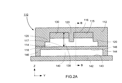
[0060] Fig. 2 - 4 show a particle separation microstructure 110 according
to
another embodiment. The microstructure 110 comprises a body 112 having a flow
channel 114 and a control channel 140 extending through the body 112.
[0061] Fig. 2 and Fig. 3 illustrate an embodiment where the second wall
118 is the
reversibly actuatable wall. It is to be understood that the control channel
140 may also
be defined where the first wall 116 is the reversibly actuatable wall. The
control channel
140 is defined by at least a portion of the reversibly actuatable first or
second wall
(116,118) and an opposing third wall 142. As shown in Fig.2 and Fig. 3, the
control
channel 140 is defined by at least a portion of the reversibly actuatable
second wall 180,
a third wall 142, and a pair of opposing side walls (144, 146), and an opening
for
receiving a pressurized fluid. The first surface 148 of the second wall 118
and the first
surface 143 of the third wall 142 are disposed in an opposing spaced apart
relationship.
[0062] The control channel 140 applies a force F, where the force F is
applied as
a pressure, to the reversibly actuatable first or second wall (116,118). As
shown in Fig. 2
and 3, the force F is applied to the reversibly actuatable second wall 118.
The control
channel 140 may be of any shape or size suitable for applying a force F as
described
- 12-
CA 02825093 2013-07-18
WO 2012/097450
PCT/CA2012/000066
above. For example, the control channel 140 used to apply pressure to the flow
channel
114 may be a rectangular cavity situated underneath the flow channel. In one
embodiment, the control channel 140 overlaps the flow channel 114 entirely
with suitable
alignment tolerance. It is to be understood that the control channel 140 may
be of
different dimensions that the flow channel 114. The flow channel 114 may
extend
longitudinally beyond the ends of the control channel 140. Alternatively, the
control
channel 140 may extend laterally beyond the side of the flow channel 114.
[0063] The distance between the first and second opposing walls (116,
118) of
the flow channel 114, is modulated by the application of a force, for example
a positive
differential pressure, from the control channel 140 to the flow channel 114
which deflects
the actuatable first or second wall (116,118) into contact with the protrusion
120 when the
flow channel 114 is in the second position as shown in Figures 3A and 3B.
[0064] The control channel 140 may have a single opening through which
fluid is
reversibly moved in and out. Alternatively, the control channel 140 may have a
separate
inlet and outlet for the movement of fluid into and out of the control
channel. Where the
control channel 140 has a single port such that the control channel 140 is a
'dead-end'
chamber or 'dead-end' channel, the control channel may be filled with a
pressurizing fluid
by a dead-end fill to remove any trapped air within the chamber. A "dead-end
fill" is a
well known method of filling dead-end chambers or dead-end channels with a
fluid under
pressure. For example, when a fluid is initially injected into a control
channel structure,
the fluid will follow the path of least resistance, and leave some regions of
the control
channel unfilled, or partially filled. The gas-permeability of some
elastomeric materials
used in microfluidic fabrication of flexible membranes may be utilized to
allow for dead-
end channels to be filled. The control channel fluid may be under a pressure
of about
100 mbar to about 4 bar or any amount therebetween. The control channel fluid
may be
air, water, or any other suitable pressurizable fluids.
[0065] The first or second reversibly actuatable wall (116, 118)
comprises a
flexible material capable of moving between first and second positions. In one
embodiment, the first or second reversibly actuatable wall (116, 118) is a
flexible
membrane formed between the flow channel 114 and control channel 140 which
deflects
into the flow channel 114 when actuated. The membrane may be of substantially
constant thickness, for example between about 10 pm and about 50 pm in
thickness, or
any thickness therebetween. Flexible membranes in microfluidic devices are
known for
their use as valves which partially or completely occlude the flow channel.
- 13-
CA 02825093 2013-07-18
WO 2012/097450
PCT/CA2012/000066
[0066] The flow channel 140 may comprise a plurality of protrusions 20 as
shown
in Fig. 2A and Fig. 3A. One protrusion 120 may be centrally located in the
cross section
flow channel 140 such that the central protrusion extends along a central
longitudinal axis
of the flow channel 140, and one protrusion may be located proximate each of
the
opposing side walls (121, 122) and extend substantially along each edge of the
flow
channel, adjacent the flow channel side walls (121, 122). It is to be
understood that the
flow channel 140 may comprise any number of protrusions that enable the first
and
second flow channel surface (115, 117) to be substantially parallel when the
flow channel
is in the second position.
[0067] The flow channel 114 may further comprise at least one recess 150
formed
in at least one of the first surface 115 of the first wall 116 and the second
surface 117 of
the second wall 118 for trapping larger, non-deformable particles from the
flow of particles
when the flow channel is in the second position. These recesses 150
temporarily trap the
larger, non-deformable particles within the flow channel when the flow channel
is moved
from an open to the restricted position and prevent the occlusion of the flow
channel 114.
[0068] The flow channel 114 may further comprise at least two ribs 130
transversely disposed across the flow channel 114 and extending from the at
least one
protrusion 120 toward one of the opposing side walls (121,122) to form at
least one
recess 150 within the flow channel 114 as shown in Fig. 5. The ribs 130 may
extend
from the protrusion 120 at an angle of about 90 degrees relative to the
longitudinal axis of
the flow channel. Alternatively, the ribs 130 may extend from the
protrusion120 at an
angle between about 30 degrees to about 90 degrees, or any angle in between,
relative
to a longitudinal axis of the flow channel 114 as shown in Fig. 5. It is to be
understood
that the ribs 130 extending from a protrusion 120 may abut a side wall (121,
122) or may
abut a second protrusion 120 as shown in Fig. 2A and 3A.
[0069] Changes in the distance between the first and second opposing
walls
(116, 118) of the flow channel 114, in essence the height 138 of the flow
channel 114,
can be made by adjusting the force applied to the actuatable first or second
wall. These
changes in force actuate the first or second wall (116, 118) and move the flow
channel
114 between the open and restricted positions. By changing the distance
between the
first and second opposing walls and altering the height 138 of the flow
channel,
separation of specific particle types may by selected as shown in Fig. 6. It
is to be
understood that the at least one protrusion extending from the first surface
of the first wall
of the flow channel may be any shape or size suitable to allow the first and
second
opposing channel walls to be substantially parallel in at least the second
position. Fig. 6
- 14 -
CA 02825093 2013-07-18
WO 2012/097450
PCT/CA2012/000066
illustrates the pressure required to move the flow channel from a first to a
second position
to facilitate the entrapment of microspheres of a known size in the flow
channel.
[0070] In operation, the microstructure receives a flow of particles
driven through
the flow channel through the application of pressure. Preferably, the pressure
may be
greater than about 10 mbar and less than 200 mbar, however it is to be
understood that
the pressure may be any pressure suitable to drive the flow through the flow
channel.
When the flow channel is in the open position, a heterogeneous mixture flow of
particles
freely passes through the flow channel in the direction of fluid flow. In the
semi-closed or
restricted position, only the smaller and/or more deformable particle types
within the
heterogeneous mixture of particles are capable of passing through the flow
channel. The
larger and/or more rigid particle types within the heterogeneous mixture of
particles are
retained at the flow channel inlet and/or within the recesses of the flow
channel which act
as particle traps. Upon return of the flow channel to the open position, the
retained
particles are released back into the flow channel to continue to move
downstream
through the flow channel. Following a flow of particles passing through the
flow channel,
when the flow channel is in the restricted position, a buffer solution may be
introduced
into the flow channel to elute any smaller and/or more deformable particle
types
remaining in the flow channel, prior to the flow channel moving to the open
position.
Then, when the flow channel is moved into the open position, a buffer solution
may be
introduced again into the flow channel to elute any larger and/or more rigid
particle types
trapped in the flow channel. These separate elution phases further enable
particle
separation.
[0071] Fig. 7 shows another embodiment of the microstructure 210
comprising
a plurality of control channels (240a, 240b, 240c, 240d) to selectively
attenuate the flow of
particles in the flow channel 214, as illustrated in Fig 7. Each of the
plurality of control
channels (240a, 240b, 240c, 240d) are separately connected and isolated from
one
another. Each of the control channels (240a, 240b, 240c, 240d) may be
substantially
perpendicular to the longitudinal axis of the flow channel. Each control
channel (240a,
240b, 240c, 240d) modulates the flow through a corresponding portion of the
flow
channel, where the corresponding portion is illustrated by a shared portion of
the
reversibly actuatable second wall 218. The control channels (240a, 240b, 240c,
240d)
may be filled with a fluid and pressurized at different times, such that each
control
channel (240a, 240b, 240c, 240d) modulates the flow channel height 238
separately,
without changing the overall flow channel 214 volume. For example, the control
channels
(240a, 240b, 240c, 240d) may be divided into two sets of control channels that
are inter-
- 15-
CA 02825093 2013-07-18
WO 2012/097450
PCT/CA2012/000066
digitated and each set of control channels modulate different portions of the
flow channel
between the first and second positions. In an initial state, a first set of
control channels
(240a, 240c) of the particle separation microstructure 210 deflect a portion
of the
actuatable second wall (218a, 218c) into portions of the flow channel 214,
moving those
portions of the flow channel into a constricted position, while the second set
control
channels (240b, 240d) do not deflect portions of the actuatable second wall
(218b, 218d)
and the corresponding portions of the flow channel 214 remain in an open
position. In a
second state, the deflection of portions of the actuatable second wall (218a,
218b, 218c,
218d) of the control channels (240a, 240b, 240c, 240d) is reversed such that
the first set
of control channels (240a, 240c) do not deflect portions of the actuatable
second wall
(218a, 218c), and the second set of control channels (240b, 240d) deflect
portions of the
actuatable second wall (218b, 218d) moving the corresponding portions of the
flow
channel 214 into the constricted position. This dynamic modulation of the
particle
separation microstructure 210 operates by rapidly moving between these open
and
restricted flow channel positions, alternating the flow channel geometry
without changing
the overall flow channel volume. It is to be understood a similar
configuration of control
channels may be achieved by connecting a plurality of microstructures in
series. The
control channels of alternating microstructures may be interconnected in order
to reduce
the number of control channels that must be separately actuated.
[0072] Apparatus and Method for Particle Separation
[0073] Fig. 8 illustrates an apparatus for the separation of particles.
The
apparatus 70 comprises at least one microstructure 10, however it is to be
understood
that a plurality of microstructures 10 may be implemented in parallel or in
series as
described below, thus alternative embodiments may employ less or more
parallelization.
The apparatus 70 comprises a plurality of valves (72a, 72b, 72c, 72d) and a
plurality of
conduits (73, 75, 77, 78) connect to the flow channel inlet 74 or flow channel
outlet 76 for
receiving and discharging a plurality of different flows. The conduits (73,
75, 77, 78) are
connected to the flow channel inlet 74 and flow channel outlet 76 by any
suitable means
and direct the plurality of flows through the flow channel of the
microstructure 10. The
open and closed state of the flow control valves (72a, 72b, 72c, 72d)
correspond to the
open and restricted positions of the flow channel to separate particles from
the flow of
particles, thus facilitating the selective separation of particle types and
subsequent
direction of the selected particle types to different conduits.
[0074] In one embodiment, the apparatus 70 comprises a sample conduit 73
and a buffer conduit 75 connected to the flow channel inlet 74, and a first
particle conduit
- 16-
CA 02825093 2013-07-18
WO 2012/097450
PCT/CA2012/000066
77 and a second particle conduit 78 connected to the flow channel outlet 76.
The plurality
of valves (72a, 72b, 72c, 72d), for example standard microfluidic control
valves, control
the direction of flow into and out of the flow channel. The inflow control
valves 72a, 72b
are disposed between each of the sample and buffer conduits respectively (73,
75) and
the flow channel inlet 74 of the microstructure for modulating the flows
received by the
flow channel. The outflow control valves 72c, 72d are disposed between the
outlet 76 of
the flow channel of the microstructure and each of first particle and second
particle
conduits respectively (77, 78) for facilitating the individual collection of
separated
particles. The separated particles may be collected, stored, and extracted.
[0075] It will be understood that the particle separation apparatus 70
may include
a plurality of conduits for receiving and discharging a plurality of flows for
separation of
two or more particle types from the flow of particles.
[0076] As shown in Fig. 8, the apparatus 70 operates on a 3-stage cycle
illustrated in panels (A), (B) and (C). In operation, each of the sample inlet
73 and the
buffer inlet 75 are maintained under a pressure greater than the pressure
maintained at
the first and second particle outlets for driving a flow through the flow
channel preferably
under a pressure greater than about 20 mbar and less than about 500 mbar. Each
of the
first particle outlet 77 and the second particle outlet 78 are maintained at
about
atmospheric pressure.
[0077] In the first stage of the operational cycle, illustrated in Fig
8A, a
heterogeneous mixture of particles, comprising target particles 90 and
background
particles 92, is provided to the flow channel inlet 74 by the sample conduit
73. Inflow
control valve 72b is closed preventing the flow of a buffer into the flow
channel of the
microstructure. Outflow control valve 72d is closed preventing the flow of any
particles
through second particle conduit 78. Inflow control valve 72a is open
permitting the flow of
a sample solution comprising a heterogeneous mixture of target 90 and
background 92
particles into the flow channel of the microstructure 10. The open state of
the inflow
control valve 72a and outflow control valve 72c facilitates the flow of
particles into the flow
channel of the microstructure 10. The microstructure 10 is held in the semi-
closed or
restricted position at a pressure, preferably not less than about 20 mbar
above the
sample inlet pressure. The target particles 90 are larger, more rigid, or
larger and more
rigid than the background particles 92. The target particles 90 are retained
at the flow
channel inlet 74 of the flow channel of the microstructure 10, while the
background
particles 92 flow through the flow channel, through outflow control valve 72c,
into the first
particle conduit 77 where the background particles 92 may be collected.
- 17-
CA 02825093 2013-07-18
WO 2012/097450
PCT/CA2012/000066
[0078] In the second stage of the operational cycle, illustrated in Fig
8B, inflow
control valve 72a is closed and inflow control valve 72b is opened, to
facilitate flow of a
buffer solution devoid of particles from the buffer conduit 75 into the flow
channel inlet 74
of the microstructure 10 to purge the flow channel of any remaining background
particles
92. The microstructure 10 continues to be held in the semi-closed position at
a pressure,
preferably not less than about 20 mbar above the buffer inlet pressure to
facilitate the
continued flow through the flow channel and the trapping of the target
particles 14. The
background particles 92 continue to flow through the flow channel, through
outflow control
valve 72c, to the first particle conduit 77 where the background particles 92
may be
collected.
[0079] In the third stage of the operational cycle, illustrated in Fig
8C, outflow
control valve 72c is closed, outflow control valve 72d is opened, and the
pressure applied
to the actuatable flow channel wall is removed to move the flow channel of
microstructure
to the open position. A flow of buffer solution from the buffer conduit 75 is
introduced
into the flow channel inlet 74 to purge the flow channel of the previously
entrapped target
particles 90. The buffer and the released target particles flow through the
flow channel,
through outflow control valve 72d, to the second particle conduit 78 where the
target
particles 90 may be collected.
[0080] This three phase operational cycle facilitates the continuous
separation of
the target particles 90 from background particles 92. Each phase of the
operational cycle
may be controlled by a user. It is to be understood that the operation of the
particle
separation apparatus may be controlled manually, through a computer program,
or
through other suitable means. The length of each stage in the operation cycle
is variable
and selectable by a user. Effectiveness of the separation of target particles
from
background particles can be measured by purity, defined as the ratio of target
particles
with background particles at the second particle conduit 78, or capture
efficiency, defined
as the ratio of the target particles at the second particle conduit 78 with
the background
particles at the sample conduit 73. The effectiveness of the separation may be
varied by
adjusting the period of time spent in each of the three phases. It is to be
understood
particles collected at the second particle conduit 78 may be re-circulated to
the sample
conduit 73 and then the process repeated to improve the overall effectiveness
of the
separation. Furthermore, it is to be understood that the buffer solution is
free of particles
and is non-reactive with both the microstructure and the flow of particles.
[0081] For example, if the initial concentration of target particle,
volumetric flow
rate, and number of parallelized channels is known then the time required to
have a
-18-
CA 02825093 2013-07-18
WO 2012/097450
PCT/CA2012/000066
desired number target particles trapped at each parallel connected apparatus
can be
determined. This estimated time period is a suitable period for the first
stage of particle
separation operation. It is to be understood that knowledge of the volumetric
flow rate
enables the estimation of the time required to purge the flow channel of
background
particles. This purging time is a suitable period for the second stage of
operation. A
similar period to the purging time is typically required to purge the target
particles to the
second particle outlet and is a suitable period of time the third stage of
operation.
[0082] Fig. 9 shows multi-stage apparatus 100 comprising a plurality of
serially
connected particle separation apparatus 170, comprising sample conduit 173,
buffer
conduit 175, first particle conduit 177, and second particle conduit 178. Each
of the
serially connected particle separation apparatus 170 are interconnected at a
particle
outlet (177, 178) of a first apparatus 170a to the sample inlet 173a of the
second
apparatus 170 by a connector 179, for example a serpentine shaped conduit. The
multistage apparatus 100 facilitates the repeated enrichment of a single
sample. Multi-
stage serial purification is a process well known in the art wherein an
enrichment process
which yields a certain purity, for example 90% purity, is implemented multiple
times in
series to yield a much greater purity, for example three times for 99.9%
purity. .
[0083] Using the multi-stage apparatus 100, the separation method as
described
above yields a first flow mixture comprising a large concentration of target
particles 90
and a small concentration of background particles 92 at the second particle
conduit 178a.
The concentration of target particles in the flow mixture at second particle
conduit 178a is
greater the concentration of target particles provided at the sample conduit
173 but may
not necessarily have an acceptable purity level. The first flow mixture enters
a second
apparatus 170b at sample conduit 173b and the separation process is repeated
and
yields a second flow mixture at the second particle conduit 178b. The second
flow
mixture then enters a third apparatus 170 c at sample conduit 173c and the
separation
process is repeated again yielding a third flow mixture at the second particle
conduit
178c. The process is repeated a number of times until the desired target
particle purity
level is achieved.
[0084] In one embodiment, serpentine shaped conduits (shown in Fig. 9)
may be
included in series with and preceding the inflow control valves (72a, 72b) in
order to
increase the overall hydrodynamic resistance of the flow channel of the
microstructure 10,
and/or reduce variation in hydrodynamic resistance caused by the deflection of
the first or
second walls of the flow channel when the device is in use, and/or to
temporarily store
separated particles from the previous stage.
- 19-
CA 02825093 2013-07-18
WO 2012/097450
PCT/CA2012/000066
[0085] Method of selectively modifying particle velocity
[0086] In one embodiment, the present disclosure provides a method for
selectively attenuating the velocities of specific particle types flowing
through a flow
channel of the particle separation microstructure 10.
[0087] A heterogeneous mixture of a flow of particles is flowed through
the flow
channel 14 of the microstructure 10 while the control channel 40 is
periodically
pressurized and depressurized, moving the flow channel between the open and
restricted
positions, according to a set 'duty cycle'. A duty cycle is defined as the
ratio between the
time period the flow channel is in the open position (free flow configuration)
and the time
period for the flow channel to complete one cycle. One cycle is defined as the
period of
time that it takes for the flow channel to move from an open position, to a
semi-closed or
restricted position, and return to an open position. The duty cycle controls
the ratio of the
velocity of trapped particles versus the free-flowing particles, and thus,
facilitates the
ability to modulate the average velocities of the particles flowing through
the flow channel
by determining the length of time the target particles are immobilized in the
flow channel
14, within the flow channel recesses 50. The distinct transient flow
characteristics of
different particle types result in different net velocities that enable
particle separation over
the length of the flow channel. The net velocity of each particle type in the
channel may
be estimated using a linear fit of the displacement data graph shown in Fig.
10. The
microstructure described herein, having a dynamic flow channel geometry,
provides a
method to selectively attenuate the flow rate of different particle types
based on their
physical properties.
[0088] The controlled movement of the flow channel between open and
restricted
positions enables chromatographic separation of particles, for example cells,
based on
their physical properties of size, deformability, or size and deformability.
In liquid
chromatography, mixture having a number of different components is infused
through a
structure, or column, that imparts different flow rates to different
components. The
difference in flow rate between the different components enables one component
of a
mixture to be concentrated relative to another component as the mixture
travels through
the structure. The microstructure described herein can impart different flow
rates to
different particles based on their physical properties of size, deformability,
or size and
deformability, where particles trapped by the flow channel in the semi-closed
position
travel at a slower speed than particles not trapped by the flow channel in the
semi-closed
position. Therefore, the ability of the microstructure to selectively trap
specific particles in
- 20 -
CA 02825093 2013-07-18
WO 2012/097450
PCT/CA2012/000066
the semi-closed position, the microstructure enables a chromatographic
separation of
these particles.
[0089] The time period for the open position (TopEN) and semi-closed
position
(TO, initial flow pressure and the pressure of the flow channel during the
open position
(PopEN) and semi closed position (P) may be determined by calculation or may
be
determined empirically. For example, TOPEN may range from about 0.5 to about
20
seconds, or any amount therebetween, for example about 1, 2, 3, 4,5, 6, 7, 8,
9, 10, 11,
12, 13, 14, 15, 16, 17, 18, or 19 seconds, or any amount therebetween. As an
example,
Tsc may range from about 0.5 to about 20 seconds, or any amount therebetween,
for
example about 1, 2, 3, 4,5, 6, 7, 8, 9, 10, 11, 12, 13, 14, 15, 16, 17, 18, or
19 seconds, or
any amount therebetween. As an example, POPEN may range from about 20 mbar to
about
500 mbar or any amount therebetween, for example about 30, 40, 50, 60, 70, 80
90, 100,
110, 120, 130, 140, 150, 160, 170, 180, 190, 200, 210, 220, 230 , 240, 250,
260, 270,
280, 290, 300, 350, 400, 450, or any amount therebetween. As an example, Psc
may
range from about 20 mbar to about 500 mbar or any amount therebetween, for
example
about 30, 40, 50, 60, 70, 8090, 100, 110, 120, 130, 140, 150, 160, 170, 180,
190, 200,
210, 220, 230, 240, 250, 260, 270, 280, 290, 300, 350, 400, 450 or any amount
therebetween. The duty cycle may be determined from these values as described
above,
and may range from about 0.1 to 1.0 or any amount therebetween, for example
about 0.2,
0.3, 0.4, 0.5, 0.6, 0.7, 0.8 or 0.9, or any amount therebetween.
[0090] Modulation of the flow channel geometry continuously disturbs the
contact
between the particles and the microstructure of the flow channel, thereby
reducing the
potential for particle adsorption and clogging problems that plague
traditional filtration-
based particle separation methods. By altering the operating duty cycle of the
particle
separation apparatus, adsorption of particles to inner surfaces of the flow
channel, and
obstruction of the flow channel is decreased or prevented. Modulation of the
flow channel
duty cycle facilitates the ability of a user to control the trapped particle
density in the
dynamic flow channel, which in turn enables a user to vary the incoming flow
of target
particle populations in real-time.
[0091] The pressure of the fluid applied to the flow channel (flow
pressure) may
be non-zero, or, from about 5 mbar to about 50 mbar, or any amount
therebetween. For
example about 10, 15, 20, 25, 30, 35, 40 or 45 mbar, or any amount
therebetween.
[0092] Apparatus Fabrication
[0093] Multilayer soft lithography (MSL) is a well-known fabrication
technique that
allows for facile and robust fabrication of microfluidic devices having
hundreds to
- 21 -
CA 02825093 2013-07-18
WO 2012/097450
PCT/CA2012/000066
thousands of microscopic reaction chambers, valves, pumps, fluidic logic
elements and
other components. Xia & Whitesides, 1998 (Angewandte Chemie-International
Edition
37:551 -575; herein incorporated by reference) describe and review procedures,
material
and techniques for soft lithography, including MSL.
[0094] The general idea of multilayer soft lithography (MSL) is to
iteratively stack
layers of polymers, for example polydimethylsiloxane (PDMS), of varying
thickness on top
of each other. Thin and thick layers of PDMS with stoichiometric ratios of
base and
hardener, respectively less than and higher than 10:1 are formed on separate
wafers.
For example, a thinner layer may be obtained using a base:hardener ratio of
20:1 and
spun onto a silicon wafer substrate. A thicker layer may be obtained using a
base:hardener ratio of 5:1. Photoresist patterns previously made on the wafers
will define
the microfluidic channels of the device, for example the flow channels and the
control
channels. The thick layer is then peeled away from the wafer and placed on top
of the
thin wafer. After baking, the excess components in each layer will bond and
form a
PDMS 'chip' composed of two layers of channels. Methods of working with
elastomers
and applying them in microfluidic applications are known in the art; see U.S.
Pat. No.
6,929,030; Scherer et al. Science 2000, 290, 1536-1539; Unger et al. Science
2000, 288,
113- 116; McDonald et al. Ace. Chem. Res. 2002, 35, 491-499; Thorsen, T. et
al,.
Science 2002, 298, 580-584; Liu, J. et al. Anal. Chem. 2003, 75, 4718-4723;
Rolland et
al. 2004 JACS 126:2322- 2323, PCT publications WO 02/43615 and WO 01/01025.
[0095] Various polymers, including but not limited to soft polymers, may
be used
in microfluidic devices and systems. Examples of polymers that may be useful
in
fabrication of all, or a portion of a microfluidic device according to various
aspects of the
invention include elastomers. Elastomers may be generally characterized by a
wide
range of thermal stability, high lubricity, water repellence and physiological
inertness.
Other desirable characteristics of elastomers may vary with the application.
It is within the
ability of one of skill in the art to select a suitable elastomer or
combination of elastomers
for the desired purpose. Examples of elastomers include silicone, PDMS,
photocurable
perfluoropolyethers (PFPEs), fluorosilicones, polyisoprene, polybutadiene,
polychloroprene, polyisobutylene, polyurethanes, poly(styrene-butadiene-
styrene), vinyl-
silane crosslinked silicones, and the like. Elastomers may be optically clear,
or may be
opaque, or have varying degrees of transparency. In some embodiments of the
present
disclosure, it may be desirable to use a biocompatible elastomer. PDMS is one
of the first
developed and more widely used elastomers in soft lithography applications.
Where
PDMS is described as the elastomer used in various embodiments of the
invention, it is
- 22 -
CA 02825093 2013-07-18
WO 2012/097450
PCT/CA2012/000066
for exemplary purposes only, and the choice of alternate elastomers is within
the
knowledge of one skilled in the art. A variety of elastomers suitable for use
in microfluidic
applications, and their various properties and examples of applications are
described in
U.S. Patent No. 6,929,030.
[0096] Other components may be incorporated into the particle separation
apparatus during fabrication - micron-scale valves, pumps, channels, fluidic
multiplexers,
perfusion chambers and the like may be integrated during MSL. Methods of
making and
integrating such components are described in, for example, U.S. Patent No's.
7,144,616,
7,113,910, 7,040,338, 6,929,030, 6,899,137, 6,408,878, 6,793,753, 6,540,895;
US Patent
Applications 2004/0224380, 2004/0112442; PCT Applications WO 2006/060748.
[0097] Once fabricated, one or more walls of a flow channel, via or other
space
within the microstructure may be treated or coated with a surface treatment
agent. For
example, the channels, via or other space may be temporarily filled with a
fluid
comprising bovine serum albumin (BSA) or a polymer (e.g. to prevent or reduce
non-
specific adhesion of particles, particularly cells. Examples of such polymers
include
polyethylene glycol of varying polymer molecular weight, such as are available
in the art.
One of skill in the art will be able to select a suitable polymer size and
concentration to
deposit sufficient polymer or protein on the surface, while maintaining a
suitable viscosity
to allow for handling and fluid flow within the device when preparing the
treatment.
Following treatment of the surface, the flow channel, via or other space may
be flushed
with a second fluid (e.g. a buffer, media, phosphate buffered saline (PBS) or
the like) to
remove any leftover albumin or polymer.
[0098] It is to be understood a microstructure is a structure comprising
features
where one or more dimensions measure less than about 1 mm.
[0099] The heterogeneous mixture of a flow of particles may comprise at
least two
or more types of particles or species of particles or populations of
particles. The types or
species or populations of particles may differ in size, rigidity, or both size
and rigidity.
Additionally, one or more of the particles may comprise a selectable marker,
or an
identifiable marker.
[00100] A particle may be any discrete material which can be flowed
through a
microscale system. For example particles may include beads, cells and the
like. For
example, polymer beads (e.g., polystyrene, polypropylene, latex, nylon and
many others),
silica or silicon beads, clay or clay beads, ceramic beads, glass beads,
magnetic beads,
metallic beads, inorganic compound beads, and organic compound beads can be
used. A
- 23 -
CA 02825093 2013-07-18
WO 2012/097450
PCT/CA2012/000066
variety of particles are commercially available, e.g., those typically used
for
chromatography (see, e.g., the 1999 Sigma "Biochemicals and Reagents for Life
Sciences Research" Catalog from Sigma (Saint Louis, Mo.), e.g., pp. 1921-2007;
The
1999 Suppleco "Chromatography Products" Catalogue, and others), as well as
those
commonly used for affinity purification (e.g., Dynabeads.TM. from Dynal, as
well as many
derivatized beads, e.g., various derivatized Dynabeads.TM. (e.g., the various
magnetic
Dynabeads.TM, which commonly include coupled reagents) supplied e.g., by
Promega,
the Baxter lmmunotherapy Group, and many other sources).
[00101] Particles may be suspended in any suitable fluid, including
buffer, saline,
water, culture medium, blood, plasma, serum, cell or tissue extract, urine or
the like, or a
combination thereof.
[00102] Cells may be obtained from, or found within, for example, cell
culture, an
environmental sample, a subject's body fluids, or a tissue sample. Cells may
be
eukaryotic cells, including plant cells. A cell culture may be included in a
process for
isolating, enriching, or isolating and enriching one or more particular cell
types or cell
species. Tissue samples may be obtained by, for example, curettage,
exfoliation, tissue
scraping or swabbing, needle aspiration biopsy or needle (core) biopsy,
incisional biopsy
for sampling tissue, or excisional biopsy, which may entail total removal of
the tissue of
interest. Body fluids include, for example, blood, bone marrow, plasma, serum,
adipose
tissue, sputum, urine, semen, amniotic fluid, cord blood, cerebrospinal fluid
or the like.
[00103] An environmental sample may comprise a fluid and one or more
species of
particle. For example, the environmental sample may comprise fresh or salt
water (e.g.
seawater, lake water, water from a treatment facility, sewer outflow or other
water
samples that may be acquired when monitoring a location or environment. The
environmental sample may comprise soil, plant matter, or other matter that may
be found
when monitoring a location or environment. The environmental sample may
comprise
particles, such as those exemplified herein, including eukaryotic cells,
and/or prokaryotic
cells, and/or minerals, particulates or the like.
[00104] A subject may be an animal, such as a mammal, reptile, bird or
fish;
examples of mammals include a rodent, cat, dog, primate, sheep, cow, pig,
horse or
ferret; examples of rodents include a mouse, rat, guinea pig or hamster;
examples of
primates include a human, a monkey, chimpanzee, rhesus macaque or green
monkey.
[00105] Examples of cells include red blood cells, white blood cells,
peripheral
blood mononucleocyte (PBMC), stem cells, tumor cells, cancer cells (primary or
immortalized), animal or human cell lines (primary cell lines or immortalized
cell lines) and
- 24 -
CA 02825093 2013-07-18
WO 2012/097450
PCT/CA2012/000066
the like. Examples of stem cells include adult stem cells, somatic stem cells,
embryonic
stem cells, non-embryonic stem cells, pluripotent stem cells, induced
pluripotent stem
cells, totipotent stem cells, multipotent stem cells, unipotent stem cells,
hematopoetic
stem cells, neural stem cells, mesenchymal stem cells, endothelial stem cells,
and the
like Cancer cells may be from any type of cancer or tumor. Non-limiting
examples of
different types of cancers and tumors include: carcinomas, such as neoplasms
of the
central nervous system, including glioblastoma, astrocytoma, oligodendroglial
tumors,
ependymal and choroid plexus tumors, pineal tumors, neuronal tumors,
medulloblastoma,
schwannoma, meningioma, and meningeal sarcoma; neoplasms of the eye, including
basal cell carcinoma, squamous cell carcinoma, melanoma, rhabdomyosarcoma, and
retinoblastoma; neoplasms of the endocrine glands, including pituitary
neoplasms,
neoplasms of the thyroid, neoplasms of the adrenal cortex, neoplasms of the
neuroendocrine system, neoplasms of the gastroenteropancreatic endocrine
system, and
neoplasms of the gonads; neoplasms of the head and neck, including head and
neck
cancer, neoplasms of the oral cavity, pharynx, and larynx, and odontogenic
tumors;
neoplasms of the thorax, including large cell lung carcinoma, small cell lung
carcinoma,
non-small cell lung carcinoma, malignant mesothelioma, thymomas, and primary
germ
cell tumors of the thorax; neoplasms of the alimentary canal, including
neoplasms of the
esophagus, stomach, liver, gallbladder, the exocrine pancreas, the small
intestine,
veriform appendix, and peritoneum, adneocarcinoma of the colon and rectum, and
neoplasms of the anus; neoplasms of the genitourinary tract, including renal
cell
carcinoma, neoplasms of the renal pelvis, ureter, bladder, urethra, prostate,
penis, testis;
and female reproductive organs, including neoplasms of the vulva and vagina,
cervix,
adenocarcinoma of the uterine corpus, ovarian cancer, gynecologic sarcomas,
and
neoplasms of the breast; neoplasms of the skin, including basal cell
carcinoma,
squamous cell carcinoma, dermatofibrosarcoma, Merkel cell tumor, and malignant
melanoma; neoplasms of the bone and soft tissue, including osteogenic sarcoma,
malignant fibrous histiocytoma, chondrosarcoma, Ewing's sarcoma, primitive
neuroectodermal tumor, and angiosarcoma; neoplasms of the hem atopoietic
system,
including myelodysplastic sydromes, acute myeloid leukemia, chronic myeloid
leukemia,
acute lymphocytic leukemia, HTLV-1 and T-cell leukemia/lymphoma, chronic
lymphocytic
leukemia, hairy cell leukemia, Hodgkin's disease, non-Hodgkin's lymphomas, and
mast
cell leukemia; and neoplasms of children, including acute lymphoblastic
leukemia, acute
myelocytic leukemias, neuroblastoma, bone tumors, rhabdomyosarcoma, lymphomas,
renal tumors, and the like.
- 25 -
CA 02825093 2013-07-18
WO 2012/097450
PCT/CA2012/000066
[00106] Where the particle is a cell, separation of one or more cell types
or species
from another in media, blood or other fluid has several applications. Without
limitation,
such applications may include leukapheresis, blood bank processing, separation
of
asynchronous cells in culture, enrichment of selected cell types (e.g. stem
cells from cord
blood or bone marrow or adipose tissue), identification and/or enumeration of
rare cell
types (e.g. circulating tumor cells in the blood). Such circulating tumor
cells may be of
particular diagnostic, prognostic or clinical interest as markers of the
development and
extent of cancer and/or metastasis. Circulating tumor cells (CTC) demonstrate
physical
differences from other hematological cells, namely size and rigidity. These
physical
differences may be able to be exploited in other cell types such as white
blood cells
(WBCs), cardiac myocytes, mesenchymal stem cells (MSCs), and pluripotent stem
cells.
Additionally, it may be beneficial to separate red blood cells from other
cells in a blood
sample to facilitate subsequent analysis. For example, the polymerase chain
reaction
(PCR) and considerable effort has been expended miniaturizing this reaction.
Haemoglobin in RBCs is an inhibitor of the PCR reaction and thus the presence
of RBCs
is detrimental in PCR reactions. This phenomenon motivated research in WBC
enrichment with Carlson et al in 1997 and Wilding et a/ in 1998.
[00107] Examples
[00108] Example 1 ¨ Microfabrication
[00109] An embodiment of the particle separation microstructure of the
present
disclosure, shown in Fig. 2, having a dynamically adjustable flow channel
height is a two-
layer microstructure fabricated using multilayer soft lithography of
polydimethylsiloxane
(PDMS) silicone. Multilayer soft lithography (MSL) is a well-known fabrication
technique
that allows for facile and robust fabrication of microfluidic devices having
hundreds to
thousands of microscopic reaction chambers, valves, pumps, fluidic logic
elements and
other components. Xia & Whitesides, 1998 (Angewandte Chemie-International
Edition
37:551 -575; herein incorporated by reference) describe and review procedures,
material
and techniques for soft lithography, including MSL.
[00110] Molds for the flow layer comprising the flow channel microstructure
and the
control layer comprising the control channel microstructure were fabricated
separately on
silicon wafers. The flow layer was fabricated in four photolithographic steps
to facilitate
the required flow layer geometry. The control layer was fabricated in a single
photolithographic step. The patterns for all five masks were drawn using
Solidworks
DWG Editor.
[00111] The SU-8 part of the flow layer mold was fabricated on a cleaned
100 mm
- 26 -
CA 02825093 2013-07-18
WO 2012/097450
PCT/CA2012/000066
silicon wafer. After dehydration baking on a hotplate at 200 C for 5 min, SU-8
3010 was
spread onto the wafer at 500 rpm for 10 seconds, and then spun at 2250 rpm for
30 s.
The wafer was then soft baked at 95 C on the hot plate for 5 minutes before
being
exposed to UV light in a mask aligner for 90 s. The exposed wafer was given a
post
exposure bake in the sequence of 65 C for 1 minute, 95 C for 5 minutes and
then 65 C
for 1 minute. The wafer was then developed using SU-8 developer (MicroChem). A
second layer of SU-8 3005 was spread onto the wafer at 500 rpm for 10 seconds,
and
then spun at 3000 rpm for 30 s. The wafer was then soft baked at 95 C on the
hot plate
for 5 minutes before being exposed to UV light in a mask aligner for 60 s The
exposed
wafer was given a post exposure bake in the sequence of 65 C for 1 minute, 95
C for 3
minutes and then 65 C for 1 minute. The wafer was then developed using SU-8
developer (MicroChem). A third layer of SU-8 3025 was spread onto the wafer at
500
rpm for 10 seconds, and then spun at 4000 rpm for 30 s. The wafer was then
soft baked
at 95 C on the hot plate for 5 minutes before being exposed to UV light in a
mask aligner
for 60 s The exposed wafer was given a post exposure bake in the sequence of
65 C for
1 minute, 95 C for 5 minutes and then 65 C for 1 minute. The wafer was then
developed
using SU-8 developer (MicroChem). The SPR part of the flow layer was added to
the
silicon wafer containing the SU-8 microstructures. SPR 220-7.0 photoresist was
spin-
coated on the wafer at 550 rpm for 50 s, and the resultant edge bead was
removed
manually. The coated wafer was soft baked on hotplates set at 65 C for 1
minute, 95 C
for 3 minutes, and then 65 C for 1 minute. The designed mask for the SPR
pattern was
then aligned with the SU-8 pattern and exposed in 5 30 s bursts with a 30
second interval
between bursts. After waiting for approximately 30 min, the wafer was
developed using
MF-319 developer (MicroChem). Finally, the developed wafer was annealed for 10
min
on a 95 C hotplate to create a rounded channel profile. The control layer is
made from
SU-8 3025 and fabricated in the same protocol as second SU-8 layer of the flow
layer.
[00112]
Silicon wafers containing the flow channel and control channel were
replicated using a plastic molding technique.( S. P. Desai, D. M. Freeman and
J.
Voldman, Lab Chip, 2009, 9, 1631-1637). The microstructure was fabricated from
PDMS
plastic molds using multilayer soft-lithography of RTV 615 silicone (Momentive
Performance Materials). The control channel was spun onto a plastic copy of
the silicon
wafer at 1800 rpm. The flow channel was cast molded from its plastic master
and
diffusion bonded to the control layer in a 65 C oven for 1 hour. The bonded
devices were
cut and punched using a 0.5 mm diameter punch (Technical Innovations,
Angleton, TX,
USA) to create fluid ports. In preparation for bonding, both the PDMS devices
and clean
- 27 -
CA 02825093 2013-07-18
WO 2012/097450
PCT/CA2012/000066
glass slides were activated using 40 s of air plasma (Harrick Plasma, Ithaca,
NY, USA).
The completed microstructure was prepared for experiments by initially filling
the control
channels with de-ionized water using 200 mbar of pressure. Subsequently, the
flow
channels were infused with phosphate buffered saline containing 5% bovine
serum
albumin, and incubated for 30 minutes to prevent non-specific adsorption of
the cells onto
the surface of the PDMS.
[00113] Example 2- Particle Separation Analysis
[00114] A suspension of rigid polystyrene microspheres of known size
(Bangs
Labs) were flowed through the separation channel. The flow channel was
initially in the
open position and all microspheres passed through the channel unimpeded. The
pressure applied to the flow channel by the control channel was gradually
increased as
microspheres continued to flow through the channel. As the pressure applied
increased,
the flexible membrane was deflected into the flow channel effectively moving
the flow
channel into the second, restricted position, thereby decreasing the flow
channel height
along the length of the flow channel. The pressure required to move the flow
channel into
the second position, and in turn, trap a single microsphere from the
suspension, about
half of microspheres from suspension, and almost all microspheres from
suspension were
recorded. The point at which almost all the microspheres from the suspension
were
trapped is indicative of the point at which the flow channel reached the
second position,
and the first and second opposing flow channel surfaces were substantially
parallel.
These three measurements are indicated in Fig. 7 by the bottom error bound,
data point,
and top error bound respectively. This experiment was repeated for three
different
suspensions of microspheres, where the microspheres in each of the three
suspensions
had a diameter 6.47 pm, 7.27 pm, 9.45 pm, and 10.14 pm respectively. The
results
illustrated in Fig. 7 clearly show the pressure required to move the flow
channel from an
open position to a semi-closed position. Manufacturer quoted standard
deviation in
microsphere diameter form error bounds in sphere diameter.
[00115] The results illustrate that larger particles require substantially
less pressure
to be captured or trapped at the inlet or within the flow channel than smaller
particles,
indicating that particle selectability on the basis of size and deformability
may be
facilitated by moving the flow channel between a first and second position by
controlling
the magnitude of the pressure applied to the flow channel.
[00116] Example 3 ¨Particle Velocity Analysis
[00117] L1210 mouse lymphoma cells were grown in suspension culture using
RPM! 1640 (lnvitrogen) containing 10% fetal bovine serum and 1%
- 28 -
CA 02825093 2013-07-18
WO 2012/097450
PCT/CA2012/000066
penicillin/streptomycin, at 37 C in a 100% humidified atmosphere containing 5%
CO2.
The cell suspension was diluted using phosphate buffered saline containing 5%
bovine
serum albumin. Cell viability was assessed using L3224 LIVE/DEAD
Viability/Cytotoxicity
Kit (Invitrogen) following the manufacturer's instructions. Peripheral blood
mononuclear
cells were prepared from whole blood collected from healthy volunteers
following
informed consent. Whole blood was drawn into 6 mL sodium heparin containing
tubes.
Peripheral blood mononuclear cells were obtained using Histopaque 1077 (Sigma-
Aldrich) according to manufacturer's instructions, and then resuspended at a
concentration of 10x106 cells per mL in AIM 5 media (Invitrogen). Red blood
cells were
purified from whole blood and used within 48 hours of donation. Before
testing, each of
the three cell types was re-suspended to a concentration of 7x10⁸ cells per
mL. Fluids
infused into the microstructure were supplied from 15 mL sealed conical tubes
(Fisher
Scientific) with custom fabricated caps that enable the tubes to act as
pressurized
reservoirs. The liquid connection between the reservoirs and the
microstructure was
made using 0.5 mm ID flexible Tygon tubing (Cole- Parmer). The microstructure -
to-tube
interface was created using 19 mm long 23 gauge stainless steel tubing (New
England
Small Tube, Litchfield, NH, USA) that forms a stretch seal between the PDMS
device and
the Tygon tubing. A multi-channel pressure control system (Fluigent MFCS-4C,
France)
was used to pressurize the reservoirs connected to the flow channel. A custom-
made
pressure control system was used to pressurize the reservoirs connected to the
control
channel to deflect the flexible membrane and move the flow channel into the
restricted
position. Pressure applied to the control channel could be turned on and off
automatically
using solenoid valves controlled by a MSP430 microprocessor (Texas
Instruments) and
custom developed PC software.
[00118] Videos of the cell motion inside the nnicrochannel were acquired
using a
Nikon Ti-U inverted microscope and a Nikon DS-2MBW CCD camera. The
displacement
of individual cells was measured using frame-by-frame tracking in the captured
videos.
The net velocity of each cell was determined using the slope of a linear fit
to the
displacement data. Cell diameters were measured in suspension using the Nikon
NIS-
Elements image capture software supplied with the CCD camera.
[00119] The results illustrate that red blood cells (RBCs) are highly
deformable,
discoid-shaped cells with an 8 mm diameter and a 2 mm thickness. Because the
dimensions of RBCs are small relative to the length scale of the
microstructures, these
cells essentially follow the flow of the bulk liquid. PBMCs primarily consist
of lymphocytes
and have a measured mean diameter of 7.2 mm with a standard deviation of 0.6
mm.
- 29 -
CA 02825093 2013-07-18
WO 2012/097450
PCT/CA2012/000066
MLCs were grown from an immortalized cell line and used in experiments between
4 and
6 days after passage. During this period, these cells had a mean diameter of
10.0 mm
with a standard deviation of 1.4 mm. MLCs were chosen because their size and
shape
are somewhat similar to PBMCs, but their rigidity is likely to be
significantly greater
because of their enlarged nucleus.
[00120] The flow properties of each of the three cell types in the dynamic
micro
flow channel were tracked by following the displacement of individual cells
over a fixed
2500 mm section of the micro flow channel. Representative cell displacement
data
graphs and video images are shown in Fig. 10 and Fig. 11. The sample fluid was
infused
into the micro flow channel with a pressure of 20 mbar, while the membrane
inflation
pressure was modulated between 100 mbar for the open flow channel position and
230
mbar for the semi-closed flow channel position. The time to complete one
cycle, i.e. for
the membrane to move from the first position, to the second position, and
return to the
first position, had a period of 6 seconds and a duty cycle of 50%. The timing
of each data
graph was adjusted to match the phase of the membrane cycles in Fig. 9. Cell
viability
was checked repeatedly along the length of the microchannel using the
fluorescence
signal produced by the L3224 LIVE/DEAD viability assay (Invitrogen). No
changes in cell
viability were observed, which is consistent with previous observations of
eukaryotic cells
compressed by PDMS membrane microvalves.
[00121] Fig. 12 shows the duty cycle dependence of the average velocity of
each
of the three cell types. These measurements were taken with open and semi-
closed flow
channel positions at flow channel pressures set at 100 and 230 mbar
respectively. The
average velocity of MLCs shows a decreasing trend as the duty cycle is reduced
from
100%, whereas the net flow rates of PBMCs and RBCs are nearly identical to
each other
and show little dependence on duty cycle. The average velocity of PBMCs and
RBCs
shows a general tendency to increase at lower frequencies. This trend results
from
reduced interactions between these cells and the surfaces of the dynamic
microchannel.
Below a 40% duty cycle, the average velocity of MLCs also begins to increase
with
decreasing duty cycle. Similar to over-pressurizing the flow channel, this
property is
caused by the motion of the membrane that excludes some of the larger MLCs
because
the length of time the flow channel is in the open position is insufficient
for these cells to
enter the micro flow channel.
[00122] In the preceding description, for purposes of explanation,
numerous details
are set forth in order to provide a thorough understanding of the embodiments.
However,
- 30 -
CA 02825093 2013-07-18
WO 2012/097450
PCT/CA2012/000066
it will be apparent to one skilled in the art that these specific details are
not required. In
other instances, well-known electrical structures and circuits are shown in
block diagram
form in order not to obscure the understanding. For example, specific details
are not
provided as to whether the embodiments described herein are implemented as a
software
routine, hardware circuit, firmware, or a combination thereof.
[00123] The above-described embodiments are intended to be examples only.
Alterations, modifications and variations can be effected to the particular
embodiments by
those of skill in the art without departing from the scope, which is defined
solely by the
claims appended hereto.
[00124] REFERENCES
[00125] 1. Mark JE. Polymer data handbook. New York: Oxford University
Press;
1999.
[00126] 2. Quake SR, Scherer A. From micro- to nanofabrication with soft
materials. Science. 2000 Nov. 24;290(5496):1536-40.
[00127] 3. Xia Y, Whitesides GM. Soft lithography. Annual Review of
Materials
Science. 1998 08/0128(1):153-84.
[00128] 4. Effenhauser CS, Bruin GJM, Paulus A, Ehrat M. Integrated
capillary
electrophoresis on flexible silicone microdevices: Analysis of DNA restriction
fragments
and detection of single DNA molecules on microchips. Anal Chem. 1997
09/01;69(17):3451-7.
[00129] 5. Quake SR, Scherer A, Spence C, Chou HP. In: Gourley PL, editor.
Microfabricated devices for sizing DNA and sorting cells. Micro- and
nanofabricated
structures and devices for biomedical environmental applications; 1998;; 1998.
[00130] 6. Fu AY, Chou H, Spence C, Arnold FH, Quake SR. An integrated
microfabricated cell sorter. Anal Chem. 2002 06/01;74(11):2451-7.
[00131] 7. Unger MA, Chou HP, Thorsen T, Scherer A, Quake SR. Monolithic
microfabricated valves and pumps by multilayer soft lithography. Science. 2000
04/07;288(5463):113-6.
[00132] 8. Hansen C. Microfluidic technologies for structural biology
[dissertation]. ;
2004.
[00133] 9. Vollmer AP, Probstein RF, Gilbert R, Thorsen T. Development of
an
integrated microfluidic platform for dynamic oxygen sensing and delivery in a
flowing
medium. Lab Chip. 2005;5:1059-66.
[00134] 10. Lee JN, Jiang X, Ryan D, Whitesides GM. Compatibility of
mammalian
cells on surfaces of poly(dimethylsiloxane). Langmuir. 2004 12/01;20(26):11684-
91.
- 31 -
CA 02825093 2013-07-18
WO 2012/097450
PCT/CA2012/000066
[00135] 11. Simpson, T. R. E. et al. Influence of interfaces on the rates
of
crosslinking in poly (dimethyl siloxane) coatings. Journal of polymer science.
2004;42(6):1421.
[00136] 12. Melin J, Quake SR. Microfluidic large-scale integration: The
evolution
of design rules for biological automation. Annu Rev Biophys Biomol Struct.
2007
06/01;36(1):213-31.
[00137] 13. VanDelinder V, Groisman A. Perfusion in microfluidic cross-
flow:
Separation of white blood cells from whole blood and exchange of medium in a
continuous flow. Anal Chem. 2007 03/01;79(5):2023-30.
[00138] 14. Goulpeau J, Trouchet D, Ajdari A, Tabeling P. Experimental
study and
modeling of polydimethylsiloxane peristaltic micropumps. J Appl Phys. 2005
08/15;98(4):044914.
[00139] 15. Kartalov EP, Scherer A, Quake SR, Taylor CR, Anderson WF.
Experimentally validated quantitative linear model for the device physics of
elastomeric
microfluidic valves. J Appl Phys. 2007 03/15;101(6):064505-1,064505-4.
[00140] 16. Squires TM, Quake SR. Microfluidics: Fluid physics at the
nanoliter
scale. Rev Mod Phys. 2005 10/06;77(3):977.
[00141] 17. Bruus H. Theoretical microfluidics - capillary effects. In:
Oxford
University Press; 2008. p. 123-41.
[00142] 18. Stone H. Introduction to fluid dynamics for microfluidic
flows. CMOS
Biotechnology. 2007:5-30.
[00143] 19. Thorsen T, Maerkl SJ, Quake SR. Microfluidic large-scale
integration.
Science. 2002 Oct 18;298(5593):580-4.
[00144] 20. Ajdari A. Steady flows in networks of microfluidic channels:
Building on
the analogy with electrical circuits. Comptes Rendus Physique. 2004 6;5(5):539-
46.
[00145] 21. Dondelinger RM. Hematology analyzers. Biomedical
Instrumentation &
Technology. 2009;43(4):300-4.
[00146] 22. The human heart - blood [homepage on the Internet]. . 2010.
Available
from: http://www.fi.edu/learn/heart/blood/blood.html.
[00147] 23. Chu RW. Leukocytes in blood transfusion: Adverse effects and
their
prevention. Hong Kong Med J;5(3):280-4.
[00148] 24. Toner M, Irimia D. Blood on a chip. Annu Rev Biomed Eng. 2005
08/15;7(1):77-103.
[00149] 25. Guller U, Zajac P, Schnider A, Bosch B, Vorburger S, Zuber M,
et al.
Disseminated single tumor cells as detected by real-time quantitative PCR
represent a
- 32 -
CA 02825093 2013-07-18
WO 2012/097450
PCT/CA2012/000066
prognostic factor in patients undergoing surgery for colorectal cancer. Ann
Surg. 2002
December;236(6):768-76.
[00150] 26. Koch M, Kienle P, Kastrati D, Antolovic D, Schmidt J, Herfarth
C, et al.
Prognostic impact of hematogenous tumor cell dissemination in patients with
stage II
colorectal cancer. International Journal of Cancer. 2006;118(12):3072-7.
[00151] 27. Thorban S, Rosenberg R, Maak M, Friederichs J, Gertler R,
Siewert J.
Impact of disseminated tumor cells in gastrointestinal cancer. Expert Review
of Molecular
Diagnostics. 2006 05/01;6(3):333-43.
[00152] 28. Elshimali YI, Grody WW. The clinical significance of
circulating tumor
cells in the peripheral blood. Diagnostic Molecular Pathology. 2006
December;15(4):187-
94.
[00153] 29. Reuben JM, Krishnamurthy S, Woodward W, Cristofanilli M. The
role of
circulating tumor cells in breast cancer diagnosis and prediction of therapy
response.
Expert opinion. 2008;2(4):339.
[00154] 30. Urtishak S, Alpaugh RK, Weiner LM, Swaby RF. Clinical utility
of
circulating tumor cells: A role for monitoring response to therapy and drug
development.
Biomarkers in Medicine. 2008 04/01;2(2):137-45.
[00155] 31. Pappas D, Wang K. Cellular separations: A review of new
challenges
in analytical chemistry. Anal Chim Acta. 2007 10/3;601(1):26-35.
[00156] 32. Murthy S, Sethu P, Vunjak-Novakovic G, Toner M, Radisic M.
Size-
based microfluidic enrichment of neonatal rat cardiac cell populations. Biomed
Microdevices. 2006 09/01;8(3):231-7.
[00157] 33. Alberts Bea. Molecular biology of the cell. In: 4th ed. ;
2002.
[00158] 34. Bao G, Suresh S. Cell and molecular mechanics of biological
materials. Nat Mater. 2003 print;2(11):715-25.
[00159] 35. Tan JL, Tien J, Pirone DM, Gray DS, Bhadriraju K, Chen CS.
Cells
lying on a bed of microneedles: An approach to isolate mechanical force. PNAS.
2003
February 18;100(4):1484-9.
[00160] 36. Galbraith CG, Sheetz MP. A micromachined device provides a new
bend on fibroblast traction forces. Proceedings of the National Academy of
Sciences of
the United States of America. 1997 August 19;94(17):9114-8.
[00161] 37. Rosenbluth MJ, Lam WA, Fletcher DA. Force microscopy of
nonadherent cells: A comparison of leukemia cell deformability. Biophys J.
2006
4/15;90(8):2994-3003.
- 33 -
CA 02825093 2013-07-18
WO 2012/097450
PCT/CA2012/000066
[00162] 38. Sarah E Cross ea. AFM-based analysis of human metastatic
cancer
cells. Nanotechnology. 2008;19(38):384003.
[00163] 39. Yap B, Kamm RD. Mechanical deformation of neutrophils into
narrow
channels induces pseudopod protrusion and changes in biomechanical properties.
J Appl
Physiol. 2005 May 1;98(5):1930-9.
[00164] 40. A. Bos M, van Vliet T. Interfacial rheological properties of
adsorbed
protein layers and surfactants: A review. Adv Colloid Interface Sci. 2001
7/27;91(3):437-
71.
[00165] 41. Norde W, Lyklema J. Thermodynamics of protein adsorption.
theory
with special reference to the adsorption of human plasma albumin and bovine
pancreas
ribonuclease at polystyrene surfaces. J Colloid Interface Sci. 1979
9;71(2):350-66.
[00166] 42. Lee S, Writ's J. An aqueous-based surface modification of
poly(dimethylsiloxane) with poly(ethylene glycol) to prevent biofouling.
Langmuir. 2005
12/01;21(25):11957-62.
[00167] 43. Andrade J, Hlady V. Protein adsorption and materials
biocompatibility:
A tutorial review and suggested hypotheses. Biopolymers/Non-Exclusion HPLC.
1986:1-
63.
[00168] 44. Cancer [homepage on the Internet]. . 2009 February 10.
Available
from: http://vvww.who.int/mediacenter/factsheets/fs297/en/index.html.
[00169] 45. Weiss L, Dimitrov D. A fluid mechanical analysis of the
velocity,
adhesion, and destruction of cancer cells in capillaries during metastasis.
Cell Biochem
Biophys. 1984 03/01;6(1):9-22.
[00170] 46. McCusker J, Dawson MT, Noone D, Gannon F, Smith T. Improved
method for direct PCR amplification from whole blood. Nucleic Acids Res.
1992;20:6747.
[00171] 47. Carlson RH, Gabel CV, Chan SS, Austin RH, Brody JP, Winkelman
JW. Self-sorting of white blood cells in a lattice. Phys Rev Lett. 1997
09/15;79(11):2149.
[00172] 48. Wilding P, Kricka LJ, Cheng J, Hvichia G, Shoffner MA, Fortina
P.
Integrated cell isolation and polymerase chain reaction analysis using silicon
microfilter
chambers. Anal Biochem. 1998 3/15;257(2):95-100.
[00173] 49. Kricka U, Wilding P. Microchip PCR. Analytical & Bioanalytical
Chemistry. 2003 11;377(5):820-5.
[00174] 50. Sethu P, Sin A, Toner M. Microfluidic diffusive filter for
apheresis
(leukapheresis). Lab Chip. 2006;6(1):83-9.
[00175] 51. Seal SH. A sieve for the isolation of cancer cells and other
large cells
from the blood. Cancer. 1964;17(5):637-42.
- 34 -
CA 02825093 2013-07-18
WO 2012/097450
PCT/CA2012/000066
[00176] 52. Vona G, Sabile A, Louha M, Sitruk V, Romana S, Schutze K, et
al.
Isolation by size of epithelial tumor cells : A new method for the
immunomorphological
and MolecularCharacterization of circulating tumor cells. Am J Pathol. 2000
January
1;156(1):57-63.
[00177] 53. Mohamed H, McCurdy LD, Szarowski DH, Duva S, Turner JN,
Caggana M. Development of a rare cell fractionation device: Application for
cancer
detection. NanoBioscience, IEEE Transactions on. 2004;3(4):251-6.
[00178] 54. Tan S, Yobas L, Lee G, Ong C, Lim C. Microdevice for the
isolation
and enumeration of cancer cells from blood. Biomed Microdevices. 2009
08/01;11(4):883-
92.
[00179] 55. Anderson JR, Chiu DT, Jackman RJ, Cherniavskaya 0, McDonald
JC,
Wu H, et al. Fabrication of topologically complex three-dimensional
microfluidic systems
in PDMS by rapid prototyping. Anal Chem. 2000 07/01;72(14):3158-64.
[00180] 56. Cao X, Eisenthal R, Hubble J. Detachment strategies for
affinity-
adsorbed cells. Enzyme Microb Technol. 2002 7/1;31(1-2):153-60.
[00181] 57. Cozens-Roberts C, Lauffenburger DA, Quinn JA. Receptor-
mediated
cell attachment and detachment kinetics. I. probabilistic model and analysis.
Biophys J.
1990 10;58(4):841-56.
[00182] 58. Dainiak M, Galaev I, Kumar A, Plieva F, Mattiasson B.
Chromatography of living cells using supermacroporous hydrogels, cryogels. Adv
Biochem Eng /Biotechnol. 2007(106):101-27.
[00183] 59. Ettre LS. Nomenclature for chromatography. Pure & Appl Chem.
1993;65(4):819-72.
[00184] 60. Studer V, Hang G, Pandolfi A, Ortiz M, Anderson WE, Quake SR.
Scaling properties of a low-actuation pressure microfluidic valve. J Appl
Phys. 2004
01/01;95(1):393-8.
[00185] 61. Hui AYN, Wang G, Lin B, Chan W. Microwave plasma treatment of
polymer surface for irreversible sealing of microfluidic devices. Lab Chip.
2005;5(10):1173.
[00186] 62. Bollinger A, Butti P, Barras J-, Trachsler H, Siegenthaler W.
Red blood
cell velocity in nailfold capillaries of man measured by a television
microscopy technique.
Microvasc Res. 1974 1;7(1):61-72.
[00187] 63. Butti P, Intaglietta M, Reimann H, Holliger C, Bollinger A,
Anliker M.
Capillary red blood cell velocity measurements in human nailfold by
videodensitometric
method. Microvasc Res. 1975 9;10(2):220-7.
- 35 -
CA 02825093 2013-07-18
WO 2012/097450
PCT/CA2012/000066
[00188] 64. Bazargan V. Micro flow control using thermally responsive
polymer
solutions. 2008:1-152.
[00189] 65. Desai SP, Freeman DM, Voldman J. Plastic masters¨rigid
templates
for soft lithography. Lab Chip. 2009(9):1631-7.
[00190] 66. Duffy DC, McDonald JC, Schueller OJA, Whitesides GM. Rapid
prototyping of microfluidic systems in poly(dimethylsiloxane). Anal Chem. 1998
12/01;70(23):4974-84.
[00191] All citations are herein incorporated by reference.
- 36 -