Note: Descriptions are shown in the official language in which they were submitted.
CA 02825453 2013-07-23
WO 2012/123387
PCT/EP2012/054193
A Method of Analyzing Chromosomal Translocations and a System Therefore
FIELD
The present disclosure relates to systems and methods for analyzing
chromosomal
translocations, and in particular to analysis of chromosomal translocation by
in situ
hybridization.
BACKGROUND
The diagnosis, prognosis, and determination of treatment of disease based on
the
interpretation of tissue or cell samples taken from a diseased organism has
expanded dramatically over the past few years. In addition to traditional
histological staining techniques and immunohistochemical assays, in situ
techniques such as in situ hybridization and in situ polymerase chain reaction
are
now used to help diagnose disease states in humans. Thus, there are a variety
of
techniques that can assess not only cell morphology, but also the presence of
specific macromolecules within cells and tissues.
Molecular cytogenetic techniques, such as chromogenic in situ hybridization
(CISH) combine visual evaluation of chromosomes (karyotypic analysis) with
molecular techniques. Molecular cytogenetics methods are based on
hybridization
of a nucleic acid probe to its complementary nucleic acid within a cell. A
probe for
a specific chromosomal region will recognize and hybridize to its
complementary
sequence on a metaphase chromosome or within an interphase nucleus (for
example in a tissue sample). Probes have been developed for a variety of
diagnostic
and research purposes.
Sequence probes hybridize to single copy DNA sequences in a specific
chromosomal region or gene. These are the probes used to identify the
chromosomal critical region or gene associated with a syndrome or condition of
interest. On metaphase chromosomes, such probes hybridize to each chromatid,
usually giving two small, discrete signals per chromosome.
Hybridization of sequence probes, such as repeat depleted probes or unique
sequence probes (see for example U.S. 2011/0160076), has made possible
detection of chromosomal abnormalities associated with numerous diseases and
syndromes, including constitutive genetic anomalies, such as microdeletion
syndromes, chromosome translocations, gene amplification and aneuploidy
CA 02825453 2013-07-23
WO 2012/123387
PCT/EP2012/054193
- 2 -
syndromes, neoplastic diseases as well as pathogen infections. Most commonly
these techniques are applied to standard cytogenetic preparations on
microscope
slides. In addition, these procedures can be used on slides of formalin-fixed
paraffin embedded tissue, blood or bone marrow smears, and directly fixed
cells or
other nuclear isolates.
The information obtained from these assays can be used to diagnose disease in
a
patient, determine the prognosis of a patient that has a disease, and also to
determine the course of treatment for a patient with a disease. In many
instances,
the presence of a particular marker can be associated with the predicted
efficacy of
a drug.
Non-small cell lung cancer (NSCLC) is a disease in which malignant (cancer)
cells
form in the tissues of the lung. NSCLC is actually a group of lung cancers
that are
named for the kinds of cells found in the cancer and how the cells look under
a
microscope. The three main types of non-small cell lung cancer are squamous
cell
carcinoma, large cell carcinoma, and adenocarcinoma. NSCLC is the most
common kind of lung cancer.
Squamous cell carcinoma is a cancer that begins in squamous cells, which are
thin,
flat cells that look like fish scales. This is also called epidermoid
carcinoma. Large
cell carcinoma is a cancer that may begin in several types of large cells.
Adenocarcinoma is a cancer that begins in the cells that line the alveoli and
make
substances such as mucus. Other less common types of non-small cell lung
cancer
are: pleomorphic, carcinoid tumor, salivary gland carcinoma, and unclassified
carcinoma.
Smoking cigarettes, pipes, or cigars is the most common cause of NSCLC. The
earlier in life a person starts smoking, the more often a person smokes, and
the
more years a person smokes, the greater the risk. If a person has stopped
smoking,
the risk becomes lower as the years pass.
Tests and procedures to detect, diagnose, and stage non-small cell lung cancer
are
often done at the same time. The following tests and procedures are generally
used:
Chest x-ray; CBC; Sputum test to look for cancer cells; Bone scan; CT scan of
the
chest; MRI of the chest; Positron emission tomography (PET) scan; and
Thoracentesis. In some instances, biopsies are taken and analyzed. If the
biopsy
reveals the presence of lung cancer, more imaging tests will be done to
determine
the stage of the cancer. Stage relates to the size of the tumor and the extent
to
CA 02825453 2013-07-23
WO 2012/123387
PCT/EP2012/054193
- 3 -
which it has spread. Non-small cell lung cancer is divided into five stages:
Stage 0
- the cancer has not spread beyond the inner lining of the lung; Stage I - the
cancer
is small and has yet to spread to the lymph nodes; Stage II - the cancer has
spread
to some lymph nodes near the original tumor; Stage III - the cancer has spread
to
nearby tissue or spread to far away lymph nodes; Stage IV - the cancer has
spread
to other organs of the body such as the other lung, brain, or liver.
There are many different types of treatment for non-small cell lung cancer.
Treatment depends upon the stage of the cancer. Surgery is the often the first
line
of treatment for patients with non-small cell lung cancer that has not spread
beyond
nearby lymph nodes. The surgeon may remove: One of the lobes of the lung
(lobectomy); only a small part of the lung (wedge or segment removal); the
entire
lung (pneumonectomy). Some patients need chemotherapy. Chemotherapy uses
drugs to kill cancer cells and stops new ones from growing. Chemotherapy alone
is
often used when the cancer has spread (stage IV).
In some instances, a genetic analysis is done to determine the best course of
treatment for NSCLC. For example, some patients with particular mutations in
the
EGFR gene respond to EGFR tyrosine kinase inhibitors such as gefitinib. As
another example, the 7% of NSCLC with EML4-ALK translocations may benefit
from ALK inhibitors which are in clinical trials.
Break-apart probe systems have been used for analysis of tissues from NSCLC
patients. However, due to the nature of the chromosomal rearrangements that
occur in NSCLC, there can be a problem with false positive results, especially
where the rearrangement is within the same chromosome, such as an inversion.
In
these cases, it may not be possible to properly resolve the signals from each
set of
break-apart probes. The signals can appear as two separate signals even though
no
rearrangement has occurred. This can be a real problem, both due to obtaining
incorrect results and the scarcity of biopsy material. Three color systems
have been
used for chromosomal analysis. See, e.g., Makretsov et al., Genes, Chromosomes
and Cancer, 40:152-57 (2004); Martin-Subero, et al., Cancer Res., 66(21):10332-
38
(2006); Yoshimoto et al., Neoplasia 8(6):465-69 (2006); Renne et al., J. Mol.
Diagnost., 7(3): 352-56 (2005). However, none of these systems have been
applied
to solve problems associated with false positive results in break-apart probe
systems. Break-apart probe systems which address the problem of false positive
results would provide a benefit to patients afflicted with cancer.
CA 02825453 2013-07-23
WO 2012/123387
PCT/EP2012/054193
- 4 -
SUMMARY
The present disclosure relates to systems and methods for analyzing
chromosomal
translocations, and in particular to analyzing chromosomal translocation by in
situ
hybridization.
In illustrative embodiments, a method for analyzing a sample for a chromosomal
translocation associated with a breakpoint comprises contacting the sample
with a
first nucleic acid probe comprising a first sequence configured to hybridize
to
genomic DNA located 5' to the breakpoint, a second nucleic acid probe
comprising
a second sequence configured to hybridize to genomic DNA located 3' to the
breakpoint, and a third nucleic acid probe comprising a third sequence
configured
to hybridize to genomic DNA adjacent to the breakpoint. The method further
comprises establishing conditions suitable for the probes to hybridize to the
genomic DNA in the sample and detecting hybridization of the probes by
detecting
a first signal associated with the first nucleic acid probe, a second signal
associated
with the second nucleic acid probe, and a third signal associated with the
third
nucleic acid probe. In one embodiment, the method further comprises
identifying a
sample order and orientation, the sample order and orientation being a
sequence of
the first signal, the second signal, and the third signal longitudinally
arranged along
a chromosome. In another embodiment, the method further comprises comparing
the sample order and orientation with a control order and orientation. In
another
embodiment, the control order and orientation is a sequence of the first
signal, the
second signal, and the third signal longitudinally arranged along a
chromosome,
wherein the chromosome is known to be devoid of a chromosomal translocation
associated with a breakpoint.
In illustrative embodiments, the third sequence is configured to hybridize to
genomic DNA 5' and adjacent to the breakpoint, and comparing the sample order
and orientation with a control order and orientation includes establishing
whether
the sample order and orientation includes inversion of the first signal and
the third
signal as compared to the control order and orientation. In one embodiment,
the
third sequence is configured to hybridize to genomic DNA 3' and adjacent to
the
breakpoint, and comparing the sample order and orientation with a control
order
and orientation includes establishing whether the sample order and orientation
includes inversion of the second signal and the third signal as compared to
the
control order and orientation. In another embodiment, the third sequence is
configured to hybridize to genomic DNA adjacent to the breakpoint located both
5'
CA 02825453 2013-07-23
WO 2012/123387
PCT/EP2012/054193
- 5 -
and 3' of the breakpoint, and comparing the sample order and orientation with
a
control order and orientation includes establishing whether the sample order
and
orientation includes inversion of the either the first signal or the second
signal with
the third signal as compared to the control order and orientation.
In illustrative embodiments, the method comprises determining the control
order
and orientation by analyzing a control known to be devoid of the chromosomal
translocation associated with the breakpoint comprising, wherein determining
includes contacting the control with the first nucleic acid probe comprising
the first
sequence configured to hybridize to genomic DNA located 5' to the breakpoint,
the
second nucleic acid probe comprising the second sequence configured to
hybridize
to genomic DNA located 3' to the breakpoint, and the third nucleic acid probe
comprising the third sequence configured to hybridize to genomic DNA adjacent
to
the breakpoint. The method further comprises establishing conditions suitable
for
the probes to hybridize to the genomic DNA in the control and detecting
hybridization of the probes by detecting a first signal associated with the
first
nucleic acid probe, a second signal associated with the second nucleic acid
probe,
and a third signal associated with the third nucleic acid probe.
In illustrative embodiments, the nucleic acid probes comprise nucleic acid
selected
from the group consisting of RNA, DNA, PNA, LNA and combinations thereof
labeled with a detectable moiety. In one embodiment, the detectable moiety is
selected from the group consisting of a hapten, an enzyme, a fluorescent
molecule,
a luminescent molecule and a radioactive molecule. In another embodiment, the
detectable moiety is a hapten, and the first, second and third nucleic acid
probes are
labeled with different first, second and third haptens, respectively. In some
embodiments, haptens are selected from the group consisting of biotin, 2,4-
dintropheyl (DNP), fluorescein derivatives, digoxygenin (DIG), 5-nitro-3-
pyrozolecarbamide (nitropyrazole, NP), 4,5,-dimethoxy-2-nitrocinnamide
(nitrocinnamide, NCA), 2-(3
,4-dimethoxypheny1)-quinoline-4-carb amide
(phenylquinolone, DPQ), 2,1,3 -benzoxadiazole-5-carbamide (benzofurazan, BF),
3 -hydroxy-2-quinoxalinecarb amide (hydroxyquinoxaline, HQ), 4-
(dimethylamino)azobenzene-4'-sulfonamide (DABSYL), rotenone isoxazoline
(Rot), (E)-2-
(2-(2-oxo-2,3 -dihydro-1H-benzo[b] [ 1,4] diazepin-4-
yl)phenozy)acetamide (benzodiazepine, BD), 7-(diethylamino)-2-oxo-2H-
chromene-3-carboxylic acid (coumarin 343, CDO), 2-acetamido-4-methy1-5-
thiazolesulfonamide (thiazolesulfonamide, T S), and p-
methoxyphenylpyrazopodophyllamide (Podo).
CA 02825453 2013-07-23
WO 2012/123387
PCT/EP2012/054193
- 6 -
In other illustrative embodiments, the method includes detecting that includes
contacting the sample antibodies specific to the label haptens, for example,
with
first, second and/or third antibodies specific for the first, second, and
third haptens,
respectively. In one embodiment, detecting further comprises detecting haptens
(e.g. first, second, and third haptens) using anti-hapten recognition and
enzymatic
signal amplification.
In illustrative embodiments, a kit for analyzing a sample for a chromosomal
translocation associated with a breakpoint comprises a first nucleic acid
probe
having a sequence configured to hybridize to a portion of the genomic DNA that
is
located 5' to the breakpoint, a second nucleic acid probe having a sequence
configured to hybridize to a portion of the genomic DNA that is located 3' to
the
breakpoint, and a third nucleic acid probe having a sequence configured to
hybridize to a portion of DNA that is adjacent to the breakpoint. In one
embodiment, the third nucleic acid probe has a sequence configured to
hybridize to
a portion of DNA adjacent to and spanning the breakpoint on both the 5' and 3'
sides of the breakpoint. In another embodiment, the first, second, and third
nucleic
acid probes are haptenated with a first, second, and third hapten, the kit
further
comprising detection reagents configured to enable visualization of the first,
second, and third hapten. In another embodiment, the detection reagents are
chromogenic detection reagents configured to enable bright-field visualization
of
the first, second, and third hapten.
In illustrative embodiments, a method for diagnosing a disease associated with
a
chromosomal translocation associated with a breakpoint in a patient sample
comprises contacting the patient sample with a series of nucleic acid probes,
the
series selected so that in the absence of the chromosomal translocation
associated
with the breakpoint, the series hybridizes to the patient sample according to
a first
order and orientation, and so that in the presence of the chromosomal
translocation
associated with the breakpoint, the series hybridizes to the patient sample
according
a different order and orientation and detecting whether the series of nucleic
acid
probes hybridizes to the patient sample according to the first order and
orientation,
wherein detecting the first order and orientation provides a diagnosis that
the
patient sample does not have the chromosomal translocation associated with the
breakpoint in the patient sample. In one embodiment, the first order and
orientation is a predetermined sequence of three signals longitudinally
arranged
along a chromosome. In
another embodiment, detecting includes using
chromogenic detection reagents visualized using bright-field imaging.
CA 02825453 2013-07-23
WO 2012/123387
PCT/EP2012/054193
- 7 -
BRIEF DESCRIPTION OF THE DRAWINGS
FIG. 1 is a schematic depiction of a chromosome showing a breakpoint
region and probes configured to hybridize thereto;
FIG. 2(A-D) show a schematic depiction of an exemplary detection scheme;
FIG. 3 is a schematic depiction of a chromosome showing two breakpoint
locations at which an inversion chromosomal translocation can occur and probes
configured to hybridize thereto;
FIG. 4 is a schematic depiction showing the chromosome of FIG. 3
subsequent to the inversion chromosomal translocation and the resulting
localization of the probes;
FIG. 5(A-B) is a magnified top plan view showing the signal reported for
(A) wild-type ALK and (B) rearranged ALK as would be seen using triple
colorimetric detection and bright-field imaging;
FIG. 6(A-B) are photographic images corresponding to FIG. 5(A-B)
respectively;
FIG. 7 is a schematic depiction of two chromosomes showing two
breakpoint locations at which a rearrangement chromosomal translocation can
occur;
FIG. 8 is a schematic depiction showing the chromosome of FIG. 7
subsequent to the rearrangement chromosomal translocation and the resulting
localization of the probes; and
FIG. 9(A-B) is a magnified top plan view showing the signal reported for
(A) wild-type ALK and (B) rearranged ALK as would be seen using triple
colorimetric detection and bright-field imaging.
DEFINITIONS
Unless otherwise explained, all technical and scientific terms used herein
have the
same meaning as commonly understood by one of ordinary skill in the art to
which
this disclosure belongs. Definitions of common terms in molecular biology can
be
found in Benjamin Lewin, Genes V, published by Oxford University Press, 1994
(ISBN 0-19-854287-9); Kendrew et al. (eds.), The Encyclopedia of Molecular
Biology, published by Blackwell Science Ltd., 1994 (ISBN 0-632-02182-9); and
CA 02825453 2013-07-23
WO 2012/123387
PCT/EP2012/054193
- 8 -
Robert A. Meyers (ed.), Molecular Biology and Biotechnology: a Comprehensive
Desk Reference, published by VCH Publishers, Inc., 1995 (ISBN 1-56081-569-8).
The singular terms "a", "an", and "the" include plural referents unless
context
clearly indicates otherwise. Similarly, the word "or" is intended to include
"and"
unless the context clearly indicates otherwise. The term "plurality" is used
synonymously with the phrase "more than one," that is, two or more. It is
further to
be understood that all base sizes or amino acid sizes, and all molecular
weight or
molecular mass values, given for nucleic acids or polypeptides are
approximate,
and are provided for description. The term "comprises" means "includes." The
abbreviation, "e.g.," is derived from the Latin exempli gratia, and is used
herein to
indicate a non-limiting example. Thus, the abbreviation "e.g.," is synonymous
with
the term "for example." Although methods and materials similar or equivalent
to
those described herein can be used in the practice or testing of this
disclosure,
suitable methods and materials are described below.
Antibody: "Antibody" collectively refers to immunoglobulins or immunoglobulin-
like molecules (including by way of example and without limitation, IgA, IgD,
IgE,
IgG and IgM, combinations thereof, and similar molecules produced during an
immune response in any vertebrate, for example, in mammals such as humans,
goats, rabbits and mice) and antibody fragments that specifically bind to a
molecule
of interest (or a group of highly similar molecules of interest) to the
substantial
exclusion of binding to other molecules (for example, antibodies and antibody
fragments that have a binding constant for the molecule of interest that is at
least
103 M-1 greater, at least 104 M-1 greater or at least 105 M-1 greater than a
binding
constant for other molecules in a biological sample.
More particularly, "antibody" refers to a polypeptide ligand comprising at
least a
light chain or heavy chain immunoglobulin variable region which specifically
recognizes and binds an epitope of an antigen. Antibodies are composed of a
heavy
and a light chain, each of which has a variable region, termed the variable
heavy
(VH) region and the variable light (VL) region. Together, the VH region and
the
VL region are responsible for binding the antigen recognized by the antibody.
This includes intact immunoglobulins and the variants and portions of them
well
known in the art. Antibody fragments include proteolytic antibody fragments
[such
as F(ab')2 fragments, Fab' fragments, Fab'-SH fragments and Fab fragments as
are
known in the art], recombinant antibody fragments (such as sFy fragments, dsFy
CA 02825453 2013-07-23
WO 2012/123387
PCT/EP2012/054193
- 9 -
fragments, bispecific sFy fragments, bispecific dsFy fragments, F(ab)'2
fragments,
single chain Fv proteins ("scFv"), disulfide stabilized Fv proteins ("dsFv"),
diabodies, and triabodies (as are known in the art), and camelid antibodies
(see, for
example, U.S. Pat. Nos. 6,015,695; 6,005,079-5,874,541; 5,840,526; 5,800,988;
and 5,759,808). A scFv protein is a fusion protein in which a light chain
variable
region of an immunoglobulin and a heavy chain variable region of an
immunoglobulin are bound by a linker, while in dsFvs, the chains have been
mutated to introduce a disulfide bond to stabilize the association of the
chains. The
term also includes genetically engineered forms such as chimeric antibodies
(for
example, humanized murine antibodies), heteroconjugate antibodies (such as,
bispecific antibodies). See also, Pierce Catalog and Handbook, 1994-1995
(Pierce
Chemical Co., Rockford, Ill.); Kuby, J., Immunology, 3rd Ed., W.H.
Freeman
& Co., New York, 1997.
Typically, a naturally occurring immunoglobulin has heavy (H) chains and light
(L) chains interconnected by disulfide bonds. There are two types of light
chain,
lambda (X) and kappa (k). There are five main heavy chain classes (or
isotypes)
which determine the functional activity of an antibody molecule: IgM, IgD,
IgG,
IgA and IgE.
Each heavy and light chain contains a constant region and a variable region
(the
regions are also known as "domains"). In combination, the heavy and the light
chain variable regions specifically bind the antigen. Light and heavy chain
variable
regions contain a "framework" region interrupted by three hypervariable
regions,
also called "complementarity-determining regions" or "CDRs". The extent of the
framework region and CDRs have been defined (see, Kabat et al., Sequences of
Proteins of Immunological Interest, U.S. Department of Health and Human
Services, 1991). The Kabat database is now maintained online. The sequences of
the framework regions of different light or heavy chains are relatively
conserved
within a species. The framework region of an antibody, that is the combined
framework regions of the constituent light and heavy chains, serves to
position and
align the CDRs in three-dimensional space.
The CDRs are primarily responsible for binding to an epitope of an antigen.
The
CDRs of each chain are typically referred to as CDR1, CDR2, and CDR3,
numbered sequentially starting from the N-terminus, and are also typically
identified by the chain in which the particular CDR is located. Thus, a VH
CDR3 is
located in the variable domain of the heavy chain of the antibody in which it
is
CA 02825453 2013-07-23
WO 2012/123387
PCT/EP2012/054193
- 10 -
found, whereas a VL CDR1 is the CDR1 from the variable domain of the light
chain of the antibody in which it is found. An antibody that binds RET will
have a
specific VH region and the VL region sequence, and thus specific CDR
sequences.
Antibodies with different specificities (i.e. different combining sites for
different
antigens) have different CDRs. Although it is the CDRs that vary from antibody
to
antibody, only a limited number of amino acid positions within the CDRs are
directly involved in antigen binding. These positions within the CDRs are
called
specificity determining residues (SDRs).
"Binding or stable binding" refers to the association between two substances
or
molecules, such as the hybridization of one nucleic acid molecule (e.g., a
binding
region) to another (or itself) (e.g., a target nucleic acid molecule). A
nucleic acid
molecule binds or stably binds to a target nucleic acid molecule if a
sufficient
amount of the nucleic acid molecule forms base pairs or is hybridized to its
target
nucleic acid molecule to permit detection of that binding.
A nucleic acid molecule is the to be "complementary" with another nucleic acid
molecule if the two molecules share a sufficient number of complementary
nucleotides to form a stable duplex or triplex when the strands bind
(hybridize) to
each other, for example by forming Watson-Crick, Hoogsteen or reverse
Hoogsteen
base pairs. Stable binding occurs when a nucleic acid molecule remains
detectably
bound to a target nucleic acid sequence (e.g., genomic target nucleic acid
sequence)
under the required conditions.
Complementarity is the degree to which bases in one nucleic acid molecule
(e.g.,
target nucleic acid probe) base pair with the bases in a second nucleic acid
molecule (e.g., genomic target nucleic acid sequence). Complementarity is
conveniently described by percentage, that is, the proportion of nucleotides
that
form base pairs between two molecules or within a specific region or domain of
two molecules.
In the present disclosure, "sufficient complementarity" means that a
sufficient
number of base pairs exist between one nucleic acid molecule or region thereof
and
a target nucleic acid sequence (e.g., genomic target nucleic acid sequence) to
achieve detectable binding. A thorough treatment of the qualitative and
quantitative
considerations involved in establishing binding conditions is provided by
Beltz et
al. Methods Enzymol. 100:266-285, 1983, and by Sambrook et al. (ed.),
Molecular
CA 02825453 2013-07-23
WO 2012/123387
PCT/EP2012/054193
- 11 -
Cloning. A Laboratory Manual, 2nd ed., vol. 1-3, Cold Spring Harbor Laboratory
Press, Cold Spring Harbor, N.Y., 1989.
A "computer implemented algorithm" is an algorithm or program (set of
executable
code in a computer readable medium) that is performed or executed by a
computing device at the command of a user. In the context of the present
disclosure, computer implemented algorithms can be used to facilitate (e.g.,
automate) selection of polynucleotide sequences with particular
characteristics,
such as identification of repetitive (or other undesired, e.g., background
producing)
nucleic acid sequences or unique binding regions of a target nucleic acid
sequence.
Typically, a user initiates execution of the algorithm by inputting a command,
and
setting one or more selection criteria, into a computer, which is capable of
accessing a sequence database. The sequence database can be encompassed within
the storage medium of the computer or can be stored remotely and accessed via
a
connection between the computer and a storage medium at a nearby or remote
location via an intranet or the internet. Following initiation of the
algorithm, the
algorithm or program is executed by the computer, e.g., to select one or more
polynucleotide sequences that satisfy the selection criteria. Most commonly,
the
selected polynucleotide sequences are then displayed (e.g., on a screen) or
outputted (e.g., in printed format or onto a computer readable medium).
The terms "conjugating, joining, bonding or linking" refer to covalently
linking one
molecule to another molecule to make a larger molecule. For example, making
two
polypeptides into one contiguous polypeptide molecule, or to covalently
attaching a
hapten or other molecule to a polypeptide, such as an scFv antibody. In the
specific
context, the terms include reference to joining a specific binding molecule
such as
an antibody to a signal generating moiety, such as a semi-conductor
nanocrystal.
The linkage can be either by chemical or recombinant means. "Chemical means"
refers to a reaction between the antibody moiety and the effector molecule
such
that there is a covalent bond formed between the two molecules to form one
molecule.
The term "coupled", when applied to a first atom or molecule being "coupled"
to a
second atom or molecule can be both directly coupled and indirectly coupled. A
secondary antibody provides an example of indirect coupling. One specific
example of indirect coupling is a rabbit anti-hapten primary antibody that is
bound
by a mouse anti-rabbit IgG antibody, that is in turn bound by a goat anti-
mouse IgG
antibody that is covalently linked to a detectable label.
CA 02825453 2013-07-23
WO 2012/123387
PCT/EP2012/054193
- 12 -
The term "corresponding" in reference to a first and second nucleic acid (for
example, a binding region and a target nucleic acid sequence) indicates that
the first
and second nucleic acid share substantial sequence identity or complementarity
over at least a portion of the total sequence of the first and/or second
nucleic acid.
Thus, a binding region corresponds to a target nucleic acid sequence if the
binding
region possesses substantial sequence identity or complementarity (e.g.,
reverse
complementarity) with (e.g., if it is at least 80%, at least 85%, at least
90%, at least
95%, or even 100% identical or complementary to) at least a portion of the
target
nucleic acid sequence. For example, a binding region can correspond to a
target
nucleic acid sequence if the binding region possesses substantial sequence
identity
to one strand of a double-stranded target nucleic acid sequence (e.g., genomic
target DNA sequence) or if the binding region is substantially complementary
to a
single-stranded target nucleic acid sequence (e.g. RNA or an RNA viral
genome).
A "genome" is the total genetic constituents of an organism. In the case of
eukaryotic organisms, the genome is contained in a haploid set of chromosomes
of
a cell. In the case of prokaryotic organisms, the genome is contained in a
single
chromosome, and in some cases one or more extra-chromosomal genetic elements,
such as episomes (e.g., plasmids). A viral genome can take the form of one or
more
single or double stranded DNA or RNA molecules depending on the particular
virus.
The term "hapten" refers to a molecule, typically a small molecule that can
combine specifically with an antibody, but typically is substantially
incapable of
being immunogenic except in combination with a carrier molecule.
The term "isolated" in reference to a biological component (such as a nucleic
acid
molecule, protein, or cell), refers to a biological component that has been
substantially separated or purified away from other biological components in
the
cell of the organism, or the organism itself, in which the component naturally
occurs, such as other chromosomal and extra-chromosomal DNA and RNA,
proteins, cells, and organelles. Nucleic acid molecules that have been
"isolated"
include nucleic acid molecules purified by standard purification methods. The
term
also encompasses nucleic acids prepared by amplification or cloning as well as
chemically synthesized nucleic acids.
A "label" is a detectable compound or composition that is conjugated directly
or
indirectly to another molecule to facilitate detection of that molecule.
Specific,
CA 02825453 2013-07-23
WO 2012/123387
PCT/EP2012/054193
- 13 -
non-limiting examples of labels include fluorescent and fluorogenic moieties,
chromogenic moieties, haptens, affinity tags, and radioactive isotopes. The
label
can be directly detectable (e.g., optically detectable) or indirectly
detectable (for
example, via interaction with one or more additional molecules that are in
turn
detectable). Exemplary labels in the context of the probes disclosed herein
are
described below. Methods for labeling nucleic acids, and guidance in the
choice of
labels useful for various purposes, are discussed, e.g., in Sambrook and
Russell, in
Molecular Cloning: A Laboratory Manual, 3rd Ed., Cold Spring Harbor Laboratory
Press (2001) and Ausubel et al., in Current Protocols in Molecular Biology,
Greene
Publishing Associates and Wiley-Intersciences (1987, and including updates).
The term "multiplex" refers to embodiments that allow multiple targets in a
sample
to be detected substantially simultaneously, or sequentially, as desired,
using plural
different conjugates. Multiplexing can include identifying and/or quantifying
nucleic acids generally, DNA, RNA, peptides, proteins, both individually and
in
any and all combinations. Multiplexing also can include detecting two or more
of a
gene, a messenger and a protein in a cell in its anatomic context.
A "nucleic acid" is a deoxyribonucleotide or ribonucleotide polymer in either
single or double stranded form, and unless otherwise limited, encompasses
analogues of natural nucleotides that hybridize to nucleic acids in a manner
similar
to naturally occurring nucleotides. The term "nucleotide" includes, but is not
limited to, a monomer that includes a base (such as a pyrimidine, purine or
synthetic analogs thereof) linked to a sugar (such as ribose, deoxyribose or
synthetic analogs thereof), or a base linked to an amino acid, as in a peptide
nucleic
acid (PNA). A nucleotide is one monomer in a polynucleotide. A nucleotide
sequence refers to the sequence of bases in a polynucleotide.
A nucleic acid "segment" is a subportion or subsequence of a target nucleic
acid
molecule. A nucleic acid segment can be derived hypothetically or actually
from a
target nucleic acid molecule in a variety of ways. For example, a segment of a
target nucleic acid molecule (such as a genomic target nucleic acid molecule)
can
be obtained by digestion with one or more restriction enzymes to produce a
nucleic
acid segment that is a restriction fragment. Nucleic acid segments can also be
produced from a target nucleic acid molecule by amplification, by
hybridization
(for example, subtractive hybridization), by artificial synthesis, or by any
other
procedure that produces one or more nucleic acids that correspond in sequence
to a
CA 02825453 2013-07-23
WO 2012/123387
PCT/EP2012/054193
- 14 -
target nucleic acid molecule. A particular example of a nucleic acid segment
is a
binding region.
A "probe" or a "nucleic acid probe" is a nucleic acid molecule or set of
nucleic acid
molecules that is capable of hybridizing with a target nucleic acid molecule
(e.g.,
genomic target nucleic acid molecule) and, when hybridized to the target, is
capable of being detected either directly or indirectly. Thus probes permit
the
detection, and in some examples quantification, of a target nucleic acid
molecule.
In particular examples, a probe includes a plurality of nucleic acid
molecules,
which include binding regions derived from the target nucleic acid molecule
and
are thus capable of specifically hybridizing to at least a portion of the
target nucleic
acid molecule. A probe can be referred to as a "labeled nucleic acid probe,"
indicating that the probe is coupled directly or indirectly to a detectable
moiety or
"label," which renders the probe detectable.
The term "semi-conductor nanocrystal" refers to a nanoscale particle that
exhibits
size-dependent electronic and optical properties due to quantum confinement.
Semi-conductor nanocrystal s have, for example, been constructed of semi-
conductor materials (e.g., cadmium selenide and lead sulfide) and from
crystallites
(grown via molecular beam epitaxy), etc. A variety of semi-conductor
nanocrystals
having various surface chemistries and fluorescence characteristics are
commercially available from Life Technologies (see, for example, U.S. Pat.
Nos.
6,815,064, 6,682596 and 6,649,138). Semi-conductor nanocrystals are also
commercially available from eBiosciences and Evident Technologies. Other semi-
conductor nanocrystals include alloy semi-conductor nanocrystals such as
ZnSSe,
ZnSeTe, ZnSTe, CdSSe, CdSeTe, ScSTe, HgSSe, HgSeTe, HgSTe, ZnCdS,
ZnCdSe, ZnCdTe, ZnHgS, ZnHgSe, ZnHgTe, CdHgS, CdHgSe, CdHgTe,
ZnCdSSe, ZnHgSSe, ZnCdSeTe, ZnHgSeTe, CdHgSSe, CdHgSeTe, InGaAs,
GaAlAs, and InGaN semi-conductor nanocrystals (Alloy semi-conductor
nanocrystals and methods for making the same are disclosed, for example, in US
Application Publication No. 2005/0012182 and PCT Publication WO
2005/001889).
A "sample" is a biological specimen containing genomic DNA, RNA (including
mRNA), protein, or combinations thereof, obtained from a subject. Examples
include, but are not limited to, chromosomal preparations, peripheral blood,
urine,
saliva, tissue biopsy, surgical specimen, bone marrow, amniocentesis samples
and
autopsy material. In one example, a sample includes genomic DNA or RNA. In
CA 02825453 2013-07-23
WO 2012/123387
PCT/EP2012/054193
- 15 -
some examples, the sample is a cytogenetic preparation, for example which can
be
placed on microscope slides. In particular examples, samples are used
directly, or
can be manipulated prior to use, for example, by fixing (e.g., using
formalin).
The term "signal generating moiety" refers to a composition or molecule that
generates a signal that is detectable by an assay.
The term "specific binding moiety" refers to a member of a binding pair.
Specific
binding pairs are pairs of molecules that are characterized in that they bind
each
other to the substantial exclusion of binding to other molecules (for example,
specific binding pairs can have a binding constant that is at least 103 M-1
greater,
104 M-1 greater or 105 M-1 greater than a binding constant for either of the
two
members of the binding pair with other molecules in a biological sample).
Particular examples of specific binding moieties include specific binding
proteins
(for example, antibodies, lectins, avidins such as streptavidins, and protein
A),
nucleic acids sequences, and protein-nucleic acids. Specific binding moieties
can
also include the molecules (or portions thereof) that are specifically bound
by such
specific binding proteins.
The term "specific binding agent" refers to a molecule that comprises a
specific
binding moiety conjugated to a signal generating moiety.
A "subject" includes any multi-cellular vertebrate organism, such as human and
non-human mammals (e.g., veterinary subjects).
A "target nucleic acid sequence or molecule" is a defined region or particular
sequence of a nucleic acid molecule, for example a genome (such as a gene or a
region of mammalian genomic DNA containing a gene of interest) or an RNA
sequence. In an example where the target nucleic acid sequence is a target
genomic
sequence, such a target can be defined by its position on a chromosome (e.g.,
in a
normal cell), for example, according to cytogenetic nomenclature by reference
to a
particular location on a chromosome; by reference to its location on a genetic
map;
by reference to a hypothetical or assembled contig; by its specific sequence
or
function; by its gene or protein name, or by any other means that uniquely
identifies it from among other genetic sequences of a genome. In some
examples,
the target nucleic acid sequence is mammalian or viral genomic sequence. In
other
examples, the target nucleic acid sequence is an RNA sequence.
CA 02825453 2013-07-23
WO 2012/123387
PCT/EP2012/054193
- 16 -
In some examples, alterations of a target nucleic acid sequence (e.g., genomic
nucleic acid sequence) are "associated with" a disease or condition. That is,
detection of the target nucleic acid sequence can be used to infer the status
of a
sample with respect to the disease or condition. For example, the target
nucleic acid
sequence can exist in two (or more) distinguishable forms, such that a first
form
correlates with absence of a disease or condition and a second (or different)
form
correlates with the presence of the disease or condition. The two different
forms
can be qualitatively distinguishable, such as by polynucleotide polymorphisms,
and/or the two different forms can be quantitatively distinguishable, such as
by the
number of copies of the target nucleic acid sequence that are present in a
cell.
A "vector" is any nucleic acid that acts as a carrier for other ("foreign")
nucleic acid
sequences that are not native to the vector. When introduced into an
appropriate
host cell a vector may replicate itself (and, thereby, the foreign nucleic
acid
sequence) or express at least a portion of the foreign nucleic acid sequence.
In one
context, a vector is a linear or circular nucleic acid into which a target
nucleic acid
sequence of interest is introduced (for example, cloned) for the purpose of
replication (e.g., production) and/or manipulation using standard recombinant
nucleic acid techniques (e.g., restriction digestion). A vector can include
nucleic
acid sequences that permit it to replicate in a host cell, such as an origin
of
replication. A vector can also include one or more selectable marker genes and
other genetic elements known in the art. Common vectors include, for example,
plasmids, cosmids, phage, phagemids, artificial chromosomes (e.g., BAC, PAC,
HAC, YAC) and hybrids that incorporate features of more than one of these
types
of vectors. Typically, a vector includes one or more unique restriction sites
(and in
some cases a multi-cloning site) to facilitate insertion of a target nucleic
acid
sequence.
DETAILED DESCRIPTION
The present disclosure relates to systems and methods for analyzing
chromosomal
rearrangements, and in particular to analysis of chromosomal rearrangements by
in
situ hybridization. Chromosomal rearrangements place genes in new linkage
relationships and generate chromosomes without normal pairing partners. The
present disclosure is not limited to the analysis of any particular type of
chromosomal rearrangement. In some embodiments, the chromosomal
rearrangement occurs within the same chromosome. An example of this type of
rearrangement is an inversion. In some embodiments, the rearrangement is a
CA 02825453 2013-07-23
WO 2012/123387
PCT/EP2012/054193
- 17 -
translocation. In a translocation, a segment from one chromosome is
transferred to
a nonhomologous chromosome or to a new site on the same chromosome.
Nonreciprocal translocations are one-way translocations in which a chromosomal
segment is transferred to a nonhomologous chromosome. Reciprocal
translocations, on the other hand, involve the exchange of segments from two
nonhomologous chromosomes. A gene fusion may be created when the
rearrangement joins two otherwise separated genes, the occurrence of which is
common in cancer. The chromosomal breakpoint is the region of the chromosome
where the double strand of the normally arranged chromosome is broken so that
the
rearrangement can occur. Translocation requires two double strand breaks.
The present disclosure provides probes and probe systems for use in detection
of a
target gene sequence in a biological sample. In preferred embodiments, the
target
sequence is a gene and surrounding sequences (5' and 3') that are prone to
rearrangement. Depending on the chromosomal breakpoints, a rearrangement can
result in the disruption or misregulation of normal gene function. These
molecular
rearrangements, in many cases, are considered to be the primary cause of
various
cancers. Indeed, over the past few decades, clinical cytogeneticists have been
able
to link specific chromosome breakpoints to clinically defined cancers,
including
subtypes of leukemias, lymphomas, and sarcomas. Virtually all of the
rearrangements observed in tumors have arisen through somatic mutations, so
these
are not inherited in families.
Analyses of the DNA sequences surrounding many of these rearrangement
breakpoints have provided important mechanistic insights into cancer. In some
instances, the rearrangement places the coding sequence of a first gene in
proximity
to the regulatory sequence for a second gene. The first rearrangement of this
kind
to be described was a rearrangement involving chromosomes 8 and 14 in patients
with Burkitt's lymphoma. This particular rearrangement places the MYC proto-
oncogene from chromosome 8 under the control of the powerful immunoglobin
heavy chain gene (IGH) promoter on chromosome 14. The MYC protein normally
triggers signals for cell proliferation, and the rearrangement causes high
levels of
MYC overexpression in lymphoid cells, where the IGH promoter is normally
active.
In other cancers, rearrangements fuse the coding sequences of two genes
together
to generate potent oncogenes. An example of historic interest is the
Philadelphia
chromosome, which was initially identified as a minute, or unusually small,
CA 02825453 2013-07-23
WO 2012/123387
PCT/EP2012/054193
- 18 -
chromosome in patients with chronic myelogenous leukemia (CIVIL). The
Philadelphia chromosome is actually a product of a reciprocal translocation
involving small segments at the ends of the q arms of chromosomes 9 and 22.
Subsequent molecular analyses involving multiple laboratories revealed that
the
translocation fused the coding sequence of the BCR (breakpoint cluster region)
gene on chromosome 22 with the coding sequence of the ABL gene on
chromosome 9. The BCR-ABL fusion protein encoded by the chimeric gene is a
protein tyrosine kinase that constitutively activates signaling pathways
involved in
cell growth and proliferation. Knowledge of this particular breakpoint has led
to a
successful treatment for CML, because investigators were able to use the
sequence
information to overexpress and crystallize the BCR-ABL protein, which in turn
led
to the development of drugs that inhibit this protein's activity.
In some preferred embodiments, the probes and probe systems are utilized for
in
situ hybridization procedures, for example, fluorescence in situ hybridization
(FISH), colorimetric in situ hybridization (CISH), and silver in situ
hybridization
(SISH). In some embodiments, the biological sample includes a tissue section
(such as obtained by biopsy) or a cytology sample (such as a Pap smear or
blood
smear). Other types of assays in which the disclosed probes and probe systems
can
be used are readily apparent to those skilled in the art, and particular
examples are
discussed below.
In some preferred embodiments, the probe systems comprise at least three
probes
for analysis of a particular target sequence that comprises a chromosomal
breakpoint. In preferred embodiments, each probe preferably comprises a
plurality
of probes that hybridize to a defined area of the genomic DNA. In preferred
embodiments, the probe sets are designed with a bioinformatic tool such as the
Human Genome Browser and Repeat Masker. In preferred embodiments,
repetitive elements are eliminated from the probe design. In some preferred
embodiments, the probes are synthesized by polymerase chain reaction (PCR)
processes. For example, in some embodiments, the Primer3 program (on the world
wide web at primer3.sourceforge.net) is used to design primers to the unique
sequences across the defined area of the chromosome. In some embodiments, the
designed PCR fragments and primers are analyzed for similarity to the human
genome and transcripts, for example, with Human BLAT and Blastnt programs (on
the world wide web at genome.ucsc.edu/cgi-bin/hgBlat). Fragments that exhibit
high similarity to other regions (i.e., other defined areas of the chromosome
to
which other probes are being designed) are excluded and all PCR fragments are
CA 02825453 2013-07-23
WO 2012/123387
PCT/EP2012/054193
- 19 -
verified by sequencing. In some preferred embodiments, the PCR fragments are
ligated, random amplified, and labeled by nick translation using a nucleotide
(e.g.,
dUTP or dCTP) conjugated to a hapten (described in more detail below).
Referring now to FIG. 1, shown is a schematic representation of a chromosome
100
having a breakpoint region 120. Across breakpoint region 120, a first nucleic
acid
probe 121, a second nucleic acid probe 123, and a third nucleic acid probe 122
may
be configured to hybridize to breakpoint region 120. Arrows (124, 125, 126)
are
illustrative breakpoint locations for a breakpoint in three exemplary probe
configurations. In one embodiment, probes (121, 122, 123) are configured to
place
the breakpoint at the 5' end of probe 121 and at the 3' end of probe 122 as is
shown
by arrow 124. Similarly, a probe configuration placing the breakpoint at the
3' end
of probe 123 and at the 5' end of probe 122 is shown by arrow 126. In another
embodiment, a probe configuration placing the breakpoint within the span of
probe
122 is shown by arrow 125. The probes can be configured to place the
breakpoint
in several different locations in the context of the probes. Breakpoint
locations
shown by the arrows (124, 125, 126) are merely exemplary of locations
understood
at this time to be useful. Localization of the breakpoint within probe 121 or
probe
123 is also reasonable, although it may not be a preferred embodiment. In
illustrative embodiments, the probes are configured to give rise to distinct
signals.
Accordingly, FIG. 1 shows the probes with distinct shading (e.g. probe 121
depicted with vertical striping, probe 122 is depicted as solid black, and
probe 123
is depicted with horizontal striping). In some embodiments, these probes will
be
configured to include labels so that they are visually distinguished from each
other.
While not being limited to a particular detection approach, FIG. 2(A-D) show
an
illustrative approach to detecting distinct labels subsequent to hybridization
to the
sample's genetic DNA.
Referring now to FIG. 2(A-D), shown is a schematic of an illustrative approach
to
analyzing a sample for a chromosomal translocation associated with a
breakpoint.
A breakpoint region 27 is depicted as being spanned by a first nucleic acid
probe
21, a second nucleic acid probe 23, and a third nucleic acid probe 22. The
nucleic
acid probes are labeled, a first label shown as diamond 221, a second label
shown
as a triangle 222, and a third label 223 shown as a pentagon. While the probes
are
shown with a single label, this representation is merely symbolic. Each probe
would actually be labeled with a plurality of labels. For example, the first
nucleic
acid probe 21 may include a 700 kb nucleic acid sequence nick translated to a
multiplicity of smaller haptenated oligonucleotide probe species. Exemplary
CA 02825453 2013-07-23
WO 2012/123387
PCT/EP2012/054193
- 20 -
locations for a breakpoint are shown as arrows 24, 25, and 26. FIG. 2(A-D)
show
an illustrative method for analyzing a sample comprising (A) contacting the
sample
with at least three probes and establishing conditions appropriate for
hybridization
of those probes with the genetic DNA found in the sample, (B) contacting the
sample with an antibody 28 directed towards one of the labeled probes, (C)
contacting the sample with a second antibody 29 conjugated to a plurality of
enzyme molecules, and (D) contacting the sample with a detection reagent that
results in the deposition of a detectable species 19 proximally to the probe
using
enzymatic deposition.
Referring again to FIG. 2A, in an illustrative embodiment, contacting the
sample
with a first nucleic acid probe includes a first nucleic acid probe 21 having
a first
sequence configured to hybridize to genomic DNA located 5' to the breakpoint.
Three potential breakpoints are shown by arrows 24, 25, and 26. Regardless of
which breakpoint position is selected, probe 21 remains 5' of the breakpoint.
Similarly, a second nucleic acid probe 23 having a second sequence configured
to
hybridize to genomic DNA located 3' to the breakpoint is shown in a location
3' to
each of the breakpoint positions. The method for analyzing a sample comprises
contacting the sample with a third nucleic acid probe comprising a third
sequence
configured to hybridize to genomic DNA adjacent to the breakpoint. An
exemplary sequence configured to hybridize to genomic DNA adjacent to the
breakpoint is shown as probe 22. As indicated by exemplary breakpoints 24, 25,
and 26, probe 22 is either adjacent to the breakpoint by its position directly
to one
side or the other (e.g. shown by arrows 24 or 26) or by spanning the
breakpoint
(arrow 25).
Referring now to FIG. 2B, shown is a representation of the illustrative step
of
contacting the sample with an antibody 28 directed towards one of the labeled
probes. For example, a hapten-labeled probe may be detected by contacting the
sample with an anti-hapten antibody. FIG.
2(A-D) show an exemplary
hybridization of three probes and subsequent detection of one of those probes.
Sequential or concurrent detection strategies could be used to detect
hybridization
of the other probes. That is, additional antibodies specifc to label 222 and
label
223 could be contacted to the sample simultaneously or sequentially to
antibody
28. Furthermore, the step represented by FIG. 2C of contacting the sample with
a
second antibody 29 conjugated to a plurality of enzyme molecules may be
accompanied simultaneously or sequentially with like steps to detect label 222
and
label 223. In the same manner, the detection step represented by FIG. 2D may
be
CA 02825453 2013-07-23
WO 2012/123387
PCT/EP2012/054193
-21 -
accompanied simultaneously or sequentially with like steps to deposit
additional
detectable species corresponding to label 222 and label 223. Illustratively,
the
detectable species for labeling each of the labels is distinct.
Referring to FIG. 1, probes 121, 122, and 123 are shown with distinct
patterns.
Similarly, with reference to FIG. 2A-2D, labels 221, 222, and 223 are shown
with
distinct shapes. The use of distinct patterns and shapes is intended to
indicate that
diverse detection strategies can be used for the detection of these various
probes.
As such, detection chemistries can be selected that allow for the
differentiation of
the location of the various probes. In
illustrative embodiments, detecting
hybridization of the probes includes detecting a first signal associated with
the first
nucleic acid probe, a second signal associated with the second nucleic acid
probe,
and a third signal associated with the third nucleic acid probe. In one
embodiment,
the signals are distinct. In some preferred embodiments, the first, second and
third
probes are labeled with different detectable moieties, such as haptens, which
allow
hybridization of each of the three probes to be resolved.
In some embodiments of the present disclosure, the systems comprise a first
nucleic acid probe set that hybridizes to a portion of the genomic DNA that is
5' to
a chromosomal breakpoint (i.e., a first defined area of the genomic DNA), a
second
nucleic acid probe set that hybridizes to a portion of the genomic DNA that is
3' to
the chromosomal breakpoint (i.e., a second defined area of the genomic DNA),
and
a third nucleic acid probe set comprising a 5' portion and a 3' portion and
which
hybridizes to 5' and 3' sequences adjacent to the chromosomal breakpoint
region
so that the third nucleic acid probe spans (i.e., hybridizes to a defined
region
spanning) the chromosomal breakpoint region in the absence of a
rearrangement(i.e., a third defined area of the genomic DNA). It will be
appreciated that the probe set to the breakpoint region comprises a portion of
individual probes that hybridize to the genomic DNA 5' to the breakpoint
(i.e., 5'
hybridizing portion) and a portion of individual probes that hybridize to the
genomic DNA 3' to the breakpoint (i.e., 3' hybridizing portion). In
embodiments
where the breakpoint is within a gene, the systems may comprise a first
nucleic
acid probe that hybridizes to a 5' noncoding region of a target sequence, a
second
nucleic acid probe that hybridizes to 3' noncoding region of a target
sequence, and
a third nucleic acid probe comprising a 5' portion and a 3' portion and which
hybridizes to 5' and 3' sequences adjacent to the breakpoint of the target
sequence
so that the third nucleic acid probe spans (i.e., hybridizes to a defined
region
CA 02825453 2013-07-23
WO 2012/123387
PCT/EP2012/054193
- 22 -
spanning) across the breakpoint of the target sequence when the target
sequence is
not rearranged.
In illustrative embodiments, the method of analyzing a sample includes
detection
of a translocation. Referring now to FIG. 3, shown is a translocation that
occurs on
the same chromosome (e.g. EML4-ALK fusion gene). Referring now to FIG. 7,
shown is a translocation that occurs between different chromosomes (e.g. KIF5B-
ALK fusion or TFG-ALK fusion). Referring again to FIG. 3, shown is an
exemplary method and application of a system for analyzing a sample for a
chromosomal translocation associated with a breakpoint. Shown is a
representation
of Chromosome 2 10, a breakpoint region associated with the ALK gene 20, and a
breakpoint associated with the EML4 gene 30. For the EML4 and ALK genes, a
distance 11 between the genes is about 12 Mb, ALK being located at 2p23 and
EML4 being located at 2p21. Across the breakpoint region associated with the
ALK gene 20, three probes have been configured to have sequences complimentary
to unique regions of the ALK gene. A first probe 321 is complimentary to a
sequence 3' to the breakpoint, a second probe 323 is complementary to a
sequence
5' to the breakpoint, and a third probe 322 is complimentary to a sequence
spanning the breakpoint. Chromosome 2 10 is shown in FIG. 3 in its wild-type
without a translocation. FIG. 4 shows Chromosome 2 410 that includes an
inversion associated with the ALK-EML4 fusion gene. The impact of the
inversion
on the localization of the probes is indicated by the localization of probes
321, 322,
and 323. That is, the chromosomal translocation can be identified by the
distinct
manner in which the probes hybridize to the genetic DNA. Referring now to FIG.
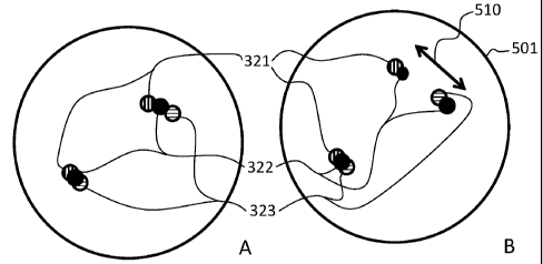
5(A-B), shown are schematics showing the manner in which a chromosomal spread
500 having a wild-type gene configuration (FIG. 5A) could be distinguished
from a
chromosomal spread 501 having an ALK-EML4 fusion gene (FIG. 5B) according
to a method described herein. In particular, FIG. 5A corresponds to the
schematic
shown in FIG. 3; the sequence of the labeling is in the order of 321, 322, and
323.
Referring now to FIG. 6A, shown is an embodiment where probe 321 was detected
with red chromogen, probe 322 was detected with blue chromogen, and probe 323
was detected with yellow chromogen. Accordingly, as shown pictorially in FIG.
6A and schematically in FIG. 5A, the order and orientation of the probes
generates
a signal having an order and orientation of red, blue, and yellow aligned
longitudinally along the length of the chromosome. In FIG. 6A, the red, blue,
and
yellow signals are indicated with arrows marked with "R" (red signal), "B"
(blue
signal) or "Y" (yellow signal). Similarly, FIG. 5B corresponds to the
schematic
CA 02825453 2013-07-23
WO 2012/123387
PCT/EP2012/054193
- 23 -
shown in FIG. 4; the sequence of the labeling is in the order of 321, 322,
323, and
322 arranged in two separate clusters. Referring now to FIG. 6B, shown is an
embodiment where probe 321 was detected with red chromogen, probe 322 was
detected with blue chromogen, and probe 323 was detected with yellow
chromogen. Accordingly, as shown pictorially in FIG. 6B and schematically in
FIG. 5B, the order of the probes generates a signal having an order and
orientation
of red, blue, yellow, and blue arranged in two clusters of signals, one
comprising
red and blue and the other comprising yellow and blue. In FIG. 6B, the red,
blue,
and yellow signals are indicated with arrows marked with "R" (red signal), "B"
(blue signal) or "Y" (yellow signal). In FIG. 5B, it can be seen that one copy
of the
gene remains in the wild-type configuration, but the second copy of the gene
shows
an inversion ISH signal. The inversion ISH signal includes a split of probe
322
that spans the breakpoint so that two signals (shown as blue or as the black
dot in
FIG. 6B and 5B respectively). The signals from the split probe may be of
diminished intensity due to the fact that the same length of probe is
localized in two
places. In a wild-type chromosome, the probes are configured to be in close
proximity to each other on a chromosome resulting in tightly clustered ISH
signals.
This can be seen clearly in FIG. 5A. For a chromosome that includes a
translocation, the ISH signals exhibit a spread as shown in FIG. 5B as double
arrow
510.
In one embodiment, the method of analyzing a sample includes detection of a
translocation. Referring now to FIG. 7, shown is a translocation that occurs
between different chromosomes (e.g. KIF5B-ALK fusion or TFG-ALK fusion).
Shown is a representation of Chromosome 2 10, a breakpoint region associated
with the ALK gene 20, and a representation of Chromosome 10 50 and breakpoint
associated with the KIF5B gene 52. According to this translocation, region 12
of
Chromosome 2 10 translocates with region 512 of Chromosome 10 50 according to
the arrow 55. This translocation results in the modified chromosomes shown in
FIG. 8, modified Chromosome 2 810 and modified Chromosome 10 850. Across
the breakpoint region associated with the ALK gene 20, three probes having
sequences complimentary to unique regions of the ALK gene have been designed.
A first probe 321 is complimentary to a sequence 3' to the breakpoint, a
second
probe 323 is complimentary to a sequence 5' to the breakpoint, and a third
probe
322 is complementary to a sequence spanning the breakpoint . These probes,
spanning the breakpoint region associated with the ALK gene 20, are shown in
FIG. 3. Referring now to FIG. 9(A-B), schematics representing the manner in
CA 02825453 2013-07-23
WO 2012/123387
PCT/EP2012/054193
- 24 -
which a chromosomal spread 900 having a wild-type gene configuration (FIG. 9A)
may be distinguished from a chromosomal spread 901 having a KIF5B-ALK fusion
gene (FIG. 9B) according to a method described herein. In particular, FIG. 9A
corresponds to the schematic shown in FIG. 7; the sequence of the labeling is
in the
order and orientation of 321, 322, and 323 arranged longitudinally along the
length
of the chromosome in a tightly distributed cluster. As shown schematically in
FIG.
9A, the order of the probes generates a signal having a first order (e.g. red,
black,
and blue). Similarly, FIG. 9B corresponds to the schematic shown in FIG. 8;
the
sequence of the labeling is in the order and orientation of 321, 322, 323, and
322
(e.g. red-black separated from blue-black). In FIG. 9B, it can be seen that
one copy
of the gene remains in the wild-type configuration, but the second copy of the
gene
shows a translocated ISH signal. The translocated ISH signal includes a split
of
probe 322 that spans the breakpoint so that two signals (shown as the black
dots in
FIG. 9B) are separated by a substantial distance, represented by double-arrow
910.
The signals from the split probe may be of diminished intensity due to the
fact that
the same length of probe is localized in two places. The distance between the
signals shown in FIG. 9B, when contrasted to the clustered signals shown in
FIG.
9A, provides evidence that the fusion gene is present.
In illustrative embodiments, a method according to the present disclosure
includes
detecting hybridization of the probes by detecting a first signal associated
with the
first nucleic acid probe, a second signal associated with the second nucleic
acid
probe, and a third signal associated with the third nucleic acid probe. As
shown in
FIG. 5(A-B) and 9(A-B), when a chromosomal rearrangement has occurred, a
signal is generated where a signal (e.g., a colorimetric signal, fluorometric
signal or
luminescent signal from an appropriate label as described in more detail
below)
from the third nucleic acid probe separately co-localizes with each of the
signals
from the first and second nucleic acid probes. As can be seen, there is a
distinct
first signal comprising a signal from the first nucleic acid probe and a
signal from
the third nucleic probe and a distinct second signal comprising a signal from
the
second nucleic acid probe and signal from the third nucleic acid probe. The
first
and second distinct signals can be located on genomic DNA belonging to the
same
or different chromosomes depending on the rearrangement. In samples where
rearrangement has occurred, the probe set corresponding to the breakpoint
region is
split, with separate portions of the probe set hybridizing to the 5' and 3'
regions
flanking the breakpoint, i.e., the 5' hybridizing portion of the probe set
hybridizes
to the 5' end of the rearranged target sequence and the 3' hybridizing portion
of the
CA 02825453 2013-07-23
WO 2012/123387
PCT/EP2012/054193
- 25 -
probe set hybridizes to the 3' portion of the rearranged target sequence. This
hybridization pattern leads to a split (i.e., two separate signals) for the
third probe.
When the third probe is labeled with a separate color from the first two
probes, the
resolution of the assay and the ability to distinguish false positive signals
is greatly
enhanced.
As also shown in FIG. 5(A-B) and 9(A-B), when a chromosomal translocation has
not occurred, a signal is generated where a signal (e.g., a colorimetric
signal,
fluorometric signal or luminescent signal from an appropriate label as
described in
more detail below) from the third nucleic acid probe co-localizes with each of
the
signals from the first and second nucleic acid probes. As can be seen, there
is a
single signal comprising signals from the first, second and third nucleic
acids. In
this situation, the first and second probes hybridize to the 5' and 3' regions
of the
target sequence and the third probe hybridizes to the target sequence such
that it
spans (i.e., hybridizes to a region spanning) the presumptive breakpoint.
In some embodiments of the present disclosure, the systems comprise a first
nucleic acid probe set that hybridizes to a portion of the genomic DNA that is
5' to
a chromosomal breakpoint (i.e., a first defined area of the genomic DNA), a
second
nucleic acid probe set that hybridizes to a portion of the genomic DNA that is
3' to
the chromosomal breakpoint (i.e., a second defined area of the genomic DNA),
and
a third nucleic acid probe set that hybridizes to an area of genomic
immediately 5'
to the chromosomal breakpoint region in the absence of a rearrangement (i.e.,
a
third defined area of the genomic DNA), and in preferred embodiments,
hybridizes
to a target rearranged gene (e.g., ALK as depicted in FIG. 3). In alternative
embodiments, the third nucleic acid probe set hybridizes to an area of genomic
immediately 3' to the chromosomal breakpoint region in the absence of a
rearrangement (i.e., a third defined area of the genomic DNA), and in
preferred
embodiments, hybridizes to a target rearranged gene. In
some preferred
embodiments, the first, second and third probes are labeled with different
detectable moieties, such as haptens, which allows hybridization of each of
the
three probes to be resolved. For example, when a chromosomal rearrangement has
occurred, a signal is generated where a signal (e.g., a colorimetric signal,
fluorometric signal or luminescent signal from an appropriate label as
described in
more detail herein) from the third nucleic acid probe separately co-localizes
with
the signal from the 5' probe in a changed orientation as compared to the non-
rearranged genomic DNA. In preferred embodiments, the changed orientation is
an
inverted orientation as depicted, for example, in FIG. 5B. As used herein, the
term
CA 02825453 2013-07-23
WO 2012/123387
PCT/EP2012/054193
- 26 -
inverted when used in reference to a probe hybridization patter refers to an
orientation which is the opposite of that observed in a wild-type sample. In
alternative embodiments, where the third probe set hybridizes to an area of
genomic immediately 3' to the chromosomal breakpoint region in the absence of
a
rearrangement, the signal from the third probe set co-localizes with the 3'
probe.
In some embodiments, the first, second and third nucleic acid probes comprise
a
detectable moiety. In some embodiments, the detectable moiety is selected from
the group consisting of a hapten, an enzyme, a fluorescent molecule, a
luminescent
molecule and a radioactive molecule. In some embodiments, the detectable
moiety
is a hapten, and the first, second and third nucleic acid probes are labeled
with
different first, second and third haptens, respectively. In some embodiments,
the
different first, second and third haptens are selected from the group
consisting of
biotin, 2,4-Dintrophey1 (DNP), Fluorescein deratives, Digoxygenin (DIG), 5-
Nitro-
3 -pyroz olecarb ami de (nitropyrazole, NP), 4,5, -Dim ethoxy-2-nitrocinnami
de
(nitrocinnamide, NCA), 2-(3 ,4-
Dimethoxypheny1)-quinoline-4-carb amide
(phenylquinolone, DPQ), 2,1,3-Benzoxadiazole-5-carbamide (benzofurazan, BF),
3 -Hydroxy-2-quinoxalinecarb amide (hydroxyquinoxaline, HQ), 4-
(Dimethylamino)azobenzene-4'-sulfonamide (DABSYL), Rotenone isoxazoline
(Rot), (E)-2-
(2-(2-oxo-2,3-dihydro-1H-benzo[b] [1,4] diazepin-4-
yl)phenozy)acetamide (benzodiazepine, BD), 7-(diethylamino)-2-oxo-2H-
chromene-3-carboxylic acid (coumarin 343, CDO), 2-Acetamido-4-methy1-5-
thiazolesulfonamide (thiazolesulfonamide, T S), and p-
Mehtoxyphenylpyrazopodophyllamide (Podo). In some embodiments, the
detecting further comprises contacting the sample with first, second and/or
third
antibodies specific for the first, second and third haptens, respectively. In
some
embodiments, the first, second and third antibodies are conjugated to an
enzyme.
In some embodiments, the enzyme is selected from the group consisting of
horseradish peroxidase, alkaline phosphatase, acid phosphatase, glucose
oxidase, f3-
galactosidase, P-glucuronidase and 13-lactamase. In some embodiments, the
methods further comprise contacting the sample with antibodies that bind to
the
first, second, and/or third antibodies. In some embodiments, the antibodies
that
bind to the first, second and/or third antibodies are conjugated to an enzyme.
In
some embodiments, the enzyme is selected from the group consisting of
horseradish peroxidase, alkaline phosphatase, acid phosphatase, glucose
oxidase, f3-
galactosidase, P-glucuronidase and 13-lactamase. In some embodiments, the
CA 02825453 2013-07-23
WO 2012/123387
PCT/EP2012/054193
- 27 -
antibodies that bind to the first, second and/or third antibodies are
conjugated to
different fluorescent molecules.
In some embodiments, the methods further comprise contacting the sample with
colorimetric detection reagents. In some embodiments, the methods further
comprise contacting the sample with colorimetric detection reagents. In some
embodiments, the detecting comprises a process selected from the group
consisting
of colorimetric detection, fluorometric detection, and radiometric detection.
In
some embodiments, the presence of a chromosomal translocation is indicated by
hybridization of the first nucleic acid probe portion to the genomic DNA that
is
located 5' to the breakpoint, hybridization of the second nucleic acid probe
portion
to the genomic DNA that is located 3' to the breakpoint, and the separate
hybridization of the 5' portion of the third nucleic acid probe to the 5'
sequence
adjacent to the breakpoint and the 3' portion of the third nucleic acid probe
to the
3' sequence adjacent to the breakpoint. In some embodiments, the absence of a
chromosomal translocation is indicated by hybridization of the first nucleic
acid
probe portion to the genomic DNA that is located 5' to the breakpoint,
hybridization of the second nucleic acid probe portion to the genomic DNA that
is
located 3' to the breakpoint, and the hybridization of the 5' portion of the
third
nucleic acid probe to the 5' sequence adjacent to the breakpoint and the 3'
portion
of the third nucleic acid probe to the 3' sequence adjacent to the breakpoint
so that
the third probes hybridize to a region of the genomic DNA spanning the
breakpoint.
In some embodiments, the present disclosure provides systems for analyzing a
sample suspected of having a chromosomal translocation associated with a
breakpoint comprising: a first nucleic acid probe that hybridizes to a portion
of the
genomic DNA that is located 5' to the breakpoint, a second nucleic acid probe
that
hybridizes to a portion of the genomic DNA that is located 3' to the
breakpoint,
and a third nucleic acid probe that hybridizes to a portion of DNA that is
adjacent
to the breakpoint. In some embodiments, the third nucleic acid probe further
comprises a 5' portion and 3' portion, wherein the 5' portion hybridizes to a
portion of the genomic DNA that is 5' and adjacent to the breakpoint and the
3'
portion hybridizes to a portion of the genomic DNA that is 3' and adjacent to
the
breakpoint so that the third nucleic acid probe hybridizes to a region of the
genomic
DNA spanning the breakpoint in the absence of a rearrangement. In some
embodiments, the third nucleic acid probe hybridizes to a portion of genomic
DNA
that is 5' and adjacent to the breakpoint so that in the presence of a
rearrangement a
CA 02825453 2013-07-23
WO 2012/123387
PCT/EP2012/054193
- 28 -
signal detected for the first nucleic acid probe and a signal detected for the
third
nucleic acid probe have an orientation which is inverted as compared to the
orientation of the signal detected for the first nucleic acid probe and the
signal
detected for the third nucleic acid probe in the absence of a rearrangement.
In
some embodiments, the third nucleic acid probe hybridizes to a portion of
genomic
DNA that is 3' and adjacent to the breakpoint so that in the presence of a
rearrangement a signal detected for the second nucleic acid probe and a signal
detected for the third nucleic acid probe have an orientation which is
inverted as
compared to the orientation of the signal detected for the second nucleic acid
probe
and the signal detected for the third nucleic acid probe in the absence of a
rearrangement.
In some embodiments, the present provides kits for analyzing a sample
suspected
of having a chromosomal translocation associated with a breakpoint comprising:
a
first nucleic acid probe that hybridizes to a portion of the genomic DNA that
is
located 5' to the breakpoint, a second nucleic acid probe that hybridizes to a
portion of the genomic DNA that is located 3' to the breakpoint, and a third
nucleic
acid probe that hybridizes to a portion of DNA that is adjacent to the
breakpoint.
In some embodiments, the third nucleic acid probe further comprises a 5'
portion
and 3' portion, wherein the 5' portion hybridizes to a portion of the genomic
DNA
that is 5' and adjacent to the breakpoint and the 3' portion hybridizes to a
portion
of the genomic DNA that is 3' and adjacent to the breakpoint so that the third
nucleic acid probe hybridizes to a region of the genomic DNA spanning the
breakpoint in the absence of a rearrangement. In some embodiments, the third
nucleic acid probe hybridizes to a portion of genomic DNA that is 5' and
adjacent
to the breakpoint so that in the presence of a rearrangement a signal detected
for the
first nucleic acid probe and a signal detected for the third nucleic acid
probe have
an orientation which is inverted as compared to the orientation of the signal
detected for the first nucleic acid probe and the signal detected for the
third nucleic
acid probe in the absence of a rearrangement. In some embodiments, the third
nucleic acid probe hybridizes to a portion of genomic DNA that is 3' and
adjacent
to the breakpoint so that in the presence of a rearrangement a signal detected
for the
second nucleic acid probe and a signal detected for the third nucleic acid
probe
have an orientation which is inverted as compared to the orientation of the
signal
detected for the second nucleic acid probe and the signal detected for the
third
nucleic acid probe in the absence of a rearrangement.
CA 02825453 2013-07-23
WO 2012/123387
PCT/EP2012/054193
- 29 -
In some embodiments, the present disclosure provides methods for diagnosing a
disease associated with a chromosomal translocation associated with a
breakpoint
comprising:
providing a sample from a patient suspected of having a disease associated
with a
chromosomal translocation associated with a breakpoint and providing a first
nucleic acid probe that hybridizes to a portion of the genomic DNA that is
located
5' to the breakpoint, a second nucleic acid probe that hybridizes to a portion
of the
genomic DNA that is located 3' to the breakpoint, and a third nucleic acid
probe
that hybridizes to a portion of DNA that is adjacent to the breakpoint;
hybridizing
the probes to genomic DNA in the sample; detecting hybridization of the probes
to
the genomic DNA in the sample; using results from the detection to provide a
diagnosis of the disease in the patient.
In some embodiments, the present disclosure provides methods for predicting
the
outcome for a patient suffering from a disease associated with a chromosomal
translocation associated with a breakpoint comprising: providing a sample from
a
patient suspected of having a disease associated with a chromosomal
translocation
associated with a breakpoint and providing a first nucleic acid probe that
hybridizes
to a portion of the genomic DNA that is located 5' to the breakpoint, a second
nucleic acid probe that hybridizes to a portion of the genomic DNA that is
located
3' to the breakpoint, and a third nucleic acid probe that hybridizes to a
portion of
DNA that is adjacent to the breakpoint; hybridizing the probes to genomic DNA
in
the sample; detecting hybridization of the probes to the genomic DNA in the
sample; and using results from the detection to provide a prognosis related to
the
disease in the patient.
In some embodiments, the present disclosure provides methods of determining a
therapy for patients suffering from a disease associated with a chromosomal
translocation associated with a breakpoint comprising: providing a sample from
a
patient suspected of having a disease associated with a chromosomal
translocation
associated with a breakpoint and providing a first nucleic acid probe that
hybridizes
to a portion of the genomic DNA that is located 5' to the breakpoint, a second
nucleic acid probe that hybridizes to a portion of the genomic DNA that is 3'
to the
breakpoint, and a third nucleic acid probe that hybridizes to a portion of DNA
that
is adjacent to the breakpoint; hybridizing the probes to genomic DNA in the
sample; detecting hybridization of the probes to the genomic DNA in the
sample;
CA 02825453 2013-07-23
WO 2012/123387
PCT/EP2012/054193
- 30 -
and using results from the detection to determine a therapeutic treatment for
the
patient.
In previously described two-color break-apart probe systems, there has been a
problem due to the gap between 5' and 3' break-apart probes. Dependent on how
5'
and 3' probe signals are angled within a cell, 5' and 3' probe signals can be
seen as
2 separate probe signals even though no rearrangement has occurred. This is a
false positive signal. The current system resolves these problems by
introducing a
third probe that generates a signal in conjunction with the first two probes.
This
system is especially useful where the rearrangement occurs with the same
chromosome (e.g., an inversion) although the system also has the advantage of
generating easy to read signals when the translocation occurs between
chromosomes. Various embodiments are described in more detail below.
A. TARGET NUCLEIC ACID PROBES
The present disclosure utilizes nucleic acid probes. In preferred embodiments,
the
nucleic acid probe is a probe set that binds or hybridizes to a defined area
of a
genomic DNA (i.e, the target nucleic acid sequence) in a sample as described
above. Preferably, the nucleic acid probe comprises any suitable nucleic acid,
such
as RNA (Ribonucleic acid), DNA (Deoxyribonucleic acid), LNA (Locked Nucleic
Acid), PNA (Peptide Nucleic Acid) or combinations thereof, and can comprise
both standard nucleotides such as ribonucleotides and deoxyribonucleotides and
nucleotide analogs.
In some embodiments, the nucleic acid probe set is greater than 80%
complementary to the desired target nucleic acid sequence, preferably greater
than
90% complementary to the desired target nucleic acid sequence, more preferably
greater than 99% complementary to the desired target nucleic acid sequence,
and
most preferably about 100% complementary to the desired target nucleic acid
sequence. In general, design of the nucleic acid probe is accomplished using
practices that are standard in the art. For example, sequences that have self
complementarity, such that the resulting probes would either fold upon
themselves,
or hybridize to each other at the expense of binding to the target nucleic
acid, are
generally avoided.
One consideration in choosing a length for the target probe portion is the
complexity of the sample containing the target nucleic acid. For example, the
human genome is approximately 3X10⁹ base pairs in length. Any 10-nucleotide
CA 02825453 2013-07-23
WO 2012/123387
PCT/EP2012/054193
- 31 -
sequence will appear with a frequency of approximately 2,861 times in 3
billion
base pairs. A target probe portion of this length would have a poor chance of
binding uniquely to a 10 nucleotide region within a target having a sequence
the
size of the human genome. If the target sequence were within a 3 kb plasmid,
however, such an oligonucleotide might have a very reasonable chance of
binding
uniquely. By this same calculation it can be seen that an oligonucleotide of
16
nucleotides (i.e., a 16-mer) is the minimum length of a sequence that is
mathematically likely to appear once in 3X10⁹ base pairs. This level of
specificity
may also be provided by two or more shorter nucleic acid sequences if they are
configured to bind in a cooperative fashion (i.e., such that they can produce
the
intended complex only if both or all are bound to their intended target
sequences),
wherein the combination of the short sequences provides the desired
specificity.
A second consideration in choosing target probe portion length is the
temperature
range in which the target probe portion will be expected to function. A 16-mer
of
average base content (50% G-C bases) will have a calculated Tm of about 41oC,
depending on, among other things, the concentration of the probe and its
target, the
salt content of the reaction and the precise order of the nucleotides. As a
practical
matter, longer target probe portions are usually chosen to enhance the
specificity of
hybridization. For example, target probe portions of from 20 to 25 nucleotides
in
length can be used, as they are highly likely to be specific if used in
reactions
conducted at temperatures which are near their Tms (within about 5oC of the
Tm).
In preferred embodiments, the nucleic acid probe set is designed taking these
considerations into account, so that the target probe portion will hybridize
to a
target nucleic acid under suitable conditions defined by the user.
The nucleic acid can be selected manually, or with the assistance of a
computer
implemented algorithm that optimizes primer selection based on desired
parameters, such as temperature, length, GC content, etc. Numerous computer
implemented algorithms or programs for use via the internet or on a personal
computer are available. For example, to generate multiple binding regions from
a
target nucleic acid sequence (e.g., genomic target nucleic acid sequence),
regions
of sequence devoid of repetitive (or other undesirable, e.g., background-
producing)
nucleic acid sequence are identified, for example manually or by using a
computer
algorithm. Within a target nucleic acid sequence (e.g., genomic target nucleic
acid
sequence) that spans several to several-hundred kilobases, typically numerous
CA 02825453 2013-07-23
WO 2012/123387
PCT/EP2012/054193
- 32 -
binding regions that are substantially or completely free of repetitive (or
other
undesirable, e.g., background-producing) nucleic acid sequences are
identified.
The nucleic acid probes can be synthesized by any known method. In some
embodiments, the sequences encoding the nucleic acid probes are cloned into a
plasmid expression vector. The nucleic probe is preferably transcribed from
the
vector with an RNA polymerase to provide an RNA molecule encoding the nucleic
acid probe. In some embodiments, the nucleic acid probe is chemically
synthesized, for example, using phosphoramidite analogs. In some embodiments,
DNA probes are synthesized by propagation, purification and restriction
digestion
of plasmid DNA to provide a DNA molecule encoding the target nucleic acid
probe. The double stranded DNA can be subsequently melted into single strands
for use in hybridization protocols. In some embodiments, the target nucleic
acid
probes are synthesized by asymmetric PCR. In some embodiments, one primer,
could for example, be a nucleic acid analog (e.g., LNA). This process
generates a
probe with the target specific portion containing locked nucleotides and the
detection target portion being made from standard dNTP's. In some embodiments,
the LNA containing primer contains a biotin to facilitate purification of the
desired
strand.
In some embodiments, the nucleic acid probes comprise one or detectable
moieties.
In some embodiments, the detectable moieties are directly detectable, while in
other embodiments, the detectable moieties are indirectly detectable. In some
embodiments, the detectable moieties are incorporated into the detection
probe. In
some embodiments, the detectable moieties are signal generating moieties that
produce a detectable signal. In some embodiments, the detectable moiety is
conjugated to nucleotides or nucleotide analogs used in the synthesis of the
detection probe. For example, nucleoside phosphoramidites that are conjugated
to
a desired detectable moiety are used to synthesize a detection probe via
chemical
synthesis as is known in the art.
In some embodiments, the detectable moiety is detected indirectly. In some
embodiments, the detectable moiety is a first member of a binding molecule
system
that includes first and second or first second and third members. In these
embodiments, nucleotides conjugated to a first member of a binding pair are
incorporated into the detection probe, preferably via the use nucleoside
phosphoramidites conjugated to the first member of the binding pair. The
sample
is then contacted with a specific binding agent comprising the second member
of
CA 02825453 2013-07-23
WO 2012/123387
PCT/EP2012/054193
- 33 -
the binding pair (i.e., a specific binding moiety). In some embodiments, the
second
member of the binding pair is conjugated to a signal generating moiety and
used
detect the detection probe via binding to the first member of the binding
pair. In
other embodiments, the sample is contacted with a third binding member which
binds to the second binding member. In these embodiments, the third binding
member is conjugated to a signal generating moiety. Examples of suitable
binding
molecule systems include, but are not limited to, avidin, biotin, haptens,
anti-
hapten antibodies, and anti-antibody antibodies, and combinations thereof. For
example, in some embodiments, the detectable moiety portion of the detection
probe comprises one or more haptenylated nucleotides. These haptenylated
nucleotides are detected by the use of an antihapten antibody and an anti-
(antihapten antibody) antibody that is conjugated to a signal generating
moiety.
Accordingly, in some embodiments, the present disclosure provides nucleic acid
probes that comprise one or more nucleotides that are conjugated to the first
member of a binding molecule system. In some embodiments, the first member of
the binding molecule system is a hapten. In some embodiments, the detectable
moiety portion of the detection probe is a nucleic acid molecule that
incorporates
dNTPs covalently attached to hapten molecules (such as a nitro-aromatic
compound (e.g., dinitrophenyl (DNP)), biotin, fluorescein, digoxigenin, etc.).
Methods for conjugating haptens and other labels to dNTPs (e.g., to facilitate
incorporation into labeled probes) are well known in the art. For examples of
procedures, see, e.g., U.S. Pat. Nos. 5,258,507, 4,772,691, 5,328,824, and
4,711,955. Indeed, numerous labeled dNTPs are available commercially, for
example from Invitrogen Detection Technologies (Molecular Probes, Eugene,
Oreg.). A label can be directly or indirectly attached of a dNTP at any
location on
the dNTP, such as a phosphate (e.g., a, I or y phosphate) or a sugar.
A variety of haptens may be used in the nucleic acid probe. Such haptens
include,
but are not limited to, pyrazoles, particularly nitropyrazoles; nitrophenyl
compounds; benzofurazans; triterpenes; ureas and thioureas, particularly
phenyl
ureas, and even more particularly phenyl thioureas; rotenone and rotenone
derivatives, also referred to herein as rotenoids; oxazole and thiazoles,
particularly
oxazole and thiazole sulfonamides; coumarin and coumarin derivatives;
cyclolignans, exemplified by Podophyllotoxin and Podophyllotoxin derivatives;
and combinations thereof Specific examples of haptens include, but are not
limited to, 2,4-Dintrophey1 (DNP), Biotin, Fluorescein deratives (FITC, TAMRA,
Texas Red, etc.), Digoxygenin (DIG), 5-Nitro-3-pyrozolecarbamide
(nitropyrazole,
CA 02825453 2013-07-23
WO 2012/123387
PCT/EP2012/054193
- 34 -
NP), 4,5, -Dimethoxy-2-nitrocinnamide (nitrocinnamide,
NCA), 2-(3 ,4-
Dimethoxypheny1)-quinoline-4-carb amide (phenylquinolone, DPQ), 2,1,3 -
B enzoxadiazole-5-carb amide (benzofurazan, BF), 3 -
Hydroxy-2-
quinoxalinecarbamide (hydroxyquinoxaline, HQ), 4-(Dimethylamino)azobenzene-
4' -sulfonamide (DAB SYL), Rotenone isoxazoline (Rot), (E)-2-(2-(2-oxo-2,3-
dihydro-1H-benzo[b][1,4]diazepin-4-yl)phenozy)acetamide (benzodiazepine, BD),
7-(diethylamino)-2-oxo-2H-chromene-3-carboxylic acid (coumarin 343, CDO), 2-
Acetamido-4-methy1-5-thiazolesulfonamide (thiazolesulfonamide, TS), and p-
Mehtoxyphenylpyrazopodophyllamide (Podo). These haptens and their use in
probes are described in more detail in co-owned applications US Pat. Publ.
Nos.
2008/0305497, 2008/0268462, and 2008/0057513.
In embodiments where the nucleic acid probe comprises haptens, the second
member of the binding molecule system is preferably a molecule that binds to
the
hapten such as an antigen binding molecule. Examples of suitable antigen
binding
molecules include, but are not limited to, antibodies, immunoglobulins or
immunoglobulin-like molecules (including by way of example and without
limitation, IgA, IgD, IgE, IgG and IgM), antibody fragments such as F(ab')2
fragments, Fab' fragments, Fab'-SH fragments and Fab fragments as are known in
the art, recombinant antibody fragments (such as sFy fragments, dsFy
fragments,
bispecific sFy fragments, bispecific dsFy fragments, F(ab)'2 fragments, single
chain Fv proteins ("scFv"), disulfide stabilized Fv proteins ("dsFv"),
diabodies, and
triabodies (as are known in the art), and camelid antibodies (see, for
example, U.S.
Pat. Nos. 6,015,695; 6,005,079-5,874,541; 5,840,526; 5,800,988; and
5,759,808).
In some embodiments, a detectable moiety that generates a detectable signal is
attached, covalently or otherwise, to the antigen binding molecule. Examples
of
suitable second binding pair members include, but are not limited to anti-DNP,
anti-biotin, anti-FITC, anti-DIG, anti-NP, anti-NCA, anti-DPQ, anti-BF, anti-
HQ,
anti-DABSYL, anti-Rot, anti-BD, anti-CDO, anti-TS, and anti-Podo antibodies
that
are conjugated to a detectable moiety that generates a detectable signal. In
further
embodiments, second member of the binding molecule system is an anti-hapten
primary antibody that does not comprise a detectable moiety. In
these
embodiments, the third member of the binding molecule system is a secondary
anti-antibody (such as a goat anti-mouse IgG antibody) that comprises a
detectable
moiety that generates a signal is utilized for generating a detectable signal.
As described above, the detection probe can be directly detectable or
indirectly
detectable. In some direct detection embodiments, the detection probe
comprises
CA 02825453 2013-07-23
WO 2012/123387
PCT/EP2012/054193
- 35 -
detectable moieties (e.g., signal generating moieties) that generate a
detectable
signal, while in some indirect detection embodiments, a specific binding agent
comprising a member of a binding molecule system (such as a secondary
antibody)
that is conjugated to a signal generating moiety that generates a detectable
signal is
utilized. In these embodiments, a variety of signal generating moieties that
generate a detectable signal may be incorporated into the detection probe or
conjugated to the member of the binding pair.
In preferred embodiments, the signal generating moiety can be detected by any
known or yet to be a discovered mechanism including absorption, emission
and/or
scattering of a photon (including radio frequency, microwave frequency,
infrared
frequency, visible frequency and ultra-violet frequency photons). Signal-
generating moieties include colored, fluorescent, phosphorescent and
luminescent
molecules and materials, catalysts (such as enzymes) that convert one
substance
into another substance to provide a detectable difference (such as by
converting a
colorless substance into a colored substance or vice versa, or by producing a
precipitate or increasing sample turbidity), and paramagnetic and magnetic
molecules or materials.
Particular examples of signal-generating moieties include fluorescent
molecules (or
fluorochromes). Numerous fluorochromes are known to those of skill in the art,
and can be selected, for example from Invitrogen, e.g., see, The Handbook--A
Guide to Fluorescent Probes and Labeling Technologies, Invitrogen Detection
Technologies, Molecular Probes, Eugene, Oreg.). Examples of particular
fluorophores that can be attached (for example, chemically conjugated) to a
nucleic
acid molecule or protein such as an antigen binding molecule include, but are
not
limited to, 4-acetamido-4'-isothiocyanatostilbene-2,2'disulfonic acid,
acridine and
derivatives such as acridine and acridine isothiocyanate, 5-(2'-
aminoethyl)aminonaphthalene-1-sulfonic acid (EDANS), 4-amino-N43-
vinylsulfonyl)phenyl]naphthalimide-3,5 disulfonate (Lucifer Yellow VS), N-(4-
anilino-1-naphthyl)maleimide, anthranilamide, Brilliant Yellow, coumarin and
derivatives such as coumarin, 7-amino-4-methylcoumarin (AMC, Coumarin 120),
7-amino-4-trifluoromethylcouluarin (Coumaran 151); cyanosine; 4',6-diaminidino-
2-phenylindole (D API); 5',5"-
dibromopyrogallol-sulfonephthalein
(Bromopyrogallol Red); 7-di
ethyl amino-3 -(4'-i s othi ocy anatopheny1)-4-
m ethyl coumarin; di ethyl enetri amine pentaacetate; 4,4'-dii s othi ocy
anatodi hy dro-
stilbene-2,2'-disulfonic acid; 4,4'-diisothiocyanatostilbene-2,2'-disulfonic
acid; 5-
[dimethylamino]naphthalene-1-sulfonyl chloride (DNS, dansyl chloride); 4-(4'-
CA 02825453 2013-07-23
WO 2012/123387
PCT/EP2012/054193
- 36 -
dimethylaminophenylazo)benzoic acid (DAB CYL); 4-
dimethylaminophenylazopheny1-4'-isothiocyanate (DABITC); eosin and
derivatives such as eosin and eosin isothiocyanate; erythrosin and derivatives
such
as erythrosin B and erythrosin isothiocyanate; ethidium; fluorescein and
derivatives
such as 5-carboxyfluorescein (FAM), 5-(4,6-dichlorotriazin-2-
yl)aminofluorescein
(DTAF), 2'7'-dimethoxy-4'5'-dichloro-6-carboxyfluorescein (JOE), fluorescein,
fluorescein isothiocyanate (FITC), and QFITC (XRITC); 2',7'-
difluorofluorescein
(OREGON GREENTM); fluorescamine; IR144; IR1446; Malachite Green
isothiocyanate; 4-methylumbelliferone; ortho cresolphthalein; nitrotyrosine;
pararosaniline; Phenol Red; B-phycoerythrin; o-phthaldialdehyde; pyrene and
derivatives such as pyrene, pyrene butyrate and succinimidyl 1-pyrene
butyrate;
Reactive Red 4 (CibacronTM Brilliant Red 3B-A); rhodamine and derivatives such
as 6-carboxy-X-rhodamine (ROX), 6-carboxyrhodamine (R6G), lissamine
rhodamine B sulfonyl chloride, rhodamine (Rhod), rhodamine B, rhodamine 123,
rhodamine X isothiocyanate, rhodamine green, sulforhodamine B, sulforhodamine
101 and sulfonyl chloride derivative of sulforhodamine 101 (Texas Red);
N,N,N',N'-tetramethyl-6-carboxyrhodamine (TAMRA); tetramethyl rhodamine;
tetramethyl rhodamine isothiocyanate (TRITC); riboflavin; rosolic acid and
terbium chelate derivatives.
Other suitable fluorophores include thiol-reactive europium chelates which
emit at
approximately 617 nm (Heyduk and Heyduk, Analyt. Biochem. 248:216-27, 1997;
J. Biol. Chem. 274:3315-22, 1999), as well as GFP, Lissamine.TM.,
diethylaminocoumarin, fluorescein chlorotriazinyl, naphthofluorescein, 4,7-
dichlororhodamine and xanthene (as described in U.S. Pat. No. 5,800,996 to Lee
et
al.) and derivatives thereof. Other fluorophores known to those skilled in the
art
can also be used, for example those available from Invitrogen Detection
Technologies, Molecular Probes (Eugene, Oreg.) and including the ALEXA
FLUORTM series of dyes (for example, as described in U.S. Pat. Nos. 5,696,157,
6,130,101 and 6,716,979), the BODIPY series of dyes (dipyrrometheneboron
difluoride dyes, for example as described in U.S. Pat. Nos. 4,774,339,
5,187,288,
5,248,782, 5,274,113, 5,338,854, 5,451,663 and 5,433,896), Cascade Blue (an
amine reactive derivative of the sulfonated pyrene described in U.S. Pat. No.
5,132,432) and Marina Blue (U.S. Pat. No. 5,830,912).
In addition to the fluorochromes described above, a fluorescent label can be a
fluorescent nanoparticle, such as a semiconductor nanocrystal, (e.g. QDOT
NANOCRYSTALS, Life Technologies; see also, U.S. Pat. Nos. 6,815,064,
CA 02825453 2013-07-23
WO 2012/123387
PCT/EP2012/054193
- 37 -
6,682,596 and 6,649,138). Semiconductor nanocrystals are microscopic particles
having size-dependent optical and/or electrical properties. When semiconductor
nanocrystals are illuminated with a primary energy source, a secondary
emission of
energy occurs of a frequency that corresponds to the band-gap of the
semiconductor material used in the semiconductor nanocrystal. This emission
can
be detected as colored light of a specific wavelength or fluorescence.
Semiconductor nanocrystals with different spectral characteristics are
described in
e.g., U.S. Pat. No. 6,602,671. Semiconductor nanocrystals that can be coupled
to a
variety of biological molecules (including dNTPs and/or nucleic acids) or
substrates by techniques described in, for example, Bruchez et. al. (1998)
Science
281:2013-6, Chan et al. (1998) Science 281:2016-8, and U.S. Pat. No.
6,274,323.
Formation of semiconductor nanocrystals of various compositions are disclosed
in,
e.g., U.S. Pat. Nos. 6,927,069; 6,914,256; 6,855,202; 6,709,929; 6,689,338;
6,500,622; 6,306,736; 6,225,198; 6,207,392; 6,114,038; 6,048,616; 5,990,479;
5,690,807; 5,571,018; 5,505,928; 5,262,357 and in U.S. Patent Publication No.
2003/0165951 as well as PCT Publication No. 99/26299 (published May 27, 1999).
Separate populations of semiconductor nanocrystals can be produced that are
identifiable based on their different spectral characteristics. For example,
semiconductor nanocrystals can be produced that emit light of different colors
based on their composition, size or size and composition. For example, semi-
conductor nanocrystals that emit light at different wavelengths based on size
(565
nm, 655 nm, 705 nm, or 800 nm emission wavelengths), which are suitable as
fluorescent labels in the probes disclosed herein are available from
Invitrogen.
Additional signal-generating moieties include, for example, radioisotopes
(such as
3H, 35S and 32P), metal chelates such as DOTA and DPTA chelates of radioactive
or paramagnetic metal ions like Gd3+, and liposomes.
Signal-generating moieties also include enzymes, for example horseradish
peroxidase, alkaline phosphatase, acid phosphatase, glucose oxidase, f3-
galactosidase, P-glucuronidase or 13-lactamase. Where the detectable label
includes
an enzyme, a chromogen, fluorogenic compound, or luminogenic compound can be
used in combination with the enzyme to generate a detectable signal (numerous
of
such compounds are commercially available, for example, from Life
Technologies). Particular examples of chromogenic compounds include
diaminobenzidine (DAB), 4-nitrophenylphospate (pNPP), fast red,
bromochloroindolyl phosphate (BCIP), nitro blue tetrazolium (NBT), BCIP/NBT,
CA 02825453 2013-07-23
WO 2012/123387
PCT/EP2012/054193
- 38 -
fast red, AP Orange, AP blue, tetramethylbenzidine (TMB), 2,2'-azino-di-[3-
ethylbenzothiazoline sulphonate] (AB TS), o-dianisidine, 4-chloronaphthol (4-
CN),
nitropheny1-0-D-galactopyranoside (ONPG), o-phenylenediamine (OPD), 5-
bromo-4-chl oro-3 -indolyl-fl-gal actopyranosi de (X-Gal), m ethylumb
elliferyl- .beta. -
D-galactopyranoside (MU-Gal), p-nitrophenyl-a-D-galactopyranoside (PNP), 5-
bromo-4-chl oro-3 -indolyl- . b eta. -D-glucuroni de (X-Gluc), 3 -amino-9-
ethyl carbazol
(AEC), fuchsin, iodonitrotetrazolium (INT), tetrazolium blue and tetrazolium
violet.
Alternatively, an enzyme can be used in a metallographic detection scheme. For
example, SISH procedures involve metallographic detection schemes for
identification and localization of a hybridized genomic target nucleic acid
sequence. Metallographic detection methods include using an enzyme, such as
alkaline phosphatase, in combination with a water-soluble metal ion and a
redox-
inactive substrate of the enzyme. The substrate is converted to a redox-active
agent
by the enzyme, and the redox-active agent reduces the metal ion, causing it to
form
a detectable precipitate. (See, for example, U.S. Patent Application
Publication No.
2005/0100976, PCT Publication No. 2005/003777 and U.S. Patent Application
Publication No. 2004/0265922). Metallographic detection methods include using
an oxido-reductase enzyme (such as horseradish peroxidase) along with a water
soluble metal ion, an oxidizing agent and a reducing agent, again to form a
detectable precipitate. (See, for example, U.S. Pat. No. 6,670,113).
In some embodiments, the signal-generating moiety is a fluorescent protein.
Fluorescent proteins also can be used as a carrier, or can be coupled to a
carrier, to
facilitate visualization. For example, green fluorescent protein (GFP) was
originally isolated from the light-emitting organ of the jellyfish Aequorea
victoria.
Chimeric GFP fusions can be expressed in situ by gene transfer into cells, and
can
be localized to particular sites within the cell by appropriate targeting
signals.
Spectral variants with blue, cyan and yellowish-green emissions have been
successfully generated from the Aequorea GFP, but none exhibit emission maxima
longer than 529 nm. GFP-like proteins have been isolated from Anthozoa (coral
animals) that significantly expanded the range of colors available for
biological
applications. The family of GFP-like proteins' deposited in sequence databases
now includes approximately 30 significantly different members. Fluorescent
proteins refers to proteins that can become spontaneously fluorescent through
the
autocatalytic synthesis of a chromophore. Proteins that fluoresce at red or
far-red
wavelengths (red fluorescent proteins or RFPs) are known. RFPs can be used in
CA 02825453 2013-07-23
WO 2012/123387
PCT/EP2012/054193
- 39 -
combination with other fluorescent proteins that fluoresce at shorter
wavelengths
for both multicolor labeling and fluorescence resonance energy transfer (FRET)
experiments. Commercially available RFPs are derived from two wild-type GFP-
like proteins. DsRed (drFP583) has excitation and emission maxima at 558 nm
and
583 nm, respectively. A far-red fluorescent protein was generated by
mutagenesis
of a chromoprotein that absorbs at 571 nm. HcRedl (Clontech) has excitation
and
emission maxima at 588 nm and 618 nm, respectively. The fluorescent protein
that
emits fluorescence at the longest wavelength (without any mutations being
introduced) is eqFP611, cloned from the sea anemone Entacmaea quadricolor.
This
protein absorbs at 559 nm and emits at 611 nm.
B. USE OF PROBES AND PROBE SYSTEMS
The present disclosure provides methods of using the disclosed probes and
probe
systems. For example, the probes can be used to detect and analyze a target
nucleic
acid molecule. In one example, the method includes contacting one or more of
the
disclosed target nucleic acid probes with a sample that includes nucleic acid
molecules under conditions sufficient to permit hybridization between the
nucleic
acid molecules in the sample and the target nucleic acid probes. The sample is
then
contacted with the detection probe under conditions sufficient to permit
hybridization between the detection probe and the target nucleic acid probes.
The
detection probe is then detected as described above.
The probes and probe systems of the present disclosure can be used for nucleic
acid
detection, such as in situ hybridization procedures (e.g., fluorescence in
situ
hybridization (FISH), chromogenic in situ hybridization (CISH) and silver in
situ
hybridization (SISH)). Hybridization between complementary nucleic acid
molecules is mediated via hydrogen bonding, which includes Watson-Crick,
Hoogsteen or reversed Hoogsteen hydrogen bonding between complementary
nucleotide units. For example, adenine and thymine are complementary
nucleobases that pair through formation of hydrogen bonds. If a nucleotide
unit at a
certain position of a probe of the present disclosure is capable of hydrogen
bonding
with a nucleotide unit at the same position of a DNA or RNA molecule (e.g., a
target nucleic acid sequence) then the oligonucleotides are complementary to
each
other at that position. The probe and the DNA or RNA are complementary to each
other when a sufficient number of corresponding positions in each molecule are
occupied by nucleotide units which can hydrogen bond with each other, and thus
produce detectable binding. A probe need not be 100% complementary to its
target
CA 02825453 2013-07-23
WO 2012/123387
PCT/EP2012/054193
- 40 -
nucleic acid sequence (e.g., genomic target nucleic acid sequence) to be
specifically hybridizable. However sufficient complementarity is needed so
that the
probe binds, duplexes, or hybridizes only or substantially only to a target
nucleic
acid sequence when that sequence is present in a complex mixture (e.g., total
cellular DNA or RNA).
In situ hybridization involves contacting a sample containing a target nucleic
acid
sequence (e.g., genomic target nucleic acid sequence) in the context of a
metaphase
or interphase chromosome preparation (such as a cell or tissue sample mounted
on
a slide) with a probe (i.e., the target nucleic acid probe described above)
specifically hybridizable or specific for the target nucleic acid sequence
(e.g.,
genomic target nucleic acid sequence). The slides are optionally pretreated,
e.g., to
remove paraffin or other materials that can interfere with uniform
hybridization.
The chromosome sample and the probe are both treated, for example by heating
to
denature the double stranded nucleic acids. The probe (formulated in a
suitable
hybridization buffer) and the sample are combined, under conditions and for
sufficient time to permit hybridization to occur (typically to reach
equilibrium).
The chromosome preparation is washed to remove excess target nucleic acid
probe,
and detection of specific labeling of the chromosome target is performed.
According to some embodiments of the present disclosure, the detection is
facilitated by hybridization of a detection probe to the target nucleic acid
probe.
The detection probe may be detected by direct detection or by indirect
detection.
For example, in some direct detection embodiments, the detection probe is
labelled
with one or more fluorescent compounds, and the sample is analyzed by
fluorescence microscopy or imaging. In some indirect detection embodiments,
the
detection probe comprises one or more detectable moieties comprising first
members of a binding system (i.e., a hapten or biotin) which are detected by
contacting the sample with second or second and third members of the binding
system as described above. For a general description of in situ hybridization
procedures, see, e.g., U.S. Pat. No. 4,888,278. Numerous procedures for
fluorescence in situ hybridization (FISH), chromogenic in situ hybridization
(CISH) and silver in situ hybridization (SISH) are known in the art. For
example,
procedures for performing FISH are described in U.S. Pat. Nos. 5,447,841,
5,472,842, 5,427,932, and for example, in Pinkel et al., Proc. Natl. Acad.
Sci.
83:2934-2938, 1986; Pinkel et al., Proc. Natl. Acad. Sci. 85:9138-9142, 1988,
and
Lichter et al., Proc. Natl. Acad. Sci. 85:9664-9668, 1988. CISH is described
in,
e.g., Tanner et al., Am. J. Pathol. 157:1467-1472, 2000, and U.S. Pat. No.
CA 02825453 2013-07-23
WO 2012/123387
PCT/EP2012/054193
-41 -
6,942,970. Additional detection methods are provided in U.S. Pat. No.
6,280,929.
Exemplary procedures for detecting viruses by in situ hybridization can be
found in
Poddighe et al., J. Clin. Pathol. 49:M340-M344, 1996.
Numerous reagents and detection schemes can be employed in conjunction with
FISH, CISH, and SISH procedures to improve sensitivity, resolution, or other
desirable properties. In some embodiments, the detection probe, or specific
binding
agent (such as an antibody, e.g., a primary antibody, receptor or other
binding
agent) comprises an enzyme that is capable of converting a fluorogenic or
chromogenic composition into a detectable fluorescent, colored or otherwise
detectable signal (e.g., as in deposition of detectable metal particles in
SISH). As
indicated above, the enzyme can be attached directly or indirectly via a
linker to the
relevant probe or detection reagent. Examples of suitable reagents (e.g.,
binding
reagents) and chemistries (e.g., linker and attachment chemistries) are
described in
U.S. Patent Application Publication Nos. 2006/0246524; 2006/0246523, and
2010/0136652.
In other embodiments, detection probes labeled with fluorophores (including
fluorescent dyes and semi-conductor nanocrystals) can be directly optically
detected when performing FISH. Alternatively, the detection probe can be
labeled
with a non-fluorescent molecule, such as a hapten (such as the following non-
limiting examples: biotin, digoxygenin, DNP, and various oxazoles, pyrrazoles,
thiazoles, nitroaryls, benzofurazans, triterpenes, ureas, thioureas,
rotenones,
coumarin, courmarin-based compounds, Podophyllotoxin, Podophyllotoxin-based
compounds, and combinations thereof), ligand or other indirectly detectable
moiety. Detection probes labeled with such non-fluorescent molecules (and the
target nucleic acid sequences to which they bind) can then be detected by
contacting the sample (e.g., the cell or tissue sample to which the probe is
bound)
with a labeled detection reagent, such as an antibody (or receptor, or other
specific
binding partner) specific for the chosen hapten or ligand. The detection
reagent can
be labeled with a fluorophore (e.g., semi-conductor nanocrystal) or with
another
indirectly detectable moiety, or can be contacted with one or more additional
specific binding agents (e.g., secondary or specific antibodies), which can in
turn
be labeled with a fluorophore. Optionally, the detectable label is attached
directly
to the antibody, receptor (or other specific binding agent). Alternatively,
the
detectable label is attached to the binding agent via a linker, such as a
hydrazide
thiol linker, a polyethylene glycol linker, or any other flexible attachment
moiety
with comparable reactivities. For example, a specific binding agent, such as
an
CA 02825453 2013-07-23
WO 2012/123387
PCT/EP2012/054193
- 42 -
antibody, a receptor (or other anti-ligand), avidin, or the like can be
covalently
modified with a fluorophore (or other label) via a heterobifunctional
polyalkylene
glycol linker such as a heterobifunctional polyethylene glycol (PEG) linker. A
heterobifunctional linker combines two different reactive groups selected,
e.g.,
from a carbonyl-reactive group, an amine-reactive group, a thiol-reactive
group and
a photo-reactive group, the first of which attaches to the label and the
second of
which attaches to the specific binding agent.
It will be appreciated by those of skill in the art that by appropriately
selecting
labeled detection probes and/or labeled binding pairs, multiplex detection
schemes
can be produced to facilitate detection of multiple target nucleic acid
sequences or
multiple portions of a target nucleic acid sequence (e.g., genomic target
nucleic
acid sequences) in a single assay (e.g., on a single cell or tissue sample or
on more
than one cell or tissue sample). For example, in preferred embodiments, a
first
detection probe that corresponds to a first portion of the target sequence can
be
labeled with a first hapten, such as DIG, a second detection probe that
corresponds
to a second portion of the target nucleic acid sequence can be labeled with a
second
hapten, such as DNP, and a third detection probe that corresponds to a third
portion
of the target nucleic acid sequence can be labeled with a third hapten, such
as NP.
Following exposure of the sample to the probe sets, the bound probes can be
detected by second or second and third members of the binding system. Standard
light or fluorescent microscopes are an inexpensive tool for the detection of
reagents and probes incorporating colorimetric or fluorescent compounds.
One preferred embodiment is an example of the approach shown in FIG. 3, which
shows a detection scheme for detecting an ALK translocation. The three probes
in
FIG. 3 are each labeled with a different detectable moiety, in this example
different
haptens. The first nucleic acid probe to the 5' non-coding region of the
target is
labeled with DIG. The second nucleic acid probe to the 3' non-coding region of
the target is labeled with DNP. The third nucleic acid probe which spans the
presumptive breakpoint is labeled with NP. After hybridization, the three
distinct
probes can be detected with different signal generating systems, for example,
with
colorimetric reagents, fluorescent agents, semi-conductor nanocrystals, or
other
suitable signal generating moieties. FIG. 6(A-B) shows the use of colorimetric
reagents to generate signals specific for each of the three probes. In the
exemplary
system, the reagents are applied sequentially. However, with other systems,
the
reagents can be added simultaneously if appropriate. In one example, the
sample is
contacted with a mouse anti-NP antibody. The sample is then contacted with a
CA 02825453 2013-07-23
WO 2012/123387
PCT/EP2012/054193
- 43 -
goat anti-mouse antibody that is conjugated to horse radish peroxidase. The
sample is then contacted with reagents for silver detection. This generates a
signal
for the third nucleic acid probe. The sample is then contacted with a mouse
anti-
DIG antibody. Next, the sample is contacted with a goat anti-mouse antibody
conjugated to alkaline phosphatase. The sample is then contacted with reagents
for
fast blue detection. This generates a signal for the first nucleic acid probe.
The
sample is then contacted with reagents to block alkaline phosphatase activity.
Next
the sample is contacted with a rabbit anti-DNP antibody followed by a goat ant-
rabbit antibody conjugated to alkaline phosphatase. The sample is then
contacted
with reagents for fast red detection. This generates a signal for the second
nucleic
acid probe. Following these detection steps, the sample can be analyzed by
microscopy for simultaneous visualization of signals specific for each of the
three
probes as described above. The resulting image that can be generated using
this
approach is shown in FIG. 6(A-B).
It will be appreciated by those of skill in the art that the detection systems
and
reagents described above, as well as other reagents and detection systems
known in
the art, may be substituted for these exemplary reagents. For example,
alternative
systems could use direct labeled nucleic acid probes, labeled first
antibodies, or
different combinations of first and second antibodies. Signal generating
moieties
used to label the probes or antibodies could be selected from, for example,
other
colorimetric reagents, fluorescent molecules, luminescent molecules, and semi-
conductor nanocrystals. It will be appreciated that there are a number of
different
schemes for generating detectable signals specific for each of the three
probes
utilized in the described probe systems.
C. TARGETS
A target nucleic acid sequence according to the present disclosure can any
sequence that comprises a chromosomal breakpoint involved in a chromosomal
translocation event. In particular embodiments, the target sequence is a
genomic
target sequence or genomic subsequence, for example from a eukaryotic genome,
such as a human genome. Target nucleic acid probes can be generated which
correspond to essentially any genomic target sequence that includes at least a
portion of unique non-repetitive DNA.
In some embodiments, the target nucleic acid molecule can be a sequence
associated with (e.g., correlated with, causally implicated in, etc.) a
disease. In
CA 02825453 2013-07-23
WO 2012/123387
PCT/EP2012/054193
- 44 -
some embodiments, a target sequence is selected that is associated with a
disease or
condition, such that detection of hybridization can be used to infer
information
(such as diagnostic or prognostic information for the subject from whom the
sample is obtained) relating to the disease or condition. In certain
embodiments,
the selected target nucleic acid molecule is a target nucleic acid molecule
associated with a neoplastic disease (or cancer). In some embodiments, the
genomic target sequence is a sequence that comprises a chromosomal breakpoint
associated with a chromosomal translocation associated with a cancer. Examples
of such translocations include those identified in the Atlas of Genetics and
Cytogenetics in Oncology and Haematology, available on the world wide web at
atlasgeneticsoncology.org//Anomalies/Anomliste.
The target nucleic acid sequence (e.g., genomic target nucleic acid sequence)
can
span any number of base pairs. In some embodiments, the target nucleic acid
sequence spans at least 1000 base pairs. In specific examples, a target
nucleic acid
sequence (e.g., genomic target nucleic acid sequence) is at least 10,000, at
least
50,000, at least 100,000, at least 150,000, at least 250,000, or at least
500,000 base
pairs in length (such as 100 kb to 600 kb, 200 kb to 500 kb, or 300 kb to 500
kb).
In examples, where the target nucleic acid sequence is from a eukaryotic
genome
(such as a mammalian genome, e.g., a human genome), the target sequence
typically represents a small portion of the genome (or a small portion of a
single
chromosome) of the organism (for example, less than 20%, less than 10%, less
than
5%, less than 2%, or less than 1% of the genomic DNA (or a single chromosome)
of the organism).
In some embodiments, the information derived from analysis of the
hybridization
of the probes to the target sequence is used to make a diagnosis or prognosis
related
to an outcome for cancer. In some embodiments, the cancer is non-small cell
lung
cancer and the rearrangement is an ALK rearrangement.
Oncogenic
rearrangements of the anaplastic lymphoma kinase (ALK) gene occur in some non
small-cell lung cancers (NSCLC). The chromosomal rearrangements that interrupt
the ALK gene and fuse it with another gene result in the creation of oncogenic
ALK fusion genes. In turn, these enhance cell proliferation and survival. In
some
embodiments, the ALK fusion gene is ALK-EML4.
In some embodiments, where the assays indicate an ALK rearrangement, the
information is used to select a therapeutic treatment for the patient
depending on
the presence and type of the ALK rearrangement. In some embodiments, the
CA 02825453 2013-07-23
WO 2012/123387
PCT/EP2012/054193
- 45 -
therapeutic treatment is administration of an ALK inhibitor. Examples of ALK
inhibitors include, but are not limited to, PF02341066 (Pfizer).
D. KITS
In some embodiments, the present disclosure provides kits including at least
the
first, second and third nucleic acid probes. In some embodiments, the first
nucleic
acid probe hybridizes to a portion of the chromosome that is 5' to a
chromosomal
breakpoint, the second nucleic acid probe hybridizes to a portion of the
chromosome that is 3' to the chromosomal breakpoint, and the third nucleic
acid
probe comprises a 5' portion and a 3' portion and which hybridizes to 5' and
3'
sequences adjacent to the chromosomal breakpoint so that the third nucleic
acid
probes spans the chromosomal breakpoint in the absence of a translocation. In
some embodiments, kits for in situ hybridization procedures such as FISH,
CISH,
and/or SISH include at least first second and third target nucleic acid probes
as
described herein. In some embodiments, the kits further include one or more
detection reagents for use in conjunction with the at least one target nucleic
acid
probes. In some embodiments, the kits further include at least one specific
binding
agent for use in conjunction with the first, second and third nucleic acid
probes.
Accordingly, kits can include one or more target nucleic acid probes, one or
more
detection probes, and one or more specific binding agents.
The kits can also include one or more reagents for performing an in situ
hybridization assay, or for producing a probe. For example, a kit can include
at
least one nucleic acid molecule (or population of such molecules), along with
one
or more buffers, labeled dNTPs, a labeling enzyme (such as a polymerase),
primers, nuclease free water, and instructions for producing a labeled probe.
In one example, the kit includes first, second and third nucleic acid probes
and one
or more specific binding agents along with buffers and other reagents for
performing in situ hybridization such as paraffin pretreatment buffer,
protease(s)
and protease buffer, prehybridization buffer, hybridization buffer, wash
buffer,
counterstain(s), mounting medium, or combinations thereof The kit can
optionally
further include control slides for assessing hybridization and signal of the
probe.
CA 02825453 2013-07-23
WO 2012/123387
PCT/EP2012/054193
- 46 -
E. AUTOMATION
A person of ordinary skill in the art will appreciate that embodiments of the
method
disclosed herein for using hapten conjugates can be automated. Ventana Medical
Systems, Inc. is the assignee of a number of United States patents disclosing
systems and methods for performing automated analyses, including U.S. Pat.
Nos.
5,650,327, 5,654,200, 6,296,809, 6,352,861, 6,827,901 and 6,943,029, and U.S.
published application Nos. 20030211630 and 20040052685. Particular
embodiments of polymeric hapten staining procedures can be conducted using
various automated processes.
Additional details concerning exemplary working embodiments are provided in
the
working examples.
EXAMPLES
Example 1
Materials and methods
ALK triple probe design
The break-apart in situ hybridization (ba-ISH) assay is designed to assess the
arrangements of the ALK gene loci (ALK-EML4 fusion). Three probes are
generated to hybridize with the neighboring centromeric region (770 kb) and
telomeric region (683 kb) of the ALK gene and ALK gene region (728 Kb) (FIG.
1). ALK gene probe is labeled with NP hapten, 5'ALK probe is labeled with DIG
hapten, and 3'ALK probe is labeled with DNP hapten.
Automated brightfield break-apart in situ hybridization protocol
All optimization and performance evaluation for brightfield in situ
hybridization
ALK gene ba-ISH assay are conducted with the BenchMark XT automated slide
processing system (Ventana Medical Systems, Inc., Tucson, AZ). The ba-ISH
instrument software is created so that all steps from baking to
counterstaining can
be conducted without interruption. The slides are baked on the instrument at
65 C
for 20 minutes to melt paraffin followed by Liquid Coverslip (Ventana Medical
Systems, Inc.) primed EZ Prep (Ventana Medical Systems, Inc.)
deparaffinization
step. DNA targets are retrieved by the combination of heat-treatment with Cell
Conditioning 2 (acidic pH citrate buffer, Ventana Medical Systems, Inc.) and
tissue
CA 02825453 2013-07-23
WO 2012/123387
PCT/EP2012/054193
- 47 -
digestion with ISH Protease 2 or ISH Protease 3 (Ventana Medical Systems,
Inc.).
Appropriate protease digestion time is determined for each tissue sample due
to
different tissue fixation and processing conditions of clinical samples. The
cocktail
of 5' and 3' ALK and ALK probes is formulated with human placental DNA (2
mg/ml) in a Ventana hybridization buffer. The probes and target DNA are co-
denatured at 85 C for 20 minutes and hybridization is conducted at 44 C for 5
hours. Stringency wash steps are conducted at 72 C with 2X SSC (Ventana
Medical Systems, Inc.). The sequence of ISH signal detection is performed
with: 1)
horseradish peroxidase (HRP)-based silver detection; 2) alkaline phosphatase
(AP)-
based blue detection; and 3) AP-based red detection. NP hapten is detected
with
mouse anti-NP antibody followed by HRP-conjugated goat anti-mouse antibody.
HRP enzyme is colored with silver acetate, hydroquinone, and H202 (ultraView
SISH Detection Kit, Ventana Medical Systems, Inc.). DIG hapten is labeled with
mouse anti-DIG antibody, the anti-DIG antibody is reacted with AP-conjugated
goat anti-mouse antibody, and AP enzyme is colored with fast blue detection.
Then, the AP enzyme is denatured with the hybridization buffer for 30 minutes
at
37 C. After washing the slides with 2X SSC, the third ISH detection is
performed.
DNP hapten is labeled with rabbit anti-DNP antibody, the DNP antibody is
reacted
with AP-conjugated goat anti-rabbit antibody, and AP enzyme is colored with a
fast red detection (ultraView Red ISH Detection Kit, Ventana Medical Systems,
Inc.). All slides are counterstained with Hematoxylin II (Ventana Medical
Systems,
Inc.) and Bluing Reagent (Ventana Medical Systems, Inc.). Counterstained
slides
are rinsed with distilled water containing DAWN (Proctor & Gamble Company,
Cincinnati, OH) for cleaning the slides. Finally, air-dried slides are
coverslipped
with Tissue-Tek film coverslipper (Sakura Finetek Japan, Tokyo, Japan).
Example 2
The following example describes the a process for analysis of an ALK
rearrangement with a three color break apart probe system. The slides were
baked
on the instrument at 65 C for 20 minutes to melt paraffin followed by Liquid
Coverslip (Ventana Medical Systems, Inc.) primed EZ Prep (Ventana Medical
Systems, Inc.) deparaffinization step. DNA targets were retrieved by the
combination of heat-treatment with a citrate buffer based target retrieval
solution
CC2 (Ventana Medical Systems, Inc.) and tissue digestion with ISH Protease 2
(Ventana Medical Systems, Inc.). Appropriate protease digestion time was
determined for each tissue sample due to different tissue fixation and
processing
conditions of clinical samples. The cocktail of 3 ALK probes (DNP -labeled
CA 02825453 2013-07-23
WO 2012/123387
PCT/EP2012/054193
- 48 -5'ALK probe, fluorescein-labeled internal ALK probe, and DIG-labeled
3'ALK
probe, 12 pg/m1 each) was formulated with fish DNA in a Ventana hybridization
buffer. The probes and target DNA were co-denatured at 85 C for 20 minutes and
hybridization was conducted at 44 C for 5 hours. Stringency wash steps were
conducted at 72 C with 2X SSC (Ventana Medical Systems, Inc.). Fluorescein-
labeled internal ALK probe was visualized with DAB detection after incubating
with mouse anti-fluorescein antibody followed by HRP-conjugated goat anti-
mouse antibody. DNP hapten on 5'ALK probe was labeled with rabbit anti-DNP
antibody, the anti-DNP antibody was reacted with AP-conjugated goat anti-
rabbit
antibody, and AP enzyme was colored with a fast blue detection. Then, the AP
enzyme was denatured with the hybridization buffer for 30 minutes at 37 C.
After
washing the slides with 2X SSC, the third ISH detection was performed. DIG
hapten on 3'ALK probe was labeled with mouse anti-DIG antibody, the DIG
antibody was reacted with AP-conjugated goat anti-mouse antibody, and AP
enzyme was colored with a fast red detection. All slides were counterstained
with
diluted Hematoxylin II (Ventana Medical Systems, Inc.), 1:3 in water and
Bluing
Reagent (Ventana Medical Systems, Inc.). Counterstained slides were rinsed
with
distilled water containing DAWN (Proctor & Gamble Company, Cincinnati, OH)
for cleaning the slides. Finally, air-dried slides were coverslipped with
Tissue-
Tek film coverslipper (Sakura Finetek Japan, Tokyo, Japan). The ba-ISH slides
were analyzed and photographed with a Nikon ECLPSE 90i microscope (Nikon
Instruments Inc., Melville, NY) equipped with a Nikon digital camera DS-Fi 1
(Nikon Instruments Inc.). The results are provided in FIG. 6(A-B).
According to the foregoing examples, the present disclosure provides methods
for
analyzing a sample suspected of having a chromosomal translocation associated
with a breakpoint comprising contacting a first nucleic acid probe that
hybridizes to
a portion of the genomic DNA that is located 5' to the breakpoint, a second
nucleic
acid probe that hybridizes to a portion of the genomic DNA that is located 3'
to the
breakpoint, and a third nucleic acid probe that hybridizes to a portion of DNA
that
is adjacent to the breakpoint; establishing suitable conditions for the probes
to
hybridize to genomic DNA in the sample; and detecting hybridization of the
probes
to the genomic DNA in the sample. In some embodiments, the third nucleic acid
probe further comprises a 5' portion and 3' portion, wherein the 5' portion
hybridizes to a portion of the genomic DNA that is 5' and adjacent to the
breakpoint and the 3' portion hybridizes to a portion of the genomic DNA that
is 3'
and adjacent to the breakpoint so that the third nucleic acid probe hybridizes
to a
CA 02825453 2013-07-23
WO 2012/123387
PCT/EP2012/054193
- 49 -
region of the genomic DNA spanning the breakpoint in the absence of a
rearrangement. In some embodiments, the third nucleic acid probe hybridizes to
a
portion of genomic DNA that is 5' and adjacent to the breakpoint so that in
the
presence of a rearrangement a signal detected for the first nucleic acid probe
and a
signal detected for the third nucleic acid probe have an orientation which is
inverted as compared to the orientation of the signal detected for the first
nucleic
acid probe and the signal detected for the third nucleic acid probe in the
absence of
a rearrangement. In some embodiments, the third nucleic acid probe hybridizes
to
a portion of genomic DNA that is 3' and adjacent to the breakpoint so that in
the
presence of a rearrangement a signal detected for the second nucleic acid
probe and
a signal detected for the third nucleic acid probe have an orientation which
is
inverted as compared to the orientation of the signal detected for the second
nucleic
acid probe and the signal detected for the third nucleic acid probe in the
absence of
a rearrangement. In some embodiments, the nucleic acid probes comprise nucleic
acid selected from the group consisting of RNA, DNA, PNA, LNA and
combinations thereof.