Note: Descriptions are shown in the official language in which they were submitted.
CA 02825589 2013-07-24
WO 2012/103255
PCT/US2012/022601
1
METHOD FOR COMBINED IMAGING AND TREATING ORGANS AND TISSUES
BACKGROUND OF THE INVENTION
FIELD OF THE INVENTION
[0001] The present invention relates to methods for viewing the state of a
body cavity or
an internal organ of a mammalian body to allow more accurate removal of
diseased tissue,
and more particularly, to methods for detecting tumor tissue at an interior
body site using a
fluorescent targeting construct excited by light in the visible light range,
and to treating such
tissues, in an adjuvant and/or primary treatment manner.
[0002] Many solid and liquid substances naturally emit fluorescent radiation
when
irradiated or illuminated with ultraviolet (UV), visible, or near-infrared
(NIR) light.
However, the radiation may fall within wide wavelength bands of low intensity.
In the case of
many natural objects, observations are partially obscured by natural
fluorescence emanating
simultaneously from many different compounds present in the sample under
examination. In
imaging devices such as microscopes and charged couple devices (CCDs),
therefore, it is
known to employ a filter for a selected wavelength band to screen out
undesired fluorescence
emanating from the object under observation in order to view the desired area
of
fluorescence.
[0003] In medical applications, a similar difficulty arises because both
tumors and healthy
tissue fluoresce naturally (auto fluorescence), albeit often at different
wavelengths.
Consequently, when light- activated (UV, visible or NIR) fluorescence is used
to detect
tumors against a background of healthy tissue, identification of tumor tissue
may be difficult.
However, unlike most other cells of the body, tumor cells may possess a
natural ability to
concentrate and retain hematoporphyrin derivative dyes. Based upon this
discovery, a
technique was developed wherein a hematoporphyrin derivative fluorescent dye
is
administered and allowed to concentrate in a tumor to be examined to increase
the
fluorescence from the tumor as compared with that of healthy background
tissue.
Hematoporphyrin dyes fluoresce within a fluorescence spectrum between 610 and
700 run, a
spectrum easy to detect. However, the natural fluorescence from healthy cells
may be much
more intense than that from the dyes, and has a broader fluorescence spectrum.
Thus, the use
of fluorescent dyes in diagnosis of tumors has not been wholly successful.
CA 02825589 2013-07-24
WO 2012/103255
PCT/US2012/022601
2
[0004] In endoscopic systems, it is also known to irradiate an internal
organ with visible
radiation to obtain a visible image and then to apply to the internal organ a
fluorescent dye
that concentrates in tumors over a period of time. The dye is allowed to
concentrate, and then
the internal organ is irradiated with excitation radiation specific the dye to
obtain a second
fluorescent image. A body part having abnormal or diseased tissue, such as a
cancer, may be
identified by comparing an image produced by visible radiation of the internal
organ with the
image produced by fluorescence. To aid in visualizing the images received,
endoscopic
systems commonly utilize a still or video camera attached to a fiber optic
scope having an
optical guide fiber for guiding a beam from an external radiation source to
the internal organ,
and another optical guide fiber for transmitting a fluorescent image of the
affected area to a
monitor for viewing. These two approaches are combined in a method of the type
disclosed in
U.S. Pat. No. 4,821,117, wherein a fluorescent dye is applied to an object to
be inspected, is
allowed to concentrate in the tumor, and the affected site is then alternately
irradiated with
visible light and with radiation at the excitation wavelength of the
fluorophore. Images of the
object obtained independently by visible and fluorescent light using a TV
camera are stored
in memory, and are simultaneously displayed in a television monitor to
visually distinguish
the affected area of the body part from the healthy background tissue.
[0005] In another type of procedure, such as is described in U.S. Pat. No.
4,786,813, a
beam-splitting system splits the fluorescence radiation passing though the
optical system into
at least three parts, each of which forms a respective image of the object
corresponding to
each of the wavelength regions received. A detector produces a cumulative
weighted signal
for each image point corresponding to a single point on the object. From the
weighted signal
values of the various points on the object, an image of the object having
improved contrast is
produced. This technique is used to aid in distinguishing the fluorescence
from the affected
tissue from that produced by normal tissue.
[0006] A still more complex method of visualizing images from an endoscopic
device
uses a television scanning apparatus. For example, U.S. Pat. No. 4,719,508
discloses a
method utilizing an endoscopic photographing apparatus wherein the endo scope
includes an
image sensor for successively generating image signals fed to a first frame
memory for
storing the image signals and a second frame memory for interlacing and
storing image
CA 02825589 2013-07-24
WO 2012/103255
PCT/US2012/022601
3
signals read successively from the first frame memory. The stored, interlaced
image signals
are delivered to a TV monitor for display to aid in visualizing the affected
body part.
[0007] These prior art endoscopic systems, which rely on photographic
processing of the
image of the area of interest (L e., via a TV monitor), while effective, have
historically relied
on increasingly complex and expensive equipment and substitute image
processing to
construct a diagnostic image (L e., indirect viewing) for direct viewing of
the affected body
part without image processing, as by any type of camera or image processing
device. A major
shortfall of these prior art systems is that they all require specialized
operator training and
expertise, expensive, complex and technically sophisticated equipment, and may
not be
generally available in community medical facilities. In addition, these prior
art systems may
increase the time required to complete a surgical procedure, thereby adding to
the patient's
time under anesthesia, and subsequent risks therefrom. Finally, if the
technology fails at any
time during the operative procedure, there is no advantage over direct
visualization.
[0008] Certain of the fluorescent dyes that concentrate in tumors due to
natural bodily
processes can be excited at wavelengths corresponding to those produced by
lasers to
accomplish diagnostic and therapeutic purposes. Consequently, lasers have also
been used in
procedures utilizing endoscopic systems in conjunction with fluorescent dyes
to image and
treat tumors. In one embodiment of this general method, a dye is used that
absorbs laser light
at two different wavelengths and/or laser powers, one that excites
fluorescence without
generating damaging heat in the tissue, and one that generates sufficient heat
in the dye to
destroy surrounding tissue. U.S. Pat. No. 4,768,513, for example, discloses a
procedure in
which a dye is applied to a body part suspected of containing a tumor, usually
by local
injection. The dye is allowed to concentrate in tumors and clear from healthy
tissue over a
period of days, and then the body part is irradiated with alternate pulses of
two light sources:
a white light of a known intensity and a fluorescence exciting laser light. To
compensate for
variations in intensity of the fluorescence resulting from variations in the
angle of incident
light, and the like, visualization of the tumor is computer enhanced by
calculating the
intensity of the fluorescence with respect to the known intensity of the white
light. Ablation
of a tumor detected using this method is accomplished by switching the laser
to the heat-
generating wavelength so as to destroy the cancerous tissue into which the
fluorophore has
collected.
CA 02825589 2013-07-24
WO 2012/103255
PCT/US2012/022601
4
[0009] While effective for diagnosing and treating tumors, such methods have
two major
drawbacks. Disease states other than tumors cannot be diagnosed, and laser
visualization
must be delayed for a period of two days or more after administration of the
fluorescent dye
to allow the dye to clear from normal tissue.
100101 Monoclonal antibodies and other tumor-avid compounds specific for
tumors have
been developed for use in diagnosis of tumors, both in tissue samples and in
vivo. In addition
to such ligands, certain tumor-avid moieties are disproportionately taken up
(and, or
optionally are metabolized by tumor cells). Several well-known tumor-avid
compounds are
deoxyglucose, which plays a role in glycolysis in tumor cells; somatostatin,
which binds to
and/or is taken up by somatostatin receptors in tumor cells and particularly
in endocrine
tumors; and methionine, histidine and folic acid, which are used as a
substrate for metabolism
in a wide array of tissues.
[0011] In such studies, deoxyglucose is used as a radio-tagged moiety, such
as
fluorodeoxyglucose (18F-deoxyglucose), for detection of tumors of various
types. It is
believed that tumor cells experience such a mismatch between glucose
consumption and
glucose delivery that anaerobic glycolysis must be relied upon, thereby
elevating the
concentration of the radioactive tag in tumor tissue. It is also a possibility
that the elevated
concentration of deoxyglucose in malignant tumors may be caused by the
presence of
isoenzymes of hexokinase with abnormal affinities for native glucose or its
analogs (A.
Gjedde, Chapter 6: "Glucose Metabolism," Principles of Nuclear Medicine, 2nd
Ed., W.B.
Saunders Company, Philadelphia, Pa., pages 54-69). Similarly, due to the
concentration of
methionine and somatostatin in tumor tissue, radio-tagged methionine and
somatostatin, and
fragments or analogs thereof, are used in the art for non-invasive imaging of
a variety of
tumor types. One such procedure is known as somatostatin receptor scintigraphy
(SRS).
[0012] Although these techniques have met with considerable success in
determining the
presence of tumor tissue, scintigraphic techniques are difficult to apply
during a surgical
procedure because of the equipment necessary for viewing the image provided by
the
radioisotope. Yet it is exactly at the time that the surgeon has made the
incision or entered the
body cavity that it would be most useful to "see" the outlines of the diseased
tissue in real
time and without the need for time-consuming, expensive image processing
equipment. In
CA 02825589 2013-07-24
WO 2012/103255
PCT/US2012/022601
addition, even using the best surgical techniques, it is well known that
residual microscopic
clusters of cells can and frequently are left behind after surgical excision
of malignant tissue.
[0013] Thus, there is a need in the art for improved methods that can be used
to directly
visualize a broad range of putative disease sites without the need for use of
image processing
equipment as well as eliminate microscopic residual disease cells or clusters
which are not
typically visible to the naked eye, but which can lead to local or distant
recurrence of a
malignancy. Where real-time visualization is by means of endoscopic devices,
direct
visualization (as opposed to images created by image processing equipment)
offers the
additional advantage that the equipment required is comparatively simple to
use, is not prone
to malfunction, and is less expensive than the equipment required to process
images or create
photographic displays from such images and no additional time is spent in
image processing.
In addition, there is a need in the art for a method of identifying diseased
or abnormal tissue
during surgical procedures so that immediate resection or biopsy of the
identified tissue can
be performed while the surgeon "sees" the outlines of the diseased or abnormal
tissue.
[0014] The use of adjuvant chemotherapy ( adjunctive or additional
chemotherapy given
following primary surgery for cancer) to improve survival following surgery is
well
established. The utility of adjuvant chemotherapy is due to the ability to
kill cancer cells that
are not removed at the time of surgery and that may have spread from the
primary tumor
prior to removal of the tumor. The benefit of adjuvant chemotherapy has been
demonstrated
most consistently for patients with breast cancer, lung, colon and testicular
cancer and is
being used more frequently in other tumors as well. Adjuvant chemotherapy is
typically
given for several weeks to months following the initial surgical resection.
[0015] Direct delivery of chemotherapy drugs to the tumor tissue can be
obtained by
linking the drugs to tumor-specific MAbs or tumor-avid compounds. In order for
this
concept to work, it is important to know, prior to the use of these compounds,
that they bind
selectively to tumor tissue and only minimally or not at all to normal tissue.
The expression
of tumor antigens by malignant cells provides one means of selective delivery
of the MAb
bound chemotherapy drugs. Linking a therapeutic drug (i.e. chemotherapy,
hormone, small
tumor-targeted molecule, etc) to a fluorescence-tagged tumor-specific
construct (MAb or
tumor-avid compound), would offer the chance to accurately identify and
surgically remove
all visible tumor tissue and to destroy microscopic tumor cell clusters
through the direct
CA 02825589 2013-07-24
WO 2012/103255
PCT/US2012/022601
6
cellular delivery of therapeutic drug bound to the MAb. This direct initial
treatment (surgical
removal) coupled with the adjuvant treatment (therapeutic drug bound to tumor
specific
MAb) offers the potential to improve cure rates for a wide variety of
malignancies without
the patient having to undergo systemic chemotherapy after the initial surgical
intervention.
The addition of the therapeutic drug delivery would in essence provide
"adjuvant" therapy to
kill any small clusters of tumor cells that would not typically be visible
using the imaging
techniques described.
SUMMARY OF THE INVENTION
[0016] The present invention overcomes many of these problems in the art by
providing
method(s) for in vivo identification of diseased tissue in a subject in need
thereof. As such,
the present invention relates to methods for visually detecting tumor tissue
at an interior or
exterior body site using tumor-specific fluorescent targeting constructs,
which are excited by
light in the visible range (i.e. 401-510 nrn), to allow more accurate removal
of all diseased
tissue, and for treating residual microscopic or macroscopic tumor tissue with
therapeutic
drug (chemotherapy, hormone, etc) attached to the tumor-specific fluorescent
targeting
constructs.
[0017] The invention method includes illuminating an in vivo body part of the
subject
containing diseased tissue with light having at least one excitation
wavelength in the range
from about 401 urn to about 510 nm. Fluorescence emanating from a fluorescent
targeting
construct administered to the subject and which has specifically bound to
and/or been taken
up by the diseased tissue in the body part, in response to the at least one
excitation
wavelength is directly viewed to determine the location and/or surface area of
the diseased
tissue in the subject.
[0018] In one embodiment, the fluorescent targeting construct comprises a
fluorophore-
tagged antibody (partial antibody, Fab fragment, diabody) or fluorophore-
tagged tumor avid
moiety and a therapeutic drug (chemotherapy, hormone,) molecule. The
fluorophore-tagged
antibody or fluorophore-tagged tumor avid moiety is responsive to the
excitation wavelength
administered to the subject. In another embodiment, the therapeutic drug is
any compound or
chemical, commonly accepted for therapeutic use in a patient with cancer.
CA 02825589 2013-07-24
WO 2012/103255
PCT/US2012/022601
7
[0019] In another embodiment, the present invention provides methods for
utilizing a
diagnostic procedure during surgery in a subject in need thereof. In this
embodiment of the
invention diagnostic methods, an in vivo body part (e.g., tissue or organ) of
the subject
containing diseased tissue is illuminated with light having at least one
excitation wavelength
in the range from about 401 nm to about 510 nm. The targeting construct is pre-
administered
to the subject and is specifically bound to and/or been taken up by the
diseased tissue or
organ in the body part. The targeting construct fluoresces in response to the
at least one
excitation wavelength and is directly viewed to determine the location and/or
surface area of
the diseased tissue in the subject. Because the fluorescence is directly
viewed and is
specifically bound to the diseased tissue, all or at least a portion of the
diseased tissue can be
removed. After excision of all visible diseased or malignant tissue (aided by
induced tumor
fluorescence), clusters of microscopic cancer cells will be eliminated by the
therapeutic drug
bound to the targeting construct. The targeting construct comprises a
fluorophore-tagged
antibody or fluorophore-tagged tumor avid moiety and a therapeutic drug
molecule.
[0020] In yet another embodiment, the present invention provides methods for
in vivo
diagnosis of tumor tissue in a subject in need thereof. In this embodiment,
the invention
method includes contacting samples of tumor cells obtained from the subject in
vitro with a
plurality of detectably labeled compounds, each of which binds to or is
selectively taken up
by a distinct tumor type to determine which of the compounds is bound to or
taken up by the
sample tumor cells. A biologically compatible fluorescing targeting construct
is fabricated to
contain a compound determined by this process that is tagged with a
therapeutic drug
molecule to bind to and/or be taken up by the sample tumor cells and which
fluoresces in
response to light having at least one excitation wavelength in the range from
about 401 nm to
about 510 nm. The location and/or surface area of the tumor tissue in the in
vivo body part is
diagnosed by administering a diagnostically effective amount of the targeting
construct to the
subject, allowing the targeting construct to bind to or be taken up by in vivo
tumor cells, and
directly viewing fluorescence emanating from the targeting construct bound to
or taken up in
the tumor tissue in response to illumination of the tumor tissue with a light
that provides the
required excitation wavelength.
[0021] In another embodiment, the present invention provides a means of
eliminating
microscopically small clusters of malignant cells that are not visible to the
naked eye or with
CA 02825589 2013-07-24
WO 2012/103255
PCT/US2012/022601
8
standard magnification devices by means of the delivery of therapeutic drug
molecule
attached to the fluorescent targeting construct. The therapeutic drug allows
for destruction of
the malignant cells.
BRIEF DESCRIPTION OF THE DRAWINGS
[0022] Figure 1 is an illustration depicting a methodology in an embodiment of
the
invention.
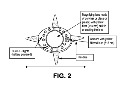
[0023] Figure 2 is a diagram of an imaging device in an embodiment of the
invention.
[0024] Figure 3 is a diagram of an imaging device in an embodiment of the
invention.
DETAILED DESCRIPTION OF THE INVENTION
[0025] The present invention provides methods for in vivo identification,
diagnosis, and
therapy of diseased tissue in a subject in need thereof. The invention method
includes
illuminating an in vivo body part of the subject containing diseased tissue
with light having at
least one excitation wavelength in the range from about 401 nrn to about 510
nrn.
Fluorescence emanating from a fluorescent targeting construct administered to
the subject
and which has specifically bound to and/or been taken up by the diseased
tissue in the body
part, in response to the at least one excitation wavelength is directly viewed
to determine the
location and/or surface area of the diseased tissue in the subject For other
references see
U.S. Pat. Nos. 4,444,744, 4,932,412, 5,697,902and 7,011,812, the entire
contents of which
are incorporated herein by reference, for additional information regarding use
of a
radioisotope for therapy when attached to an antibody.
[0026] Light having a wavelength range from 401 nm to 510 nrn lies within the
visible
range of the spectrum, in contrast to UV light, which lies within the non-
visible range from
about 4 run to about 400 nrn. Therefore, the excitation light used in practice
of the invention
diagnostic methods will contain at least one wavelength of ligjht that
illuminates surrounding
tissue as well as excites fluorescence from the fluorescent targeting
construct used in practice
of the invention methods. The excitation light may be monochromatic or
polychromatic. To
compensate for the tendency of such background effect to obscure the desired
visualization, a
filter is used to screen out wavelengths below about 515 nm in the excitation
light, thereby
eliminating wavelengths that would be reflected from healthy tissue so as to
cause loss of
CA 02825589 2013-07-24
WO 2012/103255
PCT/US2012/022601
9
resolution of the fluorescent image. Alternatively, it is possible to view the
diagnostic site
through a filter that substantially screens out wavelengths other than the
peak emission
wavelength of the fluorophore used. For example, if the fluorescent targeting
construct emits
fluorescence at a known peak emission wavelength of 515 - 520 nm, the filter
can be selected
to substantially eliminate wavelengths of light below about 515 nm. Use of a
filter in the
practice of the invention diagnostic methods is expressly intended to be
encompassed by the
term "directly viewing" as applied to the invention diagnostic methods.
[0027] Use of one or more filters to screen out wavelengths of light in a
selected
wavelength band or screen out all wavelengths except those in a narrow band is
well known
in the art and will encompass the use of such simple devices as filtering
eyeglasses worn by
the diagnostician or physician, and/or filtered viewing lenses for endoscopic
devices that are
used during the diagnostic procedure.
[0028] Operating rooms can be equipped with an overhead light that emits
wavelengths of
light in the optical spectrum useful in practice of invention diagnostic
methods, such as a
Blue LED. Such a ligibt can be utilized in the practice of the invention
diagnostic methods
merely by turning out the other lights in the operating room (to eliminate
extraneous light that
would be visibly reflected from tissue in the body part under investigation)
and shining the
excitation light into the body cavity or surgically created opening so that
the fluorescent
image received directly by the eye of the observer (e.g., the surgeon) is
predominantly the
fluorescent image emanating from the fluorophore(s) in the field of vision.
Light emanating
from a source in the 401-510 nm range could be filtered to aid in
accomplishing the goal of
direct visualization by the observer so that light reflecting from the body
part, other than that
from the fluorescing moiet(ies), is minimized or eliminated.
[0029] Light in the 401 nm to 510 nm wavelength range is readily absorbed
in tissue.
Accordingly, in the invention diagnostic methods, the diseased tissue (and
bound or taken-up
targeting construct) is "exposed" to the excitation light (e.g., by surgically
created opening or
endoscopic delivery of the light to an interior location. The invention method
is particularly
suited to in vivo detection of diseased tissue located at an interior site in
the subject, such as
within a natural body cavity or a surgically created opening, where the
diseased tissue is "in
plain view" (i.e., exposed to the human eye) to facilitate a procedure of
biopsy or surgical
excision, but would be equally applicable to visualizing malignant tissue of
the skin or
CA 02825589 2013-07-24
WO 2012/103255
PCT/US2012/022601
appendages. As the precise location and/or surface area of the tumor tissue
are readily
determined by the invention diagnostic procedure, the invention method is a
valuable guide to
the surgeon, who needs to "see" in real time the exact outlines, size, etc.,
of the diseased
tissue or mass to be resected as the surgery proceeds. Once the diseased
tissue is removed,
any residual microscopic clusters of cells, with the therapeutic drug tumor-
specific construct
attached, would be destroyed by the therapeutic drug molecule contained within
the
fluorescent targeting construct.
[0030] If the putative diseased site is a natural body cavity or surgically
produced interior
site, an endoscopic device can be used to deliver the excitation light to the
site, to receive
fluorescence emanating from the site within a body cavity, and to aid in
formation of a direct
image of the fluorescence from the diseased tissue. For example, a lens in the
endoscopic
device can be used to focus on the detected fluorescence as an aid in
visualizing the diseased
tissue. As used herein, such endoscope-delivered fluorescence is said to be
"directly viewed"
by the practitioner and the tissue or organ to which the targeting construct
binds or in which it
is taken up must be "in plain view" to the endoscope since the light used in
the invention
diagnostic procedure will not contain wavelengths of light that penetrate
tissue, such as
wavelengths in the near infrared range. Alternatively, as described above, the
excitation light
may be directed by any convenient means, such as a hand-held LED or fixed
light source,
into a body cavity or surgical opening containing a targeting construct
administered as
described herein and the fluorescent image so produced can be directly
visualized by the eye
of the observer without aid from an endoscope. With or without aid from any
type of
endoscopic device, the fluorescence produced by the invention method is such
that it can be
viewed without aid of an image processing device, such as a CCD camera, TV
monitor,
photon collecting device, and the like.
[0031] In one embodiment of the invention diagnostic methods, diseased or
abnormal
tissues or organs are contemporaneously viewed through a surgical opening to
facilitate a
procedure of biopsy or surgical excision. As the location and/or surface area
of the diseased
tissue or organ are readily determined by the invention diagnostic procedure,
the invention
method is a valuable guide to the surgeon, who needs to know the exact
outlines, size, etc., of
the mass, for example, for resection as the surgery proceeds.
CA 02825589 2013-07-24
WO 2012/103255
PCT/US2012/022601
11
[0032] Accordingly, in this embodiment, the present invention provides methods
for
utilizing a diagnostic procedure during surgery in a subject in need thereof
by irradiating an
in vivo body part of the subject containing diseased tissue with light having
at least one
excitation wavelength in the range from about 401 nm to about 510 nm, directly
viewing
fluorescence emanating from a targeting construct administered to the subject
that has
specifically bound to and/or been taken up by the diseased tissue in the body
part, wherein
the targeting construct fluoresces in response to the at least one excitation
wavelength,
determining the location and/or surface area of the diseased tissue in the
subject, and
removing at least a portion of the tumor tissue.
[0033] In yet another embodiment, the present invention provides methods for
in vivo
diagnosis of tumor tissue in a subject in need thereof. In this embodiment,
the invention
method comprises contacting samples of tumor cells obtained from the subject
in vitro with a
plurality of detectably labeled compounds, each of which binds to or is
selectively taken up
by a distinct tumor type, determining which of the compounds is bound to or
taken up by the
sample tumor cells, administering a diagnostically effective amount of at
least one
biologically compatible fluorescing targeting construct containing a compound
determined to
bind to and/or be taken up by the sample tumor cells that is tagged with a
therapeutic drug
molecule and a fluorophore responsive to at least one wavelength of light in
the range from
about 401 nm to about 510 nm, and diagnosing the location and/or surface area
of the tumor
tissue in the in vivo body part by directly viewing fluorescence emanating
from the targeting
construct bound or taken up in the tumor tissue upon irradiation thereof with
light providing
the at least one excitation wavelength for the fluorescent targeting
construct.
[0034] In one embodiment of the invention method, a single type of fluorescent
moiety is
relied upon for generating fluorescence emanating from the irradiated body
part (i.e., from
the fluorescent targeting construct that binds to or is taken up by diseased
tissue). Since
certain types of healthy tissue fluoresce naturally, in such a case it is
important to select a
fluorescent moiety for the targeting construct that has a predominant
excitation wavelength
that does not contain sufficient wavelengths in the visible range of light to
make visible the
surrounding healthy tissue and thus inhibit resolution of the diseased tissue.
Therefore, the
light source used in practice of this embodiment of the invention emits light
in the range from
CA 02825589 2013-07-24
WO 2012/103255
PCT/US2012/022601
12
about 401 nm to about 510 run. Thus, the methods of the invention involve
contact of
diseased tissue with a fluorescent targeting construct.
[0035]
Exemplary fluorescent targeting constructs include anti-tumor antigen
antibodies
(e.g., FAB fragment, bispecific antibodies, diabodies, or antibody fragments)
or tumor avid
Compounds (e.g. deoxyglucose, methionine, somatostatin, hormones, hormone
receptor
ligands) and a biologically compatible fluorescing moiety. As used herein, the
terms
"fluorophore-tagged antibody" and "fluorophore-tagged tumor avid compound"
respectively
refer to such fluorescent targeting constructs that are responsive to specific
excitation
wavelengths administered to a subject in need of the methods of the invention.
[0036] In another embodiment, the fluorescent targeting construct is
additionally tagged
with a therapeutic drug molecule (e.g. chemotherapy drug, hormone, etc). The
advantage of
including a therapeutic drug molecule is that when attached to the fluorophore-
tagged
antibody or the fluorophore-tagged tumor avid compound, they provide the dual
roles of (i)
allowing for intra-operative visual imaging (direct viewing using tumor
fluorescence) as a
guide for the operating surgeon in accurately determining the location of the
tumor or
diseased tissue, and (ii) post-surgery "cleanup" (adjuvant therapy) of any
microscopic
clusters of tissue or cells that are too small to be seen by the surgeon, but
could be a source of
local and distant recurrences of the disease/cancer. Exemplary therapeutic
drugs include but
are not limited to, the classes of drugs shown in Table lA below:
TABLE 1A
Alkylating agents
Anthracyclines
Cytoskeletal disruptors
Epothilones
Inhibitors of topoisomerase II
Nucleotide analogs and precursor analogs
Peptide antibiotics
Platinum-based agents
Vinca alkaloids and derivatives and tubulin inhibitors
Retinoids
CA 02825589 2013-07-24
WO 2012/103255
PCT/US2012/022601
13
Tyrosine kinase inhibitors
BRAF inhibitors
MEK inhibitors
mTOR inhibitors
Growth factor inhibitors
Hormones
[0037] In another embodiment, the fluorescent targeting construct is
additionally tagged
with a therapeutic isotope molecule that is both an electron emitter and a
positron (+) "beta"
emitter. See U.S. Pat. No. 6,667,024, the entire content of which is
incorporated herein by
reference, for additional information regarding use of alpha or beta emitters
for therapeutic
use. The advantage of including a therapeutic isotope molecule is that when
attached to the
fluorophore-tagged antibody or the fluorophore-tagged tumor avid compound,
they provide
the dual roles of (i) allowing for pre-surgery external imaging with a
positron emission
tomography (PET) scanner of the subject to provide additional information
and/or a guide for
the operating surgeon in accurately determining the location of the tumor or
diseased tissue,
and (ii) post-surgery "cleanup" of any microscopic clusters of tissue or cells
that are too small
to be seen by the surgeon, but could be a source of local and distant
recurrences of the
disease/cancer. The dual emitter therapeutic isotopes provide the added
benefit of providing
short half-lives, thereby providing minimal risk of radiation exposure to the
surgeon during
the procedure. Exemplary therapeutic isotopes include, but are not limited to,
those shown in
Table 1B below:
TABLE 1B
Therapeutic Isotope Half Life
Astatine-211 7h
Bismuth-213 46 min
Carbon-11 20.38 min
Chromium-51 28 d
Cobalt-57 272 d
Cobalt-60 10.5 months
Copper-64 13 h
Dysprosium-165 2 h
Erbium-169 9.4 d
Fluorine-18 1.8 h
CA 02825589 2013-07-24
WO 2012/103255
PCT/US2012/022601
14
Therapeutic Isotope Half Life
Gallium-67 78 h
Holmium-166 26h
Indium-111 2.8d
Iodine-123 13 h
Iodine-125 60 d
Iodine-131 8 d
Iridium-192 74 d
Iron-59 46d
Krypton-81m 13 sec
Lutetium-177 6.7 d
Molybdenum-99 66 h
Nitrogen-13 10 min
Oxygen-15 2 min
Palladium-103 17 d
Phosphorus-32 14 d
Potassium-42 12 h
Rhenium-186 3.8d
Rhenium-188 17 h
Rubidium-81 4.6h
Rubidium-82 65 h
Samarium-153 47 h
Selenium-75 120d
Sodium-24 15 h
Strontium-89 50 d
Strontium-92 25 d
Thallium-201 73 h
Technetium-99m 6 h
Terbium-149 4.3 min
Xenon-133 5 d
Ytterbium-169 32 d
Ytterbium-177 1.9 h
Yttrium-90 64 h
[0038] The fluorescing moiety of the targeting construct can be any chemical
or protein
moiety that is biologically compatible (e.g., suitable for in vivo
administration) and which
fluoresces in response to excitation light as described herein. Since the
targeting ligand is
administered to living tissue, biological compatibility includes the lack of
substantial toxic
effect to the individual in general if administered systemically, or to the
target tissue, if
administered locally, at the dosage administered. Non limiting examples of
fluorophores that
can be used in the practice of the invention include fluorescein, fluorescein
derivatives,
tetracycline, quinine, mithramycin, Oregon green, and cascade blue, and the
like, and
combinations of two or more thereof.
CA 02825589 2013-07-24
WO 2012/103255
PCT/US2012/022601
[0039] Additional non-limiting examples of fluorescent compounds that
fluoresce in
response to an excitation wavelength in the range from 401 nm to about 510 nm
are found in
Table 2 below:
TABLE 2
COMPOUND EXCITATION EMISSION
RANGE (nm) RANGE (nm)
Acridine Red 455-600 560-680
Acridine Yellow 470 550
Acriflavin 436 520
AFA (Acriflavin Feulgen SITSA) 355-425 460
Alexa Fluor 488 470-490 520
ACMA 430 474
Astrazon Orange R 470 540
Astrazon Yellow 7 GLL 450 480
Atabrine 436 490
Auramine 460 550
Aurophosphine 450-490 515
Aurophosphine G 450 580
Berberine Sulphate 430 550
BOBO-1, BO-PRO-1 462 481
BOPRO1 462 481
Brilliant Sulpho-flavin FF 430 520
Calcein 494 517
Calcofluor White 440 500-520
Cascade Blue 400 425
Catecholamine 410 470
Chinacrine 450-490 515
Coriphosphine 0 460 575
DiA 456 590
Di-8-ANEPPS 488 605
Di0 Pi0C18(3)] 484 501
Diphenyl Brilliant Flavine 7GFF 430 520
Euchrysin 430 540
Fluorescein 494 518
Fluorescein Iso-thiocyanate (FITC) 490 525
Fluo 3 485 503
FM1-43 479 598
Fura Red 472 (low[Cal) 657 (low[Cal)
436 (high[Ca ]) 637 (high[Ca
Genacryl Brilliant Yellow 10GF 430 485
Genacryl Pink 3G 470 583
Genacryl Yellow SGF 430 475
Gloxalic Acid 405 460
CA 02825589 2013-07-24
WO 2012/103255
PCT/US2012/022601
16
3-Hydroxypyrene-5,-8,10-TriSulfonic Acid 403 513
7-Hydroxy-4-methylcourmarin 360 455
5-Hydroxy-Tryptamine (5-HT) 380-415 520-530
Lucifer Yellow CH 425 528
Lucifer Yellow VS 430 535
Lyso Sensor Green DND-153, DND-189 442 505
Maxilon Brilliant Flavin 10 GFF 450 495
Maxilon Brilliant Flavin 8 GFF 460 495
Mitotracker Green FM 490 516
Mithramycin 450 570
NBD 465 535
NBD Amine 450 530
Nitrobenzoxadidole 460-470 510-650
Nylosan Brilliant Flavin E8G 460 510
Oregon Green 488 fluorophore 496 524
Phosphine 3R 465 565
Quinacrine Mustard 423 503
Rhodamine 110 496 520
Rhodamine 5 GLD 470 565
Rhodol Green fluorophore 499 525
Sevron Orange 440 530
Sevron Yellow L 430 490
SITS (Primuline) 395-425 450
Sulpho Rhodamine G Extra 470 570
SYTO Green fluorescent nucleic acid stains 494 + 6 515 7
Thioflavin S 430 550
Thioflavin 5 430 550
Thiozol Orange 453 480
Uranine B 420 520
YOY0-1, YOYO-PRO-1 491 509
[0040] Since the fluorescence properties of biologically compatible
fluorophores are well
known, or can be readily determined by those of skill in the art, the skilled
practitioner can
readily select a useful fluorophore or useful combination of fluorophores, and
match the
wavelength(s) of the excitation light to the fluorophore(s). The toxicity of
fluorescein is
minimal as it has been used safely in vivo in humans for many years, but the
toxicity of
additional useful fluorophores can be determined using animal studies as known
in the art.
[0041] Preferably, the targeting construct (e.g., the ligand moiety of the
invention
targeting construct) is selected to bind to and/or be taken up specifically by
the target tissue
of interest, for example to an antigen or other surface feature contained on
or within a cell
that characterizes a disease or abnormal state in the target tissue. As in
other diagnostic
CA 02825589 2013-07-24
WO 2012/103255
PCT/US2012/022601
17
assays, it is desirable for the targeting construct to bind to or be taken up
by the target tissue
selectively or to an antigen associated with the disease or abnormal state;
however, targeting
constructs containing ligand moieties that also bind to or are taken up by
healthy tissue or cell
structures can be used in the practice of the invention method so long as the
concentration of
the antigen in the target tissue or the affinity of the targeting construct
for the target tissue is
sufficiently greater than for healthy tissue in the field of vision so that a
fluorescent image
representing the target tissue can be clearly visualized as distinct from any
fluorescence
coming from healthy tissue or structures in the field of vision. For example,
colon cancer is
often characterized by the presence of carcinoembryonic antigen (CEA), yet
this antigen is
also associated with certain tissues in healthy individuals. However, the
concentration of
CEA in cancerous colon tissue is typically greater than is found in healthy
tissue, so an anti-
CEA antibody could be used as a ligand moiety in the practice of the
invention. In another
example, deoxyglucose is taken up and utilized by healthy tissue to varying
degrees, yet its
metabolism in healthy tissues, except for certain known organs, such as the
heart, is
substantially lower than in tumor. The known pattern of deoxyglucose
consumption in the
body can therefore be used to aid in determination of those areas wherein
unexpectedly high
uptake of deoxyglucose signals the presence of tumor cells.
[0042] Thus, in one embodiment, the disease or abnormal state detected by the
invention
method can be any type characterized by the presence of a known target tissue
for which a
specific binding ligand is known. For example, various heart conditions are
characterized by
production of necrotic or ischemic tissue or production of artherosclerotic
tissue for which
specific binding ligands are known. As another illustrative example, breast
cancer is
characterized by the production of cancerous tissue identified by monoclonal
antibodies to
CA15-3, CA19-9, CEA, or HER2/neu. It is contemplated that the target tissue
may be
characterized by cells that produce either a surface antigen for which a
binding ligand is
known, or an intracellular marker (i.e. antigen), since many targeting
constructs penetrate the
cell membrane. Representative disease states that can be identified using the
invention
method include such various conditions as different types of tumors,
bacterial, fungal and
viral infections, and the like. As used herein "abnormal tissue" includes
precancerous
conditions, cancer, necrotic or ischemic tissue, and tissue associated with
connective tissue
diseases, and auto-immune disorders, and the like. Further, examples of the
types of target
tissue suitable for diagnosis or examination using the invention method
include cancer of
CA 02825589 2013-07-24
WO 2012/103255
PCT/US2012/022601
18
breast, lung, colon, prostate, pancreas, skin, stomach, small intestine,
testicle, head and neck,
thyroid, gall bladder, brain, endocrine tissue, and the like, as well as
combinations of any two
or more thereof.
[0043] Representative examples of antigens for some common malignancies and
the body
locations in which they are commonly found are shown in Table 3 below.
Targeting ligands,
such as antibodies, for these antigens are known in the art.
TABLE 3
ANTIGEN TUMORS WHERE
COMMONLY FOUND
CEA (carcinoembryonic antigen) colon, breast, lung, pancreas
PSA (prostate specific antigen) prostate cancer
PSMA (prostate specific membrane antigen) prostate cancer
CA-125 ovarian cancer, breast, colon, lung
CA 15-3 breast cancer, lung, colon, pancreas,
medullary cancer of the thyroid, prostate
CA 19-9 breast cancer
HER2/neu breast cancer
a-feto protein testicular cancer, hepatic cancer
P-HCG testicular cancer, choriocarcinoma
(human
chorionic gonadotropin)
MUC-1 breast cancer, colon, lung,
MUC-2 colorectal cancer, colon, lung
TAG 72 breast cancer, colon cancer, and
pancreatic
cancer
Estrogen receptor breast cancer, uterine cancer
Progesterone receptor breast cancer, uterine cancer
AR (androgen receptor) prostate cancer
EGFr (epidermal growth factor receptor) bladder cancer
IGFr (insulin like growth factor) Sarcoma
[0044] In one embodiment of the invention method, the ligand moiety of the
targeting
construct is a protein or polypeptide, such as an antibody, or biologically
active fragment
thereof, preferably a monoclonal antibody. The supplemental fluorescing
targeting
construct(s) used in practice of the invention method may also be or comprise
polyclonal or
monoclonal antibodies tagged with a fluorophore. The term "antibody" as used
in this
invention includes intact molecules as well as functional fragments thereof,
such as Fab,
F(ab')2, and Fv that are capable of binding the epitopic determinant. These
functional
CA 02825589 2013-07-24
WO 2012/103255
PCT/US2012/022601
19
antibody fragments retain some ability to selectively bind with their
respective antigen or
receptor and are defined as follows:
(1) Fab, the fragment which contains a monovalent antigen-binding fragment of
an
antibody molecule, can be produced by digestion of whole antibody with the
enzyme papain
to yield an intact light chain and a portion of one heavy chain;
(2) Fab', the fragment of an antibody molecule that can be obtained by
treating whole
antibody with pepsin, followed by reduction, to yield an intact light chain
and a portion of the
heavy chain; two Fab' fragments are obtained per antibody molecule;
(3) (Fab1)2, the fragment of the antibody that can be obtained by treating
whole
antibody with the enzyme pepsin without subsequent reduction; F(abi)2is a
dimer of two Fab'
fragments held together by two disulfide bonds;
(4) Fv, defmed as a genetically engineered fragment containing the variable
region of
the light chain and the variable region of the heavy chain expressed as two
chains; and
(5) Single chain antibody ("SCA"), a genetically engineered molecule
containing the
variable region of the light chain and the variable region of the heavy chain,
linked by a
suitable polypeptide linker as a genetically fused single chain molecule.
[0045] Methods of making these fragments are known in the art. (See for
example,
Harlow & Lane, Antibodies: A Laboratory Manual, Cold Spring Harbor Laboratory,
New
York, 1988, incorporated herein by reference). As used in this invention, the
term "epitope"
means any antigenic determinant on an antigen to which the paratope of an
antibody binds.
Epitopic determinants usually consist of chemically active surface groupings
of molecules
such as amino acids or sugar side chains and usually have specific three
dimensional
structural characteristics, as well as specific charge characteristics.
[0046] Antibody fragments of the present invention can be prepared by
proteolytic
hydrolysis of the antibody or by expression in E. coli of DNA encoding the
fragment.
Antibody fragments can be obtained by pepsin or papain digestion of whole
antibodies by
conventional methods. For example, antibody fragments can be produced by
enzymatic
cleavage of antibodies with pepsin to provide a 5S fragment denoted F(a1:02.
This fragment
can be further cleaved using a thiol reducing agent, and optionally a blocking
group for the
CA 02825589 2013-07-24
WO 2012/103255
PCT/US2012/022601
sulfhydryl groups resulting from cleavage of disulfide linkages, to produce
3.5S Fab'
monovalent fragments. Alternatively, an enzymatic cleavage using pepsin
produces two
monovalent Fab' fragments and an Fe fragment directly. These methods are
described, for
example, by Goldenberg, U.S. Pat. Nos. 4,036,945 and 4,331,647, and references
contained
therein, which patents are hereby incorporated in their entireties by
reference. See also
Nisonhoff et al., Arch. Biochem. Biophys. 89:230, 1960; Porter, Biochem. .1
73:119, 1959;
Edelman et al., Methods in Enzymology, Vol. 1, page 422 Academic Press, 1967;
and Coligan
et al. at sections 2.8.1-2.8.10 and 2.10.1-2.10.4. Other methods of cleaving
antibodies, such
as separation of heavy chains to form monovalent light-heavy chain fragments,
further
cleavage of fragments, or other enzymatic, chemical, or genetic techniques may
also be used,
so long as the fragments bind to the antigen that is recognized by the intact
antibody.
[0047] Fv fragments comprise an association of VII and VL chains. This
association may
be noncovalent, as described in Inbar et al., Proc. Nat'l Acad. Sci. USA
69:2659, 1972.
Alternatively, the variable chains can be linked by an intermolecular
disulfide bond or
crosslinked by chemicals such as glutaraldehyde. See, e.g., Sandhu, supra.
Preferably, the Fv
fragments comprise NTH and VL chains connected by a peptide linker. These
single-chain
antigen binding proteins (sFv) are prepared by constructing a structural gene
comprising
DNA sequences encoding the VII and VL domains connected by an oligonucleotide.
The
structural gene is inserted into an expression vector, which is subsequently
introduced into a
host cell such as E. coli. The recombinant host cells synthesize a single
polypeptide chain
with a linker peptide bridging the two V domains. Methods for producing sFvs
are described,
for example, by Whitlow et al., Methods: a Companion to Methods in Enzymology,
2: 97,
1991; Bird et al., Science 242:423-426, 1988; Pack et al., Bio/Technology
11:1271-77, 1993;
Sandhu, supra, and Ladner et al., U.S. Pat. No. 4,946,778, which is hereby
incorporated by
reference in its entirety.
[0048] Another form of an antibody fragment is a peptide coding for a single
complementarity-determining region (CDR). CDR peptides ("minimal recognition
units")
can be obtained by constructing genes encoding the CDR of an antibody of
interest. Such
genes are prepared, for example, by using the polymerase chain reaction to
synthesize the
variable region from RNA of antibody-producing cells. See, for example,
Larrick et al.,
Methods: a Companion to Methods in Enzymology, 2: 106, 1991.
CA 02825589 2013-07-24
WO 2012/103255
PCT/US2012/022601
21
[0049] Antibodies which bind to a tumor cell can be prepared using an intact
polypeptide
or biologically functional fragment containing small peptides of interest as
the immunizing
antigen. The polypeptide or a peptide used to immunize an animal (derived, for
example,
from translated cDNA or chemical synthesis) can be conjugated to a carrier
protein, if
desired. Commonly used carriers that are chemically coupled to the peptide
include keyhole
limpet hemocyanin (KLH), thyroglobulin, bovine serum albumin (BSA), and
tetanus toxoid,
and the like. The coupled peptide is then used to immunize the animal (e.g., a
mouse, a rat, or
a rabbit).
[0050] The
preparation of such monoclonal antibodies is conventional. See, for example,
Kohler & Milstein, Nature 256:495, 1975; Coligan et al., sections 2.5.1-2.6.7;
and Harlow et
al., in: Antibodies: a Laboratory Manual, page 726 (Cold Spring Harbor Pub.,
1988), which
are hereby incorporated by reference. Briefly, monoclonal antibodies can be
obtained by
injecting mice with a composition comprising an antigen, verifying the
presence of antibody
production by removing a serum sample, removing the spleen to obtain B
lymphocytes,
fusing the B lymphocytes with myeloma cells to produce hybridomas, cloning the
hybridomas, selecting positive clones that produce antibodies to the antigen,
and isolating the
antibodies from the hybridoma cultures. Monoclonal antibodies can be isolated
and purified
from hybridoma cultures by a variety of well-established techniques. Such
isolation
techniques include affinity chromatography with Protein-A Sepharose, size-
exclusion
chromatography, and ion-exchange chromatography. See, for example, Coligan et
al.,
sections 2.7.1-2.7.12 and sections 2.9.1-2.9.3; Barnes et al., Purification of
Immunoglobulin
G (IgG), in: Methods in Molecular Biology, Vol. 10, pages 79-104 (Humana
Press, 1992).
[0051] Antibodies of the present invention may also be derived from subhuman
primate
antibodies. General techniques for raising therapeutically useful antibodies
in baboons can be
found, for example, in Goldenberg et al., International Patent Publication WO
91/11465
(1991) and Losman et al., 1990, Int. J. Cancer 46:310, which are hereby
incorporated by
reference. Alternatively, a therapeutically useful antibody may be derived
from a
"humanized" monoclonal antibody. Humanized monoclonal antibodies are produced
by
transferring mouse complementarity determining regions from heavy and light
variable
chains of the mouse immunoglobulin into a human variable domain, and then
substituting
human residues in the framework regions of the murine counterparts. The use of
antibody
CA 02825589 2013-07-24
WO 2012/103255
PCT/US2012/022601
22
components derived from humanized monoclonal antibodies obviates potential
problems
associated with the immunogenicity of murine constant regions. General
techniques for
cloning murine immunoglobulin variable domains are described, for example, by
Orlandi et
al., Proc. Nat'l Acad. Sci. USA 86:3833,1989, which is hereby incorporated in
its entirety by
reference. Techniques for producing humanized monoclonal antibodies are
described, for
example, by Jones et al., Nature 321:522, 1986; Riechmann et al., Nature
332:323, 1988;
Verhoeyen et al., Science 239:1534, 1988; Carter et al., Proc. Nat'l Acad.
Sci. USA 89:4285,
1992; Sandhu, Grit. Rev. Biotech. 12:437, 1992; and Singer et al., J. Immunol.
150:2844,
1993, which are hereby incorporated by reference.
[0052] A variety of methods are available for the production of monoclonal
antibodies
(see Of mice and men: hybridoma and recombinant antibodies. Immunol Today,
Little M,
Kipriyanov SM, Le Gall F, Moldenhauer G., Aug;21 (8): 364-70, 2000), and
include the
production of fully human monoclonal antibodies from rabbit hybridomas, for
example in
Pytela, et al., U.S. Pat. No. 7,429,487, and U.S. Pat. No. 8,062,867.
[0053] It is also possible to use anti-idiotype technology to produce
monoclonal antibodies
which mimic an epitope. For example, an anti-idiotypic monoclonal antibody
made to a first
monoclonal antibody will have a binding domain in the hypervariable region
which is the
"image" of the epitope bound by the first monoclonal antibody.
[0054] In a presently preferred embodiment of the invention method, the ligand
moiety in
the fluorescent targeting construct used in practice of the invention can be
selected from
among the many biologically compatible tumor-avid moieties that bind with
specificity to
receptors and/or are preferentially taken up by tumor cells, and can be used
as the ligand
moiety in the invention targeting constructs. Tumor-avid moieties that are
preferentially
"taken up" by tumor cells may enter the cells through surface or nuclear
receptors (e.g.,
hormone receptors), pores, hydrophilic "windows" in the cell lipid bilayer,
and the like.
[0055] Illustrative of this class of tumor-avid moieties are somatostatin,
somatostatin
receptor-binding peptides, deoxyglucose, methionine, histidine, folic acid,
and the like.
Particularly useful somatostatin receptor-binding peptides are a long-acting,
octapeptide
analog of somatostatin, known as octreotide (D-phenylalanyl-L-cysteinyl-L-
phenylalanyl-D-
tryptophyl-L-lysyl-Lthreonyl-N-[2-hydroxy-1-(hydroxymethyppropyl]-L-
cysteinamide
CA 02825589 2013-07-24
WO 2012/103255
PCT/US2012/022601
23
cyclic (2.fwdarvv.7)- disulfide), lanreotide, an oral formulation of
octreotide, P829, P587, and
the like. Somatostatinbinding peptides are disclosed in U.S. Pat. No.
5,871,711, and methods
for linking such peptides covalently to a radioisotope through their carboxyl
terminal amino
acid under reducing conditions are disclosed in U.S. Pat. No. 5,843,401, which
are both
incorporated herein by reference in their entireties. One of skill in the art
can readily adapt
such teachings for the preparation of fluorescence-sensitive somatostatin
receptor-binding
peptides by substituting the fluorescing moieties of this invention in the
place of a
radioisotope.
[0056] Somatostatin and somatostatin receptor-binding peptides are
particularly effective
for use as the tumor-avid moiety in the targeting construct in the invention
diagnostic
procedures when the disease state is a neuroendocrine or endocrine tumor.
Examples of
neuroendocrine tumors that can be diagnosed using the invention method include
adenomas
(GH-producing and TSH-producing), islet cell tumors, carcinoids,
undifferentiated
neuroendocrine carcinomas, small cell and non small cell lung cancer,
neuroendocrine and/or
intermediate cell carcinomas, neuroendocrine tumors of ovary, cervix,
endometrium, breast,
kidney, larynx, paranasal sinuses, and salivary glands, meningiomas, well
differentiated glia-
derived tumors, pheochromocytomas, parathyroid adenomas, neuroblastomas,
ganglioneuro(blasto)mas, paragangliomas, papillary, follicular and medullary
carcinomas in
thyroid cells, Merkel cell carcinomas, and melanomas, as well as granulomas
and
lymphomas. These tumor cells are known to have somatostatin receptors and can
be targeted
using somatostatin or somatostatin receptor binding peptides as the tumor-avid
moiety in the
invention fluorescent targeting construct.
[0057] Vasointestinal peptide (VIP), which is used in VIP receptor
scintigraphy (I.
Virgolini, Eur J aim Invest. 27(10):793-800, 1997, is also useful in the
invention method
for diagnosis of small primary adenocarcinomas, liver metastases and certain
endocrine
tumors of the gastrointestinal tract.
[0058] Another molecule illustrative of the tumor-avid moieties that are
preferentially
taken up by tumors is deoxyglucose, which is known to be preferentially taken
up in a variety
of different types of tumors. Illustrative of the types of tumors that can be
detected using
deoxyglucose as the tumor-avid ligand moiety in the fluorescent targeting
construct as
disclosed herein include Preferred tumor targets for deoxyglucose include
melanoma,
CA 02825589 2013-07-24
WO 2012/103255
PCT/US2012/022601
24
colorectal and pancreatic tumors, lymphoma (both HD and NHL), head and neck
tumors,
myeloma, cancers of ovary, cancer, breast, and brain (high grade and pituitary
adenomas),
sarcomas (grade dependent), hepatoma, testicular cancer, thyroid (grade
dependent) small
cell lung cancer, bladder and uterine cancer, and the like.
[0059] Yet other tumor-avid compounds that can be used as the targeting ligand
in an
invention fluorescing targeting construct are l-amino-cyclobutane-l-carboxylic
acid and
Lmethionine. L-methionine is an essential amino acid that is necessary for
protein synthesis.
It is known that malignant cells have altered methionine metabolism and
require an external
source of methionine.
[0060] Additional examples of biologically compatible tumor-avid compounds
that bind
with specificity to tumor receptors and/or are preferentially taken up by
tumor cells include
mammalian hormones, particularly sex hormones, neurotransmitters, and
compounds
expressed by tumor cells to communicate with each other that are
preferentially taken up by
tumor cells, such as novel secreted protein constructs arising from
chromosomal aberrations,
such as transfers or inversions within the clone.
[0061] The term "hormone" is used herein to refer to compounds that are
expressed within
a mammal for action at a remote location and includes such compounds as sex
hormones, cell
growth hormones, cytokines, endocrine hormones, erythropoietin, and the like.
As is known
in the art, a number of tumor types express receptors for hormones, for
example, estrogen,
progesterone, androgens, such as testosterone, and the like. Such hormones are
preferentially
taken up by tumor cells, for example, via specific receptors. It is also known
in the art that the
particular type of receptors expressed by a tumor cell may change over time
with the same
cell or cell mass, for example, expressing estrogen receptors at one point in
time and with the
estrogen receptors being substantially replaced with androgen receptors at
another point in
time.
[0062] Therefore, in another embodiment according to the present invention,
the invention
diagnostic method comprises prescreening of target tumor cells to determine
which receptors
are currently being expressed by the target cells. In this embodiment, the
invention diagnostic
method comprises contacting sample(s) of tumor cells obtained from a subject
in vitro with a
plurality of detectably labeled tumor-avid compounds, and determining which of
the tumor-
CA 02825589 2013-07-24
WO 2012/103255
PCT/US2012/022601
avid compounds bind to or are taken up by the sample cells. The invention
diagnostic method
further comprises administering to the subject a diagnostically effective
amount of one or
more biologically compatible fluorescing targeting constructs, each comprising
as ligand
moiety at least one of the tumor-avid compounds determined to bind to and/or
be taken up by
the tumor cells so as to allow the fluorescing targeting construct to bind to
and/or be taken up
selectively in vivo by tumor tissue, irradiating an in vivo body part of the
subject suspected of
containing the tumor tissue with light having at least one wavelength in the
excitation
spectrum of the targeting construct under conditions that substantially
eliminate extraneous
light to the in vivo body part, and directly viewing fluorescence emanating
from the
fluorescing targeting construct bound to or taken up by the tumor tissue so as
to determine the
location and/or surface area of the tumor tissue in the in vivo body part. Of
course, if the tests
determine that the tumor cells are concurrently taking up more than one tumor-
avid
compound in substantial proportion (e.g., both estrogen and progesterone), the
more than one
tumor avid compound so determined can be used as the tumor-avid ligand
moieties in the
targeting constructs in the invention diagnostic method.
[0063] Methods for obtaining test tumor cells for prescreening to determine
the type(s) of
tumor-avid compounds that are currently being taken up (e.g., by specific
receptors expressed
by the tumor cells) are well known in the art. For example, such techniques as
fine needle
aspirates, brush biopsies, core needle biopsies, pleural effusion, ascetic
fluid urine and
sputum cytology, bone marrow biopsy and aspirates, scrapings, excisional
biopsies, and the
like, can in many instances be utilized to obtain test tumor cells relatively
non-invasively.
[0064] In vitro tests useful for determining the tumor-avid compounds that are
being taken
up by test tumor cells are numerous and also well known in the art. Such in
vitro tests
generally involve either sequentially or simultaneously contacting the test
cells with a
plurality of different tumor-avid compounds. For example, the test cells can
be contacted
with a panel or library of detectably labeled hormones and/or other known
tumor-avid
compounds to determine which of the detectably labeled compounds bind to
and/or are taken
up by the test cells.
[0065] In the practice of the present invention, the fluorescent moiety
sensitive to an
excitation wavelength in the 401 nm to 510 nm range can be linked to the tumor-
avid
compound used as the ligand moiety in the targeting construct by any method
presently
CA 02825589 2013-07-24
WO 2012/103255
PCT/US2012/022601
26
known in the art for attaching two moieties, so long as the attachment of the
linker moiety to
the ligand moiety does not substantially impede binding of the targeting
construct to the
target tissue and/or uptake by the tumor cells, for example, to a receptor on
a cell. Those of
skill in the art will know how to select a ligand/linker pair that meets this
requirement. For
example, with regard to octreotide, it has been shown that coupling of a
linker to Tyr3 or
Phel of octreotide does not prevent the internalization of octreotide after
binding to the
somatostatin receptor (L. J. Hofland et al., Proc. Assoc. Am. Physicians
111:63-9, 1999). It is
also known that 1-amino-cyclobutane-1-carboxylic acid can be tagged at the 3
carbon of the
ring.
[0066] The length of the optional linker moiety is chosen to optimize the
kinetics and
specificity of ligand binding, including any conformational changes induced by
binding of
the ligand moiety to a target, such as an antigen or receptor. The linker
moiety should be long
enough and flexible enough to allow the ligand moiety and the target to freely
interact and
not so short as to cause steric hindrance between the proteinaceous ligand
moiety and the
target.
[0067] In one embodiment, the linker moiety is a heterobifunctional
cleavable cross-
linker, such as N-succinimidyl (4-iodoacety1)-aminobenzoate;
sulfosuccinimidy1(4-
iodoacety1)- arninobenzoate; 4-succinimidyl-oxycarbonyl-.alpha.-(2-
pyridyldithio) toluene;
sulfosuccinimidy1-64.alpha.-methykalpha.-(pyridyldithiol)-toluamido]hex
anoate;
Nsuccinimidy1-34-2-pyridyldithio)-proprionate; succinimidy1-643(+2-
pyridyldithio)-
proprionamido]hexanoate; sulfosuccinimidy1-643(+2-pyridyldithio)-
propionamido]hexanoate; 3-(2-pyridyldithio)-propionyl hydrazide, Elh-nan's
reagent,
dichlorotriazinic acid, S-(2-thiopyridy1)-L-cysteine, and the like. Further
bifunctional linking
compounds are disclosed in U.S. Pat. Nos. 5,349,066. 5,618,528, 4,569,789,
4,952,394, and
5,137,877, each of which is incorporated herein by reference in its entirety.
[0068] These chemical linkers can be attached to purified ligands using
numerous
protocols known in the art, such as those described in Pierce Chemicals
"Solutions, Cross-
linking of Proteins: Basic Concepts and Strategies," Seminar #12, Rockford,
Ill.
[0069] In another embodiment presently preferred, the linker moiety is a
peptide having
from about 2 to about 60 amino acid residues, for example from about 5 to
about 40, or from
CA 02825589 2013-07-24
WO 2012/103255
PCT/US2012/022601
27
about 10 to about 30 amino acid residues. This alternative is particularly
advantageous when
the ligand moiety is proteinaceous. For example, the linker moiety can be a
flexible spacer
amino acid sequence, such as those known in single-chain antibody research.
Examples of
such known linker moieties include GGGGS (SEQ ID NO:1), (GGGGS)õ (SEQ ID
NO:2),
GKSSGSGSESKS (SEQ ID NO:3), GSTSGSGKSSEGKG (SEQ ID NO:4),
GSTSGSGKSSEGSGSTKG (SEQ ID NO:5), GSTSGSGKSSEGKG (SEQ ID NO:6),
GSTSGSGKPGSGEGSTKG (SEQ ID NO:7), EGKSSGSGSESKEF (SEQ ID NO:8),
SRSSG (SEQ ID NO:9), SGSSC (SEQ ID NO:10), and the like. A Diphtheria toxin
trypsin
sensitive linker having the sequence AMGRSGGGCAGNRVGSSLSCGGLNLQAM (SEQ
ID NO:11) is also useful. Alternatively, the peptide linker moiety can be VM
or AM, or have
the structure described by the formula: AM(Cr2to 4S)n XAM wherein X is
selected from any
amino acid and n is an integer from 1 to 11 (SEQ ID NO:12). Additional linking
moieties are
described, for example, in Huston et al., PNAS 85:5879-5883, 1988; Whitlow,
M., et al.,
Protein Engineering 6:989-995, 1993; Newton et al., Biochemistry 35:545-553,
1996; A. J.
Cumber et al., Bioconj. Chem. 3:397-401, 1992; Ladurner et al., .1. MoL Biol.
273:330-337,
1997; and U.S. Pat. No. 4,894,443, the latter of which is incorporated herein
by reference in
its entirety.
[0070] The
targeting constructs and supplemental targeting constructs used in practice of
the invention method can be administered by any route known to those of skill
in the art, such
as intravenously, intraarticularly, intracisternally, intraocularly,
intraventricularly,
intrathecally, intramuscularly, intraperitoneally, intradermally,
intracavitarily, and the like, as
well as by any combination of any two or more thereof.
[0071] The most suitable route for administration will be intravenously, but
may vary
depending upon the disease state to be treated, or the location of the
suspected condition or
tumor to be diagnosed.
[0072] The
targeting construct is administered in a "diagnostically effective amount." As
used herein, a "diagnostically effective amount" refers to the quantity of a
targeting construct
necessary to aid in direct visualization of any target tissue located in the
body part under
investigation in a subject. As used herein, the term "subject" refers to any
mammal, such as a
domesticated pet, farm animal, or zoo animal, but preferably is a human.
Amounts effective
for diagnostic use will, of course, depend on the size and location of the
body part to be
CA 02825589 2013-07-24
WO 2012/103255
PCT/US2012/022601
28
investigated, the affinity of the targeting construct for the target tissue,
the type of target
tissue, as well as the route of administration.
[0073] Since individual subjects may present a wide variation in severity of
symptoms and
each targeting construct has its unique diagnostic characteristics, including,
affinity of the
targeting construct for the target, rate of clearance of the targeting
construct by bodily
processes, the properties of the fluorophore contained therein, and the like,
the skilled
practitioner will weigh the factors and vary the dosages accordingly.
[0074] The invention composition can also be formulated as a sterile
injectable suspension
according to known methods using suitable dispersing or wetting agents and
suspending
agents. The sterile injectable preparation may also be a sterile injectable
solution or
suspension in a nontoxic parenterally-acceptable diluent or solvent, for
example, as a solution
in 1-4, butanediol. Sterile, fixed oils are conventionally employed as a
solvent or suspending
medium. For this purpose any bland fixed oil may be employed, including
synthetic mono- or
diglycerides, fatty acids (including oleic acid), naturally occurring
vegetable oils like sesame
oil, coconut oil, peanut oil, cottonseed oil, etc., or synthetic fatty
vehicles like ethyl oleate, or
the like. Buffers, preservatives, antioxidants, and the like, can be
incorporated as required, or,
alternatively, can comprise the formulation.
[0075] The invention fluorescing targeting constructs can be produced by well
known
techniques. For example, well known techniques of protein synthesis can be
used to obtain
proteinaceous components of the targeting construct if the amino acid sequence
of the
component is known, or the sequence can first be determined by well known
methods, if
necessary. Some of the ligand genes are now commercially available. An
advantage of
obtaining commercially available genes is that they have generally been
optimized for
expression in E. coll. A polynucleotide encoding a protein, peptide or poly-
nucleotide of
interest, can be produced using DNA synthesis technology. Methods for
obtaining the DNA
encoding an unavailable gene and expressing a gene product therefrom are well
known and
will not be described here in detail.
[0076] A fluorescent targeting construct comprising a proteinaceous ligand
moiety, a
proteinaceous linker moiety, and a proteinaceous fluorophore can also be
produced as a
fusion protein using well known techniques wherein a host cell is transfected
with an
CA 02825589 2013-07-24
WO 2012/103255
PCT/US2012/022601
29
expression vector containing expression control sequences operably linked to a
nucleic acid
sequence coding for the expression of the fusion protein (Molecular Cloning A
Laboratory
Manual, Sambrook et al., eds., 2nd Ed., Cold Spring Harbor Laboratory, N.Y.,
1989).
[0077] As used herein, the terms "peptide" and "polypeptide" refer to a
polymer in which
the monomers are amino acid residues which are joined together through amide
bonds,
alternatively referred to as a polypeptide. When the amino acids are alpha-
amino acids, either
the L-optical isomer or the D-optical isomer can be used, the L-isomers being
preferred.
Additionally, unnatural amino acids such as beta-alanine, phenylglycine, and
homoarginine
are meant to be included. Commonly encountered amino acids that are not gene-
encoded can
also be used in the present invention, although preferred amino acids are
those that are
encodable. For a general review, see, for example, Spatola, A. F., in
Chemistry and
Biochemistry of Amino Acids, Peptides and Proteins, B. Weinstein, ed., Marcel
Dekker, New
York, p. 267,1983.
[0078] DEFINITIONS
As discussed in the body of the invention, the following terms apply.
Monoclonal Antibody (including fully human, humanized, chimeric and also
including
whole antibodies, partial antibodies, Fab fragment, bispecific antibodies,
diabodies, or
antibody fragments, etc.)
Fluorophore (any non-toxic substance with excitation spectra in the visible
light range (401-
510 nm) and with emission spectra in the visible range (520-580 nm) with
examples being
fluorescein and fluorescein like derivatives, antibiotics (i.e. tetracycline),
quinine, as well as
quantum dots)
Radioisotopes (any one of several radioisotopes to include but not limited to
Cu-64,
Rhenium-188, and Samarium-153)
Therapeutic Drug (chemotherapy.. .to include all classes of commonly accepted
chemotherapy i.e. anti-metabolites, antibiotics, DNA scission, anthracyclines,
spindle cell
inhibitors, proteasomes, mTOR inhibitors, tyrosine kinase inhibitors,
hormones, HDAC
inhibitors, epithilones, kinase inhibitors, etc.)
CA 02825589 2013-07-24
WO 2012/103255
PCT/US2012/022601
Diseased Tissue (to include cancer, endocrine adenomas, benign tumors with
systemic
effects)
Schematically it would be as follows:
¨> Diagnosis of a potentially resectable and surgically curable cancer:
¨> Identification of surface or internal antigens on or within the tumor or
other diseased
cells:
¨> Injection of fluorophore-tagged (and chemotherapy-tagged or
chemotherapy/radioisotope-tagged anti-tumor antigen MAb:
¨> Surgical resection of all visibly fluorescent tumor tissue (1-5 days after
injection of
the MAb):
¨> Adjuvant Therapy; destruction of microscopic (and not visible) residual
cancer cells
through the attached fluorophore-tagged + chemotherapy or chemotherapy/
radioisotope-tagged MAb
[0079] Although the invention has been described with reference to the above
example, it
will be understood that modifications and variations are encompassed within
the spirit and
scope of the invention. Accordingly, the invention is limited only by the
following claims.