Note: Descriptions are shown in the official language in which they were submitted.
CA 02825675 2013-07-25
WO 2012/101441
PCT/GB2012/050159
1
ROTARY MILL
The present invention relates to rotary mills and similar rotary cutting
devices and
particularly but not exclusively relates to rotary mills for use in preparing
a bone for total
or partial joint replacement surgery.
Background
It is known to replace all or part of a knee joint in which the joint surfaces
have
deteriorated, for example as a result of osteoarthritis. Such deterioration
usually starts
in only one of the tibeo-femoral compartments and may spread to the other at a
later
stage. Replacement of only one compartment of the joint can therefore be
sufficient to
provide prolonged relief from symptoms. Damaged bearing surfaces are replaced
by a
unicompartmental prosthesis which comprises a femoral implant and a tibial
implant
(usually metallic), which interface through a (polyethylene) bearing component
disposed between the two implants.
A unicompartmental or partial knee replacement (PKR) helps to conserve
undamaged
bone and restores more natural movement to the joint. Also, owing to the small
size of
the prosthesis, the surgery may be less invasive than a total knee replacement
(TKR).
However, the design requirements for partial knee replacement prostheses are
more
demanding than those for total knee replacement prostheses. Unlike in a total
knee
replacement, where one or more ligaments can be discarded and the mechanics of
the
knee can be simplified, in a unicompartmental knee replacement, all the
ligaments in
the joint must be retained and restored to their natural tensions and the
bearing
component must be completely unconstrained.
During articulation of the knee, and particularly when the joint is at full
extension, the
bearing component can impinge on femoral condylar bone tissue superior to the
femoral implant, as illustrated in Figure 1. Such impingement of the
polyethylene
bearing component onto the bone can lead to post operative pain, damage to the
bearing, increased wear and eventual failure. It is therefore essential to
remove a
sufficient amount of anterior bone on the femoral condyle during the
implantation
procedure to prevent such impingement from occurring.
CA 02825675 2013-07-25
WO 2012/101441
PCT/GB2012/050159
2
Orthopaedic surgeons conventionally use a bone chisel to manually remove the
anterior bone. However, such a manual procedure can easily be forgotten during
surgery and, even when carefully completed, results in an undesirable non
uniform
bone edge and in the removal of an uncertain and varying amount of bone.
Summary of Invention
According to the present invention, there is provided a rotary mill comprising
a body
portion having a milling surface and a central bore extending along the rotary
axis of
the body portion; and a guide portion having a guide body and a guide peg
extending
from the guide body, the guide peg operable to be received in the central bore
of the
body portion, the guide body having at least one alignment feature which is
the same
as that of a prosthesis component.
By forming the guide body to have at least one alignment feature which is the
same as
that of a prosthesis component, the guide portion can be applied to the bone
and fixed
in place using the pre prepared fixation features that will hold the
prosthesis component
in place (for example peg holes or a recess for a flange). The guide body thus
provides a precise reference of the final location of the prosthesis component
to be
implanted. The guide peg thus guides the body portion to mill an area of bone
that is in
a precise and predetermined location with respect to the eventually implanted
prosthesis component.
A further advantage to the guide body being formed in this manner is that no
additional
bone must be removed in order for it to be fixed on the bone surface. The
guide
portion, having at least one alignment feature which is the same as that of a
prosthesis
component, can fit into the necessary recesses already formed in the bone to
accommodate the final prosthesis component. This is in contrast to
conventional
guided mills which require a dedicated hole to be drilled to accommodate a
separate
guide rod.
The at least one alignment feature may be an attachment peg or may be a pair
of
attachment pegs. The attachment pegs may be operable to be received in pre
prepared prosthesis peg holes.
CA 02825675 2013-07-25
WO 2012/101441
PCT/GB2012/050159
3
The guide body may have substantially the form of a trial prosthesis component
and
may in fact comprise a trial prosthesis component.
The guide body may include at least one nodule, protruding from a surface of
the guide
body and operable to abut a corresponding abutment surface on the body
portion. The
nodules may thus act as depth stops to ensure a precise amount of bone is
removed
and avoid excessive bone removal.
The guide peg may comprise an abutment surface operable to abut a
corresponding
abutment surface in the central bore of the body portion. The guide peg may
thus not
only act to guide the angle at which the body portion mills bone surface but
may also
act as a depth stop to limit bone removal.
The abutment surface may comprise a distal surface of the peg or the abutment
surface may comprise an outwardly projecting annular shoulder.
The corresponding abutment surface of the central bore may comprise a base of
the
bore or may comprise an inwardly projecting annular shoulder.
The guide peg may project from a predetermined region of, and at a
predetermined
angle to the guide body. In this manner the region of bone to be removed may
be
precisely determined and fixed by the construction of the guide portion,
facilitating
accuracy and repeatability of milling.
The guide peg may be adjustable on the guide body, allowing the surgeon a
degree of
freedom in selection of the milled area, and to accommodate for different
patient
geometries.
The guide body may be operable to be connected to additional surgical tools,
thus
The guide body may comprise a trial femoral prosthesis component which may be
a
trial unicondylar femoral prosthesis component.
operable to guide reaming of a region superior to the anterior edge of the
guide body.
CA 02825675 2013-07-25
WO 2012/101441
PCT/GB2012/050159
4
The guide peg may project from the guide body at an angle of between 25 and 40
degrees to the axis of the attachment peg. The angle may vary according to the
size of
the rotary mill.
The guide body may be operable to be connected to a posterior osteophyte
guide.
The rotary mill may further comprise additional guide portions, each guide
portion being
of a different size so as to match differently sized prosthesis components
that are
employed for patents of differing sizes.
The rotary mill may further comprise additional guide portions, each guide
portion
having a different length, and or angle of extension of guide peg, thus also
accommodating different patent geometries.
The body portion of the rotary mill may comprise a rotary body and a guide
shaft at
least partially received within the rotary body.
The guide shaft may comprise an inner portion telescopically received within
an outer
portion and a biasing element acting between the inner and outer portions.
The guide shaft may be received within an axial bore which may be formed in
the
rotary body. The bore may be a blind bore.
The biasing element may comprise a spring which may be mounted about the inner
portion of the guide shaft.
The body portion may further comprise cooperating protrusions formed on the
rotary
body and the outer portion of the guide shaft, operable to engage one another
as a
depth stop.
The cooperating protrusions may comprise annular shoulders which may be formed
on
an inner surface of the rotary body and an outer surface of the outer portion
of the
guide shaft.
CA 02825675 2013-07-25
WO 2012/101441
PCT/GB2012/050159
The outer portion of the guide shaft may comprise a substantially hollow
shaft, a distal
end of which may comprise the central bore of the body portion, operable to
receive the
guide peg.
5 The cutting surface of the body portion may be formed on an annular
cutting tool which
may be removably attached to the rotary body.
According to another aspect of the present invention, there is provided a
method of
implanting a unicondylar femoral component comprising,
a) reaming the femoral condylar surface to accept the unicondylar
femoral component;
b) drilling peg holes for affixing the unicondylar femoral component;
c) affixing a guide portion of a rotary mill onto the prepared condylar
surface using the drilled peg holes;
d) reaming a portion of bone anterior to the affixed guide portion;
e) removing the guide portion from the bone; and
f) affixing a unicondylar femoral component to the prepared condylar
surface.
The rotary mill may be a rotary mill according to the first aspect of the
present
invention.
According to another aspect of the present invention, there is provided a
rotary cutting
tool comprising a rotary body having a cutting surface and a guide shaft at
least
partially received within the rotary body, wherein the guide shaft comprises
an inner
portion telescopically received within an outer portion, and a biasing element
acting
between the inner and outer portions.
The biasing element of the tool thus acts to damp the telescoping motion of
the guide
shaft and hence, when received within the rotary body, damps progression of
the rotary
body along the guide shaft. This damping action can assist a surgeon with fine
control
of cutting or milling operations.
The guide shaft may be received within an axial bore formed on the rotary
body. The
bore may be a blind bore.
CA 02825675 2013-07-25
WO 2012/101441
PCT/GB2012/050159
6
The rotary cutting tool may further comprise cooperating protrusions formed on
the
rotary body and the outer portion of the guide shaft, which protrusions may be
operable
to engage one another as a depth stop.
The protrusions may for example comprise annular shoulders which may be formed
on
an inside surface of the rotary body and an outer surface of the outer portion
of the
guide shaft.
The biasing element may comprise a spring. The spring may be mounted about the
inner portion of the guide shaft. The spring may act between an end of the
outer
portion of the guide shaft and an end cap formed on the inner portion of the
guide
shaft. The end cap may be formed by an end of the axial bore in which the
guide shaft
is received.
The outer portion of the guide shaft may comprise a hollow shaft, a first end
of which
may receive the inner portion and a second end of which may be operable to
receive a
guide peg.
The cutting surface of the rotary cutting tool may be formed on an annular
cutting plate
which may be removably attached to the rotary body.
An end of the rotary body may terminate in an annular receiving plate, which
may be
operable to engage the cutting plate.
The rotary cutting tool may further comprise cooperating formations on the
receiving
plate and cutting plate, which may be operable to secure the cutting plate to
the
receiving plate.
The cooperating formations may comprise protrusions, for example screw heads,
and
appropriately shaped recesses. The formations may alternatively or further
comprise
magnetic elements.
Another end of the rotary body may terminate in a drive shaft which may be
operable to
engage a drive element such as a rotary drill.
Brief Description of Drawings
CA 02825675 2013-07-25
WO 2012/101441
PCT/GB2012/050159
7
For a better understanding of the present invention, and to show more clearly
how it
may be carried into effect, reference will now be made, by way of example, to
the
following drawings, in which:-
Figure 1 shows impingement of a meniscal bearing on anterior femoral bone.
Figure 2 is a perspective view of a rotary mill.
Figure 3 is a perspective view of a body portion of a rotary mill.
Figure 4 shows a guide portion of a rotary mill in position on a femur.
Figure 5 shows a rotary mill in position on a femur.
Figure 6 illustrates bone removal by a rotary mill.
Figures 7 and 8 illustrate prosthesis components in position on a femur, with
and
without bone removal.
Figure 9 illustrates an implanted unicondylar prosthesis.
Figure 10 is a sectional view of a rotary cutting tool.
Figure 11 is a sectional view of the tool of Figure 10 in a compressed
condition.
Detailed Description
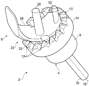
With reference to Figures 2 to 4, a rotary mill 2 comprises a body portion 4
and a guide
portion 6. The body portion 4 comprises a rotary body 8 that terminates at one
end in
an annular milling surface 10. The milling surface comprises a series of
milling teeth
12 that extend from the surface 10. In the illustrated embodiment, the milling
surface
10 is formed on an annular shoulder 14 that protrudes outwardly from the
rotary body
8. An abutment surface 15 extends radially inwardly of the projecting milling
surface
10. At a second end of the rotary body 8 an integral shank 16 extends along an
axis of
rotation 18 of the rotary body. The shank 16 is suitable for attachment to the
chuck of
CA 02825675 2013-07-25
WO 2012/101441
PCT/GB2012/050159
8
a surgical drill (not shown). A bore 20 extends through the rotary body 8
along the axis
of rotation 18 of the rotary body 8.
The guide portion 6 comprises a guide body 22 and a guide peg 24. The guide
body
22 comprises a trail unicondylar femoral prosthesis component. The guide body
thus
comprises a curved condylar plate 26 and two attachment pegs 28, 30. The
attachment pegs 28, 30 protrude from a bone contacting surface 32 of the
condylar
plate 26 at locations and angles precisely matching those of a similarly sized
prosthesis
component. The guide body can thus be attached to a prepared femoral condylar
surface in the same manner and using the same drilled peg holes as for a
prosthesis
component. In this manner, the attachment pegs 28, 30 serve to align the guide
body
with the eventual location of the femoral prosthesis, referencing off the pre
prepared
drilled femoral peg holes. The guide peg 24 is a cylindrical peg that
protrudes from an
opposite, outer surface 34 of the condylar plate 26. The guide peg 24 extends
from an
anterior portion of the condylar plate 26 along an axis that is substantially
normal to the
adjacent condylar plate surface 34. The guide peg 24 projects at an angle of
between
and 40 degrees to the axis of the attachment pegs. The precise angle is
selected
according to the size of the guide portion and associated anatomy, as
discussed in
further detail below. The guide peg 24 is dimensioned to be slidably and
rotatably
20 received within the central bore 20 of the rotary body 8. With reference
also to Figure
6, two nodules 27, 29 protrude from the outer surface 34 of the condylar plate
26. The
nodules 27, 29 are formed on opposite sides of the anterior portion of the
condylar
plate 26, in the region of the guide peg 24.
25 The rotary mill 2 is used to remove anterior bone on the femoral condyle
prior to
implantation of a unicondylar femoral prosthesis. First, the condylar surface
is
prepared to receive the prosthesis, including resection of the entire condylar
surface
and drilling of femoral peg holes. The guide portion 6 of the rotary mill 2 is
then fixed
on the femoral condyle by inserting the attachment pegs 28, 30 into the pre
drilled
femoral peg holes. The guide portion 6 can be seen in position on the femoral
condyle
in Figure 4. The body portion 4 is then seated on the guide portion 4, the
guide peg 24
being receiving within the bore 20 of the rotary body 8. The shank 16 of the
body
portion is then attached to a surgical drill and the body portion 4 is guided
to mill the
bone superior to the anterior edge of the condylar plate 26 of the guide
portion. The
guide peg 24 guides the orientation of the body portion 4, ensuring that the
milling
surface 10 removes the bone tissue from the correct location. The body portion
4
CA 02825675 2013-07-25
WO 2012/101441
PCT/GB2012/050159
9
advances along the guide peg 24 as bone tissue is removed. As the body portion
4
advances, the nodules 27, 29 act as stop pegs, upper surfaces of the nodules
27, 29
abutting the stop surface 15 that extends radially inwardly from the annular
milling
surface 10 and preventing further movement of the body portion 4, thus
limiting the
amount of bone that is removed. The body portion 4 is dimensioned so as to
ream only
the anterior bone superior to the guide portion. As can be seen from Figures 5
and 6,
the milling surface 10 does not disturb the adjacent soft tissues and so
causes minimal
damage or disruption to the surrounding structures, removing only that bone
which is
desired to be removed. As illustrated particularly in Figure 5, the guide peg
24 is
angled such that, when fully seated on the guide peg 24, the body portion 4
has only
reamed the bone superior to the anterior edge of the guide portion 6. The
reamed area
of bone can be seen at area 38 on Figure 6.
Additional tools can then be attached to the guide portion if necessary. For
example, a
posterior osteophyte guide 40 can be attached posteriorly to the guide portion
6. The
osteophyte guide is a slotted tool that may be used to guide a chisel to
remove
osteophytes from the posterior area of the femoral condyle, helping to prevent
femoral
loosening.
Once all necessary bone removal has been completed, the guide portion 6 of the
rotary
mill 2 is removed and the appropriate prosthesis component is implanted.
Figures 7
and 8 illustrate the area 38 of bone that is removed by the rotary mill 2. On
Figure 8,
this area 38 can be seen immediately superior to the femoral prosthesis
component 50.
On Figure 7, the rotary mill 2 has not been used and bone tissue remains
superior to
the prosthesis component 50. This bone tissue will cause impingement of the
meniscal
component, as illustrated in Figure 1. In contrast, and as illustrated in
Figure 9, when
the mill has been used to remove bone over the desired area 38, no impingement
of
the meniscal component is seen, even with the knee in full extension.
It is envisaged that the guide peg 24 may be integral with the guide body 22
or may be
detachable from, or adjustable relative to, the guide body 22, so as to allow
limited
adjustment of the angle of the guide peg 24 or of the height of the guide peg
24. Such
adjustment allows a degree of flexibility to the surgeon in tailoring the
rotary mill 2 to
the precise needs of individual patients. For example, if it is desired to
remove less
than the usual amount of bone, the guide peg 24 may be caused to protrude
further
from the surface of the condylar plate 26. In this instance, the guide peg 24
also acts
CA 02825675 2013-07-25
WO 2012/101441
PCT/GB2012/050159
as a stop peg, the end surface 36 of the guide peg 24 contacting the base (not
shown)
of the bore 20 and preventing further movement. The guide peg may be caused to
protrude to such an extent that it is engages as a stop peg before the stop
surface 15
of the body portion 4 contacts the nodules 27, 29 of the guide body. It is
also
5 envisaged that the guide portion 6 of the rotary mill be provided as
merely one of
severeal available guide portions, each being of a different size to
accommodate
different sizes of knee. Thus, each size of prosthesis may have an associated
guide
portion 6 of the appropriate size. Each guide portion 6 will have a suitable
guide peg,
of a height and at an angle that is determined to be most appropriate for the
associated
10 prosthesis.
It will be appreciated that the guide portion 6 may be employed together with
other
embodiments of body portion 4, including a range of rotary cutting devices.
One
embodiment of rotary cutting tool with which the guide portion 6 may be
employed is
illustrated in Figures 10 and 11. The rotary cutting tool 100 comprises a
rotary body
102 and a guide shaft 104. The guide shaft 104 is at least partially received
within a
blind axial bore 106 formed within the rotary body 102. A closed proximal end
(towards
the left in the Figures) of the rotary body 102 terminates in a drive shaft
108, operable
to be received within the chuck of a surgical drill (not shown). An open
distal end of the
rotary body 102 flares outwards to terminate in an annular receiving plate 110
extending about the opening of the axial bore 106 and described in further
detail below.
The guide shaft 104 comprises an inner portion 112 and an outer portion 114.
The
inner portion 112 comprises a solid shaft a distal end 116 of which is
telescopically
received within a proximal end 118 of the outer portion 114. The outer portion
114
comprises a substantially hollow shaft. A biasing spring 120 is mounted about
the
inner portion 112 of the guide shaft 104. The spring 120 rests at one end on
the
annular end surface 122 of the proximal end 118 of the outer portion 114. The
other
end of the spring 120 engages on an end cap 124 formed on a proximal end 126
of the
inner portion 112. In an alternative embodiment (not shown) the spring 120 may
engage on the blind end 128 of the axial bore 106 in which the guide shaft 104
is
received.
The guide shaft is received freely within the bore 106 of the rotary body 102.
An
annular shoulder 130 is formed on the inner surface of the bore 106, dividing
the bore
into a distal section and a proximal section, the distal section being of
larger inside
CA 02825675 2013-07-25
WO 2012/101441
PCT/GB2012/050159
11
diameter than the proximal section. A corresponding annular shoulder 132 is
formed
on the outer surface of the outer portion 114 of the guide shaft, dividing the
outer
portion into proximal and distal sections, the distal section being of larger
outside
diameter than the proximal section. The corresponding annular shoulders 130,
132
function as a depth stop, preventing the outer portion 114 of the guide shaft
104 from
being received into the rotary body 102 beyond a certain point. This position
is
illustrated in Figure 11. The larger diameter distal section of the outer
portion 114 of
the guide shaft may also serve to centre the guide shaft within the bore 106
of the
rotary body 102.
Referring particularly to Figure 10, the annular receiving plate 110 comprises
a series
of formations, operable to releasably engage an annular cutting plate (not
shown). The
formations comprise at least one screw head 134 and a plurality of magnets
136, the
magnets being recessed into the annular receiving plate so as to present a
smooth
surface. The annular cutting plate (not shown) comprises an annular cutting
surface,
similar to that described above with reference to the body portion 6, and an
opposed
annular engaging surface. The annular engaging surface comprises corresponding
recesses and magnetic elements enabling the cutting plate to be releasably yet
securely attached to the receiving plate 110 of the rotary body.
In use, the rotary cutting tool 100 is first assembled and then placed over
the guide peg
24 of the guide portion 6. The guide peg 24 is received within the hollow
outer portion
114 of the guide shaft until a distal end 138 of the outer portion 114 is
seated against
the surface 34 from which the guide peg 24 protrudes. The rotary body 102 is
then
connected to a surgical drill (not shown) via the drive shaft 108 and the
rotary tool is
guided to mill away the desired area of bone. During the cutting operation,
the outer
portion 114 of the guide shaft 104 remains seated in position over the guide
peg 24.
Downward pressure applied to the rotary body engages the blind end 128 of the
bore
106 against the end cap of the inner portion 112 of the guide shaft 104,
causing the
inner portion 112 to be pushed further into the outer portion 114. This action
compresses the spring 120 acting between the inner and outer portions. In this
manner, the spring 120 damps the downward motion of the rotary body, assisting
the
control of the surgeon and thus increasing the ease with which the tool may be
employed. The inner portion 112 of the guide shaft 104 continues to slide
further into
the outer portion 114 until the annular shoulder 130 on the rotary body 102
engages
the annular shoulder 132 on the outer portion 114 of the guide shaft. At this
point the
CA 02825675 2013-07-25
WO 2012/101441
PCT/GB2012/050159
12
rotary body cannot travel any further towards the bone and the drilling action
is ceased.
In this manner, the annular shoulders act as a depth stop, preventing over
reaming of
the bone.
It will be appreciated that the present invention provides a means for
accurately,
predictably and repeatably removing a targeted area of bone from the femoral
condyle.
The amount of bone removed is determined by the precise angle and height of
the
guide peg 24. These aspects of the guide peg 24 are determined when the guide
portion 6 is initially formed and can thus be carefully assessed and fixed so
as to guide
milling of precisely the correct amount of bone for the associated prosthesis.
The
present invention is also bone conserving, requiring no additional drill hole
for a guide
rod, as is conventionally required for a guided mill. By fastening to the bone
using the
existing femoral peg holes, the guide portion 6 makes use of existing
features, and
requires no additional bone removal for fixation. The femoral peg holes in
fact
determine the eventual location of the milled bone, as they provide the
location for the
guide portion 6. As these peg holes also provide location for the final
prosthesis
component, considerable time and development effort has been devoted to tools
and
techniques to ensure the accurate positioning of the peg holes in the femur.
The
present invention makes indirect use of these pre-existing tools and
techniques in
employing the femoral peg holes as the fixation means for the guide portion 6
of the
rotary mill 2.
The present invention additionally provides a rotary cutting tool optimised
for use with
the guide portion 6, the action of which is damped or cushioned, improving
ease of use
for the surgeon.