Note: Descriptions are shown in the official language in which they were submitted.
1
RECOMBINANT LAMININ-521
BACKGROUND
[0001]
[0002] This application relates to cell biology, cell differentiation, cell
therapy,
molecular biology, proteins, recombinant human proteins, nucleic acids, and
laminins.
[0003] Basal laminae (basement membranes) are sheet-like, cell-associated
extracellular matrices that play a central role in cell growth, cellular
differentiation,
cell phenotype maintenance, tissue development, and tissue maintenance. They
are
present in virtually all tissues, and appear in the earliest stages of
embryonic
development.
[0004] Basal laminae are central to a variety of architectural and cell-
interactive
functions. For example:
[0005] 1. They serve as architectural supports for tissues, providing
adhesive
substrata for cells.
[0006] 2. They create perm-selective barriers between tissue compartments
that
impede the migration of cells and passively regulate the exchange of
macromolecules. These properties are illustrated by the kidney glomerular
basement
membrane, which functions as an important filtration structure, creating an
effective
blood-tissue barrier that is not permeable to most proteins and cells.
[0007] 3. Basal laminae create highly interactive surfaces that can promote
cell
migration and cell elongation during embryogenesis and wound repair. Following
an
injury, they provide a surface upon which cells regenerate to restore normal
tissue
function.
[0008] 4. Basal laminae present information encoded in their structure to
contacting cells that is important for cellular differentiation, prevention of
apoptosis,
CA 2826252 2017-12-14
CA 02826252 2013-06-17
WO 2012/080842
PCT/IB2011/003258
2
and tissue maintenance. This information is communicated to the cells through
various receptors that include the integrins, dystroglycan, and cell surface
proteoglycans. Signaling is dependent not only on the presence of matrix
ligands
and corresponding receptors that interact with sufficient affinities, but also
on such
topographical factors as ligand density in a three-dimensional matrix
"landscape",
and on the ability of basal lamina components to cluster receptors. Because
these
matrix proteins can be long-lived, basal laminae create a "surface memory" in
the
basal lamina for resident and transient cells.
[0009] The basal lamina is largely composed of laminin and type IV collagen
heterotrimers that in turn become organized into complex polymeric structures.
Additional components include proteoglycans such as agrin and perlecan and
nidogens (entactins). To date, six type IV collagen polypeptide chains and at
least
twelve laminin subunit chains have been identified. These chains possess
shared
and unique functions and are expressed with specific temporal (developmental)
and
spatial (tissue-site specific) patterns.
[0010] Laminins
are a family of heterotrimeric glycoproteins that reside primarily
in the basal lamina. They function via binding interactions with neighboring
cell
receptors on the one side, and by binding to other laminin molecules or other
matrix
proteins such as collagens, nidogens or proteoglycans. The laminin molecules
are
also important signaling molecules that can strongly influence cellular
behavior and
function. Laminins are important in both maintaining cell/tissue phenotype, as
well as
in promoting cell growth and differentiation in tissue repair and development.
[0011] Laminins
are large, multi-domain proteins, with a common structural
organization. The laminin molecule integrates various matrix and cell
interactive
functions into one molecule.
[0012] A laminin protein molecule comprises one a-chain subunit, one I3-chain
subunit, and one y-chain subunit, all joined together in a trimer through a
coiled-coil
domain. FIG. 1 depicts the resulting structure of the laminin molecule. The
twelve
known laminin subunit chains can form at least 15 trimeric laminin types in
native
tissues. Within the trimeric laminin structures are identifiable domains that
possess
binding activity towards other laminin and basal lamina molecules, and
membrane-
bound receptors. FIG. 2 shows the three laminin chain subunits separately. For
example, domains VI, 1Vb, and IVa form globular structures, and domains V,
111b,
and IIla (which contain cysteine-rich EGF-like elements) form rod-like
structures.
CA 02826252 2013-06-17
WO 2012/080842
PCT/IB2011/003258
3
Domains I and II of the three chains participate in the formation of a triple-
stranded
coiled-coil structure (the long arm).
[0013] There exist five different alpha chains, three beta chains and three
gamma
chains that in human tissues have been found in at least fifteen different
combinations. These molecules are termed laminin-1 to laminin-15 based on
their
historical discovery, but an alternative nomenclature describes the isoforms
based
on their chain composition, e.g. laminin-111 (laminin-1) that contains alpha-
1, beta-1
and gamma-1 chains. Four structurally defined family groups of laminins have
been
identified. The first group of five identified laminin molecules all share the
131 and y1
chains, and vary by their a-chain composition (al to a5 chain). The second
group of
five identified laminin molecules, including laminin-521, all share the 132
and yl
chain, and again vary by their a-chain composition. The third group of
identified
laminin molecules has one identified member, laminin-332, with a chain
composition
of a3133y2. The fourth group of identified laminin molecules has one
identified
member, laminin-213, with the newly identified y3 chain (a2131y3).
[0014] There have been no reports of isolated laminin-521 that is free of
other
laminin chains. Thus far, there are no studies on the function of laminin-521.
Attempts to purify laminin-521 from cell sources by affinity chromatography
using
laminin chain antibodies have been unsuccessful in eliminating, for example,
laminin
131 chain, which is a component of laminin-411 and laminin-511. It would be
desirable to provide compositions that contain laminin-521 (aka LN-521) and
methods for making laminin-521.
[0100] Human embryonic stem (hES) cells hold promise for the development of
regenerative medicine for a variety of diseases, such as spinal cord and
cardiac
injuries, type I diabetes and neurodegenerative disorders like Parkinson's
disease. A
stem cell is an undifferentiated cell from which specialized cells are
subsequently
derived. Embryonic stem cells possess extensive self-renewal capacity and
pluripotency with the potential to differentiate into cells of all three germ
layers. They
are useful for therapeutic purposes and may provide unlimited sources of cells
for
tissue replacement therapies, drug screening, functional genomics and
proteomics.
[0015] A prerequisite for the development of stem cell derived cells for
regenerative medicine are methods that allow long-term cultures of pluripotent
stem
cells, chemically defined and repeatable differentiation protocols, as well as
xeno-
free cell culture systems. However, culturing of pluripotent human embryonic
stem
CA 02826252 2013-06-17
WO 2012/080842
PCT/IB2011/003258
4
cells (hES cells) and induced pluripotent stem cells (hIPS cells) has
encountered a
number of problems. One major problem has been that hES cells grow slowly in
clusters that need to be manually split for cell propagation. Dissociation of
the cells
usually leads to extensive cell death, the cloning efficiency of hES cells
after
complete dissociation being s1 %.
[0016] Maintenance of pluripotent hES cells has required complex culture
substrata, such as extracellular matrix protein mixtures like the mouse tumor
derived
Matrigel or fibroblast feeder cell layers, that may be immunogenic and toxic
and that
generate extensive batch to batch variability reducing reliability of the
experiments.
Thus far, the most successful feeder cell free substrate used for hES cell
cultures is
Matrigel, a complex tumor and BM-like extract obtained from murine Engelbreth-
Holm-Swarm (EHS) sarcoma tumor tissues. Matrigel mainly contains murine LN-
111,
type IV collagen, perlecan and nidogen, but also varying amounts of other
materials,
including growth factors and cellular proteins and, therefore, its composition
is
undefined and varies from batch-to-batch. This variability can cause
irreproducibility
of scientific results, and due to the animal origin of the substratum makes
Matrigel
unacceptable for the expansion and maintenance of hES cells for human cell
therapy.
[0017] However, successful development of more or less defined coating
materials that support self-renewal of hES and hiPS cells has recently been
reported. It has been reported that recombinant vitronectin supports adhesion
and
self-renewal of hES cells. An acrylate coating containing a variety of
peptides from
various ECM proteins has also been previously developed, and it has been shown
that a synthetic methacrylate-based polymer also facilitated adhesion and self-
renewal of hES cells.
[0018] One of the
main problems with large-scale propagation of hES cells is that
they poorly survive replating after dissociation into single cell suspension.
This, in
turn, makes passaging tedious and large-scale automated expansions impossible.
However, hES cells released into single cell suspension using trypsin
treatment in
the presence of a rho-kinase (ROCK) inhibitorl or blebbistatin" can be plated
and
expanded from single clones, but the molecules are not components of the
natural
stem cell niche, and they affect the actin cytoskeleton and thus can cause
cell
damage. Therefore, the use of the ROCK inhibitor may not be a preferred
solution
for long-term expansion of hES cells aimed for cell therapy purposes.
CA 02826252 2013-06-17
WO 2012/080842
PCT/IB2011/003258
[0019] For the purposes of regenerative medicine, there is a desire to
develop
methods that allow derivation and long-term cultures of pluripotent stem cells
under
chemically defined, xeno-free, pathogen-free, and stable batch-to-batch
conditions.
Moreover, such methods should allow fast and economically efficient scale-up
to
acquire large quantities of pluripotent hES/hiPS cells In a short period of
time.
Preferably, the methods should also allow clonal survival of human ES cells in
media
containing no synthetic inhibitor of apoptosis, that could facilitate
scientific and
clinical applications involving cell sorting or gene knock-out in the cells.
BRIEF DESCRIPTION
[0020] The present disclosure provides isolated laminin-521 (also known as LN-
521 or laminin-11) and methods for producing isolated laminin-521. In further
aspects, the present disclosure provides recombinant host cells that express
laminin-
521 chains and secrete recombinant laminin-521.
[0021] In other aspects, the present disclosure provides GMP quality
laminin-521
for culturing cells for differentiation and maintenance for the purpose of
developing
cells for human cell therapy. The present disclosure also provides
pharmaceutical
compositions, comprising isolated laminin-521 together with a pharmaceutically
acceptable carrier. Such pharmaceutical compositions can optionally be
provided
with other extracellular matrix components.
[0022] The present disclosure also provides methods to effectively generate
amounts of isolated laminin-521 for various uses. In preferred embodiments of
those
uses, recombinant laminin-521 is used. Kits comprising an amount of isolated
laminin-521, or pharmaceutical compositions thereof, effective for the desired
effect,
and instructions for the use thereof, are also disclosed.
[0023] The present disclosure also provides methods for culturing stem cells
in
monolayer cultures which facilitates cellular homogeneity, removal of the stem
cells
from a cell culture plate or other cellular support in single cell suspension,
and
replating stem cells in single cell suspension for passaging and expansion in
significant dilutions that enable expansion of stem cell cultures and large
scale
production of such cells.
[0024] In further aspects, the present disclosure describes culturing
pluripotent
stem cells on laminin-521 in monolayer culture, removing stem cells from the
cell
culture plates along with other cell supporting materials in single cell
suspension,
CA 02826252 2013-06-17
WO 2012/080842
PCT/IB2011/003258
6
and replating the stem cells from single cell suspension as single cells on a
matrix
containing laminin-521 such that this process can be performed in automated
robotic
systems with high efficiency.
[0025] In further
aspects, the present disclosure provides improved medical
devices and grafts, wherein the improvement comprises providing medical
devices
and grafts with an effective amount of isolated laminin-521.
[0026] In further
aspects, the disclosure provides improved cell culture devices,
and methods for preparing improved cell culture devices, for the growth and
maintenance of phenotypes of cells in culture, by providing an effective
amount of
isolated laminin-521 to a cell culture device for cell attachment, and
subsequent cell
stasis, proliferation, differentiation, and/or migration.
[0027] In other
aspect, the disclosure provides compositions, such as human
recombinant laminin-521, and methods for culturing and rapid expanding of
human
embryonic stem cells in vitro in undifferentiated state. The methods comprise
single
cell dissociation of human embryonic stem cells and plating them on laminin-
521
coated cell culture dishes in medium without any Rho-associated kinase (ROCK)
inhibitor (e.g. without Y-27632) (Watanabe et al., Nature Biotechnology 25,
681 - 686
(2007)). The improvement provides a possibility to expand human embryonic stem
cells in pluripotent state faster than in conventional human embryonic stem
cell
cultures.
[0028] In further aspects, the disclosure provides new materials, such as
human
recombinant laminin-521, which permit human embryonic cell survival after
dissociation into single cell suspension. The improvement provides better
cloning
survival of human embryonic stem cells, which may be advantageous for
isolating
clones (e.g. after genetic manipulations) or obtaining homogeneous and
uniformal
human embryonic stem cell populations.
[0029] Disclosed in some embodiments is isolated recombinant laminin-521,
comprising: an alpha chain comprising a polypeptide with at least 80% identity
to a
polypeptide sequence of SEQ ID NO: 1; a beta chain comprising a polypeptide
with
at least 70% identity to a polypeptide sequence of SEQ ID NO: 2; and a gamma
chain comprising a polypeptide with at least 70% identity to a polypeptide
sequence
of SEQ ID NO: 3; wherein the alpha, beta, and gamma chains are assembled into
recombinant laminin-521.
CA 02826252 2013-06-17
WO 2012/080842
PCT/IB2011/003258
7
[0030] In further embodiments, the alpha chain polypeptide has at least 90%
identity to the polypeptide sequence of SEQ ID NO: 1. The beta chain
polypeptide
may also have at least 90% identity to the polypeptide sequence of SEQ ID NO:
2.
The gamma chain polypeptide may also have at least 90% identity to the
polypeptide
sequence of SEQ ID NO: 3.
[0031] In other embodiments, the alpha chain has the polypeptide sequence
of
SEQ ID NO: 1. Furthermore, the beta chain may have the polypeptide sequence of
SEQ ID NO: 2. Even more specifically, the gamma chain may have the polypeptide
sequence of SEQ ID NO: 3.
[0032] The beta chain polypeptide may have at least 80% identity to the
polypeptide sequence of SEQ ID NO: 2. The gamma chain polypeptide may have at
least 80% identity to the polypeptide sequence of SEQ ID NO: 3.
[0033] Also disclosed in embodiments is a pharmaceutical composition,
comprising: a) the isolated recombinant laminin-521 described above; and b) a
pharmaceutically acceptable carrier.
[0034] Also disclosed in embodiments is a composition that enables self-
renewal
of pluripotent stem cells grown in vitro, comprising a growth medium and a
coating
thereover, the coating comprising recombinant laminin 521 (laminin-11).
[0035] The composition may further comprise a growth factor.
[0036] The composition may be devoid of any differentiation inhibitors. The
composition may also be devoid of any feeder cells. The composition may also
be
devoid of any differentiation inductors. In some embodiments, the composition
is
devoid of any differentiation inhibitors, feeder cells, or differentiation
inductors.
[0037] Also disclosed in embodiments is a method for maintaining the
pluripotency of pluripotent stem cells in vitro, comprising: providing a
substrate
comprising a growth medium and a coating thereover, the coating comprising
recombinant laminin 521 (laminin-11) such as that described above;
dissociating
pluripotent stem cells into a single cell suspension; and placing the
pluripotent stem
cells in the single cell suspension on the coating.
[0038] The growth medium may comprise a growth factor or growth factors.
Exemplary growth factors include basic fibroblast growth factor and insulin
growth
factor.
[0039] Sometimes, the growth medium and the coating are devoid of any
differentiation inhibitors. For example, leukemia inhibitor factor is not
present.
CA 02826252 2013-06-17
WO 2012/080842
PCT/IB2011/003258
8
[0040] The composition may be devoid of any feeder cells, such as mouse
fibroblasts or human foreskin fibroblasts. The composition may be devoid of
any
differentiation inductors, such as Noggin or keratinocyte growth factor. In
some
embodiments, the composition is devoid of any differentiation inhibitors,
feeder cells,
or differentiation inductors.
[0041] The pluripotent stem cells may be placed on the laminin-521 coating as
a
monolayer.
[0042] The pluripotent stem cells may be placed on the coating at a density of
200 cells / mm2 or less. Alternatively, the pluripotent stem cells may be
placed on
the coating at a density of 200 cells / mm2 or more. The pluripotent stem
cells can
alternatively be placed on the coating such that no two stem cells contact
each other.
[0043] Also disclosed in embodiments is isolated recombinant laminin-521
produced by a method comprising: providing host cells that express recombinant
laminin-521, wherein the recombinant laminin-521 comprises: a first chain
comprising a polypeptide with at least 80% identity to a polypeptide sequence
of
SEQ ID NO: 1, a second chain comprising a polypeptide with at least 70%
identity to
a polypeptide sequence of SEQ ID NO: 2, and a third chain comprising a
polypeptide
with at least 70% identity to a polypeptide sequence of SEQ ID NO: 3, wherein
the
first, second, and third chains are assembled into recombinant laminin-521;
growing
the host cells in a cell culture medium under conditions to stimulate
expression of the
recombinant laminin-521 chains; passing the host cell culture medium through a
column, wherein the column contains a compound that binds to the recombinant
laminin-521; washing the column to remove unbound materials; and eluting the
bound recombinant laminin-521 from the column.
[0044] Described in other embodiments are methods of maintaining the
pluripotency of pluripotent stem cells in vitro, comprising: receiving a
substrate
having a coating thereon, the coating containing an intact laminin; placing
pluripotent
stem cells and a cell culture medium on the substrate; and activating the PI3-
kinase/Akt pathway.
[0045] The intact laminin may be laminin-521 or laminin-511. In some
embodiments, the cell culture medium does not contain any growth factors, such
as
beta fibroblast growth factor (bFGF). The coating may also contain a cadherin.
The
pluripotent stem cells may be placed on the coating at a density of 200 cells
/ mm2
or less.
CA 02826252 2013-06-17
WO 2012/080842
PCT/IB2011/003258
9
[0046] The present disdosure also relates to cell culture media that can be
used
to maintain stem cells in a pluripotent state. Described in various
embodiments is a
cell culture medium that provides nutrition to pluripotent stem cells,
comprising from
greater than zero to 3.9 ng/mL of basic fibroblast growth factor (bFGF).
[0047] The cell culture medium may comprise 3.5 ng/mL or less of bFGF,
including from 0.5 to 3.5 ng/mL of bFGF.
[0048] The cell culture medium may further comprise at least one inorganic
salt,
at least one trace mineral, at least one energy substrate, at least one lipid,
at least
one amino acid, at least one vitamin, or at least one additional growth
factor. In
particular embodiments, the cell culture medium further comprises at least one
inorganic salt, at least one trace mineral, at least one energy substrate, at
least one
lipid, at least one amino acid, and at least one vitamin.
[0049] The cell culture medium may not contain any one of (1) albumin, (2)
insulin
or an insulin substitute, or (3) transferrin or a transferrin substitute.
[0050] The cell
culture medium may further comprise albumin, insulin, lithium
chloride, GABA, TGF beta 1, pipecolic acid, L-glutamine, MEM non-essential
amino
acid solution, and DMEM/F12 solution.
[0051] The cell culture medium may further comprise at least one additional
growth factor, at least one trace mineral, and at least one lipid.
[0052] Also disclosed is a system for maintaining pluripotent stem cells,
comprising: a cell culture medium comprising from greater than zero to 3.9
ng/mL of
basic fibroblast growth factor (bFGF); and a substrate for providing support
to the
stem cells.
[0053] The substrate may contain laminin-521 or laminin-511. The substrate may
also contain a cadherin.
[0054] These and other non-limiting characteristics of the disclosure are more
particularly disclosed below.
BRIEF DESCRIPTION OF THE DRAWINGS
[0055] The patent or application file contains at least one drawing executed
in
color. Copies of this patent or patent application publication with color
drawing(s) will
be provided by the Office upon request and payment of the necessary fee.
CA 02826252 2013-06-17
WO 2012/080842
PCT/IB2011/003258
[0058] The following is a brief description of the drawings, which are
presented
for the purposes of illustrating the exemplary embodiments disclosed herein
and not
for the purposes of limiting the same.
[0057] FIG. 1 is a rotary shadowing electron microscopy picture of a
recombinant
laminin molecule.
[0058] FIG. 2 shows the structural motifs of laminin a, 8, and y chains.
The N-
terminal, internal, and C-terminal globular domains are depicted as white
ovals. The
coiled-coil forming domains (1 and 11) are shown as white rectangles. The rod-
like
structures (domains V, 111b, and 111a) are depicted as grey rectangles.
[0059] FIG. 3 shows the results of characterization of human recombinant
laminin-521 using SOS-PAGE. An immunoblot of recombinant laminin-521 was
made under non-reducing (labeled as ox) and reducing (labeled as red)
conditions.
Proteins on 3-8% gels were transferred onto PVDF membranes followed by
staining
with antibodies to laminin a5 (2F7), 82 (MAB2066), 81 (MAB1921) and y1 (H-19).
[0080] FIGS. 4 and 5 show the Crystal Violet staining of human ES cells
adherent
to dishes coated in Matrigel (Mg), mouse laminin-111 (mLN111), human
recombinant-laminin-511 (r-laminin-511 or r-LN-511), human recombinant-laminin-
521 (r-laminin-521 or r-LN-521), or a mixture of r-laminin-511 and r-laminin-
521 (mix)
after one day in culture. Both Figures are magnified 5x. In FIG. 4, the cells
were
cultured without ROCK inhibitor. In FIG. 5, the cells were cultured with ROCK
inhibitor Y-27632.
[0061] FIG. 6 is a graph showing the adhesion of human ES cells to human r-
laminin-521 (labeled as LN521), human r-laminin-511 (LN511), and Matrigel (Mg)
coated dishes after one hour in culture without ROCK inhibitor. Error bars
show the
standard error of measurement. (n=3).
[0062] FIG. 7 is a graph showing the adhesion of human ES cells to dishes
coated in either Matrigel (Mg), mouse laminin-111 (mLN111), human r-laminin-
511
(LN511), human r-laminin-521 (LN521), or a mixture of r-laminin-511 and r-
laminin-
521 (mix), after one day in culture and without ROCK inhibitor. Also included
are
results for dishes with ROCK inhibitor and either Matrigel (In&Mg), mouse
laminin-
111 (In&mLN111), human r-laminin-511 (In&LN511), human r-laminin-521
(In&LN521), or a mixture of r-laminin-511 and r-laminin-521 (In&mix), also
after one
CA 02826252 2013-06-17
WO 2012/080842
PCT/IB2011/003258
11
day in culture. The cells for both experiments were plated at the same density
on the
same cell culture dish. Error bars show standard error of measurement (n=3).
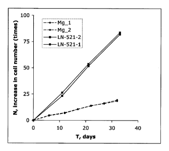
[0063] FIG. 8 shows the growth curves for human ES cells cultured on r-laminin-
521 (LN-521_1 and LN-521_2) using single cell dissociation passaging and for
human ES cells cultured on Matrigel (Mg_1 and Mg_2) using passaging in small
clumps. The latter cells were passaged as described in Rodin et al., Nature
BiotechnoL, vol. 28, pp. 611-615 (2010).
[0064] FIG. 9 is a FACS analysis of human ES cells after several single cell
dissociation passages on r-laminin-521 for OCT4, a marker of pluripotency. The
percentage of positive cells is listed in parentheses.
[0065] FIG. 10 is a FACS analysis of human ES cells after several passages on
Matrigel using splitting in small clumps for OCT4. The percentage of positive
cells is
listed in parentheses.
[0066] FIG. 11
shows the results of a real-time quantitative RT-PCR analysis
which was used to compare numbers of mRNA transcripts of the pluripotency
markers 0ct4 and Nanog in human ES cells cultured on human r-laminin-521
(LN521) after several single cell dissociation passaging and in the cells
cultured on
Matrigel (Mg) after several passaging of the cells in clumps. Error bars show
95%
confidence intervals.
[0067] FIG. 12 is a Western blot comparing the levels of phosphorylated myosin
light chain (P-MLC) in stem cells grown on Matrigel (Mg), LN-111, LN-511, and
LN-
521 one hour after plating.
[0068] FIG. 13 is a Western blot comparing the levels of phosphorylated myosin
light chain (P-MLC) in stem cells grown on Matrigel (Mg), LN-111, LN-511, and
LN-
521 six hours after plating.
[0069] FIG. 14 is a graph comparing the relative migration of human embryonic
stem cells grown on Matrigel (Mg), LN-111, LN-511, and LN-521 between five and
seven hours after plating.
[0070] FIG. 15 is a graph comparing the relative migration of human embryonic
stem cells grown on LN-521 with IgG and IgM blocking antibodies applied (IgGM)
and with a function blocking antibody to integrin 131 (b1) applied. This shows
that
blocking the integrin significantly reduced motility.
CA 02826252 2013-06-17
WO 2012/080842
PCT/IB2011/003258
12
[0071] FIG. 16 is
a graph comparing the relative survival of hES cells on LN-521
in the presence of water, DMSO, LY294002 (Akt inhibitor), wortmannin (PI3K/Akt
inhibitor), and PD 98059 (MEK1 inhibitor). This showed that activation of
PI3K/Akt
was necessary for hES cell survival on LN-521. The medium contained bFGF.
[0072] FIG. 17 is
a graph comparing the relative survival of hES cells on LN-521
in the presence of DMSO, LY294002 (Akt inhibitor), and PD 98059 (MEK1
inhibitor).
No exogenous bFGF was present.
[0073] FIG. 18 is a Western blot comparing cells treated with LY 294002
against
control cells (DMSO) and collected one hour after plating on LN-521.
[0074] FIG. 19 is a Western blot comparing cells treated with PD98059 against
control cells (DMSO) and collected one hour after plating on LN-521.
[0075] FIG. 20 is
a graph showing the relative levels of Akt2 phosphorylation on
cell lysates collected one hour after plating on Matrigel (Mg), LN-111, LN-
511, or LN-
521, obtained using ELISA.
[0076] FIG. 21 is
a graph showing the relative levels of Akt1 phosphorylation on
cell lysates collected one hour after plating on Matrigel (Mg), LN-111, LN-
511, or LN-
521, obtained using ELISA.
[0077] FIG. 22 is a graph showing the growth curve of HS181 hES cells cultured
in a low bFGF medium (03_3.9) compared to a higher bFGF medium (03_100).
[0078] FIG. 23 is a graph showing the relative mRNA expression level for two
pluripotency markers (0ct4 and Nanog) in HS181 hES cells cultured in a low
bFGF
medium (03_3.9) compared to a higher bFGF medium (03_100).
[0079] FIG. 24 is a picture showing an early stage derivation of a new human
embryonic stem HS841 cell line on laminin-521/E-Cadherin matrix.
DETAILED DESCRIPTION
[0080] A more complete understanding of the compositions and methods
disclosed herein can be obtained by reference to the accompanying drawings.
These figures are merely schematic representations based on convenience and
the
ease of demonstrating the present disclosure, and are, therefore, not intended
to
define or limit the scope of the exemplary embodiments.
[0081] Although specific terms are used in the following description for the
sake
of clarity, these terms are intended to refer only to the particular structure
of the
embodiments selected for illustration in the drawings, and are not intended to
define
13
or limit the scope of the disclosure. In the drawings and the following
description
below, it is to be understood that like numeric designations refer to
components of
like function.
[0082]
[0083] Unless otherwise stated, the techniques utilized in this
application may be
found in any of several well-known references such as: Molecular Cloning: A
Laboratory Manual (Sambrook, et al., 1989, Cold Spring Harbor Laboratory
Press),
Gene Expression Technology (Methods in Enzymology, Vol. 185, edited by D.
Goeddel, 1991. Academic Press, San Diego, Calif.), "Guide to Protein
Purification" in
Methods in Enzymology (M. P. Deutshcer, ed., (1990) Academic Press, Inc.); PCR
Protocols: A Guide to Methods and Applications (Innis, et al. 1990. Academic
Press,
San Diego, Calif.), Culture of Animal Cells: A Manual of Basic Technique,
Second
Ed. (R. I. Freshney. 1987. Liss, Inc. New York, N.Y.), Gene Transfer and
Expression
Protocols, pp. 109-128, ed. E. J. Murray, The Humana Press Inc., Clifton,
N.J.), or
the Ambion 1998 Catalog (Ambion, Austin, Tex.).
[0084] As used herein, the term "isolated nucleic acid sequence" refers to
a
nucleic acid sequence that is free of gene sequences which naturally flank the
nucleic acid in the genomic DNA of the organism from which the nucleic acid is
derived (i.e., genetic sequences that are located adjacent to the gene for the
isolated
nucleic molecule in the genomic DNA of the organism from which the nucleic
acid is
derived). The "isolated" sequence may, however, be linked to other nucleotide
sequences that do not naturally flank the recited sequence, such as a
heterologous
promoter sequence, or other vector sequences. It is not necessary for the
isolated
nucleic acid sequence to be free of other cellular material to be considered
"isolated", as a nucleic acid sequence according to the disclosure may be part
of an
expression vector that is used to transfect host cells (see below).
[0085] The present disclosure provides recombinant expression vectors
comprising a full length laminin 132 chain nucleic acid sequence (SEQ ID NO:
4) of
the human laminin 132 chain. In some embodiments, the expression vectors
comprise
a nucleic acid encoded by SEQ ID NO: 4, operatively linked to a heterologous
promoter (i.e. is not the naturally occurring promoter for the given 132
laminin chain).
A promoter and a laminin 132 chain nucleic acid sequence are "operatively
linked"
CA 2826252 2017-12-14
CA 02826252 2013-06-17
WO 2012/080842
PCT/IB2011/003258
14
when the promoter is capable of driving expression of the laminin 132 chain
DNA into
RNA.
[0086] As used herein, the term "vector" refers to a nucleic acid molecule
capable
of transporting another nucleic acid to which it has been linked. One type of
vector is
a "plasmid", which refers to a circular double stranded DNA into which
additional
DNA segments may be cloned. Another type of vector is a viral vector, wherein
additional DNA segments may be cloned into the viral genome. Certain vectors
are
capable of autonomous replication in a host cell into which they are
introduced (e.g.,
bacterial vectors having a bacterial origin of replication and episomal
mammalian
vectors). Other vectors (e.g., non-episomal mammalian vectors), are integrated
into
the genome of a host cell upon introduction into the host cell, and thereby
are
replicated along with the host genome. Moreover, certain vectors are capable
of
directing the expression of genes to which they are operatively linked. Such
vectors
are referred to herein as "recombinant expression vectors" or simply
"expression
vectors". In the present disclosure, the expression of the laminin polypeptide
sequence is directed by the promoter sequences of the disclosure, by
operatively
linking the promoter sequences of the disclosure to the gene to be expressed.
In
general, expression vectors of utility in recombinant DNA techniques are often
in the
form of plasmids. In the present specification, "plasmid" and "vector" may be
used
interchangeably, as the plasmid is the most commonly used form of vector.
However, the disclosure is intended to include other forms of expression
vectors,
such as viral vectors (e.g., replication defective retroviruses, adenoviruses
and
adeno-associated viruses), which serve equivalent functions.
[0087] The vector may also contain additional sequences, such as a polylinker
for
subcloning of additional nucleic acid sequences, or a polyadenylation signal
to effect
proper polyadenylation of the transcript. The nature of the polyadenylation
signal is
not believed to be crucial to the successful practice of the methods of the
disclosure,
and any such sequence may be employed, including but not limited to the SV40
and
bovine growth hormone poly-A sites. Also contemplated as an element of the
vector
is a termination sequence, which can serve to enhance message levels and to
minimize readthrough from the construct into other sequences. Additionally,
expression vectors typically have selectable markers, often in the form of
antibiotic
resistance genes, that permit selection of cells that carry these vectors.
CA 02826252 2013-06-17
WO 2012/080842
PCT/IB2011/003258
[0088] In further embodiments, the present disclosure provides host cells
transfected with the laminin 132 chain-expressing recombinant expression
vectors
disclosed herein. As used herein, the term "host cell" is intended to refer to
a cell into
which a nucleic acid of the present disclosure, such as a recombinant
expression
vector, has been introduced. Such cells may be prokaryotic, which can be used,
for
example, to rapidly produce a large amount of the expression vectors of the
disclosure, or may be eukaryotic, for functional studies.
[0089] The terms "host cell" and "recombinant host cell" are used
interchangeably
herein. It should be understood that such terms refer not only to the
particular
subject cell but to the progeny or potential progeny of such a cell. Because
certain
modifications may occur in succeeding generations due to either mutation or
environmental influences, such progeny may not, in fact, be identical to the
parent
cell, but are still included within the scope of the term as used herein.
[0090] The host cells can be transiently or stably transfected with one or
more of
the expression vectors of the disclosure. Such transfection of expression
vectors into
prokaryotic and eukaryotic cells can be accomplished via any technique known
in the
art, including but not limited to standard bacterial transformations, calcium
phosphate
co-precipitation, electroporation, or liposome mediated-, DEAE dextran
mediated-,
polycationic mediated-, or viral mediated transfection. (See, for example,
Molecular
Cloning: A Laboratory Manual (Sambrook, et al., 1989, Cold Spring Harbor
Laboratory Press; Culture of Animal Cells: A Manual of Basic Technique,
2nd
Ed. (R. I. Freshney. 1987. Liss, Inc. New York, N.Y.).
[0091] In another aspect, the present disclosure provides an isolated full
length
human laminin 132 chain polypeptide consisting of the amino acid sequence of
SEQ
ID NO: 2.
[0092] As used herein, an "isolated polypeptide" refers to a polypeptide that
is
substantially free of other proteins, including other laminin chains, and gel
agents,
such as polyacrylamide and agarose. In preferred embodiments, the isolated
laminin
polypeptide is free of detectable contaminating laminin chains. Thus, the
protein can
either be isolated from natural sources, or recombinant protein can be
isolated from
the transfected host cells disclosed above.
[0093] In another aspect, the present disclosure provides isolated laminin-
521. As
used herein, the term "laminin-521" refers to the protein formed by joining
a5, 82 and
y1 chains together. The term should be construed as encompassing both
CA 02826252 2013-06-17
WO 2012/080842
PCT/IB2011/003258
16
recombinant laminin-521 and heterotrimeric laminin-521 from naturally
occurring
sources. In preferred embodiments, the laminin-521 comprises recombinant
laminin-
521 (or "r-laminin-521").
[0094] As used herein, the term "r-laminin-521" refers to recombinant
heterotrimeric laminin-521, expressed by a host cell that has been transfected
with
one or more expression vectors comprising at least one nucleic acid sequence
encoding a laminin-521 chain selected from the a5, 132 and y1 chains, or
processed
or secreted forms thereof. Such r-laminin-521 can thus comprise a5, in, and yl
sequences from a single organism, or from different organisms. Various laminin-
521
chain DNA sequences are known in the art, and the use of any such sequence to
prepare the r-laminin-521 of the disclosure is contemplated. (See, for
example,
Pouliot, N. et al., Experimental Cell Research 261(2):360-71, (2000); Kikkawa,
Y. et
al., Journal of Cell Science 113 (Pt 5):869-76, (2000); Church, H J. et al.,
Biochemical Journal 332 (Pt 2):491-8, (1998); Sorokin, L M. et al.,
Developmental
Biology 189(2):285-300, (1997); Miner, J H. et al., Journal of Biological
Chemistry
270(48):28523-6, (1995); Sorokin, L. et al., European Journal of Biochemistry
223(2):603-10, (1994)). In preferred embodiments, the r-laminin-521 is formed
from
recombinant human a5, 132, and y1 polypeptide chains.
[0095] The disclosure encompasses those laminin molecules wherein only one or
two chains that make up the recombinant heterotrimeric laminin-521 are encoded
by
endogenous laminin-521 chains. In preferred embodiments, each of the a5, 132,
and
y1 polypeptide chains are expressed recombinantly.
[0096] The laminin-
521 is an intact protein. The term "intact" refers to the protein
being composed of all of the domains of the a-chain, 13-chain, and y-chain,
with the
three chains being joined together to form the heterotrimeric structure. The
protein is
not broken down into separate chains, fragments, or functional domains. The
term
"chain" refers to the entirety of the alpha, beta, or gamma chain of the
laminin
protein. The term "fragment" refers to any protein fragment which contains
one, two,
or three functional domains that possesses binding activity to another
molecule or
receptor. However, a chain should not be considered a fragment because each
chain possesses more than three such domains. Similarly, an intact laminin
protein
should not be considered a fragment. Examples of functional domains include
Domains I, II, Ill, IV, V, VI, and the G domain.
CA 02826252 2013-06-17
WO 2012/080842
PCT/IB2011/003258
17
[0097] Laminin-521 is a secreted protein, which is capable of being
directed to
the endoplasmic reticulum (ER), secretory vesicles, and the extracellular
space as a
result of a signal sequence. If the secreted protein is released into the
extracellular
space, the secreted protein can undergo extracellular processing to produce a
"mature" protein. Such processing events can be variable, and thus may yield
different versions of the final "mature protein". For example, the lengths of
the a5,
132, and y1 chains may vary between proteins. However, the final mature
protein still
has the same functionality, even though the chain lengths vary. The isolated
laminin-521 of the present disclosure includes heterotrimers comprising both
the full
length polypeptide chains and any such naturally processed laminin-521
polypeptide
chains.
[0098] As used herein, a laminin-521 polypeptide chain refers to a polypeptide
chain according to one or more of the following:
[0099] (a) a polypeptide chain that comprises a polypeptide structure
selected
from the group consisting of: R1-R2-R3, R1-R2-R4, R3, R4, R1-R3, R1-R4, R2-R3,
and R2-R4, wherein R1 is an amino terminal methionine; R2 is a signal sequence
that is capable of directing secretion of the polypeptide, wherein the signal
sequence
may be the natural signal sequence for the particular laminin chain, that of
another
secreted protein, or an artificial sequence; R3 is a secreted laminin chain
selected
from the group consisting of a a5 chain, a 62 chain, and a y1 chain; and R4 is
a
secreted a5, 132, or y1 laminin chain that further comprises an epitope tag
(such as
those described below), which can be placed at any position within the laminin
chain
amino acid sequence; or
[0100] (b) a polypeptide chain that is encoded by a polynucleotide that
hybridizes
under high or low stringency conditions to the coding regions, or portions
thereof, of
one or more of the recombinant laminin-521 chain DNA sequences disclosed
herein
(SEQ ID NO: 4, SEQ ID NO: 5, SEQ ID NO: 6), or complementary sequences
thereof; or
[0101] (c) a polypeptide chain that has at least 70% identity to one or more
of the
disclosed laminin-521 polypeptide chain amino acid sequences (SEQ ID NO: 1,
SEQ
ID NO: 2, SEQ ID NO: 3), preferably at least 80% identity, and most preferably
at
least about 90% identity.
[0102] "Stringency of hybridization" is used herein to refer to washing
conditions
under which nucleic acid hybrids are stable. The disclosure also includes
nucleic
CA 02826252 2013-06-17
WO 2012/080842
PCT/IB2011/003258
18
acids that hybridize under high stringency conditions (as defined herein) to
all or a
portion of the coding sequences of the laminin chain polynucleotides disclosed
herein, or their complements. The hybridizing portion of the hybridizing
nucleic acids
is typically at least 50 nucleotides in length. As known to those of ordinary
skill in the
art, the stability of hybrids is reflected in the melting temperature (TM) of
the hybrids.
TM decreases approximately 1-1.5 C with every 1% decrease in sequence
homology. In general, the stability of a hybrid is a function of sodium ion
concentration and temperature. Typically, the hybridization reaction is
performed
under conditions of lower stringency, followed by washes of varying, but
higher,
stringency. As used herein, high stringency refers to an overnight incubation
at 42 C
in a solution comprising 50% formamide, 5x SSC (750 mM NaCI, 75 mM sodium
citrate), 50 mM sodium phosphate (pH 7.6), 5x Denhardrs solution, 10% dextran
sulfate, and 20 pg/m1 denatured, sheared salmon sperm DNA, followed by washing
the filters in 0.1x SSC at about 65 C
[0103] Also contemplated are laminin-521-encoding nucleic acid sequences that
hybridize to the polynudeotides of the present disclosure at lower stringency
hybridization conditions. Changes in the stringency of hybridization and
signal
detection are primarily accomplished through the manipulation of formamide
concentration (lower percentages of formamide result in lowered stringency);
salt
conditions, or temperature. For example, lower stringency conditions include
an
overnight incubation at 37 C in a solution comprising 6x SSPE (20x SSPE=3M
NaCI; 0.2M NaH2PO4 ; 0.02M EDTA, pH 7.4), 0.5% SDS, 30% formamide, 100 pg/m1
salmon sperm blocking DNA; followed by washes at 50 C with lx SSPE, 0.1% SDS.
In addition, to achieve even lower stringency, washes performed following
stringent
hybridization can be done at higher salt concentrations (e.g. 5x SSC).
[0104] Note that variations in the above conditions may be accomplished
through
the inclusion and/or substitution of alternate blocking reagents used to
suppress
background in hybridization experiments. Typical blocking reagents include
Denhardts reagent, BLOTTO, heparin, denatured salmon sperm DNA, and
commercially available proprietary formulations. The inclusion of specific
blocking
reagents may require modification of the hybridization conditions described
above,
due to problems with compatibility.
[0105] As used herein, "percent identity" of two amino acids or of two nucleic
acids is determined using the algorithm of Karlin and Altschul (Proc. Natl.
Acad. Sci.
CA 02826252 2013-06-17
WO 2012/080842
PCT/IB2011/003258
19
USA 87:2264.2268, 1990), modified as in Karlin and Altschul (Proc. Natl. Acad.
Sci.
USA 90:5873-5877, 1993). Such an algorithm is incorporated into the NBLAST and
XBLAST programs of Altschul et al. (J. Mol. Biol. 215:403-410, 1990). BLAST
nucleotide searches are performed with the NBLAST program, score 100,
wordlength=12, to determine nucleotide sequences identity to the nucleic acid
molecules of the disclosure. BLAST protein searches are performed with the
XBLAST program, score=50, wordlength=3, to determine an amino acid sequence
identity to a polypeptide of the disclosure. To obtain gapped alignments for
comparison purposes, Gapped BLAST is utilized as described in Altschul et al.
(Nucleic Acids. Res. 25:3389-3402, 1997). When utilizing BLAST and Gapped
BLAST programs, the default parameters of the respective programs (e.g.,
XBLAST
and NBLAST) are used.
[0106] Further embodiments of the present disclosure include polynucleotides
encoding laminin-521 chain polypeptides having at least 70% identity,
preferably at
least 80% identity, and most preferably at least 90% identity to one or more
of the
polypeptide sequences contained in SEQ ID NO: 4, SEQ ID NO: 5, and SEQ ID NO:
6.
[0107] As used herein, "a5 polynucleotide" refers to polynucleotides encoding
a
laminin a5 chain. Such polynucleotides can be characterized by one or more of
the
following: (a) polynucleotides that encode polypeptides which share at least
70%
identity, preferably 80% identity, and most preferably at least 90% identity
with a
sequence selected of SEQ ID NO: 5; (b) polynucleotides that hybridize under
low or
high stringency conditions to the coding sequence of SEQ ID NO: 5 or
complementary sequences thereof; or (c) polynucleotides encoding a laminin a5
chain polypeptide with a general structure selected from the group consisting
of R1-
R2-R3, R1-R2-R4, R3, R4, R1-R3, R1-R4, R2-R3, and R2-R4, wherein R1 and R2
are as described above, R3 is a secreted a5 chain, and R4 is a secreted a5
chain
that comprises an epitope tag.
[0108] As used herein, "132 polynucleotides" refers to polynucleotides
encoding a
132 laminin chain of the same name. Such polynucleotides can be characterized
by
one or more of the following: (a) polynucleotides that encode polypeptides
which
share at least 70% identity, preferably at least 80%, and most preferably at
least
90% identity with the sequence of SEQ ID NO: 4; (b) polynucleotides that
hybridize
under low or high stringency conditions to the coding sequences of SEQ ID NO:
4, or
CA 02826252 2013-06-17
WO 2012/080842
PCT/IB2011/003258
complementary sequences thereof; or (c) polynucleotides encoding a polypeptide
with a general structure selected from R1-R2-R3, R1-R2-R4, R3, R4, R1-R3, R1-
R4,
R2-R3, and R2-R4, wherein R1 and R2 are as described above, R3 is a secreted
132
chain, and R4 is a secreted 132 chain that comprises an epitope tag.
[0109] As used herein, "y1 polynucleotides" refers to polynucleotides encoding
a
y1 laminin chain of the same name. Such polynucleotides can be characterized
by
one or more of the following: (a) polynucleotides that encode polypeptides
which
share at least 70% identity, preferably at least 80%, and most preferably at
least
90% identity with the sequence of SEQ ID NO: 6; (b) polynucleotides that
hybridize
under low or high stringency conditions to the coding sequence of SEQ ID NO: 6
or
complementary sequences thereof; or (c) polynucleotides that encode a
polypeptide
with a general structure selected from R1-R2-R3, R1-R2-R4, R3, R4, R1-R3, R1-
R4,
R2-R3, and R2-R4, wherein R1 and R2 are as described above, R3 is a secreted
y1
chain, and R4 is a secreted y1 chain that comprises an epitope tag.
[0110] As used herein, the term "epitope tag" refers to a polypeptide sequence
that is expressed as part of a chimeric protein, where the epitope tag serves
as a
recognition site for binding of antibodies generated against the epitope tag,
or for
binding of other molecules that can be used for affinity purification of
sequences
containing the tag.
[0111] In
preferred embodiments, cDNAs encoding the laminin a5, 132 and y1
chains, or fragments thereof, are subcloned into an expression vector.
Alternatively,
laminin a5, 132 and/or y1 gene sequences, including one or more introns, can
be
used for sub-cloning into an expression vector.
[0112] In other
aspects, the present disclosure provides laminin-521 expressing-
cells that have been transfected with an expression vector containing promoter
sequences that are operatively linked to nucleic acid sequences encoding at
least
one polypeptide sequence comprising a sequence selected from the group
consisting of the a5, 132 and y1 chains of laminin-521, wherein the
transfected cells
secrete heterotrimeric laminin-521 containing the recombinant laminin chain.
In
preferred embodiments, the cells are systematically transfected with
recombinant
expression vectors containing promoter sequences that are operatively linked
to
nucleic acid sequences encoding polypeptide sequences comprising the a5, 132
and
y1 chains of laminin-521, which are even more preferably all human chains.
After the
CA 02826252 2013-06-17
WO 2012/080842
PCT/IB2011/003258
21
multiple transfections, the cells express recombinant laminin-521 chains,
which form
the heterotrimeric r-laminin-521.
[0113] Transfection of the expression vectors into eukaryotic cells can be
accomplished via any technique known in the art, including but not limited to
calcium
phosphate co-precipitation, electroporation, or liposome mediated-, DEAE
dextran
mediated-, polycationic mediated-, or viral mediated transfection.
Transfection of
bacterial cells can be done by standard methods.
[0114] In
preferred embodiments, the cells are stably transfected. Methods for
stable transfection and selection of appropriate transfected cells are known
in the art.
In other preferred embodiments, a CMV promoter driven expression vector is
used in
a human kidney embryonic 293 cell line.
[0115] Any cell capable of expressing and secreting the r-laminin-521 can be
used. Preferably, eukaryotic cells are used, and most preferably mammalian
cells
are used, including but not limited to kidney and epithelial cell lines. The
promoter
sequence used to drive expression of the individual chains or r-laminin-521
may be
constitutive (driven by any of a variety of promoters, including but not
limited to,
CMV, SV40, RSV, actin, EF) or inducible (driven by any of a number of
inducible
promoters including, but not limited to, tetracycline, ecdysone, steroid-
responsive).
Carbohydrate and disulfide post-translational modifications are believed to be
required for laminin-521 protein folding and function. This makes the use of
eukaryotic cells preferable for producing functional r-laminin-521, although
other
systems are useful for obtaining, for example, antigens for antibody
production. In
most preferred embodiments, the mammalian cells do not express the laminin (32
chain endogenously. In other preferred embodiments, the cells do not express
all of
the laminin-521 chains endogenously.
[0116] The protein may comprise additional sequences useful for promoting
purification of the protein, such as epitope tags and transport signals.
Examples of
such epitope tags include, but are not limited to FLAG (Sigma Chemical, St.
Louis,
Mo.), myc (9E10) (Invitrogen, Carlsbad, Calif.), 6-His (Invitrogen; Novagen,
Madison,
Wis.), and HA (Boehringer Manheim Biochemicals). Examples of such transport
signals include, but are not limited to, export signals, secretory signals,
nuclear
localization signals, and plasma membrane localization signals.
[0117] In some
embodiments, at least one of the laminin chain polypeptide
sequences, or fragments thereof, is operatively linked to a nucleic acid
sequence
CA 02826252 2013-06-17
WO 2012/080842
PCT/IB2011/003258
22
encoding an "epitope tag", so that at least one of the chains is expressed as
a fusion
protein with an expressed epitope tag. The epitope tag may be expressed as the
amino terminus, the carboxy terminus, or internal to any of the polypeptide
chains
comprising r-laminin-521, so long as the resulting r-laminin-521 remains
functional.
[0118] In other embodiments, one of the r-laminin-521 chains is expressed as a
fusion protein with a first epitope tag, and at least one other r-laminin
chain is
expressed as a fusion protein with a second different epitope tag. This
permits
multiple rounds of purification to be carried out. Alternatively, the same
epitope tag
can be used to create fusion proteins with more than one of the r-laminin
chains.
[0119] In further embodiments, the epitope tag can be engineered to be
cleavable
from the r-laminin-521 chain(s). Alternatively, no epitope tag is fused to any
of the r-
laminin-521 chains, and the r-laminin-521 is isolated by standard techniques,
including but not limited to affinity chromatography using laminin-521
specific
antibodies or other laminin-521 binding molecules.
[0120] Media from cells transfected with a single laminin chain are
initially
analyzed on Western blots using laminin chain-specific antibodies. The
expression of
single laminin chains following transfection is generally intracellular.
Clones showing
reactivity against individual transfected chain(s) are verified by any
appropriate
method, such as PCR, reverse transcription-PCR, or nucleic acid hybridization,
to
confirm incorporation of the transfected gene. Preferably, analysis of genomic
DNA
preparations from such clones is done by PCR using laminin chain-specific
primer
pairs. Media from transfected clones producing all three chains are further
analyzed
for r-laminin-521 secretion and/or activity, by any appropriate method,
including
Western blot analysis and cell binding assays.
[0121] In preferred embodiments, purification of r-laminin-521 is
accomplished by
passing media from the transfected cells through an antibody affinity column.
In
some embodiments, antibodies against a peptide epitope expressed on at least
one
of the recombinant chains are attached to an affinity column, and bind the r-
laminin-
521 that has been secreted into the media. The r-laminin-521 is removed from
the
column by passing excess peptide over the column. Eluted fractions are
analyzed by
any appropriate method, including gel electrophoresis and Western blot
analysis. In
further embodiments, the peptide epitope can be cleaved after purification. In
other
embodiments, two or three separate r-laminin chains are expressed as fusion
proteins, each with a different epitope tag, permitting two or three rounds of
CA 02826252 2013-06-17
WO 2012/080842
PCT/IB2011/003258
23
purification and a doubly or triply isolated r-laminin-521. The epitope tag
can be
engineered so as to be cleavable from the r-laminin-521 chain(s) after
purification.
Alternatively, no epitope tag is fused to any of the r-laminin-521 chains, and
the r-
laminin-521 is isolated by standard techniques, including but not limited to
affinity
chromatography using laminin-521 specific antibodies or other laminin-521
binding
molecules.
[0122] In other embodiments, purification of r-laminin-521 is accomplished
by
passing media from the transfected cells through a gel-filtration
chromatography
column. Eluted fractions are analyzed by any appropriate method, including gel
electrophoresis and Western blot analysis. Fractions containing r-laminin-521
are
collected and purity of the specimen is evaluated by any appropriate method,
including gel electrophoresis and Western blot analysis. In some embodiments,
the
protein solution can be passed through a gel-filtration chromatography column
again
to gain higher purity of the protein. In some embodiments, to achieve higher
purity of
r-laminin-521 solution, the media or r-laminin-521 solution from the previous
purification steps can be passed through an ion-exchange column. Eluted
fractions
are analyzed by any appropriate method, including gel electrophoresis and
Western
blot analysis. Fractions containing r-laminin-521 are collected and purity of
the
specimen is evaluated by any appropriate method, including mentioned above.
[0123] The laminin-521 polypeptide chains of the present disclosure also
indude
(i) substitutions with one or more of the non-conserved amino acid residues,
where
the substituted amino acid residues may or may not be one encoded by the
genetic
code, or (ii) substitution with one or more amino acid residues having
substituent
groups, or (iii) fusion of the mature polypeptide with another compound, such
as a
compound to increase the stability and/or solubility of the polypeptide (for
example,
polyethylene glycol), or (iv) fusion of the polypeptide with additional amino
acids,
such as an IgG Fc fusion region peptide, or leader or secretory sequence, or a
sequence facilitating purification. Such variant polypeptides are deemed to be
within
the scope of those skilled in the art from the teachings herein.,
[0124] For example, polypeptide variants containing amino acid
substitutions of
charged amino acids with other charged or neutral amino acids may produce
proteins with improved characteristics, such as less aggregation. Aggregation
of
pharmaceutical formulations both reduces activity and increases clearance due
to
the aggregate's immunogenic activity. (Pinckard et al., Clin. Exp. lmmunol.
2:331-
CA 02826252 2013-06-17
WO 2012/080842
PCT/IB2011/003258
24
340 (1967); Robbins et al., Diabetes 36: 838-845 (1987); Cleland et al., Crit.
Rev.
Therapeutic Drug Carrier Systems 10:307-377 (1993).)
[0125] In particular embodiments, the isolated laminin-521 comprises three
chains. The first chain comprises a polypeptide with at least 80% identity to
a
polypeptide sequence of SEQ ID NO: 1 (i.e. the a5 laminin chain). The second
chain
comprises a polypeptide with at least 70% identity to a polypeptide sequence
of SEQ
ID NO: 2 (i.e. the 82 laminin chain). The third chain comprises a polypeptide
with at
least 70% identity to a polypeptide sequence of SEQ ID NO: 3 (i.e. the y1
laminin
chain). These first, second, and third chains are assembled into recombinant
laminin-521.
[0126] In more specific embodiments, the polypeptide of the first chain has
at
least 80% identity to the polypeptide sequence of SEQ ID NO: 1, the
polypeptide of
the second chain has at least 80% identity to the polypeptide sequence of SEQ
ID
NO: 2, and the polypeptide of the third chain has at least 80% identity to the
polypeptide sequence of SEQ ID NO: 3.
[0127] In more specific embodiments, the polypeptide of the first chain has
at
least 90% identity to the polypeptide sequence of SEQ ID NO: 1, the
polypeptide of
the second chain has at least 90% identity to the polypeptide sequence of SEQ
ID
NO: 2, and the polypeptide of the third chain has at least 90% identity to the
polypeptide sequence of SEQ ID NO: 3.
[0128] In particular embodiments, the first chain comprises the polypeptide
sequence of SEQ ID NO: 1, the second chain comprises the polypeptide sequence
of SEQ ID NO: 2, and the third chain comprises the polypeptide sequence of SEQ
ID
NO: 3.
[0129] In particular embodiments, the first chain is the polypeptide
sequence of
SEQ ID NO: 1, the second chain is the polypeptide sequence of SEQ ID NO: 2,
and
the third chain is the polypeptide sequence of SEQ ID NO: 3.
[0130] The present disclosure further provides pharmaceutical compositions
comprising isolated laminin-521 and a pharmaceutically acceptable carrier. In
preferred embodiments, the pharmaceutical composition comprises isolated r-
laminin-521. According to these aspects of the disclosure, other agents can be
included in the pharmaceutical compositions, depending on the condition being
treated. The pharmaceutical composition may further comprise one or more other
compounds, including but not limited to any of the collagens, other laminin
types,
CA 02826252 2013-06-17
WO 2012/080842
PCT/IB2011/003258
fibronectin, vitronectin, cadherins, integrins, a-dystroglycan,
entactin/nidogen, a-
dystroglycan, glycoproteins, proteoglycans, heparan sulfate proteoglycan,
glycosaminoglycans, epidermal growth factor, vascular endothelial growth
factor,
fibroblast growth factor, or nerve growth factors, and peptide fragments
thereof.
[0131]
Pharmaceutical preparations comprising isolated laminin-521 can be
prepared in any suitable form, and generally comprise the isolated laminin-521
in
combination with a pharmaceutically acceptable carrier. The carriers can be
injectable carriers, topical carriers, transdermal carriers, and the like. The
preparation may advantageously be in a form for topical administration, such
as an
ointment, gel, cream, spray, dispersion, suspension or paste. The preparations
may
further advantageously include preservatives, antibacterials, antifingals,
antioxidants,
osmotic agents, and similar materials in composition and quantity as is
conventional.
Suitable solutions for use in accordance with the disclosure are sterile, are
not
harmful for the proposed application, and may be subjected to conventional
pharmaceutical operations such as sterilization and/or may contain
conventional
adjuvants, such as preservatives, stabilizers, wetting agents, emulsifiers,
buffers etc.
For assistance in formulating the compositions of the present disclosure, one
may
refer to Remington's Pharmaceutical Sciences, 15th Ed., Mack Publishing Co.,
Easton, Pa. (1975).
[0132] In further
aspects, the present disclosure comprises medical devices with
improved biocompatibility, wherein the exterior surfaces of the devices are
coated
with isolated laminin-521 or pharmaceutical compositions thereof, alone or in
combination with other proteins or agents that serve to increase the
biocompatibility
of the device surface. The coated device stimulates cell attachment (such as
endothelial cell attachment), and provides for diminished inflammation and/or
infection at the site of entry of the device.
[0133] Such medical devices can be of any material used for implantation into
the
body, and preferably are made of or coated with a biocompatible metal, such as
stainless steel or titanium. Alternatively, the device is made of or coated
with a
ceramic material or a polymer, such as polyester, polyglycolic acid, or a
polygalactose-polyglycolic acid copolymer.
[0134] If the
device is made of a natural or synthetic biodegradable material in the
form of a mesh, sheet or fabric, isolated laminin-521 or pharmaceutical
compositions
thereof may be applied directly to the surface thereof. Appropriate cells may
then be
CA 02826252 2013-06-17
WO 2012/080842
PCT/IB2011/003258
26
cultured on the matrix to form transplantable or implantable devices,
including dental
abutment pieces, needles, metal pins or rods, indwelling catheters, colostomy
tubes,
surgical meshes and any other appliance for which coating with isolated
laminin-521
is desirable. Alternatively, the devices may be implanted and cells may be
permitted
to attach in vivo.
[0135] Coupling of the isolated laminin-521 may be non-covalent (such as by
adsorption), or by covalent means. The device may be immersed in, incubated
in, or
sprayed with the isolated laminin-521 or pharmaceutical compositions thereof.
[0136] The dosage regimen for various treatments using the isolated laminin-
521
of the present disclosure is based on a variety of factors, including the type
of injury
or condition, the age, weight, sex, medical condition of the individual, the
severity of
the condition, and the route of administration. Thus, the dosage regimen may
vary
widely, but can be determined routinely by a physician using standard methods.
Laminins are extremely potent molecules, and one or a few molecules per cell
could
produce an effect. Thus, effective doses in the pico-gram per milliliter range
are
possible if the delivery is optimized.
[0137] In other
aspects, the present disclosure provides a new material, isolated
laminin-521, which permits human embryonic stem cell survival after
dissociation
into single cell suspension. The stem cells were dissociated into single cell
suspension by Trypsin-EDTA treatment, pelleted by centrifugation, resuspended
into
03 medium, filtered through a 40 tm sieve, and plated at a density of 30
Kcells/cm2
on cell culture dishes precoated by either isolated laminin-521, laminin-511,
or
Matrigel. After one day in culture, the cells plated on Matrigel died, as is
known in
the art. The human embryonic stem cells plated on laminin-521 and in most
cases
on laminin-511 survived and started to proliferate, forming small colonies of
pluripotent cells.
[0138] In further
aspects, the present disclosure provides a method to expand
human embryonic stem cells in pluripotent state. It has been shown that human
embryonic stem cells plated in single cell suspension on laminin-521 survived
and
proliferated at a higher rate than that of classical methods known in the art.
After 3
passages (1 month) the cells passaged in a single cell suspension underwent
the
same number of cell divisions as that of cell cultures passaged in pieces on
Matrigel
CA 02826252 2013-06-17
WO 2012/080842
PCT/IB2011/003258
27
after 20 passages (3 months). Therefore, the new method was advantageous in
terms of time and labor, which may provide significant economical profits.
[0139] Laminin-521 is normally expressed and secreted by human pluripotent
embryonic stem cells and can also be found in the kidneys, neuromuscular
junctions,
lungs, and placenta.
[0140] The availability of pure laminin-521 would enable studies of the
effects of
the protein on cellular differentiation and maintenance of cellular
phenotypes. Thus,
numerous research and therapeutic purposes including, but not limited to,
treating
injuries to tissues, promoting cell attachment, expansion and migration, ex
vivo cell
therapy, improving the biocompatibility of medical devices, and preparing
improved
cell culture devices and media, would be furthered if pure intact isolated
laminin-521
were available. Also, the effects of pure laminin-521 on stem cells would be
important to study as this protein is expressed in the early mammalian embryo.
[0141] Thus, there is a need in the art for isolated laminin-521 for
research and
therapeutic purposes, and methods for making isolated laminin-521. Laminin-521
can serve as a matrix for long-term self-renewal and fast multiplication of
dissociated
human ES and induced pluripotent stem (iPS) cells under completely chemically
defined, feeder-free, and animal-protein-free (xeno-free) conditions. A LN-521
based
system is easy to use and easy to automate, and allows fast scale-up of human
ES/iPS cultures.
[0142] The present disclosure also relates to a cell culture medium that can
be
used to provide nutrition to cells, particularly stem cells. In this regard,
stem cells
typically require two things to be cultured: (1) a substrate or coating that
provides a
structural support for the stem cell; and (2) a cell culture medium to provide
nutrition
to the stem cell. The substrate or coating (1) is generally placed on, for
example, a
petri dish or some other container.
[0143] As used herein, the term "self-renewal" refers to the ability of the
stem cell
to go through numerous cycles of cell division and remain undifferentiated
(i.e.
pluripotent). Pluripotency itself refers to the ability of the stem cell to
differentiate
into any cell type. The term "proliferation" refers to the ability of the stem
cell to
divide. Survival refers to the ability of the stem cell to live, whether
differentiated or
undifferentiated, and does not require the stem cell to maintain its ability
to divide or
to differentiate.
CA 02826252 2013-06-17
WO 2012/080842
PCT/IB2011/003258
28
[0144] The cell
culture medium of the present disclosure is particularly suitable for
being used with a substrate that contains laminin-521 and/or laminin-511.
These
laminins activate 06131 integrins, which in turn leads to activation of the
PI3K/Akt
pathway. This increases the pluripotency, self-renewal, and/or proliferation
of the
stem cells. It is contemplated that the substrate may consist of laminin-521
or
laminin-511, either intact, as separate chains, or as fragments thereof.
Recombinant
laminin-521 and recombinant laminin-511 are commercially available. Many
different
molecules can activate the PI3K/Akt pathway, though with different
efficiencies. For
example, TGF beta 1 and bFGF activate this pathway. The use of laminin-521
and/or laminin-511 allows the quantity of such molecules to be reduced in the
cell
culture medium. Laminin-521 conveys the highest dose of signal via a6131
integrin,
activating the PI3K/Akt pathway. The use of laminin-521 allows for single-cell
suspension passaging without the addition of cell-detrimental rho-kinase
(ROCK)
inhibitor to increase cell survival after single-cell enzymatic dissociation.
Previously,
single-cell enzymatic passage of human ES cells without using artificial
apoptosis
inhibitors was impossible. The simplicity of the passaging procedure means the
experimental variance is reduced and allows the process to be automated for
high-
throughput cell culture and results without the extensive training and costs
of cell
culture staff. In addition, human ES and iPS cells plated on laminin-521 or
laminin-
511 grow as a monolayer, which makes the culture homogeneous since cells are
equally exposed to the matrix and the cell culture medium. Such human ES cell
cultures, grown in a chemically defined, xeno-free environment on laminin-521,
passaged as single cells in the absence of ROCK inhibitor expand continuously
for
months at an even better growth rate compared to cells grown on Matrigel
passaged
as clumps. These pluripotent long-term expanded cells homogeneously express
0ct4 and remain karyotypically normal. Thus, one can obtain human ES and iPS
cells with sustained survival and proliferation capacity.
[0145] The average contact area and spreading homogeneity is much larger for
cells cultured on laminin-511 compared to other available substrata. Human ES
cells grown on laminin-511 over 3 months maintain pluripotency and can
generate
teratomas after engraftment into SCID mice. Laminin-511 also supports the self-
renewal of mouse ES cells for over 5 months without the presence of LIF or
feeder
cells, when other known matrices are unable to do so for longer than a couple
of
weeks.
CA 02826252 2013-06-17
WO 2012/080842 PCT/IB2011/003258
29
[0146] The stem cells to be grown with this cell culture medium can be induced
pluripotent stem cells, embryonic stem cells, adult stem cells, fetal stem
cells,
amniotic stem cells, and generally any pluripotent stem cell.
[0147] Typically, cell culture media include a large number and a large amount
of
various growth factors and cytokines to inhibit differentiation and improve
proliferation. One advantage of the cell culture medium of the present
disclosure is
that it does not contain as many growth factors or cytokines, or such high
amounts.
[0148] Most generally, the cell culture medium of the present disclosure
requires
lower amounts of basic fibroblast growth factor (bFGF) than typically used. It
is
contemplated that the cell culture medium may comprise from greater than zero
to
3.9 nanograms per milliliter (ng/mL) of bFGF. The bFGF is human bFGF so that
the
cell culture medium is totally human and defined. In some more specific
embodiments, the cell culture medium may comprise 3.5 or lower ng/mL of bFGF.
In
other embodiments, the cell culture medium may comprise from 0.5 to 3.5 ng/mL
of
bFGF. In some embodiments, the cell culture medium may have zero bFGF, i.e. no
bFGF is present.
[0149] Generally, the cell culture medium includes a liquid phase in which
at least
one inorganic salt, at least one trace mineral, at least one energy substrate,
at least
one lipid, at least one amino acid, at least one vitamin, and at least one
growth factor
(besides bFGF) are dissolved. Table 1 below includes a list of various such
ingredients which may be present in the cell culture medium of the present
disclosure, and the minimum and maximum concentrations if the ingredient is
present. The values are presented in scientific notation. For example, "4.1E-
01"
should be interpreted as 4.1 x lel.
Table 1.
molar Min. Max. Min. Max.
mass Conc. Conc. Conc. Conc.
Ingredient (g/mol) (mM) (mM) (ng/mL)
(ng/mL)
INORGANIC SALTS
Calcium chloride
(Anhydrous) 110.98 4.1E-
01 1.6E+00 4.6E+04 1.8E+05
HEPES 238.3
5.9E+00 1.8E+01 1.4E+06 4.2E+06
Lithium Chloride (MI) 42.39 4.9E-
01 1.5E+00 2.1E+04 6.2E+04
Magnesium chloride
(Anhydrous) 95.21 1.2E-
01 3.6E-01 1.1E+04 3.4E+04
CA 02826252 2013-06-17
WO 2012/080842
PCT/IB2011/003258
Magnesium Sulfate
(MgSO4) 120.37 1.6E-01
4.8E-01 1.9E+04 5.8E+04
Potassium chloride (KCI) 74.55 1.6E+00
4.9E+00 1.2E+05 3.6E+05
Sodium bicarbonate
(NaHCO3) 84.01 9.0E+00
4.4E+01 7.6E+05 3.7E+06
Sodium chloride (NaCI) 58.44 4.7E+01
1.4E+02 2.8E+06 8.3E+06
Sodium phosphate,
dibasic (Anhydrous) 141.96 2.0E-01 5.9E-01 2.8E+04
8.3E+04
Sodium phosphate,
monobasic monohydrate
(NaH2PO4-H20) _ 137.99 1.8E-01
5.3E-01 2.4E+04 7.3E+04
TRACE MINERALS
Ferric Nitrate (Fe(NO3)3-
9H20) 404 4.9E-05 1.9E-
04 2.0E+01 7.5E+01
Ferrous sulfate
heptahydrate (FeSO4-
7H20) 278.01 5.9E-04
1.8E-03 1.6E+02 4.9E+02
Copper(l I ) sulfate
pentahyd rate (CuSO4-
5H20) 249.69 2.0E-06
8.0E-06 5.1E-01 2.0E+00
Zinc sulfate heptahydrate
(ZnSO4-7H20) 287.56 5.9E-04
1.8E-03 1.7E+02 5.1E+02
Ammonium Metavanadate
NH4V03 116.98 5.5E-06
1.6E-05 6.4E-01 1.9E+00
Manganese Sulfate
monohyd rate (MnSO4-
H20) 169.02 9.9E-07
3.0E-06 1.7E-01 5.0E-01
NiSO4-6H20 262.85 4.9E-07
1.5E-06 1.3E-01 3.8E-01
Selenium 78.96 8.9E-05
2.7E-04 7.0E+00 2.1E+01
Sodium Meta Silicate
Na2Si 03 -9H20 284.2 4.8E-04 1.4E-03 1.4E+02
4.1E+02
SnCl2 189.62 6.2E-07
1.9E-06 1.2E-01 3.5E-01
Molybdic Acid, Ammonium
salt 1235.86 9.9E-07
3.0E-06 1.2E+00 3.7E+00
CdC12 183.32 6.1E-06
1.8E-05 1.1E+00 3.4E+00
CrCI3 158.36 9.9E-07
3.0E-06 1.6E-01 4.7E-01
AgNO3 169.87 4.9E-07
1.5E-06 8.3E-02 2.5E-01
A1C13 -6H20 241.43 2.4E-06 7.3E-06 5.9E-01
1.8E+00
Barium Acetate
(Ba(C2H302)2) 255.42 4.9E-06
1.5E-05 1.3E+00 3.8E+00
CoCl2 -6H20 237.93 4.9E-06 1.5E-05 1.2E+00
3.5E+00
Ge02 104.64 2.5E-06
7.5E-06 2.6E-01 7.8E-01
KBr 119 4.9E-07 1.5E-
06 5.9E-02 1.8E-01
KI 166 5.0E-07 1.5E-
06 8.3E-02 2.5E-01
NaF 41.99 4.9E-05
1.5E-04 2.1E+00 6.2E+00
RbCI 120.92 4.9E-06
1.5E-05 5.9E-01 1.8E+00
CA 02826252 2013-06-17
WO 2012/080842
PCT/IB2011/003258
31
ZrOCl2 -8H20 178.13 4.9E-06 1.5E-05 8.7E-01
2.6E+00
ENERGY SUBSTRATES
D-Glucose 180.16 6.9E+00
2.1E+01 1.2E+06 3.7E+06
Sodium Pyruvate 110.04 2.0E-01 5.9E-01 2.2E+04
6.5E+04
LIPIDS
, Linoleic Acid 280.45 9.4E-05 2.8E-04 2.6E+01
7.9E+01
Lipoic Acid 206.33 2.0E-04 7.8E-04 4.1E+01
1.6E+02
Arachidonic Acid 304.47 6.5E-06 1.9E-05 2.0E+00
5.9E+00
Cholesterol 386.65 5.6E-04
1.7E-03 2.2E+02 6.5E+02
DL-alpha tocopherol-
acetate 472.74 1.5E-04
4.4E-04 6.9E+01 2.1E+02
Linolenic Acid 278.43 3.5E-05 1.0E-04 9.7E+00
2.9E+01
Myristic Acid 228.37 4.3E-05 1.3E-04 9.8E+00
2.9E+01
Oleic Acid 282.46 3.5E-05 1.0E-04 9.8E+00
2.9E+01
Palmitic Acid 256.42 3.8E-05 1.1E-04 9.8E+00
2.9E+01
Palmitoleic acid 254.408 3.9E-05 1.2E-04 9.8E+00
2.9E+01
Stearic Acid 284.48 3.4E-05 1.0E-04 9.8E+00
2.9E+01
AMINO ACIDS
L-Alanine 89.09 2.5E-02
2.1E-01 2.2E+03 1.8E+04
L-Arginine hydrochloride 147.2 2.7E-01 1.5E+00 4.0E+04
2.2E+05
L-Asparagine-H20 150.13 5.0E-02
2.1E-01 7.5E+03 3.1E+04
L-Aspartic acid 133.1 2.5E-02 2.1E-01 3.3E+03
2.7E+04
L-Cysteine-HCI-H20 175.63 3.9E-02
1.2E-01 6.9E+03 2.1E+04
L-Cystine dihydrochloride 313.22 3.9E-02 1.2E-01 1.2E+04
3.7E+04
L-Glutamic acid 147.13 2.5E-02 2.1E-01 3.7E+03
3.0E+04
L-Glutamine 146.15 1.5E+00
4.4E+00 2.1E+05 6.4E+05
Glycine 75.07 1.5E-01
4.4E-01 1.1E+04 3.3E+04
L-Histidine
monohydrochloride
monohydrate 209.63 5.9E-02
1.8E-01 1.2E+04 3.7E+04
L-Isoleucine 131.17 1.6E-01
4.9E-01 2.1E+04 6.4E+04
L-Leucine 131.17 1.8E-01
5.3E-01 2.3E+04 7.0E+04
L-Lysine hydrochloride 182.65 2.0E-01 5.9E-01 3.6E+04
1.1E+05
L-Methionine 149.21 4.5E-02
1.4E-01 6.8E+03 2.0E+04
L-Phenylalanine 165.19 8.5E-02
2.5E-01 1.4E+04 4.2E+04
L-Proline 115.13 1.1E-01
3.2E-01 1.2E+04 3.7E+04
L-Serine 105.09 1.5E-01
4.4E-01 1.5E+04 4.6E+04
L-Threonine 119.12 1.8E-01
5.3E-01 2.1E+04 6.3E+04
L-Tryptophan 204.23 1.7E-02
5.2E-02 3.5E+03 1.1E+04
L-Tyrosine disodium salt
hydrate 225.15 8.4E-02
3.7E-01 1.9E+04 8.4E+04
L-Valine 117.15 1.8E-01
5.3E-01 2.1E+04 6.2E+04
CA 02826252 2013-06-17
WO 2012/080842
PCT/IB2011/003258
32
VITAMINS
Ascorbic acid 176.12 1.3E-01 3.8E-01 2.2E+04
6.7E+04
Biotin 244.31 5.6E-06
1.7E-05 1.4E+00 4.1E+00
B12 1355.37 2.0E-04
5.9E-04 2.7E+02 8.0E+02
Choline chloride 139.62 2.5E-02 7.5E-02 3.5E+03
1.1E+04
D-Calcium pantothenate 238.27 1.8E-03 1.4E-02 4.4E+02
3.4E+03
Folic acid 441.4 2.4E-03 7.1E-03 1.0E+03
3.1E+03
i-Inositol 180.16 2.7E-02
1.1E-01 4.9E+03 1.9E+04
Niacinamide 122.12 6.5E-03
2.0E-02 7.9E+02 2.4E+03
Pyridoxine hydrochloride 205.64 3.8E-03 1.1E-02 7.8E+02
2.4E+03
Riboflavin 376.36 2.3E-04
6.8E-04 8.6E+01 2.6E+02
Thiamine hydrochloride 337.27 3.3E-03 3.6E-02 1.1E+03
1.2E+04
GROWTH FACTORS/PROTEINS
GABA 103.12 0 1.5E+00 0 1.5E+05
Pipecolic Acid 129 0 1.5E-03 0 1.9E+02
bFGF 18000 0 2.17E-07 0 3.9E+00
TGF beta 1 25000 0 3.5E-08 0 8.8E-01
Human Insulin 5808 0 5.9E-03 0 3.4E+04
Human Holo-Transferrin 78500 0 2.1E-04 0 1.6E+04
Human Serum Albumin 67000 0 2.9E-01 0 2.0E+07
Glutathione (reduced) 307.32 0 9.6E-03 0 2.9E+03
OTHER COMPONENTS
Hypoxanthine Na 136.11 5.9E-03
2.6E-02 8.0E+02 3.6E+03
Phenol red 354.38 8.5E-03
2.5E-02 3.0E+03 9.0E+03
Putrescine-2HCI 161.07 2.0E-04
5.9E-04 3.2E+01 9.5E+01
Thymidine 242.229 5.9E-04
1.8E-03 1.4E+02 4.3E+02
2-mercaptoethanol 78.13 4.9E-02
1.5E-01 3.8E+03 1.1E+04
Pluronic F-68 8400 1.2E-02 3.5E-02 9.8E+04
2.9E+05
Tween 80 1310 1.6E-04 4.9E-
04 2.2E+02 6.5E+02
[0150] The liquid phase of the cell culture medium may be water, serum, or
albumin.
[0151] Many of the ingredients or components listed above in Table 1 are not
necessary, or can be used in lower concentrations.
[0152] It is
contemplated that the cell culture medium may contain insulin or an
insulin substitute. Similarly, the cell culture medium may contain transferrin
or a
transferrin substitute.
CA 02826252 2013-06-17
WO 2012/080842
PCT/IB2011/003258
33
[0153] In more specific embodiments, it is contemplated that the cell
culture
medium may not (1) contain albumin, (2) insulin or insulin substitute, (3)
transferrin
or transferrin substitute, or any combination of these three components.
[0154] It should be noted that other cell culture mediums may contain
growth
factors such as interleukin-1 beta (IL-113 or catabolin), interleukin-6 (IL6),
or pigment
epithelium derived factor (PEDF). Such growth factors are not present in the
cell
culture medium of the present disclosure.
[0155] One specific formula for a cell culture medium is provided in Table 2:
Table 2.
Ingredient Amount Unit
bFGF 0.39 microgram
(pg)
Albumin 1.34 milligram
(mg)
Insulin 2 mg
Lithium Chloride 4.23 mg
GABA 0.01 mg
TGF beta 1 0.06 P9
Pipecolic acid 0.013 mg
L-glutamine 2.92 grams
MEM non-essential amino acid solution 1 mL
DMEM/F12 100 mL
[0156] In this regard, MEM non-essential amino acid solution is typically
provided
in a 100x concentrate. The MEM of Table 2 is used after dilution back to lx,
and
contains the following amino acids in the following concentration listed in
Table 3:
Table 3.
Concentration
MEM Amino Acids (ng/mL)
Glycine 7.50E+03
L-Alanine 8.90E+03
L-Asparagine 1.32E+04
L-Aspartic acid 1.33E+04
L-Proline 1.15E+04
L-Serine 1.05E+04
[0157] DMEM/F12 contains the following ingredients listed in Table 4:
Table 4.
Concentration
DMEM/F12 Ingredients (ng/mL)
Glycine 187.5
L-Alanine 44.5
CA 02826252 2013-06-17
WO 2012/080842
PCT/IB2011/003258
34
Concentration
DMEM/F12 Ingredients (ng/mL)
L-Arginine hydrochloride 1475
L-Asparagine-H20 75
L-Aspartic acid 66.5
L-Cysteine hydrochloride-H20 175.6
L-Cystine 2HCI 312.9
L-Glutamic Acid 73.5
L-Glutamine 3650
L-Histidine hydrochloride-H20 314.8
L-Isoleucine 544.7
L-Leucine 590.5
L-Lysine hydrochloride 912.5
L-Methionine 172.4
L-Phenylalanine 354.8
L-Proline 172.5
L-Serine 262.5
L-Threonine 534.5
L-Tryptophan 90.2
L-Tyrosine disodium salt dihydrate 557.9
L-Valine 528.5
Biotin 0.035
Choline chloride 89.8
D-Calcium pantothenate 22.4
Folic Acid 26.5
Niacinamide 20.2
Pyridoxine hydrochloride 20
Riboflavin 2.19
Thiamine hydrochloride 21.7
Vitamin B12 6.8
i-lnositol 126
Calcium Chloride (CaCl2) (anhyd.) 1166
Cupric sulfate (CuSO4-5H20) 0.013
Ferric Nitrate (Fe(NO3)3-9H20) 0.5
Ferric sulfate (FeSO4-7H20) 4.17
Magnesium Chloride (anhydrous) 286.4
Magnesium Sulfate (MgSO4) (anhyd.) 488.4
Potassium Chloride (KCI) 3118
Sodium Bicarbonate (NaHCO3) 24380
Sodium Chloride (NaCI) 69955
CA 02826252 2013-06-17
WO 2012/080842
PCT/IB2011/003258
Concentration
DMEM/F12 Ingredients (ng/mL)
Sodium Phosphate dibasic
(Na2HPO4) anhydrous 710.2
Sodium Phosphate monobasic
(NaH2PO4-H20) 625
Zinc sulfate (ZnSO4-7H20) 4.32
D-Glucose (Dextrose) 31510
Hypoxanthine Na 23.9
Linoleic Acid 0.42
Lipoic Acid 1.05
Phenol Red 81
Putrescine 2HCI 0.81
Sodium Pyruvate 550
Thymidine 3.65
[0158] The combination of the laminin substrate with the cell culture medium
of
the present disclosure results in a cell culture system that can be cheaper,
yet
provides higher efficiency in maintaining pluripotent stem cells. In essence,
all that is
required is a laminin and a minimal amount of nutrition. It is
particularly
contemplated that the laminin used in combination with this cell culture
medium is
either LN-511 or LN-521.
[0159] The following examples are for purposes of further illustrating the
present
disclosure. The examples are merely illustrative and are not intended to limit
devices made in accordance with the disclosure to the materials, conditions,
or
process parameters set forth therein.
EXAMPLES
EXAMPLE 1
[0160] Cloning of the Human Laminin 132 cDNA
[0161] The 5.6 kb fragment of human laminin 62 cDNA was PCR-amplified from
human liver cDNA library (BD Biosciences) using primers 5'-
GTGGTACCCACAGGCAGAGTTGAC-3' (SEQ ID NO: 7) and 5'-
GCTCTAGAGCTCTTCAGTGCATAGGC-3' (SEQ ID NO: 8) thus introducing artificial
Xbal and Kpnl cutting sites on the ends of the fragment. To decrease the error
rate
during the PCR amplification, Phusioe high-fidelity PCR Kit (Finnzymes) was
used.
CA 02826252 2013-06-17
WO 2012/080842
PCT/IB2011/003258
36
Subsequently, the fragment was digested with Xbal and Kpnl and subcloned into
pSK vector digested with the same restriction endonucleases (pSKHLAMB2
plasmid). To verify the integrity of the sequence, several clones of pSKHLAMB2
plasmid were sequenced. Sequencing was performed on an ABI PRISM."'" 310
Genetic Analyzer (Perkin Elmer) using ABI PRISM BigDyeTm Terminator Cycle
Sequencing kit (PE Applied Biosystems). Only complete matches with the NCBI
database human laminin 62 sequence were selected for further cloning.
[0162] Expression Constructs
[0163] For expression of the human laminin 62 chain pSKHLAMB2 plasmid was
digested with Xbal and Kpnl and subcloned into Xbal-Kpnl treated pcDNA 3.1(+)
vector (Invitrogen).
[0164] The constructs used for expression of human laminin a5 (HLN5Full.pcDNA
construct) and y1 (HG1 construct) have been described previously (Doi, M. et
al., J.
Biol. Chem. 277(15), 12741-8 (2002)).
[0165] Anti bodies
[0166] Anti-laminin 132 (MAB2066) monoclonal antibody (mAb) was purchased
from R@D Systems. Anti-laminin a5 mAb (2F7) was purchased from Abnova. Anti-
laminin 61 mAb (MAB1921) was purchased from Chemicon. Anti-laminin y1 (H-190)
rabbit polyclonal antibody was purchased from Santa Cruz Biotechnology, Inc.
[0167] Production and Purification of Recombinant Laminin-521
[0168] r-laminin-521 was produced in human embryonic kidney cells (HEK293,
ATCC CRL-1573) cultured in DMEM, 10% FCS in humidified 5% CO2 atmosphere at
37 C. Wild-type cells were transfected using the standard calcium-phosphate
method with the HG1 construct and stable colonies were selected using 100
mg/ml
hygromycin (Cayla). All further cell culture and clonal expansion was carried
out in
continuous presence of relevant selection antibiotics. A highly expressing
clone was
then transfected with the human laminin 132 construct and stable clones were
selected using 500 mg/ml G418 (Life Technologies). A clone highly expressing
both
laminin y1 and laminin 62 was finally transfected with the HLN5Full.pcDNA
construct
and stable colonies were selected using 200 mg/ml zeocin (Cayla). The clones
showing the highest secretion were expanded further.
CA 02826252 2013-06-17
WO 2012/080842
PCT/IB2011/003258
37
[0169] For production of r-laminin-521, confluent cells were cultured in
DMEM for
up to five days. r-laminin-521 was affinity purified using anti-FLAG M2 matrix
(Sigma). The collected medium was incubated in batch mode with the matrix
overnight at 4 C with agitation. Bound r-laminin-521 was competitively eluted
with 50
mg/ml FLAG peptide (Sigma) in TBS/E (50 mM Tris-CI, pH 7.5, 150 mM NaCI, 1 mM
EDTA) at room temperature. The elute was concentrated and the buffer was
replaced by PBS using 30 kD cut-off ultrafiltration (Millipore). Finally the
concentrated solution was passed through 0.2 mm filter to remove self-
aggregated
polymers.
[0170] Characterization of Recombinant Laminin-521
[0171] Secreted laminin in medium and after purification was characterized
using
3-8% gradient SDS-PAGE. Proteins were visualized using Sypro staining (Bio-
Rad)
or transferred onto PVDF (FIG. 3). The membranes were probed with antibodies
described above. After washing, the membranes were incubated with HRP-
conjugated goat antibodies. The immunoreactivity was detected by a
chemiluminescent kit (Life Science Products) according to the manufacturer's
instructions.
Methods
[0172] Human ES cell cultures.
[0173] Human ES cells were cultured on r-laminin-521¨coated laboratory dishes
in chemically defined 03 medium (described in Rodin et al., Nature
Biotechnol., vol.
28, pp. 611-615 (2010)) at 37 C in 5% CO2. Cells were routinely passed once
every
10-12 days by exposure to Trypsin-EDTA solution (GIBCO Invitrogen) for 5
minutes
at 37 C. They were then gently pipetted to break into single-cell suspension
and
defined trypsin inhibitor (GIBCO lnvitrogen) was added. The cell suspension
was
centrifuged at for 4 minutes, the supernatant discarded, the cell pellet
resuspended
in prewarmed 03 medium, and cells were then passed through a 40 1.1.m sieve.
After
that cells were plated on new r-laminin-521¨coated dishes at a concentration
30
Kcells/cm2 (1:25-1:30 split ratio). Cells were fed once a day with fresh
medium
prewarmed in an incubator for 1 hour, except for the first day after a
passage, when
only a few drops of fresh medium were added. Control cells of the same line
were
CA 02826252 2013-06-17
WO 2012/080842
PCT/IB2011/003258
38
cultured on Matrigel (BD Biosciences) in 03 medium as described in Rodin et
al.,
Nature Blotechnol., vol. 28, pp. 611-615 (2010). Control cells were passaged
in
pieces.
[0174] Cell culture dish coating.
[0175] Ninety-six-well tissue cell culture plates were coated overnight at
4 C with
sterile solutions of the ECM proteins mouse LN-111 (Invitrogen), human
recombinant
LN-511, and human recombinant LN-521, all at a concentration of 30 g/m1 (5
g/cm2). For control cells, BD MatrigelTM hESC qualified (BD Biosciences) was
used
according to the manufacturers instructions.
[0176] Cell adhesion assay.
[0177] The assay was performed as described (Extracellular Matrix Protocols,
2000). Briefly, 96-well plates were coated by extracellular matrix proteins as
described above and blocked by 03 medium containing bovine serum albumin. The
ES cells were plated at a cell density of 600 cell / mm2 upon extracellular
matrix-
coated plates and were left to adhere for either 1 hour or 1 day at the cell
incubator.
Non-adherent cells were washed away, and adherent cells were fixed for 20 min
by
5% glutaraldehyde, stained by 0.1% Crystal Violet.
[0178] Real-time PCR quantification of mRNAs.
[0179] Total RNA was isolated and cDNA was synthesized as described in Rodin
et al., Nature BiotechnoL, vol. 28, pp. 611-615 (2010). Real-time quantitative
RT-
PCR Taqman assays were performed using the Applied Biosystems 7300 Real-Time
PCR System. All reactions were done in quadruplicate with predeveloped gene
expression assay mix (Applied Biosystems) containing primers and a probe for
the
mRNA of interest. Additional reactions for each experiment included
predeveloped
gene expression assay mix for GAPDH, used to normalize the RNA input. All data
were analyzed with 7300 System SOS Software version 1.4.
[0180] FACS analysis.
[0181] OCT4 expression was analyzed as described in Ludwig, T.E. et al.
Derivation of human embryonic stem cells in defined conditions. Nat.
Biotechnol. 24,
39
185-187 (2006). Cells were run on FACSCalibur Flow Cytometer (Becton
Dickinson). Data were analyzed with CellQuest software (Becton Dickinson).
Results for Example 1
[0182] To find out how different cell culture coatings affect single cell
survival of
human ES cells in 03 medium without any additives, we completely dissociated
HS181 cells and plated them on either Matrigel , mouse laminin-111, human r-
laminin-511, human r-laminin-521, or a mixture of r-laminin-511 and r-laminin-
521.
The results are seen in FIG. 4. As expected, almost no cells remained attached
to
Matrigel or mouse laminin-111 by 24 hours after plating. In contrast, the
cells on
human r-laminin-521, the mixture, and at a less extent on human r-laminin-511
survived.
[0183] The experiment was repeated on human ES cells in 03 medium treated
with ROCK inhibitor Y-27632. These results are seen in FIG. 5. The stem cells
remained attached on all 5 coatings.
[0184] To quantify this effect, we performed cell adhesion experiments on
all of
the above-mentioned coatings at 1 hour and 1 day after plating (i.e., each of
the five
coatings, with and without Y-27632). FIG. 6 shows the results for the 1-hour
experiment for LN-521, LN-511, and Matrigel (MG) without Y-27632. The
adhesion
of human ES cells in 03 medium without any additives was roughly the same at
this
time point.
[0185] FIG. 7 shows the results for the 1-day experiment for all of the
coatings.
The plates containing the Y-27632 inhibitor are labeled as "In&" along with
the
coating. At 1 day after plating, the stem cells adhered to LN-521 without
additives 20
times better than to Matrigel without additives. In addition, the results
showed that
the adhesion of cells to LN-521 without additives was similar to the adhesion
of cells
on all dishes including the ROCK inhibitor. In other words, no ROCK inhibitor
is
necessary with LN-532 coating to obtain results similar to coatings that do
contain
the ROCK inhibitor.
[0186] We cultured human ES cells on human r-laminin-521. passaging them
after complete dissociation into single cell suspension every 10-12 days in
1:25 to
1:30 ratios. The cells proliferated robustly with a stable and high rate for
at least 9
passages (3 months). Moreover, after 3 passages (1 month), they underwent the
same number of cell doublings as stem cells after 20 passages (100 days)
cultured
CA 2826252 2017-12-14
CA 02826252 2013-06-17
WO 2012/080842
PCT/IB2011/003258
using conventional methods. This is illustrated in FIG. 8, which shows the
increase in
the number of cells versus time. The cells on LN-521 increased at a much
faster
rate than on Matrigel. Thus, the new r-laminin-521 hES cell culture method was
advantageous in terms of time and labor.
[0187] To confirm the identity of hES cells after several single cell
suspension
passages on human r-laminin-521 in 03 medium without any additives, we
performed FACS analysis for 0ct4, a marker of pluripotency. FIG. 9 shows the
results for cells grown on r-LN-521, while FIG. 10 shows the results for cells
grown
on Matrigel. The percentage of positive cells is listed in parentheses. R-LN-
521 had
a much higher percentage of pluripotent cells.
[0188] In addition, the number of mRNA transcripts of the pluripotency markers
0ct4 and Nanog was quantified. FIG. 11 shows these results. For both markers,
the
cells on LN-521 showed higher numbers of transcripts.
[0189] Both methods showed that the new methods provides hES cells of the
same or better quality than the current method, which uses Matrigel as a cell
culture
dish coating material and passaging of the cells in small clumps.
EXAMPLE 2
[0190] In this Example 2, expressed and purified recombinant LN-521 that was
also expressed in pluripotent hES cells was studied for its effects on
cultured hES
and iPS cells. The results showed that LN-521, alone, strongly supports self-
renewal
of pluripotent hES and iPS cells, and, importantly, it allows survival of
pluripotent
stem cells following plating of trypsinized stem cells in single cell
suspension on LN-
521 at high dilutions. The effects of LN-521 were shown to be mediated by
signaling
via a6131 integrin through induction of hES cell migration and PI3K/Akt
pathway
activation. The results may be applied for an effective and even automated
expansion method for large-scale production of pluripotent hES and iPS cells,
as
well as for development of new medium formulations for self-renewal of
pluripotent
stem cells. The new hES/hiPS cell culture method described here closely
resembles
standard cell culture methods, e.g. that are used to culture fibroblasts, and,
therefore, it does not demand special skills. It consumes significantly less
cell
coating material per one cell division and is time efficient, thus providing
significant
CA 02826252 2013-06-17
WO 2012/080842
PCT/IB2011/003258
41
economical profits in comparison with the previous procedures for culturing
hES/hIPS cells.
[0191] Human recombinant LN-521, which has previously not been available in
pure form, was produced by cloning full-length laminin 62 cDNA and carrying
out a
triple-transfection of HEK293 cells with the human laminin a5,132 and yl
chains.
Human recombinant LN-111 and LN-121 were similarly generated following cloning
of full-length al and 132 chain cDNAs and triple transfection of HEK293 cells
with
human al, 131 and yl and al, 132 and yl chain cDNAs, respectively. The
purified LN-
521, LN-111 and LN-121 proteins were shown to contain, respectively, pure
a5,132,
and 71 chains, al, 131 and yl chains, as well as al, 132 and yl chains, as
shown by
protein staining and Western blot analysis.
Methods
[0192] Human ES and IPS cell cultures.
[0193] Human ES cells of HS181 and HS401 were cultured on LN-521-coated
culture dishes in 03 medium (described in Rodin et al., Nature Biotechnol.,
vol. 28,
pp. 611-615 (2010) with pH was adjusted to 7.35), mTeSR1 (STEMCELL
Technologies) and chemically defined and xeno-free TeSR2 (STEMCELL
Technologies) at 37 C, 5% CO2. Initially, cells of the lines were transferred
onto a
LN-521 coating from a human feeder cell layer by careful scratching using a
sterile
knife with subsequent trypsinization, or the cells were trypsinized into
single cell
suspension from a LN-511 coating. The cells were provided once a day with
fresh
medium pre-warmed in an incubator for one hour, except for the first day after
plating, when only a few drops of fresh medium vere added. Cells were
routinely
passed once in 10-12 days by exposure to Trypsin/EDTA (GIBCO Invitrogen
Corporation, Paisley, Scotland) for 5 minutes at 37 C, 5% CO2. They were then
gently pipetted to break into single-cell suspension and a Defined Trypsin
Inhibitor
(GIBCO Invitrogen) was added. The cell suspension was centrifuged at 25 rcf
for 4
minutes, the supernatant was discarded, the cell pellet was resuspended in
prewarmed 03 medium, and the cells were then passed through a 40 pm sieve.
Subsequently, the cells were plated on new LN-521¨coated dishes at a
concentration of 30,000 cells/cm2 in 1:25-1:30 split ratios. Control cells of
the same
CA 02826252 2013-06-17
WO 2012/080842
PCT/IB2011/003258
42
line were cultured on Matrigel (STEMCELL Technologies) and LN-511 in 03 medium
as described previously.9
[0194] For defined and xeno-free cultures, TeSR2 (STEMCELL Technologies)
medium and defined free from any animal derived component TrypLETmSelect
(GIBCO Invitrogen Corporation, Paisley, Scotland) enzyme were used. The cells
were passed every 10-14 days by exposure to TrypLETmSelect for 4-5 minutes at
37 C, 5% CO2. Then, the enzyme was carefully aspirated and prewarmed TeSR2
medium was added. After that, the cells were gently pipetted to break into
single-cell
suspension, centrifuged, the supernatant was discarded, and the cell pellet
was
resuspended in prewarmed TeSR2. The cells were then passed through a 40
sieve and plated on a fresh LN-521 coated dish.
[0195] Tissue cell culture plates were coated overnight at 4 C with sterile
solutions of the ECM proteins, such as human recombinant LN-521, all at a
concentration of 30 pg/m1 (5 lig/cm2). Control plates were coated with
Matrigel
according to the STEMCELL Technologies' instructions. Prior to use, dishes
were
pre-warmed in an incubator for one hour after which prewarmed 03 medium was
added without additional washing of the dish. After applying a fresh LN-521
solution
to a new dish, the remaining LN-521 solution could be used at least once more.
All
the adhesion, survival, and inhibition of survival experiments were carried
out using
fresh coating materials.
[0196] The human iPS cells, ChiPSW line, were derived from lentivirally
transduced human foreskin fibroblasts (HFF) with OCT-4/S0X2/NANOG/LIN28
reprogramming genes. The ChiPSW line had normal male karyotype, 46,XY, in
repeated testing at different passages. Prior to cell culture and passaging
=experiments, the cells were first characterized in vitro for expression of
pluripotency
markers. Immunofluorescence study with antibodies against 0ct3/4 (SC-5279),
Nanog (SC-33759), TRA-1-60 (SC-21705) and SSEA4 (sc-21704) showed that the
cells expressed all those markers of pluripotency. RT-PCR analysis confirmed
that
the cells lacked expression of the viral transgenes. The pluripotency of
ChiPSW cells
was confirmed by in vitro (embryoid bodies formation and immunofluorescence
study) and in vivo (injection subcutaneously into SCID beige mice)
experiments.
Cells of ChiSW line could be further differentiated into beating
cardiomyocytes. They
were also capable of undergoing hematopoietic differentiation.
CA 02826252 2013-06-17
WO 2012/080842
PCT/IB2011/003258
43
[0197] Before taken to the present feeder free cultures, the iPS cells were
maintained and expanded over irradiated human foreskin fibroblasts. Knock-out
serum (KSR, Invitrogen)-supplemented media was used for propagation,
supplemented with 8 ng/ml of basic fibroblast growth factor (bFGF, R&D
Systems).
Cells were fed on a daily basis and weekly passaged using collagenase IV (1
mg/ml,
Roche) or manual dissection when required.
[0198] Prior to these experiments, the CutB1.2 cells were shown to express
pluripotency markers Oct4/Nanog/Sox2/TRA-1-601TRA-1-81, to lack expression of
the viral transgenes, and the pluripotency of the cells was confirmed both by
in vivo
and in vitro differentiation studies.
[0199] Laminin-521 and other coating materials.
[0200] Human recombinant LN-521, available from BioLamina, AB, Stockholm
(www.biolamina.com), was produced in human embryonic kidney cells (HEK293;
ATCC CRL-1573) sequentially transfected with full-length laminin 71, 132 and
a5
constructs essentially as described previously. For protein production, the
HEK293
cells were cultured in Dulbecco's modified Eagle's medium (DMEM) supplemented
with GlutaMax I for up to six days. The LN-521 molecules were affinity-
purified using
anti-FLAG matrix (Sigma), and then characterized using 3-8 % and 4-15 %
gradient
SDS-PAGE under reducing and nonreducing conditions. The proteins were
visualized using Sypro Ruby (Bio-Rad) protein staining and immunostaining of
the
chains on polyvinylidene difluoride membranes. To further characterize the
protein,
Western blot analysis with antibodies against the laminin a5, f32 and 71
chains was
performed. Human recombinant laminin-111 and laminin-121 were produced
similarly to LN-521 and shown to contain the correct chains of predicted
molecular
sizes by Western and SDS-PAGE. If not otherwise stated, the corresponding
mouse
laminin-111 (Invitrogen) was indicated as LN-111.
[0201] Reagents and antibodies.
[0202] InSolutionTM LY 294002 (a specific Akt inhibitor), InSolutionTM
Wortmannin (a specific PI3K inhibitor), and InSolutionTM 98059 (a specific
MEK1/Erk inhibitor) were purchased from Calbiochem. Antibodies to phospho-Akt
(#4060), total-Akt (#9272), phospho-Erk (#9101), and total-Erk (#9102) were
44
obtained from Cell Signaling Technology. PathScan Phospho-Akt1 sandwich ELISA
kit (#7160), PathScan total Akt1 sandwich ELISA kit (#7170), PathScan Phospho-
Akt2 sandwich ELISA kit (#7048), and PathScan total Akt2 sandwich ELISA kit
(#7046) were also purchased from Cell Signaling Technology. Antibodies to
Calnexin
(#ab10286) were obtained from Abcam. Function blocking antibodies to various
integrin subunits, mouse isotype antibodies, and a-dystroglycan were purchased
from Millipore. Since function blocking antibodies to aV (MAB1980) and a6
(MAB1378) were presented in a solution with sodium azide, before the
inhibition of
survival experiment all the antibodies to alpha integrin subunits were
dialyzed thrice
against 03 medium for 2 hours each time. Antibodies to Lutheran receptor and a-
fetoprotein, as well as rat isotype control antibodies were obtained from R&D
Systems. Antibodies to 0ct4, Nanog, SSEA-4, smooth muscle actin and MAP-2 were
purchased from Millipore.
[0203] Immunaluorescence.
[0204] For
immunofluorescence studies, ES cells were cultured and fixed in 8-
well slide chambers (BD Biosciences) or 96-well plate wells by 4 %
paraformaldehyde, permeabilized by 0.1 $3/0 Triton -X and blocked by 10 %
bovine
fetal serum (GIBCO Invitrogen Corporation) in phosphate-saline buffer (PBS)
containing 0.1 % Tween -20 (Sigma-Aldrich, St. Louis,
http://www.sigmaaldrich.com)
for one hour. Incubation with primary antibody was performed for 1.5 hours at
room
temperature. Incubation with secondary antibody and 4,6-diamidino-2-
phenylindole
(DAPI, Molecular Probes) was performed for 40 minutes. Between incubations,
the
specimens were washed with 0.1% Tweenk20 in PBS buffer three to five times.
Specimens were preserved in a fluorescence mounting medium (Dako, Glostrup,
Denmark, http://www.dako.com), and observed under a fluorescence microscope
(Leica, Heerbrugg, Switzerland, http://www.leica.corn).
[0205] Real-time PCR quantification of different mRNAs.
[0206] Total RNA was isolated using Absolutely RNA Microprep Kit (Stratagene,
La Jolla CA, www.stratagene.com) according to the manufacturer's instructions.
cDNA was synthesized using 0.2 pg of total RNA in 20 pl reaction mixture,
containing oligo(dT)12-18 primers and Superscript ll reverse transcriptase
(GIBCO
CA 2826252 2017-12-14
45
Invitrogen Corporation), according to the manufacturer's instructions. Real-
time
quantitative RT-PCR Taqman assays were performed using the Applied Biosysteme
7300 Real-Time PCR System (Applied Biosystems, Foster City, CA). All reactions
were done in quadruplicates with the use of a pre-developed gene expression
assay
mix (Applied Biosystems) containing primers and a probe for the mRNA of
interest.
Additional reactions for each experiment included pre-developed gene
expression
assay mix for GAPDH for normalizing the RNA input. All data was analyzed with
7300 System SDS Software v 1.4.
[0207] FAGS analysis.
[0208] Cells were removed from the culture dish with Trypsin/EDTA, dissociated
into single cell suspension and resuspended in ice-cold FACS buffer (2 % fetal
bovine serum, 0.1 % sodium azid in Hank's buffer). Incubation with primary
antibodies against SSEA-4 (from Millipore, Billerica, MA,
http://www.millipore.com)
was performed for one hour on ice. Then, the cells were washed three times
with
ice-cold FAGS buffer. Subsequently, the cells were probed in FAGS buffer with
1:400
dilution of Alexa Fluor anti-mouse secondary antibodies (GIBCO Invitrogen
Corporation) for 30 minutes in the dark, and washed four times. Control cells
were
incubated with mouse immunoglobulins and, subsequently, with the secondary
antibody as described above. Cells were analyzed on FACSCalibur Flow Cytometer
(Becton Dickinson, San Jose, CA). Data were analyzed with the CellQuest
software
(Becton Dickinson).
[0209] Karyotyping.
[0210] Karyotyping
of the cell lines was carried out using standard Q-banding
techniques. Samples of cells were treated with colcemid KaryoMAX (0.1 g/ml;
Gibco Invitrogen Corporation) for up to 4 hours, followed by dissociation with
Trypsin/EDTA solution (Gibco Invitrogen Corporation). The cells were pelleted
via
centrifugation and re-suspended in pre-warmed 0.0375 M KCI hypotonic solution
and
incubated for 10 minutes. Following centrifugation, the cells were resuspended
in
fixative (3:1 methanol:acetic acid). Metaphase spreads were prepared on glass
microscope slides and G-banded by brief exposure to trypsin and stained with
4:1
CA 2826252 2017-12-14
CA 02826252 2013-06-17
WO 2012/080842
PCT/IB2011/003258
46
Gurrs/Leishmann's stain (Sigma-Aldrich Co.). A minimum of 10 metaphase spreads
were analyzed and additional 20 were counted.
[0211] Teratoma formation.
[0212] Teratoma formation experiments were performed by implantation of
approximately 106 cells beneath the testicular capsule of a young (7-week-old)
severe combined immunodeficiency (SCID) mouse. Three animals per each cell
line
were used. Teratoma growth was observed by weekly palpation, and the mice were
sacrificed eight weeks after the implantation. The teratomas were fixed, and
sections
were stained with hematoxylin and eosin (HE) or with hematoxylin, eosin and
PAS
(HE-PAS). The presence of tissue components of all three embryonic germ line
layers was demonstrated, as analyzed from the stained sections. All animal
experiments were performed at the infection-free animal facility of the
Karolinska
University Hospital in accordance with ethical committee approval.
[0213] Embryoid body formation.
[0214] ES cells from LN-521 coated cell culture dishes were exposed to
TrypLETmSelect for one minute at 37 C, 5% CO2, washes two times with a medium,
broken into large pieces and cultured in suspension in low adhesion plates.
The
medium used for this was Knockout DMEM (GIBCO Invitrogen Corporation)
supplemented with 2 mM L-glutamine, 20% fetal calf serum (GIBCO Invitrogen
Corporation), 0.1 mM 0-mercaptoethanol (GIBCO Invitrogen Corporation) and 1%
non-essential amino acids (GIBCO Invitrogen Corporation). After 1-2 weeks in
suspension, the embryoid bodies were transferred onto gelatin coated tissue
cell
culture 96-well plates (Sarstedt), cultured for 1-2 weeks, then fixed, stained
with
antibodies against markers of all three embryonic germ line layers (smooth
muscle
actin, MAP-2 and a-fetoprotein) and analyzed as described above for
immunofluorescence.
[0215] Cell adhesion assay.
[0216] Briefly, 96-
well plates were coated by extracellular matrix proteins as
described above and blocked by 03 medium containing bovine serum albumin. The
ES cells were plated at cell density of 50,000 cells/cm2 onto extracellular
matrix-
CA 02826252 2013-06-17
WO 2012/080842
PCT/IB2011/003258
47
coated plates and left to adhere for 1 hour in a cell incubator. After that
the plates
were washed 3 times with the medium to remove the non-adherent cells, and then
the adherent cells were fixed for 20 minutes by 5% glutaraidehyde, stained by
0.1%
Crystal Violet. (Kebo Lab, Spanga, Sweden,http://www.kebolab.se). After one
hour
and 3 washes with water, Crystal Violet was extracted with 10% acetic acid and
quantified by measuring optical density at 570 nm. All the experiments were
performed in quadruplicate.
102171 Cell survival and inhibition of survival assays.
[0218] The survival assay was performed as described for the cell adhesion
assay above, except that the cells were left in the cell incubator for 24
hours. For
inhibition of survival assay, the cells were kept in a medium with function
blocking
antibodies at the concentrations recommended by the manufacturer or pathway
inhibitors at concentrations indicated in the text for 30 minutes, and then
plated on
the coated dishes. All the experiments were performed in quadruplicate.
[0219] Western blotting and ELISA.
[0220] HS 181
cells were trypsinized into single cell suspension as described
above. For inhibition experiments, the cells were kept in 03 medium with
blocking
antibodies or pathway inhibitors for 30 minutes and then 450K cells were
plated on
35 mm dishes precoated with the appropriate matrix coating. For other
experiments,
same number of cells was plated directly after trypsinization. In all cases
the cells
were allowed to spread for 1 hour at 37 C, 5% CO2. After two washings in ice-
cold
PBS, the plates with cells were snap frozen in liquid nitrogen and stored at -
80 C. To
prepare samples for western blots and ELISA, the plates were slowly thawed and
kept on ice with 100-150 I of lysis buffer (50 mM Tris-HCI, pH7.5, 150 mM
NaCI,
0.5% deoxycholate, 0.5% SDS, 1% Triton X-100, 1% Igepal, CompleteTM (Roche)
and Phospho-StopTM (Roche)) on top. Then, the cells were scraped, pipetted and
sheared through a 27G 3/4" needle. After that, the cell pellets were clarified
by
centrifugation at 16,100 rcf for 15 minutes at 4 C. For western blots, 4-12%
gradient
gels were used for SDS electrophoresis and the proteins were transferred to
PVDF
membranes. The membrane was hybridized with the antibody of interest according
to the manufacturer's instructions. Chemoluminescent HRP-substrate from
CA 02826252 2013-06-17
WO 2012/080842
PCT/IB2011/003258
48
Amersham Biosciences was used for visualization. For the densitometry analysis
the
films were scanned at 2,400 dpi and analyzed by the Chemilmager5500 program
(11D-Multi Line densitometry mode). For ELISA the samples were applied to the
wells
according to the manufacturer's instructions.
[0221] In vivo imaging and migration assay.
[0222] 24-well plates were coated by extracellular matrix proteins and blocked
by
03 medium containing bovine serum albumin. The ES cells were plated at cell
density of 30,000-40,000 cells/cm2 onto extracellular matrix-coated plates and
left to
adhere for half an hour in a cell incubator. After that, the plate was
transferred into
high content imaging system Operetta (PerkinElmer) equipped with environmental
control unit, which allowed to keep 37 C, 5% CO2. For two movies that were
made,
the brighffield images were taken once in 15 minutes during 24 hours after
plating
using Harmony software (PerkinElmer), exported, and analyzed using ImageJ
software (NIH, the US). For migration assay the images were taken every seven
minutes during 18 hours after plating. Images acquired between the fifth and
seventh
hours after plating were analyzed using MTrackJ plug-in (University Medical
Center,
Rotterdam, The Netherlands). 100 attached cells on each coating were traced
and
mean distances from the current to the previous point of the track were
calculated.
Error bars show standard error of the mean s.e.m. (n=100).
[0223] Statistics.
Statistical significance was determined the by Student's two-
tailed t-test for unequal variances.
Discussion of Results for Example 2
[0224] Pluripotent
hES cells express al, a5, 131, p2 and 71 laminin chains. To
compare the adhesion and clonal survival of hES cells plated from single cell
suspension to different coating substrata, hES cells growing as monolayers on
LN-
511 or as clusters on a feeder layer were trypsinized into single cell
suspension in
03 medium and plated on cell culture dishes coated with Matrigel, LN-111, LN-
511,
LN-521 or a mixture of LN-511 and LN-521, in the absence or presence of a ROCK
inhibitor (Y-27632) and analyzed after 24 hours.
CA 02826252 2013-06-17
WO 2012/080842
PCT/IB2011/003258
49
[0225] The cells did not survive on Matrigel or LN-111, but they did so as
single
cells on human recombinant LN-511 and LN-521, as well as on the mixture of the
two. However, the survival of cells on LN-521 was significantly higher than on
LN-
511 (compared in numbers below). Cells plated on human recombinant LN-111 or
LN-121 failed to survive after 24 hours (data not shown), which demonstrates
that
the presence of the 132 chain in a laminin trimer, as such, is not sufficient
to support
the effect. In the presence of ROCK inhibitor Y-27632, the hES cells survived
on all
surfaces.
[0226] There were, however, clear differences with regard to cell shapes
between
cells growing on LN-521 in the absence or presence of ROCK inhibitor 24 hours
after
plating. In the absence of ROCK inhibitor 24 hours after plating, cells
growing on LN-
521 were round, while the cells growing in the presence of ROCK inhibitor
adopted
spindle or crescent like shape possibly caused by rearrangement of the actin
cytoskeleton.
[0227] To test if LN-521 could be used as a cell coating material for long-
term
self-renewal of human pluripotent cells, HS181, HS401, H1 cells and human iPS
ChIPSW and CutB1.2 cells were cultured on the protein in 03, mTeSR1 or TeSR2
media. Cells growing in 03 or TeSR1 media were passed in single cells
suspension
every 10-14 days at ratios of 1:20-1:30. Pluripotent hES cells proliferated at
a stable
rate similar or higher to that of cells grown on LN-511 or Matrigel when
passed in
small clumps. Thus, one passage on LN-521 yielded the same or higher number of
cell divisions than that of control cells passed twice in clumps. HS181,
HS401, and
H1 cells proliferated for at least 24, 5 and 15 passages, respectively (9, 2
and 6
months) in an 03 medium. CutB1.2 iPS and ChiPSW cells have been cultured for 5
and 3 passages in mTeSR1, respectively. Interestingly, dissociated hES cells
could
be cultured on LN-521 under completely defined and xeno-free conditions, using
TeSR2 medium and TrypLE Select enzyme. The plating efficacy after a passage
was slightly lower than that of the cells in 03 or mTeSR1 media, and the
dissociated
cells were passed normally every 10-14 days in 1:15-1:20 ratios. H1 and HS401
cells have been cultured for 12 and 4 passages, respectively (5 and 1.5
months) in
TeSR2.
[0228] The hES
cells were usually plated in single cell suspension at 30,000 cells
per 1 cm2 of culture dish coated with LN-521, after which individual cells
lacked any
CA 02826252 2013-06-17
WO 2012/080842
PCT/IB2011/003258
direct contacts with each other. Eight hours after plating, the hES cells
could be
observed as single cells expressing pluripotency markers 0ct4, Nanog, and
Sox2.
Twenty four hours after plating, the cells formed small monolayer colonies
that
eventually combined into large islands of monolayers covering most of the
well.
[0229] Cells grown on LN-521 showed stable expression levels of pluripotency
markers 0ct4, Nanog, and SSEA4, which were similar to those in cells plated on
Matrigel and passed as small clumps. To compare the level of spontaneous
differentiation in LN-521 and Matrigel cultures, the amount of mRNAs for
differentiation markers PAX6, SOX17 and SOX7 expressed in Matrigel cultures
and
in LN-521 cultures were compared after one (10 days) and 10 passages (4
months).
Quantitative RT-PCR revealed similar or less levels of expression of all three
markers of differentiation in LN-521 cultures independently on the passage
number.
[0230] Karyotypes were confirmed to be normal for hES cell lines HS181 and H1
after 12 and 10 passages, respectively, on LN-521 in the 03 medium; and for H1
after 7 passages in TeSR2 medium. Histological examination of teratomas formed
in
SCID mice after injection of HS181 and H1 cells cultured for 13 and 12
passages,
respectively, on LN-521 in 03 medium, and H1 cells cultured for 7 passages on
LN-
521 in TeSR2 revealed development of tissues containing all three germ
lineages of
the human embryo. Differentiation in vitro also revealed, that the cells in
all three
cases retained the competence to form embryoid bodies expressing markers of
mesoderm (smooth-muscle actin), ectoderm (MAP-2) and endoderm (ot-
fetoprotein).
[0231] To quantify the efficacy of the coating substrata, adhesion after one
hour
and survival after 24 hours of dissociated hES cells on Matrigel, LN-111, LN-
511,
LN-521, and an equal mixture of LN-511 and LN-521 were studied. Interestingly,
after one hour about the same 75-80% of cells had adhered to all the coatings,
although the spreading of the cells was clearly better on LN-521 and LN-511
than on
Matrigel or LN-111 (not shown). After 24 hours, almost no cells had survived
on
Matrigel or LN-111, and very few on LN-511. In contrast, the survival of hES
cells on
LN-521 was approximately 20 times higher than on Matrigel, and three times
higher
than on LN-511.
[0232] To qualitatively compare the effects of LN-521 and ROCK inhibitor (Y-
27632), we plated cells from the same single cell suspension at the same
plating
density (40,000 cells per 1 cm2) in a medium containing 10 ptM of Y-27632 and
CA 02826252 2013-06-17
WO 2012/080842
PCT/IB2011/003258
51
studied their survival on the different coatings. The untreated cells on LN-
521 and Y-
27632 treated cells on Matrigel showed similar survival rate 24 hours after
plating.
Interestingly, even Y-27632 treated human ES cells survived better on LN-521
than
on Matrigel.
[0233] If exogenous bFGF was removed from the 03 medium, the cells still
showed 20 times higher survival on LN-521 than on Matrigel. Moreover, hES
cells
survived on LN-521 24 hours after plating, even in a medium lacking all the
growth
factors of the TeSR1 formulation (bFGF, LiCI, y-aminobutyric acid (GABA),
pipecolic
acid and TGF(3)16, suggesting that the survival mechanism is independent of
signaling induced by those factors.
[0234] An assay
for inhibition of cell survival using function-blocking antibodies to
potential receptors for LN-521 on the plasma membrane showed that antibodies
to
integrins a6, and to a slightly lesser extent to (31, inhibited survival of
human ES cells
on LN-521. Function-blocking antibodies to other tested integrin subunits, as
well as
to Lutheran receptor and a-dystroglycan, showed very little if any effects on
human
ES cell survival on LN-521.
[0235] Recently, it has been shown that ROCK inhibitors and blebbistatin act
through abrogation of actin-myosin contractility, which is mediated by
phosphorylation of the myosin light chain (MLC). To test if LN-521 had similar
activity, levels of phosphorylated MLC were compared between the dissociated
cells
on Matrigel, LN-111, LN-511, and LN-521 one and six hours after plating (FIG.
12
and FIG. 13). Interestingly, western blot showed that phosphorylation of MLC
was
even higher in the cells on LN-511 and LN-521 than that in the cells on
Matrigel and
LN-111. Since actin-myosin rearrangements are essential not only for
contractions,
but also for cell motility, we surmised that the cell migration could be
caused by
interaction between LN-521 and its integrin receptor a661. in vivo imaging of
the
cells on Matrigel and LN-521 revealed that the cells migrated much faster on
the
latter and survived by aggregation into small fast moving colonies. Migration
of hES
cells on the four coatings was also compared between the fifth and seventh
hours
after plating when the cells were still attached in all cases (FIG. 14).
Motility of the
hES cells on LN-521 was higher than that of the cells on other coatings and
correlated with the ability to survive on them. Treatment with function
blocking
CA 02826252 2013-06-17
WO 2012/080842
PCT/IB2011/003258
52
antibody to integrin 131 significantly reduced motility (FIG. 15) and adhesion
(data not
shown) of the cells on LN-521.
[0236] An extensive body of data has shown that activation of the MEK1/Erk or
P13-kinase/Akt pathways by integrins can block anoikis. The effects of LY
294002
and PD 98059, specific inhibitors of Akt and MEK1, respectively, were examined
to
explore the potential role of these pathways in hES cell survival on LN-521.
Blockade
of Akt activation by LY 294002 was found to facilitate detachment and hES cell
death, with no cells surviving 24 hours after plating on LN-521 (FIG. 16). In
contrast,
treatment with PD 98059 did not affect the survival that severely. hES cells
treated
with another PI3K/Akt specific inhibitor, Wortmannin, also failed to survive
24 hours
after plating on LN-521, confirming that activation of PI3K/Akt was necessary
for the
cell survival on LN-521.
[0237] Since bFGF is known to be a potent activator of the MEK1/Erk pathway,
the influence of which cannot be fully inhibited by PD 98059, we performed the
same
experiment in 03 medium without exogenous bFGF with the same result (FIG. 17).
The efficacy of LY 294002 and PD 98059 treatment was confirmed by western blot
analysis of the treated and control cells collected one hour after plating on
LN-521
(FIG. 18 and FIG. 19).
[0238] Western blot analysis of extracts of cells growing on Matrigel and LN-
521
with antibodies to phospho-Akt showed that the PI3K/Akt pathway was active in
both
cases (data not shown). To determine the levels of Akt activation in the cells
on
different coating, ELISA was performed on cell lysates collected one hour
after
plating on Matrigel, LN-111, LN-511 and LN-521 when the cells still did not
have
direct contacts with each other (FIG. 20 and FIG. 21). The level of Akt2
phosphorylation in the cells on different coatings correlated with
survivability on
them.
[0239] It has been demonstrated that a6 and 01 integrins are the most
abundantly
expressed integrin isoforms in human ES cells among alpha and beta subunits
respectively. Integrin a6(31 shows a broad spectrum of specificity towards
different
laminins, but the binding affinity for LN-521 or LN-511 is higher than that
for LN-111.
Recently, it has been established that 132 laminins have higher affinity for
integrins
than the 01 laminins. Therefore, having the highest affinity for the integrin,
LN-521
can provide the best anchorage for migration and can convey the highest dose
of
CA 02826252 2013-06-17
WO 2012/080842
PCT/IB2011/003258
53
signal via a631 integrin resulting in the best survivability of dissociated
pluripotent
hES cells, although e.g. LN-511 also can do it at a lesser extent.
[0240] The results of this work may facilitate culturing and expansion of
pluripotent human stem cells in general, and even make automated expansion of
such cells possible including cell aimed for clinical applications. The
survival of
dissociated hES cells on LN-521 appears to be dependent on migration and the
cells
can therefore not survive after plating at ultralow densities. The new hES
cell culture
method described here utilizes only a naturally occurring LN-521 adhesion
protein
that does not damage the cytoskeleton as ROCK inhibitor or blebbistatin
treatment
do. Thus, LN-521 most probably favors survival of the cells only with the
correct
integrin profile on the cell surface. The present results also showed that hES
cells
plated on LN-521 at relatively low densities of 20,000-30,000 cells per 1 cm2
could
survive and multiply at least as efficiently as in the other hES culture
systems. The
new method closely resembles standard cell culture procedures, e.g. culturing
of
fibroblasts, and, therefore, hES cell cultures on LN-521 do not demand
specially
trained personnel, which has been a major problem before since culturing of
hES
cells has been a technological challenge.
[0241] The widely used TeSR1 formulation for human pluripotent stem cell self-
renewal was initally developed for use with Matrigel and it contains high
doses of
bFGF that mostly targets the MEK1/Erk pathway, which is not widely considered
to
have a beneficial effect for pluripotent hES cells. The present results can
lead to
development of new medium formulations utilizing benefits of LN-521 as coating
material and specifically targeting pathways, which are important for human ES
cell
self-renewal.
[0242] In summary, the present work has demonstrated that LN-521, normally
secreted by pluripotent hES, can as a sole coating material support self-
renewal of
human pluripotent stem cells in culture, similar to LN-511. Both laminins
facilitated
growth of the cells as homogenous monolayers in vitro. However, an important
difference between the two laminins is that hES/hiPS cells could be
trypsinized into
single cell suspension, and plated and effectively expanded from single cells
on LN-
521, as opposed to manual splitting of cell clusters, as is currently required
for
expansion of hES/iPS cells grown on Matrigel or feeder cells. The results of
this work
CA 02826252 2013-06-17
WO 2012/080842
PCT/IB2011/003258
54
may facilitate culturing of pluripotent human stem cells and facilitate
automated
expansion of such cells.
EXAMPLE 3
[0243] Human embryonic stem cells were cultured upon a laminin-521 substrate
in two different cell culture mediums. The two cell culture mediums differed
in the
amount of bFGF, 3.9 ng/ml and 100 ng/ml.
[0244] FIG. 22 shows the growth curve of HS181 hES cells cultured in an 03
medium comprising 3.9 ng/mL of bFGF with a laminin-521 substrate after 5
passages (40 days) in solid black. The 03 medium was a variant of the
commercially available chemically defined mTeSR1 medium with bovine serum
albumin as the only animal derived component. The growth curve of HS181 hES
cells cultured in an 03 medium comprising 100 ng/mL of bFGF with a laminin-521
substrate is shown in dashes. The cells were dissociated into single cell
suspension
for passaging. As seen here, the growth curve for the lower amount of bFGF was
as
good as or better than the higher amount of bFGF.
[0245] FIG. 23 shows the relative amount of mRNA transcripts for pluripotency
markers 0ct4 and Nanog after 5 passages (40 days) for both media, which was
obtained using real-time quantitative reverse transcription polymerase chain
reaction
(RT-PCR) analysis. Again, similar amounts were obtained in both cell culture
mediums, indicating that the stem cells in the lower amount of bFGF maintained
pluripotency. Thus, a lower amount of bFGF can be used and still obtain good
results, particularly in combination with the laminin-521 substrate.
EXAMPLE 4
[0246] In Examples
1 and 2, the dissociated stem cells survived mostly through
active motility on LN-521 and association into small effectively growing and
migrating
monolayer islands. The cells plated at very low, cloning densities died
through
anoikis, a specialized from of programmed cell death. Normally, integrin-
related
signaling from extracellular matrix molecules, e.g. laminins, and cadherin-
related
cell-cell signaling, can prevent anoikis. The most abundant cadherin isoform
on the
human ES cell surface is epithelial-cadherin (E-Cadherin). The experiments in
Examples 3 were performed to find out if a combination of LN-521 and E-
Cadherin
could protect human ES cells from anoikis and allow clonal survival of the
cells.
CA 02826252 2013-06-17
WO 2012/080842
PCT/IB2011/003258
[0247] hES cells were plated in mTeSR1 medium on different coatings at a
density of 250 cells per cm2 and monitored after 5 days in culture using an
alkaline
phosphatase staining kit. Neither laminin-521 nor E-Cadherin alone permitted
efficient clonal survival of individualized hES cells.
[0248] Next, combinations of laminin-521 and E-Cadherin were tested to
determine whether the combinations could sustain clonal survival of the cells.
Fixed
amounts of laminin-521 were used for the cell culture dish coating in
combination
with titrated E-Cadherin. Clonal survival of cells was achieved at different
ratios of
E-Cadherin to laminin-521 including from about 1:10 w/w to about 1:5 w/w.
[0249] Additional tests were performed to test the effects of various
modifications
to the mTeSR1 medium. A mixture of E-Cadherin to laminin-521 in a ratio of
about
1:10 w/w was used to coat the plates. Unexpectedly, it was discovered that a
2x
increase in albumin concentration significantly improved clonal survival of
hES cells
on the laminin-521/E-Cadherin matrix. The rate of individualized hES cell
survival
under these conditions was from 10 to 15% and was at least one order of
magnitude
higher than that of the cells on Matrigel, laminin-521, or E-Cadherin alone in
both
mTeSR1 medium and mTeSR1 medium with additional albumin. Time-lapse
photography of the cells confirmed that laminin-521/E-Cadherin coatings
facilitated
cell survival through proliferation of the single cells, not through the
aggregation of
different cells. Laminin-521/E-Cadherin similarly sustained clonal survival of
cells in
completely chemically defined and xeno-free TeSR2 medium with the addition of
recombinant human serum albumin (rHSA).
[0250] In additional testing, hES cell lines were derived. Twenty-four well
tissue
cell culture dish plates were washed twice with Dulbecco's Phosphate Buffered
Saline. Next, the dish plates were coated for 2 hours at 37 C with sterile
solution
containing: 48 pL of 100 pg/mL laminin-521 (BioLamina AB, Stockholm); 6 pL of
82
pg/mL E-Cadherin (R@DSystems); and 300 pL of DPBS with calcium and
magnesium (GIBCO) to produce a laminin-521/E-Cadherin matrix.
[0251] The donated embryos used to derive the hES cell lines were obtained
from
an accredited in vitro fertilization clinic after the consent of both partners
and ethics
approval were obtained. Only fresh or frozen embryos, which could not be used
for
infertility treatment, were used in the derivation procedures. One or two
cells were
isolated from an 8-cell stage embryo using a micropipette after making a small
opening to the zona pellucida with a laser apparatus designed for this
purpose. This
CA 02826252 2013-06-17
WO 2012/080842
PCT/IB2011/003258
56
procedure has been accredited for clinical use in pre-implantation genetic
diagnostics (PGD). The cells were placed on the laminin-521/E-Cadherin matrix
in
TeSR2 medium with additional rHSA. The cells successfully attached and started
to
proliferate. The parental embryos were allowed to grow to a blastocyst stage
and
frozen. The embryo culture was carried out using xeno-free standardized in
vitro
fertilization culture media in drops under oil at 37 C and 5% 02/10% CO2.
After
removal of the zona pellucida, the inner cell masses of five human blastocysts
were
mechanically isolated and plated in 4-well culture plates (Nunc) onto the
laminin-
521/E-Cadherin matrix. Following an initial 48 hours of culture, the culture
medium
was replaced on a daily basis. After 10 to 14 days, the outgrowths were
mechanically isolated and replated onto laminin-521/E-Cadherin matrix.
Mechanical
passaging was used for the subsequent 2 to 3 passages after which colonies
were
passaged using TrypLE Select (GIBCO).
[0252] Using the
laminin-521/E-Cadherin matrix described above with the
mTeSR1 medium including additional bovine albumin, three new hES cell lines
were
derived from six cultured blastocysts. Four days after plating, the inner cell
masses
gave stem cell-like outgrowths. FIG. 24 shows an early stage derivation of a
new
human embryonic stem HS841 cell line on laminin-521/E-Cadherin matrix.
Morphologically typical human embryonic stem cells growing out from the inner
cell
mass of a day six blastocyst four days after mechanical isolation of the inner
cell
mass and plating on laminin-521/E-Cadherin matrix are shown.
[0253]
Unexpectedly, the cell culture system and method gave stable, hES cell
lines in 3 out of 6 embryos (50%), a derivation rate higher than that of
standard
methods.
[0254] The use of a laminin-521/E-Cadherin matrix and TeSR2 medium with
additional rHSA, a completely chemically defined and xeno-free environment,
yielded
similar results in the derivation of new hES cell lines.
[0255] It will be
appreciated that variants of the above-disclosed and other
features and functions, or alternatives thereof, may be combined into many
other
different systems or applications. Various presently unforeseen or
unanticipated
alternatives, modifications, variations or improvements therein may be
subsequently
made by those skilled in the art which are also intended to be encompassed by
the
following claims.