Note: Descriptions are shown in the official language in which they were submitted.
CA 02826258 2013-08-01
WO 2012/107370 PCT/EP2012/051900
1
AN IMAGING DEVICE WITH IMAGE ACQUISITION RATE
OPTIMIZATION
BACKGROUND OF THE INVENTION
The field of the invention is that of echographic
imaging. The
invention relates to methods and devices
for imaging and treating pathologies of human organs.
It relates more particularly to methods and devices
for imaging with high spatial and temporal resolution
based on the use of synthetic methods. The
temporal
resolution increases as the number of firings necessary
to reconstruct the image decreases. The
image
acquisition rate, i.e. the number of images per unit
time, is directly related to this temporal resolution.
Standard echographic imaging systems utilize the
echoes backscattered by the medium to be probed,
generally a biological tissue, to analyze the variations
in acoustic impedance characteristic of biological
structures and thus to reconstruct an image of that
medium.
An ultrasound image is typically obtained by
generating and transmitting beams focused at a given
focal distance and transmitted in a given direction to
produce what is referred to as a line of the image. This
is shown in Figure 1, in which a
delay law LR
diagrammatically represented by a dashed line curve is
applied to an array of transducers T1 to TN. This
generates a beam B focused around a point F. Transverse
scanning, diagrammatically represented by an arrow, is
effected over the length of the array of transducers.
The corresponding line of the image is then reconstructed
by focusing the received signals. The
whole image is
obtained by transverse scanning of the area of interest
using successively offset imaging lines. An optimum
imaging area ZIO is then observed.
This imaging method generally uses matrix, linear,
or curved echographic probes comprising a plurality of
CA 02826258 2013-08-01
WO 2012/107370 PCT/EP2012/051900
2
transducers, for example piezoelectric elements, used for
transmission and reception. These
transducers are
controlled individually via independent electronic
channels capable of applying to them electrical signals
delayed relative to one another.
Transmission focusing
is effected by applying delays to the various signals
transmitted. These
delays correspond to the time of
flight differences between the various antenna elements
and the focal point, thus creating the acoustic
equivalent of a lens.
Thereafter, dynamic focusing laws, i.e. a delay law
for each reconstructed pixel, are used on reception to
isolate the acoustic signatures coming from a given
location of the medium and reconstituting its acoustic
image. This is known as beamforming.
This method, which is very widely used in commercial
systems, is called the mode B method. Image
quality is
optimum for depths close to the focal distance but is
degraded on moving away from the focal spot.
The number of characteristic firings to produce such
an image is generally equal to the number of
reconstructed lines and is of the same order of magnitude
as the number of antenna elements, typically 128 or 256.
Variants of this method have been developed.
The depth multi-focus method consists in determining
a plurality of focal distances and reconstructing the
line portions situated in the vicinity of the various
focal points. This
method improves image quality but
increases the number of firings necessary by a factor Nthc
that is the number of focal distances used. This is
shown in Figure 2 and described in US patent 5 113 706.
Successive delay laws LR1 to LR4 are transmitted, each
generating a beam focused at a different point F1 to F4.
It is seen that a wider optimum imaging area is obtained.
The synthetic transmit aperture method consists in
transmitting unfocused beams emanating successively from
each of the elements of the antenna and then
3
reconstructing for each of the firings a so-called "low
resolution" image by reception focusing. This is
shown
in Figures 3A and 3B. In Figure
3A, a first antenna
element transmits a wave towards a diffusing medium M.
The signal is diffused and reflected by the medium.
Then, in Figure 3B, a second antenna element T2 transmits
the same wave toward the medium M, and so on for all of
the antenna elements Ti to TN.
The data set acquired after transmission from each
of the antenna elements Ti to TN in succession is called
the complete data set. The final
image is obtained by
summing the partial images coherently in amplitude and in
phase, which images are referred to as "low resolution"
images. In contrast to a standard imaging mode, an image
is obtained with dynamic transmission focusing, which
focusing is synthetic. It is for
this reason that the
term synthetic transmit aperture is used. The image
obtained in this way is of optimum quality and the number
of firings necessary is equal to the number of antenna
elements.
The above method has the major drawback of not
enabling areas that are too far from the antenna to be
imaged. The ratio
between the signal and the thermal
noise caused by the sensors is lower than for the
standard method by a factor \Net, Net being the number of
the elements of the antenna. This is
because, for
imaging the same pixel, the standard method requires only
one acquisition, whereas the coherent synthesis method
requires the acquisition of Net firings, that is to say,
for Gaussian white noise, AiNet times more noise. This is
described in US patent 5 623 928 and in US patent
4 604 697.
To alleviate the problem of the signal-to-noise
ratio of synthetic aperture imaging, a so-called spatial
coding approach has been developed. This is
based on
defining and using a transmission matrix. The
transmission matrix is defined by concatenating the
CA 2826258 2018-05-03
CA 02826258 2013-08-01
WO 2012/107370 PCT/EP2012/051900
4
various weighting laws of the antenna during successive
transmissions.
In the special case of acquiring the complete data
set, the transmission matrix used is the identity matrix.
The spatial coding method consists in sounding the
medium with the weighting laws contained in the
transmission matrix, chosen beforehand to be reversible.
This is shown in Figure 4 where it is seen that each of
the transducers Ti to TN transmits with a different but
predefined intensity. The
intensities for each firing
constitute a vector of a transmission matrix ME grouping
the successive intensities at each transducer. The
signals acquired in this way are then projected into the
so-called canonic base, i.e. each matrix composed of
signals received by the transducers at a given time
during the transmission-acquisition process is leftward
multiplied by the inverse of the transmission matrix.
This technique enables the complete data set to be
acquired from a transmission matrix ME that is different
from the identity matrix. To be more
precise, any
transmission matrix may be used on condition that it may
be inverted.
The major benefit of this technique is that it
enables improvement of the signal-to-noise ratio of the
synthetic aperture imaging method by a factor equal to
the determinant of the transmission matrix.
This method, initially introduced by Chiao, notably
in US patent 6 048 315, in the context of medical
ultrasound, as mainly used with Hadamard transmission
matrices. These are
easier to implement and they make
optimum signal-to-noise ratios possible.
The aperture synthesis and incoherent summing
methods are sometimes used simultaneously, for example as
in document US 2003/0149257.
A synthesis method that is not based on the
transmission matrix consists in coherent summing of
images formed from transmissions of unfocused depointed
CA 02826258 2013-08-01
WO 2012/107370 PCT/EP2012/051900
waves. Here a
delay law is applied such that the wave
front is at a predetermined angle to the surface of the
probe. In this way, the transmitted wave propagates in a
direction at a particular angle to the normal to the
5 probe. This
method offers the same performance as
spatial coding and is described in document US
2003/0125628. There it
is a question of synthesizing
dynamic focusing on transmission by transmitting
unfocused waves at different angles. That
technique is
close to the aperture synthesis method described above,
with the difference that unfocused waves are transmitted
instead of circular waves.
A number of methods have been developed in recent
years, most often based on standard mode B imaging
methods and aiming to augment the image acquisition rate.
The multi-line method, shown in Figure 5A, consists
in widening the transmission beam B using a particular
transmission law LRE different from the particular
reception laws LRR1 and LRR4 and adapted to enable the
reconstruction of a plurality of N
- line lines in parallel
(here four lines in parallel). The
image acquisition
rate is multiplied by Ni_i_ne but image quality in terms of
resolution and contrast is degraded. This is
described
in the document by D.P. Shattuck et al. "Explososcan - a
Parallel Processing Technique for High-Speed Ultrasound
Imaging with Linear Phased-Arrays", Journal of the
Acoustical Society of America, vol. 75, pp. 1273-1282,
1984. An optimum imaging area ZIO similar to that of the
mode B method is obtained.
The multi-beam method shown in Figure 5B consists in
simultaneously transmitting a plurality of Nbpar beams B1
to B3 each focused at a point Fl to F3 using simultaneous
transmission laws LR1 to LR3 and reconstructing a
plurality of lines simultaneously. That
method reduces
the number of firings by a factor Nbeõ but degrades image
quality. That
method is known from the thesis of
J. Bercoff, "L'imagerie echographique ultrarapide et son
CA 02826258 2013-08-01
WO 2012/107370 PCT/EP2012/051900
6
application A l'etude de la viscoelasticite du corps
humain" [Ultrafast echographic imaging and application to
studying the viscoelasticity of the human body], Paris 7,
2004. The
optimum imaging area ZIO obtained is similar
to that obtained with the mode B method.
For its part, the unfocused wave mode, shown in
Figure 50, consists in transmitting an unfocused wave OP
and in reconstructing all of the lines of the imaged area
ZI simultaneously. The
unfocused wave may be a plane
wave generated with no phase shifting applied to
transmission by the various antenna elements Ti to TN.
That method, which is optimum in image acquisition rate,
exhibits strongly degraded image quality.
With the growth of 3D imaging systems, a so-called
adaptive image acquisition rate imaging method has
recently been developed. That method consists in taking
into account the imaging context to adapt the image
acquisition rate and consequently adapt image quality.
US patent 6 346 079 discloses estimating the
movement of the medium to be imaged and adjusting the
number of firings accordingly. The movement is estimated
by measuring the correlation of the brightness of
successive images or by Doppler analysis of the acquired
signals. The imaging method is of the mode B type. The
number of firings necessary is varied by varying the
aperture of the transmitted beams and thus reducing the
number of imaged lines. Once
again, the image
acquisition rate is improved to the detriment of image
quality.
As already seen, synthetic aperture methods furnish
a set of complex so-called "low resolution" images. It
is possible to weight the coherent summing in various
ways as a function of what is required.
Thus spatial weighting may be effected. That
consists in weighting the pixels of the low-resolution
images as a function of the position of the pixel
relative to the transmitter. If it is
in the main
CA 02826258 2013-08-01
WO 2012/107370 PCT/EP2012/051900
7
transmission lobe, it is given a maximum weighting,
whereas if it is outside that lobe its weighting is close
to zero. That
weighting enables the quality of the
images to be greatly increased.
Conventionally used
cardinal sine, Tchebychev, or Hanning type weighting
gives good results but the number of firings necessary is
not reduced.
Weighting may also be effected by coherence
measurement.
Statistical measurements are then effected
on the low-resolution sets of pixels, notably coherence
measurements. Since
anechoic areas are theoretically
incoherent (white noise), weighting by the coherence map
is going to lead to an increase in contrast. The idea is
to use the coherence measurement of the same pixel
between the so-called low-resolution images to improve
the quality of the final so-called high-resolution image.
That approach may be accentuated by weighting with the
exponential of the coherence, but that leads to an
increase in the sharpness of speckle. Finally,
the use
of a pre-adjusted error function enables contrast to be
increased without degrading speckle. Nevertheless, it is
again not possible to improve the image acquisition rate
and that technique may be applied only to synthetic
aperture systems.
The constant improvement in computation power and
the increasing integration of programmable electronics of
the field programmable gate array (FPGA) type are
changing the nature of the problem of ultrasound imaging
image acquisition rates. The
image acquisition rate is
less and less limited by the image reconstruction time
but rather by the flight time of the beams or, in other
words, the number of firings necessary to reconstruct the
final image.
In parallel with this, the requirements for imaging
with a high temporal resolution are of three kinds:
CA 02826258 2013-08-01
WO 2012/107370 PCT/EP2012/051900
8
= Echography of the heart, where improving the
temporal resolution would enable valve pathologies to be
detected;
= Elastography, where it is necessary to visualize
the propagation of shear waves in tissues with high
temporal resolution;
= 3D imaging, where standard focused transmission
methods are unable to achieve high temporal resolutions.
Improving temporal resolution usually consists in
widening the transmitted beams to enable the number of
firings to be reduced. This reduction has the effect of
degrading image quality in terms of resolution and
contrast.
OBJECT AND SUMMARY OF THE INVENTION
The invention relates to an imaging method aiming to
optimize the trade-off between image acquisition rate and
image quality.
The present invention proposes a solution enabling
reduction of the number of firings transmitted without
loss of image quality. The invention relates to a device
enabling improvement of the image acquisition rate
compared to the techniques conventionally used but
retaining optimum image quality. It also enables the
image acquisition rate to be rendered adjustable as a
function of the imaging context.
Thus the present invention has for its main
objective to alleviate the drawbacks and limitations of
the prior art devices and methods by proposing a method
of acquiring high-resolution ultrasound images of a
medium using an array of N transducers using successive
transmission matrices, the method being characterized in
that it comprises the steps of:
a) acquiring at least one preliminary image of the
medium;
b) defining an area of interest in the preliminary
image;
9
c) determining inter-transducer correlation matrices
corresponding to the area of interest and to an unwanted
area;
d) determining a characteristic matrix of the area
of interest resulting from the product of the inverse of
the inter-transducer correlation matrix of the unwanted
area by the inter-transducer correlation matrix of the
area of interest;
e) calculating eigenvectors and eigenvalues of the
characteristic matrix of the area of interest, these
vectors, classified as a function of their corresponding
eigenvalues, defining a transmission matrix;
f) selecting the K eigenvectors associated with the
K highest eigenvalues from the N eigenvectors;
g) firing K waves weighted by the selected K
eigenvectors, each selected vector weighting the signals
transmitted by the transducers during a firing, these
vectors defining a truncated transmission matrix composed
of, in a first part, the K vectors of the family of
eigenvectors associated with the K highest eigenvalues of
the characteristic matrix and, in a second part, columns
of zeros corresponding to the firings that are not
effected;
h) receiving the signals returned by the medium;
i) multiplying a reception matrix constituted of the
acquired signals completed by data set to zero
corresponding to the firings not effected by the inverse
of the transmission matrix to obtain a complete data set;
j) extracting low-resolution images
from the
complete data set as in synthetic aperture echographic
imaging methods; and
k) reconstructing the high-
resolution image
corresponding to the area of interest by coherent
summing, as in synthetic aperture echographic imaging
methods, of the low-resolution images extracted from the
complete data set.
CA 2826258 2018-05-03
CA 02826258 2013-08-01
WO 2012/107370 PCT/EP2012/051900
With the proposed method, it is possible to achieve
a very high temporal resolution simply by reducing the
size of the image.
Astutely and in a manner adaptable at will to a
5 given particular area of interest, the invention moves
away from the standard trade-off between image
acquisition rate and image quality towards a trade-off
between image acquisition rate and image size. The
invention makes adjustment of the image acquisition rate
10 possible simply by controlling the reduction of the size
of the image obtained. The size
of the high-resolution
image obtained is then typically the size of the area of
interest.
The method of the invention consists in transmitting
an invertible transmission matrix having various vectors
that generate beams enabling energy to be concentrated
spatially. Each
firing that is transmitted contributes
to the reconstruction of a specific area, in such a
manner that the suppression of this transmission law
leads to a loss of the image of this area but does not
degrade the quality of the other areas of the image to be
reconstructed.
If, to accelerate image acquisition, some of the
laws contained in the transmission matrix are not
transmitted, N3uppr vectors being suppressed in the
truncated transmission matrix, the corresponding signals
are assumed to be zero and the image acquisition rate is
therefore increased by a factor Net/ (Nef-Nõpp,-) , where Net
is the number of transducers. The size
of the image is
reduced by the corresponding area, but image quality
remains optimum.
Weighting consists, for each firing, in multiplying
the signals transmitted by each of the sensors by each of
the components of the eigenvector corresponding to the
firing concerned.
The K waves fired during the step h) are
advantageously unfocused.
CA 02826258 2013-08-01
WO 2012/107370 PCT/EP2012/051900
11
This feature is entirely suited to use of the method
of the invention, which introduces a spatial filter based
on an invertible matrix. Nevertheless, it is noted here
that a device using focused waves could equally benefit
from the invention to increase the image acquisition
rate. The invention may typically be used with arrays of
pre-focused transducers, for example a curved transducer
array.
In one implementation, for the characteristic matrix
having I normalized eigenvalues meaningfully different
from zero, K is equal to I.
The number I of normalized eigenvalues close to 1 is
a function of the chosen area of interest. In this
implementation, once the area of interest has been
determined, the number K = I is defined and the reduced
number of firings that define the increased image
acquisition rate is then fixed automatically by that
number. This
choice of the number K may be a default
choice provided in a device implementing the invention.
If so, it should be observed that the number K is
automatically modified by the device as a function of the
area of interest and of the number of meaningfully non-
zero eigenvalues of the characteristic matrix. The image
quality obtained in the area of interest is then the
optimum, whilst also obtaining an optimum image
acquisition rate increase.
In another implementation, the method includes a
step of adjusting a trade-off between image acquisition
rate and image quality in the area of interest, the
number K being a function of the adjustment of the trade-
off between the image acquisition rate and image quality
in the area of interest.
In this implementation, the number K may be less
than the number I if a highly-increased image acquisition
rate is required or if a lesser quality of the image
defined during the adjustment step suffices.
CA 02826258 2013-08-01
WO 2012/107370 PCT/EP2012/051900
12
The adjustment step may consist in adjusting a
number K fixed independently of the number I. If so, the
increase in the image acquisition rate is imposed and
imposes a given image quality. The
adjustment may also
consist in an image quality requirement. It may be
a
question of automatic adjustments as a function of
parameters external to the operation of the invention or
adjustments offered directly to an operator.
In a preferred implementation, the transmission
matrix is composed of prolate (flattened) spheroidal
vectors.
This is a high-performance approach that optimizes
the energy radiated in a cone. The
transmission matrix
is then composed of prolate spheroidal vectors (Slepian,
D. (1978), "Prolate Spheroidal Wave Function, Fourier
Analysis, and Uncertainty - V: The Discrete Case", The
Bell System Technical Journal) with parameters Nee and B.
Nee is the number of probe elements, B = (dy0)/(Xx0), d is
the distance between sensors, X is the wavelength of the
acoustic pulse transmitted, and xo and yo define a cone of
interest.
According to one particular feature, for the area of
interest defined as being the complete image, the
transmission matrix includes transmission laws
corresponding to the prolate spheroidal vectors with a
parameter B large enough to cover the complete image.
This feature authorizes reducing the number of
firings by eliminating firings that correspond to vectors
having eigenvalues significantly less than 1. The base
of the prolate spheroidal vectors is the optimum in terms
of the mean square error, i.e. it minimizes the loss of
energy caused by truncating the base, this loss of energy
exactly corresponding to the sum of the eigenvalues
corresponding to the suppressed eigenvectors. It is
for
this reason that the eigenvectors retained are those that
correspond to the highest eigenvalues and suppressing the
CA 02826258 2013-08-01
WO 2012/107370 PCT/EP2012/051900
13
other vectors does not compromise the acquisition of the
complete image.
According to one particular feature of the
invention, the high-resolution image is reconstructed
from the complete data set for the area of interest
defining an acquired area with a high image acquisition
rate and high image quality, the rest of the image being
a so-called background area reconstructed from a data set
obtained at a lower image acquisition rate with different
transmission laws.
The imaging area is then divided into two areas
called the high image acquisition rate area and the
background area. Two
transmission laws are then chosen
and specifically adapted to imaging each of the areas.
The transmission laws dedicated to the high image
acquisition rate area are those of the invention. Since
the high image acquisition rate area requires a smaller
number of firings than the background area, it is imaged
at a higher image acquisition rate than the background.
The step of reconstructing the high-resolution image
on the principle of synthetic aperture imaging
corresponding to the area of interest (D1) advantageously
employs spatial weighting of the low-resolution images by
coherence measurement.
The term "spatial weighting" means different
weighting for each pixel, produced here for each
coherence measurement. With
such a feature, the image
obtained has better contrast between echogenic areas and
anechoic areas.
According to one particular feature, the method
comprises a preliminary step of measuring movements in
the preliminary image (ZI) and/or of the transducers (Ti
to TN), the number of firings effected and thus the
number of eigenvectors selected being modified as a
function of the measured movements, thus modifying the
image acquisition rate.
14
These movements may be detected and measured in
various known ways, for example by correlating the
successively acquired signals, by accelerometer and/or
gyro measurement of the movements of the probe, or by
correlating the brightnesses of the successive
reconstructed images.
This obtains an image acquisition method with an
automatically adaptive image acquisition rate. The step
of measuring movements may also determine the area in
which those movements are situated and thus define
automatically an area of interest.
According to another particular feature, the method
comprises a step of an operator selecting an area of
interest.
This approach is of particular benefit for systems
such as ultrasound cardiac imaging systems in which high
temporal resolution is required over areas of small size.
The invention also provides a device for acquiring
high-resolution ultrasound images of a medium, the device
comprising an array of N transducers and a module for
controlling the transducers using successive transmission
matrices, and being characterized in that it further
comprises:
= means for defining an area of interest in a
previously-acquired image;
= means for determining inter-transducer correlation
matrices corresponding to the area of interest and to an
unwanted area;
= means for determining a matrix
that is
characteristic of the area of interest and that results
from the product of the inverse of the inter-transducer
correlation matrix of the unwanted area by the inter-
transducer correlation matrix of the area of interest;
= means for calculating the eigenvectors and values
of the characteristic matrix of the area of interest,
these vectors, classified as a function of their
CA 2826258 2018-05-03
CA 02826258 2013-08-01
WO 2012/107370 PCT/EP2012/051900
corresponding eigenvalues, defining a transmission
matrix;
= means for selecting K eigenvectors associated with
the K highest eigenvalues;
5 = the control module being further adapted to fire K
waves weighted by the selected K eigenvectors, each
selected vector weighting the signals transmitted by the
transducers during a firing, these vectors defining a
truncated transmission matrix composed of, in a first
10 part, the K vectors of the family of eigenvectors
associated with the K highest eigenvalues of the
characteristic matrix and, in a second part, columns of
zeros corresponding to the firings that are not effected;
- means for receiving the signals returned by the
15 medium;
= means for taking the acquired signals, together
with data that is set to zero and that corresponds to the
firings not effected in order to make up a receiving
matrix, and multiplying by the inverse of the
transmission matrix in order to obtain a complete data
set;
- means for extracting low-resolution images from
the complete data set as in synthetic aperture
echographic imaging methods; and
= means for reconstructing the high-resolution image
corresponding to the area of interest (D1) by coherent
summing, as in synthetic aperture echographic imaging
methods, of the low-resolution images extracted from the
complete data set.
In a preferred embodiment, the various steps of the
method of the invention are determined by computer
program instructions.
Consequently, the invention also provides a computer
program on an information medium, the program being
adapted to be executed in a computer and including
instructions adapted to execute the steps of the method
of the invention.
16
The program may use any programming language and take
the form of source code, object code, or a code
intermediate between source code and object code, such as a
partially-compiled form, or any other desirable form.
The invention also provides a computer-readable
storage medium on which is stored a computer program
including instructions for execution of the steps of the
method described herein, when the computer program is
executed by a computer.
The information medium may be any entity or device
capable of storing the program. For example, the medium may
include storage means, such as a read-only memory (ROM), for
example a CD ROM or a micro-electronic circuit ROM, or
magnetic storage means, for example a floppy disk, a hard
disk, a flash memory, a universal serial bus (USE) key, etc.
In contrast, the information medium may be a
transmissible medium such as an electrical or optical
signal, which may be routed via an electrical or optical
cable, by radio, or by other means. The
program of the
invention may in particular be downloaded over an Internet-
type network.
Alternatively, the information medium may be an
integrated circuit in which the program is incorporated,
the circuit being adapted to execute the method in question
or to be used in its execution.
BRIEF DESCRIPTION OF THE DRAWINGS
Other features and advantages of the present invention
emerge from the description given below with reference to
the appended drawings, which show one non-limiting
implementation of the present invention. In the figures:
= Figure 1 shows diagrammatically the operation of a
mode B echograph;
= Figure 2 shows diagrammatically the operation of a
multi-focus mode echograph;
= Figures 3A and 3B show diagrammatically the
operation of an echograph acquiring a complete data set;
CA 2826258 2018-05-03
CA 02826258 2013-08-01
WO 2012/107370 PCT/EP2012/051900
17
= Figure 4 shows diagrammatically the operation of
an echograph using spatial coding;
= Figures 5A, 5B, and 50 show diagrammatically the
operation of two methods of accelerating image
acquisition by an echograph operating in multi-line mode,
multi-beam mode, and unfocused wave mode;
= Figure 6 shows diagrammatically an area imaged
beforehand and the determination of an area of interest
ZINT;
= Figure 7 shows an example of a far-field area of
interest;
= Figure 8 shows an example of a near-field area of
interest;
= Figure 9 shows a first example of a spheroidal
matrix characteristic of the particular area of interest
shown in Figures 7 and 8;
- Figure 10 shows the eigenvalues as a function of
their index for the matrix from Figures 7 and 8;
= Figure 11A to 11D show four flattened spheroidal
beams;
= Figure 12 shows the evolution of the set of
eigenvalues with the size of the angular sector defining
the area of interest;
= Figure 13 shows a spheroidal matrix characteristic
of an area of interest with an angular aperture of 60
determined during the method of the invention;
= Figure 14 shows the eigenvalues as a function of
their index for the matrix from Figure 13;
= Figures 15A to 15D show, in negative, the results
obtained for the first 4, 10, 20 firings and for all of
the firings, respectively, of the spheroidal matrix from
Figure 13;
= Figures 16A and 16B show the performance of the
method of the invention obtained by applying the matrix
from Figure 13; and
= Figure 17 shows diagrammatically the operation of
the multi-image acquisition rate mode of the invention.
CA 02826258 2013-08-01
WO 2012/107370 PCT/EP2012/051900
18
DETAILED DESCRIPTION OF ONE IMPLEMENTATION
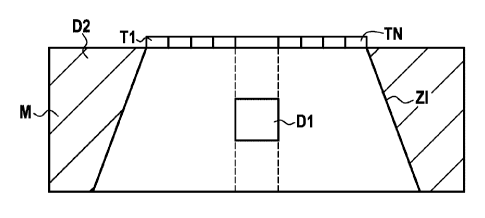
Figure 6 shows diagrammatically the outcome of the
first step of the method, that of acquiring an imaged
area ZI of the medium M to be imaged from which an area
of interest D1 is determined.
This area of interest D1 may be determined
automatically in the device using the method of the
invention by analyzing the image or the successive images
when it is a question of imaging moving structures.
The area of interest D1 may also be determined
manually by an operator selecting an area within the
image area following display of the image area.
The method of the invention then comprises the step
of determining inter-transducer correlation matrices
corresponding to the area of interest and the unwanted
area.
To this end, the method of the invention considers
an array of acoustic transducers Ti to TN of dimension N,
an echographic probe, for example, and a region of
interest D, an area in space that it is required to
image.
X(t)=[xl(t), x2(t) xN(t)li
denotes the vector
composed of the signals acquired by each of the
transducers of the probe at time t.
Of interest are the signals that would be received
by the probe if an infinite number of infinitesimal
sources were to transmit a wave. In the
general case,
R(T) is the correlation matrix of X(t) and is defined as
follows: R(T) = EfX(t)X*(t-I)}.
E{.} represents the mathematical expectation
operator and * represents the conjugate and transposed
operator.
The elements of the matrix R(T) are constituted of
cross correlation terms of the signals acquired at each
of the transducers: Rki(T)= Efxk(t)xe*(t- 1)1.
CA 02826258 2013-08-01
WO 2012/107370 PCT/EP2012/051900
19
In the very special case of the Invention, of
interest are the inter-transducer correlation matrices
obtained with an infinite number of infinitesimal
monochromatic sources distributed in a particular area D.
The variations of Rke(T) as a function of the parameter T
then depend on the geometry of the particular area
chosen.
For the requirements of the invention, time
dependency is eliminated by integration relative to T,
the aim being to calculate the energy at the output of
the spatial filter. There is
determined in this way an
instantaneous inter-transducer correlation matrix Jke that
is not dependent on the times t and T and that is adapted
to the geometry of the area D and to the array of
transducers:
E it ) (t ¨ 1L)))1CITL: (II)
(1)
Note that ha(D) = E It(t) xi* kt)i , i . e. j(D) = EiX()X*(0).
A spatial filter is constituted of a battery of
regular linear and invariant filters, i.e. the output of
each of these filters is obtained from the convolution
product of the input signal of the filter and the impulse
response of the filter.
The convolution product is defined as follows:
+co
7(t) = u(0);-.,(t ¨0)c119, (2)
where v(t) is the output signal, u(t) the input signal,
and h(t) the impulse response of the filter.
A spatial filter on N transducers is constituted of
N vectors hk of dimension M defining the length of the
filters. The spatial filter is written H. Its
response
to a signal vector acquired at the transducers X is as
follows:
y = hk (t) * xiE(t). ( 3 )
k =1
CA 02826258 2013-08-01
WO 2012/107370 PCT/EP2012/051900
In the general case, a space-time filter has two
dimensions: a space dimension (dimension of the sensors),
and a time dimension. The invention relates to the very
5 special case for which the length of the filters M is
equal to 1 because hk(t) does not depend on time and
becomes hk. An intentional limitation to a purely spatial
filter is obtained by weighting the transducers with a
law that does not depend on time. Then H=Ehl, h2,
10 The convolution product of equation (3) then becomes
a product between scalars, and the response at the
output of the spatial filter becomes:
(4)
15 k=1
The energy of the signal at the output of the filter
is, by definition:
8 A E{y2 ct)}
(5)
By injecting equation (4) into equation (5) there is
20 obtained:
S = 21111X(Ci_r(H1 = E{x CAE = *ADM (
6)
The filter H is then searched for, which is a vector
or a weighting or apodization law that maximizes the
ratio p of the energies 81 and 82 corresponding to two
areas D1 and D2, D1 being the area of interest and D2 the
unwanted area (deemed of no utility) of the rest of the
half-space in front of the probe. To this
end, the
gradient of p, denoted ap, is cancelled out.
AT
(7)
¨ (8)
By canceling out ap, there is obtained:
(32)J(D1)R= PH (9)
CA 02826258 2013-08-01
WO 2012/107370 PCT/EP2012/051900
21
In other words, the filter H0pt that maximizes the
ratio of the energies 81 and 82 is the eigenvector of the
matrix MC=j(D2)-1/090, called the characteristic matrix of
the area of interest, associated with its greatest
eigenvalue 0. Moreover, the energy at the output of the
filter is then equal to the eigenvalue that is associated
with it: p = ILk at the output of the filter Hk. The
invention thus comprises, for each defined area of
interest, a step of determining a so-called
characteristic matrix of the area of interest resulting
from the product of the inverse of the inter-transducer
correlation matrix of the unwanted area by the inter-
transducer correlation matrix of the area of interest.
The major idea of the invention consists in
constructing the sequence of imaging firings with
weighting laws on the transducers corresponding to the
eigenvectors of the characteristic matrix of the area of
interest MC=.1(D2)-1J(D1) linked to the highest eigenvalues.
The method of the invention thus comprises the step
of calculating the eigenvectors and eigenvalues of the
characteristic matrix of the area of interest. These
vectors, classified as a function of their corresponding
eigenvalues, define a transmission matrix. To be
more
precise, these eigenvectors are going to constitute the
first K columns of the transmission matrix, and the other
components of this matrix are set to zero, signifying
that no firing is effected, which enables an increase in
the image acquisition rate. By construction, the matrix
constituted by the vectors Hk is invertible. Note however
that it is not necessarily orthogonal.
This method enables N-K firings to be dispensed with
and thus the image acquisition rate to be increased in a
ratio _____________________________________________________________ The loss
of energy G in the area of
N-K
interest is perfectly quantified and has the value:
(10)
Pk
k=K-I-1
CA 02826258 2013-08-01
WO 2012/107370 PCT/EP2012/051900
22
Figure 7 shows an area of interest D1 that is
particularly advantageous. An
example is given for a
linear probe with this kind of definition of the area of
interest Dl.
Here the region of interest D1 is defined by a
circular arc placed in theory at an infinite distance
from the probe (far field) and by an angle 0. The
unwanted area D2 corresponds to the rest of the half-
space in front of the probe.
Under such circumstances, it can be shown that:
MC =.1kAD2rilki(Di) (11)
clsnD
where B= .. , A=-r d is the inter-transducer distance, f
f
is the center frequency of the wave, and c is the speed
of sound in the medium.
The elements Pk of the matrix P with dimensions N X
N are defined by Pm =sin[217(f-k)B]/[ r(f-k)].
The eigenvectors of the matrix P define the prolate
spheroidal sequence of order N and of bandwidth B that is
described in Slepian, D. (1978), "Prolate Spheroidal Wave
Function, Fourier Analysis, and Uncertainty - V: The
Discrete Case", The Bell System Technical Journal. Here,
N is the number of transducers of the probe. This
sequence is a fairly standard signal processing tool,
especially in spectral analysis. The equations (9) and
(11) show that the prolate spheroidal vectors maximize
the ratio of the energy transmitted in the sector [-0; 0]
relative to the energy radiated in the half-space in
front of the probe without the sector [-0; 0]. It has
been shown in Forster, P. & Vezzosi, G. (1987),
"Application of Spheroidal Sequences to Array
Processing", Proceedings IEEE International Conference on
Acoustics, Speech and Signal Processing, that only the
2BN-2 highest eigenvalues of the prolate spheroidal
bases, and thus of the matrix MC defined by equation
(11), are meaningful, i.e. the others are very close to
CA 02826258 2013-08-01
WO 2012/107370 PCT/EP2012/051900
23
zero, and the eigenvectors that are associated with them
contribute only negligible energy in the sector [-0; 0].
In other words 2BN-2 firings are sufficient for an
optimum image.
It has been shown that the prolate spheroidal
vectors furnish an optimum solution for far-field imaging
in an angular sector.
In contrast, ultrasound medical imaging systems do
not produce far-field images but near-field images. Thus
the invention proposes to use the spheroidal bases, the
optimum for far-field imaging, for echographic imaging,
i.e. for near-field imaging. To what
approximation and
under what conditions the spheroidal sequences address
the near-field problem is explained.
Figure 8 shows a near-field area of interest D1 and
the definition of a cone of interest C in which the near-
field approximations of the spheroidal bases are to be
studied.
The situation considered here is that of an area D1
constituting a rectangle situated behind a segment [Mt,
M--] situated at a distance xo from the probe. In this
case, it can be shown that the characteristic matrix MC
has the following expression:
.24:- = == (12)
7 ?,f
in which
E (13)
MC is then a complex matrix. Equation (12) represents MC
in its form of the product of its modulus by a complex
exponential of type MCke=reis% The
terms of MC are thus
entirely defined by their modulus and their phase term y.
At the orders of magnitude of ultrasound echography,
the phase term y is very close to zero. For example, for
a probe with 192 elements, center frequency 5 MHz, inter-
sensor distance 0.3 mm, and a segment [W, MI] placed at a
CA 02826258 2013-08-01
WO 2012/107370 PCT/EP2012/051900
24
distance of at least 5 mm, then the phase y of the
equation (12) is still negligible:
c2 (k = - .1
'= <2.16 1.0-4
The following approximation is therefore perfectly
valid for medical ultrasound imaging:
sirir - .Li
D1
AWN ( 1 4 )
Equation (14) shows that the spheroidal bases
constitute a more than adequate approximation of the
optimum vectors for an area of interest constituting a
segment placed in the near field. The parameter B of the
spheroidal functions then depends on the ratio between
the length of the segment and its distance from the
antenna. Equation
(13) shows clearly that the
characteristic matrix MC remains constant if the ratio
y0/x0 remains constant; in other words, the area of
interest constitutes a cone in front of the probe. The
aperture of this cone depends on the ratio yo/xo. This
cone is defined in equivalent manner by the angle
0 = arcTan(yo/xo).
For a particular example, the method of the
invention therefore determines this matrix MC and
calculates the associated eigenvalues for an angle c1) of
approximately 23 , a probe with 128 elements that have an
inter-transducer distance of 0.3 mm, a center frequency
of 5 MHz, and a speed of sound of 1540 m/s.
Figure 9 is a two-dimensional representation of a
characteristic matrix MC in which the intensities of the
elements (i, j) are represented by a color code.
Figure 10 shows an energy criterion of the eigenvalues
EVP as a function of their index j for the Figure 8
matrix.
It is seen that the eigenvalues having a rank
greater than 50 have values that are virtually zero, the
CA 02826258 2013-08-01
WO 2012/107370 PCT/EP2012/051900
energy 6 radiated by the corresponding eigenvectors in the
sector [-23 ; 23 ] is therefore also zero.
The eigenvectors associated with the non-negligible
eigenvalues are generally then selected. By non-
5 negligible eigenvalues is meant eigenvalues close to 1.
As may be seen in Figure 10, even for relatively wide
sectors of interest, exceeding 2 X 60 , the number of
quasi-zero eigenvalues remains high.
The eigenvectors corresponding to the negligible
10 eigenvalues are not fired, thus enabling the image
acquisition rate to be increased.
Since the energy radiated by these vectors in the
sector of interest corresponds to their eigenvalues, this
energy is also quasi-zero. Thus not using these vectors
15 does not change in any way the final image since they do
not contribute to sounding the imaged area.
Reducing the number of firings while retaining the
complete image size may even be envisaged. The
spheroidal bases enable an image of sufficient quality to
20 be obtained by reducing the number of firings, including
in near-field imaging.
Using the FIELD II software described in Jensen,
J.A. (1996), "Field: A program for simulating ultrasound
systems", 10th Nordic-Baltic Conference on Biomedical
25 Imaging, pp. 351-353, Vol. 34, Supplement 1, Part 1, the
transmitted beams F1, F2, F40, and F100 have been
simulated using the 1st, 2nd, 40th and 100th spheroidal
vectors. The
spatial distributions of the transmitted
acoustic intensity are shown in Figures 11A to 11D,
respectively.
The first beams, i.e. those associated with the
highest eigenvalues, send energy only in the sector of
interest, while the last, associated with the quasi-zero
eigenvalues, send energy only outside the area of
interest.
Effecting the firings corresponding to the quasi-
zero eigenvalues and performing the corresponding
CA 02826258 2013-08-01
WO 2012/107370 PCT/EP2012/051900
26
acquisition does not enhance the image because these
firings send energy outside the area of interest. For
example, it should be observed in Figure 11D that the
transmission effected is of no benefit for the area to be
imaged.
In this example, the improvement in image
acquisition rate is 1.78 without modifying the quality of
the image in the area of interest D1. The
image
acquisition rate increase could of course be greater if
the number of eigenvectors were reduced, but at the cost
of lower image quality.
It is important to note that the number of
eigenvectors necessary for optimum image quality is a
function of the size of the region of interest Dl. In
this example this means the size of the angular sector.
Figure 12 shows the sets of eigenvalues of MC for an
energy criterion EVP as a function of their index j for
values of y in the range 100 to 70 using the same
parameters as before, same frequency, etc.
It is thus apparent that the number of zero
eigenvalues decreases as the size of the sector
increases.
It should be noted that spheroidal bases constitute
a special case and the general method consists in
maximizing the ratio of the energy in the area of
interest to the energy in the unwanted area by taking the
first eigenvectors of the matrix MC that correspond to
the area of interest D1, to the area D2, and to the
geometry of the probe.
Moreover, the example given here relates to two-
dimensional imaging, but the method also applies to
three-dimensional imaging.
Figure 13 shows a second example of a characteristic
matrix MC for an area of interest with an angular
aperture of 60 . Figure 14
shows the associated
eigenvalues. It is seen that a greater number of firings
must be effected for a similar result from the point of
CA 02826258 2013-08-01
WO 2012/107370 PCT/EP2012/051900
27
view of image quality. An application example has been
used to evaluate the performance of the method of the
invention.
The probe used is a linear probe with Net = 128
elements functioning at 5 MHz. The aperture of the probe
elements is 30 . The
spheroidal matrix adopted is the
Figure 13 matrix, of size 128 and with the parameter B
defined as follows:
B= dsinco
A
where d is the inter-sensor distance 0.3 mm, A is the
wavelength of 0.3 mm, and cot, is the aperture half-angle of
the area of interest, here 15 .
Here B = 0.259. The parameters B and Net suffice to
dimension the flattened spheroidal matrix. The number of
non-redundant firings, i.e. firings corresponding to the
number of meaningful eigenvalues, for such a
configuration is given by the equation:
Nsph =2NefB-2
Here Nsph = 64.
Note here that the method therefore enables imaging
of the whole of the area of interest with an optimum
quality for half the number of firings compared to the
mode B method, which is optimum only around the focal
distance.
Each of the Nsrp firings is transmitted into the
medium, i.e. the same temporal burst is applied to the
various elements but weighted on the ith firing of the
sequence by the ith column vector of the transmission
matrix, constituted for a first part by the eigenvectors
of the characteristic matrix of the area of interest
shown in Figure 13 and for a second part of zero vectors.
Each acquisition is stored in a 3D signal matrix in
which the rows are the temporal samples, the columns are
the sensors, and the depths are the firings, so that Ciik
corresponds to the sample acquired at the time i*Fe,
CA 02826258 2013-08-01
WO 2012/107370 PCT/EP2012/051900
28
where F, is the sampling frequency of the system at the
,th
J sensor for the kth acquisition.
At the end of the acquisition process, the signal
matrix is then a matrix/tensor of dimension Nssmpis*Nee*Nsph.
It is concatenated with the zero matrix of dimension
N,õlide*Nee*(Nee-N) to form the complete signal matrix of
dimension Nsample *Nei *Nee = The
complete data set is then
reconstructed. For
this, each slice, corresponding to a
given time, constituting a matrix of dimension Nee*Nee is
then leftward multiplied by the inverse of the
transmission matrix, which is constant in time, in order
to reconstruct the complete data set.
Each slice of the complete data set in the
capture/time plane is then reconstructed by application
of the corresponding delay laws to form one of the Ne/
"low resolution" images. These
are then summed
coherently to obtain the high-resolution image.
Note that each acquisition provides information on
only a limited area of the image. The Nsph
usable
spheroidal vectors have precisely the property of having
orthogonal spatial spectra. They are furthermore ordered
spatially. The
vector Ni provides information on the
angular sectors [0i, el] and [-0111, -01] for the far
field. For the near field it provides information on the
widths [Xi, Xid] and [-Xid, -Xi]. The transmission of the
first Ni vectors (Ni < Nn) leads to a reduction in the
size of the image and a further improvement in the image
acquisition rate N,pljNi. It is thus seen that the image
acquisition rate is variable.
The invention enables the image acquisition rate to
be adjusted by a simple command to reduce the size of the
image obtained and thus by simple definition of an area
of interest size. This
adjustment of the image
acquisition rate may be effected manually by the operator
or automatically by a movement detection and image
acquisition rate adjustment algorithm.
CA 02826258 2013-08-01
WO 2012/107370 PCT/EP2012/051900
29
Figures 15A to 15D show, in negative, the results
obtained for the first 4, 10, 20 firings and for all of
the firings of the Figure 13 spheroidal matrix,
respectively. It is
seen that provided that the point
target is centered in the imaged area, the resolution is
virtually not degraded. The
signal-to-clutter ratio,
which corresponds to the quality of the contrast in the
anechoic areas, does not vary either. Only the
size of
the image is reduced.
The table below sets out experimental results
obtained from in vitro experiments.
Sph Full Sph 60 Sph 30 Sph 20 Sph 10 Sph 4
Lateral 1.62 mm 1.62 mm 1.68 mm 2.02
resolution
Axial 0.65 mm 0.66 mm 0.67 mm 0.74
resolution
Number of 128 60 30 20 10 4
firings
CTR in dB -29.5 -29.5 -28.3 -27.5 -27.4 -28.4
Width of 40 mm 40 mm 40 mm 40 mm 30
mm 20 mm
imaged
area
With the invention, in contrast to mode B, the
quality of the image is constant for all depths.
Figure 16A shows the contrast C as a function of the
lightening in terms of the number of firings AT (inverted
abscissa axis) for the application of two types of
matrix: Hadamard matrices used for spatial coding and
characteristic matrices of an area of interest as
determined in accordance with the invention. It is seen
that, even for a very low number of firings, the
invention enables very good contrast to be obtained.
Figure 16B shows the variation of the size of the
image as a function of the number of firings obtained
with the invention. It is seen
that the size of the
image achieves its optimum level from 20 firings.
CA 02826258 2013-08-01
WO 2012/107370 PCT/EP2012/051900
It is possible to use the method of the invention to
acquire an entire imaged area over which an area of
interest is obtained that is imaged with high quality and
an area external to the area of interest is obtained that
5 is imaged with low quality.
In one particular implementation, effecting multi-
image acquisition rate acquisitions is also envisaged.
Thus as shown in Figure 17, the imaging area is then
divided into two areas, the first called the high image
10 acquisition rate area FCC and the second called the
background area FAC. Two
transmission laws are then
chosen and specifically adapted to imaging each of the
areas. For example a first transmission law is then
activated every 10 images and is used to acquire the
15 entire imaging area, and the next 9 images are acquired
with the transmission laws of the invention.
Another implementation consists in effecting
acquisitions with high image acquisition rate for the two
areas, but with different image qualities, by giving
20 preference to the area of interest. The area of interest
is sounded in accordance with the principle of the
present invention, the remainder of the image being
sounded in accordance with the principles of high image
acquisition rate with degraded image quality. The high
25 image acquisition rate and degraded image quality
transmission laws may be: imaging using unfocused waves
as shown in Figure 5C, multi-beam imaging as shown in
Figure 5B, multi-line imaging as shown in Figure 5A, or
synthetic aperture imaging with depointed plane waves as
30 described in document US 2003/0125628. The
number of
unfocused waves transmitted is then relatively small to
satisfy the high-image acquisition rate criterion, which
is possible only to the detriment of image quality. Two
transmission sequences alternate: acquisition of the area
of interest with the transmission laws of the invention,
and acquisition of the remainder of the image with
CA 02826258 2013-08-01
WO 2012/107370 PCT/EP2012/051900
31
transmission laws corresponding to the high-image
acquisition rate mode with low-image quality.
Note finally that various implementations may be
arrived at conforming to the principles of the invention.