Note: Descriptions are shown in the official language in which they were submitted.
CA 02826453 2013-08-01
USE OF AN ANTI-CD200 ANTIBODY FOR PROLONGING THE
SURVIVAL OF ALLOGRAFTS
Technical Field
The field of the invention is medicine, immunology, molecular biology, and
protein chemistry.
Background
Transplantation of cells, tissues, and organs has become very common and is
often
a life-saving procedure. Organ transplantation is the preferred treatment for
most
patients with chronic organ failure. Despite great improvement in treatinents
to inhibit
rejection, however, rejection continues to be the single largest impediment to
successful
organ transplantation. Rejection includes not only acute rejection but also
chronic
rejection. One-year survival rates for transplanted kidneys average 88.3% with
kidneys
from deceased donors and 94A% with kidneys received from living donors.
The corresponding five year survival rates for the transplanted kidneys are
63.3%
and 76.5%. [OPTN/SRTR Annual Report (2002) Chapter 1 of the Annual Report
produced by the Scientific Registry of Transplant Recipients (SRTR) in
collaboration with
the Organ Procurement and Transplantation Network (OPTN).1 The one year
survival
rates are 80.2% and 76.5% for livers from deceased and living donors,
respectively. The
corresponding five year liver graft survival rates are 63.5% and 73.0%
(OPTN/SRTR
Annual Report, 2002). The use of immunosuppressant drugs, e.g., cyclosporine A
and
more recently tacrolimus, has dramatically improved the success rate of organ
transplantation especially by preventing acute rejection. But as the numbers
above show,
there is still a need to improve the success rates, both short-term and
especially long-term.
For example, as seen from the above nutnbers
CA 02826453 2013-08-01
WO 2012/106634
PCT/US2012/023831
for kidney and liver transplants, the five year failure rates for these
transplanted
organs are on the order of 25-35%.
In the year 2001 alone there were more than 23,000 patients who received an
organ transplant of which approximately 19,000 were kidney or liver (OPTN/SRTR
Annual Report, 2002). For this one year of transplants alone, with present
techniques
it can be expected that approximately 5,000-6,000 of these transplanted
kidneys and
livers will fail within five years. These numbers do not even include other
transplanted organs or transplanted tissues or cells such as bone marrow.
There are multiple types of transplants. These are described in Abbas et al.
(2000) Cell Mol Immunol (4th edition), pages 363-383 (W.B. Saunders Company,
New York). A graft transplanted from one individual to the same individual is
called
an autologous graft or autograft. A graft transplanted between two genetically
identical or syngeneic individuals is called a syngeneic graft. A graft
transplanted
between two genetically different individuals of the same species is called an
allogeneic graft or
allograft. A graft transplanted between individuals of different species is
called a
xenogeneic graft or xenograft. The molecules that are recognized as foreign on
allografts are called alloantigens and those on xenografts are called
xenoantigens.
The lymphocytes or antibodies that react with alloantigens or xenoantigens are
described as being alloreactive or xenoreactive, respectively.
Currently more than 40,000 kidney, heart, lung, liver and pancreas transplants
are performed in the United States each year (Abbas et al., 2000). Other
possible
transplants
include, but are not limited to, vascular tissue, eye, cornea, lens, skin,
bone marrow,
muscle, connective tissue, gastrointestinal tissue, nervous tissue, bone, stem
cells,
islets,
cartilage, hepatocytes, and hematopoietic cells. Unfortunately, there are many
more
candidates for a transplant than there are donors. In view of the foregoing
number of
transplants needed and the limitations of existing therapies, it is clear that
new,
therapeutically efficacious methods for prolonging the survival of allografts
are
needed.
2
CA 02826453 2013-08-01
WO 2012/106634
PCT/US2012/023831
Summary
The present disclosure relates to compositions and methods useful for
modulating an immune response in a mammal. As elaborated on in the description
and exemplified in the working examples, the inventors have discovered that an
anti-
CD200 antibody is therapeutically effective as a single-agent therapy (such
therapy is
also referred to herein as a "monotherapy") to substantially prolong the
survival of a
renal allograft in a recipient mammal. The benefits of this discovery to
transplant
recipients are numerous. For example, use of an anti-CD200 antibody as a
monotherapy can improve the quality of life for a renal allograft recipient,
as allograft
rejection is generally treated with one or more immunosuppressive agents, many
of
which either alone or in combination can result in serious side-effects such
as
alopecia, bone marrow depletion, gastrointestinal upset, pruritis,
thrombocytopenia,
anemia, nephrotoxicity, pancreatitis, and infection. Even within narrow
therapeutic
dose ranges, immunosuppressive agents (e.g., calcineurin inhibitors such as
cyclosporine A (CsA) and FK-506) can be, for example, extremely nephrotoxic.
Calne et al. (1978) Lancet 2:1323-1327 and Gaston (2009) Clin J Am Soc Nephrol
4(12):2029-2034. Treatment with subtherapeutic dosages of CsA or FK-506
results in
significantly lower risk of nephrotoxicity, but with a significant reduction
in
therapeutic benefit with respect to graft survival. See, e.g., Seron and
Moreso (2004)
Transplant Proc 36:257S. Given the limitations and side effects attendant to
calcineurin therapies, for example, it is clearly of great value to identify
new
compounds capable of reducing the requirement of these inhibitors (whether in
dose
level or length of treatment) while maintaining a high level of therapeutic
efficacy
with respect to prolonging graft survival. The disclosure demonstrates that an
anti-
CD200 antibody is such a compound.
The ability to prolong renal allograft survival using an anti-CD200 antibody,
in the absence of one or more additional immunosuppressive agents, offers
renal
allograft recipients the same or even greater therapeutic effect without many
of the
debilitating side-effects associated with immunosuppressive agent therapy
(e.g.,
combination therapy). Moreover, the one or more additional immunosuppressive
agents often must be administered to the patient chronically or, perhaps,
indefinitely
in order to maintain graft survival. As is clear from the disclosure and
exemplified in
the working examples, an anti-CD200 antibody monotherapy can, in some
3
CA 02826453 2013-08-01
WO 2012/106634
PCT/US2012/023831
embodiments, be administered for seven to fourteen days after transplantation
and yet
still achieve long-term survival of the grafts even without need for further
immunosuppressive therapy.
Notwithstanding the efficacy of anti-CD200 antibody monotherapy, the anti-
CD200 antibodies described herein are also useful as a therapeutic platform ¨
offering
flexible, alternative therapeutic options for transplant patients. For
example, the
inventors have discovered that therapeutic administration of an anti-CD200
antibody
to an allograft-bearing mammal can allow for early withdrawal (and/or a
reduced dose
amount) of one or more additional immunosuppressive agents being administered
to
the mammal, yet still maintain therapeutic efficacy. As described in the
working
examples, administration of an anti-CD200 antibody to an allograft organ-
bearing
mammal allows for one or both of an early withdrawal and a lower dosage of a
concurrent calcineurin inhibitor therapy, yet still maintain therapeutic
efficacy in
prolonging the survival of the allograft. In another example, mycophenolate-
free or ¨
reduced therapeutic options are also provided herein.
The inventors also discovered that subcutaneous administration ¨ or a more
localized or depot delivery ¨ of an anti-CD200 antibody to a mammal can
prolong the
survival of an allograft organ as effectively as systemic delivery of the
antibody. As
exemplified in the working examples, subcutaneous administration of an anti-
CD200
antibody as a monotherapy can substantially prolong the survival of a renal
allograft
in recipient mammals as well as intravenous delivery of the antibody. The
examples
also provide the results of experiments in which subcutaneous administration
of an
anti-CD200 antibody, in combination with one or more additional
immunosuppressive
agents, can prolong the survival of allograft organs such as a heart. Many
benefits are
attendant to subcutaneous or depot delivery of an anti-CD200 antibody. For
example,
for therapeutic applications that require frequent and/or chronic
administration,
subcutaneous or depot delivery can allow for fewer administrations of the
therapeutic
overall (with a higher concentration of the therapeutic to be deposited at
each interval
slowly releasing the compound to the mammal). Secondly, subcutaneous (or
depot)
delivery, along with systemic forms of delivery, of an anti-CD200 antibody
provides
more patient choice regarding how and when the therapeutic is administered.
For
example, in some embodiments, it can be possible for a patient to self-
administer an
anti-CD200 antibody, avoiding the need, for example, to travel to a hospital
for such
medication or arrange for an in-home nurse visit, which can be both costly and
4
CA 02826453 2013-08-01
WO 2012/106634
PCT/US2012/023831
inconvenient. Therefore, increased patient choice ultimately manifests an
increased
patient compliance by providing an easy self-administration alternative for
patients
bearing an allograft.
To this end, the disclosure provides aqueous solutions comprising an anti-
CD200 antibody, and therapeutic kits containing the solutions, for use in
applications
in which subcutaneous administration of the antibody would be beneficial. The
solutions can contain the anti-CD200 antibody at a concentration of at least
10
mg/mL.
Accordingly, in one aspect, the disclosure features a method for prolonging
the survival of a renal allograft. The method comprises administering to a
recipient
mammal in need thereof an anti-CD200 antibody as a single agent (a
monotherapy) in
an amount effective to prolong the survival of a renal allograft in the
recipient
mammal. In some embodiments, the method can also include transplanting the
renal
allograft into the recipient mammal. In some embodiments, the methods can
further
comprise, prior to removal from the donor mammal from which the renal
allograft
was obtained, administering an anti-CD200 antibody to the donor mammal.
In some embodiments, the anti-CD200 antibody is administered to the
recipient mammal for at least seven (e.g., at least eight, nine, 10, 11, 12,
13, 14, 15,
16, 17, 18, 19, 20, 21, 22, 23, 24, 25, 26, 27, 28, 29, 30, 31) days following
transplantation of the renal allograft into the recipient mammal. In some
embodiments, the anti-CD200 antibody is administered at least once per day for
up to
seven (e.g., up to eight, nine, 10, 11, 12, 13, 14, 15, 16, 17, 18, 19, 20,
21, 22, 23, 24,
25, 26, 27, 28, 29, 30, 31, 32, 33, 34, 35, 36, 37, 38, 39, 40, 41, 42, 43,
44, 45, 46, 47,
48, 49, or 50) days following transplantation of the renal allograft into the
recipient
mammal. In some embodiments, the anti-CD200 antibody is administered at least
once per day for at least seven, but less than 30 (e.g., less than 29, 28, 27,
26, 25, 24,
23, 22, 21, 20, 19, 18, 17, 16, 15, 14, 13, 12, 11, 10, 9, or 8) days
following
transplantation of the renal allograft into the recipient mammal. In some
embodiments, the anti-CD200 antibody can be administered in a dose large
enough to
remain effective for at least two (e.g., at least two, three, four, five, six,
seven, eight,
nine, ten, 11, 12, 13, or 14) days following transplantation of an allograft
to the
recipient mammal, with the antibody being administered as often as necessary
to
maintain an effective dose (e.g., a single dose may be large enough to remain
effective for 14 days, in which event only a single dose would be required
once every
5
CA 02826453 2013-08-01
WO 2012/106634
PCT/US2012/023831
14 days or only once if an effective amount of the antibody is required for
only 14
days). In some embodiments, an effective amount of the anti-CD200 antibody is
maintained in the recipient mammal for at least seven (e.g., at least eight,
nine, 10, 11,
12, 13, 14, 15, 16, 17, 18, 19, 20, 21, 22, 23, 24, 25, 26, 27, or 28 or more)
days. As
noted above, it is understood that a single dose of the anti-CD200 antibody
can be
sufficient to maintain an effective amount of the anti-CD200 antibody in the
mammal
for at least seven (e.g., at least eight, nine, 10, 11, 12, 13, 14, 15, 16,
17, 18, 19,20,
21, 22, 23, 24, 25, 26, 27, or 28 or more) days.
Though the particular dosing schedule (e.g., amount, frequency, and/or
interval) employed may vary from patient to patient, an anti-CD200 antibody
described herein can be administered to a mammal (e.g., a patient) in need
thereof
under such a regimen so as to maintain an effective amount of the antibody in
the
mammal for at least seven (e.g., at least eight, nine, 10, 11, 12, 13, 14, 15,
16, 17, 18,
19, 20, 21, 22, 23, 24, 25, 26, 27, 28, 29, 30, 31) days following
transplantation of the
renal allograft into the recipient mammal.
In some embodiments, the anti-CD200 antibody is administered to the
recipient mammal prior to, and following, transplantation of the renal
allograft into
the recipient mammal. For example, the anti-CD200 antibody can be administered
to
the recipient mammal for at least one week prior to transplantation of the
renal
allograft into the recipient mammal. In some embodiments, at least two (e.g.,
at least
three, four, five, six, seven, eight, nine, or even 10 or more) doses of the
anti-CD200
antibody are administered to the recipient mammal prior to transplantation of
the renal
allograft into the recipient mammal.
In some embodiments, the renal allograft is fully MHC mismatched with
respect to the recipient mammal. In some embodiments, the recipient mammal is
presensitized to the renal allograft. In some embodiments, the renal allograft
is an
ABO-mismatch with respect to the recipient mammal.
In some embodiments, the anti-CD200 antibody is intravenously administered
to the recipient mammal. In some embodiments, the anti-CD200 antibody is
subcutaneously administered to the recipient mammal. In some embodiments, the
anti-CD200 antibody is intramuscularly administered to the recipient mammal.
In some embodiments, administration of the anti-CD200 antibody results in
renal allograft survival for at least 100 days. In some embodiments,
administration of
the anti-CD200 antibody results in a renal allograft survival of at least six
months
6
CA 02826453 2013-08-01
WO 2012/106634
PCT/US2012/023831
(e.g., seven months, eight months, nine months, 10 months, 11 months, 12
months, 16
months, 18 months, 20 months, or 24 months or more). In some embodiments,
administration of the anti-CD200 antibody results in long term renal allograft
survival.
In some embodiments, the recipient mammal and the renal allograft donor are
human.
In another aspect, the disclosure features a method for prolonging the
survival
of an allograft organ in a recipient mammal, which method comprises
administering
to an allograft organ recipient in need thereof: (a) one or more
immunosuppressive
agents; and (b) an anti-CD200 antibody, to thereby prolong the survival of the
graft in
the patient. In some embodiments, administration of the anti-CD200 antibody
allows
for a shorter duration of treatment with at least one of the one or more
immunosuppressive agents, relative to the duration of treatment with the at
least one
immunosuppressive agent in the absence of the anti-CD200 antibody. In some
embodiments, administration of the anti-CD200 antibody allows for a reduced
dose
level or amount requirement for at least one of the one or more
immunosuppressive
agents, relative to the dose level or amount of the at least one
immunosuppressive
agent in the absence of the anti-CD200 antibody.
In some embodiments, at least one of the immunosuppressive agents can be an
IL-2 inhibitor. For example, in some embodiments, at least one of the
immunosuppressive agents is an mTOR inhibitor such as rapamycin. In some
embodiments, at least one of the immunosuppressive agents is a calcineurin
inhibitor
such as cyclosporine A or FK-506.
In some embodiments, administration of the anti-CD200 antibody to the
recipient mammal shortens the duration of treatment with at least one
immunosuppressive agent by at least 20%. In some embodiments, administration
of
the anti-CD200 antibody to the recipient mammal shortens the duration of
treatment
with at least one immunosuppressive agent by at least 50%.
In some embodiments, the methods described herein provide an alternative
therapeutic strategy for patients sensitive to mycophenolate therapy, e.g.,
MMF
therapy. In such embodiments, the specification provides a mycophenolate-free
alternative that includes administering to the patient an anti-CD200 antibody
and a
calcineurin inhibitor (e.g., cyclosporine A or tacrolimus), e.g., wherein the
inhibitor is
administered in an amount and/or a frequency that is less than the
corresponding
7
CA 02826453 2013-08-01
WO 2012/106634
PCT/US2012/023831
amount or frequency of the calcineurin inhibitor required to treat the patient
in the
absence of the anti-CD200 antibody therapy.
In some embodiments, e.g., where a patient is sensitive to calcineurin
inhibitors, the methods described herein provide calcineurin inhibitor-free
alternative
options for patients in which an anti-CD200 antibody is administered to the
patient in
conjunction with a mycophenolate containing compound (e.g., MMF). The
mycophenolate compound can be administered to the patient in an amount and/or
at a
frequency that is less than the amount or frequency of the compound required
to treat
the patient in the absence of the anti-CD200 antibody therapy.
In another aspect, the disclosure features a method for prolonging the
survival
of an allograft in a recipient mammal, the method comprising chronically
administering to the mammal (e.g., a human): (a) an anti-CD200 antibody
described
herein and (b) a mycophenolate-containing compound (e.g., MMF) to thereby
prolong
the survival of the allograft in the mammal. In some embodiments, the anti-
CD200
antibody and/or mycophenolate-containing compound is chronically administered
for
at least seven days. In some embodiments, the anti-CD200 antibody or
mycophenolate-containing compound is chronically administered for at least 14
days.
In some embodiments, chronic administration of the anti-CD200 antibody allows
for a
reduced amount and/or frequency of administration of the mycophenolate-
containing
compound required to maintain an effective amount in the mammal, as compared
to
the amount and/or frequency of the compound required to maintain an effective
amount in the absence of the antibody.
In another aspect, the disclosure features a method for prolonging the
survival
of an allograft in a recipient mammal, the method comprising chronically
administering to the mammal (e.g., a human): (a) an anti-CD200 antibody
described
herein and (b) an IL-2 inhibitor (e.g., a calcineurin inhibitor such as
cyclosporine A)
to thereby prolong the survival of the allograft in the mammal. In some
embodiments,
the anti-CD200 antibody and/or IL-2 inhibitor is chronically administered for
at least
seven days. In some embodiments, the anti-CD200 antibody or IL-2 inhibitor is
chronically administered for at least 14 days. In some embodiments, chronic
administration of the anti-CD200 antibody allows for a reduced amount and/or
frequency of administration of the IL-2 inhibitor required to maintain an
effective
amount in the mammal, as compared to the amount and/or frequency of the
inhibitor
required to maintain an effective amount in the absence of the antibody.
8
CA 02826453 2013-08-01
WO 2012/106634
PCT/US2012/023831
In another aspect, the disclosure features a method for prolonging the
survival
of an allograft in a recipient mammal, wherein the method comprises: after
(and,
optionally prior to and/or during) transplantation of the allograft,
administering to the
recipient mammal: (a) an anti-CD200 antibody and (b) one or more additional
immunosuppressive agents, wherein the one or more additional immunosuppressive
agents include a mycophenolate compound (e.g., MMF) and an IL-2 inhibitor
(such as
a calcineurin inhibitor, e.g., cyclosporine A) and wherein one or more of the
additional immunosuppressive agents are administered in a lower dose and/or
less
frequently than the dose or frequency required for equivalent therapeutic
efficacy in
the absence of the anti-CD200 antibody. An equivalent therapeutic efficacy can
be,
e.g., the standard or historical efficacy observed in a patient population
administered
the one or more additional immunosuppressive agents in the absence of a
concomitant
anti-CD200 antibody therapy.
It is understood that in combination therapies described herein including an
anti-CD200 antibody and one or more immunosuppressants, "one or more
immunosuppressive agents" can be used interchangeably with the term "one or
more
additional immunosuppressive agents".
In yet another aspect, the disclosure features a method for prolonging the
survival of an allograft organ in a recipient mammal, which method comprises
administering to an allograft organ recipient mammal in need thereof: (a) one
or more
immunosuppressive agents; and (b) an anti-CD200 antibody, to thereby prolong
the
survival of the graft in the mammal, wherein the anti-CD200 antibody is
subcutaneously administered to the recipient mammal or intravenously
administered
to the recipient mammal.
In some embodiments, administration of the anti-CD200 antibody allows a
shorter duration of treatment with at least one of the one or more
immunosuppressive
agents, relative to the duration of treatment with the at least one
immunosuppressive
agent in the absence of the anti-CD200 antibody. In some embodiments,
administration of the anti-CD200 antibody allows for a reduced dose level or
amount
requirement for at least one of the one or more immunosuppressive agents,
relative to
the dose level or amount of the at least one immunosuppressive agent in the
absence
of the anti-CD200 antibody.
In some embodiments of any of the methods described herein, the anti-CD200
antibody is subcutaneously administered to the recipient mammal. In some
9
CA 02826453 2013-08-01
WO 2012/106634
PCT/US2012/023831
embodiments of any of the methods described herein, the anti-CD200 antibody is
intravenously administered to the recipient mammal.
In some embodiments of any of the methods described herein, the methods
can further comprise, prior to removal from the donor mammal from which the
allograft organ was obtained, administering an anti-CD200 antibody to the
donor
mammal.
In some embodiments, the allograft is fully MHC mismatched with respect to
the recipient mammal. In some embodiments, the recipient mammal is
presensitized
to the allograft. In some embodiments, the allograft is an ABO-mismatch with
respect to the recipient mammal.
In some embodiments, at least one of the one or more immunosuppressive
agents is selected from the group consisting of adriamycin, azathioprine,
busulfan,
cyclophosphamide, fludarabine, 5-fluorouracil, methotrexate, mycophenolate
mofetil,
mycophenolate sodium, a non-steroidal anti-inflammatory drug, and an IL-2
inhibitor
(e.g., an mTOR inhibitor such as rapamycin) or a calcineurin inhibitor such as
FK-
506 or cyclosporine A).
In some embodiments, two or more immunosuppressive agents are
administered to the recipient mammal. In some embodiments, at least two of the
two
or more immunosuppressive agents are cyclosporine A and cyclophosphamide, FK-
506 and cyclophosphamide, or a calcineurin inhibitor (cyclosporine A or FK-
506) and
a mycophenolate compound (e.g., mycophenolate mofetil or mycophenolate
sodium).
In yet another aspect, the disclosure features a method for transplanting an
allograft organ into a recipient mammal. The method comprises: (a) prior to
transplantation of an allograft organ into a recipient mammal, administering
an anti-
CD200 antibody to the recipient mammal; (b) transplanting the allograft organ
into
the recipient mammal; and (c) administering an anti-CD200 antibody to the
recipient
mammal following transplantation of the allograft organ.
In some embodiments, the anti-CD200 antibody is subcutaneously or
intravenously administered to the recipient mammal. In some embodiments, the
anti-
CD200 antibody is administered as a single-agent therapy (a monotherapy).
In some embodiments, the methods can include, prior to removal from the
donor mammal from which the allograft organ was obtained, administering an
anti-
CD200 antibody to the donor mammal.
CA 02826453 2013-08-01
WO 2012/106634
PCT/US2012/023831
In some embodiments, the allograft is fully MHC mismatched with respect to
the recipient mammal. In some embodiments, the recipient mammal is
presensitized
to the allograft. In some embodiments, the allograft is an ABO-mismatch with
respect to the recipient mammal.
In some embodiments, the methods can include administering to the recipient
mammal one or more immunosuppressive agents such as any of the
immunosuppressive agents described herein. For example, at least one of the
one or
more immunosuppressive agents is selected from the group consisting of
adriamycin,
azathioprine, busulfan, cyclophosphamide, fludarabine, 5-fluorouracil,
methotrexate,
mycophenolate mofetil, mycophenolate sodium, a non-steroidal anti-inflammatory
drug, and an IL-2 inhibitor (e.g., an mTOR inhibitor such as rapamycin) or a
calcineurin inhibitor such as FK-506 or cyclosporine A).
In some embodiments, two or more immunosuppressive agents are
administered to the recipient mammal. In some embodiments, at least two of the
two
or more immunosuppressive agents are cyclosporine A and cyclophosphamide, FK-
506 and cyclophosphamide, or a calcineurin inhibitor (cyclosporine A or FK-
506) and
a mycophenolate compound (e.g., mycophenolate mofetil or mycophenolate
sodium).
In some embodiments, administration of the anti-CD200 antibody allows a
shorter duration of treatment with at least one of the one or more
immunosuppressive
agents, relative to the duration of treatment with the at least one
immunosuppressive
agent in the absence of the anti-CD200 antibody. In some embodiments,
administration of the anti-CD200 antibody allows for a reduced dose level or
amount
requirement for at least one of the one or more immunosuppressive agents,
relative to
the dose level or amount of the at least one immunosuppressive agent in the
absence
of the anti-CD200 antibody.
In some embodiments of any of the methods described herein, the allograft
organ is selected from the group consisting of a kidney, a lung, a heart, a
pancreas,
vascular tissue, a liver or one or more lobes thereof, skin, an eye,
gastrointestinal
tissue, nervous tissue, muscle tissue, bone or cartilage, bone marrow,
connective
tissue, red blood cells, islet cells, a cornea, and a lens from an eye. The
allograft
organ is, in some embodiments, a heart or a kidney.
In some embodiments, the anti-CD200 antibody is administered to the
recipient mammal for at least seven (e.g., at least eight, nine, 10, 11, 12,
13, 14, 15,
16, 17, 18, 19, 20, 21, 22, 23, 24, 25, 26, 27, 28, 29, 30, 31) days following
11
CA 02826453 2013-08-01
WO 2012/106634
PCT/US2012/023831
transplantation of the allograft into the recipient mammal. In some
embodiments, the
anti-CD200 antibody is administered at least once per day for up to seven
(e.g., up to
eight, nine, 10, 11, 12, 13, 14, 15, 16, 17, 18, 19, 20, 21, 22, 23, 24, 25,
26, 27, 28, 29,
30, 31, 32, 33, 34, 35, 36, 37, 38, 39, 40, 41, 42, 43, 44, 45, 46, 47, 48,
49, or 50) days
following transplantation of the allograft into the recipient mammal. In some
embodiments, the anti-CD200 antibody is administered at least once per day for
at
least seven, but less than 30 (e.g., less than 29, 28, 27, 26, 25, 24, 23, 22,
21, 20, 19,
18, 17, 16, 15, 14, 13, 12, 11, 10, 9, or 8) days following transplantation of
the
allograft into the recipient mammal. In some embodiments of any of the methods
described herein, anti-CD200 antibody is administered to the recipient mammal
once
every two days. In some embodiments of any of the methods described herein,
the
antibody can be administered at least once a week. In some embodiments of any
of
the methods described herein, the antibody can be administered at least once
every
two weeks (e.g., at least once every 12, 13, 14, 15, or 16 days).
In some embodiments of any of the methods described herein, at least one of
the one or more immunosuppressive agents is chronically administered to the
recipient mammal.
In some embodiments of any of the methods described herein, the anti-CD200
antibody inhibits the interaction between CD200 and CD200 receptor.
In some embodiments of any of the methods described herein, the anti-CD200
antibody comprises a variant heavy chain constant region that has reduced
effector
function, as compared to the corresponding non-variant form of the heavy chain
constant region.
In some embodiments of any of the methods described herein, the anti-CD200
antibody is a whole antibody. In some embodiments of any of the methods
described
herein, the anti-CD200 antibody is a human antibody, a humanized antibody, a
chimeric antibody, a rodent antibody, a deimmunized antibody, or a primatized
antibody.
In some embodiments of any of the methods described herein, the anti-CD200
antibody is a CD200-binding fragment of a whole anti-CD200 antibody. The CD200-
binding fragment can be one selected from the group consisting of a single-
chain
antibody, an Fab, an Fab', an F(ab)'2, an F(ab')3, an Fv, an Fd, a minibody, a
diabody,
and a single domain antibody. In some embodiments of any of the methods
described
herein, the anti-CD200 antibody is samalizumab.
12
CA 02826453 2013-08-01
WO 2012/106634
PCT/US2012/023831
In some embodiments of any of the methods described herein, the recipient
mammal is a human and the allograft organ is obtained from a human.
In yet another aspect, the disclosure features a method for prolonging the
survival of an allograft organ in a recipient mammal, which method comprises
administering to a recipient mammal bearing an allograft organ an anti-CD200
antibody in an amount and with a frequency sufficient to produce and maintain
in the
recipient mammal the occurrence of a desired immunomodulatory effect and thus
prolong the survival of the allograft organ in the recipient mammal.
In another aspect, the disclosure features a method for prolonging the
survival
of an allograft in a recipient mammal, which method comprises: determining the
relative dose amounts of (i) an anti-CD200 antibody effective to produce a
desired
immunomodulatory effect in a recipient mammal bearing an allograft organ; and
administering to the recipient mammal the relative dose amount of the anti-
CD200
antibody with a frequency sufficient to maintain in the recipient mammal the
desired
immunomodulatory effect.
In yet another aspect, the disclosure features a method for prolonging the
survival of an allograft organ in a recipient mammal, which method comprises
administering to a recipient mammal bearing an allograft organ: (a) an anti-
CD200
antibody and (b) one or more immunosuppressive agents, wherein the antibody
and
one or more immunosuppressive agents are administered in an amount and with a
frequency sufficient to produce and maintain in the recipient mammal the
occurrence
of a desired immunomodulatory effect and thus prolong the survival of the
allograft
organ in the recipient mammal.
In another aspect, the disclosure features a method for prolonging the
survival
of an allograft in a recipient mammal, which method comprises: determining the
relative dose amounts of (i) an anti-CD200 antibody and (ii) one or more
immunosuppressive agents, effective to produce a desired immunomodulatory
effect
in a recipient mammal bearing an allograft organ; and administering to the
recipient
mammal the relative dose amounts of the anti-CD200 antibody and one or more
immunosuppressive agents with a frequency sufficient to maintain in the
recipient
mammal the desired immunomodulatory effect.
As detailed in the working examples, the inventors discovered that
administration of an anti-CD200 antibody to transplant recipient mammals
reduces
the expression of SHIP ( SH2-containing Enositol 51-e.bosphatase) by
splenocytes in
13
CA 02826453 2013-08-01
WO 2012/106634
PCT/US2012/023831
the mammals. SHIP is an intracellular phosphatase that, upon stimulation by
PI3-
kinase, represses the proliferation, survival, and activation of hematopoietic
cells.
Lioubin et al. (1996) Mol Cell Biol 14:5682-5691 and Liu et al. (1997) J Biol
Chem
272:8983-8988.
SHIP-deficient mice reportedly exhibit an increased number of monocytes and
macrophages, their hematopoietic progenitors having enhanced survival,
proliferation,
and differentiation. In addition, SHIP-deficient mice also fail to acutely
reject MHC
mismatched bone marrow and are resistant to the development of graft-versus-
host
disease (GVHD) after allogeneic bone marrow transplantation. Wang et al.
(2002)
Science 295:2094-2097. Furthermore, T cells from SHIP-deficient mice have an
enhanced capacity to develop into Tregs. Kerr (2008) Curr Stem Cell Res Ther
3(2):99-106.
While the disclosure is not bound by any particular theory or mechanism of
action, the inventors believe the therapeutic effect of an anti-CD200 antibody
administered to allograft recipient mammals derives, at least in part, from a
SHIP-
dependent mechanism. That is, administration of an anti-CD200 antibody to an
allograft-bearing mammal reduces SHIP expression by immune cells, which in
turn
results in, among other things, monocytes and macrophages, impaired antigen-
specific
T cell proliferation, enhanced Treg development, and a more pronounced Thl
cytokine phenotype. Accordingly, in some embodiments, an anti-CD200 antibody,
with or without one or more additional immunosuppressive agents, can be
administered to an allograft recipient in an amount and with a frequency
sufficient to
maintain reduced SHIP expression by immune cells in a biological sample
obtained
from the mammal. That is, the desired immunomodulatory effect can be reduced
SHIP expression by a plurality of immune cells (e.g., T cells, B cells,
granulocytes,
monocytes, and/or macrophages) in a biological sample (e.g., a blood sample or
spleen tissue sample) obtained from the mammal. The mechanism, again while not
limiting the scope of the disclosure, provides insight as to why inhibiting
CD200, an
immunosuppressive protein, is useful for prolonging the survival of allografts
in
recipient mammals.
In some embodiments, the desired immunomodulatory effect is selected from
the group consisting of: (i) a decrease in the expression of CD40 by CD11c
'CD49b-
cells, relative to the expression level of CD40 by cells of the same
histological type in
the recipient mammal prior to administration of the anti-CD200 antibody and
the one
14
CA 02826453 2013-08-01
WO 2012/106634
PCT/US2012/023831
or more immunosuppressive agents; (ii) a decrease in the expression of MHC
class II
by CD11c 'CD49b- cells, relative to the expression level of MHC class II by
cells of
the same histological type in the recipient mammal prior to administration of
the anti-
CD200 antibody and the one or more immunosuppressive agents; (iii) a decrease
in
the expression of CD80 by CD11c 'CD49b- cells, relative to the expression
level of
CD80 by cells of the same histological type in the recipient mammal prior to
administration of the anti-CD200 antibody and the one or more
immunosuppressive
agents; (iv) an increase in the expression of IL-12 by CD1 1 c 'CD49b- cells,
relative to
the expression level of IL-12 by cells of the same histological type in the
recipient
mammal prior to administration of the anti-CD200 antibody and the one or more
immunosuppressive agents; (v) an increase in the concentration of regulatory T
cells,
relative to the concentration of regulatory T cells of the same histological
type in the
recipient mammal prior to administration of the anti-CD200 antibody and the
one or
more immunosuppressive agents; (vi) an increase in the concentration of Gr-
1 'CD1 lb 'CD45 cells, relative to the concentration of Gr-1 'CD1 lb 'CD45
cells of
the same histological type in the recipient mammal prior to administration of
the anti-
CD200 antibody and the one or more immunosuppressive agents; (vii) a decrease
in
the concentration of F4/80 'CD45 cells, relative to the concentration of F4/80
'CD45
cells of the same histological type in the recipient mammal prior to
administration of
the anti-CD200 antibody and the one or more immunosuppressive agents; (viii) a
decrease in the concentration of CD3 'CD25 T cells, relative to the
concentration of
CD3 'CD25 T cells of the same histological type in the recipient mammal prior
to
administration of the anti-CD200 antibody and the one or more
immunosuppressive
agents; (ix) a decrease in the concentration of CD3 'CD8 T cells, relative to
the
concentration of CD3 'CD8 T cells of the same histological type in the
recipient
mammal prior to administration of the anti-CD200 antibody and the one or more
immunosuppressive agents; (x) an increase in the concentration of CD3 'CD2OOR'
cells, relative to the concentration of CD3 'CD2OOR cells of the same
histological
type in the recipient mammal prior to administration of the anti-CD200
antibody and
the one or more immunosuppressive agents; (xi) a decrease in the concentration
of
CD19 'CD45 cells, relative to the concentration of CD19 'CD45 T cells of the
same
histological type in the recipient mammal prior to administration of the anti-
CD200
antibody and the one or more immunosuppressive agents; and (xii) a decrease in
the
expression of SHIP by a plurality of immune cells (e.g., T cells, B cells,
and/or
CA 02826453 2013-08-01
WO 2012/106634
PCT/US2012/023831
macrophages) in a biological sample obtained from the recipient mammal. In
some
embodiments, the regulatory T cells are CD4'CD25 ToxP3 ' cells. In some
embodiments, the CD11c'CD49b- cells are antigen presenting cells (e.g.,
dendritic
cells). In some embodiments, the concentration of a particular cell population
discussed herein is the concentration of the cell population relative to the
total
splenocyte population. In some embodiments, a change in at least two of the
above
biomarkers indicates that a desired immunomodulatory effect occurred in the
recipient
mammal. In some embodiments, changes in at least three (e.g., at least four,
at least
five, at least six, at least seven, at least eight, or all) of the biomarkers
indicates that
an immunomodulatory effect has occurred in the recipient mammal.
In some embodiments, at least a 10 (e.g., at least an 11, 12, 13, 14, 15, 20,
25,
30, 35, 40, 45, 50, 55, 60, 65, 70, or 75 or more) % decrease in the
expression of
CD40 by CD11c'CD49b- cells indicates that a desired immunomodulatory effect
has
occurred in the recipient mammal.
In some embodiments, at least a 10 (e.g., at least an 11, 12, 13, 14, 15, 20,
25,
30, 35, 40, 45, 50, 55, 60, 65, 70, or 75 or more) % decrease in the
expression of
MHC class II by CD11c'CD49b- cells indicates that a desired immunomodulatory
effect has occurred in the recipient mammal.
In some embodiments, at least a 50 (e.g., at least a 51, 52, 53, 54, 55, 56,
57,
58, 59, 60, 61, 62, 63, 64, 65, 66, 67, 68, 69, 70, 71, 72, 73, 74, 75, 76,
77, 78, 79, 80,
81, 82, 83, 84, 85, 86, 87, 88, 89, or 90 or more) % decrease in the
expression of
CD80 by CD11c'CD49b- dendritic cells indicates that a desired immunomodulatory
effect has occurred in the recipient mammal.
In some embodiments, at least a 50 (e.g., at least a 51, 52, 53, 54, 55, 56,
57,
58, 59, 60, 61, 62, 63, 64, 65, 66, 67, 68, 69, 70, 71, 72, 73, 74, 75, 76,
77, 78, 79, 80,
81, 82, 83, 84, 85, 86, 87, 88, 89, or 90 or more) % increase in the
expression of IL-12
by CD11c'CD49b- cells indicates that a desired immunomodulatory effect has
occurred in the recipient mammal.
In some embodiments, at least a 50 (e.g., at least a 75, 100, 125, 150, 175,
200,
225, 250, 275, 300, 325, 350, 375, or 400 or more) % increase in the
concentration of
Gr-1'CD11b 'CD45 ' cells indicates that a desired immunomodulatory effect has
occurred in the recipient mammal.
In some embodiments, at least at least a 50 (e.g., at least a 75, 100, 125,
150,
175, 200, 225, 250, 275, 300, 325, 350, 375, or 400 or more) % increase in the
16
CA 02826453 2013-08-01
WO 2012/106634
PCT/US2012/023831
concentration of regulatory T cells indicates that a desired immunomodulatory
effect
has occurred in the recipient mammal.
In some embodiments, a least a 50 (e.g., at least a 51, 52, 53, 54, 55, 56,
57,
58, 59, 60, 61, 62, 63, 64, 65, 66, 67, 68, 69, 70, 71, 72, 73, 74, 75, 76,
77, 78, 79, 80,
81, 82, 83, 84, 85, 86, 87, 88, 89, or 90 or more) % decrease in the
concentration of
F4/80 'CD45 ' cells indicates that a desired immunomodulatory effect has
occurred in
the recipient mammal.
In some embodiments, a least a 50 (e.g., at least a 51, 52, 53, 54, 55, 56,
57,
58, 59, 60, 61, 62, 63, 64, 65, 66, 67, 68, 69, 70, 71, 72, 73, 74, 75, 76,
77, 78, 79, 80,
81, 82, 83, 84, 85, 86, 87, 88, 89, or 90 or more) % decrease in the
concentration of
CD3 'CD25 ' T cells indicates that a desired immunomodulatory effect has
occurred in
the recipient mammal.
In some embodiments, at least a 10 (e.g., at least an 11, 12, 13, 14, 15, 20,
25,
30, 35, 40, 45, 50, 55, 60, 65, 70, or 75 or more) % decrease in the
concentration of
CD3 'CD8 ' T cells indicates that a desired immunomodulatory effect has
occurred in
the recipient mammal.
In some embodiments, at least a 5 (e.g., at least a5, 6, 7, 8, 9, 10, 11, 12,
13,
14, 15, 20, 25, 30, 35, 40, 45, 50, 55, 60, 65, 70, or 75 or more) % increase
in the
concentration of CD3 'CD4 T cells indicates that a desired immunomodulatory
effect
has occurred in the recipient mammal.
In some embodiments, at least a 5 (e.g., at least a 5, 6, 7, 8, 9, 10, 11, 12,
13,
14, 15, 20, 25, 30, 35, 40, 45, 50, 55, 60, 65, 70, or 75 or more) % increase
in the
concentration of CD3 'CD2OOR' cells indicates that a desired immunomodulatory
effect has occurred in the recipient mammal.
In some embodiments, a least a 50 (e.g., at least a 51, 52, 53, 54, 55, 56,
57,
58, 59, 60, 61, 62, 63, 64, 65, 66, 67, 68, 69, 70, 71, 72, 73, 74, 75, 76,
77, 78, 79, 80,
81, 82, 83, 84, 85, 86, 87, 88, 89, or 90 or more) % decrease in the
concentration of
CD19'CD45 ' cells indicates that a desired immunomodulatory effect has
occurred in
the recipient mammal.
In some embodiments, at least a 20 (e.g., at least 25, 30, 35, 40, 45, 50, 55,
60,
65, 70, or 75) % reduction in SHIP expression by a plurality of immune cells
(e.g., T
cells, B cells, and/or macrophages) indicates that a desired immunomodulatory
effect
has occurred in the recipient mammal.
17
CA 02826453 2013-08-01
WO 2012/106634
PCT/US2012/023831
In some embodiments, at least one of the one or more immunosuppressive
agents is selected from the group consisting of adriamycin, azathioprine,
busulfan,
cyclophosphamide, cyclosporine A, fludarabine, 5-fluorouracil, methotrexate,
mycophenolate mofetil, mycophenolate sodium, a non-steroidal anti-inflammatory
drug, rapamycin, and FK-506. For example, at least one of the one or more
immunosuppressive agents is cyclosporine A.
In yet another aspect, the disclosure provides an aqueous solution comprising
an anti-CD200 antibody at a concentration of at least, or equal to,
approximately 10
(e.g., 11, 12, 13, 14, 15, 16, 17, 18, 19, 20, 25, 30, 35, 40, 45, 50, 55, 60,
65, 70, 75,
80, 85, 90, 95, or 100 or more) mg/mL.
In another aspect, the disclosure provides a kit comprising (i) any of the
anti-
CD200 antibody-containing aqueous solutions described herein; and (ii) a means
for
delivering the solution to a mammal.
In some embodiments, the means is suitable for subcutaneous or intramuscular
delivery of the solution to the mammal. In some embodiments, the means is a
syringe
or an injection pen.
In some embodiments, the kits can further include one or more
immunosuppressive agents for use in prolonging the survival of an allograft
organ in a
mammal. The agents can be selected from the group consisting of adriamycin,
azathioprine, busulfan, cyclophosphamide, cyclosporine A, fludarabine, 5-
fluorouracil, methotrexate, mycophenolate mofetil, mycophenolate sodium, a non-
steroidal anti-inflammatory drug, rapamycin, and FK-506. In some embodiments,
the
kits comprise one or both of a calcineurin inhibitor (e.g., cyclosporine A or
FK-506)
and cyclophosphamide. In some embodiments, the kits contain one or both of a
calcineurin inhibitor (e.g., cyclosporine A or FK-506) and a mycophenolate
compound. In some embodiments, the kits comprise mycophenolate mofetil,
mycophenolate sodium, rapamycin, or FK-506.
In yet another aspect, the disclosure features a kit comprising one or more
containers, wherein each container comprises a sterile solution comprising an
anti-
CD200 antibody at a concentration of at least 10 mg/mL, and wherein each
container
comprises at least one pharmaceutical unit dosage form of the anti-CD200
antibody.
In some embodiments, each container comprises between 0.05 mg to 10 mg of the
anti-CD200 antibody. In some embodiments, the kits contain between about 1 mg
18
CA 02826453 2013-08-01
WO 2012/106634
PCT/US2012/023831
and 100 mg of the anti-CD200 antibody. In some embodiments, each container has
a
volume of 0.1 mL to 1 mL, inclusive.
In some embodiments, at least one container comprises an aqueous solution
suitable for subcutaneous injection to a mammal or for intramuscular injection
to a
mammal.
In some embodiments of any of the kits described herein, the anti-CD200
antibody inhibits the interaction between CD200 and CD200 receptor. The anti-
CD200 antibody comprises a variant heavy chain constant region that has
reduced
effector function, as compared to the corresponding non-variant form of the
heavy
chain constant region. The anti-CD200 antibody can be a whole antibody. In
some
embodiments, the anti-CD200 antibody is a human antibody, a humanized
antibody, a
chimeric antibody, a rodent antibody, a deimmunized antibody, or a primatized
antibody.
In some embodiments, the anti-CD200 antibody is a CD200-binding fragment
of a whole anti-CD200 antibody. For example, the CD200-binding fragment is
selected from the group consisting of a single-chain antibody, an Fab, an
Fab', an
F(ab)'2, an F(ab')3, an Fv, an Fd, a minibody, a diabody, and a single domain
antibody. In some embodiments, the anti-CD200 antibody is samalizumab.
In another aspect, the disclosure features a pre-filled syringe comprising a
sterile solution comprising an anti-CD200 antibody at a concentration of at
least 10
mg/mL. In some embodiments, the solution is formulated for subcutaneous
administration. In some embodiments, the solution is formulated for
intramuscular
administration.
In some embodiments, the syringe comprises at least one pharmaceutical unit
dosage form of the anti-CD200 antibody in the solution. In some embodiments,
the
syringe comprises between about 1 mg and 100 mg of the anti-CD200 antibody. In
some embodiments, the pharmaceutical unit dosage form has a volume of no more
than 1 mL (e.g., no more than 0.5 mL).
In some embodiments of any of the pre-filled syringes described herein, the
anti-CD200 antibody inhibits the interaction between CD200 and CD200 receptor.
The anti-CD200 antibody may comprise a variant heavy chain constant region
that
has reduced effector function, as compared to the corresponding non-variant
form of
the heavy chain constant region. The anti-CD200 antibody can be a whole
antibody.
In some embodiments, the anti-CD200 antibody is a human antibody, a humanized
19
CA 02826453 2013-08-01
WO 2012/106634
PCT/US2012/023831
antibody, a chimeric antibody, a rodent antibody, a deimmunized antibody, or a
primatized antibody.
In some embodiments, the anti-CD200 antibody is a CD200-binding fragment
of a whole anti-CD200 antibody. For example, the CD200-binding fragment is
selected from the group consisting of a single-chain antibody, an Fab, an
Fab', an
F(ab)'2, an F(ab')3, an Fv, an Fd, a minibody, a diabody, and a single domain
antibody. In some embodiments, the anti-CD200 antibody is samalizumab.
"Polypeptide," "peptide," and "protein" are used interchangeably and mean
any peptide-linked chain of amino acids, regardless of length or post-
translational
modification. The CD200 proteins described herein can contain or be wild-type
proteins or can be variants that have not more than 50 (e.g., not more than
one, two,
three, four, five, six, seven, eight, nine, ten, 12, 15, 20, 25, 30, 35, 40,
or 50)
conservative amino acid substitutions. Conservative substitutions typically
include
substitutions within the following groups: glycine and alanine; valine,
isoleucine, and
leucine; aspartic acid and glutamic acid; asparagine, glutamine, serine and
threonine;
lysine, histidine and arginine; and phenylalanine and tyrosine.
The CD200 proteins described herein also include "antigenic peptide
fragments" of the proteins, which are shorter than full-length CD200 proteins,
but
retain at least 10% (e.g., at least 10%, at least 15%, at least 20%, at least
25%, at least
30%, at least 35%, at least 40%, at least 50%, at least 55%, at least 60%, at
least 70%,
at least 80%, at least 90%, at least 95%, at least 98%, at least 99%, at least
99.5%, or
100% or more) of the ability of the full-length protein to induce an antigenic
response
in a mammal (see below under "Methods for Producing an Antibody"). Antigenic
peptide fragments of a CD200 protein include terminal as well as internal
deletion
variants of the protein. Deletion variants can lack one, two, three, four,
five, six,
seven, eight, nine, ten, 11, 12, 13, 14, 15, 16, 17, 18, 19, or 20 amino acid
segments
(of two or more amino acids) or non-contiguous single amino acids. Antigenic
peptide fragments can be at least 6 (e.g., at least 7, 8, 9, 10, 11, 12, 13,
14, 15, 16, 17,
18, 19, 20, 21, 22, 23, 24, 25, 26, 27, 28, 29, 30, 31, 32, 33, 34, 35, 36,
37, 38, 39, 40,
41, 42, 43, 44, 45, 46, 47, 48, 49, 50, 55, 60, 65, 70, 75, 80, 85, 90, 95,
100, 110, 120,
130, 140, 150, 160, 170, 180, 190, or 200 or more) amino acid residues in
length (e.g.,
at least 6 contiguous amino acid residues in any one of SEQ ID NOs:1 to 3). In
some
embodiments, an antigenic peptide fragment of a human CD200 protein is less
than
225 (e.g., less than 200, 190, 180, 170, 160, 150, 140, 130, 120, 110, 100,
95, 90, 85,
CA 02826453 2013-08-01
WO 2012/106634
PCT/US2012/023831
80, 75, 60, 50, 49, 48, 47, 46, 45, 44, 43, 42, 41, 40, 39, 38, 37, 36, 35,
34, 33, 32, 31,
30, 29, 28, 27, 26, 25, 24, 23, 22, 21, 20, 19, 18, 17, 16, 15, 14, 13, 12,
11, 10, 9, 8, or
7) amino acid residues in length (e.g., less than 225 contiguous amino acid
residues in
any one of SEQ ID NOs:1 to 3). In some embodiments, an antigenic peptide
fragment of a full-length CD200 protein is at least 6, but less than 225,
amino acid
residues in length.
In some embodiments, the human CD200 protein can have an amino acid
sequence that is, or is greater than, 70 (e.g., 71, 72, 73, 74, 75, 76, 77,
78, 79, 80, 81,
82, 83, 84, 85, 86, 87, 88, 89, 90, 91, 92, 93, 94, 95, 96, 97, 98, 99, or
100) %
identical to the human CD200 protein having the amino acid sequence depicted
in
SEQ ID NO:1 or SEQ ID NO:2 (see below).
Percent (%) amino acid sequence identity is defined as the percentage of
amino acids in a candidate sequence that are identical to the amino acids in a
reference sequence, after aligning the sequences and introducing gaps, if
necessary, to
achieve the maximum percent sequence identity. Alignment for purposes of
determining percent sequence identity can be achieved in various ways that are
within
the skill in the art, for instance, using publicly available computer software
such as
BLAST, BLAST-2, ALIGN, ALIGN-2 or Megalign (DNASTAR) software.
Appropriate parameters for measuring alignment, including any algorithms
needed to
achieve maximal alignment over the full-length of the sequences being compared
can
be determined by known methods.
Amino acid sequences for exemplary human CD200 proteins as well as
antigenic peptide fragments thereof are known in the art and are set forth
below.
As used herein, an anti-CD200 antibody includes both whole antibodies and
CD200-binding fragments of the whole antibodies. Whole antibodies include
different antibody isotypes including IgM, IgG, IgA, IgD, and IgE antibodies.
The
term "antibody" includes a polyclonal antibody, a monoclonal antibody, a
chimerized
or chimeric antibody, a humanized antibody, a primatized antibody, a
deimmunized
human antibody, and a fully human antibody. The antibody can be made in or
derived from any of a variety of species, e.g., mammals such as humans, non-
human
primates (e.g., monkeys, baboons, or chimpanzees), horses, cattle, pigs,
sheep, goats,
dogs, cats, rabbits, guinea pigs, gerbils, hamsters, rats, and mice. The
antibody can be
a purified or a recombinant antibody.
21
CA 02826453 2013-08-01
WO 2012/106634
PCT/US2012/023831
As used herein, the term "antibody fragment," "antigen-binding fragment," or
similar terms refer to a fragment of an antibody that retains the ability to
bind to an
antigen (e.g., human CD200 or a fragment thereof as defined herein), e.g., a
single
chain antibody, a single chain Fv fragment (scFv), an Fd fragment, an Fab
fragment,
an Fab' fragment, or an F(ab')2 fragment. An scFv fragment is a single
polypeptide
chain that includes both the heavy and light chain variable regions of the
antibody
from which the scFv is derived. In addition, intrabodies, minibodies,
triabodies, and
diabodies are also included in the definition of antibody and are compatible
for use in
the methods described herein. See, e.g., Todorovska et al. (2001) J Immunol
Methods
248(1):47-66; Hudson and Kortt (1999) J Immunol Methods 231(1):177-189; Poljak
(1994) Structure 2(12):1121-1123; Rondon and Marasco (1997) Annual Review of
Microbiology 51:257-283, the disclosures of each of which are incorporated
herein by
reference in their entirety. Bispecific antibodies (including DVD-Ig
antibodies; see
below) are also embraced by the term "antibody." Bispecific antibodies are
monoclonal, preferably human or humanized, antibodies that have binding
specificities for at least two different antigens.
CD200-binding fragments of antibodies also include, e.g., single domain
antibodies such as camelized single domain antibodies. See, e.g., Muyldermans
et al.
(2001) Trends Biochem Sci 26:230-235; Nuttall et al. (2000) Curr Pharm Biotech
1:253-263 ; Reichmann et al. (1999) J Immunol Meth 231:25-38; PCT application
publication nos. WO 94/04678 and WO 94/25591; and U.S. patent no. 6,005,079,
all
of which are incorporated herein by reference in their entireties. In some
embodiments, the disclosure provides single domain antibodies comprising two
VH
domains with modifications such that single domain antibodies are formed.
As used herein, the term "chronically" (e.g., to chronically administer a
compound), or similar terms, refers to a method of administration in which an
agent
(e.g., an anti-CD200 antibody described herein and/or an immunosuppressive
agent)
is administered to a subject (e.g., a transplant patient) in an amount and
with a
frequency sufficient to maintain an effective amount of the agent in the
subject for at
least seven (e.g., at least eight, nine, 10, 11, 12, 13, 14, 15, 16, 17, 18,
19, 20, 21, 22,
23, 24) days. In some embodiments, an agent can be chronically administered to
a
subject for at least one (e.g., at least two, three, four, five, or six)
month(s). In some
embodiments, an agent can be chronically administered to a subject for a year
or
more.
22
CA 02826453 2013-08-01
Unless otherwise defined, all technical and scientific terms used herein have
the
same meaning as commonly understood by one of ordinary skill in the art to
which this
disclosure pertains. In case of conflict, the present document, including
definitions, will
control. Preferred methods and materials are described below, although methods
and
materials similar or equivalent to those described herein can also be used in
the practice or
testing of the presently disclosed methods and compositions.
Other features and advantages of the present disclosure, e.g., methods for
prolonging the survival of an allogratt organ in a recipient mammal, wilt be
apparent from
the following description, the examples, and from the claims.
Brief Description of the Drawings
Figs. 1 to 12 are bar graphs depicting the characterization of various immune
cell
populations in mice bearing cardiac allografts. In each graph, the subject
cells were
obtained from each of five different groups of graft-bearing mice, the
individual groups
treated as follows: (Group 1) a control antibody that does not bind to CD200;
(Group 2) an
anti-CD200 antibody; (Group 3) cyclosporine A; (Group 4) a combination of the
control
antibody and cyclosporine A; and (Group 5) a combination of the anti-CD200
antibody
and cyclosporine A. (Additional details of the treatment regimen for each
group are
provided in Example 5 below.) The Y axis of Figs. 1 to 4 is in units of mean
fluorescence
intensity (MF1), which is a measure of the relative expression level of a
given antigen
(e.g., CD40 (Fig. 1), MHC class II (Fig. 2), CD80 (Fig. 3), and IL-12 (Fig.
4)) on a per cell
basis. The Y axis of Figs. 5 to 12 is in percentage of a given cell type in a
population of
isolated splenocytes.
Fig. 1 depicts the level of CD40 expression by CD11c+ (gated on CD49b.)
dendritic cells obtained from mice from each of the groups.
Fig. 2 depicts the level of MIIC class II expression by CD11 e (gated on
CD49b") dendritic cells obtained from mice from each of the groups.
Fig. 3 depicts the level of CD80 expression by CDI lc+ (gated on CD49b")
dendritic cells obtained from mice from each of the groups.
Fig. 4 depicts the level of intracellular IL-12 expression by CD1 le (gated
on CD49b-) dendritic cells obtained from mice from each of the groups.
23
CA 02826453 2013-08-01
WO 2012/106634
PCT/US2012/023831
Fig. 5 depicts the percentage of T regulatory CD4 'CD25 'FoxP3 ' cells,
relative to the total isolated splenocyte population, obtained from mice from
each of the groups.
Fig. 6 depicts the percentage of Gr-1 'CD1 lb 'CD45 ' cells, relative to
the total isolated splenocyte population, obtained from mice from each of the
groups.
Fig. 7 depicts the percentage of F4/80 'CD45 ' cells, relative to the total
isolated splenocyte population, obtained from mice from each of the groups.
Fig. 8 depicts the percentage of CD3 'CD25 ' cells, relative to the total
isolated splenocyte population, obtained from mice from each of the groups.
Fig. 9 depicts the percentage of CD3 'CD8 ' cells, relative to the total
isolated splenocyte population, obtained from mice from each of the groups.
Fig. 10 depicts the percentage of CD3 'CD4 ' cells, relative to the total
isolated splenocyte population, obtained from mice from each of the groups.
Fig. 11 depicts the percentage of CD3 'CD2OOR cells, relative to the
total isolated splenocyte population, obtained from mice from each of the
groups.
Fig. 12 depicts the percentage of CD19 'CD45 ' cells, relative to the
total isolated splenocyte population, obtained from mice from each of the
groups.
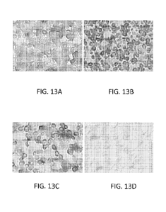
Figures 13A-13D show a series of photographs of immunostained spleen cells,
which
photographs depict the level of SHIP (SH2-containing Inosito1-5'-Phosphatase)
expression by the spleen cells. The spleen cells depicted in each photograph
were
isolated from BALB/c mice immunized with five (5) million allogeneic (B6
mouse)
spleen cells (administered intraperitoneally). The immunized mice were further
administered an anti-CD200 antibody (with effector function) [Figure 13A] or a
control antibody (with effector function) [Figure 13B]. Following treatment,
the
spleens were harvested, fixed, subjected to immunohistochemistry (see below).
Figure 13C depicts SHIP expression by spleen cells of mice that were not
immunized
with the allogeneic spleen cells. Figure 13D depicts spleen cells from
immunized
mice that were not stained with a primary anti-SHIP antibody. Each
experimental
group represented above included three mice. A representative photograph from
each
group is provided.
24
CA 02826453 2013-08-01
WO 2012/106634
PCT/US2012/023831
Fig. 14 is a bar graph depicting average relative SHIP expression by
splenocytes
obtained from BALB/c mice immunized with five (5) million B6 splenocytes as
described above. The immunized mice were further administered an anti-CD200
antibody (with effector function) [Antibody 3; see Example 3] or a control
antibody
(with effector function) [Antibody 4; see Example 3]. One group of mice,
"sham",
received neither immunization nor antibody treatment. Each experimental group
represented above included three mice. Following treatment, the spleens of the
mice
were harvested, fixed, and subjected to immunohistochemistry. The average
relative
expression from spleen cell sections was quantified using densitometry and is
reported in total pixels (x E+7).
Fig. 15 is a bar graph depicting average relative SHIP expression by
splenocytes
obtained from FcyR2b-deficient BALB/c mice immunized with five (5) million B6
splenocytes as described above. The immunized mice were further administered
an
anti-CD200 antibody (with effector function) [Antibody 3; see Example 3] or a
control antibody (with effector function) [Antibody 4; see Example 3]. One
group of
mice, "sham", received neither immunization nor antibody treatment. Each
experimental group represented above included three mice. Following treatment,
the
spleens of the mice were harvested, fixed, and subjected to
immunohistochemistry.
The average relative expression from spleen cell sections was quantified using
densitometry and is reported in total pixels (x E+7).
Detailed Description
The present disclosure provides anti-CD200 antibodies (including CD200-
binding fragments of the antibodies), pharmaceutical compositions, and kits,
each of
which is useful for modulating an immune response in a mammal. As elaborated
on
in this section, the antibodies (or compositions or kits) can be used alone,
or in
combination, in methods for prolonging the survival of a graft in a recipient
mammal
(e.g., a human). While in no way intended to be limiting, suitable
applications in
which the antibodies, kits, and compositions can be used are set forth in this
section
and exemplified in the working Examples.
CA 02826453 2013-08-01
WO 2012/106634
PCT/US2012/023831
Anti-CD200 Antibodies
The disclosure features antibodies that bind to a human CD200 polypeptide
(sometimes the antibodies are referred to herein as "anti-CD200 antibodies").
Also
featured are antigen-binding (CD200-binding) fragments of the antibodies. In
some
embodiments, an anti-CD200 antibody described herein binds to an extracellular
epitope within the human CD200 protein. For example, the anti-CD200 antibody
can
bind to an extracellular epitope in the human CD200 protein, which protein has
the
following amino acid sequence:
MERLVIRMPFSHLSTYSLVWVMAAVVLCTAQVQVVTQDEREQLYTPASLKC
SLQNAQEALIVTWQKKKAVSPENMVTFSENHGVVIQPAYKDKINITQLGLQN
STITFWNITLEDEGCYMCLFNTFGFGKISGTACLTVYVQPIVSLHYKFSEDHLN
ITCSATARPAPMVFWKVPRSGIENSTVTLSHPNGTTSVTSILHIKDPKNQVGKE
VICQVLHLGTVTDFKQTVNKGYWFSVPLLLSIVSLVILLVLISILLYWKRHRNQ
DREP (SEQ ID NO:1; GenBank Accession No. NP 005935.2). SEQ ID NO:1
depicts the amino acid sequence for a full-length, precursor human CD200
isoform A
protein. In some embodiments, an anti-CD200 antibody described herein binds to
an
extracellular epitope in the human CD200 protein, which protein has the
following
amino acid sequence:
MERLTLTRTIGGPLLTATLLGKTTINDYQVIRMPFSHLSTYSLVWVMAAVVLC
TAQVQVVTQDEREQLYTPASLKCSLQNAQEALIVTWQKKKAVSPENMVTFS
ENHGVVIQPAYKDKINITQLGLQNSTITFWNITLEDEGCYMCLFNTFGFGKISG
TACLTVYVQPIVSLHYKFSEDHLNITCSATARPAPMVFWKVPRSGIENSTVTL
SHPNGTTSVTSILHIKDPKNQVGKEVICQVLHLGTVTDFKQTVNKGYWFSVPL
LLSIVSLVILLVLISILLYWKRHRNQDREP (SEQ ID NO:2; GenBank Accession
No. NP 001004196.2). SEQ ID NO:2 depicts the amino acid sequence of a full-
length CD200 isoform B protein. In some embodiments, the anti-CD200 antibody
binds to an extracellular epitope present in a human CD200 protein which
protein has
the following amino acid sequence:
VIRMPFSHLSTYSLVWVMAAVVLCTAQVQVVTQDEREQLYTTASLKCSLQN
AQEALIVTWQKKKAVSPENMVTFSENHGVVIQPAYKDKINITQLGLQNSTITF
WNITLEDEGCYMCLFNTFGFGKISGTACLTVYVQPIVSLHYKFSEDHLNITCS
ATARPAPMVFWKVPRSGIENSTVTLSHPNGTTSVTSILHIKDPKNQVGKEVIC
26
CA 02826453 2013-08-01
WO 2012/106634
PCT/US2012/023831
QVLHLGTVTDFKQTVNKGYWFSVPLLLSIVSLVILLVLISILLYWKRHRNQDR
GELSQGVQKMT
(SEQ ID NO:3; GenBank Accession No. CAA28943.1; Figure 3 of McCaughan et al.
(1987) Immunogenetics 25:329-335). SEQ ID NO:3 is an exemplary sequence for a
full-length human CD200 protein.
In some embodiments, the anti-CD200 antibody can bind to the extracellular
portion of an CD200 protein at an epitope within or overlapping with, e.g.:
(i) amino
acids 1 to 233 of the amino acid sequence depicted in SEQ ID NO:1; (ii) amino
acids
1 to 258 of the amino acid sequence depicted in SEQ ID NO:2; or amino acids 1
to
229 of the amino acid sequence depicted in SEQ ID NO:3.
In some embodiments, the anti-CD200 antibody binds to an extracellular
epitope within the human CD200 protein lacking the leader sequence. For
example,
an anti-CD200 antibody described herein can bind to a CD200 protein at an
epitope
within or overlapping with amino acids 31 to 233 of the amino acid sequence
depicted
in SEQ ID NO:1, which corresponds to the extracellular portion of the mature
form of
human CD200 isoform A less the amino terminal leader sequence. In some
embodiments, an anti-CD200 antibody described herein can bind to a CD200
protein
at an epitope within or overlapping with amino acids 56 to 258 of the amino
acid
sequence depicted in SEQ ID NO:2, which corresponds to the extracellular
portion of
the mature form of human CD200 isoform B less the amino terminal leader
sequence.
In some embodiments, an anti-CD200 antibody described herein can bind to a
CD200
protein at an epitope within or overlapping with amino acids 27 to 229 of the
amino
acid sequence depicted in SEQ ID NO:3, which corresponds to the extracellular
portion of the mature form of human CD200 less the amino terminal leader
sequence.
An "epitope" refers to the site on a protein (e.g., a human CD200 protein)
that
is bound by an antibody. "Overlapping epitopes" include at least one (e.g.,
two, three,
four, five, or six) common amino acid residue(s).
In some embodiments, the anti-CD200 antibody specifically binds to a human
CD200 protein (e.g., the human CD200 protein having the amino acid sequence
depicted in SEQ ID NO:1, SEQ ID NO:2, SEQ ID NO:3, or the extracellular
domains
of the mature forms of the CD200 proteins). The terms "specific binding" or
"specifically binds" refer to two molecules forming a complex (e.g., a complex
between an anti-CD200 antibody and a CD200 protein) that is relatively stable
under
physiologic conditions. Typically, binding is considered specific when the
27
CA 02826453 2013-08-01
WO 2012/106634
PCT/US2012/023831
association constant (Ka) is higher than 106 M-1. Thus, an anti-CD200 antibody
can
specifically bind to a CD200 protein with a Ka of at least (or greater than)
106 (e.g., at
least or greater than 107, 108, 109, 1010, 10111012, 1013, 1014, or 1015 or
higher) M-1.
Examples of antibodies that specifically bind to a human CD200 protein are
described
in, e.g., U.S. Patent Nos.: 7,408,041; 7,427,665; 7,435,412; and 7,598,353,
the
disclosures of each of which are incorporated herein by reference in their
entirety.
The amino acid sequence for several exemplary anti-CD200 antibodies are
described in, e.g., U.S. Patent No. 7,408,041. For example, the anti-CD200
antibody
can comprise the amino acid sequence of the heavy and light chain variable
regions of
one of the Fab antibodies ¨ d1B10, d1A5, d1B5, c2aB7, clA10, or c2aA10 ¨
depicted
in Fig. 23 of U.S. Patent No. 7,408,041, the sequences depicted in Fig. 23
being
incorporated herein by reference in their entirety. In some embodiments, an
anti-
CD200 antibody described herein contains a paired set of heavy chain CDRs and
light
chain CDRs of one of the Fab antibodies depicted in Fig. 23 of U.S. Patent No.
7,408,041. For example, an anti-CD200 antibody described herein contains the
paired
set of CDRs from the d1B10 Fab antibody: a heavy chain CDR1 (HCDR1)
comprising the following sequence: GFTFSGFAMS (SEQ ID NO:4); a heavy chain
CDR2 (HCDR2) comprising the following sequence: SISSGGTTYYLDSVKG (SEQ
ID NO:5); a heavy chain CDR3 (HCDR3) comprising the following sequence:
GNYYSGTSYDY (SEQ ID NO:6); a light chain CDR1 (LCDR1) comprising the
following sequence: RASESVDSYGNSFMH (SEQ ID NO:7); a light chain CDR2
(LCDR2) comprising the following sequence: RASNLES (SEQ ID NO:8); and a light
chain CDR3 (LCDR3) comprising the following sequence: QQSNEDPRT (SEQ ID
NO:9).
In another example, an anti-CD200 antibody described herein can contain the
paired set of CDRs from the d1A5 Fab antibody: (i) a HCDR1 comprising the
following sequence: GFNIKDYYMH (SEQ ID NO:10); a HCDR2 comprising the
following sequence: WIDPENGDTKYAPKFQG (SEQ ID NO:11); a HCDR3
comprising the following sequence: KNYYVSNYNFFDV (SEQ ID NO:12); a
LCDR1 comprising the following sequence: SASSSVRYMY (SEQ ID NO:13); a
LCDR2 comprising the following sequence: DTSKLAS (SEQ ID NO:14); and a
LCDR3 comprising the following sequence: FQGSGYPLT (SEQ ID NO:15).
In another example, an anti-CD200 antibody described herein can comprise
the paired set of CDRs from the d1B5 Fab antibody: a HCDR1 comprising the
28
CA 02826453 2013-08-01
WO 2012/106634
PCT/US2012/023831
following amino acid sequence: GFNIKDYYIH (SEQ ID NO:16); a HCDR2
comprising the following amino acid sequence: WIDPEIGATKYVPKFQG (SEQ ID
NO:17); a HCDR3 comprising the following amino acid sequence:
LYGNYDRYYAMDY (SEQ ID NO:18); a LCDR1 comprising the following amino
acid sequence: KASQNVRTAVA (SEQ ID NO:19); a LCDR2 comprising the
following amino acid sequence: LASNRHT (SEQ ID NO:20); and a LCDR3
comprising the following amino acid sequence: LQHWNYPLT (SEQ ID NO:21).
In another example, an anti-CD200 antibody described herein can contain the
paired set of CDRs from the c2aB7 Fab antibody: a HCDR1 comprising the amino
acid sequence: GYSFTDYIIL (SEQ ID NO:22); a HCDR2 comprising the amino acid
sequence: HIDPYYGSSNYNLKFKG (SEQ ID NO:23); a HCDR3 comprising the
amino acid sequence: SKRDYFDY (SEQ ID NO:24); a LCDR1 comprising the
amino acid sequence: KASQDINSYLS (SEQ ID NO:25); a LCDR2 comprising the
amino acid sequence: RANRLVD (SEQ ID NO:26); and a LCDR3 comprising the
amino acid sequence: LQYDEFPYT (SEQ ID NO:27). Samalizumab contains the
aforementioned paired CDR set of the c2aB7 Fab antibody originally set forth
in Fig.
23 of U.S. Patent No. 7,408,041.
In yet another example, an anti-CD200 antibody described herein can contain
a paired set of CDRs from the c 1A10 Fab antibody: a HCDR1 comprising the
amino
acid sequence: GYTFTEYTMH (SEQ ID NO:28); a HCDR2 comprising the amino
acid sequence: GVNPNNGGALYNQKFKG (SEQ ID NO:29); a HCDR3 comprising
the amino acid sequence: RSNYRYDDAMDY (SEQ ID NO:30); a LCDR1
comprising the amino acid sequence: KSSQSLLDIDEKTYLN (SEQ ID NO:31); a
LCDR2 comprising the amino acid sequence: LVSKLDS (SEQ ID NO:32); and a
LCDR3 comprising the amino acid sequence: WQGTHFPQT (SEQ ID NO:33).
And in yet another example, an anti-CD200 antibody described herein can
contain a paired set of CDRs from the c2aA10 Fab antibody: a HCDR1 comprising
the amino acid sequence: AFNIKDHYMH (SEQ ID NO:34); a HCDR2 comprising
the amino acid sequence: WIDPESGDTEYAPKFQG (SEQ ID NO:35); a HCDR3
comprising the amino acid sequence: FNGYQALDQ (SEQ ID NO:36); a LCDR1
comprising the amino acid sequence: TASSSVSSSYLH (SEQ ID NO:37); a LCDR2
comprising the amino acid sequence: STSNLAS (SEQ ID NO:38); and a LCDR3
comprising the amino acid sequence: RQYHRSPPIFT (SEQ ID NO:39).
29
CA 02826453 2013-08-01
WO 2012/106634
PCT/US2012/023831
Additional exemplary sets of CDRs of anti-CD200 antibodies are described in,
e.g., U.S. Patent No. 7,427,665. In some embodiments, the anti-CD200 antibody
is
samalizumab (Alexion Pharmaceuticals, Inc., Cheshire, CT).
Methods for determining whether an antibody binds to a protein antigen
and/or the affinity for an antibody to a protein antigen are known in the art.
For
example, the binding of an antibody to a protein antigen can be detected
and/or
quantified using a variety of techniques such as, but not limited to, Western
blot, dot
blot, surface plasmon resonance (SPR) method (e.g., BIAcore system; Pharmacia
Biosensor AB, Uppsala, Sweden and Piscataway, N.J.), or enzyme-linked
immunosorbent assay (ELISA). See, e.g., Harlow and Lane (1988) "Antibodies: A
Laboratory Manual" Cold Spring Harbor Laboratory Press, Cold Spring Harbor,
N.Y.;
Benny K. C. Lo (2004) "Antibody Engineering: Methods and Protocols," Humana
Press (ISBN: 1588290921); Borrebaek (1992) "Antibody Engineering, A Practical
Guide," W.H. Freeman and Co., NY; Borrebaek (1995) "Antibody Engineering," 2nd
Edition, Oxford University Press, NY, Oxford; Johne et al. (1993) J Immunol
Meth
160:191-198; Jonsson et al. (1993) Ann Biol Clin 51:19-26; and Jonsson et al.
(1991)
Biotechniques 11:620-627.
In some embodiments, the anti-CD200 antibody can crossblock binding of
another antibody that binds to an epitope within, or overlapping with, a human
CD200
protein. In some embodiments, the anti-CD200 antibody can crossblock binding
of an
antibody that binds to an epitope within, or overlapping with, a peptide
fragment of a
human CD200 protein. The peptide fragment can be a fragment of a human CD200
protein having the amino acid sequence depicted in, e.g., any one of SEQ ID
NOs:1 to
3. As used herein, the term "crossblocking antibody" refers to an antibody
that lowers
the amount of binding of anti-CD200 antibody to an epitope on a CD200 protein
relative to the amount of binding of the anti-CD200 antibody to the epitope in
the
absence of the antibody. Suitable methods for determining whether a first
antibody
crossblocks binding of a second antibody to an epitope are known in the art.
Methods for identifying the epitope to which a particular antibody (e.g., an
anti-CD200 antibody) binds are also known in the art. For example, the binding
epitope of an anti-CD200 antibody can be identified by measuring the binding
of the
antibody to several (e.g., three, four, five, six, seven, eight, nine, 10, 15,
20, or 30 or
more) overlapping peptide fragments of a CD200 protein (e.g., several
overlapping
fragments of a protein having the amino acid sequence depicted in, e.g., any
one of
CA 02826453 2013-08-01
WO 2012/106634
PCT/US2012/023831
SEQ ID NOs:1 to 3). Each of the different overlapping peptides is then bound
to a
unique address on a solid support, e.g., separate wells of a multi-well assay
plate.
Next, the anti-CD200 antibody is interrogated by contacting it to each of the
peptides
in the assay plate for an amount of time and under conditions that allow for
the
antibody to bind to its epitope. Unbound anti-CD200 antibody is removed by
washing each of the wells. Next, a detectably-labeled secondary antibody that
binds
to the anti-CD200 antibody, if present in a well of the plate, is contacted to
each of the
wells, and unbound secondary antibody is removed by washing steps. The
presence
or amount of the detectable signal produced by the detectably-labeled
secondary
antibody in a well is an indication that the anti-CD200 antibody binds to the
particular
peptide fragment associated with the well. See, e.g., Harlow and Lane (supra),
Benny
K. C. Lo (supra), and U.S. Patent Application Publication No. 20060153836, the
disclosure of which is incorporated by reference in its entirety. A particular
epitope to
which an antibody binds can also be identified using BIAcore chromatographic
techniques (see, e.g., Pharmacia BIAtechnology Handbook, "Epitope Mapping,"
Section 6.3.2, (May 1994); and Johne et al. (1993) J Immunol Methods 160:191-
8).
In some embodiments, an anti-CD200 antibody, or a CD200-binding fragment
thereof, described herein binds to a human CD200 polypeptide expressed on the
surface of a cell. Methods for determining whether an antibody binds to a
protein
expressed on the surface of a cell are known in the art and described in,
e.g.,
Petermann et al. (2007) J Clin Invest 117(12):3922-3929; Rijkers et al. (2008)
Mol
Immunol 45(4):1126-35; and Kretz-Rommel (2007) J Immunol 178(9):5595-5605.
In some embodiments, an anti-CD200 antibody or CD200-binding fragment
thereof described herein inhibits the interaction between CD200 protein and
the
CD200 receptor. Methods for determining whether an agent (such as an anti-
CD200
antibody) inhibits the interaction between CD200 and CD200R are known in the
art
and described in, e.g., Hatherly and Barclay (2004) Eur J Immunol 34(6):1688-
1694.
In some embodiments, the antibody inhibits the interaction between CD200 and
its
receptor by at least 20 (e.g., at least 25, 30, 35, 40, 45, 50, 55, 60, 65,
70, 75, 80, 85,
90, 95, or even 100) % as compared the level of interaction between CD200 and
its
receptor in the absence of the antibody.
In some embodiments, the anti-CD200 antibody or CD200-binding fragment
thereof inhibits the formation of osteoclasts in vitro and/or in vivo.
Suitable methods
for determining whether an antibody inhibits the formation of osteoclasts are
known
31
CA 02826453 2013-08-01
WO 2012/106634
PCT/US2012/023831
in the art and described in, e.g., PCT Publication No. WO 08/089022 and Cui et
al.
(2007) Proc Natl Acad Sci USA 104(36):14436-14441. For example, murine bone
marrow cells can be cultured in the presence of, e.g., RANKL and M-CSF in the
presence or absence of an anti-CD200 antibody. A decrease in the percentage of
osteoclasts formed from the bone marrow cells in the presence of the antibody
as
compared to the percentage of osteoclasts formed in the absence of the
antibody
indicates that the antibody inhibits osteoclast formation in vitro.
Since CD200 is expressed on normal cells such as endothelial cells it could be
in some embodiments advantageous to administer a variant anti-CD200 antibody
(or
CD200-binding fragment thereof) with a constant region modified so that it
does not
mediate, or has decreased ability to mediate, antibody-dependent cell-mediated
cytotoxicity (ADCC) or complement-dependent cytotoxicity (CDC). CD200
expression is also upregulated on some activated normal cells (e.g., activated
T cells),
rendering such cells vulnerable to killing by an anti-CD200 antibody with
effector
function. It may be advantageous to use an anti-CD200 antibody having
diminished,
or lacking, effector function to avoid killing of these cells by ADCC or CDC.
The
effector function of an anti-CD200 antibody can be eliminated by replacing an
immunoglobulin constant region that has effector function (e.g., the IgG1
constant
domain) for a constant region that does not have effector function (e.g., an
IgG2/IgG4
fusion constant region). Additional methods for reducing or eliminating the
effector
function of an antibody heavy chain constant region are described below.
Methods for Producing an Antibody
Suitable methods for producing an antibody, or antigen-binding fragments
thereof, in accordance with the disclosure are known in the art (see, e.g.,
U.S. Patent
No. 7,408,041 and PCT Application Publication No. WO 09/014745) and described
herein. For example, monoclonal anti-CD200 antibodies may be generated using
CD200-expressing cells, a CD200 polypeptide, or an antigenic fragment of CD200
polypeptide, as an immunogen, thus raising an immune response in animals from
which antibody-producing cells and in turn monoclonal antibodies may be
isolated.
The sequence of such antibodies may be determined and the antibodies or
variants
thereof produced by recombinant techniques. Recombinant techniques may be used
to produce chimeric, CDR-grafted, humanized and fully human antibodies based
on
32
CA 02826453 2013-08-01
WO 2012/106634
PCT/US2012/023831
the sequence of the monoclonal antibodies as well as polypeptides capable of
binding
to human CD200.
Moreover, antibodies derived from recombinant libraries ("phage antibodies")
may be selected using CD200-expressing cells, or polypeptides derived
therefrom, as
bait to isolate the antibodies or polypeptides on the basis of target
specificity. The
production and isolation of non-human and chimeric anti-CD200 antibodies are
well
within the purview of the skilled artisan.
Recombinant DNA technology can be used to modify one or more
characteristics of the antibodies produced in non-human cells. Thus, chimeric
antibodies can be constructed in order to decrease the immunogenicity thereof
in
diagnostic or therapeutic applications. Moreover, immunogenicity can be
minimized
by humanizing the antibodies by CDR grafting and, optionally, framework
modification. See, U.S. Patent Nos. 5,225,539 and 7,393,648, the contents of
each of
which are incorporated herein by reference.
Antibodies can be obtained from animal serum or, in the case of monoclonal
antibodies or fragments thereof, produced in cell culture. Recombinant DNA
technology can be used to produce the antibodies according to established
procedure,
including procedures in bacterial or preferably mammalian cell culture. The
selected
cell culture system preferably secretes the antibody product.
In another embodiment, a process for the production of an antibody disclosed
herein includes culturing a host, e.g., E. coli or a mammalian cell, which has
been
transformed with a hybrid vector. The vector includes one or more expression
cassettes containing a promoter operably linked to a first DNA sequence
encoding a
signal peptide linked in the proper reading frame to a second DNA sequence
encoding
the antibody protein. The antibody protein is then collected and isolated.
Optionally,
the expression cassette may include a promoter operably linked to
polycistronic (e.g.,
bicistronic) DNA sequences encoding antibody proteins each individually
operably
linked to a signal peptide in the proper reading frame.
Multiplication of hybridoma cells or mammalian host cells in vitro is carried
out in suitable culture media, which include the customary standard culture
media
(such as, for example Dulbecco's Modified Eagle Medium (DMEM) or RPMI 1640
medium), optionally replenished by a mammalian serum (e.g. fetal calf serum),
or
trace elements and growth sustaining supplements (e.g. feeder cells such as
normal
mouse peritoneal exudate cells, spleen cells, bone marrow macrophages, 2-
33
CA 02826453 2013-08-01
WO 2012/106634
PCT/US2012/023831
aminoethanol, insulin, transferrin, low density lipoprotein, oleic acid, or
the like).
Multiplication of host cells which are bacterial cells or yeast cells is
likewise carried
out in suitable culture media known in the art. For example, for bacteria
suitable
culture media include medium LE, NZCYM, NZYM, NZM, Terrific Broth, SOB,
SOC, 2 x YT, or M9 Minimal Medium. For yeast, suitable culture media include
medium YPD, YEPD, Minimal Medium, or Complete Minimal Dropout Medium.
In vitro production provides relatively pure antibody preparations and allows
scale-up production to give large amounts of the desired antibodies.
Techniques for
bacterial cell, yeast, plant, or mammalian cell cultivation are known in the
art and
include homogeneous suspension culture (e.g. in an airlift reactor or in a
continuous
stirrer reactor), and immobilized or entrapped cell culture (e.g. in hollow
fibers,
microcapsules, on agarose microbeads or ceramic cartridges).
Large quantities of the desired antibodies can also be obtained by multiplying
mammalian cells in vivo. For this purpose, hybridoma cells producing the
desired
antibodies are injected into histocompatible mammals to cause growth of
antibody-
producing tumors. Optionally, the animals are primed with a hydrocarbon,
especially
mineral oils such as pristane (tetramethyl-pentadecane), prior to the
injection. After
one to three weeks, the antibodies are isolated from the body fluids of those
mammals. For example, hybridoma cells obtained by fusion of suitable myeloma
cells with antibody-producing spleen cells from Balb/c mice, or transfected
cells
derived from hybridoma cell line Sp2/0 that produce the desired antibodies are
injected intraperitoneally into Balb/c mice optionally pre-treated with
pristane. After
one to two weeks, ascitic fluid is taken from the animals.
The foregoing, and other, techniques are discussed in, for example, Kohler and
Milstein, (1975) Nature 256:495-497; U.S. Patent No. 4,376,110; Harlow and
Lane,
Antibodies: a Laboratory Manual, (1988) Cold Spring Harbor, the disclosures of
which are all incorporated herein by reference. Techniques for the preparation
of
recombinant antibody molecules are described in the above references and also
in,
e.g.:W097/08320; U.S. Patent No. 5,427,908; U.S. Patent No. 5,508,717; Smith
(1985) Science 225:1315-1317; Parmley and Smith (1988) Gene 73:305-318; De La
Cruz et al. (1988) J Biol Chem 263:4318-4322; U.S. Patent No. 5,403,484; U.S.
Patent No. 5,223,409; W088/06630; W092/15679; U.S. Patent No. 5,780,279; U.S.
Patent No. 5,571,698; U.S. Patent No. 6,040,136; Davis et al. (1999) Cancer
Metastasis Rev 18(4):421-5; Taylor et al. (1992) Nucleic Acids Res 20: 6287-
6295;
34
CA 02826453 2013-08-01
WO 2012/106634
PCT/US2012/023831
and Tomizuka et al. (2000) Proc Natl Acad Sci USA 97(2): 722-727, the contents
of
each of which are incorporated herein by reference in their entirety.
The cell culture supernatants are screened for the desired antibodies,
preferentially by immunofluorescent staining of CD200-expressing cells, by
immunoblotting, by an enzyme immunoassay, e.g. a sandwich assay or a dot-
assay, or
a radioimmunoassay.
For isolation of the antibodies, the immunoglobulins in the culture
supernatants or in the ascitic fluid may be concentrated, e.g., by
precipitation with
ammonium sulfate, dialysis against hygroscopic material such as polyethylene
glycol,
filtration through selective membranes, or the like. If necessary and/or
desired, the
antibodies are purified by the customary chromatography methods, for example
gel
filtration, ion-exchange chromatography, chromatography over DEAE-cellulose
and/or (immuno-) affinity chromatography, e.g. affinity chromatography with
one or
more surface polypeptides derived from a CD200-expressing cell line, or with
Protein-A or -G.
Another embodiment provides a process for the preparation of a bacterial cell
line secreting antibodies directed against a CD200 protein in a suitable
mammal. For
example a rabbit is immunized with pooled samples from CD200-expressing tissue
or
cells or CD200 polypeptide or fragments thereof. A phage display library
produced
from the immunized rabbit is constructed and panned for the desired antibodies
in
accordance with methods well known in the art (such as, e.g., the methods
disclosed
in the various references incorporated herein by reference).
Hybridoma cells secreting the monoclonal antibodies are also disclosed. The
preferred hybridoma cells are genetically stable, secrete monoclonal
antibodies
described herein of the desired specificity, and can be expanded from deep-
frozen
cultures by thawing and propagation in vitro or as ascites in vivo.
In another embodiment, a process is provided for the preparation of a
hybridoma cell line secreting monoclonal antibodies against a CD200 protein.
In that
process, a suitable mammal, for example a Balb/c mouse, is immunized with one
or
more polypeptides or antigenic fragments of CD200 or with one or more
polypeptides
or antigenic fragments derived from a CD200-expressing cell, the CD200-
expressing
cell itself, or an antigenic carrier containing a purified polypeptide as
described.
Antibody-producing cells of the immunized mammal are grown briefly in culture
or
fused with cells of a suitable myeloma cell line. The hybrid cells obtained in
the
CA 02826453 2013-08-01
WO 2012/106634
PCT/US2012/023831
fusion are cloned, and cell clones secreting the desired antibodies are
selected. For
example, spleen cells of Balb/c mice immunized with a CD200-expressing Chronic
Lymphocytic Leukemia (CLL) cell line are fused with cells of the myeloma cell
line
PAI or the myeloma cell line Sp2/0-Ag 14. The obtained hybrid cells are then
screened for secretion of the desired antibodies and positive hybridoma cells
are
cloned.
Methods for preparing a hybridoma cell line include immunizing Balb/c mice
by injecting subcutaneously and/or intraperitoneally an immunogenic
composition
containing human CD200 protein (or an immunogenic fragment thereof) several
times, e.g., four to six times, over several months, e.g., between two and
four months.
Spleen cells from the immunized mice are taken two to four days after the last
injection and fused with cells of the myeloma cell line PAI in the presence of
a fusion
promoter, preferably polyethylene glycol. Preferably, the myeloma cells are
fused
with a three- to twenty-fold excess of spleen cells from the immunized mice in
a
solution containing about 30% to about 50% polyethylene glycol of a molecular
weight around 4000. After the fusion, the cells are expanded in suitable
culture media
as described supra, supplemented with a selection medium, for example HAT
medium, at regular intervals in order to prevent normal myeloma cells from
overgrowing the desired hybridoma cells.
The antibodies and fragments thereof can be "chimeric." Chimeric antibodies
and antigen-binding fragments thereof comprise portions from two or more
different
species (e.g., mouse and human). Chimeric antibodies can be produced with
mouse
variable regions of desired specificity spliced onto human constant domain
gene
segments (for example, U.S. Patent No. 4,816,567). In this manner, non-human
antibodies can be modified to make them more suitable for human clinical
application
(e.g., methods for treating or preventing an immune associated disorder in a
human
subject).
The monoclonal antibodies of the present disclosure include "humanized"
forms of the non-human (e.g., mouse) antibodies. Humanized or CDR-grafted mAbs
are particularly useful as therapeutic agents for humans because they are not
cleared
from the circulation as rapidly as mouse antibodies and do not typically
provoke an
adverse immune reaction. Methods of preparing humanized antibodies are
generally
well known in the art. For example, humanization can be essentially performed
following the method of Winter and co-workers (see, e.g., Jones et al. (1986)
Nature
36
CA 02826453 2013-08-01
WO 2012/106634
PCT/US2012/023831
321:522-525; Riechmann et al. (1988) Nature 332:323-327; and Verhoeyen et al.
(1988) Science 239:1534-1536), by substituting rodent CDRs or CDR sequences
for
the corresponding sequences of a human antibody. Also see, e.g., Staelens et
al.
(2006) Mol Immunol 43:1243-1257. In some embodiments, humanized forms of non-
human (e.g., mouse) antibodies are human antibodies (recipient antibody) in
which
hypervariable (CDR) region residues of the recipient antibody are replaced by
hypervariable region residues from a non-human species (donor antibody) such
as a
mouse, rat, rabbit, or non-human primate having the desired specificity,
affinity, and
binding capacity. In some instances, framework region residues of the human
immunoglobulin are also replaced by corresponding non-human residues (so
called
"back mutations"). In addition, phage display libraries can be used to vary
amino
acids at chosen positions within the antibody sequence. The properties of a
humanized antibody are also affected by the choice of the human framework.
Furthermore, humanized and chimerized antibodies can be modified to comprise
residues that are not found in the recipient antibody or in the donor antibody
in order
to further improve antibody properties, such as, for example, affinity or
effector
function.
Fully human antibodies are also provided in the disclosure. The term "human
antibody" includes antibodies having variable and constant regions (if
present)
derived from human germline immunoglobulin sequences. Human antibodies can
include amino acid residues not encoded by human germline immunoglobulin
sequences (e.g., mutations introduced by random or site-specific mutagenesis
in vitro
or by somatic mutation in vivo). However, the term "human antibody" does not
include antibodies in which CDR sequences derived from the germline of another
mammalian species, such as a mouse, have been grafted onto human framework
sequences (i.e., humanized antibodies). Fully human or human antibodies may be
derived from transgenic mice carrying human antibody genes (carrying the
variable
(V), diversity (D), joining (J), and constant (C) exons) or from human cells.
For
example, it is now possible to produce transgenic animals (e.g., mice) that
are
capable, upon immunization, of producing a full repertoire of human antibodies
in the
absence of endogenous immunoglobulin production. (See, e.g., Jakobovits et al.
(1993) Proc Natl Acad Sci USA 90:2551; Jakobovits et al. (1993) Nature 362:255-
258; Bruggemann et al. (1993) Year in Immunol 7:33; and Duchosal et al. (1992)
Nature 355:258.) Transgenic mice strains can be engineered to contain gene
37
CA 02826453 2013-08-01
WO 2012/106634
PCT/US2012/023831
sequences from unrearranged human immunoglobulin genes. The human sequences
may code for both the heavy and light chains of human antibodies and would
function
correctly in the mice, undergoing rearrangement to provide a wide antibody
repertoire
similar to that in humans. The transgenic mice can be immunized with the
target
protein (e.g., a CD200 protein, fragments thereof, or cells expressing CD200
protein)
to create a diverse array of specific antibodies and their encoding RNA.
Nucleic
acids encoding the antibody chain components of such antibodies may then be
cloned
from the animal into a display vector. Typically, separate populations of
nucleic acids
encoding heavy and light chain sequences are cloned, and the separate
populations
then recombined on insertion into the vector, such that any given copy of the
vector
receives a random combination of a heavy and a light chain. The vector is
designed
to express antibody chains so that they can be assembled and displayed on the
outer
surface of a display package containing the vector. For example, antibody
chains can
be expressed as fusion proteins with a phage coat protein from the outer
surface of the
phage. Thereafter, display packages can be screened for display of antibodies
binding
to a target.
In addition, human antibodies can be derived from phage-display libraries
(Hoogenboom et al. (1991) J Mol Biol 227:381; Marks et al. (1991) J Mol Biol
222:581-597; and Vaughan et al. (1996) Nature Biotech 14:309 (1996)).
Synthetic
phage libraries can be created which use randomized combinations of synthetic
human antibody V-regions. By selection on antigen fully human antibodies can
be
made in which the V-regions are very human-like in nature. See, e.g., U.S.
Patent
Nos. 6,794,132; 6,680,209; and 4,634,666, and Ostberg et al. (1983) Hybridoma
2:361-367, the contents of each of which are incorporated herein by reference
in their
entirety.
For the generation of human antibodies, also see Mendez et al. (1998) Nature
Genetics 15:146-156 and Green and Jakobovits (1998) J Exp Med 188:483-495, the
disclosures of which are hereby incorporated by reference in their entirety.
Human
antibodies are further discussed and delineated in U.S. Patent Nos.:
5,939,598;
6,673,986; 6,114,598; 6,075,181; 6,162,963; 6,150,584; 6,713,610; and
6,657,103 as
well as U.S. Patent Application Publication Nos. 20030229905 Al, 20040010810
Al,
20040093622 Al, 20060040363 Al, 20050054055 Al, 20050076395 Al, and
20050287630 Al. See also International Patent Application Publication Nos. WO
94/02602, WO 96/34096, and WO 98/24893, and European Patent No. EP 0 463 151
38
CA 02826453 2013-08-01
WO 2012/106634
PCT/US2012/023831
Bl. The disclosures of each of the above-cited patents, applications, and
references
are hereby incorporated by reference in their entirety.
In an alternative approach, others, including GenPharm International, Inc.,
have utilized a "minilocus" approach. In the minilocus approach, an exogenous
Ig
locus is mimicked through the inclusion of pieces (individual genes) from the
Ig
locus. Thus, one or more VH genes, one or more DH genes, one or more JH genes,
a
mu constant region, and a second constant region (preferably a gamma constant
region) are formed into a construct for insertion into an animal. This
approach is
described in, e.g., U.S. Patent Nos.: 5,545,807; 5,545,806; 5,625,825;
5,625,126;
5,633,425; 5,661,016; 5,770,429; 5,789,650; 5,814,318; 5,591,669; 5,612,205;
5,721,367; 5,789,215; 5,643,763; 5,569,825; 5,877,397; 6,300,129; 5,874,299;
6,255,458; and 7,041,871, the disclosures of which are hereby incorporated by
reference. See also European Patent No. 0 546 073 B1, International Patent
Application Publication Nos. WO 92/03918, WO 92/22645, WO 92/22647, WO
92/22670, WO 93/12227, WO 94/00569, WO 94/25585, WO 96/14436, WO
97/13852, and WO 98/24884, the disclosures of each of which are hereby
incorporated by reference in their entirety. See further Taylor et al. (1992)
Nucleic
Acids Res 20: 6287; Chen et al. (1993) Int Immunol 5:647; Tuaillon et al.
(1993) Proc
Natl Acad Sci USA 90: 3720-4; Choi et al. (1993) Nature Genetics 4: 117;
Lonberg et
al. (1994) Nature 368: 856-859; Taylor et al. (1994) Int Immunol 6: 579-591;
Tuaillon
et al. (1995) J Immunol 154: 6453-65; Fishwild et al. (1996) Nature Biotechnol
14:
845; and Tuaillon et al. (2000) Eur J Immunol 10: 2998-3005, the disclosures
of each
of which are hereby incorporated by reference in their entirety.
In certain embodiments, de-immunized anti-CD200 antibodies or antigen-
binding fragments thereof are provided. De-immunized antibodies or antigen-
binding
fragments thereof are antibodies that have been modified so as to render the
antibody
or antigen-binding fragment thereof non-immunogenic, or less immunogenic, to a
given species (e.g., to a human). De-immunization can be achieved by modifying
the
antibody or antigen-binding fragment thereof utilizing any of a variety of
techniques
known to those skilled in the art (see, e.g., PCT Publication Nos. WO
04/108158 and
WO 00/34317). For example, an antibody or antigen-binding fragment thereof may
be de-immunized by identifying potential T cell epitopes and/or B cell
epitopes within
the amino acid sequence of the antibody or antigen-binding fragment thereof
and
removing one or more of the potential T cell epitopes and/or B cell epitopes
from the
39
CA 02826453 2013-08-01
WO 2012/106634
PCT/US2012/023831
antibody or antigen-binding fragment thereof, for example, using recombinant
techniques. The modified antibody or antigen-binding fragment thereof may then
optionally be produced and tested to identify antibodies or antigen-binding
fragments
thereof that have retained one or more desired biological activities, such as,
for
example, binding affinity, but have reduced immunogenicity. Methods for
identifying
potential T cell epitopes and/or B cell epitopes may be carried out using
techniques
known in the art, such as, for example, computational methods (see e.g., PCT
Publication No. WO 02/069232), in vitro or in silico techniques, and
biological assays
or physical methods (such as, for example, determination of the binding of
peptides to
MHC molecules, determination of the binding of peptide:MHC complexes to the T
cell receptors from the species to receive the antibody or antigen-binding
fragment
thereof, testing of the protein or peptide parts thereof using transgenic
animals with
the MHC molecules of the species to receive the antibody or antigen-binding
fragment thereof, or testing with transgenic animals reconstituted with immune
system cells from the species to receive the antibody or antigen-binding
fragment
thereof, etc.). In various embodiments, the de-immunized anti-CD200 antibodies
described herein include de-immunized antigen-binding fragments, Fab, Fv,
scFv,
Fab' and F(ab')2, monoclonal antibodies, murine antibodies, engineered
antibodies
(such as, for example, chimeric, single chain, CDR-grafted, humanized, and
artificially selected antibodies), synthetic antibodies and semi-synthetic
antibodies.
In some embodiments, a recombinant DNA comprising an insert coding for a
heavy chain variable domain and/or for a light chain variable domain of an
anti-
CD200 antibody or a CD200 protein-expressing cell line is produced. The term
DNA
includes coding single stranded DNAs, double stranded DNAs consisting of said
coding DNAs and of complementary DNAs thereto, or these complementary (single
stranded) DNAs themselves.
Furthermore, a DNA encoding a heavy chain variable domain and/or a light
chain variable domain of anti-CD200 antibodies can be enzymatically or
chemically
synthesized to contain the authentic DNA sequence coding for a heavy chain
variable
domain and/or for the light chain variable domain, or a mutant thereof. A
mutant of
the authentic DNA is a DNA encoding a heavy chain variable domain and/or a
light
chain variable domain of the above-mentioned antibodies in which one or more
amino
acids are deleted, inserted, or exchanged with one or more other amino acids.
Preferably said modification(s) are outside the CDRs of the heavy chain
variable
CA 02826453 2013-08-01
WO 2012/106634
PCT/US2012/023831
domain and/or the CDRs of the light chain variable domain of the antibody in
humanization and expression optimization applications. The term mutant DNA
also
embraces silent mutants wherein one or more nucleotides are replaced by other
nucleotides with the new codons coding for the same amino acid(s). The term
mutant
sequence also includes a degenerate sequence. Degenerate sequences are
degenerate
within the meaning of the genetic code in that an unlimited number of
nucleotides are
replaced by other nucleotides without resulting in a change of the amino acid
sequence originally encoded. Such degenerate sequences may be useful due to
their
different restriction sites and/or frequency of particular codons which are
preferred by
the specific host, particularly E. coli, to obtain an optimal expression of
the heavy
chain murine variable domain and/or a light chain murine variable domain.
The term mutant is intended to include a DNA mutant obtained by in vitro
mutagenesis of the authentic DNA according to methods known in the art.
For the assembly of complete tetrameric immunoglobulin molecules and the
expression of chimeric antibodies, the recombinant DNA inserts coding for
heavy and
light chain variable domains are fused with the corresponding DNAs coding for
heavy
and light chain constant domains, then transferred into appropriate host
cells, for
example after incorporation into hybrid vectors.
Recombinant DNAs including an insert coding for a heavy chain murine
variable domain of an anti-CD200 antibody-expressing cell line fused to a
human
constant domain IgG, for example yl, y2, y3 or y4, in particular embodiments
yl or
y4, may be used. Recombinant DNAs including an insert coding for a light chain
murine variable domain of an antibody fused to a human constant domain lc or
k,
preferably lc, are also provided.
Another embodiment pertains to recombinant DNAs coding for a recombinant
polypeptide wherein the heavy chain variable domain and the light chain
variable
domain are linked by way of a spacer group, optionally comprising a signal
sequence
facilitating the processing of the antibody in the host cell and/or a DNA
sequence
encoding a peptide facilitating the purification of the antibody and/or a
cleavage site
and/or a peptide spacer and/or an agent.
Accordingly, the monoclonal antibodies or antigen-binding fragments of the
disclosure can be naked antibodies or antigen-binding fragments that are not
conjugated to other agents, for example, a therapeutic agent or detectable
label.
Alternatively, the monoclonal antibody or antigen-binding fragment can be
41
CA 02826453 2013-08-01
WO 2012/106634
PCT/US2012/023831
conjugated to an agent such as, for example, a cytotoxic agent, a small
molecule, a
hormone, an enzyme, a growth factor, a cytokine, a ribozyme, a peptidomimetic,
a
chemical, a prodrug, a nucleic acid molecule including coding sequences (such
as
antisense, RNAi, gene-targeting constructs, etc.), or a detectable label
(e.g., an NMR
or X-ray contrasting agent, fluorescent molecule, etc.). In certain
embodiments, an
anti-CD200 antibody or antigen-binding fragment (e.g., Fab, Fv, single-chain
(scFv),
Fab', and F(ab')2) is linked to a molecule that increases the half-life of the
antibody or
antigen-binding fragment (see above).
Several possible vector systems are available for the expression of cloned
heavy chain and light chain genes in mammalian cells. One class of vectors
relies
upon the integration of the desired gene sequences into the host cell genome.
Cells
which have stably integrated DNA can be selected by simultaneously introducing
drug resistance genes such as E. coli gpt (Mulligan and Berg (1981) Proc Natl
Acad
Sci USA, 78:2072-2076) or Tn5 neo (Southern and Berg (1982) J Mol Appl Genet
1:327-341). The selectable marker gene can be either linked to the DNA gene
sequences to be expressed, or introduced into the same cell by co-transfection
(Wigler
et al. (1979) Cell 16:777-785). A second class of vectors utilizes DNA
elements
which confer autonomously replicating capabilities to an extrachromosomal
plasmid.
These vectors can be derived from animal viruses, such as bovine
papillomavirus
(Sarver et al. (1982) Proc Natl Acad Sci USA, 79:7147-7151), polyoma virus
(Deans
et al. (1984) Proc Natl Acad Sci USA 81:1292-1296), or SV40 virus (Lusky and
Botchan (1981) Nature 293:79-81).
Since an immunoglobulin cDNA is comprised only of sequences representing
the mature mRNA encoding an antibody protein, additional gene expression
elements
regulating transcription of the gene and processing of the RNA are required
for the
synthesis of immunoglobulin mRNA. These elements may include splice signals,
transcription promoters, including inducible promoters, enhancers, and
termination
signals. cDNA expression vectors incorporating such elements include those
described by Okayama and Berg (1983) Mol Cell Biol 3:280-289; Cepko et al.
(1984)
Cell 37:1053-1062; and Kaufman (1985) Proc Natl Acad Sci USA 82:689-693.
As is evident from the disclosure, the anti-CD200 antibodies can be used in
therapies (e.g., therapies for an immune associated disorder), including
combination
therapies.
42
CA 02826453 2013-08-01
WO 2012/106634
PCT/US2012/023831
In the therapeutic embodiments of the present disclosure, bispecific
antibodies
are contemplated. Bispecific antibodies are monoclonal, preferably human or
humanized, antibodies that have binding specificities for at least two
different
antigens. In the present case, one of the binding specificities is for the
human CD200
antigen and the other one is for any other antigen.
Methods for making bispecific antibodies are within the purview of those
skilled in the art. Traditionally, the recombinant production of bispecific
antibodies is
based on the co-expression of two immunoglobulin heavy-chain/light-chain
pairs,
where the two heavy chains have different specificities (Milstein and Cuello
(1983)
Nature 305:537-539). Antibody variable domains with the desired binding
specificities (antibody-antigen combining sites) can be fused to
immunoglobulin
constant domain sequences. The fusion preferably is with an immunoglobulin
heavy-
chain constant domain, including at least part of the hinge, CH2, and CH3
regions.
DNAs encoding the immunoglobulin heavy-chain fusions and, if desired, the
immunoglobulin light chain, are inserted into separate expression vectors, and
are co-
transfected into a suitable host organism. For further details of illustrative
currently
known methods for generating bispecific antibodies see, e.g., Suresh et al.
(1986)
Methods Enzymol 121:210-228; PCT Publication No. WO 96/27011; Brennan et al.
(1985) Science 229:81-83; Shalaby et al. J Exp Med (1992) 175:217-225;
Kostelny et
al. (1992) J Immunol 148(5):1547-1553; Hollinger et al. (1993) Proc Natl Acad
Sci
USA 90:6444-6448; Gruber et al. (1994) J Immunol 152:5368-5474; and Tutt et
al.
(1991) J Immunol 147:60-69. Bispecific antibodies also include cross-linked or
heteroconjugate antibodies. Heteroconjugate antibodies may be made using any
convenient cross-linking methods. Suitable cross-linking agents are well known
in
the art, and are disclosed in U.S. Pat. No. 4,676,980, along with a number of
cross-
linking techniques.
Various techniques for making and isolating bispecific antibody fragments
directly from recombinant cell culture have also been described. For example,
bispecific antibodies have been produced using leucine zippers. See, e.g.,
Kostelny et
al. (1992) J Immunol 148(5):1547-1553. The leucine zipper peptides from the
Fos
and Jun proteins may be linked to the Fab' portions of two different
antibodies by
gene fusion. The antibody homodimers may be reduced at the hinge region to
form
monomers and then re-oxidized to form the antibody heterodimers. This method
can
also be utilized for the production of antibody homodimers. The "diabody"
43
CA 02826453 2013-08-01
WO 2012/106634
PCT/US2012/023831
technology described by Hollinger et al. (1993) Proc Natl Acad Sci USA 90:6444-
6448 has provided an alternative mechanism for making bispecific antibody
fragments. The fragments comprise a heavy-chain variable domain (VH) connected
to a light-chain variable domain (VL) by a linker which is too short to allow
pairing
between the two domains on the same chain. Accordingly, the VH and VL domains
of
one fragment are forced to pair with the complementary VL and VH domains of
another fragment, thereby forming two antigen-binding sites. Another strategy
for
making bispecific antibody fragments by the use of single-chain Fv (scFv)
dimers has
also been reported. See, e.g., Gruber et al. (1994) J Immunol 152:5368-5374.
Alternatively, the antibodies can be "linear antibodies" as described in,
e.g., Zapata et
al. (1995) Protein Eng 8(10):1057-1062. Briefly, these antibodies comprise a
pair of
tandem Fd segments (VH-CH1-VH-CH1) which form a pair of antigen binding
regions.
Linear antibodies can be bispecific or monospecific.
The disclosure also embraces variant forms of bispecific antibodies such as
the
tetravalent dual variable domain immunoglobulin (DVD-Ig) molecules described
in
Wu et al. (2007) Nat Biotechnol 25(11):1290-1297. The DVD-Ig molecules are
designed such that two different light chain variable domains (VL) from two
different
parent antibodies are linked in tandem directly or via a short linker by
recombinant
DNA techniques, followed by the light chain constant domain. Methods for
generating DVD-Ig molecules from two parent antibodies are further described
in,
e.g., PCT Publication Nos. WO 08/024188 and WO 07/024715, the disclosures of
each of which are incorporated herein by reference in their entirety.
Effector functions
The interaction of antibodies and antibody-antigen complexes with cells of the
immune system affects a variety of responses, referred to herein as effector
functions.
Exemplary effector functions include Fc receptor binding, phagocytosis, down-
regulation of cell surface receptors (e.g., B cell receptor; BCR), etc. Other
effector
functions include ADCC, whereby antibodies bind Fc receptors on natural killer
(NK)
cells or macrophages leading to cell death, and CDC, which is cell death
induced via
activation of the complement cascade (reviewed in Daeron (1997) Annu Rev
Immunol
15:203-234; Ward and Ghetie (1995) Therapeutic Immunol 2:77-94; and Ravetch
and
Kinet (1991) Annu Rev Immunol 9:457-492). Such effector functions generally
44
CA 02826453 2013-08-01
WO 2012/106634
PCT/US2012/023831
require the Fc region to be combined with a binding domain (e.g., an antibody
variable domain) and can be assessed using various assays as herein disclosed.
Several antibody effector functions, including ADCC, are mediated by Fc
receptors (FcRs), which bind the Fc region of an antibody. In ADCC, NK cells
or
macrophages bind to the Fc region of the antibody complex and promote lysis of
the
target cell. The cross-linking of FcRs on NK cells triggers perforin/granzyme-
mediated cytotoxicity, whereas in macrophages this cross-linking promotes the
release of mediators such as nitric oxide (NO), TNF-a, and reactive oxygen
species.
For CD200-positive target cells, an anti-CD200 antibody binds to the target
cell and
the Fc region directs effector function to the target cell. The affinity of an
antibody
for a particular FcR, and hence the effector activity mediated by the
antibody, may be
modulated by altering the amino acid sequence and/or post-translational
modifications
of the Fc and/or constant region of the antibody.
FcRs are defined by their specificity for immunoglobulin isotypes; Fc
receptors for IgG antibodies are referred to as FcyR, for IgE as FccR, for IgA
as FcaR
and so on. Three subclasses of FcyR have been identified: FcyRI (CD64), FcyRII
(CD32) and FcyRIII (CD16). Because each FcyR subclass is encoded by two or
three
genes, and alternative RNA splicing leads to multiple transcripts, a broad
diversity in
FcyR isoforms exists. The three genes encoding the FcyRI subclass (FcyRIA,
FcyRIB
and FcyRIC) are clustered in region 1q21.1 of the long arm of chromosome 1;
the
genes encoding FcyRII isoforms (FcyRIIA, FcyRIIB and FcyRIIC) and the two
genes
encoding FcyRIII (FcyRIIIA and FcyRIIIB) are all clustered in region 1q22.
These
different FcR subtypes are expressed on different cell types (reviewed in
Ravetch and
Kinet (1991) Annu Rev Immunol 9:457-492). For example, in humans, FcyRIIIB is
found only on neutrophils, whereas FcyRIIIA is found on macrophages,
monocytes,
natural killer (NK) cells, and a subpopulation of T-cells. Notably, FcyRIIIA
is the
only FcR present on NK cells, one of the cell types implicated in ADCC.
FcyRI, FcyRII and FcyRIII are immunoglobulin superfamily (IgSF) receptors;
FcyRI has three IgSF domains in its extracellular domain, while FcyRII and
FcyRIII
have only two IgSF domains in their extracellular domains. Another type of Fc
receptor is the neonatal Fc receptor (FcRn). FcRn is structurally similar to
major
histocompatibility complex (MHC) and consists of an a-chain noncovalently
bound
to 132-microg1obu1in.
CA 02826453 2013-08-01
WO 2012/106634
PCT/US2012/023831
The binding site on human and murine antibodies for FcyR have been
previously mapped to the so-called "lower hinge region" consisting of residues
233-
239 (EU index numbering as in Kabat et al. (1991) Sequences of Proteins of
Immunological Interest, 5th Ed. Public Health Service, National Institutes of
Health,
Bethesda, Maryland). Woof et al. (1986) Molec Immunol 23:319-330; Duncan et
al.
(1988) Nature 332:563; Canfield and Morrison (1991) J Exp Med 173:1483-1491;
Chappel et al. (1991) Proc Natl Acad Sci USA 88:9036-9040. Of residues 233-
239,
P238 and S239 have been cited as possibly being involved in binding.
Other previously cited areas possibly involved in binding to FcyR are: G316-
K338 (human IgG) for human FcyRI (by sequence comparison only; no substitution
mutants were evaluated) (Woof et al. (1986) Molec Immunol 23:319-330); K274-
R301 (human IgG1) for human FcyRIII (based on peptides) (Sarmay et al. (1984)
Molec Immunol 21:43-51); Y407-R416 (human IgG) for human FcyRIII (based on
peptides) (Gergely et al. (1984) Biochem Soc Trans 12:739-743 (1984)); as well
as
N297 and E318 (murine IgG2b) for murine FcyRII (Lund et al. (1992) Molec
Immunol 29:53-59).
Human effector cells are leukocytes which express one or more FcRs and
perform effector functions. In certain embodiments, the cells express at least
FcyRIII
and perform ADCC effector function. Examples of human leukocytes which mediate
ADCC include peripheral blood mononuclear cells (PBMC), natural killer (NK)
cells,
monocytes, cytotoxic T cells and neutrophils. Effector cells may be isolated
from a
native source thereof, e.g. from blood or PBMCs.
In CDC, the antibody-antigen complex binds complement, resulting in the
activation of the complement cascade and generation of the membrane attack
complex. Activation of the classical complement pathway is initiated by the
binding
of the first component of the complement system (Clq) to antibodies (of the
appropriate subclass) which are bound to their cognate antigen; thus the
activation of
the complement cascade is regulated in part by the binding affinity of the
immunoglobulin to Clq protein. Clq and two serine proteases, Clr and Cls, form
the
complex Cl, the first component of the CDC pathway. Clq is a hexavalent
molecule
with a molecular weight of approximately 460,000 and a structure in which six
collagenous "stalks" are connected to six globular head regions. Burton and
Woof
(1992) Advances in Immunol 51:1-84. To activate the complement cascade, it is
necessary for Clq to bind to at least two molecules of IgGl, IgG2, or IgG3,
but only
46
CA 02826453 2013-08-01
WO 2012/106634
PCT/US2012/023831
one molecule of IgM, attached to the antigenic target (Ward and Ghetie (1995)
Therapeutic Immunol 2:77-94). To assess complement activation, a CDC assay,
e.g.
as described in Gazzano-Santoro et al. (1996) J Immunol Methods 202:163, can
be
performed.
It has been proposed that various residues of the IgG molecule are involved in
binding to Clq including the G1u318, Lys320 and Lys322 residues on the CH2
domain, amino acid residue 331 located on a turn in close proximity to the
same beta
strand, the Lys235 and G1y237 residues located in the lower hinge region, and
residues 231 to 238 located in the N-terminal region of the CH2 domain. See,
e.g.,
Xu et al. (1993) J Immunol 150:152A; PCT publication no. WO 94/29351; Tao et
al.
(1993) J Exp Med 178:661-667; Brekke et al. (1994) Eur J Immunol 24:2542-2547;
Burton et al. (1980) Nature 288:338-344; and U.S. Patent Nos. 5,648,260 and
5,624,821. It has further been proposed that the ability of IgG to bind Clq
and
activate the complement cascade also depends on the presence, absence or
modification of the carbohydrate moiety positioned between the two CH2 domains
(which is normally anchored at Asn297). See, e.g., Ward and Ghetie (1995)
Therapeutic Immunology 2:77-94. In certain embodiments, one or more of these
residues may be modified, substituted, or removed or one or more amino acid
residues
may be inserted so as to enhance or decrease CDC activity of the anti-CD200
antibodies provided herein.
Methods for decreasing or eliminating effector function
Effector functions involving the constant region of the target-specific
antibody
may be modulated by altering properties of the constant or Fc region. Altered
effector
functions include, for example, a modulation in one or more of the following
activities: ADCC, CDC, apoptosis, binding to one or more Fc-receptors, and pro-
inflammatory responses. Modulation refers to an increase, decrease, or
elimination of
an effector function activity exhibited by a subject antibody as compared to
the
activity of a second antibody. In certain embodiments, the second antibody is
an
antibody possessing a naturally-occurring effector function that has not been
modified. In particular embodiments, modulation includes situations in which
an
activity is abolished or completely absent. Further, in some instances, a non-
variant
47
CA 02826453 2013-08-01
WO 2012/106634
PCT/US2012/023831
antibody may exhibit effector function activity similar or equivalent to the
activity of
the chC2aB7-hG1 or the hB7V3V2-hG1 antibodies disclosed herein.
A variant constant region with altered FcR binding affinity and/or ADCC
activity and/or altered CDC activity is a polypeptide which has either
enhanced or
diminished FcR binding activity and/or ADCC activity and/or CDC activity
compared
to the native or parent polypeptide or to a polypeptide comprising a native
sequence
or constant region. A polypeptide variant which displays increased binding to
an FcR
binds at least one FcR with greater affinity than the parent polypeptide. A
polypeptide variant which displays decreased binding to an FcR binds at least
one
FcR with lower affinity than a parent polypeptide. Such variants which display
decreased binding to an FcR may possess little or no appreciable binding to an
FcR,
e.g., 0 to 50% (e.g., less than 50, 49, 48, 47, 46, 45, 44, 43, 42, 41, 40,
39, 38, 37, 36,
35, 34, 33, 32, 31, 30, 29, 28, 27, 26, 25, 24, 23, 22, 21, 20, 19, 18, 17,
16, 15, 14, 13,
12, 11, 10, 9, 8, 7, 6, 5, 4, 3, 2, or 1 %) of the binding to the FcR as
compared to the
level of binding of a native sequence immunoglobulin constant or Fc region to
the
FcR. Similarly, a variant anti-CD200 antibody that displays altered ADCC
and/or
CDC activity may exhibit either increased or reduced ADCC and/or CDC activity
compared to the native or parent polypeptide. For example, in some
embodiments,
the anti-CD200 antibody comprising a variant constant region can exhibit
approximately 0 to 50% (e.g., less than 50, 49, 48, 47, 46, 45, 44, 43, 42,
41, 40, 39,
38, 37, 36, 35, 34, 33, 32, 31, 30, 29, 28, 27, 26, 25, 24, 23, 22, 21, 20,
19, 18, 17, 16,
15, 14, 13, 12, 11, 10, 9, 8, 7, 6, 5, 4, 3, 2, or 1 %) of the ADCC and/or CDC
activity
of the native form of the constant region. An anti-CD200 antibody comprising a
variant constant region displaying reduced ADCC and/or CDC may exhibit reduced
or no ADCC and/or CDC activity as exemplified herein.
A native sequence Fc or constant region comprises an amino acid sequence
identical to the amino acid sequence of an Fc or constant chain region found
in nature.
A variant or altered Fc or constant region comprises an amino acid sequence
which
differs from that of a native sequence heavy chain region by virtue of at
least one
amino acid modification, insertion, or deletion, for example. In certain
embodiments,
the variant or altered constant region has at least one amino acid
substitution,
insertion, and/or deletion, compared to a native sequence constant region or
to the
constant region of a parent polypeptide, e.g. from about one to about one
hundred
amino acid substitutions, insertions, and/or deletions in a native sequence
constant
48
CA 02826453 2013-08-01
WO 2012/106634
PCT/US2012/023831
region or in the constant region of the parent polypeptide. In some
embodiments, the
variant or altered constant region herein will possess at least about 70%
homology
(similarity) or identity with a native sequence constant region and/or with a
constant
region of a parent polypeptide, and in some instances at least about 75% and
in other
instances at least about 80% homology or identity therewith, and in other
embodiments at least about 85%, 90% or 95% homology or identity therewith. The
variant or altered constant region may also contain one or more amino acid
deletions
or insertions. Additionally, the variant constant region may contain one or
more
amino acid substitutions, deletions, or insertions that results in altered
post-
translational modifications, including, for example, an altered glycosylation
pattern.
Antibodies or antigen-binding fragments thereof with altered or no effector
functions may be generated by engineering or producing antibodies with variant
constant, Fc, or heavy chain regions; recombinant DNA technology and/or cell
culture
and expression conditions may be used to produce antibodies with altered
function
and/or activity. For example, recombinant DNA technology may be used to
engineer
one or more amino acid substitutions, deletions, or insertions in regions
(such as, for
example, Fc or constant regions) that affect antibody function including
effector
functions. Alternatively, changes in post-translational modifications, such
as, e.g.,
glycosylation patterns, may be achieved by manipulating the cell culture and
expression conditions by which the antibody is produced. Suitable methods for
introducing one or more substitutions, additions, or deletions into an Fc
region of an
antibody are well known in the art and include, e.g., standard DNA mutagenesis
techniques as described in, e.g., Sambrook et al. (1989) "Molecular Cloning: A
Laboratory Manual, 2nd Edition," Cold Spring Harbor Laboratory Press, Cold
Spring
Harbor, N.Y.; Harlow and Lane (1988), supra; Borrebaek (1992), supra; Johne et
al.
(1993), supra; PCT publication no. WO 06/53301; and U.S. patent no. 7,704,497.
Accordingly, certain aspects and methods of the present disclosure relate to
anti-CD200 antibodies with altered effector functions that comprise one or
more
amino acid substitutions, insertions, and/or deletions. In some embodiments,
such a
variant anti-CD200 antibody exhibits reduced or no effector function. In some
embodiments, a variant antibody comprises a hybrid constant region, or a
portion
thereof, such as a G2/G4 hybrid constant region (see e.g., Burton et al.
(1992) Adv
Immun 51:1-18; Canfield et al. (1991) J Exp Med 173:1483-1491; and Mueller et
al.
(1997) Mol Immunol 34(6):441-452). For example (and in accordance with Kabat
49
CA 02826453 2013-08-01
WO 2012/106634
PCT/US2012/023831
numbering), the IgG1 and IgG4 constant regions contain G249G250 residues
whereas
the IgG2 constant region does not contain residue 249, but does contain G250.
In a
G2/G4 hybrid constant region, where the 249-250 region comes from the G2
sequence, the constant region can be further modified to introduce a glycine
residue at
position 249 to produce a G2/G4 fusion having G249/G250.
In addition to using a G2/G4 construct as described above, anti-CD200
antibodies with reduced effector function may be produced by introducing other
types
of changes in the amino acid sequence of certain regions of the antibody. Such
amino
acid sequence changes include but are not limited to the Ala-Ala mutation
described
in, e.g., PCT Publication nos. WO 94/28027 and WO 98/47531; and Xu et al.
(2000)
Cell Immunol 200:16-26. Thus, in some embodiments, anti-CD200 antibodies with
mutations within the constant region including the Ala-Ala mutation may be
used to
reduce or abolish effector function. According to these embodiments, the
constant
region of an anti-CD200 antibody comprises a mutation to an alanine at
position 234
or a mutation to an alanine at position 235. Additionally, the constant region
may
contain a double mutation: a mutation to an alanine at position 234 and a
second
mutation to an alanine at position 235. In one embodiment, the anti-CD200
antibody
comprises an IgG4 framework, wherein the Ala-Ala mutation would describe a
mutation(s) from phenylalanine to alanine at position 234 and/or a mutation
from
leucine to alanine at position 235. In another embodiment, the anti-CD200
antibody
comprises an IgG1 framework, wherein the Ala-Ala mutation would describe a
mutation(s) from leucine to alanine at position 234 and/or a mutation from
leucine to
alanine at position 235. An anti-CD200 antibody may alternatively or
additionally
carry other mutations, including the point mutation K322A in the CH2 domain
(Hezareh et al. (2001) J Virol 75:12161-8). An antibody with said mutation(s)
in the
constant region may furthermore be a blocking or non-blocking antibody.
Additional substitutions that, when introduced into a heavy chain constant
region, result in decreased effector function are set forth in, e.g., Shields
et al. (2001)
J Biol Chem 276(9):6591-6604. See particularly Table 1 ("Binding of human IgG1
variants to human FcRn and FcyR) of Shields et al., the disclosure of which is
incorporated herein by reference in its entirety. By screening a library of
anti-IgE
antibodies, each antibody of the library differing by one or more
substitutions in the
heavy chain constant region, for binding to a panel of Fc receptors (including
FcRn,
CA 02826453 2013-08-01
WO 2012/106634
PCT/US2012/023831
FcyRI, FcyRIIA, FcyRIIB, and FcyRIIIA), the authors identified a number of
substitutions that modulate specific Fc-Fc receptor interactions. For example,
a
variant IgG2a heavy chain constant region in which the CH2 domain contains a
D265A substitution (heavy chain amino acid numbering according to Kabat et al.
(supra)) results in a complete loss of interaction between the variant
constant region
and IgG Fc receptors FcyRIIB, FcyRIII, FcyRI, and FcyRIV. Shields et al.
(2001) at
page 6595, Table 1. See also Baudino et al. (2008) J Immunol 181:6664-6669
(supra).
Changes within the hinge region also affect effector functions. For example,
deletion of the hinge region may reduce affinity for Fc receptors and may
reduce
complement activation (Klein et al. (1981) Proc Natl Acad Sci USA 78: 524-
528).
The present disclosure therefore also relates to antibodies with alterations
in the hinge
region.
In some embodiments, anti-CD200 antibodies may be modified to either
enhance or inhibit complement dependent cytotoxicity (CDC). Modulated CDC
activity may be achieved by introducing one or more amino acid substitutions,
insertions, or deletions in an Fc region of the antibody. See, e.g., U.S.
patent no.
6,194,551. Alternatively or additionally, cysteine residue(s) may be
introduced in the
Fc region, thereby allowing interchain disulfide bond formation in this
region. The
homodimeric antibody thus generated may have improved or reduced
internalization
capability and/or increased or decreased complement-mediated cell killing.
See, e.g.,
Caron et al. (1992) J Exp Med 176:1191-1195 and Shopes (1992) Immunol 148:2918-
2922; PCT publication nos. WO 99/51642 and WO 94/29351; Duncan and Winter
(1988) Nature 322:738-40; and U.S. Patent Nos. 5,648,260 and 5,624,821.
Homodimeric antibodies with enhanced anti-tumor activity may also be prepared
using heterobifunctional cross-linkers as described in Wolff et al. (1993)
Cancer
Research 53:2560-2565. Alternatively, an antibody can be engineered which has
dual
Fc regions and may thereby have enhanced complement lysis and ADCC
capabilities.
See, e.g., Stevenson et al. (1989) Anti-Cancer Drug Design 3:219-230.
Another potential means of modulating effector function of antibodies
includes changes in glycosylation, which is summarized in, e.g., Raju (2003)
BioProcess International 1(4):44-53. According to Wright and Morrison, the
microheterogeneity of human IgG oligosaccharides can affect biological
functions
51
CA 02826453 2013-08-01
WO 2012/106634
PCT/US2012/023831
such as CDC and ADCC, binding to various Fc receptors, and binding to Clq
protein.
(1997) TIBTECH 15:26-32. Glycosylation patterns of antibodies can differ
depending
on the producing cell and the cell culture conditions (Raju, supra). Such
differences
can lead to changes in both effector function and pharmacokinetics. See, e.g.,
Israel
et al. (1996) Immunology 89(4):573-578; Newkirk et al. (1996) Clin Exp Immunol
106(2):259-264. Differences in effector function may be related to the IgG's
ability
to bind to the Fcy receptors (FcyRs) on the effector cells. Shields et al.
have shown
that IgG, with variants in amino acid sequence that have improved binding to
FcyR,
can exhibit up to 100% enhanced ADCC using human effector cells. (2001) J Biol
Chem 276(9):6591-6604. While these variants include changes in amino acids not
found at the binding interface, both the nature of the sugar component as well
as its
structural pattern may also contribute to the differences observed. In
addition, the
presence or absence of fucose in the oligosaccharide component of an IgG can
improve binding and ADCC. See, e.g., Shields et al. (2002) J Biol Chem
277(30):26733-26740. An IgG that lacked a fucosylated carbohydrate linked to
Asn297 exhibited normal receptor binding to the FcyRI receptor. In contrast,
binding
to the FcyRIIIA receptor was improved 50-fold and accompanied by enhanced
ADCC, especially at lower antibody concentrations.
Shinkawa et al. demonstrated that an antibody to the human IL-5 receptor
produced in a rat hybridoma showed more than 50% higher ADCC when compared to
the antibody produced in Chinese hamster ovary cells (CHO) (Shinkawa et al.
(2003)
J Biol Chem 278(5):3466-73). Monosaccharide composition and oligosaccharide
profiling showed that the rat hybridoma-produced IgG had a lower content of
fucose
than the CHO-produced protein. The authors concluded that the lack of
fucosylation
of an IgG1 has a critical role in enhancement of ADCC activity.
A different approach was taken by Umana et al. who changed the
glycosylation pattern of chCE7, a chimeric IgG1 anti-neuroblastoma antibody.
((1999) Nat Biotechnol 17 (2):176-180). Using tetracycline, they regulated the
activity
of a glycosyltransferase enzyme (GnTIII) which bisects oligosaccharides that
have
been implicated in ADCC activity. The ADCC activity of the parent antibody was
barely above background level. Measurement of ADCC activity of the chCE7
produced at different tetracycline levels showed an optimal range of GnTIII
expression for maximal chCE7 in vitro ADCC activity. This activity correlated
with
the level of constant region-associated, bisected complex oligosaccharide.
Newly
52
CA 02826453 2013-08-01
WO 2012/106634
PCT/US2012/023831
optimized variants exhibited substantial ADCC activity. Similarly, Wright and
Morrison produced antibodies in a CHO cell line deficient in glycosylation and
showed that antibodies produced in this cell line were incapable of complement-
mediated cytolysis. (1994) J Exp Med 180:1087-1096. Thus, as known alterations
that affect effector function include modifications in the glycosylation
pattern or a
change in the number of glycosylated residues, the present disclosure relates
to a
CD200 antibody wherein glycosylation is altered to either enhance or decrease
effector function(s) including ADCC and CDC. Altered glycosylation includes a
decrease or increase in the number of glycosylated residues as well as a
change in the
pattern or location of glycosylated residues.
Still other approaches exist for altering the effector function of antibodies.
For
example, antibody-producing cells can be hypermutagenic, thereby generating
antibodies with randomly altered polypeptide residues throughout an entire
antibody
molecule. See, e.g., PCT publication no. WO 05/011735. Hypermutagenic host
cells
include cells deficient in DNA mismatch repair. Antibodies produced in this
manner
may be less antigenic and/or have beneficial pharmacokinetic properties.
Additionally, such antibodies may be selected for properties such as enhanced
or
decreased effector function(s).
It is further understood that effector function may vary according to the
binding affinity of the antibody. For example, antibodies with high affinity
may be
more efficient in activating the complement system compared to antibodies with
relatively lower affinity (Marzocchi-Machado et al. (1999) Immunol Invest
28:89-
101). Accordingly, an antibody may be altered such that the binding affinity
for its
antigen is reduced (e.g., by changing the variable regions of the antibody by
methods
such as substitution, addition, or deletion of one or more amino acid
residues). An
anti-CD200 antibody with reduced binding affinity may exhibit reduced effector
functions, including, for example, reduced ADCC and/or CDC.
Antibody Conjugates
The antibodies described herein can be modified, e.g., prior to expression or
following their expression or purification. The modifications can be covalent
or non-
covalent modifications. Such modifications can be introduced into the
antibodies by,
e.g., reacting targeted amino acid residues of the polypeptide with an organic
derivatizing agent that is capable of reacting with selected side chains or
terminal
53
CA 02826453 2013-08-01
WO 2012/106634
PCT/US2012/023831
residues. Suitable sites for modification can be chosen using any of a variety
of
criteria including, e.g., structural analysis or amino acid sequence analysis
of the
antibodies.
In some embodiments, the antibodies can be conjugated to a heterologous
moiety. The heterologous moiety can be, e.g., a heterologous polypeptide, a
therapeutic agent (e.g., a toxin or a drug), or a detectable label such as,
but not limited
to, a radioactive label, an enzymatic label, a fluorescent label, or a
luminescent label.
Suitable heterologous polypeptides include, e.g., an antigenic tag (e.g.,
FLAG,
polyhistidine, hemagglutinin (HA), glutathione-S-transferase (GST), or maltose-
binding protein (MBP)) for use in purifying the antibodies. Heterologous
polypeptides also include polypeptides that are useful as diagnostic or
detectable
markers, for example, luciferase, green fluorescent protein (GFP), or
chloramphenicol
acetyl transferase (CAT). Where the heterologous moiety is a polypeptide, the
moiety
can be incorporated into an antibody described herein as a fusion protein.
Suitable radioactive labels include, e.g., 32P5 33P5 14C5 12515 13115 35-5
and 3H.
Suitable fluorescent labels include, without limitation, fluorescein,
fluorescein
isothiocyanate (FITC), green fluorescence protein (GFP), DyLight 488,
phycoerythrin
(PE), propidium iodide (PI), PerCP, PE-Alexa Fluor 700, Cy5, allophycocyanin,
and Cy7. Luminescent labels include, e.g., any of a variety of luminescent
lanthanide
(e.g., europium or terbium) chelates. For example, suitable europium chelates
include
the europium chelate of diethylene triamine pentaacetic acid (DTPA) or
tetraazacyclododecane-1,4,7,10-tetraacetic acid (DOTA). Enzymatic labels
include,
e.g., alkaline phosphatase, CAT, luciferase, and horseradish peroxidase.
Two proteins (e.g., an antibody and a heterologous moiety) can be cross-
linked using any of a number of known chemical cross linkers. Examples of such
cross linkers are those which link two amino acid residues via a linkage that
includes
a "hindered" disulfide bond. In these linkages, a disulfide bond within the
cross-
linking unit is protected (by hindering groups on either side of the disulfide
bond)
from reduction by the action, for example, of reduced glutathione or the
enzyme
disulfide reductase. One suitable reagent, 4-succinimidyloxycarbonyl-a-methyl-
a (2-
pyridyldithio) toluene (SMPT), forms such a linkage between two proteins
utilizing a
terminal lysine on one of the proteins and a terminal cysteine on the other.
Heterobifunctional reagents that cross-link by a different coupling moiety on
each
54
CA 02826453 2013-08-01
WO 2012/106634
PCT/US2012/023831
protein can also be used. Other useful cross-linkers include, without
limitation,
reagents which link two amino groups (e.g., N-5-azido-2-
nitrobenzoyloxysuccinimide), two sulfhydryl groups (e.g., 1,4-bis-
maleimidobutane),
an amino group and a sulfhydryl group (e.g., m-maleimidobenzoyl-N-
hydroxysuccinimide ester), an amino group and a carboxyl group (e.g., 44p-
azidosalicylamidoThutylamine), and an amino group and a guanidinium group that
is
present in the side chain of arginine (e.g., p-azidophenyl glyoxal
monohydrate).
In some embodiments, a radioactive label can be directly conjugated to the
amino acid backbone of the antibody. Alternatively, the radioactive label can
be
included as part of a larger molecule (e.g., 121 in meta-[125I]iodophenyl-N-
hydroxysuccinimide ([125I]mIPNHS) which binds to free amino groups to form
meta-
iodophenyl (mIP) derivatives of relevant proteins (see, e.g., Rogers et al.
(1997) J
Nucl Med 38:1221-1229) or chelate (e.g., to DOTA or DTPA) which is in turn
bound
to the protein backbone. Methods of conjugating the radioactive labels or
larger
molecules/chelates containing them to the antibodies described herein are
known in
the art. Such methods involve incubating the proteins with the radioactive
label under
conditions (e.g., pH, salt concentration, and/or temperature) that facilitate
binding of
the radioactive label or chelate to the protein (see, e.g., U.S. Patent No.
6,001,329).
Methods for conjugating a fluorescent label (sometimes referred to as a
"fluorophore") to a protein (e.g., an anti-CD200 antibody) are known in the
art of
protein chemistry. For example, fluorophores can be conjugated to free amino
groups
(e.g., of lysines) or sulfhydryl groups (e.g., cysteines) of proteins using
succinimidyl
(NHS) ester or tetrafluorophenyl (TFP) ester moieties attached to the
fluorophores. In
some embodiments, the fluorophores can be conjugated to a heterobifunctional
cross-
linker moiety such as sulfo-SMCC. Suitable conjugation methods involve
incubating
an antibody protein with the fluorophore under conditions that facilitate
binding of the
fluorophore to the protein. See, e.g., Welch and Redvanly (2003) "Handbook of
Radiopharmaceuticals: Radiochemistry and Applications," John Wiley and Sons
(ISBN 0471495603).
In some embodiments, the antibodies can be modified, e.g., with a moiety that
improves the stabilization and/or retention of the antibodies in circulation,
e.g., in
blood, serum, or other tissues. For example, an anti-CD200 antibody can be
PEGylated as described in, e.g., Lee et al. (1999) Bioconjug Chem 10(6): 973-
8;
Kinstler et al. (2002) Advanced Drug Deliveries Reviews 54:477-485; and
Roberts et
CA 02826453 2013-08-01
WO 2012/106634
PCT/US2012/023831
al. (2002) Advanced Drug Delivery Reviews 54:459-476. The stabilization moiety
can improve the stability, or retention of, the antibody by at least 1.5
(e.g., at least 2,
5, 10, 15, 20, 25, 30, 40, or 50 or more) fold.
In some embodiments, the antibodies described herein can be glycosylated. In
some embodiments, an antibody described herein can be subjected to enzymatic
or
chemical treatment, or produced from a cell, such that the antibody has
reduced or
absent glycosylation. Methods for producing antibodies with reduced
glycosylation
are known in the art and described in, e.g., U.S. patent no. 6,933,368; Wright
et al.
(1991) EMBO J 10(10):2717-2723; and Co et al. (1993) Mol Immunol 30:1361.
Pharmaceutical Compositions and Formulations
The compositions containing an anti-CD200 antibody can be formulated as a
pharmaceutical composition, e.g., for administration to a recipient mammal to
prolong
the survival of an allograft organ. The pharmaceutical compositions will
generally
include a pharmaceutically acceptable carrier. As used herein, a
"pharmaceutically
acceptable carrier" refers to, and includes, any and all solvents, dispersion
media,
coatings, antibacterial and antifungal agents, isotonic and absorption
delaying agents,
and the like that are physiologically compatible. The compositions can include
a
pharmaceutically acceptable salt, e.g., an acid addition salt or a base
addition salt.
See, e.g., Berge et al. (1977) J Pharm Sci 66:1-19.
The compositions can be formulated according to standard methods.
Pharmaceutical formulation is a well-established art, and is further described
in, e.g.,
Gennaro (2000) "Remington: The Science and Practice of Pharmacy," 20th
Edition,
Lippincott, Williams & Wilkins (ISBN: 0683306472); Ansel et al. (1999)
"Pharmaceutical Dosage Forms and Drug Delivery Systems," 7th Edition,
Lippincott
Williams & Wilkins Publishers (ISBN: 0683305727); and Kibbe (2000) "Handbook
of Pharmaceutical Excipients American Pharmaceutical Association," 3rd Edition
(ISBN: 091733096X). In some embodiments, a composition can be formulated, for
example, as a buffered solution at a suitable concentration and suitable for
storage at
2-8 C. In some embodiments, a composition can be formulated for storage at a
temperature below 0 C (e.g., -20 C or -80 C).
The pharmaceutical compositions can be in a variety of forms. These forms
include, e.g., liquid, semi-solid and solid dosage forms, such as liquid
solutions (e.g.,
56
CA 02826453 2013-08-01
WO 2012/106634
PCT/US2012/023831
injectable and infusible solutions), dispersions or suspensions, tablets,
pills, powders,
liposomes and suppositories. The preferred form depends, in part, on the
intended
mode of administration and therapeutic application. For example, compositions
containing an anti-CD200 antibody intended for systemic or local delivery can
be in
the form of injectable or infusible solutions. Accordingly, the compositions
can be
formulated for administration by a parenteral mode (e.g., intravenous,
subcutaneous,
intraperitoneal, or intramuscular injection). "Parenteral administration,"
"administered parenterally," and other grammatically equivalent phrases, as
used
herein, refer to modes of administration other than enteral and topical
administration,
usually by injection, and include, without limitation, intravenous,
intranasal,
intraocular, pulmonary, intramuscular, intraarterial, intrathecal,
intracapsular,
intraorbital, intracardiac, intradermal, intrapulmonary, intraperitoneal,
transtracheal,
subcutaneous, subcuticular, intraarticular, subcapsular, subarachnoid,
intraspinal,
epidural, intracerebral, intracranial, intracarotid and intrasternal injection
and infusion
(see below).
The compositions can be formulated as a solution, microemulsion, dispersion,
liposome, or other ordered structure suitable for stable storage at high
concentration.
Sterile injectable solutions can be prepared by incorporating an antibody
described
herein in the required amount in an appropriate solvent with one or a
combination of
ingredients enumerated above, as required, followed by filtered sterilization.
Generally, dispersions are prepared by incorporating an anti-CD200 antibody
described herein into a sterile vehicle that contains a basic dispersion
medium and the
required other ingredients from those enumerated above. In the case of sterile
powders for the preparation of sterile injectable solutions, methods for
preparation
include vacuum drying and freeze-drying that yield a powder of the antibody
described herein plus any additional desired ingredient from a previously
sterile-
filtered solution thereof The proper fluidity of a solution can be maintained,
for
example, by the use of a coating such as lecithin, by the maintenance of the
required
particle size in the case of dispersion and by the use of surfactants.
Prolonged
absorption of injectable compositions can be brought about by including in the
composition a reagent that delays absorption, for example, monostearate salts
and
gelatin.
In certain embodiments, the anti-CD200 antibody can be prepared with a
carrier that will protect the compound against rapid release, such as a
controlled
57
CA 02826453 2013-08-01
WO 2012/106634
PCT/US2012/023831
release formulation, including implants and microencapsulated delivery
systems.
Biodegradable, biocompatible polymers can be used, such as ethylene vinyl
acetate,
polyanhydrides, polyglycolic acid, collagen, polyorthoesters, and polylactic
acid.
Many methods for the preparation of such formulations are known in the art.
See,
e.g., J.R. Robinson (1978) "Sustained and Controlled Release Drug Delivery
Systems," Marcel Dekker, Inc., New York.
In some embodiments, an anti-CD200 antibody described herein can be
modified, e.g., with a moiety that improves its stabilization and/or retention
in
circulation, e.g., in blood, serum, or other tissues. The stabilization moiety
can
improve the stability, or retention of, the antibody by at least 1.5 (e.g., at
least 2, 5, 10,
15, 20, 25, 30, 40, or 50 or more) fold.
In some embodiments, an anti-CD200 antibody is present in unit dosage form,
which can be particularly suitable for self-administration. A formulated
product of the present disclosure can be included within a container,
typically, for
example, a vial, cartridge, prefilled syringe or disposable pen. A doser such
as the
doser device described in U.S. Patent No. 6,302,855 may also be used, for
example,
with an injection system of the present disclosure.
An injection system of the present disclosure may employ a delivery pen as
described in U.S. Patent No. 5,308,341. Pen devices, most commonly used for
self-
delivery of insulin to patients with diabetes, are well known in the art. Such
devices
can comprise at least one injection needle (e.g., a 31 gauge needle of about 5
to 8 mm
in length), are typically pre-filled with one or more therapeutic unit doses
of a
therapeutic solution, and are useful for rapidly delivering the solution to a
subject with
as little pain as possible.
One medication delivery pen includes a vial holder into which a vial of
insulin
or other medication may be received. The vial holder is an elongate generally
tubular
structure with proximal and distal ends. The distal end of the vial holder
includes
mounting means for engaging a double-ended needle cannula. The proximal end
also
includes mounting means for engaging a pen body which includes a driver and
dose
setting apparatus. A disposable medication (e.g., anti-CD200 antibody)
containing
vial for use with the prior art vial holder includes a distal end having a
pierceable
elastomeric septum that can be pierced by one end of a double-ended needle
cannula.
The proximal end of this vial includes a stopper slidably disposed in fluid
tight
engagement with the cylindrical wall of the vial. This medication delivery pen
is used
58
CA 02826453 2013-08-01
WO 2012/106634
PCT/US2012/023831
by inserting the vial of medication into the vial holder. A pen body then is
connected
to the proximal end of the vial holder. The pen body includes a dose setting
apparatus
for designating a dose of medication to be delivered by the pen and a driving
apparatus for urging the stopper of the vial distally for a distance
corresponding to the
selected dose. The user of the pen mounts a double-ended needle cannula to the
distal
end of the vial holder such that the proximal point of the needle cannula
pierces the
septum on the vial. The patient then selects a dose and operates the pen to
urge the
stopper distally to deliver the selected dose. The dose selecting apparatus
returns to
zero upon injection of the selected dose. The patient then removes and
discards the
needle cannula, and keeps the prior art medication delivery pen in a
convenient
location for the next required medication administration. The medication in
the vial
will become exhausted after several such administrations of medication. The
patient
then separates the vial holder from the pen body. The empty vial may then be
removed and discarded. A new vial can be inserted into the vial holder, and
the vial
holder and pen body can be reassembled and used as explained above.
Accordingly, a
medication delivery pen generally has a drive mechanism for accurate dosing
and ease
of use.
A dosage mechanism such as a rotatable knob allows the user to accurately
adjust the amount of medication that will be injected by the pen from a
prepackaged
vial of medication. To inject the dose of medication, the user inserts the
needle under
the skin and depresses the knob once as far as it will depress. The pen may be
an
entirely mechanical device or it may be combined with electronic circuitry to
accurately set and/or indicate the dosage of medication that is injected into
the user.
See U.S. Patent No. 6,192,891.
In some embodiments, the needle of the pen device is disposable and the kits
include one or more disposable replacement needles. Pen devices suitable for
delivery of the any one of the presently featured antibody solutions are also
described
in, e.g., U.S. patent nos. 6,277,099; 6,200,296; and 6,146,361, the
disclosures of each
of which are incorporated herein by reference in their entirety. A microneedle-
based
pen device is described in, e.g., U.S. patent no. 7,556,615, the disclosure of
which is
incorporated herein by reference in its entirety. See also the Precision Pen
Injector
(PPI) device, Mo11yTM, manufactured by Scandinavian Health Ltd.
The present disclosure also presents controlled-release or extended-release
formulations suitable for chronic and/or self-administration of a medication.
The
59
CA 02826453 2013-08-01
WO 2012/106634
PCT/US2012/023831
various formulations can be administered to a patient in need of treatment
(e.g., a
recipient of an allograft) with the medication (e.g., an antibody of the
present
disclosure and at least one immunosuppressive agent) by intravenous
administration
as a bolus or by continuous infusion over a period of time.
In some embodiments, an anti-CD200 antibody is formulated for sustained-
release, extended-release, timed-release, controlled-release, or continuous-
release
administration. In some embodiments, depot formulations are used to administer
the
antibody to the subject in need thereof. In this method, the antibody is
formulated
with one or more carriers providing a gradual release of active agent over a
period of
a number of hours or days. Such formulations are often based upon a degrading
matrix which gradually disperses in the body to release the active agent.
One formulation suitable for depot injection of an anti-CD200 antibody relies
upon a polymeric depot system. The polymer can be a biodegradable polymer such
as
poly (lactic acid) (PLA) and/or poly (lactic-co-glycolic acid) (PLGA) and may
be in
the form of a solution in a solvent, a pre-polymer mixed with an initiator,
encapsulated polymer particles or polymer microspheres. The polymer or polymer
particles entrap the active agent and are gradually degraded releasing the
agent by
slow diffusion and/or as the matrix is absorbed. Examples of such systems
include
those described in U.S. patent nos. 4,938,763; 5,480,656; and 6,113,943, and
can
result in delivery of active agents over a period of up to several months.
Another depot system was set forth in U.S. patent no. 5,807,573, which system
is lipid-based ¨ a diacylglycerol, a phospholipid and optionally water,
glycerol,
ethylene glycol or propylene glycol. Suitable depot formulations are also
described
in, e.g., U.S. patent application publication no. 20060165800 (describing an
injectable
depot gel composition for systemic and local delivery of a beneficial agent to
a
subject over a short duration of time); Bari et al. (2010) Int J Pharm Sci Rev
Res
3(1):1-10 (describing prolonged release formulations suitable for parenteral
delivery
of a therapeutic protein into a mammal); and U.S. patent application
publication no.
20090010928, which describes depot antibody formulations, including, e.g., a
composition comprising monoclonal antibody at 5 mg/mL, formulated in aqueous
buffer consisting of 50 mM L-histidine, 150 mM NaC1, adjusted to pH 6.0 with
HC1.
Anti-CD200 antibody compositions can be prepared in dosage unit form for
ease of administration and uniformity of dosage. "Dosage unit form," as used
herein
refers to physically discrete units suited as unitary dosages for the subject
to be
CA 02826453 2013-08-01
WO 2012/106634
PCT/US2012/023831
treated; each unit containing a predetermined quantity of active compound
calculated
to produce the desired therapeutic effect in association with the selected
pharmaceutical carrier.
The disclosure provides aqueous solutions comprising an antibody (such as
samalizumab) that binds to CD200. In some embodiments, the solutions can be
high
concentration solutions of an anti-CD200 antibody. Such solutions are
sometimes
referred to herein as "high concentration antibody solutions." As used herein,
a "high
concentration" of an anti-CD200 antibody in an aqueous solution is a
concentration of
the antibody that is at least, equal to, or greater than, 10 (e.g., at least,
equal to, or
greater than, 11, 12, 13, 14, 15, 16, 17, 18, 19, 20, 21, 22, 23, 24, 25, 30,
35, 40, 45,
50, 55, 60, 65, 70, 75, 80, 85, 90, 95, 100, 105, 110, 115, 120, 125, 130,
135, 140,
145, 150, 155, 160, 165, 170, 175, 180, 185, 190, 195, 200, 205, 210, 215,
220, 225,
230, 235, 240, 245, or 250) mg/mL. In some embodiments, the anti-CD200
antibody
is present in the solution at a concentration of more than 200 (e.g., more
than 200,
205, 210, 215, 220, 225, 230, 235, 240, 245, 250, 255, 260, 265, 270, 275,
280, 285,
or 290) mg/mL. In some embodiments, the antibody is present in the solution at
a
concentration of, e.g., 10 mg/mL to 200 mg/mL, 20 mg/mL to 200 mg/mL, 30 mg/mL
to 200 mg/mL, 40 mg/mL to 200 mg/mL, 50 mg/mL to 200 mg/mL, 60 mg/mL to 200
mg/mL, 70 mg/mL to 200 mg/mL, 80 mg/mL to 200 mg/mL, 90 mg/mL to 200
mg/mL, 100 mg/mL to 200 mg/mL, 110 mg/mL to 200 mg/mL, 120 mg/mL to 200
mg/mL, 130 mg/mL to 200 mg/mL, 140 mg/mL to 200 mg/mL, 150 mg/mL to 200
mg/mL, 10 mg/mL to 100 mg/mL, 20 mg/mL to 100 mg/mL, 30 mg/mL to 100
mg/mL, 40 mg/mL to 100 mg/mL, 50 mg/mL to 100 mg/mL, 60 mg/mL to 100
mg/mL, 70 mg/mL to 100 mg/mL, 80 mg/mL to 100 mg/mL, 90 mg/mL to 100
mg/mL, 10 mg/mL to 150 mg/mL, 20 mg/mL to 150 mg/mL, 30 mg/mL to 150
mg/mL, 40 mg/mL to 150 mg/mL, 50 mg/mL to 150 mg/mL, 60 mg/mL to 150
mg/mL, 70 mg/mL to 150 mg/mL, 80 mg/mL to 150 mg/mL, 90 mg/mL to 150
mg/mL, 100 mg/mL to 150 mg/mL, 110 mg/mL to 150 mg/mL, 120 mg/mL to 150
mg/mL, 40 mg/mL to 50 mg/mL, 10 mg/mL to 250 mg/mL, 20 mg/mL to 250
mg/mL, 30 mg/mL to 250 mg/mL, 40 mg/mL to 250 mg/mL, 50 mg/mL to 250
mg/mL, 60 mg/mL to 250 mg/mL, 70 mg/mL to 250 mg/mL, 80 mg/mL to 250
mg/mL, 90 mg/mL to 250 mg/mL, 100 mg/mL to 250 mg/mL, 110 mg/mL to 250
mg/mL, 120 mg/mL to 250 mg/mL, 130 mg/mL to 250 mg/mL, 140 mg/mL to 250
mg/mL, 150 mg/mL to 250 mg/mL, 160 mg/mL to 250 mg/mL, 170 mg/mL to 250
61
CA 02826453 2013-08-01
WO 2012/106634
PCT/US2012/023831
mg/mL, 180 mg/mL to 250 mg/mL, 190 mg/mL to 250 mg/mL, 200 mg/mL to 250
mg/mL, greater than 200 mg/mL (e.g., at least 201 mg/mL) to 250 mg/mL, or
greater
than 200 mg/mL (e.g., 201 mg/mL or greater) to 300 mg/mL.
In some embodiments, an anti-CD200 antibody is for use, and formulated as
such, as a monotherapy. In some embodiments, an anti-CD200 antibody can be
formulated with, or for use with, one or more additional active agents. For
example,
the one or more additional active agents can be useful for prolonging the
survival of
an allograft organ in a mammal. Such agents include, e.g., the monoclonal anti-
CD3
antibody OKT3, anti-thymocyte globulin (ATG), cyclosporine A, or tacrolimus
(FK
506). Additionally, glucocorticoids and/or azathioprine (or other purine
analogs) may
be administered to the host prior to transplant. Drugs used to aid in
preventing or
inhibiting transplant rejection include, but are not limited to, ATG or ALG,
OKT3,
daclizumab, basiliximab, corticosteroids, 15-deoxyspergualin, LF15-0195,
cyclosporins, tacrolimus, purine analogs such as azathioprine, methotrexate, a
mycophenolate compound (e.g., mycophenolate mofetil or mycophenolate sodium),
6-mercaptopurine, bredinin, brequinar, leflunomide, cyclophosphamide,
sirolimus,
anti-CD4 monoclonal antibodies, CTLA4-Ig, rituxan, anti-CD154 monoclonal
antibodies, anti-LFA1 monoclonal antibodies, anti-LFA-3 monoclonal antibodies,
anti-CD2 monoclonal antibodies, and anti-CD45 antibodies.
The numerous drugs utilized to delay graft rejection (i.e., to prolong graft
survival) work in a variety of ways. Cyclosporine A is one of the most widely
used
immunosuppressive drugs for inhibiting graft rejection. It is an inhibitor of
interleukin-2 or IL-2 (it prevents mRNA transcription of interleukin-2). More
directly, cyclosporine inhibits calcineurin activation that normally occurs
upon T cell
receptor stimulation. Calcineurin dephosphorylates NFAT (nuclear factor of
activated
T cells), thereby enabling NFAT to enter the nucleus and bind to interleukin-2
promoter. By blocking this process, cyclosporine A inhibits the activation of
the CD4 '
T cells and the resulting cascade of events which would otherwise occur.
Tacrolimus
is another immunosuppressant that acts by inhibiting the production of
interleukin-2
via calcineurin inhibition. Rapamycin (sirolimus), SDZ RAD, and interleukin-2
receptor blockers are
drugs that inhibit the action of interleukin-2 and therefore prevent the
cascade of
events described above. Inhibitors of purine or pyrimidine biosynthesis are
also used
to inhibit graft rejection. These inhibitors prevent DNA synthesis and thereby
inhibit
62
CA 02826453 2013-08-01
WO 2012/106634
PCT/US2012/023831
cell division including T cell proliferation. The result is the inhibition of
T cell
activity by
preventing the formation of new T cells. Inhibitors of purine synthesis
include
azathioprine, methotrexate, mycophenolate mofetil (MMF), mycophenolate sodium
(Novartis), and mizoribine (bredinin). Inhibitors of pyrimidine synthesis
include
brequinar sodium and leflunomide.
Cyclophosphamide is an inhibitor of both purine and pyrimidine synthesis.
Yet another method for inhibiting T cell activation is to treat the recipient
with
antibodies to T cells. OKT3 is a murine monoclonal antibody against CD3 which
is
part of the T cell receptor. This antibody initially activates T cells through
the T cell
receptor, then induces apoptosis of the activated T cell.
Numerous other drugs and methods for delaying allotransplant rejection are
known to and used by persons of skill in the art. One approach is to deplete T
cells,
e.g., by irradiation. Depletion of T cells has often been used in bone marrow
transplants, especially if there is a partial mismatch of major HLA.
Administration to
the recipient of an inhibitor (blocker) of the CD40 ligand-CD40 interaction
and/or a
blocker of the CD28-B7 interaction has also been used (U.S. patent no.
6,280,957).
PCT application publication no. WO 01/37860 discloses the administration of an
anti-CD3 antibody and IL-5 to inhibit the Thl immune response. PCT application
publication no. WO 00/27421 teaches a method for prophylaxis or treatment of
corneal transplant rejection by administering a tumor necrosis factor-a
antagonist.
Glotz et al. (2002) Am J Transplant 2:758-760 show that administration of
intravenous immunoglobulins (IVIg) can induce a profound and sustained
decrease in
the titers of anti-HLA antibodies thereby allowing survival of an HLA-
mismatched
organ. Similar protocols have included plasma exchanges (Xaube et al. (1984)
Lancet
1:824-828) or immunoadsorption techniques coupled to immunosuppressive agents
(Hiesse et al. (1992) Nephrol Dial Transplant 7:944-951) or a combination of
these
methods (Montgomery et al., 2000 Transplantation 70:887-895). Changelian et
al.
(2003) Science 302:875-878 disclose a model in which immunosuppression is
caused
by
an oral inhibitor of Janus kinase 3 (JAK3), which is an enzyme necessary for
the
proper signaling of cytokine receptors which use the common gamma chain (yc)
(Interleukins-2, -4, -7, -9, -15, -21), the result being an inhibition of T
cell activation.
63
CA 02826453 2013-08-01
WO 2012/106634
PCT/US2012/023831
Antisense nucleic acids against ICAM-1 have been used alone or in
combination with a monoclonal antibody specific for leukocyte-function
associated
antigen 1 (LFA-1)
in a study of heart allograft transplantation (Stepkowski, supra). Similarly,
an anti-
ICAM-1 antibody has been used in combination with anti-LFA-1 antibody to treat
heart allografts (Stepkowski, supra). Antisense oligonucleotides have
additionally
been used in conjunction with cyclosporine in rat heart or kidney allograft
models,
resulting in a synergistic effect to prolong the survival of the grafts
(Stepkowski,
supra). Chronic transplant rejection has been treated by administering an
antagonist
of TGF-I3, which is a cytokine involved in differentiation, proliferation, and
apoptosis
(U.S. patent application publication no. 2003/0180301).
When the anti-CD200 antibody is to be used in combination with a second
active agent, or when two or more different anti-CD200 antibodies are to be
used, the
agents can be formulated separately or together. For example, the respective
pharmaceutical compositions can be mixed, e.g., just prior to administration,
and
administered together or can be administered separately, e.g., at the same or
different
times (see below).
As described above, a composition can be formulated such that it includes a
therapeutically effective amount of an anti-CD200 antibody or the composition
can be
formulated to include a sub-therapeutic amount of the antibody and a sub-
therapeutic
amount of one or more additional active agents such that the components in
total are
therapeutically effective for prolonging the survival of an allograft in a
mammal. In
some embodiments, a composition can be formulated to include two or more anti-
CD200 antibodies, each at sub-therapeutic doses, such that the antibodies in
combination are at a concentration that is therapeutically effective for
prolonging
graft survival. Methods for determining a therapeutically effective dose of an
anti-
CD200 antibody are known in the art and described herein.
Biological Samples and Sample Collection
Suitable biological samples for use in the methods described herein include
any biological fluid, population of cells, or tissue or fraction thereof,
which includes
one or more white blood cell populations. A biological sample can be, for
example, a
specimen obtained from a subject (e.g., a mammal such as a human) or can be
derived
from such a subject. For example, a sample can be a tissue section obtained by
64
CA 02826453 2013-08-01
WO 2012/106634
PCT/US2012/023831
biopsy, or cells that are placed in or adapted to tissue culture. A biological
sample
can also be a biological fluid such as urine, whole blood or a fraction
thereof (e.g.,
plasma), saliva, semen, sputum, cerebral spinal fluid, tears, or mucus. A
biological
sample can be further fractionated, if desired, to a fraction containing
particular cell
types. For example, a whole blood sample can be fractionated into serum or
into
fractions containing particular types of blood cells such as red blood cells
or white
blood cells (leukocytes). If desired, a biological sample can be a combination
of
different biological samples from a subject such as a combination of a tissue
and fluid
sample. In some embodiments, the biological sample comprises spleen tissue or
splenic immune cell populations.
The biological samples can be obtained from a subject, e.g., a recipient
mammal bearing an allograft organ such as an allograft kidney or heart. Any
suitable
methods for obtaining the biological samples can be employed, although
exemplary
methods include, e.g., phlebotomy, swab (e.g., buccal swab), lavage, or fine
needle
aspirate biopsy procedure. Non-limiting examples of tissues susceptible to
fine
needle aspiration include lymph node, lung, thyroid, breast, and liver.
Biological
samples can also be obtained from bone marrow. Samples can also be collected,
e.g.,
by microdissection (e.g., laser capture microdissection (LCM) or laser
microdissection (LMD)), bladder wash, smear (PAP smear), or ductal lavage.
Methods for obtaining and/or storing samples that preserve the activity or
integrity of cells in the biological sample are well known to those skilled in
the art.
For example, a biological sample can be further contacted with one or more
additional
agents such as appropriate buffers and/or inhibitors, including protease
inhibitors, the
agents meant to preserve or minimize changes in the cells (e.g., changes in
osmolarity
or pH) or denaturation of cell surface proteins (e.g., GPI-linked proteins) or
GPI
moieties on the surface of the cells. Such inhibitors include, for example,
chelators
such as ethylenediamine tetraacetic acid (EDTA), ethylene glycol tetraacetic
acid
(EGTA), protease inhibitors such as phenylmethylsulfonyl fluoride (PMSF),
aprotinin, and leupeptin. Appropriate buffers and conditions for storing or
otherwise
manipulating whole cells are described in, e.g., Pollard and Walker (1997),
"Basic
Cell Culture Protocols," volume 75 of Methods in molecular biology, Humana
Press;
Masters (2000) "Animal cell culture: a practical approach," volume 232 of
Practical
approach series, Oxford University Press; and Jones (1996) "Human cell culture
protocols," volume 2 of Methods in molecular medicine, Humana Press.
CA 02826453 2013-08-01
WO 2012/106634
PCT/US2012/023831
A sample also can be processed to eliminate or minimize the presence of
interfering substances. For example, a biological sample can be fractionated
or
purified to remove one or more materials (e.g., cells) that are not of
interest. Methods
of fractionating or purifying a biological sample include, but are not limited
to, flow
cytometry, fluorescence activated cell sorting, and sedimentation.
Biomarkers and Applications
The inventors have identified and provided herein several biomarkers, a
change in one or more of which being consistent with the production of a
desired
immunomodulatory effect by an anti-CD200 antibody administered to a recipient
mammal bearing an allograft organ. That is, a change in one or more of the
identified
biomarkers is correlated with prolonged allograft survival in a recipient
mammal.
The biomarkers are recited below in this section and exemplified in the
working
examples.
A "desired immunomodulatory effect," an "anti-CD200 antibody-associated
immunomodulatory effect," and grammatically similar terms, as used herein,
refer to
a measurable, desirable immunological effect in a mammal attributable to the
biological activity of an anti-CD200 antibody administered to the mammal. For
example, the inventors have observed that following administration of an anti-
CD200
antibody (e.g., in combination with at least one immunosuppressive agent) to a
mammal, the concentration of regulatory T cells increases, whereas the
concentration
of CD3 'CD4 ' and CD3 'CD8 T cells decreases in recipient mammals bearing
allografts. Also observed was that upon administration of an anti-CD200
antibody,
the expression level of CD40, MHC class II, and CD80, by CD11 c ' (CD49b-)
cells
(e.g., CD11c+ (CD49131) antigen presenting cells) decreases, whereas the
intracellular
expression level of IL-12 increased in this subset. Additional changes in the
concentration of several immune cell populations (e.g., F4/80+CD45+,
CD3+CD25+,
CD3+CD2OOR', and CD19+CD45+ cells) were also observed in allograft recipient
mammals treated with an anti-CD200 antibody. While not being bound by any
particular theory or mechanism of action, the inventors believe that
monitoring a
mammal treated with an anti-CD200 antibody (and optionally one or more
immunosuppressive agents) for a change (e.g., an increase or decrease) in one
or more
of these biomarkers is useful for, among other things, determining whether the
anti-
CD200 antibody is capable of producing a biological effect in the mammal to
whom
66
CA 02826453 2013-08-01
WO 2012/106634
PCT/US2012/023831
the antibody is administered. Moreover, monitoring changes in one or more of
the
biomarkers is also useful for identifying a dose ¨ a threshold dose (or a
dosing
schedule) ¨ of an anti-CD200 antibody, such as samalizumab, that by virtue of
its
immunomodulatory effect in the mammal, is sufficient to achieve a clinically-
meaningful effect in the disease (i.e., sufficient to prolong the survival of
an allograft
in a recipient mammal).
Thus, in accordance with the present disclosure, to determine whether an anti-
CD200 antibody (e.g., a variant anti-CD200 antibody that has reduced or no
effector
function) has produced a desired immunomodulatory effect (e.g., an anti-CD200
antibody-associated immunomodulatory effect) in a mammal (e.g., a human), a
practitioner can measure, e.g., the concentration of regulatory T cells (e.g.,
CD4 'CD25 'FoxP3 ' cells) in a biological sample from a mammal administered an
anti-CD200 antibody. An increase in the concentration of the cells in the
sample as
compared to the concentration of cells of the same histological type in a
control
sample indicates that the anti-CD200 antibody has produced a desired
immunomodulatory effect in the mammal. In some embodiments, the practitioner
need not measure first-hand the concentration of the regulatory T cells in the
biological sample. For example, a practitioner (e.g., a medical professional
or a
diagnostic scientist or technician) provided with information regarding: (i)
the
concentration of regulatory T cells in a biological sample from an allograft
recipient
mammal administered the antibody and (ii) a control cell concentration can
determine
whether the antibody has produced a desired immunomodulatory effect in the
human
using the information, e.g., comparing the concentration of regulatory T cells
in the
biological sample with the concentration of such cells in the control sample,
wherein
an increase in the concentration of the regulatory T cells in the biological
sample as
compared to a control concentration of the cells indicates that the anti-CD200
antibody has produced a desired immunomodulatory effect in the human.
Similarly, methods for determining whether a desired immunomodulatory
effect has occurred in the mammal can include, e.g., determining the
concentration of
Gr-1 'CD1 lb 'CD45 ' cells in a biological sample obtained from an allograft
recipient
mammal treated with an anti-CD200 antibody (and optionally with at least one
immunosuppressive agent), wherein an increase in the concentration of the Gr-
1 'CD1 lb 'CD45 ' cells as compared to a control concentration of Gr-1 'CD1 lb
'CD45 '
67
CA 02826453 2013-08-01
WO 2012/106634
PCT/US2012/023831
cells, indicates that the anti-CD200 antibody has produced a desired
immunomodulatory effect in the recipient mammal.
Methods for determining whether a desired immunomodulatory effect has
occurred in the mammal can include, e.g., determining the concentration of
F4/80 'CD45 ' cells in a biological sample obtained from an allograft
recipient
mammal treated with an anti-CD200 antibody (and optionally with at least one
immunosuppressive agent), wherein a reduction in the concentration of the
F4/80 'CD45 ' cells as compared to a control concentration of F4/80 'CD45 '
cells,
indicates that the anti-CD200 antibody has produced a desired immunomodulatory
effect in the recipient mammal.
Methods for determining whether a desired immunomodulatory effect has
occurred in the mammal can include, e.g., determining the concentration of
CD3 'CD25 ' cells in a biological sample obtained from an allograft recipient
mammal
treated with an anti-CD200 antibody (and optionally with at least one
immunosuppressive agent), wherein a reduction in the concentration of the
CD3 'CD25 ' cells as compared to a control concentration of CD3 'CD25 ' cells,
indicates that the anti-CD200 antibody has produced a desired immunomodulatory
effect in the recipient mammal.
Methods for determining whether a desired immunomodulatory effect has
occurred in the mammal can include, e.g., determining the concentration of
CD3 'CD8 ' cells or CD3 'CD8 ' cells in a biological sample obtained from an
allograft
recipient mammal treated with an anti-CD200 antibody (and optionally with at
least
one immunosuppressive agent), wherein a reduction in the concentration of one
or
both of these cell populations as compared to a control concentration of the
cells,
indicates that the anti-CD200 antibody has produced a desired immunomodulatory
effect in the recipient mammal.
In some embodiments, to determine whether an anti-CD200 antibody (e.g., a
variant anti-CD200 antibody that has reduced or no effector function) has
produced a
desired immunomodulatory effect in a recipient mammal (and thereby the mammal
has been administered a dose of the antibody sufficient to affect the
treatment of the
mammal via, among other things, its immunomodulatory activity), a practitioner
can
measure the concentration of CD3 'CD2OOR cells in a biological sample from a
mammal administered an anti-CD200 antibody. An increase in the concentration
of
CD3 'CD2OOR' cells in the biological sample as compared to the concentration
of
68
CA 02826453 2013-08-01
WO 2012/106634
PCT/US2012/023831
cells of the same histological type in a control sample indicates that the
anti-CD200
antibody has produced a desired immunomodulatory effect in the mammal. While
not
being bound by any particular theory or mechanism of action, the inventors
believe
that an increase in the concentration of CD3 'CD2OOR cells is potentially a
compensatory response by these cells to the anti-CD200 antibody. Thus, the
concentration of CD3 'CD2OOR' cells serves as an indirect biomarker to monitor
(or
detect) the immunomodulatory effect of an anti-CD200 antibody in the mammal to
which the anti-CD200 antibody is administered.
It is understood that the methods for determining whether a desired
immunomodulatory effect has occurred in a recipient mammal can involve an
analysis
of two or more (e.g., two, three, four, five, six, seven, eight, or nine) of
the biomarker
cell populations disclosed herein.
Methods for measuring the concentration of specific cell populations (e.g.,
CD4 'CD25 'FoxP3 ' regulatory T cells) are well known in the art and include,
among
other methods, flow cytometry. See, e.g., Chen et al. (2009) Mol Immunol
46(10):1951-1963. In some embodiments, a practitioner can interrogate a
biological
sample obtained from a post-treatment patient (a patient to which an anti-
CD200
antibody has already been administered) for the concentration of cells of a
particular
subset of cells. For example, a practitioner can determine the concentration
of
CD3 'CD4 ' T cells and/or the concentration of activated CD3 VCD8' T cells
present in
a biological sample from a post-treatment patient. In each of these two cases,
a
reduction in the concentration of the cells of the given subset, as compared
to control
concentration of cells of the same histological type, indicates that the anti-
CD200
antibody has produced in the human a desired immunomodulatory effect.
As described above, determining whether an anti-CD200 antibody (e.g., a
variant anti-CD200 antibody with decreased or no effector function) has
produced a
desired immunomodulatory effect in a human can be performed by comparing the
concentration of cells of a specific subtype in a biological sample obtained
from a
patient following administration of the anti-CD200 antibody (the post-
treatment
CD4 'CD25 'FoxP3 ' regulatory T cell concentration) to the concentration of
cells of
the same histological type in a control sample. In some embodiments, control
sample
is obtained from the patient prior to administering to the patient the anti-
CD200
antibody. In some embodiments, the control sample can be (or can be based on),
e.g.,
a collection of samples obtained from one or more (e.g., two, three, four,
five, six,
69
CA 02826453 2013-08-01
WO 2012/106634
PCT/US2012/023831
seven, eight, nine, 10, 15, 20, 25, 30, 35, or 40 or more) healthy individuals
that have
not been administered an anti-CD200 antibody (e.g., a control concentration of
cells
of the same histological type can be an average of the concentration of the
cells in one
or more control samples obtained from patients who have not been administered
an
anti-CD200 antibody). In some embodiments, the control sample can be or can be
based on, e.g., a collection of samples obtained from one or more (e.g., two,
three,
four, five, six, seven, eight, nine, 10, 15, 20, 25, 30, 35, or 40 or more)
allograft
recipient mammals, but who have not been administered an anti-CD200 antibody.
For example, to determine whether an anti-CD200 antibody has produced a
desired
immunomodulatory effect in a human administered the antibody, a practitioner
can
compare the post-treatment concentration to the typical concentration, or
average
concentration, of cells of the same histological type present in humans who
have not
been administered an anti-CD200 antibody or at least do not have a detectable
level of
an anti-CD200 antibody in a biological sample obtained from the humans.
In some embodiments, determining whether an anti-CD200 antibody (e.g., a
variant anti-CD200 antibody having reduced or no effector function) has
produced a
desired immunomodulatory effect in a human can be performed by querying
whether
the post-treatment cell concentration falls within a predetermined range
indicative of
the occurrence of a desired immunomodulatory effect by an anti-CD200 antibody
in a
human. In some embodiments, determining whether an anti-CD200 antibody has
produced a desired immunomodulatory effect in a human can include querying if
the
post-treatment cell concentration for a given histological type of cell falls
above or
below a predetermined cut-off value. A cut-off value is typically the
concentration of
cells of a given histological type above or below which is considered
indicative of a
certain phenotype ¨ namely the occurrence of a desired immunomodulatory effect
in a
human produced by an anti-CD200 antibody.
In some embodiments, to determine whether an anti-CD200 antibody (e.g., a
variant anti-CD200 antibody that has reduced or no effector function) has
produced a
desired immunomodulatory effect in the human (and thereby the human has been
administered a dose of the antibody sufficient to affect the treatment of the
patient via,
among other things, its immunomodulatory activity), a practitioner can
quantify the
expression of CD40, CD80, MHC class II, and/or intracellular IL-12 by antigen
presenting cells (e.g., CD11c 'CD49b- cells) in a biological sample from an
allograft
recipient mammal administered an anti-CD200 antibody. A reduction in the
CA 02826453 2013-08-01
WO 2012/106634
PCT/US2012/023831
expression level of CD40, CD80, or MHC class II by CD11c CD49131 cells in the
biological sample as compared to the corresponding expression level by cells
of the
same histological type in a control sample indicates that the anti-CD200
antibody has
produced a desired immunomodulatory effect in the recipient mammal. In
contrast,
an increase in the intracellular expression level of IL-12 by CD1 1 c CD49131
cells in
the biological sample as compared to the corresponding expression level by
cells of
the same histological type in a control sample indicates that the anti-CD200
antibody
has produced a desired immunomodulatory effect in the human.
As described above, the practitioner need not measure first-hand the
expression level of a given antigen by cells in the blood sample. For example,
a
practitioner provided with information regarding: (i) the expression level of
CD40 by
CD11c CD49131 cells in a biological sample from the recipient mammal
administered
the antibody and (ii) the expression level of CD40 by cells of the same
histological
type in a control sample can determine whether the antibody has produced a
desired
immunomodulatory effect in the recipient mammal using the information, e.g.,
comparing the expression level of CD40 by CD11c CD49131 cells in the
biological
sample with the expression level of CD40 by such cells in the control sample,
wherein
a reduction in the level of CD40 expression by the CD11c CD49131 cells in the
biological sample as compared to expression level of CD40 by cells of the same
histological type in the control sample indicates that the anti-CD200 antibody
has
produced a desired immunomodulatory effect in the human.
In some embodiments, a practitioner can detect and/or quantitate the level of
SHIP expression by immune cells in a biological sample obtained from the
recipient
mammal as a measure of whether a desired immunomodulatory effect has been
produced in the human. In some embodiments, the biological sample is a blood
sample. In some embodiments, the biological sample comprises or is cells from
a
spleen biopsy.
In some embodiments, a reduction in SHIP expression by a plurality of
immune cells (e.g., T cells, B cells, macrophages, subsets of any of the
foregoing, or a
population comprising one or more of the foregoing) by at least 10 (e.g., at
least 15,
20, 25, 30, 35, 40, 45, 50, 55, 60, 65, 70, or 75 or more) % indicates that
the anti-
CD200 antibody has produced a desired immunomodulatory effect in the human.
Suitable methods for quantifying the expression level of SHIP, CD40, CD80,
MHC class II, and/or IL-12 by cells (e.g., splenocytes or leukocytes such as T
cells)
71
CA 02826453 2013-08-01
WO 2012/106634
PCT/US2012/023831
are known in the art and described herein. For example, such methods include
Western blotting, dot blotting, and flow cytometry, which are useful for
quantifying
expression of protein, or reverse transcriptase polymerase chain reaction (RT-
PCR)
and Northern blotting analysis for quantifying expression of mRNA. See, e.g.,
Walker et al. (2009) Exp Neurol 215(1):5-19; Rijkers et al. (2008) Mol Immunol
45(4):1126-1135; and Voehringer et al. (2004) J Biol Chem 279(52):54117-54123.
See generally Sambrook et al. (1989) "Molecular Cloning: A Laboratory Manual,
2nd
Edition," Cold Spring Harbor Laboratory Press, Cold Spring Harbor, N.Y. and
Ausubel et al. (1992) "Current Protocols in Molecular Biology," Greene
Publishing
Associates. Suitable methods for detecting and/or quantitating the expression
level of
SHIP by immune cells are further exemplified in the working examples.
As described above, in some embodiments, the control sample is a biological
sample obtained from the subject recipient mammal prior to administering to
the
mammal the anti-CD200 antibody. In some embodiments, the control expression
level can be based on, e.g., the average expression level of expression of a
given
antigen by cells of the same histological type obtained from one or more
(e.g., two,
three, four, five, six, seven, eight, nine, 10, 15, 20, 25, 30, 35, or 40 or
more) healthy
individuals that have not been administered an anti-CD200 antibody. The
control
expression level can be based on, e.g., the average expression level of a
given antigen
by cells of the same histological type obtained from one or more (e.g., two,
three,
four, five, six, seven, eight, nine, 10, 15, 20, 25, 30, 35, or 40 or more)
recipient
mammals bearing allografted organs, but who have not been administered an anti-
CD200 antibody.
In some embodiments, determining whether an anti-CD200 antibody (e.g., a
variant anti-CD200 antibody having decreased or no effector function) has
produced a
desired immunomodulatory effect in a human can be performed by querying
whether
the post-treatment expression level of an antigen falls within a predetermined
range
indicative of the occurrence of an immunomodulatory effect by an anti-CD200
antibody in a human. In some embodiments, determining whether an anti-CD200
antibody has produced a desired immunomodulatory effect in a human can include
querying if the post-treatment expression level of a given antigen by a given
histological type of leukocytes falls above or below a predetermined cut-off
value. In
this case, the cut-off value is typically the level of expression (e.g., mRNA
or protein
expression) by CD11c 'CD49b- cells above or below which is considered
indicative of
72
CA 02826453 2013-08-01
WO 2012/106634
PCT/US2012/023831
a certain phenotype ¨ namely the occurrence of a desired immunomodulatory
effect in
a human produced by an anti-CD200 antibody.
Any of the above described biomarker-based methods can include
administering an anti-CD200 antibody to a recipient mammal in an amount and
with a
frequency to produce and/or maintain in the recipient mammal a desired
immunomodulatory effect to thereby prolong the survival of the allograft in
the
mammal.
Methods for Treatment
The disclosure also features methods for prolonging the survival of an
allograft organ in a recipient mammal. In some embodiments, the methods can
include administering to a recipient mammal in need thereof an anti-CD200
antibody
as a single agent in an amount effective to prolong the survival of a renal
allograft in
the recipient mammal. In some embodiments, the methods can include
administering
to a recipient mammal in need thereof an anti-CD200 antibody in combination
with
one or more immunosuppressive agents to thereby prolong the survival of an
allograft.
The compositions can be administered to a subject, e.g., a human subject,
using a variety of methods that depend, in part, on the route of
administration. The
route can be, e.g., intravenous injection or infusion (IV), subcutaneous
injection (SC),
intraperitoneal (IP), or intramuscular (IM) injection. Certain inhibitors,
e.g., small
molecules, can be orally administered to a subject.
Administration can be achieved by, e.g., local infusion, injection, or by
means
of an implant. The implant can be of a porous, non-porous, or gelatinous
material,
including membranes, such as sialastic membranes, or fibers. The implant can
be
configured for sustained or periodic release of the composition to the
subject. (See,
e.g., U.S. Patent Application Publication No. 20080241223; U.S. Patent Nos.
5,501,856; 4,863,457; and 3,710,795; EP488401; and EP 430539, the disclosures
of
each of which are incorporated herein by reference in their entirety.) The
composition
can be delivered to the subject by way of an implantable device based on,
e.g.,
diffusive, erodible, or convective systems, e.g., osmotic pumps, biodegradable
implants, electrodiffusion systems, electroosmosis systems, vapor pressure
pumps,
electrolytic pumps, effervescent pumps, piezoelectric pumps, erosion-based
systems,
or electromechanical systems.
73
CA 02826453 2013-08-01
WO 2012/106634
PCT/US2012/023831
A suitable dose of an anti-CD200 antibody described herein, which dose is
capable of prolonging the survival of an allograft in a recipient mammal, can
depend
on a variety of factors including, e.g., the age, sex, and weight of a subject
to be
treated and the particular antibody used. For example, a different dose of an
anti-
CD200 antibody may be required to treat a recipient mammal bearing a cardiac
allograft as compared to the dose of an antibody that is required to treat the
same
subject bearing a renal allograft. Other factors can include, e.g., other
medical
disorders concurrently or previously affecting the subject, the general health
of the
subject, the genetic disposition of the subject, diet, time of administration,
rate of
excretion, drug combination, and any other additional therapeutics that are
administered to the subject. It should also be understood that a specific
dosage and
treatment regimen for any particular subject will depend upon the judgment of
the
treating medical practitioner (e.g., doctor or nurse).
An antibody described herein can be administered as a fixed dose, or in a
milligram per kilogram (mg/kg) dose. In some embodiments, the dose can also be
chosen to reduce or avoid production of antibodies or other host immune
responses
against one or more of the active antibodies in the composition. While in no
way
intended to be limiting, exemplary dosages of an antibody include, e.g., 1-100
lg/kg,
0.5-50 lg/kg, 0.1-100 lg/kg, 0.5-25 lg/kg, 1-20 lg/kg, and 1-10 lg/kg, 1-100
mg/kg,
0.5-50 mg/kg, 0.1-100 mg/kg, 0.5-25 mg/kg, 1-20 mg/kg, and 1-10 mg/kg.
Exemplary dosages of an antibody described herein include, without limitation,
0.1
ilg/kg, 0.5 lg/kg, 1.0 lg/kg, 2.0 lg/kg, 4 lg/kg, and 8 lg/kg, 0.1 mg/kg, 0.5
mg/kg,
1.0 mg/kg, 2.0 mg/kg, 4 mg/kg, and 8 mg/kg.
A pharmaceutical composition can include a therapeutically effective amount
of an antibody described herein. Such effective amounts can be readily
determined by
one of ordinary skill in the art based, in part, on the effect of the
administered
antibody, or the combinatorial effect of the antibody and one or more
additional active
agents, if more than one agent is used. A therapeutically effective amount of
an
antibody described herein can also vary according to factors such as the
disease state,
age, sex, and weight of the individual, and the ability of the antibody (and
one or
more additional active agents) to elicit a desired response in the individual,
e.g.,
amelioration of at least one condition parameter, e.g., amelioration of at
least one
symptom of allograft rejection. A therapeutically effective amount is also one
in
74
CA 02826453 2013-08-01
WO 2012/106634
PCT/US2012/023831
which any toxic or detrimental effects of the composition are outweighed by
the
therapeutically beneficial effects.
Suitable human doses of an anti-CD200 antibody described herein can further
be evaluated in, e.g., Phase I dose escalation studies. See, e.g., van Gurp et
al. (2008)
Am J Transplantation 8(8):1711-1718; Hanouska et al. (2007) Clin Cancer Res
13(2,
part 1):523-531; and Hetherington et al. (2006) Antimicrobial Agents and
Chemotherapy 50(10): 3499-3500.
The terms "therapeutically effective amount" or "therapeutically effective
dose," or similar terms used herein are intended to mean an amount of an agent
that
will elicit the desired biological or medical response. In some embodiments, a
composition described herein contains a therapeutically effective amount of an
anti-
CD200 antibody. In some embodiments, the composition contains any of the
antibodies described herein and one or more (e.g., two, three, four, five,
six, seven,
eight, nine, 10, or 11 or more) additional therapeutic agents such that the
composition
as a whole is therapeutically effective. For example, a composition can
contain an
anti-CD200 antibody described herein and an immunosuppressive agent, wherein
the
antibody and agent are each at a concentration that when combined are
therapeutically
effective for prolonging the survival of an allograft in a recipient mammal.
Toxicity and therapeutic efficacy of such compositions can be determined by
known pharmaceutical procedures in cell cultures or experimental animals
(e.g.,
animal models of allograft rejection). These procedures can be used, e.g., for
determining the LD50 (the dose lethal to 50% of the population) and the ED50
(the
dose therapeutically effective in 50% of the population). The dose ratio
between toxic
and therapeutic effects is the therapeutic index and it can be expressed as
the ratio
LD50/ED50. An anti-CD200 antibody that exhibits a high therapeutic index is
preferred. While compositions that exhibit toxic side effects may be used,
care should
be taken to design a delivery system that targets such compounds to the site
of
affected tissue and to minimize potential damage to normal cells and, thereby,
reduce
side effects.
The data obtained from the cell culture assays and animal studies can be used
in formulating a range of dosage for use in humans. The dosage of such
antibodies
lies generally within a range of circulating concentrations of the anti-CD200
antibody
that include the ED50 with little or no toxicity. The dosage may vary within
this range
depending upon the dosage form employed and the route of administration
utilized. A
CA 02826453 2013-08-01
WO 2012/106634
PCT/US2012/023831
dose can be formulated in animal models to achieve a circulating plasma
concentration range that includes the IC50(i.e., the concentration of the test
compound
which achieves a half-maximal inhibition of symptoms) as determined in cell
culture.
Such information can be used to more accurately determine useful doses in
humans.
Levels in plasma may be measured, for example, by high performance liquid
chromatography or by ELISA.
A "subject," as used herein, can be any mammal. For example, a subject can
be a human, a non-human primate (e.g., monkey, baboon, or chimpanzee), a
horse, a
cow, a pig, a sheep, a goat, a dog, a cat, a rabbit, a guinea pig, a gerbil, a
hamster, a
rat, or a mouse. In some embodiments, the subject is an infant (e.g., a human
infant).
In some embodiments, the subject is a female.
As used herein, a subject "in need of prevention," "in need of treatment," or
"in need thereof," refers to one, who by the judgment of an appropriate
medical
practitioner (e.g., a doctor, a nurse, or a nurse practitioner in the case of
humans; a
veterinarian in the case of non-human mammals), would reasonably benefit from
a
given treatment (such as treatment with a composition comprising an anti-CD200
antibody.
In some embodiments, the anti-CD200 antibody can be administered to the
recipient mammal for at least seven (e.g., at least eight, nine, ten, 11, 12,
13, or 14)
days following transplantation of an allograft to the recipient mammal. In
some
embodiments, the anti-CD200 antibody can be administered to the recipient
mammal
at least once per day. In some embodiments, the anti-CD200 antibody can be
administered by continuous infusion, e.g., by way of a pump. In some
embodiments,
the anti-CD200 antibody can be administered in a dose large enough to remain
effective for at least two (e.g., at least two, three, four, five, six, seven,
eight, nine,
ten, 11, 12, 13, or 14) days following transplantation of an allograft to the
recipient
mammal, with the antibody being administered as often as necessary to maintain
an
effective dose (e.g., a single dose may be large enough to remain effective
for 14
days, in which event only a single dose would be required once every 14 days
or only
once if an effective amount of the antibody is required for only 14 days). In
some
embodiments, the anti-CD200 antibody can be administered to the recipient
mammal
prior to transplantation of the allograft organ. For example, an anti-CD200
antibody
can be administered to a recipient mammal, e.g., at least once per day or once
per
week prior to transplantation of the allograft organ.
76
CA 02826453 2013-08-01
WO 2012/106634
PCT/US2012/023831
In some embodiments, the mammal is a human. In some embodiments, the
allograft is MHC mismatched. In some embodiments, the MHC mismatched allograft
is an HLA mismatched allograft. In some embodiments, the recipient mammal is
ABO mismatched to the allograft organ.
The donor allograft organ can be, e.g., a kidney, a lung, a heart, a pancreas,
vascular tissue, a liver, skin, an eye, a hand, a finger, gastrointestinal
tissue, nervous
tissue, muscle tissue (e.g., smooth or skeletal muscle tissue), bone or
cartilage, bone
marrow (e.g., hematopoietic cells), connective tissue, or red blood cells. In
some
embodiments, the donor graft organ can be a portion of a full organ, e.g., one
or more
lobes of a liver, islet cells from a pancreas, or the cornea or lens of an
eye.
In some embodiments, the methods can include administering to a recipient
mammal in need thereof an anti-CD200 antibody in combination with one or more
(e.g., one, two, three, four, or five or more) immunosuppressive agents to
thereby
prolong the survival of an allograft. Suitable immunosuppressive agents for
use in the
methods are described herein and known in the art.
In some embodiments, an immunomodulatory treatment method such as
plasmapheresis, splenectomy, or immunoadsorption, can be used in combination
with
the anti-CD200 antibody therapy.
In some embodiments, administration of the anti-CD200 antibody allows for a
shorter duration of treatment with at least one of the one or more
immunosuppressive
agents, relative to the duration of treatment with the at least one
immunosuppressive
agent in the absence of the anti-CD200 antibody. For example, administration
of the
anti-CD200 antibody to the recipient mammal can reduce the duration of
treatment
with at least one immunosuppressive agent by at least about 20% (e.g., at
least about
25%, 30%, 35%, 40%, 45%, 50%, 55%, 60%, 65%, 70%, 75%, 80%, 85%, 90% or
more). In some embodiments, administration of the anti-CD200 antibody allows
for a
reduced amount of at least one immunosuppressive agent, relative to the amount
of
the agent in the absence of the anti-CD200 antibody, required to prolong the
survival
of an allograft in a recipient mammal. For example, administration of the anti-
CD200
antibody to the recipient mammal can reduce by at about 20% (e.g., at least
about
25%, 30%, 35%, 40%, 45%, 50%, 55%, 60%, 65%, 70%, 75%, 80%, 85%, 90% or
more) the amount of at least one immunosuppressive agent necessary to affect
increased survival of the allograft organ in a recipient mammal. In some
embodiments, administration of the anti-CD200 antibody allows for a shorter
duration
77
CA 02826453 2013-08-01
WO 2012/106634
PCT/US2012/023831
of treatment and reduced amount of at least one immunosuppressive agent,
relative to
in the absence of the anti-CD200 antibody, required to prolong the survival of
an
allograft organ.
As used herein, increased survival includes, e.g., at least about 10% (e.g.,
at
least about 15%, 20%, 25%, 30%, 40%, 45%, 50%, 60%, 70%, 80%, 90%, 100%,
110%, 120%, 130%, 140%, 150% or more than 200%) increase in the survival of an
allograft organ as compared to relative allograft organ survival in the
absence of
treatment with an anti-CD200 antibody (and, in some embodiments, a combination
therapy of the antibody and one or more immunosuppressive agents). In some
embodiments, administration of an anti-CD200 antibody as a monotherapy (or in
combination with one or more immunosuppressive agents) can increase the
survival
of an allograft in a recipient mammal by at least about 1.5 (e.g., at least
about 2, 3, 4,
5, 6, 7, 8, 9, or 10 or more) fold as compared to the relative organ allograft
survival in
a recipient mammal in the absence of treatment. Survival time can be measured,
e.g.,
in days, weeks, months, or years. In some embodiments, administration of an
anti-
CD200 antibody in accordance with any of the methods described herein can
prolong
the survival of an allograft organ in a recipient mammal by at least six
months, seven
months, eight months, nine months, 10 months, 12 months, 18 months, 24 months,
or
36 months.
In some embodiments, administration of an anti-CD200 antibody as a
monotherapy to a recipient mammal bearing a renal allograft can lead to long-
term
survival of the allograft organ. Long term survival of an allograft can be,
e.g., at least
about 5 years, at least about 7.5 years, at least about 10 years, or at least
about 15
years or more following transplantation of the allograft organ.
In some embodiments, the methods described herein can include, after
administering the anti-CD200 antibody, monitoring the mammal for a change in
the
condition of the allograft. Monitoring a mammal for an improvement in
allograft
survival, as defined herein, means evaluating the subject for a change in a
graft
rejection parameter, e.g., an improvement in one or more symptoms of the
disease. In
some embodiments, the evaluation is performed at least 1 hour, e.g., at least
2, 4, 6, 8,
12, 24, or 48 hours, or at least 1 day, 2 days, 4 days, 10 days, 13 days, 20
days or
more, or at least 1 week, 2 weeks, 4 weeks, 10 weeks, 13 weeks, 20 weeks or
more,
after an administration. The human can be evaluated in one or more of the
following
periods: prior to beginning of treatment; during the treatment; or after one
or more
78
CA 02826453 2013-08-01
WO 2012/106634
PCT/US2012/023831
elements of the treatment have been administered. Evaluating can include
evaluating
the need for further treatment, e.g., evaluating whether a dosage, frequency
of
administration, or duration of treatment should be altered. It can also
include
evaluating the need to add or drop a selected therapeutic modality, e.g.,
adding or
dropping any of the treatments for a disorder described herein.
In some embodiments, monitoring the progress and/or effectiveness of a
therapeutic treatment includes monitoring the level of CD200 expression before
and
after treatment. For example, pre-treatment levels of CD200 can be ascertained
and,
after at least one administration of the therapy, levels of CD200 can again be
determined. A decrease in CD200 levels can be indicative of an effective
treatment
(see below). Measurement of CD200 levels can be used by the practitioner as a
guide
for increasing dosage amount or frequency of the therapy. It should of course
be
understood that CD200 levels can be directly monitored or, alternatively, any
marker
that correlates with CD200 can be monitored.
In some embodiments, e.g., embodiments involving kidney allografts, the
methods can include monitoring kidney function before, during, and/or after
treatment
with an anti-CD200 antibody. Suitable methods for monitoring kidney function
are
known in the art and include, e.g., monitoring hemoglobin, serum creatinine,
proteinuria, blood glucose, and serum lipids in the recipient mammal. See,
e.g.,
Marcen et al. (2010) NDT Plus 3(supplement 2):ii2-ii8; Fiebiger et al. (2004)
Health
Qual Life Outcomes 2:2; and Tinti et al. (2010) Transplant Proc 42(1):4047-
4048.
Suitable methods for monitoring the function of other allograft organs, e.g.,
heart,
lung, liver, or skin, are also well known in the art of medicine.
In some embodiments, after it is determined that an anti-CD200 antibody has
produced a desired immunomodulatory effect in a recipient mammal, a medical
practitioner may elect to administer to the mammal the anti-CD200 antibody in
an
amount and with a frequency sufficient to maintain the occurrence of the
immunomodulatory effect to thereby prolong the survival of the allograft.
Methods
for therapeutically administering an anti-CD200 antibody to a human are well
known
in the art and described in, e.g., U.S. Patent No. 7,408,041.
It is believed to be beneficial to administer an anti-CD200 antibody to a
recipient mammal in an amount and with a frequency sufficient to sustain the
changes
in the one or more biomarkers described herein. Methods for detecting
expression or
a change in expression or a change in the concentration of a given cell
population are
79
CA 02826453 2013-08-01
WO 2012/106634
PCT/US2012/023831
well known in the art (e.g., Western blot, immunohistochemistry, and flow
cytometry
techniques) and described herein. For example, following the administration of
an
anti-CD200 antibody to a human, the level of CD40 expression by CD11c ' (CD49b-
)
cells can be determined by flow cytometry analysis of cells present in a
biological
sample obtained from a recipient mammal. The CD40 expression level by CD11c '
(CD49131) cells post-treatment can be compared to a control expression level
and/or
the level of CD40 expression of the cell of the same histological type prior
to
treatment with the antibody, wherein a reduction in the level of CD40
expression by
the cells indicates that the anti-CD200 antibody has been administered to the
recipient
mammal in an amount and with a frequency sufficient to reduce CD40 expression
by
the cells.
Through an iterative process, a medical practitioner can determine the
appropriate dose amount, and frequency of administration of each dose,
required to
maintain the occurrence of an immunomodulatory effect in the mammal. For
example, a medical practitioner can administer to a recipient mammal at least
two
(e.g., at least three, four, five, six, seven, or eight or more) times an anti-
CD200
antibody in an amount that reduces (or is at least expected to reduce) the
level of
expression of CD40 by the CD11 c CD4913,- antigen presenting cells. The at
least two
doses should be spaced apart in time by at least one (e.g., at least two,
three, four,
five, six, seven, eight, nine, 10, 11, 12, 13, or even 14) day(s). Biological
samples
(e.g., blood or tissue samples (e.g., spleen tissue samples)) containing
immune cell
populations of interest are obtained from the mammal at various times, e.g.,
prior to
the first anti-CD200 antibody administration, between the first dose and at
least one
additional dose, and at least one biological sample collection following the
second
dose. In some embodiments, biological samples may be collected at least two
times
between doses and/or at least one time after the final dose administered to
the
recipient mammal. The subject cells in each biological sample obtained are
then
interrogated for expression of a specific antigen (e.g., CD40, CD80, MHC class
II, or
IL-12) or quantified to determine their concentration, ultimately to determine
whether
the amount and/or the frequency of administration of the anti-CD200 antibody
are
sufficient to maintain an immunomodulatory effect in the recipient mammal.
CA 02826453 2013-08-01
WO 2012/106634
PCT/US2012/023831
Kits
The disclosure also features therapeutic kits containing, among other things,
one or more of the anti-CD200 antibodies described herein. The antibodies can
be in
solution or, e.g., in dry form (e.g., lyophilized or freeze-dried form). Kits
comprising
a dry form of one or more anti-CD200 antibodies can also include, e.g., one or
more
solutions useful for solubilizing the antibody such as pharmaceutically
acceptable
buffers, carriers, excipients, etc. The therapeutic kits can contain, e.g., a
suitable
means for delivery of one or more anti-CD200 antibodies to a patient in need
thereof,
e.g., a mammal afflicted with, suspected of having, or at risk for developing,
an
inflammatory disorder. In some embodiments, the kits contain a suitable means
for
delivery of the antibodies to a mammal bearing an allografted organ or to the
donor
mammal from which the allograft organ was obtained. In some embodiments, the
means is suitable for invasive (e.g., intravascular (e.g., intravenous),
subcutaneous, or
intramuscular) delivery of the solution to a mammal. In some embodiments, the
means is suitable for subcutaneous delivery of the antibody or antigen-binding
fragment thereof to the subject. For example, the means can be a syringe or an
osmotic pump. In some embodiments, the kit contains a means that is pre-loaded
with
an anti-CD200 antibody solution to be administered to a mammal. For example, a
therapeutic kit can contain a syringe pre-filled with an aqueous solution
(e.g., a pen
device containing the solution) described herein or the kit can contain a pump
(e.g., an
osmotic pump) and one or more disposable cassettes configured for use with the
pump, the cassettes pre-filled with an aqueous solution described herein. In
some
embodiments, the means for delivering the high concentration solution is a pen
device
for drug delivery.
In some embodiments, for example, in embodiments where an anti-CD200
antibody is to be administered to a mammal in combination with one or more
immunosuppressive agents, the kit can include one or more additional
immunosuppressants such as any recited herein. For example, a therapeutic kit
can
include, without limitation, adriamycin, azathioprine, busulfan,
cyclophosphamide,
cyclosporine A, fludarabine, 5-fluorouracil, methotrexate, mycophenolate
mofetil,
mycophenolate sodium, a non-steroidal anti-inflammatory drug, an mTOR
inhibitor
81
CA 02826453 2013-08-01
WO 2012/106634
PCT/US2012/023831
such as rapamycin, and/or FK-506. In some embodiments, the kits can include
one or
more IL-2 inhibitors such as any of those described herein.
The following examples are intended to illustrate, not limit, the invention.
Examples
Example 1. Single-Agent Therapy for Prolonging Renal Allograft Survival
Allograft rejection is generally treated with one or more immunosuppressive
agents, e.g., calcineurin inhibitors, many of which either alone or in
combination can
result in serious adverse drug interactions and side-effects including, but
not limited
to: alopecia, bone marrow depletion, gastrointestinal upset, pruritis,
thrombocytopenia, anemia, nephrotoxicity, pancreatitis, and infection.
Moreover, in
order to maintain graft survival in patients it is often necessary to continue
to
administer the one or more immunosuppressants chronically, sometimes for the
life of
the patient. Thus, it is of great value to identify novel compounds capable of
prolonging with fewer adverse effects the survival of grafts in recipient
mammals as
an alternative to current immunosuppressive therapies.
The present study involved the evaluation of an anti-CD200 antibody as a
monotherapy for prevention, delay, or reduction in the severity of renal
allograft
rejection using a fully-MHC mismatched life-supporting renal transplantation
mouse
model. The subject murine, monoclonal anti-CD200 antibody binds to mouse CD200
and inhibits the interaction between mouse CD200 and its CD200 receptor. The
amino acid sequence of the variable region of this antibody is set forth in
PCT
application publication no. WO 09/014745 (particularly, e.g., Fig. 10,
OX90mG2a),
the disclosure of which is incorporated herein by reference in its entirety.
This
murine anti-CD200 antibody is effectorless ¨ comprising a variant IgG2a heavy
chain
constant region in which the CH2 domain contains a D265A substitution (heavy
chain
amino acid numbering according to Kabat et al. (supra)). The substitution
results in a
complete loss of interaction between the variant constant region and IgG Fc
receptors
FcyRIIB, FcyRIII, FcyRI, and FcyRIV. See, e.g., Baudino et al. (2008)J Immunol
181:6664-6669 (supra). In addition, the FcyRI binding site of the antibody was
made
nonfunctional by substituting the leucine at position 236 with a glutamic acid
residue.
The Clq binding site was made nonfunctional by substituting glutamic acid 319,
82
CA 02826453 2013-08-01
WO 2012/106634
PCT/US2012/023831
lysine 321 and lysine 323 with alanine residues (Steurer et al. (1995) J
Immunol
155:1165). Furthermore, asparagine at position 298 of the heavy chain was
changed
to glutamine to eliminate the conserved N-linked glycosylation site. In
accordance
with the instant disclosure, the murine anti-CD200 antibody is one containing
a
variant heavy chain constant region having reduced effector function relative
to the
non-variant form of the heavy chain constant region. The control antibody used
in the
experiment described in this section is a murine monoclonal antibody, which
does not
bind to CD200. The control antibody, like the anti-CD200 antibody, comprises a
variant IgG2a heavy chain constant region in which the CH2 domain contains a
D265A substitution, thereby rendering the control antibody "effectorless."
In the studies described in this section, BALB/c mice were renal allograft
recipients and C57BL/6 mice were donors of the renal allografts. The mice were
approximately 10 weeks old, weighing approximately 22-23 grams at the time of
surgery. Left renal transplants were performed in this study. After bilateral
nephrectomies in the recipient, the harvested C57BL/6 donor graft was
revascularized
with end-to-side anastomoses between the donor renal artery and the recipient
abdominal aorta. The donor renal vein and recipient inferior vena cava were
also
joined. Subsequently, an end-to-end ureteric anastomosis was made. Graft
rejection
leading to death was the indicator for the endpoint of rejection, while mice
with long-
term surviving grafts were euthanized at postoperative day (POD) 100. Given
the
time constraints on the number of individual surgeries that can be performed
during a
single day, the surgeries, even within experimental groups of mice, were
staggered
over several days. However, all surgeries were performed by the same
microsurgeon
to ensure consistency.
The study included six groups of eight mice, each mouse bearing a life-
supporting renal allograft. The groups were treated under the following dosing
schedules, with dosing beginning at the time of transplant: (1) graft-bearing
mice
intravenously administered 75 i.ig of the anti-CD200 antibody each day for 14
days;
(2) graft-bearing mice intravenously administered each day for 14 days 75 i.ig
of an
effectorless control antibody, which does not bind to CD200 but contains the
aforementioned mutations including the D265A substitution; (3) graft-bearing
mice
subcutaneously administered the anti-CD200 antibody each day for 14 days; (4)
graft-
bearing mice subcutaneously administered each day for 14 days 75 i.ig of the
83
CA 02826453 2013-08-01
WO 2012/106634
PCT/US2012/023831
effectorless control antibody; (5) graft-bearing mice intravenously
administered 75 i.ig
of the anti-CD200 antibody each day for seven days; and (6) graft-bearing mice
intravenously administered 75 i.ig of the control antibody each day for seven
days.
The results of the experiment, by experimental Group, are set forth in Table
1.
Table 1. Results of Single Agent Administration of an Anti-CD200 Antibody
Individual Survival Mean Survival
Group
(days) (days)
1 >100, >100, >100, >100,
>100
>100, >100, >100, >100
2 29, 35, 37, 37, 38, 40, 42,
43 37.6 4.4
3 >100, >100, >100, >100,
>100
>100, >100, >100, >100
4 31, 36, 37, 38, 38, 40, 46,
47 39.1 5.3
5 >100, >100, >100, >100,
>100
>100, >100, >100, >100
6 33, 35, 37, 37, 38, 41, 43,
46 34.1 4.6
1- Each number in this column represents the survival measured in days for an
individual mouse of a given group. A numeric value annotated with a ">" refers
to a
subject mouse that continues to survive beyond the number of days indicated.
As shown in Table 1, graft-bearing mice from each of the three groups treated
with the control antibody died as a result of graft rejection less than 40
days after
transplantation - 34.1 4.6, 39.1 5.3, and 37.6 4.4 for Groups 6, 4, and
2 mice,
respectively. In contrast, all mice from Groups 1, 3, and 5 survived to the
study end
point: >100 days.
The results of this experiment indicate that an anti-CD200 antibody
administered as a single agent therapy prolongs renal allograft survival in
mice. The
results also indicate that administration of an anti-CD200 antibody as a
single agent
for a limited duration (e.g., daily for between seven to 14 days), rather than
chronically beyond 14 days or for the duration of the time the allograft organ
is
resident in the recipient, is effective to condition the allograft organ for
survival in the
host.
84
CA 02826453 2013-08-01
WO 2012/106634
PCT/US2012/023831
Example 2. Single-Agent Therapy for Prolonging Renal Allograft Survival in
Presensitized Recipients
A series of experiments, similar to those described in Example 1, were
performed to evaluate the ability of the above-described therapeutic anti-
CD200
antibody as a single agent to prolong renal allograft survival in a
presensitized
recipient mammal. In these experiments, the presensitization was induced by
prior
immunization of donor splenocytes to the recipient mammals.
For these experiments BALB/c recipient mice were presensitized by injecting
intraperitoneally recipient mice with 5 x 10⁶ C57BL/6 mouse donor splenocytes
14
days prior to renal transplantation from the same donor (using the method of
Pruitt
and Bollinger (1991) J Surg Res 50(4):350-355). This model is designed to
mimic
presensitized transplantation in humans, especially in relation to accelerated
humoral
rejection. In general, presensitization can occur not only as a result of
having
received an earlier allograft, but can also be caused by having received
multiple blood
transfusions or in women who have been pregnant. Besides such presensitization
methods, allografts with an ABO mismatch will be rapidly attacked and rejected
because of preformed antibodies to the ABO antigens unless steps are taken to
prevent such an attack.
The study included four groups of five to seven mice, each mouse bearing a
life-supporting renal allograft. The groups were treated under the following
dosing
schedules in Table 2, with dosing beginning at the time of transplant.
85
CA 02826453 2013-08-01
WO 2012/106634
PCT/US2012/023831
Table 2.
Treatment (Concurrent)
Group N Antibody' MMF2 _________________________________ FK-5063
1 5 a-CD200*, NA NA
75 lig per day
for 14 days
2 5 Control Ab, NA NA
75 lig per day
for 14 days
3 6 a-CD200*, 80 mg/kg NA
75 lig per day per day
for 14 days for 14
days
4 7 a-CD200*, NA 8 mg/kg per
75 lig per day day for 14
for 14 days days
1: The antibody, whether the anti-CD200 antibody or control antibody, was
subcutaneously administered to the recipient mammal.
2: "MMF" refers to mycophenolate mofetil and was administered orally.
3: FK-506 was administered orally.
*: Murine monoclonal anti-CD200 antibody described in Example 1.
"Control Ab" refers to the control antibody described in Example 1.
N refers to the number of renal allograft-bearing mice in each group.
The interim results of this ongoing experiment are provided in Table 3.
Table 3. Results* of Single Agent Administration of an Anti-CD200 Antibody
using
the Pre-sensitized Model
Individual Mean Survival P
value (T-test)
Group Survival Time
(days)
1 37, 38, 41, 42, 45
40.6 1.4 vs Group 2, P=0.009
2 13, 13, 14, 14, 15 13.8 0.4
3 65, 67, 70, 72, 77, 80 71.8 2.4 vs
Group 1,
P=0.043; vs Group 5,
P=0.0034
4 33*, 40, 56, 60, 62,
54.3 4.8 vs Group 1, P=0.149
64, 65
1- Each number in this column represents the survival measured in days for an
individual mouse of a given group.
*The mouse died on day 33.
As evidenced by the initial results provided in Table 3, all five of the mice
under
evaluation in Group 2 (presensitized graft-bearing mice treated with the
control
86
CA 02826453 2013-08-01
WO 2012/106634
PCT/US2012/023831
antibody) survived no longer than 15 days. In contrast, the mean graft
survival of
Group 1 graft-bearing mice under observation was 40.6 1.4 days. Similarly,
the
mean graft survival times observed for the grafts of Groups 3 and 4 mice were
71.8
2.4 and 54.3 4.8, respectively. These results indicate that treatment of pre-
sensitized renal allograft-bearing mice with an anti-CD200 antibody as a
single agent
can prolong the survival of the renal allografts in the mice. The results also
underscore that subcutaneous delivery of an anti-CD200 antibody to a recipient
mammal bearing a renal allograft is a therapeutically viable route of
administration.
Example 3. Therapeutic Equivalency of an IgG2a Anti-CD200 Antibody and an
Effectorless Anti-CD200 Antibody
An experiment was performed to evaluate the therapeutic efficacy ¨ in
prolonging the survival of an allograft ¨ of an effectorless anti-CD200
antibody as
compared to an anti-CD200 antibody that possesses effector function (in this
case an
IgG2a antibody).
The studies described in this section examined graft survival in a C57BL/6 to
BALB/c fully MHC-mismatched mouse heart transplantation model. Each
experimental group included 4-6 animals. Some of the experimental groups were
treated with antibodies. Antibody 1 is a murine, monoclonal anti-CD200
antibody
that binds to mouse CD200 and inhibits the interaction between mouse CD200 and
its
CD200 receptor. Antibody 1, the anti-CD200 antibody described in Example 1, is
effectorless.. Antibody 2, the control antibody described in Example 1, is a
murine
monoclonal antibody, which does not bind to CD200. Antibody 2 also comprises a
variant IgG2a heavy chain constant region in which the CH2 domain contains a
D265A substitution, thereby rendering the control antibody "effectorless."
Antibody
3 is a murine monoclonal antibody that shares a variable region with Antibody
1.
Antibody 3 contains a non-variant form of the heavy chain IgG2a constant
region and
thus possesses effector function. Antibody 4 is a control, murine monoclonal
antibody that shares a variable region with Antibody 2. Antibody 4 also
contains a
non-variant form of the IgG2a heavy chain constant region and thus possesses
effector
function.
The mice of each group were treated as follows, with dosing beginning at the
time of transplant:
Group 1: graft-bearing mice were untreated;
87
CA 02826453 2013-08-01
WO 2012/106634
PCT/US2012/023831
Group 2: graft-bearing mice were subcutaneously administered cyclosporine A
each day of the study at a dose of 15 mg/kg;
Group 3: graft-bearing mice were subcutaneously administered cyclosporine A
each day of the study at a dose of 5 mg/kg;
Group 4: Antibody 3 was intravenously administered to each graft-bearing
mouse once per day for 14 days at a dose of 100 i.ig and, concurrently, the
mice were
also subcutaneously administered cyclosporine A each day of the study at a
dose of 15
mg/kg;
Group 5: Antibody 4 was intravenously administered to each graft-bearing
mouse once per day for 14 days at a dose of 100 i.ig and, concurrently, the
mice were
also subcutaneously administered cyclosporine A each day of the study at a
dose of 15
mg/kg;
Group 6: Antibody 1 was intravenously administered to each graft-bearing
mouse once per day for 14 days at a dose of 100 i.ig and, concurrently, the
mice were
also subcutaneously administered cyclosporine A each day of the study at a
dose of 15
mg/kg
Group 7: Antibody 1 was intravenously administered to each graft-bearing
mouse once per day for 14 days at a dose of 100 i.ig and, concurrently, the
mice were
also subcutaneously administered cyclosporine A each day of the study at a
dose of 5
mg/kg; and
Group 8: Antibody 2 was intravenously administered to each graft-bearing
mouse once per day for 14 days at a dose of 100 i.ig and, concurrently, the
mice were
also subcutaneously administered cyclosporine A each day of the study at a
dose of 15
mg/kg;
The results of this experiment are set forth in Table 4.
88
CA 02826453 2013-08-01
WO 2012/106634 PCT/US2012/023831
Table 4.
Individual Survival Mean Survival (days)
Group
(days)
1 8, 8, 9, 9 Historical data
8.5 0.6
2 14, 15, 15, 16, 16, 17 Historical data
15.5 1.1
3 9, 10, 10, 10, 11, 11 Historical data
10.2 0.8
4 100(A), 100(A), 100(A-), >100
100(A-), 100(B)
5 15, 16, 16, 17, 17 16.2 0.8
6 100(A), 100(A), 100(B), >100
100(B), 100(B)
7 58, 59, 61, 61, 63 60.4 2.0
8 15, 15, 16, 17, 17 16 1
1- Each number in this column represents the survival measured in days for an
individual mouse of a given group.
A numeric value annotated with a ">" refers to a subject mouse that continued
to
survive beyond the number of days indicated.
The degree of pulsation was scored as: "A", beating strongly; "B", mild
decline in the
intensity of pulsation; "C", noticeable decline in the intensity of pulsation;
or "D",
complete session of cardiac impulses. "A-" indicates a qualitative degree of
pulsation
between "A" and "B".
As shown in Table 3, the grafts of Group 4 mice - those treated with a
combination therapy of Antibody 1 and 15 mg/ml/day CsA - and the grafts of
Group
5 mice - those treated with a combination therapy of Antibody 3 and 15
mg/ml/day
CsA - continued to survive in the recipient mice at the time of sacrifice at
100 days
post-transplantation. In contrast, allografts in untreated recipient mice had
a mean
survival time of 8.5 days. Grafts of recipient mice treated with CsA alone (at
15
mg/ml/day) survived, on average, only to approximately day 15. Administration
of
either control antibody - Antibody 2 or Antibody 4 - in combination with 15
mg/kg/day of CsA only maintained survival of allograft hearts to approximately
day
16.
In addition to having a markedly extended survival in recipient mice, the
cardiac grafts in the mice of Groups 4 and 6 were, by qualitative assessment
of
pulsation, functioning well. That is, all of the grafts were either beating
strongly or
only exhibiting mild signs of decline in the intensity of pulsation.
89
CA 02826453 2013-08-01
WO 2012/106634
PCT/US2012/023831
The results of this experiment indicate that therapeutic administration of an
anti-CD200 antibody can prolong the survival of an allograft, regardless of
whether
the antibody possesses, or lacks, effector function. The results also indicate
that
intravenous administration is a therapeutically effective route of delivery
for an anti-
CD200 antibody in the treatment of allograft rejection in mammals.
Example 4. Administration of an Anti-CD200 Antibody Allows Early Withdrawal of
Immunosuppressive Therapies
Even with narrow therapeutic dose ranges, calcineurin inhibitors such as
Cyclosporine A (CsA) and FK-506 can be extremely nephrotoxic. Calne et al.
(1978)
Lancet 2:1323-1327 and Gaston (2009) Clin J Am Soc Nephrol 4(12):2029-2034.
Treatment with subtherapeutic levels of CsA or FK-506 results in significantly
lower
risk of nephrotoxicity, but with a significant reduction in therapeutic
benefit with
respect to graft survival. See, e.g., Seron and Moreso (2004) Transplant Proc
36:257S. Given the limitations and side effects attendant to calcineurin
therapies, it is
clearly of value to identify new compounds capable of reducing the requirement
of
these inhibitors (whether in dose level or length of treatment) while
maintaining a
high level of therapeutic efficacy with respect to prolonging graft survival.
1.
An experiment was performed to evaluate whether use of an anti-CD200
antibody can reduce the length of time in which CsA must be administered to a
recipient mammal to prolong the survival of an allograft. As described in
Example 3,
these studies examined graft survival in a C57BL/6 to BALB/c fully MHC-
mismatched mouse heart transplantation model. Each experimental group included
five (5) animals. The five experimental groups were treated as follows, with
dosing
beginning at the time of transplant:
Group 1: Antibody 1 (from Example 3) was subcutaneously administered to
each graft-bearing mouse once per day for 14 days at a dose of 100 ug and,
thereafter,
twice per week at the same dose for the remainder of the study; concurrently,
the mice
were also subcutaneously administered CsA for 42 days at a dose of 15 mg/kg;
Group 2: Antibody 1 was subcutaneously administered to each graft-bearing
mouse once per day for 7 days at a dose of 100 ug and, thereafter, twice per
week at
CA 02826453 2013-08-01
WO 2012/106634 PCT/US2012/023831
the same dose for remainder of the study; concurrently, the mice were also
subcutaneously administered CsA for 28 days at a dose of 15 mg/kg and,
thereafter,
once per day at a dose of 5 mg/kg;
Group 3: Antibody 2 (from Example 3) was subcutaneously administered to
each graft-bearing mouse once per day for 14 days at a dose of 100 ilg and,
thereafter,
twice per week at the same dose for remainder of the study; concurrently, the
mice
were also subcutaneously administered CsA for 42 days at a dose of 15 mg/kg;
Group 4: Antibody 1 was subcutaneously administered to each graft-bearing
mouse once per day for 14 days at a dose of 100 ilg and, thereafter, twice per
week at
the same dose for the remainder of the study; concurrently, the mice were also
subcutaneously administered CsA for 28 days at a dose of 15 mg/kg and,
thereafter,
once per day at a dose of 5 mg/kg; and
Group 5: Antibody 1 was subcutaneously administered to each graft-bearing
mouse once per day for 14 days at a dose of 100 ilg and, thereafter, twice per
week at
the same dose for remainder of the study; concurrently, the mice were also
subcutaneously administered CsA for the first 14 days at a dose of 15 mg/kg.
The results of this experiment are set forth in Table 5.
Table 5.
Individual Survival Mean Survival (days)
Group
(days)
1 100(A-), 100(B), 100(B-), >100
100(B-), 100(B-)
2 100(A), 100(A), 100(A-), >100
100(A-), 100(A-)
3 15, 15, 17, 18, 20 17 2.1
4 100(A), 100(A), 100(A), >100
100(A), 100(A)
5 29, 35, 36, 37, 42 35.8 4.7
1- Each number in this column represents the survival measured in days for an
individual mouse of a given group.
A numeric value annotated with a ">" refers to a subject mouse that continues
to
survive beyond the number of days indicated.
The degree of pulsation was scored as: "A", beating strongly; "B", mild
decline in the
intensity of pulsation; "C", noticeable decline in the intensity of pulsation;
or "D",
complete session of cardiac impulses. "A-" indicates a qualitative degree of
pulsation
91
CA 02826453 2013-08-01
WO 2012/106634
PCT/US2012/023831
between "A" and "B". "B-" indicates a qualitative degree of pulsation between
"B"
and "C".
As shown in Table 5, cardiac allografts of Group 3 mice treated with Antibody
2, the control antibody, and CsA exhibited a mean survival time of
approximately 17
days. In contrast, the allografts of Group 1 mice, which mice were treated
with the
anti-CD200 antibody (Antibody 1), continued to thrive at the time of sacrifice
at 100
days post-transplantation. Grafts of mice treated with Antibody 1 in
combination
with CsA at 15 mg/kg/day for the entire study also thrived until the time of
sacrifice
(100 days), in contrast to the historical mean survival of cardiac allografts
treated with
CsA alone of 15.5 days (see Example 3, Table 4).
The cardiac allografts of Group 1 mice, which mice were only treated for 42
days with CsA, also continued to thrive at the time of sacrifice. Similarly,
the grafts
of Group 2 mice, which treatment group involved reduction of the dose of CsA
from
15 mg/kg to 5 mg/kg at day 29, also remained viable at the time of sacrifice.
Moreover, the grafts of Group 5 mice, who were treated with CsA for only 14
days,
exhibited a mean survival time of approximately 35.8 days ¨ a survival time
twice as
long as chronic CsA treatment at 15 mg/kg/day (Example 3, Table 4, Group 2)
and
three time as long as chronic CsA treatment at 5 mg/kg/day (Example 3, Table
4,
Group 3).
In total, these results indicate that administration of an anti-CD200 antibody
is
effective to reduce the amount of CsA administered and/or the length of time
CsA is
administered to recipient mammals, while preserving the graft survival
benefits
observed in higher doses or more frequent administration of CsA. That is, the
results
indicate that an anti-CD200 antibody is useful for reducing the requisite
therapeutic
dose of calcineurin inhibitors such as CsA, while maintaining a high level of
therapeutic efficacy with respect to prolonging graft survival.
2.
An experiment was also performed to evaluate whether use of an anti-CD200
antibody can reduce the duration or amount of mycophenolate mofetil that is
necessary to prolong the survival of an allograft in a recipient mammal.
First, a pilot
experiment was performed to determine the dose of, and duration of,
mycophenolate
mofetil required to prolong allograft survival in mice. As described in
Example 3,
92
CA 02826453 2013-08-01
WO 2012/106634
PCT/US2012/023831
these studies examined graft survival in a C57BL/6 to BALB/c fully MHC-
mismatched mouse heart transplantation model. Each experimental group included
five (5) animals. The three experimental groups were treated as follows:
Group 1-PE (pilot experiment): cardiac allograft-bearing mice were orally
administered mycophenolate mofetil at a dose of 120 mg/kg each day of the
study
beginning at the time of transplant;
Group 2-PE: cardiac allograft-bearing mice were orally administered
mycophenolate mofetil at a dose of 80 mg/kg each day of the study beginning at
the
time of transplant; and
Group 3-PE: cardiac allograft-bearing mice were orally administered FK-506
at a dose of 16 mg/kg for each day of the study beginning at the time of
transplant.
The results of this experiment are provided below in Table 6A.
Table 6A.
Individual Survival Mean
Survival (days)
Group
(days)
1PE 20, 21, 22, 23, 25 22.2 1.9
2PE 18, 19, 20, 22, 22 20.2 1.8
3PE 23, 25, 25, 26, 27 25.2 1.5
1- Each number in this column represents the survival measured in days for an
individual mouse of a given group.
As shown in Table 6A, mean survival time for cardiac allografts in mice
treated with the FK-506 regimen (Group 3PE) was approximately 25.2 days. The
mean survival time for cardiac allografts in mice treated with a high dose
(120 mg/kg;
Group 1PE) of mycophenolate mofetil was approximately 22.2 days, whereas mice
treated with an intermediate dose (80 mg/kg; Group 2PE) of mycophenolate
mofetil
maintained their cardiac allografts for approximately 20.2 days.
To determine whether therapeutic administration of an anti-CD200 antibody
was effective to reduce the duration and/or amount of immunosuppressant
required to
prolong a cardiac allograft, the following experiment was performed using the
C57BL/6 to BALB/c fully MHC-mismatched mouse heart transplantation model.
93
CA 02826453 2013-08-01
WO 2012/106634 PCT/US2012/023831
Each experimental group included five (5) animals. The five experimental
groups
were treated as follows, with treatment beginning at the time of transplant:
Group 1: Antibody 1 (from Example 3) was subcutaneously administered to
each graft-bearing mouse once per day for 14 days at a dose of 100 ilg;
concurrently,
the mice were also orally administered mycophenolate mofetil at 80 mg/kg per
day
for the entire study;
Group 2: Antibody 2 (from Example 3) was subcutaneously administered to
each graft-bearing mouse once per day for 14 days at a dose of 100 ilg;
concurrently,
the mice were also orally administered mycophenolate mofetil at 80 mg/kg per
day
for the entire study;
Group 3: Antibody 1 (from Example 3) was subcutaneously administered to
each graft-bearing mouse once per day for 14 days at a dose of 100 ilg;
concurrently,
the mice were (a) subcutaneously administered cyclosporine A at a dose of 15
mg/kg
per day for 28 days and (b) orally administered mycophenolate mofetil at a
dose of 80
mg/kg per day for the entire study; and
Group 4: Antibody 2 (from Example 3) was subcutaneously administered to
each graft-bearing mouse once per day for 14 days at a dose of 100 ilg;
concurrently,
the mice were (a) subcutaneously administered cyclosporine A at a dose of 15
mg/kg
per day for 28 days and (b) orally administered mycophenolate mofetil at a
dose of 80
mg/kg per day for the entire study.
The results of this experiment are set forth in Table 6B.
Table 6B.
Individual Survival Mean Survival (days)
Group
(days)
1 62, 63, 63, 64, 65 63.4 1.1
2 18, 19, 21, 23, 24 21 2.1
3 100(A), 100(A), 100(A), >100
100(A), 100(A-)
4 36, 39, 40, 41, 41 39.4 2.1
1- Each number in this column represents the survival measured in days for an
individual mouse of a given group.
94
CA 02826453 2013-08-01
WO 2012/106634
PCT/US2012/023831
A numeric value annotated with a ">" refers to a subject mouse that continues
to
survive beyond the number of days indicated.
The degree of pulsation was scored as: "A", beating strongly; "B", mild
decline in the
intensity of pulsation; "C", noticeable decline in the intensity of pulsation;
or "D",
complete session of cardiac impulses. "A-" indicates a qualitative degree of
pulsation
between "A" and "B".
With respect to mycophenolate mofetil, cardiac allografts of Group 1 mice
treated with the Antibody 2, the control antibody, and 80 mg/kg per day of MMF
exhibited a mean survival time of approximately 21 days, which is similar to
the
survival time of cardiac grafts treated with only MMF (approximately 20.2
days; see
Group 2PE results above). In contrast, the cardiac allografts of mice treated
with an
anti-CD200 antibody (Antibody 1) and MMF survived over three times as long
(approximately 63.4 days). The increased allograft survival time in mice
treated with
an anti-CD200 antibody was also nearly three times longer than in mice treated
with a
high dose (120 mg/kg) of MMF (see Table 6A, Group 1PE). These results indicate
that therapeutic administration of an anti-CD200 antibody is effective to
reduce the
amount of MMF, while greatly increasing the survival time of allografts in
recipient
mammals.
Example 5. Cell Populations as Biomarkers of Efficacy of an Anti-CD200
Antibody
Therapy
Early detection of rejection is a major focus of medicine and research in the
care of transplant recipients. Detection of allograft organ rejection prior to
the onset
of organ dysfunction can provide an opportunity for successful treatment of
this
condition using, e.g., one or more immunosuppressive therapies. It is
similarly
important to determine whether a compound is therapeutically efficacious,
and/or
continues to be efficacious, as early as possible to avoid irreversible loss
of function
of the allograft organ. Early determination can provide the medical
practitioner with
time and options for altering the dose amount or frequency of a current
medication
and/or prescribing a new therapy to the patient, which may offer more
therapeutic
success in preventing graft rejection.
An experiment was performed to study the characteristics of certain immune
cell populations in recipient mice bearing a cardiac allograft organ and
treated with an
anti-CD200 antibody. As administration of an anti-CD200 antibody in
combination
with at least one immunosuppressive agent can prolong the survival of a
cardiac
CA 02826453 2013-08-01
WO 2012/106634
PCT/US2012/023831
allograft (see above), the experiment sought to characterize the cell
populations that
are indicative of a pro-graft survival immunomodulatory effect in the
recipient
mammals. While not bound by any particular theory or mechanism of action, it
was
believed that changes in such cell populations in animals exhibiting prolonged
graft
survival could be useful tools for determining therapeutic efficacy or
likelihood of
therapeutic efficacy in other recipient mammals treated with an anti-CD200
antibody.
The following cell populations were investigated:
(1) CD11 c ' CD49b- dendritic cells, which are dendritic cells selected for
using
CD11c/CD49b- bead-guided cell sorting;
(2) CD4 'CD25 'FoxP3 ' cells, which are regulatory T (Treg) cells. Treg cells
are a subset of T cells with the ability to suppress harmful immunological
reactions to self and foreign antigens;
(3) Gr-1 'CD1lb 'CD45 ' cells are myeloid cells, also referred to as myeloid-
derived suppressor cells or (MDSCs) [Gabrilovich et al. (2007) Cancer
Res 67(1):425-426], which are a heterogeneous cellular population
containing macrophages, granulocytes, immature dendritic cells, and early
myeloid precursors;
(4) F4/80 'CD45 ' cells (a macrophage population within an isolated
splenocyte population);
(5) CD3 'CD25 ' cells, which are a lymphocyte subpopulation;
(6) CD3 'CD8 ' cells, which are cytotoxic T cells;
(7) CD3 'CD4 ' cells, which are so-called helper T cells;
(8) CD3 'CD2OOR cells, which are a CD200R positive T cell population; and
(9) CD19'CD45 ' cells, which represent a mixed population of pro-B to
mature B cells (during development) and follicular dendritic cells.
In addition, CD40, MHC class II, CD80, and IL-12 expression were evaluated on
a
CD11c CD4913,- dendritic cell population.
As described in Examples 3 and 4 above, the present study involved treating
cardiac allograft recipient mice (the C57BL/6 to BALB/c fully MHC-mismatched
mouse heart transplantation model). Each experimental group included three (3)
animals.
The groups of mice were treated as follows (see also Example 4(1), above),
with treatment beginning at time of transplant:
Group 1: Antibody 2 (control antibody, Example 3) was intravenously
administered to each graft-bearing mouse once per day for 14 days at a dose of
100
1-tg;
Group 2: Antibody 1 (anti-CD200 antibody lacking effector function, Example
3) was intravenously administered to each graft-bearing mouse once per day for
14
days at a dose of 100 ilg;
96
CA 02826453 2013-08-01
WO 2012/106634
PCT/US2012/023831
Group 3: graft-bearing mice were subcutaneously administered CsA each day
of the study at a dose of 15 mg/kg for 14 days;
Group 4: Antibody 2 (Example 3) was intravenously administered to each
graft-bearing mouse once per day for 14 days at a dose of 100 ilg, and,
concurrently,
the mice were also subcutaneously administered CsA each day of the study at a
dose
of 15 mg/kg for 14 days; and
Group 5: Antibody 1 (Example 3) was intravenously administered to each
graft-bearing mouse once per day for 14 days at a dose of 100 ilg, and,
concurrently,
the mice were also subcutaneously administered CsA each day of the study at a
dose
of 15 mg/kg for 14 days.
At day 14, mice were sacrificed. Spleens were harvested from the mice and
cells isolated for analysis using flow cytometry methods. The methods employed
the
use of a panel of detectably-labeled monoclonal antibodies, each of which is
specific
for a given antigen and bears a different fluorophore. Cell populations from
each of
the three mice from each group were evaluated independently. The results of
the
characterization of the above-described cell populations are set forth below.
CD40 expression by CD11+ CD49b- Dendritic Cells
Using flow cytometry, the level of CD40 expression by CD11 ' CD49b- cells
(dendritic cells) obtained from each mouse (N1, N2, and N3) was evaluated.
CD40 is
a co-stimulatory molecule found on dendritic cells, for example, whose
engagement
by CD40 ligand results in dendritic cell activation. While not being bound to
any
particular theory or mechanism of action, a reduction in the level of CD40 by
antigen
presenting cells (APCs), such as dendritic cells, and thus a reduction in APC
activation, would likely inhibit or reduce an anti-graft immune response in a
recipient
mammal.
The results of this analysis, shown in Fig. 1 and Table 7A, are provided in
units of mean fluorescence intensity (MFI), which is a measure of the relative
level of
CD40 expression per cell. The average level of expression by cells from the
three
animals is provided in the Table as well as the standard deviation within an
experimental group. A T test was also performed on the data set to determine
whether
the results are statistically significant.
97
CA 02826453 2013-08-01
WO 2012/106634
PCT/US2012/023831
Table 7A. CD40 Expression by CD ' CD49b- Dendritic cells
Treatment
Group 1 Group 2 Group 3 Group 4 Group
5
Antibody 2 Antibody 1 CsA
Antibody 2 Antibody 1
+ CsA + CsA
N1 10.5 10.1 8.03 10.3 7.77
N2 12.4 17.9 15.3 14.9 8.2
N3 13.3 12.9 10.5 13.7 9.4
Mean 12.0667 13.6333 11.2767 12.9667
8.45667
Stdev 1.42945 3.95137 3.69671 2.38607
0.84477
"Stdev" refers to standard deviation.
As shown in Fig. 1 and Table 7A, administration of Antibody 2 (the control
antibody) alone to cardiac-allograft bearing mice resulted in an average MFI
for
CD40 of 12.067. In contrast, administration of Antibody 1 (an anti-CD200
antibody
that lacks effector function) along with CsA to such mice resulted in a
statistically
significant decrease in CD40 expression by CD11 ' (gated on CD49b) cells. This
reduction in expression was correlated with a prolonging of graft survival in
Antibody
1 + CsA-treated mice (see Example 1). A decrease in CD40 expression was not
observed, however, in cells of this type obtained from animals treated with
Antibody
1 alone, CsA alone, or with a combination of Antibody 2 and CsA. These results
indicate that a reduction in CD40 expression is associated with increased
allograft
survival in cardiac allograft-bearing mammals treated with an anti-CD200
antibody
and CsA.
MHC class II expression by CD11+ CD49b-Dendritic Cells
Using flow cytometry, the level of MHC class II expression by CD11 ' CD49b-
dendritic cells obtained from each mouse (N1, N2, and N3) was evaluated. MHC
class II molecules are found on a variety of APCs and are involved in antigen
recognition and antigen-specific activation of immune cells. While not being
bound
to any particular theory or mechanism of action, a reduction in the level of
MHC class
II by APCs, such as dendritic cells, and thus a reduction in APC activation,
would
likely inhibit or reduce an anti-graft immune response in a recipient mammal.
The results of this analysis, shown in Fig. 2 and Table 7B, are provided in
MFI
units as a measure of the relative level of MHC class II expression per cell.
The
98
CA 02826453 2013-08-01
WO 2012/106634
PCT/US2012/023831
average level of expression by cells from the three animals is provided in the
Table as
well as the standard deviation within an experimental group. A T test was also
performed on the data set to determine whether the results are statistically
significant.
Table 7B. MHC class II Expression by CD11 ' CD49b- Dendritic cells
Treatment
Group 1 Group 2 Group 3 Group 4 Group
5
Antibody 2 Antibody 1 CsA
Antibody 2 Antibody 1
+ CsA + CsA
N1 260 127 311 238 146
N2 291 114 382 252 157
N3 340 108 439 319 205
Mean 297 116.333 377.333 269.667
169.333
Stdev 40.3361 9.71253 64.1275 43.2936
31.3741
"Stdev" refers to standard deviation.
As shown in Fig. 2 and Table 7B, administration of Antibody 2 (the control
antibody) alone to cardiac-allograft bearing mice resulted in an average MFI
for MHC
class II of 297. In contrast, administration of an anti-CD200 antibody
(Antibody 1)
results in a statistically significant decrease in the MHC class II expression
by cells of
the same histological type (average MFI of 116.33). Similarly, administration
of the
anti-CD200 antibody along with CsA resulted in a statistically significant
decrease in
MHC class II expression in this cell type. A reduction in MHC class II
expression
was not observed, however, in mice treated with CsA alone, or with CsA and the
control antibody (Antibody 2). These results indicate that a reduction in MHC
class II
expression is associated with increased allograft survival in cardiac
allograft-bearing
mammals treated with an anti-CD200 antibody and CsA.
CD80 expression by CD1 1+ CD49b-Dendritic Cells
Using flow cytometry, the level of CD80 expression by CD11 ' CD49b-
dendritic cells obtained from each mouse (N1, N2, and N3) was evaluated. CD80
(also referred to as B7-1) is expressed by a variety of APCs and provides a co-
stimulatory signal necessary for activation and survival of T cells. While not
being
bound to any particular theory or mechanism of action, a reduction in the
level of
CD80 by APCs, such as dendritic cells, and thus a reduction in T cell
activation,
would likely inhibit or reduce an anti-graft immune response in a recipient
mammal.
99
CA 02826453 2013-08-01
WO 2012/106634
PCT/US2012/023831
The results of this analysis are provided in Fig. 3 and Table 7C, being
reported
as a measure of the relative level of CD80 expression per cell (MFI). The
average
level of expression by cells from the three animals is provided in the Table
as well as
the standard deviation within an experimental group. A T test was also
performed on
the data set to determine whether the results are statistically significant.
Table 7C. CD80 Expression by CD11 ' CD49b- Dendritic cells
Treatment
Group 1 Group 2 Group 3 Group 4 Group 5
Antibody 2 Antibody 1 CsA
Antibody 2 Antibody 1
+ CsA + CsA
N1 120 50.1 45.4 99.5 37.9
N2 109.2 48.4 49.9 82.3 31.4
N3 106.7 53.9 47.5 97.6 32.5
Mean 111.967 50.8 47.6 93.1333 33.9333
Stdev 7.06847 2.81603 2.2517 9.42992 3.47898
"Stdev" refers to standard deviation.
As shown in Fig. 3 and Table 7C, administration of Antibody 2 (the control
antibody) alone to cardiac-allograft bearing mice resulted in an average MFI
for
CD80 of approximately 112. In contrast, administration of an anti-CD200
antibody
(Antibody 1) or CsA results in a statistically significant decrease in CD80
expression
by cells of the same histological type (average MFI of 50.8 and 47.6 for
Antibody 1
and CsA, respectively). Similarly, administration of the anti-CD200 antibody
along
with CsA resulted in a statistically significant decrease in CD80 expression
in this cell
type. A reduction in CD80 expression was not observed, however, in mice
treated
with CsA and the control antibody (Antibody 2). These results indicate that a
reduction in CD80 expression by CD11 'CD49b- dendritic cells is associated
with
increased allograft survival in cardiac allograft-bearing mammals treated with
an anti-
CD200 antibody and CsA.
IL-12 expression by CD11+ CD49b- Dendritic Cells
Using flow cytometry, the level of IL-12 expression by CD11 'CD49b- cells
(dendritic cells) obtained from each mouse (N1, N2, and N3) was evaluated. The
results of this analysis are provided in Fig. 4 and Table 7D, being reported
as a
measure of the relative level of IL-12 expression per cell (MFI). The average
level of
expression by cells from the three animals is provided in the Table as well as
the
100
CA 02826453 2013-08-01
WO 2012/106634
PCT/US2012/023831
standard deviation within an experimental group. A T test was also performed
on the
data set to determine whether the results are statistically significant.
Table 7D. IL-12 Expression by CD11 ' CD49b- Dendritic cells
Treatment
Group 1 Group 2 Group 3 Group 4 Group
5
Antibody 2 Antibody 1 CsA
Antibody 2 Antibody 1
+ CsA + CsA
N1 4.08 8.57 4.78 5.94 9.26
N2 5.97 6.71 6.26 6.04 8.46
N3 5.66 6.37 6.61 6.21 9.45
Mean 5.23667 7.21667 5.88333 6.06333
9.05667
Stdev 1.01362 1.18429 0.97141 0.1365
0.52539
"Stdev" refers to standard deviation.
As shown in Fig. 4 and Table 7D, administration of Antibody 2 (the control
antibody) alone to cardiac-allograft bearing mice resulted in an average MFI
for IL-12
of approximately 5.24. In contrast, administration of an anti-CD200 antibody
(Antibody 1) or anti-CD200 antibody plus CsA results in a statistically
significant
increase in IL-12 expression by cells of the same histological type.
Similarly,
administration of the anti-CD200 antibody along with CsA resulted in a
statistically
significant increase in IL-12 expression in this cell type. An increase in IL-
12
expression was not observed, however, in mice treated with CsA alone or with
CsA
and the control antibody (Antibody 2). These results indicate that an increase
in IL-
12 expression by CD11 CD4913,- dendritic cells is associated with increased
allograft
survival in cardiac allograft-bearing mammals treated with an anti-CD200
antibody or
an anti-CD200 antibody in combination with CsA.
CD4+CD25+FoxP3+ Cells
Using flow cytometry, the percentage of CD4 'CD25 'FoxP3 ' cells, within a
total population of splenocytes obtained from each mouse (N1, N2, and N3), was
evaluated. CD4 'CD25 'FoxP3 ' cells are regulatory T (Treg) cells, a subset of
T cells
with the ability to suppress harmful immunological reactions to self and
foreign
antigens. While not being bound to any particular theory or mechanism of
action, an
increase in the concentration of such cells would likely inhibit or reduce an
anti-graft
immune response in a recipient mammal.
101
CA 02826453 2013-08-01
WO 2012/106634
PCT/US2012/023831
The results of this analysis are provided in Fig. 5 and Table 7E, being
reported
as a measure of the percentage of CD4 'CD25 ToxP3 ' cells within a population
of
CD3 ' T cells isolated from the spleen of the mice. The average percentage of
these
cells from each of the three animals is provided in the Table as well as the
standard
deviation within each experimental group. A T test was also performed on the
data
set to determine whether the results are statistically significant.
Table 7E. Percentage of CD4 'CD25 'FoxP3 ' cells
Treatment
Group 1 Group 2 Group 3 Group 4 Group 5
Antibody 2 Antibody 1 CsA
Antibody 2 Antibody 1
+ CsA + CsA
N1 6.3 13.3 12.1 10.3 22.2
N2 2.49 15.1 5.71 15.3 22
N3 7.01 12.4 8.2 14.7 23.81
Mean 5.26667 13.6 8.67 13.4333 22.67
Stdev 2.43073 1.37477 3.22082 2.73008 0.99232
"Stdev" refers to standard deviation.
As shown in Fig. 5 and Table 7E, administration of Antibody 2 (the control
antibody) alone to cardiac-allograft bearing mice resulted in an average
percentage of
CD4 'CD25 'FoxP3 ' cells of approximately 5.27%. In contrast, administration
of an
anti-CD200 antibody (Antibody 1) results in a statistically significant
increase in the
percentage of cells of the same histological type (13.6%). Similarly,
administration of
the anti-CD200 antibody along with CsA resulted in a statistically significant
increase
in the percentage of this cell type (22.7%). An increase in percentage of
CD4 'CD25 'FoxP3 ' cells was, however, also observed in mice treated with CsA
and
the control antibody (Antibody 2), but not with CsA alone. These results
indicate that
an increase in the percentage of Tregs is associated with increased allograft
survival
in cardiac allograft-bearing mammals treated with an anti-CD200 antibody or an
anti-
CD200 antibody in combination with CsA.
Gr-1+CD11b+CD45+ Cells
Using flow cytometry, the percentage of Gr-1 'CD1 lb 'CD45 ' cells, within a
total population of splenocytes obtained from each mouse (N1, N2, and N3), was
evaluated. Gr-1 'CD1 lb 'CD45 ' cells, are myeloid cells, also referred to as
myeloid-
102
CA 02826453 2013-08-01
WO 2012/106634
PCT/US2012/023831
derived suppressor cells or (MDSCs) [Gabrilovich et al. (2007) Cancer Res
67(1):425-426], which are a heterogeneous cellular population containing
macrophages, granulocytes, immature dendritic cells, and early myeloid
precursors.
While not being bound to any particular theory or mechanism of action, an
increase in
the concentration of such suppressor cells would likely inhibit or reduce an
anti-graft
immune response in a recipient mammal.
The results of this analysis are provided in Fig. 6 and Table 7F, being
reported
as a measure of the percentage Gr-1 'CD1 lb 'CD45 ' cells within a population
of
lymphocytes isolated from the spleen of the mice. The average percentage of
these
cells from each of the three animals is provided in the Table as well as the
standard
deviation within each experimental group. A T test was also performed on the
data
set to determine whether the results are statistically significant.
Table 7F. Percentage of Gr-1 'CD1 lb 'CD45 ' cells
Treatment
Group 1 Group 2 Group 3 Group 4 Group 5
Antibody 2 Antibody 1 CsA
Antibody 2 Antibody 1
+ CsA + CsA
N1 8.46 16.8 3.71 18.6 37.7
N2 10.23 12.7 5.5 16.1 34.28
N3 7.75 17.9 8.4 13.32 34.1
Mean 8.8133 15.8 5.87 16.0067 35.36
Stdev 1.2772 2.74044 2.36679 2.64124 2.0285
"Stdev" refers to standard deviation.
As shown in Fig. 6 and Table 7F, administration of Antibody 2 (the control
antibody) alone to cardiac-allograft bearing mice resulted in an average
percentage of
Gr-1 'CD1 lb 'CD45 ' cells of approximately 8.81%. In contrast, administration
of an
anti-CD200 antibody (Antibody 1) results in a statistically significant
increase in the
percentage of cells of the same histological type (15.8%). Similarly,
administration of
the anti-CD200 antibody along with CsA resulted in a statistically significant
increase
in the percentage of this cell type (35.36%). An increase in percentage of
CD4 'CD25 'FoxP3 ' cells was, however, also observed in mice treated with CsA
plus
the control antibody (Antibody 2), but not with CsA alone. These results
indicate that
an increase in the percentage of Gr-1 'CD1 lb 'CD45 ' myeloid suppressor cells
is
associated with increased allograft survival in cardiac allograft-bearing
mammals
103
CA 02826453 2013-08-01
WO 2012/106634
PCT/US2012/023831
treated with an anti-CD200 antibody or an anti-CD200 antibody in combination
with
CsA.
F4/80+CD45+ Cells
Using flow cytometry, the percentage of F4/80 'CD45 ' cells, within a total
population of splenocytes obtained from each mouse (N1, N2, and N3), was
evaluated. F4/80 'CD45 ' cells, are immune effector cells, which are a
heterogeneous
cellular population containing macrophages, granulocytes, immature dendritic
cells,
and early myeloid precursors. While not being bound to any particular theory
or
mechanism of action, a decrease in the concentration of such cells would
likely inhibit
or reduce an anti-graft immune response in a recipient mammal.
The results of this analysis are provided in Fig. 7 and Table 7G, being
reported
as a measure of the percentage F4/80 'CD45 ' cells within a population of
lymphocytes
isolated from the spleen of the mice. The average percentage of these cells
from each
of the three animals is provided in the Table as well as the standard
deviation within
each experimental group. A T test was also performed on the data set to
determine
whether the results are statistically significant.
Table 7G. Percentage of F4/80 'CD45 ' cells
Treatment
Group 1 Group 2 Group 3 Group 4 Group 5
Antibody 2 Antibody 1 CsA
Antibody 2 Antibody 1
+ CsA + CsA
N1 11.8 21.88 2.93 8.44 2.46
N2 15.2 24.2 4.88 11.6 3.8
N3 12.21 26.1 3.01 8.83 1.81
Mean 13.07 24.06 3.60667 9.62333 2.69
Stdev 1.85599 2.11348 1.10346 1.72291 1.01474
"Stdev" refers to standard deviation.
As shown in Fig. 7 and Table 7G, administration of Antibody 2 (the control
antibody) alone to cardiac-allograft bearing mice resulted in an average
percentage of
F4/80 'CD45 ' cells of approximately 13.07%. In contrast, administration of an
anti-
CD200 antibody (Antibody 1) results in a statistically significant increase in
the
percentage of cells of the same histological type (24.06%). Administration of
the
anti-CD200 antibody along with CsA resulted in a statistically significant
decrease in
104
CA 02826453 2013-08-01
WO 2012/106634
PCT/US2012/023831
the percentage of this cell type (2.69%). A decrease in the percentage of
F4/80 'CD45 ' cells was, however, also observed in mice treated with CsA plus
the
control antibody (Antibody 2), and also with CsA alone. These results indicate
that a
decrease in the percentage of F4/80 'CD45 ' cells is associated with increased
allograft
survival in cardiac allograft-bearing mammals treated with an anti-CD200
antibody in
combination with CsA.
CD3+CD25+ Cells
Using flow cytometry, the percentage of CD3 'CD25 ' cells, within a total
population of splenocytes obtained from each mouse (N1, N2, and N3), was
evaluated. CD3 'CD25 ' cells are an activated T cell subset. While not being
bound to
any particular theory or mechanism of action, a decrease in the concentration
of such
cells would likely inhibit or reduce an anti-graft immune response in a
recipient
mammal.
The results of this analysis are provided in Fig. 8 and Table 7H, being
reported
as a measure of the percentage CD3 'CD25 ' cells within a population of
lymphocytes
isolated from the spleen of the mice. The average percentage of these cells
from each
of the three animals is provided in the Table as well as the standard
deviation within
each experimental group. A T test was also performed on the data set to
determine
whether the results are statistically significant.
Table 7H. Percentage CD3 'CD25 ' cells
Treatment
Group 1 Group 2 Group 3 Group 4 Group 5
Antibody 2 Antibody 1 CsA
Antibody 2 Antibody 1
+ CsA + CsA
N1 23.7 17.8 20.5 20.4 10.3
N2 30.2 15.4 21.2 18.2 12
N3 31.5 14.3 17.8 20.7 13.7
Mean 28.4667 15.8333 19.8333 19.7667 12
Stdev 4.17892 1.78979 1.79536 1.365 1.7
"Stdev" refers to standard deviation.
As shown in Fig. 8 and Table 7H, administration of Antibody 2 (the control
antibody) alone to cardiac-allograft bearing mice resulted in an average
percentage of
CD3 'CD25 ' cells of approximately 28.47%. In contrast, administration of an
anti-
CD200 antibody (Antibody 1) results in a statistically significant decrease in
the
105
CA 02826453 2013-08-01
WO 2012/106634
PCT/US2012/023831
percentage of cells of the same histological type (15.83%). Administration of
the
anti-CD200 antibody along with CsA resulted in a statistically significant
decrease in
the percentage of this cell type (12%). A decrease in the percentage of CD3
'CD25 '
cells was, however, also observed in mice treated with CsA and the control
antibody
(Antibody 2) and in mice treated with CsA alone. These results indicate that a
decrease in the percentage of CD3 'CD25 ' cells is associated with increased
allograft
survival in cardiac allograft-bearing mammals treated with an anti-CD200
antibody in
combination with CsA.
CD3+CD8+ Cells
Using flow cytometry, the percentage of CD3 'CD8 ' cells, within a total
population of splenocytes obtained from each mouse (N1, N2, and N3), was
evaluated. CD3 'CD8 ' cells are cytotoxic T cells. While not being bound to
any
particular theory or mechanism of action, a decrease in the concentration of
such cells
would likely inhibit or reduce an anti-graft immune response in a recipient
mammal.
The results of this analysis are provided in Fig. 9 and Table 71, being
reported
as a measure of the percentage CD3 'CD8 ' cells within a population of
lymphocytes
isolated from the spleen of the mice. The average percentage of these cells
from each
of the three animals is provided in the Table as well as the standard
deviation within
each experimental group. A T test was also performed on the data set to
determine
whether the results are statistically significant.
Table 71. Percentage CD3 'CD8 ' cells
Treatment
Group 1 Group 2 Group 3 Group 4 Group 5
Antibody 2 Antibody 1 CsA
Antibody 2 Antibody 1
+ CsA + CsA
N1 21.5 18.9 26.3 29.9 15.2
N2 25.7 19.9 21.8 28.1 13.5
N3 22.3 17.1 24.2 26.3 17.4
Mean 23.1667 18.6333 24.1 28.1
15.3667
Stdev 2.2301 1.41892 2.25167 1.8
1.95533
"Stdev" refers to standard deviation.
As shown in Fig. 9 and Table 71, administration of Antibody 2 (the control
antibody) alone to cardiac-allograft bearing mice resulted in an average
percentage of
CD3 'CD8 ' cells of approximately 23.17%. In contrast, administration of an
anti-
106
CA 02826453 2013-08-01
WO 2012/106634
PCT/US2012/023831
CD200 antibody (Antibody 1) results in a statistically significant decrease in
the
percentage of cells of the same histological type (18.6%). Administration of
the anti-
CD200 antibody along with CsA resulted in a statistically significant decrease
in the
percentage of this cell type (15.4%). These results indicate that a decrease
in the
percentage of CD3 'CD8 ' cells is associated with increased allograft survival
in
cardiac allograft-bearing mammals treated with an anti-CD200 antibody or an
anti-
CD200 antibody in combination with CsA.
CD3+CD4+ Cells
Using flow cytometry, the percentage of CD3 'CD4 ' cells, within a total
population of splenocytes obtained from each mouse (N1, N2, and N3), was
evaluated. CD3 'CD4 ' cells are helper T cells. While not being bound to any
particular theory or mechanism of action, a decrease in the concentration of
such cells
would likely inhibit or reduce an anti-graft immune response in a recipient
mammal.
The results of this analysis are provided in Fig. 10 and Table 7J, being
reported as a measure of the percentage CD3 'CD4 ' cells within a population
of
lymphocytes isolated from the spleen of the mice. The average percentage of
these
cells from each of the three animals is provided in the Table as well as the
standard
deviation within each experimental group. A T test was also performed on the
data
set to determine whether the results are statistically significant.
Table 7J. Percentage CD3 'CD4 ' cells
Treatment
Group 1 Group 2 Group 3 Group 4 Group 5
Antibody 2 Antibody 1 CsA
Antibody 2 Antibody 1
+ CsA + CsA
N1 52.9 63.3 38.4 50.2 51.2
N2 65 61.7 39.5 53.5 45.8
N3 54.6 59.2 44.1 51 50.6
Mean 57.5 61.4 40.6667 51.5667 49.2
Stdev 6.55057 2.0664 3.02379 1.72143 2.95973
"Stdev" refers to standard deviation.
As shown in Fig. 10 and Table 7J, administration of Antibody 2 (the control
antibody) alone to cardiac-allograft bearing mice resulted in an average
percentage of
CD3 'CD4 ' cells of approximately 57.5%. Administration of the anti-CD200
antibody along with CsA resulted in a decrease in the percentage of this cell
type
107
CA 02826453 2013-08-01
WO 2012/106634
PCT/US2012/023831
(49.2%). Administration of the anti-CD200 antibody alone resulted in a slight
increase in the percentage of the T cells.
CD3+CD200R+ Cells
Using flow cytometry, the percentage of CD3 'CD2OOR cells, within a total
population of splenocytes obtained from each mouse (N1, N2, and N3), was
evaluated. CD3 'CD2OOR' cells are a CD2OOR' subset of T cells. The results of
this
analysis are provided in Fig. 11 and Table 7K, being reported as a measure of
the
percentage CD3 'CD2OOR' cells within a population of lymphocytes isolated from
the
spleen of the mice. The average percentage of these cells from each of the
three
animals is provided in the Table as well as the standard deviation within each
experimental group. A T test was also performed on the data set to determine
whether
the results are statistically significant.
Table 7K. Percentage CD3 'CD2OOR' cells
Treatment
Group 1 Group 2 Group 3 Group 4 Group 5
Antibody 2 Antibody 1 CsA
Antibody 2 Antibody 1
+ CsA + CsA
N1 19.8 23.3 14.1 14.9 21.8
N2 16.82 22.3 12.8 10.8 19.7
N3 16.8 28.8 19.2 10.9 18.1
Mean 17.8067 24.8 15.3667 12.2
19.8667
Stdev 1.72631 3.5 3.3828 2.3388
1.85562
"Stdev" refers to standard deviation.
As shown in Fig. 11 and Table 7K, administration of Antibody 2 (the control
antibody) alone to cardiac-allograft bearing mice resulted in an average
percentage of
CD3 'CD2OOR' cells of approximately 17.8%. In contrast, administration of an
anti-
CD200 antibody (Antibody 1) results in an increase in the percentage of cells
of the
same histological type (24.8%). Administration of the anti-CD200 antibody
along
with CsA resulted in a slight increase in the percentage of this cell type
(19.87%).
CD19+CD45+ Cells
Using flow cytometry, the percentage of CD19 'CD45 ' cells, within a total
population of splenocytes obtained from each mouse (N1, N2, and N3), was
evaluated. CD19 'CD45 ' cells are a CD45 ' subset of B cells. The results of
this
108
CA 02826453 2013-08-01
WO 2012/106634
PCT/US2012/023831
analysis are provided in Fig. 12 and Table 7L, being reported as a measure of
the
percentage CD19'CD45 ' cells within a population of lymphocytes isolated from
the
spleen of the mice. The average percentage of these cells from each of the
three
animals is provided in the Table as well as the standard deviation within each
experimental group. A T test was also performed on the data set to determine
whether
the results are statistically significant.
Table 7L. Percentage CD19 'CD45 ' cells
Treatment
Group 1 Group 2 Group 3 Group 4 Group
5
Antibody 4 Antibody 3 CsA
Antibody 4 Antibody 3
+ CsA + CsA
N1 23.7 17.8 20.5 20.4 10.3
N2 30.2 15.4 21.2 18.2 12
N3 31.5 14.3 17.8 20.7 13.7
Mean 28.4667 15.8333 19.8333 19.7667 12
Stdev 4.17892 1.78979 1.79536 1.36504 1.7
"Stdev" refers to standard deviation.
As shown in Fig. 12 and Table 7L, administration of Antibody 2 (the control
antibody) alone to cardiac-allograft bearing mice resulted in an average
percentage of
CD19'CD45 ' cells of approximately 28.4667%. In contrast, administration of an
anti-CD200 antibody (Antibody 1) results in a statistically significant
decrease in the
percentage of cells of the same histological type (15.8333%). Administration
of the
anti-CD200 antibody along with CsA resulted in a statistically significant
increase in
the percentage of this cell type (12%). These results indicate that a decrease
in the
percentage of CD19 'CD45 ' cells is associated with increased allograft
survival in
cardiac allograft-bearing mammals treated with an anti-CD200 antibody or an
anti-
CD200 antibody in combination with CsA.
Example 6. Efficacy of Combination Therapies for Prolonging Allograft Survival
As described in Example 4, the duration of treatment with, or the dose level
of, an immunosuppressive agent (e.g., a calcineurin inhibitor such as FK-506
or CsA)
required to prolong the survival of an allograft in a recipient mammal can be
reduced
by administration of an anti-CD200 antibody to the recipient mammal. An
experiment was performed to determine the therapeutic effect of an anti-CD200
109
CA 02826453 2013-08-01
WO 2012/106634 PCT/US2012/023831
antibody used in combination with a calcineurin inhibitor and a mycophenolate
compound.
As described in Example 3, these studies examined graft survival in a
C57BL/6 to BALB/c fully MHC-mismatched mouse heart transplantation model.
Each experimental group included five (5) animals. The five experimental
groups
were treated as follows:
Group 1: graft-bearing mice were orally administered FK-506 at a dose of 16
mg/kg per day for the duration of the study;
Group 2: graft-bearing mice were subcutaneously administered an anti-CD200
antibody (Antibody 1, Example 3) at a dose of 100 i.ig each day for 14 days
and,
concurrently, mycophenolate mofetil at a dose of 80 mg/kg per day;
Group 3: graft-bearing mice were subcutaneously administered a control
antibody that does not bind to CD200 (Antibody 2, Example 3) at a dose of 100
i.ig
each day for 14 days and, concurrently, mycophenolate mofetil at a dose of 80
mg/kg
per day;
Group 4: graft-bearing mice were subcutaneously administered an anti-CD200
antibody (Antibody 1, Example 3) at a dose of 100 i.ig each day for 14 days
and,
concurrently, (a) orally administered mycophenolate mofetil at a dose of 80
mg/kg per
day and (b) orally administered FK-506 at a dose of 16 mg/kg per day for 28
days;
and
Group 5: graft-bearing mice were subcutaneously administered the control
antibody (Antibody 2, Example 3) at a dose of 100 i.ig each day for 14 days
and,
concurrently, (a) orally administered mycophenolate mofetil at a dose of 80
mg/kg per
day and (b) orally administered FK-506 at a dose of 16 mg/kg per day for 28
days.
The results of this experiment are set forth in Table 8.
Table 8.
Group Mean Survival (days)
1 25.2 1.5
2 63.4 1.1
3 21 2.6
4 >100
5 39.2 3.9
A numeric value annotated with a ">" refers to a subject mouse that continues
to
survive beyond the number of days indicated.
110
CA 02826453 2013-08-01
WO 2012/106634
PCT/US2012/023831
As shown in Table 8, administration of a high dose of the calcineurin
inhibitor FK-506 for the duration of the study (until failure of the cardiac
graft) only
resulted in a mean graft survival of approximately 25.2 days (Group 1). Co-
administration of an anti-CD200 antibody with mycophenolate mofetil resulted
in a
graft survival of approximately 63.4 days (Group 2). However, the triple
therapy of
an anti-CD200 antibody, mycophenolate mofetil, and FK-506 resulted in
indefinite
graft survival (in this experiment, until sacrifice at 100 days). In contrast,
a triple
therapy of mycophenolate mofetil, FK-506, and the control antibody resulted in
a
graft survival of approximately 39.2 days (Group 5). The increased organ
survival in
recipient mammals of Group 4 was statistically significant as compared to the
survival
of grafts in Group 2 and Group 5 mice. The mean survival of the grafts of
Group 2
mice was statistically significant as compared to the survival of Group 1 and
Group 3
mouse allografts. Notably, administration of the anti-CD200 antibody allows
for a
reduction in the duration of treatment required with FK-506 (from daily for
the entire
study to just 28 days). These results indicate that anti-CD200 antibody is
useful for
reducing the duration of treatment with a calcineurin inhibitor required for
prolonging
graft survival (see also Example 4(2)). The results also indicate that the
particular
therapy combination of an anti-CD200 antibody, a mycophenolate compound (or a
compound possessing similar functional properties), and a calcineurin
inhibitor is
useful for prolonging the survival of an allograft organ in a recipient
mammal.
An experiment was performed to determine the therapeutic effect of an anti-
CD200 antibody used in combination with the mTOR inhibitor rapamycin.
As described in Example 3, these studies examined graft survival in a
C57BL/6 to BALB/c fully MHC-mismatched mouse heart transplantation model.
Each experimental group included five (5) animals. The three experimental
groups
were treated as follows, with treatment beginning at the time of
transplantation:
Group 1: graft-bearing mice were orally administered rapamycin at a dose of 2
mg/kg per day for 14 days;
Group 2: graft-bearing mice were subcutaneously administered an anti-CD200
antibody (Antibody 1, Example 3) at a dose of 100 i.ig each day for 14 days
and,
concurrently, orally administered rapamycin at a dose of 2 mg/kg per day for
14 days;
and
111
CA 02826453 2013-08-01
WO 2012/106634 PCT/US2012/023831
Group 3: graft-bearing mice were subcutaneously administered a control
antibody (Antibody 2, Example 3) at a dose of 100 ilg each day for 14 days
and,
concurrently, orally administered rapamycin at a dose of 2 mg/kg per day for
14 days.
The results of this experiment are set forth in Table 9.
Table 9.
Group Mean Survival (days)
1 42.6 4.7
2 >100
3 36.2 3.0
A numeric value annotated with a ">" refers to a subject mouse that continues
to
survive beyond the number of days indicated.
As shown in Table 9, administration of rapamycin alone, or in combination
with a control antibody that does not bind to CD200, results in a mean graft
survival
in recipient animals of approximately 42.6 and 36.2 days, respectively. In
contrast,
co-administration of rapamycin with an anti-CD200 antibody (Group 2) resulted
in
indefinite allograft survival (which was statistically significant against the
mean
survival of grafts from Groups 1 and 3 mice). These results indicate that the
particular combination of an anti-CD200 antibody and an mTOR inhibitor such as
rapamycin is useful for prolonging the survival of an allograft organ in a
recipient
mammal.
Example 7. Effect of an Anti-CD200 Antibody on SHIP Expression by Splenocytes
An experiment was performed to evaluate the effect of anti-CD200 antibody
treatment on SHIP expression by splenocytes in immunized mice. To induce an
immune response, BALB/c mice were immunized with five (5) million splenocytes
(red blood cell-depleted) isolated from B6 mice. Immediately following
immunization, the mice were intraperitoneally administered Antibody 3 (Example
3
above) or Antibody 4 (Example 3 above) at a dose of 5 mg/kg/day. . The mice
were
sacrificed on day 14.
The mouse spleens were removed and fixed with 4% paraformaldehyde (PFA)
overnight at 4 C. The spleens were then washed with phosphate buffered saline
(PBS) (pH 7.4) and then soaked in a 30% sucrose solution. The spleens were
112
CA 02826453 2013-08-01
WO 2012/106634
PCT/US2012/023831
embedded in cryoprotective embedding medium (optimal cutting temperature (OCT)
compound). 5-10 gm sections of the spleen were cut using a microtome-cryostat
and
placed on slides for air drying. The sections were then treated for 15 minutes
with
hydrogen peroxide followed by three washes with PBS.
The sections were then incubated for 30 minutes at room temperature with a
blocking solution containing 3% bovine serum albumin, 3% normal rabbit serum,
and
0.3% Triton X1OOTM in PBS. Following the incubation, the sections were
incubated
with a goat polyclonal anti-SHIP1 antibody (Santa Cruz Biotechnology; M-14) at
1:100 in the blocking solution overnight at 4 C. After the overnight
incubation, the
sections were washed three times with PBS. Next, the slides were incubated
with a
biotinylated rabbit anti-goat antibody (Vectorstain ABC kit PK-1005) (1:1000
in
blocking solution) for one hour at room temperature and then washed three
times with
PBS.
An avidin-peroxidase complex (1:200 in blocking solution) was contacted to the
sections for one hour at room temperature and then the sections were again
washed
three times with PBS. The presence or amount of SHIP protein was visualized by
contacting the sections with peroxidase substrate DAB for approximately five
to ten
minutes.
As shown in Fig. 13B, allogeneic cell immunization induced SHIP expression
by the BALB/c spleen cells (see Fig. 13B) as compared to SHIP expression by
spleen
cells from non-immunized mice (see Fig. 13C). However, administration of
Antibody
3 substantially reduced the expression of SHIP (see Fig. 13A). Each
experimental
group represented above included three mice. A representative photograph from
each
group is provided.
SHIP1 protein has been shown to bind to complexed FcyR2b in an 5H2-
dependent manner. See, e.g., Muraille et al. (2000) Immunol Lett 72(1):7-15.
FcyR2b, which is expressed on immune cells of the spleen, can also complex
with the
IgG2a isotype Fc region present in Antibody 3 used in the above experiment.
Thus,
the inventors reasoned, it is possible that any effect on SHIP expression
levels in
splenocytes could be due, not to CD200 antagonism, but to the Fc region of the
antibody administered to the mice. In other words, the inventors sought to
determine
whether the observed therapeutic effect of anti-CD200 antibody was target-
mediated
(i.e., via a CD200-SHIP pathway) or Fc-mediated (via an FcyR2b-SHIP pathway).
113
CA 02826453 2013-08-01
Therefore, another experiment was performed to determine whether the antibody-
dependent reduction in SHIP expression by spleen cells from immunized mice
required
the antibody's effector function. Wild type BALB/c mice as well as FcyR2b-
deficient
BALB/c mice were immunized with five (5) million B6 allogeneic spleen cells
followed
by administration of 100 )1g of Antibody 3 or Antibody 4. One group of mice,
"sham",
received neither immunization nor antibody treatment. Each experimental group
represented above included three mice.
As shown in Figs. 14 and 15, Antibody 3 administration, as compared to
Antibody
4 administration, significantly reduced SHIP expression in immunized mice
regardless of
whether the spleen cells expressed FcyR2b. These results indicate that the
reduction in
SHIP expression following anti-CD200 antibody administration is due to CD200
antagonism, rather than the interaction of the antibody Fc region with Fc
receptor yR2b.
Again, while the disclosure is not bound by any particular theory or mechanism
of action,
the results also support the position that the graft survival-prolonging
effect of an
antagonist anti-CD200 antibody therapy in mammals derives, at least in part,
from
modulation of SHIP expression.
While the present disclosure has been described with reference to the specific
embodiments thereof, it should be understood by those skilled in the art that
various
changes may be made and equivalents may be substituted. In addition, many
modifications may be made to adapt a particular situation, material,
composition of matter,
process, process step or steps. All such modifications are intended to be
within the scope
of the disclosure. The scope of the claims should not be limited by particular
embodiments set forth herein, but should be construed in a manner consistent
with the
specification as a whole.
114