Note: Descriptions are shown in the official language in which they were submitted.
CA 02826643 2015-03-03
APPARATUS AND METHOD FOR ISOLATING LEUKOCYTES AND
TUMOR CELLS BY FILTRATION
BACKGROUND OF THE INVENTION
[00021 Cancer is the second leading cause of death in the United States. There
is an ever-
growing need for accurate analysis of oncogenic markers for the diagnosis and
prognosis of
cancer. For example, detection of an array of oncogenic markers may allow
physicians to
detect early stage cancer and to monitor cancer progression. With knowledge of
a patient's
responsiveness to anticancer therapies prior to drug initiation, physicians
could select the best
course of treatment for each individual patient. Furthermore, routine analysis
of drug
effectiveness during the course of treatment may reveal a patient's
unresponsiveness to
specific anti-cancer drugs. This information could be used to improve the
selection of drug
treatment regimens.
[0003I Current methods for oncogenic marker analysis are based on
interrogating
malignant cells in a heterogeneous mixture of normal and cancer cells such as
whole blood.
Methods such as the LeukoLOCK Total RNA Isolation System (Ambion)capture
circulating
malignant cells from whole blood by passing a blood sample through disposable
leukocyte
depletion filters. Typically, these depletion filters are flushed with buffers
such as PBS as the
malignant cells are recovered. This wash step changes the intracellular
concentration of
anticancer drugs previously exposed to the cells, thereby possibly causing de
novo signaling
responses within the cells and altering expression of oncogenic markers.
Consequently,
expression of oncogenic markers in the analyzed sample could inaccurately
reflect a patient's
response to specific anticancer therapies. This could lead to incorrect
diagnostic and/or
prognostic evaluations. The present invention overcomes this potential source
of error by
providing methods and apparatuses for isolating a subset of blood cells
without changing the
intracellular concentration of an anticancer drug.
1
CA 02826643 2013-08-06
WO 2012/154257 PCT/US2012/025491
BRIEF SUMMARY OF THE INVENTION
[0004] The present invention provides apparatuses and methods for isolating,
harvesting,
and/or recovering a subset of blood cells such as normal leukocytes, diseased
leukocytes,
malignant leukocytes, leukemia cells, foam cells, and/or circulating tumor
cells (CTCs) from
a blood sample by filtration without changing the intracellular (in vivo)
concentration of a
therapeutic agent i.e., anticancer drug (e.g., a tyrosine kinase inhibitor).
In certain aspects,
the present invention provides cell isolation apparatuses comprising a
filtration device and a
collection tube.
[0005] In one aspect, the present invention provides an apparatus for
isolating and
separating leukocytes from red blood cells in a whole blood sample, the
apparatus
comprising:
a filtration device comprising an upper chamber, a lower chamber, and one or
more stacked filter membranes between the upper and lower chambers, wherein
the one or
more stacked filter membranes are capable of retaining the leukocytes; and
a collection tube for collecting the red blood cells from the whole blood
sample, wherein the filtration device is placed on top of the collection tube,
and wherein the
red blood cells are separated from the leukocytes and are collected in the
collection tube
following centrifugation. In a preferred aspect, the lower chamber is disposed
between the
upper chamber and the collection tube.
[0006] In another embodiment, the present invention provides a method for
preparing a
lysate of leukocytes from a whole blood sample without substantial dilution of
a therapeutic
agent (e.g., an anticancer drug), the method comprising:
(a) loading the whole blood sample into a cell isolation (filtration)
apparatus
such as an apparatus as described herein;
(b) centrifuging the apparatus to capture the leukocytes on the one or more
stacked filter membranes to separate red blood cells into a collection tube;
and
(c) lysing the leukocytes captured on the one or more stacked filter membranes
with lysis buffer but without a wash step between steps (b) and (c) to thereby
prepare a lysate
of leukocytes.
[0007] In another aspect, the present invention provides a method for
monitoring the
efficacy of an anticancer drug in a subject, wherein the subject has a
hematological
malignancy, comprising:
2
CA 02826643 2013-08-06
WO 2012/154257 PCT/US2012/025491
administering the anticancer drug to the subject, wherein the first
administration of the anticancer drug is at time T1;
measuring the activation state and or expression level of BCR-ABL at a time
T2 in a sample from the subject; and
determining a course of treatment based upon the activation state and or
expression level of BCR-ABL.
[0008] In certain embodiments, the method further comprises measuring the
activation state
of BCR-ABL at To, i.e., prior to the first administration of the anticancer
drug. In certain
instances, the hematological malignancy is a lymphoma or a leukemia such as
chronic
myelogenous leukemia (CML). The time difference between T1 and T2 is about 1
week to
about 6 months such as 1,2, 3,4, 5, 6, 7, 8, 9, 10, 11, 12, 13, 14, 15, 16,
17, 18, 19, 20, 21,
22, 23, or 24 week(s). The time difference between To and T1 is about 1 day to
about 3
weeks. In certain other aspects, the methods further include measuring
expression and or
activation levels of at least one other signal transduction molecule such as
CRKL, AKT,
STAT5 and SRC.
[0009] In certain aspects, the course of treatment is selected from changing
the anticancer
drug dose, changing the anti-cancer drug, including an additional anticancer
drug, changing
the length of treatment and staying the existing course of treatment.
[0010] In certain aspects, the sample comprises an extract of isolated cells.
In certain
aspects, the isolated cells are incubated in vitro with at least one
anticancer drug (e.g., 2
anticancer drugs) at To (prior to initiation of treatment). In other
instances, the isolated cells
are incubated in vitro with at least two anticancer drugs at T2, prior to
determining the course
of treatment.
[0011] In yet another embodiment, the present invention provides a method for
selecting an
anticancer drug in a subject having a hematological malignancy:
measuring the activation state level of BCR-ABL in an isolated cell from a
sample from the subject;
incubating the isolated cell with at least one anticancer drug prior to
initiation
of treatment;
measuring the activation state level of BCR-ABL in the incubated cells; and
selecting a course of treatment based upon the activation state level of BCR-
ABL.
3
CA 02826643 2013-08-06
WO 2012/154257 PCT/US2012/025491
[0012] In certain aspects, the course of treatment is selected from the group
consisting of
selecting the anticancer drug, selecting the anticancer dose, and determining
the length of
treatment. In certain other aspects, the methods further include measuring
expression and or
activation levels of at least one other signal transduction molecule such as
CRKL, AKT,
STAT5 and SRC.
[0013] As such, the present invention provides: a method for selecting an
anticancer drug
in a subject having a hematological malignancy, the method comprising:
1) measuring the activation state level of BCR-ABL in an isolated cell from a
sample from the subject;
2) incubating the isolated cell with at least one anticancer drug prior to
initiation of treatment;
3) measuring the activation state level of BCR-ABL in the incubated cells; and
selecting a course of treatment based upon the activation state level of BCR-
ABL.
[0014] The present invention also provides a method for monitoring the
efficacy of an
anticancer drug in a subject, wherein the subject has a hematological
malignancy, the method
comprising:
a) measuring the activation state of BCR-ABL at To, prior to the first
administration of the anticancer drug;
b) administering the anticancer drug to the subject, wherein the first
administration of the anticancer drug is at time Ti;
c) measuring the activation state and or expression level of BCR-ABL at a
time T2 in a sample from the subject; and
d) determining a course of treatment based upon the activation state and or
expression level of BCR-ABL.
[0015] Other objects, features, and advantages of the present invention will
be apparent to
one of skill in the art from the following detailed description and figures.
BRIEF DESCRIPTION OF THE DRAWINGS
[0016] FIG. 1 illustrates a flow diagram of one embodiment of the present
invention.
4
CA 02826643 2013-08-06
WO 2012/154257 PCT/US2012/025491
[0017] FIG. 2A-G illustrate an embodiment cell isolation apparatus. FIG. 2A is
an
embodiment of an upper chamber. FIG. 2B-C show an upper chamber with cap; FIG.
2D-E
is a lower chamber; and FIG. 2F-G is a collection tube.
[0018] FIG. 3A-D illustrate embodiments of the lower chamber with various
funnel
functionality.
[0019] FIG. 4A-D illustrate embodiments of the cell isolation apparatus. FIG
4A is an
embodiment of the upper and lower chamber; FIG 4B is an embodiment of the
upper and
lower chamber; FIG. 4C is an embodiment of a collection tube; and FIG. 4D is
an
embodiment of an aggregation of an upper chamber, a lower chamber and a
collection tube.
[0020] FIG. 5A-D illustrates another embodiment of the cell isolation
apparatus. FIG 5A is
an embodiment of the upper and lower chamber; FIG 5B is an embodiment of the
upper and
lower chamber with cap; FIG. 5C is an embodiment of a collection tube with
cap; and FIG.
5D is an embodiment of a lower chamber and funnel.
[0021] FIG. 6A-C illustrate yet another embodiment of the cell isolation
apparatus. FIG 6A
is an embodiment of the upper and lower chamber; FIG 6B is an embodiment of
the upper
and lower chamber with a middle sleeve; FIG. 6C is an embodiment of a lower
chamber.
[0022] FIG. 7A-B illustrate that both total and phosphorylated BCR-ABL can be
detected
and measured in cell lysates prepared from K562 cells by filtration. The
levels of total BCR-
ABL in cells following filtration are similar to levels observed in unfiltered
samples.
Additionally, FIG. 7B shows the levels of phosphorylated BCR-ABL in K562 cells
after
filtration are comparable to the levels detected in unprocessed cells.
[0023] FIG. 8A-B illustrates that both total (FIG. 8A) and phosphorylated BCR-
ABL levels
(FIG. 8B) can be detected and measured in cell lysates, wherein the cell
lysates are prepared
from blood samples spiked with K562 cells, filtered through filtration
membranes, and
analyzed by microarray such as the proximity-mediated immunoassay described
herein. FIG.
8B shows that the percentage recovery of total and phosphorylated BCR-ABL in
different
samples that were centrifuged at various speeds can be compared. The highest
percentage of
phospho-BCR-ABL (63.60%) and total BCR-ABL (141.55%) signal recovered was from
using PALL filtration membrane and centrifuging at 600rpm.
[0024] FIG. 9A-B illustrates that phosphorylated BCR-ABL levels (A) can be
detected and
measured in cell lysates prepared from blood samples spiked with varying
amounts of K562
cells, filtered through filtration membranes, and analyzed by microarray such
as the
5
CA 02826643 2013-08-06
WO 2012/154257 PCT/US2012/025491
proximity-mediated immunoassay described herein. The methods of the present
invention
can be used to detect the levels of phospho-BCR-ABL in samples spiked with
K562 cells. In
particular, the measured levels of phosphorylated BCR-ABL relate to the number
of K562
cells added to the blood samples. FIG. 9B shows total BCR-ABL recovery.
[0025] FIG. 10 tabulates patients analyzed in one embodiment of the invention.
Patient 1
has active CML and has been receiving treatment since December 2006. Patient 2
who also
has active CML has been receiving imatinib treatment since January.
[0026] FIG. 11A-B illustrates that Patient 1 (A) has a lower amount of phospho-
BCR-ABL
per ml (e.g., 10,979 CU/ml + 1,245 CU/ml) of blood as compared to Patient 2
(e.g., 185,934
CU/ml + 11,019 CU/ml) (B), suggesting that Patient 1 is responding to imatinib
treatment.
The values were determined without subtracting the blood background.
[0027] FIG. 12A-B show the detection of activated (phosphorylated) levels of
BCR-ABL
as determined by methods described herein. Cell lysate samples isolated from
Patient 1 were
diluted 1:5 and 1:20 according to the methods described in Example 6. The
standard sample
represents untreated K562 cell lysates with varying # of cells per 80 1 of
lysate (e.g, 10000,
3000, 1000, 300, 100, 30, 10, or 0 cells / 80 1). The top panel of FIG. 12A
shows the images
of the BCR-ABL CEER Assays.
[0028] FIG. 13A-B shows that in vitro treatment with imatinib of blood sample
from
Patient 1 dramatically decreased the amount of phosphorylated BCR-ABL, as
compared to
nilotinib treatment. FIG. 13A shows the images of the BCR-ABL CEER Assays.
[0029] FIG. 14 shows that activated BCR-ABL levels in Patient l's blood sample
change
when treated with increasing amounts of BCR-ABL inhibitor (e.g., imatinib or
nilotinib).
Different drug concentrations were incubated with Patient l's blood sample for
1.5 hours at
37 C. The mean CU value after liuM imatinib treatment was 26, and 110 after
0.1 M
imatinib treatment. The top panel of FIG. 13 shows the images of the BCR-ABL
CEER
Assays.
[0030] FIG. 15A-B show that imatinib is more effective than nilotinib at
reducing activated
BCR-ABL protein in Patient l's blood sample. The bar graphs show that liuM
imatinib
treatment decreased activated BCR-ABL levels (A) as compared to the untreated
sample.
FIG. 15 B is after subtraction of blood background.
[0031] FIG. 16A-D illustratesthe pathway profile of other phosphorlyated
signaling
transduction pathway components such as CRKL (A), AKT (B), STAT5 (C) and SRC
(D). It
6
CA 02826643 2013-08-06
WO 2012/154257 PCT/US2012/025491
shows dasatinib therapy can reduce the levels of activated AKT, STAT4 and SRC
in Patient
l's blood sample. In vitro treatment of Patient 's blood samples with liuM
dasatinib was
more effective than either 10 M imatinib or 10 M nilotinib.
[0032] FIG. 17 shows that Patient l's blood sample contains very high levels
of total BCR
(about 8,000,000 CU/ml).
[0033] FIG. 18A-B illustrate that nilotinib is more effective compared to
imatinib at
decreasing activated BCR-ABL levels in in vitro-treated blood samples from
Patient 2. FIG.
18A shows that in vitro incubation of Patient 2's blood sample with 10 M
nilotinib was the
most effective treatment at reducing the % recovery of phospho BCR-ABL signal.
Phosphorylated BCR-ABL levels were detected and measured following an in vitro
treatment
of patient blood samples with different dosages of BCR-ABL inhibitors for 1.5
hours at 37 C.
FIG. 18B shows that increasing dosages of nilotinib decreases activated BCR-
ABL while
imatinib has no effect on Patient 2's blood sample. The % recovery of phospho
BCR-ABL
signal decreased to 39.35% with 10 M nilotinib, and only 96.46% with 10 M
imatinib.
[0034] FIG. 19A-D show that in vitro treatment of Patient 2's blood sample
with dasatinib
can reduce the levels of activated CRKL (A), AKT (B), STAT5 (C) and SRC (D).
On the
other hand, similar treatment with either imatinib or nilotinib treatment
reduces only
phosphorylated AKT.
[0035] FIG. 20A-D show that phosphorylated CRKL levels can be detected and
measured
in several patients' blood samples that were also treated with tyrosine kinase
inhibitors in
vitro. BCR-ABL inhibitors such as imatinib and nilotinib can reduce CRKL
levels only in
blood samples from Patient 1, and not Patient 2. FIG. 20A-B show that phospho
CRKL level
(CU/ml of blood) decreased in Patient 1 samples in vitro treated with either
10 M imatinib or
10 M nilotinib, as compared to the non-treated sample. Similarly, FIG. 20C-D
show that in
Patient 1 samples, the percentage of phospho CRKL signal decreased upon in
vitro treatment.
A similar response was not seen in Patient 2 samples.
[0036] FIG. 21A-D illustrates that Patient 1 and Patient 2 do not similarly
respond to
imatinib and nilotinib. Activated AKT increased in samples from Patient 1
following
imatinib treatment, and yet they decreased in samples from Patient 2. In
response to
nilotinib, AKT levels remain mostly unchanged in samples from Patient 1 as
compared to
non-treated samples, and they greatly decrease in samples from Patient 2. FIG.
21A-B show
the results as calculated as picograms of activated AKT per 1000 cells
assayed. FIG. 21C-D
7
CA 02826643 2013-08-06
WO 2012/154257 PCT/US2012/025491
shows the results as determined as a percentage of AKT signal recovered from
the CEER
Assay.
[0037] FIG. 22A-B show activated STAT5 profiles of in vitro-treated blood
samples from
Patient 1 (A) and Patient 2 (B). Dasatinib treatment decreased phospho-STAT5
levels in
samples from Patient 1 and 2. Imatinib or nilotinib treatment did not change
activated
STAT5 to the same extent.
[0038] FIG. 23A-D show that samples from both Patient 1 and 2 have lower
levels of
phospho-SRC in response to imatinib, nilotinib and dasatinib. FIG. 23A-B
illustrate
phospho-SRC levels as calculated as picograms per 1000 cells assayed. FIG. 23C-
D
illustrate phospho SRC levels as a percentage of phospho SRC signal recovered.
[0039] FIG. 24 represents a list of patients who participated in this study.
The patients were
diagnosed with CML and received targeted treatment. In vivo modulations of BCR-
ABL
inhibition via CEER Assay were monitored in these patients
[0040] FIG. 25 represents a list of some patients who participated in this
study. An asterisk
indicates a blood sample that was processed using the tube embodiment of the
cell isolation
apparatus of the present invention. The other blood samples were processed
using the 96-
well embodiment.
[0041] FIG. 26 illustrates the expression level of BCR-ABL, BCR and ABL in a
blood
sample from a normal, healthy subject.
[0042] FIG. 27A-B illustrate the activated BCR-ABL levels of Patients 1 (A)
and 7 (B) at
multiple time points. WBC = white blood cell. pBCR-ABL= phospho-BCR-ABL. tBCR-
ABL= total BCR-ABL. %P/T = phospho-BCR-ABLABCR-ABL in percentage.
[0043] FIG. 28A-C illustrate the BCR-ABL profile of Patient 2 at multiple time
points.
FIG. 28A shows that the pBCR-ABL/WBC ratio dropped in the blood drawn on 5/11
and was
increasing by 10/12. An asterisk indicates that the pBCR-ABL data was
multiplied by 10 to
make the value visible on the graph. FIG. 28B shows that the pBCR-ABL/total
BCR-ABL
ratio was lowest on 5/11. FIG. 28C shows results of quantitative RT-PCR
analysis using the
MolecularMD kit for BCR-ABL and low levels of mRNA. The percentage of BCR-
ABL/ABL varies with the amount of mRNA present in the sample.
[0044] FIG. 29A-B illustrate the WBC count and pBCR-ABL/WBC ratio of Patient 3
at
multiple time points (A). FIG. 29B shows the change in total and activated BCR-
ABL levels
and mRNA percentages.
8
CA 02826643 2013-08-06
WO 2012/154257 PCT/US2012/025491
[0045] FIG. 30A-B illustrate the total and activated BCR-ABL levels of Patient
8 at
multiple time points (A). FIG. 30B shows the change in total and activated BCR-
ABL levels
and mRNA percentages after treatment was changed from imatinib to dasatinib.
[0046] FIG. 31A-B illustrates the total and activated BCR-ABL levels in
Patients 14 (B)
and 18 (A) at multiple time points.
[0047] FIG. 32A-B shows Patient 14's response to in vitro treatment of
imatinib or
nilotinib. Total and phospho-BCR-ABL levels decreased upon drug treatment.
DETAILED DESCRIPTION OF THE INVENTION
I. Introduction
[0048] The present invention advantageously provides novel apparatuses and
methods for
isolating or recovering a subset of blood cells such as normal and/or
malignant leukocytes,
leukemia cells, foam cells, and/or circulating tumor cells (CTCs) from blood
samples by
filtration without changing the intracellular concentration of a therapeutic
agent such as an
anticancer drug (e.g., a tyrosine kinase inhibitor such as, e.g., imatinib
mesylate (Gleevec ),
nilotinib (Tasigna ), dasatinib (Sprycer), bosutinib (SKI-606), gefltinib
(Iressa ), sunitinib
(Sutent ), erlotinib (Tarceva ), lapatinib (GW-572016; Tykerb ), canertinib
(CI 1033),
semaxinib (SU5416), vatalanib (PTK787/ZK222584), sorafenib (BAY 43-9006;
Nexavar ),
leflunomide (SU101), vandetanib (ZACTIMATm; ZD6474), ponatinib (AP24534), and
combinations thereof). Contrary to the art, the apparatuses and methods of the
present
invention provide cell lysates from recovered cells such as leukocytes,
leukemia cells, foam
cells, and/or circulating tumor cells without substantial dilution of a
therapeutic agent such as
an anticancer drug (e.g., a tyrosine kinase inhibitor).
[0049] The BCR-ABL fusion protein is associated with chronic myelogenous
leukemia
(CML) as well as acute lymphoblastic leukemia (ALL). In particular, the BCR-
ABL protein
is an active tyrosine kinase that is critical to cancer pathogenesis. Although
imatinib
(Gleevec ) is currently the first line therapy for newly diagnosed patients
with CML, about
20-25% of patients do not achieve durable complete cytogenetic responses.
Studies have
shown that the reactivation of BCR-ABL kinase activity in the presence of
continued
imatinib treatment is the major cause of resistance. As such, the measurement
of BCR-ABL
activity finds utility in predicting response to therapy with tyrosine kinase
inhibitors such as
imatinib as well as in identifying patients who develop resistance to such
inhibitors.
9
CA 02826643 2015-03-03
I00501 In certain embodiments, the apparatuses and methods of the present
invention can
be used to isolate or recover cells of interest (e.g., leukocytes, leukemia
cells, foam cells,
and/or circulating tumor cells) from a sample such as blood and prepare
lysates therefrom,
wherein analytes such as, e.g., BCR-ABL that are present in the resulting cell
lysate can be
interrogated for their expression and/or activation levels using an assay such
as a
Collaborative Enzyme Enhanced Reactive-immunoassay (CEERTM) (also known as
C011aborative Proximity ImmunoAssay (COPIA)). CEERTM is described in the
following
patent documents: PCT Publication No. WO 2008/036802; PCT Publication No. WO
2009/012140; PCT Publication No. WO 2009/108637; PCT Publication No. WO
2010/132723; PCT Publication No. WO 2011/008990; and PCT Application No.
PCT/US2010/053386, filed October 20, 2010.
[00511 In particular embodiments, expression/activation profiling of one or
more oncogenic
fusion proteins, substrates thereof, and/or other signal transduction pathway
proteins (e.g.,
BCR-ABL, BCR, ABL, CRKL, JAK2, STAT5, Sre, FAK, c-ABL, c-CBL, SHC, SHP-2,
VAV, BAP-1, AKT, SRC, EGFR, HER-2, HER-3, HER-4, VEGFR-1, VEGFR-2, VEGFR-3,
PDGFR, c-Met, c-KIT, IGF-IR, PI3K, etc.) can be performed on cell lysates
prepared using
the apparatuses and methods of the present invention to determine the efficacy
of inhibitor
therapy for patients with BCR-ABL mediated diseases (e.g., chronic myelogenous
leukemia).
In some instances, patients may be receiving inhibitor therapy such as
treatment with tyrosine
kinase inhibitors as described herein. In particular instances, leukemia cells
are isolated from
blood samples of such patients without substantial dilution of the tyrosine
kinase inhibitor. In
certain other instances, the expression/activation profiling of oncogenic
fusion proteins
and/or signal transduction pathway components in a sample following in vitro
treatment with
tyrosine kinase inhibitors can provide valuable information to enable a
clinician to select an
effective therapeutic regimen.
100521 As a non-limiting example, a blood sample from a patient receiving
tyrosine kinase
inhibitor therapy can be analyzed to determine the effectiveness of the
therapy. The patient's
blood can be drawn and cells of interest such as leukocytes, leukemia cells,
and/or circulating
tumor cells are isolated by filtration using the apparatuses and methods of
the invention. The
cells are then lysed and interrogated using an assay such as CEERTM to
determine the effect of
tyrosine kinase inhibitor treatment on the activation state and/or total
amount of one or a
plurality of oncogenic fusion proteins (e.g., BCR-ABL), substrates thereof
(e.g., BCR-ABL
substrates such as CRKL, JAK2, STAT5, Src, FAK, c-ABL, c-CBL, SHC, SHP-2, VAV,
CA 02826643 2013-08-06
WO 2012/154257 PCT/US2012/025491
and/or BAP-1), and/or other signal transduction molecules. In particular
embodiments, the
number of leukocytes, leukemia cells, and/or circulating tumor cells and the
profile of
phosphorylated BCR-ABL and other signaling transduction pathway components can
be
determined. The phosphorylation signal ratio can also be calculated from the
analysis and
used to determine the patient's prognosis. In particular embodiments, the
efficacy of tyrosine
kinase inhibitor therapy can be monitored in a patient by administering a
tyrosine kinase
inhibitor at time T1, measuring the activation state and/or expression level
of BCR-ABL at a
time T2 in a sample from the patient, and determining a course of treatment
based upon the
activation state and/or expression level of BCR-ABL.
[0053] As another non-limiting example, a blood sample from a patient (e.g.,
not receiving
tyrosine kinase inhibitor treatment) can be in vitro incubated with one or
more inhibitors prior
to isolation of leukocytes, leukemia cells and/or circulating tumor cells
(CTCs). In particular
instances, whole blood samples harvested from patients diagnosed with CML are
treated with
one or more tyrosine kinase inhibitors (e.g., imatinib, nilotinib, dasatinib,
etc.). Cells of
interest such as leukemia cells are isolated by filtration using the
apparatuses and methods of
the present invention. The cells are then lysed and interrogated using an
assay such as, e.g.,
CEERTM to determine the effect of tyrosine kinase inhibitor treatment on the
activation state
and/or total amount of one or a plurality of oncogenic fusion proteins (e.g.,
BCR-ABL),
substrates thereof (e.g., BCR-ABL substrates such as CRKL, JAK2, STAT5, Src,
FAK, c-
ABL, c-CBL, SHC, SHP-2, VAV, and/or BAP-1) and/or other signal transduction
molecules.
In particular embodiments, a suitable tyrosine kinase inhibitor can be
selected for the patient
based upon measuring the activation state or level of BCR-ABL in isolated
cells from the
sample, incubating the isolated cells with at least one anticancer drug such
as one or more
tyrosine kinase inhibitors prior to initiation of treatment, measuring the
activation state or
level of BCR-ABL in the incubated cells, and selecting a course of treatment
based upon the
activation state or level of BCR-ABL.
II. Definitions
[0054] As used herein, the following terms have the meanings ascribed to them
unless
specified otherwise.
[0055] The term "cancer" includes any member of a class of diseases
characterized by the
uncontrolled growth of aberrant cells. The term includes all known cancers and
neoplastic
conditions, whether characterized as malignant, benign, soft tissue, or solid,
and cancers of all
stages and grades including pre- and post-metastatic cancers. Non-limiting
examples of
11
CA 02826643 2013-08-06
WO 2012/154257 PCT/US2012/025491
different types of cancer include hematological malignancies (e.g., leukemia,
lymphoma);
osteogenic sarcomas (e.g., Ewing sarcoma); soft tissue sarcomas (e.g.,
Dermatofibrosarcoma
Protuberans (DFSP), rhabdomyosarcoma); other soft tissue malignancies,
papillary thyroid
carcinomas; prostate cancer; gastric cancer (e.g., stomach); breast cancer;
lung cancer (e.g.,
non-small cell lung cancer); digestive and gastrointestinal cancers (e.g.,
colorectal cancer,
gastrointestinal stromal tumors, gastrointestinal carcinoid tumors, colon
cancer, rectal cancer,
anal cancer, bile duct cancer, and small intestine cancer); esophageal cancer;
gallbladder
cancer; liver cancer; pancreatic cancer; appendix cancer; ovarian cancer;
renal cancer (e.g.,
renal cell carcinoma); cancer of the central nervous system; skin cancer;
choriocarcinomas;
and head and neck cancers. As used herein, a "tumor" comprises one or more
cancerous
cells.
[0056] A "hematological malignancy" includes any type of cancer that affects
the blood,
bone marrow, and/or lymph nodes. Examples of hematological malignancies
include, but are
not limited to, leukemia, lymphoma, and multiple myeloma. Non-limiting
examples of
different kinds of leukemia include chronic myelogenous leukemia (CML), acute
lymphoblastic leukemia (ALL), chronic lymphocytic leukemia (CLL), acute
myelogenous
leukemia (AML), and large granular lymphocytic leukemia. Subtypes of CML
include, e.g.,
chronic monocytic leukemia. Subtypes of ALL include, e.g., precursor B-cell
acute
lymphoblastic leukemia, pro-B-cell acute lymphoblastic leukemia, precursor T-
cell acute
lymphoblastic leukemia, and acute biphenotypic leukemia. Subtypes of CLL
include, e.g., B-
cell prolymphocytic leukemia. Subtypes of AML include, e.g., acute
promyelocytic
leukemia, acute myeloblastic leukemia, and acute megakaryoblastic leukemia.
Examples of
different kinds of lymphoma include, but are not limited to, Hodgkin's
lymphoma (four
subtypes) and non-Hodgkin lymphoma, such as, e.g., small lymphocytic lymphoma
(SLL),
diffuse large B-cell lymphoma (DLBCL), follicular lymphoma (FL), mantle cell
lymphoma
(MCL), hairy cell leukemia (HCL), marginal zone lymphoma (MZL), Burkitt's
lymphoma
(BL), post-transplant lymphoproliferative disorder (PTLD), T-cell
prolymphocytic leukemia
(T-PLL), B-cell prolymphocytic leukemia (B-PLL), Waldenstrom's
macroglobulinemia (also
known as lymphoplasmacytic lymphoma), and other NK- or T-cell lymphomas.
[0057] The term "analyte" includes any molecule of interest, typically a
macromolecule
such as a polypeptide, whose presence, amount, and/or identity is determined.
In certain
instances, the analyte is a cellular component of a cancerous cell, preferably
an oncogenic
fusion protein or a signal transduction molecule.
12
CA 02826643 2013-08-06
WO 2012/154257 PCT/US2012/025491
[0058] The term "transform" or "transforming" includes a physical and/or
chemical change
of an analyte or sample to extract the analyte or to change or modify the
analyte as defined
herein. As used herein, an extraction, a manipulation, a chemical
precipitation, an ELISA, a
complexation, an immuno-extraction, a physical or chemical modification of the
analyte or
sample to measure a level or concentration or activation state of an analyte
all constitute a
transformation. In other words, as long as the analyte or sample is not
identical before and
after the transformation step, the change or modification is a transformation.
[0059] As used herein, the term "dilution series" is intended to include a
series of
descending concentrations of a particular sample (e.g., cell lysate) or
reagent (e.g., antibody).
A dilution series is typically produced by a process of mixing a measured
amount of a
starting concentration of a sample or reagent with a diluent (e.g., dilution
buffer) to create a
lower concentration of the sample or reagent, and repeating the process enough
times to
obtain the desired number of serial dilutions. The sample or reagent can be
serially diluted at
least 2, 3, 4, 5, 6, 7, 8, 9, 10, 15, 20, 25, 30, 35, 40, 45, 50, 100, 500, or
1000-fold to produce
a dilution series comprising at least 2, 3, 4, 5, 6, 7, 8, 9, 10, 11, 12, 13,
14, 15, 16, 17, 18, 19,
20, 25, 30, 35, 40, 45, or 50 descending concentrations of the sample or
reagent. For
example, a dilution series comprising a 2-fold serial dilution of a capture
antibody reagent at
a 1 mg/ml starting concentration can be produced by mixing an amount of the
starting
concentration of capture antibody with an equal amount of a dilution buffer to
create a 0.5
mg/ml concentration of the capture antibody, and repeating the process to
obtain capture
antibody concentrations of 0.25 mg/ml, 0.125 mg/ml, 0.0625 mg/ml, 0.0325
mg/ml, etc.
[0060] The term "fusion protein" or "chimeric protein" includes a protein
created through
the joining of two or more genes which originally encode separate proteins.
Such gene
fusions are typically generated when a chromosomal translocation replaces the
terminal
exons of one gene with intact exons from a second gene. This creates a single
gene which
can be transcribed, spliced, and translated to produce a functional fusion
protein. In
particular embodiments, the fusion protein is an oncogenic fusion protein,
i.e., a fusion
protein involved in oncogenesis. Examples of oncogenic fusion proteins
include, but are not
limited to, BCR-ABL, DEK-CAN, E2A-PBX1, RARa-PML, IREL-URG, CBF13-MYH11,
AML1-MTG8, EWS-FLI, LYT-10-Cal, HRX-ENL, HRX-AF4, NPM-ALK, IGH-MYC,
RUNX1-ETO, TEL-TRKC, TEL-AML1, MLL-AF4, TCR-RBTN2, COL lAl-PDGF, E2A-
HLF, PAX3-FKHR, ETV6-NTRK3, RET-PTC, TMRSS-ERG, and TPR-MET.
[0061] The term "signal transduction molecule" or "signal transducer" includes
proteins
and other molecules that carry out the process by which a cell converts an
extracellular signal
13
CA 02826643 2013-08-06
WO 2012/154257 PCT/US2012/025491
or stimulus into a response, typically involving ordered sequences of
biochemical reactions
inside the cell. Examples of signal transduction molecules include, but are
not limited to,
receptor tyrosine kinases such as EGFR (e.g., EGFR/HER-1/ErbBl, HER-
2/Neu/ErbB2,
HER-3/ErbB3, HER-4/ErbB4), VEGFR-1/FLT-1, VEGFR-2/FLK-1/KDR, VEGFR-3/FLT-4,
FLT-3/FLK-2, PDGFR (e.g., PDGFRA, PDGFRB), c-Met, c-KIT/SCFR, INSR (insulin
receptor), IGF-IR, IGF-IIR, IRR (insulin receptor-related receptor), CSF-1R,
FGFR 1-4,
HGFR 1-2, CCK4, TRK A-C, MET, RON, EPHA 1-8, EPHB 1-6, AXL, MER, TYR03, TIE
1-2, TEK, RYK, DDR 1-2, RET, c-ROS, V-cadherin, LTK (leukocyte tyrosine
kinase), ALK
(anaplastic lymphoma kinase), ROR 1-2, MUSK, AATYK 1-3, RTK 106, and truncated
forms of the receptor tyrosine kinases such as p95ErbB2; non-receptor tyrosine
kinases such
as Src, Frk, Btk, Csk, Abl, Zap70, Fes/Fps, Fak, Jak, Ack, and LIMK; tyrosine
kinase
signaling cascade components such as Akt, MAPK/ERK, MEK, RAF, PLA2, MEKK,
JNKK,
INK, p38, Shc (p66), PI3K, Ras (e.g., K-Ras, N-Ras, H-Ras), Rho, Racl, Cdc42,
PLC, PKC,
p70 S6 kinase, p53, cyclin D1, STAT1, STAT3, PIP2, PIP3, PDK, mTOR, BAD, p21,
p2'7,
ROCK, IP3, TSP-1, NOS, PTEN, RSK 1-3, JNK, c-Jun, Rb, CREB, Ki67, and
paxillin;
nuclear hormone receptors such as estrogen receptor (ER), progesterone
receptor (PR),
androgen receptor, glucocorticoid receptor, mineralocorticoid receptor,
vitamin A receptor,
vitamin D receptor, retinoid receptor, thyroid hormone receptor, and orphan
receptors;
nuclear receptor coactivators and repressors; and combinations thereof
[0062] The term "sample" as used herein includes any biological specimen
obtained from a
patient. Samples include, without limitation, whole blood, plasma, serum,
ductal lavage
fluid, nipple aspirate, lymph (e.g., disseminated tumor cells of the lymph
node), bone marrow
aspirate, saliva, urine, stool (i.e., feces), sputum, bronchial lavage fluid,
tears, fine needle
aspirate (e.g., harvested by random periareolar fine needle aspiration), any
other bodily fluid,
a tissue sample (e.g., tumor tissue) such as a biopsy of a tumor (e.g., needle
biopsy) or a
lymph node (e.g., sentinel lymph node biopsy), and cellular extracts thereof
In some
embodiments, the sample is whole blood or a fractional component thereof such
as plasma,
serum, red blood cells, leukocytes such as peripheral blood mononuclear cells,
and/or rare
circulating cells. In particular embodiments, the sample is obtained by
isolating leukocytes
or circulating cells of a solid tumor from whole blood or a cellular fraction
thereof using any
technique known in the art. In other embodiments, the sample is a formalin
fixed paraffin
embedded (FFPE) tumor tissue sample, e.g., from a solid tumor.
[0063] As used herein, the term "circulating cells" comprises extratumoral
cells that have
either metastasized or micrometastasized from a solid tumor. Examples of
circulating cells
14
CA 02826643 2013-08-06
WO 2012/154257 PCT/US2012/025491
include, but are not limited to, circulating tumor cells, cancer stem cells,
and/or cells that are
migrating to the tumor (e.g., circulating endothelial progenitor cells,
circulating endothelial
cells, circulating pro-angiogenic myeloid cells, circulating dendritic cells,
etc.).
[0064] A "biopsy" refers to the process of removing a tissue sample for
diagnostic or
prognostic evaluation, and to the tissue specimen itself. Any biopsy technique
known in the
art can be applied to the methods and compositions of the present invention.
The biopsy
technique applied will generally depend on the tissue type to be evaluated and
the size and
type of the tumor (i.e., solid or suspended (i.e., blood or ascites)), among
other factors.
Representative biopsy techniques include excisional biopsy, incisional biopsy,
needle biopsy
(e.g., core needle biopsy, fine-needle aspiration biopsy, etc.), surgical
biopsy, and bone
marrow biopsy. Biopsy techniques are discussed, for example, in Harrison's
Principles of
Internal Medicine, Kasper, et al., eds., 16th ed., 2005, Chapter 70, and
throughout Part V.
One skilled in the art will appreciate that biopsy techniques can be performed
to identify
cancerous and/or precancerous cells in a given tissue sample.
[0065] The term "subject" or "patient" or "individual" typically includes
humans, but can
also include other animals such as, e.g., other primates, rodents, canines,
felines, equines,
ovines, porcines, and the like.
[0066] An "array" or "microarray" comprises a distinct set and/or dilution
series of capture
antibodies immobilized or restrained on a solid support such as, for example,
glass (e.g., a
glass slide), plastic, chips, pins, filters, beads (e.g., magnetic beads,
polystyrene beads, etc.),
paper, membrane (e.g., nylon, nitrocellulose, polyvinylidene fluoride (PVDF),
etc.), fiber
bundles, or any other suitable substrate. The capture antibodies are generally
immobilized or
restrained on the solid support via covalent or noncovalent interactions
(e.g., ionic bonds,
hydrophobic interactions, hydrogen bonds, Van der Waals forces, dipole-dipole
bonds). In
certain instances, the capture antibodies comprise capture tags which interact
with capture
agents bound to the solid support. The arrays used in the assays of the
present invention
typically comprise a plurality of different capture antibodies and/or capture
antibody
concentrations that are coupled to the surface of a solid support in different
known/addressable locations.
[0067] The term "capture antibody" is intended to include an immobilized
antibody which
is specific for (i.e., binds, is bound by, or forms a complex with) one or
more analytes of
interest in a sample such as a cellular extract of leukocytes or rare
circulating cells. In
preferred embodiments, the capture antibody is restrained on a solid support
in an array.
CA 02826643 2013-08-06
WO 2012/154257 PCT/US2012/025491
Suitable capture antibodies for immobilizing any of a variety of oncogenic
fusion proteins or
signal transduction molecules on a solid support are available from Upstate
(Temecula, CA),
Biosource (Camarillo, CA), Cell Signaling Technologies (Danvers, MA), R&D
Systems
(Minneapolis, MN), Lab Vision (Fremont, CA), Santa Cruz Biotechnology (Santa
Cruz, CA),
Sigma (St. Louis, MO), and BD Biosciences (San Jose, CA).
[0068] The term "detection antibody" as used herein includes an antibody
comprising a
detectable label which is specific for (i.e., binds, is bound by, or forms a
complex with) one
or more analytes of interest in a sample. The term also encompasses an
antibody which is
specific for one or more analytes of interest, wherein the antibody can be
bound by another
species that comprises a detectable label. Examples of detectable labels
include, but are not
limited to, biotin/streptavidin labels, nucleic acid (e.g., oligonucleotide)
labels, chemically
reactive labels, fluorescent labels, enzyme labels, radioactive labels, and
combinations
thereof Suitable detection antibodies for detecting the activation state
and/or total amount of
any of a variety of oncogenic fusion proteins or signal transduction molecules
are available
from Upstate (Temecula, CA), Biosource (Camarillo, CA), Cell Signaling
Technologies
(Danvers, MA), R&D Systems (Minneapolis, MN), Lab Vision (Fremont, CA), Santa
Cruz
Biotechnology (Santa Cruz, CA), Sigma (St. Louis, MO), and BD Biosciences (San
Jose,
CA). As a non-limiting example, phospho-specific antibodies against various
phosphorylated
forms of signal transduction molecules such as EGFR, c-KIT, c-Src, FLK-1,
PDGFRA,
PDGFRB, Akt, MAPK, PTEN, Raf, and MEK are available from Santa Cruz
Biotechnology.
[0069] The term "activation state-dependent antibody" includes a detection
antibody which
is specific for (i.e., binds, is bound by, or forms a complex with) a
particular activation state
of one or more analytes of interest in a sample. In preferred embodiments, the
activation
state-dependent antibody detects the phosphorylation, ubiquitination, and/or
complexation
state of one or more analytes such as one or more oncogenic fusion proteins or
signal
transduction molecules. In some embodiments, the phosphorylation of the ABL
kinase
domain of the BCR-ABL fusion protein is detected using an activation state-
dependent
antibody. In other embodiments, the phosphorylation of members of the EGFR
family of
receptor tyrosine kinases and/or the formation of heterodimeric complexes
between EGFR
family members is detected using activation state-dependent antibodies.
[0070] Non-limiting examples of activation states of oncogenic fusion proteins
that are
suitable for detection with activation state-dependent antibodies include
phosphorylated
forms of BCR-ABL, DEK-CAN, E2A-PBX1, RARa-PML, IREL-URG, CBF13-MYH11,
AML1-MTG8, EWS-FLI, LYT-10-Cal, HRX-ENL, HRX-AF4, NPM-ALK, IGH-MYC,
16
CA 02826643 2013-08-06
WO 2012/154257 PCT/US2012/025491
RUNX1-ET0, TEL-TRKC, TEL-AML1, MLL-AF4, TCR-RBTN2, COL lAl-PDGF, E2A-
HLF, PAX3-FKHR, ETV6-NTRK3, RET-PTC, TMRSS-ERG, and TPR-MET. Examples of
activation states (listed in parentheses) of signal transduction molecules
that are suitable for
detection with activation state-dependent antibodies include, but are not
limited to, EGFR
(EGFRvIII, phosphorylated (p-) EGFR, EGFR:Shc, ubiquitinated (u-) EGFR, p-
EGFRvIII);
ErbB2 (p95 :truncated (Tr)-ErbB2, p-ErbB2, p95:Tr-p-ErbB2, HER-2:Shc,
ErbB2:PI3K,
ErbB2:EGFR, ErbB2:ErbB3, ErbB2:ErbB4); ErbB3 (p-ErbB3, ErbB3:PI3K, p-
ErbB3:PI3K,
ErbB3:Shc); ErbB4 (p-ErbB4, ErbB4:Shc); c-Met (p-c-Met or c-Met/HGF complex),
ER (p-
ER (S118, 5167); IGF-1R (p-IGF-1R, IGF-1R:IRS, IRS:PI3K, p-IRS, IGF-1R:PI3K);
INSR
(p-INSR); KIT (p-KIT); FLT3 (p-FLT3); HGFRI (p-HGFRI); HGFR2 (p-HGFR2); RET (p-
RET); PDGFRa (p-PDGFRa); PDGFRP (p-PDGFRP); VEGFRI (p-VEGFRI,
VEGFRI:PLCg, VEGFR1:Src); VEGFR2 (p-VEGFR2, VEGFR2:PLCy, VEGFR2:Src,
VEGFR2:heparin sulfate, VEGFR2:VE-cadherin); VEGFR3 (p-VEGFR3); FGFR1 (p-
FGFR1); FGFR2 (p-FGFR2); FGFR3 (p-FGFR3); FGFR4 (p-FGFR4); Tiel (p-Tiel); Tie2
(p-Tie2); EphA (p-EphA); EphB (p-EphB); NFKB and/or IKB (p-IK (S32), p-NFKB
(S536),
p-P65:IKBa); Akt (p-Akt (T308, S473)); PTEN (p-PTEN); Bad (p-Bad (S112, 5136),
Bad:14-3-3); mTor (p-mTor (S2448)); p7056K (p-p70S6K (T229, T389)); Mek (p-Mek
(5217, 5221)); Erk (p-Erk (T202, Y204)); Rsk-1 (p-Rsk-1 (T357, S363)); Jnk (p-
Jnk (T183,
Y185)); P38 (p-P38 (T180, Y182)); Stat3 (p-Stat-3 (Y705, S727)); Fak (p-Fak
(Y576)); Rb
(p-Rb (S249, T252, S780)); Ki67; p53 (p-p53 (S392, S20)); CREB (p-CREB
(5133)); c-Jun
(p-c-Jun (S63)); cSrc (p-cSrc (Y416)); and paxillin (p-paxillin (Y118)).
[0071] The term "activation state-independent antibody" includes a detection
antibody
which is specific for (i.e., binds, is bound by, or forms a complex with) one
or more analytes
of interest in a sample irrespective of their activation state. For example,
the activation state-
independent antibody can detect both phosphorylated and unphosphorylated forms
of one or
more analytes such as one or more oncogenic fusion proteins or signal
transduction
molecules.
[0072] The term "nucleic acid" or "polynucleotide" includes
deoxyribonucleotides or
ribonucleotides and polymers thereof in either single- or double-stranded form
such as, for
example, DNA and RNA. Nucleic acids include nucleic acids containing known
nucleotide
analogs or modified backbone residues or linkages, which are synthetic,
naturally occurring,
and non-naturally occurring, and which have similar binding properties as the
reference
nucleic acid. Examples of such analogs include, without limitation,
phosphorothioates,
phosphoramidates, methyl phosphonates, chiral-methyl phosphonates, 2'-0-methyl
17
CA 02826643 2013-08-06
WO 2012/154257 PCT/US2012/025491
ribonucleotides, and peptide-nucleic acids (PNAs). Unless specifically
limited, the term
encompasses nucleic acids containing known analogues of natural nucleotides
that have
similar binding properties as the reference nucleic acid. Unless otherwise
indicated, a
particular nucleic acid sequence also implicitly encompasses conservatively
modified
variants thereof and complementary sequences as well as the sequence
explicitly indicated.
[0073] The term "tyrosine kinase inhibitor" includes any of a variety of
therapeutic agents
or drugs that act as selective or non-selective inhibitors of receptor and/or
non-receptor
tyrosine kinases. Without being bound to any particular theory, tyrosine
kinase inhibitors
generally inhibit target tyrosine kinases by binding to the ATP-binding site
of the enzyme.
Examples of tyrosine kinase inhibitors include, but are not limited to,
imatinib (Gleevec ;
STI571), nilotinib (Tasignac)), dasatinib (Spryce18), bosutinib (SKI-606),
gefitinib (Iressac)),
sunitinib (Sutent ; SU11248), erlotinib (Tarceva ; OSI-1774), lapatinib
(GW572016;
GW2016), canertinib (CI 1033), semaxinib (SU5416), vatalanib
(PTK787/ZK222584),
sorafenib (BAY 43-9006), leflunomide (SU101), vandetanib (ZactimaTM; ZD6474),
ponatinib
(AP24534), derivatives thereof, analogs thereof, and combinations thereof
Additional
tyrosine kinase inhibitors suitable for use in the present invention are
described in, e.g., U.S.
Patent Nos. 5,618,829, 5,639,757, 5,728,868, 5,804,396, 6,100,254, 6,127,374,
6,245,759,
6,306,874, 6,313,138, 6,316,444, 6,329,380, 6,344,459, 6,420,382, 6,479,512,
6,498,165,
6,544,988, 6,562,818, 6,586,423, 6,586,424, 6,740,665, 6,794,393, 6,875,767,
6,927,293, and
6,958,340. One of skill in the art will know of other tyrosine kinase
inhibitors suitable for
use in the present invention. In certain instances, the tyrosine kinase
inhibitor is administered
in a pharmaceutically acceptable form including, without limitation, an alkali
or alkaline
earth metal salt such as an aluminum, calcium, lithium, magnesium, potassium,
sodium, or
zinc salt; an ammonium salt such as a tertiary amine or quaternary ammonium
salt; and an
acid salt such as a succinate, tartarate, bitartarate, dihydrochloride,
salicylate, hemisuccinate,
citrate, isocitrate, malate, maleate, mesylate, hydrochloride, hydrobromide,
phosphate,
acetate, carbamate, sulfate, nitrate, formate, lactate, gluconate,
glucuronate, pyruvate,
oxalacetate, fumarate, propionate, aspartate, glutamate, or benzoate salt.
[0074] The term "incubating" is used synonymously with "contacting" and
"exposing" and
does not imply any specific time or temperature requirements unless otherwise
indicated.
[0075] The term "course of therapy" includes any therapeutic approach taken to
relieve or
prevent one or more symptoms associated with a cancer such as a hematological
malignancy
(e.g., leukemia, lymphoma, etc.). The term encompasses administering any
compound, drug,
procedure, and/or regimen useful for improving the health of an individual
with cancer and
18
CA 02826643 2013-08-06
WO 2012/154257 PCT/US2012/025491
includes any of the therapeutic agents described herein. One skilled in the
art will appreciate
that either the course of therapy or the dose of the current course of therapy
can be changed
(e.g., increased or decreased) based upon the expression and/or activation
levels of one or
more oncogenic fusion proteins and/or signal transduction molecules determined
using the
__ methods of the present invention.
III. Description of the Embodiments
[0076] The present invention advantageously provides novel apparatuses and
methods for
isolating or recovering a subset of blood cells such as, e.g., leukocytes
(e.g., normal and/or
malignant leukocytes), leukemia cells, foam cells, and/or circulating tumor
cells (CTCs) from
__ blood samples by filtration without changing the intracellular
concentration of a therapeutic
agent such as an anticancer drug (e.g., a tyrosine kinase inhibitor such as,
e.g., imatinib
mesylate (Gleevecc)), nilotinib (Tasignac)), dasatinib (Sprycer), bosutinib
(SKI-606),
gefitinib (Iressac)), sunitinib (Sutentc)), erlotinib (Tarcevac)), lapatinib
(GW-572016;
Tykerbc)), canertinib (CI 1033), semaxinib (SU5416), vatalanib
(PTK787/ZK222584),
__ sorafenib (BAY 43-9006; Nexavarc)), leflunomide (SU101), vandetanib
(ZACTIMATm;
ZD6474), ponatinib (AP24534), and combinations thereof). Contrary to the art,
the
apparatuses and methods of the present invention provide cell lysates from
recovered cells
such as leukocytes, leukemia cells, foam cells, and/or circulating tumor cells
without
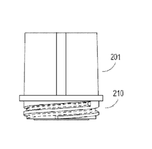
substantial dilution of a therapeutic agent such as an anticancer drug (e.g.,
a tyrosine kinase
inhibitor).
[0077] In certain instances, the present invention provides apparatuses and
methods for
isolating tumor cells from a homogenate, lysate, or cellular extract of a
solid tumor.
[0078] In particular embodiments, the apparatuses and methods of the present
invention
substantially remove plasma, which contains proteases and phosphatases that
can degrade or
__ desphosphorylate target proteins such as analytes of interest, and also
substantially remove
interfering proteins that can affect target protein assays.
[0079] FIG. 1 represents one embodiment of a method to isolate and harvest
tumor cells of
the present invention. Those of skill in the art will recognize other changes
and modifications
to the method within the scope of the present invention.
__ [0080] In one method 100, tumor cells from a patient such as a patient
suffering from CML
(optionally being treated) are isolated and harvested. As shown in FIG. 1,
whole blood is
collected 110 and filtered to remove red blood cells. In certain instances,
the whole blood
from patients can be treated or non-treated with an anticancer drug, such as a
BCR-ABL
19
CA 02826643 2013-08-06
WO 2012/154257 PCT/US2012/025491
inhibitor, prior to isolation. Advantageously, the methods herein ensure that
the amount and
concentration of the inhibitor or therapeutic agent present in the cells in
vivo is maintained in
vitro. In certain aspects, the present invention provides a method for
preparing a lysate of
leukocytes from a whole blood sample without substantial dilution or
essentially no dilution
of a therapeutic agent such as an anticancer drug. The collected whole blood
is loaded into
an apparatus as described herein. In certain aspects, the blood is freshly
drawn prior to
isolation of the leukocytes. If a fresh blood sample is unavailable, blood
samples can be
processed within a period such as 3 hours, 6 hours, 12 hours, 18 hours, 24
hours (1 day), 36
hours, 48 hours, and the like after being drawn. Samples are typically kept at
room
temperature prior to processing. In certain aspects, a protease and/or
phosphatase inhibitor
can be added to the blood sample 110. Thereafter, the blood is mixed by for
example, gently
inverting up and down in a tube or vial.
[0081] Afterwards, the erythrocytes are removed 121 typically by
centrifugation through a
filter or membrane. In certain aspects, an especially designed filtration
apparatus is used as
shown herein. Preferably, the erythrocytes are present in the collection tube
after
centrifugation. In one aspect, the method includes centrifuging the vial or
tube apparatus to
capture or isolate the leukocytes 142 on a filter membrane such as a stacked
collection of
filter membranes (one or more filters), and to separate red blood cells (and
plasma) into a
collection tube.
[0082] After filtration or centrifugation of the red blood cells (and plasma),
a lysis buffer is
used to lyse the captured leukocytes 167. In one aspect, protein later lysis
buffer can be used.
After capture, the leukocytes are thereafter lysed, but without a wash step
after capture to
thus prepare a lysate of leukocytes. The therapy concentration in the whole
blood cells is the
same before and after the procedure 100. In some instances, the therapy
concentration is 10
ILIM before procedure 100 and 10 ILIM after procedure 100. In other instances,
the therapy
concentration is 1 ILIM before procedure 100 and 1 ILIM after procedure 100.
In yet other
instances, the therapy concentration is 0.1 ILIM before procedure 100 and 0.1
ILIM after
procedure 100. The lysate is then collected 173 in a second collection tube,
e.g., by
centrifugation. The lysate from the leukocytes is without substantial dilution
or essentially no
dilution of a therapeutic agent such as an anticancer drug. That is, the in
vivo cellular
concentration of a therapeutic agent (e.g., anticancer drug) is essentially or
substantially the
same as the in vitro concentration of the therapeutic agent (e.g., anticancer
drug) in the cell
lysate.
CA 02826643 2013-08-06
WO 2012/154257 PCT/US2012/025491
[0083] In another aspect, the present invention provides a method for
preparing a lysate of
leukocytes (e.g., normal, malignant, and/or diseased leukocytes) from a whole
blood sample
without substantial dilution of a therapeutic agent (e.g., an anticancer
drug), the method
comprising:
(a) loading the whole blood sample into a cell isolation (filtration)
apparatus
such as an apparatus as described herein;
(b) centrifuging the apparatus to capture the leukocytes on the one or more
stacked filter membranes and to separate red blood cells (and plasma) into a
collection tube;
and
(c) lysing the leukocytes captured on the one or more stacked filter membranes
with lysis buffer but without a wash step between steps (b) and (c) to thereby
prepare a lysate
of leukocytes.
[0084] In certain embodiments, the method of the invention further comprises
replacing the
collection tube with a second collection tube between steps (b) and (c). In
certain other
embodiments, the method of the invention further comprises centrifuging the
apparatus
containing the second collection tube after lysing the leukocytes in step (c)
and collecting the
lysate of leukocytes in the second collection tube.
[0085] In some embodiments, the whole blood sample is obtained from a subject
receiving
a therapeutic agent (e.g., an anticancer drug). In other embodiments, the
whole blood sample
is incubated in vitro with a therapeutic agent (e.g., an anticancer drug)
prior to loading into
the apparatus.
[0086] In further embodiments, the whole blood sample is obtained from a
subject having
or suspected of having atherosclerosis or receiving treatment for
atherosclerosis (e.g., statin
therapy). In other embodiments, the whole blood sample is obtained from a
subject having or
suspected of having a cancer such as a hematological malignancy (e.g., a
leukemia such as
chronic myelogenous leukemia (CML)) or receiving treatment for the cancer
(e.g., anticancer
drug therapy).
[0087] In particular embodiments, the expression and/or activation level of at
least one
oncogenic fusion protein and/or signal transduction molecule is measured in
the lysate of
leukocytes. In preferred embodiments, the at least one oncogenic fusion
protein is BCR-
ABL. Additional examples of oncogenic fusion proteins and/or signal
transduction
molecules of interest are described herein.
21
CA 02826643 2013-08-06
WO 2012/154257 PCT/US2012/025491
[0088] As such, in one aspect, the present invention provides an apparatus for
isolating and
separating leukocytes (e.g., normal, malignant, and/or diseased leukocytes)
from red blood
cells (and plasma) in a whole blood sample, the apparatus comprising:
a filtration device comprising an upper chamber, a lower chamber, and one or
more stacked filter membranes between the upper and lower chambers, wherein
the one or
more stacked filter membranes are capable of retaining the leukocytes; and
a collection tube for collecting the red blood cells (and plasma) from the
whole blood sample, wherein the filtration device is placed on top of the
collection tube, and
wherein the red blood cells (and plasma) are separated from the leukocytes and
are collected
in the collection tube following centrifugation.
[0089] In some embodiments, the whole blood sample is loaded into the upper
chamber of
the filtration device. In other embodiments, the filtration device comprises
two, three, or four
stacked filter membranes. In certain embodiments, the upper chamber further
comprises a
snap-cap lid attached thereto.
[0090] In further embodiments, the apparatus further comprises a second
collection tube,
wherein the (first) collection tube containing the red blood cells (and
plasma) is replaced with
the second collection tube following (a first) centrifugation. In some
instances, a lysate of the
leukocytes is collected in the second collection tube following the addition
of lysis buffer to
the upper chamber and (a second) centrifugation. In particular instances, the
lysis buffer is
added to the upper chamber without washing the one or more stacked filter
membranes. In
some embodiments, the lysis buffer is incubated above the filter for at least
1, 5, 10, 15, 20,
30, 60, or 120 minutes, preferably between about 15 to about 30 minutes, at 4
C (or on ice)
prior to centrifugation and collection in the second collection tube.
[0091] In alternative embodiments, the apparatus further comprises a second
collection
tube, wherein the one or more stacked filter membranes are removed from the
filtration
device (e.g., with forceps) following centrifugation and placed into the
second collection
tube. In some instances, the second collection tube contains lysis buffer, and
the leukocytes
are lysed after the one or more stacked filter membranes are placed or
incubated into the
second collection tube.
[0092] In certain instances, the lysate prepared using the apparatus of the
present invention
comprises a cellular extract of normal and/or malignant (e.g., cancerous)
leukocytes such as
granulocytes (polymorphonuclear leukocytes), which include, e.g., neutrophils,
basophils,
and eosinophils; agranulocytes (mononuclear leukocytes), which include, e.g.,
peripheral
22
CA 02826643 2015-03-03
blood mononuclear cells such as lymphocytes and monocytes, leukemia cells,
which include,
e.g., chronic myelogenous leukemia (CML) cells; macrophages, which include,
e.g., foam
cells; and mixtures thereof.
[0093] In certain embodiments, the leukocytes, leukemia cells, foam cells,
circulating cells,
or other cells present in the whole blood sample can be stimulated in vitro
with one or more
growth factors before, during, and/or after incubation with one or more
therapeutic agents
such as one or more anticancer drugs of interest. Stimulatory growth factors
include, but are
not limited to, epidermal growth factor (EGF), heregulin (HRG), TGF-a, PIGF,
angiopoietin
(Ang), NRG 1, PGF, TNF-a, VEGF, PDGF, IGF, FGF, HGF, cytokines, and the like.
Protocols for the stimulation and lysis of cells found in whole blood are
described in PCT
Publication No. WO 2008/036802.
[00941 In certain embodiments, the whole blood sample is obtained from a
subject having
or suspected of having cancer. In some instances, the cancer may be caused by
the formation
of an oncogcnic fusion protein due to a chromosomal translocation in the
cancer cells. Non-
limiting examples of such cancers include a hematological malignancy, an
osteogenic
sarcoma, a soft tissue sarcoma, and combinations thereof. In particular
embodiments, the
hematological malignancy is a leukemia or lymphoma. In one preferred
embodiment, the
leukemia is chronic myelogenous leukemia (CM L). In other instances, the
subject is either
receiving or not receiving anticancer drug therapy.
[0095] In certain other embodiments, the anticancer drug comprises an anti-
signaling agent
(i.e., a cytostatic drug) such as a monoclonal antibody or a tyrosine kinase
inhibitor; an anti-
proliferative agent; a chemotherapeutic agent (i.e., a cytotoxic drug); a
hormonal therapeutic
agent; a radiotherapeutic agent; a vaccine; and/or any other compound with the
ability to
reduce or abrogate the uncontrolled growth of aberrant cells such as cancerous
cells. In some
embodiments, the isolated cells are treated with one or more anti-signaling
agents, anti-
proliferative agents, and/or hormonal therapeutic agents in combination with
at least one
chemotherapeutic agent.
[0096] Examples of anti-signaling agents include, without limitation,
monoclonal
antibodies such as trastuzumab (Herceptin ), alemtuzumab (Campath ),
bevacizumab
(Avastin ), cetuximab (Erbitux ), gemtuzumab (Mylotare), panitumumab
(VcctibixTM,
rituximab (Rituxae), and tositumomab (BEX.XAR ); tyrosine kinase inhibitors
such as
imatinib mesylate (Gleevec ), nilotinib (Tasigna ), dasatinib (Sprycel ),
bosutinib (SKI-
23
CA 02826643 2013-08-06
WO 2012/154257 PCT/US2012/025491
606), gefitinib (Iressac)), sunitinib (Sutentc)), erlotinib (Tarcevac)),
lapatinib (GW-572016;
Tykerbc)), canertinib (CI 1033), semaxinib (SU5416), vatalanib
(PTK787/ZK222584),
sorafenib (BAY 43-9006; Nexavarc)), leflunomide (SU101), ponatinib (AP24534),
and
vandetanib (ZACTIMATm; ZD6474); and combinations thereof.
[0097] Exemplary anti-proliferative agents include mTOR inhibitors such as
sirolimus
(rapamycin), temsirolimus (CCI-779), and everolimus (RAD001); Akt inhibitors
such as
1L6-hydroxymethyl-chiro-inosito1-2-(R)-2-0-methy1-3-0-octadecyl-sn-
glycerocarbonate, 9-
methoxy-2-methylellipticinium acetate, 1,3-dihydro-1-(1-((4-(6-pheny1-1H-
imidazo[4,5-
g]quinoxalin-7-yl)phenyl)methyl)-4-piperidiny1)-2H-benzimidazol-2-one, 10-(4'-
(N-
diethylamino)buty1)-2-chlorophenoxazine, 3-formylchromone thiosemicarbazone
(Cu(II)C12
complex), API-2, a 15-mer peptide derived from amino acids 10-24 of the proto-
oncogene
TCL1 (Hiromura et at., J. Biol. Chem., 279:53407-53418 (2004), KP372-1, and
the
compounds described in Kozikowski et at., J. Am. Chem. Soc., 125:1144-1145
(2003) and
Kau et at., Cancer Cell, 4:463-476 (2003); and combinations thereof
[0098] Non-limiting examples of chemotherapeutic agents include platinum-based
drugs
(e.g., oxaliplatin, cisplatin, carboplatin, spiroplatin, iproplatin,
satraplatin, etc.), alkylating
agents (e.g., cyclophosphamide, ifosfamide, chlorambucil, busulfan, melphalan,
mechlorethamine, uramustine, thiotepa, nitrosoureas, etc.), anti-metabolites
(e.g., 5-
fluorouracil, azathioprine, 6-mercaptopurine, methotrexate, leucovorin,
capecitabine,
cytarabine, floxuridine, fludarabine, gemcitabine (Gemzarc)), pemetrexed
(ALIMTAc)),
raltitrexed, etc.), plant alkaloids (e.g., vincristine, vinblastine,
vinorelbine, vindesine,
podophyllotoxin, paclitaxel (Taxor), docetaxel (Taxoterec)), etc.),
topoisomerase inhibitors
(e.g., irinotecan, topotecan, amsacrine, etoposide (VP16), etoposide
phosphate, teniposide,
etc.), antitumor antibiotics (e.g., doxorubicin, adriamycin, daunorubicin,
epirubicin,
actinomycin, bleomycin, mitomycin, mitoxantrone, plicamycin, etc.),
pharmaceutically
acceptable salts thereof, stereoisomers thereof, derivatives thereof, analogs
thereof, and
combinations thereof
[0099] Examples of hormonal therapeutic agents include, without limitation,
aromatase
inhibitors (e.g., aminoglutethimide, anastrozole (Arimidexc)), letrozole
(Femarac)), vorozole,
exemestane (Aromasinc)), 4-androstene-3,6,17-trione (6-0X0), 1,4,6-
androstatrien-3,17-
dione (ATD), formestane (Lentaronc)), etc.), selective estrogen receptor
modulators (e.g.,
bazedoxifene, clomifene, fulvestrant, lasofoxifene, raloxifene, tamoxifen,
toremifene, etc.),
steroids (e.g., dexamethasone), fmasteride, and gonadotropin-releasing hormone
agonists
24
CA 02826643 2013-08-06
WO 2012/154257 PCT/US2012/025491
(GnRH) such as goserelin, pharmaceutically acceptable salts thereof,
stereoisomers thereof,
derivatives thereof, analogs thereof, and combinations thereof.
[0100] Non-limiting examples of cancer vaccines include ANYARA from Active
Biotech,
DCVax-LB from Northwest Biotherapeutics, EP-2101 from IDM Pharma, GV1001 from
Pharmexa, 10-2055 from Idera Pharmaceuticals, INGN 225 from Introgen
Therapeutics and
Stimuvax from Biomira/Merck.
[0101] Examples of radiotherapeutic agents include, but are not limited to,
radionuclides
such as 47Sc, 64Cu, 67Cu, 89Sr, 86Y, 87Y, NY, 105Rh, 111Ag, 111In, 117msn,
149pm, 153sm, 166H05
1 =
177Lu, 186Re, 188Re, 211At, and 22 BI, optionally conjugated to antibodies
directed against
tumor antigens.
[0102] In certain other embodiments, the whole blood sample is obtained from a
subject
having or suspected of having atherosclerosis (also known as arteriosclerotic
vascular disease
or ASVD). Atherosclerosis is a disease typically affecting arterial blood
vessels, a chronic
inflammatory response in the walls of arteries, caused largely by the
accumulation of
macrophages such as foam cells and promoted by low-density lipoproteins
(plasma proteins
that carry cholesterol and triglycerides) without adequate removal of fats and
cholesterol
from the macrophages by functional high density lipoproteins (HDL). Examples
of drugs
suitable for the treatment of atherosclerosis include, without limitation,
statins such as
atorvastatin (Lipitor and Torvast), fluvastatin (Lescol), lovastatin (Mevacor,
Altocor,
Altoprev), mevastatin (Compactin), pitavastatin (Livalo, Pitava), pravastatin
(Pravachol,
Selektine, Lipostat), rosuvastatin (Crestor), simvastatin (Zocor, Lipex),
combinations thereof,
as well as combination preparations such as ezetimibe and simvastatin
(Vytorin), lovastatin
and niacin (Advicor), atorvastatin and amlodipine besylate (Caduet), and
simvastatin and
niacin (Simcor). In some instances, the subject is either receiving or not
receiving therapy
with an atherosclerosis drug such as a statin.
[0103] In other embodiments, the whole blood sample is incubated in vitro with
one or
more therapeutic agents such as one or more anticancer drugs prior to
isolation of leukocytes.
In particular embodiments, leukocytes that are retained or captured on the
filter membranes
comprise normal leukocytes, malignant leukocytes, or combinations thereof
[0104] In particular embodiments, the apparatuses of the invention provide for
preparing
the lysate or cellular extract from whole blood samples by recovering or
isolating cells of
interest such as malignant leukocytes (e.g., chronic myelogenous leukemia
(CML) cells)
without any wash steps after cell recovery or isolation. The cellular extract
thus obtained can
CA 02826643 2013-08-06
WO 2012/154257 PCT/US2012/025491
be analyzed for the level of expression and/or activation of one or more
oncogenic fusion
proteins such as BCR-ABL, substrates thereof, pathways thereof, or
combinations thereof.
Without being bound to any particular theory, eliminating the need for any
wash steps after
cell isolation is advantageous because cells of interest can be recovered from
blood without
changing the intracellular concentration of a therapeutic agent such as an
anticancer drug
(e.g., a tyrosine kinase inhibitor). As set forth in the Examples below, cell
isolation using the
apparatuses described herein without any wash steps is contrary to the art-
accepted practice
of washing cells after isolation and provides cellular extracts from recovered
cells without
substantial dilution of a therapeutic agent such as an anticancer drug (e.g.,
a tyrosine kinase
inhibitor such as, e.g., Gleevec , Tasigna , Spryce18, etc.) inside the cells.
[0105] In particular embodiments, the apparatuses of the present invention are
substantially
similar or identical to the apparatus depicted herein. One skilled in the art
will appreciate that
the dimensions of one or more components of the apparatus described herein and
illustrated
can be varied, taking into account parameters such as, for example, the volume
of sample to
be loaded into the apparatus, the type of centrifuge to be used to spin the
apparatus, the
volume of lysis buffer to be added to the upper chamber of the apparatus, etc.
[0106] Turning now to FIG. 2A-G, as shown therein, there is a filtration
device or
apparatus for sample collection. FIG. 2A is the upper portion or chamber of
the apparatus
201 which is a cylindrical tube with male helical ridges or threads 210. The
upper portion
with cap 215 is shown in FIG. 2B-C. This upper portion 201 can optionally have
a cap 215
that snaps shut to prevent spilling of the sample. In certain embodiments, the
snap-cap lid
215 is tethered via strap 217 to the upper portion and can be used to securely
close the
opening of the upper chamber after a sample and/or a reagent is added to the
filtration device.
The upper chamber 201 of the apparatus of the present invention preferably
attaches to a
lower camber portion or chamber 222 as shown in FIG. 2 D-E. The threads 210 of
the upper
portion fit securely into female grooves 221 of the lower portion or chamber
222. Preferably,
the inner diameters of the upper and lower chambers are similar as to create a
cylindrical tube
which allows liquids to pass therethrough.
[0107] In certain aspects, the lower chamber 222 of the filtration device or
apparatus is a
cylindrical tube with an internal screw thread at one end 221 (FIG. 2E). In
certain aspects,
one or more (e.g., a plurality such as two, three, four, five, six, seven,
eight, nine, ten, or
more) stacked filter membranes is placed between the screw threads of the
upper chamber
and lower chamber before the chambers are securely attached together. The
filter(s) sits on
pin wheel 225 of the lower chamber as shown in FIG. 2E.
26
CA 02826643 2013-08-06
WO 2012/154257 PCT/US2012/025491
[0108] In particular aspects, the filter membranes can be 2-4 (e.g., 2, 3, or
4) layers of
filters such as Pall filters (e.g., Leukosorb Medium). In other aspects, the
filtration device is
an assembly of separate chambers and filters which are joined together prior
to use such as in
a kit. The filtration device can be placed on top of a collection tube 250 as
shown in FIG. 2F-
G for the separation of red blood cells (and plasma) from a patient blood
sample. The
portions of FIG. 2A, FIG. 2D and FIG. 2F join or fit together to form one
embodiment of the
apparatus filtration device of the present invention. The filtration device
(upper and lower
chambers) can be placed on top of a collection vessel 250 for the separation
of red blood cells
from a patient blood sample. The lower chamber of the assembled filtration
device is
positioned in the collection vessel via opening 255 and the upper chamber is
on top of the
opening of the collection vessel. Examples of collection vessel 250 include,
but are not
limited to, tubes such as plastic culture tubes having a capacity of 1 ml, 2
ml, 3 ml, 4 ml, 5
ml, 6 ml, 7 ml, 8 ml, 14m1, 16 ml and the like.
[0109] In certain aspects, the lower portion of the filtration device of the
present invention
has a built-in or optional funnel in the lower portion. As shown in FIG. 3A-E,
the funnel may
have a certain angular dimension to ensure that the sample passes into and
through the lower
chamber into the collection tube without going down the inner wall of the
lower portion. For
example, FIG 3A shows a internal funnel 301 of about 2 off the horizontal.
FIG 3B shows
an internal funnel 310 of approximately 7 off the horizontal. In certain
other aspects, FIG.
3C shows a funnel approximately 12 off the horizontal. FIG. 3D and FIG. 3E
are yet other
embodiments of the funnel design of the present invention. A skilled artisan
will understand
and appreciate that the funnel design can be any angle such that the filtrate
stays off the walls
of the lower portion. Suitable angles include 105 205305 405505 605 705 805
905 1005 1105 1205
13 , 14 , 15 , 16 , 17 , 18 , 19 , 20 or more for the funnel portion. In an
alternative
embodiment, FIG. 3E shows a funnel insert 325 into the lower portion of a
filtration device of
the present invention.
[0110] In certain aspects, the filtration device of the present invention is
shown in FIG. 4A-
D. FIG. 4A shows the exploded view of the upper portion or chamber 410, a
lower portion or
chamber 420 and a filter stack 412 in-between. In FIG. 4A, view 405 is looking
down on the
device and view 422 is looking up at the device. FIG. 4B shows the upper
portion 430 and
the lower chamber or portion 420 screwed together. In FIG. 4B, view 405 is
looking down
on the device and view 425 is looking up at the device. FIG. 4C shows
collection tube 435.
In operation, in certain aspects, collection tube 435 preferably holds red
blood cells. In FIG.
4C, view 434 is looking down on the device and view 440 is looking up at the
device. FIG.
27
CA 02826643 2013-08-06
WO 2012/154257 PCT/US2012/025491
4D depicts the collection tube 435 joined with the upper chamber 410 and lower
chamber or
portion 420. In FIG. 4D, view 405 is looking down on the device and view 461
is looking up
at the device.
[0111] In some embodiments, in operation, a volume (e.g., 1 ml) of patient
blood treated
with a solution mixture comprising a protease and/or a phosphatase inhibitors
is loaded into
the upper chamber of an assembled cell isolation apparatus. The apparatus can
be
centrifuged in a tabletop or clinical centrifuge, such as an Allegra 6R
centrifuge (Beckman),
Sorvall Legend centrifuge (Thermo Scientific), or Heraeus Megafuge centrifuge
(Kendro). In
some aspects, the apparatus is centrifuged for 5-30 minutes at 600-2,000 rpms
at 4 C. After
centrifugation, the collection vessel containing the red blood cells (and
plasma) is removed
from the cell isolation apparatus, capped and set aside.
[0112] In further embodiments, a second collection vessel is attached to the
filtration
device. Non-limiting examples of a second collection tube for the cell lysate
include 1.5 ml
and 2 ml microcentrifuge tubes. Without a washing step, lysis buffer is added
to the upper
chamber of the filtration device. The upper chamber is capped and the
filtration device and
second collection tube are shaken vigorously. In certain instances, the cell
isolation
apparatus is incubated at 4 C for at least 1, 5, 10, 15, 20, 30, 60, or 120
minutes, preferably
between about 15 to about 30 minutes. The apparatus can then be centrifuged
(e.g., 3,000rpm
for 5 min). The cell lysate can be transferred to another centrifuge vessel
such as a
microcentrifuge tube for storage at -70 C.
[0113] FIG. 5A-D show an alternative embodiment of the filtration device or
apparatus 500
for sample collection of the present invention. In this embodiment, there is
an upper portion
501 and a bottom portion or chamber 530 with a sleeve or middle connector 522.
The middle
connector or sleeve 522 can optionally be fixed to either the upper 501 or
bottom portion 530.
As shown in FIG. 5B, in certain aspects, the upper portion has ridges or male
ridges and can
be fixed to the sleeve or bottom portion. In FIG. 5B, the apparatus is shown
with cap 510.
FIG. 5C shows that the bottom portion 530 can optionally have a cap 534 to
keep this portion
sealed. FIG. 5D depicts a lower chamber or portion 522 clearly showing female
grooves.
This portion can optionally be fixed to the bottom portion such that the
sleeve in integral to
the bottom portion of the device.
[0114] FIG. 6A-C show yet another aspect of the present invention. FIG. 6 A is
a cross-
sectional view of a device of the present invention, wherein the bottom
portion 610 is joined
with the upper portion 625 with sleeve 633 enabling joinder. Inset FIG. 6B is
a close up of
28
CA 02826643 2013-08-06
WO 2012/154257 PCT/US2012/025491
the portions joined together with threads 625 and middle section 633 with a
funnel section,
Collection chamber 610 is also shown. FIG. 6C shows middle section with
pinwheel
geometry. The filter stack optionally sits on this pinwheel.
[0115] In other further embodiments, after the cell isolation apparatus is
centrifuged and
the red blood cells are collected and set aside in the collection tube, the
upper chamber and
lower chamber of the filtration device are separated (e.g., unscrewed). Using
forceps, the
filter membranes 422 (FIG. 4A) containing the separated leukocytes and/or
circulating tumor
cells are placed in a second collection vessel containing cell lysis buffer.
Non-limiting
examples of a second collection vessel include 1.5 ml and 2 ml microcentrifuge
tubes. In
some instances, the second collection vessel is further incubated at 4 C for
at least 1, 5, 10,
15, 20, 30, 60, or 120 minutes, preferably between about 15 to about 30
minutes. In other
aspects, the second collection vessel is placed on ice and briefly vortexed
for 10 seconds
every 10 minutes for a total of 30 minutes. In some embodiments, the cell
lysate is stored at -
70 C. In other embodiments, the vessel containing the lysate is centrifuged to
remove the
filter membranes and cell fragments. The supernatant of the cell lysate can be
transferred to
another tube for storage at -70 C.
[0116] In some embodiments, the apparatuses of the invention are provided as a
sterile kit.
In some instances, the sterile kit comprises a filtration device comprising an
upper chamber
(optionally with an attached snap-cap lid), lower chamber and a stack of one
or more filter
membranes (e.g, 1, 2, 3, or 4 layers of filter membrane), and one or a
plurality of collection
tubes (optionally with snap-cap lids). Each of the components can be packaged
separately
and the kit assembled or each of the components can be placed in to a sterile
package. In
other instances, the sterile kit comprises a tube filter unit and one or a
plurality of collection
tubes (optionally with snap-cap lids). The tube filter unit comprises a
cylindrical tube affixed
at one end with one or more (e.g., a plurality of two, three, four, five, six,
seven, eight, nine,
ten, or more) filter membranes, wherein the membranes are able to retain
healthy and
malignant leukocytes and/or circulating tumor cells from whole blood samples.
In particular
instances, the filter membranes are (e.g., 2, 3, or 4) layers of filters such
as PALL filters (e.g.,
Leukosorb Medium). In yet other instances, the sterile kit comprises a tube
filter unit, a
plastic adaptor and one or a plurality of collection tubes (optionally with
snap-cap lids). The
adaptor is positioned in the opening of a collection tube and securely
attached to the filter
membrane of the tube filter unit.
[0117] In further embodiments, the apparatus of the present invention
comprises a plurality
of filtration devices or tube filter units and a multi-well or multi-tube
collection vessel such
29
CA 02826643 2013-08-06
WO 2012/154257 PCT/US2012/025491
as a 2-well, 12-well, 24-well, 48-well, or 96-well plate. In certain
instances, the plurality of
filtration devices can be substantially similar to at least two or more of the
devices as
depicted in FIG. 4D. In certain other embodiments, the array of filtration
devices can be a
multi-well plate such as a 96-well cell isolation plate. For example, a first
96-well filtration
plate can be fitted with filter membranes (e.g., LeukoLOCK (Life
Technologies), or Acroprep
(PALL) or Leukosorb (PALL)) or membranes substantially similar thereto. In
other
embodiments, the first 96-well filtration plate is substantially similar to or
is a commercially
available multi-well plate fitted with filter membranes, such as but not
limited to, Millipore
Cat. #MAMIC8510 and #MSBCS1210. In some instances, the commercially available
multi-
well plates are sterile and comprise a first multi-well plate fitted with
filter membranes and a
second multi-well plate that fits under the first multi-well plate and can be
used as a
collection vessel. In these embodiments, fresh collected blood can be loaded
into wells of a
first 96-well cell isolation plate. The first multi-well plate is analogous to
both the upper and
lower chambers of the filtration device discussed above. A second 96-well
microplate can
serve as a blood collection plate (which is analogous to the collection tube)
and can be placed
under the first multi-well plate i.e., the cell isolation plate with filter
membranes. This plate
assembly can be centrifuged at room temperature for about 5 min at a speed
ranging from
about 600 rpm to 3,000 rpm, such as e.g., about 600 rpm, 1,000 rpm, 2,000 rpm,
and 3,000
rpm. After centrifugation, the filter membrane can be transferred to a
centrifugation tube and
the cells on the filter membrane can be treated with a volume of lysis buffer
(e.g., 300 1 of
lysis buffer) and vortexed briefly, in order to lyse the cells. The
centrifugation tube
containing the cell lysate can be placed on ice for about 30 min and subjected
to brief
vortexing about every 10 minutes. Thereafter the tube can be centrifuged for
about 15
minutes to separate the cellular debris from the supernatant. The supernatant
containing the
lysate can be collected and analyzed. In certain aspects, the plurality of
filtration devices are
manipulated with robotic armature under computer control. High throughput
sample analysis
is carried-out and the plurality of samples is analyzed. Steps of procedure
100, such as the
addition of lysis buffer to lyse the captured leukocytes 167, are optionally
performed with a
computerized robotic system. In some aspects, in vitro treatment of patient
blood sample
with anticancer drug and sample analysis as described herein are performed in
a high
throughput manner.
IV. Drug Selection and Optimization for Cancer Therapy
[0118] In certain aspects, the present invention provides methods for
monitoring the
efficacy of cancer therapy in subjects with a hematological malignancy. In
certain aspects,
CA 02826643 2013-08-06
WO 2012/154257 PCT/US2012/025491
the present invention provides methods for the selection of appropriate
therapies to down-
regulate or shut down one or more deregulated signaling pathways. In certain
other aspects,
the present invention provides methods for optimizing therapy and/or reducing
toxicity in a
subject having cancer and receiving a course of therapy for the treatment of
cancer. Thus, the
present invention may be used to facilitate the design of personalized
therapies based on the
particular molecular signature provided by the collection of activated
oncogenic fusion
proteins and/or signal transduction proteins in a given patient's cancer or
tumor.
[0119] Accordingly, in one particular aspect, the present invention provides a
method for
monitoring the efficacy of an anticancer drug in a subject, wherein the
subject has a
hematological malignancy, comprising:
(a) administering the anticancer drug to the subject, wherein the first
administration of the anticancer drug is at time T1;
(b) isolating cells of a cancer at a time T2 in a sample from the subject;
(c) lysing the isolated cells to produce a cellular extract;
(d) measuring the activation state and or expression level of an oncogenic
fusion protein at a time T2 in a sample from the subject; and
determining a course of treatment based upon the activation state and or
expression level of the oncogenic fusion protein.
[0120] In certain embodiments, the method further comprises measuring the
activation state
of the oncogenic fusion protein at To, i.e., prior to the first administration
of the anticancer
drug. In some instances, the oncogenic fusion protein is BCR-ABL. In certain
instances, the
hematological malignancy is a lymphoma or a leukemia such as chronic
myelogenous
leukemia (CML). The time difference between T1 and T2 is about 1 week to about
6 months
such as 1, 2, 3, 4, 5, 6, 7, 8, 9, 10, 11, 12, 13, 14, 15, 16, 17, 18, 19, 20,
21, 22, 23, or 24
week(s). The time difference between To and T1 is about 1 day to about 3
weeks. In certain
other aspects, the methods further include measuring expression and or
activation levels of at
least one other signal transduction molecule such as CRKL, AKT, STAT5 and SRC.
[0121] In certain aspects, the course of treatment is selected from changing
the anticancer
drug dose, changing the anti-cancer drug, including an additional anticancer
drug, changing
the length of treatment and staying the existing course of treatment.
[0122] In certain aspects, the sample comprises an extract of isolated cells.
In certain
aspects, the isolated cells are incubated in vitro with at least one
anticancer drug (e.g., 2
anticancer drugs) at To (prior to initiation of treatment). In other
instances, the isolated cells
31
CA 02826643 2013-08-06
WO 2012/154257 PCT/US2012/025491
are incubated in vitro with at least two anticancer drugs at T2, prior to
determining the course
of treatment.
[0123] In yet other embodiments, the present invention provides a method for
selecting an
anticancer drug in a subject having a hematological malignancy:
(a) isolating cells of a cancer from a subject;
(b) lysing the isolated cells to produce a cellular extract;
(c) measuring the activation state level of an oncogenic fusion protein in an
isolated cell from a sample from the subject;
(d) incubating the isolated cell with at least one anticancer drug prior to
initiation of treatment;
(e) lysing the isolated cells incubated with at least one anticancer drug
prior to
initiation of treatment to produce a cellular extract;
(f) measuring the activation state level of the oncogenic fusion protein in
the
incubated cells; and
(g) selecting a course of treatment based upon the activation state level of
the
oncogenic fusion protein.
[0124] In another embodiment, the present invention provides a method for
selecting an
anticancer drug in a subject having a hematological malignancy:
(a) isolating cells of a cancer from a subject;
(b) lysing the isolated cells to produce a cellular extract;
(c) measuring the activation state level of BCR-ABL in an isolated cell from a
sample from the subject;
(d) incubating the isolated cell with at least one anticancer drug prior to
initiation of treatment;
(e) lysing the isolated cells incubated with at least one anticancer drug
prior to
initiation of treatment to produce a cellular extract;
(f) measuring the activation state level of BCR-ABL in the incubated cells;
and
(g) selecting a course of treatment based upon the activation state level of
BCR-ABL.
[0125] In certain aspects, the course of treatment is selected from the group
consisting of
selecting the anticancer drug, selecting the anticancer dose, and determining
the length of
treatment. In certain other aspects, the methods further include measuring
expression and or
32
CA 02826643 2013-08-06
WO 2012/154257 PCT/US2012/025491
activation levels of at least one other signal transduction molecule such as
CRKL, AKT,
STAT5 and SRC.
[0126] In another aspect, the present invention provides a method for
optimizing therapy
and/or reducing toxicity in a subject having cancer and receiving a course of
therapy for the
treatment of cancer, the method comprising:
(a) isolating cancer cells after administration of an anticancer drug (e.g.,
one
or more tyrosine kinase inhibitors such as Gleevec , Tasignac), Sprycel ,
etc.);
(b) lysing the isolated cells to produce a cellular extract;
(c) measuring a level of expression and/or activation (e.g., phosphorylation)
of
an oncogenic fusion protein in the cellular extract using an assay described
herein; and
(d) comparing the measured level of expression and/or activation of the
oncogenic fusion protein to a level of expression and/or activation of the
oncogenic fusion
protein measured at an earlier time during the course of therapy; and
(e) determining a subsequent dose of the course of therapy for the subject or
whether a different course of therapy should be administered to the subject
based upon the
comparison from step (d).
[0127] In particular embodiments, both total and activated (e.g.,
phosphorylated) oncogenic
fusion protein (e.g., BCR-ABL) levels are measured in the cellular extract in
accordance with
the antibody-based assays of the present invention and a ratio of activated to
total oncogenic
fusion protein levels (e.g., ratio of phospho/total BCR-ABL protein levels)
can be calculated
and used to evaluate the course of therapy for a subject, e.g., by comparing
the phospho/total
ratio of oncogenic fusion protein levels to a ratio of the same calculated for
the subject at an
earlier time (e.g., at an earlier time while on anticancer drug therapy or at
a point in time prior
to anticancer drug therapy).
[0128] In another aspect, the present invention provides a method for
selecting a suitable
anticancer drug for the treatment of a cancer, the method comprising:
(a) isolating cells of a cancer after administration of an anticancer drug, or
prior to incubation with an anticancer drug;
(b) lysing the isolated cells to produce a cellular extract;
(c) determining a level of expression and/or activation (e.g.,
phosphorylation)
of an oncogenic fusion protein in the cellular extract using an assay
described herein; and
(d) determining whether the anticancer drug is suitable or unsuitable for the
treatment of the cancer by comparing the level of expression and/or activation
detected for
33
CA 02826643 2013-08-06
WO 2012/154257 PCT/US2012/025491
the oncogenic fusion protein with a reference expression and/or activation
profile generated
in the absence of the anticancer drug.
[0129] In a preferred embodiment, the method for selecting a suitable
anticancer drug for
the treatment of a cancer comprises:
(a) isolating cells of a cancer after administration of an anticancer drug, or
prior to incubation with an anticancer drug;
(b) lysing the isolated cells to produce a cellular extract;
(c) determining a level of expression and/or activation (e.g.,
phosphorylation)
of an oncogenic fusion protein in the cellular extract using an assay
comprising a dilution
series of capture antibodies specific for the oncogenic fusion protein,
wherein the capture
antibodies are restrained on a solid support;
(d) comparing the level of expression and/or activation detected for the
oncogenic fusion protein with a reference expression and/or activation profile
generated in
the absence of the anticancer drug; and
(e) indicating that the anticancer drug is suitable for the treatment of the
cancer when the level of expression and/or activation detected for the
oncogenic fusion
protein is changed (e.g., substantially decreased) compared to the reference
expression and/or
activation profile.
[0130] In some embodiments, the methods of the present invention may be useful
to aid or
assist in the selection of a suitable anticancer drug for the treatment of a
cancer such as, e.g.,
a hematological malignancy. In other embodiments, the methods of the present
invention
may be useful for improving the selection of a suitable anticancer drug for
the treatment of a
cancer such as, e.g., a hematological malignancy. In certain embodiments, the
method
further or alternatively comprises the step of indicating that the anticancer
drug is unsuitable
for the treatment of the cancer when the level of expression and/or activation
detected for the
oncogenic fusion protein is not changed (e.g., not substantially decreased)
compared to the
reference expression and/or activation profile. In further embodiments, one or
more signal
transduction molecules present in the cellular extract are detected in
addition to one or more
oncogenic fusion proteins, and the anticancer drug is determined to be
suitable or unsuitable
based on this "molecular profile."
[0131] In yet another aspect, the present invention provides a method for
identifying the
response of a cancer to treatment with an anticancer drug, the method
comprising:
(a) isolating cells of a cancer after administration of an anticancer drug, or
prior to incubation with an anticancer drug;
34
CA 02826643 2013-08-06
WO 2012/154257
PCT/US2012/025491
(b) lysing the isolated cells to produce a cellular extract;
(c) determining a level of expression and/or activation (e.g.,
phosphorylation)
of an oncogenic fusion protein in the cellular extract using an assay
described herein; and
(d) identifying the cancer as responsive or non-responsive to treatment with
the anticancer drug by comparing the level of expression and/or activation
detected for the
oncogenic fusion protein with a reference expression and/or activation profile
generated in
the absence of the anticancer drug.
[0132] In a preferred embodiment, the method for identifying the response of a
cancer to
treatment with an anticancer drug comprises:
(a) isolating cells of a cancer after administration of an anticancer drug, or
prior to incubation with an anticancer drug;
(b) lysing the isolated cells to produce a cellular extract;
(c) determining a level of expression and/or activation (e.g.,
phosphorylation)
of an oncogenic fusion protein in the cellular extract using an assay
comprising a dilution
series of capture antibodies specific for the oncogenic fusion protein,
wherein the capture
antibodies are restrained on a solid support;
(d) comparing the level of expression and/or activation detected for the
oncogenic fusion protein with a reference expression and/or activation profile
generated in
the absence of the anticancer drug; and
(e) indicating that the cancer is responsive to treatment with the anticancer
drug when the level of expression and/or activation detected for the oncogenic
fusion protein
is changed (e.g., substantially decreased) compared to the reference
expression and/or
activation profile.
[0133] In some embodiments, the methods of the present invention may be useful
to aid or
assist in the identification of the response of a cancer such as, e.g., a
hematological
malignancy, to treatment with an anticancer drug. In other embodiments, the
methods of the
present invention may be useful for improving the identification of the
response of a cancer
such as, e.g., a hematological malignancy, to treatment with an anticancer
drug. In certain
embodiments, the method further or alternatively comprises the step of
indicating that the
cancer is non-responsive to treatment with the anticancer drug when the level
of expression
and/or activation detected for the oncogenic fusion protein is not changed
(e.g., not
substantially decreased) compared to the reference expression and/or
activation profile. In
further embodiments, one or more signal transduction molecules present in the
cellular
CA 02826643 2013-08-06
WO 2012/154257 PCT/US2012/025491
extract are detected in addition to one or more oncogenic fusion proteins, and
the cancer is
identified as responsive or non-responsive to treatment based on this
"molecular profile."
[0134] In still yet another aspect, the present invention provides a method
for predicting the
response of a subject having cancer to treatment with an anticancer drug, the
method
comprising:
(a) isolating cells of a cancer after administration of an anticancer drug, or
prior to incubation with an anticancer drug;
(b) lysing the isolated cells to produce a cellular extract;
(c) determining a level of expression and/or activation (e.g.,
phosphorylation)
of an oncogenic fusion protein in the cellular extract using an assay
described herein; and
(d) predicting the likelihood that the subject will respond to treatment with
the
anticancer drug by comparing the level of expression and/or activation
detected for the
oncogenic fusion protein with a reference expression and/or activation profile
generated in
the absence of the anticancer drug.
[0135] In a preferred embodiment, the method for predicting the response of a
subject
having cancer to treatment with an anticancer drug comprises:
(a) isolating cells of a cancer after administration of an anticancer drug, or
prior to incubation with an anticancer drug;
(b) lysing the isolated cells to produce a cellular extract;
(c) determining a level of expression and/or activation (e.g.,
phosphorylation)
of an oncogenic fusion protein in the cellular extract using an assay
comprising a dilution
series of capture antibodies specific for the oncogenic fusion protein,
wherein the capture
antibodies are restrained on a solid support;
(d) comparing the level of expression and/or activation detected for the
oncogenic fusion protein with a reference expression and/or activation profile
generated in
the absence of the anticancer drug; and
(e) indicating that the subject will likely respond to treatment with the
anticancer drug when the level of expression and/or activation detected for
the oncogenic
fusion protein is changed (e.g., substantially decreased) compared to the
reference expression
and/or activation profile.
[0136] In some embodiments, the methods of the present invention may be useful
to aid or
assist in the prediction of a subject's likelihood of responding to treatment
with an anticancer
drug for a cancer such as, e.g., a hematological malignancy. In other
embodiments, the
methods of the present invention may be useful for improving the prediction of
a subject's
36
CA 02826643 2013-08-06
WO 2012/154257 PCT/US2012/025491
likelihood of responding to treatment with an anticancer drug for a cancer
such as, e.g., a
hematological malignancy. In certain embodiments, the method further or
alternatively
comprises the step of indicating that the subject will not likely respond to
treatment with the
anticancer drug when the level of expression and/or activation detected for
the oncogenic
fusion protein is not changed (e.g., not substantially decreased) compared to
the reference
expression and/or activation profile. In further embodiments, one or more
signal transduction
molecules present in the cellular extract are detected in addition to one or
more oncogenic
fusion proteins, and the likelihood that the subject will respond to treatment
is predicted
based on this "molecular profile."
[0137] In a further aspect, the present invention provides a method for
determining whether
a subject having cancer is resistant to treatment with an anticancer drug, the
method
comprising:
(a) isolating cells of a cancer after administration of an anticancer drug, or
prior to incubation with an anticancer drug;
(b) lysing the isolated cells to produce a cellular extract;
(c) determining a level of expression and/or activation (e.g.,
phosphorylation)
of an oncogenic fusion protein in the cellular extract using an assay
described herein; and
(d) determining whether the subject is resistant or sensitive to treatment
with
the anticancer drug by comparing the level of expression and/or activation
detected for the
oncogenic fusion protein with a reference expression and/or activation profile
generated in
the absence of the anticancer drug or in the presence of the anticancer drug
at an earlier time.
[0138] In a preferred embodiment, the method for determining whether a subject
having
cancer is resistant to treatment with an anticancer drug comprises:
(a) isolating cells of a cancer after administration of an anticancer drug, or
prior to incubation with an anticancer drug;
(b) lysing the isolated cells to produce a cellular extract;
(c) determining a level of expression and/or activation (e.g.,
phosphorylation)
of an oncogenic fusion protein in the cellular extract using an assay
comprising a dilution
series of capture antibodies specific for the oncogenic fusion protein,
wherein the capture
antibodies are restrained on a solid support;
(d) comparing the level of expression and/or activation detected for the
oncogenic fusion protein with a reference expression and/or activation profile
generated in
the absence of the anticancer drug or in the presence of the anticancer drug
at an earlier time;
and
37
CA 02826643 2013-08-06
WO 2012/154257 PCT/US2012/025491
(e) indicating that the subject is resistant to treatment with the anticancer
drug
when the level of expression and/or activation detected for the oncogenic
fusion protein is not
changed (e.g., not substantially decreased) compared to the reference
expression and/or
activation profile.
[0139] In some embodiments, the methods of the present invention may be useful
to aid or
assist in the identification of a subject having cancer who is resistant to
treatment with an
anticancer drug or in the determination of whether a subject having cancer is
resistant to
treatment with an anticancer drug, wherein the subject has a cancer such as,
e.g., a
hematological malignancy. In other embodiments, the methods of the present
invention may
be useful for improving the identification of a subject having cancer who is
resistant to
treatment with an anticancer drug or the determination of whether a subject
having cancer is
resistant to treatment with an anticancer drug, wherein the subject has a
cancer such as, e.g., a
hematological malignancy.
[0140] In certain embodiments, the method further or alternatively comprises
the step of
indicating that the subject is sensitive to treatment with the anticancer drug
when the level of
expression and/or activation (e.g., phosphorylation) detected for the
oncogenic fusion protein
is changed (e.g., substantially decreased) compared to the reference
expression or activation
profile. Non-limiting examples of reasons why a subject having cancer would be
resistant to
treatment with an anticancer drug include the presence of one or more
mutations in the
oncogenic fusion protein of interest (e.g., BCR-ABL), non-compliance with the
therapeutic
regimen, and/or administration of a suboptimal drug dose. With regard to a
suboptimal drug
dose of the anticancer drug, the method can further comprise the step of
increasing the next
or subsequent dose of the anticancer drug administered to the subject. In
further
embodiments, one or more signal transduction molecules present in the cellular
extract are
detected in addition to one or more oncogenic fusion proteins, and the subject
is identified as
resistant or sensitive to treatment based on this "molecular profile."
V. Oncogenic Fusion Proteins
[0141] In particular embodiments, expression/activation profiling of one or
more oncogenic
fusion proteins, alone or in combination with expression/activation profiling
of substrates
thereof and/or other signal transduction pathway proteins can be performed on
cell lysates
prepared using the apparatuses and methods of the present invention, e.g., to
determine the
efficacy of tyrosine kinase inhibitor therapy for patients in need thereof
(e.g., patients with a
BCR-ABL mediated disease such as chronic myelogenous leukemia). The oncogenic
fusion
38
CA 02826643 2013-08-06
WO 2012/154257 PCT/US2012/025491
proteins and other analytes are advantageously interrogated in cell lysates
prepared by the
apparatuses and methods of the invention without changing the intracellular
concentration of
the tyrosine kinases inhibitor.
[0142] In certain embodiments, translocations in human tumors that cause the
formation of
oncogenic fusion proteins and their associated neoplasms include, but are not
limited to, the
following:
[0143] Chronic myelogenous leukemia (CML): Philadelphia chromosome is a
translocation which results in BCR/ABL (kinase).
[0144] Acute lymphoblastic leukemia (ALL): Chimeric oncogenic proteins
include:
Cytogenetic translocation Molecular genetic abnormality %
cryptic t(12;21) TEL/AML1 (kinase) 25.4%
t(1;19)(q23;p13) E2A/PBX (PBX1) 4.8%
t(9;22)(q34;q11) BCR/ABL fusion (P185) 1.6%
t(4;11)(q21;q23) MLL/AF4 fusion 1.6%
t(8;14)(q24;q32) IGH/MYC fusion
t(11;14)(p13;q11) TCR/RBTN2 fusion
[0145] Burkitt's lymphoma: c-myc gene translocation t(8;14)(q24;q32). The most
common chimeric oncoprotein is c-myc/IGH.
[0146] AML: translocation of a part of chromosome 8 to chromosome 21 The
resulting
chimeric oncoprotein is RUNX1/ETO. Another translocation t(12;15)(p13;q25)
results in the
TEL/TrkC (kinase) chimeric oncoprotein.
[0147] Ewing sarcoma: translocation between chromosomes 11 and 22. The
resulting
chimeric oncoprotein is EWS/FLI (transcription factor).
[0148] DFSP: Over 95% of DFSP tumors have the chromosomal translocation
t(17;22),
which results in the chimeric oncoprotein COL lAl/PDGF (binds and activates
PDGFR).
[0149] Acute promyelocytic leukemia: a translocation denoted as
t(15;17)(q22;q12). The
resulting chimeric oncoprotein is RARa/PML (transcription complex protein).
[0150] Pro-B-cell acute lymphoblastic leukemia: translocation t(17;19), which
results in
the chimeric oncoprotein E2A/HLF (apoptosis inhibitor).
39
CA 02826643 2013-08-06
WO 2012/154257 PCT/US2012/025491
[0151] Acute pre-B-cell leukemia: translocation t(1;19). The chimeric
oncoprotein is
E2A/Pbx1 (kinase substrate).
[0152] Rhabdomyosarcoma : translocation of t(2:13)(q35;q14), which results in
the
chimeric oncoprotein PAX3/FKHR (transcription factor).
[0153] A soft tissue malignancy of very young children: t(12;15)(p13;q25)
rearrangement
which results in the following chimeric oncoprotein: protein tyrosine kinase
ETV6/NTRK3
(kinase).
[0154] Papillary thyroid carcinoma: the chimeric oncoprotein is RET/PTC
(kinase).
[0155] Prostate cancer: the chimeric oncoprotein is TMRSS/ERG (kinase).
[0156] Additional examples of translocations in human tumors that cause the
formation of
oncogenic fusion proteins and their associated neoplasms:
'/erg rnyIoid leukemia
O.t1.:;?!*f44:VM.iiiikM.40W09.'4V*04K000ffiiinirbgSMSMign!;Ni!;!M
ci(kk.000imNim.]mi*000i0000moggg!v.sim.].mmgsgig.].]]]!!Nommfim
:poovotomiktm*oo-0000.i:.omaggw.NgomigiAik]]]]Hignii]
:P.?,t?-f.W.AP.MZMMi0.0VOIPPi;t.Y.PIPPq..O.WaMkiggi*gZPE!:!:!:!aMMPMW
Adapted from G.M. Cooper, Oncogenes, 2n0 ed. Boston and London: Jones and
Bartlett, 1995.
VI. Examples
[0157] The following examples are offered to illustrate, but not to limit, the
claimed
invention.
Example 1. Exemplary Cell Isolation Apparatuses.
[0158] This example describes exemplary cell isolation apparatuses for the
separation of
leukocytes from patient whole blood using a filtration method. Embodiments and
aspects of
a cell isolation apparatus of the present invention are depicted in FIG. 2-6.
[0159] The cell isolation apparatus of the present invention is used to
separate red blood
cells from other cells present in whole blood. In particular, leukocytes
and/or circulating
CA 02826643 2013-08-06
WO 2012/154257 PCT/US2012/025491
tumor cells are separated from significant numbers of red blood cells and
plasma present in
whole blood. The cell isolation apparatus of the present invention comprises a
filtration
device and a collection vessel. The filtration device is an assembly
comprising an upper
chamber, a lower chamber and one or more (e.g., a plurality of two, three,
four, five, six,
seven, eight, nine, ten, or more) stacked filter membranes between the upper
and lower
chambers. The upper and lower chambers can be manufactured from materials such
as, but
not limited to, polypropylene and polystyrene. In certain embodiments, the
filter membrane
has a pore size of 8 m and a thickness of 355.6-558.8 m, and has a leukocyte
retention
yield of 70-80%. A non-limiting example of a filter membrane includes White
Blood Cell
Isolation (Leukosorb) Medium (PALL Cat. No. BSP0669).
[0160] Typically, 1 ml of patient blood treated with protease inhibitors and
phosphatase
inhibitors is loaded into the upper chamber of the cell isolation apparatus.
The unit is
centrifuged for 5-30 minutes at 600-2000 rpms (e.g., 800 rpms) at 4 C in a
tabletop centrifuge
such as an Allegra 6R centrifuge (Beckman), Sorvall Legend centrifuge (Thermo
Scientific),
or Heraeus Megafuge centrifuge (Kendro). In certain instances, the first
collection vessel
containing the red blood cells is removed from the cell isolation apparatus
and a second
collection vessel is attached. Without washing steps, 200 1-1 ml of lysis
buffer is added to
the upper chamber of the filtration device. The upper chamber is capped and
the filtration
device and second collection vessel are shaken vigorously for 15-30 minutes at
4 C. The
filtration device and second collection vessel are placed in a centrifuge and
spun at about
3,000 rpm for about 5 minutes. The cell lysate can be transferred to another
vessel for
storage at -70 C.
[0161] In other instances, the first collection vessel containing the red
blood cells is
removed from the cell isolation apparatus and the filtration device containing
the filter
membranes is disassembled. The upper chamber and the lower chamber are
unscrewed to
separate them. Using forceps, the filter membranes containing the separated
leukocytes are
placed in a second collection vessel containing lysis buffer. Non-limiting
examples of a
second collection vessel include 1.5 ml and 2 ml microcentrifuge tubes. In
some instances,
the second collection vessel is further incubated at 4 C for at least 1, 5,
10, 15, 20, 30, 60, or
120 minutes, preferably between about 15 to about 30 minutes. In other
instances, the second
collection vessel is placed on ice and briefly vortexed for 10 seconds every
10 minutes for a
total of 30 minutes. In some embodiments, the cell lysate is stored at -70 C.
In other
embodiments, the vessel containing the lysate is centrifuged to remove the
filter membranes
41
CA 02826643 2013-08-06
WO 2012/154257 PCT/US2012/025491
and cell fragments. The supernatant of the cell lysate is transferred to
another tube for
storage at -70 C.
[0162] In yet another embodiment, the cell isolation apparatus comprises a
tube filter unit
and a collection vessel (FIG. 26). The tube filter unit comprises a
cylindrical tube affixed at
one end with one or more (e.g., a plurality of two, three, four, five, six,
seven, eight, nine, ten,
or more) filter membranes, wherein the membranes are able to retain healthy
and malignant
leukocytes and/or circulating tumor cells from whole blood samples. In
particular instances,
the filter membranes are (e.g., 2, 3, or 4) layers of filters such as PALL
filters (e.g.,
Leukosorb Medium). In certain instances, each filter has a pore size of 8 m,
a thickness of
355.6-558.8 m, and a leukocyte retention yield of 70-80%. In certain
instances, the tube
filter unit is attached to a collection vessel by way of a plastic adapter.
The filter membrane
of the tube filter unit is securely attached to the adapter which is
positioned in the opening of
a collection tube. Examples of collection vessels include, but are not limited
to, 3 ml, 5 ml, 8
ml, 14 ml and 16 ml plastic culture tubes. In certain embodiments, 1 ml of
patient whole
blood treated with protease inhibitors and phosphatase inhibitors is loaded
into the top of the
tube filter unit of the cell isolation apparatus. The apparatus is centrifuged
for 5-30 minutes
at 600-2000 rpms (e.g., 800 rpms) at 4 C in a tabletop centrifuge such as an
Allegra 6R
centrifuge (Beckman), Sorvall Legend centrifuge (Thermo Scientific), or
Heraeus Megafuge
centrifuge (Kendro). In certain instances, the collection tube containing the
red blood cells is
removed from the cell isolation apparatus and a new collection tube is
attached. Without
washing steps, 200 p 1- 1 ml of lysis buffer is added to the opening of the
tube filter unit of the
cell isolation apparatus. The apparatus is sealed or capped, and then shaken
vigorously for
15-30 minutes at 4 C. The cell isolation apparatus is centrifuged and spun at
about 3000 rpm
for about 5 minutes in a tabletop centrifuge. The cell lysate can then be
transferred from the
collection vessel to another vessel for storage at -70 C.
[0163] In still yet another embodiment, the cell isolation apparatus of the
present invention
comprises a plurality of filtration devices or tube filter units and a multi-
well or multi-tube
collection vessel. Filtration devices and tube filter units described above
can be attached to
wells of a multi-format plate, such as a 12- well , 24- well, 48-well, or 96-
well plate.
Example 2. Protocol for Tumor Cell Isolation From CML Patient Blood By
Filtration
Method Using Cell Isolation Apparatus.
[0164] This example details the protocol used to isolate and harvest CML tumor
cells in
individual patient whole blood using a method of the present invention. The
whole blood
42
CA 02826643 2013-08-06
WO 2012/154257 PCT/US2012/025491
from patient can be in vitro treated or non- treated with BCR-ABL inhibitors
prior to
isolation.
[0165] Freshly drawn blood sample are best for isolation of tumor cells. If
fresh samples
cannot be obtained, blood samples can be processed within 1-3 days after being
drawn.
Samples collected in EDTA (Becton Dickenson cat. no. #366643, EDTA (K2)
sterile tubes)
are sent to for processing the day they are drawn. Samples must be kept at
room temperature
prior to processing. It is important to avoid refrigerating or freezing the
samples at this point.
[0166] The blood sample received in EDTA tubes (BD #366643) is treated with
protease
and phosphatase inhibitors. The tube of blood is mixed gently by slowly
inverting 4-6 times,
and then 0.05 mL of a protease and phosphatase inhibitor cocktail per lmL
blood is added.
At this point in the protocol, tumor and white blood cells can be isolated
from the blood
sample by proceeding to the section of this example entitled "Isolation and
lysis of tumor and
white blood cells". Optionally, the blood sample can be treated with a drug
prior to
proceeding to that section.
Drug treatment of patient blood samples
[0167] The drug such as a BCR-ABL inhibitor are diluted in Hank's Balanced
Saline
Solution (HBSS) before it is added to the blood sample. 1.0 mL of the
patient's blood sample
is placed into a culture tube. The desired concentration of drug is added to
each aliquot of the
patient's blood. For instance, 10 [il, 1 pl or 0.1 pi that correspond to a
drug concentration of
10 [tM, 1 [tIVI or 0.1 [tIVI is added to a tube. One milliliter (1 ml) of
untreated blood in a
separate tube can be used as a control. The tubes are then incubated fro 4
hours at 37 C in a
CO2 incubator. Afterwards, the drug treated sample can be further processed
according to the
procedure of the next section in order to isolate and lyse the tumor and white
blood cells of
the sample.
Isolation and lysis of tumor and white blood cells
[0168] Gently mix the blood sample to be added to the filtration device by
slowly inverting
the tube 4-6 times. Using a lml pipette, load lmL of the blood sample to the
upper chamber
of the filtration device that is housed in a collection tube. After the sample
has been loaded,
snap close the filtration device. The cell isolation apparatus is place in an
Allegra 6R
centrifuge and centrifuged for 15 minutes at 1,000 rpms at 4 C. After
centrifugation, the cell
isolation apparatus is removed from the centrifuge. The first collection tube
that contains the
red blood cells is removed from the filtration unit and replaced with a
second, clean
43
CA 02826643 2013-08-06
WO 2012/154257 PCT/US2012/025491
collection tube. The first collection tube containing blood is capped for
safety and kept at
room temperature for later use.
[0169] 200 uL of lysis buffer (kept on ice) is added to the upper chamber of
the filtration
device containing the membrane with the white blood cells, and then the unit
is snapped
closed. The cell isolation apparatus is placed on a shaking (rotating)
platform that is kept at
4 C. The shaking platform along with the cell isolation apparatus undergoes
three cycles
comprising of shaking for 2 minutes and then resting 5 minutes per cycle.
Next, the cell
isolation apparatus is centrifuged at 2,000rpm for 10 minutes at 4 C. The cell
lysate located
in the second collection tube is transferred to a 2mL centrifugation tube with
a lmL pipette
for use in CEER immunoassays such as the BCR-ABL assay and other pathway
marker
assay. optionally, the cell lysate is stored at -70 C.
Example 3. Leukocyte Isolation And Harvesting By Filtration Method From Whole
Blood Without Dilution Of Anticancer Drug.
[0170] The example illustrates a protocol for the isolation and lysis of
leukocytes from
patient whole blood using a filtration method. In addition to normal, healthy
leukocytes,
malignant leukocytes such as chronic myelogenous leukemia (CML) tumor cells
from whole
blood can be isolated without diluting drug concentrations and without
interfering quantities
of contaminating red blood cells. In certain embodiments, whole blood from a
patient can be
in vitro treated or non-treated with one or more tyrosine kinase inhibitors
(e.g., imatinib
mesylate (Gleevecc)), nilotinib (Tasignac)), dasatinib (Spryce18), bosutinib
(SKI-606),
gefitinib (Iressac)), sunitinib (Sutent8), erlotinib (Tarcevac)), lapatinib
(GW-572016;
Tykerb8), canertinib (CI 1033), semaxinib (SU5416), vatalanib
(PTK787/ZK222584),
sorafenib (BAY 43-9006; Nexavarc)), leflunomide (SU101), vandetanib
(ZACTIMATm;
ZD6474), ponatinib (AP24534), and combinations thereof) prior to isolation.
Chronic
myelogenous leukemia is a cancer of the white blood cells. It is a form of
leukemia
characterized by the increased and unregulated growth of predominantly myeloid
cells in the
bone marrow and the accumulation of these cells in the blood. Therefore,
determining the
level of oncogenic markers in CML tumors in patients by extracting tumor cells
along with
leukocytes away from significant numbers of red blood cells and plasma in the
blood
becomes critical for diagnostic and prognostic evaluations. This example
describes a
filtration method that enables the recovery of leukocytes and/or circulating
tumor cells from
patient blood without dilution of anticancer drug.
[0171] Typically, patient blood is drawn into blood collection tubes
containing EDTA or
other anticoagulants and mixed gently by inversion. The blood sample is stored
at room
44
CA 02826643 2013-08-06
WO 2012/154257 PCT/US2012/025491
temperature and processed within 24-72 hours, and is typically not
refrigerated or frozen.
The tube containing the whole blood is mixed by gentle inversion up and down
and treated
with a solution mixture comprising protease inhibitors and phosphatase
inhibitors. The
solution mixture can comprise sodium orthovanadate (200 mM, at a final
concentration of
2mM), Sigma Protease Inhibitor (50x; at a final concentration of lx), and Halt
Phosphatase
Inhibitor (100x; at a final concentration of 2x) and is admixed with the
patient's blood
sample. Examples of protease inhibitors and phosphatase inhibitors include,
but are not
limited to, Halt Protease and Phosphatase Inhibitor Cocktail (Therma
Scientific); complete
ULTRA and PhosSTOP (Roche Applied Science); Protease Inhibitor Set (EMD
Chemicals);
and Phosphatase Inhibitor Cocktail Set I-IV (EMD Chemicals).
[0172] A cell isolation apparatus is used to separate the red blood cells from
other cells
such as leukocytes and/or circulating tumor cells in patient blood sample. The
cell isolation
apparatus comprises a filtration device and a collection vessel as
illustrated. In certain
aspects, the filtration device is assembled by inserting one or more (e.g., a
plurality of two,
three, four, five, six, seven, eight, nine, ten, or more) leukocyte-retaining
filter membranes
between the upper chamber and the lower chamber of the filtration device. The
filter
membrane can have a pore size of 8 m, a thickness of 355.6-558.8 m, and a
leukocyte
retention yield of 70-80%. A non-limiting example of a filter membrane
includes White
Blood Cell Isolation (Leukosorb) Medium (PALL Cat. No. B5P0669). The assembled
filtration device is placed on top of a collection vessel. The cell isolation
apparatus is
uncapped before the blood sample is loaded into it. Examples of collection
vessels include,
but are not limited to, tubes such as plastic culture tubes having a capacity
of 3 ml, 5 ml, 8
ml, 14 ml, or 16 ml.
[0173] An exemplary method of isolating and lysing leukocytes and/or
circulating tumor
cells from a patient's blood sample includes the following steps. 1 ml of
blood sample pre-
treated with protease inhibitors and phosphatase inhibitors is gently mixed by
inversion.
Next, 1 ml of the blood sample is loaded into the upper chamber of the cell
isolation
apparatus as assembled as described above. The cell isolation apparatus is
placed into a
clinical or tabletop centrifuge, such as an Allegra 6R centrifuge (Beckman),
Sorvall Legend
centrifuge (Thermo Scientific), or Heraeus Megafuge centrifuge (Kendro).
Typically, the
cells are centrifuged for 5-30 minutes at 600-2,000 rpms (e.g., 800 rpms) at 4
C. Adaptors
can be used during centrifugation to secure the cell isolation apparatus into
the centrifuge
rotor. After centrifugation, the cell isolation apparatus is removed from the
centrifuge and
the filtration device is separated from the collection vessel which contains
red blood cells that
CA 02826643 2013-08-06
WO 2012/154257 PCT/US2012/025491
have passed through the filtration device. The collection vessel with the red
blood cells can
be capped for biosafety and set aside.
[0174] In certain embodiments, a new collection vessel, such as, but not
limited to, another
collection tube or a microcentrifuge tube is placed under the filtration
device. Without the
addition of wash steps, 200 1- 1 ml of lysis buffer is added to the upper
chamber of the
filtration device. The upper chamber is capped and the filtration device and
new collection
vessel are shaken vigorously for 15-30 minutes at 4 C. The filtration device
and new
collection vessel are placed in a centrifuge such as a microcentrifuge and
spun at about 3,000
rpm for about 5 minutes. The cell lysate can be transferred to another
centrifuge vessel such
as a microcentrifuge tube for storage at -70 C.
[0175] In another embodiment, after the cell isolation apparatus is removed
from the
centrifuge and the filtration device is separated from the collection tube
containing the red
blood cells, the one or a plurality of filter membranes between the upper and
lower chambers
of the filtration device are isolated. The upper and lower chambers are
detached (e.g.,
unscrewed) and the filter membranes are isolated using forceps. The membranes
are placed
into a new collection vessel, such as a 1.5 ml or 2.0 ml microcentrifuge tube,
containing 1 ml
of cell lysis buffer. To lyse the cells on the filter membranes, the new
collection vessel is
vortexed immediately. The vessel is then placed on ice and briefly vortexed
for 10 seconds
every 10 minutes for a total of 30 minutes. The cell lysate is then
transferred into another
vessel, such as a 1.5 ml or 2.0 ml microcentrifuge tube and stored at -70 C.
Example 4. Isolation of Cells By Filtration Method Using a 96-well Cell
Isolation
Apparatus.
[0176] This example demonstrates the recovery of isolated cells from a sample
such as, for
example, whole blood, serum, plasma, urine, sputum, bronchial lavage fluid,
tears, nipple
aspirate, lymph, saliva, and/or fine needle aspirate (FNA) using a filtration
method or a
filtration method in conjunction with magnetic bead capture, wherein the
isolated cells can be
used in the present invention to detect the activation state and/or total
amount of one or a
plurality of oncogenic fusion proteins (e.g., BCR-ABL) and/or signal
transduction molecules
(e.g., EGFR, HER-2, HER-3, HER-4, VEGFR-1, VEGFR-2, VEGFR-3, PDGFR, c-Met, c-
KIT, IGF-IR, SHC, PI3K, etc.). In particular, this example demonstrates the
recovery of
K562 cells (i.e., cells from a human chronic myelogenous leukemia cell line)
from blood
spiked with K562 cells using a filtration method alone or a filtration method
after magnetic
bead capture with anti-CD45 antibodies, followed by the preparation of a K562
cell lysate
and determination of the expression and/or activation status of one or more
oncogenic fusion
46
CA 02826643 2013-08-06
WO 2012/154257 PCT/US2012/025491
proteins (e.g., BCR-ABL), substrates thereof, pathways thereof, or
combinations thereof.
This example also demonstrates the recovery of a subset of blood cells (i.e.,
leukocytes) from
patient blood samples using either a filtration method or a filtration method
prior to magnetic
bead capture with anti-CD45 antibodies, followed by the preparation of a cell
lysate and
determination of the expression and/or activation status of one or more
oncogenic fusion
proteins (e.g., BCR-ABL), substrates thereof, pathways thereof, or
combinations thereof By
eliminating the need for any wash steps after patient sample collection and
cell isolation, the
methods described herein are advantageous because cells of interest can be
recovered from
blood without changing the intracellular concentration of an anticancer drug
such as a
tyrosine kinase inhibitor. Contrary to the art, the methods described in this
example provide
cell lysates from recovered cells without substantial dilution of an
anticancer drug such as a
tyrosine kinase inhibitor (e.g., Gleevec , Tasigna , Sprycel , etc.).
[0177] A 96-well cell isolation plate can be prepared to isolate leukocytes
and/or K562
cells from fresh collected blood. First, the original membrane from a 96-well
filtration plate
can be removed and replaced with filter membranes (e.g., LeukoLOCK (Life
Technologies),
Acroprep (PALL) and Leukosorb (PALL)). In these embodiments, fresh collected
blood with
or without spiked K562 cells can be loaded into wells of a 96-well cell
isolation plate. A
second 96-well microplate can serve as a blood waste collection plate and
should be placed
under the cell isolation plate with filter membranes. The plate assembly can
be centrifuged at
room temperature for about 5 min at a speed ranging from about 600 rpm to
3,000rpm, such
as e.g., about 600 rpm, 1,000 rpm, 2,000 rpm, and 3,000 rpm. After
centrifugation, the filter
membrane can be transferred to a centrifugation tube and the cells on the
filter membrane can
be treated with a volume of lysis buffer (e.g., 300 1 of lysis buffer) and
vortexed briefly and
immediately in order to lyse the cells. The centrifugation tube containing the
cell lysate can
be placed on ice for about 30 min and subjected to brief vortexing about every
10 minutes.
The tube can be centrifuged for about 15 minutes to separate the cellular
debris from the
supernatant. The supernatant containing the lysate can be collected and
analyzed by
microarray such as a proximity-mediated immunoassay to detect oncogenic fusion
proteins
(e.g., BCR-ABL) and/or signal transduction molecules (e.g., EGFR, HER-2, HER-
3, HER-4,
VEGFR-1, VEGFR-2, VEGFR-3, PDGFR, c-Met, c-KIT, IGF-IR, SHC, PI3K, etc.).
[0178] The fresh collected blood with or without spiked K562 cells can undergo
the
filtration method for cell isolation described herein after initial processing
by magnetic bead
capture with anti-CD45 antibodies. Fresh collected blood with or without
spiked K562 cells
can be incubated with washed magnetic beads coupled with anti-CD45 antibodies
(e.g.,
47
CA 02826643 2013-08-06
WO 2012/154257 PCT/US2012/025491
CD45 Dynalbeads (Invitrogen)). Washed magnetic beads can be prepared according
to the
manufacturer's instructions. For example, the procedure may include the
following steps: 1)
transfer 100 1 of magnetic beads coupled to anti-CD45 antibodies to a 1.5m1
centrifuge tube;
2) add lml of buffer and mix gently; 3) place the centrifuge tube on the
magnet for 1 min; 4)
remove supernatant; and 5) remove the tube from the magnet and resuspend the
magnetic
beads in 100 1 of buffer. In particular instances, 100 1 of the washed beads
can be added to
each of the K562 spiked blood samples. The samples can then be incubated on a
rotator in a
cold room at 4 C or at room temperature for about 20 mins to 2 hours. The
incubation time
may be, e.g., at least 20 minutes, 30 minutes, 1 hour, 1.5 hours or 2 hours.
Next, the samples
can be placed on the magnet (e.g., DynaMag magnet). The supernatant can be
collected and
loaded into the wells of a 96-well cell isolation plate. The method for cell
isolation by
filtration can be performed as described above.
[0179] A subset of blood cells (e.g., leukocytes) can be isolated from fresh
collected CML
patient blood samples by the filtration method described herein. In particular
instances,
blood samples can be collected from CML patients taking tyrosine kinase
inhibitors (e.g.,
imatinib mesylate (Gleevecc)), nilotinib (Tasignac)), dasatinib (Spryce18),
bosutinib (SKI-
606), gefitinib (Iressa8), sunitinib (Sutent8), erlotinib (Tarcevac)),
lapatinib (GW-572016;
Tykerb8), canertinib (CI 1033), semaxinib (SU5416), vatalanib
(PTK787/ZK222584),
sorafenib (BAY 43-9006; Nexavarc)), leflunomide (SU101), vandetanib
(ZACTIMATm;
ZD6474), and combinations thereof). An advantage of the present invention is
that the blood
samples from patients, including those from patients receiving tyrosine kinase
therapy, do not
require additional washing or processing. Similarly as described above, a 96-
well cell
isolation plate can be prepared by removing the original membrane from a 96-
well filtration
plate and replacing it with a membrane filter (e.g., LeukoLOCK (Life
Technologies) or
Acroprep (PALL)) able to capture blood cells of interest (e.g., leukocytes).
Fresh collected
blood from a CML patient can be loaded into wells of a 96-well cell isolation
plate, and a
second 96-well microplate can be placed under the cell isolation plate with
filter membranes.
The plate assembly can be centrifuged at room temperature for about 5 min at a
speed
ranging from about 600 rpm to 3,000 rpm, such as, e.g., about 600 rpm, 1,000
rpm,
2,000rpm, and 3,000rpm. After centrifugation, the filter membrane can be
transferred to a
centrifugation tube. To lyse the cells, about 300 1 of lysis buffer can be
added to the tube
and then the tube can be vortexed briefly. The tube containing the cell lysate
can be placed
on ice for about 30 min and subjected to brief vortexing about every 10
minutes. The tube
can be centrifuged for about 15 minutes to separate the cellular material from
the supernatant.
48
CA 02826643 2015-03-03
The supernatant containing the lysate can be collected and analyzed by a
microarray assay
such as a proximity-mediated immunoassay described herein.
101801 Fresh collected blood from a CML patient can be initially processed by
magnetic
bead capture with anti-CD45 antibodies prior to the filtration method for cell
isolation
described herein. In certain embodiments, washed magnetic beads coupled with
anti-CD45
antibodies (e.g., CD45 Dynalbeads (Invitrogen)) can be incubated with blood
collected from
CML patients. Washed magnetic beads can be prepared according to the
manufacturer's
instructions, for example, the procedure may include the following steps: 1)
transfer 1001.1.1 of
magnetic beads coupled to anti-CD45 antibodies to a 1.5m1 centrifuge tube; 2)
Add lml of
buffer and mix gently; 3) place the centrifuge tube on the magnet for 1 mm; 4)
remove
supernatant; and 5) remove the tube from the magnet and resuspend the magnetic
beads in
100 1 of buffer. In particular instances, 100111 of the washed beads can be
added to each of
the K562 spiked blood samples. The samples can then be incubated on a rotator
in a cold
room at 4 C or at room temperature for about 20 mins to 2 hours. The
incubation time may
be, e.g., at least 20 minutes, 30 minutes, 1 hour, 1.5 hours or 2 hours. Next,
the samples can
be placed on the magnet. The supernatant can be collected and loaded into the
wells of a 96-
well cell isolation plate. The method for cell isolation by filtration can be
performed as
described above.
[01811 FIG. 7 illustrates that both total and phosphorylated BCR-ABL can be
detected and
measured in cell lysates prepared from K562 cells isolated from blood samples
spiked with
K562 cells and by using the leukocyte filtration method of the present
invention. FIG. 7A
shows that the levels of total BCR-ABL in cells following filtration were
similar to levels
observed in unfiltered samples. Additionally, FIG. 7B shows that the levels of
phosphorylated BCR-ABL in K562 cells after filtration were comparable to the
levels
detected in unprocessed cells.
[01821 FIG. 8A-B illustrate that both total and phosphorylated BCR-ABL levels
were
detected and measured in cell lysates, wherein the cell lysates were prepared
from blood
samples spiked with K562 cells, filtered through filtration membranes, and
analyzed by
microarray such as a proximity assay such as a Collaborative Proximity
Immunoassay
(COPIA) described in PCT Application No. PCT/US2010/042182, filed July 15,
2010, and
US Patent Publication Nos. 20080261829, 20090035792, and 20100167945. In
particular,
5,000 cells isolated by the methods described herein were used in a BCR-ABL
CEER assay.
49
CA 02826643 2013-08-06
WO 2012/154257 PCT/US2012/025491
The percentage recovery of total and phosphorylated BCR-ABL in different
samples that
were centrifuged at various speeds was compared. FIG. 8A shows that there was
a 141.55%
recovery of total BCR-ABL signal in isolated K562 cells following the cell
isolation method
of the present invention that includes a filtration apparatus with PALL filter
membranes and a
centrifugation step at a speed of 600 rpm. In comparison, when the cell
isolation method
included LeukoLock filter membranes and a centrifugation step at a speed of
600 rpm, the
total BCR-ABL signal recovery was 128.88%. Notably, the percentage recovery
decreased
to 62.71% when the centrifugation speed was increased to 1,000 rpm. FIG. 8B
shows the
percentage recovery of phosphorylated BCR-ABL signal in cells isolated from 1
ml blood
samples spiked with K562 cells and by using the methods of the present
invention. 63.60%
of the phosphorylated BCR-ABL signal was detected in K562 cells isolated using
the
filtration method that included isolating cells with the PALL filtration
membrane and
centrifuging the filtration apparatus at 600rpm. 59.64% of the phosphorylated
BCR-ABL
signal was detected in K562 cells isolated using the filtration method that
included isolating
cells with the LeukoLOCK filtration membrane and centrifuging the filtration
apparatus at
1,000rpm. When the centrifugation speed was decreased to 600rpm, the
percentage recovery
decreased to 57.89% with the LeukoLock membrane.
[0183] FIG. 9A illustrates that phosphorylated BCR-ABL levels were detected
and
measured in cell lysates prepared from blood samples spiked with varying
amounts of K562
cells, filtered through filtration membranes, and analyzed by microarray such
as the
proximity-mediated immunoassay described herein. The methods of the present
invention
were used to detect the levels of phospho-BCR-ABL in samples spiked with K562
cells. In
particular, the measured levels of phosphorylated BCR-ABL relate to the number
of K562
cells added to the blood samples. The percentage recovery of the
phosphorylated BCR-ABL
signal was 123.32% for the sample spiked with 300,000 K562 cells, 75.21% for
the sample
spiked with 100,000 K562 cells, 63.16% for the sample spiked with 30,000 K562
cells, and
159.12% for the sample spiked with 10,000 K562 cells.
[0184] FIG. 9B illustrates the BCR-ABL signal in K562 cells recovered after
filtration
when the cells are spiked in blood. The percentage recovery of the
phosphorylated BCR-
ABL signal was 63.60% for the sample spiked with 1,000,000 K562 cells and the
total was
108.61%.
CA 02826643 2013-08-06
WO 2012/154257 PCT/US2012/025491
Example 5. Protocol for Tumor Cell Isolation From CML Patient Blood By
Filtration
Method Using 96-well Cell Isolation Apparatus.
[0185] The example illustrates a protocol for the isolation and harvesting of
chronic
myelogenous leukemia (CML) tumor cells from patient whole blood using a
filtration
method. In a preferred embodiment, the whole blood from a patient can be in
vitro treated or
non-treated with tyrosine kinase inhibitors (e.g., imatinib mesylate
(Gleevecc)), nilotinib
(Tasignac)), dasatinib (Spryce18), bosutinib (SKI-606), gefitinib (Iressac)),
sunitinib (Sutent8),
erlotinib (Tarcevac)), lapatinib (GW-572016; Tykerb8), canertinib (CI 1033),
semaxinib
(SU5416), vatalanib (PTK787/ZK222584), sorafenib (BAY 43-9006; Nexavarc)),
leflunomide
(SU101), vandetanib (ZACTIMATm; ZD6474), ponatinib (AP24534), and combinations
thereof) prior to isolation. Chronic myelogenous leukemia is a cancer of the
white blood
cells. It is a form of leukemia characterized by the increased and unregulated
growth of
predominantly myeloid cells in the bone marrow and the accumulation of these
cells in the
blood. 95% of CML cancer cells express BCR-ABL oncoprotein. Therefore,
determining
the level of BCR-ABL in CML tumors in patients by extracting tumor cells along
with white
blood cells away from red blood cells in the blood becomes critical. This
example describes
a filtration method that enables the recovery of tumor cells (e.g. leukocytes
and other white
blood cells) from patient blood.
[0186] Typically, patient blood is drawn into blood collection tubes contain
EDTA and
mixed gently by inversion. The whole blood sample is stored at room
temperature and
processed within 24 hours. The filtration plate comprises a 96-well plate with
a membrane at
the bottom of the well which can allow for the recovery of white blood cells
(e.g., leukocytes)
from total blood. If a filtration plate is not commercially available, the
filtration plate can be
prepared through a series of steps such as, but not limited to a) removing the
original filter
membrane from a Whatman 96-well Unifilter plate; b) removing the LeukoLOCK
Total RNA
filter membrane from its filter cartridge housing; c) punching a 0.25 inch
diameter hole into
the LeukoLOCK Total RNA filter membrane so the newly created membrane circles
fit into a
well of the 96-well filtration plate; and d) gently placing the new LeukoLOCK
Total RNA
filter membrane circles into the Whatman 96-well Unifilter plate without its
original
membrane. Next, a 96-well microplate is placed under the 96-well filtration
plate and both
plates are sealed together with tape to form a filtration plate duet. The 96-
well microplate
serves to collect the pass-through blood.
[0187] The filtration method can include the following steps. A patient blood
sample is
mixed with a lml pipette by pipetting up and down for 5-10 times. Using a lml
pipette, 300
51
CA 02826643 2015-03-03
1.t1 of patient blood is loaded into a well of the pre-made 96-well filtration
plate duet. The
plate duet is centrifuged in a table-top centrifuge (e.g., Allegra 6R (Beckman
Coulter)) for 5
minutes at 3,000 rpm at room temperature. After centrifugation, the plate duet
is removed
from the centrifuge. The tape is removed and the plates of the plate duet are
separated.
Using 114 mm (41/2") dissecting forceps, the LeukoLOCK filter membrane circle
is removed
from the well of the filtration plate and placed into a 2 ml centrifugation
tube containing 300
I of protein lysis Buffer. Next, the centrifugation tube is vortexed
immediately. The
centrifugation tube is placed on ice and briefly vortexed for 10 seconds every
10 minutes for
a total of 30 minutes. The lysate is transferred into a new 2 ml
centrifugation tube using a
1ml pipette. At this point, the lysate is be stored at -70 C or used in an
assay that detects the
activation state and/or total amount of one or a plurality of oncogenic fusion
proteins (e.g.,
BCR-ABL) and/or signal transduction molecules (e.g., EGFR, HER-2, HER-3, HER-
4,
VEGFR- VEGFR-2, VEGFR-3, PDGFR, c-Met, c-KIT, IGF-IR, SHC, PI3K, etc.).
[0188] The filtration method can also include the following steps. A patient
blood sample
is treated with tyrosine kinase inhibitors prior to filtration to recover
tumor cells (e.g.
leukocytes and other white blood cells) from patient blood. In particular
instances, 1.2 ml of
fresh collected patient blood is transferred to a culture tube and specific
concentration ranges
of tyrosine kinase inhibitor drugs are added, such as 10 p.M, I [tM or 0.1
i_tM Dasatinib; 10
.,M, 1 i_tM or 0.1 p..M lmatinib; and 10 M, 14M or 0.1 i_tM Nilotinib. The
blood is
incubated for about Ito 24 hours (e.g., 1, 2, 4, 6, 8, 10, 12, 14, 16, 18, 20,
22, or 24 hours) at
37 C in a CO2 incubator or at room temperature.
Example 6. Protocol for CEER Immuno-Microarray for Determining Phospho-BCR-
ABL, Total BCR-ABL and BCR levels from Patient 2's Blood Samples from the
First
and Second Blood Draws.
[01891 This example illustrates a procedure for performing a CEER Immuno-
Microarray to
detect the expression and activation of BCR-ABL in a patient's blood sample.
Leukocytes
and circulating tumor cells are isolated according to the methods described
herein. The
isolated cells are lysed and used in a proximity assay such as a Collaborative
Proximity
Immunoassay (COPIA) described in PCT Application No. PCT/US2010/042182, filed
July
15, 2010, and US Patent Publication Nos. 20080261829, 20090035792, and
20100167945.
In particular embodiments, the protocol includes treating the patient's blood
sample with
magnetic beads that bind BCR.
52
CA 02826643 2013-08-06
WO 2012/154257 PCT/US2012/025491
Dilution of K562 Cell Lysate for CEER Immuno-Microarray.
[0190] Untreated K562 cell lysates were prepared according to the methods of
the present
invention, such as described in Examples 2 and 3. Serial dilutions of
untreated K462 cell
lysates in Assay Dilution Buffer were performed according to Table 1. The cell
lysates were
screened by three CEER slides.
Table 1
Final concentration Vol. of Assay
Vol. of K562 cells # of cells/80
pl
of cells Dilution Buffer
125 cells/ pl 10 p1(5,617 cells/ pi) 440 pi
10,000
37.5 cells/ pl 135 p1(125 cells/ pl) 315 pl
3,000
12.5 cells/ pl 135 pi (37.5 cells/ pi) 270 pi
1,000
3.75 cells/ pl 135 pi (12.5 cells/ pi) 315 pi
300
1.25 cells/ pl 135 pi (3.75 cells/ pi) 270 pi
100
0.375 cells/ pl 135 pi (1.25 cells/ pi) 315 pi
30
0.125 cells/ pl 135 p1(0.375 cells/ pl) 270 pl
10
0 cells/ pl 0 450 pl 0
Dilution of Cell Lysate from Patient Blood Sample for CEER Immuno-Microarray.
[0191] Cell lysates prepared from patient blood samples were prepared
according to the
methods of the present invention, such as described in Examples 1, 2 and 10.
The procedure
of diluting patient cell lysate for use with three CEER slides is illustrated
in Table 2.
Table 2
Vol. of Assay
Vol. of lysate Vol. to remove after dilution f
Dilution Buffer
1:2.5 dilution 160 [L1 pooled patient
150 1 (incubate with 150 [L1 of
240 [L1
of lysate cell lysate beads; 1:5)
1:5 dilution of 150 [L1 of 1:2.5
150 [L1
lysate dilution
1:10 dilution
150 1 (incubate with 150 [L1 of
75 [L1 of 1:2.5 dilution 225 [L1
of lysate beads; 1:20)
1:20 dilution 150 [L1 of 1:10
150 [L1
of lysate dilution
53
CA 02826643 2013-08-06
WO 2012/154257 PCT/US2012/025491
Procedure for CEER Immuno-Microarray.
1. Blocking slide:
1.1. Rinse the slide 2x with TBST.
1.2. Block the slide with 80[il of protein-free (TBS)
Blocking Buffer for 1
hr.
1.3. Wash 2x with TBST, after blocking step.
1.4. Add 10[il of 1mM Na3VO4 per ml of Assay Dilution Buffer
(2%BSA/0.1% triton/10mM EDTA/ TBS).
2. Incubation with cell lysate:
2.1. Perform serial dilution of cell lysate with Assay Dilution Buffer as
described in Tables ## and ##.
2.2. Remove an aliquot of the cell lysate for incubation with
beads and add
an equivalent volume of Assay Dilution Buffer to the aliquots of lysate
that will not be incubated with magnetic beads (e.g., BCR-1684 beads).
2.3. Add 80p1 of cell lysate to the slide and seal the slide.
2.4. Incubate overnight at room temperature.
2.5. Wash the slide 5x such that the first wash is a quick
rinse with 1.25m1
TBST and the remaining washes are for 3 minutes each.
3. Incubation with detection antibody:
3.1. Dilute the labeled antibodies to the appropriate concentrations in
Assay
Dilution Buffer.
3.1.1 4G10-HRP: dilute 1:320 (Millipore #05-777)
3.1.2 BCR-GO-AF5129: dilute 1:80 and 1:160 (R&D # AF5129)
3.1.3 Abl-HRP-AF5414: dilute 1:900 (R&D # AF5414)
3.1.4 BCR-HRP-1684-B-Dextran: dilute 1:80 (Epitomics # 1684-B)
3.1.5 GO-Dextran: dilute 1:80
3.2. Add 80p1 of antibody solution to the appropriate slide
and incubate for
2 hours at room temperature.
3.2.1 For Free BCR slide: BCR-HRP 1:80, GO-Dextran 1:80.
3.2.2 For Total BCR-ABL slide: Abl-HRP 1:900, BCR-GO 1:80.
3.2.3 For Phospho-BCR-ABL slide: 4G10-HRP 1:320, BCR-GO
1:160.
3.3. Wash the slide 5x such that the first wash is a quick
rinse with 1.25m1
TBST and the remaining washes are for 3 minutes each.
54
CA 02826643 2013-08-06
WO 2012/154257 PCT/US2012/025491
4. Tyramide mediated signal amplification:
4.1. Add 80 ul of biotin-tyramide at 1:320 dilution in 50 mM glucose/PBS
(for BCR-HRP+GO-Dextran).
4.2. Add 80 ul of biotin-tyramide at 6.25ug/mL in 50 mM glucose/PBS-RK
(for 4G10-HRP+ BCR-GO and ABL-HRP+ BCR-GO).
4.3. Incubate for 15 minutes in the dark.
4.4. Wash the slide 5x w such that the first wash is a quick rinse with
1.25m1 TBST and the remaining washes are for 3 minutes each.
5. Incubation with Alexa Fluor conjugated streptavidin:
5.1. Incubate with 80 ul of Streptavidin-Alexa 647 at 0.4m/m1 in Assay
Dilution Buffer (1:4,000 dilution) for 40 minutes.
5.2. Wash the slide 5x such that the first wash is a quick rinse with
1.25m1
TBST and the remaining washes are for 3 minutes each.
5.3. Wash once with water.
5.4. Remove the frame and rinse the slide a couple of times with water.
5.5. Centrifuge the slide at 1500 rpm in 50m1 tube for 3 minutes.
6. Dry and scan slide on Perkin Elmer scanner at the appropriate laser
setting:
6.1. Dry the slide.
6.2. Scan the slide on Perkin Elmer scanner at the appropriate laser
setting
6.3. Save images and scan sets to files and server.
Example 7. Method for Selecting an Anticancer Therapy for a Patient With a
Hematological Malignancy Characterized by Activated BCR-ABL Levels.
[0192] This example demonstrates a method for selecting an anticancer therapy
for a
patient with a BCR-ABL mediated diseases (e.g., chronic myelogenous leukemia).
A patient
is previously untreated for a BCR-ABL mediated disease and has not yet
received drugs such
as tyrosine kinase inhibitors (e.g., imatinib mesylate (Gleevecc)), nilotinib
(Tasignac)),
dasatinib (Spryce18), bosutinib (SKI-606), gefitinib (Iressac)), sunitinib
(Sutent8), erlotinib
(Tarcevac)), lapatinib (GW-572016; Tykerb8), canertinib (CI 1033), semaxinib
(5U5416),
vatalanib (PTK787/ZK222584), sorafenib (BAY 43-9006; Nexavarc)), leflunomide
(SU101),
vandetanib (ZACTIMATm; ZD6474), ponatinib (AP24534), and combinations
thereof). A
patient's blood sample is drawn and incubated with different anticancer drugs
at varying
dosages for 1.5 hours at 37 C. Following this in vitro drug treatment,
leukocytes and/or
circulating tumor cells are recovered from the patient blood. The isolated
cells are lysed and
CA 02826643 2015-03-03
used in a proximity assay such as a Collaborative Proximity Immunoassay
(COPIA)
described in PCT Application No. PCT/US2010/042182, filed July 15, 2010, and
US Patent
Publication Nos. 20080261829, 20090035792, and 20100167945. The pathway
profiles,
based upon the expression/activation profiling of analytes of signaling
transduction pathway
proteins (e.g., BCR-ABL, BCR, ABL, CRKL, AKT, SRC) in the drug treated patient
samples, are determined in the presence of anticancer drugs. The profiles are
used to select
an anticancer treatment regimen aimed at achieving a positive clinically
outcome.
[01931 As such, the present invention provides a method for selecting an
anticancer drug in
a subject having a hematological malignancy, the method comprising 1)
measuring the
activation state level of BCR-ABL in an isolated cell from a sample from the
subject,)
incubating the isolated cell with at least one anticancer drug prior to
initiation of treatment; 3)
measuring the activation state level of BCR-ABL in the incubated cells; and
selecting a
course of treatment based upon the activation state level of BCR-ABL. The
present invention
also provides a method for monitoring the efficacy of an anticancer drug in a
subject, wherein
the subject has a hematological malignancy, the method comprising: 1)
measuring the
activation state of BCR-ABL at To, prior to the first administration of the
anticancer drug; 2)
administering the anticancer drug to the subject, wherein the first
administration of the
anticancer drug is at time Ti; 2) measuring the activation state and or
expression level of
BCR-ABL at a time T2 in a sample from the subject; and 3) determining a course
of treatment
based upon the activation state and or expression level of BCR-ABL.
Example 8. Patient 1: Pathway Profiling to Determine Efficacy of Treatment
and/or to
Select the Best Treatment Strategy Based on In Vitro BCR-ABL Inhibition
Profile
[0194] This example demonstrates the determination of the efficacy of
inhibitor therapies
for patients with BCR-ABL mediated diseases (e.g., chronic myelogenous
leukemia), based
upon the expression/activation profiling of analytes of signaling transduction
pathway
proteins (e.g., BCR-ABL, BCR, ABL, CRKL, AKT, SRC) in the subject's blood
sample. In
particular instances, patients may be receiving inhibitor therapy such as
treatment with
tyrosine kinase inhibitors (e.g., imatinib mesylate (Gleevec ), Mlotinib
(Tasigna), dasatinib
(Sprycel ), bosutinib (SKI-606), gefitinib (Iressa ), sunitinib (Sutent ),
erlotinib (Tarceva ),
lapatinib (GW-572016; Tykerb:), canertinib (CI 1033), semaxinib (SU5416),
vatalanib
(PTK787/ZI(222584), sorafenib (BAY 43-9006; Nexavars), lefiunomide (SU101),
vandetanib (ZACTIMATm; ZD6474), ponatinib (AP24534), and combinations
thereof). In
other embodiments, the presence and/or activation state of a BCR-ABL substrate
such as
56
CA 02826643 2015-03-03
CRKL, AKT, STAT5 and SRC can be measured using a proximity assay such as a
Collaborative Proximity Immunoassay (COPIA) described in PCT Application No.
PCT/US2010/042182, filed July 15, 2010, and U.S. Patent Publication Nos.
2008/026182
2009/0035792, and 2010/0167945. In addition, the expression/activation
profiling of
kinases and other signaling transduction pathway components in the subject's
sample
following in vitro treatment with tyrosine kinase inhibitors can provide
valuable
information to enable the clinician to select an effective therapeutic
regimen.
101951 In an exemplary example, blood samples from a patient (Patient 1) were
analyzed to
determine the effectiveness of the patient's imatinib therapy. Patient 1 is a
55- year old
white, female with a primary diagnosis of chronic myelogenous leukemia (CML).
She has
active CML and has been receiving imatinib since diagnosis. The patient's
blood was drawn
and leukocytes were isolated using methods described above. In brief, Patient
l's whole
blood sample was filtered through a filtration plate to recover leukocytes and
circulating
tumor cells. The cells were then lysed and used in a proximity assay (e.g.,
CEER and
COPIA) that detects the activation state and/or total amount of one or a
plurality of oncogenic
fusion proteins (e.g., BCR-ABL) and/or signal transduction molecules (e.g.,
EGFR, HER-2,
HER-3, HER-4, VEGFR-1, VEGFR-2, VEGFR-3, PDGFR, c-Met, c-KIT, IGF-IR, SHC,
PI3K). In specific instances, the dilution series of capture antibodies used
in the proximity
assay is diluted 1:5 or 1:20 to achieve the desired concentrations. The number
of white blood
cells and the profile of phosphorylatcd BCR-ABL and other signaling
transduction pathway
components were determined using the proximity assay. The phosphorylation
signal ratio
was also calculated from the analysis and used to determine the patient's
prognosis.
101961 In a preferred embodiment, the patient's blood sample can be in vitro
incubated
with inhibitor treatments prior to isolation of leukocytes or circulating
tumor cells. In
particular instances, whole blood samples harvested from patients diagnosed
with CML are
treated with 0.11.1M, 11.IM or 1011M BCR-ABL inhibitor (e.g., imatinib,
nilotinib and
dasatinib) for 1.5 hours at 37 C. The leukocytes or circulating tumor cells
are isolated from
the whole blood using a filtration method and lysed using techniques known to
those in the
art. The cell lysates are then used in a proximity assay to determine the
effect of BCR-ABL
inhibitor treatment on the activation state and/or total amount of one or a
plurality of
oncogcnic fusion proteins (e.g., BCR-ABL) and/or signal transduction
molecules. In certain
embodiments, in vitro treatment with BCR-ABL inhibitors can reduce the levels
of
phosphorylated CRKL. In certain instances, CRKL activation in a patient sample
can be due
57
CA 02826643 2013-08-06
WO 2012/154257 PCT/US2012/025491
to BCR-ABL activation. In yet another embodiment, a specific inhibitor such as
dasatinib
may be able to attenuate the activated forms of AKT, STAT5 and SRC. In other
instances,
other inhibitors such as imatinib and nilotinib may not reduce the levels of
phosphorylated
AKT and STAT5 in the same patient. In particular instances, phosphorylated AKT
and
STAT signaling may not be dependent on BCR-ABL activation state. In another
aspect, a
patient currently receiving imatinib will likely respond to and should
received a combination
therapy such as imatinib and dasatinib due to attenuated expression/activation
of BCR-ABL
substrates such as CRKL, STAT5 and SRC.
[0197] FIG. 10 describes the patients analyzed in this study. Patient 1 has
active CML and
has been receiving treatment for at least 5 years. Patient 2 who also has
active CML has been
receiving imatinib treatment for 1 year. FIG. 11A-B illustrate that Patient 1
had a lower
amount of phospho-BCR-ABL per ml of blood as compared to Patient 2 (10,979
CU/ml
versus 185,934 CU/ml), suggesting that Patient 1 was responding to imatinib
treatment. FIG.
12A-B show the detection of total and activated (phosphorylated) levels of BCR-
ABL as
determined by a sandwich ELISA, following filtration isolation of leukocytes
and other
circulating tumor cells. The proximity assay can detect levels of phosphor-BCR-
ABL in
K562 cells. FIG. 13A-B show that in vitro treatment with imatinib of blood
sample from
Patient 1 dramatically decreased the amount phosphorylated BCR-ABL, as
compared to
nilotinib treatment. FIG. 14 and 15 show that activated BCR-ABL levels in
Patient l's blood
sample were changed when treated with increasing amounts of BCR-ABL inhibitor.
In this
experiment, blood samples from Patient 1 were treated for 1.5 hours in vitro
with varying
amounts of BCR-ABL inhibitors (e.g., 1 0 M, 1 M or 0.1 M imatinib, or 10
M, 1 M or
0.1 M nilotinib). The results show that the mean phospho-BCR-ABL signal was 84
CU for
10 M imatinib, 26 CU for 104 imatinib, and 110 CU for 0.1 M imatinib. The mean
level
of activated BCR-ABL signal was 47 CU for 10 M nilotinib, 61 CU for 104
nilotinib, and
306 CU for 0.104 nilotinib. FIG. 15A-B show that imatinib was more effective
than
nilotinib at reducing activated BCR-ABL protein in Patient l's blood sample.
The
percentage recovery of activated BCR-ABL signal was -18.46% in the sample
treated in vitro
with 1 M imatinib and 19.15% in the sample exposed to 1 M nilotinib (FIG.
15B). FIG.
16A-D illustrates the pathway profile of other phosphorlyated signaling
transduction pathway
components such as CRKL (A), AKT (B), STAT5 (C) and SRC (D). It shows that
dasatinib
therapy, and not imatinib or nilotinib, resulted in reduced levels of
activated AKT, STAT4
and SRC in Patient l's blood sample. FIG. 17 shows that Patient l's blood
sample contained
very high levels of total BCR (8 million CU/ml).
58
CA 02826643 2015-03-03
Example 9. Patient 2: Pathway Profiling to Determine Efficacy of Treatment
and/or to
Select the Best Treatment Strategy Based on In Vitro BCR-ABL Inhibition
Profile.
[01981 This example demonstrates the determination of the efficacy of
inhibitor therapies
for patients with BCR-ABL mediated diseases (e.g., chronic myelogenous
leukemia), based
upon the expression/activation profiling of analytes of signaling transduction
pathway
proteins (e.g., BCR-ABL, BCR, ABL, CRKL, AKT, SRC) in the subject's blood
sample. In
particular instances, patients may be receiving inhibitor therapy such as
treatment with
tyrosine kinase inhibitors (e.g., imatinib mesylate (Gleevec ), nilotinib
(Tasigna), dasatinib
(Sprycel ), bosutinib (SKI-606), gefitinib (Iressa ), sunitinib (Sutent ),
erlotinib (Tarceva ),
lapatinib (GW-572016; Tykee), canertinib (Cl 1033), semaxinib (SU5416),
vatalanib
(PTK787/ZK222584), sorafenib (BAY 43-9006; Nexavan, lefiunomide (SU101),
vandetanib (ZACTIMATm; ZD6474), ponatinib (AP24534), and combinations
thereof). In
other embodiments, the presence and/or activation state of a BCR-ABL substrate
such as
CRKL, AKT, STAT5 and SRC can be measured using a proximity assay such as a
Collaborative Proximity Immunoassay (COPIA) described in PCT Application No.
PCT/US2010/042182, filed July 15, 2010, and US Patent Publication Nos.
2008/0261829,
2009/0035792, and 2010/0167945. In addition, the expression/activation
profiling of
kinases and other signaling transduction pathway components in the subject's
sample
following in vitro treatment with tyrosine kinase inhibitors can provide
valuable
information to enable the clinician to select an effective therapeutic
regimen.
[01991 In an exemplary example, the patient (Patient 2) is a 39-year old
white, male
diagnosed with CML in January. Patient 2 has been receiving imatinib since
diagnosis and
has active disease. In a preferred embodiment, the patient's blood is drawn
and leukocytes
are isolated using methods described above. In brief, Patient 2's whole blood
sample was
filtered through a filtration plate to recover leukocytes and circulating
tumor cells. The cells
were then lysed and used in a proximity assay (e.g., CEER and COPIA) that
detects the
activation state and/or total amount of one or a plurality of oncogenic fusion
proteins (e.g.,
BCR-ABL) and/or signal transduction molecules (e.g., EGFR, HER-2, HER-3, HER-
4,
VEGFR-1, VEGFR-2, VEGFR-3, PDGFR, c-Met, e-KIT, IGF-IR, SHC, PI3K). In
specific
instances, the dilution series of capture antibodies used in the proximity
assay may be diluted
1:5 or 1:20 to achieve the desired concentrations. The number of white blood
cells and the
profile of phosphorylated BCR-ABL and other signaling transduction pathway
components
59
CA 02826643 2013-08-06
WO 2012/154257 PCT/US2012/025491
can be determined using the proximity assay. The phosphorylation signal ratio
can also be
calculated from the analysis and used to determine the patient's prognosis.
[0200] In a preferred embodiment, patient's blood sample can be in vitro
incubated with
inhibitor treatments prior to isolation of leukocytes or circulating tumor
cells. In particular
instances, whole blood samples harvested from patients diagnosed with CML are
treated with
luM BCR-ABL inhibitor (e.g., imatinib, nilotinib and dasatinib) for 1.5 hours
at 37 C. The
leukocytes or circulating tumor cells are isolated from the whole blood using
a filtration
method and lysed using techniques known to those in the art. The cell lysates
are then used
in a proximity assay to determine the effect of BCR-ABL inhibitor treatment on
the
activation state and/or total amount of one or a plurality of oncogenic fusion
proteins (e.g.,
BCR-ABL) and/or signal transduction molecules. In certain embodiments, in
vitro treatment
with nilotinib, and not imatinib will reduce the percentage of phospho-BCR-ABL
recovered
in a patient's blood sample. In a particular instance, a patient with this
pathway profile will
likely respond better to nilotinib therapy, compared to imatinib. In other
embodiments, in
vitro treatment with BCR-ABL inhibitors can have no effect on phosphorylated
CRKL. In
another embodiment, a specific inhibitor such as dasatinib may be able to
attenuate the
activated forms of AKT, STAT5 and SRC. In other instances, other inhibitors
such as
imatinib and nilotinib may reduce the levels of phosphorylated AKT in the same
patient
sample. In yet another instance, phosphorylated STAT5 and SRC are reduced by
about 20%
due to in vitro treatment with imatinib and nilotinib. In another aspect, a
patient currently
receiving imatinib will likely respond better to and should received dasatinib
therapy, due to
attenuated expression/activation of BCR-ABL substrates such as AKT, STAT5 and
SRC.
[0201] FIG. 18A-B show that phosphorylated BCR-ABL was detected and measured
following an in vitro treatment of Patient 2's blood samples with different
dosages of BCR-
ABL inhibitors for 1.5 hours at 37 C. It also shows that nilotinib is more
effective compared
to imatinib at decreasing activated BCR-ABL in vitro-treated blood samples
from Patient 2.
Increasing the concentration of nilotinib (e.g., 0.1 M, 1 M. and 10 M)
decreased activated
BCR-ABL, while varying imatinib concentration had less effect (FIG. 18B).
Imatinib had
very little effect on activated BCR-ABL levels in Patient 2. FIG. 19A-D shows
that in vitro
treatment of Patient 2's blood sample with dasatinib reduced the levels of
activated AKT (B),
STAT5 (C) and SRC (D). On the other hand, similar treatment with either
imatinib or
nilotinib treatment reduced only phosphorylated AKT (e.g., 55.54% in 1 0 M
imatinib
treatment sample compared to 100% in non-treated sample).
CA 02826643 2015-03-03
Example 10. Comparison of Pathway Profiles of Blood Samples From Patients on
Imatinib for Chronic Myelogenous Leukemia
[02021 The example demonstrates that pathway profiles based upon the
expression/activation profiling of analytes of signaling transduction pathway
proteins (e.g.,
BCR-ABL, BCR, ABL, CRKL, AKT, SRC) in the subject's blood sample can be
determined
and compared to establish the efficacy of various therapeutic regimens. In a
preferred
embodiment, the presence and/or activation state of a BCR-ABL substrate such
as CRKL,
AKT, STAT5 and SRC can be measured using a proximity assay such as a
Collaborative
Proximity Immunoassay (COPIA) described in PCT Application No.
PCT/US2010/042182,
filed July 15, 2010, and U.S. Patent Publication Nos. 2008/0261829,
2009/0035792, and
2010/0167945. In other embodiments, patients may be receiving inhibitor
therapy such as
treatment with tyrosine kinase inhibitors (e.g., imatinib mesylate (Gleevec ),
nilotinib
(Tasigna ), dasatinib (Sprycel ), bosutinib (SKI-606), gefitinib (Iressa ),
sunitinib
(Sutent ), erlotinib (Tarcevae), lapatinib (GW-572016; Tykerb ), canertinib
(CI 1033),
semaxinib (SU5416), vatalanib (PTK787/ZK222584), sorafenib (BAY 43-9006;
Nexavar ),
leflunomide (Sub!), vandetanib (ZACTIMATm; ZD6474), ponatinib (AP24534), and
combinations thereof). In other embodiments, patient blood samples can be
treated with
tyrosine kinase inhibitors in vitro for 1.5 hours at 37 C. After which,
circulating tumor cells
and/or leukocytes can be recovered from the blood sample using filtration
methods
described herein. The isolated cells can be lysed and used in a proximity
assay (e.g., CEER
and COPIA) that detects the activation state and/or total amount of one or a
plurality of
oncogenic fusion proteins (e.g., BCR-ABL) and/or signal transduction molecules
(e.g.,
EGFR, HER-2, HER-3, HER-4, VEGFR-1, VEGFR-2, VEGFR-3, PDGFR, c-Met, c-KIT,
IGF-IR, SHC, PI3K, CRKL, AKT, STAT5, SRC). The measured levels of these
proteins
can be compared between samples from the same patient or from others. The
comparison
of pathway profiles enable a clinician to select the most effective therapy
for a patient with
a BCR-ABL mediated disease.
[02031 FIG. 20A-D show that phosphorylated CRKL levels were detected and
measured in
blood samples that were also treated with tyrosine kinase inhibitors in vitro.
In this
experiment, the capture antibodies used in the proximity assays were diluted
1:10 or 1:50 to
achieve the desired concentrations. Patient I showed higher levels of
activated CRKL
compared to Patient 2. BCR-ABL inhibitors such as imatinib and nilotinib
reduced CRKL
levels only in blood samples from Patient 1, and not Patient 2.
61
CA 02826643 2013-08-06
WO 2012/154257 PCT/US2012/025491
[0204] FIG. 21A-D illustrate that Patient 1 and Patient 2 do not respond
similarly to
imatinib and nilotinib. Activated AKT levels increase in samples from Patient
1 following
imatinib treatment, and yet they decrease in samples from Patient 2. In
response to nilotinib,
AKT levels remain mostly unchanged in samples from Patient 1, and they greatly
decrease in
samples from Patient 2.
[0205] FIG. 22A-B show that in vitro dasatinib treatment can decrease phospho-
STAT5
levels in samples from Patient 1 (A) and 2 (B). Activated STAT5 levels are
similar in
samples from either Patient 1 or Patient 2 that received no treatment, 10 M
imatinib and
M nilotinib.
10 [0206] FIG. 23A-D show that samples from both Patient 1 and 2 have lower
levels of
phospho-SRC in response to imatinib, nilotinib and dasatinib. Dasatinib was
more effective
at decreasing phospho-SRC levels compared to imatinib and nilotinib in both
Patient 1 and
Patient 2 samples.
Example 11 Detecting and Monitoring Activation of BCR-ABL in CML Patients.
[0207] This example illustrates the methods of the present invention of
monitoring
treatment response in a patient diagnosed with CML. This example illustrates
that the
methods can detect the expression and activation state of multiple proteins
from a limited
number of cells. This example shows that the methods for using the cell
isolation apparatus
of the present invention and the CEER immunoassay provides a more sensitive
and
quantitative analysis of functional target modulations, as compared to mRNA-
based analysis.
[0208] Traditional methods for monitoring CML treatment response include
cytogenetic
testing, bone marrow aspiration smear evaluation, fluorescence in situ
hybridization (FISH)
of Ph chromosome, and real-time quantitative polymerase chain reaction (Q-
PCR).
Typically, cytogenetic testing or bone aspiration smear evaluation is
performed at 3, 6, and
12 months of treatment or until CCyR attained. Currently, Q-PCR is the most
sensitive test
for treatment response. The present invention provides methods for monitoring
in vivo
modulations of BCR-ABL kinase inhibition that are more sensitive than Q-PCR
and do not
require the removal or dilution of drug in the patient's blood sample.
[0209] In this study, the levels of total and activated BCR-ABL were analyzed
from CML
patients using the methods of the present invention. FIG. 24 represents a
table of patients
evaluated in this study. The date of the diagnosis and the patient's course of
treatment were
recorded. Blood was drawn at various time points during the course of the
study.
Leukocytes and circulating tumor cells were isolated from the patient blood
samples and
62
CA 02826643 2015-03-03
lysed according to methods described herein. Cell lysate was processed and
analyzed by
methods described in Example 6. Modulations of the expression and activation
of BCR-ABL
and other signaling molecules (e.g., AKT, SRC, CRKL and STAT5) in the patient
blood
sample were determined using a proximity assay (e.g., COP IA or CEER).
Detailed
descriptions of a proximity assay such as a Collaborative Proximity
Immunoassay (COPIA)
described in PCT Application No. PCT/US2010/042182, filed July 15, 2010, and
US Patent
Publication Nos. 2008/0261829, 2009/0035792, and 2010/0167945.
Monitoring CML progression in patients following drug treatment.
[02101 FIG. 25 represents the total and activated BCR-ABL levels detected in
patient
samples using the methods of the present invention. Further details of the
patient samples are
described in this example.
[02111 Total BCR, ABL and BCR-ABL levels were determined from a blood sample
from
a normal, healthy subject. As expected, BCR-ABL levels were negative (see,
FIG. 26).
102121 Patient 1 was diagnosed with CML in December 2006 and received imatinib
(Gleevec) treatment. To determine Patient l's response to imatinib, total and
activated BCR-
ABL levels were analyzed at three time points (1/31, 4/25, and 10/24). Patient
1 had a
phosphorylated BCR-ABL/whitc blood cell ratio of 0.130 at time point 1, a
ratio of 0.133 at
the time point 2 and a ratio of 0.078 at the third time point 3 (FIG. 27A).
The change in
phosphorylated BCR-ABL levels across the time points was not detect by mRNA
expression
assay as mRNA values were 0.04+0.01% and 0.04% at time point 2 and 3,
respectively.
RNA expression assay detects active tumor cells at three log reduction from a
standard
baseline value. The advantage of the CEER immuno-microarray is that it detects
phospho-
BCR-ABL at more than three logs reduction.
[02131 The results from Patient 7 show that mRNA levels of BCR-ABL levels were
undetectable, yet total and activated BCR-ABL levels were detected using the
CEER
Immuno-mieroassay. The method of the present invention proved to belOx more
sensitive
for detecting activated BCR-ABL than total BCR-ABL. In particular, CEER
Immunoassay
detects the expression and activation state of BCR-ABL in patients with an
active tumor cell
to total white blood cell ration of 0.05%, which corresponds to a greater than
four log
reduction from a standard baseline value. Patient 7 was diagnosed with CML in
May and
initiated imatinib treatment in April. Response to treatment was monitored at
two time points
63
CA 02826643 2013-08-06
WO 2012/154257 PCT/US2012/025491
post-treatment (e.g., June and Feb). The results show that phospho-BCR-ABL
decreased
with time (see, bar graph and line graph of FIG. 27B).
[0214] Patient 2 was diagnosed in Jan and received imatinib treatment in Feb.
Leukocytes
and CTCs from the patient were isolated and lysed using the 96-well embodiment
of the cell
__ isolation apparatus from blood drawn on 2/2 and 3/2. The tube embodiment of
the cell
isolation apparatus of the present invention was used from blood drawn on
10/12 and 12/21.
Using methods of the present invention, it was determined that Patient 2
expressed a lower
level of activated BCR-ABL at the time point in May (see, e.g., bar graph and
line graph of
FIG. 28A, B). Yet, the level increased by Oct. The results from the CEER
Immunoassay
__ correlates with the mRNA expression data. The accuracy of Q-PCR using
standard methods
(e.g., MolecularMD kit for BCR-ABL) and low levels of mRNA are highlighted in
FIG. 28C.
The % of BCR-ABL to ABL varied with amount of mRNA present in the sample.
Monitoring in vitro drug response in patient samples.
[0215] To determine Patient 2's response to drug treatment in vitro, a blood
sample was
__ treated with various amounts of either imatinib or nilotinib and the total
and activated state of
BCR-ABL was assayed. The CEER immunoassay was able to detect the response to
drug
treatment in Patient 7's sample, thus demonstrating that this assay is a
useful tool for
determining the best therapy for a patient.
[0216] Patient 3 was diagnosed with CML and received dasatinib (Sprycel)
treatment.
__ Total and activated BCR-ABL levels were monitored at 5 time points (2/07
4/04/, 7/25, 8/22/
and 10/17). The blood drawn on 2/07 and 4/04 were processed using the 96-well
embodiment of the cell isolation apparatus and blood drawn in 7/25 and 8/22
were processed
using the tube embodiment of the apparatus of the present invention. FIG. 29A
shows that
the pBCR/WBC ratio was lowest on 10/17. FIG. 29B illustrates that phospho-BCR-
ABL
__ level peaked in the sample from 7/25 and decreased to its lowest level in
the 10/17 sample.
Patient 3 responded to dasatinib and had lower levels of activated BCR-ABL at
the last time
point, as highlighted in FIG. 29A. The results of the CEER assay for phospho-
BCR-ABL
correlate with the mRNA expression data.
[0217] Patient 8 was diagnosed with CML on July 2007 and was changed to
dasatinib
__ treatment from imatinib on 05/25. Blood was drawn on 5/18 and 6/20 and
processed using
the 96-well embodiment of the cell isolation apparatus of the present
invention. The tube
embodiment of the present invention was used to isolate and lyse leukocytes
and CTCs from
blood drawn on 7/18, 08/18 and 10/13. FIG. 30A shows that the phospho BCR-
ABL/WBC
64
CA 02826643 2015-03-03
ratio decrease from 5/18 to 8/18, yet increased on 03/13. The phospho BCR-
ABL/total BCR-
ABL ratio increased while the patient was receiving dasatinib (FIG. 30B),
possibly due to
progression of CML.
[0218] Patient 18 responded to an initial treatment of nilotinib on 08/20, and
then to a
treatment of ponatinib on 08/22. All blood drawn from the patient in this
study was
processed using the tube embodiment of the cell isolation apparatus of the
present invention.
The results from methods of the present invention show that the pBCR-ABL/WBC
ratio
decreased during the course of therapy. The mRNA ratio as determined by
standard methods
known to those skilled in the art also shows a decrease in BCR-ABL during the
measured
time period.
102191 Patient 14 was diagnosed with CML in June and received hydroxyurea
treatment on
6/28 and dasatinib in 08/29. Blood drawn on 8/29 and 10/03 was processed using
the tube
embodiment of the cell isolation apparatus of the present invention. FIG. 31B
shows that the
activated phospho-BCR-ABL level (pBCR-ABL/WBC ratio) decreased after the first
time
point (6/28) and was at its lowest at the last time point (Oct 20). A blood
sample from patient
14 was in vitro treated with different concentrations of either imatinib or
nilotinib. FIG. 32
A-B show that Patient 14's in vitro response to drug treatment lead to a
reduction in
phosphorylated BCR-ABL upon imatinib or nilotinib treatment.
Conclusion
[0220] This example illustrates the analysis of BCR-ABL expression and
phosphorylation
in cell lysates obtained from 20 CML patients. This example illustrates the
use of methods of
the present invention, including the cell isolation method and the CEER
immunoassay. In
particular, while blood cells and circulating tumor cells were isolated from a
sample of
patient's whole blood without removing or diluting the drug level in the
blood. Different
levels of BCR-ABL kinase inhibition was observed in patients receiving
targeted treatment.
This example also shows that the detection of BCR-ABL by the CEER immunoassay
has a
high level of functional sensitivity and is of clinical use for monitoring CML
progression.
The methods of the present invention can be used for screening and monitoring
the efficacy
of the drug which is a great benefit to CML patients receiving targeted
therapy. Likewise, the
methods can assist a clinician to determine the most effective treatment
options for a patient.
[02211 All publications and patent applications cited in this specification
including PCT
Application No. PCT/US2010/053386, filed October 20, 2010.
CA 02826643 2015-03-03
Although the foregoing invention has been described in some detail by way of
illustration
and example for purposes of clarity of understanding, it will be readily
apparent to those of
ordinary skill in the art in light of the teachings of this invention that
certain changes and
modifications may be made thereto without departing from the scope of the
appended
claims.
66