Note: Descriptions are shown in the official language in which they were submitted.
CA 02826739 2013-08-07
WO 2012/110053 - 1 -
PCT/EP2011/000713
Apparatus for measuring optical properties of an object
The invention relates to an apparatus for measuring optical properties of an
object.
In application of the invention, in particular the human eye enters into
consideration
by way of object to be surveyed. In the following, the invention will be
elucidated
with regard to the measurement of optical properties of the eye.
The surveying of the optical properties of the eye is fundamental for
refractive
operations ¨ that is to say, surgical interventions in respect of the eye for
the
purpose of altering the refractive power thereof in order to cure or alleviate
visual
disturbances. A widely known ophthalmological intervention of this type is
LASIK. In
this case, corneal tissue is ablated in targeted manner by laser radiation in
order to
improve the imaging properties of the eye. It has become evident that the
resection
of material that is required for the improvement of the visual acuity of the
patient
can be determined with good results with so-called ray tracing. In the case of
ray
tracing ¨ that is to say, the mathematical back-tracing of ray paths through
the eye -
an optimal ablation profile ¨ that is to say, a preset for the resection of
the corneal
tissue - is computed by optimisation of the ray trajectories. Extensive
measurements
in respect of the eye are required for this; in particular, the parameters
constituted
by wavefront, topography of the outer and inner surfaces of the cornea, outer
and
inner surfaces of the lens, as well as the optical lengths in the eye, have to
be
determined, in order to obtain good outcomes for the visual acuity of the
patient
after the refractive operation.
The object underlying the invention is to make available an apparatus with
which the
optical properties of an object - such as, in particular, an eye ¨ can be
determined
quickly and comprehensively.
For this purpose the invention provides an apparatus in which a wavefront
sensor
and an optical coherence tomograph are integrated.
By an 'optical coherence tomograph' in the sense of the invention, an
apparatus for
optical coherence tomography is to be understood.
CA 02826739 2013-08-07
WO 2012/110053 - 2 -
PCT/EP2011/000713
Wavefront sensors as such are known in the state of the art; in particular,
they
operate in accordance with the Tscherning principle, in accordance with the
Hartmann-Shack principle, or in accordance with the curvature-sensor
principle.
Instruments for optical coherence tomography (OCT) are also known as such, it
being possible for the OCT to be realised in different ways; in particular, a
distinction
is made between time-domain OCT and frequency-domain OCT.
In particular, a finding underlying the present invention is that wavefront
sensors and
optical coherence tomographs can be combined with one another in very
advantageous manner, whereby not only instrumental components both for the
wavefront determination and for the optical coherence tomography are capable
of
being employed jointly but also, at the same time, a plurality of parameters
required
for the ray tracing elucidated above can be ascertained very quickly with high
precision without the patient having to be confronted with different measuring
systems. With OCT, in particular determinations of length can be carried out
on and
in the eye.
Moreover, a finding underlying the invention is that by virtue of the
integration -
described above - of wavefront determination and optical coherence tomography
a
plurality of optical parameters, complementing one another optimally, of the
object
to be surveyed can be acquired, in particular for the aforementioned ray
tracing, for
which all the requisite determinants can be ascertained in virtually a single
measuring procedure. The term 'measuring' here encompasses both the
quantitative
determination of a magnitude and the relative determination thereof.
The invention makes it possible to employ one and the same common radiation-
source both for the wavefront sensor and for the optical coherence tomograph.
Another variant of the invention provides that optical components of the
apparatus
are employed both for radiation bundles of the wavefront sensor and for
radiation
bundles of the optical coherence tomograph. This not only reduces the
instrumental
complexity but also facilitates the alignments and enhances the accuracy of
measurement as well as the compatibility of the results of measurement
acquired
with both systems.
CA 02826739 2013-08-07
WO 2012/110053 - 3 -
PCT/EP2011/000713
A broadband laser that is suitable for optical coherence tomography, a
broadband
LED or a superluminescent diode is preferably employed by way of common
radiation-source.
In the following, embodiments of the invention will be elucidated in more
detail on
the basis of the drawings.
Shown are:
Figure 1 schematically for the purpose of elucidation, a wavefront sensor
according
to the Tscherning principle;
Figure 2 an apparatus in which a wavefront sensor according to the Tscherning
principle and a device for optical coherence tomography are integrated;
Figure 3 a modification of the apparatus according to Figure 2, with two
detector
systems;
Figure 4 schematically for the purpose of elucidation, a wavefront sensor
according
to the Hartmann-Shack principle;
Figure 5 an apparatus in which a wavefront sensor according to the Hartmann-
Shack principle and a device for optical coherence tomography are
integrated; and
Figure 6 an apparatus in which a wavefront sensor according to the curvature-
sensor principle and a device for optical coherence tomography are
integrated.
Wavefront sensors according to Tscherning are well-known to a person skilled
in the
art. According to Figure 1, optical properties of the overall optical system
constituted
by the eye 10 can be determined with such a wavefront sensor. Ordinarily in
this
connection, radiation 14' generated by a laser 12' is split up via an aperture
mask 16
into a plurality of partial beams which strike the eye 10 in parallel and
generate on
the retina 20 of the eye an image 24 in the form of individual dots,
corresponding to
the partial beams. In the process the radiation passes through a beam-splitter
18'.
CA 02826739 2013-08-07
WO 2012/110053 - 4 -
PCT/EP2011/000713
Radiation reflected on the surface of said beam-splitter and not used any
further
arrives at a beam trap 22.
The image generated on the retina 20 in the form of a pattern of dots is
contained in
the radiation 25 coming from the eye 10 and is projected into a camera 30' via
the
beam-splitter 18 (Figure 1, radiation 26') and imaging optics 28. From the
deviations
of the positions of the individual dots from set position values, optical
imaging errors
of the eye are determined in a manner known as such.
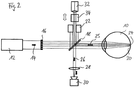
Figure 2 shows an integration, according to the invention, of an optical
coherence
tomograph into a wavefront sensor according to Figure 1. A spectrally
broadband
laser source, which is designed in such a way that an OCT is capable of being
implemented with it, now serves as radiation-source. With the beam-splitter 18
a
reference beam required for the OCT is generated in the form of a partial beam
(which in Figure 2 is deflected upwards). The partial beam is widened to the
diameter of the beam that comes out of the eye.
If the OCT is implemented in accordance with the time-domain process, the
optical
path-length of the reference beam has to be altered. This can be done, for
example,
by controlled mechanical movement of the retroreflector 32 or, for example,
through
the use of a path-length changer, such as, for example, rotating prisms,
mirrors or
such like.
Radiation 25 coming from the eye 10 is deflected downwards in Figure 2 in the
direction of the arrow 26 via the beam-splitter 18, and arrives at a detector
30 via
optics 28. The beam coming from the retroreflector 32 also passes through the
beam-splitter 18 and arrives at the detector 30 by way of reference beam. In
the
detector system 30 the reference beam and the measuring beam coming from the
eye are superimposed. The reference beam generates a background, and the
reflections coming from the eye are superimposed on this background. If the
reference beam and the reflection are incoherent, the image generated in the
detector 30 is capable of being evaluated in conventional manner. If the
differences
in optical path-length between reference beam and reflections are very small,
the
superimposing beams are coherent, and interference phenomena occur in the
detector, being evaluated in a manner known as such for optical coherence
tomography.
CA 02826739 2013-08-07
WO 2012/110053 - 5 -
PCT/EP2011/000713
Likewise, in this embodiment of the invention the pattern of dots coming from
the
eye 10 and described above can be recorded with the detector 30 and evaluated
in
accordance with the Tscherning principle in a manner known as such, in order
to
determine wavefront aberrations that were generated by the optical system
constituted by the eye.
By way of detector 30, cameras known for this purpose may be employed, but for
short measuring-times fast detectors should be provided, such as high-speed
cameras, photodiodes with respectively assigned preamplifiers, or other arrays
of
detectors.
If the OCT is implemented in accordance with the so-called Fourier domain,
arrays of
detectors known for this purpose may be employed in combination with a
dispersive
element (prism, grating).
Figure 3 shows a modification of the apparatus according to Figure 2, to the
effect
that, in addition to the detector 30, a further high-speed detector 40 is
employed. A
beam-splitter 36 couples a partial radiation out of the radiation 25 coming
from the
eye, and via optics 38 said partial radiation arrives at the high-speed
detector 40 for
the implementation of the OCT. Instead of the beam-splitter, a folding mirror
may
also be provided. With the use of different detectors for the ascertainment of
the
wavefront aberration, on the one hand, and for the OCT, on the other hand, it
is
possible to obtain a high two-dimensional local resolution for the wavefront
measurement in targeted manner, whereas for the optical coherence tomography a
rapid evaluation of the signal with a detector that is suitable for this
purpose is made
possible.
Figure 4 shows schematically a wavefront sensor operating in accordance with
the
Hartmann-Shack principle. In all the Figures, components that correspond to
one
another or that are functionally similar are provided with the same reference
symbols. In the case of the Hartmann-Shack principle, the retina 20 of the eye
10 is
illuminated with a punctiform laser beam 14' from a laser 12'. The light
scattered on
the retina 20 emerges from the eye 10 in the form of a distinctly wider
radiation
bundle. This radiation bundle 42 is deflected downwards in Figure 4 (arrow 44)
by
the beam-splitter 18 and is then broken down via a lens array 46 into partial
beams
which are focused onto a CCD detector 50. The image is a pattern of dots. In
the
case of a wavefront without aberration, a set pattern of dots arises on the
detector.
CA 02826739 2013-08-07
WO 2012/110053 - 6 -
PCT/EP2011/000713
If a real eye is surveyed, as a rule the dots of the image are not situated
exactly at
the positions of the set pattern of the dots. From the deviations of the
imaged dots
from the set dots, the curvature of the wavefront is determined in a manner
known
as such, and from this the optical properties of the eye are then inferred.
Figure 5 shows the linkage of an optical coherence tomograph with a wavefront
sensor according to Figure 4. For this purpose a spectrally broadband laser
radiation-source 12 is employed as a common radiation-source for the wavefront
sensor and for the optical coherence tomograph. The reference beam required
for
the OCT is generated with the beam-splitter 18 (Figure 5, reference symbol
54). The
retroreflector 32 widens the reference beam reflected on it. An array of
mirrors,
described in more detail further below, or a deformable mirror 56 is arranged
in the
* beam path of the reference beam.
As in the case of the embodiment according to Figures 2 and 3, the alteration
of
path-length can be carried out, for example, with a path-length changer 34 or
by
mechanical movement of the retroreflector 32 (in the case of time domain).
A lens array 46 splits up the radiation 58 coming from the eye and generates
individual dots in the detector 50. The reference beam required for the OCT
arises
from a plane wavefront and therefore impinges on other points of the detector,
so
that no interference between the beams takes place. In order to enable
interference, the aforementioned array of mirrors or a deformable mirror 56 is
provided. For example, mirrors are known that are assembled in the form of an
array from individually addressable individual mirrors (MEMS), or deformable
mirrors
are also known with which radiation can be controlled. The array of mirrors or
the
deformable mirror 56 is controlled in such a way that the superposition of
measuring
beam and reference beam that is necessary for an interference takes place on
the
detector 50.
Corresponding to the embodiment according to Figure 3, elucidated above, also
in
the case of the apparatus according to Figure 5 partial radiation can be
directed onto
a second detector system 50' by means of a beam-splitter 36. The
aforementioned
detectors also enter into consideration here by way of detector systems.
Figure 6 shows a further variant of the invention, in which the curvature-
sensor
principle, known as such, is employed for the wavefront sensor. Here (just as
in the
CA 02826739 2013-08-07
WO 2012/110053 - 7 -
PCT/EP2011/000713
embodiments described above) an LED or an SLD (superluminescent diode) may be
employed by way of radiation-source. A collimated light beam 14 is generated
and is
guided onto the retina. The light back-scattered on the retina emerges from
the eye
in the form of a wider radiation bundle. In the embodiment of a curvature
sensor
that is shown, the radiation bundle impinges on a beam-splitter 60 after
passing
through focusing optics 28. The light transmitted by the beam-splitter 60
impinges
directly on the detector arrangement of the camera. The light reflected on the
beam-splitter 60 is directed onto the camera detectors in temporally offset
manner
via a further deflection on the mirror 62 and hence via a longer optical path.
The
optical path for the radiation transmitted by the beam-splitter 60 is
preferentially
shorter than the back focal length of the focusing optics. For the reflected
radiation
portion which is deflected via the mirror 62 the optical path-length is
preferentially
longer than the back focal length, this also being indicated schematically in
Figure 6.
The wavefront can then be ascertained, in a manner known as such, from a point-
to-
point contrast of the two recorded intensities. Figure 6 shows, furthermore,
the
indication of an optical coherence tomograph with the components, already
elucidated above, constituted by retroreflector 32 and path-length changer 34
for the
reference beam (also called reference arm). In the embodiment a broadband
laser
12 serves as radiation-source for the OCT. A beam-splitter 64 couples
radiation
portions 68 out of the radiation coming from the eye 10, and these radiation
portions
are projected via optics 72 into a high-speed detector 70 for the OCT. As in
the
above examples, also in this embodiment the time domain is employed for the
OCT,
and the modifications elucidated above on the basis of the other embodiments
may
likewise be employed here analogously.
The determinations of optical properties of an object that are possible with
the
apparatuses that have been described are not only of use for refractive
surgery in
respect of the eye but may also serve for the computation of intraocular
lenses, for
cataract diagnosis, fundus examination and for the construction of
refractometers.
CA 02826739 2013-08-07
WO 2012/110053 - 8 -
PCT/EP2011/000713
List of Reference Symbols
eye
12' laser
s 12 radiation-source (broadband)
14 emitted beam
16 aperture mask
18 beam-splitter
retina
n 22 beam trap
24 image
radiation (measuring arm)
26 radiation
28 optics
15 30' camera
detector
32 retroreflector
34 path-length changer
36 beam-splitter
20 38 optics
high-speed detector
42 radiation
44 radiation
46 lens array
25 46' lens array
CCD detector
52 dot image
54 radiation
56 array of mirrors / deformable mirror
30 58 radiation
beam-splitter
62 mirror
64 beam-splitter
66 radiation
35 68 radiation
high-speed detector / OCT
72 optics