Note: Descriptions are shown in the official language in which they were submitted.
CA 02827354 2013-09-03
SYSTEMS AND METHODS FOR DETERMINING
FLUID MOBILITY IN ROCK SAMPLES
FIELD OF THE INVENTION
[0003] The present invention relates to systems and methods for determining
fluid
mobility in rock samples. More particularly, the present invention relates to
systems
and methods for determining fluid mobility in rock samples using time-lapse
positron
emission particle tracking ("PEPT").
BACKGROUND OF THE INVENTION
[0004] One of the biggest challenges to determining fluid mobility in rock
samples is
the very fine microstructure in some rock samples ¨ like shale. Shale
significantly
differs from all other formation rocks in the grain size, which corresponds to
very
low values for porosity (0) and permeability (k).
[0005] Conventional techniques for determining fluid mobility in rock
samples
include:
- 1 -
CA 02827354 2013-09-03
WO 2012/122110
PCT/US2012/027749
= Nuclear Magnetic Resonance (NMR);
= 3D CT scanning, that enables contouring density differences i.e. contrast
in the
scanned image;
= X-ray diffraction; and
= SEM evaluation of rock samples.
[0006] One of the main disadvantages is that such conventional techniques
can only operate
with and handle very small rock samples referred to as core plugs, which are
not
representative of the entire sample ¨ much less the reservoir. In other words,
the fluid
mobility determined by such conventional techniques is not representative of
the in-situ fluid
mobility and thus, must be adjusted for a more accurate representation. The
determination of
fluid mobility by any of the foregoing conventional techniques may be improved
by
"upscaling," which extrapolates petrophysical properties from the core-plug
scale to
determine the simulation-grid scale. Many upscaling techniques are well known
and available
such as, for example, power-law average, renormalization, pressure-solver,
tensor and
pseudofunction techniques. In short, upscaling replaces a number of
heterogeneous fine grid
blocks with one equivalent coarse homogeneous grid block. The essence of
conventional
upscaling requires averaging and extrapolation, which therefore, leads to the
loss of
information and creates the common problem of blurring the spatially
continuous extremes
such as, for example, shale barriers and open fractures. Oil and gas recovery
mainly depends
on the spatial connectivity of the extreme (ultra-low) permeability values,
particularly
characteristic of small scale shale pores. Determining fluid mobility in rock
samples without
upscaling is therefore, preferred.
-2-
CA 02827354 2013-09-03
WO 2012/122110
PCT/US2012/027749
[0007] Other conventional techniques for determining fluid mobility in
rock samples include:
i) grinding the rock samples; ii) removing all of the water content from the
rock samples; and
iii) injecting He or Hg into the rock samples. These techniques, however, are
not optimal
because they skew the original geo-mechanical properties of the rock sample
before
determining fluid mobility and may still require upscaling for very small rock
samples (core
plugs).
[0008] As a result, applications of positron radiation detection have
emerged from more
traditional medical imaging applications using Positron Emission Tomography
("PET"). In
standard PET imaging, data acquired in two-dimensional (2D) or three-
dimensional (3D)
form are stored in sinograms that consist of rows and columns representing
angular and radial
samplings, respectively. The raw data are pre-binned by the hardware into the
sinogram
format. Due to the pre-binning operation, the data in the sinogram format
result in lower
resolution from the original raw data, which results in the loss of valuable
information about
the scanned object. In sinogram format, the acquired data in each row are
compressed
(summed) along the depth of the object and must be decompressed to provide
information
along this direction. In Nonmedical Applications of a Positron Camera, Nuclear
Instruments
and Methods in Physics Research A310, 1991, pp. 423-434, written by
Hawkesworth, et al.,
for example, PET is used to produce images every ten (10) minutes to track
fluid flow in rock
samples. This technology, however, is limited because the acquired data is
stored in
sinograms, which require more time to process than the original raw data
stored in list mode.
As a result, the image rate is slow (1 every 10 minutes), which lowers the
image resolution.
-3-
CA 02827354 2013-09-03
WO 2012/122110
PCT/US2012/027749
[0009] In Porosity and Pathway Determination in Crystalline Rock by
Positron Emission
Tomography and Neutron Radiography, Earth and Planetary Science Letters 140,
1996, pp.
213-225, written by Degueldre, et al., the fluid pathway and porosity in
crystalline rock
samples have been studied with a high-resolution PET camera. The results
demonstrate
original water carrier features in granodiorite pieces 20 cm in size and in
simulated features
with porosities on the order of 20%. In Positron Emission Tomography of Large
Rock
Samples Using a Multiring PET Instrument, IEEE Transactions on Nuclear
Science, Vol. 44,
No. 1, 1997, pp. 26-30, written by Maguire, et al., the use of PET has been
extended to a
multi-ring PET camera and demonstrates that the measurements of porosity in
large rock
samples (21.5 cm) are indeed practicable using 3D acquisition techniques.
These techniques,
however, are limited to images of just the rock sample structure and
therefore, do not illustrate
dynamic fluid mobility within the rock sample structure. These techniques also
suffer the
same shortcomings as other techniques that store acquired data in the sinogram
format.
[0010] Present research by the University of Cape Town in South Africa
utilizes time-lapse
PEPT, tags sub-20 tm rock particles and liquids with tags (tracers) having
activities of ¨100
Ci to determine the mobility of a rock particle in a slurry and a liquid in a
column of glass
beads. Here, the activity of the tag refers to the radioactive decay in which
an unstable (i.e.
(radio)active) atomic nucleus looses energy by emitting one or more ionizing
particles (i.e.
ionizing energy). In PEPT applications, the (radio)active tag emits positrons.
Because the
acquired data is stored in list mode, the images may be produced at a rate of
more than one
image per second. As a result, fluid mobility may be determined more
accurately because the
image quality is improved. This research, however, has not been applied to
determine fluid
-4-
CA 02827354 2013-09-03
mobility in rock samples, particularly rock samples with small scale pores
(less than
IAD) like shale, because the tag used for the particles is too large for
accurate
determination of fluid (gas and liquid) mobility.
SUMMARY OF THE INVENTION
[0011] The present invention therefore, meets the above needs and
overcomes one or
more deficiencies in the prior art by providing systems and methods for
determining
fluid mobility in rock samples using time-lapse position emission particle
tracking.
[0012] In one embodiment, the present invention includes a method for
determining
fluid mobility in a rock sample using time-lapse positron emission particle
tracking,
which comprises the steps of i) selecting a porous rock sample; ii) selecting
a fluid for
the rock sample; iii) selecting a tag for the fluid; iv) placing the rock
sample in a
pressurized container; v) introducing the fluid and the tag into pores within
the rock
sample; vi) recording gamma-ray emissions from the tag using a positron
emission
tomography camera as the tag traverses with the fluid through the pores in the
rock
sample placed in the pressurized container; vii) converting the gamma-ray
emissions
into images at a rate of more than one image every second; and viii)
displaying the
images.
[0013] In another embodiment, the present invention includes a method for
determining fluid mobility in a rock sample using time lapse positron emission
particle tracking, which comprises the steps of: i) selecting a porous rock
sample with
a permeability less than one micro-darcy; ii) selecting a fluid for the rock
sample that
is a gas; iii) selecting a tag for the fluid that is the gas or another gas;
iv) placing the
rock sample in a pressurized container; v) introducing the fluid and the tag
into pores
- 5 -
CA 02827354 2013-09-03
within the rock sample; vi) recording gamma-ray emissions from the tag in a
list
mode file using a positron emission tomography camera as the tag traverses
with the
fluid through the pores in the rock sample placed in the pressurized
container; vii)
converting the gamma-ray emissions into images; and viii) displaying the
images.
[0014] Additional aspects, advantages and embodiments of the invention
will become
apparent to those skilled in the art from the following description of the
various
embodiments and related drawings.
BRIEF DESCRIPTION OF THE DRAWINGS
[0015] The present invention is described below with references to the
accompanying
drawings in which like elements are referenced with like referenced numerals,
and in
which:
[0016] FIG. 1 illustrates PEPT for a single radionuclide tag.
[0017] FIG. 2 is a block diagram illustrating one embodiment of a system
for
implementing the present invention.
[0018] FIG. 3 illustrates coincidence event counting.
[0019] FIG. 4 if a flow diagram illustrating one embodiment of a method
for
implementing the present invention.
DETAILED DESCRIPTION OF THE PREFERRED EMBODIMENTS
[0020] The subject matter of the present invention is described with
specificity,
however, the description itself is not intended to limit the scope of the
invention. The
subject matter thus,
- 6 -
CA 02827354 2013-09-03
WO 2012/122110
PCT/US2012/027749
might also be embodied in other ways, to include different steps or
combinations of steps
similar to the ones described herein, in conjunction with other technologies.
Moreover,
although the term "step" may be used herein to describe different elements of
methods
employed, the term should not be interpreted as implying any particular order
among or
between various steps herein disclosed unless otherwise expressly limited by
the description
to a particular order. While the following description refers to the oil and
gas industry, the
systems and methods of the present invention are not limited thereto and may
also be applied
to other industries to achieve similar results.
[0021] PEPT is basically a technique for measuring the trajectory of one
or more tags, which
may be used to tag a solid rock particle or a fluid. The tag may be any
radioactive nuclide
(radionuclide) capable of positron emission. In FIG. 1, for example, the
trajectory of a single
rock particle tagged with a radionuclide tag is illustrated using PEPT and a
PET camera. The
radionuclide tag decays through the emission of a positron, which is the
antiparticle of an
electron. A positron produced in a nuclear decay will rapidly annihilate with
an electron,
resulting in a pair of 511 keV gamma rays that are emitted almost in opposite
directions. If
both of these gamma rays are detected at two different points, thereby
defining a line of
response ("LOR"), then the origin of the gamma ray emissions must have
occurred
somewhere along the LOR. In other words, the LOR substantially corresponds to
a line
joining a pair of opposing detectors.
[0022] The position of the radionuclide tag can be determined within the
field of view of a
PET camera using only a small number of measured LOR's. The activity of the
tag, however,
must be sufficient for enough LOR's to be measured in order to accurately
reflect the
-7-
CA 02827354 2013-09-03
WO 2012/122110
PCT/US2012/027749
trajectory of the moving tag. In particular, tags of significantly smaller
sizes must be used for
PEPT to be reliably accurate for determining fluid mobility in small-scale
shale pores. In
principle, only two detectors are necessary, however, additional detectors may
be used as long
as they are paired ¨ meaning positioned opposite one another along a line
passing through the
center of the PET camera. Because many thousands of gamma-ray emissions can be
detected
with a PET camera and processed each second, the possibility of determining
the position of
one or more fast moving radionuclide tags may be realized. Consequently, PEPT
may be
used to determine fluid mobility in rock samples by tagging a fluid with one
or more
radionuclide tags.
System Description
[0023] The present invention may be implemented through a computer-
executable program of
instructions, such as program modules, generally referred to as software
applications or
application programs executed by a computer. The software may include, for
example,
routines, programs, objects, components, and data structures that perform
particular tasks or
implement particular abstract data types. The software forms an interface to
allow a computer
to react according to a source of input. The software may also cooperate with
other code
segments to initiate a variety of tasks in response to data received in
conjunction with the
source of the received data. The software may be stored and/or carried on any
variety of
memory media such as CD-ROM, magnetic disk, bubble memory and semiconductor
memory
(e.g., various types of RAM or ROM). Furthermore, the software and its results
may be
transmitted over a variety of carrier media such as optical fiber, metallic
wire and/or through
any of a variety of networks such as the Internet.
-8-
CA 02827354 2013-09-03
WO 2012/122110
PCT/US2012/027749
[0024] Moreover, those skilled in the art will appreciate that the
invention may be practiced
with a variety of computer-system configurations, including hand-held devices,
multiprocessor systems, microprocessor-based or programmable-consumer
electronics,
minicomputers, mainframe computers, and the like. Any number of computer-
systems and
computer networks are acceptable for use with the present invention. The
invention may be
practiced in distributed-computing environments where tasks are performed by
remote-
processing devices that are linked through a communications network. In a
distributed-
computing environment, program modules may be located in both local and remote
computer-
storage media including memory storage devices. The present invention may
therefore, be
implemented in connection with various hardware, software or a combination
thereof, in a
computer system or other processing system.
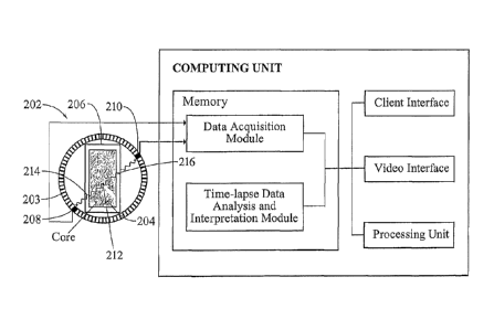
[0025] Referring now to FIG. 2, a block diagram illustrates one embodiment
of a system for
implementing the present invention on a computer. The system includes a
computing unit,
sometimes referred to a computing system, which contains memory, application
programs, a
client interface, a video interface, a processing unit and a PET camera 202.
The computing
unit is only one example of a suitable computing environment and is not
intended to suggest
any limitation as to the scope of use or functionality of the invention.
[0026] The PET camera 202 may include any conventional PET camera, such
as, for
example, the ECAT 'EXACT' 3D model 966 PET camera manufactured by Siemens. The
camera 202 has 48 standard bismuth germanate detector elements grouped in
blocks of 8x8
forming a detector ring with a diameter of 82 cm and an axial field of view of
23.4 cm. The
camera 202 is preferable over other conventional cameras due to its size. The
camera 202 is
-9-
CA 02827354 2013-09-03
WO 2012/122110
PCT/US2012/027749
capable of maintaining a sustained data acquisition rate of about 4 million
coincidence events
per second. The camera 202 also has a superior geometry for studying
cylindrical systems in
3D and would allow handling of large rock samples, which represents a
tremendous
improvement over the standard petrophysical core-plug measurements in rock
physics labs.
As illustrated in FIG. 2, a shale rock sample 204 about 50 cm in height and
about 20 cm in
thickness may be positioned in a pressurized container 206. The container 206
is pressurized
to simulate pressures and/or temperatures imposed on the rock sample 204 in-
situ. Detectors
208, 210 detect emissions generated by pairs of registered incident gamma
rays, which are
combined in coincidence circuitry within a short time window. In this manner,
position
information is gained from the detected radiation without the need of a
physical collimator
(i.e. electronic collimation). For simplicity, the PET camera 202 only
illustrates a pair of
detectors 208, 210. In practice, all of the detectors in the detector ring 203
are directly wired
to the Data Acquisition Module. Alternatively, all of the detectors in the
detector ring 203
may be wirelessly connected to the Data Acquisition Module.
[0027] Alternatively, the PET camera 202 could be manufactured on a much
smaller scale
and positioned in a drillstring for deployment downhole in a wellbore.
Existing technology,
such as the Halliburton RSCT and HRSCT coring tools, could be retrofitted to
host a smaller
scale PET camera. For example, the RSCT tool drills perpendicularly to the
bore-hole to
recover rock samples of 15/16" in OD and 1-3/4" in length. Each rock sample
may be
withdrawn into a container in the tool that can be pressurized for delivery of
the fluid tagged
with the radionuclide tag. Depending on the environmental conditions downhole,
the PET
camera 202 and the computing unit (except the client interface/video
interface) may be carried
-10-
CA 02827354 2013-09-03
WO 2012/122110
PCT/US2012/027749
by the drillstring with the RSCT tool. Alternatively, only the PET camera 202
may be carried
by the drillstring with the RSCT tool if the environmental conditions are not
conducive to
positioning the computing unit in the drillstring. Fluid mobility data can be
transmitted to the
client interface/video interface at the surface for analysis over a fast
optical line, for example.
After determining fluid mobility for the rock sample, it may be transferred to
a storage tube.
[0028] The memory primarily stores the application programs, which may
also be described
as program modules containing computer-executable instructions, executed by
the computing
unit for implementing the present invention described herein and illustrated
in FIGS. 2-4.
The memory therefore, includes a Data Acquisition Module and a Time-Lapse Data
Analysis
and Interpretation Module, which enable the methods illustrated and described
in reference to
FIG. 4.
[0029] The Data Acquisition Module records raw data (gamma ray emissions)
in a list mode
file. The gamma ray emissions are recorded as detected signals, which are
recorded in
chronological order so that each signal has a time stamp and the coordinates
for each detector.
When the signals significantly match or overlap for a pair of opposing
detectors, a coincident
event is defined. This mode of recording the raw data is further illustrated
in FIG. 3 for
recording coincidence events and is routinely utilized with a PET camera.
Channels 1 and 2,
for example, illustrate two independent signals representing a pair of
opposing gamma ray
emissions detected by a pair of opposing PET camera detectors at different
times. The sum
channel separates the coincidence events from other events (signals) by
summing to
determine a coincidence event within a predetermined short time interval. The
Data
Acquisition Module may therefore, be calibrated in a way to amplify the signal
for only the
-11-
CA 02827354 2013-09-03
WO 2012/122110
PCT/US2012/027749
time intervals where the amplitudes of the signals for channels 1 and 2
substantially overlap
within a certain predefined short time interval. The coincidence event for the
amplified signal
amplitude therefore, may correspond to a pair of opposing gamma rays detected
coincidentally within the predefined time interval. Each coincident event is
recorded in
chronological order so that each coincidence event has a time stamp and the
coordinates for
each of the two opposing detectors. Based on the coordinates for each of the
two opposing
detectors, the LOR may be easily determined. The Data Acquisition Module may
record
thousands of coincidence events per second. The list mode file therefore,
secures the highest
amount of available information for the raw data. Although case dependent, the
size of the
list mode file is much larger than that of a sinogram and can exceed hundreds
of megabytes or
even gigabytes of data. Once recorded, the data in the list mode file must be
converted to
form images that can then be used to determine fluid mobility.
[0030] The Time-lapse Data Analysis and Interpretation Module converts the
data in the list
mode file to images that can be used to determine fluid mobility. The
conversion may be
performed using conventional methods such as, for example, simple
backprojection, filtered
backprojection or iterative methods. The Time-Lapse Data Analysis and
Interpretation
Module uses a different method, however, to convert the list mode file to
images. Each list
mode file is segmented into time slices (typically of the order of a
millisecond). The time-
sliced data are triangulated to get the x,y,z,t coordinates for each tag,
which enables tracking
multiple tags in the field of view of the PET camera simultaneously. In this
manner, tracking
multiple tags may be extended to images and optimized for the size of the data
matrices
related to the number of image voxels. Any, well known and widely available
image
-12-
CA 02827354 2013-09-03
WO 2012/122110
PCT/US2012/027749
processing tool may be applied to optimize the quality of the image.
Optionally, attenuation
correction may be applied to improve the resolution of the processed voxel
image by
correcting for the so-called scattered and random coincidence, which
contribute to the
uncertainty of interpretation. Furthermore, uncertainty is associated with the
speed of the
moving tag. It seems that for slowly moving or stationary tags, the
uncertainty is about half
the detector size (i.e. about 2 mm). As the speed of the tag increases, this
uncertainty
increases proportionately and may require further investigation. Dealing with
"non-
continuous" data (i.e. fluid/gas propagation discretized into (ultra) short
timeframes) may
reduce this uncertainty, however.
[0031] Although the computing unit is shown as having a generalized
memory, the computing
unit typically includes a variety of computer readable media. By way of
example, and not
limitation, computer readable media may comprise computer storage media. The
computing
system memory may include computer storage media in the form of volatile
and/or
nonvolatile memory such as a read only memory (ROM) and random access memory
(RAM).
A basic input/output system (BIOS), containing the basic routines that help to
transfer
information between elements within the computing unit, such as during start-
up, is typically
stored in ROM. The RAM typically contains data and/or program modules that are
immediately accessible to and/or presently being operated on by the processing
unit. By way
of example, and not limitation, the computing unit includes an operating
system, application
programs, other program modules, and program data.
[0032] The components shown in the memory may also be included in other
removable/non-
removable, volatile/nonvolatile computer storage media or they may be
implemented in the
-13-
CA 02827354 2013-09-03
WO 2012/122110
PCT/US2012/027749
computing unit through application program interface ("API"), which may reside
on a
separate computing unit connected through a computer system or network. For
example only,
a hard disk drive may read from or write to non-removable, nonvolatile
magnetic media, a
magnetic disk drive may read from or write to a removable, non-volatile
magnetic disk, and
an optical disk drive may read from or write to a removable, nonvolatile
optical disk such as a
CD ROM or other optical media. Other removable/non-removable, volatile/non-
volatile
computer storage media that can be used in the exemplary operating environment
may
include, but are not limited to, magnetic tape cassettes, flash memory cards,
digital versatile
disks, digital video tape, solid state RAM, solid state ROM, and the like. The
drives and their
associated computer storage media discussed above provide storage of computer
readable
instructions, data structures, program modules and other data for the
computing unit.
[0033] A client may enter commands and information into the computing unit
through the
client interface, which may be input devices such as a keyboard and pointing
device,
commonly referred to as a mouse, trackball or touch pad. Input devices may
include a
microphone, joystick, satellite dish, scanner, or the like. These and other
input devices are
often connected to the processing unit through a system bus, but may be
connected by other
interface and bus structures, such as a parallel port or a universal serial
bus (USB).
[0034] A monitor or other type of display device may be connected to the
system bus via an
interface, such as a video interface. A graphical user interface ("GUI") may
also be used with
the video interface to receive instructions from the client interface and
transmit instructions to
the processing unit. In addition to the monitor, computers may also include
other peripheral
-14-
CA 02827354 2013-09-03
WO 2012/122110
PCT/US2012/027749
output devices such as speakers and printer, which may be connected through an
output
peripheral interface.
[0035] Although many other internal components of the computing unit are
not shown, those
of ordinary skill in the art will appreciate that such components and their
interconnection are
well known.
Method Description
[0036] Referring now to FIG. 4, a flow diagram illustrates one embodiment
of a method 400
for implementing the present invention.
[0037] In step 402, a porous rock sample is selected. The rock sample may
be selected based
upon a number of criteria including, but not limited to, porosity
characteristics and
permeability. Shale, for example, may be selected as a porous rock sample with
a
permeability of less than one micro-darcy.
[0038] In step 404, a fluid for the rock sample is selected. The fluid may
be selected, for
example, based upon various criteria including, but not limited to, fluid
indigenous to the rock
sample. Thus, the fluid may be a gas or a liquid. If a shale rock sample is
selected, then a
fluid for the rock sample representing an indigenous fluid may be methane
(CH4) gas since
methane is the main chemical constituent in shale gas.
[0039] In step 406, a tag for the fluid is selected. The radionuclide tag
should resemble the
fluid it is being used to tag for consistent results in determining fluid
mobility. For example, a
liquid should be tagged with a liquid radionuclide tag and a gas should be
tagged with a gas
-15-
CA 02827354 2013-09-03
radionuclide tag. For even better results, the radionuclide tag should have a
chemical
composition as close to the chemical composition of the fluid as possible. In
this
manner, the mobility of the fluid tag with the radionuclide tag will be closer
to the
true mobility of the fluid in the rock sample without the radionuclide tag. In
turn, the
activity of the radionuclide tag depends on both its size and composition.
Thus, for
shale, if a methane (CH4) gas is selected as the fluid, a preferred
radionuclide tag for
the gas would be C11.
[0040] In step 408, the rock sample is placed in a pressurized container.
The
pressurized container, for example, may resemble the pressurized container 206
described in reference to FIG. 2.
[0041] In step 410, the fluid and the tag are introduced into pores
within the rock
sample. The fluid and the tag may be introduced into the pores within the rock
sample by injecting the fluid and the tag into the pressurized container at
one end
under a constant pressure and a constant temperature. Alternatively, the fluid
and the
tag may be introduced into the pores within the rock sample by injecting the
fluid and
the tag directly into the pores within the rock sample before the rock sample
is placed
in the pressurized container and applying a constant pressure and a constant
temperature to the rock sample after it is placed in the pressurized container
with the
fluid and the tag. The fluid is tagged when it is introduced with the tag into
the pores
within the rock sample. At this point, the tag is attached to the fluid as the
tag
traverses with the fluid through the pores within the rock sample and/or the
tag travels
with the fluid as it traverses with the fluid through the pores within the
rock sample.
Furthermore, multiple tags may be introduced with the fluid into the pores
within the
rock sample. The fluid and the tag are introduced into the pores within the
rock
sample at a constant flow rate, a constant pressure
- 16-
CA 02827354 2013-09-03
WO 2012/122110
PCT/US2012/027749
and a constant temperature. The flow rate, the constant pressure, the constant
temperature,
the fluid and the tag may be selected based upon a flow rate, a pressure, and
a temperature for
a fluid that is indigenous to the rock sample, which represents a target
fluid.
[0042] In step 412, gamma-ray emissions from the tag are recorded as the
tag traverses with
the fluid through the pores in the rock sample. Preferably, the gamma-ray
emissions are
recorded in a list mode file. The gamma-ray emissions may be recorded using
the PET
camera 202 and the Data Acquisition Module described in reference to FIG. 2.
[0043] In step 414, the gamma-ray emissions recorded in step 412 are
converted into images.
The gamma-ray emissions may be converted into images at a rate of more than
one image
every second using the Time-Lapse Data Analysis and Interpolation module
described in
reference to FIG. 1
[0044] In step 416, the images form step 414 are displayed. The images may
be displayed
next to each other or consecutively using the client/video interface described
in reference to
FIG. 2. Fluid mobility may therefore, be determined by viewing the displayed
images or
using the displayed images to determine a permeability for the rock sample.
[0045] The proposed time-lapse PEPT technology therefore, greatly improves
over state-of-
the-art imaging technology because it actually images the fluid propagating
through the rock
sample under different net pressures. The time-lapse PEPT goes even further by
performing
un-compromized high-resolution imaging of fluid mobility and interactive
scanning of rock
samples with small-scale pores unprecedented in the rock physics industry.
-17-
CA 02827354 2013-09-03
WO 2012/122110
PCT/US2012/027749
[0046] Horizontal drilling and hydraulic fracturing have made it feasible
to extract huge
amounts of natural gas trapped in shale formations. The objective of
fracturing techniques is
to expose the maximum possible surface area of the rock formation and provide
a reasonable
path for the fluid to produce back to the wellbore. Fracturing techniques are
therefore,
designed to achieve long effective fracture half-lengths and improve fracture
conductivity in
rocks with mili-darcy (mD) to micro-darcy (p1D) rock permeabilities.
Fracturing techniques
however, need to also address nano-darcy (nD) rock permeabilities in shale
rocks that
geologists used to consider seals. Permeability of a rock sample is defined as
the ability of
the rock sample to transmit fluids through the pore spaces, which influences
the fluid flow
rate, the fluid's movement and drainage of the fluid. The experimental
determination of
permeability in shale rock samples by standard rock physics laboratory
measurements is
extremely challenging and time-consuming. Therefore, rather than determining
the bulk
permeability for the shale rock sample, it is common practice to determine the
Fracture
Conductivity Ratio (Cr) by the following equation:
=
= k _fracture W fracture
(1)
kreservoir = 1fracture
where kfi'acture refers to fracture permeability (in mD), w
fracture represents the width of the
fracture (in ft), kreservoir is the formation/reservoir permeability (in mD)
and 14
vatcture represents
the fracture half-length (in ft), It is common to refer to the product of
kfraclure and wfrachire as the
fracture conductivity (in mD ft),
-18-
CA 02827354 2013-09-03
WO 2012/122110
PCT/US2012/027749
[0047]
The quantitative information on the fluid mobility, acquired with the time-
lapse PEPT
imaging will directly enhance the knowledge on kreservoir, Wfracture and
lfracture parameters. As
such, the time-lapse PEPT imaging will provide a unique quantitative estimate
on how the
fluid mobility changes as a result of fracturing, particularly at high fluid
injection rates, where
the conventional PET imaging fails. Furthermore, the time-lapse PEPT imaging
will reduce
the uncertainty in quantifying the Fracture Conductivity Ration (Cr) and
moreover, the
Natural Fracture Conductivity Index (NFCI) by direct one-to-one comparison of
the fluid
mobility of the pre-fracture rock sample with the post-fracture rock sample.
This will provide
for more accurate determination of fracturing production success, through the
estimation of
the Stimulated Reservoir Volume (SRV), defined as the product of the
Stimulated Area and
the Net Pay. The standard industrial practice for calculating SRV's usually
introduces high
uncertainty and systematic error in the volume estimates, mainly due to the
inaccurate and
uncertain estimates of the fracture connectivity.
The three-dimensional PEPT fluid
propagation imaging will produce a) more accurate estimates on the
directionality of fractures
deduced from the fluid distribution as a function of time, b) more
quantitatively sound
estimates of fracture connectivity and c) improved correlation and reduced
error in the
estimates of SRV.
[0048]
Recent laboratory experiments performed on a number of shale rock samples
reveal
that the effective permeability of the shale rock sample can be changed from
nD to IAD when
the shale rock sample is fractured. This suggests that even unsupported
fractures (i.e. without
the permeability support by proppant packs) may be capable of contributing to
production in
ultra-low permeability shale rocks. It is foreseen that by using the time-
lapse PEPT imaging,
-19-
CA 02827354 2013-09-03
it will be possible to derive quantitative (at least empirical) estimates on
the
correlation between the effective permeability of fractured rock, the
estimates of SRV
and the speed of fluid propagation front, directly from the reconstructed
three-
dimensional PEPT image. This will enable optimization and more time-and cost-
efficient design of the fracturing and re-fracturing jobs by improving the
knowledge
on the correlation of fluid propagation and the fracturing attributes (e.g.
closure
stress), stimulation parameters (e.g. presence and type of proppants) and
production
data (e.g. pressure) as well as reduce uncertainty of the practical
operational and
economic variables, such as, for example: a) the amount of extractable
hydrocarbons
(e.g. Original Gas In Place), b) optimum well perforation interval, c)
drainage
area/volume of wells, d) recovery factor, e) optimum spacing units and f)
optimum
steer, direction and angle of the wells.
[0049]
While the present invention has been described in connection with presently
preferred embodiments, it will be understood by those skilled in the art that
it is not
intended to limit the invention to those embodiments. It is therefore,
contemplated
that various alternative embodiments and modifications may be made to the
disclosed
embodiments without departing from the scope of the invention defined by the
appended claims and equivalents thereof.
- 20 -