Note: Descriptions are shown in the official language in which they were submitted.
CA 02827404 2013-08-14
WO 2012/116319
PCT/US2012/026605
SUTURE MESH AND METHOD OF USE
RELATED APPLICATION
[0001] This application claims priority to United States Provisional
Application No.
61/446,540 filed on February 25, 2011 which is incorporated by reference
herein in its entirety.
FIELD OF THE INVENTION
[0002] The field relates to affixing sutures using a transosseous tunnel.
BACKGROUND
[0003] Surgical procedures and devices are known for creating transosseous
tunnels for
attachment of soft tissues, such as tendon and ligaments to bone. Both
artificial and natural
materials are attached to these soft tissues to repair them, and surgical
procedures may move soft
tissues from one location to another to repair a damaged, torn or severed
tendons or ligaments.
[0004] U.S. Pat. Pub. 2008/0188936 and WO 2008/097901 disclose details of
rotator cuff
repair. The human shoulder is a complex system of hard and soft tissues that
exhibits
extraordinary mobility using coordinated activation of a variety of muscles,
simultaneously. A
conventional arthroscopic repair of a torn supraspinatus tendon is disclosed
in the background of
the publication, reporting that 20-60% of rotator cuff repairs fail. The
publication discloses the
dilemma with surgery of this type. Any artificial addition to the tissues that
takes up too much
stress on living tissues can lead to atrophy of the living tissues. In order
to heal properly, living
tissues must be exposed to a certain level of stress that is within a nominal
range for healing.
These ranges are known in the art, but devices and techniques that provide
optimal healing are
not available. Bioabsorbable materials are disclosed both in the background
and as an
embodiment of the publication's invention. The publication teaches away from
making tension
members from non-absorbable materials; however, the absorbable materials of
the patch and
tension members may be reinforced by non-absorbable materials, such as by
including non-
absorbable fibers in a patch material or tension members, and may be attached
by bone anchors.
The publication teaches providing coatings on its medical devices, providing
biologically active
agents for improving healing, for example.
1
CA 02827404 2013-08-14
WO 2012/116319 PCT/US2012/026605
[0005] U.S. Pat. No. 5,268,001 discloses a bone fastener for fixing
either a suture or a
rivet within a predrilled bone hole. The background of this issued patent
summarizes the variety
of materials and types of bone anchors historically available for using in
anchoring soft tissues to
bones, either as a rivet or as a suture anchor. The patent discloses a hand
held means 70, in
Figures 3.1 and 3.2 of the patent, which is useful in setting an anchor within
an annular portion,
fixing the anchor and annular portion within a pre-drilled hole in a bone.
[0006] U.S. Pat. No. 7,651,495 discloses a method and apparatus for
preventing
migration of sutures through transosseous tunnels. Its improved method for
attaching soft tissues
to bone passes a suture through a transosseous tunnel and uses the suture to
affix the soft tissue
to the bone. The improved apparatus is an eyelet, which is placed into an end
of the bone
opposite of the soft tissue and through which the suture passes. The eyelet
may be threaded,
interference fit or pressure fit using a two piece insertion/expandable
member, with the
expandable member anchoring the insertion member within the pre-drilled hole
through the
bone. The patent discloses in its background that it is known that
transosseous tunnels are the
gold standard of rotator cuff repair, but migration of sutures through the
bone itself is a
significant complication, particularly in older patients. The patent discloses
a known attachment
mechanism using plate-like augmentation to reinforce the bone, teaches
significant
disadvantages of this technique and teaches the advantages of its eyelet
approach to preventing
migration of the suture through degradation under the force of the sutures on
the bone. U.S. Pat.
No. 5,725,529 discloses another bone fastener having similar characteristics
to other disclosed
bone fasteners.
[0007] U.S. Pat. Pub. 2010/0191248 discloses an arthroscopic tunnel guide
for rotator
cuff repair. Its tunnel guide provides a transosseous tunnel having a fixed,
non-zero radius of
curvature using a bone cutting instrument. This disclosure provides a tool and
drill guide that
provides a curvature to a transosseous tunnel drilled arthroscopically,
allowing arthroscopic
surgeons use of a portion of the humeral head that was previously only
available by open
surgery.
[0008] U.S. Pat. Pub. 2010/0191247 discloses another apparatus for
drilling a
transosseous tunnel. The reference teaches that the drill bit tip acts as an
anchor at the far cortex
2
CA 02827404 2013-08-14
WO 2012/116319
PCT/US2012/026605
distal from the surface of the humeral head adjacent to the soft tissues, when
the anchor is
detached from the drill bit, at the distal end of the transosseous tunnel
formed by the drill bit tip.
The applicant believes that there is a concern with the use of this device in
the way described in
the publication, which introduces a risk of possible axillary nerve injury
during the described
procedure, which drills to the inferior-medial aspect of the humeral head.
[0009] All
of the listed patents and publications in this background are incorporated by
reference in their entirety herein for the purposes of providing background
and materials
selection for biocompatible materials. None of the listed patents provide for
a time-saving
method to prevent the migration of sutures through transosseous tunnels, while
repairing
damaged tissues and promoting healing.
SUMMARY
[00010] Herein, a suture or sutures is defined as any linear material,
traditionally cat gut,
silk, polymer thread, metal wire or combinations thereof, that is used to
stitch or secure together
tissues, whether or not permanent or bioabsorbable, unless otherwise
indicated.
[00011] A suture mesh may comprise a tape or a net and may be combined with a
suture
having raised portion along at least a portion of the length of the suture.
The raised portions may
take the form of a bead, ball, ratchet surface, oblong spheroid and/or disk,
for example. In one
example, a transosseous suture net comprises, on a retention end, a retainer
and, on an opposite
end of the net, one or more sutures extending from the net. The opposite end
and/or body of the
net may be sized in diameter and length to extend entirely through a
transosseous tunnel such
that the suture or sutures extending from the opposite end extend from a
portion of the net
outside of the transosseous tunnel. Alternatively, one or more sutures may
extend from a suture
net and may include the raised portions on a portion of the suture that
extends through the
transosseous tunnel, preventing or reducing suture migration by keeping the
sutures from cutting,
abrading or pulling through surrounding bone, and the suture net may extend
over a portion of
the bone preventing or reducing cutting, abrading or pulling through of the
suture net through the
underlying bone material. The net and suture materials may be made of a
variety of
biocompatible materials that are known in the art, such as a high density
polyethylene. The net
may be made of a sheet and may have holes formed or punched in the sheet or
may be a weave
3
CA 02827404 2013-08-14
WO 2012/116319 PCT/US2012/026605
or mat. In one example, the interstitial holes in the net are sized to
encourage bone in-growth,
and the net acts as a scaffold structure for the formation of living tissues,
aiding in the healing
process. For example, the net may include one or more agents intended to
improve healing or in-
growth of living tissue, such as disclosed in the references disclosed in the
background section.
A mesh tape may comprise a mesh having sutures extending from one or both ends
of the tape
and/or may provide anchors or attachment points for attaching one or more
sutures to mesh. A
mesh may be dimensioned and used as a suture net or may be used as a
reinforcing mesh for a
tendon or the like. A retention clip may be combined with a suture having
raised portions, such
that the clip may be secured on one of the raised portions and fixing the
raised portions in
relation to tissues of a patient, a mesh or a net, for example.
[00012] Notably, the net and mesh are not a plug or anchor. Instead, both are
pliable or
flexible materials. The net, mesh or a suture extending from the net or mesh
may be inserted
through a transosseous tunnel, such as by using an insertion device, a probe
or a snare. Also, a
net or mesh may be used as a support for a tendon or other patient tissues
during the process of
healing and/or thereafter. An insertion device may have a handle, as disclosed
in the references
cited in the background section. Alternatively, a tool may not be necessary. A
tool may be used
merely to push the net or mesh or a suture through the transosseous tunnel or
a snare may be
used to pull a net or mesh or suture through the transosseous tunnel.
[00013] Other combinations and variations of the features disclosed herein may
be
recognized as within the scope and breadth of the disclosed inventions, which
are not limited to
the specific examples provided. Advantages of the features disclosed are
surprising and
unexpected and include reduced time for surgical repairs and improved outcomes
compared to
known products and procedures.
BRIEF DESCRIPTION OF THE DRAWINGS
[00014] Figure lA illustrates a retention end of one example of a transosseous
suture net.
[00015] Figure 1B illustrates a detail view and partial cross section of a
portion of the
retention end of Figure 1A.
4
CA 02827404 2013-08-14
WO 2012/116319 PCT/US2012/026605
[00016] Figure 1C illustrates an example of a suture end of a transosseous
suture net,
disposed at an opposite end of the net from the retention end illustrated in
Figures lA and 1B, for
example.
[00017] Figure 1D illustrates another example of a suture end.
[00018] Figure lE illustrates a detail view of an example of a portion of a
suture extending
from a suture end, presenting a suture with retention balls made of a material
that is crosslinkable
in situ.
[00019] Figure 1F illustrates a detail view of another example of a portion of
a suture
extending from a suture end, providing a ratcheted surface, and Figure 1G
illustrates a detail
view of yet another example of a suture end providing a ratcheted surface.
[00020] Figure 2A illustrates an example of the use of sutures in one example
of a suture
end in combination with features of a retention end.
[00021] Figure 2B illustrates another example of the use of sutures in one
example of a
suture end in combination with an alternative feature of a retention end.
[00022] Figure 3A illustrates an example of a cross section of the features
shown in
Figures 2A and 2B, for example.
[00023] Figure 3B illustrates an example of a suture engaging the feature
illustrated in
Figure 3A.
[00024] Figure 4A illustrates another example of a cross section of the
features shown in
Figures 2A and 2B, for example.
[00025] Figure 4B illustrates an example of a suture engaging the feature
illustrated in
Figure 4A.
[00026] Figure 5 illustrates a detail view of a woven structure that may be
used for the
body in one example of a net.
CA 02827404 2013-08-14
WO 2012/116319 PCT/US2012/026605
[00027] Figure 6 illustrates a detail view of a non-woven structure that may
be used for
the body in another example of a net.
[00028] Figure 7 illustrates an example of a retention feature that is
retainable on a
retainer.
[00029] Figure 8 illustrates an example of a suture feature that is retainable
on a retainer.
[00030] Figure 9 illustrates an example of a transosseous suture net being
used to repair
damage to a tendon or ligament in an exemplary method.
[00031] Figure 10 illustrates one example of a mesh having a reinforced region
to which
one or more sutures may be attached by a user or may be pre-attached.
[00032] Figures 11A-C illustrate views of an example of a suture clip engaged
on a raised
portion of a suture with Figure 11C illustrating how such a suture clip may be
used with a mesh
or net.
[00033] Figures 12A-B illustrate alternative forms for raised portions on a
suture.
[00034] Figure 13 illustrates a portion of a humerus bone that displays a torn
tendon, as an
example of a repair that may be benefited using a process including a mesh or
net according to
the examples.
[00035] Figure 14 illustrates a step in an exemplary process.
[00036] Figure 15 illustrates a subsequent step in the exemplary process.
[00037] Figure 16 illustrates another subsequent step in the exemplary
process.
[00038] Figure 17 illustrates yet another subsequent step in the exemplary
process.
[00039] Figure 18 illustrates a process for engaging a retention clip on a
raised portion of
a suture attached at one end to a reinforced portion of a mesh and on an
opposite end by the
raised portions extending through and engaging the mesh.
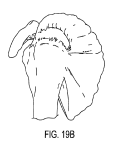
[00040] Figures 19A-B illustrate a humeral bone before and after a tear of a
ligament.
6
CA 02827404 2013-08-14
WO 2012/116319 PCT/US2012/026605
[00041] Figure 20 illustrates the use of sutures in the prior art to repair a
tendon with bone
anchors inserted into a surface of the humeral head.
[00042] Figure 21 illustrates a subsequent step for applying a net to
reinforce the tendon
using sutures to reinforce a tear in the tendon illustrated in Figure 20.
DETAILED DESCRIPTION
[00043] This detailed description provides examples that should not be
interpreted as
limiting the scope and breadth of the appended claims. The features of these
examples may be
combined and arranged as recited in the claims, notwithstanding the particular
examples
provided.
[00044] Figure lA illustrates a retention end of one example of a transosseous
suture net.
Such a net may be used to prevent the suture net from pulling through the
transosseous channel
drilled into a bone, for example. The net 61 may be comprised of a woven
fabric, such as the
fabric illustrated in Figure 5, which shows a fabric 51 comprises of strands,
which may be a solid
or spun strands, for example. Alternatively, the mesh 61 may be comprised of a
material having
holes 69, which may be formed as part of the process of fabrication of the
mesh, such as by a die
injection, hot forming, forging, machining, stamping, etching or other
processes that result in a
continuous material with holes 69 in the mesh 61, as illustrated in Figure 6,
for example. In the
example in Figure 1A, the mesh 61 is a seamless tubular mesh. In an
alternative embodiment, the
mesh 61 may be formed as a tape or sheet, for example.
[00045] Figure 1B illustrates a detail view and partial cross section of a
portion of the
retention end of Figure 1A, for example. In this example, the retention end 11
comprises a
portion of the mesh 61, which does not comprise holes 69, that is wrapped and
fused about a
retention ring 14, which may be made of a metal, a more rigid polymer, a
ceramic or another
material that tends to prevent the retention end 11 from being drawn through a
transosseous
channel drilled through a bone. Figure 9 illustrates use of such a device with
the retention end
11 being used to place the end of a transosseus suture net fabric 51 at a
surface of a patient's
bone. Several attachment points 70, 82 are illustrated in Figure 1A, such as a
tab 70 having a
fastening hole 75 or holes or an attached suture 80 with a retention end 82
and a suture end 81
7
CA 02827404 2013-08-14
WO 2012/116319 PCT/US2012/026605
having an optionally integrated suture needle, for example. The tabs 70 or
sutures 82 may be
attached to the retention end 11, such as by a retention mechanism 73, 83
having a hole 74, 84
through which the retention ring 14 passes, as illustrated in the detailed
views of Figures 7 and 8,
for example. Figure 1C illustrates an example of a suture end 81 on an
opposite end of a
transosseous suture net 61, for example. This suture end 81 may be inserted
through a
transosseous tunnel from either end of the tunnel and may be used for fixing
tissues to a patient's
bone, for example. For example, a suture 63 incorporating a suture end 81 may
be integrally
formed with and/or from a material of the mesh 61 as illustrated in Figure 1C.
In the example of
Figure 1D, examples of suture ends 81, 45 are illustrated, one having a suture
needle 81 and the
other having a hole 45 formed in the end. The suture 63 may have raised
portions 43 formed
along the length of the suture 63, which allows the suture to be adjustably
retained, for example.
[00046] Figures 1E-1F illustrate alternative examples of an end portion of a
suture 473,
263, 471, presenting a suture with an adjustable ratchet mechanism as part of
a suture. In Figure
1E, retention balls 43 are connected by ligands 41 to each other and to a
suture 47, 471, and the
retention balls 43 may be made of a material that is crosslinkable in situ
with a retention tab 70,
for example. Figure 1F illustrates a detail view of another example of a
portion of a suture
extending from a suture end, providing a ratchet surface 140 extending from a
suture 163 and a
tapered surface 141 preferably for a one-way ratcheting mechanism that resists
or prevents a
suture from pulling back through a retention tab 70 or a hole in a tissue, for
example, while
allowing the suture to pull through the tab 70 in order to tighten the suture
and/or to fix a tissue
in place. Figure 1G illustrates an alternative ratchet mechanism the presents
a flat form factor
with a raised portion 240 and a tapered portion 243 extending between a slot,
slit or hole 245 and
a tape-like suture 263. Any number or length of raised portions may be
provided at or before
and end of a suture to allow for adjustable retention of the suture in a tab
or tissue, for example.
[00047] Figure 2A illustrates an example of a monolithic retention end 20 and
the use of
sutures in one example of a suture end 33, 43 in combination with features of
the retention end
20, such as tabs 32, 42. Figure 2B illustrates an alternative example of the
use of sutures in one
example of a suture end 23 in combination with an alternative feature of a
retention end, having a
pair of holes in each tab 22. Figures 3A and 3B illustrate an example of a
cross section of a tab
32 and a suture 33 as illustrated in Figures 2A and 2B, while Figures 4A and
4B illustrate an
8
CA 02827404 2013-08-14
WO 2012/116319 PCT/US2012/026605
alternative example of a suture tab 42 engaging a ball in a suture 43, which
may be made of a
polymer crosslinkable with the material of the tab 42, for example, such as
using a chemical
reaction or an ultraviolet reaction to initiate or increase the rate of a
crosslinldng reaction, for
example.
[00048] Figure 9 illustrates an example of a transosseous suture net being
used to repair
damage to a tendon or ligament in an exemplary method. In this example, a
tendon
(supraspinatus M.) is being secured in place by a suture net 51 passing
through a transosseus
channel formed through the humeral head. The structure of the suture net 51
allows the surgeon
to pull the suture net 51 through the channel from the lower portion of the
channel to the upper
portion of the channel. Then, the suture needles 81 may be used to draw the
sutures 47 through
the tendon and through a retention mechanism, such as the tabs 70 located in
the retention end 11
of the suture net 51, for example. In this example, balls 43 assist the
surgeon to adjust the
tension on the sutures in discreet increments by pulling the balls 43 through
holes in the tabs 70.
Surprisingly, one or more of these devices provides for rapid attachment and
adjustment of the
fixation devices and substantially prevent suture pull through, which can lead
to normal sutures
working or cutting their way through the a portion of the bone that defines
the transosseus
channel.
[00049] Figure 10 illustrates a mesh 2101 having one or more sutures 2107
attached to a
reinforced portion 2103 at an anchor point 2109. Each of the sutures 2107 may
include a portion
comprising raised portions 2143, such as beads, balls or the like. A lead 2145
may extend from
the portion comprising raised portions. For example, the mesh may comprise a
tape that includes
one end 2103 that may be folded-over and joined together, such as by fusing,
welding, stitching
or the like. For example, the material may be a heat-processable material that
can be fused
together by heating, such as with the application of pressure during the
heating process or,
alternatively, without the application of such pressure. For example, the mesh
may be made of a
polyester, ultrahigh molecular weight polyethylene or the like. Herein,
ultrahigh molecular
weight is defined as a molecular weight greater than 300,000. In one example,
the ultrahigh
molecular weight polyethylene is covalently crosslinked. The additional
stiffness and strength
may provide a material that requires no tabs for retaining devices 2043, such
as beads, balls or
ratchet surfaces, formed or added to the ends of the sutures for temporarily
or permanently fixing
9
CA 02827404 2013-08-14
WO 2012/116319 PCT/US2012/026605
the sutures to a mesh tape 2001, such as with crosslinking caused by a
chemical reaction or
radiation or without such crosslinking.
[00050] The sutures 2107 extending from a mesh may be made of the same
material as the
mesh or may be made of a different material and may be joined to the mesh by a
surgeon or a
technician or may be pre-affixed at one or more anchor points. In one example,
an anchor point
may comprise one or more holes formed in the reinforced end, allowing a suture
to be tied to the
reinforced region by knotting one end of a suture through one or more of the
holes. Beads, balls,
ratchet surfaces and the like may be made integrally with the sutures or may
be added, such as by
fusing these features onto a length of suture, for example. Welding, bonding,
adhering or press
fitting may be used to add these retention or ratchet structures to the line
of a suture, for example.
[00051] In one example, a clip 2041, such as illustrated in Figures 11A-C, may
be added
to aid in retention of a retaining device 2043, such as illustrated in Figures
10, 12A and 1B, for
example. A tape 2101 may be disposed during surgery between a rotator cuff
tendon and the
skin, as illustrated in the process shown in Figures 14-18, for example.
Sutures 2107 may be
joined at one end to an end 2103 of the mesh tape and at an opposite end of
the sutures 2107 to a
different location of the mesh after passing the sutures through a bone anchor
and/or the patients
tissues, such as a bone tunnel, muscle and tendons, for example. A clip 2041
may be disposed
between a mesh 2001 and the skin, as illustrated in the detailed view of
Figure 11C, for example.
A low-profile clip may be used for a retaining device 2043 that is reduced in
diameter or shaped
as an oblate sphere 2045, disk 2047 or the like, as illustrated in Figures 12A-
B, for example. In
Figures 11A-C, a clip 2041 engages a retention portion 2043 of a suture, which
retention portion
2043 is inserted into a conformingly-shaped recess of the clip 2041 defined by
a plurality of
prongs 2042. A tapered portion 2048 of each prong 2042 of a clip 2041 helps to
spread the
prongs as a force is applied to insert a spherically-shaped retaining device
within the clip.
[00052] In the example of Figure 18, a tool 2500 includes a handle 2501
removably
attachable at one end 2505 to a hole 2042. For example, the hole 2042 may
comprise threads
2044, and the tool 2500 may be threadingly engaged to the clip 2041. While the
retention
portion of the suture may be inserted through a tension by pressure of a stiff
end of the suture or
using a needle, a punch or the like to provide a hole in the tendon through
which the retention
CA 02827404 2013-08-14
WO 2012/116319 PCT/US2012/026605
portion is passed, reversibly or irreversibly, depending on the shape of the
retention portion. For
example, the retention portion may adjustably retain the suture in the tendon,
allowing the user to
apply a desired tension on the mesh or net. A clip may prevent the raised
retention portion from
being pulled back through the mesh, tape, net and / or tendon, as illustrated
in Figures 11C and
18, for example. For example, using the tool 2500 in a minimally invasive
procedure, the clip
may be engaged onto the retention portion, when the retention portion is
properly tensioned, and
the tool may be withdrawn by unthreading the tool from the threaded hole in
the clip.
[00053] The process for using a tape 2101 may be combined with a suture mesh
61 used
as an anchor or with other anchors. If used with a suture mesh 61, the sutures
of the suture mesh
61 may be inserted through the patient's tissue and through the tape 2101 to
secure the patient's
tissue between the bone and the tape 2101, for example. In the alternative or
in addition to
suture mesh 61 anchors, other bone anchors may be used or sutures with balls,
beads or ratchet
surfaces may be inserted directly through transosseous channels. Surprisingly,
the balls, beads or
ratchet surfaces may be capable of reducing or preventing undesired pull
through (i.e. cutting
through) of the suture in the transosseus channel, if the balls, beads or
ratchet surfaced retaining
devices extend along the portion of the suture that extends through the
transosseus channel,
allowing the surgeon to pass a suture 2107 through the channel without use of
a suture mesh 61,
for example.
[00054] The mesh, sutures and/or clips may be made of non-bioadsorbable
materials such
as a polyester, ultrahigh molecular weight polyethylene or the like or a
bioabsorbable material,
such as a polylactide (i.e. polylactic acid based polymers), polyglycolide
(i.e. polyglycolic acid
based polymers) or other biocompatible and absorbable polymers that break down
and are
absorbed over time (or combinations of these). In one example, glycolide-based
copolyesters
have aliphatic polyester based co-monomers or non-aliphatic polyester based co-
monomers. For
example, linear aliphatic polyesters, such as lactices, carbonates and epsilon
caprolactones may
be used or poly-p-dioxanone may be used with or without copolymerized
radiostabilizers. In one
example, a trimethylene carbonate or a may be used. A gamma sterilizable
biocompatible and
absorbable polymer may be used, such as a poly(ethylene 1,4-phenylene-bis-
oxyacetate).
Polylactides, such as poly-L-lactide (PLLA) may be used with or without
copolymerization with
aliphatic polycarbonates such as trimethylene carbonate and with or without
the presence of self-
11
CA 02827404 2013-08-14
WO 2012/116319 PCT/US2012/026605
eluting agents such as to manage local pH during degradation, reduce
inflammatory responses or
to prevent infections, for example. Polymers including a cyclic diester or
poly(ethylene oxide)
may be used in order to modulate hydrophilicity, for example. A poly(ester-
amide) may be
selected with film forming properties to form a mesh tape, for example, which
may be degraded
by enzyme and or nonenzymatic mechanisms. In one example, the material is a
biologic, such as
a poly-4-hydroxybutyrate, i.e. is biologically-derived or harvested from
bacteriological or plant
processes.
[00055] In Figures 14-18, an example of a process for using a mesh, such as a
tape, is
illustrated. A boring device 2110, such as drill, bores a hole through a bone,
which may be bored
as illustrated or may be bored in the opposite direction. In one example, a
curved channel is
bored to provide an arcuate transosseous tunnel. The end of the boring device
or another device
may be used to draw a snare 2230 through the tunnel formed in the bone, and
the snare may be
inserted through a hole formed in a tendon, as illustrated in the example of
Figure 15, for
example, while a gripping device 2220 pulls a portion of the tendon 2221 into
position on the
humerus 2200. An opposite end 2231 of the snare 2230 extends out of the
opposite side of the
tunnel in the bone, allowing the surgeon to draw an end 2145 of one or more
sutures through the
tunnel in the bone using the snare 2230, as illustrated in Figures 16 and 17,
for example. In
Figure 16, the mesh, net or tape 2250 is rolled in order for it to fit through
a trochar, endoscope
or other device being used in a minimally invasive procedure. In Figure 17,
the mesh, net or tape
2250 is unrolled and is pulled into position by the snares. A suture is
anchored to the mesh at
one end 2109. In Figure 18, the mesh, net or tape is shown conformingly fit
over a portion of the
tendon and a portion of the bone, with an anchor point 2255 anchoring the
suture on one end to
the mesh, net or tape such as by forming a knot or stitch and at the other end
the raised retention
portion of the suture and the clip retain the mesh, net or tape in position
and adjustably apply a
tension on the mesh, net or tape. in one alternative, as illustrated in Figure
17, holes 2111 may
be provided as an anchor point for one or more sutures to be anchored to the
mesh, net or tape.
A tension may be applied up to one-half of the mean or median tensile force
applied to the
repaired tendon under normal conditions, for example. By retaining the mesh,
net or tape in
place by one or more sutures in this manner, the suture mesh, net or tape may
secure the tendon
to the bone without removing all of the load on the tendon. A tension may be
selected by the
surgeon for optimal healing of the tendon repair, for example.
12
CA 02827404 2013-08-14
WO 2012/116319
PCT/US2012/026605
[00056] Unlike the prior art, the suture net, mesh or tape may be used without
bone
anchors, the net, mesh or tape may extend over a portion of the tendon and
over a portion of the
bone where the particular tendon does not extend. Furthermore, the mesh, net
or tape may
distribute the load on the tendon over a larger surface of the tendon, as
illustrated in Figure 18
than would be the case for discrete suture knots and bone anchors as
illustrated in the prior art
drawing of Figure 20, for example. Thus, the examples illustrate advantages
over the prior art
devices that fail to stimulate optimal healing and require greater time to
complete a repair and
potentially cause more damage to the repaired tendon.
13